melithiazol biosynthesis
TRANSCRIPT
Chemistry & Biology, Vol. 10, 939–952, October, 2003, 2003 Elsevier Science Ltd. All rights reserved. DOI 10.1016/j .chembiol .2003.09.012
Melithiazol Biosynthesis: Further Insightsinto Myxobacterial PKS/NRPS Systems and Evidencefor a New Subclass of Methyl Transferases
esting targets for genetic engineering and mutasyn-thesis strategies aimed at the production of novel andaltered structures, ideally giving rise to substances withimproved biological activities. The monomers used arecoenzyme A esters of short chain carboxylic acids in
Stefan Weinig,1,2 Hans-Jurgen Hecht,1 Taifo Mahmud,3
and Rolf Muller1,2,*1Gesellschaft fur Biotechnologische Forschung
mbH (GBF)Mascheroder Weg 138124 Braunschweig PKSs and amino acids in NRPSs. These precursors are2 Institut fur Pharmazeutische Biologie attached as thioesters to carrier proteins via a phospho-Mendelssohnstr. 1 pantetheinyl arm that needs to be transferred posttrans-Technische Universitat lationally to conserved serine residues in each carrier38106 Braunschweig protein [4]. The selection for each monomer is performedGermany by acyl transferase domains (AT domains in PKS) and3 University of Washington adenylation domains (A domains in NRPS). After loadingDepartment of Chemistry of the megasynthetase, the condensation reactions be-Seattle, Washington 98195 tween the monomers are catalyzed by ketosynthase
(KS) domains in PKS and by condensation (C) domainsin NRPS, which leads to the formation of carbon-carbonbonds by Claisen ester condensation and to peptideSummarylinkages, respectively.
During the last few years, hybrid systems employingA DNA region of 41.847 base pairs from MelittangiumPKS and NRPS biochemistry in the course of the assem-lichenicola Me l46 is shown to be responsible for thebly of one product have been reported [5–7], which re-biosynthesis of the potent electron transport inhibitorsulted in intensified studies aimed at the combinationmelithiazol. Melithiazol is formed by a combined poly-of PKS/NRPS assembly lines for combinatorial biosyn-ketide synthase/peptide synthetase system resemblingthesis [8–10].the myxothiazol megasynthetase from Stigmatella
Although relatively rarely studied in comparison toaurantiaca DW4/3-1. Both natural products share anactinomycetes, myxobacteria have been shown to be aalmost identical core region but employ different startermajor source of secondary metabolites, especially thosemolecules. Additionally, melithiazol contains a termi-
nal methyl ester instead of the amide moiety found that show the typical features of PKS/NRPS hybrids: thein myxothiazol. Similar to myxothiazol formation, the incorporation of amino acids and short chain carboxylicmethyl ester is formed via an amide intermediate, which acids into their backbones [11]. Cloning of the responsi-is converted by a hydrolase and an unusual type of ble genes revealed biosynthetic enzymes that harborSAM (S-adenosyl-L-methionine)-dependent methyl- PKS and NRPS modules, even present on one singletransferase into the methyl ester. When transferred open reading frame [12, 13], and a variety of other novelinto the myxothiazol A (amide) producer, these two features have been described from multimodular myxo-genes lead to the formation of the methyl ester of bacterial systems [14–19]. Cyanobacteria seem to bemyxothiazol. The methyl transferase described is a equally rich in “nonstandard” biosynthetic systems, asmember of a protein subfamily of a previously un- has been shown in recent genetic studies [20–22].known function lacking a typical SAM binding motif. Myxothiazol [23, 24] and melithiazol [25, 26] represent
two highly efficient electron transport inhibitors of theeukaryotic respiratory chain which are produced by theIntroductionmyxobacteria Stigmatella aurantiaca DW4/3-1 and Mel-ittangium lichenicola Me l46, respectively. These com-Polyketide synthases (PKSs) and nonribosomal peptide
synthetases (NRPSs) are important enzymatic systems pounds are similar, but some significant biosyntheticwhich are responsible for the formation of an immense differences can be expected due to structural differ-variety of natural products [1–3]. Starting from simple ences (see Figure 1). The myxothiazol hybrid PKS/NRPSbuilding blocks, such as short chain carboxylic acids biosynthetic gene cluster was analyzed mainly basedand amino acids, secondary metabolites with various on in silico analysis of the corresponding biosyntheticbiological activities are assembled in microorganisms. The genes [12].resulting low molecular weight polyketides and polypep- In order to explore the structural diversity and thetides are widely used by the pharmaceutical and agro- underlying biosynthetic mechanisms as well as the evo-chemical industry (e.g., erythromycin, rapamycin, FK506, lutionary origin of the biosynthetic machineries, wecyclosporin, avermectin, lovastatin, and epothilone). cloned and analyzed the melithiazol gene cluster. Bio-
Both PKSs and NRPSs are characterized as large, synthetic origins of the unusual isobutyrate starter unitmultifunctional, and modular enzyme complexes which and the terminal methyl ester in melithiazol as well asassemble secondary metabolites in a stepwise fashion. the terminal amide in myxothiazol were analyzed usingTheir corresponding biosynthetic machineries are inter- classical feeding experiments. Additionally, by heterolo-
gous expression a novel type of SAM-dependent methyltransferase and a hydrolase are shown to be involved*Correspondence: [email protected]
Chemistry & Biology940
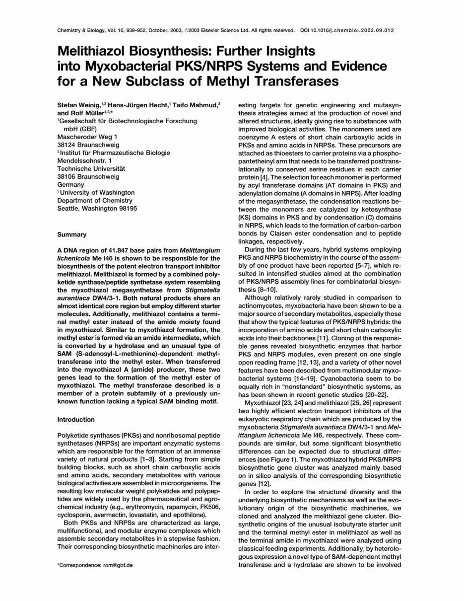
Figure 1. Examples for Myxobacterial Secondary Metabolites which Are Biosynthesized by PKS and NRPS
in the methyl ester formation starting from an amide aurantiaca DW4/3-1, M. lichenicola Me l46 uses onlyone secondary metabolic Ppant transferase, which mustintermediate.be encoded outside of the melithiazol biosynthetic genecluster.Results and Discussion
Analysis of the modular structure of the melithiazolmegasynthetase revealed that melB most likely encodesCloning and Analysis of the Melithiazol
Biosynthetic Gene Cluster the first module of the biosynthetic gene cluster (seebelow). The modular organization of PKSs and NRPSsDue to the structural similarity of myxothiazol and meli-
thiazol, the genes involved in their biosynthesis were involves activation and condensation of the followingcarboxylic acid or amino acid onto the growing chainexpected to be similar in sequence. In Southern blot
experiments, it could be shown that genomic DNA of M. catalyzed by an acyl-transferase (AT) domain and a KSdomain (PKS) or an adenylation (A) and a condensationlichenicola Me l46 hybridized under stringent conditions
with mtaE and mtaF gene probes. A cosmid library of (C) domain (NRPS). The resulting �-keto acid (oligopep-tide in NRPS) may subsequently be processed by �-keto-M. lichenicola Me l46 was prepared and used for colony
hybridzation experiments, resulting in the identification acyl-reductase (KR) domains, �-hydroxy-acyl-dehydra-tase (DH) domains, and enoyl-reductase (ER) domainsof a cosmid (M1) that gave positive signals with both
probes. End sequences from cosmid M1 (T3 and the T7 or N-methyl transferase (N-MT) and epimerase (E) do-mains in NRPS (reviewed in [1]). Additional domains forends) were compared to the myxothiazol gene cluster,
revealing M1 to harbor a fragment of the melithiazol C-methylation [29, 30] and O-methylation [12, 18] ofintermediates have recently been reported, and thegene cluster starting with the AT domain of the mtaE
analog melE (see Figure 2A). Using oligonucleotides de- function of heterocyclization (HC) and oxidation (Ox)domains involved in the formation of oxazoline and thia-signed to bind to this end sequence, cosmid pools of
the gene library were screened for the presence of over- zoline rings and their subsequent oxidation to oxazoleand thiazole structures has been elucidated [3, 31, 32].lapping cosmids [12]. These were subjected to end se-
quencing, and the derived sequences were used to lo- Sequence motifs typical for PKS domains [33, 34] andNRPS domains [3, 35] were detected in MelB-G (Tablecate the rest of the melithiazol gene cluster on cosmid
M2 (M2 ends in the AT of the mtaF analog melF). Cos- 1 and Figure 2B). The acyl carrier protein domains (ACP)and the peptidyl carrier protein (PCP) domain of MelB-Gmids M1 and M2 were sequenced and revealed the
presence of several open reading frames (ORFs) with contain the Prosite consensus signature of the putativebinding site for the 4�-phosphopantetheine (Ppant) co-similarity to PKSs and NRPS, which were designated
melB-K (melithiazol; see Figure 2A). No gene encoding factor (Prosite signature numbers PS00012, R2082, andL2104). The codon bias of the genes reported is in accor-a phosphopantetheinyl (Ppant)-transferase similar to
mtaA was found. MtaA was shown to have a broad dance with other genes from myxobacteria [36]. Theoverall G�C content of the mel-biosynthetic gene clus-substrate specificity [27] and is required for the produc-
tion of myxothiazol [12] and at least two additional PKS ter, which spans approximately 42 kbp, is 69.7%.Eight further ORFs were identified upstream and down-and PKS/NRPS hybrid compounds produced by S. aura-
ntiaca DW4/3-1 (G. Hofle, S. Wenzel, H. Bode, and R.M., stream of the melithiazol PKS/NRPS genes. Based onthe sequence similarities of the encoded proteins tounpublished data; [28]). It is thus likely that similar to S.
Melithiazol and Myxothiazol Biosynthesis941
Figure 2. The mta and mel Biosynthetic Gene Clusters
(A) Comparison of gene arrangements in the mta and mel biosynthetic gene clusters. Arrows indicate the direction of transcription of eachgene. The checkered arrow symbolizes mtaA encoding a phosphopantetheinyl transferase. PKS modules are shown in green, NRPS modulesare shown in red, and ORFs with unknown function are depicted in white. Hatched arrows indicate genes encoding modifying enzymes. PKS,polyketide synthase; NRPS, nonribosomal peptide synthetase.(B) Models of myxothiazol and melithiazol biosynthesis. PKS modules are colored in green, NRPS modules are shown in red, and unusualdomains are shown in yellow. Domains depicted in light green are assumed to be derived from each other (see text). The TE domain is shownin blue. Building blocks incorporated by PKS modules are depicted with green bonds, whereas molecule moieties assembled by NRPSmodules are shown with red bonds. ACP, acyl carrier protein; KS, �-ketoacyl-ACP synthase; KR, �-ketoacyl-ACP reductase; AT, acyl transferase;DH, �-hydroxy-acyl-ACP dehydratase; O-MT, O-methyl transferase; ER, enoyl reduktase; S, spacer region; PCP, peptidyl carrier protein; C,condensation domain; HC, heterocyclization domain; A, adenylation domain; Ox, oxidation domain; MOX, monooxygenase domain; TE,thioesterase.
Chemistry & Biology942
Table 1. Deduced Functions of ORFs in the Melithiazol Biosynthetic Gene Cluster
A. PKS and NRPS Parts of the Gene Cluster
Proposed FunctionProtein (Gene) Size (bp/Da) (Protein Domains with Their Position in the Sequence)
MelB (melB) 3,153 bp, PKS domains: KS (16-443), AT (555-870), ACP (962-1028)111,621 Da
MelC (melC) 3,972 bp, NRPS domains: HC (52-452), A (538-1001), PCP (1025-1093) Ox (1025-1093)146,206 Da
MelD (melD) 9,855 bp, NRPS domains: HC (74-473), A (560-1291), Ox (989-1241), PCP (1319-1383); PKS domains:357,430 Da KS (1447-1874), AT (1981-2296), DH (2307-2521), S (2612-2849), KR (2882-3134),
ACP (3157-3226)MelE (melE) 5,739 bp, PKS domains: KS (35-429), AT (573-890), O-MT (972-1246), S (1252-1454), KR (1486-1746),
206,932 Da ACP (1783-1849)MelF (melF) 4,083 bp, PKS domains: KS (39-464), AT (571-886), O-MT (946-1221), ACP (1272-1338)
148,434 DaMelG (melG) 5,247 bp, NRPS domains: C (68-367), A (555-1380), MonoOx (753-1114), PCP (1401-1469), TE (1488-1748)
192,469 Da
B. ORFs Encoded Upstream and Downstream of melB-melG
Sequence Similarity toProposed Function of the (Source Plus Additional Accession No. of the
Protein (Gene) Size (bp/Da) Similar Protein Available Information) Similarity/Identity Similar Protein
ORF1 (orf1) 699 bp, Similar to glycosyl trans- Archaeoglobus 40%/28% C6929824,529 Da ferases fulgidus
ORF2 (orf2) 1,359 bp, L-amino acid oxidase Crotalus adamanteus 70%/51% JE026648,004 Da
ORF3 (orf3) 963 bp, Nucleotide sugar Escherichia coli WbnF 64%/48% AAD5049435,377 Da epimerase
ORF4 (orf4) 282 bp, Glutaredoxin Yersinia pestis GrxC 59%/45% NP_40373010,290 Da
MelH (melH) 990 bp, 2-hydroxyhepta-2,4- Methanothermobacter 56%/40% A6906836,189 Da diene-1,7-dioate- thermautotrophicus
isomerase MTH1507MelJ (melJ) 1,088 bp, Nitrilase Arabidopsis thaliana 39%/26% P46010
37,051 Da NIT3MelK (melK) 906 bp, Methyl transferase Actinosynnema pretio- 41%/28% AF453501.1
33,612 bp sum Asm10ORF7 (orf7) 1,788 bp ABC-transporter Nostoc punctiforme 61%/44% ZP_00109759
64,332 Da Npun4209
proteins from the databases, ORF2, MelJ, and MelK activated substrate to the first ACP domain (comparethe biosynthetic gene clusters of erythromycin [33], ra-seemed to be involved in the biosynthesis of melithiazol
(see Table 1 and Figures 2A and 4). The gene encoding pamycin [5, 34], and rifamycin [6]). Alternative starterscan be used to initiate the biosynthesis of the polyke-MelH is located directly behind melG, which is similar
to the gene order in the mta gene cluster. Nevertheless, tides, but most frequently acetyl-CoA, malonyl-CoA,propionyl-CoA, or methylmalonyl-CoA are employed. IfmelH and mtaH do not seem to be required for melithia-
zol and myxothiazol biosynthesis (see the accompa- activated dicarboxylic acids are used, modified KS do-mains can be found at the beginning of the first module.nying manuscript [65] for a discussion of the putative
function of melH). These have lost their condensation activity but effec-tively decarboxylate the ACP-bound dicarboxylic acid,giving rise to the starter moiety. Because the active siteModel for the Melithiazol Biosynthetic Pathway
Here it is demonstrated that the melithiazol biosynthetic cysteine of these KS domains is mutated to glutamine,they have been designated KSQ domains [37]. In themachinery is very similar to the myxothiazol system and
belongs to the class of hybrid biosynthetic systems case of melB, the modular organization looks similar:the protein starts with a KS domain that is followed bycomposed of PKS and NRPS modules. The biosynthesis
switches from PKS type biochemistry (MelB) to NRPS an AT and an ACP domain.Several myxobacterial PKSs show an atypically ar-(MelCD), back to PKS (MelDEF), and finally back to
NRPS (MelG). MelD belongs to the few known proteins ranged starter module with ACP-KS-AT-AT-KR-ACP[12, 18, 19, 38]. In these systems the first AT loads thein which a NRPS module is covalently linked to a PKS
module, which makes it an ideal target to study PKS/ starter molecule, whereas the second AT loads the firstextender unit ([39]; P. Leadlay, C. Wilkinson, S.W., andNRPS interaction.
The modular structure of type I PKSs usually starts R.M., unpublished data). M. lichenicola Me l46 producesonly melithiazol A (and melithiazol C, which is presum-with an AT or a CoA-ligase domain responsible for the
recognition (and, in the case of CoA-ligases, for activa- ably a degradation product), which is characterized bya dehydro-isobutyrate starter molecule [26]. Myxobac-tion) of the starter molecule followed by transfer of the
Melithiazol and Myxothiazol Biosynthesis943
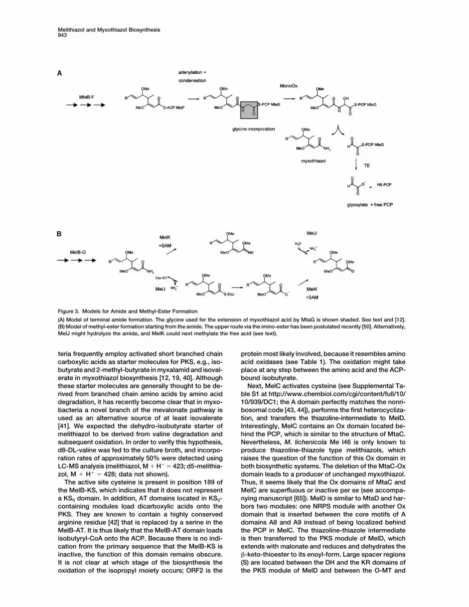
Figure 3. Models for Amide and Methyl-Ester Formation
(A) Model of terminal amide formation. The glycine used for the extension of myxothiazol acid by MtaG is shown shaded. See text and [12].(B) Model of methyl-ester formation starting from the amide. The upper route via the imino-ester has been postulated recently [50]. Alternatively,MelJ might hydrolyze the amide, and MelK could next methylate the free acid (see text).
teria frequently employ activated short branched chain protein most likely involved, because it resembles aminoacid oxidases (see Table 1). The oxidation might takecarboxylic acids as starter molecules for PKS, e.g., iso-
butyrate and 2-methyl-butyrate in myxalamid and isoval- place at any step between the amino acid and the ACP-bound isobutyrate.erate in myxothiazol biosynthesis [12, 19, 40]. Although
these starter molecules are generally thought to be de- Next, MelC activates cysteine (see Supplemental Ta-ble S1 at http://www.chembiol.com/cgi/content/full/10/rived from branched chain amino acids by amino acid
degradation, it has recently become clear that in myxo- 10/939/DC1; the A domain perfectly matches the nonri-bosomal code [43, 44]), performs the first heterocycliza-bacteria a novel branch of the mevalonate pathway is
used as an alternative source of at least isovalerate tion, and transfers the thiazoline-intermediate to MelD.Interestingly, MelC contains an Ox domain located be-[41]. We expected the dehydro-isobutyrate starter of
melithiazol to be derived from valine degradation and hind the PCP, which is similar to the structure of MtaC.Nevertheless, M. lichenicola Me l46 is only known tosubsequent oxidation. In order to verify this hypothesis,
d8-DL-valine was fed to the culture broth, and incorpo- produce thiazoline-thiazole type melithiazols, whichraises the question of the function of this Ox domain inration rates of approximately 50% were detected using
LC-MS analysis (melithiazol, M � H� � 423; d5-melithia- both biosynthetic systems. The deletion of the MtaC-Oxdomain leads to a producer of unchanged myxothiazol.zol, M � H� � 428; data not shown).
The active site cysteine is present in position 189 of Thus, it seems likely that the Ox domains of MtaC andMelC are superfluous or inactive per se (see accompa-the MelB-KS, which indicates that it does not represent
a KSQ domain. In addition, AT domains located in KSQ- nying manuscript [65]). MelD is similar to MtaD and har-bors two modules: one NRPS module with another Oxcontaining modules load dicarboxylic acids onto the
PKS. They are known to contain a highly conserved domain that is inserted between the core motifs of Adomains A8 and A9 instead of being localized behindarginine residue [42] that is replaced by a serine in the
MelB-AT. It is thus likely that the MelB-AT domain loads the PCP in MelC. The thiazoline-thiazole intermediateis then transferred to the PKS module of MelD, whichisobutyryl-CoA onto the ACP. Because there is no indi-
cation from the primary sequence that the MelB-KS is extends with malonate and reduces and dehydrates the�-keto-thioester to its enoyl-form. Large spacer regionsinactive, the function of this domain remains obscure.
It is not clear at which stage of the biosynthesis the (S) are located between the DH and the KR domains ofthe PKS module of MelD and between the O-MT andoxidation of the isopropyl moiety occurs; ORF2 is the
Chemistry & Biology944
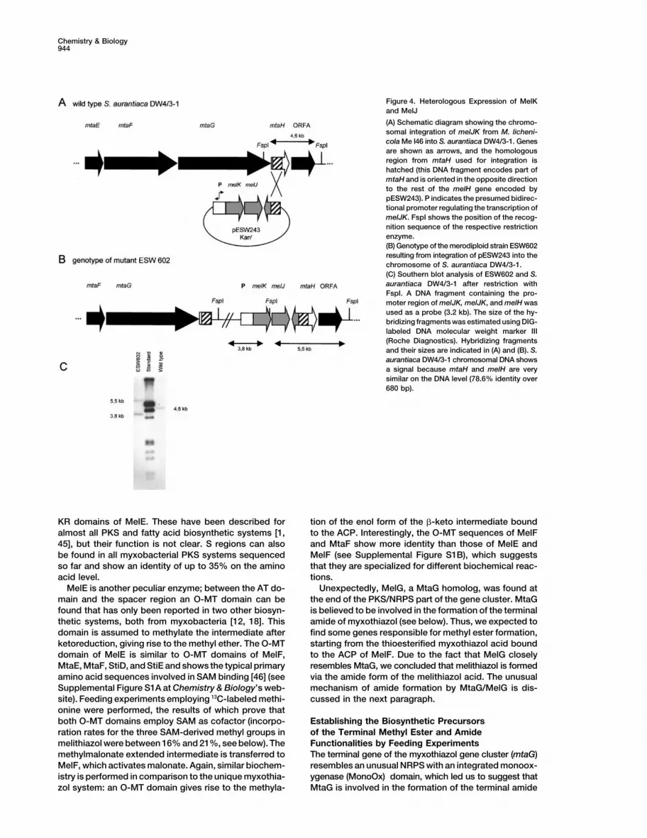
Figure 4. Heterologous Expression of MelKand MelJ
(A) Schematic diagram showing the chromo-somal integration of melJK from M. licheni-cola Me l46 into S. aurantiaca DW4/3-1. Genesare shown as arrows, and the homologousregion from mtaH used for integration ishatched (this DNA fragment encodes part ofmtaH and is oriented in the opposite directionto the rest of the melH gene encoded bypESW243). P indicates the presumed bidirec-tional promoter regulating the transcription ofmelJK. FspI shows the position of the recog-nition sequence of the respective restrictionenzyme.(B) Genotype of the merodiploid strain ESW602resulting from integration of pESW243 into thechromosome of S. aurantiaca DW4/3-1.(C) Southern blot analysis of ESW602 and S.aurantiaca DW4/3-1 after restriction withFspI. A DNA fragment containing the pro-moter region of melJK, melJK, and melH wasused as a probe (3.2 kb). The size of the hy-bridizing fragments was estimated using DIG-labeled DNA molecular weight marker III(Roche Diagnostics). Hybridizing fragmentsand their sizes are indicated in (A) and (B). S.aurantiaca DW4/3-1 chromosomal DNA showsa signal because mtaH and melH are verysimilar on the DNA level (78.6% identity over680 bp).
KR domains of MelE. These have been described for tion of the enol form of the �-keto intermediate boundto the ACP. Interestingly, the O-MT sequences of MelFalmost all PKS and fatty acid biosynthetic systems [1,
45], but their function is not clear. S regions can also and MtaF show more identity than those of MelE andMelF (see Supplemental Figure S1B), which suggestsbe found in all myxobacterial PKS systems sequenced
so far and show an identity of up to 35% on the amino that they are specialized for different biochemical reac-tions.acid level.
MelE is another peculiar enzyme; between the AT do- Unexpectedly, MelG, a MtaG homolog, was found atthe end of the PKS/NRPS part of the gene cluster. MtaGmain and the spacer region an O-MT domain can be
found that has only been reported in two other biosyn- is believed to be involved in the formation of the terminalamide of myxothiazol (see below). Thus, we expected tothetic systems, both from myxobacteria [12, 18]. This
domain is assumed to methylate the intermediate after find some genes responsible for methyl ester formation,starting from the thioesterified myxothiazol acid boundketoreduction, giving rise to the methyl ether. The O-MT
domain of MelE is similar to O-MT domains of MelF, to the ACP of MelF. Due to the fact that MelG closelyresembles MtaG, we concluded that melithiazol is formedMtaE, MtaF, StiD, and StiE and shows the typical primary
amino acid sequences involved in SAM binding [46] (see via the amide form of the melithiazol acid. The unusualmechanism of amide formation by MtaG/MelG is dis-Supplemental Figure S1A at Chemistry & Biology’s web-
site). Feeding experiments employing 13C-labeled methi- cussed in the next paragraph.onine were performed, the results of which prove thatboth O-MT domains employ SAM as cofactor (incorpo- Establishing the Biosynthetic Precursors
of the Terminal Methyl Ester and Amideration rates for the three SAM-derived methyl groups inmelithiazol were between 16% and 21%, see below). The Functionalities by Feeding Experiments
The terminal gene of the myxothiazol gene cluster (mtaG)methylmalonate extended intermediate is transferred toMelF, which activates malonate. Again, similar biochem- resembles an unusual NRPS with an integrated monoox-
ygenase (MonoOx) domain, which led us to suggest thatistry is performed in comparison to the unique myxothia-zol system: an O-MT domain gives rise to the methyla- MtaG is involved in the formation of the terminal amide
Melithiazol and Myxothiazol Biosynthesis945
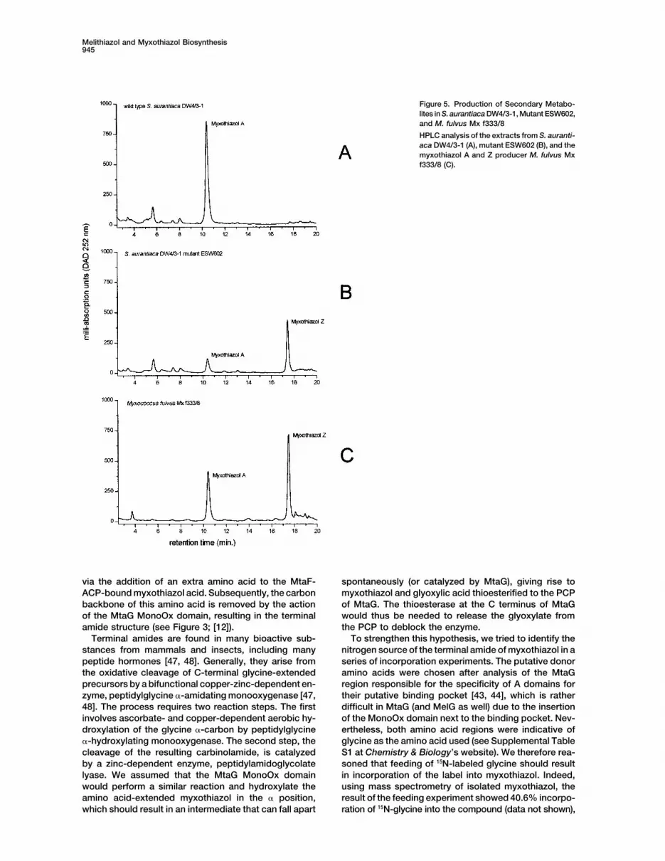
Figure 5. Production of Secondary Metabo-lites in S. aurantiaca DW4/3-1, Mutant ESW602,and M. fulvus Mx f333/8
HPLC analysis of the extracts from S. auranti-aca DW4/3-1 (A), mutant ESW602 (B), and themyxothiazol A and Z producer M. fulvus Mxf333/8 (C).
via the addition of an extra amino acid to the MtaF- spontaneously (or catalyzed by MtaG), giving rise tomyxothiazol and glyoxylic acid thioesterified to the PCPACP-bound myxothiazol acid. Subsequently, the carbon
backbone of this amino acid is removed by the action of MtaG. The thioesterase at the C terminus of MtaGwould thus be needed to release the glyoxylate fromof the MtaG MonoOx domain, resulting in the terminal
amide structure (see Figure 3; [12]). the PCP to deblock the enzyme.To strengthen this hypothesis, we tried to identify theTerminal amides are found in many bioactive sub-
stances from mammals and insects, including many nitrogen source of the terminal amide of myxothiazol in aseries of incorporation experiments. The putative donorpeptide hormones [47, 48]. Generally, they arise from
the oxidative cleavage of C-terminal glycine-extended amino acids were chosen after analysis of the MtaGregion responsible for the specificity of A domains forprecursors by a bifunctional copper-zinc-dependent en-
zyme, peptidylglycine �-amidating monooxygenase [47, their putative binding pocket [43, 44], which is ratherdifficult in MtaG (and MelG as well) due to the insertion48]. The process requires two reaction steps. The first
involves ascorbate- and copper-dependent aerobic hy- of the MonoOx domain next to the binding pocket. Nev-ertheless, both amino acid regions were indicative ofdroxylation of the glycine �-carbon by peptidylglycine
�-hydroxylating monooxygenase. The second step, the glycine as the amino acid used (see Supplemental TableS1 at Chemistry & Biology’s website). We therefore rea-cleavage of the resulting carbinolamide, is catalyzed
by a zinc-dependent enzyme, peptidylamidoglycolate soned that feeding of 15N-labeled glycine should resultin incorporation of the label into myxothiazol. Indeed,lyase. We assumed that the MtaG MonoOx domain
would perform a similar reaction and hydroxylate the using mass spectrometry of isolated myxothiazol, theresult of the feeding experiment showed 40.6% incorpo-amino acid-extended myxothiazol in the � position,
which should result in an intermediate that can fall apart ration of 15N-glycine into the compound (data not shown),

Chemistry & Biology946
Melithiazol and Myxothiazol Biosynthesis947
whereas 15N-labeled glutamate and ammonium chloride generation of the gene library, presumably due to thepresence of DNases, the strain showed resistancewere not incorporated at all. This suggests that the ter-
minal amide of myxothiazol is derived directly from an against almost every antibiotic tested (kanamycin, hy-gromycin, streptomycin, apramycin, and tetracyclin).extra glycine that is intermediary attached to the mole-
cule. Interestingly, serine has been shown to be the Few spontaneously resistant colonies were detectedusing ampicillin, which encouraged us to try a �-lacta-nitrogen source of the terminal amide of nosiheptide
[49], which indicates a similar mechanism for amide mase as marker gene in conjugation and electroporationexperiments aimed at the inactivation of melE. Becauseformation in the producer Streptomyces actuosus and
possibly for other natural products with terminal amide these experiments were not successful, we decided toheterologously express melithiazol biosynthetic genesmoieties as well (e.g., thiostreptone).
Currently, two more examples of NRPS with inserted in S. aurantiaca DW4/3-1 to unambigously prove thatthe right gene cluster was cloned.MonoOx domains can be found in the databases (hypo-
thetical proteins from Nostoc punctiforme and Ralsto- MelK and MelJ seemed to be responsible for the for-mation of the methyl ester. In addition, earlier studiesnia solanacearum which show the domain structure
A-MonoOx-PCP-KS-AT-ACP-Aminotransferase-C-A- employing M. fulvus Mx f333/8 suggested that myxothia-zol Z is derived from myxothiazol A via the iminoesterPCP… and …KS-AT-ACP-Aminotransferase-MonoOx-
C-A-PCP…, respectively). None of these proteins seems intermediate (see Figure 3, [50]). Given the structuralanalogy between myxothiazol Z and melithiazol A, weto be involved in terminal amide formation, because the
MonoOx domains are not within the C-terminal modules, speculated that melithiazol A is derived from a melithia-zol-amide analogously, which would employ the imi-nor are they located within an A domain. Nevertheless,
both proteins belong to the class of PKS/NRPS hybrids. noester of the amide as an intermediate (Figure 3). Nev-ertheless, the amide could not be identified from M.In contrast, the unusual arrangement of domains in
MtaG can be found in MelG as well (see Figure 2B). It lichenicola Me l46. Alternatively, one could speculatethat the methyl ester is formed similarly to the methyla-is thus reasoned that melithiazol in close biosynthetic
analogy is also formed via the amide intermediate. This tion of bacterial chemotaxis transmembrane receptors,e.g., by the protein methyl transferase CheR and thewould represent a unique mechanism to generate a
methyl ester, because the glycine extender has to be methyl esterase/amidase CheB from Salmonella typhi-murium [51, 52]. The alternative route would thus involveremoved completely in order to generate the ester. It
seemed likely that melithiazol is biosynthesized by a production of the free myxothiazol acid by MelJ, whichthen would have to be methylated (Figure 3). MelJ is amethyl transferase (which would presumably employ
SAM as cofactor) and a hydrolase. The gene products protein with all the hallmarks of the nitrilase superfamilyof proteins, which includes the amidases. The activeof melK and melJ, which are located downstream of
melG, indeed show some similarities to such enzymes site cysteine can be found in position 145 and the cata-lytic triad glutamate44-lysine112-cysteine145 is responsible(Table 1). To verify the dependence of the methyl ester
formation on a SAM-dependent enzyme, a feeding ex- for hydrolysis [53]. Both possibilities for methyl esterformation are currently under investigation by heterolo-periment employing [13CH3]methionine was performed
and showed incorporation rates of 16%–21% into the gous expression and purification of MelJ and MelK. Themechanisms could also be distinguished by feedingthree methoxy-goups of melithiazol A, which was de-
tected using 13C-NMR spectroscopy (data not shown). 13C/18O-labeled acetate. If the former is correct, 18O willonly be present in the methoxyl oxygen; if the latter isSimilar results have been obtained in feeding experi-
ments with Myxococcus fulvus Mx f333/8, the producer correct, then both the methoxyl and carbonyl oxygenswill be labeled so one and two isotope shifts, respec-of the methyl ester of myxothiazol A (designated myxo-
thiazol Z) (see Figure 1; [50]). tively, should be observed on the carbonyl carbon.In order to verify the involvement of MelK and MelJ in
methyl ester formation, both genes and their presumedHeterologous Expression of the PutativeMethyltransferase MelK and the Hydrolase promoter region were transferred into the myxothiazol
A producer S. aurantiaca DW4/3-1 (see Figure 4). ForMelJ from the Melithiazol Gene Clusterin the Myxothiazol A Producer homologous recombination, the DNA region of MtaH
was used because this gene is not required for myxothi-M. lichenicola Me l46 proved to be a difficult organismto work with genetically. After severe problems in the azol formation (see accompanying manuscript [65]).
Figure 6. Alignment of the MelK Subfamily of SAM-Dependent Methyl Transferases
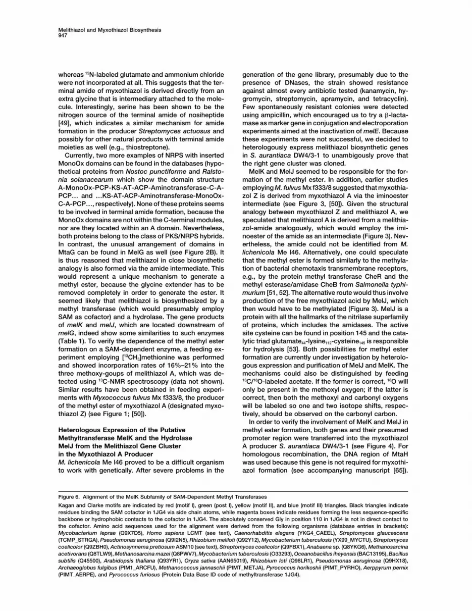
Kagan and Clarke motifs are indicated by red (motif I), green (post I), yellow (motif II), and blue (motif III) triangles. Black triangles indicateresidues binding the SAM cofactor in 1JG4 via side chain atoms, while magenta boxes indicate residues forming the less sequence-specificbackbone or hydrophobic contacts to the cofactor in 1JG4. The absolutely conserved Gly in position 110 in 1JG4 is not in direct contact tothe cofactor. Amino acid sequences used for the alignment were derived from the following organisms (database entries in brackets):Mycobacterium leprae (Q9X7D5), Homo sapiens LCMT (see text), Caenorhabditis elegans (YKG4_CAEEL), Streptomyces glaucescens(TCMP_STRGA), Pseudomonas aeruginosa (Q9I2N5), Rhizobium meliloti (Q92Y12), Mycobacterium tuberculosis (YX99_MYCTU), Streptomycescoelicolor (Q9ZBH0), Actinosynnema pretiosum ASM10 (see text), Streptomyces coelicolor (Q9FBX1), Anabaena sp. (Q8YKG6), Methanosarcinaacetivorans (Q8TLW9), Methanosarcina mazei (Q8PWV7), Mycobacterium tuberculosis (O33293), Oceanobacillus iheyensis (BAC13195), Bacillussubtilis (Q45500), Arabidopsis thaliana (Q93YR1), Oryza sativa (AAN65019), Rhizobium loti (Q98LR1), Pseudomonas aeruginosa (Q9HX18),Archaeoglobus fulgibus (PIM1_ARCFU), Methanococcus jannaschii (PIMT_METJA), Pyrococcus horikoshii (PIMT_PYRHO), Aerppyrum pernix(PIMT_AERPE), and Pyrococcus furiosus (Protein Data Base ID code of methyltransferase 1JG4).

Chemistry & Biology948
After transfer of both genes into the myxothiazol pro- conserved, which has already been observed by Martinand McMillan as common to the methyltransferases.ducer, the genotype of the mutant strain was analyzed
by Southern hybridizations verifying the correctness of The absolutely conserved Gly in position 110 in 1JG4 isnot in direct contact with the cofactor, but on one sidethe integration in mutant ESW602 (Figure 4). HPLC-MS
analysis of ESW602 culture broth revealed the presence of the binding pocket. It is located in a position whereany amino acid side chain would interfere with the cofac-of myxothiazol A and myxothiazol Z (see Figure 5; LC-
MS analysis: myxothiazol A, M � H� � 503; myxothiazol tor sulfur atom. Thus, it can be expected that MelK, asAsm10 and LCMT, binds the SAM-cofactor similarly toZ, M � H� � 488), which clearly establishes that MelK
and MelJ are responsible for the transformation of the the other methyltransferases and contains the typicalSAM binding fold.�-methoxyacrylate amide into the �-methoxyacrylate
ester.
Evolutionary Aspects of Melithiazoland Myxothiazol BiosynthesisMelK Is a SAM-Dependent Methyl TransferaseIt is interesting to note that the myxothiazol gene clusterwith Poorly Conserved SAM Binding Sitemust have evolved from the melithiazol gene cluster orSearches for MelK homologs in Swall using Fasta3 [54]vice versa, which can be shown by sequence compari-gave 41 related proteins, mostly from mycobacteria,son (Figure 2); MelC-H and MtaC-H are highly homolo-streptomyces, and methanosarcina with unknown func-gous (the encoding genes are very similar in sequencetion, as well as the methyltransferase Asm10 from theas well; see Figure 2) and are presumed to have similarActinosynnema pretiosum ansamitocin biosyntheticfunctions in melithiazol and myxothiazol biosynthesis.gene cluster [55]. Asm10 is the only biochemically char-Even the large identical repeat within the AT of mtaDacterized member of this group of proteins. The recom-and mtaF (1223 bp, 99.7% identity) is found in melD andbinant enzyme catalyzes the final step in ansamitocinmelF as well (1222 bp, 99.1% identity). Unexpectedly,biosynthesis, the SAM-dependent N-methylation of thewhen the mel and mta repeat regions are compared,amide linkage (G. Shang, P. Spiteller, L. Bai, B.J. Carroll,they are not identical and do not show higher similaritiesT.-W. Yu, and H.G. Floss, personal communication). Nowith each other than the complete genes. MtaB andsignificant similarities to structurally known SAM-depen-MelB are involved in the formation of the PKS starterdent methyl transferases specific either for small mole-molecule, which is different in both biosyntheses. Ac-cule, protein, DNA, or RNA were detected initially. Forcordingly, the corresponding genes show lower similar-these, it has been demonstrated that they share onlyity scores. Nevertheless, the KS-AT-ACP fragment oflittle sequence identity but contain a highly conservedthe second module of MtaB clearly resembles MelB. Allstructural fold with the poorly conserved SAM bindingthe other KS, AT, and ACP domains of the myxothiazolresidues [56]. In contrast to the O-MT domains of MelEgene cluster are much less similar to those of MelBand MelF, which match the formerly proposed core(Figure 2). This suggests that MelB evolved from MtaBsequences perfectly (see Supplemental Figure S1 atby deletion of the reductive loop of the second module ofChemistry & Biology’s website; [46]), these signatureMtaB accompanied by the additional loss of the loadingsequences are difficult to identify in MelK and the relatedmodule and the first module of MtaB. Alternatively, do-sequences. A brain protein phosphatase 2A leucine car-main and module insertions might have caused the gen-boxyl methyltransferase (LCMT) uses SAM as cofactor.eration of MtaB from MelB. Unexpectedly, no similaritiesIt harbors four regions that coincide fairly well with thewere found in the sequences flanking the PKS/NRPSKagan and Clarke binding motifs I–III and the additionalregions. No similar genes to mtaA and ORFA are locatedpost I motif [57]. However, a profile fitting alignment ofupstream and downstream of the mel region in M. li-the MelK-related sequences with the protein-L-isoas-chenicola Me l46.partate O-methyltransferase from Pyrococcus furiosus
(Protein Data Bank ID code 1JG4) and other sequencesfrom the Pimt branch of SAM-dependent methyltransfer- Significanceases (EC 2.1.1.77) shows reasonable similarity of themotifs identified by Kagan and Clarke in the MelK- Myxobacteria are a rich source of secondary metabo-
lites with biological activity. In contrast to actinomy-related sequences, making it plausible to include theseenzymes in the large family of SAM-dependent methyl- cetes, molecular analyses of biosynthetic systems are
rare, and genetic systems are still lacking for mosttransferases. Figure 6 shows an alignment of MelK andrelated proteins from diverse organisms together with myxobacterial genera. Nevertheless, a considerable
variety of hybrid PKS/NRPS compounds from myxo-some typical Pimt methyltransferases. The Kagan andClarke motifs are indicated by red (motif I), green (post bacteria makes the corresponding biosynthetic ma-
chineries very interesting targets to study. The melithi-I), yellow (motif II), and blue (motif III) triangles. Blacktriangles indicate residues binding the SAM cofactor in azol PKS/NRPS hybrid described in this publication
has been analyzed because of the effectiveness of the1JG4 via side chain atoms, which are therefore se-quence specific, while magenta boxes indicate residues compound as inhibitor of the eukaryotic respiratory
chain. In addition, the availability of the gene clusterforming less sequence specific backbone or hydropho-bic contacts to the cofactor in 1JG4. Two of the four allows the comparison with the myxothiazol system,
which is similar but shows significant biosynthetic dif-residues forming side chain contacts to the cofactorin 1JG4 display conservative amino acid substitutions ferences. Considerable information can be gained
about unusual domains in PKS and NRPS, e.g., O-methyl(Gly/Ala 108 and Glu/Asp 128); all other residues are not
Melithiazol and Myxothiazol Biosynthesis949
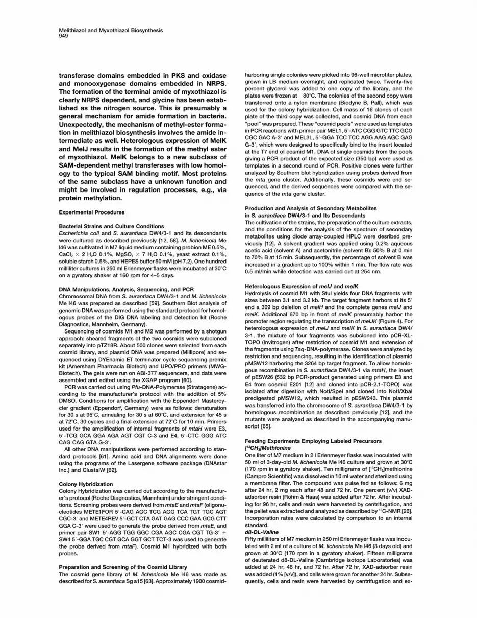
harboring single colonies were picked into 96-well microtiter plates,transferase domains embedded in PKS and oxidasegrown in LB medium overnight, and replicated twice. Twenty-fiveand monooxygenase domains embedded in NRPS.percent glycerol was added to one copy of the library, and theThe formation of the terminal amide of myxothiazol isplates were frozen at �80�C. The colonies of the second copy were
clearly NRPS dependent, and glycine has been estab- transferred onto a nylon membrane (Biodyne B, Pall), which waslished as the nitrogen source. This is presumably a used for the colony hybridization. Cell mass of 16 clones of each
plate of the third copy was collected, and cosmid DNA from eachgeneral mechanism for amide formation in bacteria.“pool” was prepared. These “cosmid pools” were used as templatesUnexpectedly, the mechanism of methyl-ester forma-in PCR reactions with primer pair MEL1, 5�-ATC CGG GTC TTC GCGtion in melithiazol biosynthesis involves the amide in-CGC GAC A-3� and MEL3L, 5�-GGA TCC TCC AGG AAG AGC GAGtermediate as well. Heterologous expression of MelKG-3�, which were designed to specifically bind to the insert located
and MelJ results in the formation of the methyl ester at the T7 end of cosmid M1. DNA of single cosmids from the poolsof myxothiazol. MelK belongs to a new subclass of giving a PCR product of the expected size (350 bp) were used as
templates in a second round of PCR. Positive clones were furtherSAM-dependent methyl transferases with low homol-analyzed by Southern blot hybridization using probes derived fromogy to the typical SAM binding motif. Most proteinsthe mta gene cluster. Additionally, these cosmids were end se-of the same subclass have a unknown function andquenced, and the derived sequences were compared with the se-might be involved in regulation processes, e.g., viaquence of the mta gene cluster.
protein methylation.
Production and Analysis of Secondary MetabolitesExperimental Procedures in S. aurantiaca DW4/3-1 and Its Descendants
The cultivation of the strains, the preparation of the culture extracts,Bacterial Strains and Culture Conditions and the conditions for the analysis of the spectrum of secondaryEscherichia coli and S. aurantiaca DW4/3-1 and its descendants metabolites using diode array-coupled HPLC were desribed pre-were cultured as described previously [12, 58]. M. lichenicola Me viously [12]. A solvent gradient was applied using 0.2% aqueousl46 was cultivated in M7 liquid medium containing probion ME 0.5%, acetic acid (solvent A) and acetonitrile (solvent B): 50% B at 0 minCaCl2 � 2 H2O 0.1%, MgSO4 � 7 H2O 0.1%, yeast extract 0.1%, to 70% B at 15 min. Subsequently, the percentage of solvent B wassoluble starch 0.5%, and HEPES buffer 50 mM (pH 7.2). One hundred increased in a gradient up to 100% within 1 min. The flow rate wasmilliliter cultures in 250 ml Erlenmeyer flasks were incubated at 30�C 0.5 ml/min while detection was carried out at 254 nm.on a gyratory shaker at 160 rpm for 4–5 days.
Heterologous Expression of melJ and melKDNA Manipulations, Analysis, Sequencing, and PCRHydrolysis of cosmid M1 with StuI yields four DNA fragments withChromosomal DNA from S. aurantiaca DW4/3-1 and M. lichenicolasizes between 3.1 and 3.2 kb. The target fragment harbors at its 5�Me l46 was prepared as described [59]. Southern Blot analysis ofend a 309 bp deletion of melH and the complete genes melJ andgenomic DNA was performed using the standard protocol for homol-melK. Additional 670 bp in front of melK presumably harbor theogous probes of the DIG DNA labeling and detection kit (Rochepromoter region regulating the transcription of melJK (Figure 4). ForDiagnostics, Mannheim, Germany).heterologous expression of melJ and melK in S. aurantiaca DW4/Sequencing of cosmids M1 and M2 was performed by a shotgun3-1, the mixture of four fragments was subcloned into pCR-XL-approach: sheared fragments of the two cosmids were subclonedTOPO (Invitrogen) after restriction of cosmid M1 and extension ofseparately into pTZ18R. About 500 clones were selected from eachthe fragments using Taq-DNA-polymerase. Clones were analyzed bycosmid library, and plasmid DNA was prepared (Millipore) and se-restriction and sequencing, resulting in the identification of plasmidquenced using DYEnamic ET terminator cycle sequencing premixpMSW12 harboring the 3264 bp target fragment. To allow homolo-kit (Amersham Pharmacia Biotech) and UPO/PRO primers (MWG-gous recombination in S. aurantiaca DW4/3-1 via mtaH, the insertBiotech). The gels were run on ABI-377 sequencers, and data wereof pESW26 (532 bp PCR-product generated using primers E3 andassembled and edited using the XGAP program [60].E4 from cosmid E201 [12] and cloned into pCR-2.1-TOPO) wasPCR was carried out using Pfu-DNA-Polymerase (Stratagene) ac-isolated after digestion with NotI/SpeI and cloned into NotI/XbaIcording to the manufacturer’s protocol with the addition of 5%predigested pMSW12, which resulted in pESW243. This plasmidDMSO. Conditions for amplification with the Eppendorf Mastercy-was transferred into the chromosome of S. aurantiaca DW4/3-1 bycler gradient (Eppendorf, Germany) were as follows: denaturationhomologous recombination as described previously [12], and thefor 30 s at 95�C, annealing for 30 s at 60�C, and extension for 45 smutants were analyzed as described in the accompanying manu-at 72�C, 30 cycles and a final extension at 72�C for 10 min. Primersscript [65].used for the amplification of internal fragments of mtaH were E3,
5�-TCG GCA GGA AGA AGT CGT C-3 and E4, 5�-CTC GGG ATCFeeding Experiments Employing Labeled PrecursorsCAG CAG GTA G-3�.[13CH3]MethionineAll other DNA manipulations were performed according to stan-One liter of M7 medium in 2 l Erlenmeyer flasks was inoculated withdard protocols [61]. Amino acid and DNA alignments were done50 ml of 3-day-old M. lichenicola Me l46 culture and grown at 30�Cusing the programs of the Lasergene software package (DNAstar(170 rpm in a gyratory shaker). Ten milligrams of [13CH3]methionineInc.) and ClustalW [62].(Campro Scientific) was dissolved in 10 ml water and sterilized usinga membrane filter. The compound was pulse fed as follows: 6 mgColony Hybridizationafter 24 hr, 2 mg each after 48 and 72 hr. One percent (v/v) XAD-Colony Hybridization was carried out according to the manufactur-adsorber resin (Rohm & Haas) was added after 72 hr. After incubat-er’s protocol (Roche Diagnostics, Mannheim) under stringent condi-ing for 96 hr, cells and resin were harvested by centrifugation, andtions. Screening probes were derived from mtaE and mtaF (oligonu-the pellet was extracted and analyzed as described by 13C-NMR [26].cleotides METE1FOR 5�-CAG AGC TCG AGG TCA TGT TGC AGTIncorporation rates were calculated by comparison to an internalCGC-3� and METE4REV 5�-GCT CTA GAT GAG CCC GAA GCG CTTstandard.GGA C-3� were used to generate the probe derived from mtaE, andd8-DL-Valineprimer pair SW1 5�-AGG TGG GGC CGA AGC CGA CGT TG-3� �Fifty milliliters of M7 medium in 250 ml Erlenmeyer flasks was inocu-SW4 5�-GGA TGC CGT GCA GGT GCT TCT-3 was used to generatelated with 2 ml of a culture of M. lichenicola Me l46 (3 days old) andthe probe derived from mtaF). Cosmid M1 hybridized with bothgrown at 30�C (170 rpm in a gyratory shaker). Fifteen milligramsprobes.of deuterated d8-DL-Valine (Cambridge Isotope Laboratories) wasadded at 24 hr, 48 hr, and 72 hr. After 72 hr, XAD-adsorber resinPreparation and Screening of the Cosmid Librarywas added (1% [v/v]), and cells were grown for another 24 hr. Subse-The cosmid gene library of M. lichenicola Me l46 was made as
described for S. aurantiaca Sg a15 [63]. Approximately 1900 cosmid- quently, cells and resin were harvested by centrifugation and ex-
Chemistry & Biology950
tracted twice (first using 30 ml methanol and then with 20 ml ace- 7. Gehring, A.M., DeMoll, E., Fetherston, J.D., Mori, I., Mayhew,tone). Combined extracts were dried under vacuo and redissolved G.F., Blattner, F.R., Walsh, C.T., and Perry, R.D. (1998). Ironin 500 l methanol. The analysis was performed after injection of 5 acquisition in plague: modular logic in enzymatic biogenesis ofl of the extract into a HPLC-MS Agilent 1100 LC system. The yersiniabactin by Yersinia pestis. Chem. Biol. 5, 573–586.following solvents were used: A, 5% acetonitrile, 95% water, 5 mM 8. O’Connor, S.E., Chen, H.W., and Walsh, C.T. (2002). Enzymaticammonium acetate (NH4Ac), 0.003% acetic acid; B, 95% acetonitrile, assembly of epothilones: The EpoC subunit and reconstitution5% water, 5 nM NH4Ac, 0.003% acetic acid. A solvent gradient was of the EpoA-ACP/B/C polyketide and nonribosomal peptide in-applied as follows: 10% B at 0 min to 100% B within 30 min, then terfaces. Biochemistry 41, 5685–5694.10 min with 100% B. The flow rate was 0.3 ml/min and the UV 9. Admiraal, S.J., Khosla, C., and Walsh, C.T. (2002). The loadingdetection was carried out at 220 nm. For mass detection, a PE Sciex and initial elongation modules of rifamycin synthetase collabo-API 2000 mass spectrometer equipped with a TurbolonSpray source rate to produce mixed aryl ketide products-1. Biochemistry 41,was used. 5313–5324.15N-Glycine, 15N-Glutamate, and 15N-Ammonium Chloride 10. Cane, D., and Walsh, C. (1999). The parallel and convergentS. aurantiaca DW 4/3-1 was inoculated in 20 ml tryptone-starch universes of polyketide synthases and nonribosomal peptidemedium [41] and cultivated at 28�C (200 rpm in a gyratory shaker)
synthetases. Chem. Biol. 6, R319–R325.for 2 days. Ten milliliters of the culture was then transferred to a
11. Reichenbach, H., and Hofle, G. (1999). Myxobacteria as produc-500 ml erlenmeyer flask containing 200 ml tryptone-starch medium,
ers of secondary metabolites. In Drug Discovery from Nature,and the flask was shaken at 30�C (160 rpm). Thirty milligrams of
S. Grabley and R. Thieriecke, eds. (Berlin: Springer Verlag), pp.each of the labeled precursors (15N-Glycine, 15N-glutamate, and149–179.15N-ammonium chloride) was dissolved in 2 ml water and sterilized
12. Silakowski, B., Schairer, H.U., Ehret, H., Kunze, B., Weinig, S.,through a 0.22 m diameter pore size ultrafilter (Millipore, Millex-Nordsiek, G., Brandt, P., Blocker, H., Hofle, G., Beyer, S., et al.GV4). The labeled precursors were pulse fed in two equal portions(1999). New lessons for combinatorial biosynthesis from myxo-24 and 48 hr after inoculation. Cells were harvested after 4 days ofbacteria: the myxothiazol biosynthetic gene cluster of Stig-cultivation, and the products were extracted with acetone. Myxothi-matella aurantiaca DW4/3–1. J. Biol. Chem. 274, 37391–37399.azol was purified by silica gel column chromatography (n-hex:EtOAc
13. Paitan, Y., Alon, G., Orr, E., Ron, E.Z., and Rosenberg, E. (1999).5:1 � n-hex-EtOAc 1:1, EtOAc) and subsequently by HPLC (YMC-The first gene in the biosynthesis of the polyketide antibioticPack ODS-AQ, 250 �10 mm, methanol:water 82:18). The incorpora-TA of Myxococcus xanthus codes for a unique PKS moduletion rate of 15N into myxothiazol was determined using single ioncoupled to a peptide synthetase. J. Mol. Biol. 286, 465–474.monitoring in a electrospray mass spectrometer [64].
14. Molnar, I., Schupp, T., Ono, M., Zirkle, R.E., Milnamow, M.,Nowak-Thompson, B., Engel, N., Toupet, C., Stratman, A., Cyr,D.D., et al. (2000). The biosynthetic gene cluster for the microtu-Acknowledgmentsbule-stabilizing agents epothilones A and B from Sorangium
The authors would like to thank Prof. Dr. H. Reichenbach for strain cellulosum So ce90. Chem. Biol. 7, 97–109.M. lichenicola Me l46. We are grateful to Prof. Dr. G. Hofle and Dr. 15. Julien, B., Shah, S., Ziermann, R., Goldman, R., Katz, L., andB. Kunze for help with the analysis of myxothiazol and its derivatives Khosla, C. (2000). Isolation and characterization of the epothi-and to M. Scharfe and Dr. H. Blocker for their support concerning the lone biosynthetic gene cluster from Sorangium cellulosum.sequencing of cosmid M2. B.R.T. Higa contributed to the analysis of Gene 249, 153–160.the 15N-feeding studies. Work in R.M.’s laboratory was supported 16. Silakowski, B., Kunze, B., Nordsiek, G., Blocker, H., Hofle, G.,by the Deutsche Forschungsgemeinschaft (grants Mu1254/3-3 and and Muller, R. (2000). The myxochelin iron transport regulonMu1254/6-1). We acknowledge the generous gift of 15N-labeled sub- of the myxobacterium Stigmatella aurantiaca Sg a15. Eur. J.stances and the helpful comments by B. Carroll and Prof. Dr. H.G. Biochem. 267, 6476–6485.Floss regarding this manuscript. 17. Gaitatzis, N., Kunze, B., and Muller, R. (2001). In vitro reconstitu-
tion of the myxochelin biosynthetic machinery of Stigmatellaaurantiaca Sg a15: Biochemical characterization of a reductive
Received: April 7, 2003 release mechanism from nonribosomal peptide synthetases.Revised: July 31, 2003 Proc. Natl. Acad. Sci. USA 98, 11136–11141.Accepted: August 6, 2003 18. Gaitatzis, N., Silakowski, B., Kunze, B., Nordsiek, G., Blocker,Published: October 17, 2003
H., Hofle, G., and Muller, R. (2002). The biosynthesis of thearomatic myxobacterial electron transport inhibitor stigmatellinis directed by a novel type of modular polyketide synthase. J.
ReferencesBiol. Chem. 277, 13082–13090.
19. Silakowski, B., Nordsiek, G., Kunze, B., Blocker, H., and Muller,1. Cane, D.E. (1997). Polyketide and nonribosomal polypeptideR. (2001). Novel features in a combined polyketide synthase/biosynthesis. Chem. Rev. 97, 2463–2464.non-ribosomal peptide synthetase: The myxalamid biosynthetic2. Staunton, J., and Weissman, K.J. (2001). Polyketide biosynthe-gene cluster of the myxobacterium Stigmatella aurantiaca Sgsis: a millennium review. Nat. Prod. Rep. 18, 380–416.a15. Chem. Biol. 8, 59–69.3. Konz, D., and Marahiel, M.A. (1999). How do peptide synthe-
20. Nishizawa, T., Ueda, A., Asayama, M., Fujii, K., Harada, K., Ochi,tases generate structural diversity? Chem. Biol. 6, R39–R48.K., and Shirai, M. (2000). Polyketide synthase gene coupled to4. Lambalot, R.H., Gehring, A.M., Flugel, R.S., Zuber, P., LaCelle,the peptide synthetase module involved in the biosynthesis ofM., Marahiel, M.A., Reid, R., Khosla, C., and Walsh, C.T. (1996).the cyclic heptapeptide microcystin. J. Biochem. (Tokyo) 127,A new enzyme superfamily–the phosphopantetheinyl transferases.779–789.Chem. Biol. 3, 923–936.
21. Tillett, D., Dittmann, E., Erhard, M., von Dohren, H., Borner, T.,5. Molnar, I., Aparicio, J.F., Haydock, S.F., Khaw, L.E., Schwecke,and Neilan, B.A. (2000). Structural organization of microcystinT., Konig, A., Staunton, J., and Leadlay, P.F. (1996). Organisationbiosynthesis in Microcystis aeruginosa PCC7806: an integratedof the biosynthetic gene cluster for rapamycin in Streptomycespeptide-polyketide synthetase system. Chem. Biol. 7, 753–764.hygroscopicus: analysis of genes flanking the polyketide syn-
22. Chang, Z.X., Flatt, P., Gerwick, W.H., Nguyen, V.A., Willis, C.L.,thase. Gene 169, 1–7.and Sherman, D.H. (2002). The barbamide biosynthetic gene6. August, P.R., Tang, L., Yoon, Y.J., Ning, S., Muller, R., Yu, T.W.,cluster: a novel marine cyanobacterial system of mixed polyke-Taylor, M., Hoffmann, D., Kim, C.G., Zhang, X., et al. (1998).tide synthase (PKS)-non-ribosomal peptide synthetase (NRPS)Biosynthesis of the ansamycin antibiotic rifamycin: deductionsorigin involving an unusual trichloroleucyl starter unit. Gene 296,from the molecular analysis of the rif biosynthetic gene cluster
of Amycolatopsis mediterranei S699. Chem. Biol. 5, 69–79. 235–247.
Melithiazol and Myxothiazol Biosynthesis951
23. Gerth, K., Irschik, H., Reichenbach, H., and Trowitzsch, W. 41. Mahmud, T., Bode, H.B., Silakowski, B., Kroppenstedt, R.M.,(1980). Myxothiazol, an antibiotic from Myxococcus fulvus (myx- Xu, M., Nordhoff, S., Hofle, G., and Muller, R. (2002). A novelobacterales). I. Cultivation, isolation, physico-chemical and bio- biosynthetic pathway providing precursors for fatty acid biosyn-logical properties. J. Antibiot. 33, 1474–1479. thesis and secondary metabolite formation in myxobacteria. J.
24. Trowitzsch, W., Reifenstahl, G., Wray, V., and Hofle, G. (1980). Biol. Chem. 277, 32768–32774.Myxothiazol, an antibiotic from Myxococcus fulfus (Myxobac- 42. Long, P.F., Wilkinson, C.J., Bisang, C.P., Cortes, J., Dunster,teraoles) II. Structure elucidation. J. Antibiot. 33, 1480–1490. N., Oliynyk, M., McCormick, E., McArthur, H., Mendez, C., Salas,
25. Sasse, F., Bohlendorf, B., Hermann, M., Kunze, B., Forche, E., J.A., et al. (2002). Engineering specificity of starter unit selectionSteinmetz, H., Hofle, G., and Reichenbach, H. (1999). Melithia- by the erythromycin-producing polyketide synthase. Mol. Mi-zols, new �-methoxyacrylate inhibitors of the respiratory chain crobiol. 43, 1215–1225.isolated from myxobacteria–production, isolation, physico- 43. Challis, G., Ravel, J., and Townsend, C. (2000). Predictive, struc-chemical and biological Properties. J. Antibiot. 52, 721–729. ture-based model of amino acid recognition by nonribosomal
26. Bohlendorf, B., Herrmann, M., Hecht, H.J., Sasse, F., Forche, peptide synthetase adenylation domains. Chem. Biol. 7, 211–224.E., Kunze, B., Reichenbach, H., and Hofle, G. (1999). Antibiotics 44. Stachelhaus, T., Mootz, H.D., and Marahiel, M.A. (1999). Thefrom gliding bacteria—melithiazols A-N: new antifungal �-meth- specificity-conferring code of adenylation domains in nonribo-oxyacrylates from myxobacteria. Eur. J. Org. Chem. 10, 2601– somal peptide synthetases. Chem. Biol. 6, 493–505.2608. 45. Smith, S. (1994). The animal fatty acid synthase: one gene, one
27. Gaitatzis, N., Hans, A., Muller, R., and Beyer, S. (2001). The polypeptide, seven enzymes. FASEB J. 8, 1248–1259.mtaA gene of the myxothiazol biosynthetic gene cluster from 46. Kagan, R.M., and Clarke, S. (1994). Widespread occurrence ofStigmatella aurantiaca DW4/3–1 encodes a phosphopantethei- three sequence motifs in diverse S-adenosylmethionine-depen-nyl transferase that activates polyketide synthases and poly- dent methyltransferases suggest a common structure for thesepeptide synthetases. J. Biochem. 129, 119–124.
enzymes. Arch. Biochem. Biophys. 310, 417–427.28. Silakowski, B., Kunze, B., and Muller, R. (2001). Multiple hybrid
47. Kulathila, R., Merkler, K.A., and Merkler, D.J. (1999). Enzymaticpolyketide synthase/non-ribosomal peptide synthetase gene
formation of C-terminal amides. Nat. Prod. Rep. 16, 145–154.clusters in the myxobacterium Stigmatella aurantiaca. Gene
48. Schoof, L., Veelaert, D., Vanden Broeck, J., and De Loof, A.275, 233–240.
(1997). Peptides in the locusts, Locusta migratoria and Schis-29. Gehring, A.M., DeMoll, E., Fetherston, J.D., Mori, I., Mayhew,
tocerca gregaria. Peptides 18, 145–156.G.F., Blattner, F.R., Walsh, C.T., and Perry, R.D. (1998). Iron
49. Mocek, U., Knaggs, A.R., Tsuchiya, R., Nguyen, T., Beale, J.M.,acquisition in plague: modular logic in enzymatic biogenesis of
and Floss, H.G. (1993). Biosynthesis of the modified peptideyersiniabactin by Yersinia pestis. Chem. Biol. 5, 573–586.
antibiotic nosiheptide in Streptomyces actuosus. J. Am. Chem.30. Kennedy, J., Auclair, K., Kendrew, S.G., Park, C., Vederas, J.C.,Soc. 115, 7557–7568.and Hutchinson, C.R. (1999). Modulation of polyketide synthase
50. Steinmetz, H., Forche, E., Reichenbach, H., and Hofle, G. (2000).activity by accessory proteins during lovastatin biosynthesis.Biosynthesis of myxothiazol Z, the ester-analog of myxothiazolScience 284, 1368–1372.A in Myxococcus fulvus. Tetrahedron 56, 1681–1684.31. Chen, H.W., O’Connor, S., Cane, D.E., and Walsh, C.T. (2001).
51. Djordjevic, S., and Stock, A. (1997). Crystal structure of theEpothilone biosynthesis: assembly of the methylthiazolylcar-chemotaxis receptor methyltransferase CheR suggests a con-boxy starter unit on the EpoB subunit. Chem. Biol. 8, 899–912.served structural motif for binding S-adenosylmethionine. Curr.32. Walsh, C.T., Chen, H.W., Keating, T.A., Hubbard, B.K., Losey,Biol. 5, 545–558.H.C., Luo, L.S., Marshall, C.G., Miller, D.A., and Patel, H.M.
52. Koshland, D., Jr. (1988). Chemotaxis as a model second-mes-(2001). Tailoring enzymes that modify nonribosomal peptidessenger system. Biochemistry 27, 5829–5833.during and after chain elongation on NRPS assembly lines. Curr.
53. Pace, H., and Brenner, C. (2001). The nitrilase superfamily: clas-Opin. Chem. Biol. 5, 525–534.sification, structure and function. Genome Biol. 2, 0001.0001–33. Donadio, S., Staver, M.J., McAlpine, J.B., Swanson, S.J., and0001.0008.Katz, L. (1991). Modular organization of genes required for com-
54. Pearson, W.R., and Lipman, D.J. (1988). Improved tools for bio-plex polyketide biosynthesis. Science 252, 675–679.logical sequence comparison. Proc. Natl. Acad. Sci. USA 85,34. Schwecke, T., Aparicio, J.F., Molnar, I., Konig, A., Khaw, L.E.,2444–2448.Haydock, S.F., Oliynyke, M., Caffrey, P., Cortes, J., Lester, J.B.,
55. Yu, T.-W., Bai, L., Clade, D., Hoffmann, D., Toelzer, S., Trinh,et al. (1995). The biosynthetic gene cluster for the polyketideK.Q., Xu, J., Moss, S.J., Leistner, E., and Floss, H.G. (2002). Theimmunosuppressant rapamycin. Proc. Natl. Acad. Sci. USA 92,biosynthetic gene cluster of the maytansinoid antitumor agent7839–7843.ansamitocin from Actinosynnema pretiosum. Proc. Natl. Acad.35. Kleinkauf, H., and von Dohren, H. (1997). Products of secondarySci. USA 99, 7968–7973.metabolism. In Biotechnology, H. Kleinkauf and H. von Dohren,
56. Martin, J., and McMillan, F. (2002). SAM (dependent) I AM: theeds. (Weinheim, Germany: Verlag Chemie), pp. 277–322.S-adenosylmethionine-dependent methyltansferase fold. Curr.36. Shimkets, L. (1993). The myxobacterial genome. In Myxobac-Opin. Struct. Biol. 12, 783–793.teria II, M. Dworkin and D. Kaiser, eds. (Washington, D.C.: Ameri-
57. De Baere, I., Derua, R., Janssens, V., Van Hoof, C., Waelkens,can Society for Microbiology), pp. 85–108.E., Merlevede, W., and Goris, J. (1999). Purification of porcine37. Bisang, C., Long, P., Cortes, J., Westcott, J., Crosby, J., Ma-brain protein phosphatase 2A leucine carboxyl methyltransfer-tharu, A.-L., Cox, R., Simpson, T., Staunton, J., and Leadlay, P.ase and cloning of the human homologue. Biochemistry 38,(1999). A chain initiation factor common to both modular and16539–16547.aromatic polyketide synthases. Nature 401, 502–505.
58. Silakowski, B., Ehret, H., and Schairer, H.U. (1998). fbfB, a gene38. Ligon, J., Hill, S., Beck, J., Zirkle, R., Molnar, I., Zawodny, J.,encoding a putative galactose oxidase, is involved in Stig-Money, S., and Schupp, T. (2002). Characterization of the bio-matella aurantiaca fruiting body formation. J. Bacteriol. 180,synthetic gene cluster for the antifungal polyketide soraphen A1241–1247.from Sorangium cellulosum So ce26. Gene 285, 257–267.
59. Neumann, B., Pospiech, A., and Schairer, H.U. (1992). Rapid39. Wilkinson, C.J., Frost, E.J., Staunton, J., and Leadlay, P.F.isolation of genomic DNA from Gram negative bacteria. Trends(2001). Chain initiation on the soraphen-producing modularGenet. 8, 332–333.polyketide synthase from Sorangium cellulosum. Chem. Biol.
60. Bonfield, J., Smith, K., and Staden, R. (1995). Nucleic Acids Res8, 1197–1208.23, 4992–4999.40. Trowitzsch-Kienast, W., Wray, V., Gerth, K., Reichenbach, H.,
61. Sambrook, J., Fritsch, E.F., and Maniatis, T. eds. (1989). Molecu-and Hofle, G. (1986). Antibiotika aus Gleitenden Bakterien,lar Cloning: A Laboratory Manual, Second Edition (Cold SpringXXVIII: Biosynthese des Myxothiazols in Myxococcus fulvus Mx
f16. Liebigs Ann. Chem. 10, 93–98. Harbor, NY: Cold Spring Harbor Laboratory Press).

Chemistry & Biology952
62. Thompson, J.D., Higgins, D.G., and Gibson, T.J. (1994). CLUS-TAL W: Improving the sensitivity of progressive multiple se-quence alignment through sequence weighting, position-spe-cific gap penalties and weight matrix choice. Nucleic Acids Res.22, 4673–4680.
63. Beyer, S., Kunze, B., Silakowski, B., and Muller, R. (1999). Meta-bolic diversity in myxobacteria - identification of the myxalamidand the stigmatellin biosynthetic gene cluster of Stigmatellaaurantiaca Sg a15 and a combined polyketide- (poly)peptidegene cluster from the epothilon producing strain Sorangiumcellulosum So ce90. Biochim. Biophys. Acta 1445, 185–195.
64. Morgan, J., Joyce-Menekse, M., Rowlands, R., Gilbert, I., andLloyd, D. (2001). Rapid and sensitive quantitation of antibioticsin fermentations by electrospray mass spectrometry. RapidCommun. Mass Spectrom. 15, 1229–1238.
65. Weinig, S., Mahmud, T., and Muller, R. (2003). Markerless muta-tions in the myxothiazol biosynthetic gene cluster: a delicatemegasynthetase with a superfluous nonribosomal peptide syn-thetase domain. Chem. Biol. 10, this issue, 953–960.
Accession Numbers
The nucleotide sequence reported here has been submitted to theEMBL database under accession number AJ557546.