mast cell leukemia associated with undefined morphology and chronic basophilic leukemia
TRANSCRIPT

Cehreli et al. BMC Hematology 2014, 14:17http://biomedcentral.com/2052-1839/14/17
CASE REPORT Open Access
Mast cell leukemia associated with undefinedmorphology and chronic basophilic leukemiaCavit Cehreli1*, Inci Alacacioglu1, Ozden Piskin1, Halil Ates1, Ruksan Cehreli2, Gizem Calibasi3, Erdinc Yuksel4,Sermin Ozkal5 and Guner H Ozsan1
Abstract
Background: Mast cell leukemia (MCL) is rare type of neoplasia with an incidence of 1% in a large series of 342adult patients with systemic mastocytosis (SM). Chronic basophilic leukemia (CBL) is an extremely rare type ofleukemia with appearance of 7 cases in the literature.
Case presentation: A 73 year-old female patient who presented with weaknes, had a prolonged duration ofhematologic remission after treatment of her CBL by hydroxyurea (HU). Evolution of SM occurring as a secondneoplasia concurrently with relapse of de novo CBL was demonstrated by mast cells (MCs) infiltration in the bonemarrow (BM) biopsy and smear and increase in tryptase level. Transformation to MCL with simultaneous occurranceof accelerated phase of CBL were documented by the appearance of MCs in both BM and peripheral blood (PB)smears, antigen expressions detected by flow cytometry and spesific stains. Sequence analysis of c-kit gene revealedc-kit exon 11 K550N mutation. Undefined associations of MCL with different mast cell morphology, increase in IL-6level and accelerated phase of de novo CBL was described.
Conclusion: Elevations in CRP and IL-6 levels occurring with increases in basophil counts to high levels revealedthat febrile episodes with abdominal pain seen in our patient were induced by increase in IL-6 levels released fromneoplastic basophils. Neoplastic basophils with diffuse and coarse basophilic granules possibly mimic neutrophilswith toxic granules and cause wrong characterization of neoplastic basophils as neutrophils by the automatedblood cell counters and misleaded physicians.
Keywords: Mast cell leukemia, Mastocytosis, Basophilia, Interleukin-6, Dysplasia
BackgroundMast cell leukemia (MCL) is rare type of neoplasia with anincidence of 1% in a large series of 342 adult patients withsystemic mastocytosis (SM) [1] and accounting for <1% ofall mastocytosis in the French Referance Center for Masto-cytosis (CEREMAST) [2]. SM associated with clonalhematologic non-mast cell disease (SM-AHNMD) was thesecond most common SM subgroup (N = 138, 40%) in thiscohort of patients [1]. Of The SM-AHNMD group, 89%had an associated myeloid malignancy group. This groupincluded subgroups of, SM-myeloproliferative neoplasia(SM-MPN), SM-chronic myelomonocytic leukemia (SM-CMML) and SM-myelodysplastic syndrome (SM-MDS). Asignificant proportion exhibited prominent eosinophilia
* Correspondence: [email protected] of Hematology, Dokuz Eylul University School of Medicine, 35330Inciralti, Izmir, TurkeyFull list of author information is available at the end of the article
© 2014 Cehreli et al.; licensee BioMed CentralCommons Attribution License (http://creativecreproduction in any medium, provided the orDedication waiver (http://creativecommons.orunless otherwise stated.
(>1.5 × 10⁹/l), especially those with SM-MPN and 39%harbored the FIP1L1-PDGFRA fusion [1,3]. A literaturesearch was performed by using PubMed database for allproven MCL cases according to WHO criteria and 51adult patients with MCL were detected appearing as denovo, (n = 30) and secondary, (n = 11). The median agesfor de novo and secondary cases were 51.5 (18–78) and35.0 (5–75) respectively. Median survival was 6 months(0.5-98) in all adult patients seen in the literature [2].CBL is an extremely rare type of leukemia with the ap-
pearance of seven cases in the literature [4-7]. Four of theseven patients reported by Pardanani et al. identified byscreening of electronic data base in Mayo Clinic [4] MCL,occuring as a second neoplasia in association with un-defined MC morphology and de novo CBL relapse havenot been described in the World’s literature [4-7]. A caseof MCL occuring as a second neoplasia in association with
Ltd. This is an Open Access article distributed under the terms of the Creativeommons.org/licenses/by/2.0), which permits unrestricted use, distribution, andiginal work is properly credited. The Creative Commons Public Domaing/publicdomain/zero/1.0/) applies to the data made available in this article,

Cehreli et al. BMC Hematology 2014, 14:17 Page 2 of 7http://www.biomedcentral.com/2052-1839/14/17
undefined MC morphology, increase in IL-6 levels and ac-celerated phase of de novo CBL was described.
Case presentationA 73 year old Turkish female patient who presentedwith weakness, epigastric fullness and decreased appe-tite had a prolonged duration of hematologic remissionby the treatment of her CBL with hydroxyurea (HU)[7]. At presentation, complete blood counts (CBC)showed marked decrease in hemoglobin. Differentialcount made by automated blood cell counter revealed30% segmented neutrophils, 30% eosinophils, 32% lym-phocytes and 8% monocytes. However marked baso-philia and eosinophilia at different stages of maturationdetected by manual differential in PB smear revealedthat automated blood cell counters wrong characterizedthe neoplastic basophilic cells with coarse and diffusebasophilic granules as neutrophils, as shown in Table 1.
Table 1 Showing blood counts and chemistry profiles
LaboratoryFindings
AtPresentation
1. month 3. month
(×109/L)
White Blood Cells 4.6 59.0 170.0
Basophil count 2.6 36.5 79.9
Eosinophil count - - 4.5
Mast cell count - - 28.9
Platelet 134.0 157 80.0
Hgb (g/dL) 5.7 8.9 7.1
ManualDifferentialCounts (%)
Seg. Basophil 46 10 10
Basophil Band 8 12 5
Basophil Meta. 3 25 14
Basophil Myelo. - 15 18
Promyelocyte - - 9
Eosinophilic series 29 6 27
Myeloblast - - -
Mast cell - - 17
Tryptase (μg/L) † 42.9 51.2
Histamine (nmol/L)††
- 1.2
IL-6 (pg/mL) ††† - 38.5
CRP (mg/dL) - 85.6 225
LDH (U/L) - 561 1221
Treatments Watch-wait HU 1 g/L(×3 days)
leukaph. + HU 1.5 g/l (×3days) + prednisolon 40 mg
Magnification: 160 X 135 mm†by F.E.I.A ImmunoCAP PHADIA, N < 11.4, treshold 11.4/l : 95th percentile (Cerba La†††by Electrochemiluminescence ECLIA Roche (N < 7).
She was firstly transfused with 4 units of packed redblood cells (rbc) for severe anemia and maintenancetherapy with HU was discontinued. Results of rutin la-boratory studies with gastroscopy and colonoscopy failedto reveal gastrointestinal bleeding and hemolysis.Bone marrow (BM) aspiration showed hypercellularity,
basophilic hyperplasia, eosinophilia, increase in megakar-yocytes and aggregates of mast cells. Suppressions in neu-trophilic and in erythroid lineages induced by basophilichyperplasia and MCs infiltration were seen. BM biopsyshowed increase in megakaryocytes and eosinophils atdifferent stages of maturation. Increased numbers of MCsin paratrabecular location were highlighted by immuno-histochemical staining for CD117 and tryptase (Figure 1a,b and c). Increase in serum level of tryptase was shown inTable 1.Diagnosis of SM occurring as second neoplasia concur-
rently with de novo CBL that relapsed after prolonged
5. month 7. month 7,5. month 8.month
19.0 149.0 250.0 315.0
9.8 77.4 122.8
5.3 4.1 -
3.0 23.8 78.7
43.0 43.9 41.0
8.9 8.9 7.2
14 -
2 1
20 13
16 25
- 26
28 -
- 2
16 25
30 55.9
- 5174
İmatinib 300 mg/d + HU 0.5 g/d
leukaph. +Etoposide50 mg
leukaph. + i.v.Cyclophos-phamide
1000 mg
EX
b. Paris France, Ref no: 887821335110 and Ref no 9197749), †† by RIA (N < 10),
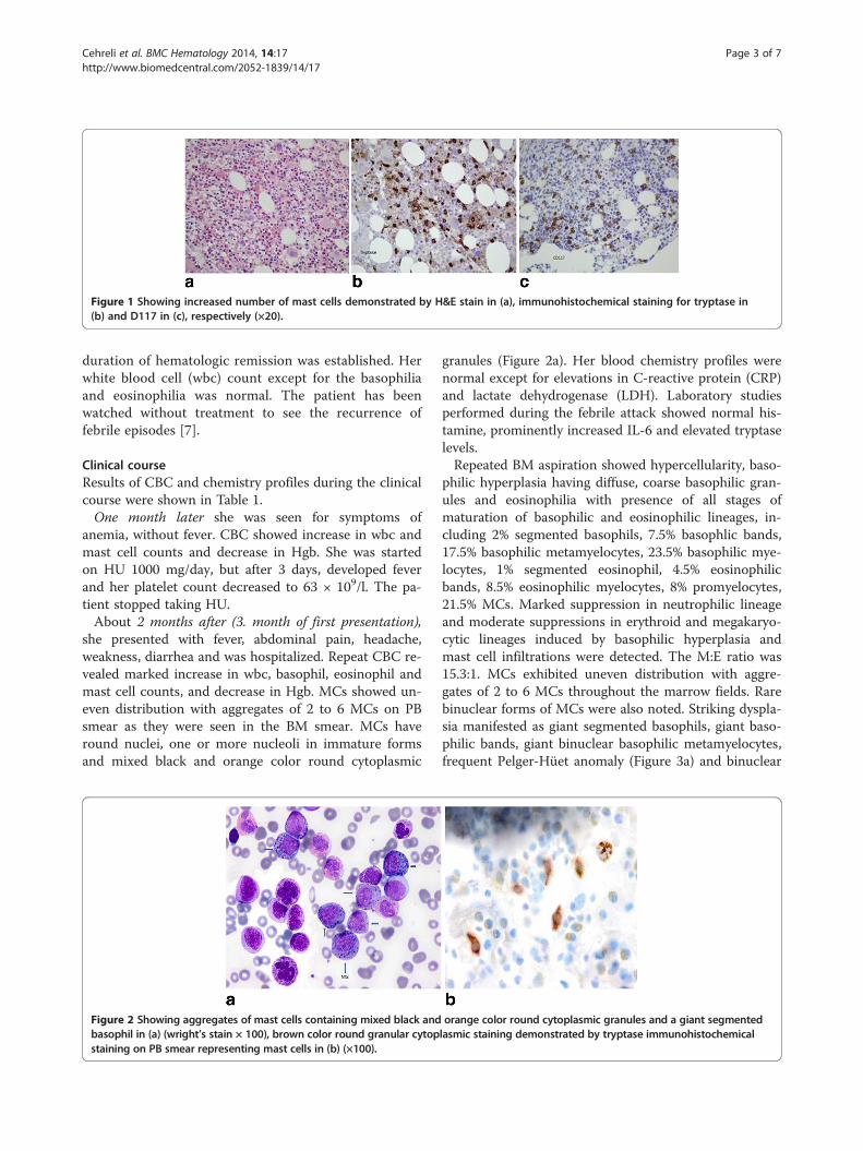
Figure 1 Showing increased number of mast cells demonstrated by H&E stain in (a), immunohistochemical staining for tryptase in(b) and D117 in (c), respectively (×20).
Cehreli et al. BMC Hematology 2014, 14:17 Page 3 of 7http://www.biomedcentral.com/2052-1839/14/17
duration of hematologic remission was established. Herwhite blood cell (wbc) count except for the basophiliaand eosinophilia was normal. The patient has beenwatched without treatment to see the recurrence offebrile episodes [7].
Clinical courseResults of CBC and chemistry profiles during the clinicalcourse were shown in Table 1.One month later she was seen for symptoms of
anemia, without fever. CBC showed increase in wbc andmast cell counts and decrease in Hgb. She was startedon HU 1000 mg/day, but after 3 days, developed feverand her platelet count decreased to 63 × 109/l. The pa-tient stopped taking HU.About 2 months after (3. month of first presentation),
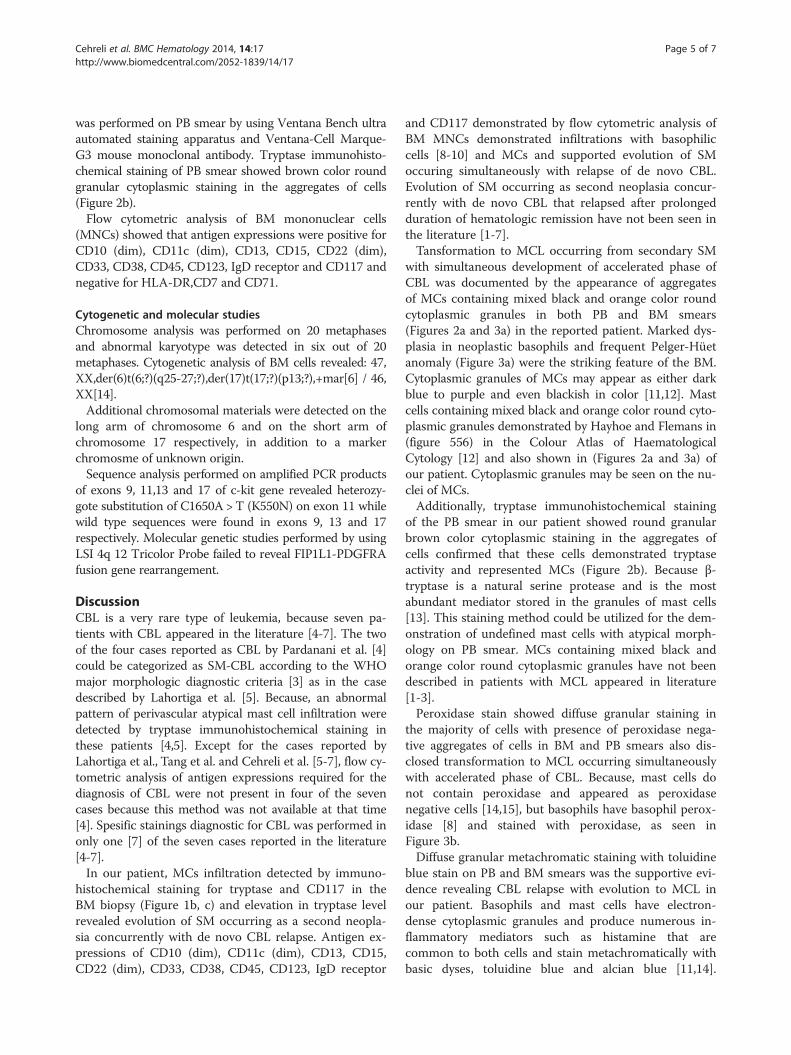
she presented with fever, abdominal pain, headache,weakness, diarrhea and was hospitalized. Repeat CBC re-vealed marked increase in wbc, basophil, eosinophil andmast cell counts, and decrease in Hgb. MCs showed un-even distribution with aggregates of 2 to 6 MCs on PBsmear as they were seen in the BM smear. MCs haveround nuclei, one or more nucleoli in immature formsand mixed black and orange color round cytoplasmic
Figure 2 Showing aggregates of mast cells containing mixed black andbasophil in (a) (wright’s stain × 100), brown color round granular cytopstaining on PB smear representing mast cells in (b) (×100).
granules (Figure 2a). Her blood chemistry profiles werenormal except for elevations in C-reactive protein (CRP)and lactate dehydrogenase (LDH). Laboratory studiesperformed during the febrile attack showed normal his-tamine, prominently increased IL-6 and elevated tryptaselevels.Repeated BM aspiration showed hypercellularity, baso-
philic hyperplasia having diffuse, coarse basophilic gran-ules and eosinophilia with presence of all stages ofmaturation of basophilic and eosinophilic lineages, in-cluding 2% segmented basophils, 7.5% basophlic bands,17.5% basophilic metamyelocytes, 23.5% basophilic mye-locytes, 1% segmented eosinophil, 4.5% eosinophilicbands, 8.5% eosinophilic myelocytes, 8% promyelocytes,21.5% MCs. Marked suppression in neutrophilic lineageand moderate suppressions in erythroid and megakaryo-cytic lineages induced by basophilic hyperplasia andmast cell infiltrations were detected. The M:E ratio was15.3:1. MCs exhibited uneven distribution with aggre-gates of 2 to 6 MCs throughout the marrow fields. Rarebinuclear forms of MCs were also noted. Striking dyspla-sia manifested as giant segmented basophils, giant baso-philic bands, giant binuclear basophilic metamyelocytes,frequent Pelger-Hüet anomaly (Figure 3a) and binuclear
orange color round cytoplasmic granules and a giant segmentedlasmic staining demonstrated by tryptase immunohistochemical

Cehreli et al. BMC Hematology 2014, 14:17 Page 4 of 7http://www.biomedcentral.com/2052-1839/14/17
eosinophilic metamyelocytes were seen. A few gianthypersegmented megakaryocytes, hypogranular formsand rare megakaryoblasts were also observed.Transformation to MCL from secondary SM occuring
concurrently with accelerated phase of CBL relapse wasdetected 2 months after evolution to SM. The patientunderwent 2 consecutive prophylactic leukapheresis toreduce to basophil count below 40 × 109/l for theprevention of cytokine release from dead neoplasticcells induced by chemotherapy. She was transfused with2 units of packed rbc and the treatment was resumedwith increase in daily dose of HU to 1500 mg. Despiteprophylactic leukapheresis, she developed febrile epi-sode 3 days after chemotherapy. Her fever returned tonormal with corticosteroid therapy, 40 mg/day, for3 days, but she developed hematochezia due todecrease in Plt count to 28 × 109/l. HU therapy wasdiscontinued and she received supportive transfusionwith plt packs and packed rbc.The patient has received no treatment for about two
months since she has required continuous supportive pltand rbc transfusions. Her wbc counts ranged betwen 4.5 ×10⁹/l and 18.3 × 10⁹/l and Plt counts ranged between 4 ×10⁹/l and 25 × 109/l repectively during this period.About 2 moths later (5. month of first presentation), she
was reevaluated for symptoms of anemia. CBC showed in-creases in basophil, eosinophil and mast cell counts anddecrease in Hgb. Treatment with interferon alpha-2b(INFα-2b) was considered, but could not be administeredbecause of persistent thrombocytopenia. She was treatedwith imatinib 300 mg/day combined with HU 500 mg/dayduring the ensuing month. She feld better, but decreasesin Hgb to 7 g/dl and Plt count to 20 × 109/l were noted.Combination therapy was stopped and she was given sup-portive plt and rbc transfusions.Approximately, 7 weeks later (7. month), she developed
fever, abdominal pain, fatigue and her spleen was palpable
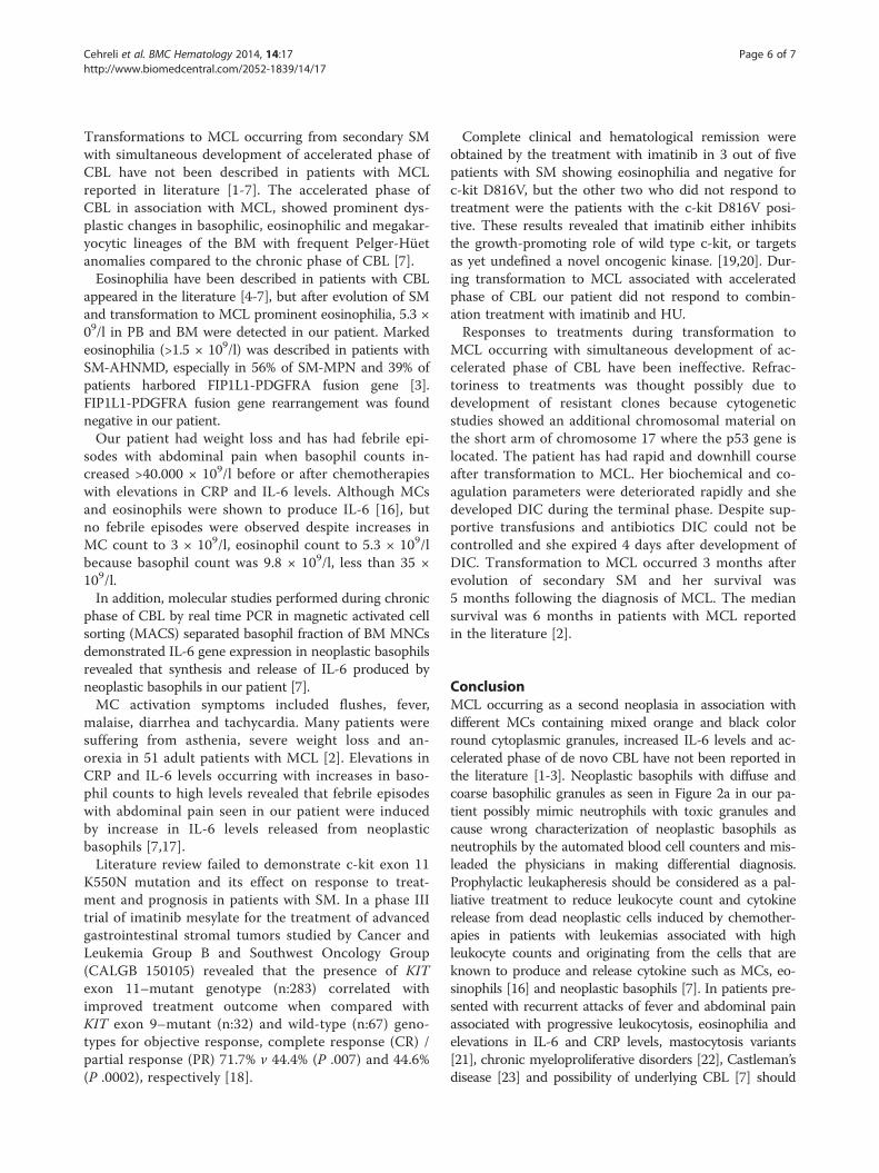
Figure 3 Demonstrating basophils at various stages of maturation,giawith mixed black and orange color round cytoplasmic granules and Pelgranular staining by peroxidase stain in peroxidase positive basophils amyeloperoxidase negative mast cells in the BM in (b) (Peroxidase stain,
4 cm below the left costal margin. CBC showed markedincreases in wbc, basophil, eosinophil and mast cellcounts and decrease in Hgb. The patient underwent 2consecutive leukapheresis. Treatment with oral etopo-side 50 mg/day was started, but on the thirth day ofchemotherapy she developed fever and was hospital-ized. Repeat CBC showed a wbc of 2.3 × 10⁹/l, Hgb7.7 g/dl and Plt count 2 × 10⁹/l. Oral etoposide was dis-continued because of thrombocytopenia (2 × 10⁹/l) andleukopenia (2.3 × 10⁹/l). She was treated with antibi-otics and supportive transfusions. The patient was thenfollowed for 2 weeks (7.5 month) without chemother-apy, but developed fever, abdominal pain and a rapidincrease in wbc cunt to 250 × 109/l was detected. Shewas started on leukapheresis and given i.v cyclophos-phamide 1000 mg, but despite these treatments 2 weeksafter (8.monts), her wbc count continued to climb. Herbiochemical and coagulation parameters were deterio-rated rapidly. Repeat CBC revealed prominent increasein wbc, basophil and mast cell counts and decreases inHgb and Plts. Coagulation profile revealed disseminatedintravascular coagulation (DIC) during terminal phase.DIC was due to febrile neutropenia and infection thatresulted in septic shock. Despite supportive manage-ments with transfusions of fresh frozen plasmas, pltpacks and packed rbc. DIC could not be controlled andshe expired 4 days after development of DIC. Thepatient died 5 months after the diagnosis of MCL.
Results of laboratory studiesToluidine blue stain on PB and BM smears demonstrateddiffuse granular metachromatic staining in the great major-ity of cells including MCs and basophils when transform-ation to MCL was detected. Peroxidase stain of BM smearshowed diffuse granular staining in <60% of non-erythroidnucleated BM cells and peroxidase negative aggregates ofcells (Figure 3b). Tryptase immunohistochemical staining
nt binuclear basophilic metamyelocyte, aggregates of mast cellsger-Huёt anomalies in (a) (Wright’s stain, ×100). Showing diffusend absence of staining in aggregates of cells that representing×100).
Cehreli et al. BMC Hematology 2014, 14:17 Page 5 of 7http://www.biomedcentral.com/2052-1839/14/17
was performed on PB smear by using Ventana Bench ultraautomated staining apparatus and Ventana-Cell Marque-G3 mouse monoclonal antibody. Tryptase immunohisto-chemical staining of PB smear showed brown color roundgranular cytoplasmic staining in the aggregates of cells(Figure 2b).Flow cytometric analysis of BM mononuclear cells
(MNCs) showed that antigen expressions were positive forCD10 (dim), CD11c (dim), CD13, CD15, CD22 (dim),CD33, CD38, CD45, CD123, IgD receptor and CD117 andnegative for HLA-DR,CD7 and CD71.
Cytogenetic and molecular studiesChromosome analysis was performed on 20 metaphasesand abnormal karyotype was detected in six out of 20metaphases. Cytogenetic analysis of BM cells revealed: 47,XX,der(6)t(6;?)(q25-27;?),der(17)t(17;?)(p13;?),+mar[6] / 46,XX[14].Additional chromosomal materials were detected on the
long arm of chromosome 6 and on the short arm ofchromosome 17 respectively, in addition to a markerchromosme of unknown origin.Sequence analysis performed on amplified PCR products
of exons 9, 11,13 and 17 of c-kit gene revealed heterozy-gote substitution of C1650A > T (K550N) on exon 11 whilewild type sequences were found in exons 9, 13 and 17respectively. Molecular genetic studies performed by usingLSI 4q 12 Tricolor Probe failed to reveal FIP1L1-PDGFRAfusion gene rearrangement.
DiscussionCBL is a very rare type of leukemia, because seven pa-tients with CBL appeared in the literature [4-7]. The twoof the four cases reported as CBL by Pardanani et al. [4]could be categorized as SM-CBL according to the WHOmajor morphologic diagnostic criteria [3] as in the casedescribed by Lahortiga et al. [5]. Because, an abnormalpattern of perivascular atypical mast cell infiltration weredetected by tryptase immunohistochemical staining inthese patients [4,5]. Except for the cases reported byLahortiga et al., Tang et al. and Cehreli et al. [5-7], flow cy-tometric analysis of antigen expressions required for thediagnosis of CBL were not present in four of the sevencases because this method was not available at that time[4]. Spesific stainings diagnostic for CBL was performed inonly one [7] of the seven cases reported in the literature[4-7].In our patient, MCs infiltration detected by immuno-
histochemical staining for tryptase and CD117 in theBM biopsy (Figure 1b, c) and elevation in tryptase levelrevealed evolution of SM occurring as a second neopla-sia concurrently with de novo CBL relapse. Antigen ex-pressions of CD10 (dim), CD11c (dim), CD13, CD15,CD22 (dim), CD33, CD38, CD45, CD123, IgD receptor
and CD117 demonstrated by flow cytometric analysis ofBM MNCs demonstrated infiltrations with basophiliccells [8-10] and MCs and supported evolution of SMoccuring simultaneously with relapse of de novo CBL.Evolution of SM occurring as second neoplasia concur-rently with de novo CBL that relapsed after prolongedduration of hematologic remission have not been seen inthe literature [1-7].Tansformation to MCL occurring from secondary SM
with simultaneous development of accelerated phase ofCBL was documented by the appearance of aggregatesof MCs containing mixed black and orange color roundcytoplasmic granules in both PB and BM smears(Figures 2a and 3a) in the reported patient. Marked dys-plasia in neoplastic basophils and frequent Pelger-Hüetanomaly (Figure 3a) were the striking feature of the BM.Cytoplasmic granules of MCs may appear as either darkblue to purple and even blackish in color [11,12]. Mastcells containing mixed black and orange color round cyto-plasmic granules demonstrated by Hayhoe and Flemans in(figure 556) in the Colour Atlas of HaematologicalCytology [12] and also shown in (Figures 2a and 3a) ofour patient. Cytoplasmic granules may be seen on the nu-clei of MCs.Additionally, tryptase immunohistochemical staining
of the PB smear in our patient showed round granularbrown color cytoplasmic staining in the aggregates ofcells confirmed that these cells demonstrated tryptaseactivity and represented MCs (Figure 2b). Because β-tryptase is a natural serine protease and is the mostabundant mediator stored in the granules of mast cells[13]. This staining method could be utilized for the dem-onstration of undefined mast cells with atypical morph-ology on PB smear. MCs containing mixed black andorange color round cytoplasmic granules have not beendescribed in patients with MCL appeared in literature[1-3].Peroxidase stain showed diffuse granular staining in
the majority of cells with presence of peroxidase nega-tive aggregates of cells in BM and PB smears also dis-closed transformation to MCL occurring simultaneouslywith accelerated phase of CBL. Because, mast cells donot contain peroxidase and appeared as peroxidasenegative cells [14,15], but basophils have basophil perox-idase [8] and stained with peroxidase, as seen inFigure 3b.Diffuse granular metachromatic staining with toluidine
blue stain on PB and BM smears was the supportive evi-dence revealing CBL relapse with evolution to MCL inour patient. Basophils and mast cells have electron-dense cytoplasmic granules and produce numerous in-flammatory mediators such as histamine that arecommon to both cells and stain metachromatically withbasic dyses, toluidine blue and alcian blue [11,14].
Cehreli et al. BMC Hematology 2014, 14:17 Page 6 of 7http://www.biomedcentral.com/2052-1839/14/17
Transformations to MCL occurring from secondary SMwith simultaneous development of accelerated phase ofCBL have not been described in patients with MCLreported in literature [1-7]. The accelerated phase ofCBL in association with MCL, showed prominent dys-plastic changes in basophilic, eosinophilic and megakar-yocytic lineages of the BM with frequent Pelger-Hüetanomalies compared to the chronic phase of CBL [7].Eosinophilia have been described in patients with CBL
appeared in the literature [4-7], but after evolution of SMand transformation to MCL prominent eosinophilia, 5.3 ×09/l in PB and BM were detected in our patient. Markedeosinophilia (>1.5 × 10⁹/l) was described in patients withSM-AHNMD, especially in 56% of SM-MPN and 39% ofpatients harbored FIP1L1-PDGFRA fusion gene [3].FIP1L1-PDGFRA fusion gene rearrangement was foundnegative in our patient.Our patient had weight loss and has had febrile epi-
sodes with abdominal pain when basophil counts in-creased >40.000 × 10⁹/l before or after chemotherapieswith elevations in CRP and IL-6 levels. Although MCsand eosinophils were shown to produce IL-6 [16], butno febrile episodes were observed despite increases inMC count to 3 × 10⁹/l, eosinophil count to 5.3 × 10⁹/lbecause basophil count was 9.8 × 10⁹/l, less than 35 ×109/l.In addition, molecular studies performed during chronic
phase of CBL by real time PCR in magnetic activated cellsorting (MACS) separated basophil fraction of BM MNCsdemonstrated IL-6 gene expression in neoplastic basophilsrevealed that synthesis and release of IL-6 produced byneoplastic basophils in our patient [7].MC activation symptoms included flushes, fever,
malaise, diarrhea and tachycardia. Many patients weresuffering from asthenia, severe weight loss and an-orexia in 51 adult patients with MCL [2]. Elevations inCRP and IL-6 levels occurring with increases in baso-phil counts to high levels revealed that febrile episodeswith abdominal pain seen in our patient were inducedby increase in IL-6 levels released from neoplasticbasophils [7,17].Literature review failed to demonstrate c-kit exon 11
K550N mutation and its effect on response to treat-ment and prognosis in patients with SM. In a phase IIItrial of imatinib mesylate for the treatment of advancedgastrointestinal stromal tumors studied by Cancer andLeukemia Group B and Southwest Oncology Group(CALGB 150105) revealed that the presence of KITexon 11–mutant genotype (n:283) correlated withimproved treatment outcome when compared withKIT exon 9–mutant (n:32) and wild-type (n:67) geno-types for objective response, complete response (CR) /partial response (PR) 71.7% v 44.4% (P .007) and 44.6%(P .0002), respectively [18].
Complete clinical and hematological remission wereobtained by the treatment with imatinib in 3 out of fivepatients with SM showing eosinophilia and negative forc-kit D816V, but the other two who did not respond totreatment were the patients with the c-kit D816V posi-tive. These results revealed that imatinib either inhibitsthe growth-promoting role of wild type c-kit, or targetsas yet undefined a novel oncogenic kinase. [19,20]. Dur-ing transformation to MCL associated with acceleratedphase of CBL our patient did not respond to combin-ation treatment with imatinib and HU.Responses to treatments during transformation to
MCL occurring with simultaneous development of ac-celerated phase of CBL have been ineffective. Refrac-toriness to treatments was thought possibly due todevelopment of resistant clones because cytogeneticstudies showed an additional chromosomal material onthe short arm of chromosome 17 where the p53 gene islocated. The patient has had rapid and downhill courseafter transformation to MCL. Her biochemical and co-agulation parameters were deteriorated rapidly and shedeveloped DIC during the terminal phase. Despite sup-portive transfusions and antibiotics DIC could not becontrolled and she expired 4 days after development ofDIC. Transformation to MCL occurred 3 months afterevolution of secondary SM and her survival was5 months following the diagnosis of MCL. The mediansurvival was 6 months in patients with MCL reportedin the literature [2].
ConclusionMCL occurring as a second neoplasia in association withdifferent MCs containing mixed orange and black colorround cytoplasmic granules, increased IL-6 levels and ac-celerated phase of de novo CBL have not been reported inthe literature [1-3]. Neoplastic basophils with diffuse andcoarse basophilic granules as seen in Figure 2a in our pa-tient possibly mimic neutrophils with toxic granules andcause wrong characterization of neoplastic basophils asneutrophils by the automated blood cell counters and mis-leaded the physicians in making differential diagnosis.Prophylactic leukapheresis should be considered as a pal-liative treatment to reduce leukocyte count and cytokinerelease from dead neoplastic cells induced by chemother-apies in patients with leukemias associated with highleukocyte counts and originating from the cells that areknown to produce and release cytokine such as MCs, eo-sinophils [16] and neoplastic basophils [7]. In patients pre-sented with recurrent attacks of fever and abdominal painassociated with progressive leukocytosis, eosinophilia andelevations in IL-6 and CRP levels, mastocytosis variants[21], chronic myeloproliferative disorders [22], Castleman’sdisease [23] and possibility of underlying CBL [7] should
Cehreli et al. BMC Hematology 2014, 14:17 Page 7 of 7http://www.biomedcentral.com/2052-1839/14/17
be considered in the differential diagnosis. Manual differ-ential count should be performed to rule out CBL.
Consent sectionThe daughter of the patient has given her consent forthe case report to be published.
Competing interestsThe authors declare that they have no competing interests.
Authors’ contributionsCC, IA and OP: are the academic members actively involved in thepreparation of the manuscript, diagnosis, and follow-up of the patient.HA: BM cultures, flow cytometric analysis. RC: Medical nutrition therapyand literature support. GC: Molecular study for c-Kit gene sequencing. EY:Cytogenetic analysis. SO: Evaluation of the bone marrow biopsy. GHO:Performed prophylactic leukapheresis for the management of patient. Allauthors read and approved the final manuscript.
AcknowledgmentThe authors wish to give their appreciations to Mehmet Ali Ozcan, MD, FatihDemirkan, MD, for their consultations and suggestions and Namik Sanli,Instructor for his laboratory assistance and are greatly indebted to the DokuzEylul University Supportive Society of the Department of Hematology &Oncology for the grant support.
Author details1Division of Hematology, Dokuz Eylul University School of Medicine, 35330Inciralti, Izmir, Turkey. 2Institute of Oncology, Dokuz Eylul University School ofMedicine, Izmir, Turkey. 3Department of Basic Oncology, Dokuz EylulUniversity Institute of Oncology, İzmir, Turkey. 4Departments of MedicalBiology and Genetics, Dokuz Eylul University School of Medicine, Izmir,Turkey. 5Institute of Pathology Dokuz Eylul University School of Medicine,Izmir, Turkey.
Received: 22 December 2013 Accepted: 3 September 2014Published: 13 September 2014
References1. Lim KH, Tefferi A, Lasho TL, Finke C, Patnaik M, Butterfield JH, McClure RF, Li
CY, Pardanani A: Systemic mastocytosis in 342 consecutive adults:survival studies and prognostic factors. Blood 2009, 113:5127–5736.
2. Georgin-Lavialle S, Lhermitte L, Dubreoil P, Chandesris M-O, Hermine O,Damaj G: Mast cell leukemia. Rev Article Blood 2013, 121:1285–1295.
3. Pardanani A: Systemic mastocytosis in adults: 2012 Update on diagnosis,risk stratification and management. Am J Hematology 2012, 87:402–411.
4. Pardanani AD, Morice WG, Hoyer JD, Tefferi A: Chronic basophilicleukemia: a distinct clinico-pathologic entitiy. Eur J Haematol 2003,71:18–22.
5. Lahortiga I, Akin C, Cools J, Wilson TM, Mentens N, Arthur DC, Maric T, NoelP, Kocabas C, Marynen P, Lessin LS, Wlodarska J, Robin C, Metcalf DD:Activity of imatinib in systemic mastocytosis with chronic basophilicleukemia and a PRKG2-PDGFRB fusion. Hematol J 2008, 93:49–56.
6. Tang G, Woods LJ, Wang SA, Brettler D, Andersen M, Miron PM, Pechet L,Woda BA, Hao S: Chronic basophilic leukemia: a rare form of chronicmyeloproliferative neoplasm. Hum Pathol 2009, 40:1194–1199.
7. Cehreli C, Ates H, Cehreli R, Sercan Z, Demirkan F: New paraneoplasticsyndrome in chronic basophilic leukemia. Int J Hematol 2013, 97:498–504.
8. Toba K, Koike T, Shibata A, Hashimoto S, Takahashi M, Masuka M, AzegamiT, Takahashi H, Aizava Y: Novel technique for the direct flowcytofluorometric analysis of human basophils in unseparated blood andbone marrow, and the characterization of phenotype and peroxidase ofhuman basophils. Cytometry 1999, 35:249–259.
9. Han X, Jorgensen JL, Brahmandan A, Schiette E, Huh YO, Shi Y, Awagu S,Chen W: Immunophenotypic study of basophils by multiparameter flowcytometry. Arc Pathol Lab Med 2008, 132:813–819.
10. Chen K, Cerutti A: The function and regulation of immunoglobulin D.Curr Opin Immunol 2011, 23:345–352.
11. Befus AD, Denburg JA: Basophilic leuocytosis: Mast Cells and Basophils.Wintrobe’s Clinical Hematology Twelfth Edition, Volume One. Phladelphia,USA: Wolters Klumer Health/ Lippincott Williams & Wilkins; 2008:236–248.
12. Hayhoe FGJ, Flemans RJ: A Color Atlas of Haematological Cytology. 2ndedition. London, England: ELBS with Wolfe Publishing; 1987:160–177.
13. Payne V, Kamp PC: Mast cell tryptase: a review of its physiology andclinical significance. Anesthesia 2004, 59:695–703.
14. Daniel MT, Flandrin G, Bernard J: Acute mast cell leukemia. Cytochemicaland Ultrastructural study, about a particular case (author’s translation).Nouv Rev Fr Hematol 1975, 3:319–332.
15. Pinkus GS, Pinkus JL: Myeloperoxidase: a specific marker for myeloid cellsin paraffin sections. Mod Pathol 1991, 6:733–741.
16. Guzman C, Hallal-Calleros C, Lopez-Griego L, Morales-Montor J: Interleukin-6 acytokine with a pleiotropic role in the neuroimmunoendocrine Network.Open Neuroendocrinol J 2010, 3:152–160.
17. Dinarello CA, Cannon JG, Mancilla J, Bishai I, Lees J, Coceani F: Interleukin- 6 asan endogeneous pyrogen: induction of prostaglandin E2 in brain but not inperipheral blood mononuclear cells. Brain Res 1991, 562:199–206.
18. Heinrich MC, Owzar K, Corless CL, Hollis D, Borden EC, Fletcher CD, RyanCW, von Mehren M, Blanke CD, Rankin C, Benjamin RS, Bramwell VH,Demetri GD, Bertagnolli MM, Fletcher JA: Correlation of Kinase Genotypeand Clinical Outcome in the North American Intergroup Phase III Trial ofImatinib Mesylate for Treatment of Advanced Gastrointestinal StromalTumor: CALGB 150105 Study by Cancer and Leukemia Group B andSouthwest Oncology Group. J Clin Onc 2008, 26:5360–5367.
19. Pardanani A, Elliott M, Reeder T, Li C-Y, Baxter EJ, Cross NCP, Tefferi A:Imatinib for systemic mast-cell disease. Lancet 2003, 362:535–537.
20. Akin C, Brockow K, D’Ambrosio C, Kirshenbaum AS, Ma Y, Longley BJ,Metcalfe DD: Effects of tyrosine kinase inhibitor STI571 on human mastcells bearing wild-type or mutated c-kit. Exp Hematol 2003, 31:686–692.
21. Brockow K, Akin C, Huber M, Metcalfe DD: IL-6 levels predict diseasevariant and extent of organ involvement in patients with mastocytosis.Clin Immunol 2005, 115:216–223.
22. Panteli KE, Hatzimicael EC, Bouranta PK, Katsaraki A, Seferiadis K,Stebbing J, Baurantas KL: Serum interleukin (IL)-1, IL-2, sIL-2Ra, IL-6and thrombopoietin levels in patients with chronic myeloproliferatvediseases. Br J Haematol 2005, 130:709–715.
23. El-Osta HE, Kurzrock R: Castleman’s disease: from basic mechanisms tomolecular therapeutic. Oncologist 2011, 16:497–511.
doi:10.1186/2052-1839-14-17Cite this article as: Cehreli et al.: Mast cell leukemia associated withundefined morphology and chronic basophilic leukemia. BMCHematology 2014 14:17.
Submit your next manuscript to BioMed Centraland take full advantage of:
• Convenient online submission
• Thorough peer review
• No space constraints or color figure charges
• Immediate publication on acceptance
• Inclusion in PubMed, CAS, Scopus and Google Scholar
• Research which is freely available for redistribution
Submit your manuscript at www.biomedcentral.com/submit