march 2019 final thesis andrea vasquez - harvest
TRANSCRIPT
EVALUATION OF DEXTROSE AS AN EFFECTIVE ANTI-EMETIC IN PEDIATRIC
PATIENTS UNDERGOING GENERAL ANESTHESIA FOR AMBULATORY DENTAL
PROCEDURES: A NON-INFERIORITY, RANDOMIZED CONTROL TRIAL
A Thesis Submitted to the College of
Graduate and Postdoctoral Studies
in Partial Fulfillment of the Requirements
for the Degree of Master of Science
in Health Sciences
University of Saskatchewan
Saskatoon, SK Canada
By
ANDREA VASQUEZ CAMARGO
© Copyright Andrea Vasquez Camargo, August 2018. All rights reserved.
ii
PERMISSION TO USE
In presenting this thesis as partial fulfilment of the requirements for a postgraduate degree
from the University of Saskatchewan, I agree that the Libraries of this University may make it
freely available for inspection. I further agree that permission for copying of this thesis in any
manner, in whole or in part, for scholarly purposes may be granted by the professor or professors
who supervised my thesis work or, in their absence, by the Vice Dean (Research) or the Dean of
the College of Medicine. It is understood that any copying or publication or use of this thesis or
parts thereof for financial gain shall not be allowed without permission. It is also understood that
due recognition shall be given to me and to the University of Saskatchewan in any scholarly use
which may be made of any material in my thesis.
Requests for permission to copy or to make other use of material in this thesis in whole or
part should be addressed to:
Vice Dean (Research)
College of Medicine
University of Saskatchewan
5D40 Health Sciences Building
Box 19, 107 Wiggins Road
Saskatoon, Saskatchewan
S7N 5E5, Canada
Dean College of Graduate and Postdoctoral Studies
University of Saskatchewan
116 Thorvaldson Building, 110 Science Place
Saskatoon, Saskatchewan
S7N 5C9 Canada
iii
ABSTRACT
Vomiting is a frequent postoperative complication in children receiving general
anesthesia, with reported incidences of 8.9 to 42%, and is the fourth most common indication for
unexpected hospital admission. Intravenous fluids containing dextrose are commonly used in
children. Although studies using these intravenous fluids in the perioperative period have shown
improvement in the postoperative recovery, including reducing the incidence of postoperative
vomiting in adults, similar studies have not been done in pediatric patients.
In this dissertation, I have described the efficacy of intraoperative intravenous dextrose
compared to ondansetron as a prophylactic antiemetic in children undergoing ambulatory dental
procedures under general anesthesia.
A double-blinded randomized control trial was conducted of 300 healthy children, aged 3
to 9 years without known risk factors for postoperative vomiting, who underwent ambulatory
dental procedures under general anesthesia. Patients were randomized into two groups based on
antiemetic prophylaxis. The control group received dexamethasone (0.15 mg/kg IV) and
ondansetron (0.05 mg/kg IV); the intervention group received dexamethasone (0.15 mg/kg IV)
and intravenous 5% Dextrose in 0.9% normal saline maintenance fluid. The approach used to
analyze the data was based on an intention to treat analysis. The primary outcome, emesis in the
post-anaesthetic care unit within 2 hours after surgery, was compared using Chi-Square. The
secondary outcomes were analysed by T-test and non-parametric analysis when appropriate. A
non-inferiority analysis of intraoperative intravenous dextrose relative to ondansetron was
conducted with δ = 7.5 % as the non-inferiority limit.
290 patients were analyzed (intervention group N=144, control group N=146).
Demographics and intraoperative anaesthetic management were similar between groups. Emesis
in PACU was also similar between groups. Emesis in the post-anesthetic care unit was not
iv
significantly different between groups (p = 0.11) with a postoperative vomiting proportion of
7.64 % and 3.45% for the intervention and control groups respectively, and a proportion
difference of 4.2% (CI 95% -1.0, 9.5). The results of this study were inconclusive in
demonstrating that intravenous dextrose is not less effective than ondansetron in preventing
postoperative vomiting.
v
AKNOWLEDGEMENTS
I want to give thanks to God for all the blessings He has given me.
Thank you to my supervisors Dr. Grant Miller and Dr. John Gamble for their guidance, expertise
and support. I would have not been able to complete this project without their mentorship.
I thank Dr. Kelly Fedoruk for her insight and contribution to this project, and to Juan Martinez
for his enormous assistance during the data collection and study process.
I thank my committee members Dr. William McKay, Dr. Alan Rosenberg and Dr. Angela Busch
for their valuable time and contributions to this project, and to my external examiner Dr. Paul
Babyn.
Special thanks to the Prairie View Surgical Center in Saskatoon where the study was conducted,
to all the nurses, dentists and anesthesiologists for their participation in the study. My sincere
thanks to the patients and parents who participated in this study.
I am thankful for the financial support that this project received from the Department of Surgery.
I am also thankful to the Clinical Research Support Unit, especially Dr. Hyun J. Lim and Dr.
Prosanta Mondal for their assistance with the randomization process and data analysis. Also,
special thanks go to Jennifer O’Brien, research coordinator for the Department of Anesthesia, for
her constant support along the way.
I am grateful to Heather McWhinney, my editor, for her assistance reviewing the first chapters of
my work.
Finally, I want to express my deepest thanks to my family and friends, for all their patience,
encouragement and love.
vi
DEDICATION
To my grandmother, my mother, and my sister
LUCILA DELGADO DE CAMARGO, GILMA CAMARGO
and
MARIA ALEJANDRA VASQUEZ
vii
Table of Contents
PERMISSION TO USE ................................................................................................................ ii
ABSTRACT ................................................................................................................................. iii
ACKNOWLEDGEMENTS .......................................................................................................... v
DEDICATION .............................................................................................................................. vi
TABLE OF CONTENTS ............................................................................................................ vii
LIST OF TABLES ....................................................................................................................... xi
LIST OF FIGURES .................................................................................................................. xiii
LIST OF ABREVIATIONS ...................................................................................................... xiv
CHAPTER 1 INTRODUCTION ................................................................................................. 1 1.1 Thesis structure ....................................................................................................................... 1
1.2 Background ............................................................................................................................. 1
1.3 Hypothesis .............................................................................................................................. 3
1.4 Purpose of the study ................................................................................................................ 4
CHAPTER 2 LITERATURE REVIEW ..................................................................................... 5 2.1 Nausea and vomiting ............................................................................................................. 5
2.1.1 Definitions ............................................................................................................ 5
2.1.2 Pathophysiology of nausea and vomiting ............................................................ 6
2.1.2.1 The neural system associated with nausea .................................................. 6
2.1.2.2 The act of vomiting .................................................................................. 14
2.2 Postoperative Nausea and vomiting (PONV) ...................................................................... 15
2.2.1 Risk factors associated with postoperative nausea and vomiting ...................... 15
2.2.1.1 Patient-related risk factors associated with PONV .................................. 16
2.2.1.2 Surgery-related risk factors associated with PONV .................................. 18
2.2.1.3 Perioperative medications associated with PONV .................................... 20
2.3 Prevention and management of PONV ................................................................................ 25
2.3.1 Antiemetic medications ...................................................................................... 25
2.3.1.1 Serotonin (5-HT3) receptor antagonists ....................................................... 25
2.3.1.1.1 Side effects of Serotonin receptor antagonists ................................... 30
viii
2.3.1.2 Substance P – Neurokinin (NK-1) receptor antagonists ............................... 32
2.3.1.2.1 Side effects of Substance P – Neurokinin (NK-1) receptor
antagonists ............................................................................................ 33
2.3.1.3 Dopamine (D2) receptor antagonists and Butyrophenones ........................... 33
2.3.1.3.1 Side effects of Dopamine receptor antagonists and
Butyrophenones .......................................................................................... 34
2.3.1.4 Corticosteroids ............................................................................................... 35
2.3.1.4.1 Side effects of Corticosteroids ............................................................. 37
2.3.1.5 Histamine type 1 (H1) receptor antagonists ................................................... 38
2.3.1.5.1 Side effects of Histamine type 1 (H1) receptor antagonists ................. 39
2.3.1.6 Muscarinic cholinoceptor antagonists (Anticholinergics) ............................... 39
2.3.1.6.1 Side effects of muscarinic cholinoceptor antagonist ............................ 40
2.3.1.7 Other antiemetic agents .................................................................................. 40
2.3.2 Non-pharmacologic prophylaxis of PONV .......................................................... 42
2.3.2.1 Intravenous fluids ........................................................................................... 42
2.3.3 Current guidelines for prophylaxis and management of PONV .......................... 43
2.3.3.1 Baseline PONV risk reduction ....................................................................... 44
2.3.3.2 One or two interventions for PONV prevention in adults .............................. 45
2.3.3.3 PONV prophylaxis ......................................................................................... 46
2.3.3.4 Antiemetic therapy for patient who did not receive prophylaxis or in whom
prophylaxis therapy failed ..................................................................................... 47
2.3.3.5 Implementation of the guidelines ................................................................... 48
2.3.4 Effects of different interventions on glucose metabolism ..................................... 50
2.3.4.1 Glucose metabolism ....................................................................................... 51
2.3.4.1.1 Glucoregulatory hormones .................................................................... 51
2.3.4.1.2 Fasting and its metabolic effect during surgery ..................................... 52
2.3.4.1.3 Preoperative fasting conditions and guidelines ..................................... 55
2.3.4.1.4 Effect of anesthetic medications on glucose metabolism ...................... 58
2.3.4.1.5 Intravenous dextrose containing solutions and its effect on
glucose metabolism ............................................................................... 59
2.3.4.2 Effect of perioperative carbohydrates on the post-operative recovery
period ............................................................................................................... 62

ix
2.3.4.2.1 Oral carbohydrates in the perioperative period ..................................... 62
2.3.4.3 Intravenous dextrose for the management of PONV ....................................... 64
CHAPTER 3 METHODS ........................................................................................................... 66 3.1 Study population ................................................................................................................... 66
3.1.1 Exclusion criteria ......................................................................................................... 66
3.2 Ethical considerations .......................................................................................................... 67
3.3 Study design ......................................................................................................................... 68
3.3.1 Study protocol .............................................................................................................. 68
3.3.1.1 Recruitment of participants and consent process ............................................ 68
3.3.1.2 Conduction of the study .................................................................................. 69
3.4 Data collection ..................................................................................................................... 73
3.5 Statistical analysis ................................................................................................................ 73
3.5.1 Non-inferiority analysis ............................................................................................... 74
CHAPTER 4 STUDY RESULTS ............................................................................................... 77 4.1 Main study results ................................................................................................................ 77
4.2 Non-inferiority analysis results ............................................................................................ 86
CHAPTER 5 DISCUSSION AND CONCLUSIONS ............................................................... 89 5.1 Review of the hypothesis ...................................................................................................... 89
5.2 Discussion of the results ...................................................................................................... 91
5.3 Study limitations .................................................................................................................. 95
5.4 Future directions .................................................................................................................. 97
5.5 Conclusions .......................................................................................................................... 97
LIST OF REFERENCES ........................................................................................................... 98
Appendix A Clinical Trials Registration (NTC 01912807) ..................................................... 114
Appendix B Written consent and assent .................................................................................. 115
Appendix C Script for consent process ................................................................................... 121
Appendix D Brochure given to parents / legal guardians ....................................................... 124
Appendix E Data collection sheet ............................................................................................ 126
Appendix F Instructions given to anesthesiologists ............................................................... 131

x
Appendix G Table with intravenous fluid infusion rates and doses for dexamethasone and study medication based on participant’s weight .................................................................... 132
Appendix H Script for the 24 hr follow up phone call .......................................................... 134
xi
List of Tables
Table Page
Table 2-1. Risk of PONV in adults ............................................................................................... 16
Table 2-2. Risk of PONV in children ............................................................................................. 17
Table 2-3. Risk of PONV in children based on the POVOC score ............................................... 18
Table 2-4. ASA Physical Status Classification System ................................................................ 21
Table 2-5. Classification of antiemetics ........................................................................................ 27
Table 2-6. Pharmacokinetics of antiemetic medications ............................................................... 29
Table 2-7. Recommended antiemetic preventive and management doses guidelines
in adults ...................................................................................................................... 41
Table 2-7a. Recommended antiemetic doses prophylaxis guidelines in children ......................... 42
Table 2-8. Evidence Grade Practice Recommendations for Therapeutic /
Prevention interventions .................................................................................................. 44
Table 2-9. Prophylactic and management interventions for prevention of PONV
in patients with no prevention or low-risk patients .......................................................... 48
Table 2-10. Pharmacologic combination for POV prophylactic therapy in children
and adults ......................................................................................................................... 50
Table 2-11. Preoperative fasting guidelines based on type of fluid or food ingested ................... 57
Table 3-1. Sample size parameters ................................................................................................ 67
Table 4-1. Demographic characteristics by group ......................................................................... 79
Table 4-2. Distribution of anesthetic type by group ...................................................................... 80
Table 4-3. Procedure characteristics and perioperative IV fluids and opioid use
distributed by group ..................................................................................................... 83
xii
Table 4-4. Primary and secondary outcomes by group ................................................................. 85
Table 4-5. Proportion difference of the active control and the present study (DexPo) .................. 86
xiii
List of Figures
Figure Page
Figure 2-1. Central Pattern Generator ............................................................................................. 9
Figure 2-1a. Lateral view of the brain and the main stem ......................................................... 9
Figure 2-1b. Lateral view of the brain and the main stem . ....................................................... 9
Figure 2-2. Innervation of the gastrointestinal tract: Efferent pathways to the anatomical areas
involved in the vomiting process ................................................................................... 10
Figure 2-3. Brain pathways involved in the vomiting process ...................................................... 12
Figure 2-4. Area postrema ............................................................................................................. 13
Figure 3-1. Differences between control and treatment interventions .......................................... 71
Figure 4-1. Clinical trial report based on 2010 CONSORT flow diagram .................................... 78
Figure 4-2. Correlation between dextrose dose and blood glucose levels among participants ..... 81
Figure 4-3. Proportion of postoperative vomiting by group ......................................................... 84
Figure 4-4. Non-inferiority analysis of the active control and study intervention ………..…..... 87
xiv
LIST OF ABBREVIATIONS Abbreviation
5-HT3 : Serotonin (5 hydroxy tryptamine)
Ach : Acethylcholine
AP : Area Postrema
ASA : American Society of Anesthesiologists
CB1R, CB2R : Cannabinoid receptors
CI : Confidence Interval
CPG : Central Pattern Generator
CTZ : Chemoreceptor Trigger Zone
D2 : Dopamine
D5NS : 5% Dextrose in 0.9% Normal Saline
DVC : Dorsal Vagal Complex
DVMN : Dorsal Vagal Motor Nucleus
ECG : Electrocardiogram
EG : Evidence Grade
ENT : Ears, Nose, Throat
FDA : Food and Drug Administration
FFA : Free Fatty Acid
GABA A : Gamma-aminobutyric acid type A
GI : Gastrointestinal
GIP : Glucose-dependent Insulinotropic Peptide
GLP – 1 : Glucagon like peptide – 1
H : Histamine
IQR : Interquartile Range
IV : Intravenous
MIS : Minimally Invasive Surgery
Mu : Mu type opioid receptor
N : Total population Size
n : Group size
N2O : Nitrous Oxide
xv
NK 1R : Substance P- Neurokinin Receptor
NK1 : Substance P- Neurokinin
NMDA : N – methyl – D – aspartate
NPO : Nil per os
NS : Normal Saline
NST : Nucleus of the Solitary Tract
PACU : Postanesthetic Care Unit
PADSS : Postanesthetic Discharge Scoring System
PDNV : Postdischarge Nausea and Vomiting
PONV : Postoperative Nausea and Vomiting
POV : Postoperative Vomiting
POVOC : Postoperative Vomiting in Children
RCT : Randomized Control Trial
REB : Research Ethics Board
SAA : Society for Ambulatory Anesthesia
SD : Standard Deviation
TIVA : Total Intravenous Anesthesia
1
CHAPTER 1 INTRODUCTION
1.1.Thesis structure
This thesis contains five chapters. This chapter introduces the problem of postoperative
nausea and vomiting and provides a brief review of the possible solutions. Chapter Two reviews
the literature on the pathophysiology of nausea and vomiting, the prevention of these
complications in the postoperative period and different strategies and guidelines to address this
problem. Chapter Three presents an overview of the design and methodologies used in the
randomized control trial conducted and gives a description of the test statistics employed for the
analysis of the data. Chapter Four presents the results of the study and analysis of the data.
Chapter Five contains the main findings of the study, the discussion and conclusions.
1.2.Background
One of the most frequently encountered complications following surgical procedures,
especially those performed under general anesthesia is postoperative nausea and vomiting
(PONV), causing morbidity and dissatisfaction among adult and pediatric patients (Baines, 1996;
Longnecker, Brown, Newman, & Zapol, 2011). Along with postoperative pain and behavioural
disturbances, PONV is the most common reason for inpatient admission following ambulatory
surgery (Shnaider & Chung, 2006). This finding was confirmed in a review of 10772 pediatric
patients who underwent day surgery (Awad et al., 2004). This study found that after pain,
surgical complications and surgery late in the day, PONV was the fourth most common cause of
unexpected hospital admission.

2
Studies of PONV have been conducted on adult populations more often than on pediatric
patients even though it has been estimated that the overall incidence of nausea and vomiting after
surgery in children is twice that of adults. The incidence of PONV in children is reported to be
between 8.9 and 42% of all pediatric surgical cases and up to 80% of cases of surgery-specific
postoperative vomiting (POV) (Gan et al., 2014; Kovac, 2007). This high incidence of POV may
be associated with the inability of young children to communicate the sensation of nausea, which
they may experience either in the post-anesthetic care unit or at home after discharge (Kotiniemi
et al., 1997). PONV can lead to numerous complications, including dehydration, electrolyte
abnormalities, suture dehiscence, bleeding and life-threatening airway compromise, resulting in
possible detrimental long-term effects (Apfel et al., 2002; Scuderi & Conlay, 2003). Given these
potentially serious complications, preventing PONV before, during and immediately after surgery
is important.
A number of guidelines have been published on the management of PONV, including the
prophylactic administration of pharmacologic antiemetic therapies in both adults and children
(American Society of Anesthesiologists., 2013; Gan et al., 2003; Gan, Meyer et al., 2007; Gan et
al., 2014). Among other pharmacologic antiemetic therapies, the administration of dextrose-
containing solutions has recently been highlighted as a potential intervention for decreasing the
incidence of PONV and improving recovery in ambulatory surgery patients (Dabu-Bondoc et al.,
2013; Patel et al., 2013). One theory for the effectiveness of dextrose is its effect on muscle
contraction in the gastrointestinal tract (Russo, Fraser, & Horowitz, 1996). It is thought that
dextrose given orally creates high osmotic pressure, which acts directly on the bowel wall (Dabu-
Bondoc et al., 2013), but the exact mechanism of action is unclear. In a review, Reid et al. (2009)
discussed the use of dextrose intravenously as a potential therapy that may help to rehydrate
children with a deficit secondary to acute gastroenteritis.
The theory behind the rehydration of children suggests that a lack of carbohydrate intake
secondary to persistent emesis leads to free fatty acid breakdown and ketone surplus, which
results in a ketoacidosis state and a perpetual cycle of nausea and vomiting (Reid & Losek,
2009). The administration of intravenous dextrose stimulates insulin release, reducing free fatty
acid breakdown and ketosis and, in turn, decreases nausea and vomiting (Levy & Bachur, 2007).
3
Although patients undergoing surgery are not acutely ill with gastrointestinal disease, they are
fasting with a reduced carbohydrate intake in the preoperative period.
Studies examining the effects of oral dextrose being loaded preoperatively in pediatric
patients show an association between the oral administration of simple sugar and a reduction in
PONV (Hausel, Nygren, Thorell, Lagerkranser, & Ljungqvist, 2005). In children, ingestion of
sugar containing liquids prior to surgery may be potentially difficult because they arrive in the
holding area anxious, fasting and fearful.
A number of studies in adults have been done to assess the role of dextrose on reducing
postoperative nausea and vomiting. One of these studies indicated that postoperative intravenous
dextrose administration results in decreased rates of antiemetic administration and PACU length
of stay, but with no significant difference in the incidence of postoperative nausea and vomiting
(Dabu-Bondoc et al., 2013). Another study considered the same population and the same
outcomes but using intra-operative rather than post-operative intravenous dextrose containing
solutions. Again, results showed no significant difference between the intervention and control
group in the incidence of PONV (Patel et al., 2013).
Although several studies have been conducted on preventing PONV in adults, few have
investigated the use of intravenous interventions used in the pediatric population (Apfel,
Heidrich et al., 2012; Shen, Chen, Wu, Cherng, & Tam, 2014; M. D. Smith et al., 2014).
1.3.Hypothesis
The hypothesis of this study was that administering intravenous dextrose during the
operative period to children undergoing ambulatory dental procedures under general anesthesia
would reduce the incidence of postoperative vomiting.
4
1.4.Purpose of the study
The main purpose of this study is to investigate the efficacy of intravenous dextrose in
preventing POV in a pediatric population undergoing dental procedures under general anesthesia
in a surgical centre.
A secondary purpose is to assess other potential benefits of dextrose, a safe intervention
commonly used to provide maintenance fluids for pediatric patients. The potential benefits
include improving the recovery of pediatric surgical patients undergoing same-day surgery under
general anesthesia, reducing the amount of required rescue antiemetic medications, and
improving patient and parent satisfaction.
5
CHAPTER 2 LITERATURE REVIEW
This chapter explores the definition and pathophysiology of nausea and vomiting, mainly
as a postoperative complication. Special attention is given to the significance of postoperative
nausea and vomiting in children, methods of studying it, and the current literature on its
management and treatment.
2.1. Nausea and vomiting
2.1.1. Definitions
The following definitions will apply for the purpose of this thesis (Becker, 2010;
McCracken, Houston, & Lefebvre, 2008; Shinpo, Hirai, Maezawa, Totsuka, & Funahashi, 2012)
- Nausea is a subjective feeling of the need to vomit. The nauseated patient does not
necessarily vomit or retch.
- Postoperative Nausea and Vomiting (PONV) is defined as nausea and/or vomiting
within 24 hours after surgery.
- Postoperative Vomiting (POV) is defined as vomiting within 24 hours after surgery.
- Regurgitation is the effortless passage of gastric contents into the mouth.
6
- Retching is the muscular events of vomiting without expulsion of vomitus, also referred
to by patients as “the dry heaves.”
- Vomiting or emesis is the oral expulsion of gastrointestinal contents, as a result of
contractions of the gut and the thoraco-abdominal wall musculature.
2.1.2. Pathophysiology of nausea and vomiting
Nausea, vomiting and, more specifically, the emetic reflex has been considered an
essential part of the defense mechanism of the body. This mechanism includes the identification
and removal of accidentally ingested toxins and the alert system for the presence of
gastrointestinal irritation and centrally related stimuli or conditions, including brain tumours and
the side effects caused by certain medications (Mitchelson, 1992; Pleuvry, 2012). Despite their
importance in the body’s defence mechanism, nausea and vomiting are considered to be a
significant undesirable feeling among patients, leading researchers to recognize their impact on
patients’ wellbeing. Research has focused on understanding the mechanisms behind the
occurrence of these conditions, and the development of new medications and strategies for their
prevention and management.
The concepts of neuroanatomical and physiological pathways associated with the process
of vomiting are central to understanding the physiopathology of emesis in general and how
different medications and their mechanisms of action are involved in this process.
2.1.2.1 The neural system associated with vomiting
The process of vomiting is initiated by the stimulation of a diffuse area, known as the
vomiting center, located in the medulla oblongata within the brainstem. Initial studies performed
by Wang and Borison in the early 1950s led to the introduction of the concept of the vomiting
centre (Hornby, 2001). Subsequent studies using experimental animals have failed to demonstrate
a specific “centre,” but rather identified an area called the parvicellular reticular formation, which
contains some of the neuroanatomical connections important for the vomiting process
7
(Benarroch, 2011). Different authors have also referred to this area as a coordinated control
system or the central pattern generator (CPG) (Horn, Wallisch, Homanics, & Williams, 2014; A.
D. Miller & Leslie, 1994) thus maintaining consistency with research findings that indicate a
network of multiple neural connections that activates the neurons in appropriate sequence,
producing a coordinated motor output translated into the act of vomiting (Hornby, 2001).
The CPG receives afferent information that is integrated to generate an efferent signal, a
process that is coordinated by the Nucleus of the Solitary Tract (NST), also located in the
medulla oblongata. The NST has neural projections that integrate pathways, including neurons
from the reticular formation, the respiratory center, somatic and autonomic nervous system, and
the CPG in response to the incoming stimuli (Benarroch, 2011; Horn et al., 2014). The NST,
along with the Area Postrema (AP) and the dorsal vagal motor nucleus (DVMN), comprises the
so-called Dorsal Vagal Complex (DVC), the major site of afferent nerve fibres of the vagal nerve
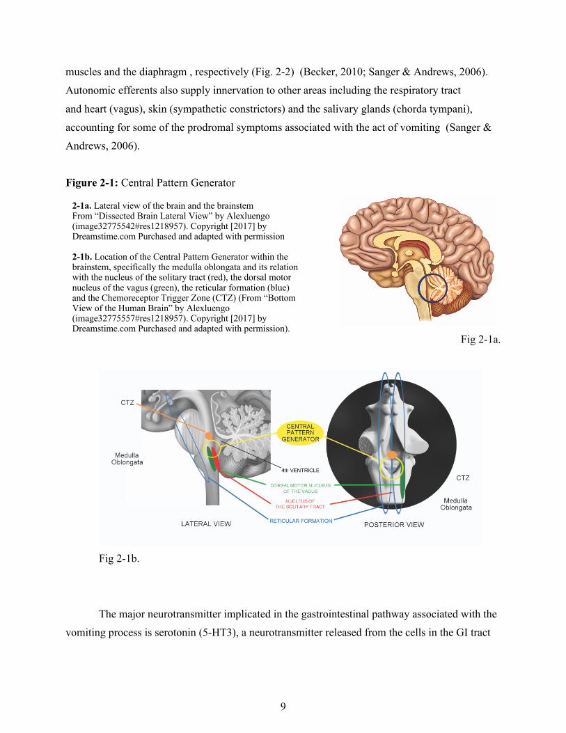
(Fig. 2-1) (Becker, 2010; Benarroch, 2011; Horn et al., 2014; Hornby, 2001; Mori et al., 2010).
Vomiting can be triggered by the activation of five main pathways that have direct neural
projections to the NST. The five pathways are as follows: The AP, gastrointestinal vagal afferent
fibres, the forebrain, the vestibular region and the cerebellum (Becker, 2010; Horn et al., 2014) .
The first and one of the most important areas involved in the process of vomiting is the
AP, also know as the Chemoreceptor Trigger Zone (CTZ). The AP constitutes one of the sensory
circumventricular organs that serve as an interface between the cerebrospinal fluid and the brain
parenchyma. This organ is located in the medulla oblongata along the walls of the fourth
ventricle and receives its blood supply from the posterior inferior cerebral arteries. This area is
characterized by lack of a blood-brain barrier, influencing the easy passage of substances present
in the blood, regardless of their lipid solubility or molecular size (i.e. large peptides such as
amylin), allowing irritants to be in direct contact with this chemosensitive region (Becker, 2010;
Benarroch, 2011; Shinpo et al., 2012).
The AP is interconnected to the NST and the lateral parabrachial nucleus through direct
visceral afferent projections originated via the vagus nerve. Simultaneously, the AP has
numerous projections to other neural structures, including the dorsal nucleus of the vagus and the

8
nucleus ambiguous for the control of gastrointestinal effectors, phonation and swallowing. The
AP also receives different descending inputs from the paraventricular nucleus, which constitutes
the main autonomic centers of the hypothalamus (Benarroch, 2011).
Numerous receptors located in the CTZ are important for the stimulation of the NST and
the CPG, including serotonin (5-HT3), dopamine (D2), acethylcholine (Ach), histamine (H),
opioid (mu), cannabinoid (CB1R, CB2R), and substance P - neuro-kinin receptor (NK1)
(Hornby, 2001; Welliver, 2013). Animal studies have demonstrated the presence of higher
concentrations of 5-HT3, D2 and opioid receptors more so than the other types of receptors in this
chemosensitive region. Studies in humans have shown that the stimulation of these three specific
receptors at the CTZ by drugs acting as agonists trigger nausea and vomiting. Similar studies
have demonstrated the antiemetic effect of 5-HT3 and D2 receptor antagonists in the area but
have failed to show similar antiemetic effects from opioid antagonist agents (Pleuvry, 2012).
The second pathway involves the gastrointestinal (GI) tract from the esophagus to the
ileum by the initiation of afferent impulses originated in mechanoreceptors and chemoreceptors
along the GI tract, which sense changes in the GI wall distension and the presence of substances
(for example, acids, alkalis, toxins and irritants) in the GI mucosa. As a result, the visceral
information from the GI receptors, especially those from the upper GI tract, generates afferent
impulses that are transmitted via the gastrointestinal vagal afferent fibres and the spinal afferent
system. The vagal afferent fibres project connections to the NST and the CPG, followed by
secondary and third-order neuronal projections, that ascend to the thalamus, hypothalamus, the
amygdala and the sensory cortex, which in response stimulate efferent pathways to the
anatomical areas involved in the vomiting process (Holtmann & Talley, 2014; Pleuvry, 2012).
The close relation of different structures to the afferent fibres of the vagal nerve supports the
importance of the integrity of the abdominal vagus as a stimulus of the vomiting process
(Hornby, 2001; A. D. Miller & Leslie, 1994).
The efferent pathways include the cranial nerves (5th, 7th, 9th, 10th and 12th) that innervate
the upper gastrointestinal tract; the vagal and sympathetic nerves that innervate the oesophagus
and stomach, causing proximal relaxation; the small intestine, triggering a retrograde contraction;
and the spinal somato-motor and phrenic motor neurons that innervate the abdominal wall
9
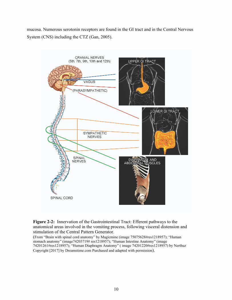
muscles and the diaphragm , respectively (Fig. 2-2) (Becker, 2010; Sanger & Andrews, 2006).
Autonomic efferents also supply innervation to other areas including the respiratory tract
and heart (vagus), skin (sympathetic constrictors) and the salivary glands (chorda tympani),
accounting for some of the prodromal symptoms associated with the act of vomiting (Sanger &
Andrews, 2006).
Figure 2-1: Central Pattern Generator
Fig 2-1a.
Fig 2-1b.
The major neurotransmitter implicated in the gastrointestinal pathway associated with the
vomiting process is serotonin (5-HT3), a neurotransmitter released from the cells in the GI tract
2-1a. Lateral view of the brain and the brainstem From “Dissected Brain Lateral View” by Alexluengo (image32775542#res1218957). Copyright [2017] by Dreamstime.com Purchased and adapted with permission 2-1b. Location of the Central Pattern Generator within the brainstem, specifically the medulla oblongata and its relation with the nucleus of the solitary tract (red), the dorsal motor nucleus of the vagus (green), the reticular formation (blue) and the Chemoreceptor Trigger Zone (CTZ) (From “Bottom View of the Human Brain” by Alexluengo (image32775557#res1218957). Copyright [2017] by Dreamstime.com Purchased and adapted with permission).
10
mucosa. Numerous serotonin receptors are found in the GI tract and in the Central Nervous
System (CNS) including the CTZ (Gan, 2005).
Figure 2-2: Innervation of the Gastrointestinal Tract: Efferent pathways to the anatomical areas involved in the vomiting process, following visceral distension and stimulation of the Central Pattern Generator. (From “Brain with spinal cord anatomy” by Magicmine (image 75075628#res1218957); “Human stomach anatomy” (image74203719# res1218957); “Human Intestine Anatomy” (image 74201261#res1218957); “Human Diaphragm Anatomy” ( image 74201220#res1218957) by Nerthuz Copyright [2017] by Dreamstime.com Purchased and adapted with permission).
11
The third pathway responds to the activation of one or more descending projections from
the cerebral cortex and thalamus and can trigger vomiting through stimulation via histamine (H)
and acethylcholine (Ach) receptors. Different factors are believed to trigger nausea and vomiting
involving the cerebral cortex, including emotions, anxiety, raised intracranial pressure and
meningeal irritation (Neoh, Adkinson, Montgomery, & Hurlow, 2014). This activation can be
seen after the stimulation of the temporal lobe (amygdala) and the insular cortex during an
epileptic seizure, which can be associated with ictal vomiting (Horn et al., 2014).
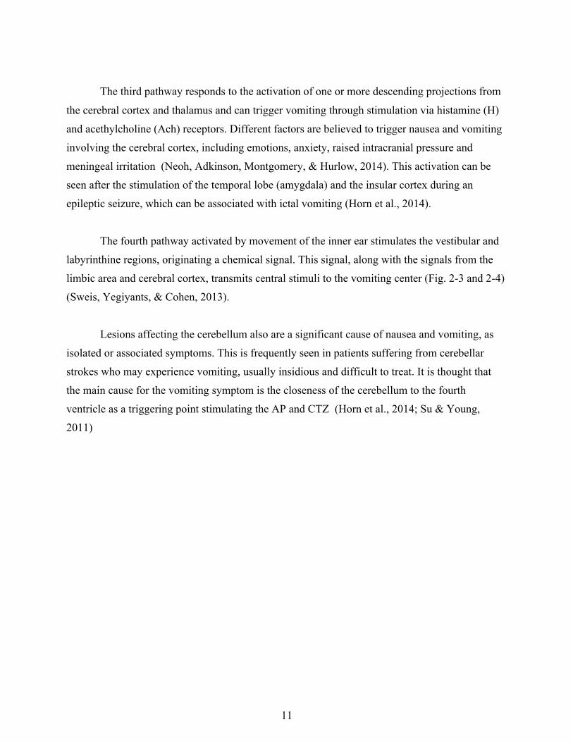
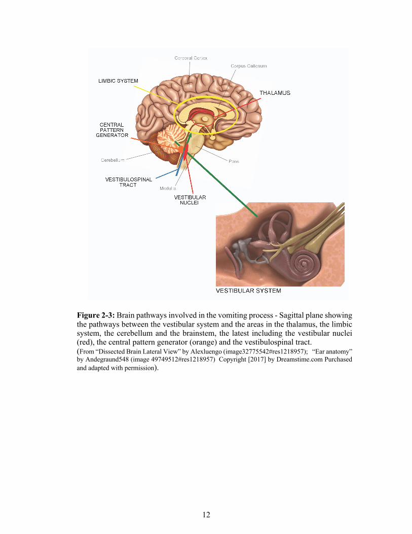
The fourth pathway activated by movement of the inner ear stimulates the vestibular and
labyrinthine regions, originating a chemical signal. This signal, along with the signals from the
limbic area and cerebral cortex, transmits central stimuli to the vomiting center (Fig. 2-3 and 2-4)
(Sweis, Yegiyants, & Cohen, 2013).
Lesions affecting the cerebellum also are a significant cause of nausea and vomiting, as
isolated or associated symptoms. This is frequently seen in patients suffering from cerebellar
strokes who may experience vomiting, usually insidious and difficult to treat. It is thought that
the main cause for the vomiting symptom is the closeness of the cerebellum to the fourth
ventricle as a triggering point stimulating the AP and CTZ (Horn et al., 2014; Su & Young,
2011)
12
Figure 2-3: Brain pathways involved in the vomiting process - Sagittal plane showing the pathways between the vestibular system and the areas in the thalamus, the limbic system, the cerebellum and the brainstem, the latest including the vestibular nuclei (red), the central pattern generator (orange) and the vestibulospinal tract. (From “Dissected Brain Lateral View” by Alexluengo (image32775542#res1218957); “Ear anatomy” by Andegraund548 (image 49749512#res1218957) Copyright [2017] by Dreamstime.com Purchased and adapted with permission).
13
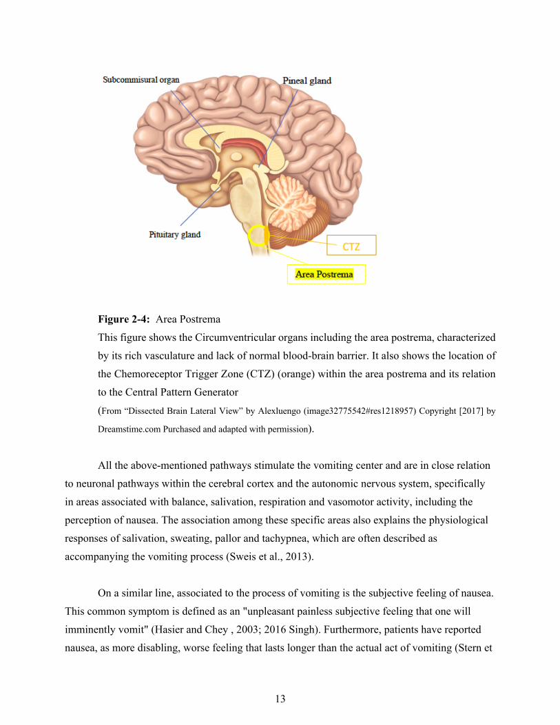
Figure 2-4: Area Postrema
This figure shows the Circumventricular organs including the area postrema, characterized
by its rich vasculature and lack of normal blood-brain barrier. It also shows the location of
the Chemoreceptor Trigger Zone (CTZ) (orange) within the area postrema and its relation
to the Central Pattern Generator
(From “Dissected Brain Lateral View” by Alexluengo (image32775542#res1218957) Copyright [2017] by
Dreamstime.com Purchased and adapted with permission).
All the above-mentioned pathways stimulate the vomiting center and are in close relation
to neuronal pathways within the cerebral cortex and the autonomic nervous system, specifically
in areas associated with balance, salivation, respiration and vasomotor activity, including the
perception of nausea. The association among these specific areas also explains the physiological
responses of salivation, sweating, pallor and tachypnea, which are often described as
accompanying the vomiting process (Sweis et al., 2013).
On a similar line, associated to the process of vomiting is the subjective feeling of nausea.
This common symptom is defined as an "unpleasant painless subjective feeling that one will
imminently vomit" (Hasier and Chey , 2003; 2016 Singh). Furthermore, patients have reported
nausea, as more disabling, worse feeling that lasts longer than the actual act of vomiting (Stern et
14
al, 2011; 2016 Singh). Similar to the process of vomiting, the stimuli for nausea is also originated
from the vestibular area, the visceral pathway and the CTZ as explained earlier. Studies have
suggested that apart from the pathways involved in the process of vomiting, the process of nausea
also correlates with specific areas of the cerebral cortex that are involved in higher cognitive
function and emotion including the medial prefrontal cortex (Miller, 1999; Napadow et al, 2013;
Singh, 2016). This may explain why nausea may be present in the absence of vomiting, through
the persistent stimulation of these areas in the cerebral cortex (Napadow et al, 2013; Horm,
2008; Singh, 2016).
Having described the different pathways associated with the process of nausea and
vomiting, we turn now to the final result, the act of vomiting.
2.1.2.1 The act of vomiting
The act of vomiting comprises three phases known as the pre-ejection phase, the ejection
phase and the post-ejection phase. The pre-ejection phase includes prodromal signs and
symptoms perceived as nausea, pallor, salivation and visceral function changes, such as
tachycardia. This phase can last from minutes to days, depending on the type of stimuli.
The ejection phase comprises retching and the final expulsion of the gastric contents.
Retching is the synchronous contraction of the diaphragm, external intercostal muscles and
abdominal muscles with a closed glottis, leading to a change in the intra-thoracic and intra-
abdominal pressure, decreasing and increasing, respectively. Vomiting completes this phase. It is
the expulsion of gastric contents after relaxation of the upper esophageal sphincter and intense
contraction of the abdominal muscles, leading to an increase of intra-thoracic and intra-
abdominal pressure to about 100 mmHg (Pleuvry, 2012).
The post-ejection phase is the final phase in this process and follows a characteristic
posture adopted to minimize the strain of muscles and structures not involved in the process and
to optimize compression that other muscles may apply over the stomach.

15
Patients may experience prodromal signs and symptoms pertaining to the pre-ejection
phase that can potentially progress to the actual act of vomiting. The patient’s ability to
communicate and express this uncomfortable feeling may be encouraged if therapy for the
prevention or management of symptoms is received in a timely fashion. However, children,
especially younger patients, may have major difficulty communicating their discomfort to care
givers, making the recognition of high risk patients an important component for the adequate
prevention and treatment of this condition.
2.2 Postoperative nausea and vomiting
Many conditions have been associated with the occurrence of nausea and vomiting.
Examples of these conditions include side effects from medications, ingestion of toxins, motion,
traumatic events and perioperative factors, the latter related to PONV (Becker, 2010).
PONV is recognized as one of the most common causes of morbidity following surgery
(Apfel, Philip et al., 2012; Gan et al., 2014). Its impact on patient recovery emphasizes the
importance of recognizing associated factors, allowing clinicians to identify patients at higher
risk of this postoperative complication.
2.2.1. Risk factors associated with postoperative nausea and vomiting
Risk factors associated with postoperative nausea and vomiting (PONV) are generally
divided into three main groups: 1. Patient-related factors; 2. factors related to the nature and
extent of the surgical procedure; and 3. factors associated with medications administered during
the perioperative period including analgesics and anaesthetic agents (Apfel et al., 2012; Becker,
2010).
16
Total Score PONV risk 0 10% 1 20% 2 40% 3 60% 4 80%
2.2.1.1. Patient-related risk factors associated with PONV
Patient-related risk factors have found to be consistent in numerous studies that evaluated
the occurrence of postoperative nausea and vomiting. In adults, the factors identified to be
independent predictors include female sex, younger age, non-smoking status and prior history of
PONV or motion sickness (Apfel et al., 2012; Apfel et al., 2012; Horn et al., 2014). One meta-
analysis of 22 studies and a total of 95154 patients identified similar main risk factors for PONV
and showed an overall incidence of PONV of 35% (18-45%) (Apfel et al., 2012). Although the
reason why being a female increases the risk of PONV more than twice that of males (OR=2.6) is
unknown, this risk in female patients, especially in post-pubertal females, persists throughout life
and is identified as the strongest independent patient-related predictor factor (Apfel et al., 2012;
Gan et al., 2014). Other patient-related factors such as history of migraine, high body mass index
and physical status classification are less likely to have a consistent correlation with increased risk
of PONV (Becker, 2010; Gan et al., 2014; Horn et al., 2014). The main risk factors and their
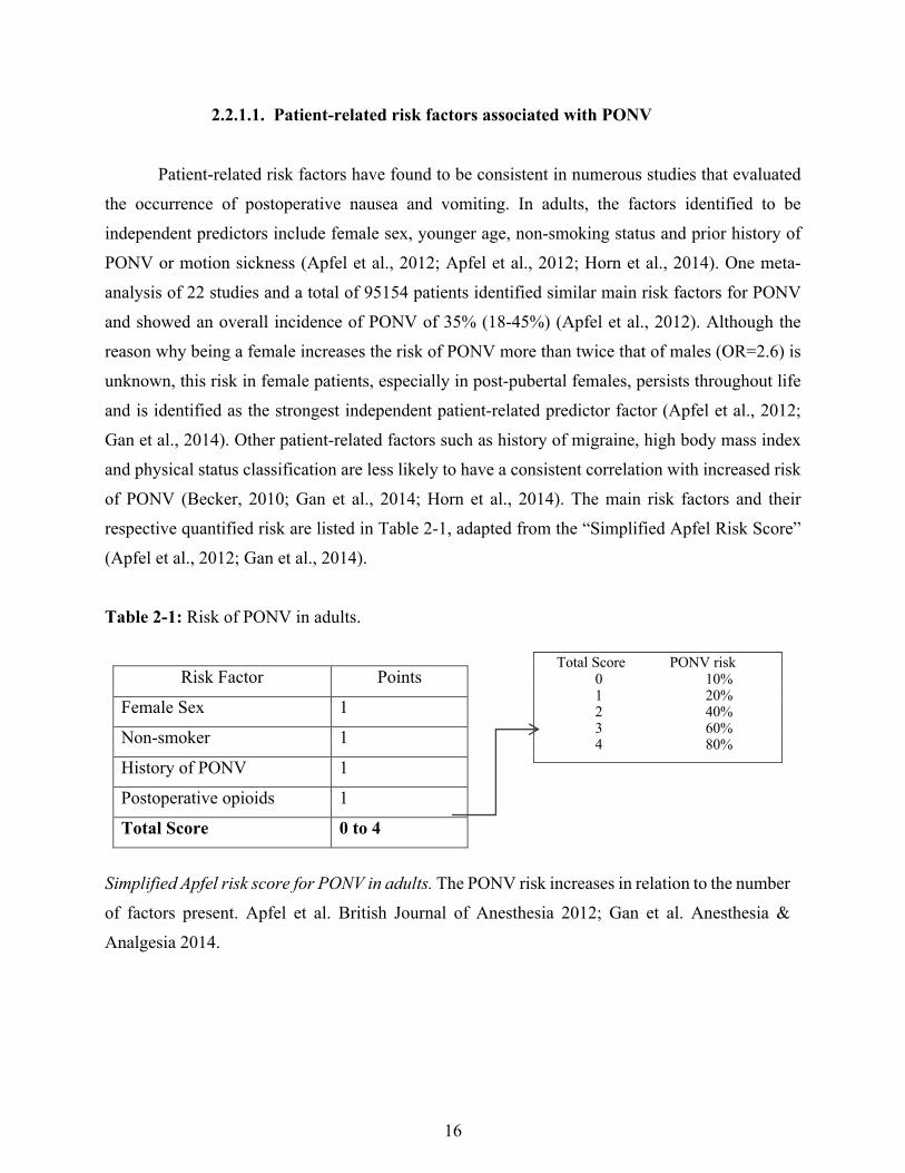
respective quantified risk are listed in Table 2-1, adapted from the “Simplified Apfel Risk Score”
(Apfel et al., 2012; Gan et al., 2014).
Table 2-1: Risk of PONV in adults.
Risk Factor Points
Female Sex 1
Non-smoker 1
History of PONV 1
Postoperative opioids 1
Total Score 0 to 4
Simplified Apfel risk score for PONV in adults. The PONV risk increases in relation to the number
of factors present. Apfel et al. British Journal of Anesthesia 2012; Gan et al. Anesthesia &
Analgesia 2014.
17
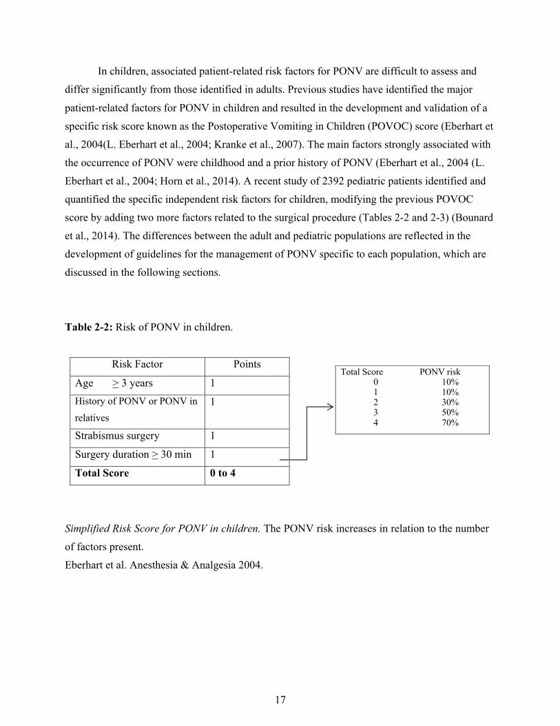
In children, associated patient-related risk factors for PONV are difficult to assess and
differ significantly from those identified in adults. Previous studies have identified the major
patient-related factors for PONV in children and resulted in the development and validation of a
specific risk score known as the Postoperative Vomiting in Children (POVOC) score (Eberhart et
al., 2004(L. Eberhart et al., 2004; Kranke et al., 2007). The main factors strongly associated with
the occurrence of PONV were childhood and a prior history of PONV (Eberhart et al., 2004 (L.
Eberhart et al., 2004; Horn et al., 2014). A recent study of 2392 pediatric patients identified and
quantified the specific independent risk factors for children, modifying the previous POVOC
score by adding two more factors related to the surgical procedure (Tables 2-2 and 2-3) (Bounard
et al., 2014). The differences between the adult and pediatric populations are reflected in the
development of guidelines for the management of PONV specific to each population, which are
discussed in the following sections.
Table 2-2: Risk of PONV in children.
Risk Factor Points
Age > 3 years 1
History of PONV or PONV in
relatives
1
Strabismus surgery 1
Surgery duration > 30 min 1
Total Score 0 to 4
Simplified Risk Score for PONV in children. The PONV risk increases in relation to the number
of factors present.
Eberhart et al. Anesthesia & Analgesia 2004.
Total Score PONV risk 0 10% 1 10% 2 30% 3 50% 4 70%
18
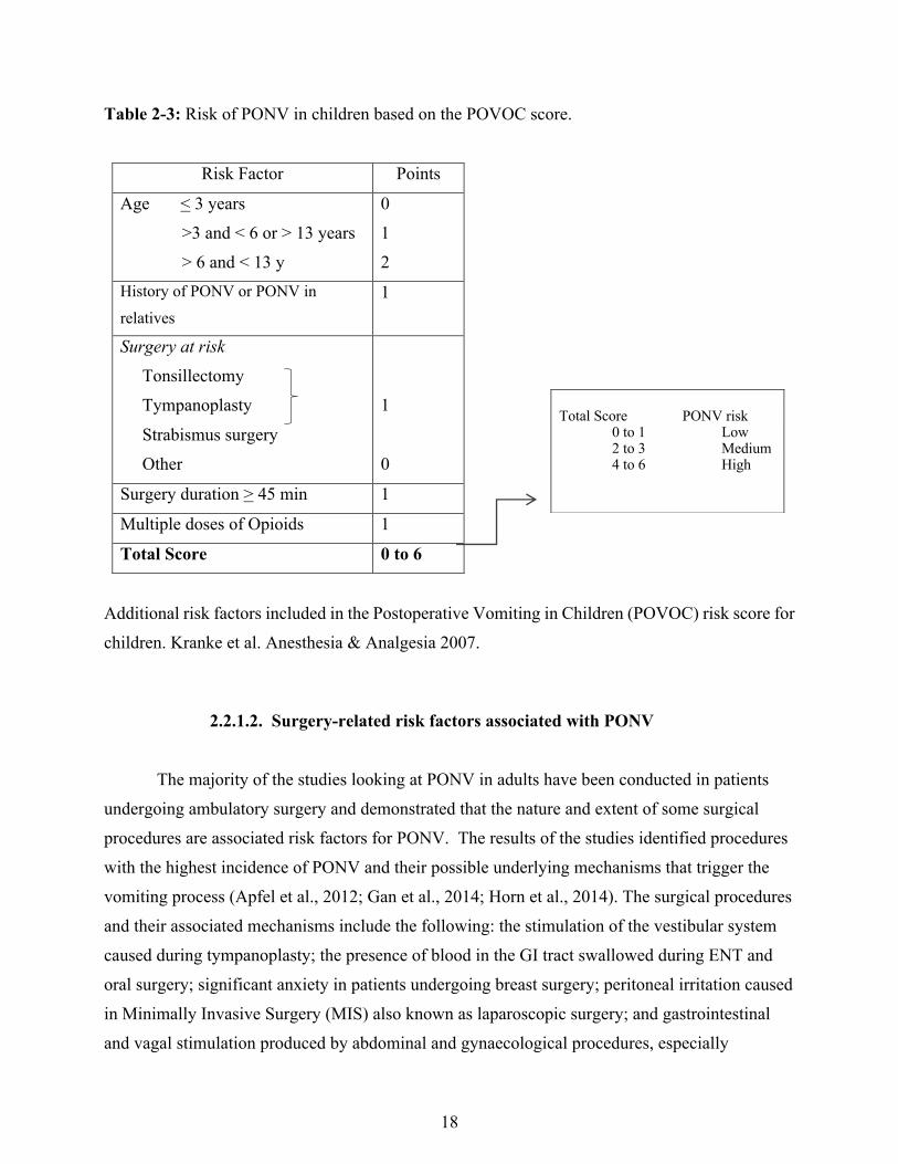
Table 2-3: Risk of PONV in children based on the POVOC score.
Risk Factor Points
Age < 3 years
>3 and < 6 or > 13 years
> 6 and < 13 y
0
1
2
History of PONV or PONV in
relatives
1
Surgery at risk Tonsillectomy
Tympanoplasty
Strabismus surgery
Other
1
0
Surgery duration > 45 min 1
Multiple doses of Opioids 1
Total Score 0 to 6
Additional risk factors included in the Postoperative Vomiting in Children (POVOC) risk score for
children. Kranke et al. Anesthesia & Analgesia 2007.
2.2.1.2. Surgery-related risk factors associated with PONV
The majority of the studies looking at PONV in adults have been conducted in patients
undergoing ambulatory surgery and demonstrated that the nature and extent of some surgical
procedures are associated risk factors for PONV. The results of the studies identified procedures
with the highest incidence of PONV and their possible underlying mechanisms that trigger the
vomiting process (Apfel et al., 2012; Gan et al., 2014; Horn et al., 2014). The surgical procedures
and their associated mechanisms include the following: the stimulation of the vestibular system
caused during tympanoplasty; the presence of blood in the GI tract swallowed during ENT and
oral surgery; significant anxiety in patients undergoing breast surgery; peritoneal irritation caused
in Minimally Invasive Surgery (MIS) also known as laparoscopic surgery; and gastrointestinal
and vagal stimulation produced by abdominal and gynaecological procedures, especially
Total Score PONV risk
0 to 1 Low 2 to 3 Medium 4 to 6 High
19
hysterectomy (Becker, 2010). Despite these possible explanations, none of the procedures have
been demonstrated to be independent predictor factors for PONV (Apfel et al., 2012).
Surgery-related risk factors in pediatric patients include the type and duration of the
surgical procedure (more than 30 minutes) as highlighted in the modified POVOC score (Table
3) (Bounard et al., 2014). Most of the previous studies of PONV in the pediatric population were
conducted in patients undergoing strabismus correction surgery, leading to the identification of
only this procedure as the main surgery-related risk factor for PONV (Apfel et al., 2012; L.
Eberhart et al., 2004). The new revised POVOC score includes tonsillectomy and tympanoplasty,
along with strabismus corrective surgery, as the main surgical procedures identified as
independent risk factors associated with the occurrence of PONV (Bounard et al., 2014).
The duration of the surgical procedure is the other significant factor in this group. This is
directly related to the length of exposure to anesthetic agents, which are discussed with the next
group of risk factors (Becker, 2010).
In addition, it is important to review the impact of sedation or general anesthesia in
patients undergoing non-surgical procedures. The National Clinical Guide Centre (NCGC) and
the National Institute for Health and Clinical Excellence (NICE) guidelines for the sedation in
children and young people, delineate the standard procedure for procedural and treatment
sedation in this population (National Clinical Guideline Centre, 2010). These guidelines define
sedation as “ a state of depressed consciousness. There are depths or levels of sedation that range from minor to major depression of consciousness”, without causing significant depression
of airway reflexes or breathing, and General anesthesia as “drug-induced loss of consciousness during which patients are not rousable, even by painful stimulation. Patients require assistance in maintaining a patent airway” (National Clinical Guideline Centre, 2010). To achieve this, the
NICE guidelines present various important aspects for the success of procedural sedation and
general anesthetic in children undergoing diagnostic or therapeutic procedures. These aspects
include adequate pediatric patient preparation, the involvement of parents and an appropriate
child-oriented environment (National Clinical Guideline Centre, 2010).
20
The most common non-surgical procedures requiring sedation or general anesthetic,
accounting for 90% are classified as painless imaging procedures such as MRI or CT scan;
painful procedures such as changes of wound dressings, minor trauma ER procedures and
orthopedic manipulation; dental procedures and endoscopy. In a quality assurance, prospective
study that included 922 children ages birth to 18 year of age, who underwent sedation (n=782) or
general anesthesia (n=140) for CT scan (n=392) or MRI (n=530) procedures (Malviya 2000), the
time of the procedure was related to adequacy of the sedation ranging from 37 to 52 minutes.
These impacted their exposure to the sedative or anesthetic agent and possible side effects after
the procedure. Overall only 12 patients (1.3%) presented with post procedural nausea and
vomiting, among other medication related adverse effects which in total accounted for 3.6% of
the population. Although, the direct association of nausea and vomiting was not reported, in
general it was reported that the use of a single or multiple agents did not show any difference in
overall adverse effects (Malviya 2000).
2.2.1.3. Perioperative medications associated with PONV
The last group of factors that influence PONV, known as emetogenic agents, consist of
analgesics and anaesthetic medications used in the perioperative period. Among analgesics,
opioids are an important part of the perioperative management because of the pain control they
provide, which contributes to the anesthetic process. Despite their benefit, opioids have been
identified as one of the primary risk factors associated with PONV (Apfel et al., 2012; Horn et
al., 2014). Theories behind the proemetic effect of opioid medications rely on their action on μ
receptors located within and outside the blood-brain barrier, the area postrema, and possibly the
nucleus of the solitary tract (Horn et al., 2014). It is believed that the degree of risk of PONV is
most likely due to the total opioid dose administered, rather than the agent and time it is given
during the perioperative period (Becker, 2010). Furthermore, some studies suggest that
intraoperative opioid use is less likely to be a continuous stimulus compared to opioid use in the
postoperative period because the emetogenic stimuli is relatively stronger (Apfel et al., 2012;
Horn et al., 2014). In addition, the fact that PONV during the postoperative period is more
frequently seen with ambulation suggests that a vestibular component may be implicated (Becker,
2010; Longnecker et al., 2011).

21
The other emetogenic agents are the anaesthetic medications used during the anaesthetic
process. The anesthetic procedure depends on the following: The type of anaesthesia chosen
based on the nature and length of the surgical procedure; the level of sedation needed; the setting
where the surgical procedure will take place (hospital or outpatient setting); the patient’s
underlying physical and physiological status prior to surgery, also known as the American
Society of Anesthesiologists (ASA) physical status classification (ASA Classification, Table 2-
4); and the qualifications and experience of the anaesthetic provider (American Society of
Anesthesiologists., 2014; R. D. Miller, Eriksson, Fleisher, Wiener-Kronish, & Young, 2010). The
ASA status classification does not include the type of anesthesia or the nature of the surgical
procedure; instead, it focuses on the quantification of the patient’s risk associated with the
surgery and the anesthetic. The presence of underlying medical conditions and aging are the key
determinant factors for the type of anesthetic to be administered (Longnecker et al., 2011). The
types of anaesthesia are broadly classified as general, regional, monitored anaesthesia care and
local anaesthesia (Longnecker et al., 2011; R. D. Miller et al., 2010). Among anaesthetic types,
general anaesthesia is recognized as a significant risk factor for the occurrence of PONV
compared to regional anaesthesia (Apfel, Stoecklein, & Lipfert, 2005).
Table 2-4: ASA Physical Status Classification System.
Physical Status
Description
ASA 1 A normal healthy patient
ASA 2 A patient with mild systemic disease
ASA 3 A patient with severe systemic disease
ASA 4 A patient with severe systemic disease that is a constant threat to life
ASA 5 A moribund patient who is not expected to survive without the operation
ASA 6 A declared brain-dead patient whose organs are being removed for donor purposes
E A patient requiring an emergency operation
American Society of Anesthesiologists (ASA) Adapted from Miller, R.D. et.al, Miller’s
Anesthesia, Churchill Livingstone Elsevier, 2010

22
To better understand the role of general anesthesia as a risk factor for PONV, it is
important to explain the main components of this process. General anesthesia has three main
phases known as induction, maintenance and emergence or recovery. These phases should be
achieved during the anesthetic process in order to provide the ideal operative conditions and
accomplish patient safety and satisfaction (Longnecker et al., 2011). The anesthetic process uses
two main types of anesthetic medications: inhalational anesthetic agents, known as gaseous
(nitrous oxide) and volatile (e.g., sevoflurane and isoflurane) anesthetics; and intravenous
anesthetics. Among their potential mechanisms of action, specific pathways have been identified
to decrease neuronal excitability by enhancing their inhibitory activity via inhibition (intravenous
anesthetic – propofol) and modulation (volatile anesthetics) of gamma-aminobutyric acid type A
(GABAA) receptor, or the inhibition of a potent excitatory glutamate receptor, the N-methyl-D-
aspartate (NMDA) (Nitrous oxide).
Inhalational anesthetics are also considered the strongest anesthesia-related risk factor for
the occurrence of postoperative nausea and vomiting (PONV), especially volatile anesthetics,
more so than nitrous oxide, because their high potential to produce PONV increases with the
length of exposure (Apfel et al., 2002; Becker, 2010; Gan et al., 2014; Horn et al., 2014).
In a literature review, the strong dose-response association between the exposure to
volatile anesthetics and the incidence of PONV was most commonly seen during the early
postoperative period (0 to 2 hours postoperatively) (Apfel et al., 2002). This association was
supported by a significant 19% reduction in PONV incidence when the use of volatile anesthetics
was avoided during the anesthetic process (Apfel et al., 2005). In addition, in a large controlled
multicenter study of 17,201 participants, the incidence of PONV was found to be similar among
different volatile anesthetics (Apfel et al., 2005; Forrest et al., 1990). The same results were
found in a meta-analysis that compared the modern volatile anesthetics - sevofluorane and
desflurane – and showed that the emetogenic effect of the volatile anesthetic was about the same
for both medications (Apfel et al., 2005; Macario, Dexter, & Lubarsky, 2005).
Compared to volatile anesthetics, nitrous oxide (N2O) has a weaker association with the
occurrence of PONV. Studies have shown that the omission of nitrous oxide decreases the risk of
PONV by a statistical but not clinical significance (Apfel et al., 2005; Divatia, Vaidya, Badwe,

23
& Hawaldar, 1996). One meta-analysis of 30 studies and 4598 patients demonstrated that when
nitrous oxide was avoided, an overall reduction of 20% in the risk of PONV was seen. This
reduction was found to be small when comparing the absolute difference in the incidence of
PONV between N2O and non-N2O groups (Fernandez-Guisasola, Gomez-Arnau, Cabrera, &
Garcia del Valle, 2010). Another meta-analysis of 29 studies and 10317 patients showed that the
increased incidence of PONV was seen in direct relation to the time of exposure, and was
especially pronounced after an hour of exposure (Peyton & Yx Wu, 2014).
In a Cochrane Interventional Review of 16 studies and 900 children, the increased risk of
PONV associated with inhalational anesthesia in children was found to be consistent with
previous studies, showing a PONV incidence difference between sevoflurane and propofol of
32.6% and 16.1%, respectively, when used during the anesthetic process (Ortiz, Atallah, Matos,
& da Silva, 2014). These findings confirm the recognition of volatile anesthetics as the strongest
anesthesia-related predictor factor associated with PONV, followed by the use of nitrous oxide
and the duration of anesthesia (Gan et al., 2014; Ortiz et al., 2014). Despite the emetogenic
effects, inhalational anesthetics continue to be used, especially in children, for the induction of
general anesthesia when intravenous access is not available at the time of the procedure
(Longnecker et al., 2011).
In contrast to inhalational anesthetics, propofol has been associated with known
antiemetic properties (Apfel et al., 2005; Becker, 2010; Gan et al., 2014). Propofol is a sedative-
hypnotic commonly used for intravenous induction and maintenance of general anesthesia and
monitored conscious sedation (Longnecker et al., 2011). Numerous studies done in adults, have
demonstrated a significant reduction of PONV when propofol is used as the maintenance
anesthetic. In one of the studies, a Randomized Controlled Trial of 2010 patients, the incidence of
PONV was reduced significantly when propofol was used as a continuous infusion or Total
Intravenous Anesthesia (TIVA) compared to inhalational anesthesia. This study showed a
reduction of the absolute risk of PONV by 15% among inpatients and by 18% among outpatients
(Visser, Hassink, Bonsel, Moen, & Kalkman, 2001). Another Randomized Controlled Trial with
5199 patients showed a similar PONV risk reduction of 19% in the propofol group (Apfel et al.,
2004). Likewise, a study of 1180 patients showed a significant difference, especially in the early
postoperative period (0-2h), between the group that received inhalational anesthetics compared to
24
the group that received propofol, the latter associated with a significantly lower incidence of
PONV (Apfel et al., 2002). These studies confirm the benefit of propofol showed by the
significant reduction of PONV and its role as a potential protective factor.
Despite its positive effect on PONV, prolonged Propofol infusions may cause serious
complications including Propofol Infusion Syndrome which has been described in adult patients,
specifically in patients at increased risk. This would include but not be limited to patients with
previous prolonged use of Propofol, with underlying metabolic disorders, critically ill patients,
and those with carbohydrate depletion conditions. Additionally, Propofol Infusion Syndrome has
been also described in children with Friederich’a ataxia and metabolic diseases, and may
influence anesthesiologists to avoid this medication as a maintenance anesthetic agent
(Mirrakhimov et al. 2015; Wolf, et al. 2001; Wolf A.R., Potter, F. 2004).
Different authors have emphasized the potential effect of some of the discussed
medications used in the perioperative period on the gastrointestinal function, which may be
disrupted after their use (Chassard et al., 2002; Holtmann & Talley, 2014; Wallden, Thorn,
Lovqvist, Wattwil, & Wattwil, 2006). Numerous studies have demonstrated that opioid
medications have a significant inhibitory effect on GI motility. Opioid medications are mediated
via opioid receptors centrally and peripherally. Yuan et al. showed that even at small doses,
morphine has a significant inhibitory effect on gastric emptying that poses an increased risk for
PONV and potential aspiration (Wallden et al., 2006; Yuan, Foss, O'Connor, Roizen, & Moss,
1998). The inhibitory effect on gastric emptying has also been demonstrated in general
anesthetics. A study with 50 participants that compared two anesthetic techniques, propofol-
remifentanil and opioid-free sevoflurane, failed to show a significant difference on gastric
emptying between the two groups, but when compared with gastric emptying in a normal state
(no surgery, no anesthesia), a significant delay in gastric emptying pattern was seen with the use
of both anesthetics. As shown in the study, the effect of inhaled anesthetic agents on delayed
gastric emptying may cease after the agent is discontinued (Wallden et al., 2006). Although some
evidence suggests that high doses of propofol may inhibit GI motility, a study performed on 10
healthy volunteers showed that propofol used at subhypnotic doses (light sedation) was not
associated with gastric emptying delay (Chassard et al., 2002).
25
Overall, the evidence presented in this section supports the importance of recognizing the
major risk factors for the occurrence of PONV included in the simplified risk score for adults and
children. This recognition represents the first step in the prevention and management of this
condition.
2.3 Prevention and management of PONV In past decades, most research on PONV has emphasized the need to reduce the incidence
of PONV by identifying patients at high risk for this condition and by developing strategies
focused on the prevention and management of PONV.
2.3.1 Antiemetic medications It has become commonplace to differentiate antiemetic medications according to their
mechanism of action and their efficacy on the antagonist effect at the specific receptor site within
the vomiting center and associated areas. Based on their site of action, antiemetics can be
classified as Serotonin (5-HT3) receptor antagonists, substance P - neurokinin (NK1) receptor
antagonists, dopamine (D2) receptor antagonists, corticosteroids, butyrophenones, histamine
type1 (H1) receptor antagonists and muscarinic cholinoceptor antagonists (anticholonergics)
(Table 2-5).
2.3.1.1. Serotonin type 3 (5-HT3) receptor antagonists
The main agents in this category are ondansetron, dolasetron, granisetron, tropisetron and
palonosetron. Despite their similar mechanisms of action, these medications differ in their
chemical configuration and pharmacological properties, including their affinity for the 5-HT3
receptor, the duration of effect, the dose response and the P450 (CYP) system component
involved in their metabolism (Gan, 2005).

26
Serotonin receptor (5-HT3) antagonist agents were initially developed to effectively
control radiation- and chemotherapy-induced nausea and vomiting because this type of therapy
triggers the release of serotonin from the gastrointestinal wall, stimulating the vomiting center as
well (Becker, 2010). In the mid 1980s, the effects of metoclopramide were attributed partially to
serotonin antagonism, which prompted the development of selective serotonin receptor
antagonists, thus improving the management of nausea and vomiting (Gan, 2005).
The 5-HT3 receptors, identified as sodium channel type receptors, are localized in the
Central Nervous System (CNS) in the Area Postrema (AP) and throughout the peripheral tissue,
especially in the bowel (via vagal afferents) and those areas involved in the vomiting process. As
mentioned, the afferent signals travel along the vagal nerve, reaching the nucleus of the solitary
tract and the chemoreceptor trigger zone (CTZ), thus stimulating the Central Pattern Generator in
the brainstem (Becker, 2010; Gan, 2005). All these areas have abundant 5-HT3 receptors,
suggesting that the inhibition of these receptors at multiple levels of the vomiting process may
denote a key element for the efficacy of serotonin receptor antagonists (Gan, 2005).
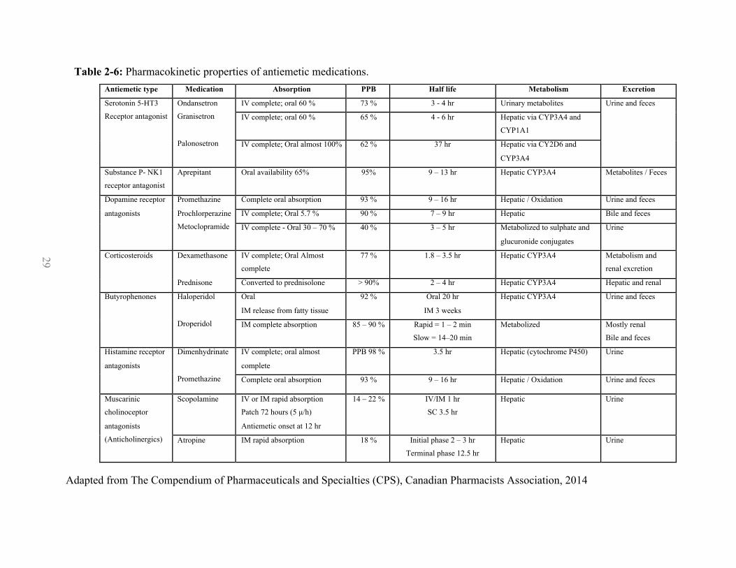
The pharmacokinetic properties of serotonin 5-HT3 receptor antagonists are shown in
Table 2-6. Of all the agents, ondansetron has the shortest half-life (3.4 hours), which can be
prolonged in elderly patients (Gan, 2005; Longnecker et al., 2011) and may be relevant when
comparing different medications and the incidence of PONV, as shown in a randomized
controlled trial of 75 patients (Sun, Klein, & White, 1997). This study showed that the incidence
of PONV in the early postoperative period was similar when ondansetron was administered at
different times during the perioperative period (induction and emergence), but that there was a
significantly lower requirement of rescue antiemetics in the recovery area when ondansetron was
administered at the end of the procedure. This observation can possibly be explained by the
relatively short half-life of ondansetron, which could contribute to the apparent ineffectiveness of
this medication when administered at the beginning of the procedure (Sun et al., 1997).
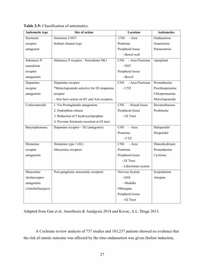
27
Table 2-5: Classification of antiemetics.
Antiemetic type Site of action Location Antiemetics
Serotonin
receptor
antagonist
Serotonin 5-HT3
Sodium channel type
CNS - Area
Postrema
Peripheral tissue
- Bowel wall
Ondansetron
Granisetron
Palonosetron
Substance P-
neurokinin
receptor
antagonists
Substance P receptor - Neurokinin NK1 CNS - Area Postrema
- NST
Peripheral tissue
- Bowel
Aprepitant
Dopamine
receptor
antagonists
Dopamine receptor
*Metoclopramide selective for D2-dopamine
receptor
- Also have action on H1 and Ach receptors.
CNS - Area Postrema
- CTZ
Promethazine
Prochlorperazine
Chlorpromazine
Metoclopramide
Corticosteroids 1. Via Prostaglandin antagonism
2. Endorphine release
3. Reduction of 5-hydroxytriptophan
4. Prevents Serotonin secretion at GI tract
CNS - Neural tissue
Peripheral tissue
- GI Tract
Dexamethasone
Prednisone
Butyrophenones Dopamine receptor – D2 (antagonist) CNS - Area
Postrema
- CTZ
Haloperidol
Droperidol
Histamine
receptor
antagonists
Histamine type 1 (H1)
Muscarinic receptors
CNS - Area
Postrema
Peripheral tissue
- GI Tract
- Labyrintine system
Dimenhydrinate
Promethazine
Cyclizine
Muscarinic
cholinoceptor
antagonists
(Anticholinergics)
Post-ganglionic muscarinic receptors Nervous System
-ANS
- Medulla
Oblongata
Peripheral tissue
- GI Tract
Scopolamine
Atropine
Adapted from Gan et al. Anesthesia & Analgesia 2014 and Kovac, A.L. Drugs 2013.
A Cochrane review analysis of 737 studies and 103,237 patients showed no evidence that
the risk of emetic outcome was affected by the time ondansetron was given (before induction,

28
during induction, intraoperatively or postoperatively), although treatment of nausea and vomiting
was seen more often in cases when ondansetron was given intraoperatively (Carlisle &
Stevenson, 2006).
Of the first generation 5-HT3 receptor antagonist medications, ondansetron is the most
commonly used antiemetic and is considered to be the “gold standard” when compared with other
antiemetics (Gan et al., 2014; Horn et al., 2014; Skolnik & Gan, 2014). Since 1991, different
studies assessing the antiemetic efficacy of ondansetron have shown that this medication is as
effective as other serotonin receptor antagonists and other antiemetics, including dexamethasone
(Apfel et al., 2004; Gan, 2005; Subramaniam et al., 2001). A randomized controlled trial of the
data of 4123 patients found that ondansetron, dexamethasone and droperidol each reduced the
risk of postoperative nausea and vomiting by about 26 percent, indicating that the different
antiemetic medications are similarly effective for the prevention of PONV (Apfel et al., 2004).
Compared to ondansetron and other medications in this category, palonosetron, a second
generation 5HT3 antagonist, has a prolonged plasma half-life of 40 hours, which may explain its
complete response (no vomiting episodes or rescue antiemetics) during the first 24 hours after the
administration of a single dose before induction of anesthesia, as shown in a study of 574 patients
(Candiotti, Kovac, Melson, Clerici, & Gan, 2008). A RCT of 98 participants that evaluated the
efficacy of a single dose of palonosetron compared to ondansetron for the prevention of PONV in
the first 24 hours following surgery failed to show any significant difference in number of PONV
episodes, suggesting that both medications had similar efficacy despite their different half-lives
(Laha, Hazra, & Mallick, 2013; Skolnik & Gan, 2014).
29
Table 2-6: Pharmacokinetic properties of antiemetic medications.
Adapted from The Compendium of Pharmaceuticals and Specialties (CPS), Canadian Pharmacists Association, 2014
Antiemetic type Medication Absorption PPB Half life Metabolism Excretion
Serotonin 5-HT3
Receptor antagonist
Ondansetron
Granisetron
Palonosetron
IV complete; oral 60 % 73 % 3 - 4 hr Urinary metabolites Urine and feces
IV complete; oral 60 % 65 % 4 - 6 hr Hepatic via CYP3A4 and
CYP1A1
IV complete; Oral almost 100% 62 % 37 hr Hepatic via CY2D6 and
CYP3A4
Substance P- NK1
receptor antagonist
Aprepitant Oral availability 65% 95% 9 – 13 hr Hepatic CYP3A4 Metabolites / Feces
Dopamine receptor
antagonists
Promethazine
Prochlorperazine
Metoclopramide
Complete oral absorption 93 % 9 – 16 hr Hepatic / Oxidation Urine and feces
IV complete; Oral 5.7 % 90 % 7 – 9 hr Hepatic Bile and feces
IV complete - Oral 30 – 70 % 40 % 3 – 5 hr Metabolized to sulphate and
glucuronide conjugates
Urine
Corticosteroids Dexamethasone
Prednisone
IV complete; Oral Almost
complete
77 % 1.8 – 3.5 hr Hepatic CYP3A4 Metabolism and
renal excretion
Converted to prednisolone > 90% 2 – 4 hr Hepatic CYP3A4 Hepatic and renal
Butyrophenones Haloperidol
Droperidol
Oral
IM release from fatty tissue
92 % Oral 20 hr
IM 3 weeks
Hepatic CYP3A4 Urine and feces
IM complete absorption 85 – 90 % Rapid = 1 – 2 min
Slow = 14–20 min
Metabolized Mostly renal
Bile and feces
Histamine receptor
antagonists
Dimenhydrinate
Promethazine
IV complete; oral almost
complete
PPB 98 % 3.5 hr Hepatic (cytochrome P450) Urine
Complete oral absorption 93 % 9 – 16 hr Hepatic / Oxidation Urine and feces
Muscarinic
cholinoceptor
antagonists
(Anticholinergics)
Scopolamine
IV or IM rapid absorption
Patch 72 hours (5 µ/h)
Antiemetic onset at 12 hr
14 – 22 % IV/IM 1 hr
SC 3.5 hr
Hepatic Urine
Atropine IM rapid absorption 18 % Initial phase 2 – 3 hr
Terminal phase 12.5 hr
Hepatic Urine
29
30
In children, the use of ondansetron as a prophylactic agent for PONV has been studied as well. In a meta-analysis of 13 randomized controlled trials and 2,006 pediatric patients, seven of the studies found the incidence of PONV to be lower in the ondansetron group (37.2%) compared to placebo (65.6%). Only one of the studies analyzed compared ondansetron with dexamethasone, finding an overall PONV incidence that was not significantly different between the groups (Shen et al., 2014).
A large volume of published studies compared the efficacy of ondansetron when
administered at different doses, based on the recommended prophylactic dose of ondansetron as 4 mg intravenously (IV) or 8 mg orally (Gan et al., 2014). A systematic review, including fifty-three studies and 12,889 patients who received either a placebo or any of the different ondansetron regimens for the prevention of PONV, found that at 4 mg, the Number Needed to Treat (NNT) for the prevention of vomiting and nausea was 6 and 7, respectively (Tramer, Reynolds, Moore, & McQuay, 1997). The previously mentioned Cochrane review showed that the risk of PONV was increased by 1.43 when onsansetron doses were halved, but the person’s age, gender or type of surgery did not impact the effect of the medication (Carlisle & Stevenson, 2006). In children, the dose used in the majority of the studies ranged between 0.1 and 0.2 mg/kg, (Shen et al., 2014), which correlates poorly with the current recommendations of 0.05-0.1 mg/kg (up to 4 mg) for the prevention and management of PONV in children (Gan et al., 2014).
2.3.1.1.1. Side effects of serotonin (5-HT3) receptor antagonists General side effects and their incidence have been identified with the use of 5-HT3 receptor antagonists. The most commonly reported side effects in adults and children are headache (4-22%) and constipation (1-19%) (Canadian Pharmacists Association, 2014; Carlisle & Stevenson, 2006).
Other reported adverse effects include diarrhea (8-16%), malaise or fatigue (9-13%), drowsiness (8%), anxiety (6%), fever (7-8 %), urinary retention (5%), itchiness (1-5%) and pain at the injection site (4%), flushing (<1%), dizziness (0.01%), movement disorders (0.1 – 0.3%)
31
and electrocardiographic changes, including QTc prolongation (<1%), which could lead to the development of Torsades de Pointes (Canadian Pharmacists Association, 2014).
There are special warnings related to potential ECG changes. As suggested in the e-
Compendium of Pharmaceuticals and Specialties (e-CPS), “All 5-HT3 receptor antagonists cause a dose-dependent prolongation of the QTc interval” (Canadian Pharmacists Association, 2014). 5-HT3 antagonists’ mechanism of action consists of blocking sodium channel conductance, requiring special precautions in patients with significant electrolyte imbalance, pre-existing cardiac conduction abnormalities or arrhythmias, as well as in other patients receiving medications that could potentially prolong the QTc interval (tricyclic antidepressants, venlafaxine citalopram, clarithromycin and methadone) (Canadian Pharmacists Association, 2014; Gan, 2005).
A systematic review failed to find any reports of cardiac arrhythmias in adults associated
with only one dose of ondansetron. In contrast, this review found the occurrence of cardiac arrhythmias in 49 reports associated with more than a single dose of ondansetron, mainly administered intravenously. Correlation was seen in patients with risk factors for developing cardiac arrhythmias, including electrolyte imbalance, cardiac arrhythmias or interaction with medications also associated with QTc prolongation (Freedman, Uleryk, Rumantir, & Finkelstein, 2014). Based on the updated Drug Safety Communication published by the Food and Drug Administration in 2012, ondansetron was associated with dose dependent prolonged QTc and routine screening, and ECG studies were not necessary unless patients with a significant medical history and associated risk factors were identified or when an intravenous dose over 16 mg of ondansetron was required (Food and Drug Administration, U.S., 2012; Freedman et al., 2014).
Likewise, adverse effects of 5-HT3 in children have been reported with similar occurrences to those described in adults. However, in a systematic review, fewer publications and reported cases of prolonged QTc were found for pediatric patients (Freedman et al., 2014). Eleven pediatric cases associated with prolonged QTc interval were identified, but, in many cases, ondansetron was used concomitantly with other pro-arrhythmic medications. The cases failed to show a significant correlation between the administration of ondansetron and the occurrence of QTc prolongation (Freedman et al., 2014). Another randomized study of 80 healthy

32
pediatric patients failed to find significant ECG differences when ondansetron and droperidol were compared. Both medications had an increased QTc interval that remained within the normal limits (Mehta, Sanatani, & Whyte, 2010). Similar recommendations are to be followed for both children and adults, including the identification of patients at higher risk of developing cardiac arrhythmias and proper administration of these 5-HT3 antagonists’ medications following the recommended preventive and management doses guidelines (Tables 2-7 and 2-7a) (Gan et al., 2014).
2.3.1.2. Substance P - neurokinin (NK1) receptor antagonists
Substance P – Neurokinin (NK1) receptor antagonists prevent the binding of substance P to the neurokinin receptor (NK1R). Since the discovery of substance P and its receptor in 1931, this substance was isolated from the emetic centers of the brain at the NST (Nucleus of the Solitary Tract) and the AP (Area Postrema) and from the bowel. However, it was not until the 1990s that the antagonist medications that would block the receptor effectively and had a prolonged half-life (40 hours) were used to manage nausea and vomiting especially delayed chemotherapy-induced nausea and vomiting (Aziz, 2012; Gan et al., 2014). Aprepitant is the only NK1R antagonist medication approved by the FDA. A RCT comparing aprepitant and ondansetron for the prevention of PONV showed similar responses at 24 hours in the postoperative period. In the longer postoperative period, aprepitant was significantly more effective than ondansetron beyond the 24-hour period after surgery for the reduction of nausea and vomiting (Diemunsch et al., 2007; Gan et al., 2014). Another multicenter RCT that analyzed data from 805 participants showed similar results, demonstrating superior efficacy compared to ondansetron in the prevention of vomiting in the first 24- and 48- hour period, albeit not for nausea control or antiemetic rescue medication requirements (Gan, Apfel et al., 2007).
33
2.3.1.2.1. Side effects of substance P - neurokinin (NK1) receptor
antagonists The main adverse effect of NK1R antagonists is associated with the increased bioavailability of dexamethasone when administered concurrently with these medications, increasing the incidence of infection among patients treated with both medications (Aziz, 2012). These medications are mostly used for chemotherapy-induced nausea and vomiting, but further studies are needed to investigate their benefit in the prevention of PONV and their use in the pediatric population. The recommended doses are listed in Tables 2-7 and 2-7a (Gan et al., 2014).
2.3.1.3. Dopamine (D2) receptor antagonists and butyrophenones
The antiemetic effect of dopamine (D2) receptor antagonists and Butyrophenones is attributed to a competitive antagonism of dopamine (D2) receptors at the CTZ (Chemoreceptor Trigger Zone) / AP (Area Postrema), specifically by the inhibition of adenylate cyclase, altering the content of AMPc within the neurons located at the AP and the NST (Horn et al., 2014; Skolnik & Gan, 2014).
Medications that pertain to this category include promethazine, prochlorperazine Chlorpromazine and metoclopramide. Haloperidol and droperidol, other dopamine receptor antagonist agents also known as butyrophenones, are frequently used as highly potent neuroleptics, and their antiemetic properties have also been recognized (H. S. Smith, Cox, & Smith, 2012).
Although an antipsychotic, chlorpromazine was used for years as an effective antiemetic in patients requiring antineoplastic medications, and the published experienced using this medication as an antiemetic agent is significant. With the introduction of newer antiemetic medications, when chlorpromazine was compared for its antiemetic properties it was not as effective. In a study comparing other antiemetics with chlorpromazine in children, this
34
medication failed to show significant improvement preventing vomiting as the other medications, and was associated with possible preventable extrapyramidal symptoms (Dupuis et al., 2013; Marshall, Kerr, Vowels, O'Gorman-Huges, & White, 1989).
Metoclopramide, a derivative of procainamide, is a commonly used antiemetic that also acts as a selective agonist of a specific type of serotonin receptor (5-HT3), which creates prokinetic action along the upper gastrointestinal tract by increasing the lower esophageal sphincter tone, thereby increasing gastric motility and promoting gastric emptying (Becker, 2010). A multicentre study of 3140 patients compared different doses of metoclopramide in combination with dexamethasone (Wallenborn et al., 2006). The risk of vomiting increased after smaller doses of metoclopramide were given (Carlisle & Stevenson, 2006). Previous studies have shown that droperidol is as effective as dexamethasone and ondansetron for the prevention of PONV, including a Cochrane review, which also demonstrated that the effectiveness of the medication was not influenced by the patient’s age, gender, the type of surgery performed, or the time in relation to the procedure for which the medication was administered. It was evident that smaller doses of droperidol increased the risk of PONV compared to larger doses (Horn et al., 2014). This type of medication is not used as a first line antiemetic prophylaxis as will be explained later in the section. 2.3.1.3.1. Side effects of dopamine (D2) receptor antagonists The inhibition of D2 receptors’ main adverse effects is mediated by interfering with the dopamine transmission within the basal ganglia, resulting in different movement disorders known as extrapyramidal syndrome. This syndrome includes akathisia, tardive dyskinesia and Parkinsonian symptoms (Becker, 2010). The incidence of extrapyramidal syndrome is dose dependent, as shown in a study of 3140 patients who received metoclopramide at different doses along with dexamethasone. The antiemetic effects were seen mostly within the early postoperative period (0-12 hour). Doses between 10 and 25 mg and a higher dose (50 mg) were needed to achieve a prolonged antiemetic (from 12 to 24 hours). Other adverse effects documented in the study included hypotension and tachycardia, with an incidence of either event
35
that increased with the dose (8.8% for 0 mg, 11.2% for 10 mg, 12.9% for 25 mg and 17.9% for 50 mg) (Food and Drug Administration, U.S., 2001; Wallenborn et al., 2006). A safety alert was released in 2001 by the Food and Drug Administration after cases of death associated with QTc prolongation and Torsades de Pointes with the use of droperidol were reported. The safety alert included new dosage ranges for droperidol below the previously recommended dose and instructions to use this medication as a last resource once patients have failed to respond to other first line antiemetic medications (Food and Drug Administration, U.S., 2001; Gan et al., 2014). Further studies have demonstrated that droperidol had similar safety parameters to ondansetron at doses for the prevention of PONV below the previously recommended dose (Mehta et al., 2010). Recommended doses for dopamine receptor antagonists for prevention and management of PONV are shown in Tables 2-7 and 2-7a.
2.3.1.4. Corticosteroids
In 1993, the antiemetic effect of corticosteroids was described for the first time, followed by subsequent studies that confirmed its efficacy. After all these years, the antiemetic mechanism of action of corticosteroids for the prevention of PONV is not well understood (Henzi, Bernhard, Tramèr, & Phil, 2000). Different mechanisms of action have been proposed for the antiemetic effect of glucocorticoids, including the following (Henzi et al., 2000; Holte & Kehlet, 2002).
1. Via prostaglandin antagonism 2. Endorphine release resulting in mood elevation with a subsequent sense of well being and
appetite stimulation. 3. Possible reduction of 5-hydroxytryptophan in neural tissue and decrease in tryptophan,
which may have proemetic effects. 4. Prevention of serotonin release from the gastrointestinal tract secondary to its anti-
inflammatory effect.
36
5. Possible potentiation of other antiemetics through the sensitization of pharmacological receptors, including the 5-HT3 receptor.
Multiple studies have been conducted comparing glucocorticoids, mainly a single dose of
dexamethasone alone or in combination with other antiemetics. Dexamethasone has been shown to have an important role for the prevention of PONV in several studies (Holte & Kehlet, 2002). For example, a systematic review that analyzed 17 trials and data from 1947 patients found that dexamethasone was effective in the prevention of PONV (Henzi et al., 2000). This systematic review found seven trials, four trials in adults and three in children, comparing dexamethasone with placebo. The results were found to be statistically significant in favour of dexamethasone for the prevention of postoperative vomiting. The same review found ten studies comparing dexamethasone alone with combination of dexamethasone and another antiemetic. The only significant benefit by enhancing the antiemetic effect was seen with the concomitant use of dexamethasone with ondansetron.
Multiple studies have been conducted on the pediatric population showing similar benefits for the prevention of PONV as those shown in adults. One randomized study of 147 children undergoing tonsillectomy showed that a single dexamethasone injection at the induction of the anesthesia reduced the incidence of PONV (Hermans, De Pooter, De Groote, De Hert, & Van der Linden, 2012)In a meta-analysis of 13 RCTs, including 2006 children, the incidence of postoperative vomiting was found to be higher in the placebo group than in the dexamethasone group (68.2% vs. 34.3%) (Shen et al., 2014). In this study, the combination of ondansetron and dexamethasone was significantly more effective for the reduction of postoperative vomiting than each antiemetic administered separately. Similarly, a Cochrane review for the use of steroids in children analyzed data from 15 studies and 1273 pediatric patients and found a significant difference in the incidence of vomiting between patients who received intravenous dexamethasone and those who received placebo (21% vs. 48%), favouring steroids for the prevention of postoperative vomiting (Steward, Grisel, & Meinzen-Derr, 2011)
37
2.3.1.4.1. Side effects of corticosteroids
One of the important concerns for corticosteroid administration in children is the possible
side effects that a single steroid dose can cause. Some of the feared side effects in children have been documented after the high dose long-term use of corticosteroids. Some of these side effects are decreased growth, peptic ulceration, psychological effects, including aggressive behaviour and attention deficit hyperactivity disorder, and osteonecrosis of the hip or any other joint (Yee & Cox, 2013). This last devastating adverse effect was documented in a study of 1409 children who received high doses of prednisone for the treatment of leukemia for a period of over 7 years (Mattano, Sather, Trigg, & Nachman, 2000). In this study, the incidence of osteonecrosis was 9.3% and was higher in patients over 10 years of age (Mattano, Sather, Trigg, & Nachman, 2000). Similar results were documented in a recently published retrospective study of 1095 oncologic children, with a cumulative incidence of 3.6% for the occurrence of osteonecrosis in children (Hyakuna et al., 2014). However, single and lower dose corticosteroids have been found not to cause such detrimental effects and could prevent osteonecrosis, as demonstrated in a review where the higher cumulative steroid dose was shown to correlate with worse effects (Winkel, Pieters, Wind, Bessems, & van den Heuvel-Eibrink, 2014).
The incidence of infection in patients receiving a single perioperative dose of dexamethasone was similar in both dexamethasone and placebo groups in different studies, as shown in a systematic review and meta-analysis of 45 studies and 5795 patients (Waldron, Jones, Gan, Allen, & Habib, 2013). Forty studies (1449 patients) failed to report any significant difference in the incidence of wound infection in patients that received dexamethasone or placebo. The studies also failed to show any significant difference in delayed healing. Another study conducted of septic patients show no difference in patient outcomes and complication rates between single dose dexamethasone and control groups (Yee & Cox, 2013). Similarly, a recent study of 431 patients failed to show any significant difference for an increased risk of developing wound complications or delayed wound healing between the dexamethasone and placebo groups (Bolac, Wallace, Broadwater, Havrilesky, & Habib, 2013).
These findings suggest that a single cortisone dose does not increase the incidence of postoperative wound infection. Based on the evidence, the use of dexamethasone effectively
38
prevents postoperative nausea and vomiting and should be given after the induction of anesthesia as recommended in the Consensus Guidelines for the Management of PONV (Gan et al., 2014). Recommended doses for the prevention and management of PONV are shown in Tables 2-7 and 2-7a.
2.3.1.5. Histamine type 1 (H1) receptor antagonists
Numerous histamine type 1 (H1) receptor antagonists have antiemetic properties but are not specific on preventing PONV. Agents in this category include promethazine, cyclizine and dimenhydrinate, the latest being the most common antihistamine used for the management of PONV (Horn et al., 2014). These medications are known to antagonize histamine type 1 (H1) and muscarinic cholinergic receptors (RxFiles, 2012). Their main antiemetic effect is attributed to the inhibition of histamine and probably muscarinic receptors localized in the AP (Area Postrema) and the vestibular nucleus. Second-generation antihistamines (for example, astemizole) do not have effective antiemetic properties, as they cannot cross the blood-brain barrier.
A randomized control trial of 133 patients showed a decreased incidence of PONV when dimenhydrinate was used compared to the placebo (L. Eberhart, Seeling, Bopp, Morin, & Georgieff, 1999), which was comparable to a meta-analysis of 18 studies and 3045 patients, showing that dimenhydrinate had a better relative benefit than the placebo (64% vs. 54%) (Kranke, Morinz, Roewer, & Eberhart, 2002). In a Cochrane review, there was not convincing evidence of the superior antiemetic effect of dimenhydrinate when compared to ondansetron, although cyclizine, another histamine type 1 receptor antagonist, did show a similar antiemetic response when compared to other medications, including ondansetron and dexamethasone (Carlisle & Stevenson, 2006).
39
2.3.1.5.1. Side effects of histamine type 1 (H1) receptor antagonists
Although there is insufficient data on the optimal timing, dose response and safety profile of histamine type 1 (H1) receptor antagonists for the management of PONV, common side effects are related to their associated anticholinergic properties. The most commonly reported side effects include drowsiness, urinary retention, dry mouth and blurred vision (Gan et al., 2014; RxFiles, 2012).
2.3.1.6. Muscarinic cholinoceptor antagonists (sp. anticholonergics).
The main mechanism of action of muscarinic cholinoceptor antagonists is secondary to the competitive inhibition at the post-ganglionic muscarinic receptors in the autonomic nervous system, specifically the parasympathetic nervous system and the subsequent reduction of exocrine secretions and of gastrointestinal peristalsis. It also has central effects by the inhibition of cholinergic transmission in the vestibular region and the inhibition of muscarinic receptor located in the medulla oblongata (Horn et al., 2014; Pleuvry, 2012; RxFiles, 2012).
Medications included in this category are scopolamine and atropine. Scopolamine is a central acting anticholinergic agent initially used for the treatment of motion sickness and is the most commonly used agent used in this category. This agent has been used recently as an adjunct therapy for the prevention and management of PONV (Apfel et al., 2010; Gan et al., 2014). The short half-life of this medication requires it to be administered transdermally (Horn et al., 2014). A meta-analysis of 25 studies and 3298 patients demonstrated the efficacy of transdermal scopolamine for the prevention of PONV, with both early and late patch application (Apfel et al., 2010). In another study, a multicenter RCT that recruited 620 participants, the combination of transdermal scopolamine and intravenous ondansetron was more effective than ondansetron alone in the prevention of PONV within the first 24 hours following surgery but not later. The authors reported that the effectiveness of the combined therapy was not associated with adverse effects, specifically those associated with the scopolamine patch, suggesting a good tolerance of patients to this medication (Gan, Sinha, Kovac, Jones, & Cohen, 2009; Skolnik & Gan, 2014).
40
2.3.1.6.1. Side effects of muscarinic cholinoceptor antagonists
Different adverse effects associated with anticholinergic agents have been reported and divided into early and late adverse effects. Early adverse effects include dry mouth, blurred vision and sedation. Late adverse effects include central cholinergic syndrome and confusion, especially in elderly patients with underlying cognitive impairment (Apfel et al., 2010; Gan et al., 2014; RxFiles, 2012). A meta-analysis demonstrated that the most common associated side effect of scopolamine was visual disturbances especially between 24 to 48 hours from the time of the application when compared to placebo (45% vs 8% RR 3.35, p-value < 0.001) (Apfel et al., 2010).
2.3.1.7. Other antiemetic agents As described above, one of the anaesthetic agents that have known antiemetic properties is propofol, which is used as an intravenous agent for the induction and maintenance of anesthesia. Studies have demonstrated that propofol may decrease the incidence of early PONV, and it has therefore been recommended by the guidelines for the management of PONV to reduce the baseline risk for this complication (Gan et al., 2014).
Other medications that are effective as adjuvant therapies in combination with first line antiemetic agents include alpha2-agonists (clonidine), which has weak and short term effects; mirtazapine (noradrenergic), an antidepressant that has been shown to delay the onset of PONV when combined with dexamethasone; and gabapentin, which has been shown to be effective in the prevention of PONV, especially when used in combination with dexamethasone; and benzodiazepines (midazolam), which have been shown in anxiolytic comparative studies to be effective in the prevention of PONV when used preoperatively and in combination with ondansetron or dexamethasone (Gan et al., 2014; Kovac, 2013).
The use of dextrose, orally and intravenously, for the prevention of PONV has been the
target of several studies, mainly in the adult population. In recent years, this intervention has received special attention because of the positive impact during the recovery period on patients who undergo elective surgical procedures (Hausel et al., 2005; M. D. Smith et al., 2014). A
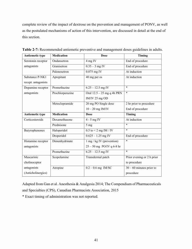
41
complete review of the impact of dextrose on the prevention and management of PONV, as well as the postulated mechanisms of action of this intervention, are discussed in detail at the end of this section.
Table 2-7: Recommended antiemetic preventive and management doses guidelines in adults.
Antiemetic type Medication Dose Timing
Serotonin receptor antagonists
Ondansetron 4 mg IV End of procedure
Granisetron 0.35 – 3 mg IV End of procedure
Palonosetron 0.075 mg IV At induction
Substance P-NK1 recept. antagonists
Aprepitant 40 mg per os At induction
Dopamine receptor antagonists
Promethazine 6.25 – 12.5 mg IV *
Prochlorperazine
Oral 12.5 – 25 mg q 4h PRN IM/IV 25 mg OD
*
Metoclopramide 20 mg PO Single dose 10 – 20 mg IM/IV
2 hr prior to procedure End of procedure
Antiemetic type Medication Dose Timing
Corticosteroids Dexamethasone 4 - 5 mg IV At induction
Prednisone 5 mg *
Butyrophenones Haloperidol 0.5 to < 2 mg IM / IV *
Droperidol 0.625 – 1.25 mg IV End of procedure
Histamine receptor antagonists
Dimenhydrinate
1 mg / kg IV (prevention) 25 – 50 mg PO/IV q 6-8 hr
*
Promethazine 6.25 – 12.5 mg IV *
Muscarinic cholinoceptor antagonists (Anticholinergics)
Scopolamine Transdermal patch Prior evening or 2 h prior to procedure
Atropine 0.2 – 0.6 mg IM/SC 30 – 60 minutes prior to procedure
Adapted from Gan et al. Anesthesia & Analgesia 2014; The Compendium of Pharmaceuticals and Specialties (CPS), Canadian Pharmacists Association, 2015 * Exact timing of administration was not reported.

42
Table 2-7a: Recommended antiemetic dose prophylaxis guidelines in children.
Antiemetic type Medication Dose Timing
Serotonin receptor antagonists
Ondansetron a
50 – 100 mcg/kg IV OD up to 4 mg
End of procedure
Granisetron 40 mcg /kg IV OD up to 0.6 mg
End of procedure
Tropisetron 0.1 mg/kg IV OD 0.2 up to 2 mg
At induction
Corticosteroids Dexamethasone 150 mcg/kg IV OD up to 5 mg
At induction
Butyrophenones Droperidol 10 – 15 mcg/kg IV OD up to 1.25 mg
End of procedure
Histamine receptor antagonists
Dimenhydrinate
0.5 mg/kg IV OD up to 25 mg
*
Adapted from Gan et al. Anesthesia & Analgesia 2014; The Compendium of Pharmaceuticals and Specialties (CPS), Canadian Pharmacists Association, 2015 * Exact timing of administration was not reported. a Approved for prevention of POV in children aged 1 month and older
2.3.2 Non-pharmacologic prophylaxis
2.3.2.1 Intravenous fluid
Reducing the baseline PONV risk is important for the prevention and further management of this complication. One of the strategies that has shown its efficacy is adequate intravenous (IV) fluid hydration in the perioperative period (Gan et al., 2014). Different studies have been conducted to identify the best fluid hydration strategy for the perioperative setting and the amount that would assist in the reduction of PONV incidence. One of these studies compared two different IV fluid infusion rates (10 ml/kg vs. 30 ml/kg) in 141 adult patients undergoing elective surgery (Magner, McCaul, Carton, Gardiner, & Buggy, 2004). There was a significant difference in the incidence of emesis (25.7% vs. 8.6%) and severe nausea (15.7% vs. 2.9%) with the higher
43
IV fluid infusion rate (Magner, McCaul, Carton, Gardiner, & Buggy, 2004). Other studies in the pediatric population have had similar findings. A RCT of 100 children randomized into two different IV fluid infusion rates (10 ml/kg vs. 30 ml/kg of Ringer’s Lactate) showed a decrease in the incidence of PONV in children who received the higher IV fluid infusion rate (Goodarzi, Matar, Shafa, Townsend, & Gonzalez, 2006). Similar studies were analyzed in a quantitative review of 15 trials, which compared conservative IV fluid therapy with supplemental (higher IV infusion rate) crystalloid fluid administration. Most of the studies showed a significant difference between the groups, favouring the group that received supplemental fluid over the conservative fluid group (Apfel, Meyer et al., 2012). This additional strategy may contribute to the effective prevention and management of PONV in both adults and children.
In view of all that has been mentioned so far, it is evident that research has focused on the development of new medications that, acting as agonists or antagonists of different emetic active sites, can provide a vast pool of options for the treatment and management of postoperative nausea and vomiting.
2.3.3 Current Guidelines for prophylaxis and management of PONV Numerous published guidelines for the management of PONV are found in the literature, including recently updated 2014 consensus guidelines from the Society for Ambulatory Anesthesiology (SAA). This consensus was based on previous guidelines published in 2003 and 2007 as well as a systematic literature review conducted by the SAA (Gan et al., 2003; Gan et al., 2007; Gan et al., 2014). The guidelines contain different features of the management of PONV and include updated data on risk factor assessment for individuals at increased risk for PONV, new treatment options for the management of this condition using single and combined options, dosing and optimal timing for antiemetic prophylaxis, and new recommendations on the prevention and management of PONV (Gan et al., 2014). All the recommendations are based on the level of evidence supporting them and applied as guidelines for evidence-based medicine. Evidence Grade (EG) and grade practice recommendation guidelines are delineated in Table 8 (Centre for Evidence-Based Medicine, Oxford, 2014).
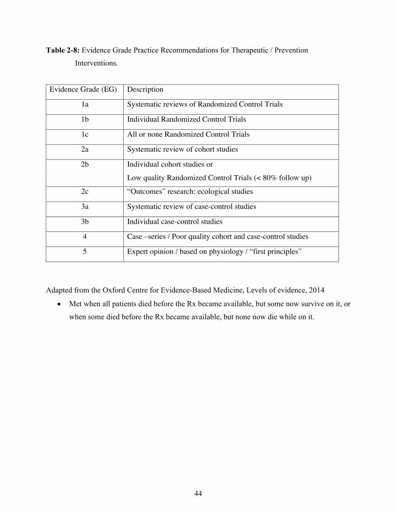
44
Table 2-8: Evidence Grade Practice Recommendations for Therapeutic / Prevention Interventions.
Evidence Grade (EG) Description
1a Systematic reviews of Randomized Control Trials
1b Individual Randomized Control Trials
1c All or none Randomized Control Trials
2a Systematic review of cohort studies
2b Individual cohort studies or
Low quality Randomized Control Trials (< 80% follow up)
2c “Outcomes” research; ecological studies
3a Systematic review of case-control studies
3b Individual case-control studies
4 Case –series / Poor quality cohort and case-control studies
5 Expert opinion / based on physiology / “first principles”
Adapted from the Oxford Centre for Evidence-Based Medicine, Levels of evidence, 2014
• Met when all patients died before the Rx became available, but some now survive on it, or when some died before the Rx became available, but none now die while on it.
45
2.3.3.1. Baseline PONV risk reduction
As mentioned in previous sections, despite differences between adult and children PONV risk assessment, the incidence of PONV in both populations increases proportionally to the number of risk factors present in an individual (Tables 2-2, 2-3 and 2-4) (Apfel, Laara, Koivuranta, Greim, & Roewer, 1999; Apfel et al., 2012; Bounard et al., 2014). The updated guidelines give recommendations on strategies for reducing the baseline risk, including the use of regional anesthesia and avoidance of general anesthesia, minimizing the use of nitrous oxide (EG 1a) and volatile anesthetics (EG 2a), both known risk factors for PONV. Evidence of the negative impact of inhalation anesthetic agents as risk factors for PONV was discussed earlier (EG 1a). Other recommendations to decrease the baseline risk for PONV include the minimization of perioperative opioid use (EG 1a), adequate hydration (EG 1a) and the preferable use of propofol as an antiemetic agent for the induction and maintenance of the anesthetic process (EG 1a) (Gan et al., 2014). One study that demonstrated the efficacy of these recommendations in reducing the baseline risk factors for the prevention of PONV was the IMPACT study, which evaluated 6 strategies for reducing PONV in 5199 participants (Apfel et al., 2004). This study found that the combination of propofol given as TIVA and oxygen had additive positive effects in reducing PONV risk by 25%.
2.3.3.2. One or two interventions for PONV prevention in adults Multiple studies have shown the efficacy of different antiemetic medications for the prevention and management of PONV. However, only a few of these medications have been approved and recommended as first and second line agents in the current guidelines from the SAA (Gan et al., 2014). Evidence on the efficacy of these agents was reviewed previously. A RTC comparing different strategies of single and combined agents showed that the combination of different antiemetic agents for the prevention and management of PONV had favourable outcomes compared to the use of a single agent, especially in patients with more than one risk factor for PONV (Apfel et al., 2004; Gan et al., 2014). 5-HT3 antagonists are one of the
46
most commonly used agents in combination with droperidol or dexamethasone, showing better results and lowering the incidence of PONV (Apfel et al., 2004). To minimize potential adverse effects when medications are combined, dosing adjustments are required. This adjustment is reflected in studies where ondansetron was used in combination with dexamethasone, in which the maximum recommended dose was lower than the dose recommended when each medication was used as a single agent. The dosing relation for each recommended agent (single and combined) in children and adults at medium and high risk are listed in Tables 2-9 (Gan et al., 2014).
2.3.3.3. PONV prophylaxis
Although prophylaxis for PONV is recommended only for those patients at medium and high risk, there is insufficient evidence to support the practice of giving prophylaxis to all patients undergoing surgical procedures. For those patients at high risk for PONV, the updated guidelines recommend routine prophylaxis with at least two antiemetics from different classes of combination therapy to optimize their adjunct effects (Gan et al., 2014). Most studies on combined antiemetic therapy have demonstrated the efficacy of ondansetron, dexamethasone or droperidol in combination compared to their use as single agents. A systematic review and meta-analysis of 8 studies and 811 patients showed a trend towards the increased benefit of combination therapy over single drug use when ondansetron and droperidol were compared (L. H. Eberhart, Morin, Bothner, & Georgieff, 2000). Another meta-analysis of randomized controlled trials that reviewed 33 trials and 3,447 patients demonstrated similar antiemetic effects and safety profiles when comparing one of the 5-HT3 antagonist agents with either dexamethasone or droperidol (Habib, El-Moalem, & Gan, 2004). These combination regimens were significantly more effective than either medication used as single agent. When dexamethasone was compared with six different antiemetic medications, a meta-analysis of 17 trials and 1946 patients demonstrated that dexamethasone was an effective antiemetic as a single agent but also that its antiemetic effect was increased, especially when combined with one of the 5-HT3 antagonist agents (Henzi et al., 2000).
47
In children, the same principles for PONV prophylaxis in adults should be followed when applying the SAA guidelines as this population is at higher risk for PONV. The presence of risk factors in children suggests the use of at least two antiemetic agents from different classes for the prevention strategy, which should include a 5-HT3 antagonist agent with dexamethasone as recommended by the guidelines, unless a contraindication exists (Gan et al., 2014; Hohne, 2014). If the patient presents POV, the guidelines recommend administering a different class of antiemetic, which is different from the antiemetic agent used initially for prophylaxis.
2.3.3.4. Antiemetic therapy for patients who have not received prophylaxis or
in whom prophylaxis therapy has failed
Management of patients with established PONV, in whom prophylactic therapy has failed or who failed to receive prophylactic therapy can be controversial. The guidelines recommend that the treatment for these patients should include the administration of an antiemetic agent of a different class than that previously used in the preoperative or intraoperative period (Table 2-9) (Gan et al., 2014). In a RCT of 2199 patients who received prophylactic antiemetic therapy with ondansetron, the repeated dose of the same antiemetic agent for the treatment of PONV in those patients in whom the prophylactic therapy was not successful offered no additional control of this complication (Kovac et al., 1999). The SAA guidelines recommend not giving the same prophylactic antiemetic agent for the treatment of PONV within 6 hours following the initial dose, as it confers no additional benefit (Gan et al., 2014). If patients complain of PONV in the immediate postoperative period, measures are recommended, starting by making a complete evaluation to rule out any physical or other causes related to medications that were administered in the perioperative period (Gan et al., 2014; Hohne, 2014).
If no prophylactic agent is given, a low-dose 5-HT3 antagonist agent should be administered in the postoperative period as these agents have been adequately studied for established PONV (Gan et al., 2014). Alternative medications for established PONV include dexamethasone, droperidol, promethazine and propofol, the latest being as effective as
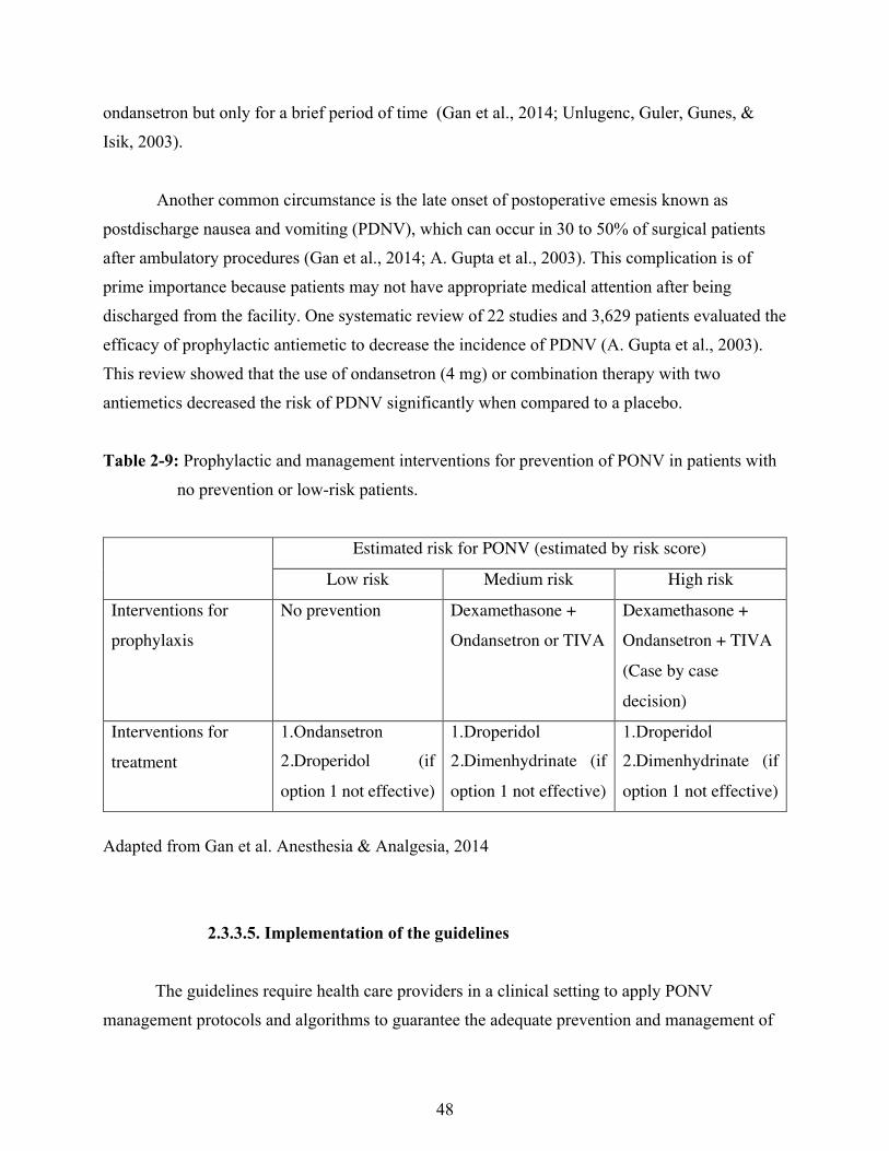
48
ondansetron but only for a brief period of time (Gan et al., 2014; Unlugenc, Guler, Gunes, & Isik, 2003). Another common circumstance is the late onset of postoperative emesis known as postdischarge nausea and vomiting (PDNV), which can occur in 30 to 50% of surgical patients after ambulatory procedures (Gan et al., 2014; A. Gupta et al., 2003). This complication is of prime importance because patients may not have appropriate medical attention after being discharged from the facility. One systematic review of 22 studies and 3,629 patients evaluated the efficacy of prophylactic antiemetic to decrease the incidence of PDNV (A. Gupta et al., 2003). This review showed that the use of ondansetron (4 mg) or combination therapy with two antiemetics decreased the risk of PDNV significantly when compared to a placebo. Table 2-9: Prophylactic and management interventions for prevention of PONV in patients with no prevention or low-risk patients.
Estimated risk for PONV (estimated by risk score)
Low risk Medium risk High risk
Interventions for
prophylaxis
No prevention Dexamethasone +
Ondansetron or TIVA
Dexamethasone +
Ondansetron + TIVA
(Case by case
decision)
Interventions for
treatment
1.Ondansetron 2.Droperidol (if
option 1 not effective)
1.Droperidol 2.Dimenhydrinate (if
option 1 not effective)
1.Droperidol 2.Dimenhydrinate (if
option 1 not effective)
Adapted from Gan et al. Anesthesia & Analgesia, 2014
2.3.3.5. Implementation of the guidelines
The guidelines require health care providers in a clinical setting to apply PONV
management protocols and algorithms to guarantee the adequate prevention and management of
49
this common complication. The SAA has created an algorithm (Fig. 2-5) to be followed as a guideline, which starts with the proper recognition of risk factors and the selection of the most appropriate management strategy (Table 2-10) (Gan et al., 2014). A study comparing the incidence of PONV with the incidence documented by nursing staff found that only 42% of PONV cases in the PACU and 29% of PONV cases among admitted patients were recognized and documented by nursing staff (Franck et al., 2010). This finding indicates that recognizing patients at higher risk for developing PONV and avoiding treating patients until after symptoms are present may increase the effectiveness of the antiemetic medications given.
Educational strategies can be associated with an increased implementation of antiemetic
prophylaxis. A study of 384 patients undergoing elective surgery compared the incidence of PONV before and after the implementation of an educational strategy by placing the Apfel’s simplified score scale in each chart, thereby allowing nurses to recognize patients at higher risk of developing PONV (Sigaut et al., 2010). The study results showed that there was no significant difference in the antiemetic prophylaxis administration rate between the overall patient populations (before 31.4% vs. after 36.8%). A significant difference was seen on the rate of antiemetic prophylaxis administered to high-risk patients, which were identified based on the Apfel’s simplified score (before 36.4% vs. after 52.8%). Other studies have shown a suboptimal PONV prevention reflected in the low proportion of patients (37%) that received the specified antiemetic prophylaxis (Gan et al., 2014). The SAA guidelines support the implementation of an antiemetic multimodal prevention strategy, which follows protocols that allow anaesthesiologists and healthcare professionals to facilitate and standardize the prevention and management of PONV (Gan et al., 2014). A prospective, observational study followed 134 patients who underwent elective laparoscopic cholecystectomy to evaluate the efficacy of a standardized anaesthesia/analgesia protocol for the identification of postoperative complications such as pain and PONV. The results showed an incidence of moderate and severe PONV of 11% and 2% respectively and identified pain and PONV as predictors of an extended stay in the PACU (Jensen, Kehlet, & Lund, 2007). Another observational study of 500 patients who underwent elective surgery showed that the implementation of a standardized anesthetic protocol, including PONV prevention, was effective for a prompt and smooth postoperative recovery (Bergland, Gislason, & Reader, 2008). This

50
study shows how multimodal prevention and standardized protocols may improve the identification, prevention and management of PONV compared to a strict risk-based approach without standardized procedures (Gan et al., 2014).
So far, a clear understanding of the different options in the armamentarium for the prevention and management of PONV has been presented. The updated guidelines from the Society for Ambulatory Anesthesia provide an important framework not only for the treatment of PONV but also for the need to recognize patients at higher risk and our role as healthcare providers for the prevention, implementation of guidelines and timely intervention in this particular setting. As discussed above, one of the options for the prevention and management of PONV is the use of oral or intravenous dextrose, an intervention that may add safe and positive results to the existing armamentarium of antiemetic medications and interventions. Table 2-10: Pharmacologic combination for POV prophylactic therapy in children and adults.
Combination therapy and suggested dose Level of
evidence
Adults Droperidol + dexamethasone
5-HT3 receptor antagonist + dexamethasone
5-HT3 receptor antagonist + droperidol
5-HT3 receptor antagonist + dexamethasone + dropedriol
1a
1a
1a
2a
Children Ondansetron 0.05 mg/kg + dexamethasone 0.015 mg/kg
Ondansetron 0.1 mg/kg + droperidol 0.015 mg/kg
Tropisetron 0.1 mg/kg + dexamethasone 0.5mg/kg
1a
1a
1a
Adapted from Gan et al. Anesthesia & Analgesia, 2014
2.3.4 Effects of different interventions on glucose metabolism
Before examining the role of intravenous dextrose in the prevention of PONV, it is necessary to review the impact of fasting, surgery and administration of dextrose and other medications on the glucose metabolism, as this will assist in explaining theories of the underlying mechanism of action and possible antiemetic properties of this intervention.
51
2.3.4.1 Glucose Metabolism
Over recent decades, researchers have investigated the impact of glucose metabolism impairment, especially in conditions such as diabetes, leading to a better understanding of the physiology involved and possible treatment options. For this purpose, numerous studies have compared glucose metabolism in normal individuals and in diabetic patients, leading to the current knowledge in this area.
Plasma glucose concentration is the relation between the glucose that enters the circulation balanced by the glucose removal rate from the circulation. The main three sources of glucose in the circulation are from the following: intestinal absorption, which depends on how fast glucose appears in the circulation after gastric emptying; glycogenolysis (glycogen breakdown); and gluconeogenesis produced from lactate, amino acids (alanine) and glycerol (fatty tissue) during the fasting period (Aronoff, Berkowitz, Shreiner, & Want, 2004; Nygren, 2006).
2.3.4.1.1 Glucoregulatory hormones
Glucoregulatory hormones are important for the maintenance of circulating glucose concentrations within normal narrow levels. These hormones include the following: glucagon (released from the alfa-cells of the pancreas); insulin and amylin (derived from pancreatic Beta-cells); glucagon-like peptide-1; (GLP-1) and glucose-dependent insulinotropic peptide (GIP) (derived from the L-cells of the intestine); epinephrine and cortisol (released from adrenal gland); and growth hormone (Aronoff et al., 2004). Although the role of some of these hormones will be discussed related to the administration of dextrose, specific details regarding each of these hormones and the physiopathology involved in glucose metabolism is beyond the goals of this thesis.
52
Glucagon is one of the main hormones involved in the process of hepatic glucose production through glycogenolysis and gluconeogenesis during the fasting period. During the first 8 to 12 hours of fasting, the main source of glucose is glycogenolysis, whereas gluconeogenesis is the principal source during longer periods of fasting (Aronoff et al., 2004). These processes are the mechanisms used to maintain plasma glucose levels, as glucose leaves the circulation at a constant rate during fasting periods.
In contrast to the main role of glucagon as a regulator of glucose appearance in plasma,
insulin is one of the main anabolic hormones secreted in response to increased levels of glucose and amino acids following a meal, and regulates the use of glucose in the tissues, mainly in the skeletal muscles and the adipose tissue, lowering the glucose plasma concentration. This hormone also suppresses the endogenous production of glucose (glycogenolysis and gluconeogenesis) by its direct action and the paracrine effect suppressing glucagon release. Insulin secretion is stimulated by different stimuli that include glucose, which is the most potent stimulus of insulin release; increased amino acid plasma concentration released from the intestine; and the triggering of the parasympathetic system through vagus nerve stimulation (Aronoff et al., 2004).
2.3.4.1.2. Fasting and its metabolic effect during surgery
As discussed earlier, glucose metabolism and the resulting glucose plasma levels are regulated based on glucose uptake and the kind of stimulus activated. The metabolic adaptation of the body to periods of fasting allows the body to maintain normal blood glucose levels to provide the energy required for all necessary functions. This situation is seen normally at night when natural fasting takes place. Overnight fasting follows a post-absorptive state after the last meal is absorbed, lowering glucose levels that stimulate glycogenolysis and gluconeogenesis and promoting the entry of neo-glucose into the circulation. In general, liver glycogen is used within 24 hours and gluconeogenesis is the source of glucose, especially for those tissues that depend on glucose for their survival (Nygren, 2006). The lack of substrate present during the fasting state stimulates glucagon and catecholamine production, increases tissue insulin resistance, decreases

53
glucose usage in peripheral tissues and reduces insulin levels, which induces the mobilization of free fatty acids (FFA) and fatty acid oxidation, resulting in the formation of ketone bodies known as ketogenesis (Fukao et al., 2014; Nygren, 2006; Schricker, Latterman, Wykes, & Carli, 2004). Different studies have tried to correlate the energy metabolites as a function of fasting time by measuring blood total ketone bodies, blood glucose and FFA. One prospective study that compared the metabolic response to fasting in children and adults, found that despite the maintenance of similar plasma glucose concentrations within the first 18 hours of fasting among participants, children developed rapid and higher concentrations of total ketone bodies compared to adults over the initial 30 hours of fasting (p<0.001) (Haymond, Karl, Clarke, Pagliara, & Santiago, 1982). Similar results were found when blood lactate was measured, demonstrating higher levels in children compared with adults within 6 to 30 hours of fasting (p<0.002). Taken together, these results may suggest a direct correlation between the adaptation process in fasting states and aging, which can be explained by different characteristics that differentiate infancy and childhood from adulthood. These characteristics include higher energy demands as a function of body weight during infancy, which decreases more than two times in adulthood, better glycogen stores and increased muscle mass during childhood, providing a greater reservoir of protein that serves as substrate for gluconeogenesis during the fasting period (Bonnefont et al., 1990; Fukao et al., 2014). Research on glucose metabolism has been conducted primarily in children with symptoms associated with suspected pathologies caused by underlying errors of glucose metabolism. The metabolic response in patients with these pathologies is normally evaluated by implementing controlled fasting tests, which are also used in research and associated study protocols. In a prospective study that included 48 children who were referred for metabolic evaluation and 11 children with underlying known inherited hypoketotic or hyperketotic related conditions, all participants underwent fasting for a 24-hour period (Bonnefont et al., 1990; Fukao et al., 2014). Results showed that younger children (1 – 7 years) had higher values of plasma ketone bodies and lower levels of glucose when compared to older pediatric patients, demonstrating that after 15 hours of fasting ketosis developed faster in younger children than in older children. Similar findings were described in a prospective study that included 167 children,
54
showing that patients younger than 7 years of age had lower plasma glucose levels and higher levels of total ketone bodies and FFA (van Veen et al., 2011). Similar to the results of glucose metabolism in fasting periods, surgery is characterized by insulin resistance, which is developed shortly after the start of a surgical procedure and becomes more pronounced, particularly soon after the end of the procedure (Nygren, 2006). This characteristic impairment of insulin sensitivity may be the result of the perioperative elevation of counter regulatory hormones, glucagon and cortisol seen in situations of increased metabolic stress, including surgery (Schricker, Lattermann, Fiset, Wykes, & Carli, 2001). Different studies have demonstrated that insulin sensitivity was reduced by 50% in otherwise healthy individuals after uncomplicated elective surgery (open cholecystectomy), which was directly proportional to the extent and magnitude of the surgical procedure (Thorell, Nygren, & Ljungqvist, 1999). In addition, studies have shown that insulin resistance leads to elevated insulin and glucose plasma levels secondary to an increased gluconeogenesis response. This response can be attenuated by a reduction of about 50% of postoperative insulin resistance when preoperative fasting is avoided, as shown in different studies involving the administration of preoperative glucose (Nygren, 2006).
Previous studies have shown a negative effect of insulin resistance by increasing postoperative complications in patients with type 2 diabetes with associated hyperglycemia (Behdad, Mortazavizadeh, Ayatollahi, Khadiv, & Khalilzadeh, 2014). It has been suggested that measuring insulin resistance following elective surgery could provide information on the degree of metabolic disturbance caused by the surgical procedure and provide a parameter of the length of stay, which has been found to have a significant correlation with the degree of insulin resistance (Thorell et al., 1999). A prospective study by Hahn et al. (2013) showed different results in 52 non-diabetic patients with insulin resistance prior to surgery who had an apparent lower incidence of postoperative complications, especially lower postoperative nausea and vomiting and fewer episodes of hypotension compared to those patients with no insulin resistance. Insulin resistance was calculated after the implementation of an intravenous glucose tolerance test during the preoperative fasting period of all participants (Hahn & Ljunggren, 2013)
55
However, compared to the reduced metabolic rates seen during fasting, the metabolic response during surgery is increased. The catabolic response triggered by conditions that increase metabolic stress, including surgical procedures and trauma, is characterized by an elevated substrate oxidation and the resulting glycogen, lipid and protein breakdown, leading to subsequent loss of body protein as substrate for the production of neo-glucose during the gluconeogenesis process. A prospective study that assessed the associated dynamic changes of glucose and protein metabolism during surgery in 12 patients found that independent of the anesthetic technique (desflurane vs. IV propofol), there was a decreased rate of protein synthesis and associated whole protein breakdown, decreased aminoacid oxidation and glucose production, and reduced glucose clearance (Schricker et al., 2001).
Hyperketonemia associated with gluconeogenesis during the fasting period has been shown to increase total ketone bodies levels, specifically blood acetoacetate (1.0 mmol/L) and hydroxybutyrate (2-3 mmom/L) However, a similar response has not been demonstrated during surgery, as prospective studies have shown that mild hyperketonemia was evident at 2 hours following abdominal surgery with concentrations documented below 0.5 mmol/L (Schricker et al., 2001). These findings may suggest that elevation of total ketone bodies in plasma may be most likely associated with the fasting metabolic effect during the preoperative period rather than with a direct effect from the increased metabolic stress related to the surgical procedure. Preoperative fasting has been the focus of multiple studies seeking to determine not only its metabolic effects, as discussed previously, but also the appropriate timing for fasting to optimize patient preparation and avoid perioperative vomiting and possible pulmonary aspiration. 2.3.4.1.3 Preoperative fasting conditions and guidelines
Numerous recommendations and guidelines have been published to determine the appropriate fasting period in healthy patients needed to ensure complete gastric emptying prior to an elective operative procedure. According to the American Society of Anaesthesiologists (ASA) guidelines, preoperative fasting is defined as “a prescribed period of time before a procedure when patients are not allowed the oral intake of fluids or solids” (American Society of

56
Anesthesiologists, ASA., 2011). These guidelines were initially published in 1999, when concerns were raised about the risk of perioperative pulmonary aspiration (American Society of Anesthesiologists, ASA., 2011; Longnecker et al., 2011; Maltby, 2006). This condition is defined as the aspiration of gastric contents that may occur after the anesthetic induction, either during the procedure or during the immediate postoperative period. Perioperative pulmonary aspiration may have detrimental consequences, including aspiration pneumonia, respiratory complications and airway compromise (American Society of Anesthesiologists, ASA., 2011; Longnecker et al., 2011). The first studies looking at preoperative fasting times date to the 1970s, when complete NPO “nil per os” fasting after midnight was mandated (Maltby, 2006; Roberts, 2013). Some researchers focused their attention on gastric emptying after the ingestion of fluids or solids. A RCT demonstrated that in healthy patients, complete gastric emptying after the ingestion of clear fluids required a period of 2 hours to pass through the stomach, and that the residual gastric volume was not different to that of patients who had ingested fluids 2 hours or 12 to 16 hours before the procedure (Maltby, 2006). It was also reported that because of the difference in processing solid food based on the size and quantity of the particles ingested, gastric emptying takes longer. This was confirmed by measuring the food processing time until the stomach was completely emptied following the ingestion of a light meal compared to indigestible food, which lasted for a period of 4 and 6 to 12 hours, respectively (Maltby, 2006; Worobitz & Pounder, 1985). A meta-analysis of randomized controlled trials showed that for clear fluids, preoperative fasting periods of 2 to 4 hours accounted for smaller gastric volumes and higher pH values (> 2.5) compared to those patients with fasting periods restricted to more than 4 hours prior to the procedure. In children, similar findings were shown when the same fasting periods were compared (American Society of Anesthesiologists, ASA., 2011). Regarding breast milk and infant formula, there is insufficient literature to evaluate the appropriate timing for fasting prior to a procedure (American Society of Anesthesiologists, ASA., 2011; Cook-Sather, Harris, Chiavacci, Gallagher, & Schreiner, 2003). The literature on preoperative fasting failed to show any effect of the preoperative fasting period in the incidence of emesis or pulmonary aspiration (American Society of Anesthesiologists, ASA., 2011).
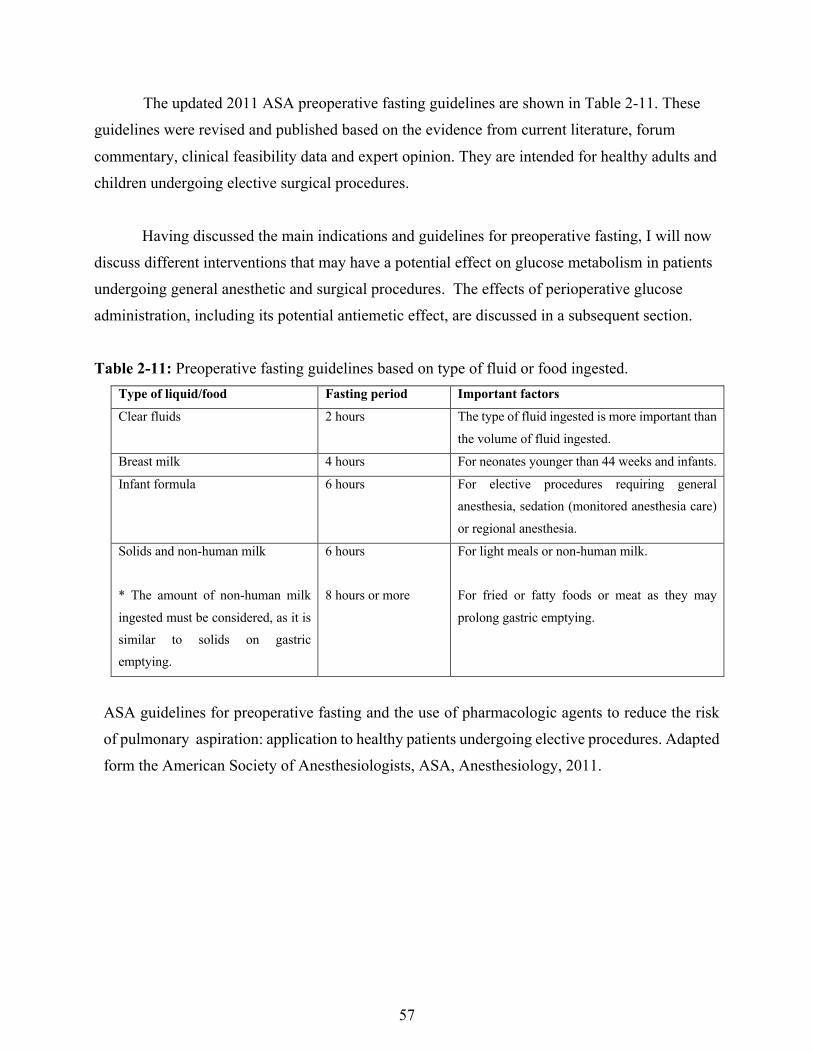
57
The updated 2011 ASA preoperative fasting guidelines are shown in Table 2-11. These guidelines were revised and published based on the evidence from current literature, forum commentary, clinical feasibility data and expert opinion. They are intended for healthy adults and children undergoing elective surgical procedures.
Having discussed the main indications and guidelines for preoperative fasting, I will now discuss different interventions that may have a potential effect on glucose metabolism in patients undergoing general anesthetic and surgical procedures. The effects of perioperative glucose administration, including its potential antiemetic effect, are discussed in a subsequent section. Table 2-11: Preoperative fasting guidelines based on type of fluid or food ingested.
Type of liquid/food Fasting period Important factors
Clear fluids 2 hours The type of fluid ingested is more important than the volume of fluid ingested.
Breast milk 4 hours For neonates younger than 44 weeks and infants.
Infant formula 6 hours For elective procedures requiring general anesthesia, sedation (monitored anesthesia care) or regional anesthesia.
Solids and non-human milk * The amount of non-human milk ingested must be considered, as it is similar to solids on gastric emptying.
6 hours 8 hours or more
For light meals or non-human milk. For fried or fatty foods or meat as they may prolong gastric emptying.
ASA guidelines for preoperative fasting and the use of pharmacologic agents to reduce the risk of pulmonary aspiration: application to healthy patients undergoing elective procedures. Adapted form the American Society of Anesthesiologists, ASA, Anesthesiology, 2011.
58
2.3.4.1.4. Effect of anesthetic medications on glucose metabolism
Several animal studies have shown the effect of volatile anesthetics on glucose metabolism, mainly the impairment of glucose tolerance by decreasing insulin sensitivity. In vivo studies have also suggested that one of the possible mechanisms for volatile anesthetic action on impairment of insulin sensitivity is the direct inhibition of insulin secretion by pancreatic B-cells when volatile anesthetics are used, specifically halothane, isoflurane and enflurane. These results were also observed in a prospective study performed in 30 patients using sevoflurane based anesthesia, which demonstrated that stress hormone levels are not affected by the administration of this medication, and confirming, in part, the direct insulin secretion inhibition theory (Tanaka, Nabatame, & Tanifuji, 2005). In the study, the metabolic response was triggered by an intravenous glucose tolerance test, administered prior to the surgical stimulation. This study also suggested that glucose intolerance observed during the anesthetic period without surgical stimulation is independent of the anesthetic dose. Furthermore, the authors believed that the increased catecholamine levels following endotracheal intubation could contribute to higher glucose and lower insulin plasma levels, the later related to impaired insulin secretion caused by both volatile anesthetics and the catecholamine effect.
When volatile anesthetics were compared with propofol anesthetics, study results were not homogeneous. A study performed in animals demonstrated two main differences between volatile anesthetics and propofol: first, sevoflurane was found to attenuate insulin secretion induced by glucose plasma levels, whereas propofol was found to enhance insulin secretion; second, sevoflurane did not impair insulin sensitivity, whereas propofol did appear to do so (Sato et al., 2013). Another animal study using an intravenous glucose tolerance test to stimulate a metabolic response in rhesus monkeys showed that when propofol was used for sedative purposes, an increase of insulin secretion was demonstrated with increased glucose levels, which were normalized as expected after the glucose infusion was completed (Kim et al., 2014). Compared to animals that did not receive any type of sedation it can be suggested that the main differences between groups were stimulated in part by the physical stress that was relieved in the sedated animals. Although these results cannot be extrapolated to humans, they do suggest that other factors, including increased metabolic stress, influence the physiological response among individuals.

59
A study performed in humans showed different results by measuring the catabolic
response during surgery, comparing both volatile anesthetics (desflurane) and propofol (Schricker et al., 2001). The study failed to demonstrate any significant difference between groups when different metabolic parameters were compared, including glucose production and clearance, protein breakdown and aminoacid oxidation. After comparing anesthetic based groups, endogenous glucose production as well as glucose clearance were both decreased and an increment in glucose and cortisol plasma levels was demonstrated.
Further studies are necessary to elucidate the exact mechanism and metabolic response
that different anesthetic techniques have on healthy individuals. As far as stimulants affecting glucose metabolism associated with the perioperative period are concerned, multiple factors influence the patient’s metabolic response, including the anesthetic technique, magnitude and extent of the surgical procedure, as well as underlying medical conditions.
In addition, it is important to discuss the role of glucose administration and its different metabolic and postoperative effects during the perioperative period.
2.3.4.1.5 Intravenous dextrose containing solutions and its effect on glucose
metabolism
It is important to clarify that dextrose is the natural form of glucose also known as D-glucose, and is the term used for the commercial intravenous fluid solutions containing glucose. For the purpose of this thesis the term dextrose will be used to reference intravenous solutions containing glucose, and the term glucose to refer to blood sugar and its metabolism.
Questions have been raised about the possible metabolic responses to intravenous administered dextrose containing solutions and its effect on glucose metabolism. One of the first studies performed in 1979 included 12 participants who were evaluated during controlled fasting conditions and received a dextrose infusion with or without insulin infusion. Results showed that the main method used by the organism to regulate glucose plasma levels among individuals was the adjustment of the endogenous glucose production, rather than the variations in tissue glucose
60
uptake, affected by insulin sensitivity (Wolfe, Allsop, & Burke, 1979). More recent studies have supported these results, including a prospective study of 14 patients who received a dextrose infusion during general anesthesia, whether or not the surgical procedure was elective(Schricker et al., 2004). Results demonstrated that the administration of dextrose, attenuated among patients who underwent surgery, reduced endogenous glucose production in all participants. Similar increases in plasma glucose levels and plasma insulin concentration also evidenced the inhibitory effect of exogenous dextrose over endogenous glucose production when groups were compared.
Another effect of dextrose solutions administration is the volume change among the fluid compartments of the body affected by the osmotic transportation of water into the intravascular compartment with a subsequent increase in intravascular blood volume that moves glucose into the cells. This volume increment change is reversed after glucose is rapidly metabolized into the tissues and its osmotic effect is eliminated. A study comparing three different intravenous solutions -- dextrose 2.5%, dextrose 5% and Ringer’s Lactate -- showed that plasma glucose and insulin concentrations increased as expected in the dextrose solutions compared to Ringer’s solution (Sjostrand, Edsberg, & Hahn, 2001). The volume effect of the solutions compared to Ringer’s was similar, assuming that dextrose solutions are never used as volume expanders. A slight rebound hypoglycemia effect took place in patients who received dextrose 5%, similar to that reported in patients who experienced a sudden discontinuation of parenteral nutrition.
Hypoglycaemia is one of the possible effects of fasting, which, although uncommon, may have detrimental consequences. In children, the reported incidence of hypoglycaemia in fasting patients is up to 31% among studies, a percentage that is considered even higher in infants. To prevent hypoglycaemia, glucose solutions are used perioperatively. Such solutions do not exceed an infusion rate of 300 mg/kg/h (4-6 mg/kg/min), representing the basic glucose production in pediatric patients. This target glucose content for intravenous dextrose solutions may assist in the prevention of hyperglycaemia, allowing the effective management of possible low glucose plasma concentrations, especially in fasting patients with subsequent lipid mobilization and ketogenesis. Hyperglycaemia may induce diuresis and associated fluid imbalanced from dehydration, mostly in patients with increased glucose levels for prolonged periods (Leelanukrom & Cunliffe, 2000).
61
Acute postoperative hyponatremia, defined as “a decline in serum sodium within a 48-hour period to less than 130 mmol/L”, is another possible adverse effect after fluid infusion using hypotonic solutions (Carr, Cornish, Predy, & et, 2009). Furthermore, severe hyponatremia is most commonly seen in infants when plasma sodium concentrations reach levels below 125 mmol/L (Hongnat, Murat, & Saint-Maurice, 1991). Acute hyponatremia is associated with neurologic manifestations following the abrupt change in sodium concentration, the movement of electrolyte-free water into the brain cells and subsequent cerebral edema. Children and infants are more vulnerable to these changes and early signs of acute hyponatremia may be difficult to recognize, although, children develop these symptoms earlier, compared to adults, as they have limited space and room for brain cells to swell. Early signs and symptoms of acute hyponatremia may range from non-specific (headache, nausea, vomiting, confusion and somnolence) to respiratory depression, seizures, coma, brain herniation and death (Carr et al., 2009; Hongnat et al., 1991).
Prevention of hyponatremia can be obtained by providing adequate maintenance IV fluids, however experts relate to the fact that for children, there is no consensus on an ideal IV maintenance solution to be used. Therefore, especially in younger patients, different measures should be in place to avoid this potential complication, including knowledge of local guidelines, adequate record and documentation of fluid balance in hospitalized patients. In addition, it is crucial to use balanced salt isotonic solutions, as those are closer to the extracellular composition, instead of hypotonic solutions such as dextrose containing solutions in water, which lack appropriate balanced electrolyte composition (Carr et al., 2009; Hongnat et al., 1991).
The previous discussion indicates that different interventions in the perioperative period can potentially inhibit or promote glucose metabolism during the fasting or postprandial periods. These interventions may also affect the fluid or electrolyte balance necessary to maintain an adequate body response during periods of increased metabolic demands.
In the following paragraphs, the discussion will focus on the potential effects of perioperative carbohydrate administration associated with the recovery period.

62
2.3.4.2. Effect of perioperative carbohydrates on the recovery period
There is evidence that dextrose administration may improve recovery outcomes in patients undergoing surgical procedures. Research to evaluate outcomes has focused on the implementation of protocols using oral or intravenous carbohydrates at any time during the perioperative period.
2.3.4.2.1 Oral carbohydrates in the perioperative period
As discussed previously, preoperative fasting and surgical stress have common significant consequences for glucose metabolism, especially for insulin resistance, which can be reduced by avoiding preoperative fasting and administering dextrose intravenous infusions or oral glucose supplements. The principal behind this intervention relies on the possible effects of metabolic response to fasting and surgery, a reduction of catabolic metabolism and of insulin resistance, improved metabolism and strength of muscle fibres and the potential effects on overall postoperative recovery (Henriksen, Hessov, Vind Hansen, Haraldsted, & Rodt, 2003).
Multiple studies have focused on determining the adequate glucose administration, dose and timing of this intervention. A RCT included 127 participants who underwent elective laparoscopic surgery and were allocated to either fast or consume 400 ml of placebo or a carbohydrate drink, both given in the morning prior to the procedure. Results showed that the incidence of PONV was similar among groups in the first 12 hours, but the incidence increased in the fasting group during the following 12 hours, suggesting that the administration of oral carbohydrates may reduce PONV after laparoscopic cholecystectomy (Hausel et al., 2005).
The use of oral carbohydrate containing fluids may increase the gastric volume prior to surgery, increasing the risk of postoperative vomiting and pulmonary aspiration, an argument against the establishment of this practice. A RCT that included seventy patients showed that the administration of oral glucose solutions preoperatively did not increase the gastric volume or the
63
gastric pH in participants compared to those who underwent a preoperative fasting period (Yagci et al., 2008). Another prospective study that enrolled 60 patients evaluated the postoperative recovery outcomes after the administration of oral glucose before and after the surgical procedure when compared with preoperative fasting. The study showed an increase in gastric volume in the control group and an increment of the gastric pH in the intervention group, suggesting that the difference in carbohydrate solution composition and different fasting times may have influenced these results. Results also failed to demonstrate any difference in the incidence of postoperative nausea, but overall showed that preoperative carbohydrate solutions reduced postoperative discomfort including thirst, weakness and anxiety (Yildiz, Gunal, Yilmaz, & Yucel, 2013).
These results were supported by a Cochrane review that included 27 RCT and 1976
participants, comparing the use of preoperative carbohydrate solutions with the use of placebo or fasting and their influence on postoperative recovery. Results of 14 studies (913 participants) demonstrated that the administration of carbohydrates prior to surgery did not increase the risk of aspiration pneumonitis, or the risk of postoperative complications when compared with placebo or fasting. Differences were also shown when the length of hospital stay was evaluated in 19 trials (1351 participants), showing that hospital stays were shorter when carbohydrates were used compared with placebo or fasting. No conclusive difference in the incidence of postoperative nausea or vomiting was found in this meta-analysis (M. D. Smith et al., 2014).
Overall, the use of oral carbohydrate solutions in the perioperative period has shown to be
safe and beneficial for improving postoperative recovery, although its use has shown inconclusive results for decreasing the incidence of PONV. Despite not being associated with increased aspiration pneumonitis, many clinicians continue to be reluctant to use preoperative glucose and prefer to maintain their practice by implementing clear guidelines on preoperative fasting. This response has led researchers to evaluate the administration of intravenous dextrose, instead of oral administration, and its association with the prevention of PONV.
64
2.3.4.3. Intravenous dextrose for the management of PONV
There has been recent renewed interest in the use of intravenous dextrose containing solutions for the prevention and management of PONV. The safe and longstanding use of dextrose containing crystalloid solutions as part of the resuscitative fluid therapy in pediatric patients with significant dehydration has been associated with the stimulation of insulin production, faster ketosis and vomiting resolution (Reid & Losek, 2009). These observations have led clinicians to inquire about the relationship of intravenous dextrose containing solutions to an enhanced overall postoperative recovery especially for the prevention of PONV.
Studies evaluating the potential effect of dextrose containing solutions have shown mixed
results. A prospective study of 108 adult patients failed to demonstrate any significant difference in the incidence of PONV when comparing an intravenous compound sodium lactate with a large dextrose concentration (50%) with a similar intravenous compound without dextrose (McCaul et al., 2003). Further studies evaluated these results by changing the intravenous solutions administered, as seen in a RCT that enrolled 162 participants who underwent outpatient gynaecologic, urologic or breast surgical procedures. This RCT compared the use of intravenous Ringer’s Lactate, with or without dextrose 5% administered during the anesthetic emerge phase. The study also showed mixed results in the incidence of PONV in the early postoperative period (2hr), and an intergroup difference of 3.4% in the incidence of PONV during the first 24 hours following the surgical procedure, with a lower incidence in the intervention group compared to the placebo group (Patel et al., 2013).
Similarly, another RCT performed of gynaecological adult patients, including 62
participants, compared the administration of Ringer’s Lactate with or without dextrose 5% solution administered at the end of the surgical procedure. Although the incidence of postoperative nausea was not significantly different between groups (p>0.05), a decreased use of antiemetic rescue medications in the recovery period (p = 0.02) was demonstrated along with a significant shorter length of stay in the recovery area (PACU) (p=0.03) (Dabu-Bondoc et al., 2013). These studies suggest that there is a relationship between the use of intravenous dextrose containing solutions and postoperative recovery in patients undergoing ambulatory, elective procedures.
65
The present clinical trial resulted from reviewing the above-mentioned studies, which
evaluated the possible benefits of intravenous dextrose-containing solutions for the prevention and management of PONV in adults, and the realization that, to date, studies evaluating this intervention in the pediatric population are lacking. Despite the minimal understanding of the mechanism of action behind the possible benefit of this intervention for the prevention of PONV, intravenous dextrose solutions continue to be available, inexpensive and well tolerated by patients. The next chapter describes methodology used for the present study, outlining the process and execution of the clinical trial.

66
CHAPTER 3
METHODS
This chapter describes the methodology of the study, outlining the methods from the
study design through the study population selection and ethical considerations to data collection processes and analysis. 3.1 Study population
The study population consisted of 290 otherwise healthy children who met the inclusion criteria as follows: male or female sex; aged three to nine years; minimal preoperative risk based on the American Society Anesthesiologists physical status (ASA) I and II (Table 2-4) and confirmed by the anesthesiologist the day of the dental procedure; and patients who underwent ambulatory dental procedures under general anesthesia from December 2013 to August 2014 at Prairieview Surgical Centre in Saskatoon, Saskatchewan, Canada.
A sample size of 284 participants was calculated based on the null hypothesis that the
early POV rate in the intervention group would be 7.5% or more (non-inferiority based on clinical judgement and literature review) than the control group, with a reference of early POV rate in the control group 25% (Yun-Dun, 2014; Shen Y, Chen C, Wu C, Cherng Y, Tam K, 2014). A confidence interval (CI) [0.14, 0.36], a power of 0.8 (80%), and 5% significance (two-sided alpha of 0.05) (Table 3-1) was used for the calculation.
3.1.1 Exclusion criteria
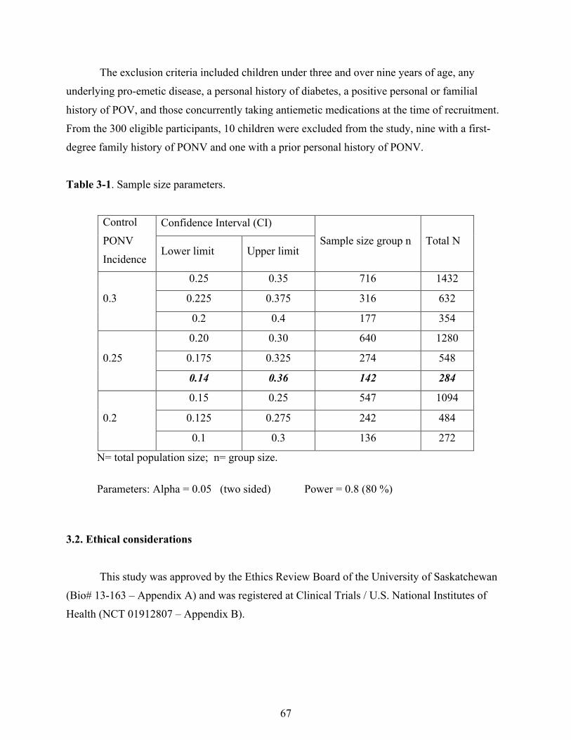
67
The exclusion criteria included children under three and over nine years of age, any underlying pro-emetic disease, a personal history of diabetes, a positive personal or familial history of POV, and those concurrently taking antiemetic medications at the time of recruitment. From the 300 eligible participants, 10 children were excluded from the study, nine with a first-degree family history of PONV and one with a prior personal history of PONV. Table 3-1. Sample size parameters.
Control PONV Incidence
Confidence Interval (CI) Sample size group n Total N
Lower limit Upper limit
0.3
0.25 0.35 716 1432
0.225 0.375 316 632
0.2 0.4 177 354
0.25
0.20 0.30 640 1280
0.175 0.325 274 548
0.14 0.36 142 284
0.2
0.15 0.25 547 1094
0.125 0.275 242 484
0.1 0.3 136 272
N= total population size; n= group size.
Parameters: Alpha = 0.05 (two sided) Power = 0.8 (80 %)
3.2. Ethical considerations
This study was approved by the Ethics Review Board of the University of Saskatchewan (Bio# 13-163 – Appendix A) and was registered at Clinical Trials / U.S. National Institutes of Health (NCT 01912807 – Appendix B).
68
3.3 Study design
This was an interventional prospective non-inferiority study allocated as a Randomized
Controlled Trial with parallel assignment of participants. Participants, caregivers, healthcare providers and clinical investigators were blinded to group assignment throughout the perioperative period.
The randomization, consisting of 50 blocks with six patients each, was performed by the Clinical Research Support Unit, College of Medicine, University of Saskatchewan, prior to commencing the study.
Allocation concealment was performed by giving an opaque envelope to the anaesthesiologist, which contained the corresponding group code (A or B) for the patient based on the randomization performed previously.
3.3.1 Study protocol
3.3.1.1 Recruitment of participants and consent process
In this specific population, due to the nature of the dental procedure (cleaning and/or extractions) and the minor risk in this healthy population (ASA I and II), patients were not seen prior to the day of surgery, as standard protocol. Patients were referred directly by their dentist, pediatrician or family doctor; none of the patients contacted the surgeon prior to the day of dental procedure; and many of them had to travel from outside the city for the procedure. These factors made it difficult to contact patients and parents or legal guardians before the day of the dental procedure or to contact them through the surgeon’s office. Because of the difficulty of contacting the patients or their families in advance, consent was obtained the same day of the procedure.
The day of procedure, all patients were assessed in the waiting room by the anesthesiologist confirming the patient's low operative risk and by the surgeon prior to the procedure. Prospective participants and their parents or legal guardians were also contacted by one of the researchers in the waiting area approximately one hour before their child was taken to
69
the Operating Room for the dental procedure. The parents or legal guardians and the participant were left for a few minutes after the study objectives, purpose and enrolment implications were explained. They were provided with a copy of the consent form and a brochure containing general information about the study (Appendix C, D and E). Shortly after this, the researchers met again with the participant and parents or legal guardian to answer all their questions. At this time, the families decided whether to participate in the study. After participation was confirmed, consent was signed by the parent or legal guardian and assent was given by the patient (when considered capable to assent); as well, demographic data was collected from parents or legal guardians (Appendix F).
3.3.1.2 Conduction of the study
In order to maintain the blindness of the study, the solutions were prepared daily by the
research assistant prior to the recruitment of participants. Ondansetron or normal saline were drawn into a syringe (colorless liquid) and coded. The commercial IV infusion bags, either NS or D5NS, were covered identically and coded as “A” or “B”.
The subjects were allocated to one of two groups based on antiemetic prophylaxis: the intervention or control group. The intervention group (144 participants) received dexamethasone (0.15 mg/kg IV maximum 5 mg) and intravenous 5% dextrose in 0.9% normal saline (D5NS) maintenance fluid; the control group (146 participants) received dexamethasone (0.15 mg/kg IV maximum 5 mg) and ondansetron (0.05 mg/kg IV maximum 4 mg). All doses were based on the recommendations from the guidelines for ambulatory anesthesia for children (Gan et al., 2014).
The randomization assignment was given to the research assistant who prepared the study
drug in advanced (ondansetron vs. normal saline) in identically appearing clear syringes (study drug “A or B”) and covered the commercial labeling on intravenous solutions (NS vs. D5NS) in the same manner. Kits were made before patient recruitment for each of the groups (control and intervention) with the necessary elements for the study (IV maintenance solution bag and clear syringe), with the exception of dexamethasone, which was given by the anaesthesiologists from the anaesthesia medications stock, according to antiemetic recommended weight-based doses (Gan et al., 2014). The research assistant was the only one who knew the code in order to
70
maintain the blind nature for the rest of the participants. The IV solution (NS vs. D5NS) was placed in a Hospira – Abbott plum A+ IV infusion
pump (Plum® A+ Infusion System, Hospira Inc, Lake Forest, IL) by the research assistant, and the infusion rate was calculated based on the patient’s weight and the “4-2-1” rule for maintenance fluid (Meyers, 2009). Once IV access was established with an IV line containing Ringer’s Lactate and the patient was intubated, the maintenance solution was connected and infused throughout the operative period. Ringer’s Lactate was available to the anesthesiologists to administer as additional fluid as per their preference.
We tried to find a solution for the IV tubing connection by providing a Y connection close
to the patient’s IV line but found it expensive and not typically required by most anesthesiologists.
The protocol was designed so that an envelope would be given to the anesthesiologist
containing the listed instructions and a table with corresponding dosages for the dexamethasone based on the patient’s weight, ondansetron and IV maintenance fluid rate (Appendix G and H, respectively).
For the purpose of the study, general anesthetic was defined as the medication induced
state of loss of protective reflexes and unconsciousness through the administration of general anesthetic medications, requiring intubation of the trachea via endotracheal tube (Miller 2010). Because there was no standardized protocol for the administration of anesthetic medications, the medications given to the patient for the induction and maintenance of the anesthetic procedure were chosen and administered at the discretion of the anesthetic provider.
However, the antiemetic medications to be used were those named in the study protocol
(dexamethasone, ondansetron or study medication). The researcher explained to the anesthesiologist that other antiemetic medications were not to be administered as prophylactics. There were no modifications to the planned dental procedure.
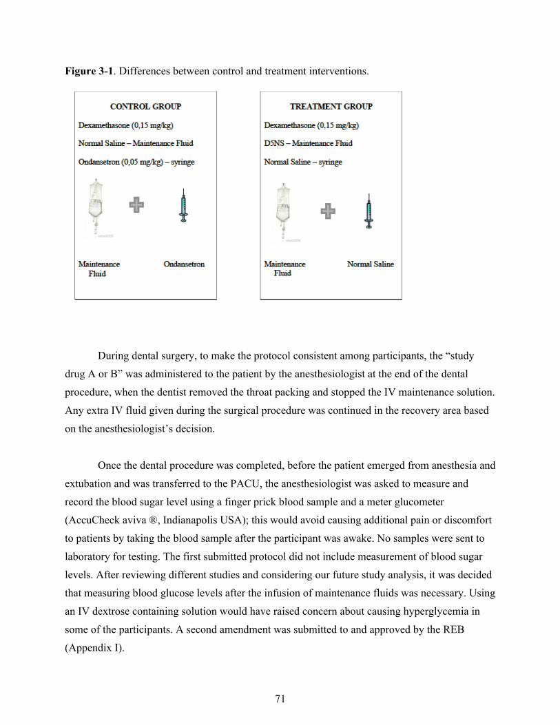
71
Figure 3-1. Differences between control and treatment interventions.
During dental surgery, to make the protocol consistent among participants, the “study
drug A or B” was administered to the patient by the anesthesiologist at the end of the dental procedure, when the dentist removed the throat packing and stopped the IV maintenance solution. Any extra IV fluid given during the surgical procedure was continued in the recovery area based on the anesthesiologist’s decision.
Once the dental procedure was completed, before the patient emerged from anesthesia and
extubation and was transferred to the PACU, the anesthesiologist was asked to measure and record the blood sugar level using a finger prick blood sample and a meter glucometer (AccuCheck aviva ®, Indianapolis USA); this would avoid causing additional pain or discomfort to patients by taking the blood sample after the participant was awake. No samples were sent to laboratory for testing. The first submitted protocol did not include measurement of blood sugar levels. After reviewing different studies and considering our future study analysis, it was decided that measuring blood glucose levels after the infusion of maintenance fluids was necessary. Using an IV dextrose containing solution would have raised concern about causing hyperglycemia in some of the participants. A second amendment was submitted to and approved by the REB (Appendix I).
72
The anesthesiologists were not compliant with all the parts of the protocol, as it created an
extra procedure, adding more time to their main work. Because of this non-compliance, it was decided that the research assistant and researcher would become involved in the practical aspects of the protocol. This involvement included setting up, connecting, starting and stopping the maintenance IV fluids to and from the main IV line, and measuring and recording the blood glucose level at the end of the procedure. This improved the anesthesiologists compliance and their participation in the study.
After the patients had emerged from the anesthetic and were seen to be stable, they were
transferred to the Post Anesthetic Care Unit (PACU) for postoperative recovery, where they were assessed by nursing staff and remained until they were ready to be discharged based on the Post Anesthetic Discharge Scoring System (PADSS) (Ead, 2006) and the institution guidelines.
Nursing staff and researchers assessed the presence of POV and the number of episodes of it in the PACU. For the purpose of the study protocol, Postoperative Vomiting (POV) was defined as vomiting (expulsion of gastric content) within 24 hours after surgery. In the PACU area, the period was accounted and defined as early POV either immediate or within the first 2 hours after the dental procedure and emerge from the general anesthesia. Late POV was defined as presence of vomiting within the 24 hours from the time of discharge from the facility. Retching and nausea were not accounted nor recorded as it was difficult for patients to communicate and could create confusion on parents or legal guardians.
The initial study protocol included the use of a pictoric Postoperative nausea scale, (Baxter, Watcha, Baxter, Leong, & Wyatt, 2011) to measure postoperative nausea. This scale has been validated in pediatric cancer patients over seven years of age who are undergoing chemotherapy. Since the majority of our patients were under five years of age, this scale was unsuitable for use with our study population. Additionally, we observed that patients were irritable during the early postoperative recovery, which made the use of the scale an additional stress for patients and parents or legal guardians. It was thus decided to avoid the use of this instrument and assess patients only by the presence of postoperative vomiting. This assessment

73
tool is reliable and causes less confusion than the scale when used with young children, who find it difficult to express the sensation of nausea.
Analgesics and antiemetic agents were prescribed by the anesthesiologist for the recovery
period and given according to the nursing staff’s assessment and institutional guidelines, which provided standardized criteria with the department of anesthesiology.
Follow up of the patient’s recovery and incidence of emesis within 24 hours after
discharge was done by phone calls made by the researchers (Appendix J). This protocol was followed through the duration of the study without any further modifications.
3.4 Data collection
Researchers who collected the data were blinded to group assignment. The primary
outcome was incidence and the number of episodes of emesis in the PACU (immediate and early postoperative period). Secondary outcomes included the following: incidence and number of episodes of emesis within 24 hours after discharge; use of rescue antiemetic medications in the immediate and late recovery period; intraoperative blood glucose level; unplanned hospital admission for POV; delays in discharge from PACU due to POV; and return to hospital or a hospital clinic for a medical assessment due to POV. Intraoperative data, including anesthetic medications and doses administered, were recorded in the operating room from the anesthetic record (Appendix F).
3.5 Statistical analysis
Statistical analysis was performed with the assistance of the Biostatical Support Unit associated with the Department of Surgery (University of Saskatchewan) using SAS software (v 9.4; SAS Cary, NC, U.S.A.). All analyses were performed on an intention-to-treat basis (S. K. Gupta, 2011).
74
Variables were checked regarding assumptions underlying the use of parametric and nonparametric statistics and analyzed accordingly.
To examine the association between categorical variables and primary outcome (proportion of participants who presented emesis during the early postoperative period), secondary outcomes (proportion of participants who presented emesis during the late postoperative period [24 hr]), and antiemetic rescue medication requirement, Chi-square test or Fisher’s exact test were used as appropriate.
Median and interquartile ranges were reported for those continuous variables that did not meet normality criteria. Mean and SD were reported for normally distributed variables. Two-sample T-tests were used to compare the mean between groups (e.g. intervention vs. control) for continuous normally distributed variables, including surgical procedure – extractions, procedure length, glucose level, amount of IV fluids administered and opioid and anesthetic medication dose used.
Continuous, non-normally distributed variables were compared between randomized groups using Wilcoxon tests when analyzing two samples, and Kruskal-Wallis tests when analyzing more than two samples. This comparison was used to study the relationship between primary and secondary outcomes and the anesthetic agents used for induction and maintenance, volume of IV fluid administration and opioid use.
The level of significance was set at 0.05 (two-tailed), which was used as the criterion for rejection of the null hypothesis (p >0.05).
3.5.1 Non-inferiority analysis
To understand why a non-inferiority trial was chosen, it is important to recognize the main differences between superiority, equivalence and non-inferiority clinical trials. The primary objective of a superiority clinical trial is to show that the response to a new therapy is superior compared to an established standard therapy. In turn, the purpose of equivalence clinical trials is to show that the new therapy effect is identical to the standard therapy while in non-

75
inferiority trials the purpose is to show that the new therapy effect is not worse when compared to the standard therapy (Christensen, 2007).
In this particular trial, ethical considerations were decisive for choosing to conduct a non-inferiority study protocol, based on the International Conference on Harmonization’s “Choice of control group and related issues in clinical trials” (Food and Drug Administration, U.S., 2010). This guideline suggests that an active control (vs. placebo) should be considered in patients with a higher inherent risk of developing the condition studied in the trial. For our analysis, data describing the historic effect of the active control was extracted from a randomized control trial performed in children with similar demographic characteristics to our study population, which assessed the combined, protective effect of both dexamethasone and ondansetron. This study described the proportion difference of the combination of these two drugs versus dexamethasone alone as 18% (Splinter, 2001).
Characteristics of this non-inferiority study protocol would allow us to investigate that intravenous dextrose containing solutions have a significant effect in preventing postoperative vomiting in children. The study design tests if the treatment effect of the intervention (D5NS + dexamethasone) is not inferior to the treatment effect of an active control (Ondansetron + Dexamethasone). In order to assess the effect difference between the active control (ondansetron combined with dexamethasone) and the intervention procedure (D5NS in combination with dexamethasone), a non-inferiority margin was calculated a-priori. Our choice of non-inferiority (NI) margin was guided by published principles (Food and Drug Administration, U.S., 2010). We first reviewed previous published NI margins of therapeutic trials conducted in pediatric anesthesia. A recent trial investigating the risk of hemorrhage when dexamethasone is administered to children undergoing tonsillectomy used a non-inferiority margin of 5% given that the outcome is clinically serious and life threatening (Gallagher, T.Q., et al. 2012). We felt a wider NI margin (7.5%) was appropriate for the following clinical reasons: PONV is not a clinically important as post tonsillar hemorrhage, D5NS is an inexpensive therapy, and D5NS has a long clinical history of safety. Finally, our chosen margin is less than the expected mean treatment effect of a similar dose of ondansetron when combined with dexamethasone (Splinter, 2001).
76
Having described the patient population, methods and study protocol, I will now move on to describe the results of the interventions and data analysis along with a specific non-inferiority study analysis.
77
CHAPTER 4
STUDY RESULTS
This chapter presents the results of the study including the demographic characteristics,
the intraoperative anesthetic management and the primary and secondary outcomes. In addition, this chapter describes the results from the non-inferiority analysis. 4.1 Main study results
A total of 300 participants were assessed for eligibility to participate in this clinical trial. Ten participants were ineligible for not meeting the inclusion criteria. Therefore, data from 290 patients were enrolled and analyzed as shown in Figure 4-1, based on the 2010 CONSORT group report guidelines (CONSORT, 2010).
Demographic characteristics among groups were similar as presented in Table 4-1. There
was no association between sex and increased incidence of POV (p=0.63). Nor was there an association between sex and the type of procedure performed (dental cleaning p=0.18 vs. dental extractions p=0.17). Although ethnicity was not recorded for all participants, there was an increased number of patients with indigenous ethnicity compared to the rest of the participants. It was noted that participants with indigenous ethnicity had a higher number of tooth extractions compared to Caucasians or participants with other ethnic background. Ethnicity was self-reported by parents or legal guardians.
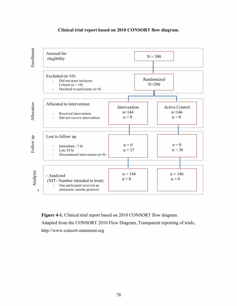
78
Clinical trial report based on 2010 CONSORT flow diagram.
Figure 4-1. Clinical trial report based on 2010 CONSORT flow diagram. Adapted from the CONSORT 2010 Flow Diagram, Transparent reporting of trials, http://www.consort-statement.org
Figure 1. Clinical Trial Report based on 2010 CONSORT flow diagram.
Figure 1. Clinical trial report based on 2010 CONSORT flow diagram. Adapted from the CONSORT 2010 Flow Diagram, Transparent reporting of trials, http://www.consort-statement.org
Follo
w u
p
Analysis
�
Enro
llmen
t A
lloca
tion
Assesed for elegibility
Excluded (n=10) - Did not meet inclusion
Criteria (n = 10) - Declined to participate (n=0) - Other reasons (n=0)
Allocated to intervention
- Received intervention - Did not receive intervention
N = 300
Randomized N=290
Intervention n=144
n = 0
Active Control n=146
n = 0
Lost to follow up
- Immediate / 2 hr - Late 24 hr - Discontinued intervention (n=0)
n = 0
n = 37
n = 0
n = 36
- Analyzed (NIT- Number intended to treat)
• One participant received an antiemetic outside protocol.
n = 144 n = 0
n = 146 n = 0
Adapted from the CONSORT 2010 Flow Diagram, Transparent reporting of trials, http://www.consort-statement.org
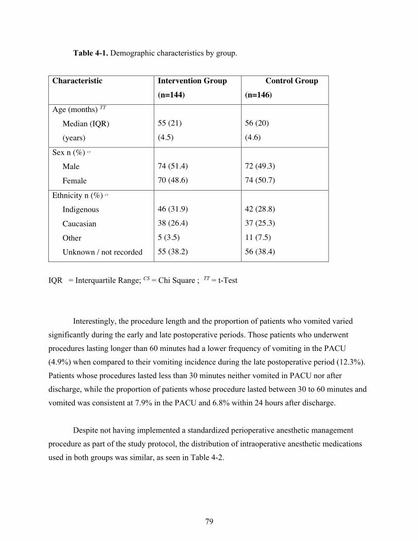
79
Table 4-1. Demographic characteristics by group.
Characteristic Intervention Group
(n=144)
Control Group
(n=146)
Age (months) TT
Median (IQR)
(years)
55 (21)
(4.5)
56 (20)
(4.6)
Sex n (%) CS
Male
Female
74 (51.4)
70 (48.6)
72 (49.3)
74 (50.7)
Ethnicity n (%) CS
Indigenous
Caucasian
Other
Unknown / not recorded
46 (31.9)
38 (26.4)
5 (3.5)
55 (38.2)
42 (28.8)
37 (25.3)
11 (7.5)
56 (38.4)
IQR = Interquartile Range; CS = Chi Square ; TT = t-Test
Interestingly, the procedure length and the proportion of patients who vomited varied
significantly during the early and late postoperative periods. Those patients who underwent procedures lasting longer than 60 minutes had a lower frequency of vomiting in the PACU (4.9%) when compared to their vomiting incidence during the late postoperative period (12.3%). Patients whose procedures lasted less than 30 minutes neither vomited in PACU nor after discharge, while the proportion of patients whose procedure lasted between 30 to 60 minutes and vomited was consistent at 7.9% in the PACU and 6.8% within 24 hours after discharge.
Despite not having implemented a standardized perioperative anesthetic management
procedure as part of the study protocol, the distribution of intraoperative anesthetic medications used in both groups was similar, as seen in Table 4-2.
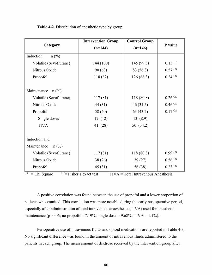
80
Table 4-2. Distribution of anesthetic type by group.
Category Intervention Group
(n=144) Control Group
(n=146) P value
Induction n (%) Volatile (Sevoflurane) Nitrous Oxide Propofol Maintenance n (%) Volatile (Sevoflurane) Nitrous Oxide Propofol Single doses TIVA Induction and Maintenance n (%) Volatile (Sevoflurane) Nitrous Oxide Propofol
144 (100) 90 (63) 118 (82)
117 (81) 44 (31) 58 (40) 17 (12) 41 (28)
117 (81) 38 (26) 45 (31)
145 (99.3) 83 (56.8) 126 (86.3)
118 (80.8) 46 (31.5) 63 (43.2) 13 (8.9) 50 (34.2)
118 (80.8) 39 (27) 56 (38)
0.13 FT 0.57 CS 0.24 CS
0.26 CS 0.46 CS 0.17 CS
0.99 CS 0.56 CS 0.23 CS
CS = Chi Square FT= Fisher’s exact test TIVA = Total Intravenous Anesthesia
A positive correlation was found between the use of propofol and a lower proportion of
patients who vomited. This correlation was more notable during the early postoperative period, especially after administration of total intravenous anaesthesia (TIVA) used for anesthetic maintenance (p=0.06; no propofol= 7.19%; single dose = 9.68%; TIVA = 1.1%).
Perioperative use of intravenous fluids and opioid medications are reported in Table 4-3.
No significant difference was found in the amount of intravenous fluids administered to the patients in each group. The mean amount of dextrose received by the intervention group after
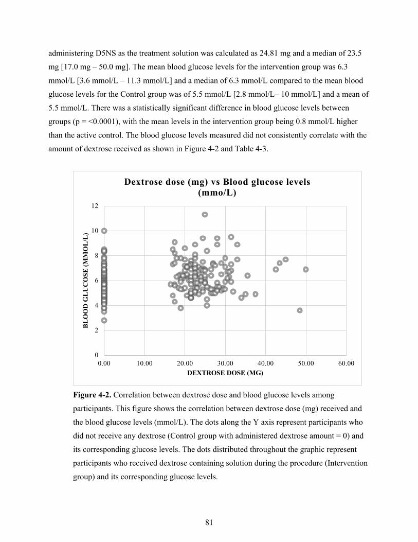
81
administering D5NS as the treatment solution was calculated as 24.81 mg and a median of 23.5 mg [17.0 mg – 50.0 mg]. The mean blood glucose levels for the intervention group was 6.3 mmol/L [3.6 mmol/L – 11.3 mmol/L] and a median of 6.3 mmol/L compared to the mean blood glucose levels for the Control group was of 5.5 mmol/L [2.8 mmol/L– 10 mmol/L] and a mean of 5.5 mmol/L. There was a statistically significant difference in blood glucose levels between groups (p = <0.0001), with the mean levels in the intervention group being 0.8 mmol/L higher than the active control. The blood glucose levels measured did not consistently correlate with the amount of dextrose received as shown in Figure 4-2 and Table 4-3.
Figure 4-2. Correlation between dextrose dose and blood glucose levels among participants. This figure shows the correlation between dextrose dose (mg) received and the blood glucose levels (mmol/L). The dots along the Y axis represent participants who did not receive any dextrose (Control group with administered dextrose amount = 0) and its corresponding glucose levels. The dots distributed throughout the graphic represent participants who received dextrose containing solution during the procedure (Intervention group) and its corresponding glucose levels.
0
2
4
6
8
10
12
0.00 10.00 20.00 30.00 40.00 50.00 60.00
BL
OO
D G
LU
CO
SE (
MM
OL
/L)
DEXTROSE DOSE (MG)
Dextrose dose (mg) vs Blood glucose levels (mmo/L)
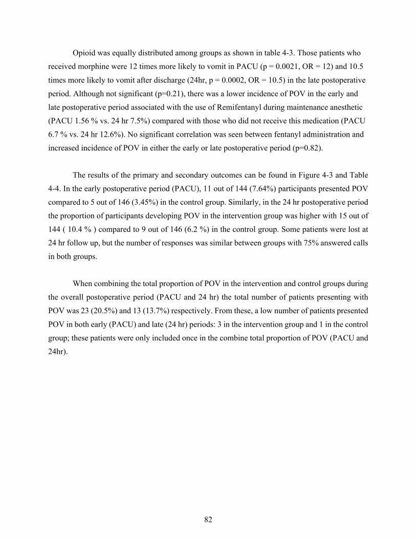
82
Opioid was equally distributed among groups as shown in table 4-3. Those patients who received morphine were 12 times more likely to vomit in PACU (p = 0.0021, OR = 12) and 10.5 times more likely to vomit after discharge (24hr, p = 0.0002, OR = 10.5) in the late postoperative period. Although not significant (p=0.21), there was a lower incidence of POV in the early and late postoperative period associated with the use of Remifentanyl during maintenance anesthetic (PACU 1.56 % vs. 24 hr 7.5%) compared with those who did not receive this medication (PACU 6.7 % vs. 24 hr 12.6%). No significant correlation was seen between fentanyl administration and increased incidence of POV in either the early or late postoperative period (p=0.82).
The results of the primary and secondary outcomes can be found in Figure 4-3 and Table 4-4. In the early postoperative period (PACU), 11 out of 144 (7.64%) participants presented POV compared to 5 out of 146 (3.45%) in the control group. Similarly, in the 24 hr postoperative period the proportion of participants developing POV in the intervention group was higher with 15 out of 144 ( 10.4 % ) compared to 9 out of 146 (6.2 %) in the control group. Some patients were lost at 24 hr follow up, but the number of responses was similar between groups with 75% answered calls in both groups.
When combining the total proportion of POV in the intervention and control groups during the overall postoperative period (PACU and 24 hr) the total number of patients presenting with POV was 23 (20.5%) and 13 (13.7%) respectively. From these, a low number of patients presented POV in both early (PACU) and late (24 hr) periods: 3 in the intervention group and 1 in the control group; these patients were only included once in the combine total proportion of POV (PACU and 24hr).
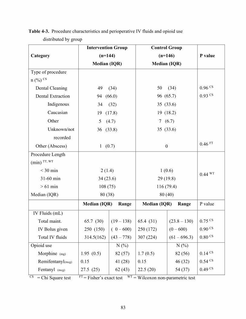
83
Table 4-3. Procedure characteristics and perioperative IV fluids and opioid use distributed by group
Category Intervention Group
(n=144)
Median (IQR)
Control Group (n=146)
Median (IQR) P value
Type of procedure n (%) CS Dental Cleaning Dental Extraction Indigenous
Caucasian
Other
Unknown/not
recorded
Other (Abscess)
49 (34) 94 (66.0)
34 (32)
19 (17.8)
5 (4.7)
36 (33.8)
1 (0.7)
50 (34) 96 (65.7)
35 (33.6)
19 (18.2)
7 (6.7)
35 (33.6)
0
0.96 CS
0.93 CS
0.46 FT
Procedure Length (min) TT, WT < 30 min 31-60 min > 61 min Median (IQR)
2 (1.4) 34 (23.6) 108 (75) 80 (38)
1 (0.6) 29 (19.8) 116 (79.4)
80 (40)
0.44 WT
Median (IQR) Range Median (IQR) Range P value
IV Fluids (mL) Total maint. IV Bolus given Total IV fluids
65.7 (30) 250 (150) 314.5(162)
(19 – 138) ( 0 – 600) (43 – 778)
65.4 (31) 250 (172) 307 (224)
(23.8 – 130) (0 – 600) (61 – 696.3)
0.75 CS 0.90 CS 0.80 CS
Opioid use Morphine (mg) Remifentanyl(mcg) Fentanyl (mcg)
1.95 (0.5) 0.15 27.5 (25)
N (%) 82 (57) 41 (28) 62 (43)
1.7 (0.5) 0.15 22.5 (20)
N (%) 82 (56) 46 (32) 54 (37)
0.14 CS 0.54 CS 0.49 CS
CS = Chi Square test FT = Fisher’s exact test WT = Wilcoxon non-parametric test
84
Proportion of postoperative vomiting by group
PACU 24 hours follow up
Figure 4-3. Proportion of postoperative vomiting by group. Blue bars represent the intervention group. Red bars represent the control group.
POV = Postoperative vomiting. PACU = Postanesthetic Care Unit.
Proportion of Postoperative Vomiting by Group
Figure 2. Represents the proportion of Postoperative Vomiting by Group. Blue bars represent the intervention group. Red bars represent the control group. POV = Postoperative vomiting. PACU = Postanesthetic Care Unit.
Pro
por
tion
of
PO
V

85
Table 4-4. Primary and secondary outcomes by group
Outcome Intervention
Group (n=144)
Control Group (n=146)
P value
POV (%)
• Immediate POV
• Early (PACU 0-2h)
• Late (24 h follow up) Answered calls (%) POV (%)
• PACU and 24h
• Any POV (PACU or 24h) Received TIVA No TIVA
0 (0)
11 (7.64)
108 (75) 15 (10.4) 3 (2.2) 23 (20.5) 1 (1.38) 21 (14.5)
0 (0)
5 (3.45)
109 (75) 9 (6.2) 1 (0.7) 13 (13.7)
1 (1.36) 11 (7.5)
0.11 CS
0.17 CS
0.36 FT 0.07 CS
Antiemetic Rescue Medication n (%) Early (PACU) Late (24h follow up)
4 (2.8)
2 (1.4)
0 (0) 0 (0)
0.059 FT
Blood Glucose level (mmol/L) Median (IQR) Mean (SD) Range
6.3 (1.8) 6.3 (1.2)
(3.6 – 11.3)
5.5 (1.2) 5.5 (1.0) (2.8 – 10)
< 0.0001TT
Delayed Home Discharge (%) 2 (1.4) 0 (0) 0.24 FT
Post-discharge Medical Assessment 0 (0) 0 (0)
I/M = Induction or Maintenance; I and M = Induction and Maintenance; POV = Postoperative vomiting; PACU = Post Anaesthetic Care Unit CS = Chi Square FT = Fisher’s exact test TT = t-test IQR = Interquartile Range SD = Standard deviation
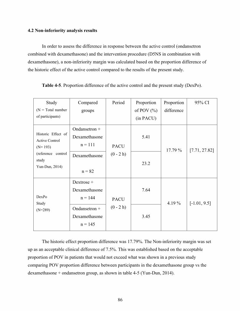
86
4.2 Non-inferiority analysis results
In order to assess the difference in response between the active control (ondansetron
combined with dexamethasone) and the intervention procedure (D5NS in combination with dexamethasone), a non-inferiority margin was calculated based on the proportion difference of the historic effect of the active control compared to the results of the present study.
Table 4-5. Proportion difference of the active control and the present study (DexPo).
Study (N = Total number of participants)
Compared groups
Period Proportion of POV (%) (in PACU)
Proportion difference
95% CI
Historic Effect of Active Control (N= 193) (reference control study Yun-Dun, 2014)
Ondansetron + Dexamethasone
n = 111 PACU (0 - 2 h)
5.41
17.79 % [7.71, 27.82] Dexamethasone
n = 82
23.2
DexPo Study (N=289)
Dextrose + Dexamethasone
n = 144 PACU (0 - 2 h)
7.64
4.19 % [-1.01, 9.5] Ondansetron + Dexamethasone
n = 145 3.45
The historic effect proportion difference was 17.79%. The Non-inferiority margin was set
up as an acceptable clinical difference of 7.5%. This was established based on the acceptable proportion of POV in patients that would not exceed what was shown in a previous study comparing POV proportion difference between participants in the dexamethasone group vs the dexamethasone + ondansetron group, as shown in table 4-5 (Yun-Dun, 2014).

87
The proportion difference between the intervention group and active control in our study was 4.19%, as shown in Table 4-5. The upper limit of the 95% CI surrounding this proportion difference was 9.5, which crossed the non-inferiority margin of 7.5%, as demonstrated in Figure 4-4.
Figure 4-4 . Non-inferiority analysis of the active control vs. study intervention.
Fig. 4.4 Error bars indicate the 95% CI (one sided).
D=Non-inferiority margin set up as 7.5% Proportion difference of POV = POV
proportion of the intervention group minus the POV proportion difference of the control group. A proportion difference that does not exceed the non-inferiority margin of 7.5% would demonstrate that the POV proportion in the intervention group is not greater than the control POV proportion by more than 7.5%. Red dot represents study POV proportion difference between intervention and control groups. Yellow dots represent different possible outcomes. (Adapted from Schumi, 2011; Food and Drug Administration, U.S. 2010; Piaggio, 2012).
Figure 10. Non-Inferiority analysis of the active control and study intervention
Fig. 10 Error bars indicate the 95% CI (one sided). '=Non-inferiority margin set up as 7.5%
Proportion difference of POV = POV proportion of the intervention group minus the POV proportion difference of the control group. A proportion difference that does not exceed the non-inferiority margin of 7.5% would demonstrate that the POV proportion in the intervention group is not greater than the control POV proportion by more than 7.5%. Red dot represents study POV proportion difference between intervention and control groups. Yellow dots represent different possible outcomes.
-3 -2 -1 0 1 2 3 4 5 6 7 8 9 10
Poss
ible
out
com
es c
ompa
red
to
Dex
Po S
tudy
out
com
e
POV Proportion Difference ' = 7.5%
Dextrose worse Dextrose better
Non Inferior
Superior
Non Inferior
Inconclusive
Inferior
Inconclusive
88
The next chapter discusses the implications of the main study findings, the study’s strengths and limitations, as well as future directions in this important area of pediatric patient care.
89
CHAPTER 5
DISCUSSION AND CONCLUSION
The present study was designed to investigate the hypothesis that the administration of
intravenous dextrose in combination with dexamethasone during the operative period would be non-inferior in reducing the incidence of postoperative vomiting (POV) in children undergoing dental day surgery under general anesthetic when compared to the standard therapy, ondansetron with dexamethasone.
According to the FDA and CONSORT guidelines for analysis and reporting non-inferiority clinical trials, the position of the confidence interval of the POV proportion difference in our study crossed the clinical margin of 7.5% indicating that the results are inconclusive. These results led us to conclude that the difference between the active control and treatment is nonsignificant and noninferiority cannot be established when intravenous dextrose in combination with dexamethasone was compared to ondansetron in combination with dexamethasone in preventing POV in surgical pediatric patients. (Food and Drug Administration, U.S., 2010; Piaggio, Elbourne, Pocock, Evans, & Altman, 2012). 5.1 Review of the hypothesis
Several reports have shown that postoperative nausea and vomiting (PONV) is one of the
common complications following surgical procedures among adults and pediatric surgical patients (Baines, 1996; Gan et al., 2014; Longnecker et al., 2011). Children are especially at increased risk of this postoperative complication with an incidence reported to be twice that of adults, that varies between 8.9 and 42% for PONV and up to 80% in surgery-specific cases for
90
postoperative vomiting (POV) (Gan et al., 2014; Kovac, 2013). This high incidence has prompted numerous investigations to identify significant associated factors influencing postoperative vomiting, evaluate different strategies for the assessment of those patients at higher risk and prevent the occurrence of PONV in this specific population.
Previous studies have identified major patient-related risk factors for PONV in children
including childhood and previous history of PONV as shown in the POVOC score (Bounard et al., 2014; L. Eberhart et al., 2004). Additionally, operative risk factors including the type and duration of the procedure and the exposure to general anaesthetics and opioids in the perioperative period also influence the occurrence of PONV (Apfel et al., 2012; Becker, 2010; Bounard et al., 2014; Gan et al., 2014). Current guidelines for the prevention and management of PONV in adults and children from the Society for Ambulatory Anaesthesia (Gan et al., 2014) recommended the use of multimodal approach for the prevention of POV in those with higher risk, based on the additive effect of antiemetic drugs acting via different mechanisms as discussed previously(Apfel et al., 2004). The guidelines also have postulated the importance of education in the identification of high-risk patients and the need to establish adequate strategies for the prevention and management of this condition. For this purpose, the efficacy of multiple antiemetic medications has been studied in adult and pediatric populations, establishing the need for combined antiemetic therapy for patients at increased risk, considering the combination of ondansetron and dexamethasone as the gold standard when compared with other antiemetics (Gan et al., 2014; Horn et al., 2014; Skolnik & Gan, 2014).
Different new strategies for the prevention of PONV including the use of oral carbohydrate solutions in the perioperative period has shown to be beneficial improving postoperative recovery, although its use has shown inconsistent results on decreasing the incidence of PONV (M. D. Smith et al., 2014). Despite the safe profile of oral carbohydrate solutions and the lack of associated increased aspiration pneumonitis (Hausel et al., 2005; M. D. Smith et al., 2014), many anaesthesiologists prefer to maintain their practice by implementing clear guidelines on preoperative fasting, avoiding the use of oral glucose containing solution preoperatively.
91
Possible benefits of using intravenous dextrose-containing solutions in preference to oral solutions for the prevention and management of PONV in adults were evaluated in previous studies. Although, the results failed to show any significant difference in the incidence of PONV, the use of dextrose containing solutions did decrease the use of antiemetic rescue medications and the PACU length of stay when compared to the standard of care therapy (Dabu-Bondoc et al., 2013; Gan et al., 2014; Patel et al., 2013). Studies evaluating this particular intervention in the pediatric population are lacking. To date, the present study is likely the first to potentially fill this gap and offer further insight into the efficacy of intravenous dextrose on the reduction of POV.
5.2 Discussion of the results
When comparing the sample size, patient population characteristics and the demographic
distribution of our study to previous studies performed in adults evaluating the use of dextrose for the prevention and management of PONV, those studies typically involved smaller sample sizes ranging from 62 to 121 patients (Dabu-Bondoc et al., 2013; McCaul et al., 2003; Patel et al., 2013). The larger sample size of 290 participants provided a significant power to our study (80%).
When looking at the ethnicity of the population, a significant number of participants had a indigenous background based on the referral made and self-identification by their parents or legal guardians (Table 13). A position statement published by the Canadian Pediatric Society (Irvine, Holve, Krol, Schroth, & Canadian Pediatric Society, First Nations, Inui and Metis Health Committee, 2011) stated that the oral health of children of First Nations communities is a major health issue. This report shows that in some Canadian First nation communities, the prevalence of early caries in childhood exceeds 90%, and identifies poverty as the major risk factor. This important health issue carries significant consequences including the need for procedures under general anesthesia, which are of short effect as relapse and decay recurrence are seen without a proper behavioural change in oral hygiene after the first procedure (Irvine et al., 2011).
Similar results were published on a report on the Findings of the First Nations Oral Health Survey (FNOHS) published in 2011 by the First Nations Information Governance Centre that reported a significant poor oral hygiene in pre-school children (aged 3-5 years) with 85.9% of children experiencing caries in the primary dentition. First Nations School age children (aged 6-
92
11 years) also presented with a high incidence of caries in their primary dentition (80.4%) and permanent dentition (67.1%) when compared with non-aboriginal children (55.2%). Similarly, when compared with non-aboriginal children, more First Nations children reported poor oral hygiene and increased frequency of pain and food avoidance as a consequence of this oral problems (The First Nations Information Governance Centre, 2011). These reports suggest a strong link between the underlying oral health of the participants and the increased number of extractions required, as seen on our results with a higher number of participants requiring extraction procedures when compared First Nation participants with Caucasian participants (69 (36%) vs 38 (20%) respectively).
The nature of the study as a randomized controlled trial allowed for an equal distribution of participants between groups in relation to main characteristics such as sex, mean age and specific considerations for conducting the study including procedure length and anesthetic agents used. These considerations were especially important after analyzing the risk of PONV, and stratifying participants based on to the Simplified Risk Score for PONV in children as reviewed previously (L. Eberhart et al., 2004).
When comparing the association between main risk factors and the incidence of POV in children, the results were consistent with data obtained in previous studies (Apfel et al., 2012; L. Eberhart et al., 2004; Gan et al., 2014). Although the type of procedure in the present study was not considered at increased risk for POV, lengthier procedures (>30-60 minutes) were associated with a higher proportion of vomiting.
Our results in regard to the relationship between POV and anesthetic methods were also
consistent with data attained in previous studies (Apfel et al., 2002; DeBalli, 2003; Fernandez-Guisasola et al., 2010). Nitrous oxide was associated with a higher proportion of POV, while the use of propofol during the entire procedure (at induction and during maintenance) was associated with a lower proportion of POV (OR = 0.28). Given that the anesthetic technique was not standardized, there was potential for considerable variation amongst drugs used for induction, maintenance and emergence. As mentioned earlier, randomization did account for such variability, as anesthetic technique did not differ significantly between groups. This makes the
93
study more clinically robust than one employing a strict anesthetic protocol, as it more closely approximates real-world conditions.
The use of analgesics, specifically opioid medications have also been associated with
increased incidence of POV(Apfel et al., 2012; Gan et al., 2014; Wallden et al., 2006). In our study, intraoperative administration of morphine was associated with a significantly higher risk of POV both immediately in PACU and 24 hrs post discharge (OR = 12.4, 10.5 respectively). In contrast, prior studies have suggested that intravenous volume expansion with the administration of crystalloid fluids is effective in the prevention of POV (Apfel CC et al., 2012; Elgueta et al., 2013). Our study results are in accord with those results, showing that patients who received >200mL were less likely to vomit while there was no statistically significant difference in the amount of fluid delivered to participants in each of the groups.
Another important finding was the effect of dextrose administration in the intervention
group. The total amount of dextrose received by the participants after administering D5NS as the treatment solution was calculated as a mean of 3.46 g and a median of 3.28 g. As expected, there was a statistically significant difference in blood glucose levels between groups with the median and mean levels in the intervention group being 0.8 mmol/L higher than the active control. It is interesting to note that the blood glucose levels did not consistently correlate directly with the amount of dextrose received as shown previously. Furthermore, the difference in glucose levels between groups was not clinically significant as majority of values varied within normal value ranges as presented in table 16 (Pinhas-Hamiel O., 2007). These findings lead us to corroborate the safety profile of the intravenous solution used in the study.
The main effect observed after using dextrose based hypertonic solutions is the transient
increase of the intravascular circulating volume due to rapid metabolism of dextrose; this effect has not been found to cause any significant hemodynamic changes (Hahn & Ljunggren, 2013; Sjostrand et al., 2001). Dextrose containing solutions are frequently used in pediatric patients to prevent hypoglycemia and dehydration during fasting periods as standard maintenance solution.
Studies have look at the safe amount of fluids to be administered in pediatric patients,
showing that the maximum infusion rate of fluids containing glucose should not exceed an
94
infusion rate of 4-6 mg/kg/min (which represents the basic glucose utilization in most pediatric patients), in order to avoid potential complications including electrolyte imbalance and dehydration secondary to increased diuresis (Leelanukrom & Cunliffe, 2000; Pinhas-Hamiel O., 2007; van Veen et al., 2011). Our study protocol used a dextrose infusion rate far below this recommended rate (2 – 3.5 mg/kg/min depending on patient weight and the length of the procedure). These findings along with the extensive in-hospital experience using dextrose containing solutions in children including D5NS, the solution used in the present study, represent little or no additional risk to the patient population (Pinhas-Hamiel O., 2007; Dubois, Gouyet, Murat, & Saint-Maurice, 1992).
Several mechanisms of action for intravenous dextrose in the prevention of PONV have
been postulated by different authors. One theory hypothesizes that dextrose affects motility within the gastrointestinal tract by creating an elevated osmotic pressure on the bowel wall, reducing the muscle contraction and in turn reducing gastric motility and nausea and vomiting (Koivuranta, Laara, Snare, & Alahuhta, 1997). Previous studies have demonstrated the likely pro-emetic effect that reduced gastric motility has on the perioperative population (Holtmann & Talley, 2014; Lang, 1990), making this theory less plausible. Another theory is based on the presumed effect seen after the use of intravenous dextrose for the rehydration of children with acute gastroenteritis. The authors postulate the lack of carbohydrate intake secondary to persistent nausea and emesis leads to free fatty acid breakdown, ketone surplus, and a subsequent nausea and vomiting cycle (Reid & Losek, 2009). Additionally, studies have shown that children
develop more rapid and higher concentrations of total ketone bodies compared to adults over the initial 6 to 30 hours of fasting (Bonnefont et al., 1990; Fukao et al., 2014; Leelanukrom & Cunliffe, 2000b; Sjostrand et al., 2001). The administration of intravenous dextrose would, in theory, stimulate insulin release, reduce free fatty acid breakdown, reduce ketosis and in turn
reduce nausea and vomiting. (Levy & Bachur, 2007) Although the pediatric ambulatory surgical
population is not likely to be acutely ill with gastrointestinal disease, they are fasted with a
reduced carbohydrate intake in the preoperative period. The preoperative administration of
simple sugar may reverse this process, suggesting this to be an underlying possible mechanism of
action, translated in the results published in prior studies with similar POV incidence, decreased
use of postoperative antiemetic medications and improvement in postoperative patient recovery (Dabu-Bondoc et al., 2013; Hausel et al., 2005; Patel et al., 2013; Yildiz et al., 2013).
95
5.3 Study Limitations
The present study has several limitations. The study population included participants with significant risk of POV, given that the age of the participants were children over the age of 3, based on the modified risk factor score (L. Eberhart et al., 2004). Excluding patients with other potential risk factors including previous personal or family history of POV gave us a standardized population, but potentially eliminated the possibility of assessing a further effect of the intervention treatment, such as patients with increased POV risk. The co-administration of the well-studied antiemetic medication dexamethasone, made it impossible to establish if the effect seen in the results discussed above were due to intravenous dextrose alone (Bernardo WM & Aires FT, 2013; de Orange, Marques, Flores, & Borges, 2012). As discussed previously, childhood is a risk factor for increased incidence of POV on its own. Excluding pediatric patients from using antiemetic medications or at least two agents as per guidelines, posed ethical considerations that prevented us from including another arms in the study in order to compare the combined treatment with single interventions and with placebo.
Another limitation included the addition of postoperative nausea for the analysis of measurable outcomes, as previously seen in studies assessing the incidence of PONV (Apfel, Roewer, & Kortilla, 2002). The initial study protocol included the incidence of postoperative nausea as one of the secondary outcomes. Despite having a severity of nausea scoring system that has been validated in pediatric cancer patients undergoing chemotherapy over 7 years of age, we were not able to apply the scale to our patient population. Most of our study population was younger than 7 years of age, with a median age of 4.5 and 4.6 years in the intervention and control group respectively. In an attempt to apply this validated scale in the older participants, the underlying effect of the general anesthesia and the nature of the procedure, made patients quite irritable and unwilling to cooperate with the implementation of the system. For this reason, this scale could not be applied in the specific study population. Therefore, the common post-operative complication of nausea without vomiting may have been prevalent and could have been significantly different between both groups.
It is difficult to determine the impact of the actual dental procedure on the occurrence of vomiting, as there is not a clear relationship between the two events. This can be seen as another
96
study limitation. Publications showing the impact of anesthetic medications on patients undergoing surgical and non-surgical procedures, may lead to the assumption that it is the effect of anesthetic medications, specifically inhaled anesthetics, that has a major impact on the occurrence of POV, more so than the procedure itself (Apfel et al., 2002; Ortiz et al., 2014). Although many factors are associated in our study population including their young age, the length of exposure to these agents had a stronger association. Different publications in patients undergoing non-surgical procedures demonstrated the incidence of vomiting after those procedures to be lower to those undergoing surgical procedures under general anesthetic, with an incidence of 1.3% (S. Malviya 2000, National Clinical Guideline Centre, 2010). This could be related also to the lower amount of medications used for sedation compared to the actual general anesthetic, although specific analysis and comparison for the incidence of PONV was not performed, instead an overall incidence of medication related adverse effects (S. Malviya 2000, National Clinical Guideline Centre, 2010).
Finally, the lack of standardize anesthetic protocol could account for an unknown possible bias in the study, although randomization was used to minimize this possibility. Additionally, anesthesiologist participation was an important part for the conduction of the study. Two major study protocol aspects led to three of the anaesthesiologists not participating in the study. This included concerns of the potential adverse effects of the standardized dexamethasone dose and the additional work that the study could have added to their daily routine. In order to minimize the concerns regarding dexamethasone, an exhaustive literature review was conducted as showed previously in the respective section, and an education effort was made to all potential participating anaesthesiologists highlighting the safety profile of this intervention (Bernardo WM & Aires FT, 2013; de Orange et al., 2012). Additionally, as mentioned in the methods section, minor modifications to the study protocol were done during the conduct of the study, in order to minimize the anesthesiologist work without compromising the nature of the study. These modifications included the involvement of the researchers in the setting of infusion pumps and the measurement of the blood glucose levels at the end of each procedure. Despite these potential concerns, anaesthesiologists participation was superb, without which the conduction of the study would have been impossible.

97
5.4 Future Directions
While our findings failed to demonstrate that dextrose administration in combination with dexamethasone is non-inferior to ondansetron in combination with dexamethasone in the reduction of the proportion of POV, many questions remain. Given the relatively low rate of POV in our study population, it may be useful to apply this intervention to a pediatric population at a higher inherent risk of POV as discussed previously.
In order to expand on the impact of D5NS in the prevention of POV another possible future research would include performing an RCT comparing Dexamethasone + Ondansetron as standard therapy vs Dexamethasone + Ondansetron + D5NS for the intervention group. We are aware of the limitation of comparing each medication separate for ethical reasons explained earlier but adding another possible antiemetic to the current treatment of choice may enhance the effect of the standard combination. 5.5 Conclusions
Returning to the hypothesis posed at the beginning of the study, the results of the
administration of dextrose containing solutions, specifically D5NS combined with dexamethasone compared to ondansetron combined with dexamethasone in preventing POV in surgical pediatric patients are inconclusive.
Some of the more significant findings to emerge from this study are the protective effect
of IV fluids and of the administration of propofol as TIVA for the induction and maintenance of general anesthetic in the prevention of POV in this population. On the other hand, lengthier procedures and the perioperative use of opioids and nitrous oxide were associated with increased incidence of POV. These results are in congruence with the results published in the literature.

98
List of References
American Society of Anesthesiologists, ASA. (2011). Practice guidelines for preoperative fasting and the use of pharmacologic agents to reduce the risk of pulmonary aspiration: Application to healthy patients undergoing elective procedures. Anesthesiology, 114(3), 495-511.
American Society of Anesthesiologists. (2013). Practice guidelines for postanesthetic care: An updated report by the american society of anesthesiologists task force on postanesthetic care. Anesthesiology, 118(2), 291-307.
American Society of Anesthesiologists. (2014). ASA physical status classification. Retrieved from https://www.asahq.org/clinical/physicalstatus.htm
Apfel CC, Meyer A, Orhan-Sungur M, Jalota L, Whelan RP, & Jukar-Rao S. (2012). Supplemental intravenous crystalloids for the prevention of postoperative nausea and vomiting: Quantitative review. British Journal of Anaesthesia, 108(6), 893-902.
Apfel, C. C., Heidrich, F. M., Jukar-Rao, S., Jalota, L., Hornuss, C., Whelan, R. P., . . . Cakmakkaya, O. S. (2012). Evidence-based analysis of risk factors for postoperative nausea and vomiting. British Journal of Anesthesia, 109(5), 742-753.
Apfel, C. C., Korttila, K., Abdalla, M., Kerger, H. T., A., Vedder, I., Zernak, C., . . . Roewer, N. (2004). A factorial trial of six interventions for the prevention of postoperative nausea and vomiting. The New England Journal of Medicine, 350(24), 2441-2451.
Apfel, C. C., Kranke, P., Hatz, M. H., Goepfert, C., Papenfuss, T., Rauch, S., . . . Roewer, N. (2002). Volatile anesthetics may be the main cause of early but not delayed postoperative vomiting: A randomized controlled trial of factorial design. British Journal of Anesthesia, 88(5), 659-668.
Apfel, C. C., Laara, E., Koivuranta, M., Greim, C., & Roewer, N. (1999). A simplified risk score for predicting postoperative nausea and vomiting. Anesthesiology, 91, 693-700.
Apfel, C. C., Meyer, A., Orhan-Sungur, M., Jalota, L., Whelan, R. P., & Jukar-Rao, S. (2012). Supplemental intravenous crystalloids for the prevention of postoperative nausea and vomiting: Quantitative review. British Journal of Anesthesia, 108(6), 893-902.
99
Apfel, C. C., Philip, B. K., Cakmakkaya, O. S., Shilling, A., Shi, Y., Leslie, J. B., . . . Kovac, A. (2012). Who is at risk for postdischarge nausea and vomiting after ambulatory surgery? Anesthesiology, 117(3), 475-486.
Apfel, C. C., Roewer, N., & Kortilla, K. (2002). How to study postoperative nausea and vomiting. Acta Anestesiologica Scandinava, 46, 921-928.
Apfel, C. C., Stoecklein, K., & Lipfert, P. (2005). PONV: A problem of inhalational anesthesia? Best Practice & Research Clinical Anesthesiology, 19(3), 485-500.
Apfel, C. C., Zhang, K., George, E., Shi, S., Jalota, L., Hornuss, C., . . . Kranke, P. (2010). Transdermal scopolamine for the prevention of postoperative nausea and vomiting: A systematic review and meta-analysis. Clinical Therapeutics, 32(12), 1987-2000.
Aronoff, S. L., Berkowitz, K., Shreiner, B., & Want, L. (2004). Glucose metabolism and regulation: Beyond insulin and glucagon. Diabetes Spectrum, 17(3), 183-190.
Awad, I. T., Moore, M., Rushe, C., Elburki, A., O'Brien, K., & Warde, D. (2004). Unplanned hospital admission in children undergoing day-case surgery. European Journal of Anaesthesiology, 21, 379-383.
Aziz, F. (2012). Naurokinin-1 receptor antagonists for chemotherapy-induced nausea and vomiting. Annals of Palliative Medicine, 1(2), 130-136.
Baines, D. (1996). Postoperative nausea and vomiting in children. Paediatric Anaesthesia, 6, 7-14.
Baxter, A. L., Watcha, M. F., Baxter, W. V., Leong, T., & Wyatt, M. M. (2011). Development and validation of a pictorial nausea rating scale for children. Pediatrics, 127, e1541-e1549.
Becker, D. E. (2010). Nausea, vomiting, and hiccups: A review of mechanisms and treatment. Anesthesia Progress, 57, 150-157.
Behdad, S., Mortazavizadeh, A., Ayatollahi, V., Khadiv, Z., & Khalilzadeh, S. (2014). The effects of propofol and isoflurane on blood glucose during abdominal hysterectomy in diabetic patients. Diabetes Metab J, 38, 311-316.
Benarroch, E. (2011). Circumventricular organs. Neurology, 77, 1198-1204.
100
Bernardo WM, & Aires FT. (2013). Efficacy of dexamethasone in the prophylaxis of nausea and vomiting during the postoperative period of laparoscopic cholecystectomy. Revista Da Associacao Medica Brasileira, 59(4), 387-391.
Bolac, C. S., Wallace, A. H., Broadwater, G., Havrilesky, L. J., & Habib, A. S. (2013). The impact of postoperative nausea and vomiting prophylaxis with dexamethasone on postoperative wound complications in patients undergoing laparotomy for endometrial cancer. ANESTHESIA &
ANALGESIA, 116(5), 1041-1047.
Bonnefont, J. P., Specola, N. B., Vassault, A., Lombes, A., Ogier, H., de Klerk, J. B. C., . . . Saudubray, J. M. (1990). The fasting test in paediatrics: Application to the diagnosis of pathological hypo- and hyperkeratotic states. Eur J Pediatr, 150, 80-85.
Bounard, N., Devys, J., Bientz, J., Lejus, C., Hebrard, A., Tirel, O., . . . Orliaguet, G. A. (2014). Development and validation of a risk score to predict the probability of postoperative vomiting in pediatric patients: The VPOP score. Pediatric Anesthesia, 24, 945-952.
Canadian Pharmacists Association, C. (2014). Ondansetron monograph. Retrieved from http://www.e-therapeutics.ca.cyber.usask.ca/cps.select.preliminaryFilter.action;jsessionid=EBDEB6C0542A4C979B4054EFFD87833A.jvm3?simplePreliminaryFilter=ondansetron&simpleIndex=brand_generic&simpleQuery=ondansetron#m703102n00042
Candiotti, K. A., Kovac, A. L., Melson, T. I., Clerici, G., & Gan, T. J. (2008). A randomized, double-blind study to evaluate the efficacy and safety of three different doses of palonosetron versus placebo for preventing postoperative nausea and vomiting. ANESTHESIA & ANALGESIA, 107, 445-451.
Carlisle, J., & Stevenson, C. A. (2006). Drugs for preventing postoperative nausea and vomiting. Cochrane Database of Systematic Reviews, (3) doi:10.1002/14651858.CD004125.pub2.
Carr, R., Cornish, P., Predy, E., & et, a. (2009). Hospital-aquired acute hyponatremia: Two reports of pediatric deaths. ISMP Canada Safety Bulletin, 9(7), 1-4.
Centre for Evidence-Based Medicine, Oxford. (2014). OCEMB levels of evidence. Retrieved from http://www.cebm.net/index.aspx?o=5653
Chassard, D., Lansiaux, S., Dulfo, F., Mion, F., Bleyzac, N., Debon, R., & Allaouchiche, B. (2002). Effects of subhypnotic doses of propofol on gastric emptying in volunteers. Anesthesiology, 97, 96-101.

101
Christensen E. (2007). Methodology of superiority vs. equivalence trials and non-inferiority trials. Journal
of Hepatology 46: 947-954.
CONSORT, G. (2010). The CONSORT statement, extension to non-inferiority and equivalence trials. Retrieved from http://www.consort-statement.org
Cook-Sather, S. D., Harris, K. A., Chiavacci, R., Gallagher, P. R., & Schreiner, M. S. (2003). A liberalized fasting guideline for formula-fed infants does not increase average gastric volume before elective surgery <br /> . ANESTHESIA & ANALGESIA, 96, 965-969.
Dabu-Bondoc, S., Vadivelu, N., Shimono, C., English, A., Kosarussavadi, B., Dai, F., . . . Feinleib, J. (2013). Intravenous dextrose administration reduces postoperativeantiemetic rescue treatment requirements and postanesthesia care unit legth of stay. ANESTHESIA & ANALGESIA, 117(3), 591-596.
de Orange, F. A., Marques, J., Flores, M., & Borges, P. S. G. N. (2012). Dexamethasone versus ondansetron in combination with dexamethasone for the prophylaxis of postoperative vomiting in pediatric outpatients: A double-blind, randomized, placebo-controlled clinical trial. Pediatric Anesthesia, 22, 890-896.
DeBalli, P. (2003). The use of propofol as an antiemetic. International Anesthesiology Clinics, 41(4), 67-77.
Diemunsch, P., Gan, T. J., Philip, B. K., Girao, M. J., Eberhart, L., Irwin, M. G., . . . Lawson, F. C. (2007). Single-dose aprepitant vs ondansetron for the prevention of postoperative nausea and vomiting: A randomized, double-blind phase III trial in patients undergoing open abdominal surgery. British Journal of Anesthesia, 99(2), 202-211.
Divatia, J. V., Vaidya, J. S., Badwe, R. A., & Hawaldar, R. W. (1996). Omission of nitrous oxide during anesthesia reduces the incidence of postoperative nausea and vomiting. Anesthesiology, 85, 1055-1062.
Dubois, M. C., Gouyet, L., Murat, I., & Saint-Maurice, C. (1992). Lactated ringer with 1% dextrose: An appropriate solution for peri-operative fluid therapy in children. Paediatric Anaesthesia, 2, 99-104.
Dupuis, L. L., Boodhan, S., Holdsworth, M., Robinson, P. D., Hain, R., Portwine, C., . . . Sung, L. (2013). Guideline for the prevention of acute nausea and vomiting due to antineoplastic medication in pediatric pediatric cancer patients. Pediatr Blood Cancer, 60, 1073 - 1082.

102
Ead, H. (2006). From aldrete to PADSS: Reviewing discharge criteria after ambulatory surgery. Journal
of PeriAnesthesia Nursing, 21(4), 259-267.
Eberhart, L., Geldner, G., Kranke, P., Morin, A. M., Schauffelen, A., Treiber, H., & Wulf, H. (2004). The development and validation of a risk score to predict the probability of postoperative vomiting in pediatric patients. ANESTHESIA & ANALGESIA, 99, 1630-1637.
Eberhart, L., Seeling, W., Bopp, T. I., Morin, A. M., & Georgieff, M. (1999). Dimenhydrinate for prevention of post-operative nausea and vomiting in female in-patients. European Journal of Anaesthesiology, 16(5), 284-289.
Eberhart, L. H., Morin, A. M., Bothner, U., & Georgieff, M. (2000). Droperidol and 5-HT3- receptor antagonists, alone or in combination, for prophylaxis of postoperative nausea and vomitng. A meta-analysis of randomised controlled trials.<br /> . Acta Anesthesiologica Scandinava, 44, 1252-1257.
Elgueta, M. F., Echevarria, G. C., De la Fuente, N., Cabrera, F., Valderrama, A., Cabezon, R., . . . Cortinez, L. I. (2013). Effect of intravenous fluid therapy on postoperative vomiting in children undergoing tonsillectomy. British Journal of Anesthesia, 110(4), 607-614.
Fernandez-Guisasola, J., Gomez-Arnau, J. I., Cabrera, Y., & Garcia del Valle, S. (2010). Association between nitrous oxide and the incidence of postoperative nausea and vomiting in adults: A systematic review and meta-analysis. Journal of the Association of Anesthetists of Great Britain and Ireland, 65, 379-387.
Food and Drug Administration, U.S. (2001). FDA safety information , inapsine (droperidol). Food and Drug Administration, doi:http://www.fda.gov/safety/medwatch/safetyinformation/safetyalertsforhumanmedicalproducts/ucm173778.htm
Food and Drug Administration, U.S. (2010). Guidance for industry: Non-inferiority clinical trials. (Guidance for Industry). U.S. Department of Health and Human Services: Food and Drug Administration.
Food and Drug Administration, U.S. (2012). FDA drug safety podcast for healthcare professionals: New information regarding QT prologation with ondansetron (zofran). Retrieved from http://www.fda.gov/drugs/drugsafety/drugsafetypodcasts/ucm310546.htm
103
Forrest, J. B., Cahalan, M. K., Rehder, K., Goldsmith, C. H., Levy, W. J., Strunin, L., . . . Munshi, C. (1990). Multicenter study of generl anesthesia. II results. Anesthesiology, 72, 262-268.
Franck, M., Radtke, F. M., Apfel, C.,C, Kuhly, R., Baumeyer, A., Brandt, C., . . . Spies, C. D. (2010). Documentation of postoperative nausea and vomiting in routine clinical practice. The Journal of International Medical Research, 38, 1034-1041.
Freedman, S. B., Uleryk, E., Rumantir, M., & Finkelstein, Y. (2014). Ondansetron and the risk of cardiac arrhythmias: A systematic review and postmarketing analysis. Annals of Emergency Medicine, 64(1), 19-25.
Fukao, T., Mitchell, G., Sass, J. O., Hori, T., Orii, K., & Aoyama, Y. (2014). Ketone body metabolism and its defects. Journal of Inherited Metabolic Disease, 37, 541-551.
Gallagher, T.Q., Hill, C., Ojha, S., Ference, E., Keamy Jr, D.G., Williams, M., Hansen, M., Maurer, C., Collins, C., Setlur, J., Capra, G.G., Brigger, M.M., Hartnick, C.J. (2012). Perioperative Dexamethasone Administration and Risk of Bleeding Following Tonsillectomy in Children A Randomized Controlled Trial. JAMA, 308 (12), 1221-1226.
Gan, T. J., Meyer, T. M. S., Apfel, C. C., Chung, F., Davis, P. J., Eubanks, S., . . . Watcha, M. (2003). Consensus guidelines for managing postoperative nausea and vomiting. ANESTHESIA & ANALGESIA, 97, 62-71.
Gan, T. J. (2005). Selective serotonin 5-HT3 receptor antagonist for postoperative nausea and vomiting. CNS Drugs, 19(3), 225-238.
Gan, T. J., Apfel, C. C., Anthony Kovac, A., Philip, B. K., Singla, N., Minkowitz, H., . . . Lawson, F. C. (2007). A randomized double-blind comparison of the NK-1 antagonist, aprepiant, versus ondansetron for the prevention of postoperative nausea and vomiting. ANESTHESIA & ANALGESIA, 104, 1082-1089.
Gan, T. J., Diemunsch, P., Habib, A. S., Kovac, A., Kranke, P., Meyer, T. A., . . . Tramèr, M. R. (2014). Consensus guidelines for the management of postoperative nausea and vomiting. ANESTHESIA & ANALGESIA, 118(1), 85-113.
Gan, T. J., Meyer, T. A., Apfel, C. C., Chung, F., Davis, P. J., Habib, A. S., . . . Watcha, M. (2007). Society for ambulatory anesthesia guidelines for the management of postoperative nausea and vomiting. ANESTHESIA & ANALGESIA, 105(6), 1615-1628.
104
Gan, T. J., Sinha, A. C., Kovac, A. L., Jones, R. K., & Cohen, S. A. (2009). A randomize, double-blind, multicenter trial comparing transdermal copolamine plus ondansetron to ondansetron alone for the prevention of ostoperative nausea nd vomitingin the outpatient setting. ANESTHESIA & ANALGESIA, 108, 1498-1504.
Goodarzi, M., Matar, M. M., Shafa, M., Townsend, J. E., & Gonzalez, I. (2006). A prospective randomized blinded study of the effect of intravenous fluid therapy on postoperative nausea and vomiting in children undergoing strabismus surgery. Pediatric Anesthesia, 16, 49-53.
Gupta, A., Wu, C. L., Elkassabany, N., Krug, C. E., Parker, S. D., & Fleisher, L. A. (2003). Does the routine prophylactic use of antiemetics affect the incidence of postdischarge nausea and vomiting following ambulatory surgery? A systematic review of randomized controlled trials. Anesthesiology, 99(2), 488-495.
Gupta, S. K. (2011). Intention-to-treat concept: A review. Perspectives in Clinical Research, 2(3), 109-12.
Habib, A. S., El-Moalem, H. E., & Gan, T. J. (2004). The efficacy of the 5-HT3 receptor antagonists combined with droperidol for PONV prophilaxis is similar to their combination with dexmethasone. A meta-analysis of randomized controlled trials. Canadian Journal of Anesthesia, 51(4), 311-319.
Hahn, R. G., & Ljunggren, S. (2013). Preoperative insulin resistance reduces complications after hip replacement surgery in non-diabetic patients. BMC Ansthesiology, 13, 39-46.
Hausel, J., Nygren, J., Thorell, A., Lagerkranser, M., & Ljungqvist, O. (2005). Randomized clinical trial of the effects of oral preoperative carbohydrates on postoperative nausea and vomiting after laparoscopic cholecystectomy. British Journal of Surgery, 92, 415-421.
Henriksen, M. G., Hessov, I., Vind Hansen, H., Haraldsted, V., & Rodt, S. A. (2003). Effects of preoperative oral carbohydrates and peptides on postoperative endocrine response, mobilization, nutrition and muscle function in abdominal surgery. Acta Anesthesiologica Scandinavica, 47, 191-199.
Henzi, I., Bernhard, W., Tramèr, M. R., & Phil, D. (2000). Dexamethasone for the prevention of postoperative nausea and vomiting: A quiantitative systematic review. ANESTHESIA &
ANALGESIA, 90(1), 186.

105
Hermans, V., De Pooter, F., De Groote, F., De Hert, S., & Van der Linden, P. (2012). Effect of dexamethasone on nausea, vomiting, and pain in paediatric tonsillectomy. British Journal of Anesthesia, 109(3), 427-431.
Hohne, C. (2014). Postoperative nausea and vomiting in pediatric anesthesia. Current Opinion in Anesthesiology, 27(3), 303-308.
Holte, K., & Kehlet, H. (2002). Perioperative single-dose glucocorticoid administration: Pathophysiologic effects and clinical implications. Journal Od American College of Surgeons, 195(5), 694-712.
Holtmann, G., & Talley, N. J. (2014). The stomach-brain axis. Best Practice & Research Clinical Gastroenterology, 28, 967-979.
Hongnat, J. M., Murat, I., & Saint-Maurice, C. (1991). Evaluation of current pediatric guidelines for fluid therapy using two different dextrose hydrating solutions. Paediatric Anaesthesia, 1, 95-100.
Horn, C. C., Wallisch, W. J., Homanics, G. E., & Williams, J. P. (2014). Pathophysiological and neurochemical mechanisms of postoperative nausea and vomiting. European Journal of Pharmacology, 722, 55-66.
Hornby, P. J. (2001). Central neurocircuitry associated with emesis. The American Journal of Medicine, 111(8A), 106S-112S.
Hyakuna, N., Shimomura, Y., Watanabe, A., Taga, T., Kikuta, A., Matsushita, T., . . . Asami, K. (2014). Assessment of corticosteroid-induced osteonecrosis in children undergoing chemotherapy for acute lymphoblastic leukemia: A report from the japanese childhood cancer and leukemia study group. Journal of Pediatric Hematology / Oncology, 36(1), 22-29.
Irvine, J., Holve, S., Krol, D., Schroth, R., & Canadian Pediatric Society, First Nations, Inui and Metis Health Committee. (2011). Early childhood caries in indigenous communities: A joint statement with the american academy of pediatrics. Paediatr Child Health, 16(6), 351-357.
Jensen, K., Kehlet, H., & Lund, C. M. (2007). Post-operative recovery profile after laparsocopic cholecystectomy: A prospective, observational study of a multimodal anaesthetic regime. Acta Anestesiologica Scandinava, 51(4), 464-471.
Kim, J. M., Shin, J., Yoon, I. H., Min, B. H., Jeong, W. Y., Lee, G. E., . . . Parker, S. D. (2014). The effect of propofol on intravenous glucose tolerance test in rhesus monkey. J Med Primatol., 43, 242-246.
106
Koivuranta, M., Laara, E., Snare, L., & Alahuhta, S. (1997). A survey of postoperative nausea and vomiting. Anaesthesia, 52, 443-449.
Kotiniemi, L. H., Ryhanen, P. T., Valanne, J., Jokela, R., Mustonen, A., & Poukkula, E. (1997). Postoperative symptoms at home following day-case surgery in children: A multicentre survey of 551 children. Anaesthesia, 52, 963-969.
Kovac, A. L. (2007). Management of postoperative nausea and vomiting in children. Pediatric Drugs, 9(1), 47-69.
Kovac, A. L. (2013). Update on the management of postoperative nausea and vomiting. Drugs, 73, 1525-1547.
Kovac, A. L., O’Connor, T. A., Pearman, M. H., Lance J. Kekoler, L. J., Edmondson, D., Baughman, V. L., . . . Creed, M. R. (1999). Efficacy of repeat intravenous dosing of ondansetron in controlling postoperative nausea and vomiting: A randomized double-blind, placebo-controlled multicenter trial. Journal of Clinical Anesthesia, 11(6), 453-459.
Kranke, P., Eberhart, L. H., Toker, H., Roewer, N., Wulf, H., & Kiefer, P. (2007). A prospective evaluation of the POVOC score for the prediction of postoperative vomiting in children. ANESTHESIA & ANALGESIA, 105(6), 1592-1597.
Kranke, P., Morinz, A. M., Roewer, N., & Eberhart, L. H. J. (2002). Dimenhydrinate for prophylaxis of postoperative nausea and vomiting: A meta-analysis of randomized controlled trials <br /> . Acta Anestesiologica Scandinava, 46, 238-244.
Laha, B., Hazra, A., & Mallick, S. (2013). Evaluation of antiemetic effect of intravenous palonosetron versus intravenous ondansetron in laparoscopic cholecystectomy: A randomized controlled trial. Indian Journal of Pharmacology, 45(1), 24-29.
Lang, I. M. (1990). Digestive tract motor correlates of vomiting and nausea. Canadian Journal of Physiology and Pharmacology, 68, 242-253.
Leelanukrom, R., & Cunliffe, M. (2000). Intraoperative fluid and glucose management in children. Paediatric Anaesthesia, 10, 353-359.
Levy, J. A., & Bachur, R. G. (2007). Intravenous dextrose during outpatient rehydration in pediatric gastroenteritis. Academic Emergency Medicine, 14, 324-331.
107
Longnecker, D. E., Brown, D. L., Newman, M. F., & Zapol, W. M. (2011). Anesthesiology (second ed.) The McGraw-Hill Companies.
Macario, A., Dexter, F., & Lubarsky, D. (2005). Meta-analysis of trials comparing postoperative recovery after anesthesia with sevoflurane or desflurane. American Society of Health-System Pharmacists, 62, 63-68.
Magner, J. J., McCaul, C., Carton, E., Gardiner, J., & Buggy, D. (2004). Effect of intraoperative intravenous crystalloid infusion on postoperative nausea and vomiting after gynecologycal laparoscopy: Comparison of 30 and 10 ml Kg-1. British Journal of Anesthesia, 93(3), 381-385.
Maltby, J. R. (2006). Fasting from midnight - the history behind the dogma. Best Practice & Research
Clinical Anesthesiology, 20(3), 363-378.
Malviya, S., Voepel-Lewis, T., Eldevik, O.P., Rockwell, D.T., Wong, J.H., Tait, A.R. (2000). Sedation and general anesthesia in children undergoing MRI and CT : adverse events and outcomes. British Journal of Anesthesia, 84 (6); 743-8.
Marshall, G., Kerr, S., Vowels, M., O'Gorman-Huges, D., & White, L. (1989). Antihemetic therapy for chemothrapy-induced vomiting: Metoclopramide, benztropine, dexamethasone and lorazepam reginem compared with chlorpromazine alone. J. Pediatr, 115, 156-160.
Mattano, L. A., Sather, H. N., Trigg, M. E., & Nachman, J. B. (2000). Osteonecrosis as a complication of treating acute lymphoblastic leukemia in children: A report from the children's cander group. Journal of Clinical Oncology, 18, 3262-3172.
McCaul, C., Moran, C., O'Cronin, D., Naughton, F., Geary, M., Carton, E., & Gardiner, J. (2003). Intravenous fluid loading with or without supplementary dextrose does not prevent nausea, vomiting and pain after laparoscopy. Canadian Journal of Anesthesia, 50(5), 440-444.
McCracken, G., Houston, P., & Lefebvre, G. (2008). Guideline for the management of postoperative nausea and vomiting. Journal of Obstetricas and Gynecology Canada, 30(7), 600-607.
Mehta, D., Sanatani, S., & Whyte, S. D. (2010). The effects of droperidol and ondansetron on dispersion of myocardial repolarization in children. Pediatric Anesthesia, 20, 905-912.
Meyers, R. S. (2009). Pediatric fluid and electrolyte therapy. Journal of Pediatric Pharmacology, 14, 204 - 211.
108
Miller, A. D., & Leslie, R. A. (1994). The area postrema and vomiting. Frontiers in Neuroendocrinology,
15(4), 301-320.
Miller, R. D., Eriksson, L. I., Fleisher, L. A., Wiener-Kronish, J. P., & Young, W. L. (2010). Miller's anesthesia (7th ed.) Churchill Livingstone Elsevier.
Mirrakhimov, A.E., Voore, P., Halytskyy, O., Khan, M., Ali, A.M. (2015). Propofol Infusion Syndrome in Adults: A Clinical Update. Crit Care Res Pract. 2015, 1-10.
Mitchelson, F. (1992). Pharmacological agents affecting emesis: A review part II. Drugs, 43(4), 443-463.
Mori, F., Perez-Torres, S., De Caro, R., Porzionato, A., Macchi, V., Beleta, J., . . . Mengod, G. (2010). The human area postrema and other nuclei related to the emetic reflex express cAMP phosphodiesterases 4B and 4D. Journal of Chemical Neuroanatomy, 40, 36-42.
National Clinical Guideline Centre. (2010). Sedation in children and young people: Sedation for diagnostic and therapeutic procedures in children and young people. Royal College of Physicians of UK. 1-385.
Neoh, K., Adkinson, L., Montgomery, V., & Hurlow, A. (2014). Management of nausea and vomiting in palliative care. British Journal of Hospital Medicine, 75(7), 391-396.
Nygren, J. (2006). The metabolic effects of fasting and surgery. Best Practice & Research Clinical Anesthesiology, 20(3), 429-438.
Ortiz, A. C., Atallah, A. N., Matos, D., & da Silva, E. (2014). Intravenous versus inhalational anesthesia for pediatric outpatient surgery. Cochrane Database of Systematic Reviews, (2), 1-63.
Patel, P., Meineke, M. N., Rasmussen, T., Anderson, D. L., Brown, J., Siddighi, S., & Applegate, R. L. (2013). The relationship of intravenous dextrose administration during emergence from anesthesia to postoperative nausea and vomiting: A randomized controlled trial. ANESTHESIA & ANALGESIA, 117(1), 34-42.
Peyton, P. J., & Yx Wu, C. (2014). Notrous oxide-related postoperative nausea and vomiting depends on duration of exposure. The American Society of Anesthesiologists, 120, 1137-1145.
109
Piaggio, G., Elbourne, D. R., Pocock, S. J., Evans, S. J. W., & Altman, D. G. (2012). Reporting of noninferiority and equivalence randomized trials. extension of the CONSORT 2010 statement. Journal of the American Medical Association, 308(24), 2594-2604.
Pinhas-Hamiel O., Z. P. (2007). Acute and chronic complications of type 2 diabetes mellitus in children and adolescents. Lancet, 369, 1823-31.
Pleuvry, B. J. (2012). Physiology and pharnachology of nausea and vomiting. Anesthesia and Intensive Care Medicine, 13(12), 598-602.
Reid, S. R., & Losek, J. D. (2009). Rehydration: Role for early use of intravenous dextrose. Pediatric Emergency Care, 25(1), 49-55.
Roberts, S. (2013). Preoperative fasting: A clinical audit. Journal of Perioperative Practice, 23(1-2), 11. doi:16
Russo, A., Fraser, R., & Horowitz, M. (1996). The effect of acute hyperglycemia on small intestinal motility in normal subjects. Diabetologia, 39, 984-989.
RxFiles, S. (2012). Nausea & vomiting: Symptom management. Retrieved from http://www.albertanps.com/articles/Nausea-and-Vomiting%20RxFiles.pdf
Sanger, G. J., & Andrews, P. L. R. (2006). Treatment of nausea and vomiting: Gaps in our knowledge. Autonomic Neuroscience: Basic and Clinical, 129, 3-16.
Sato, K., Kitamura, T., Kawamura, G., Mori, Y., Sato, R., Araki, Y., & Yamada, Y. (2013). Glucose use in fasted rats under sevoflurane anesthesia and propofol anesthesia. ANESTHESIA & ANALGESIA, 117(3), 627-633.
Schricker, T., Latterman, R., Wykes, L., & Carli, F. (2004). Effect of IV dextrose administration on glucose metabolism during surgery. Journal of Parenteral and Enteral Nutrition, 28(3), 149-153.
Schricker, T., Lattermann, R., Fiset, P., Wykes, L., & Carli, F. (2001). Integrated analysis of protein and glucose metabolism during surgery: Effects of anesthesia. Journal of Applied Physiology, 91, 2523 - 2530.
Schumi, J, Wittes, JT. Through the looking glass: understanding non-inferiority. Trials 2011; 12:106.

110
Scuderi, P. E., & Conlay, L. A. (2003). Postoperative nausea and vomiting and outcome. International
Anesthesiology Clinics, 41(4), 165-174.
Shen, Y., Chen, C., Wu, C., Cherng, Y., & Tam, K. (2014). Dexamethasone, ondansetron, and their combination and postoperative nausea and vomiting in children undergoing strabismus surgery: A meta-analysis of randomized controlled trials. Pediatric Anesthesia, 24, 490-498.
Shinpo, K., Hirai, Y., Maezawa, H., Totsuka, Y., & Funahashi, M. (2012). The role of area postrema neurons expressing H-channels in the induction mechanism of nausea and vomiting. Physiology & Behavior, 107, 98-103.
Shnaider, I., & Chung, F. (2006). Outcomes in day surgery. Current Opinion in Anesthesiology, 19, 622-629.
Sigaut, S., Merckx, P., Peuch, C., Necib, S., Pingeon, F., & Mantz, J. (2010). Does an educational strategy based on systematic preoperative assessment of simplified apfel's score decrease postoperative nausea and vomiting? Annales Franc¸aises d’Anesthe´sie Et De Re'Animation, 29(11), 765-769.
Sjostrand, F., Edsberg, L., & Hahn, R. G. (2001). Volume kinetics of glucose solutions given by intravenous solution. British Journal of Anesthesia, 87(6), 834-43.
Skolnik, A., & Gan, T. J. (2014). Update on the management of postoperative nausea and vomiting. Current Opinion in Anesthesiology, 27(6), 605-609.
Smith, H. S., Cox, L. R., & Smith, B. R. (2012). Dopamine receptor antagonists. Annals of Palliative Medicine, 1(2), 137-142.
Smith, M. D., McCall, J., Plank, J., Herbison, G. P., Soop, M., & Nygren, J. (2014). Preoperative treatment for enhancing recovery after elective surgery (review). Cochrane Database of Systematic Reviews, (8), 1-41.
Splinter, W. M. (2001). Prevention of vomiting after strabismus surgery in children: Dexamethasone alone versus dexamethasone plus low-dose ondansetron. Paediatric Anesthesia, 11(5), 591-595-http://onlinelibrary.wiley.com.cyber.usask.ca/doi/10.1046/j.1460-9592.2001.00735.x/abstract.
Steward, D. L., Grisel, J., & Meinzen-Derr, J. (2011). Steroids for improving recovery following tonsillectomy in children. Cochrane Database of Systematic Reviews, (8), 1-35.
111
Su, C., & Young, Y. (2011). Disorders affecting the fourth ventricle: Etiology and clinical correlates. Otology & Neurology, 32, 1329-1335.
Subramaniam, B., Madan, R., Sadhasivam, S., Sennaraj, B., Tamilselvan, P., Rajeshwari, S., . . . Shende, D. (2001). Dexamethasone is a cost-effective alternative to ondansetron in preventing PONV after paediatric strabismus repair. British Journal of Anesthesia, 86(1), 84-89.
Sun, R., Klein, K. W., & White, P. F. (1997). The effect of timing of ondansetron administration in outoatients undergoing otolaryngologic surgery. ANESTHESIA & ANALGESIA, 84, 331-336.
Sweis, I., Yegiyants, S. S., & Cohen, M. N. (2013). The management of postoperative nausea and vomiting: Current thoughts and protocols. Aesthetic Plastic Surgery, 37, 625-633.
Tanaka, T., Nabatame, H., & Tanifuji, Y. (2005). Insulin secretion and glucose utilization are impaired under general anesthesia with sevoflurane as well as isoflurane in a concentration-indeendent manner. Journal of Anesthesia, 19, 277 - 281.
The First Nations Information Governance Centre. (2011). Report on the findings of the first nations oral health survey (FNOHS). (). Ottawa, ON: The First Nations Information Governance Centre.
Thorell, A., Nygren, J., & Ljungqvist, O. (1999). Insulin resistance: A marker of surgical stress. Current Opinion in Clinical Nutrition and Metabolic Care, 2(1), 69-78.
Tramer, M. R., Reynolds, D. J., Moore, R. A., & McQuay, H. J. (1997). Efficacy, dose-response, and safety of ondansetron in prevention of postoperative nausea and vomiting. Anesthesiology, 87, 1277-1289.
Unlugenc, H., Guler, T., Gunes, Y., & Isik, G. (2003). Comparative study of the antiemetic efficacy f ondansetron, propofol and midazolam in the early postoperative period. European Journal of Anaesthesiology, 20, 668-673.
van Veen, M. R., van Hasselt, P. M., de Sain-van der Velden, M.G.M., Verhoeven, N., Hofstede, F. C., de Koning, T. J., & Visser, G. (2011). Metabolic profiles in children during fasting. Pediatrics, 127(4), e1021-e1027.
Visser, K., Hassink, E. A., Bonsel, G. J., Moen, J., & Kalkman, C. J. (2001). Randomized controlled trial of total intravenous anesthesia with propofol versus inhalation anesthesia with isoflurane - nitrous oxide. Anesthesiology, 95, 616-626.
112
Waldron, N. H., Jones, C. A., Gan, T. J., Allen, T. K., & Habib, A. S. (2013). Impact of perioperative dexamethasone on postoperative analgesia and side-effects: Systemic review and meta-analysis. British Journal of Anesthesia, 110(2), 191-200.
Wallden, J., Thorn, E., Lovqvist, A., Wattwil, L., & Wattwil, M. (2006). The effect of anesthetic technique on early postoperative gastric amptying: Comparison of propofol-remifentanil and opioid-free sevoflurane anesthesia. Journal of Anesthesia, 20, 261-267.
Wallenborn, J., Gelbrich, G., Bulst, D., Behrends, K., Wallenborn, H., Rohrbach, A., . . . Olthoff, D. (2006). Prevention of postoperative nausea and vomiting by metoclopramide combined with dexamethasone: Randomised double blind multicentre trial . British Medical Journal, , 1-6. doi:10.1136/bmj.38903.419549.80
Welliver, M. (2013). Nausea and vomiting: Mechanisms and treatment overview. Gastroenterology Nursing, 36(5), 378-380.
Winkel, M. L., Pieters, R., Wind, E. D., Bessems, J. H. H. M., & van den Heuvel-Eibrink, M. M. (2014). Management and treatment of osteonecrosis in children and adolescents with acute lymphoblastic leukemia. Haematologica, 99(3), 430-436.
Wolf, A., Weir, O., Segar, P., Stone, J., Shield, J. (2001). Impaired fatty acid oxidation in propofol infusion syndrome. The Lancet; 357 (9256): 606-607.
Wolf A.R., Potter, F. (2004). Propofol infusion in childre: when does an anesthetic tool become an intenssive care liability?. Paediatric Anaesthesia. 14(6):435-438.
Wolfe, R. R., Allsop, J. R., & Burke, J. F. (1979). Glucose metabolism in man: Responses to intravenous glucose infusion. Metabolism, 28(3), 210 - 220.
Worobitz, M., & Pounder, D. J. (1985). Is the stomach a useful forensic clock. Australian and New Zeland Journal of Medicine, 15, 273-276.
Yagci, G., Can, M. F., Ozturk, E., Dag, B., Ozgurtas, T., Cosar, A., & Tufan, T. (2008). Effects of preoperative carbohydrate loading on glucose metabolism and gastric contents in patients undergoing moderate surgery: A randomized, controlled trial. Nutrition, 24, 212-216.
Yee, K., & Cox, R. G. (2013). Safety of perioperative dexamethasone administration in children: Time for reflection? Canadian Journal of Anesthesia, 60, 833-839.

113
Yildiz, H., Gunal, S. E., Yilmaz, G., & Yucel, S. (2013). Oral carbohydrate supplementation reduces preoperative discomfort in laparoscopic cholecystectomy. Journal Od Investigative Surgery, 26, 89-95.
Yuan, C. S., Foss, J. F., O'Connor, M., Roizen, M. F., & Moss, J. (1998). Effects of low-dose morphine on gastric emptying in healthy volunteers. Journal of Clinical Pharmacology, 38, 1017-1020.

114
APPENDIX A. Clinical Trials Registration (NTC 01912807)
(Signature removed for privacy / security reasons)
115
APPENDIX B. Written consent and assent
Page 1/6
116
Page 2/6
117
Page 3/6
118
Page 4/6
119
Page 5/6
120
Page 6/6

121
APPENDIX C. Script for consent process
Page 1/3

122
Page 2/3
123
Page 3/3
124
APPENDIX D. Brochure given to parents / legal guardians
Side A
125
Side B
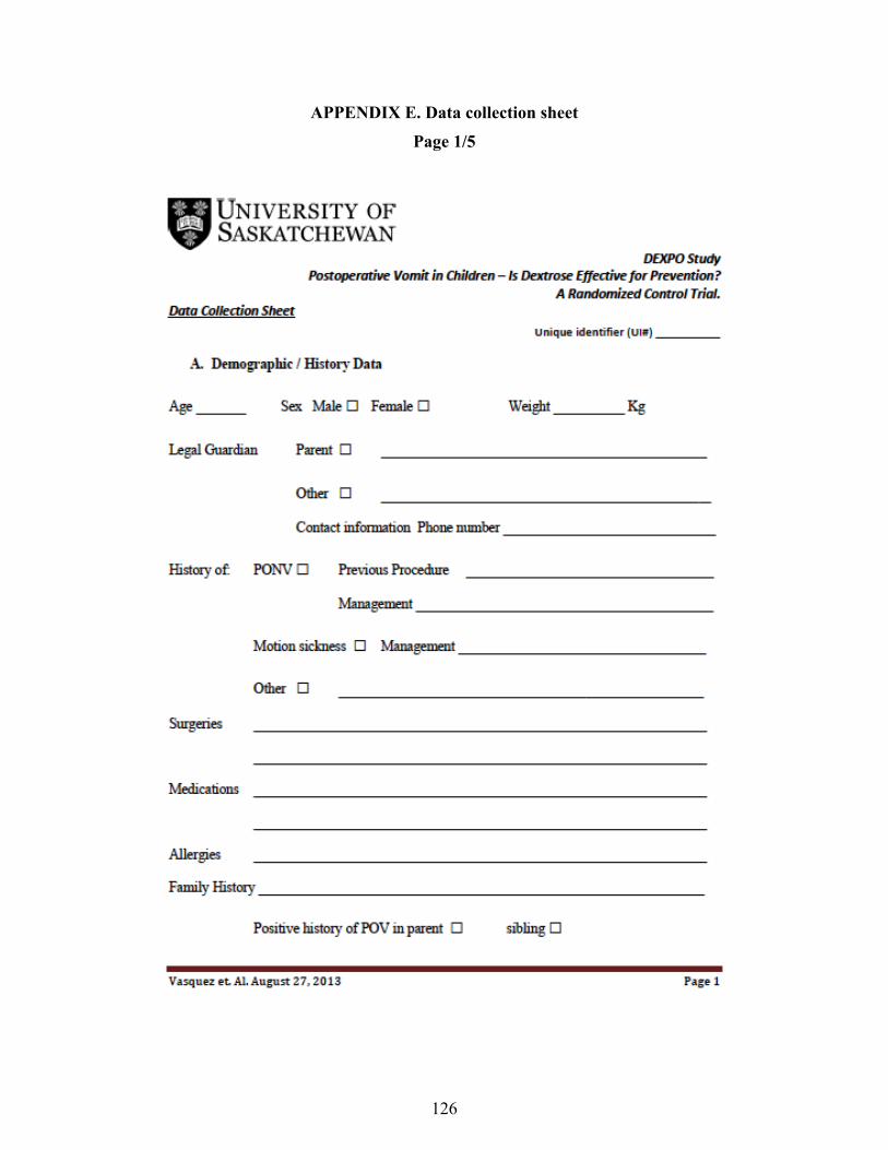
126
APPENDIX E. Data collection sheet
Page 1/5
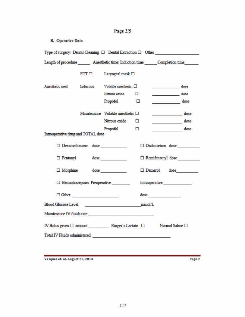
127
Page 2/5
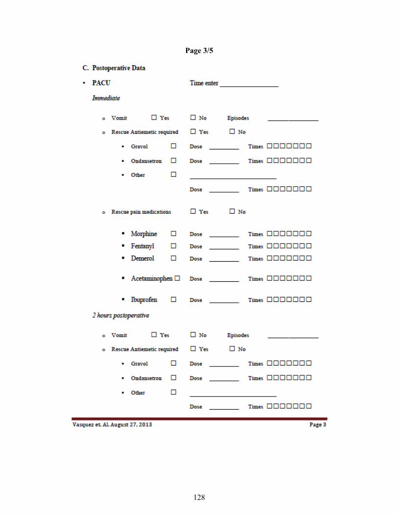
128
Page 3/5
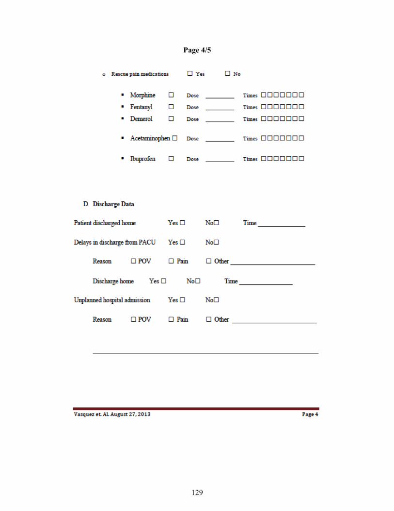
129
Page 4/5
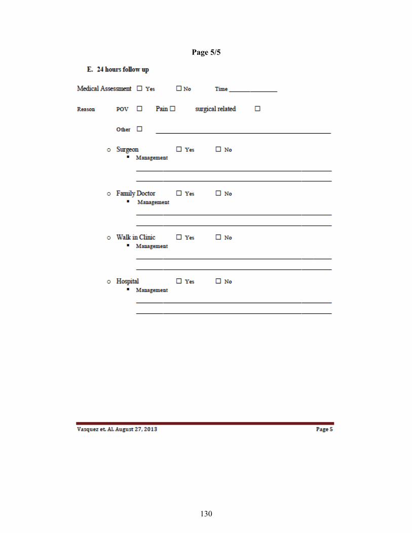
130
Page 5/5
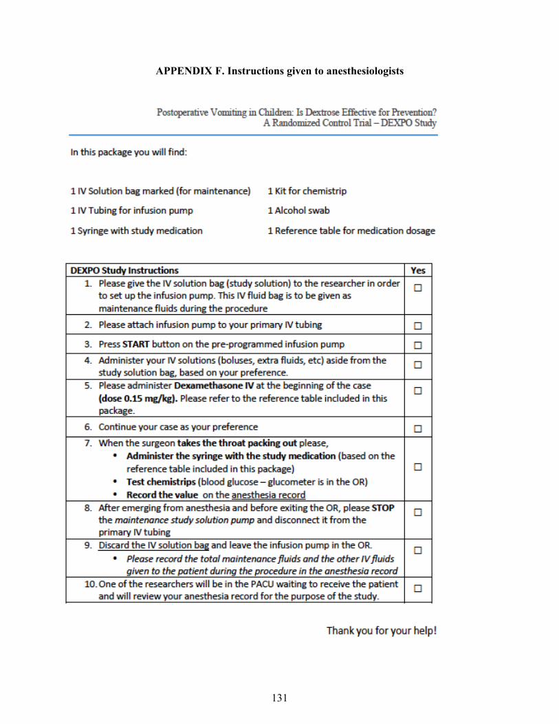
131
APPENDIX F. Instructions given to anesthesiologists

132
APPENDIX G. Table with intravenous fluid infusion rates and doses for dexamethasone
and study medication based on patient’s weight
Page 1/2
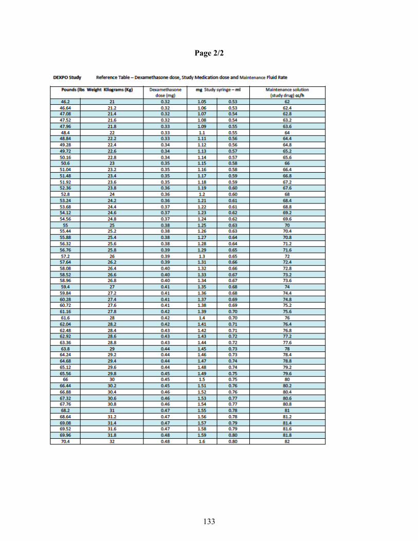
133
Page 2/2
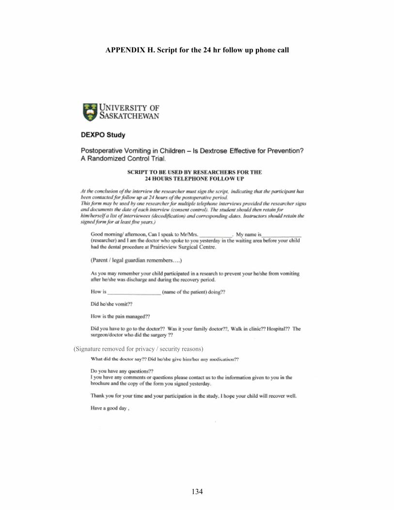
134
APPENDIX H. Script for the 24 hr follow up phone call
(Signature removed for privacy / security reasons)