magnetoencephalographic alpha band connectivity reveals differential default mode network...
TRANSCRIPT
Magnetoencephalographic alpha band connectivity reveals differentialDefault Mode Network interactions during focused attention and openmonitoring meditation
Laura Marzetti, Claudia Di_Lanzo, Filippo Zappasodi, Federico Chella, Antonino Raffone and Vittorio Pizzella
Journal Name: Frontiers in Human Neuroscience
ISSN: 1662-5161
Article type: Original Research Article
Received on: 30 Jul 2014
Accepted on: 30 Sep 2014
Provisional PDF published on: 30 Sep 2014
www.frontiersin.org: www.frontiersin.org
Citation: Marzetti L, Di_lanzo C, Zappasodi F, Chella F, Raffone A and PizzellaV(2014) Magnetoencephalographic alpha band connectivity revealsdifferential Default Mode Network interactions during focusedattention and open monitoring meditation. Front. Hum. Neurosci.8:832. doi:10.3389/fnhum.2014.00832
Copyright statement: © 2014 Marzetti, Di_lanzo, Zappasodi, Chella, Raffone and Pizzella.This is an open-access article distributed under the terms of theCreative Commons Attribution License (CC BY). The use,distribution and reproduction in other forums is permitted,provided the original author(s) or licensor are credited and thatthe original publication in this journal is cited, in accordance withaccepted academic practice. No use, distribution or reproductionis permitted which does not comply with these terms.
This Provisional PDF corresponds to the article as it appeared upon acceptance, after rigorous
peer-review. Fully formatted PDF and full text (HTML) versions will be made available soon.
Frontiers in Human Neuroscience Original Research
16/09/14
Magnetoencephalographic alpha band connectivity reveals differential 1
Default Mode Network interactions during focused attention and open 2
monitoring meditation 3
4
Laura Marzetti 1,2*, Claudia Di Lanzo1,2, Filippo Zappasodi1,2, Federico Chella1,2, Antonino Raffone3 , 5
Vittorio Pizzella1,2 6
1Department of Neuroscience, Imaging and Clinical Sciences, “G. d’Annunzio” University, Chieti, Italy 7
2Institute for Advanced Biomedical Technologies, “G. d’Annunzio” University, Chieti, Italy 8
3Department of Psychology, Sapienza University, Rome, Italy 9
* Correspondence: Laura Marzetti, PhD, Department of Neuroscience, Imaging and Clinical Sciences and Institute for 10
Advanced Biomedical Technologies, “G. d’Annunzio” University, Via dei Vestini, 66013 Chieti, Italy. 11
Keywords: meditation, mindfulness, magnetoencephalography, default mode network, resting state networks, 13 brain rhythms. 14
15
Abstract 16
According to several conceptualizations of meditation, the interplay between brain systems 17
associated to self-related processing, attention and executive control is crucial for meditative states 18
and related traits. We used magnetoencephalography to investigate such interplay in a highly selected 19
group of “virtuoso” meditators (Theravada Buddhist monks), with long-term training in the two main 20
meditation styles: focused attention (FA) and open monitoring (OM) meditation. Specifically, we 21
investigated the differences between FA meditation, OM meditation and resting state in the coupling 22
between the posterior cingulate cortex, core node of the Default Mode Network (DMN) implicated in 23
mind wandering and self-related processing, and the whole brain, with a recently developed phase 24
coherence approach. Our findings showed a state dependent coupling of PCC to nodes of the DMN 25
and of the executive control brain network in the alpha frequency band (8-12 Hz), related to different 26
attentional and cognitive control processes in FA and OM meditation, consistently with the putative 27
role of alpha band synchronization in the functional mechanisms for attention and consciousness. The 28
coupling of posterior cingulate cortex with left medial prefrontal cortex and superior frontal gyrus 29
characterized the contrast between the two meditation styles in a way that correlated with meditation 30
expertise. These correlations may be related to a higher mindful observing ability and a reduced 31
identification with ongoing mental activity in more expert meditators. 32
Notably, different styles of meditation and different meditation expertise appeared to modulate the 33
dynamic balance between fronto-parietal and DMN networks. Our results support the idea that the 34
interplay between the DMN and the fronto-parietal network in the alpha band is crucial for the 35
transition from resting state to different meditative states. 36
37
Marzetti et al. MEG DMN coherence in meditation
2
38
1. Introduction 39
40
Recently, the neural correlates of meditation states and traits have been increasingly studied in 41
cognitive and affective neuroscience (Cahn and Polich, 2006; Lutz et al., 2008; Raffone and 42
Srinivasan, 2010). Such growth of interest has been supported by several findings about the salutary 43
effects of meditation on physical and mental health, as related in particular to mindfulness based 44
programs (e.g. Chiesa and Serretti, 2010; Keng et al., 2011). Moreover, neuroimaging findings have 45
clarified the role of brain structures and processes involved in meditation and mindfulness based 46
training (e.g. Cahn and Polich, 2006; Chiesa and Serretti, 2010). 47
Several conceptualizations of meditation practice have underpinned a central role for attention and 48
cognitive control skills (Lutz et al., 2008; Malinowski 2013a, 2013b). These skills are crucial for the 49
development and maintenance of mindfulness, the intentional and non-judgmental awareness of the 50
fields of experience in the present moment, such as about perceptual, thought and feeling contents, 51
that, in turn, leads to therapeutic outcomes and wellbeing effects (Kabat-Zinn, 1994; Wallace and 52
Shapiro, 2006; Malinowski, 2013a). Meditation practices can be usefully classified into two main 53
styles – focused attention (FA) and open monitoring (OM) – depending on how the attentional 54
processes are directed (Cahn & Polich, 2006; Lutz et al., 2008). In the FA (‘concentrative’) style, 55
attention is focused on a given object in a sustained manner, and thus emphasizes sustained attention 56
and attention regulatory skills. The second style, OM meditation, involves the monitoring of any 57
content of ongoing experience, and thus emphasizes mindfulness rather than focused attention and 58
attentional control (Sumedho, 1994; Goldstein and Kornfield, 2001). 59
In this framework, the interplay between brain networks related to attention and control processes, 60
and self-related processing appears fundamental for the understanding of meditation and mindfulness 61
skills (Malinowski, 2013b). Indeed, mind-wandering and self-related processes occupy a large part of 62
mental activity of human beings (Killingsworth and Gilbert, 2010): imagining future events, thinking 63
about something different from what is currently being done, are mental states that frequently occur 64
in everyday life (Killingsworth & Gilbert, 2010). It is not surprising, then, that mind-wandering 65
parallels the brain’s mode of operation that is associated with the recruitment of the so called Default 66
Mode Network - DMN (Raichle et al, 2001). The DMN is one of the most robust among the Resting 67
State Networks (RSNs), and entails the posterior cingulate cortex, the medial prefrontal cortex, the 68
posterior lateral parietal/temporal cortices, and the parahippocampal gyrus (Raichle et al., 2001; 69
Buckner et al., 2008; Watanabe et al., 2013). These areas have shown to be co-activated during 70
passive mental states (e.g., task-unrelated cognition). 71
Another network that appears relevant for meditation and mindfulness skills is the Fronto-Parietal 72
control network (FP). The FP network includes many regions identified as supporting cognitive 73
control and decision-making, such as lateral prefrontal cortex, middle frontal gyrus, anterior 74
insula/frontal operculum, anterior cingulate cortex, and anterior inferior parietal lobule (Vincent et 75
al., 2008). The FP network also supports internally versus externally focused goal-directed cognition 76
by coupling with either the default or dorsal attention network (Christoff et al., 2009; Spreng et al., 77
2010; Spreng and Schacter, 2012). This functional interplay between the DMN and FP represents a 78
model for goal-directed cognition that might significantly contribute to the understanding of the 79
functional mechanisms underlying meditation. 80
Marzetti et al. MEG DMN coherence in meditation
Lauram 3
Functional MRI (fMRI) studies have so far shown an enhancement of BOLD functional connectivity 81
between the nodes of the DMN and executive control brain areas at rest and during meditation 82
practice in selected meditator populations (Brewer et al., 2011). The recruitment of the DMN during 83
meditation has been hypothesized to signal the involuntarily drift of attention away from the focus of 84
meditation towards mind wandering (Hasenkamp et al., 2012; Tang et al., 2012). Nevertheless, more 85
recent work has shown that activation of the DMN might serve for adaptive functions beyond 86
rumination and mind wandering (Ottaviani et al., 2013). Moreover, evidence exists for a differential 87
coupling of the DMN with other brain regions in different meditation styles (Xu et al., 2014), thus 88
indicating a possibly more complex involvement of the DMN in meditation. 89
A long tradition of studies has investigated the functional role of brain rhythms by 90
electroencephalography (EEG) or magnetoencephalography (MEG), and, in particular, the functional 91
correlate of the alpha rhythm has been long debated. Oscillations in the alpha band (8-12 Hz) have 92
been classically interpreted as the functional correlate of drowsiness, being of larger amplitude 93
during e.g. eye closed (Berger, 1929), supporting the idea of alpha power as related to an “idling” 94
state. Later evidence pointed out that an increase of alpha power is associated to deactivation of task-95
irrelevant brain areas, whereas a power decrease in alpha is associated to their activation 96
(Pfurtscheller, 2003). This idea was advanced into an alpha-inhibition hypothesis, which suggests 97
that alpha synchronization may reflect top-down control processes (Klimesch, 1996a, 1996b; 98
Klimesch et al., 2007). Opposite evidence exists for task-related increase in alpha power for high 99
level cognitive processes such as those elicited by mental calculation, mental imagery, or internally 100
driven attention (e.g., Hari et al., 1997; Cooper et al., 2003; Kounios and Beeman, 2009). Along the 101
same line, recent evidence from alpha phase-synchronization/phase-coherence, which is hypothesized 102
to represent a mechanism for short and long range communication in the brain (Lachaux et al., 1999; 103
Varela et al., 2001; Fries, 2005; Palva et al., 2005; Engel et al., 2013), suggests a direct role for alpha 104
band synchronization in the functional mechanisms of attention and consciousness (Palva and Palva, 105
2007; Knyazev et al., 2013). In this framework, it is not surprising that several studies have shown a 106
link between DMN structures and power and phase synchronization in the alpha frequency range 107
with concurrent EEG-fMRI (Laufs et al., 2003a; Laufs et al., 2003b; Mantini et al., 2007; Michels et 108
al., 2010; Jann et al., 2009; Knyazev et al., 2011; Sadaghiani et al., 2010; Sadaghiani et al., 2012) and 109
MEG (de Pasquale and Marzetti, 2014). 110
Brain rhythms, as measured by EEG and MEG, have also been studied to the specific aim of 111
disclosing the impact of meditation practices. These studies, revealed a high heterogeneity of the 112
frequency specific signatures of brain changes induced by meditation both in the same or in different 113
traditions (Cahn and Polich, 2006; Ivanovski and Malhi, 2007; Nolfe et al., 2011). In this framework, 114
evidence exists for the Individual Alpha Frequency - IAF - (Klimesch, 1996a) to be lowered as a 115
consequence of intensive meditation training in Saggar et al., 2012. Due to IAF lowering, in those 116
subjects, part of the standard alpha band was indeed pertaining to an individualized beta band, in 117
which the authors found training induced power modulations. This finding might implicitly suggest 118
that the heterogeneity reported in the literature might also be a consequence of the same 119
nomenclature used for frequency bands that might potentially not fully overlap across different 120
studies. 121
Furthermore, it should be noted that the great majority of EEG/MEG studies have investigated 122
frequency specific connectivity changes at the level of electrodes/sensors and their results cannot be 123
directly related to specific brain areas or brain networks due to volume conduction confounds 124
(Schoffelen and Gross, 2009). 125
The recent development of methods to study ongoing functional connectivity at brain level with 126
MEG, opens the way for reconciling the view on intrinsic activity as expressed by specific spatial 127
Marzetti et al. MEG DMN coherence in meditation
4
brain modes, i.e. RSNs, and alpha band role in attention and consciousness (Engel et al., 2013), 128
offering a direct window into the high complexity of brain information processing (de Pasquale et al., 129
2012; Larson-Prior et al., 2013). On the basis of such development, here we studied MEG functional 130
connectivity at the level of brain sources to investigate DMN interactions in meditation. Specifically, 131
we used a phase-coherence based approach that estimates the degree of linear 132
coupling/synchronization between oscillatory signals of distant neuronal ensembles. We hypothesize 133
that DMN internal coupling as well as its interactions with executive control (FP) network show 134
frequency specific traits which differ in FA and OM meditation, with a crucial role played by the 135
alpha rhythm. Moreover, coupling ranks might represent a marker of FA and OM meditation skills or 136
expertise. Specifically, given that the posterior cingulate cortex is the core node of the DMN 137
(Buckner et al., 2008), in this work we assessed MEG functional connectivity by mapping phase-138
coherence with respect to the posterior cingulate cortex node of the DMN across three experimental 139
conditions: FA meditation, OM meditation and a rest condition. Our study involved a highly selected 140
group of “virtuoso” meditators (Theravada Buddhist monks), with long-term training in both FA and 141
OM meditation styles (see also Manna et al., 2010), emphasizing the cultivation of attention and 142
awareness (monitoring) skills through all moments of their monastic life (Sumedho, 1994). 143
144
2. Materials and methods 145
Participants, Procedures, and Acquisition 146
An experienced meditator group of Theravada Buddhist monks was enrolled in this study. 147
Specifically, the experienced meditator group consisted in 8 Theravada Buddhist monks (all right 148
handed males, mean age 37.9 years, range 25–53 years, SD 9.4 years), with, on average, over 15,750 149
hours of meditation practice in Theravada Buddhist monasteries. The monks were recruited from the 150
Santacittarama monastery, in Italy, where they follow a Thai Forest Tradition. In this tradition, 151
monks experience regular intensive meditation retreats, with a balanced practice of Samatha 152
(Focused Attention, FA) and Vipassana (Open Monitoring, OM) meditation forms. These retreats 153
also include a long winter retreat lasting for about 3 months. Outside the retreat periods, the monks 154
typically practice 2 hours per day balanced FA and OM meditation styles, with the monastery 155
community. The experiment was conducted with the subject written informed consent according to 156
the Declaration of Helsinki, as well as with the approval of the local responsible Ethical Committee. 157
Only highly trained monk meditators were included in the study, with a minimal meditation expertise 158
of about 2500 hours, since an extensive training is necessary for reliably perform the two meditation 159
styles. The same group underwent also fMRI scans which were analyzed in a previous study by our 160
group (Manna et al., 2010). 161
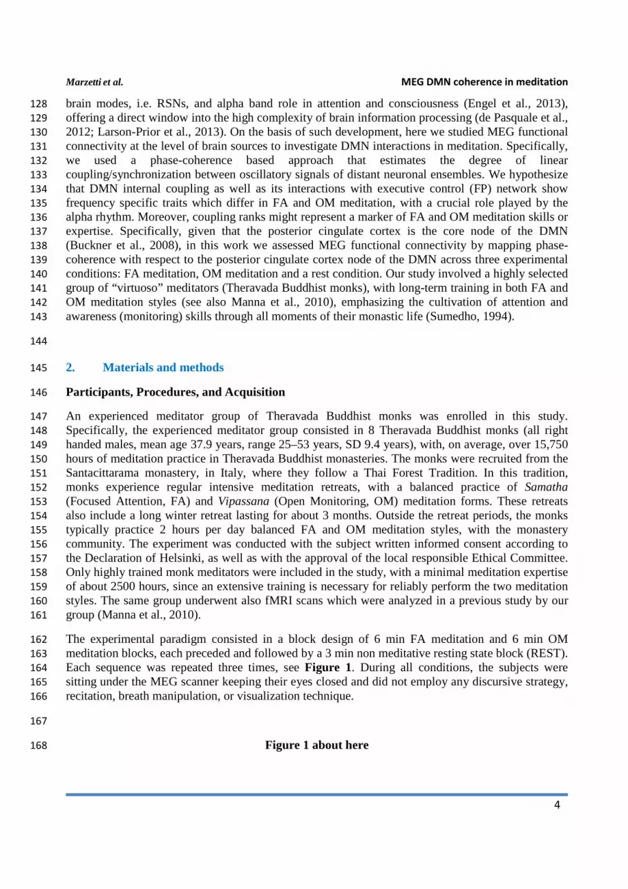
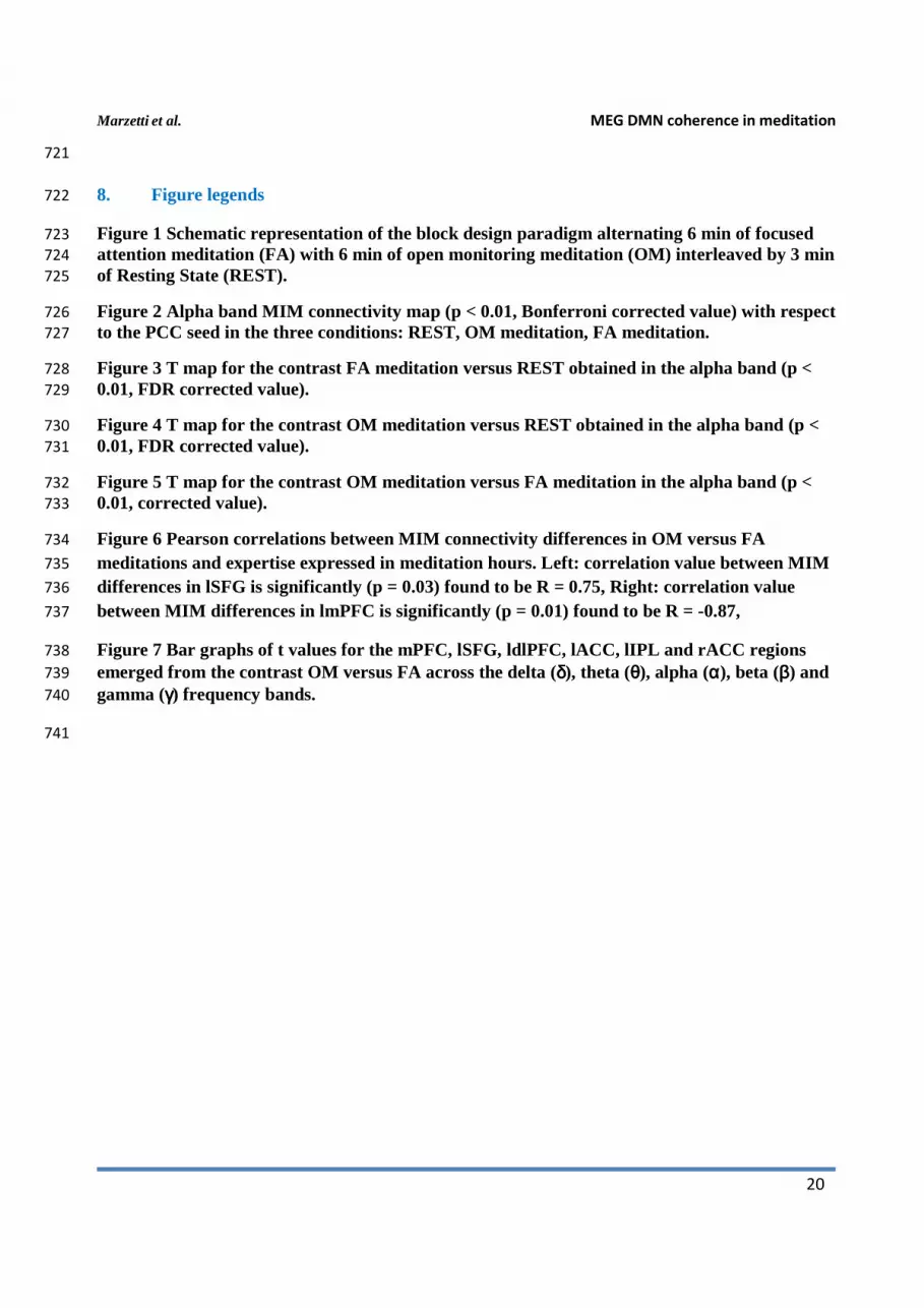
The experimental paradigm consisted in a block design of 6 min FA meditation and 6 min OM 162
meditation blocks, each preceded and followed by a 3 min non meditative resting state block (REST). 163
Each sequence was repeated three times, see Figure 1. During all conditions, the subjects were 164
sitting under the MEG scanner keeping their eyes closed and did not employ any discursive strategy, 165
recitation, breath manipulation, or visualization technique. 166
167
Figure 1 about here 168
Marzetti et al. MEG DMN coherence in meditation
Lauram 5
169
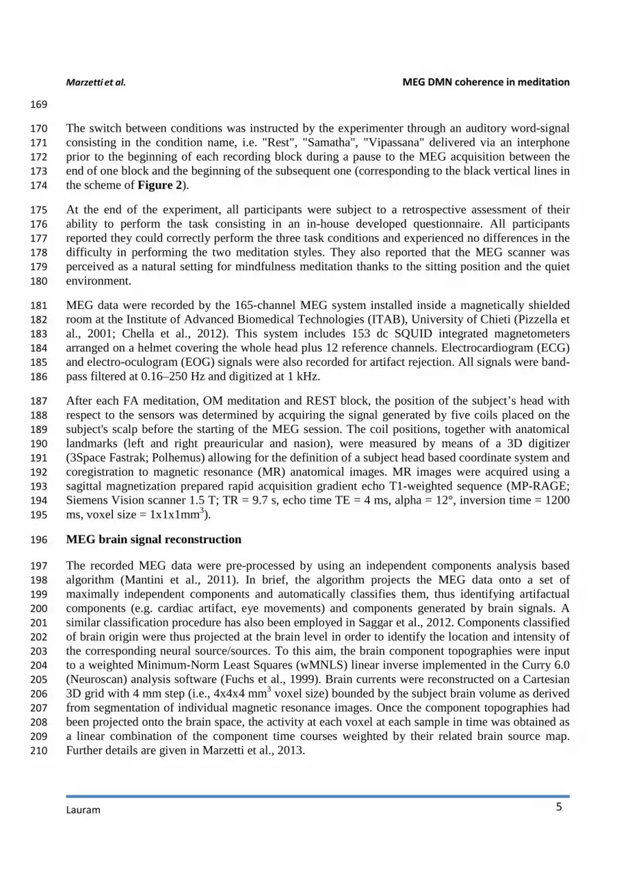
The switch between conditions was instructed by the experimenter through an auditory word-signal 170
consisting in the condition name, i.e. "Rest", "Samatha", "Vipassana" delivered via an interphone 171
prior to the beginning of each recording block during a pause to the MEG acquisition between the 172
end of one block and the beginning of the subsequent one (corresponding to the black vertical lines in 173
the scheme of Figure 2). 174
At the end of the experiment, all participants were subject to a retrospective assessment of their 175
ability to perform the task consisting in an in-house developed questionnaire. All participants 176
reported they could correctly perform the three task conditions and experienced no differences in the 177
difficulty in performing the two meditation styles. They also reported that the MEG scanner was 178
perceived as a natural setting for mindfulness meditation thanks to the sitting position and the quiet 179
environment. 180
MEG data were recorded by the 165-channel MEG system installed inside a magnetically shielded 181
room at the Institute of Advanced Biomedical Technologies (ITAB), University of Chieti (Pizzella et 182
al., 2001; Chella et al., 2012). This system includes 153 dc SQUID integrated magnetometers 183
arranged on a helmet covering the whole head plus 12 reference channels. Electrocardiogram (ECG) 184
and electro-oculogram (EOG) signals were also recorded for artifact rejection. All signals were band-185
pass filtered at 0.16–250 Hz and digitized at 1 kHz. 186
After each FA meditation, OM meditation and REST block, the position of the subject’s head with 187
respect to the sensors was determined by acquiring the signal generated by five coils placed on the 188
subject's scalp before the starting of the MEG session. The coil positions, together with anatomical 189
landmarks (left and right preauricular and nasion), were measured by means of a 3D digitizer 190
(3Space Fastrak; Polhemus) allowing for the definition of a subject head based coordinate system and 191
coregistration to magnetic resonance (MR) anatomical images. MR images were acquired using a 192
sagittal magnetization prepared rapid acquisition gradient echo T1-weighted sequence (MP-RAGE; 193
Siemens Vision scanner 1.5 T; TR = 9.7 s, echo time TE = 4 ms, alpha = 12°, inversion time = 1200 194
ms, voxel size = 1x1x1mm3). 195
MEG brain signal reconstruction 196
The recorded MEG data were pre-processed by using an independent components analysis based 197
algorithm (Mantini et al., 2011). In brief, the algorithm projects the MEG data onto a set of 198
maximally independent components and automatically classifies them, thus identifying artifactual 199
components (e.g. cardiac artifact, eye movements) and components generated by brain signals. A 200
similar classification procedure has also been employed in Saggar et al., 2012. Components classified 201
of brain origin were thus projected at the brain level in order to identify the location and intensity of 202
the corresponding neural source/sources. To this aim, the brain component topographies were input 203
to a weighted Minimum-Norm Least Squares (wMNLS) linear inverse implemented in the Curry 6.0 204
(Neuroscan) analysis software (Fuchs et al., 1999). Brain currents were reconstructed on a Cartesian 205
3D grid with 4 mm step (i.e., 4x4x4 mm3 voxel size) bounded by the subject brain volume as derived 206
from segmentation of individual magnetic resonance images. Once the component topographies had 207
been projected onto the brain space, the activity at each voxel at each sample in time was obtained as 208
a linear combination of the component time courses weighted by their related brain source map. 209
Further details are given in Marzetti et al., 2013. 210
Marzetti et al. MEG DMN coherence in meditation
6
MEG functional connectivity 211
The estimated MEG brain signals were the starting point for the study of functional coupling of 212
ongoing brain activity. Here, to map MEG functional connectivity we used an extension of the 213
imaginary part of coherence for detecting lagged coupling, namely the Multivariate Interaction 214
Measure - MIM (Ewald et al., 2012; Marzetti et al., 2013), that maximizes the imaginary part of 215
coherence between a given reference voxel (seed, s) and any other voxel (target, j). More 216
specifically, the estimated MEG signal at each brain voxel is a vector quantity that can be represented 217
through its components in a given reference system. MIM is designed to maximize the imaginary part 218
of coherence between vector quantities. The mathematical details on MIM derivation can be found in 219
Ewald et al., 2012. For the reader convenience, we briefly review MIM definition in the following. 220
Given the vector Fourier transformed signals as a function of frequency f at the seed and target 221
voxels: Xs(f) and Xj(f), respectively, and introducing the compact notation X(f) = [XsT(f) Xj
T(f)]
T, the 222
cross-spectrum between the two vectors Xs(f) and Xj(f), can be written in the block form: 223
224
and MIM between s and j is thus defined as: 225
226
In the above notation, tr indicates matrix trace, the T subscript indicates matrix transpose, 227
superscripts R and I denote the real and the imaginary parts, the -1 subscript indicates matrix inverse, 228
the * subscript indicates matrix conjugate transpose, and the capital J indicates the imaginary unit. A 229
more detailed recapitulation of the method is also given in Marzetti et al., 2013. 230
In this work, cross-spectra were estimated with Fast Fourier analysis after signal linear de-trending 231
and Hanning windowing and were averaged using time epochs of 1.0 s duration with 50% overlap 232
leading to a frequency resolution of 1 Hz. The number of averaged epochs is approximately 700 for 233
each block of the OM and FA meditations and approximately 350 for each rest block. 234
The method, being based on the maximization of imaginary coherence, largely overcomes the well-235
known limitation to the study of functional connectivity by EEG/MEG posed by signal mixing 236
artifacts, i.e. any active source in the brain contributes, in a weighted manner, to the signals measured 237
at all sensors through volume spread (see Figure 2a in Engel et al., 2013). This effect constitutes an 238
especially severe confound for estimates of brain interactions (Nolte et al., 2004; Marzetti et al., 239
2007; Schoffelen and Gross, 2009; Sekihara et al., 2011) and needs to be taken into account by 240
mapping MEG functional connectivity through robust measures. 241
Functional connectivity through MIM was here estimated with a seed based approach, i.e. between 242
the signal at the seed voxel and the signals at all other target brain voxels (approximately 28,000), 243
and, in order to investigate the role of the DMN in frequency specific coupling to other brain 244
networks in the different conditions, the seed was chosen in the posterior cingulate cortex (PCC), the 245
core node of the DMN (Buckner et al., 2008). 246
Functional connectivity was estimated for frequencies corresponding to the delta to gamma brain 247
rhythms, i.e. from 2 to 80 Hz. To improve frequency specificity, consecutive frequency bins were 248
++++
==)()()()(
)()()()()()(XC(f) *
fJCfCfJCfC
fJCfCfJCfCfXf
I
jj
R
jj
I
js
R
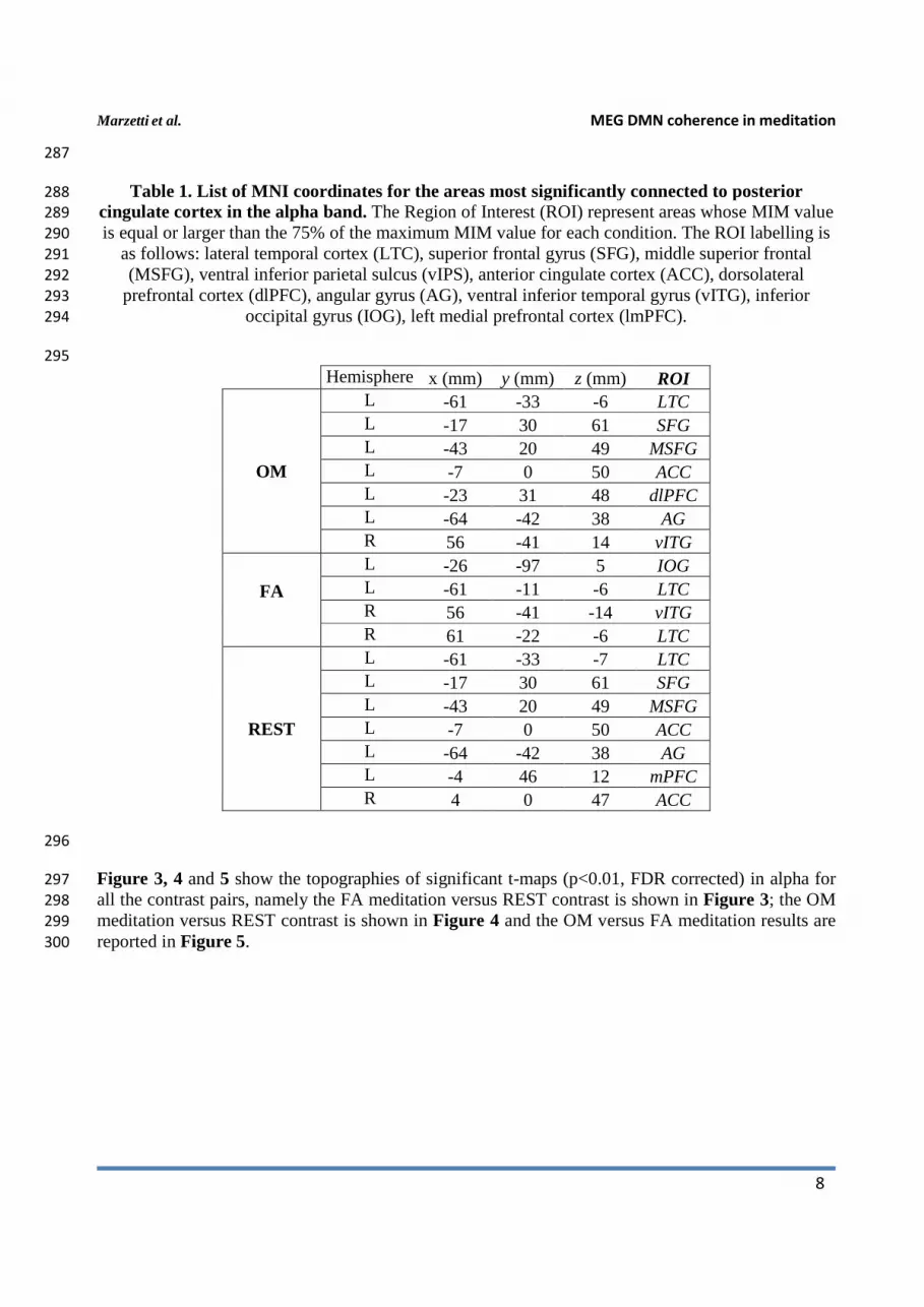
js
I
sj
R
sj
I
ss
R
ss
( ) ( ) ( )( )TI
sj
R
jj
I
sj
R
ss CCCCtr11
sjMIM−−=
Marzetti et al. MEG DMN coherence in meditation
Lauram 7
further averaged over frequency bands defined on the basis of individual alpha frequency (IAF) peak. 249
The alpha band was thus defined for each subject as IAF±2Hz; the definitions of the other frequency 250
bands were individually adjusted accordingly. On average, these bands span the following frequency 251
ranges: delta (2-3.5 Hz), theta (4-7 Hz), alpha (8-12 Hz), beta (13-30 Hz), gamma (30-80 Hz) in 252
accordance with conventional practice. 253
To investigate significant functional connectivity to PCC, a non-parametric Wilcoxon signed-rank 254
test was used to assess voxel-wise significance across subjects (p < 0.01, Bonferroni corrected). For 255
each frequency band, the MIM distribution across subjects for each voxel was compared to the 256
empirical distribution of MIM for independent sources (simulated as independent and identically 257
distributed, i.i.d., Gaussian noise) using a Monte Carlo approach with 20,000 repetitions. This 258
procedure allowed to identify significant connections to PCC for each condition after Bonferroni 259
correction for multiple comparisons. 260
A paired two tail t-test was used to compare MIM values between conditions (e.g., OM meditation 261
condition and REST) and to derive t-contrast maps between condition pairs after false discovery rate 262
correction (Benjamini and Hockberg, 1995) for multiple comparisons (p < 0.01, FDR corrected), thus 263
highlighting brain regions that are differently involved in a specific meditative state. 264
Specifically, to the aim of understanding whether different meditation styles involve differential 265
coupling between PCC and other brain areas in different frequency bands, we evaluated coupling to 266
PCC in terms of t-maps for all possible contrast pairs: i.e., OM-FA, OM-REST, FA-REST. All maps 267
were normalized to a common Montreal Neurological Institute (MNI) atlas through an affine 268
transformation implemented in SPM8 (Friston, 2003) for comparison across subjects and projected to 269
the standard brain surface for visualization by using the Caret software 270
(http://www.nitrc.org/projects/caret/; Van Essen et al., 2001). 271
3. Results 272
State-dependent MEG functional connectivity 273
Figure 2 shows condition specific alpha band MIM maps of functional connectivity to PCC after 274
correction for multiple comparisons, p<0.01, Bonferroni corrected. The PCC seed is indicated with a 275
white dot. Moreover, the areas whose MIM value is equal or larger than the 75% of the maximum 276
MIM value for each condition are listed in Table 1. Specifically, in OM meditation this procedure 277
identified the PCC coupling to left lateral temporal cortex (lLTC), left superior frontal gyrus (lSFG), 278
left middle superior frontal (lMSFG), left anterior cingulate cortex (lACC), left dorsolateral 279
prefrontal cortex (ldlPFC), left angular gyrus (lAG) and right ventral inferior temporal gyrus (rvITG). 280
In FA meditation, to right ventral inferior temporal gyrus, right and left lateral temporal cortices and 281
left inferior occipital lobe (lIOG). During REST, the highest coupling was observed with respect to 282
left medial prefrontal cortex (lmPFC), left lateral temporal cortex, left superior frontal gyrus, left 283
middle superior frontal gyrus, left anterior cingulate cortex and left angular gyrus. 284
Figure 2 about here 285
286
Marzetti et al. MEG DMN coherence in meditation
8
287
Table 1. List of MNI coordinates for the areas most significantly connected to posterior 288
cingulate cortex in the alpha band. The Region of Interest (ROI) represent areas whose MIM value 289
is equal or larger than the 75% of the maximum MIM value for each condition. The ROI labelling is 290
as follows: lateral temporal cortex (LTC), superior frontal gyrus (SFG), middle superior frontal 291
(MSFG), ventral inferior parietal sulcus (vIPS), anterior cingulate cortex (ACC), dorsolateral 292
prefrontal cortex (dlPFC), angular gyrus (AG), ventral inferior temporal gyrus (vITG), inferior 293
occipital gyrus (IOG), left medial prefrontal cortex (lmPFC). 294
295
Hemisphere x (mm) y (mm) z (mm) ROI
OM
L -61 -33 -6 LTC L -17 30 61 SFG L -43 20 49 MSFG L -7 0 50 ACC L -23 31 48 dlPFC L -64 -42 38 AG R 56 -41 14 vITG
FA
L -26 -97 5 IOG L -61 -11 -6 LTC R 56 -41 -14 vITG R 61 -22 -6 LTC
REST
L -61 -33 -7 LTC L -17 30 61 SFG L -43 20 49 MSFG L -7 0 50 ACC L -64 -42 38 AG L -4 46 12 mPFC R 4 0 47 ACC
296
Figure 3, 4 and 5 show the topographies of significant t-maps (p<0.01, FDR corrected) in alpha for 297
all the contrast pairs, namely the FA meditation versus REST contrast is shown in Figure 3; the OM 298
meditation versus REST contrast is shown in Figure 4 and the OM versus FA meditation results are 299
reported in Figure 5. 300
Marzetti et al. MEG DMN coherence in meditation
Lauram 9
301
Focused attention meditation vs. resting state 302
The alpha band connectivity in the contrast between FA meditation and REST (Figure 3) results in 303
the PCC being more coupled to left superior frontal gyrus (lSFG), left superior middle frontal gyrus 304
(lSMFG), left lateral temporal cortex (lLTC), left and right ACC during REST than during FA 305
meditation. See Table 2. 306
Figure 3 about here 307
308
Open monitoring meditation vs. resting state 309
The contrast between OM meditation and REST results in only the left intraparietal sulcus being 310
significantly less connected to PCC during OM meditation than during REST. 311
Figure 4 about here 312
313
Open monitoring vs. focused attention meditation 314
The contrast between OM meditation and FA meditation (Figure 5) highlights the stronger alpha 315
band coupling during OM meditation of PCC with the following regions: left medial prefrontal 316
cortex (lmPFC), left superior frontal gyrus (lSFG), left superior middle frontal gyrus (lSMFG), left 317
dorsolateral prefrontal cortex (ldlPFC) and left anterior cingulate cortex (lACC), left inferior parietal 318
lobule (lIPL). See Table 2. 319
Figure 5 about here 320
321
Marzetti et al. MEG DMN coherence in meditation
10
322
Table 2. List of ROIs significantly differently connected to posterior cingulate cortex in the 323
alpha band in the contrast between conditions. 324
325
Hemisphere x (mm) y (mm) z (mm) ROI
Connectivity to PCC
FA - REST
L -17 30 61 SFG REST > FA
L -43 18 43 MSFG REST > FA
L -61 -33 -6 LTC REST > FA
L -7 0 50 ACC REST > FA
R 21 8 69 SFG REST > FA
R 10 0 51 ACC REST > FA
OM - REST L -22 -58 67 IPS REST > OM
OM - FA
L -4 46 12 mPFC OM > FA
L -28 -4 69 SFG OM > FA
L -23 31 48 dlPFC OM > FA
L -1 -2 44 ACC OM > FA
L -58 -37 42 IPL OM > FA
R 2 0 46 ACC OM > FA
326
Frequency specificity 327
Notably, the coupling of PCC to the nodes of Default Mode and Fronto Parietal networks as listed in 328
Table 2 was a specific signature of the alpha band as it was not observed in the other frequency 329
bands included in the analysis. Figure 6 shows the t values, represented as bar graphs, for the OM 330
versus FA contrast. T values were extracted for the ROI coordinates listed in Table 2 for OM-FA for 331
the delta (δ), theta (θ), alpha (α), beta (β) and gamma (γ) frequency bands. This is a representative 332
situation showing the frequency specificity of the observed coupling. 333
Figure 6 about here 334
Correlation with meditation expertise 335
To evaluate to what extent the alpha band differences between conditions reported in Figure 3, 4 and 336
5 might be explained by differences in meditation expertise within the monk group, we correlated the 337
difference between MIM values in the two conditions with meditation expertise measured as overall 338
meditation hours, for all brain areas listed in Table 2. Significant Pearson correlation was found in 339
the contrast between OM and FA meditation for the lSFG and lmPFC nodes. The difference was 340
calculated using values extracted from the voxel closest (according to Euclidean distance) to the MNI 341
coordinate listed in Table 2. Nevertheless, consistent results were observed in the node surroundings 342
within a sphere of about 1 cm diameter. Specifically, a positive correlation (R = 0.75, p = 0.03) was 343
found for the difference of MIM values between OM and FA meditations in lSFG and a negative 344
correlation (R = - 0.87, p = 0.01) was found in lmPFC. See Figure 7. All the other brain regions and 345
Marzetti et al. MEG DMN coherence in meditation
Lauram 11
condition contrasts did not show a significant correlation with meditation expertise. 346
Figure 7 about here 347
4. Discussion 348
This study aimed at investigating the effects of focused attention (FA) and open monitoring (OM) 349
meditation on MEG functional connectivity to the posterior cingulate cortex (PCC), a crucial Default 350
Mode Network (DMN) node (Raichle et al., 2001; Buckner et al., 2008), in skilled long-term FA / 351
OM meditators. Overall, the results indicate that the different conditions modulate the coupling 352
between PCC and nodes of the DMN and of the Fronto-Parietal (FP) (executive) network (Vincent et 353
al., 2008) in the alpha band. The temporal richness of the MEG signal allowed us to quantify 354
functional connectivity by frequency specific patterns able to capture stationary properties of network 355
interactions in the brain. This goal was here achieved in a way robust to possible self-coupling 356
confounds deriving by MEG poor spatial resolution and signal mixing artifacts. 357
Our phase coherence based approach was able to reveal stable functional connectivity patterns with 358
respect to PCC across the meditation conditions and during the resting state. As shown in Figure 2, 359
an overall reduced connectivity was observed during the two meditation conditions in comparison to 360
the resting state, consistently with the EEG findings reported in Lehmann et al. (2012) and 361
Berkovich-Ohana et al. (2013). This reduced connectivity was here expressed both in terms of lower 362
average MIM values and of more localized connectivity topographies in meditation. Moreover, high 363
topographical similarity was shared between the resting state and the OM meditation condition, 364
compared to all other condition pairs. 365
When FA meditation was compared to the resting state, see Figure 3, a lower engagement of left and 366
right superior frontal gyrus, left middle superior frontal gyrus and lateral temporal cortex and of left 367
and right anterior cingulate cortex was observed. Indeed, in line with an earlier fMRI study (Manna 368
et al., 2010), functional deactivations during FA meditation as compared to the resting state involve 369
executive (FP) areas, which have been found activate with the DMN during mind wandering in 370
another study (Christoff et al., 2009). 371
When OM meditation was compared to the resting state, see Figure 4, the left intraparietal sulcus 372
was more connected to PCC during REST than during OM meditation. The left intraparietal sulcus 373
may thus be more involved in executive and monitoring functions in OM meditation (Lutz et al., 374
2008; Szameitat et al., 2002), rather than being as coupled to the DMN (PCC) as in the resting state. 375
More interestingly, when OM meditation was directly compared to FA meditation, see Figure 5, 376
regions in the left hemisphere belonging to the DMN (i.e., left medial prefrontal cortex) and the FP 377
network (i.e., left anterior cingulate cortex, left dorsolateral prefrontal cortex and left inferior parietal 378
lobe) were more functionally connected to PCC. Also left superior frontal gyrus, an area which has 379
been involved in both self-referential processing, linked to the DMN (e.g., Goldberg et al., 2006), and 380
executive functions and monitoring (e.g., du Boisgueheneuc et al., 2006), were more functionally 381
connected to PCC during OM meditation in comparison to FA meditation. The higher coupling 382
within the DMN can be related to the higher occurrence of thoughts and mental images during OM 383
meditation than during FA meditation (Lutz et al., 2008; Raffone and Srinivasan, 2009). Similarly, 384
the coupling with the left superior frontal gyrus can be related to an increased meta-awareness or 385
monitoring of ongoing thoughts and mental images generated with involvement of the DMN, in OM 386
as compared to FA meditation (Lutz et al., 2008; Raffone and Srinivasan, 2009). Indeed, the 387
Marzetti et al. MEG DMN coherence in meditation
12
increased monitoring (mindfulness) during OM meditation would lead to a meta-awareness that 388
enables the maintenance of the meditative state even in presence of spontaneous mentation. This 389
mindful observing ability is plausibly more developed in meditators with a higher level of expertise, 390
Figure 7, (Lutz et al., 2008), in line with our results here for (positive) correlation between functional 391
connectivity to PCC and meditation expertise, showing that coupling is differentially regulated by 392
meditation expertise in left superior frontal gyrus. Finally, a negative correlation between functional 393
coupling of left medial prefrontal cortex to PCC and meditation expertise was found, as related to the 394
OM versus FA meditation contrast, which appears consistent with an expected reduced identification 395
(self-reference) of more expert meditators with ongoing mental activity in any of the two forms of 396
meditation (Lutz et al., 2008). This evidence may also be related to an attenuated medial prefrontal 397
cortex activation with emotional thought contents (Northoff et al., 2004), likely to arise with OM 398
meditation as compared to FA meditation, with meditation expertise. By contrast, meditators with a 399
lower expertise exhibited a higher modulation of the coupling between PCC and lmPFC within the 400
DMN in shifting between FA (with lower coupling) and OM (with higher coupling) meditation 401
styles. 402
As shown in earlier neuroimaging studies, the lSFG is involved in self-related processing and 403
awareness (Golberg et al., 2006), which can be related to the notion of "narrative self" (Gazzaniga, 404
1995). In this respect, based on neuroimaging results, theorists have suggested that the brain systems 405
for the reflective self are likely to involve lateral prefrontal areas, and not just midline DMN areas 406
(Northoff et al., 2006; Tagini and Raffone, 2010). Our present study further suggests that the neural 407
mechanisms for the “narrative self” may involve the crucial coupling between PCC and lSFG in the 408
alpha band, which can be modulated by meditation, with differential effects of FA and OM styles. 409
Since the left superior frontal gyrus has also been related to executive and monitoring functions (e.g., 410
in working memory) (du Boisgueheneuc et al., 2006), the differential coupling between PCC and left 411
superior frontal gyrus in the alpha band found in our study may be linked to a differential recruitment 412
of neuronal populations in the left superior frontal gyrus for self-referential thought versus executive 413
and cognitive monitoring. Indeed, neuronal responses in lateral prefrontal cortex are highly adaptive, 414
depending on the task setting and individual differences (Duncan, 2001). Interestingly, as suggested 415
by our results, different styles of meditation and meditation expertise appeared to modulate such 416
coupling and possibly the dynamic balance between the recruitment of FP and DMN networks. 417
Notably, all of the observed effects were specific to the alpha band (Figure 6). Besides the idling 418
hypothesis, the role of alpha band phase coupling in meditation might be closely related to the role of 419
alpha band synchronization as a functional mechanism of attention and consciousness (Palva and 420
Palva, 2007; Knyazev et al., 2013). Indeed, an inhibitory role has been associated to the alpha rhythm 421
to the aim of filtering out irrelevant sensory inputs (Klimesh et al., 2007; Bonnefond and Jensen, 422
2013) in a broad range of information processing tasks including selective spatial attention (Rihs et 423
al., 2007; Van Ede et al., 2011; Foxe et al., 2011) and working memory (Sauseng et al., 2009; Hagens 424
et al., 2010; Spitzer et al., 2011; Bonnefond and Jensen, 2012). Evidence indeed suggests that FA and 425
OM meditation styles entail unique sets of attention and consciousness, and are not merely degrees of 426
a state of relaxation (Dunn et al., 1999). More specifically, our findings highlighted a possible role of 427
such rhythm in maintaining the stability of DMN internal phase coupling and subserving its 428
modulation with the FP network, according to the specific meditation state. Indeed, the presently 429
observed findings are consistent with a top-down alpha modulation hypothesis as a mechanism 430
involved in stress-therapy meditation (Kerr et al., 2011), and with the idea that cooperation between 431
the DMN and the FP network helps sustain monitoring of thoughts against compulsory self-reference 432
(identification) (Farb et al., 2007) and interference (Smallwood et al., 2012). Taken together, our 433
results support the idea that an interplay between the DMN and the FP network is crucial for the 434
Marzetti et al. MEG DMN coherence in meditation
Lauram 13
transition from resting state to different meditative states. 435
It has to be noted that it is not trivial that nodes classically ascribed by fMRI as belonging to the 436
DMN and the FP should emerge as coupled to PCC also in MEG connectivity studies. Indeed, MEG 437
provides a window into the high complexity of brain information processing at the temporal scales 438
relevant for behavior which translate into frequency resolved coupling. For this reason, different 439
systems comprised by some of the network nodes classically identified by fMRI might be represented 440
by MEG phase coherence at different frequency scales, possibly speaking for a functional 441
dissociation of network subsystems in the frequency domain. 442
A possible limitation of this study is the relatively small number of subjects included in the analysis. 443
Our emphasis was indeed on the high (“virtuoso”) skills in both FA and OM meditation of the 444
Theravada Buddhist monks involved in our study, appearing as a rare and selected sample in 445
literature. Moreover, we did not include a comparison with a group of novice practitioners. In this 446
respect, however, a recent fMRI study (Manna et al., 2010) with the same participants performing FA 447
and OM meditation, found the most relevant differences in the contrasts between FA meditation, OM 448
meditation and resting state conditions within the monk group, in line with our present focus. Indeed, 449
for novice meditators it might be difficult to control for and to differentiate between the meditative 450
states which is the primarily focus of this study. As a general remark, it is indeed difficult to 451
objectively validate what the participants are doing in a task which involves inherently subjective and 452
covert states, such as FA and OM meditations. We believe that our involvement of meditators 453
(Theravada Buddhist monks) who are expert in both FA and OM meditation has minimized the 454
chances of inaccurate performance of the two meditation tasks, and this was also evident from the 455
retrospective reports. Finally, it would be worth in the future to include in the protocol a controlled 456
manipulation of attention by introducing a task (e.g. a sustained response inhibition task as in 457
Zanesco et al., 2013). This would allow to e.g. compare task based functional connectivity before and 458
after the meditation blocks and to correlate connectivity results with task performance. 459
460
5. Conclusions 461
To summarize, the present study allowed to characterize the coupling of the major Default Mode 462
Network (DMN) node, the Posterior Cingulate Cortex (PCC), with the rest of the brain, and 463
highlighted, in a data driven manner, its coupling to nodes of DMN and Fronto-Parietal (FP) network 464
specific to the alpha band. Our findings showed that the alpha band is selectively involved in the 465
different couplings of PCC in the two meditation styles and during rest, and that a stable coupling 466
within DMN and between DMN and FP network characterized the contrast between the two 467
meditation styles, which was correlated to meditation expertise. 468
469
More generally, MEG functional connectivity was able to reveal important features of meditative 470
states in the brain which were modulated by expertise. Indeed, MEG can provide a unique 471
neuroimaging tool to study meditation and mindfulness processes thanks to its ability to recover brain 472
functional coupling in a frequency resolved manner, thus reconciling the long tradition of the EEG 473
based approach with an fMRI network based approach to meditation. Not secondary to this aim, the 474
MEG scanner appeared as a more convenient setting for performing research on meditation thanks to 475
the sitting position that the subject can maintain during the measurement, as well as to the absence of 476
any disturbing external noise inside the shielded room in which the MEG system is set. 477
Marzetti et al. MEG DMN coherence in meditation
14
6. Acknowledgements 478
The authors wish to thank the abbot of the Santacittarama monastery, Ajahn Chandapalo, and all the 479
monks of the Santacittarama and associated monasteries that kindly participated in the experiment. 480
The authors are also grateful to Dr. Raffaella Franciotti for technical assistance during MEG 481
measurements, and to Dr. Marcella Brunetti and Dr. Paolo Belardinelli for their help to build a link 482
between the University of Chieti and Dr. Raffone on the basis of common research interests. 483
484
Marzetti et al. MEG DMN coherence in meditation
Lauram 15
7. References 485
Benjamini, Y., and Hockberg, Y. (1995) Controlling the False Discovery Rate: a Practical and 486
Powerful Approach to Multiple Testing. J.R. Statist Soc. B 57(1): 289-300. 487
Berger HI. (1929) Über das Elektroenkephalogram des Menschen. Arch. Psychiat. Nervenkr. 87:527–488
570 489
Berkovich-Ohana, A., Glicksohn, J., Goldstein, A. (2013) Studying the default mode and its 490
mindfulness-induced changes using EEG functional connectivity. Soc Cogn Affect Neurosci 491
doi:10.1093/scan/nst153 492
Bonnefond M, Jensen O. (2012) Alpha oscillations serve to protect working memory maintenance 493
against anticipated distracters. Curr Biol. 22(20):1969-74. doi: 10.1016/j.cub.2012.08.029. 494
Bonnefond, M., Jensen, O. (2013) The role of gamma and alpha oscillations for blocking out 495
distraction. Commun Integr Biol. 6(1):e22702. doi: 10.4161/cib.22702. 496
Brewer, J.A., Worhunsky, P.D., Gray, J.R., Tang, Y., Weber, J. and Kober, H. (2011) Meditation 497
experience is associated with differences in default mode network activity and connectivity. 498
Proc Natl Acad Sci USA 108(50): 20254-59. 499
Buckner, R.L., Andrews-Hanna, J.R., Schacter, D.L. (2008) The brain's default network: anatomy, 500
function, and relevance to disease. Ann N Y Acad Sci. 1124:1-38. 501
Cahn, B.R., Polich, J. (2006). Meditation states and traits: EEG, ERP, and neuroimaging studies. 502
Psychological Bulletin, 132: 180–211. 503
Chella, F., Zappasodi, F., Marzetti, L., Della Penna, S., Pizzella V. (2012) Calibration of a 504
multichannel MEG system based on the Signal Space Separation method. Phys Med Biol 57: 505
4855-4870. 506
Chiesa, A., Serretti, A. (2010) A systematic review of neurobiological and clinical features of 507
mindfulness meditations. Psychol Med. 40(8):1239-52. doi: 10.1017/S0033291709991747. 508
Cooper, N.R., Croft, R.J, Dominey, S.J.J., Burgess, A.P., Gruzelier, J.H. (2003) Paradox lost? 509
Exploring the role of alpha oscillations during externally vs. internally directed attention and 510
the implications for idling and inhibition hypotheses. Int. J. Psychophysiol. 47, 65–74 511
Christoff, K., Gordon, A.M., Smallwood, J., Smith, R., Schooler, J.W. (2009) Experience sampling 512
during fMRI reveals default network and executive system contributions to mind wandering. 513
Proc. Natl. Acad. Sci. U.S.A. 106: 8719–8724. 514
de Pasquale, F., Della Penna, S., Snyder, A.Z., Marzetti, L., Pizzella, V., Romani, G.L., Corbetta, M. 515
(2012) A cortical core for dynamic integration of functional networks in the resting human 516
brain. Neuron 74:753-64. 517
de Pasquale, F. and Marzetti, L. (2014) "Temporal and spectral signatures of the Default Mode 518
Network." In MEG: From signal to Dynamic cortical networks, eds. S. Supek and C.J. Aine. 519
(Heidelberg: Springer Verlang), 451-476. 520
du Boisgueheneuc, F., Levy, R., Volle, E., Seassau, M., Duffau, H., Kinkingnehun, S., Samson, Y., 521
Zhang, S., Dubois, B. (2006) Functions of the left superior frontal gyrus in humans: a lesion 522
study. Brain 129: 3315-28. 523
Duncan, J. (2001) An adaptive coding model of neural function in prefrontal cortex. Nat Rev 524
Neurosci. 2(11):820-9. 525
Dunn, B.R., Hartigan, J.A., Mikulas, W.L. (1999) Concentration and mindfulness meditations: 526
unique forms of consciousness? Appl Psychophysiol Biofeedback. 24(3):147-65. 527
Engel, A.K., Gerloff, C., Hilgetag, C.C., Nolte, G. (2013) Intrinsic coupling modes: multiscale 528
interactions in ongoing brain activity. Neuron 80(4): 867-886. 529
Ewald, A., Marzetti, L., Zappasodi, F., Meinecke, F.C., Nolte, G. (2012) Estimating true brain 530
connectivity from EEG/MEG data invariant to linear and static transformations in sensor 531
Marzetti et al. MEG DMN coherence in meditation
16
space. Neuroimage 60: 476–488. 532
Farb, N.A., Segal, Z.V., Mayberg, H., Bean, J., McKeon, D., Fatima, Z., Anderson, A.K. (2007) 533
Attending to the present: mindfulness meditation reveals distinct neural modes of self-534
reference. Soc Cogn Affect Neurosci. 2(4):313-22. doi: 10.1093/scan/nsm030. 535
Fries, P. (2005) A mechanism for cognitive dynamics: neural communication through neuronal 536
coherence. Trend Cogn Sci 9(10):474-480. 537
Foxe, J.J., Snyder, A.C. (2011) The Role of Alpha-Band Brain Oscillations as a Sensory Suppression 538
Mechanism during Selective Attention. Front Psychol. 2: 154.doi:10.3389/fpsyg.2011.00154. 539
Friston, K. (2003). Introduction: experimental design and statistical parametric mapping. In: 540
Frackowiak et al., editors. Human brain function, 2nd Edition. 541
Fuchs, M., Wagner, M., Köhler, T., Wischmann, H.A. (1999) Linear and nonlinear current density 542
reconstructions. J Clin Neurophysiol. 16(3):267-95. 543
Gazzaniga, M.S. (1995) Principles of human brain organization derived from split-brain studies. 544
Neuron 14: 217-228. 545
Goldberg, I.I., Harel, M., and Malach, R.(2006) When the Brain Loses Its Self: Prefrontal 546
Inactivation during Sensorimotor Processing. Neuron 50:329–339. 547
doi:10.1016/j.neuron.2006.03.015 548
Goldstein, J., and Kornfield, J. (2001) Seeking the Heart of Wisdom: the Path of Insight Meditation. 549
Boston, MA: Shambhala. 550
Hagens, S., Osipova, D., Oostenveld, R., Jensen, O. (2010) Somatosensory working memory 551
performance in humans depends on both engagement and disengagement of regions in a 552
distributed network. Hum Brain Mapp. 31: 26-35. 553
Hari, R., Salmelin, R., Makela, J.P., Salenius, S., Helle, M. (1997) Magnetoencephalographic cortical 554
rhythms. Int. J. Psychophysiol. 26:51–62. 555
Hasenkamp, W., Wilson-Mendenhall C.D., Duncan, E., and Barsalou, L.W. (2012) Mind wandering 556
and attention during focused meditation: a fine-grained temporal analysis of fluctuating 557
cognitive states, Neuroimage 59,750-760. doi:10.1016/j.neuroimage.2011.07.003 558
Jann, K., Dierks, T., Boesch, C., Kottlow, M., Strik, W., Koenig, T. (2009) BOLD correlates of EEG 559
alpha phase-locking and the fMRI default mode network. Neuroimage. 45(3):903-16. 560
Kabat-Zinn, J. (1994) Wherever you go, there you are: Mindfulness Meditation in everyday life. 561
Hyperion.Keng, S.L., Smoski, M.J., Robins, C.J. (2011) Effects of mindfulness on 562
psychological health: a review of empirical studies. Clin Psychol Rev. 31(6):1041-56. doi: 563
10.1016/j.cpr.2011.04.006. 564
Keng, S.L., Smoski, M.J., Robins, C.J.(2011) Effects of mindfulness on psychological health: a 565
review of empirical studies. Clin Psychol Rev. 31(6):1041-56. doi: 10.1016/j.cpr.2011.04.006. 566
Kerr, C.E., Jones, S.R., Wan, Q., Pritchett, D.L., Wasserman, R.H., Wexler, A., et al. (2011). Effects 567
of mindfulness meditation training on anticipatory alpha modulation in primary 568
somatosensory cortex. Brain Res. Bull. 85: 96-103. 569
Killingsworth, M.A., Gilbert, D.T. (2010) A wandering mind is an unhappy mind. Science 570
330(6006):932. doi: 10.1126/science.1192439. 571
Klimesch, W. (1996a) EEG alpha and theta oscillations reflect cognitive and memory performance: a 572
review and analysis. Brain Research Reviews 29:169–195. 573
Klimesch, W. (1996b). Memory processes, brain oscillations and EEG synchronization. Int. J. 574
Psychophysiol. 24: 61–100. 575
Klimesch, W., Sauseng, P., Hanslmayr, S. (2007) EEG alpha oscillations: the inhibition–timing 576
hypothesis. Brain Res. Rev. 53: 63–88. 577
Knyazev, G.G., Slobodskoj-Plusnin, J.Y., Bocharov, A.V., Pylkova, L.V. (2011) The default mode 578
network and EEG alpha oscillations: An independent component analysis. Brain Research 579
Marzetti et al. MEG DMN coherence in meditation
Lauram 17
1402: 67–79. doi:10.1016/j.brainres.2011.05.052. 580
Knyazev, G.G. (2013) EEG correlates of self-referential processing. Front Hum Neurosci 7(264): 1-581
14, doi: 10.3389/fnhum.2013.00264. 582
Kounios, J., Beeman, M. (2009) The Aha! Moment. The Cognitive Neuroscience of Insight. Current 583
direction in psychological science. 18(4): 210:216. 584
Ivanovski, B., and Malhi, G.S. (2007) The psychological and neurophysiological concomitants of 585
mindfulness forms of meditation. Acta Neuropsychiatrica 19:76–91. doi: 10.1111/j.1601-586
5215.2007.00175.x 587
Lachaux, J.P., Rodriguez, E., Martinerie, J., Varela, F. (1999) Measuring Phase Synchrony in the 588
Brain. Hum Brain Mapp. 8:194-208. 589
Larson-Prior, L.J., Oostenveld, R., Della Penna, S., Michalareas, G., Prior, F., Babajani-Feremi, A., 590
Schoffelen, J.M., Marzetti, L., de Pasquale, F., Di Pompeo, F., Stout, J., Woolrich, M., Luo, 591
Q., Bucholz, R., Fries, P., Pizzella, V., Romani, G.L., Corbetta, M., Snyder, A.Z.; WU-Minn 592
HCP Consortium. (2013) Adding dynamics to the Human Connectome Project with MEG. 593
Neuroimage 80:190-201. 594
Laufs, H., Kleinschmidt, A., Beyerle, A., Eger, E., Salek- Haddadi, A., Preibisch, C., Krakow K. 595
(2003a). EEG-correlated fMRI of human alpha activity. Neuroimage 19: 1463–1476.doi:10. 596
1016/S1053-8119(03)00286-6. 597
Laufs, H., Krakow, K., Sterzer, P., Eger, E., Beyerle, A., Salek-Haddadi, A., Kleinschmidt, A. 598
(2003b). Electroencephalographic signatures of attentional and cognitive default modes in 599
spontaneous brain activity at rest. Proc.Natl. Acad.Sci.U.S.A. 100: 11053–11058. 600
doi:10.1073/pnas.1831638100. 601
Lehmann, D., Faber, P.L., Tei, S., Pascual-Marqui, R.D., Milz, P., Kochi, K. (2012) Reduced 602
functional connectivity between cortical sources in five meditation traditions detected with 603
lagged coherence using EEG tomography. Neuroimage 60: 1574-86. 604
Lutz, A., Slagter H.A., Dunne, J.D., Davidson, R.J. (2008) Attention regulation and monitoring in 605
meditation. Trends Cogn. Sci. 12:163–169. 606
Malinowski, P. (2013a) Neural mechanisms of attentional control in mindfulness meditation. Front. 607
Neurosci. doi: 10.3389/fnins.2013.00008. 608
Malinowski, P. (2013b) Flourishing through meditation and mindfulness. In: David, Boniwell, and 609
Conley Ayers, editors. Oxford Hand book of Happiness. Oxford: Oxford University Press 610
384–396. 611
Manna, A., Raffone, A., Perrucci, M.G., Nardo, D., Ferretti, A., Tartaro, A., Londei, A., Del Gratta, 612
C., Belardinelli, M.O., Romani, G.L. (2010) Neural correlates of focused attention and 613
cognitive monitoring in meditation. Brain Res Bull. 82(1-2): 46-56. 614
Mantini, D., Perrucci, M.G., Del Gratta, C., Romani, G.L., Corbetta, M. (2007) Electrophysiological 615
signatures of resting state networks in the human brain. Proc Natl Acad Sci USA 104: 13170-616
13175. 617
Mantini, D., Della Penna, S., Marzetti, L., de Pasquale, F., Pizzella, V., Corbetta, M., Romani, G.L. 618
(2011) A signal processing pipeline for MEG resting state networks. Brain Connectivity 1: 619
49-59. 620
Marzetti, L., Nolte, G., Perrucci, M.G., Romani, G.L., Del Gratta, C. (2007) The use of standardized 621
infinity reference in EEG coherency studies. Neuroimage 36: 48-63. 622
Marzetti, L., Della Penna, S., Snyder, A.Z., Pizzella, V., Nolte, G., de Pasquale, F., Romani, G.L., 623
Corbetta, M. (2013) Frequency specific interactions of MEG resting state activity within and 624
across brain networks as revealed by the Multivariate Interaction Measure. Neuroimage 625
79:172-83. 626
Michels, L., Bucher, K., Lüchinger, R., Klaver, P., Martin, E., Jeanmonod, D., Brandeis, D. (2010) 627
Marzetti et al. MEG DMN coherence in meditation
18
Simultaneous EEG-fMRI during a Working Memory Task: Modulations in Low and High 628
Frequency Bands. PLoS ONE 5(4):1 629
Nolfe, G. (2011) EEG and meditation. Clinical Neurophysiology 123: 631–2. 630
Nolte, G., Bai, O., Wheaton, L., Mari, Z., Vorbach, S., Hallet, M. (2004) Identifying true brain 631
interaction from EEG data using the imaginary part of coherency. Clin. Neurophysiol. 632
115:2292-2307. 633
Northoff, G., Heinzel, A., Bermpohl, F., Niese, R., Pfennig, A., Pascual-Leone, A., Schlaug, G., 634
(2004) Reciprocal Modulation and Attenuation in the Prefrontal Cortex: An fMRI Study on 635
Emotional–Cognitive Interaction. Human Brain Mapping 21:202–212. 636
Northoff, G., Heinzel, A., de Greck, M., Bermpohl, F., Dobrowolny, H., Panksepp, J. (2006) Self-637
referential processing in our brain--a meta-analysis of imaging studies on the self. 638
Neuroimage 31(1):440-57. 639
Ottaviani, C., Shapiro, D., and Couyoumdjian, A. (2013) Flexibility as the key for somatic health: 640
from mind wandering to perseverative cognition. Biol. Psychol. 94: 38-43, 641
doi:10.1016/j.biopsycho.2013.05.003 642
Palva, J.M., Palva, S., Kaila, K. (2005) Phase synchrony among neuronal oscillations in the human 643
cortex. J. Neurosci. 25: 3962–3972. 644
Palva, S., Palva, IM, (2007) New vistas for a-frequency band oscillations. Trends in Neurosciences 645
30(4): 150-158. doi:10.1016/j.tins.2007.02.001 646
Pizzella, V., Della Penna, S., Del Gratta, C., Romani, G.L. (2001) SQUID systems for biomagnetic 647
imaging, Supercond Sci Technol 14: R79-R114. 648
Pfurtscheller, G. (2003) Induced oscillations in the alpha band: functional meaning. Epilepsia 44(12): 649
2–8. 650
Raffone, A., Srinivasan, N. (2009) An adaptive workspace hypothesis about the neural correlates of 651
consciousness: insights from neuroscience and meditation studies. Prog Brain Res.176:161-652
80. doi: 10.1016/S0079-6123(09)17620-3. 653
Raffone, A., Srinivasan, N. (2010) The exploration of meditation in the neuroscience of attention and 654
consciousness. Cogn Process. 11(1):1-7. doi: 10.1007/s10339-009-0354-z. 655
Raichle, M., MacLeod, A.M., Snyder, A.Z., Powers, W.J, Gusnard, D.A., Shulman, G.L. (2001) The 656
default mode of brain function. Proc Natl Acad Sci USA 98(2): 676-82. 657
Rihs, T.A., Michel, C.M., Thut, G. (2007) Mechanisms of selective inhibition in visual spatial 658
attention are indexed by alpha-band EEG synchronization. Eur J Neurosci. 25(2):603-10. 659
Sadaghiani, S,, Scheeringa, R., Lehongre, K., Morillon, B., Giraud, A.L., Kleinschmidt, A. (2010) 660
Intrinsic connectivity networks, alpha oscillations, and tonic alertness: a simultaneous 661
electroencephalography/functional magnetic resonance imaging study. J Neurosci. 662
30(30):10243-50. doi: 10.1523/JNEUROSCI.1004-10.2010. 663
Sadaghiani, S., Scheeringa, R., Lehongre, K., Morillon, B., Giraud, A.L., D' Esposito M., 664
Kleinschmidt, A. (2012) α-band phase synchrony is related to activity in the fronto-parietal 665
adaptive control network. J Neurosci. 32(41):14305-10. doi: 10.1523/JNEUROSCI.1358-666
12.2012. 667
Saggar, M., King, B.G., Zanesco, A.P., Maclean, K.A., Aichele, S.R., Jacobs, T.L., Bridwell, D.A., 668
Shaver, P.R., Rosenberg, E.L., Sahdra, B.K., Ferrer, E., Tang, A.C., Mangun, G.R., Wallace, 669
B.A., Miikkulainen, R., Saron, C.D. (2012) Intensive training induces longitudinal changes in 670
meditation state-related EEG oscillatory activity. Front Hum Neurosci. 6:256. 671
doi:10.3389/fnhum.2012.00256. 672
Sauseng, P., Klimesch, W., Heise, K.F., Gruber, W.R., Holz, E., Karim, A.A., Glennon, M., Gerloff, 673
C., Birbaumer, N., Hummel, F.C. (2009) Brain oscillatory substrates of visual short-term 674
memory capacity. Curr Biol. 19(21):1846-52. doi: 10.1016/j.cub.2009.08.062. 675
Marzetti et al. MEG DMN coherence in meditation
Lauram 19
Schoffelen, J.M., Gross, J. (2009) Source connectivity analysis with MEG and EEG. Human Brain 676
Mapping 30: 1857-1865. 677
Sekihara, K., Owen, J.P., Trisno, S., Nagarajan, S.S. (2011) Removal of spurious coherence in MEG 678
source-space coherence analysis. IEEE Trans Biomed Eng. 58:3121-9. 679
Smallwood, J., Brown, K., Baird, B., Schooler, J.W. (2012) Cooperation between the default mode 680
network and the frontal-parietal network in the production of an internal train of thought. 681
Brain Res. 1428:60-70. doi: 10.1016/j.brainres.2011.03.072. 682
Spitzer, B., and Blankenburg, F. (2011) Stimulus-dependent EEG activity reflects internal updating 683
of tactile working memory in humans. Proc. Nat. Acad. Sci. USA 108: 8444-49. 684
Spreng, R.N., Stevens, W.D., Chamberlain, J.P., Gilmore, A.W., Schacter, D.L. (2010) Default 685
network activity, coupled with the frontoparietal control network, supports goal-directed 686
cognition. Neuroimage. 53(1):303–317. 687
Spreng, R.N., Schacter, D.L. (2012) Default network modulation and large-scale network 688
interactivity in healthy young and old adults. Cereb Cortex. 22(11):2610-21. doi: 689
10.1093/cercor/bhr339. 690
Sumedho, A. (1994) The Mind and the Way: Buddhist Reflections on Life. Wisdom Publications. 691
Szameitat, A.J., Schubert, T., Müller, K., Von Cramon, D.Y. (2002) Localization of executive 692
functions in dual-task performance with fMRI. J Cogn Neurosci. 14(8):1184-99. 693
Tagini, A., Raffone, A. (2010) The 'I' and the 'Me' in self-referential awareness: a neurocognitive 694
hypothesis. Cogn Process. 11(1):9-20. doi: 10.1007/s10339-009-0336-1. 695
Tang, Y.Y., Rothbart, M.K., Posner, M.I. (2012) Neural correlates of establishing, maintaining, and 696
switching brain states. Trends Cogn.Sci. 16: 330–337. 697
Van Ede, F., De Lange, F., Jensen, O., Maris, E. (2011) Orienting attention to an upcoming tactile 698
event involves a spatially and temporally specific modulation of sensorimotor alpha- and 699
beta- band oscillations. J. Neurosci. 31: 2016-2024. 700
Van Essen, D.C., Dickson, J., Harwell, J., Hanlon, D., Anderson, C.H., Drury, H.A. (2001) An 701
Integrated Software System for Surface-based Analyses of Cerebral Cortex. Journal of 702
American Medical Informatics Association. 8: 443-459. 703
Vincent, J.L., Kahn, I., Snyder, A.Z., Raichle, M.E., Buckner, R.L. (2008) Evidence for a 704
frontoparietal control system revealed by intrinsic functional connectivity. J Neurophysiol. 705
100(6):3328-42. doi: 10.1152/jn.90355.2008. 706
Varela, F., Lachaux, J.P., Rodriguez, E., Martinerie, J. (2001) The brain web: phase synchronization 707
and large scale integration. Nat Rev 2: 229-239. 708
Wallace, B.A., Shapiro, S.L. (2006) Mental balance and well-being: building bridges between 709
Buddhism and Western psychology. Am Psychol. 61(7): 690-701. 710
Watanabe, T., Hirose, S., Wada, H., Imai, Y., Machida, T., Shirouzu, I., Konishi, S., Miyashita Y., 711
and Masuda, N. (2013) A pairwise maximum entropy model accurately describes resting-state 712
human brain networks. Nature Communications 4, 1370, doi:10.1038/ncomms2388. 713
Xu, J., Vik, A., Groote, I.R., Lagopoulos, J., Ellingsen, Ø., Häberg, A.K., Davanger, S. (2014) Non 714
directive meditation activates defaults mode network and areas associated with memory 715
retrieval and emotional processing, Front Hum Neurosci 8(86): 1-10. 716
Zanesco, A.P., Brandon G. King, B.G., MacLean, K.A. and Saron, C.D. (2013) Executive control 717
and felt concentrative engagement following intensive meditation training. Front Hum 718
Neurosci 7(566):1-13 doi: 10.3389/fnhum.2013.00566 719
720
Marzetti et al. MEG DMN coherence in meditation
20
721
8. Figure legends 722
Figure 1 Schematic representation of the block design paradigm alternating 6 min of focused 723
attention meditation (FA) with 6 min of open monitoring meditation (OM) interleaved by 3 min 724
of Resting State (REST). 725
Figure 2 Alpha band MIM connectivity map (p < 0.01, Bonferroni corrected value) with respect 726
to the PCC seed in the three conditions: REST, OM meditation, FA meditation. 727
Figure 3 T map for the contrast FA meditation versus REST obtained in the alpha band (p < 728
0.01, FDR corrected value). 729
Figure 4 T map for the contrast OM meditation versus REST obtained in the alpha band (p < 730
0.01, FDR corrected value). 731
Figure 5 T map for the contrast OM meditation versus FA meditation in the alpha band (p < 732
0.01, corrected value). 733
Figure 6 Pearson correlations between MIM connectivity differences in OM versus FA 734
meditations and expertise expressed in meditation hours. Left: correlation value between MIM 735
differences in lSFG is significantly (p = 0.03) found to be R = 0.75, Right: correlation value 736
between MIM differences in lmPFC is significantly (p = 0.01) found to be R = -0.87, 737
Figure 7 Bar graphs of t values for the mPFC, lSFG, ldlPFC, lACC, lIPL and rACC regions 738
emerged from the contrast OM versus FA across the delta (δδδδ), theta (θθθθ), alpha (αααα), beta (ββββ) and 739
gamma (γγγγ) frequency bands. 740
741
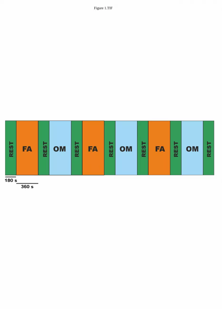
Figure 1.TIF
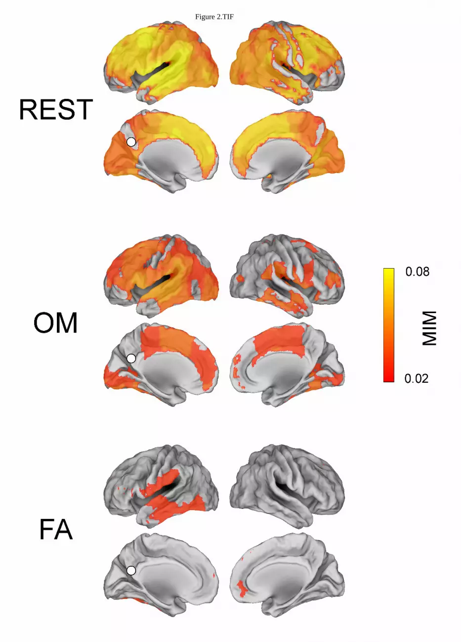
Figure 2.TIF
Figure 3.TIF
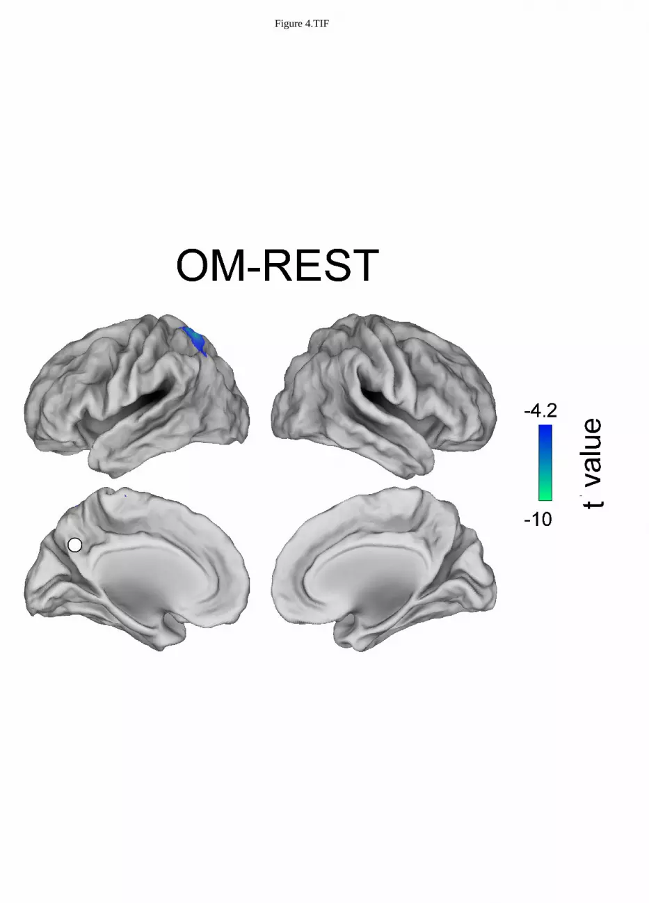
Figure 4.TIF
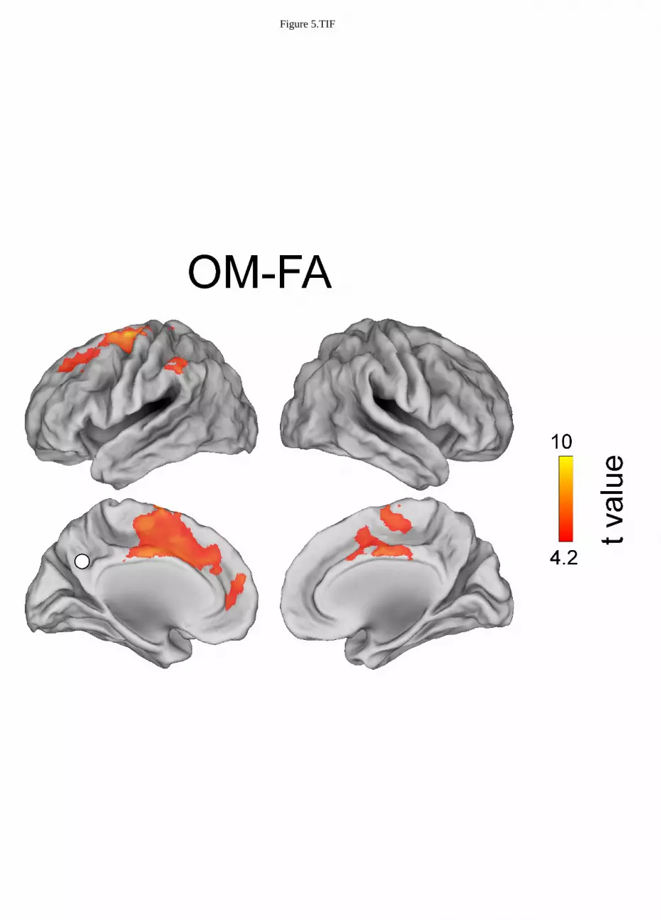
Figure 5.TIF
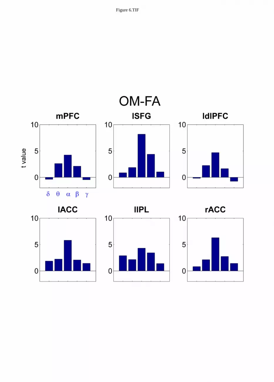
Figure 6.TIF
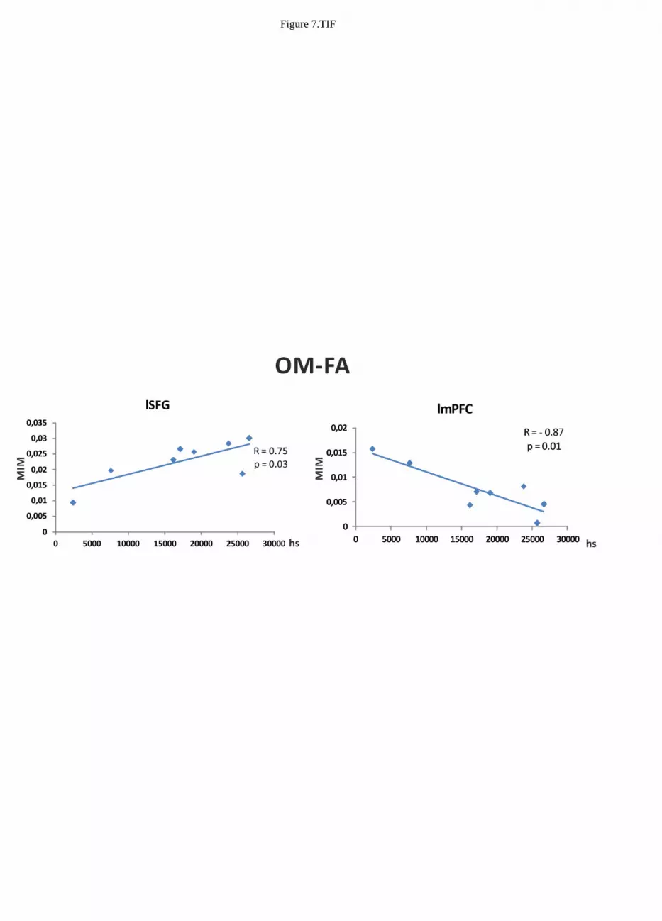
Figure 7.TIF