lysine 63-polyubiquitination guards against translesion synthesis–induced mutations
TRANSCRIPT
Lysine 63-Polyubiquitination Guardsagainst Translesion Synthesis–InducedMutationsRoland K. Chiu
1[, Jan Brun
2[, Chantal Ramaekers
1, Jan Theys
1, Lin Weng
1, Philippe Lambin
1, Douglas A. Gray
2*,
Bradly G. Wouters1*
1 Department of Radiation Oncology, GROW Research Institute, University of Maastricht, Maastricht, Netherlands, 2 Ottawa Health Research Institute, Ottawa, Ontario, Canada
Eukaryotic cells possess several mechanisms to protect the integrity of their DNA against damage. These include cell-cycle checkpoints, DNA-repair pathways, and also a distinct DNA damage–tolerance system that allows recovery ofreplication forks blocked at sites of DNA damage. In both humans and yeast, lesion bypass and restart of DNAsynthesis can occur through an error-prone pathway activated following mono-ubiquitination of proliferating cellnuclear antigen (PCNA), a protein found at sites of replication, and recruitment of specialized translesion synthesispolymerases. In yeast, there is evidence for a second, error-free, pathway that requires modification of PCNA with non-proteolytic lysine 63-linked polyubiquitin (K63-polyUb) chains. Here we demonstrate that formation of K63-polyUbchains protects human cells against translesion synthesis–induced mutations by promoting recovery of blockedreplication forks through an alternative error-free mechanism. Furthermore, we show that polyubiquitination of PCNAoccurs in UV-irradiated human cells. Our findings indicate that K63-polyubiquitination guards against environmentalcarcinogenesis and contributes to genomic stability.
Citation: Chiu RK, Brun J, Ramaekers C, Theys J, Weng L, et al. (2006) Lysine 63-polyubiquitination guards against translesion synthesis–induced mutations. PLoS Genet 2(7):e116. DOI: 10.1371/journal.pgen.0020116
Introduction
In contrast to DNA-repair pathways, DNA damage toler-ance (DDT) is characterized by bypass of DNA lesions ratherthan their direct removal or repair. The DDT pathway islikely responsible for the ability of cells to continue toproliferate with tremendous amounts of damage in theirgenomes [1]. The genetic and mechanistic basis of DDT is bestunderstood in yeast, where it is known to be an extremelyimportant determinant of the toxicity and mutagenicity ofmany DNA-damaging agents [2,3]. Often referred to as RAD6-dependent repair or post-replication repair, DDT requiresinteraction of the E2 ubiquitin (Ub) conjugase RAD6 and theE3 Ub ligase RAD18 at sites of DNA damage [4]. Here theymediate mono-ubiquitination of proliferating cell nuclearantigen (PCNA) at K164 and subsequent recruitment of aspecialized translesion synthesis (TLS) polymerase capable ofreplication past the lesion [5,6]. Several yeast and mammalianTLS polymerases have been identified, including POLg(RAD30A), POLi (RAD30b), REV1, REV3, and POLj [7].These are highly error-prone polymerases that allow repli-cation past a variety of DNA lesions [7]. POLg plays auniquely important role in the repair of UV damage as itmediates error-free bypass of thymine–thymine dimers, themost common UV-induced lesion [8]. Saccharomyces cerevisiaeRAD6 and RAD18 mutants that are unable to carry out DDTare highly sensitive to various genotoxic agents including UVirradiation and methyl methane sulfonate (MMS) [9]. Thesemutants also show a reduction in UV-induced mutations [10]that arises due to the inability to recruit the error-prone TLSpolymerases [11].
Genetic epistasis studies in yeast have established a secondarm of the DDT pathway that is distinct from TLS and is
referred to as damage avoidance [5,12–14]. This pathway isalso downstream of RAD6/RAD18, but in contrast to theerror-prone TLS pathway resolves blocked replication forksthrough an error-free manner. Its mechanism is not fullyunderstood, but may involve fork reversal and recombinationwith the undamaged replicated sister chromatid [5]. Thisdamage-avoidance pathway requires a second ubiquitinationcomplex composed of RAD5 and the UBC13/MMS2 hetero-dimer [5]. UBC13/MMS2 is a unique Ub conjugase thatsynthesizes polyUb chains linked through K63–G76 bondsrather than through the typical K48–G76 bonds [13].Although lysine 63-linked polyubiquitin (K63-polyUb) chainscan serve as competent proteolytic signals, they are less
Editor: James E. Haber, Brandeis University, United States of America
Received January 24, 2006; Accepted June 12, 2006; Published July 21, 2006
A previous version of this article appeared as an Early Online Release on June 12,2006 (DOI: 10.1371/journal.pgen.0020116.eor).
DOI: 10.1371/journal.pgen.0020116
Copyright: � 2006 Chiu et al. This is an open-access article distributed under theterms of the Creative Commons Attribution License, which permits unrestricteduse, distribution, and reproduction in any medium, provided the original authorand source are credited.
Abbreviations: 6–4PP, pyrimidine-6/4-pyrimidone; CPD, cis-syn cyclobutanepyrimidine dimer; DDT, DNA damage tolerance; DUB, deubiquitinating; FA,Fanconi’s anemia; HAT, hypoxanthine, aminopterin, and thymidine; K63-polyUb,lysine 63-linked polyubiquitin; MMS, methyl methane sulfonate; NEM, N-ethyl-maleimide; PCNA, proliferating cell nuclear antigen; SDS, sodium dodecyl sulfate;TLS, translesion synthesis; Ub, ubiquitin; UBD, ubiquitin-binding domain; UBZ,ubiquitin-binding zinc-finger; WT, wild-type; XP, Xeroderma Pigmentosum; XPV,Xeroderma Pigmentosum variant
* To whom correspondence should be addressed. E-mail: [email protected] (DAG);[email protected] (BGW)
[ These authors contributed equally to this work.
PLoS Genetics | www.plosgenetics.org July 2006 | Volume 2 | Issue 7 | e1161070
efficient at targeting substrates to the proteasome than K48-linked chains [15], and the proteolytic activity of theproteasome may not be required for error-free repair [13].In yeast, a model has emerged in which error-free damageavoidance occurs when mono-ubiquitinated PCNA becomesfurther modified by K63-polyUb via RAD5 and MMS2/UBC13.Interestingly, modification of K164 in PCNA by sumoylationrather than by ubiquitination reduces homologous recombi-nation [16,17].
There is convincing evidence that the DDT pathway, andparticularly the TLS arm, is also important in highereukaryotes including humans. Mouse and human homologsof RAD6, RAD18, PCNA, and many of the TLS polymeraseshave been identified [18]. The TLS polymerases form foci atsites of DNA damage following UV irradiation and areassociated with other proteins in the replication machinery[19]. As in yeast, RAD6 and RAD18 mediate mono-ubiquiti-nation of PCNA at K164 in UV-irradiated mammalian cells ina dose- and time-dependent manner [11]. Mono-ubiquitina-tion of human PCNA has been suggested to provide a signalfor polymerase switching since it leads to its increasedassociation with POLg via its ubiquitin-binding domain(UBD) or the UBZ (ubiquitin-binding zinc-finger) in thisTLS polymerase [20]. In vitro studies have also demonstratedthat mono-ubiquitination of PCNA in yeast can stimulate theactivities of both POLg and REV1 [21]. Recently, thedeubiquitinating (DUB) enzyme USP1 was shown to directlyremove the monoUb from PCNA, leading to the suggestionthat USP1 is required to suppress the error-prone activity ofTLS [22]. The functional importance of TLS is exemplified bythe fact that mutations in POLg are responsible for thevariant form of Xeroderma Pigmentosum (XP), a diseasecharacterized by a 2,000-fold increased risk of developingskin cancer [8]. In contrast to other XP patients, those withthe variant form (XPV) of Xeroderma Pigmentosum have nodefect in excision repair [8], but are deficient in post-
replication repair [23]. Furthermore, they display enhancedmutation at T–T sites, owing to usage of an alternative error-prone TLS polymerase [24].In contrast to TLS, the importance of the damage-
avoidance arm of DDT in mammalian cells is not yet firmlyestablished. Perhaps the strongest evidence supporting a rolefor this pathway comes from Li et al., who showed thatantisense inhibition of hMMS2 resulted in an increase inmutation frequency [25]. Nonetheless, several open questionsremain to be resolved. First, a human homolog of RAD5 hasnot yet been identified. This may be due to the fact that yeastRAD5 contains a helicase activity required for its function inDNA double-strand break repair, but is unimportant for DDT[26]. These authors speculated that RAD5 in higher organismsmay have evolved to lose this domain. Second, althoughhomologs of MMS2 exist (hMMS2 and hCroc1) and are able tofunctionally complement loss of yeast MMS2 [27], they areadditionally required for polyubiquitination of proteins inpathways unrelated to DDT [28]. Third, although evidence forhuman PCNA mono-ubiquitination is strong [11,29], there isless evidence for its polyubiquitination. High molecularweight bands in PCNA Western blots were noted in mousefibroblasts following UV irradiation [11]. However, Kan-nouche and colleagues found no evidence for polyubiquiti-nation in human fibroblasts [29]. They concluded that polyUbforms of PCNA were either insignificant, occurred only at lowlevels, or were rapidly turned over [29]. Thus, whetherpolyubiquitination of PCNA and subsequent activation ofan error-free damage-avoidance pathway is evolutionarilyconserved in humans is a source of uncertainty that we soughtto resolve.Here, we provide evidence that the ability to create K63-
based polyUb chains is required for an error-free damage-avoidance pathway in human cells. We implicate thisubiquitination step in a pathway that contributes to genomicstability by suppressing translesion polymerase–mediatedmutagenesis. Moreover, we show that DNA damage–inducedPCNA polyubiquitination is indeed conserved in human cells,suggesting that this Ub-based molecular switch plays adecision role in directing repair in either an error-free orerror-prone manner.
Results
Dominant Negative Approach to Disrupt K63-PolyUbChain AssemblyIn order to directly investigate the functional importance
of K63-linked polyUb chains in DDT, we employed a strategysimilar to that first described in yeast, to specifically inhibitassembly of these chains. In yeast, replacement of Ub with amutant in which lysine 63 is mutated to arginine (K63R)disrupts the error-free arm of DDT and results in aphenotype equivalent to loss of UBC13 or MMS2 [14]. TheK63R mutation disrupts K63-polyUb chain assembly, but hasno effect on K48-linked chains that mediate proteasomal-based protein turnover [14]. In human cells, an equivalentknock-in approach is not feasible because Ub is expressedfrom multiple genes. The UBA52 and UBA80 genes encode aUb monomer fused in frame with ribosomal subunits, whilethe UBB and UBC genes encode variable-length linearpolymers of (typically three to four Ub and nine Ub proteins,
PLoS Genetics | www.plosgenetics.org July 2006 | Volume 2 | Issue 7 | e1161071
K63-PolyUb Protects Cells from Mutation
Synopsis
Genome instability is associated with increased cancer risk, and thusconsiderable effort has been put into unraveling the mechanismsunderlying genome surveillance. Guarding the integrity of DNA are anumber of DNA-repair and cell cycle–control systems. Insight intohow these pathways become activated is crucially important to theunderstanding of carcinogenesis and in the development of cancertreatments. This study concerns a distinct pathway that promotesthe tolerance of DNA damage during its replication phase. Priorattempts to investigate this pathway in human cells have beendifficult due to extensive redundancy in the genes that carry out thisprocess. Previous knowledge from lower organisms suggested therequirement for enzymes capable of constructing a chain ofubiquitin molecules linked in a specific manner. The authors useda novel approach to disrupt the formation of these ubiquitin chainsin human cells and found that this caused a significant increase inmutations after exposure to UV light. Several lines of evidenceimplicate a family of error-prone enzymes, called translesionsynthesis polymerases, in the formation of these mutations.Furthermore, they provide evidence suggesting that proliferatingcell nuclear antigen (PCNA), a protein found at sites of replication, isthe relevant target of these chains in human cells. These findingsindicate that polyubiquitination guards against environmentalcarcinogenesis and contributes to genomic stability.
respectively) [30]. The fusion proteins are cleaved by DUBenzymes to release individual Ub monomers.
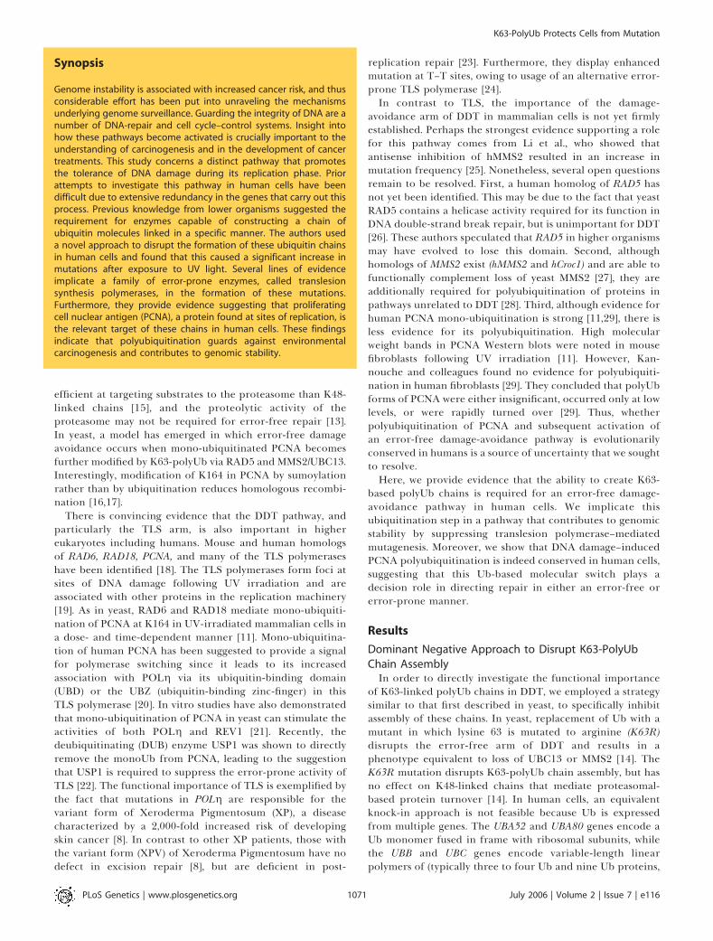
Our approach was to express the K63R-Ub mutant in transso that it competed with wild-type (WT) Ub for inclusion intopolyUb chains. Its incorporation blocks further ubiquitina-tion through K63 and thus acts in a dominant way. In aprevious study, we validated and used this approach tospecifically suppress K48-linked Ub chains by expressing aK48R-Ub mutant [31]. This same construct has also been usedto inhibit K48 polyubiquitination in transgenic mice [32].Here, we expressed a six-his-tagged K63R-Ub or WT-Ub fusedin frame with GFP from the UbC promoter (Figure 1).Expression yields a fusion protein that is cleaved, releasinga six-his-tagged Ub monomer and free GFP (Figure 1B). GFPwas used to sort pools of cells with stable high expression ofthe transgene. Both WT-Ub and K63R-Ub monomers wereefficiently incorporated into polyUb chains as evidenced bytheir detection in high molecular weight smears character-istic of the heterogeneity of ubiquitinated proteins (Figure1B). The K63R-Ub mutant did not affect normal cellproliferation as demonstrated by the identical growth rates
in the sorted stable high K63R-Ub-GFP–expressing pools andin the similarly sorted WT-Ub-GFP–expressing and theuntransfected cells (Figure 1C). Furthermore, disruptingK63-polyUb chain formation did not alter normal protea-some-mediated protein degradation of p53 or HIF1a (un-published data). These data indicate that the K63R-Ub fusionprotein is properly processed into K63R-Ub monomers,incorporates normally into chains, and does not alter theability of the proteasome to recognize polyubiquitinatedsubstrates targeted for degradation.
Disruption of K63-PolyUb Chain Assembly Sensitizes Cellsto Cisplatin—but Not UV—Induced Cell DeathCreation of stable cell lines expressing WT-Ub or K63R-Ub
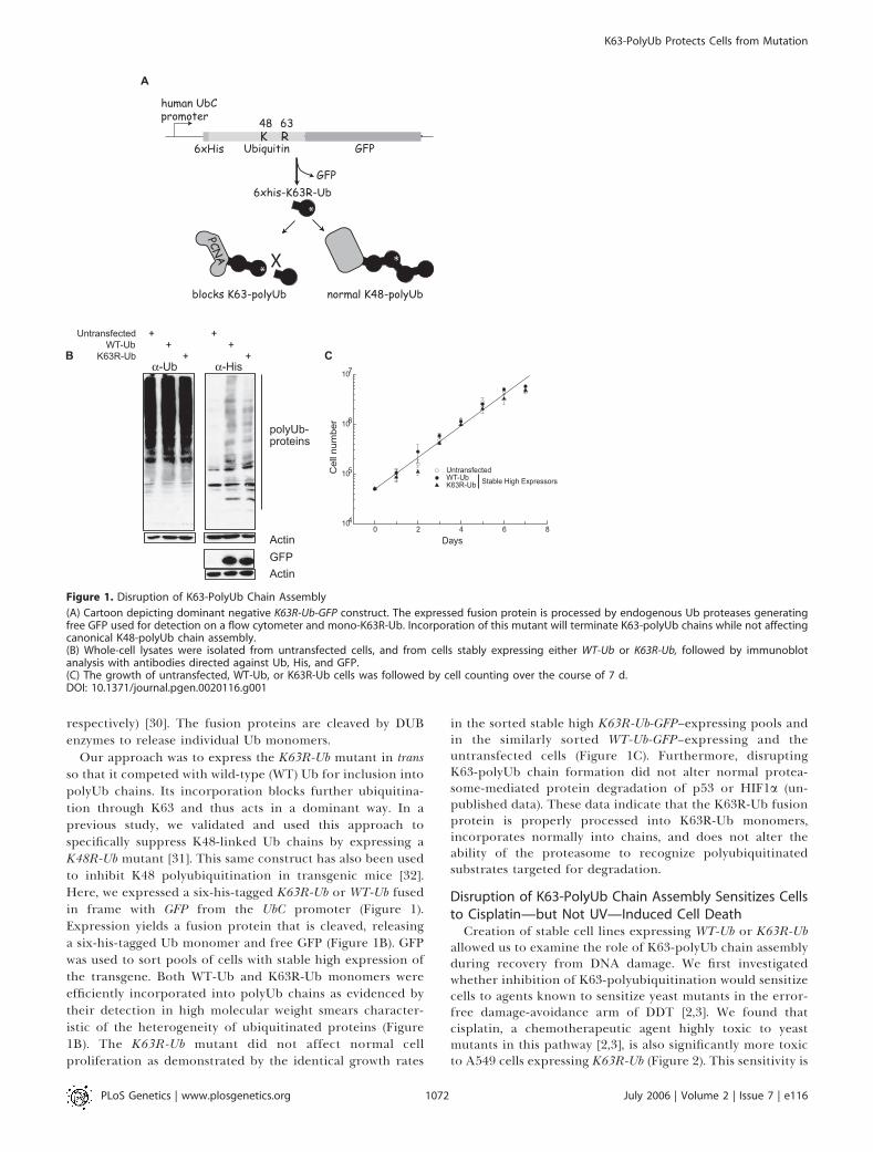
allowed us to examine the role of K63-polyUb chain assemblyduring recovery from DNA damage. We first investigatedwhether inhibition of K63-polyubiquitination would sensitizecells to agents known to sensitize yeast mutants in the error-free damage-avoidance arm of DDT [2,3]. We found thatcisplatin, a chemotherapeutic agent highly toxic to yeastmutants in this pathway [2,3], is also significantly more toxicto A549 cells expressing K63R-Ub (Figure 2). This sensitivity is
Figure 1. Disruption of K63-PolyUb Chain Assembly
(A) Cartoon depicting dominant negative K63R-Ub-GFP construct. The expressed fusion protein is processed by endogenous Ub proteases generatingfree GFP used for detection on a flow cytometer and mono-K63R-Ub. Incorporation of this mutant will terminate K63-polyUb chains while not affectingcanonical K48-polyUb chain assembly.(B) Whole-cell lysates were isolated from untransfected cells, and from cells stably expressing either WT-Ub or K63R-Ub, followed by immunoblotanalysis with antibodies directed against Ub, His, and GFP.(C) The growth of untransfected, WT-Ub, or K63R-Ub cells was followed by cell counting over the course of 7 d.DOI: 10.1371/journal.pgen.0020116.g001
PLoS Genetics | www.plosgenetics.org July 2006 | Volume 2 | Issue 7 | e1161072
K63-PolyUb Protects Cells from Mutation
specific to expression of K63R-Ub since the response of cellsexpressing either WT-Ub or K33R-Ub is identical to that ofuntransfected controls (Figure 2A and 2B). This effect was notmediated by a general inhibition of ubiquitination since A549
cells expressing the K48R-Ub mutant are not sensitized(unpublished data). Furthermore, a K63R-Ub clone that lostexpression of the transgene (as evidenced by a low GFP signal)returned to normal sensitivity (Figure 2B). These data imply
Figure 2. Cells Deficient in K63-Ub Chain Formation Are Sensitized to Cisplatin Treatment while UV Sensitivity Is Revealed only upon POLg Knockdown
(A and B) Clonogenic survival assays were used to determine sensitivity to 1 h acute treatment with cisplatin in untransfected A549 cells or in A549 cellsstably expressing WT-Ub or K63R-Ub. The mean values of three independent experiments are shown with standard error of the mean (error bars). Cellsexpressing K33R-Ub or cells that lost K63R-Ub expression revert to WT-Ub cisplatin sensitivity.(C) Cells were treated for 24 h with 100 lM cisplatin followed by Hoechst staining to detect apoptosis. The mean values of three independentexperiments are shown with standard deviation.(D) Clonogenic survival assays were used to determine sensitivity to UV irradiation in untransfected A549 cells or in A549 cells stably expressing WT-Ubor K63R-Ub.(E) Clonogenic survival of A549 cells stably expressing WT-Ub or K63R-Ub with or without POLg RNAi following 10 J/m2 UV treatment.DOI: 10.1371/journal.pgen.0020116.g002
PLoS Genetics | www.plosgenetics.org July 2006 | Volume 2 | Issue 7 | e1161073
K63-PolyUb Protects Cells from Mutation
that K63-polyUb chain assembly is essential for recovery fromat least a subset of cisplatin-induced lesions.
We also examined the functional importance of K63-polyubiquitination in the recovery from UV-induced damage.In contrast to the data with cisplatin, the cell line with stableexpression of K63R-Ub exhibited a dose response to UVirradiation that was identical to the parental cells or to cellsexpressing WT-Ub (Figure 2D). Thus, despite evidence thatK63-polyUb chains are required for cisplatin tolerance, wefound no evidence that disruption of K63-polyUb chainassembly on its own influences UV toxicity. A possibleexplanation for this lack of sensitivity to UV irradiation isthat cells can compensate for loss of K63-polyUb–dependentrepair through increased utilization of the error-prone TLSarm of the pathway. A similar situation occurs in yeast whereinhibition of the error-free damage-avoidance arm of DDTresults in a much milder UV sensitivity than mutations inRAD6 or RAD18 which additionally prevent TLS [33]. UsingsiRNA, we were able to knock down expression of POLg by;13-fold (Figure S1). Similar to inhibition of K63-polyubi-quitination, knockdown of POLg had no effect on UVsensitivity on its own. This observation is not unexpectedsince XPV cells (defective in POLg) are not sensitive to killingby UV irradiation. In contrast, knockdown of POLg in cellsalso expressing K63R-Ub did cause increased cell kill after UVtreatment (Figure 2E). This increase in UV sensitivity suggeststhat K63-polyUb and POLg function in distinct, comple-mentary pathways that mediate recovery from UV-induceddamage.
Disruption of K63-PolyUb Chain Assembly Increases UV-Induced Mutations
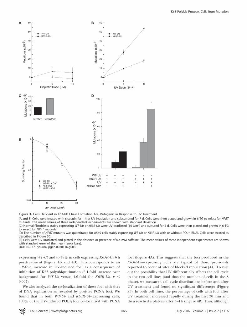
Disruption of the error-free arm in yeast is also known toresult in a dramatic increase in UV-induced mutations that issynergistic with the TLS mutant, REV3 [34]. If playing asimilar role in mammalian cells, inhibition of K63-polyubi-quitination should also increase UV-induced mutations. Wethus analyzed mutation induction at the HPRT locus after UVirradiation and cisplatin exposure in these same cell lines aswell as in normal human fibroblasts expressing WT-Ub orK63R-Ub (Figure 3). Consistent with this hypothesis, A549cells expressing K63R-Ub show a 2.5-fold increase in UV-induced mutations compared to cells expressing WT-Ub(Figure 3B), and a similar increase (2.2-fold) is observed innormal fibroblasts (Figure 3C). Untransfected and WT-Ub–expressing cells have similar mutation frequencies (unpub-lished data). The increase in mutations upon inhibition ofK63-polyubiquitination is consistent with a recent report thatused antisense to suppress the expression of MMS2 in humancells [25]. Similar to the cells expressing Ub-K63R, loss ofMMS2 led to an ;2-fold increase in UV-induced mutationswithout increasing UV-induced cell death [25]. Thus, both theenzyme that is implicated in the synthesis of K63-polyUbchains, and the chains themselves, are required for recoveryfrom UV damage through a pathway that prevents mutations.
Increases in UV-Induced Mutations Are Due to IncreasedUtilization of TLS
Many of the TLS polymerases are known to be importantcontributors to UV-induced mutagenesis as is illustrated by areduction in mutation frequency when inactivated in yeast[35–38]. The data presented thus far are consistent with a
model in which inhibition of K63-polyubiquitination in-creases UV-induced mutations owing to increased use of theerror-prone branch of the TLS pathway. However, thepossibility that K63R-Ub expression in some way increasesmutations by affecting the function of one or more TLSpolymerases cannot be ruled out. In fact, the phenotype ofcells expressing K63R-Ub is similar to that described for XPVcells. Both cell types display an increase in UV-inducedmutations with no significant change in UV-induced celldeath. In XPV cells, this is due to loss of POLg whichreplicates past T–T dimers in an error-free manner [39].Defects in POLg can be revealed by a significant increase inUV sensitivity when irradiated in the presence of caffeine, anassay used to establish the XPV phenotype [40]. However, wefound that cells expressing K63R-Ub are not similarly hyper-sensitive to this combined treatment (Figure 3E), suggestingno overt defect in POLg function in these cells.In contrast, our data suggest that POLg and K63-polyUb
chains participate in separate, alternative pathways forrecovery from UV-induced DNA damage. Consistent withthis idea, knockdown of POLg in combination with theinhibition of K63-polyUb chain assembly resulted in both anincreased toxicity to UV irradiation (Figure 2E) and in afurther increase in UV-induced mutations (Figure 3D).Interestingly, the number of mutations in cells followingknockdown of POLg in combination with inhibition of K63-polyUb chain assembly were far greater than additive. Asexpected, loss of POLg, which replicates past T–T dimerswith high fidelity, resulted in a large induction in UV-inducedmutations in WT-Ub–expressing cells (Figure 3D). Thesemutations are likely due to the activity of alternative TLSpolymerases that can substitute for POLg, but which areerror-prone across T–T dimers [41]. Additional suppressionof K63-polyUb chain assembly increased the number of UV-induced mutations by 3.5-fold. This synergistic increase inmutations strongly suggests that the inability to form K63-polyUb chains places a greater requirement on the TLSpathway, and thus POLg; it is also likely that there will be agreater requirement for other lesion bypass polymerases suchas POLf [42,43] for recovery from UV damage. Moreover, thesynergistic increase in mutations suggests that a significantproportion of the repair is normally carried out by the error-free component of the damage-avoidance pathway.To further investigate the relationship between inhibition
of K63R-polyUb chain assembly and TLS, we examined thespatial dynamics of the TLS polymerase POLg. This polymer-ase is recruited to sites of damage and can be visualized indiscrete foci that co-localize with PCNA [44]. We analyzed theeffects of K63R-Ub expression on POLg foci formation in livecells using a POLg-GFP fusion construct [44] (Figure 4). Sinceour original cells co-expressed GFP, we generated new stablelines from both A549 and HeLa cells expressing WT-Ub orK63R-Ub fused with the puromycin-resistance gene. Thesecell lines are phenotypically equivalent to the original GFP-expressing cells (an ;3-fold increase in HPRT mutants incells expressing K63R-Ub compared to WT-Ub). Similar toprevious observations [44], the majority of nonirradiated cellsshow homogenous nuclear distribution of the tagged poly-merases (Figures 4 and S2). Foci were observed in ;11%–12% of cells and likely represent sites of ongoing replication[44]. When treated with 10 J/m2 UV irradiation, thepercentage of cells with foci increased to 30% in cells
PLoS Genetics | www.plosgenetics.org July 2006 | Volume 2 | Issue 7 | e1161074
K63-PolyUb Protects Cells from Mutation
expressingWT-Ub and to 49% in cells expressing K63R-Ub 6 hposttreatment (Figure 4B and 4D). This corresponds to an;2-fold increase in UV-induced foci as a consequence ofinhibition of K63-polyubiquitination (2.4-fold increase overbackground for WT-Ub versus 4.6-fold for K63R-Ub, p ,
0.007).We also analyzed the co-localization of these foci with sites
of DNA replication as revealed by positive PCNA foci. Wefound that in both WT-Ub and K63R-Ub–expressing cells,100% of the UV-induced POLg foci co-localized with PCNA
foci (Figure 4A). This suggests that the foci produced in theK63R-Ub–expressing cells are typical of those previouslyreported to occur at sites of blocked replication [44]. To ruleout the possibility that UV differentially affects the cell cyclein the two cell lines (and thus the number of cells in the Sphase), we measured cell-cycle distributions before and afterUV treatment and found no significant differences (FigureS3). In both cell lines, the percentage of cells with foci afterUV treatment increased rapidly during the first 30 min andthen reached a plateau after 3–4 h (Figure 4B). Thus, although
Figure 3. Cells Deficient in K63-Ub Chain Formation Are Mutagenic in Response to UV Treatment
(A and B) Cells were treated with cisplatin for 1 h or UV irradiation and subcultured for 7 d. Cells were then plated and grown in 6-TG to select for HPRTmutants. The mean values of three independent experiments are shown with standard deviation.(C) Normal fibroblasts stably expressing WT-Ub or K63R-Ub were UV-irradiated (10 J/m2) and cultured for 5 d. Cells were then plated and grown in 6-TGto select for HPRT mutants.(D) The number of HPRT mutants was quantitated for A549 cells stably expressing WT-Ub or K63R-Ub with or without POLg RNAi. Cells were treated asdescribed in Figure 3C.(E) Cells were UV-irradiated and plated in the absence or presence of 0.4 mM caffeine. The mean values of three independent experiments are shownwith standard error of the mean (error bars).DOI: 10.1371/journal.pgen.0020116.g003
PLoS Genetics | www.plosgenetics.org July 2006 | Volume 2 | Issue 7 | e1161075
K63-PolyUb Protects Cells from Mutation
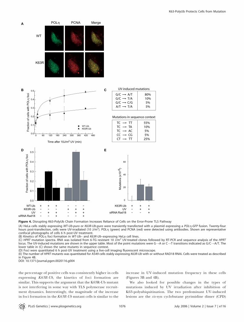
the percentage of positive cells was consistently higher in cellsexpressing K63R-Ub, the kinetics of foci formation aresimilar. This supports the argument that the K63R-Ub mutantis not interfering in some way with TLS polymerase recruit-ment dynamics. Interestingly, the magnitude of the increasein foci formation in the K63R-Ubmutant cells is similar to the
increase in UV-induced mutation frequency in these cells(Figures 3B and 4B).We also looked for possible changes in the types of
mutations induced by UV irradiation after inhibition ofK63-polyubiquitination. The two predominant UV-inducedlesions are the cis-syn cyclobutane pyrimidine dimer (CPD)
Figure 4. Disrupting K63-PolyUb Chain Formation Increases Reliance of Cells on the Error-Prone TLS Pathway
(A) HeLa cells stably expressing WT-Ub-puro or K63R-Ub-puro were transiently transfected with a plasmid expressing a POLg-GFP fusion. Twenty-fourhours post-transfection, cells were UV-irradiated (10 J/m2). POLg (green) and PCNA (red) were detected using antibodies. Shown are representativeconfocal photographs of cells 6 h post-UV treatment.(B) Kinetics of POLg foci formation in WT-Ub– and K63R-Ub–expressing HeLa cell lines.(C) HPRT mutation spectra. RNA was isolated from 6-TG resistant 10 J/m2 UV-treated clones followed by RT-PCR and sequence analysis of the HPRTlocus. The UV-induced mutations are shown in the upper table. Most of the point mutations were G!A or C!T transitions indicated as G/C!A/T. Thelower table in (C) shows the same mutants in sequence context.(D) Foci were quantitated 6 h post-UV treatment using a live-cell imaging fluorescent microscope.(E) The number of HPRT mutants was quantitated for A549 cells stably expressing K63R-Ub with or without RAD18 RNAi. Cells were treated as describedin Figure 4B.DOI: 10.1371/journal.pgen.0020116.g004
PLoS Genetics | www.plosgenetics.org July 2006 | Volume 2 | Issue 7 | e1161076
K63-PolyUb Protects Cells from Mutation
and the pyrimidine-6/4-pyrimidone (6–4PP) photoproduct[45,46]. The most common lesion is the thymine–thymineCPD (represented by T–T) followed by T–C and the thymine–cytosine 6–4PP (represented by T(6,4)C) [47]. Levels ofT(6,4)T, C–T, and C–C lesions are comparatively much lower.However, the normal spectrum of UV-induced mutationsdoes not match this pattern of damage induction. Mutationsare primarily C to T transitions that arise at T–C and C–Csites due to mis-incorporation of adenine opposite the 39C[48,49]. The weak contribution of the T–T lesion to mutationmay be explained by the activities of POLg and POLi, whichaccurately bypass T–T and T(6,4)T lesions, respectively[50,51].
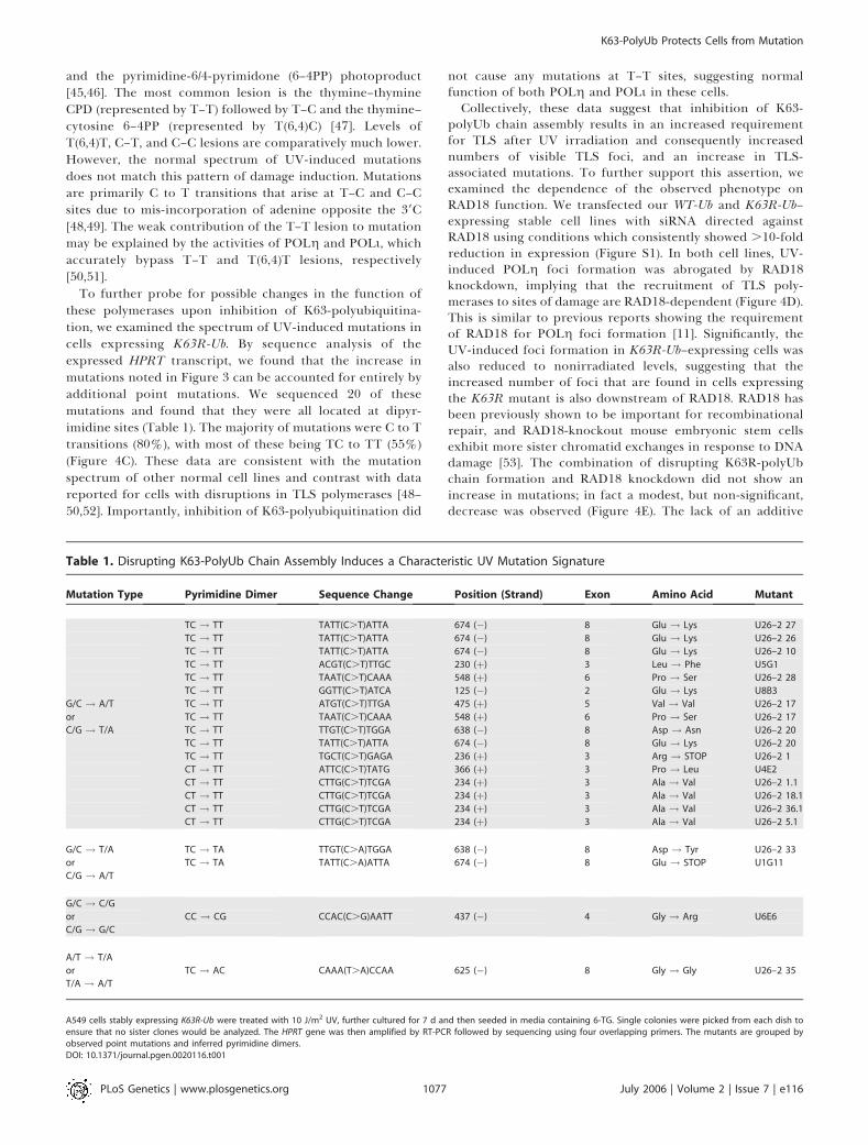
To further probe for possible changes in the function ofthese polymerases upon inhibition of K63-polyubiquitina-tion, we examined the spectrum of UV-induced mutations incells expressing K63R-Ub. By sequence analysis of theexpressed HPRT transcript, we found that the increase inmutations noted in Figure 3 can be accounted for entirely byadditional point mutations. We sequenced 20 of thesemutations and found that they were all located at dipyr-imidine sites (Table 1). The majority of mutations were C to Ttransitions (80%), with most of these being TC to TT (55%)(Figure 4C). These data are consistent with the mutationspectrum of other normal cell lines and contrast with datareported for cells with disruptions in TLS polymerases [48–50,52]. Importantly, inhibition of K63-polyubiquitination did
not cause any mutations at T–T sites, suggesting normalfunction of both POLg and POLi in these cells.Collectively, these data suggest that inhibition of K63-
polyUb chain assembly results in an increased requirementfor TLS after UV irradiation and consequently increasednumbers of visible TLS foci, and an increase in TLS-associated mutations. To further support this assertion, weexamined the dependence of the observed phenotype onRAD18 function. We transfected our WT-Ub and K63R-Ub–expressing stable cell lines with siRNA directed againstRAD18 using conditions which consistently showed .10-foldreduction in expression (Figure S1). In both cell lines, UV-induced POLg foci formation was abrogated by RAD18knockdown, implying that the recruitment of TLS poly-merases to sites of damage are RAD18-dependent (Figure 4D).This is similar to previous reports showing the requirementof RAD18 for POLg foci formation [11]. Significantly, theUV-induced foci formation in K63R-Ub–expressing cells wasalso reduced to nonirradiated levels, suggesting that theincreased number of foci that are found in cells expressingthe K63R mutant is also downstream of RAD18. RAD18 hasbeen previously shown to be important for recombinationalrepair, and RAD18-knockout mouse embryonic stem cellsexhibit more sister chromatid exchanges in response to DNAdamage [53]. The combination of disrupting K63R-polyUbchain formation and RAD18 knockdown did not show anincrease in mutations; in fact a modest, but non-significant,decrease was observed (Figure 4E). The lack of an additive
Table 1. Disrupting K63-PolyUb Chain Assembly Induces a Characteristic UV Mutation Signature
Mutation Type Pyrimidine Dimer Sequence Change Position (Strand) Exon Amino Acid Mutant
TC ! TT TATT(C.T)ATTA 674 (�) 8 Glu ! Lys U26–2 27
TC ! TT TATT(C.T)ATTA 674 (�) 8 Glu ! Lys U26–2 26
TC ! TT TATT(C.T)ATTA 674 (�) 8 Glu ! Lys U26–2 10
TC ! TT ACGT(C.T)TTGC 230 (þ) 3 Leu ! Phe U5G1
TC ! TT TAAT(C.T)CAAA 548 (þ) 6 Pro ! Ser U26–2 28
TC ! TT GGTT(C.T)ATCA 125 (�) 2 Glu ! Lys U8B3
G/C ! A/T TC ! TT ATGT(C.T)TTGA 475 (þ) 5 Val ! Val U26–2 17
or TC ! TT TAAT(C.T)CAAA 548 (þ) 6 Pro ! Ser U26–2 17
C/G ! T/A TC ! TT TTGT(C.T)TGGA 638 (�) 8 Asp ! Asn U26–2 20
TC ! TT TATT(C.T)ATTA 674 (�) 8 Glu ! Lys U26–2 20
TC ! TT TGCT(C.T)GAGA 236 (þ) 3 Arg ! STOP U26–2 1
CT ! TT ATTC(C.T)TATG 366 (þ) 3 Pro ! Leu U4E2
CT ! TT CTTG(C.T)TCGA 234 (þ) 3 Ala ! Val U26–2 1.1
CT ! TT CTTG(C.T)TCGA 234 (þ) 3 Ala ! Val U26–2 18.1
CT ! TT CTTG(C.T)TCGA 234 (þ) 3 Ala ! Val U26–2 36.1
CT ! TT CTTG(C.T)TCGA 234 (þ) 3 Ala ! Val U26–2 5.1
G/C ! T/A TC ! TA TTGT(C.A)TGGA 638 (�) 8 Asp ! Tyr U26–2 33
or TC ! TA TATT(C.A)ATTA 674 (�) 8 Glu ! STOP U1G11
C/G ! A/T
G/C ! C/G
or CC ! CG CCAC(C.G)AATT 437 (�) 4 Gly ! Arg U6E6
C/G ! G/C
A/T ! T/A
or TC ! AC CAAA(T.A)CCAA 625 (�) 8 Gly ! Gly U26–2 35
T/A ! A/T
A549 cells stably expressing K63R-Ub were treated with 10 J/m2 UV, further cultured for 7 d and then seeded in media containing 6-TG. Single colonies were picked from each dish toensure that no sister clones would be analyzed. The HPRT gene was then amplified by RT-PCR followed by sequencing using four overlapping primers. The mutants are grouped byobserved point mutations and inferred pyrimidine dimers.DOI: 10.1371/journal.pgen.0020116.t001
PLoS Genetics | www.plosgenetics.org July 2006 | Volume 2 | Issue 7 | e1161077
K63-PolyUb Protects Cells from Mutation
mutation effect supports the foci data implicating a role forK63-polyUb chain formation downstream of RAD18.
PCNA Is PolyubiquitinatedOur data support a role for the formation of K63-polyUb
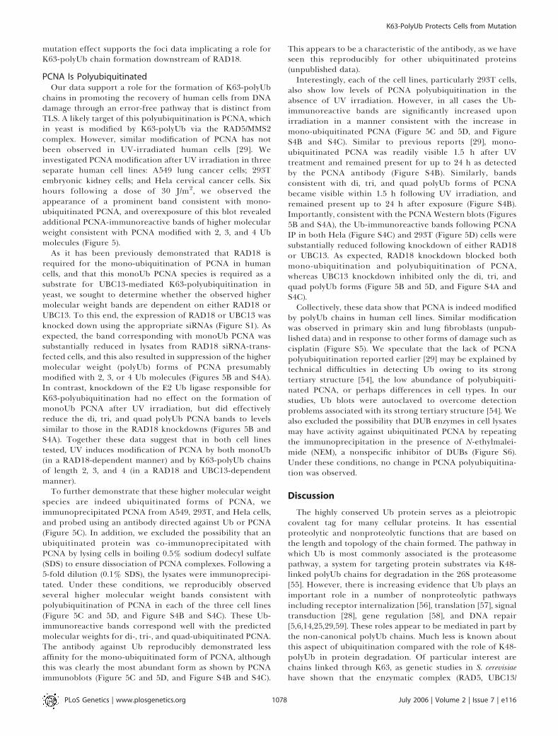
chains in promoting the recovery of human cells from DNAdamage through an error-free pathway that is distinct fromTLS. A likely target of this polyubiquitination is PCNA, whichin yeast is modified by K63-polyUb via the RAD5/MMS2complex. However, similar modification of PCNA has notbeen observed in UV-irradiated human cells [29]. Weinvestigated PCNA modification after UV irradiation in threeseparate human cell lines: A549 lung cancer cells; 293Tembryonic kidney cells; and Hela cervical cancer cells. Sixhours following a dose of 30 J/m2, we observed theappearance of a prominent band consistent with mono-ubiquitinated PCNA, and overexposure of this blot revealedadditional PCNA-immunoreactive bands of higher molecularweight consistent with PCNA modified with 2, 3, and 4 Ubmolecules (Figure 5).
As it has been previously demonstrated that RAD18 isrequired for the mono-ubiquitination of PCNA in humancells, and that this monoUb PCNA species is required as asubstrate for UBC13-mediated K63-polyubiquitination inyeast, we sought to determine whether the observed highermolecular weight bands are dependent on either RAD18 orUBC13. To this end, the expression of RAD18 or UBC13 wasknocked down using the appropriate siRNAs (Figure S1). Asexpected, the band corresponding with monoUb PCNA wassubstantially reduced in lysates from RAD18 siRNA-trans-fected cells, and this also resulted in suppression of the highermolecular weight (polyUb) forms of PCNA presumablymodified with 2, 3, or 4 Ub molecules (Figures 5B and S4A).In contrast, knockdown of the E2 Ub ligase responsible forK63-polyubiquitination had no effect on the formation ofmonoUb PCNA after UV irradiation, but did effectivelyreduce the di, tri, and quad polyUb PCNA bands to levelssimilar to those in the RAD18 knockdowns (Figures 5B andS4A). Together these data suggest that in both cell linestested, UV induces modification of PCNA by both monoUb(in a RAD18-dependent manner) and by K63-polyUb chainsof length 2, 3, and 4 (in a RAD18 and UBC13-dependentmanner).
To further demonstrate that these higher molecular weightspecies are indeed ubiquitinated forms of PCNA, weimmunoprecipitated PCNA from A549, 293T, and Hela cells,and probed using an antibody directed against Ub or PCNA(Figure 5C). In addition, we excluded the possibility that anubiquitinated protein was co-immunoprecipitated withPCNA by lysing cells in boiling 0.5% sodium dodecyl sulfate(SDS) to ensure dissociation of PCNA complexes. Following a5-fold dilution (0.1% SDS), the lysates were immunoprecipi-tated. Under these conditions, we reproducibly observedseveral higher molecular weight bands consistent withpolyubiquitination of PCNA in each of the three cell lines(Figure 5C and 5D, and Figure S4B and S4C). These Ub-immunoreactive bands correspond well with the predictedmolecular weights for di-, tri-, and quad-ubiquitinated PCNA.The antibody against Ub reproducibly demonstrated lessaffinity for the mono-ubiquitinated form of PCNA, althoughthis was clearly the most abundant form as shown by PCNAimmunoblots (Figure 5C and 5D, and Figure S4B and S4C).
This appears to be a characteristic of the antibody, as we haveseen this reproducibly for other ubiquitinated proteins(unpublished data).Interestingly, each of the cell lines, particularly 293T cells,
also show low levels of PCNA polyubiquitination in theabsence of UV irradiation. However, in all cases the Ub-immunoreactive bands are significantly increased uponirradiation in a manner consistent with the increase inmono-ubiquitinated PCNA (Figure 5C and 5D, and FigureS4B and S4C). Similar to previous reports [29], mono-ubiquitinated PCNA was readily visible 1.5 h after UVtreatment and remained present for up to 24 h as detectedby the PCNA antibody (Figure S4B). Similarly, bandsconsistent with di, tri, and quad polyUb forms of PCNAbecame visible within 1.5 h following UV irradiation, andremained present up to 24 h after exposure (Figure S4B).Importantly, consistent with the PCNAWestern blots (Figures5B and S4A), the Ub-immunoreactive bands following PCNAIP in both Hela (Figure S4C) and 293T (Figure 5D) cells weresubstantially reduced following knockdown of either RAD18or UBC13. As expected, RAD18 knockdown blocked bothmono-ubiquitination and polyubiquitination of PCNA,whereas UBC13 knockdown inhibited only the di, tri, andquad polyUb forms (Figure 5B and 5D, and Figure S4A andS4C).Collectively, these data show that PCNA is indeed modified
by polyUb chains in human cell lines. Similar modificationwas observed in primary skin and lung fibroblasts (unpub-lished data) and in response to other forms of damage such ascisplatin (Figure S5). We speculate that the lack of PCNApolyubiquitination reported earlier [29] may be explained bytechnical difficulties in detecting Ub owing to its strongtertiary structure [54], the low abundance of polyubiquiti-nated PCNA, or perhaps differences in cell types. In ourstudies, Ub blots were autoclaved to overcome detectionproblems associated with its strong tertiary structure [54]. Wealso excluded the possibility that DUB enzymes in cell lysatesmay have activity against ubiquitinated PCNA by repeatingthe immunoprecipitation in the presence of N-ethylmalei-mide (NEM), a nonspecific inhibitor of DUBs (Figure S6).Under these conditions, no change in PCNA polyubiquitina-tion was observed.
Discussion
The highly conserved Ub protein serves as a pleiotropiccovalent tag for many cellular proteins. It has essentialproteolytic and nonproteolytic functions that are based onthe length and topology of the chain formed. The pathway inwhich Ub is most commonly associated is the proteasomepathway, a system for targeting protein substrates via K48-linked polyUb chains for degradation in the 26S proteasome[55]. However, there is increasing evidence that Ub plays animportant role in a number of nonproteolytic pathwaysincluding receptor internalization [56], translation [57], signaltransduction [28], gene regulation [58], and DNA repair[5,6,14,25,29,59]. These roles appear to be mediated in part bythe non-canonical polyUb chains. Much less is known aboutthis aspect of ubiquitination compared with the role of K48-polyUb in protein degradation. Of particular interest arechains linked through K63, as genetic studies in S. cerevisiaehave shown that the enzymatic complex (RAD5, UBC13/
PLoS Genetics | www.plosgenetics.org July 2006 | Volume 2 | Issue 7 | e1161078
K63-PolyUb Protects Cells from Mutation
MMS2) that assembles these chains is required to protect cellsfrom the harmful effects of genotoxic agents by allowing thereplication machinery to bypass DNA lesions in a faithfulmanner [14]. In fact, ubiquitination of the DNA polymeraseprocessivity factor PCNA is emerging as a key ‘‘molecularswitch’’ for DDT [5,6,29]. Mono-ubiquitination of PCNApromotes error-prone TLS, while K63-polyUb activateserror-free damage avoidance. The body of evidence support-
ing the requirement of PCNA post-translational modifica-tions for DDT in mammalian cells is only now emerging.In this report, we provide evidence to support a model
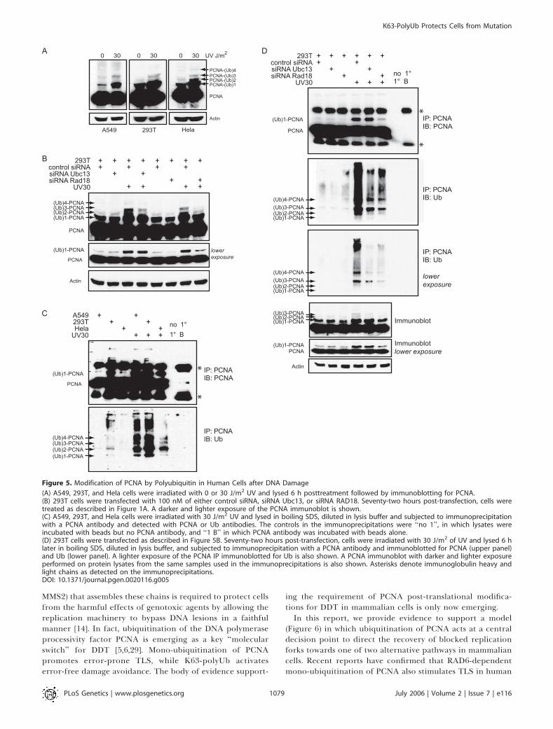
(Figure 6) in which ubiquitination of PCNA acts at a centraldecision point to direct the recovery of blocked replicationforks towards one of two alternative pathways in mammaliancells. Recent reports have confirmed that RAD6-dependentmono-ubiquitination of PCNA also stimulates TLS in human
Figure 5. Modification of PCNA by Polyubiquitin in Human Cells after DNA Damage
(A) A549, 293T, and Hela cells were irradiated with 0 or 30 J/m2 UV and lysed 6 h posttreatment followed by immunoblotting for PCNA.(B) 293T cells were transfected with 100 nM of either control siRNA, siRNA Ubc13, or siRNA RAD18. Seventy-two hours post-transfection, cells weretreated as described in Figure 1A. A darker and lighter exposure of the PCNA immunoblot is shown.(C) A549, 293T, and Hela cells were irradiated with 30 J/m2 UV and lysed in boiling SDS, diluted in lysis buffer and subjected to immunoprecipitationwith a PCNA antibody and detected with PCNA or Ub antibodies. The controls in the immunoprecipitations were ‘‘no 1’’, in which lysates wereincubated with beads but no PCNA antibody, and ‘‘1 B’’ in which PCNA antibody was incubated with beads alone.(D) 293T cells were transfected as described in Figure 5B. Seventy-two hours post-transfection, cells were irradiated with 30 J/m2 of UV and lysed 6 hlater in boiling SDS, diluted in lysis buffer, and subjected to immunoprecipitation with a PCNA antibody and immunoblotted for PCNA (upper panel)and Ub (lower panel). A lighter exposure of the PCNA IP immunoblotted for Ub is also shown. A PCNA immunoblot with darker and lighter exposureperformed on protein lysates from the same samples used in the immunoprecipitations is also shown. Asterisks denote immunoglobulin heavy andlight chains as detected on the immunoprecipitations.DOI: 10.1371/journal.pgen.0020116.g005
PLoS Genetics | www.plosgenetics.org July 2006 | Volume 2 | Issue 7 | e1161079
K63-PolyUb Protects Cells from Mutation
cells. This stimulation appears to result through directbinding of the TLS polymerases to mono-ubiquitinatedPCNA [11,29].
Our data indicate that formation of K63-polyUb chains isrequired to utilize an error-free pathway distinct from TLS.This pathway is required for cell survival from at least sometypes of DNA damage, as its inhibition cannot be compen-sated for by the alternative TLS pathway in the case ofcisplatin-damaged cells. For UV-induced damage, inhibitionof K63-polyubiquitination does not affect overall cell survival,but instead causes an increase in mutations arising from anapparent increased requirement for the error-prone branchof TLS. This is supported by several lines of evidence. First,blockade of K63-polyUb chain formation led to a 2.4-foldincrease in RAD18-dependent TLS foci after UV irradiation.
Second, we found that the number of UV-induced mutationsincreased by a similar factor in these cells, and that thespectra of these mutations are consistent with that producednormally by error-prone TLS polymerases after UV treat-ment. Third, POLg knockdown in combination with blockadeof K63-polyUb chain formation led to increased toxicity toUV irradiation, although no change was seen with eitherindividually. Fourth, an increased reliance on the TLS armupon blockade of K63-polyUb chain assembly was revealed bya synergistic increase in UV-induced mutations when ex-pressed in POLg knockdown cells. POLg knockdown cellsshowed a high mutation rate as expected, but this rateincreased by a factor of 3.5 when K63-polyUb chain assemblywas inhibited.Together, these data imply that formation of K63-polyUb
chains can activate an error-free mechanism to protect cellsagainst mutations that would otherwise be induced by theerror-prone TLS polymerases. It will be of interest todetermine whether K63-polyUb chain formation also playsa role in protection against sunlight-induced skin cancer.An obvious question that emerges is how formation of K63-
polyUb acts to suppress TLS. Recent reports have demon-strated that the TLS polymerases POLg and POLi both binddirectly and avidly to polyUb chains through newly discov-ered binding domains [20,60]. A C-terminal zinc fingerdomain of POLg and the proline residue at position 692 ofPOLi are required for the respective interaction with Ub [20].Together with our data, this suggests a possible mechanismwhereby differential ubiquitination of PCNA could act as aswitch between TLS and an alternative error-free pathway(Figure 6). In this model, the TLS polymerases are recruitedto the sites of replication through interaction with mono-ubiquitinated PCNA and subsequently mediate TLS acrossDNA lesions. Extension of the Ub chain through K63-linkedpolyubiquitination in some way suppresses TLS activity andpromotes recovery through an alternative error-free path-way. This suppression may be mediated through the recentlydiscovered ability of POLg and POLi to directly bind K63-polyUb chains. An intriguing possibility is that K63-polyUbchains are cleaved upon binding to TLS polymerases, therebyfunctionally removing them from the site of the lesion. Thispossibility is supported by the low detectable levels ofpolyubiquitinated PCNA as well as by the observed increasein POLg foci in K63R-Ub–expressing cells.Although our data suggest that PCNA is indeed a target for
K63-polyubiquitination, they do not exclude the possibilitythat other key proteins in this pathway are also importantsubstrates for these chains. Indeed, K63-polyubiquitinationoccurs on at least three proteins (RIP, NEMO, and TRAF6) inan unrelated pathway that activates NF-jB [28,61,62]. In thispathway, K63-polyUb chains on multiple proteins mayfacilitate their assembly into an active complex [62]. It istherefore intriguing to speculate that K63-polyUb chains maynot only uncouple the TLS polymerases from the site ofdamage, but may also provide a mechanism for recruitmentof other proteins required for error-free repair.Non-proteolytic roles for Ub have also been implicated in
other DNA-repair pathways that may interact with DDT, mostnotably that involving Fanconi’s anemia (FA) gene products[18]. FANCD2 becomes mono-ubiquitinated after DNAdamage and localizes to nuclear foci [63]. FANCC has beenassociated with the TLS polymerases REV1 and REV3 [64] and
Figure 6. Model of the DDT Pathway in Mammalian Cells
Recovery from a stalled replication fork at sites of DNA damage canoccur by one of two alternative pathways. Previous work has shown thatPCNA mono-ubiquitination by the RAD6/RAD18 complex stimulateslesion bypass through recruitment of the error-prone TLS polymerases.Here we show that an alternative error-free pathway requires formationof K63-polyUb chains. Blockade of this error-free pathway results inincreased use of the TLS polymerases after DNA damage and acorresponding increase in mutations. As the TLS polymerases POLgand POLi both bind directly and avidly to polyUb chains [20], it ishypothesized that the interaction with K63-polyUb causes a disengage-ment of the polymerase from the DNA, allowing other proteins tomigrate to the site of damage to perform error-free repair. This modelpredicts that K63-polyubiquitination acts to suppress environmentalcarcinogenesis by preventing genomic instability that would otherwisebe introduced by the TLS polymerases.DOI: 10.1371/journal.pgen.0020116.g006
PLoS Genetics | www.plosgenetics.org July 2006 | Volume 2 | Issue 7 | e1161080
K63-PolyUb Protects Cells from Mutation

may also interact with the BLM helicase [65], a candidate forpromoting fork reversal in the error-free damage-avoidancepathway [66]. A challenge for future investigations will be tounderstand how K63-polyUb chain assembly is regulated andhow these chains promote interaction with other pathways tomediate error-free recovery from DNA damage.
Materials and Methods
Cell culture and treatments. The construction of the Ub-expressingplasmids has been described elsewhere [31]. The POLg-GFP plasmidwas a gift of Dr. Alan R. Lehmann, (Genome Damage and StabilityCentre, University of Sussex, Falmer, Brighton, United Kingdom). Allcell lines were cultured in DMEM (Sigma, St. Louis, Missouri, UnitedStates) supplemented with 10% FBS (Sigma). A549 cells were co-transfected with WT-Ub-GFP or K63R-Ub-GFP plasmids and thepBabePuro plasmid (for selection) using FuGene 6 (Roche, Basel,Switzerland). HeLa cells were transfected with WT-Ub-puro or K63R-Ub-puro constructs using lipofectamine (Invitrogen, Carlsbad, Cal-ifornia, United States). Stable transfectants were selected in 1 lg/mlpuromycin (Sigma) and/or by flow cytometry (FACSAria, BDBiosciences Pharmingen, San Diego, California, United States).
The sensitivity to UV irradiation alone, UV combined withcaffeine, and cisplatin alone was evaluated using clonogenic survivalassays. UV irradiation was performed on 80% confluent cells in 6-cmdishes using a UVC (254-nm) germicidal lamp at a dose rate of 1 J/m2/s. UV and caffeine combination studies were carried out as above, butcells were plated in 0.4 mM caffeine immediately after UVirradiation. Cells were treated for 1 h in cisplatin diluted in culturemedia. Cells were plated in 6-cm dishes in triplicate and incubatedfor 2 wk to obtain colony formation. Colonies were fixed, stained with2% bromophenol blue in 70% ethanol, and colonies containing �50cells were counted. All experiments were normalized for platingefficiency.
The sensitivity to UV irradiation in POLg knockdown cells wasperformed as above with the exception that cells were transfectedtwice with SiGenome Smartpool reagent specific for human POLg(Dharmacon Research, Lafayette, Colorada, United States) usingoligofectamine (Invitrogen). The transfections were carried out 72and 24 h before UV treatment to achieve optimal long-termknockdown as determined by quantitative RT-PCR.
Quantitation of gene expression was performed using an AppliedBiosystems (Foster City, California, United States) 7500 Real-TimePCR system using their ‘‘assay on demand’’ technology. RAD18expression was determined with the Hs00220119_m1 probe, POLgwith the Hs00197814 probe, and 18S with the Hs99999901_s1 probe.Reactions were performed using Taqman Universal PCR Master Mixfrom Applied Biosystems.
Immunoblotting. Following the indicated treatments with eitherUV irradiation, cisplatin, and/or SiGenome Smartpool reagentspecific for human UBC13 or human RAD18 (Dharmacon), cellswere harvested in lysis buffer (20 mM Tris-HCl [pH 7.5], 150 mMNaCl, 1% Triton-X-100, 2 mM EDTA, and 5% glycerol with 200 lg/mlphenylmethylsulfonyl fluoride, 2 mM NaVO4, 2 mM NaF, and 2 mMNaPPi protease-inhibitor cocktail). Samples were sonicated, solublefractions were recovered, and proteins were quantified using theBradford protein assay (Bio-Rad). Proteins were resolved on either asingle-phase (10%) or two-phase SDS-polyacrylamide gel (10% and15%) and electroblotted onto a Hybond C nitrocellulose membrane(Amersham Pharmacia Biotech, Piscataway, New Jersey, UnitedStates). The membrane was stained with Ponceau S (Sigma) prior toWestern blotting with the indicated primary antibody. The followingantibodies were used: rabbit polyclonal Ub (Dako, Glostrup, Den-mark), mouse monoclonal RGS-His (Qiagen, Valencia, California,United States), mouse monoclonal PCNA PC10 (Chemicon, http://www.chemicon.com), rabbit polyclonal GFP (Santa Cruz Biotechnol-ogy, Santa Cruz, California, United States), and mouse monoclonalactin (Sigma). Proteins were visualized by a horseradish peroxidasemethod using ECL (Kirkegaard and Perry Laboratories, http://www.kpl.com).
Immunoprecipitation. Cells were UV-irradiated with 30 J/m2 asdescribed above and either left untreated or transfected withSiGenome Smartpool reagent specific for human UBC13 or humanRAD18 (Dharmacon). Cells were lysed (6 h after irradiation) in lysisbuffer supplementedwith 0.5%SDS.Lysateswere sonicated andboiledfor 5min followed by dilution to 0.1%SDS. After protein quantitation,500 lg of protein was incubated overnight at 4 8C with anti-PCNA (1/
200). The following day, lysates were incubated for 48 h at 4 8Cwith 100ll of Gamma-Bound Sepharose Beads (AmershamPharmacia Biotech).Beads were washed extensively in lysis buffer, and proteins were elutedby boiling in Laemmli’s SDS sample buffer. Immunoblotting wasperformed as described above except that the membranes wereautoclaved for 20 min in ddH2O after protein transfer, and proteinswere visualized using SuperSignal West Pico ChemiluminescentSubstrate (Pierce Biotechnology, Rockford, Illinois, United States).
Mutation spectrum. To eliminate background HPRT mutations,cells were cultured in hypoxanthine, aminopterin, and thymidine(HAT)–supplemented culture medium for 1 wk. UV-induced HPRTmutants were obtained by seeding 1.5 3 104 cells in 24-well plates,followed by 10 J/m2 UV irradiation 24 h later. Cells were subculturedfor 7 d, and re-seeded at 5.0 3 104 cells on 35-mm dishes in mediumcontaining 30 lM 6-thioguanine (6-TG). Individual colonies werepicked and grown until enough cells were obtained for RNA isolationusing RNA-aqueous kit (Ambion, Austin, Texas, United States). TheHPRT gene was subjected to RT-PCR, followed by sequencing usingthe following overlapping primers: HPRT1–59CTTCCTCC-TCCTGAGCAGTC39; HPRT2–59AAGCAGATGGCCACAGAACT39;HPRT3–59CCTGGCGTCGTGATTAGTG39; HPRT4–59TTTACTGGC-GATGTCAATAGGA39; HPRT5–59GACCAGTCAACAGGGGACAT39;and HPRT6 59ATGTCCCCTGTTGACTGGTC39.
Mutation frequency. HPRT mutant–free cells (1.0 3 106) wereseeded and treated the following day with either UV irradiation (0, 5,and 10 J/m2) or cisplatin (0, 5, and 10 lM for 1 h). After subculturingthe treated cells for 1 wk, 4.0 3 105 cells were seeded in selectivemedium containing 6-TG (as above) and incubated until colonieswere formed. Colonies were counted and HPRT mutation frequencywas defined after correcting for plating efficiency.
Mutation frequency in response to UV treatment in POLg andRAD18 knockdown cells was performed as above with the exceptionthat cells were transfected twice with SiGenome Smartpool reagentspecific for human POLg or human RAD18 (Dharmacon) usingoligofectamine. The transfections were performed 72 and 24 h beforeUV treatment to achieve optimal long-term knockdown as deter-mined by quantitative PCR.
Foci. A549 and Hela cells stably expressing WT-Ub-puro and K63R-Ub-puro were transiently transfected with a POLg-GFP plasmid.Twenty-four hours post-transfection, cells were UV-irradiated at adose of 10 J/m2. To observe living cells, cells were cultured in 35-mmglass-bottomed dishes (MatTek, http://www.mattek.com) with cover-slips. Real-time excitation measurements to monitor fluorescentsignals in transfected cells were subsequently performed using a live-cell microscopy unit mounted on a Leica DR IRBE invertedmicroscope (Wetzlar, Germany), equipped with a polychromator thatallows generation of light of the required wavelength, using a 633objective. Both the polychromator and filterwheel were controlled viathe PC using specialized Openlab software from Improvision (http://www.improvision.com/products/openlab). At least 100 cells werecounted for each cell line at each time point per experiment by ablinded independent observer.
The recruitment of POLg to foci was determined in response toUV irradiation in RAD18 knockdown cells performed as above withthe exception that cells were transfected twice with SiGenomeSmartpool reagent specific for human RAD18 using oligofectamine.The transfections were performed 72 and 24 h before UV treatmentto achieve optimal long-term knockdown as determined by quanti-tative PCR.
For colocalization studies, Hela cells stably expressing WT-Ub-puroand K63R-Ub-puro were transiently transfected with a POLg-GFPplasmid in a chamber slide (BD Biosciences Pharmingen). Twenty-four hours post-transfection, cells were UV-irradiated at a dose of 10J/m2. For detection of PCNA and POLg, cells were fixed in coldmethanol for 20 min at �20 8C followed by 30 sec in cold acetone.Cells were washed twice with PBS and then incubated at roomtemperature with both anti-PCNA and anti-POLg. After 1 h, cellswere washed with PBS and then incubated with FITC-conjugated goatantimouse IgG (Invitrogen) and Texas red–conjugated goat antirabbit(Invitrogen), for 45 min. After washing in PBS, cells were dehydratedfor 1 min in 70% ethanol followed by two 1-min incubations in 100%ethanol. Cells were then mounted with Fluorescent Mounting Media(Dako) and visualized by confocal microscopy.
Supporting Information
Figure S1. Knockdown of UBC13, RAD18, and POLg
(A) Hela cells were transfected with siRNA against UBC13 andanalyzed by Western blot.
PLoS Genetics | www.plosgenetics.org July 2006 | Volume 2 | Issue 7 | e1161081
K63-PolyUb Protects Cells from Mutation
(B) A549 cells were transfected twice (48 h apart) with siRNA againstRAD18 or POLg. Knockdown was analyzed 24 and 72 h post–secondtransfection for mRNA expression relative to 18S rRNA usingquantitative RT-PCR.
Found at DOI: 10.1371/journal.pgen.0020116.sg001 (28 KB PDF).
Figure S2. Increase in POLg Foci Is Also Observed in A549 Cells
A549 cells stably expressing WT-Ub or K63R-Ub were treated asdescribed in Figure 4. Two independent experiments were performed.
Found at DOI: 10.1371/journal.pgen.0020116.sg002 (16 KB PDF).
Figure S3. Cell-Cycle Profile following UV Treatment
(A) A549 cells expressing WT-Ub-GFP or K63R-Ub-GFP were treatedwith the indicated dose of UV irradiation.(B) Hela cells expressing WT-Ub-puro or K63R-Ub-puro were treatedwith 10 J/m2 UV irradiation and fixed either immediately or 6 hposttreatment. Following propidium iodide staining, cells wereanalyzed for DNA content using a FACSAria flow cytometer (BDBiosciences Pharmingen).
Found at DOI: 10.1371/journal.pgen.0020116.sg003 (33 KB PDF).
Figure S4. Modification of PCNA by Polyubiquitin in Human Cellsafter DNA Damage
(A) Hela cells were subjected to the same procedure as carrried out inFigure 5B. A darker (upper panel) and lighter exposure (lower panel)of the PCNA immunoblot is shown.(B) A549 cells were UV-irradiated as described in Figure 5A and lysedat the indicated times posttreatment. Whole-cell lysates weresubjected to immunoprecipitation with an anti-PCNA antibodyfollowed by immunoblotting for PCNA (upper panel) and Ub (lowerpanel). The controls in the immunoprecipitations are the same ascarried out in Figure 5C.�UV indicates no UV treatment.(C) Hela cells were subjected to the same procedure as performed inFigure 5D. A lighter exposure of the PCNA IP immunoblotted for Ubis shown. A PCNA immunoblot with darker and lighter exposureperformed on protein lysates from the same samples used in theimmunoprecipitations is also shown.
Found at DOI: 10.1371/journal.pgen.0020116.sg004 (67 KB PDF).
Figure S5. Cisplatin Treatment also Induces Modification of PCNA byPolyubiquitin in Human Cells
Untreated, 30 J/m2 UV-irradiated, and 160 lM cisplatin-treated A549and Hela cells were lysed 6 h posttreatment followed by immuno-blotting for PCNA.
Found at DOI: 10.1371/journal.pgen.0020116.sg005 (33 KB PDF).
Figure S6. Inhibition of DUB Enzymes Does Not Affect Appearanceof PolyUb-PCNA
A549, 293T, and Hela cells were treated with 30 J/m2 UV irradiationand lysed in the presence or absence of the general thiol protease-inhibitor NEM. Immunoprecipitation and Western blots were carriedout as described in Materials and Methods. The controls in theimmunoprecipitations were ‘‘no 1’’, in which lysates were incubatedwith beads but no PCNA antibody, and ‘‘1 B’’, in which PCNAantibody was incubated with beads alone.
Found at DOI: 10.1371/journal.pgen.0020116.sg006 (30 KB PDF).
Accession Numbers
The Entrez Gene (http://www.ncbi.nlm.nih.gov/entrez/query.fcgi?CMD¼search&DB¼gene) accession numbers for the gene andgene products discussed in this paper are BLM (641), CROC1 (7335),FANCC (2176), FANCD2 (2177), HPRT (3251), MMS2 (7336), NEMO(8517), NF-kappaB (4790), PCNA (5111), POLH (5429), POLI (11201),RAD18 (56852), RAD5 (850719), REV3 (5980), TRAF6 (7189), UBA52(7311), UBA80 (6233), UBB (7314), UBC (7316), and UBC13 (7334).
Acknowledgments
We thank Dr. Alan Lehmann (University of Sussex) for supplying thePOLg-GFP construct used for visualizing foci, Dr. Roger Woodgatefor helpful discussions, Dr. Glenn McGregor for the transfectednormal fibroblasts, Dr. Bert Schutte for confocal microscopy, Dr.Willem Voncken for critical reading of the manuscript and foradvice, and all members of the Maastro Laboratory for encourage-ment and advice.
Author contributions. RKC, JB, CR, JT, LW, PL, DAG, and BGWconceived and designed the experiments. RKC, JB, CR, and JTperformed the experiments. RKC, JB, CR, JT, LW, PL, DAG, and BGWanalyzed the data. RKC and JB contributed reagents/materials/analysistools. RKC, JB, and BGW wrote the paper.
Funding. This work was supported by the Dutch Cancer Society(grant number UM2002–2636) and by the National Cancer Instituteof Canada (grant number 014132).
Competing interests. The authors have declared that no competinginterests exist.
References1. Spivak G, Hanawalt PC (1992) Translesion DNA synthesis in the
dihydrofolate reductase domain of UV-irradiated CHO cells. Biochemistry31: 6794–6800.
2. Wu HI, Brown JA, Dorie MJ, Lazzeroni L, Brown JM (2004) Genome-wideidentification of genes conferring resistance to the anticancer agentscisplatin, oxaliplatin, and mitomycin C. Cancer Res 64: 3940–3948.
3. Simon JA, Szankasi P, Nguyen DK, Ludlow C, Dunstan HM, et al. (2000)Differential toxicities of anticancer agents among DNA repair andcheckpoint mutants of Saccharomyces cerevisiae. Cancer Res 60: 328–333.
4. Bailly V, Lauder S, Prakash S, Prakash L (1997) Yeast DNA repair proteinsRad6 and Rad18 form a heterodimer that has ubiquitin conjugating, DNAbinding, and ATP hydrolytic activities. J Biol Chem 272: 23360–23365.
5. Hoege C, Pfander B, Moldovan GL, Pyrowolakis G, Jentsch S (2002) RAD6-dependent DNA repair is linked to modification of PCNA by ubiquitin andSUMO. Nature 419: 135–141.
6. Stelter P, Ulrich HD (2003) Control of spontaneous and damage-inducedmutagenesis by SUMO and ubiquitin conjugation. Nature 425: 188–191.
7. Plosky BS, Woodgate R (2004) Switching from high-fidelity replicases tolow-fidelity lesion-bypass polymerases. Curr Opin Genet Dev 14: 113.
8. Masutani C, Kusumoto R, Yamada A, Dohmae N, Yokoi M, et al. (1999) TheXPV (xeroderma pigmentosum variant) gene encodes human DNApolymerase eta. Nature 399: 700–704.
9. Prakash L (1981) Characterization of postreplication repair in Saccharomycescerevisiae and effects of rad6, rad18, rev3 and rad52 mutations. Mol GenGenet 184: 471–478.
10. Lawrence CW (1982) Mutagenesis in Saccharomyces cerevisiae. Adv Genet 21:173–254.
11. Watanabe K, Tateishi S, Kawasuji M, Tsurimoto T, Inoue H, et al. (2004)Rad18 guides poleta to replication stalling sites through physicalinteraction and PCNA monoubiquitination. EMBO J 23: 3886–3896.
12. Brusky J, Zhu Y, Xiao W (2000) UBC13, a DNA-damage-inducible gene, is amember of the error-free postreplication repair pathway in Saccharomycescerevisiae. Curr Genet 37: 168–174.
13. Hofmann RM, Pickart CM (1999) Noncanonical MMS2-encoded ubiquitin-conjugating enzyme functions in assembly of novel polyubiquitin chains forDNA repair. Cell 96: 645–653.
14. Spence J, Sadis S, Haas AL, Finley D (1995) A ubiquitin mutant with specificdefects in DNA repair and multiubiquitination. Mol Cell Biol 15: 1265–1273.
15. Hofmann RM, Pickart CM (2001) In vitro assembly and recognition of Lys-63 polyubiquitin chains. J Biol Chem 276: 27936–27943.
16. Papouli E, Chen S, Davies AA, Huttner D, Krejci L, et al. (2005) Crosstalkbetween SUMO and ubiquitin on PCNA is mediated by recruitment of thehelicase Srs2p. Mol Cell 19: 123–133.
17. Pfander B, Moldovan GL, Sacher M, Hoege C, Jentsch S (2005) SUMO-modified PCNA recruits Srs2 to prevent recombination during S phase.Nature 436: 428–433.
18. Huang TT, D’Andrea AD (2006) Regulation of DNA repair by ubiquityla-tion. Nat Rev Mol Cell Biol 7: 323–334.
19. Tissier A, Kannouche P, Reck MP, Lehmann AR, Fuchs RP, et al. (2004) Co-localization in replication foci and interaction of human Y-familymembers, DNA polymerase pol eta and REVl protein. DNA Repair (Amst)3: 1503–1514.
20. Bienko M, Green CM, Crosetto N, Rudolf F, Zapart G, et al. (2005)Ubiquitin-binding domains in Y-family polymerases regulate translesionsynthesis. Science 310: 1821–1824.
21. Garg P, Burgers PM (2005) Ubiquitinated proliferating cell nuclear antigenactivates translesion DNA polymerases eta and REV1. Proc Natl Acad Sci US A 102: 18361–18366.
22. Huang TT, Nijman SM, Mirchandani KD, Galardy PJ, Cohn MA, et al. (2006)Regulation of monoubiquitinated PCNA by DUB autocleavage. Nat CellBiol 8: 341–347.
23. Lehmann AR, Kirk-Bell S, Arlett CF, Paterson MC, Lohman PHM, et al.(1975) Xeroderma Pigmentosum cells with normal levels of excision repairhave a defect in DNA synthesis after UV-irradiation. Proc Natl Acad Sci U SA 72: 219–223.
24. Masutani C, Araki M, Yamada A, Kusumoto R, Nogimori T, et al. (1999)
PLoS Genetics | www.plosgenetics.org July 2006 | Volume 2 | Issue 7 | e1161082
K63-PolyUb Protects Cells from Mutation
Xeroderma pigmentosum variant (XP-V) correcting protein from HeLacells has a thymine dimer bypass DNA polymerase activity. EMBO J 18:3491–3501.
25. Li Z, Xiao W, McCormick JJ, Maher VM (2002) Identification of a proteinessential for a major pathway used by human cells to avoid UV-inducedDNA damage. Proc Natl Acad Sci U S A 99: 4459–4464.
26. Chen S, Davies AA, Sagan D, Ulrich HD (2005) The RING finger ATPaseRad5p of Saccharomyces cerevisiae contributes to DNA double-strand breakrepair in a ubiquitin-independent manner. Nucleic Acids Res 33: 5878–5886.
27. Xiao W, Lin SL, Broomfield S, Chow BL, Wei YF (1998) The products of theyeast MMS2 and two human homologs (hMMS2 and CROC-1) define astructurally and functionally conserved Ubc-like protein family. NucleicAcids Res 26: 3908–3914.
28. Wang C, Deng L, Hong M, Akkaraju GR, Inoue J, et al. (2001) TAK1 is aubiquitin-dependent kinase of MKK and IKK. Nature 412: 346–351.
29. Kannouche PL, Wing J, Lehmann AR (2004) Interaction of human DNApolymerase eta with monoubiquitinated PCNA: A possible mechanism forthe polymerase switch in response to DNA damage. Mol Cell 14: 491–500.
30. Wiborg O, Pedersen MS, Wind A, Berglund LE, Marcker KA, et al. (1985)The human ubiquitin multigene family: Some genes contain multipledirectly repeated ubiquitin coding sequences. EMBO J 4: 755–759.
31. Tsirigotis M, Zhang M, Chiu RK, Wouters BG, Gray DA (2001) Sensitivity ofmammalian cells expressing mutant ubiquitin to protein-damaging agents.J Biol Chem 276: 46073–46078.
32. Gray DA, Tsirigotis M, Brun J, Tang M, Zhang M, et al. (2004) Protectiveeffects of mutant ubiquitin in transgenic mice. Ann N Y Acad Sci 1019:215–218.
33. Broomfield S, Hryciw T, Xiao W (2001) DNA postreplication repair andmutagenesis in Saccharomyces cerevisiae. Mutat Res 486: 167–184.
34. Broomfield S, Chow BL, Xiao W (1998) MMS2, encoding a ubiquitin-conjugating-enzyme-like protein, is a member of the yeast error-freepostreplication repair pathway. Proc Natl Acad Sci U S A 95: 5678–5683.
35. Goodman MF (2002) Error-prone repair DNA polymerases in prokaryotesand eukaryotes. Annu Rev Biochem 71: 17–50.
36. Lawrence CW, Hinkle DC (1996) DNA polymerase zeta and the control ofDNA damage induced mutagenesis in eukaryotes. Cancer Surv 28: 21–31.
37. Lemontt JF (1972) Induction of forward mutations in mutationallydefective yeast. Mol Gen Genet 119: 27–42.
38. Nelson JR, Lawrence CW, Hinkle DC (1996) Thymine-thymine dimer bypassby yeast DNA polymerase zeta. Science 272: 1646–1649.
39. Johnson RE, Kondratick CM, Prakash S, Prakash L (1999) hRAD30mutations in the variant form of xeroderma pigmentosum. Science 285:263–265.
40. Arlett CF, Harcourt SA, Broughton BC (1975) The influence of caffeine oncell survival in excision-proficient and excision-deficient xerodermapigmentosum and normal human cell strains following ultraviolet-lightirradiation. Mutat Res 33: 341–346.
41. Friedberg EC, Wagner R, Radman M (2002) Specialized DNA polymerases,cellular survival, and the genesis of mutations. Science 296: 1627–1630.
42. Diaz M, Watson NB, Turkington G, Verkoczy LK, Klinman NR, et al. (2003)Decreased frequency and highly aberrant spectrum of ultraviolet-inducedmutations in the HPRT gene of mouse fibroblasts expressing antisenseRNA to DNA polymerase zeta. Mol Cancer Res 1: 836–847.
43. Johnson RE, Haracska L, Prakash S, Prakash L (2001) Role of DNApolymerase zeta in the bypass of a (6–4) TT photoproduct. Mol Cell Biol 21:3558–3563.
44. Kannouche P, Broughton BC, Volker M, Hanaoka F, Mullenders LH, et al.(2001) Domain structure, localization, and function of DNA polymeraseeta, defective in xeroderma pigmentosum variant cells. Genes Dev 15: 158–172.
45. Sage E (1993) Distribution and repair of photolesions in DNA: Geneticconsequences and the role of sequence context. Photochem Photobiol 57:163–174.
46. Tornaletti S, Pfeifer GP (1996) UV damage and repair mechanisms inmammalian cells. Bioessays 18: 221–228.
47. Douki T, Cadet J (2001) Individual determination of the yield of the mainUV-induced dimeric pyrimidine photoproducts in DNA suggests a highmutagenicity of CC photolesions. Biochemistry 40: 2495–2501.
48. Stary A, Kannouche P, Lehmann AR, Sarasin A (2003) Role of DNApolymerase eta in the UV mutation spectrum in human cells. J Biol Chem278: 18767–18775.
49. Sarasin A (1999) The molecular pathways of ultraviolet-induced carcino-genesis. Mutat Res 428: 5–10.
50. Johnson RE, Washington MT, Prakash S, Prakash L (2000) Fidelity of humanDNA polymerase eta. J Biol Chem 275: 7447–7450.
51. Vaisman A, Frank EG, Iwai S, Ohashi E, Ohmori H, et al. (2003) Sequencecontext-dependent replication of DNA templates containing UV-inducedlesions by human DNA polymerase iota. DNA Repair (Amst) 2: 991–1006.
52. Masutani C, Kusumoto R, Iwai S, Hanaoka F (2000) Mechanisms of accuratetranslesion synthesis by human DNA polymerase eta. EMBO J 19: 3100–3109.
53. Tateishi S, Niwa H, Miyazaki J, Fujimoto S, Inoue H, et al. (2003) Enhancedgenomic instability and defective postreplication repair in RAD18 knock-out mouse embryonic stem cells. Mol Cell Biol 23: 474–481.
54. Swerdlow PS, Finley D, Varshavsky A (1986) Enhancement of immunoblotsensitivity by heating of hydrated filters. Anal Biochem 156: 147–153.
55. Hochstrasser M (1996) Ubiquitin-dependent protein degradation. AnnuRev Genet 30: 405–439.
56. Terrell J, Shih S, Dunn R, Hicke L (1998) A function for monoubiquiti-nation in the internalization of a G protein-coupled receptor. Mol Cell 1:193–202.
57. Spence J, Gali RR, Dittmar G, Sherman F, Karin M, et al. (2000) Cell cycle-regulated modification of the ribosome by a variant multiubiquitin chain.Cell 102: 67–76.
58. Osley MA (2004) H2B ubiquitylation: The end is in sight. Biochim BiophysActa 1677: 74–78.
59. Sugasawa K, Okuda Y, Saijo M, Nishi R, Matsuda N, et al. (2005) UV-inducedubiquitylation of XPC protein mediated by UV-DDB-ubiquitin ligasecomplex. Cell 121: 387–400.
60. Plosky BS, Vidal AE, de Henestrosa AR, McLenigan MP, McDonald JP, et al.(2006) Controlling the subcellular localization of DNA polymerases iotaand eta via interactions with ubiquitin. Embo J. In press.
61. Deng L, Wang C, Spencer E, Yang L, Braun A, et al. (2000) Activation of theIkappaB kinase complex by TRAF6 requires a dimeric ubiquitin-conjugat-ing enzyme complex and a unique polyubiquitin chain. Cell 103: 351–361.
62. Kanayama A, Seth RB, Sun L, Ea CK, Hong M, et al. (2004) TAB2 and TAB3activate the NF-kappaB pathway through binding to polyubiquitin chains.Mol Cell 15: 535–548.
63. Garcia-Higuera I, Taniguchi T, Ganesan S, Meyn MS, Timmers C, et al.(2001) Interaction of the Fanconi anemia proteins and BRCA1 in acommon pathway. Mol Cell 7: 249–262.
64. Niedzwiedz W, Mosedale G, Johnson M, Ong CY, Pace P, et al. (2004) TheFanconi anaemia gene FANCC promotes homologous recombination anderror-prone DNA repair. Mol Cell 15: 607–620.
65. Hirano S, Yamamoto K, Ishiai M, Yamazoe M, Seki M, et al. (2005)Functional relationships of FANCC to homologous recombination, trans-lesion synthesis, and BLM. EMBO J 24: 418–427.
66. Karow JK, Constantinou A, Li JL, West SC, Hickson ID (2000) The Bloom’ssyndrome gene product promotes branch migration of holliday junctions.Proc Natl Acad Sci U S A 97: 6504–6508.
PLoS Genetics | www.plosgenetics.org July 2006 | Volume 2 | Issue 7 | e1161083
K63-PolyUb Protects Cells from Mutation