lungs and mesopneumonia of scincomorph lizards (reptilia: squamata
TRANSCRIPT
ARTICLE IN PRESS
1439-6092/$ - se
doi:10.1016/j.od
�CorrespondiBiologia, Unive
147, Campus d
Tel.: +5571 263
E-mail addre
Organisms, Diversity & Evolution 5 (2005) 47–57www.elsevier.de/ode
Lungs and mesopneumonia of scincomorph lizards (Reptilia: Squamata)
Wilfried Kleina,�, Christian Reutera, Wolfgang Bohmeb, Steven F. Perrya
aAbt. Morphologie und Systematik, Institut fur Zoologie, Universitat Bonn, Poppelsdorfer Schloss, 53115 Bonn, GermanybSektion Herpetologie, Zoologisches Forschungsinstitut und Museum Alexander Koenig, Adenauerallee 160, 53113 Bonn, Germany
Received 25 April 2004; accepted 21 June 2004
Abstract
Mesopneumonia of 28 and lungs of 44 species of scincomorph lizards are described, representing mesopneumonia insix of the seven scincomorph families and lungs of all seven families of this taxon. Except for gymnophthalmids andscincids, a family typical organization of mesopneumonia occurs. In cordylids, gerrhosaurids, xantusiids and lacertids thecomplete right ventral mesopneumonium (VMp) inserts cranially on the posterior vena cava and caudally on the dorsalsurface of the right liver lobe. The left VMp is attached to the ventrolateral body wall in cordylids and lacertids; ingerrhosaurids the left VMp is short and inserts on the pericardium and the posterior vena cava; in xantusiids the leftVMp is reduced and the lungs are fused cranially with the pericardium. In scincids the VMp of both sides vary in lengthand insert on the ventral mesentery, or may be lacking completely. The visceral topology of the gymnophthalmidsCalyptommatus and Notobachia differs from the general scincomorph pattern, with the liver and stomach elongated andsituated on the right and left side of the body cavity, respectively. The left and the right VMp extend over the entirelength of the lungs and insert on the ventral mesentery. All lungs examined in the study are single-chambered and showno major structural variability of the inner surfaces, except in lacertids, some gerrhosaurids, Cordylus, and two genera ofgymnophthalmids (Echinosaura and Neusticurus). In these groups, rows of dorsomedial niches are present.r 2005 Elsevier GmbH. All rights reserved.
Keywords: Scincomorpha; Lizards; Morphology; Lungs; Mesopneumonia; Viscera
See also Electronic Supplement at: http://www.senckenberg.de/odes/05-06.htm
Introduction
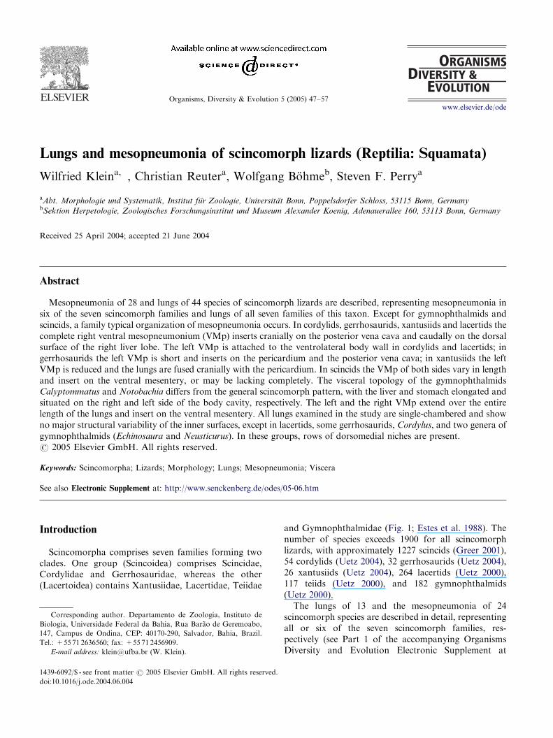
Scincomorpha comprises seven families forming twoclades. One group (Scincoidea) comprises Scincidae,Cordylidae and Gerrhosauridae, whereas the other(Lacertoidea) contains Xantusiidae, Lacertidae, Teiidae
e front matter r 2005 Elsevier GmbH. All rights reserved.
e.2004.06.004
ng author. Departamento de Zoologia, Instituto de
rsidade Federal da Bahia, Rua Barao de Geremoabo,
e Ondina, CEP: 40170-290, Salvador, Bahia, Brazil.
6560; fax: +55 71 2456909.
ss: [email protected] (W. Klein).
and Gymnophthalmidae (Fig. 1; Estes et al. 1988). Thenumber of species exceeds 1900 for all scincomorphlizards, with approximately 1227 scincids (Greer 2001),54 cordylids (Uetz 2004), 32 gerrhosaurids (Uetz 2004),26 xantusiids (Uetz 2004), 264 lacertids (Uetz 2000),117 teiids (Uetz 2000), and 182 gymnophthalmids(Uetz 2000).The lungs of 13 and the mesopneumonia of 24
scincomorph species are described in detail, representingall or six of the seven scincomorph families, res-pectively (see Part 1 of the accompanying OrganismsDiversity and Evolution Electronic Supplement at
ARTICLE IN PRESS
Fig. 1. Phylogenetic relationships of scincomorph lizards
following Estes et al. (1988).
W. Klein et al. / Organisms, Diversity & Evolution 5 (2005) 47–5748
http://www.senckenberg.de/odes/05-06.htm). In mostcases qualitative data for only one or two species perfamily is available (e.g. Milani 1894; Broman 1904; Becker1993). However, within the Teiioidea the mesopneumonia(Klein et al. 2000), and within the Varanoidea the lungstructure (Becker et al. 1989), comprise a useful characteronly at the family level. Therefore, description of a smallnumber of species per family represents a meaningfulcontribution. The aim of the present paper is to describethe mesopneumonia and provide quantitative morpholo-gical data for the lungs of representatives of all families ofscincomorph lizards, and thereby to develop somehypotheses about the functional and systematic value ofthe mesopneumonia–lung complex.
Materials and methods
Specimens from the Alexander Koenig ZoologicalResearch Institute and Museum (ZFMK) in Bonn,Germany, and from the collection of P.L.B. Rocha(Universidade Federal da Bahia, Brazil) were used.Snout-vent length (SVL), total length (TL), and bodymass (MB) were recorded as reference data (see Electr.Suppl. 05-06, Pt. 2).The visceral topology and location of mesos in each
group are presented, followed by a description of thelungs. Definitions of the mesos and representation of theresults follow Klein et al. (2000). Briefly, a standardschematic diagram, representing the parietal and visc-eral surfaces within a body cavity, is used to documentthe origin and insertion of the investigated mesos(DM ¼ dorsal mesentery, VM ¼ ventral mesentery;DMp ¼ dorsal mesopneumonium, VMp ¼ ventral me-sopneumonium). The part of a meso attached to theorgan is defined as its origin; the distal part as insertion.To visualize the lungs, a combined toluidine blue-PAS
(Periodic-Acid–Schiff reaction) en-bloc staining methodwas used. This method, which selectively stains trachealcartilage and lung parenchyma, is as follows: (1) tap
water, 6–8 h; (2) periodic acid (0.5%), 60min; (3)Schiff’s reagent, 10min; (4) tap water, 5min; (5) acidethanol (70%, 1% HCl conc.), 16 h; (6) toluidine blue(0.25%, pHo2), at least 4 h; (7) ethanol (70%), severalrepetitions to wash out superfluous toluidine blue; (8)ethanol (50%), 5min; (9) clearing and storage of lungsin glycerol.The following data for the respiratory system (glottis,
trachea and lungs) were collected [units of measurementin square brackets]: length of respiratory systemmeasured from cranial tip of glottis to most caudalend of lungs (LRS [mm]), length of lungs (LLu [mm]),length of left lung (Lle [mm]), length of right lung(Lri [mm]), length of trachea (LTr [mm]), length of extra-pulmonary bronchus (LBr [mm]), length of prehilarregion (LPh [mm]), number of niches (NNi), numberof tracheal cartilages (NTC), parenchymal type(ed ¼ ediculae, fav ¼ faveoli) and parenchymal distri-bution (het ¼ heterogeneous, hom ¼ homogeneous).LRS, LLu and LTr were standardized to SVL, LTr andLBr were standardized to LRS, LPh was standardized toLLu, and the quotients of lung length over trachea length(LLu/LTr), and length of left lung over length of rightlung (Lle/Lri) were calculated.
Results
Scincomorpha
General visceral topology
The heart lies in its pericardial cavity at the level ofthe shoulder girdle, connected caudally via the venacava to the liver. The latter is composed of two lobes,divided in the mediosagittal plane. The right lobe islarger than the left one and extends dorso-caudally,contacting the meso of the right gonad with its caudalend. The gall bladder is embedded in the ventro-caudalpart of the right liver lobe and the stomach lies on theleft side of the abdominal cavity. The lungs lie dorso-caudally to the heart and dorsally to the cranial parts ofliver and stomach. No intrapulmonary bronchi arepresent and the extra-pulmonary bronchi, when present,are short. Intestine, gonads and fat bodies, whosepresence and size vary according to the nutritionalstatus of a specimen, fill the caudal part of the bodycavity.
Scincidae
Visceral topology and mesos
The viscera of skinks show no deviation from thegeneral scincomorph pattern. DMp originate on thedorsal mid-line of the lungs and insert on the borderalimentary canal-DM. The VMp vary in their development
ARTICLE IN PRESS
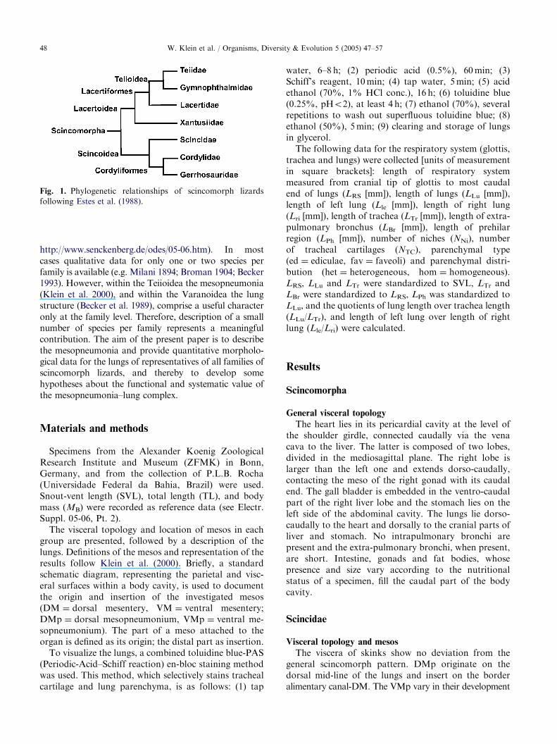
Fig. 2. (a) Attachments of mesopneumonia in Chalcides, Tiliqua and Tribolonotus; left: transverse sections through body cavity
(ventral side up), right: parietal surfaces in body cavity; DM ¼ dorsal mesentery, DMp ¼ dorsal mesopneumonium (dashed line),
li ¼ liver, lu ¼ lung, vcp ¼ vena cava posterior, VM ¼ ventral mesentery, VMp ¼ ventral mesopneumonium (solid line). (b)
Attachments of mesopneumonia in Scincus scincus. (c) Respiratory system in S. scincus, dorsal view, left lung open to show internal
structure, right lung showing major blood vessels on outer surface; scale bar ¼ 1 cm.
W. Klein et al. / Organisms, Diversity & Evolution 5 (2005) 47–57 49
on both sides. A cranial fusion of lungs with pericar-dium occurs in Chalcides, Tiliqua and Tribolonotus
(Fig. 2a). Novoeumeces and Mabuya show a completeright VMp originating in the ventral mid-line of the lungand inserting at the border esophagus-VM. The leftVMp of Novoeumeces extends from the cranial 1/4 of thelung and inserts at the base of the esophageal VM. InMabuya, on the other hand, the left VMp is lacking andthe left lung is fused cranially with the pericardium. Theright VMp of Scincus originates in the cranial 3/4 of thelung and inserts on the VM. The left VMp, by contrast,originates only on the cranial 1/4 of the lung, alsoinserting on the VM (Fig. 2b).
Respiratory system
For results per species see Electr. Suppl. 05-06, Pt. 3.The LRS ranges from 36.3% SVL in Chalcides up to56.7% SVL in Tribolonotus, with the mean at 45.3%SVL. The trachea averages 18.9% SVL (min. 15.5% inTiliqua, max. 21.2% in Scincus), the extra-pulmonary
bronchi 2.3% LRS (min. 1.6% in Mabuya, max. 3.2% inScincus), the prehilus 6.2% LLu (min. 2.5% in Novoeu-
meces, max. 12.3% in Chalcides), and the lungs 22.8%SVL (min. 13.2% in Chalcides, max. 32.3% in Tribolo-
notus). The lungs are of equal length, with Lle/Lri 1.03(min. 0.92 in Mabuya, max. 1.12 in Scincus). The NTC is51.3 (min. 33.3 in Tribolonotus, max. 62.7 in Mabuya),and LLu/LTr is 1.22 (min. 0.9 in Chalcides, max. 1.6 inTribolonotus). The lungs are long and slender, show aslightly heterogeneous parenchymal distribution withfaveoli in the cranial part of the lung and ediculae in themost caudal part. No niches are present (Fig. 2c).
Cordylidae
Visceral topology and mesos
The viscera show the general scincomorph pattern.The DMp are well developed, originate in the dorsalmid-line of each lung and insert cranially on the
ARTICLE IN PRESS
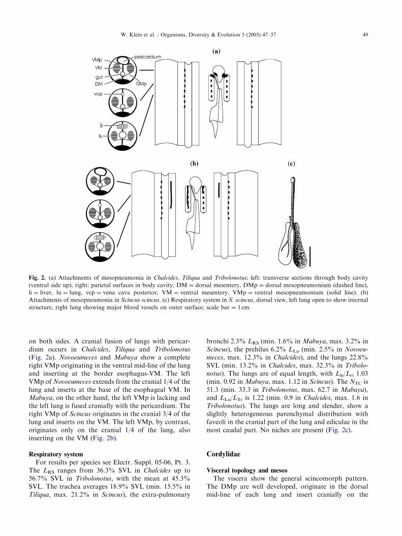
Fig. 3. (a) Attachments of mesopneumonia in a typical cordylid; layout and labeling as in Fig. 2. (b) Respiratory system in Cordylus
cordylus, dorsal view, left lung open to show internal structure, right lung showing major blood vessels on outer surface; N ¼ niches;
scale bar ¼ 1 cm.
W. Klein et al. / Organisms, Diversity & Evolution 5 (2005) 47–5750
esophagus. The insertion ends caudally on the DM. Thecomplete right VMp originates in the ventralmid-line of the lung, inserts cranially on the posteriorvena cava and caudally on the dorsal surface of theright liver lobe. The left VMp varies in length. It isshortest in Pseudocordylus, complete in Platysaurus. Ingeneral, it extends over the cranial half of the lung. Theinsertion begins cranially on the pericardium andextends caudally along the ventrolateral body wall(Fig. 3a).
Respiratory system
The dimensions of the respiratory system are similarto those of scincids, but the lungs appear more sac-like,except in Chamaesaura anguina, where they are long andslender. In Platysaurus capensis the right lung is slightlylonger than the left one (Lle/Lri 0.96), whereas inC. anguina the left lung is much longer than the rightone (Lle/Lri 1.20). Cordylus giganteus shows the longesttrachea (26.5% SVL) together with the longest respira-tory system (58.7% SVL), whereas Chamaesaura macro-
lepis shows the shortest trachea (10.6% SVL) and theshortest respiratory system (37.2% SVL). Pseudocordy-
lus melanotus has the shortest lung (17.6% SVL),whereas C. anguina has the longest lung (33.8% SVL)but the shortest prehilus (4.4% LLu). The longestprehilus (16.4% LLu) occurs in P. capensis. Extra-pulmonary bronchi are shortest in C. anguina (1.5%LRS) and C. macrolepis (2.8% LRS), longest inP. melanotus, P. capensis and P. microlepidotus (8.2%,7.9% and 7.8% LRS, respectively). The NTC ranges from32.7 in C. anguina and 39.0 in C. macrolepis up to 66.0 inPlatysaurus guttatus. Pseudocordylus melanotus has thelowest LLu/LTr (0.68), C. macrolepis and C. anguina havethe highest LLu/LTr (2.5 and 2.49, respectively). Theparenchymal distribution is slightly heterogeneous, withfaveoli cranially and ediculae caudally. Chamaesaura
anguina and C. macrolepis display highly heterogeneous
lungs, with ediculae cranially and trabecular parenchy-ma caudally.Niches are present in C. cordylus (Fig. 3b).
Gerrhosauridae
Visceral topology and mesos
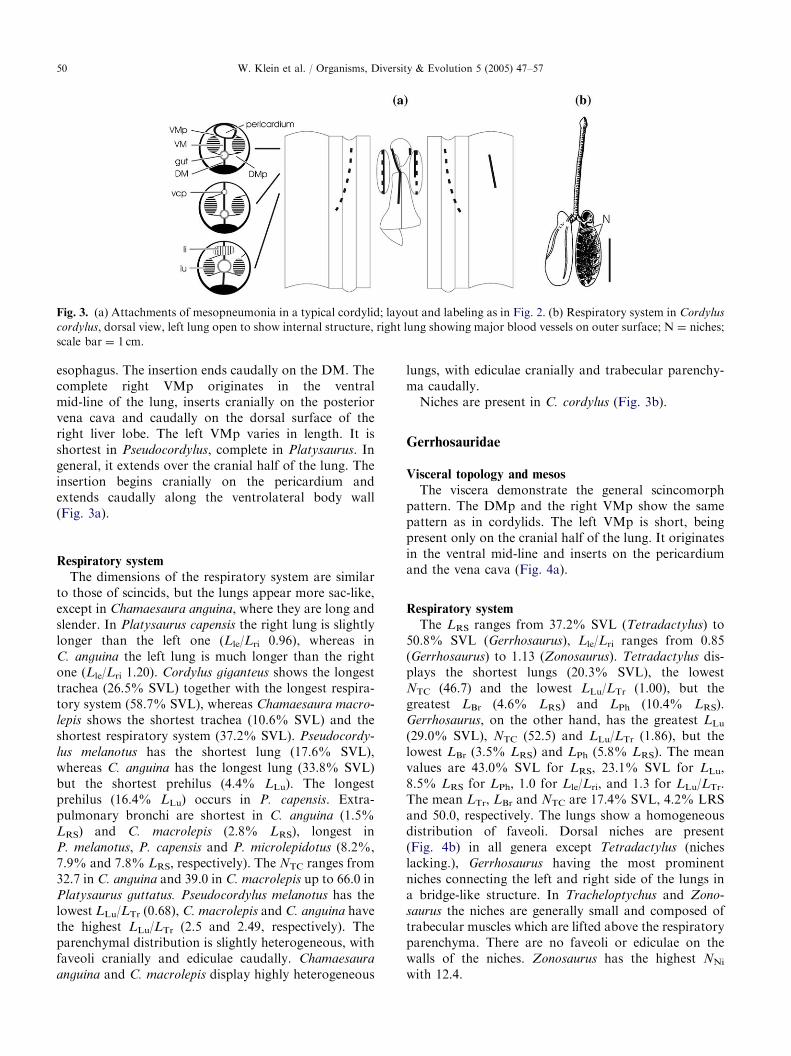
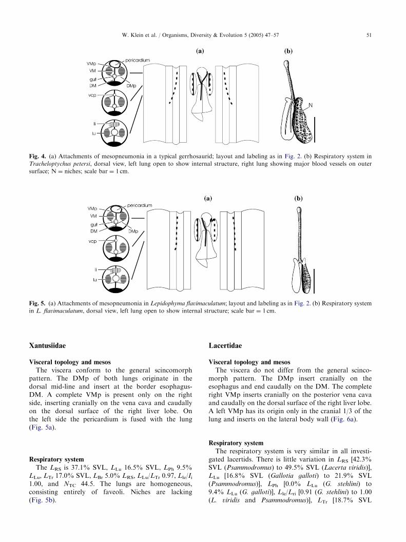
The viscera demonstrate the general scincomorphpattern. The DMp and the right VMp show the samepattern as in cordylids. The left VMp is short, beingpresent only on the cranial half of the lung. It originatesin the ventral mid-line and inserts on the pericardiumand the vena cava (Fig. 4a).
Respiratory system
The LRS ranges from 37.2% SVL (Tetradactylus) to50.8% SVL (Gerrhosaurus), Lle/Lri ranges from 0.85(Gerrhosaurus) to 1.13 (Zonosaurus). Tetradactylus dis-plays the shortest lungs (20.3% SVL), the lowestNTC (46.7) and the lowest LLu/LTr (1.00), but thegreatest LBr (4.6% LRS) and LPh (10.4% LRS).Gerrhosaurus, on the other hand, has the greatest LLu(29.0% SVL), NTC (52.5) and LLu/LTr (1.86), but thelowest LBr (3.5% LRS) and LPh (5.8% LRS). The meanvalues are 43.0% SVL for LRS, 23.1% SVL for LLu,8.5% LRS for LPh, 1.0 for Lle/Lri, and 1.3 for LLu/LTr.The mean LTr, LBr and NTC are 17.4% SVL, 4.2% LRSand 50.0, respectively. The lungs show a homogeneousdistribution of faveoli. Dorsal niches are present(Fig. 4b) in all genera except Tetradactylus (nicheslacking.), Gerrhosaurus having the most prominentniches connecting the left and right side of the lungs ina bridge-like structure. In Tracheloptychus and Zono-
saurus the niches are generally small and composed oftrabecular muscles which are lifted above the respiratoryparenchyma. There are no faveoli or ediculae on thewalls of the niches. Zonosaurus has the highest NNi
with 12.4.
ARTICLE IN PRESS
Fig. 4. (a) Attachments of mesopneumonia in a typical gerrhosaurid; layout and labeling as in Fig. 2. (b) Respiratory system in
Tracheloptychus petersi, dorsal view, left lung open to show internal structure, right lung showing major blood vessels on outer
surface; N ¼ niches; scale bar ¼ 1 cm.
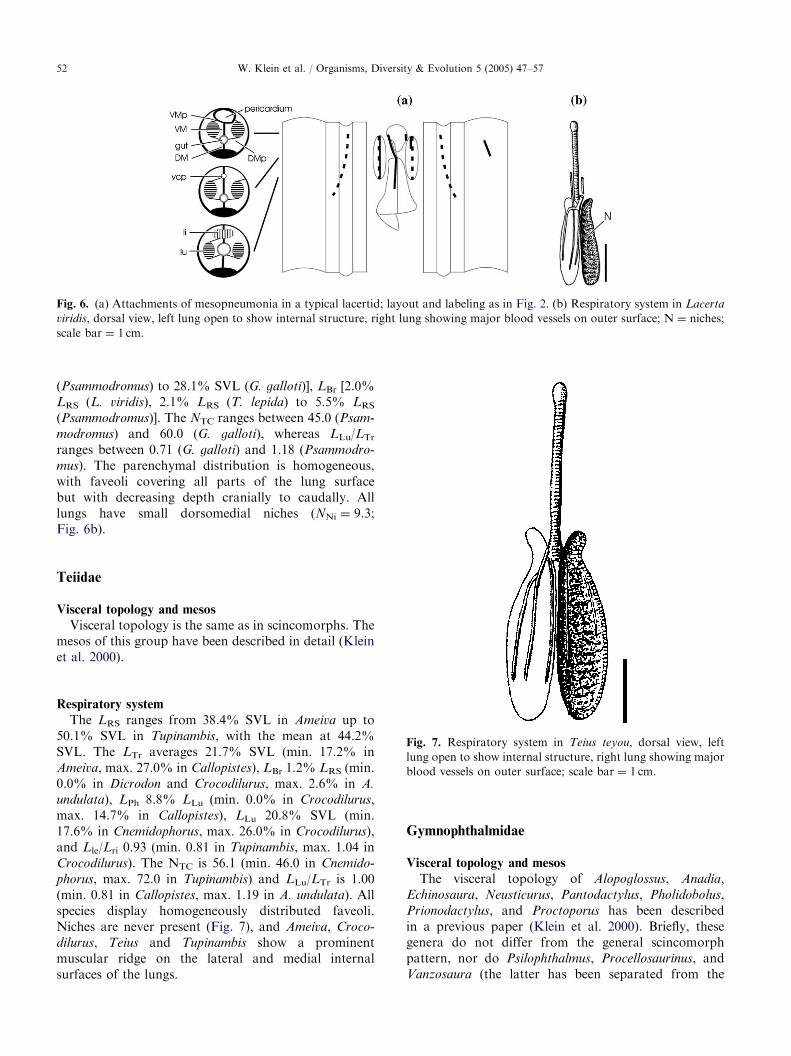
Fig. 5. (a) Attachments of mesopneumonia in Lepidophyma flavimaculatum; layout and labeling as in Fig. 2. (b) Respiratory system
in L. flavimaculatum, dorsal view, left lung open to show internal structure; scale bar ¼ 1 cm.
W. Klein et al. / Organisms, Diversity & Evolution 5 (2005) 47–57 51
Xantusiidae
Visceral topology and mesos
The viscera conform to the general scincomorphpattern. The DMp of both lungs originate in thedorsal mid-line and insert at the border esophagus-DM. A complete VMp is present only on the rightside, inserting cranially on the vena cava and caudallyon the dorsal surface of the right liver lobe. Onthe left side the pericardium is fused with the lung(Fig. 5a).
Respiratory system
The LRS is 37.1% SVL, LLu 16.5% SVL, LPh 9.5%LLu, LTr 17.0% SVL, LBr 5.0% LRS, LLu/LTr 0.97, Lle/Ii1.00, and NTC 44.5. The lungs are homogeneous,consisting entirely of faveoli. Niches are lacking(Fig. 5b).
Lacertidae
Visceral topology and mesos
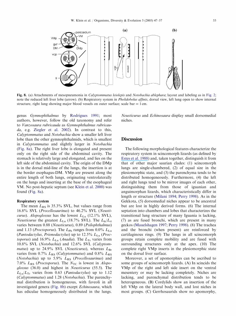
The viscera do not differ from the general scinco-morph pattern. The DMp insert cranially on theesophagus and end caudally on the DM. The completeright VMp inserts cranially on the posterior vena cavaand caudally on the dorsal surface of the right liver lobe.A left VMp has its origin only in the cranial 1/3 of thelung and inserts on the lateral body wall (Fig. 6a).
Respiratory system
The respiratory system is very similar in all investi-gated lacertids. There is little variation in LRS [42.3%SVL (Psammodromus) to 49.5% SVL (Lacerta viridis)],LLu [16.8% SVL (Gallotia galloti) to 21.9% SVL(Psammodromus)], LPh [0.0% LLu (G. stehlini) to9.4% LLu (G. galloti)], Lle/Lri [0.91 (G. stehlini) to 1.00(L. viridis and Psammodromus)], LTr [18.7% SVL
ARTICLE IN PRESS
Fig. 6. (a) Attachments of mesopneumonia in a typical lacertid; layout and labeling as in Fig. 2. (b) Respiratory system in Lacerta
viridis, dorsal view, left lung open to show internal structure, right lung showing major blood vessels on outer surface; N ¼ niches;
scale bar ¼ 1 cm.
W. Klein et al. / Organisms, Diversity & Evolution 5 (2005) 47–5752
(Psammodromus) to 28.1% SVL (G. galloti)], LBr [2.0%LRS (L. viridis), 2.1% LRS (T. lepida) to 5.5% LRS(Psammodromus)]. The NTC ranges between 45.0 (Psam-
modromus) and 60.0 (G. galloti), whereas LLu/LTrranges between 0.71 (G. galloti) and 1.18 (Psammodro-
mus). The parenchymal distribution is homogeneous,with faveoli covering all parts of the lung surfacebut with decreasing depth cranially to caudally. Alllungs have small dorsomedial niches (NNi ¼ 9.3;Fig. 6b).
Teiidae
Visceral topology and mesos
Visceral topology is the same as in scincomorphs. Themesos of this group have been described in detail (Kleinet al. 2000).
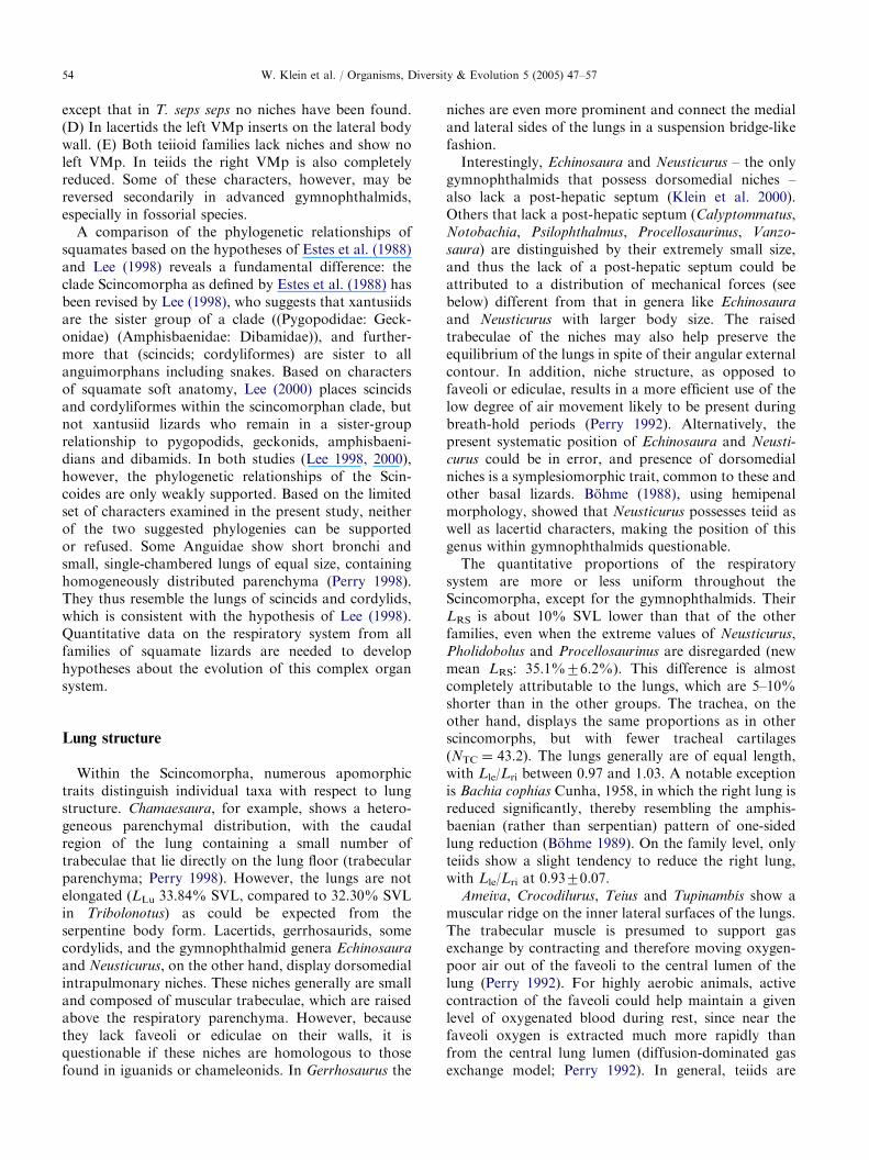
Fig. 7. Respiratory system in Teius teyou, dorsal view, left
lung open to show internal structure, right lung showing major
blood vessels on outer surface; scale bar ¼ 1 cm.
Respiratory system
The LRS ranges from 38.4% SVL in Ameiva up to50.1% SVL in Tupinambis, with the mean at 44.2%SVL. The LTr averages 21.7% SVL (min. 17.2% inAmeiva, max. 27.0% in Callopistes), LBr 1.2% LRS (min.0.0% in Dicrodon and Crocodilurus, max. 2.6% in A.
undulata), LPh 8.8% LLu (min. 0.0% in Crocodilurus,max. 14.7% in Callopistes), LLu 20.8% SVL (min.17.6% in Cnemidophorus, max. 26.0% in Crocodilurus),and Lle/Lri 0.93 (min. 0.81 in Tupinambis, max. 1.04 inCrocodilurus). The NTC is 56.1 (min. 46.0 in Cnemido-
phorus, max. 72.0 in Tupinambis) and LLu/LTr is 1.00(min. 0.81 in Callopistes, max. 1.19 in A. undulata). Allspecies display homogeneously distributed faveoli.Niches are never present (Fig. 7), and Ameiva, Croco-
dilurus, Teius and Tupinambis show a prominentmuscular ridge on the lateral and medial internalsurfaces of the lungs.
Gymnophthalmidae
Visceral topology and mesos
The visceral topology of Alopoglossus, Anadia,Echinosaura, Neusticurus, Pantodactylus, Pholidobolus,Prionodactylus, and Proctoporus has been describedin a previous paper (Klein et al. 2000). Briefly, thesegenera do not differ from the general scincomorphpattern, nor do Psilophthalmus, Procellosaurinus, andVanzosaura (the latter has been separated from the
ARTICLE IN PRESS
Fig. 8. (a) Attachments of mesopneumonia in Calyptommatus leiolepis and Notobachia ablephara; layout and labeling as in Fig. 2;
note the reduced left liver lobe (arrow). (b) Respiratory system in Pholidobolus affinis, dorsal view, left lung open to show internal
structure, right lung showing major blood vessels on outer surface; scale bar ¼ 1 cm.
W. Klein et al. / Organisms, Diversity & Evolution 5 (2005) 47–57 53
genus Gymnophthalmus by Rodrigues 1991; mostauthors, however, follow the old taxonomy and referto Vanzosaura rubricauda as Gymnophthalmus rubricau-
da, e.g. Ziegler et al. 2002). In contrast to this,Calyptommatus and Notobachia show a smaller left liverlobe than the other gymnophthalmids, which is smallestin Calyptommatus and slightly larger in Notobachia
(Fig. 8a). The right liver lobe is elongated and presentonly on the right side of the abdominal cavity. Thestomach is relatively large and elongated, and lies on theleft side of the abdominal cavity. The origin of the DMpis in the dorsal mid-line of the lungs, the insertion is atthe border esophagus-DM. VMp are present along theentire length of both lungs, originating ventrolaterallyon the lungs and inserting at the base of the esophagealVM. No post-hepatic septum (see Klein et al. 2000) wasfound (Fig. 8a).
Respiratory system
The mean LRS is 35.3% SVL, but values range from16.8% SVL (Procellosaurinus) to 46.2% SVL (Neusti-
curus). Alopoglossus has the lowest LLu (12.1% SVL),Neusticurus the greatest LLu (18.7% SVL). The Lle/Lrivaries between 0.88 (Neusticurus), 0.89 (Psilophthalmus)and 1.13 (Proctoporus). The LPh ranges from 0.0% LLu(Pantodactylus, Prionodactylus) up to 12.5% LLu (Proc-
toporus) and 16.9% LLu (Anadia). The LTr varies from10.8% SVL (Notobachia) and 12.6% SVL (Calyptom-
matus) up to 24.9% SVL (Neusticurus), whereas LBrvaries from 0.7% LRS (Calyptommatus) and 0.8% LRS(Notobachia) up to 5.9% LRS (Procellosaurinus) and7.0% LRS (Proctoporus). The NTC is lowest in Alopo-
glossus (36.0) and highest in Neusticurus (55.5). TheLLu/LTr varies from 0.63 (Pantodactylus) up to 1.12(Calyptommatus) and 1.28 (Notobachia). The parenchy-mal distribution is homogeneous, with faveoli in allinvestigated genera (Fig. 8b) except Echinosaura, whichhas ediculae homogeneously distributed in the lungs.
Neusticurus and Echinosaura display small dorsomedialniches.
Discussion
The following morphological features characterize therespiratory system in scincomorph lizards (as defined byEstes et al. 1988) and, taken together, distinguish it fromthat of other major saurian clades: (1) scincomorphlungs are single-chambered, (2) of equal size in theplesiomorphic state, and (3) the parenchyma tends to bedistributed homogeneously. Furthermore, (4) the leftand right lungs tend to be mirror images of each other,distinguishing them from those of iguanian andanguimorphan lizards, which characteristically differ inlength or structure (Milani 1894; Perry 1998). As in theGekkota, (5) dorsomedial niches appear to be ancestralbut are lost in highly derived forms. (6) The internalseptation into chambers and lobes that characterizes thetransitional lung structure of many Iguania is lacking,(7) as are fused bronchi, which are present in manygeckos (Mauelshagen 1997; Perry 1998). (8) The tracheaand the bronchi (when present) are reinforced bycartilaginous rings. (9) The lungs in all scincomorphgroups retain complete mobility and are fused withsurrounding structures only at the apex. (10) Thecomplete right VMp inserts in the plesiomorphic stateon the dorsal liver surface.Moreover, a set of apomorphies can be ascribed to
some groups of scincomorph lizards. (A) In scincids theVMp of the right and left side insert on the ventralmesentery or may be lacking completely. Niches arelacking, and parenchymal distribution tends to beheterogeneous. (B) Cordylids show an insertion of theleft VMp on the lateral body wall, and lost niches inmost groups. (C) Gerrhosaurids show no apomorphy,
ARTICLE IN PRESSW. Klein et al. / Organisms, Diversity & Evolution 5 (2005) 47–5754
except that in T. seps seps no niches have been found.(D) In lacertids the left VMp inserts on the lateral bodywall. (E) Both teiioid families lack niches and show noleft VMp. In teiids the right VMp is also completelyreduced. Some of these characters, however, may bereversed secondarily in advanced gymnophthalmids,especially in fossorial species.A comparison of the phylogenetic relationships of
squamates based on the hypotheses of Estes et al. (1988)and Lee (1998) reveals a fundamental difference: theclade Scincomorpha as defined by Estes et al. (1988) hasbeen revised by Lee (1998), who suggests that xantusiidsare the sister group of a clade ((Pygopodidae: Geck-onidae) (Amphisbaenidae: Dibamidae)), and further-more that (scincids; cordyliformes) are sister to allanguimorphans including snakes. Based on charactersof squamate soft anatomy, Lee (2000) places scincidsand cordyliformes within the scincomorphan clade, butnot xantusiid lizards who remain in a sister-grouprelationship to pygopodids, geckonids, amphisbaeni-dians and dibamids. In both studies (Lee 1998, 2000),however, the phylogenetic relationships of the Scin-coides are only weakly supported. Based on the limitedset of characters examined in the present study, neitherof the two suggested phylogenies can be supportedor refused. Some Anguidae show short bronchi andsmall, single-chambered lungs of equal size, containinghomogeneously distributed parenchyma (Perry 1998).They thus resemble the lungs of scincids and cordylids,which is consistent with the hypothesis of Lee (1998).Quantitative data on the respiratory system from allfamilies of squamate lizards are needed to develophypotheses about the evolution of this complex organsystem.
Lung structure
Within the Scincomorpha, numerous apomorphictraits distinguish individual taxa with respect to lungstructure. Chamaesaura, for example, shows a hetero-geneous parenchymal distribution, with the caudalregion of the lung containing a small number oftrabeculae that lie directly on the lung floor (trabecularparenchyma; Perry 1998). However, the lungs are notelongated (LLu 33.84% SVL, compared to 32.30% SVLin Tribolonotus) as could be expected from theserpentine body form. Lacertids, gerrhosaurids, somecordylids, and the gymnophthalmid genera Echinosaura
and Neusticurus, on the other hand, display dorsomedialintrapulmonary niches. These niches generally are smalland composed of muscular trabeculae, which are raisedabove the respiratory parenchyma. However, becausethey lack faveoli or ediculae on their walls, it isquestionable if these niches are homologous to thosefound in iguanids or chameleonids. In Gerrhosaurus the
niches are even more prominent and connect the medialand lateral sides of the lungs in a suspension bridge-likefashion.Interestingly, Echinosaura and Neusticurus – the only
gymnophthalmids that possess dorsomedial niches –
also lack a post-hepatic septum (Klein et al. 2000).Others that lack a post-hepatic septum (Calyptommatus,Notobachia, Psilophthalmus, Procellosaurinus, Vanzo-
saura) are distinguished by their extremely small size,and thus the lack of a post-hepatic septum could beattributed to a distribution of mechanical forces (seebelow) different from that in genera like Echinosaura
and Neusticurus with larger body size. The raisedtrabeculae of the niches may also help preserve theequilibrium of the lungs in spite of their angular externalcontour. In addition, niche structure, as opposed tofaveoli or ediculae, results in a more efficient use of thelow degree of air movement likely to be present duringbreath-hold periods (Perry 1992). Alternatively, thepresent systematic position of Echinosaura and Neusti-
curus could be in error, and presence of dorsomedialniches is a symplesiomorphic trait, common to these andother basal lizards. Bohme (1988), using hemipenalmorphology, showed that Neusticurus possesses teiid aswell as lacertid characters, making the position of thisgenus within gymnophthalmids questionable.The quantitative proportions of the respiratory
system are more or less uniform throughout theScincomorpha, except for the gymnophthalmids. TheirLRS is about 10% SVL lower than that of the otherfamilies, even when the extreme values of Neusticurus,Pholidobolus and Procellosaurinus are disregarded (newmean LRS: 35.1%76.2%). This difference is almostcompletely attributable to the lungs, which are 5–10%shorter than in the other groups. The trachea, on theother hand, displays the same proportions as in otherscincomorphs, but with fewer tracheal cartilages(NTC ¼ 43.2). The lungs generally are of equal length,with Lle/Lri between 0.97 and 1.03. A notable exceptionis Bachia cophias Cunha, 1958, in which the right lung isreduced significantly, thereby resembling the amphis-baenian (rather than serpentian) pattern of one-sidedlung reduction (Bohme 1989). On the family level, onlyteiids show a slight tendency to reduce the right lung,with Lle/Lri at 0.9370.07.
Ameiva, Crocodilurus, Teius and Tupinambis show amuscular ridge on the inner lateral surfaces of the lungs.The trabecular muscle is presumed to support gasexchange by contracting and therefore moving oxygen-poor air out of the faveoli to the central lumen of thelung (Perry 1992). For highly aerobic animals, activecontraction of the faveoli could help maintain a givenlevel of oxygenated blood during rest, since near thefaveoli oxygen is extracted much more rapidly thanfrom the central lung lumen (diffusion-dominated gasexchange model; Perry 1992). In general, teiids are
ARTICLE IN PRESSW. Klein et al. / Organisms, Diversity & Evolution 5 (2005) 47–57 55
highly active, whereas gymnophthalmids are mostlysecretive, sit-and-wait predators (Urban 1965; MacLean1974). On the other hand, the muscular ridge maystabilize the lungs and provide a large surface area ofciliated epithelium for transport of mucus from the largefaveolar lung surface.With regard to external airways, the values for
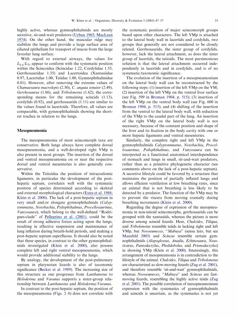
LLu/LTr appear to conform with the systematic positionwithin the Scincoidea (Scincidae 1.22, Cordylidae 1.35,Gerrhosauridae 1.35) and Lacertoidea (Xantusiidae0.97, Lacertidae 1.00, Teiidae 1.00, Gymnophthalmidae0.81). However, after removing the extreme values ofChamaesaura macrolepis (2.50), C. anguia tenuior (2.49),Gerrhosaurus (1.86), and Tribolonotus (1.62), the corre-sponding means for the remaining scincids (1.13),cordylids (0.92), and gerrhosaurids (1.11) are similar tothe values found in lacertoids. Therefore, all values arecomparable, with gymnophthalmids showing the short-est trachea in relation to the lungs.
Mesopneumonia
The mesopneumonia of most scincomorph taxa areconservative. Both lungs always have complete dorsalmesopneumonia, and a well-developed right VMp isalso present in most groups. The insertion of the dorsaland ventral mesopneumonia on or near the respectivedorsal and ventral mesenteries is also generally con-servative.Within the Teiioidea the position of intracoelomic
ligaments, in particular the development of the post-hepatic septum, correlates well with the systematicposition of species determined according to skeletaland external morphological characters (Estes et al. 1988;Klein et al. 2000). The lack of a post-hepatic septum invery small and/or elongate gymnophthalmids (Calyp-
tommatus, Notobachia, Psilophthalmus, Procellosaurinus,Vanzosaura), which belong to the well-defined ‘‘Rodri-gues-clade’’ of Pellegrino et al. (2001), could be theresult of strong adhesive forces acting upon the lungs,resulting in effective suspension and maintenance oflung inflation during breath-hold periods, and making apost-hepatic septum superfluous. It should also be notedthat these species, in contrast to the other gymnophthal-mids investigated (Klein et al. 2000), also possesscomplete left and right ventral mesopneumonia, whichwould provide additional stability to the lungs.By analogy, the development of the post-pulmonary
septum in platynotan lizards is also of taxonomicsignificance (Becker et al. 1989). The increasing size ofthis structure as one progresses from Lanthanotus toHeloderma and Varanus supports a sister-group rela-tionship between Lanthanotus and Heloderma/Varanus.In contrast to the post-hepatic septum, the position of
the mesopneumonia (Figs. 2–8) does not correlate with
the systematic position of major scincomorph groupsbased upon other characters. The left VMp is attachedto the lateral body wall in lacertids and cordylids, twogroups that generally are not considered to be closelyrelated. Gerrhosaurids, the sister group of cordylids,however, lack the lateral attachment, as does the sistergroup of lacertids, the teiioids. The most parsimonioussolution is that the lateral attachment occurred inde-pendently in lacertids and in cordylids, and is of nosystematic/taxonomic significance.The evolution of the insertion of a mesopneumonium
on the lateral body wall can be reconstructed by thefollowing steps: (1) insertion of the left VMp on the VM;(2) insertion of the left VMp on the ventral liver surface(see Fig. 599 in Broman 1904, p. 515); (3) insertion ofthe left VMp on the ventral body wall (see Fig. 600 inBroman 1904, p. 515); and (4) shifting of the insertionfrom the ventral to the lateral body wall, with reductionof the VMp to the caudal part of the lung. An insertionof the right VMp on the lateral body wall is notnecessary, because of the constant position and shape ofthe liver and its fixation in the body cavity with one ormore hepatic ligaments and ventral mesenteries.Similarly, the complete right and left VMp in the
gymnophthalmids Calyptommatus, Notobachia, Procel-
losaurinus, Psilophthalmus, and Vanzosaura can beinterpreted as a functional anatomical interdependenceof stomach and lungs in small, sit-and-wait predators,rather than as a primitive phylogenetic character (seecomments above on the lack of a post-hepatic septum).A secretive lifestyle could be favored by a structure thatmaintains the position of partially inflated lungs andallows efficient ventilation at low breathing rates, sincean animal that is not breathing is less likely to bedetected by a predator. The function of the VMp may beto prevent the viscera from moving cranially duringbreathing movements (Klein et al. 2000).Regarding the degree of expression of the mesopneu-
monia in non-teiioid scincomorphs, gerrhosaurids can begrouped with the xantusiids, whereas the picture is morediverse among scincid lizards. Here, Chalcides, Tiliqua,and Tribolonotus resemble teiids in lacking right and leftVMp, but Novoeumeces, ‘‘Mabuya’’ (sensu lato, but seeMausfeld 2003) and Scincus resemble certain gym-nophthalmids (Alopoglossus, Anadia, Echinosaura, Neus-
ticurus, Pantodactylus, Pholidobolus, and Prionodactylus)in showing VMp (Klein et al. 2000). Interestingly, thisarrangement of mesopneumonia is in contradiction to thelifestyle of the animal. Chalcides, Tiliqua and Tribolonotus
are characterized as slow-moving lizards (Zug et al. 2001),and therefore resemble ‘sit-and-wait’ gymnophthalmids,whereas Novoeumeces, ‘‘Mabuya’’ and Scincus are fast-moving lizards, resembling the highly active teiids (Zuget al. 2001). The possible correlation of mesopneumoniumexpression with the systematics of gymnophthalmidsand scincids is uncertain, as the systematics is not yet
ARTICLE IN PRESSW. Klein et al. / Organisms, Diversity & Evolution 5 (2005) 47–5756
sufficiently resolved to allow a comparison within thesetwo groups. The variation observed here within theScincidae – a group comprising more than 1200 species –also merits further study. The Gekkota – also with morethan 1000 species – show much less variation in theexpression of the VMp than do scincids (Becker 1993;Mauelshagen 1997).
Serpentine body form and topology of internal
organs
Most scincomorph lizards display similar generalvisceral topology: the lungs overlap caudally with thecranial parts of liver and stomach. In Calyptommatus
and Notobachia, however, the short lungs lie anterior tothe liver and stomach, which are on the right or left sideof the body cavity, respectively (Fig. 8a). These twogenera, together with Chamaesaura, have a very shorttrachea (LTr 10.6% SVL in Chamaesaura macrolepis,10.8% SVL in Notobachia, 12.5% SVL in Chamaesaura
anguina, and 12.6% SVL in Calyptommatus). All ofthese genera have reduced limbs, but only Calyptomma-
tus and Notobachia show an elongation of the trunk.Since Chamaesaura obtains a serpentine form to a greatextent by elongation of the tail and exhibits onlymoderate elongation of the trunk, the proportions oflungs and viscera conform to the general scincomorphpattern. Thus, with regard to internal topology there areat least three possibilities that result in serpentine bodyform and limb reduction: (1) the snake/amphisbaenian/Bachia cophias pattern, characterized by elongation ofthe trunk, reduction or elimination of one lung andelongation of the remaining one, together with the liverand the stomach, and by asymmetry of the elongatedkidneys (Gans 1975; Bohme 1989); (2) the patternshown by Calyptommatus and Notobachia, which ischaracterized by elongation of the trunk, while the lungsremain of equal size and occupy the cranial part of thebody cavity, with no overlapping with the elongatedliver and stomach; and (3) the cordylid pattern, showingelongation of the tail with no change in general visceraltopology (Chamaesaura).Camp (1923) distinguished two ecomorphs of snake-
like squamates: small, short-tailed burrowers versuslarge, long-tailed grass-swimmers. The first type can befound in amphisbaenians (Pough et al. 1998), primitivesnakes (Pough et al. 1998), and in many groups ofburrowing skinks (Caputo et al. 1995), whereas thesecond one is present in cordylids (Chamaesaura),gerrhosaurids (Tetradactylus) and lacertids (Takydro-
mus), as well as in theMabuya megalura-buettneri group.Recently, this dichotomy was supported by Wiens andSlingluff (2001), who studied the evolution of serpentinebody form in anguids and found both ecomorphs withinthe family.
For the gymnophthalmids Calyptommatus and Noto-
bachia, however, classification into the two ecomorphsdescribed above remains questionable, as the animalsare small but have long tails. These two genera areclosely related but differ in their ecology. WhereasCalyptommatus is strictly nocturnal and fossorial,Notobachia can be active during the day or night, andforages mainly in leaf litter (Rodrigues 1996). Furtherstudies of visceral morphology, locomotor mechanismsand respiration in scincomorph lizards may help toexplain the complex structure-function relationshipamong these features and the role it may play in theevolution of squamates.
Acknowledgments
We are indebted to Pedro L.B. Rocha from theUniversidade Federal da Bahia, Brazil, for providingsome of the animal material, and to Mogens L. Glass forhelpful comments on an earlier version of the manu-script. SFP was supported by the Deutsche Forschungs-gemeinschaft (DFG, Pe 267/7).
References
Becker, H.O., 1993. Vergleichende Untersuchungen am
respiratorischen Apparat anguimorpher Eidechsen: Eine
stammesgeschichtliche Deutung. Dissertation, Rheinische
Friedrich-Wilhelms-Universitat Bonn, Germany.
Becker, H.O., Perry, S.F., Bohme, W., 1989. Die Lungenmor-
phologie der Warane (Reptilia: Varanidae) und ihre
systematisch-stammesgeschichtliche Bedeutung. Bonn.
Zool. Beitr. 40, 27–56.
Bohme, W., 1988. Zur Genitalmorphologie der Sauria:
funktionelle und stammesgeschichtliche Aspekte. Bonn.
Zool. Monogr. 27, 1–176.
Bohme, W., 1989. Zur systematischen Stellung der Amphisba-
nen (Reptilia: Squamata), mit besonderer Berucksichtigung
der Morphologie des Hemipenis. Z. Zool. Syst. Evolutions-
forsch. 27, 330–337.
Broman, I., 1904. Die Entwicklungsgeschichte der Bursa
omentalis und ahnlicher Rezessbildungen bei den
Wirbeltieren. Entw.-gesch. Monogr. 1. J.F. Bergmann,
Wiesbaden.
Camp, C.C., 1923. Classification of lizards. Bull. Am. Mus.
Nat. Hist. 48, 289–481.
Caputo, V., Lanza, B., Palmieri, R., 1995. Body elongation
and limb reduction in the genus Chalcides Laurenti 1768
(Squamata: Scincidae): a comparative study. Trop. Zool. 8,
95–152.
Estes, R., Queiroz, K.de., Gauthier, J., 1988. Phylogenetic
relationships within Squamata. In: Estes, R., Pregill, G.K.
(Eds.), Phylogenetic Relationships of the Lizard Families.
Stanford University Press, Stanford, California.
Gans, C., 1975. Tetrapod limblessness: evolution and func-
tional corollaries. Am. Zool. 15, 455–467.
ARTICLE IN PRESSW. Klein et al. / Organisms, Diversity & Evolution 5 (2005) 47–57 57
Greer, A.E., 2001. Distribution of maximum snout-vent length
among species of scincid lizards. J. Herpetol. 35, 383–395.
Klein, W., Bohme, W., Perry, S.F., 2000. The mesopneumonia
and the post-hepatic septum of the Teiioidea (Reptilia:
Squamata). Acta Zool. (Stockholm) 81, 109–119.
Lee, M.S.Y., 1998. Convergent evolution and character
correlation in burrowing reptiles: toward a resolution of
squamate phylogeny. Biol. J. Linn. Soc. 65, 369–453.
Lee, M.S.Y., 2000. Soft anatomy, diffuse homoplasy, and the
relationships of lizards and snakes. Zool. Scr. 29, 101–130.
MacLean, W.P., 1974. Feeding and locomotor mechanisms of
teiid lizards: functional morphology and evolution. Pap.
Avulsos Zool. Sao Paulo 27, 179–213.
Mauelshagen, N.M.P., 1997. Die phylogenetische Bedeutung
des Luftweg-Lungen-Komplexes der Gekkota. Thesis,
Rheinische Friedrich-Wilhelms-Universitat Bonn, Ger-
many.
Mausfeld, P., 2003. Molecular phylogeny and biogeography of
the scincid lizard genus Mabuya Fitzinger, 1826: elucidating
the evolutionary history of a circumtropical lizard group.
Dissertation, Rheinische Friedrich-Wilhelms-Universitat
Bonn, Germany.
Milani, A., 1894. Beitrage zur Kenntnis der Reptilienlunge. I.
Lacertilia. Zool. Jhb. Abt. Anat. Ont. 7, 545–592.
Pellegrino, K.C.M., Rodrigues, M.T., Yonenaga-Yassuda, Y.,
Sites Jr., J.W., 2001. A molecular perspective on the
evolution of microteiid lizards (Squamata, Gymnoththal-
midae) and a new classification for the family. Biol. J. Linn.
Soc. 74, 315–338.
Perry, S.F., 1992. Gas exchange strategies in reptiles and the
origin of the avian lung. In: Wood, S.C., Weber, R.E.,
Hargens, A.R., Millard, R.W. (Eds.), Physiological Adap-
tations in Vertebrates. Marcel Dekker, New York.
Perry, S.F., 1998. Lungs: comparative anatomy, functional
morphology, and evolution. In: Gans, C., Gaunt, A.S.
(Eds.), Biology of the Reptilia. Society for the Study of
Amphibians and Reptilians, Ithaca, NY.
Pough, F.H., Andrews, R.M., Cadle, J.E., Crump, M.L.,
Savitzky, A.H., Wells, K.D., 1998. Herpetology. Prentice-
Hall, Upper Saddle River, NJ.
Rodrigues, M.T., 1991. Herpetofauna das dunas interiores do
rio Sao Francisco, Bahia, Brasil. III. Procellosaurinus: um
novo genero de microteiıdeos sem palpebra, com a
redefinicao do genero Gymnophthalmus (Sauria, Teiidae).
Pap. Avulsos Zool. Sao Paulo 37, 329–342.
Rodrigues, M.T., 1996. Lizards, snakes and amphisbaenians
from the Quaternary sand dunes of the middle rio Sao
Francisco, Bahia, Brazil. J. Herpetol. 30, 513–523.
Uetz, P., 2000. How many reptile species? Herpetol. Rev. 31,
13–15.
Uetz, P., 2004. The EMBL reptile database. European
Molecular Biology Laboratory, Heidelberg, Germany;
available at www.reptile-database.org.
Urban, E.K., 1965. Quantitative study of locomotion in teiid
lizards. Anim. Behav. 13, 513–529.
Wiens, J.J., Slingluff, J.L., 2001. How lizards turn into snakes:
a phylogenetic analysis of body-form evolution in anguid
lizards. Evolution 55, 2303–2318.
Ziegler, T., Bohme, W., Unger, J., 2002. First record of
Gymnophthalmus rubricauda Boulenger, 1902 for Paraguay,
with notes on its morphology and habitat (Reptilia: Sauria:
Gymnophthalmidae). Faun. Abh. Mus. Tierkd. Dresden
22, 347–351.
Zug, G.R., Vitt, L.J., Caldwell, J.P., 2001. Herpetology-An
Introductory Biology of Amphibians and Reptiles. Aca-
demic Press, London.