lipopolysaccharide-induced peroxisomal dysfunction exacerbates cerebral white matter injury:...
TRANSCRIPT
Available online at www.sciencedirect.com
10 (2008) 560–576www.elsevier.com/locate/yexnr
Experimental Neurology 2
Lipopolysaccharide-induced peroxisomal dysfunction exacerbates cerebralwhite matter injury: Attenuation by N-acetyl cysteine
Manjeet K. Paintlia a,1, Ajaib S. Paintlia a,1, Miguel A. Contreras a, Inderjit Singh a, Avtar K. Singh b,⁎
a Department of Pediatrics, Medical University of South Carolina, USAb Department of Pathology and Laboratory Medicine, Ralph H. Johnson VA Medical Center, Charleston, SC, USA
Received 31 July 2007; revised 20 November 2007; accepted 4 December 2007Available online 23 December 2007
Abstract
Cerebral white matter injury during prenatal maternal infection characterized as periventricular leukomalacia is the main substrate for cerebralpalsy (CP) in premature infants. Previously, we reported that maternal LPS exposure causes oligodendrocyte (OL)-injury/hypomyelination in thedeveloping brain which can be attenuated by an antioxidant agent, N-acetyl cysteine (NAC). Herein, we elucidated the role of peroxisomes inLPS-induced neuroinflammation and cerebral white matter injury. Peroxisomes are important for detoxification of reactive oxidative species(ROS) and metabolism of myelin-lipids in OLs. Maternal LPS exposure induced selective depletion of developing OLs in the fetal brain whichwas associated with ROS generation, glutathione depletion and peroxisomal dysfunction. Likewise, hypomyelination in the postnatal brain wasassociated with decrease in peroxisomes and OLs after maternal LPS exposure. Conversely, NAC abolished these LPS-induced effects in thedeveloping brain. CP brains imitated these observed changes in peroxisomal/myelin proteins in the postnatal brain after maternal LPS exposure.In vitro studies revealed that pro-inflammatory cytokines cause OL-injury via peroxisomal dysfunction and ROS generation. NAC or WY14643(peroxisome proliferators activated receptor (PPAR)-α agonist) reverses these effects of pro-inflammatory cytokines in the wild-type OLs, but notin PPAR-α(−/−) OLs. Similarly treated B12 oligodenroglial cells co-transfected with PPAR-α siRNAs/pTK-PPREx3-Luc, and LPS exposed PPAR-α(−/−) pregnant mice treated with NAC or WY14643 further suggested that PPAR-α activity mediates NAC-induced protective effects.Collectively, these data provide unprecedented evidence that LPS-induced peroxisomal dysfunction exacerbates cerebral white matter injury andits attenuation by NAC via a PPAR-α dependent mechanism expands therapeutic avenues for CP and related demyelinating diseases.Published by Elsevier Inc.
Keywords: Lipopolysaccharide; Periventricular leukomalacia; Cerebral whitematter injury; Cerebral palsy; Peroxisome proliferators-activated receptor-α;N-acetyl cysteine
Introduction
Periventricular leukomalacia (PVL) lesions result from whitematter injury which can initiate cerebral palsy (CP) in prematureinfants (Bell and Hallenbeck, 2002). PVL consists of two main
Abbreviations: PVL, periventricular leukomalacia; CP, cerebral palsy;PPAR-α, peroxisome proliferators activated receptor-α; OL, oligodendrocyte;ROS, reactive oxygen species; NAC, N-acetyl cysteine; CAT, catalase; AOX,acyl-CoA oxidase; DHAP-AT, dihydroxyacetonephosphate acyltransferase;PMP70, peroxisomal membrane protein 70; PND, postnatal day; Cyt-Mix;cytokine mixture and VLC-fatty acids, very long chain fatty acids.⁎ Corresponding author. Department of Pediatrics, 173 Ashley Avenue, 513-
Darby Children's Research Institute, Medical University of South Carolina,Charleston, SC 29425, USA. Fax: +1 843 792 7130.
E-mail address: [email protected] (A.K. Singh).1 These authors contributed equally for this study.
0014-4886/$ - see front matter. Published by Elsevier Inc.doi:10.1016/j.expneurol.2007.12.011
components i.e., focal and diffuse (Back and Rivkees, 2004).Hypoxia-ischemia is known to be one of the major causes ofPVL, but maternal infection and inflammation are suggested asother important factors involved in the development of suchlesions (Dammann and Leviton, 1997; Back and Rivkees, 2004).Systemic inflammation following maternal infection may havedeleterious effects on the fetus. Excessive secretion of pro-inflammatory cytokines is known to be toxic to the developingfetal brain (Dammann and Leviton, 2000; Bell et al., 2004),which leads to astrogliosis affecting the maturation of myelin-forming oligodendrocytes (OLs) (Leviton and Gilles, 1996;Back et al., 2002) and contributes to neonatal brain injury andlater developmental disability (Dammann and Leviton, 2000).Loss of developing oligodendrocytes (OLs) is the hallmark ofthe diffuse type of human PVL secondary to the production ofreactive oxygen species (ROS) and nitrogen species, glutamate,
561M.K. Paintlia et al. / Experimental Neurology 210 (2008) 560–576
cytokines, and adenosine in perinatal insults (Kinney and Back,1998). The understanding of mechanisms involved in the in-creased vulnerability of developing OLs to various insults maybe exploited for the development of strategies for neuroprotec-tion against the effects of such insults. One of the popular thera-peutic approaches for the inhibition of oxidant mediated injuryis the use of glutathione-modulating agents such as N-acetylcysteine (NAC). NAC is probably one of the most widely in-vestigated agents that serves as a precursor of glutathione and hasboth antioxidant and anti-inflammatory properties. NAChas beenshown to attenuate inflammation in various disease models suchas ischemia-reperfusion injury in brain (Sekhon et al., 2003; Khanet al., 2004), lethal endotoxemia (Victor et al., 2003), animalmodel of multiple sclerosis (Lehmann et al., 1994; Stanislauset al., 2005), and hypoxic-ischemic brain injury in neonatal brains(Jatana et al., 2006;Wang et al., 2007). In addition, we and othershave reported the attenuation of brain whitematter injury byNACin the systemic maternal infectionmodel of PVL (Cai et al., 2000;Paintlia et al., 2004a,b).
Animal models are useful for understanding the mechanismof developing cerebral white matter injury underlying CP. Nev-ertheless, no model reliably replicates all aspect of the humandisease. Some groups use the hypoxia/ischemia model (Backet al., 2002; McQuillen et al., 2003), whereas others use modelsof neuroinflammation (Cai et al., 2000; Bell and Hallenbeck,2002; Paintlia et al., 2004a,b), reflecting the two hypothesesregarding the etiology of PVL. In the last decade, several animalmodels have been developed which involve maternal intraper-itoneal (ip) administration of lipopolysaccharide (LPS; the cellwall component of Gram-negative bacteria) to pregnant mothers(Wang et al., 2006). In adults, LPS exposure (ip) is known toinduce cerebral effects (pyrogenic and inflammatory) via acti-vation of liver Kupfer cells and macrophages, including inter-action of cytokines with vagus nerve (Perry et al., 2003). LPSadministration (ip) at mid-gestation causes fetal death in a dosedependent manner via increase in COX-2 expression and pro-duction of prostanoids in the decidua (Silver et al., 1995). Also,maternal injection of LPS at embryonic gestation day 18 (E18)and E19 increases pro-inflammatory cytokine (TNF-α andIL-1β) expression in the maternal and fetal compartments in-cluding fetal brain (Cai et al., 2000; Paintlia et al., 2004a,b;Wang et al., 2006). Secretion of proinflammatory cytokines inthe amniotic fluid arising from intrauterine infection is shown tobe associated with brain white matter injury (Yoon et al., 1997).
LPS induced effects are known to cause peroxisomal dys-function (β-oxidation inhibition) in the liver when administered(ip) to adult animals (Khan et al., 2000). Peroxisomes are ubi-quitous, metabolically active sub-cellular organelles responsiblefor metabolism of myelin lipids (i.e., plasmalogens, cholesteroland VLC-fatty acids) (Singh, 1997) and detoxification of ROS(Schrader and Fahimi, 2004). Interestingly, accumulation of verylong chain fatty acids (VLC-fatty acids) due to impaired pero-xisomal β-oxidation is reported to be associated with the induc-tion of neuroinflammation/demyelination in human brain diseasei.e., human X-adrenoleukodystrophy (X-ALD), hereditary per-oxisomal disorder (Paintlia et al., 2003). Also, peroxisomal dys-function has been linked with ROS generation in apoptosis
(Baumgart et al., 2001), aging (Lavrovsky et al., 2000), andischemia/reperfusion injury (Deplanque et al., 2003; Schrader andFahimi, 2006) including pro-inflammatory disease processes(Poynter and Daynes, 1998). Peroxisomal dysfunctions mani-fested in inherited metabolic diseases due to one or more pero-xisomal proteins deficiencies are thought to be responsible for~18 unique fatal neurological disorders (Moser, 1996; Moser,1999). Biochemical inhibition of VLC-fatty acid β-oxidation bythioridazine causing peroxisomal dysfunction has been shown toaffect the rate of myelination in the developing brain (Van denBranden et al., 1989; Van den Branden et al., 1990). Moreover,neuroinflammation has been shown to cause peroxisomal dys-function in the CNS of an animal model of multiple sclerosis(Singh et al., 2004). Recently we and others, using a systemicmaternal infection PVLmodel, reported that LPS-induces inflam-mation and OL-injury in the developing rat brain which can beattenuated by NAC (Cai et al., 2000; Paintlia et al., 2004a,b). Thepresent study provides novel and unprecedented evidence thatLPS-induced peroxisomal dysfunction depletes OLs and exacer-bates cerebral whitematter injury (hypomyelination) in prematureinfants and that this is related to prenatal maternal infections.
Materials and methods
Chemicals & reagents
LPS (Escherichia coli, serotype 055:B5), NAC, WY14643,bovine serum albumin (BSA) and other chemicals were pur-chased from Sigma-Aldrich. Rabbit anti-glial fibrillary acidicprotein (GFAP) antibodies were purchased from Abcam (Cam-bridge, MA). Mouse and rabbit anti-NG2, -O4, -O1, -A2B5(clone A2B5, 105), -integrin alphaMOX42 (microglia), -neuronspecific enolase and -anti-oligodendrocyte (clone NS-1; RIP)antibodies were purchased from Chemicon International. Anti-myelin basic protein (MBP clone 1; 129–138) was purchasedfrom Serotec. Anti-4-HNE antibodies were purchased fromOxisResearch™. Anti-PMP70 and-anti-acyl CoA: dihydroxya-cetonephosphate-acyltransferase (DHAP-AT) antibodies weredeveloped as described earlier (Khan et al., 2005). Anti-pero-xisome proliferators-proliferated receptor (PPAR)-α antibodywas purchased from Santa Cruz. Goat anti-mouse or anti-rabbitIgG conjugated with FITC or Texas Red antibodies were pur-chased from Vector Lab.
Human brain tissues
Frozen cerebral brain tissue slices from six-human CP pa-tients and age-matched controls were obtained from the in-stitute of Brain and Tissue for Development Disorders at theUniversity of Maryland (Baltimore, MD). Brain tissue sliceswere obtained from deceased CP patients (1–3 years-of-age)including age-matched controls, which were collected at 8–11 h postmortem (indicated in patient records). Cerebral brainslices were examined by a pathologist, and tissue was cutfrom the edge of cortical white matter lesion for purificationof total RNA to determine myelin and peroxisomal proteintranscripts.
562 M.K. Paintlia et al. / Experimental Neurology 210 (2008) 560–576
Animals
All experiments were performed according to the NIHGuidelines for the Care and Use of Laboratory Animals (NIHpublication #80–23) and were approved by the MedicalUniversity of South Carolina Animal Care and Use Committee.Animals were provided with food and water ad libitum. Timed-pregnant Sprague–Dawley (SD) rats at E16 used in this studywas obtained from Harlan Laboratory (Harlan, IN). Animalswere divided into three groups: 1) LPS group—received non-pyrogenic PBS (vehicle) injection, 2 h prior to systemicmaternalinjection of LPS (0.7 mg/kg, ip) at E18; 2) NAC+ LPS group—received NAC (50 mg/kg in PBS, ip), 2 h prior to systemicmaternal injection of LPS at E18; and 3) control group—received either PBS or NAC (50 mg/kg in PBS, ip) at E18(Paintlia et al., 2004a,b). Fetuses were removed at two timepoints (24 and 48 h) post-LPS administration to collect fetalbrain tissues. Likewise, offspring from each group weresacrificed at specific postnatal days (PNDs) i.e., 9, 16, 23, and30, to collect brain tissues. PPAR-α(−/−) mice (129S4/SvJAe-Pparatm1Gonz/j strain) with matching wild-type mice strain werepurchased from Jackson Laboratory. PPAR-α(−/−) and wild-typemice were exposed to LPS (1 mg/kg) at E18 and pretreated withNAC (50 mg/kg) or PPAR-α agonist, WY14643 (1 mg/kg).Fetuses were removed at two time points (24 h and 48 h)following LPS administration. Tissues were either fixed in 10%buffered neutral formalin immediately for immunohistochem-istry or frozen in the liquid nitrogen and then stored at −70 °Cuntil later use.
FACS analysis
Fetal brains were removed from 15 fetuses/group and hemi-spheres were separated, freed from meninges, and placed in15 mL of ice-cold PBS containing 0.2% BSA. The mincedtissue was treated with trypsin-EDTA at 37 °C for 30 min underagitation at 50 ×g and passed through nylon mesh. Subse-quently, cells were collected by centrifugation, washed twice inPBS, and loaded on Percoll gradient. After centrifugation at20,000 ×g for 20 min at 4 °C, cells present below the myelinlayer down to the RBC band were collected. Cells (1×10⁶) werewashed twice and suspended in PBS containing 3% BSAfollowed by incubation with 10 µg/ml non-immune mouse IgGfor 15 min. After washing, cells were incubated with 2 µg/ml ofanti-O4, -O1, -GFAP and -neuron specific enolase antibodiesdiluted 1:100 in PBS containing 3% BSA at 4 °C for 30 min.After washing, cells were incubated with FITC-conjugated anti-mouse IgM diluted at 1:200. Cells were washed before analysisand measured in a FL-1 channel (530±15 nm band pass filter)on a FACS caliber flow cytometer (BD Biosciences) operatingwith Cell Quest™ software. Dead cells and debris were ex-cluded from the analysis by gating live cells from size/structuredensity plots, with at least 10,000 events gated. For in vitroexperiments, purified primary developing OLs were blocked inPBS containing 3% BSA followed by incubation with anti-MBP antibodies with respective secondary antibodies. Finally,cells were analyzed by FACS analysis as described above.
RNA purification and quantitative real-time-PCR analysis
Total RNA from brain tissues or cells was purified usingTRIZOL reagent (Gibco BRL) as described earlier (Paintlia et al.,2003). Single-stranded cDNA was synthesized from total RNAby using the superscript pre-amplification system for first-strandcDNA synthesis and quantitative real-time-PCR was performedas described earlier (Paintlia et al., 2003). The primer sets (IDT,Coralville, IA, USA) used was as follows; catalase (CAT; Acces-sion No, BC081853), forward: 5′-gagaggaaacgcctgtgtgag-3′,reverse: 5′-aagagcctggactcgggccc-3′; rat DHAP-AT (AccessionNo. AF218826), forward; 5′-catcatcctcacagacaaaggg-3′, reverse:5′-cttcatgcaagaggcatttgga-3′; acyl CoA oxidase (AOX; Acces-sion No. BC085743), forward: 5′-gaggtccatgaatcttaccaca-3′,reverse: 5′-gtgagtagaggaagaagttttctgtg-3′, rat and human PPAR-α(Accession Nos.NM_013196 and BC000052), forward; 5′-tcggga-tgtcacacaatgcaatcc-3′, reverse: 5′-cgtgttcacaggtaaggatttctgcc-3′; ratand humanMBP (Accession No. AF439750), forward: 5′-aatcggc-tcacaagggattcaagg-3′ and, reverse primer, 5′-gctgtctcttcct-cccagcttaaa-3′; rat PMP-70 (Accession No. D90038), forward:5′-acaccacgagtactacctgcacat-3′, reverse: 5′-ccgaactcaactgtctct-tctgtg-3′; rat peroxisomal biogenesis factor 6 (Pex6; AccessionNo. NM_057125), forward: 5′-tgcagcctcacctttctcagtgta-3′. Re-verse: 5′-cctggcaaacacttcccgaacatt-3′; Human PMP-70 (Ac-cession No. BC068509), forward: 5′-ggttggcatcactctcttcactgt-3′,reverse: 5′-tcatagttgcctctgccatcca-3′; Human DHAP-AT (Acces-sion No. AF218229), forward; 5′-tcactctcctcatgtgctcagctt-3′, re-verse: 5′-atcctctttcctgaaccctggtgt-3′; human and rat GAPDH(Accession No. DQ403053), forward: 5′-cctacccccaatgtatccgt-tgtg-3′, reverse: 5′-ggaggaatgggagttgctgttgaa-3′; and 18S rRNA(for rat, human and mouse), forward: 5′-ccagagcgaaagcatttgc-caaga-3′ and reverse: 5′-tcggcatcgtttatggtcggaact-3′. Thermalcycling conditions were as follows: activation of iTaq DNApolymerase (Bio-Rad) at 95 °C for 10 min, followed by 35 cyclesof amplification at 95 °C for 30 s and 55–60 °C for 1 min. Thedetection of threshold was set above the mean baseline fluo-rescence determined by the first 20 cycles. Amplification reac-tions in which the fluorescence increased above the thresholdwere defined as positive. In addition, dissociation or temperaturemelting curve was run each time for confirmations of specificamplification. The quantity of target gene expression was nor-malized to the correspondingGAPDHmRNAquantity in respec-tive test samples. Similar results were obtained when normalizedwith reference genes such as GAPDH or 18S rRNA.
Immunohistochemistry
Studies were performed using standard protocol as describedpreviously (Paintlia et al., 2005). Briefly, tissue sections wereincubated with anti-MBP, -DHAP-AT, -RIP, -GFAP, -PMP70and -PPAR-α antibodies at 1:200 for overnight at 4 °C for bothsingle and double labeling. Then respective secondary IgG anti-bodies conjugated with FITC or Texas red were used at dilution1:200. Sections were also incubated with Texas red or FITC-conjugated IgG without primary antibody as a negative control.After thorough washings, slides were mounted and examinedunder immunofluorescence microscopy (Olympus BX-60) with

563M.K. Paintlia et al. / Experimental Neurology 210 (2008) 560–576
an Olympus digital camera (Optronics, Goleta, CA) using a dualband-pass filter as described earlier (Paintlia et al., 2004a,b). For4-HNE immunostaining, sections were immunostained withanti-4-HNE antibodies (1:50 dilution) individually or doublelabeled with NG2 according to a standard protocol as describedearlier (Paintlia et al., 2005). Immunofluorescence intensitieswere determined using Image Pro-Plus software as describedearlier (Paintlia et al., 2005).
Assay for determination of peroxisomal enzyme activities
Enzymatic activities of long and VLC acyl-CoA synthetaseswere determined using [1-14C]-labeled fatty acids as substrate(C24:0, lignoceric acid or C16:0, palmitic acid (ARC Inc., St.Louis, MO); 150,000 dpm suspended in 0.25 mg of α-cyclodextrin/ assay). [1-14C]-labeled lignoceric acid was synthe-sized as described elsewhere (Sandhir et al., 1998). The reactionmixture was incubated 37 °C for 5min and stoppedwith 1.25ml ofDole's solution (isopropanol: n-heptane: 1 N sulfuric acid 40:10:1v/v/v). After addition of 0.45 ml of water, the mixture was washed3 times with 0.9 ml n-heptane and the radioactivity present in theaqueous solution was measure in a liquid scintillation counter(Beckman) (Lazo et al., 1991). The peroxisomal β-oxidation offatty acids to acetate was determined using [1-14C]-labeled fattyacids as substrate (C24:0, lignoceric acid or C16:0, palmitic acid);150,000 dpm suspended in 0.25 mg of α-cyclodextrin/assay)(Lazo et al., 1991). The reactionmixturewas incubated at 37 °C for1 h, and stopped with 0.625 ml of 1 M KOH in methanol. Themethanolic solutionwas then incubated at 60 °C for 1 h, neutralizedwith 0.125 ml of 6 N HCl and partitioned with 1.25 ml of chlo-roform. The amount of radioactivity in the aqueous phase repre-sents the amount of [1-14C]-labeled fatty acid oxidized to acetate.
Immunoblot analysis
Tissues were homogenized in ice-cold lysis buffer containingprotease inhibitors and protein was estimated using Bradford re-agent (Bio-Rad, Hercules, CA) and separated by SDS-PAGE fol-lowed by immunoblotting as described earlier (Paintlia et al., 2005).Immunoreactivity was detected using the ECL detection methodaccording to the manufacturer's instructions (Amersham Bios-ciences) with subsequent exposure of themembrane toX-ray films.
Lipid peroxides assay
Lipid peroxidation in the homogenate of fetal brains wasdetermined by measuring malondialdehyde (MDA), as an indi-cator of lipid peroxidation, using Bioxytech ® MDA-586 kit(OxisResearch™, Portland, OR). Results were expressed asnmol/mg of protein.
Determination of intracellular reduced GSH level
Reduced glutathione (GSH) levels were measured in fetal brainhomogenates using a glutathione detection kit (Chemicon Interna-tional) per productmanual instructions. Data are presented as nmol/mg of protein.
Generation of brain mixed cultures, generation of developingOLs and treatments
Brain mixed glial cultures were generated from PPAR-α(−/−)
and wild-type mice (Jackson's Lab) at PND 1–2 brains asdescribed earlier for rats (Paintlia et al., 2005). For purification ofOL progenitors, mixed glial cell cultureswere shaken for 30min at200 ×g at 37 °C to remove microglia, followed by further shakingfor 8 h to collect OL progenitors. Then supernatants containingOL progenitors were centrifuged and plated in 100-mm platesafter suspending them in fresh media. Cells were incubated for30min at 37 °C to remove remainingmicroglia and unattachedOLprogenitors were transferred to new plates. Cultures were exam-ined by FACS using specific markers for different cell types.Ninety-seven percent of OL progenitors were A2B5+ containingapproximately 3% of both OX42+ and GFAP+ cells. DevelopingOLs were generated from OL progenitors cultured in definedmedia (DMEM with 10% FBS and supplemented with growthfactors i.e., PDGF-α and FGF-2; 10 ng/ml each) for 96 h to enrichO4 population (confirmed by FACS analysis). Mixed glial cul-tures and developingOLswere treated in 100-mmplates at density1×105 cells/ml.
Plasmids and small interfering RNA (siRNA) oligonucleotidesand transfection
B12oligodendroglial cells (kindly provided byDr. D. Schubertfrom the Salk Institute, La Jolla, CA) were cultured in DMEMsupplemented with 10% FBS were cultured in 6-well plates atdensity of 1×10⁵ cells/ml. The source of peroxisome prolifera-tors-response element-containing reporter plasmids, pTK-PPREx3-Luc used in the study are the same as previouslydescribed (Paintlia et al., 2006). A pool of three siRNAs duplexesfor PPAR-α i.e., nucleotide 940: ggaugucacacaaugcaau, nucleo-tide 1535: gaaguucaaugccuuagaa and nucleotide 2725: cagcuc-cuuugauaugaua, and respective scramble siRNA (sequences werenot disclosed by the company) including transfection reagent andmedia, were purchased from Santa Cruz Biotechnology, Inc. Theprotocol for transient co-transfection used in these studies wasdescribed in the product manual.
VLC-fatty acid analysis
Fatty acid methyl ester (FAME) from astrocytes or primaryOL progenitors was prepared and analyzed by gas chromato-graphy as described earlier (Paintlia et al., 2005).
Measurement of ROS
ROS was determined using the membrane permeable dye 6-carboxy 2′,7′-dichlorodihydrofluorescein diacetate (DCFH2-DA)in serum-free medium as described earlier (Khan et al., 2005).
Luciferase reporter assay
Luciferase reporter assay were performed with a luciferaseassay reagent kit (Roche) as described earlier (Paintlia et al.,
564 M.K. Paintlia et al. / Experimental Neurology 210 (2008) 560–576
2006). The protein concentration was determined with theBradford protein assay (Bio-Rad; Hurcules, CA) and used tonormalize luciferase enzyme activities in samples.
Statistical analysis
Using the student's unpaired t-test and ANOVA (Student–Newman–Keuls to compare all pairs of columns) p values weredetermined for the respective experiment from three identicalexperiments using GraphPad software (GraphPad Software Inc.San Diego, CA USA). The criterion for statistical significancewas pb0.05.
Results
Maternal LPS exposure causes targeted injury of developingOLsand leading to cerebral white matter injury (hypomyelination) inthe postnatal brain
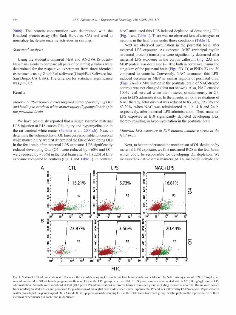
We have previously reported that a single systemic maternalLPS injection at E18 causes OLs injury and hypomyelination inthe rat cerebral white matter (Paintlia et al., 2004a,b). Next, todetermine the vulnerability ofOL lineages responsible for cerebralwhitematter injury, we first determined the fate of developingOLsin the fetal brain after maternal LPS exposure. LPS significantlyreduced developing OLs (O4+ were reduced by ~60% and O1+
were reduced by ~80%) in the fetal brain after 48 h (E20) of LPSexposure compared to controls (Fig. 1 and Table 1). In contrast,
Fig. 1. Maternal LPS administration at E18 causes the loss of developing OLs in the rwas administered to SD rat female pregnant mothers on E18 in the LPS group, wheradministration. Animals were sacrificed at E20 (48 h post LPS-administration) to remfrom similarly treated fetuses and processed for purification of brain glial cells as descscatter plots depict the percentage of O4+ (A) and O1+ (B) population of developing Oidentical experiments run each time in duplicate.
NAC attenuated this LPS-induced depletion of developing OLs(Fig. 1 and Table 1). There was no observed loss of astrocytes orneurons in the fetal brain under those conditions (Table 1).
Next we observed myelination in the postnatal brain aftermaternal LPS exposure. As expected, MBP (principal myelinstructural protein) transcripts were significantly decreased aftermaternal LPS exposure in the corpus callosum (Fig. 2A) andMBP protein was decreased (~10%) both in corpus-callosum andcingulum of the postnatal brain (Figs. 2B–D) at PNDs 23 and 30compared to controls. Conversely, NAC attenuated this LPS-induced decrease in MBP in similar regions of postnatal brain(Figs. 2A–D). Myelination in the postnatal brain of NAC-treatedcontrols was not changed (data not shown). Also, NAC enabled100% fetal survival when administered simultaneously or 2 hprior to LPS administration. In therapeutic window evaluations ofNAC therapy, fetal survival was reduced to 83.30%, 79.20% and63.30%, when NAC was administered at 1 h, 4 h and 24 h,respectively, after maternal LPS administration. Thus, maternalLPS exposure at E18 significantly depleted developing OLs,thereby resulting in hypomyelination in the postnatal brain.
Maternal LPS exposure at E18 induces oxidative-stress in thefetal brain
Next, to better understand the mechanism of OL depletion bymaternal LPS exposure, we first measured ROS in the fetal brainwhich could be responsible for developing OL depletion. Wemeasured oxidative stress markers (MDA, malondialdehyde and
at fetal brain which can be blocked by NAC. An injection of LPS (0.7 mg/kg; ip)eas NAC+LPS group animals were treated with NAC (50 mg/kg) prior to LPSove fetuses from each group including respective controls. Brains were pooled
ribed under Experimental Procedures followed by FACS analysis. RepresentativeLs in the fetal brains from each group. Scatter plots are the representative of three
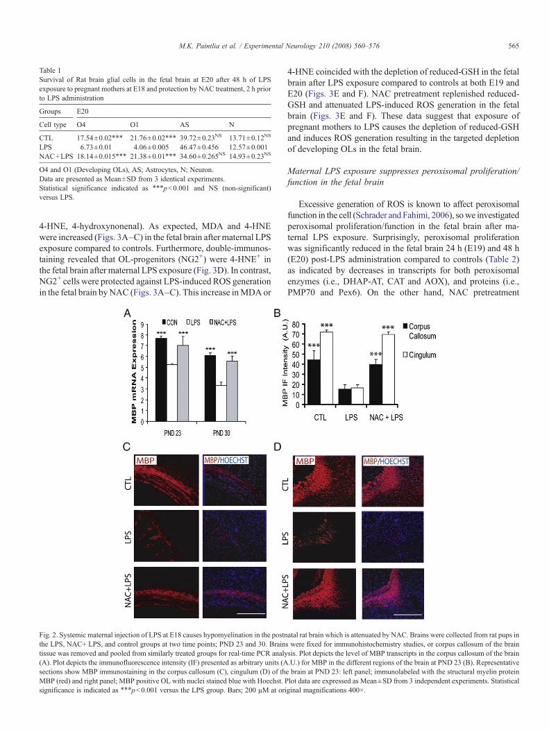
Table 1Survival of Rat brain glial cells in the fetal brain at E20 after 48 h of LPSexposure to pregnant mothers at E18 and protection by NAC treatment, 2 h priorto LPS administration
Groups E20
Cell type O4 O1 AS N
CTL 17.54±0.02⁎⁎⁎ 21.76±0.02⁎⁎⁎ 39.72±0.23NS 13.71±0.12NS
LPS 6.73±0.01 4.06±0.005 46.47±0.456 12.57±0.001NAC+LPS 18.14±0.015⁎⁎⁎ 21.38±0.01⁎⁎⁎ 34.60±0.265NS 14.93±0.23NS
O4 and O1 (Developing OLs), AS; Astrocytes, N; Neuron.Data are presented as Mean±SD from 3 identical experiments.Statistical significance indicated as ⁎⁎⁎pb0.001 and NS (non-significant)versus LPS.
565M.K. Paintlia et al. / Experimental Neurology 210 (2008) 560–576
4-HNE, 4-hydroxynonenal). As expected, MDA and 4-HNEwere increased (Figs. 3A–C) in the fetal brain after maternal LPSexposure compared to controls. Furthermore, double-immunos-taining revealed that OL-progenitors (NG2+) were 4-HNE+ inthe fetal brain after maternal LPS exposure (Fig. 3D). In contrast,NG2+ cells were protected against LPS-induced ROS generationin the fetal brain byNAC (Figs. 3A–C). This increase inMDAor
Fig. 2. Systemic maternal injection of LPS at E18 causes hypomyelination in the postnthe LPS, NAC+ LPS, and control groups at two time points; PND 23 and 30. Braintissue was removed and pooled from similarly treated groups for real-time PCR analy(A). Plot depicts the immunofluorescence intensity (IF) presented as arbitrary units (Asections show MBP immunostaining in the corpus callosum (C), cingulum (D) of thMBP (red) and right panel; MBP positive OL with nuclei stained blue with Hoechst. Psignificance is indicated as ⁎⁎⁎pb0.001 versus the LPS group. Bars; 200 µM at ori
4-HNE coincided with the depletion of reduced-GSH in the fetalbrain after LPS exposure compared to controls at both E19 andE20 (Figs. 3E and F). NAC pretreatment replenished reduced-GSH and attenuated LPS-induced ROS generation in the fetalbrain (Figs. 3E and F). These data suggest that exposure ofpregnant mothers to LPS causes the depletion of reduced-GSHand induces ROS generation resulting in the targeted depletionof developing OLs in the fetal brain.
Maternal LPS exposure suppresses peroxisomal proliferation/function in the fetal brain
Excessive generation of ROS is known to affect peroxisomalfunction in the cell (Schrader andFahimi, 2006), sowe investigatedperoxisomal proliferation/function in the fetal brain after ma-ternal LPS exposure. Surprisingly, peroxisomal proliferationwas significantly reduced in the fetal brain 24 h (E19) and 48 h(E20) post-LPS administration compared to controls (Table 2)as indicated by decreases in transcripts for both peroxisomalenzymes (i.e., DHAP-AT, CAT and AOX), and proteins (i.e.,PMP70 and Pex6). On the other hand, NAC pretreatment
atal rat brain which is attenuated by NAC. Brains were collected from rat pups ins were fixed for immunohistochemistry studies, or corpus callosum of the brainsis. Plot depicts the level of MBP transcripts in the corpus callosum of the brain.U.) for MBP in the different regions of the brain at PND 23 (B). Representativee brain at PND 23: left panel; immunolabeled with the structural myelin proteinlot data are expressed as Mean±SD from 3 independent experiments. Statisticalginal magnifications 400×.
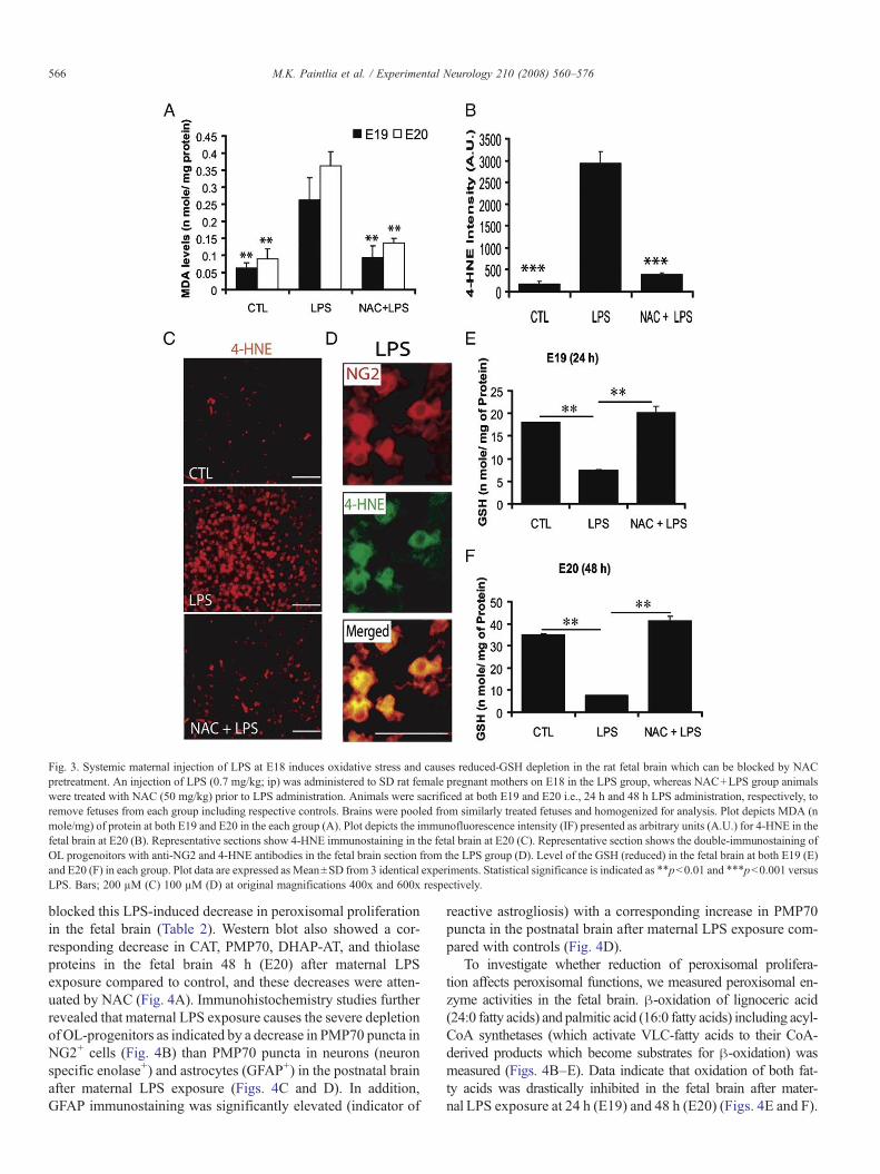
Fig. 3. Systemic maternal injection of LPS at E18 induces oxidative stress and causes reduced-GSH depletion in the rat fetal brain which can be blocked by NACpretreatment. An injection of LPS (0.7 mg/kg; ip) was administered to SD rat female pregnant mothers on E18 in the LPS group, whereas NAC+LPS group animalswere treated with NAC (50 mg/kg) prior to LPS administration. Animals were sacrificed at both E19 and E20 i.e., 24 h and 48 h LPS administration, respectively, toremove fetuses from each group including respective controls. Brains were pooled from similarly treated fetuses and homogenized for analysis. Plot depicts MDA (nmole/mg) of protein at both E19 and E20 in the each group (A). Plot depicts the immunofluorescence intensity (IF) presented as arbitrary units (A.U.) for 4-HNE in thefetal brain at E20 (B). Representative sections show 4-HNE immunostaining in the fetal brain at E20 (C). Representative section shows the double-immunostaining ofOL progenoitors with anti-NG2 and 4-HNE antibodies in the fetal brain section from the LPS group (D). Level of the GSH (reduced) in the fetal brain at both E19 (E)and E20 (F) in each group. Plot data are expressed as Mean±SD from 3 identical experiments. Statistical significance is indicated as ⁎⁎pb0.01 and ⁎⁎⁎pb0.001 versusLPS. Bars; 200 µM (C) 100 µM (D) at original magnifications 400x and 600x respectively.
566 M.K. Paintlia et al. / Experimental Neurology 210 (2008) 560–576
blocked this LPS-induced decrease in peroxisomal proliferationin the fetal brain (Table 2). Western blot also showed a cor-responding decrease in CAT, PMP70, DHAP-AT, and thiolaseproteins in the fetal brain 48 h (E20) after maternal LPSexposure compared to control, and these decreases were atten-uated by NAC (Fig. 4A). Immunohistochemistry studies furtherrevealed that maternal LPS exposure causes the severe depletionof OL-progenitors as indicated by a decrease in PMP70 puncta inNG2+ cells (Fig. 4B) than PMP70 puncta in neurons (neuronspecific enolase+) and astrocytes (GFAP+) in the postnatal brainafter maternal LPS exposure (Figs. 4C and D). In addition,GFAP immunostaining was significantly elevated (indicator of
reactive astrogliosis) with a corresponding increase in PMP70puncta in the postnatal brain after maternal LPS exposure com-pared with controls (Fig. 4D).
To investigate whether reduction of peroxisomal prolifera-tion affects peroxisomal functions, we measured peroxisomal en-zyme activities in the fetal brain. β-oxidation of lignoceric acid(24:0 fatty acids) and palmitic acid (16:0 fatty acids) including acyl-CoA synthetases (which activate VLC-fatty acids to their CoA-derived products which become substrates for β-oxidation) wasmeasured (Figs. 4B–E). Data indicate that oxidation of both fat-ty acids was drastically inhibited in the fetal brain after mater-nal LPS exposure at 24 h (E19) and 48 h (E20) (Figs. 4E and F).
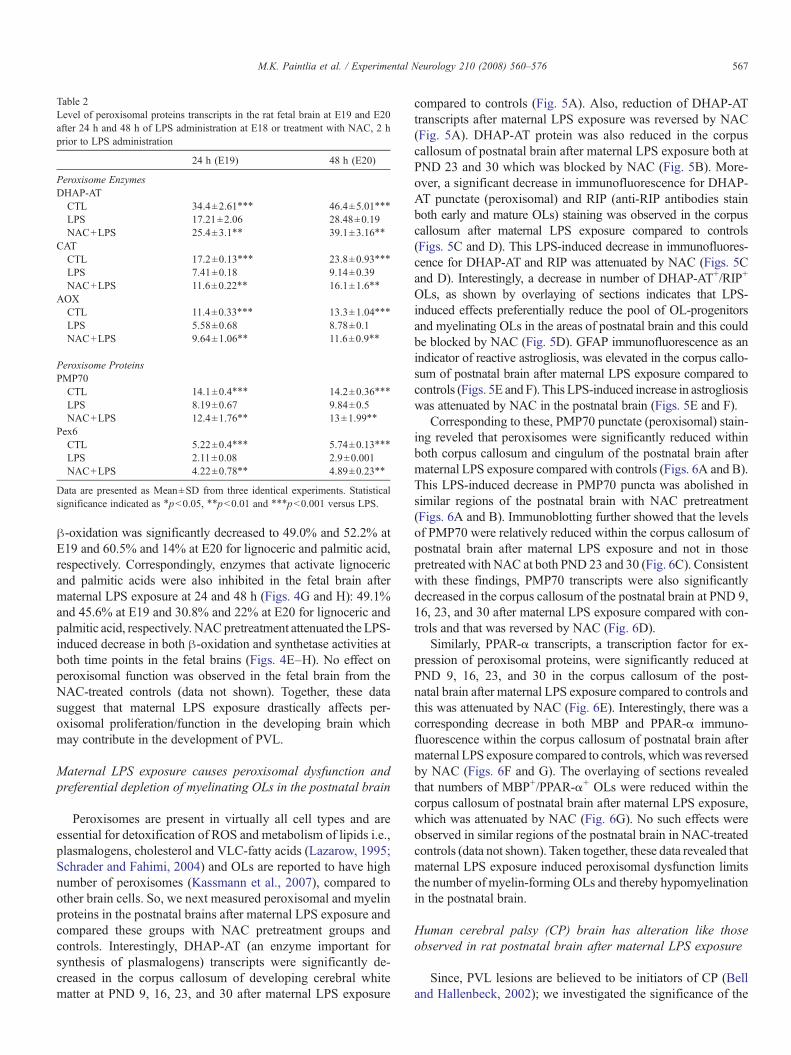
Table 2Level of peroxisomal proteins transcripts in the rat fetal brain at E19 and E20after 24 h and 48 h of LPS administration at E18 or treatment with NAC, 2 hprior to LPS administration
24 h (E19) 48 h (E20)
Peroxisome EnzymesDHAP-AT
CTL 34.4±2.61⁎⁎⁎ 46.4±5.01⁎⁎⁎
LPS 17.21±2.06 28.48±0.19NAC+LPS 25.4±3.1⁎⁎ 39.1±3.16⁎⁎
CATCTL 17.2±0.13⁎⁎⁎ 23.8±0.93⁎⁎⁎
LPS 7.41±0.18 9.14±0.39NAC+LPS 11.6±0.22⁎⁎ 16.1±1.6⁎⁎
AOXCTL 11.4±0.33⁎⁎⁎ 13.3±1.04⁎⁎⁎
LPS 5.58±0.68 8.78±0.1NAC+LPS 9.64±1.06⁎⁎ 11.6±0.9⁎⁎
Peroxisome ProteinsPMP70
CTL 14.1±0.4⁎⁎⁎ 14.2±0.36⁎⁎⁎
LPS 8.19±0.67 9.84±0.5NAC+LPS 12.4±1.76⁎⁎ 13±1.99⁎⁎
Pex6CTL 5.22±0.4⁎⁎⁎ 5.74±0.13⁎⁎⁎
LPS 2.11±0.08 2.9±0.001NAC+LPS 4.22±0.78⁎⁎ 4.89±0.23⁎⁎
Data are presented as Mean±SD from three identical experiments. Statisticalsignificance indicated as ⁎pb0.05, ⁎⁎pb0.01 and ⁎⁎⁎pb0.001 versus LPS.
567M.K. Paintlia et al. / Experimental Neurology 210 (2008) 560–576
β-oxidation was significantly decreased to 49.0% and 52.2% atE19 and 60.5% and 14% at E20 for lignoceric and palmitic acid,respectively. Correspondingly, enzymes that activate lignocericand palmitic acids were also inhibited in the fetal brain aftermaternal LPS exposure at 24 and 48 h (Figs. 4G and H): 49.1%and 45.6% at E19 and 30.8% and 22% at E20 for lignoceric andpalmitic acid, respectively. NACpretreatment attenuated the LPS-induced decrease in both β-oxidation and synthetase activities atboth time points in the fetal brains (Figs. 4E–H). No effect onperoxisomal function was observed in the fetal brain from theNAC-treated controls (data not shown). Together, these datasuggest that maternal LPS exposure drastically affects per-oxisomal proliferation/function in the developing brain whichmay contribute in the development of PVL.
Maternal LPS exposure causes peroxisomal dysfunction andpreferential depletion of myelinating OLs in the postnatal brain
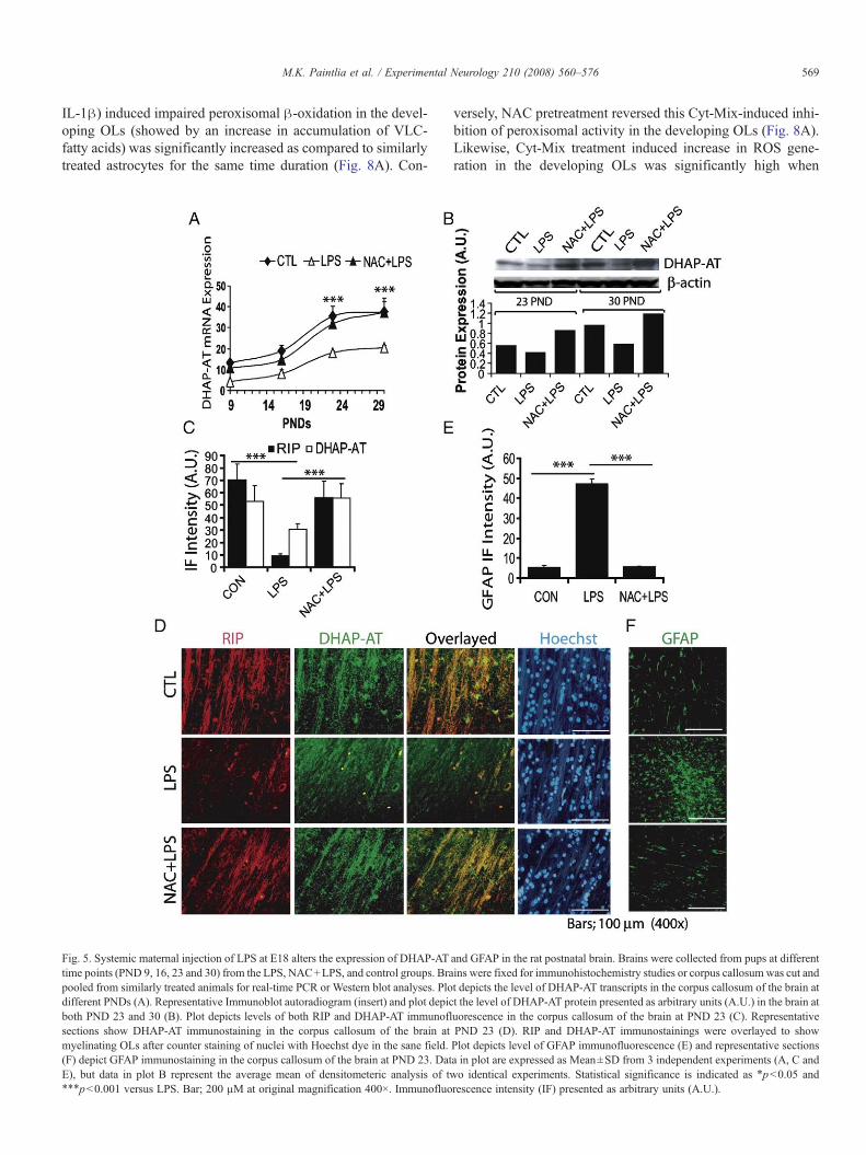
Peroxisomes are present in virtually all cell types and areessential for detoxification of ROS and metabolism of lipids i.e.,plasmalogens, cholesterol and VLC-fatty acids (Lazarow, 1995;Schrader and Fahimi, 2004) and OLs are reported to have highnumber of peroxisomes (Kassmann et al., 2007), compared toother brain cells. So, we next measured peroxisomal and myelinproteins in the postnatal brains after maternal LPS exposure andcompared these groups with NAC pretreatment groups andcontrols. Interestingly, DHAP-AT (an enzyme important forsynthesis of plasmalogens) transcripts were significantly de-creased in the corpus callosum of developing cerebral whitematter at PND 9, 16, 23, and 30 after maternal LPS exposure
compared to controls (Fig. 5A). Also, reduction of DHAP-ATtranscripts after maternal LPS exposure was reversed by NAC(Fig. 5A). DHAP-AT protein was also reduced in the corpuscallosum of postnatal brain after maternal LPS exposure both atPND 23 and 30 which was blocked by NAC (Fig. 5B). More-over, a significant decrease in immunofluorescence for DHAP-AT punctate (peroxisomal) and RIP (anti-RIP antibodies stainboth early and mature OLs) staining was observed in the corpuscallosum after maternal LPS exposure compared to controls(Figs. 5C and D). This LPS-induced decrease in immunofluores-cence for DHAP-AT and RIP was attenuated by NAC (Figs. 5Cand D). Interestingly, a decrease in number of DHAP-AT+/RIP+
OLs, as shown by overlaying of sections indicates that LPS-induced effects preferentially reduce the pool of OL-progenitorsand myelinating OLs in the areas of postnatal brain and this couldbe blocked by NAC (Fig. 5D). GFAP immunofluorescence as anindicator of reactive astrogliosis, was elevated in the corpus callo-sum of postnatal brain after maternal LPS exposure compared tocontrols (Figs. 5E and F). This LPS-induced increase in astrogliosiswas attenuated by NAC in the postnatal brain (Figs. 5E and F).
Corresponding to these, PMP70 punctate (peroxisomal) stain-ing reveled that peroxisomes were significantly reduced withinboth corpus callosum and cingulum of the postnatal brain aftermaternal LPS exposure compared with controls (Figs. 6A and B).This LPS-induced decrease in PMP70 puncta was abolished insimilar regions of the postnatal brain with NAC pretreatment(Figs. 6A and B). Immunoblotting further showed that the levelsof PMP70 were relatively reduced within the corpus callosum ofpostnatal brain after maternal LPS exposure and not in thosepretreatedwith NAC at both PND 23 and 30 (Fig. 6C). Consistentwith these findings, PMP70 transcripts were also significantlydecreased in the corpus callosum of the postnatal brain at PND 9,16, 23, and 30 after maternal LPS exposure compared with con-trols and that was reversed by NAC (Fig. 6D).
Similarly, PPAR-α transcripts, a transcription factor for ex-pression of peroxisomal proteins, were significantly reduced atPND 9, 16, 23, and 30 in the corpus callosum of the post-natal brain after maternal LPS exposure compared to controls andthis was attenuated by NAC (Fig. 6E). Interestingly, there was acorresponding decrease in both MBP and PPAR-α immuno-fluorescence within the corpus callosum of postnatal brain aftermaternal LPS exposure compared to controls, which was reversedby NAC (Figs. 6F and G). The overlaying of sections revealedthat numbers of MBP+/PPAR-α+ OLs were reduced within thecorpus callosum of postnatal brain after maternal LPS exposure,which was attenuated by NAC (Fig. 6G). No such effects wereobserved in similar regions of the postnatal brain in NAC-treatedcontrols (data not shown). Taken together, these data revealed thatmaternal LPS exposure induced peroxisomal dysfunction limitsthe number of myelin-forming OLs and thereby hypomyelinationin the postnatal brain.
Human cerebral palsy (CP) brain has alteration like thoseobserved in rat postnatal brain after maternal LPS exposure
Since, PVL lesions are believed to be initiators of CP (Belland Hallenbeck, 2002); we investigated the significance of the
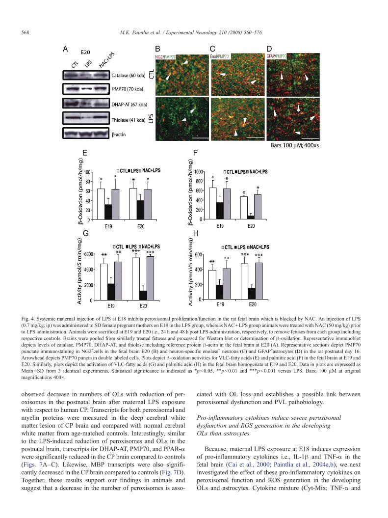
Fig. 4. Systemic maternal injection of LPS at E18 inhibits peroxisomal proliferation/function in the rat fetal brain which is blocked by NAC. An injection of LPS(0.7 mg/kg; ip) was administered to SD female pregnant mothers on E18 in the LPS group, whereas NAC+LPS group animals were treated with NAC (50 mg/kg) priorto LPS administration. Animals were sacrificed at E19 and E20 i.e., 24 h and 48 h post LPS-administration, respectively, to remove fetuses from each group includingrespective controls. Brains were pooled from similarly treated fetuses and processed for Western blot or determination of β-oxidation. Representative immunoblotdepicts levels of catalase, PMP70, DHAP-AT, and thiolase including reference protein β-actin in the fetal brain at E20 (A). Representative sections depict PMP70punctate immunostaining in NG2+cells in the fetal brain E20 (B) and neuron-specific enolase+ neurons (C) and GFAP+astrocytes (D) in the rat postnatal day 16.Arrowhead depicts PMP70 puncta in double labeled cells. Plots depict β-oxidation activities for VLC-fatty acids (E) and palmitic acid (F) in the fetal brain at E19 andE20. Similarly, plots depict the activation of VLC-fatty acids (G) and palmitic acid (H) in the fetal brain homogenate at E19 and E20. Data in plots are expressed asMean±SD from 3 identical experiments. Statistical significance is indicated as ⁎pb0.05, ⁎⁎pb0.01 and ⁎⁎⁎pb0.001 versus LPS. Bars; 100 µM at originalmagnifications 400×.
568 M.K. Paintlia et al. / Experimental Neurology 210 (2008) 560–576
observed decrease in numbers of OLs with reduction of per-oxisomes in the postnatal brain after maternal LPS exposurewith respect to human CP. Transcripts for both peroxisomal andmyelin proteins were measured in the deep cerebral whitematter lesion of CP brain and compared with normal cerebralwhite matter from age-matched controls. Interestingly, similarto the LPS-induced reduction of peroxisomes and OLs in thepostnatal brain, transcripts for DHAP-AT, PMP70, and PPAR-αwere significantly reduced in the CP brain compared to controls(Figs. 7A–C). Likewise, MBP transcripts were also signifi-cantly decreased in the CP brain compared to controls (Fig. 7D).Together, these results support our findings in animals andsuggest that a decrease in the number of peroxisomes is asso-
ciated with OL loss and establishes a possible link betweenperoxisomal dysfunction and PVL pathobiology.
Pro-inflammatory cytokines induce severe peroxisomaldysfunction and ROS generation in the developingOLs than astrocytes
Because, maternal LPS exposure at E18 induces expressionof pro-inflammatory cytokines i.e., IL-1β and TNF-α in thefetal brain (Cai et al., 2000; Paintlia et al., 2004a,b), we nextinvestigated the effect of these pro-inflammatory cytokines onperoxisomal function and ROS generation in the developingOLs and astrocytes. Cytokine mixture (Cyt-Mix; TNF-α and
569M.K. Paintlia et al. / Experimental Neurology 210 (2008) 560–576
IL-1β) induced impaired peroxisomal β-oxidation in the devel-oping OLs (showed by an increase in accumulation of VLC-fatty acids) was significantly increased as compared to similarlytreated astrocytes for the same time duration (Fig. 8A). Con-
Fig. 5. Systemic maternal injection of LPS at E18 alters the expression of DHAP-ATtime points (PND 9, 16, 23 and 30) from the LPS, NAC+LPS, and control groups. Brapooled from similarly treated animals for real-time PCR or Western blot analyses. Plodifferent PNDs (A). Representative Immunoblot autoradiogram (insert) and plot depicboth PND 23 and 30 (B). Plot depicts levels of both RIP and DHAP-AT immunoflsections show DHAP-AT immunostaining in the corpus callosum of the brain atmyelinating OLs after counter staining of nuclei with Hoechst dye in the sane field.(F) depict GFAP immunostaining in the corpus callosum of the brain at PND 23. DatE), but data in plot B represent the average mean of densitometeric analysis of tw⁎⁎⁎pb0.001 versus LPS. Bar; 200 µM at original magnification 400×. Immunofluo
versely, NAC pretreatment reversed this Cyt-Mix-induced inhi-bition of peroxisomal activity in the developing OLs (Fig. 8A).Likewise, Cyt-Mix treatment induced increase in ROS gene-ration in the developing OLs was significantly high when
and GFAP in the rat postnatal brain. Brains were collected from pups at differentins were fixed for immunohistochemistry studies or corpus callosum was cut andt depicts the level of DHAP-AT transcripts in the corpus callosum of the brain att the level of DHAP-AT protein presented as arbitrary units (A.U.) in the brain atuorescence in the corpus callosum of the brain at PND 23 (C). RepresentativePND 23 (D). RIP and DHAP-AT immunostainings were overlayed to showPlot depicts level of GFAP immunofluorescence (E) and representative sectionsa in plot are expressed as Mean±SD from 3 independent experiments (A, C ando identical experiments. Statistical significance is indicated as ⁎pb0.05 andrescence intensity (IF) presented as arbitrary units (A.U.).

570 M.K. Paintlia et al. / Experimental Neurology 210 (2008) 560–576
compared with similarly treated astrocytes (Fig. 8B). NACpretreatment reversed this Cyt-Mix induced ROS generation inboth developing OLs and astrocytes (Fig. 8B). Of note, simi-
Fig. 6. Systemic maternal injection of LPS at E18 alters the expression of PMP70, Pdifferent time points PND 9, 16, 23 and 30) from the LPS, NAC+LPS, and control grthe brain was cut and pooled from similarly treated groups for real-time PCR or Westethe different regions of the brain at 23 PNDs (A). Plot depicts the level of PMP70 iautoradiogram shows the quantitative expression of PMP70 presented as arbitrary unand 30 (C). Plots depict the level of PMP70 (D) and PPAR-a(E) transcript in the corpucallosum of the brain at PND 23 (F). Representative sections demonstrate double-imarbitrary units (A.U.) in the corpus callosum of the brain at PND 23 (G). Overlayiexpressed as Mean±SD from 3 identical experiments. Statistical significance is indic
larly treated astrocytesN 96 h showed impaired peroxisomal β-oxidation activity and severe ROS generation comparable todeveloping OLs treated for 48 h (data not shown). These data
PAR-α and MBP in the rat postnatal brain. Brains were collected from pups atoups. Brains were fixed for immunohistochemistry studies or corpus callosum ofrn blot analyses. Representative sections demonstrate PMP70 immunostaining inn the different regions of the brain at PND 23 (B). Representative immunoblotits (A.U.) with respect to β-actin in the corpus callosum of the brain at PND 23s callosum of the brain. Plot depicts both MBP and PPAR-α protein in the corpusmunostaining for MBP/PPAR-α immunofluorescence intensity (IF) presented asng of sections shows expression of PPAR-α in myelinating OLs. Plot data areated as ⁎⁎⁎pb0.001 versus LPS. Bars; 200 µM at original magnification 400×.
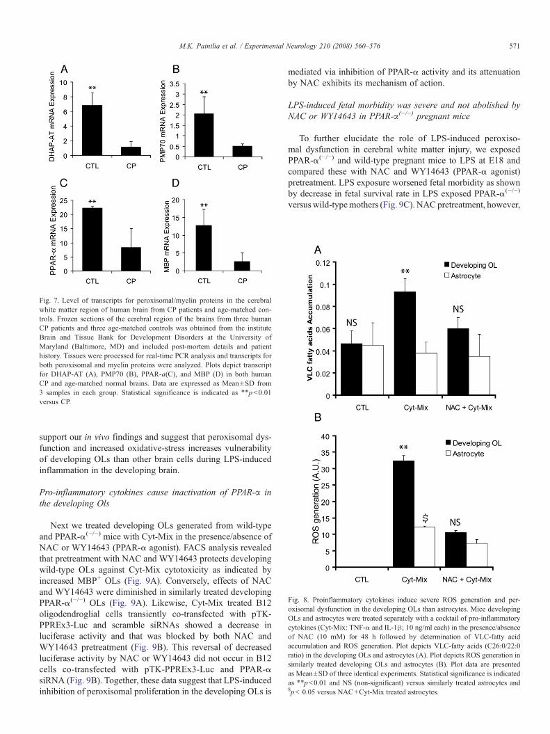
Fig. 7. Level of transcripts for peroxisomal/myelin proteins in the cerebralwhite matter region of human brain from CP patients and age-matched con-trols. Frozen sections of the cerebral region of the brains from three humanCP patients and three age-matched controls was obtained from the instituteBrain and Tissue Bank for Development Disorders at the University ofMaryland (Baltimore, MD) and included post-mortem details and patienthistory. Tissues were processed for real-time PCR analysis and transcripts forboth peroxisomal and myelin proteins were analyzed. Plots depict transcriptfor DHAP-AT (A), PMP70 (B), PPAR-a(C), and MBP (D) in both humanCP and age-matched normal brains. Data are expressed as Mean±SD from3 samples in each group. Statistical significance is indicated as ⁎⁎pb0.01versus CP.
Fig. 8. Proinflammatory cytokines induce severe ROS generation and per-oxisomal dysfunction in the developing OLs than astrocytes. Mice developingOLs and astrocytes were treated separately with a cocktail of pro-inflammatorycytokines (Cyt-Mix: TNF-α and IL-1β; 10 ng/ml each) in the presence/absenceof NAC (10 mM) for 48 h followed by determination of VLC-fatty acidaccumulation and ROS generation. Plot depicts VLC-fatty acids (C26:0/22:0ratio) in the developing OLs and astrocytes (A). Plot depicts ROS generation insimilarly treated developing OLs and astrocytes (B). Plot data are presentedas Mean±SD of three identical experiments. Statistical significance is indicatedas ⁎⁎pb0.01 and NS (non-significant) versus similarly treated astrocytes and$pb 0.05 versus NAC+Cyt-Mix treated astrocytes.
571M.K. Paintlia et al. / Experimental Neurology 210 (2008) 560–576
support our in vivo findings and suggest that peroxisomal dys-function and increased oxidative-stress increases vulnerabilityof developing OLs than other brain cells during LPS-inducedinflammation in the developing brain.
Pro-inflammatory cytokines cause inactivation of PPAR-α inthe developing Ols
Next we treated developing OLs generated from wild-typeand PPAR-α(−/−) mice with Cyt-Mix in the presence/absence ofNAC or WY14643 (PPAR-α agonist). FACS analysis revealedthat pretreatment with NAC and WY14643 protects developingwild-type OLs against Cyt-Mix cytotoxicity as indicated byincreased MBP+ OLs (Fig. 9A). Conversely, effects of NACand WY14643 were diminished in similarly treated developingPPAR-α(−/−) OLs (Fig. 9A). Likewise, Cyt-Mix treated B12oligodendroglial cells transiently co-transfected with pTK-PPREx3-Luc and scramble siRNAs showed a decrease inluciferase activity and that was blocked by both NAC andWY14643 pretreatment (Fig. 9B). This reversal of decreasedluciferase activity by NAC or WY14643 did not occur in B12cells co-transfected with pTK-PPREx3-Luc and PPAR-αsiRNA (Fig. 9B). Together, these data suggest that LPS-inducedinhibition of peroxisomal proliferation in the developing OLs is
mediated via inhibition of PPAR-α activity and its attenuationby NAC exhibits its mechanism of action.
LPS-induced fetal morbidity was severe and not abolished byNAC or WY14643 in PPAR-α(−/−) pregnant mice
To further elucidate the role of LPS-induced peroxiso-mal dysfunction in cerebral white matter injury, we exposedPPAR-α(−/−) and wild-type pregnant mice to LPS at E18 andcompared these with NAC and WY14643 (PPAR-α agonist)pretreatment. LPS exposure worsened fetal morbidity as shownby decrease in fetal survival rate in LPS exposed PPAR-α(−/−)
versuswild-typemothers (Fig. 9C). NACpretreatment, however,
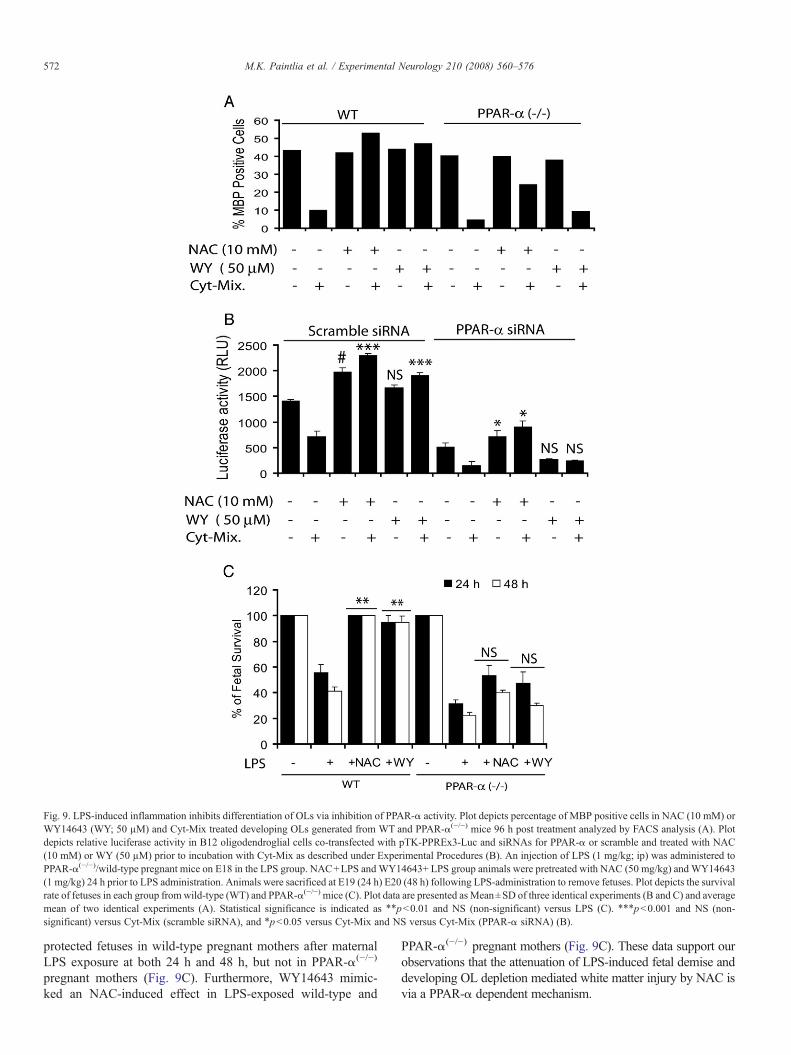
Fig. 9. LPS-induced inflammation inhibits differentiation of OLs via inhibition of PPAR-α activity. Plot depicts percentage of MBP positive cells in NAC (10 mM) orWY14643 (WY; 50 µM) and Cyt-Mix treated developing OLs generated from WT and PPAR-α(−/−) mice 96 h post treatment analyzed by FACS analysis (A). Plotdepicts relative luciferase activity in B12 oligodendroglial cells co-transfected with pTK-PPREx3-Luc and siRNAs for PPAR-α or scramble and treated with NAC(10 mM) or WY (50 µM) prior to incubation with Cyt-Mix as described under Experimental Procedures (B). An injection of LPS (1 mg/kg; ip) was administered toPPAR-α(−/−)/wild-type pregnant mice on E18 in the LPS group. NAC+LPS andWY14643+ LPS group animals were pretreated with NAC (50 mg/kg) and WY14643(1 mg/kg) 24 h prior to LPS administration. Animals were sacrificed at E19 (24 h) E20 (48 h) following LPS-administration to remove fetuses. Plot depicts the survivalrate of fetuses in each group from wild-type (WT) and PPAR-α(−/−) mice (C). Plot data are presented as Mean±SD of three identical experiments (B and C) and averagemean of two identical experiments (A). Statistical significance is indicated as ⁎⁎pb0.01 and NS (non-significant) versus LPS (C). ⁎⁎⁎pb0.001 and NS (non-significant) versus Cyt-Mix (scramble siRNA), and ⁎pb0.05 versus Cyt-Mix and NS versus Cyt-Mix (PPAR-α siRNA) (B).
572 M.K. Paintlia et al. / Experimental Neurology 210 (2008) 560–576
protected fetuses in wild-type pregnant mothers after maternalLPS exposure at both 24 h and 48 h, but not in PPAR-α(−/−)
pregnant mothers (Fig. 9C). Furthermore, WY14643 mimic-ked an NAC-induced effect in LPS-exposed wild-type and
PPAR-α(−/−) pregnant mothers (Fig. 9C). These data support ourobservations that the attenuation of LPS-induced fetal demise anddeveloping OL depletion mediated white matter injury by NAC isvia a PPAR-α dependent mechanism.
573M.K. Paintlia et al. / Experimental Neurology 210 (2008) 560–576
Discussion
Maternal LPS exposure has been shown to stimulate thesecretion of pro-inflammatory cytokines i.e., TNF-α, IL-1α andIL-6 in the maternal serum and amniotic fluid of pregnant mice(Fidel et al., 1994) mimicking maternal infection (Yoon et al.,1997). Results presented here describe the role of peroxisomesin prenatal maternal infection (LPS)-induced cerebral whitematter injury. Our conclusions are based upon the followingobservations. First, maternal LPS exposure at E18 causes ROSgeneration (MDA and 4-HNE) in the fetal brain thereby causingtargeted injury of developing OLs with relative sparing of otherglial cells (Table 1 and Figs. 1A–B and 3A–F). Second, mater-nal LPS exposure inhibits peroxisomal proliferation/function inthe fetal brain (Table 2 and Figs. 4A–E). Third, maternal LPSexposure induced effects persist in the later life of offspring asexhibited by the decrease in peroxisomal (Figs. 5A–Dand 6A–G)and myelin (Figs. 2A–D) proteins in the corpus-callosum andcingulum of the postnatal brain. These LPS-induced effects inthe developing rat brain were abolished by NAC pretreatment.Like the postnatal brain, alterations in peroxisomal and myelinproteins were apparent in the cerebral white matter of human CPbrain (Figs. 7A–D). Proinflammatory cytokines induced severeperoxisomal dysfunction and increased oxidative stress in thedeveloping OL when compared with similarly treated astrocytes(Figs. 8A–D). In addition, experiments with pro-inflammatorycytokine-exposed and NAC-or WY14643-treated developingPPAR-α(−/−)/wild-type OLs as well as transiently co-transfectedB12 OLs (with anti-PPAR-α SiRNA and reporter plasmids)revealed that NACmaintains peroxisomal proliferation/functionin the developing OLs via a PPAR-α dependent mechanism(Figs. 9A–B). These findings were further supported by LPSexposure and NAC or WY14643 treatment of pregnant PPAR-α(−/−) and wild-type pregnant mice (Fig. 9C). These findingsdescribe biochemical effects of maternal LPS-induced exacer-bation of cerebral white matter injury by peroxisomal dysfunc-tion and preferential depletion of OL-progenitor pool in thebrain. To our knowledge, this is the first report implicatingperoxisomes in prenatal maternal infection (LPS)-induced cere-bral white matter injury.
The pathogenesis of PVL appears to be multi-factorial. Hy-poxia/ischemia and maternal–fetal infections are two importantfactors. Pro-inflammatory cytokines such as IL-1 and TNF-αand brain hypoxia/ischemia are shown to be responsible for theloss of immature OLs in PVL (Levison et al., 2001; Pang et al.,2005). The majority of PVL lesions occur between 23–32 weekspost conception (Back et al., 2001; Volpe, 2001; Back, 2006), atime when the predominant population of the OL lineage con-sists of late OL progenitors (O4+) in the human fetal brain (Backet al., 2001). Our results showed a significant decrease in O4+
(~60%) and O1+ (~80%) cells in the fetal brain at E20 aftermaternal LPS exposure, versus controls with a relative sparing ofastrocytes and neurons (Fig. 1 and Table 1). In addition, OL-progenitors were showed to have high oxidative-stress asshowed by 4-HNE+/NG2+ cells in the fetal brain at E20 afterLPS exposure (Fig. 3D). This suggests that LPS-induced se-cretion of factors by brain cells causes the targeted injury of
developing OLs which is consistent with hypoxia/ischemiastudymodels (Back et al., 2001; Levison et al., 2001). Consistentwith our earlier study, which showed that an increase in PDGF-αR+/TUNEL+ OL-progenitors is corresponded with decreasedO4+ and O1+ cells in the fetal brain at E20 after maternal LPSexposure (Paintlia et al., 2004a,b). Concurrent with loss of OLprogenitors, an observed 10% MBP immunostaining in thepostnatal brain is probably attributed to the left over developingOLs that are not depleted by LPS maternal exposure (Fig. 2).
Several lines of evidence suggest that free radical injury tothe developing OLs underlies (at least in part) the pathogenesisof PVL including hypomyelination seen in long-term survivorsof preterm labor (Haynes et al., 2005). In the human CP brain,the presence of free radical injury is supported by the existenceof both oxidative and nitrative stress markers of lipid peroxi-dation and nitrosylation/nitration, respectively (Haynes et al.,2003). LPS has been shown to cause the depletion of intra-cellular GSH and an increase in MDA, a byproduct of lipidperoxidation (Millan-Plano et al., 2003; Topal et al., 2004). ThisLPS-induced oxidative-stress and lipid peroxidation leads to theproduction of potentially toxic aldehydes such as 4-HNE in thecell (Siems et al., 1992). 4-HNE has been reported to causecellular damage mainly by modification of intracellular proteins(Toyokuni et al., 1994; Uchida, 2003). In agreement with these,we observed an increase in level of MDA and 4-HNE with acorresponding decrease in reduced-GSH levels in the fetal brainafter maternal LPS exposure (Fig. 3) suggesting that LPS-induced maternal pro-inflammatory cytokines or LPS itselfactivate microglia in the fetal brain (Haynes et al., 2005). Thepresence of microglia has been demonstrated in the rat brainstarting from E17 or E18 (Dalmau et al., 1997). Activation ofmicroglia in the fetal brain triggers events associated withreactive astrogliosis which depletes the number of myelin-forming OLs leading to hypomyelination, in the postnatal brain.In support to this, we observed co-localization of 4-HNE+/NG2+ in OL progenitors in the fetal brain at E20 after LPSexposure (Figs. 3C–D) which corresponds with the decrease inpool of O4+ (late OL-progenitors) and O1+ (immature OLs)cells (Fig. 1 and Table 1) in the fetal brain. These findingssuggest that LPS-induced activation of fetal brain glial cellsresults in oxidative-stress in OL-progenitors, leading to theirtargeted injury (Back et al., 2001; Levison et al., 2001).
Reactive astrogliosis is the hallmark of neuroinflammationinduced by secretion of TNF-α and IL-1β as observed in theneonatal brain after a stab injury (Balasingam et al., 1994).Consistent with this report, we observed an increase in astro-gliosis and hypomyelination in the corpus callosum at PND 23following maternal LPS exposure (Figs. 2 and 5F). Supportingthis result, clinical studies have also shown that PVL is closelyassociated with astrogliosis in the white matter lesions of infantbrains (Deguchi et al., 1997), suggesting that astrogliosis isimportant in the cerebral white matter injury at the later age ofoffspring than previously thought: the previous hypothesis wasthat the vulnerability window of immature (O4+/O1−) OLs wasbetween 23–32 weeks of gestation (Volpe, 2001; Back, 2006).Interestingly, astrogliosis in the cerebral white matter was ac-companied by reduction of DHAP-AT and OLs in the corpus

574 M.K. Paintlia et al. / Experimental Neurology 210 (2008) 560–576
callosum of the postnatal brain (Fig. 5). DHAP-AT is an im-portant enzyme required for synthesis of plasmalogens (Purdueet al., 1997), which are strong antioxidant phospholipids im-portant for scavenging ROS and preventing cell membraneoxidation (Maeba and Ueta, 2003). Our study suggests that adecrease in DHAP-AT level may reduce plasmalogens biosyn-thesis, which likely increases the vulnerability of late OL-progenitors to oxidative-stress. Consequently, reduced DHAP-AT level decreases the number of myelin-forming OLs resultingin cerebral white matter injury in premature infants. Of note,DHAP-AT deficiency has been reported to cause abnormalmyelination, causing developmental delay (Sztriha et al., 1997)and growth failure in young children (Elias et al., 1998).
Interestingly, no significant loss of astrocytes and neuronswas observed in the fetal brain after maternal LPS exposure(Table 1) suggesting that brain cells, but not OLs, have stronganti-oxidant systems. In line with these findings, in vitro studieshave shown that TNF-α and IL-1β exposure cause severeperoxisomal dysfunction and oxidative-stress in the developingOLs as compared to astrocytes when treated similarly for sametime duration and that was attenuated by NAC (Fig. 8). Simi-larly, brain glial cells treated with antioxidants have less ROSgeneration and their cytotoxicity on OLs (Singh et al., 1998;Bahat-Stroomza et al., 2005). The observed LPS-induced ~60%reduction of O4+ cells in the fetal brain (Table 1) was due toinflammation-induced oxidative stress in OL-progenitors i.e.,NG2+/4-HNE+ (Fig. 3D), which occur along with the depletionof MBP+ OLs in the postnatal brain at PND 23 and PND 30(Fig. 2). At this point, it is still an open question whetherperoxisomal defect occurs in the developing OLs and causes celldeath or peroxisomal defect is the consequence of cell death. Anindirect evidence of impaired β-oxidation and accumulationof VLC-fatty acids in brains of X-adrenoleukodystrophy andKrabbe's patients suggested that peroxisomal dysfunctions areresponsible for OL loss and demyelination (Singh, 1997; Paintliaet al., 2003; Khan et al., 2005). In addition, deficiency of per-oxisomal enzyme DHAP-AT has been shown to cause abnormalmyelination in youngest siblings (Sztriha et al., 1997). Recentstudy shows that impaired peroxisomal biogenesis in OLs isassociated with axonal loss, demyelination and neuroinflamma-tion in the adult brain suggesting that peroxisomes are vital in OLdevelopment (Kassmann et al., 2007). Future studies will bedirected to distinguish between the cell death of mature OLs andOL-progenitors from peroxisomal dysfunction in cells that arenot depleted by LPS exposure.
Peroxisomal dysfunction has been shown to contribute to thegeneration of ROS in the cell. Defects in peroxisomal func-tioning can exacerbate demyelination in the brain (Paintlia et al.,2003), resulting in neurological deficits in animals (Elias et al.,1998). Likewise, neuroinflammation has been shown to causeperoxisomal dysfunction and demyelination in acute experi-mental autoimmune encephalomyelitis model (Singh et al.,2004). Our findings suggest that LPS-induced oxidative-stress,in part, is contributed by peroxisomal dysfunction which in-creases the vulnerability of the developing OLs to targetedinjury, perhaps due to their high lipid environment and a de-crease in synthesis of plasmalogens. In addition, oxidative
stress-induced decrease in GSH-peroxidase and an increase iniron release has been shown to increase the vulnerability of OLs(Hemdan and Almazan, 2007). LPS-induced oxidative-stressincreases the activation of microglia and astrocytes, albeit per-oxisomal functioning remains normal, likely due to the avail-ability of strong endogenous antioxidant-systems in these cells(Min et al., 2006). Notably, we observed that LPS exposureprofoundly affects both peroxisomal and mitochondrial β-oxidation in the fetal brain as shown by down-regulation ofVLC acyl-CoA ligase (peroxisomes) and long-chain acyl-CoAligase present both in peroxisomes and mitochondria, especiallyat E20. This is suggesting that maternal LPS exposure affectsmore cellular processes than just peroxisomes possibly dueto the global effects on cellular functions as a result of LPS-induced neuroinflammation and ROS generation leading tomitochondrial dysfunction (Goossens et al., 1995) and apoptoticdeath (Fiers et al., 1995) in the fetal brain.
The major function of peroxisomes in the myelin-formingOLs is the metabolism of ROS and myelin lipids (Sztriha et al.,1997; Paintlia et al., 2003). Proliferation of peroxisomes isregulated by PPAR-α in combination with the retinoic acidreceptor by acting on PPREs present in the promoter region ofmost of the genes of peroxisomal proteins (Qi et al., 2000).Increases in ROS generation and NF-κB activation have beenshown to inhibit the expression of both AOX and PPAR-α thusperoxisomal proliferation in skeletal muscle cells which contri-butes to cardiac hypertrophy (Cabrero et al., 2002; Cabreroet al., 2003). In line with these findings, WY14643 has beenshown to protect hippocampal neurons against β-amyloidpeptide induced degenerative changes in Alzheimer disease viaperoxisomal proliferation (Santos et al., 2005). Mechanistically,NAC replenishes the level of reduced-GSH and thereby pro-vides protection by quenching ROS in the fetal brain aftermaternal LPS exposure. In addition, anti-inflammatory effectsof NAC are attributed to the suppression of pro-inflammatorycytokine expression/release (Tsuji et al., 1999), adhesion mole-cule expression and activation of NF-κB in endothelial cells(Rahman et al., 1998), and activation of glial cells (Moynaghet al., 1994). The underlying detailed mechanism of NAC-induced restoration of peroxisomal proliferation/function in thedeveloping OLs via a PPAR-α dependent mechanism is cur-rently under investigation in our laboratory.
In summary, these studies reveal that prenatal maternal in-fection (LPS) induced inflammation mediated inhibition ofperoxisomal dysfunction in fetal brain contributes significantlyto OL injury and thereby exacerbates cerebral white matter injury(hypomyelination) in later life of the offspring. On the other hand,abnormal peroxisomal biogenesis inOLs has been shown to causeaxonal loss, reactive astrogliosis, inflammation and demyelina-tion in the adult brain (Kassmann et al., 2007). More detailedinvestigations are warranted to distinguish betweenOL injury dueto peroxisomal dysfunction and impaired peroxisomal biogenesis,but both studies signify the role of peroxisomes in OL dev-elopment. NAC pretreatment attenuates this LPS-inducedcerebral white matter injury by replenishment of reduced-GSH,ROS quenching and maintenance of peroxisomal proliferation/function via a PPAR-α dependent mechanism. Together, these
575M.K. Paintlia et al. / Experimental Neurology 210 (2008) 560–576
data show that LPS-induced peroxisomal dysfunction exacerbatescerebral white matter injury in the developing brain and suggestsnew therapeutic interventions to prevent the debilitating effects ofprenatal maternal infections.
Acknowledgments
We thank all members of our laboratory for their valuablecomments and help during the course of this study. We thankespecially Dr. Jennifer G. Schnellmann for her critical readingof this manuscript and Ms. Joyce Brian and Ms. Carrie Barnesfor their technical assistance. This study was supported in partby grants from the National Institutes of Health: NS-22576, NS-34741, NS-37766, NS-40810 and, C06 RR018823 and C06RR015455 from the Extramural Research Facilities Program ofthe National Center fro Research Resources.
References
Back, S.A., 2006. Perinatal white matter injury: the changing spectrum ofpathology and emerging insights into pathogeneticmechanisms.Ment. Retard.Dev. Disabil. Res. Rev. 12, 129–140.
Back, S.A., Rivkees, S.A., 2004. Emerging concepts in periventricular whitematter injury. Semin. Perinatol. 28, 405–414.
Back, S.A., Luo, N.L., Borenstein, N.S., Levine, J.M., Volpe, J.J., Kinney, H.C.,2001. Late oligodendrocyte progenitors coincide with the developmentalwindow of vulnerability for human perinatal white matter injury. J. Neurosci.21, 1302–1312.
Back, S.A., Han, B.H., Luo, N.L., Chricton, C.A., Xanthoudakis, S., Tam, J.,Arvin, K.L., Holtzman, D.M., 2002. Selective vulnerability of late oligo-dendrocyte progenitors to hypoxia-ischemia. J. Neurosci. 22, 455–463.
Bahat-Stroomza, M., Gilgun-Sherki, Y., Offen, D., Panet, H., Saada, A., Krool-Galron, N., Barzilai, A., Atlas, D., Melamed, E., 2005. A novel thiol anti-oxidant that crosses the blood brain barrier protects dopaminergic neurons inexperimental models of Parkinson's disease. Eur. J. Neurosci. 21, 637–646.
Balasingam, V., Tejada-Berges, T., Wright, E., Bouckova, R., Yong, V.W., 1994.Reactive astrogliosis in the neonatal mouse brain and its modulation bycytokines. J. Neurosci. 14, 846–856.
Baumgart, E., Vanhorebeek, I., Grabenbauer, M., Borgers, M., Declercq, P.E.,Fahimi, H.D., Baes, M., 2001. Mitochondrial alterations caused by defectiveperoxisomal biogenesis in a mouse model for Zellweger syndrome (PEX5knockout mouse). Am. J. Pathol. 159, 1477–1494.
Bell, M.J., Hallenbeck, J.M., 2002. Effects of intrauterine inflammation ondeveloping rat brain. J. Neurosci. Res. 70, 570–579.
Bell, M.J., Hallenbeck, J.M., Gallo, V., 2004. Determining the fetal inflam-matory response in an experimental model of intrauterine inflammation inrats. Pediatr. Res. 56, 541–546.
Cabrero, A., Alegret, M., Sanchez, R.M., Adzet, T., Laguna, J.C., Carrera, M.V.,2002. Increased reactive oxygen species production down-regulates per-oxisome proliferator-activated alpha pathway in C2C12 skeletal musclecells. J. Biol. Chem. 277, 10100–10107.
Cabrero, A., Merlos, M., Laguna, J.C., Carrera, M.V., 2003. Down-regulation ofacyl-CoA oxidase gene expression and increased NF-kappaB activity inetomoxir-induced cardiac hypertrophy. J. Lipid. Res. 44, 388–398.
Cai, Z., Pan, Z.L., Pang, Y., Evans, O.B., Rhodes, P.G., 2000. Cytokineinduction in fetal rat brains and brain injury in neonatal rats after maternallipopolysaccharide administration. Pediatr. Res. 47, 64–72.
Dalmau, I., Finsen, B., Tonder, N., Zimmer, J., Gonzalez, B., Castellano, B.,1997. Development of microglia in the prenatal rat hippocampus. J. Comp.Neurol. 377, 70–84.
Dammann, O., Leviton, A., 1997. Maternal intrauterine infection, cytokines, andbrain damage in the preterm newborn. Pediatr. Res. 42, 1–8.
Dammann, O., Leviton, A., 2000. Role of the fetus in perinatal infection andneonatal brain damage. Curr. Opin. Pediatr. 12, 99–104.
Deguchi, K., Oguchi, K., Takashima, S., 1997. Characteristic neuropathology ofleukomalacia in extremely low birth weight infants. Pediatr. Neurol. 16,296–300.
Deplanque, D., Gele, P., Petrault, O., Six, I., Furman, C., Bouly, M., Nion, S.,Dupuis, B., Leys, D., Fruchart, J.C., Cecchelli, R., Staels, B., Duriez, P.,Bordet, R., 2003. Peroxisome proliferator-activated receptor-alpha activa-tion as a mechanism of preventive neuroprotection induced by chronicfenofibrate treatment. J. Neurosci. 23, 6264–6271.
Elias, E.R., Mobassaleh, M., Hajra, A.K., Moser, A.B., 1998. Developmental delayand growth failure caused by a peroxisomal disorder, dihydroxyacetonepho-sphate acyltransferase (DHAP-AT) deficiency.Am. J.Med.Genet. 80, 223–226.
Fidel Jr., P.L., Romero, R., Wolf, N., Cutright, J., Ramirez, M., Araneda, H.,Cotton,D.B., 1994. Systemic and local cytokine profiles in endotoxin-inducedpreterm parturition in mice. Am. J. Obstet. Gynecol. 170, 1467–1475.
Fiers, W., Beyaert, R., Boone, E., Cornelis, S., Declercq, W., Decoster, E.,Denecker, G., Depuydt, B., De Valck, D., De Wilde, G., Goossens, V.,Grooten, J., Haegeman, G., Heyninck, K., Penning, L., Plaisance, S., Van-compernolle, K., Van Criekinge, W., Vandenabeele, P., Vanden Berghe, W.,Van de Craen, M., Vandevoorde, V., Vercammen, D., 1995. TNF-inducedintracellular signaling leading to gene induction or to cytotoxicity by nec-rosis or by apoptosis. J. Inflamm. 47, 67–75.
Goossens, V., Grooten, J., De Vos, K., Fiers, W., 1995. Direct evidence for tumornecrosis factor-induced mitochondrial reactive oxygen intermediates and theirinvolvement in cytotoxicity. Proc. Natl. Acad. Sci. U. S. A. 92, 8115–8119.
Haynes, R.L., Folkerth, R.D., Keefe, R.J., Sung, I., Swzeda, L.I., Rosenberg,P.A., Volpe, J.J., Kinney, H.C., 2003. Nitrosative and oxidative injury topremyelinating oligodendrocytes in periventricular leukomalacia. J. Neuro-pathol. Exp. Neurol. 62, 441–450.
Haynes, R.L., Baud, O., Li, J., Kinney, H.C., Volpe, J.J., Folkerth, D.R., 2005.Oxidative and nitrative injury in periventricular leukomalacia: a review.Brain Pathol. 15, 225–233.
Hemdan, S., Almazan, G., 2007. Deficient peroxide detoxification underlies thesusceptibility of oligodendrocyte progenitors to dopamine toxicity. Neuro-pharmacology 52, 1385–1395.
Jatana, M., Singh, I., Singh, A.K., Jenkins, D., 2006. Combination of systemichypothermia and N-acetylcysteine attenuates hypoxic-ischemic brain injuryin neonatal rats. Pediatr. Res. 59, 684–689.
Kassmann, C.M., Lappe-Siefke, C., Baes, M., Brugger, B., Mildner, A., Werner,H.B., Natt, O., Michaelis, T., Prinz, M., Frahm, J., Nave, K.A., 2007. Axonalloss and neuroinflammation caused by peroxisome-deficient oligodendro-cytes. Nat. Genet. 39, 969–976.
Khan,M.,Contreras,M., Singh, I., 2000. Endotoxin-induced alterations of lipid andfatty acid compositions in rat liver peroxisomes. J. Endotoxin Res. 6, 41–50.
Khan, M., Haq, E., Giri, S., Singh, I., Singh, A.K., 2005. Peroxisomal par-ticipation in psychosine-mediated toxicity: implications for Krabbe's dis-ease. J. Neurosci. Res. 80, 845–854.
Khan, M., Sekhon, B., Jatana, M., Giri, S., Gilg, A.G., Sekhon, C., Singh, I.,Singh, A.K., 2004. Administration of N-acetylcysteine after focal cerebralischemia protects brain and reduces inflammation in a rat model of experi-mental stroke. J. Neurosci. Res 76, 519–527.
Kinney, H.C., Back, S.A., 1998. Human oligodendroglial development: relation-ship to periventricular leukomalacia. Semin. Pediatr. Neurol. 5, 180–189.
Lavrovsky, Y., Chatterjee, B., Clark, R.A., Roy, A.K., 2000. Role of redox-regulated transcription factors in inflammation, aging and age-related dis-eases. Exp. Gerontol. 35, 521–532.
Lazarow, P.B., 1995. Peroxisome structure, function, and biogenesis-humanpatients and yeast mutants show strikingly similar defects in peroxisomebiogenesis. J. Neuropathol. Exp. Neurol. 54, 720–725.
Lazo, O., Singh, A.K., Singh, I., 1991. Postnatal development and isolation ofperoxisomes from brain. J. Neurochem. 56, 1343–1353.
Lehmann, D., Karussis, D., Misrachi-Koll, R., Shezen, E., Ovadia, H., Abramsky,O., 1994. Oral administration of the oxidant-scavenger N-acetyl-L-cysteineinhibits acute experimental autoimmune encephalomyelitis. J. Neuroimmunol.50, 35–42.
Levison, S.W., Rothstein, R.P., Romanko, M.J., Snyder, M.J., Meyers, R.L.,Vannucci, S.J., 2001. Hypoxia/ischemia depletes the rat perinatal subventricularzone of oligodendrocyte progenitors and neural stem cells. Dev. Neurosci. 23,234–247.
576 M.K. Paintlia et al. / Experimental Neurology 210 (2008) 560–576
Leviton, A., Gilles, F., 1996. Ventriculomegaly, delayed myelination, whitematter hypoplasia, and “periventricular” leukomalacia: how are they related?Pediatr. Neurol. 15, 127–136.
Maeba, R., Ueta, N., 2003. Ethanolamine plasmalogens prevent the oxidation ofcholesterol by reducing the oxidizability of cholesterol in phospholipidbilayers. J. Lipid Res. 44, 164–171.
McQuillen, P.S., Sheldon, R.A., Shatz, C.J., Ferriero, D.M., 2003. Selectivevulnerability of subplate neurons after early neonatal hypoxia-ischemia.J. Neurosci. 23, 3308–3315.
Millan-Plano, S., Garcia, J.J., Martinez-Ballarin, E., Reiter, R.J., Ortega-Gutierrez, S., Lazaro, R.M., Escanero, J.F., 2003. Melatonin and pinolineprevent aluminium-induced lipid peroxidation in rat synaptosomes. J. TraceElem. Med. Biol. 17, 39–44.
Min, K.J., Yang, M.S., Kim, S.U., Jou, I., Joe, E.H., 2006. Astrocytes inducehemeoxygenase-1 expression in microglia: a feasible mechanism for pre-venting excessive brain inflammation. J. Neurosci. 26, 1880–1887.
Moser, H.W., 1996. Peroxisomal disorders. Semin. Pediatr. Neurol. 3, 298–304.Moser, H.W., 1999. Peroxisomal disorders: classification and overview of bio-
chemical abnormalities. Rev. Neurol. 28 (Suppl 1), S45–S48.Moynagh, P.N., Williams, D.C., O'Neill, L.A., 1994. Activation of NF-kappa B
and induction of vascular cell adhesion molecule-1 and intracellular adhe-sion molecule-1 expression in human glial cells by IL-1. Modulation byantioxidants. J. Immunol. 153, 2681–2690.
Paintlia, A.S., Gilg, A.G., Khan, M., Singh, A.K., Barbosa, E., Singh, I., 2003.Correlation of very long chain fatty acid accumulation and inflammatorydisease progression in childhood X-ALD: implications for potential thera-pies. Neurobiol. Dis. 14, 425–439.
Paintlia, A.S., Paintlia, M.K., Singh, A.K., Stanislaus, R., Gilg, A.G., Barbosa, E.,Singh, I., 2004a. Regulation of gene expression associated with acute ex-perimental autoimmune encephalomyelitis by Lovastatin. J. Neurosci. Res.77, 63–81.
Paintlia, M.K., Paintlia, A.S., Barbosa, E., Singh, I., Singh, A.K., 2004b.N-acetylcysteine prevents endotoxin-induced degeneration of oligodendro-cyte progenitors and hypomyelination in developing rat brain. J. Neurosci.Res. 78, 347–361.
Paintlia, A.S., Paintlia,M.K., Khan,M., Vollmer, T., Singh, A.K., Singh, I., 2005.HMG-CoA reductase inhibitor augments survival and differentiation ofoligodendrocyte progenitors in animal model of multiple sclerosis. FASEB J.19, 1407–1421.
Paintlia, A.S., Paintlia, M.K., Singh, I., Singh, A.K., 2006. IL-4-inducedperoxisome proliferator-activated receptor gamma activation inhibits NF-kappaB trans activation in central nervous system (CNS) glial cells andprotects oligodendrocyte progenitors under neuroinflammatory diseaseconditions: implication for CNS-demyelinating diseases. J. Immunol. 176,4385–4398.
Pang, Y., Cai, Z., Rhodes, P.G., 2005. Effect of tumor necrosis factor-alpha ondeveloping optic nerve oligodendrocytes in culture. J. Neurosci. Res. 80,226–234.
Perry, V.H., Newman, T.A., Cunningham, C., 2003. The impact of systemicinfection on the progression of neurodegenerative disease. Nat. Rev. Neurosci.4, 103–112.
Poynter, M.E., Daynes, R.A., 1998. Peroxisome proliferator-activated receptoralpha activation modulates cellular redox status, represses nuclear factor-kappaB signaling, and reduces inflammatory cytokine production in aging.J. Biol. Chem. 273, 32833–32841.
Purdue, P.E., Zhang, J.W., Skoneczny, M., Lazarow, P.B., 1997. Rhizomelicchondrodysplasia punctata is caused by deficiency of human PEX7, ahomologue of the yeast PTS2 receptor. Nat. Genet. 15, 381–384.
Qi, C., Zhu, Y., Reddy, J.K., 2000. Peroxisome proliferator-activated receptors,coactivators, and downstream targets. Cell Biochem. Biophys 32, 187–204Spring.
Rahman, A., Kefer, J., Bando, M., Niles, W.D., Malik, A.B., 1998. E-selectinexpression in human endothelial cells by TNF-alpha-induced oxidant gene-ration and NF-kappaB activation. Am. J. Physiol. 275, L533–L544.
Sandhir, R., Khan, M., Chahal, A., Singh, I., 1998. Localization of nervonic acidbeta-oxidation in human and rodent peroxisomes: impaired oxidation inZellweger syndrome and X-linked adrenoleukodystrophy. J. Lipid Res. 39,2161–2171.
Santos, M.J., Quintanilla, R.A., Toro, A., Grandy, R., Dinamarca, M.C., Godoy,J.A., Inestrosa, N.C., 2005. Peroxisomal proliferation protects from beta-amyloid neurodegeneration. J. Biol. Chem. 280, 41057–41068.
Schrader, M., Fahimi, H.D., 2004. Mammalian peroxisomes and reactive oxy-gen species. Histochem. Cell Biol. 122, 383–393.
Schrader, M., Fahimi, H.D., 2006. Peroxisomes and oxidative stress. Biochim.Biophys. Acta 1763, 1755–1766.
Sekhon, B., Sekhon, C., Khan, M., Patel, S.J., Singh, I., Singh, A.K., 2003.N-Acetyl cysteine protects against injury in a rat model of focal cerebralischemia. Brain Res. 971, 1–8.
Siems, W.G., Grune, T., Beierl, B., Zollner, H., Esterbauer, H., 1992. Themetabolism of 4-hydroxynonenal, a lipid peroxidation product, is dependenton tumor age in Ehrlich mouse ascites cells. EXS 62, 124–135.
Silver, R.M., Edwin, S.S., Trautman, M.S., Simmons, D.L., Branch, D.W.,Dudley, D.J., Mitchell, M.D., 1995. Bacterial lipopolysaccharide-mediatedfetal death. Production of a newly recognized form of inducible cyclo-oxygenase (COX-2) in murine decidua in response to lipopolysaccharide.J. Clin. Invest. 95, 725–731.
Singh, I., 1997. Biochemistry of peroxisomes in health and disease. Mol. CellBiochem. 167, 1–29.
Singh, I., Pahan, K., Khan, M., Singh, A.K., 1998. Cytokine-mediated inductionof ceramide production is redox-sensitive. Implications to proinflammatorycytokine-mediated apoptosis in demyelinating diseases. J. Biol. Chem. 273,20354–20362.
Singh, I., Paintlia, A.S., Khan, M., Stanislaus, R., Paintlia, M.K., Haq, E., Singh,A.K., Contreras, M.A., 2004. Impaired peroxisomal function in the centralnervous systemwith inflammatorydisease of experimental autoimmune encepha-lomyelitis animals and protection by lovastatin treatment. Brain Res. 1022, 1–11.
Stanislaus, R., Gilg, A.G., Singh, A.K., Singh, I., 2005. N-acetyl-L-cysteineameliorates the inflammatory disease process in experimental autoimmuneencephalomyelitis in Lewis rats. J. Autoimmune Dis. 2, 4.
Sztriha, L.S., Nork, M.P., Abdulrazzaq, Y.M., al-Gazali, L.I., Bakalinova, D.B.,1997. Abnormal myelination in peroxisomal isolated dihydroxyacetonepho-sphate acyltransferase deficiency. Pediatr. Neurol. 16, 232–236.
Topal, T., Oter, S., Korkmaz, A., Sadir, S., Metinyurt, G., Korkmazhan, E.T.,Serdar, M.A., Bilgic, H., Reiter, R.J., 2004. Exogenously administered andendogenously produced melatonin reduce hyperbaric oxygen-inducedoxidative stress in rat lung. Life Sci. 75, 461–467.
Toyokuni, S., Uchida, K., Okamoto, K., Hattori-Nakakuki, Y., Hiai, H., Stad-tman, E.R., 1994. Formation of 4-hydroxy-2-nonenal-modified proteins inthe renal proximal tubules of rats treated with a renal carcinogen, ferricnitrilotriacetate. Proc. Natl. Acad. Sci. U. S. A. 91, 2616–2620.
Tsuji, F., Miyake, Y., Aono, H., Kawashima, Y., Mita, S., 1999. Effects ofbucillamine and N-acetyl-L-cysteine on cytokine production and collagen-induced arthritis (CIA). Clin. Exp. Immunol. 115, 26–31.
Uchida, K., 2003. 4-Hydroxy-2-nonenal: a product and mediator of oxidativestress. Prog. Lipid Res. 42, 318–343.
Van den Branden, C., Dacremont, G., Hooghe, R., Roels, F., 1989. Inhibition ofperoxisomal beta-oxidation and brain development in rats. Glia 2, 260–265.
Van den Branden, C., Leeman, J., Dacremont, G., Collumbien, R., Roels, F.,1990. Experimental inhibition of peroxisomal beta-oxidation in rats: in-fluence on brain myelination. Glia 3, 458–463.
Victor, V.M., Rocha, M., De la Fuente, M., 2003. N-acetylcysteine protects micefrom lethal endotoxemia by regulating the redox state of immune cells. FreeRadic. Res. 37, 919–929.
Volpe, J.J., 2001. Perinatal brain injury: from pathogenesis to neuroprotection.Ment. Retard. Dev. Disabil. Res. Rev. 7, 56–64.
Wang, X., Rousset, C.I., Hagberg, H., Mallard, C., 2006. Lipopolysaccharide-induced inflammation and perinatal brain injury. Semin. Fetal NeonatalMed. 11, 343–353.
Wang, X., Svedin, P., Nie, C., Lapatto, R., Zhu, C., Gustavsson,M., Sandberg,M.,Karlsson, J.O., Romero, R., Hagberg, H., Mallard, C., 2007.N-acetylcysteinereduces lipopolysaccharide-sensitized hypoxic-ischemic brain injury. Ann.Neurol. 61, 263–271.
Yoon, B.H., Jun, J.K., Romero, R., Park, K.H., Gomez, R., Choi, J.H., Kim, I.O.,1997. Amniotic fluid inflammatory cytokines (interleukin-6, interleukin-1beta, and tumor necrosis factor-alpha), neonatal brain white matter lesions,and cerebral palsy. Am. J. Obstet. Gynecol. 177, 19–26.