leurs synthetase: a first-principles investigation of the water-mediated editing reaction
TRANSCRIPT

Published: September 17, 2011
r 2011 American Chemical Society 12276 dx.doi.org/10.1021/jp2070024 | J. Phys. Chem. B 2011, 115, 12276–12286
ARTICLE
pubs.acs.org/JPCB
LeuRS Synthetase: A First-Principles Investigation of theWater-Mediated Editing ReactionMauro Boero*,†,‡
†Institut de Physique et Chimie desMat�eriaux de Strasbourg, UMR 7504 CNRS-UDS, 23 rue du Loess, BP 43, F-67034 Strasbourg, France‡Research Center for Integrated Science, Japan Advanced Institute of Science and Technology (JAIST), 1-1 Asahidai, Nomi,Ishikawa 923-1292, Japan
1. INTRODUCTION
The aminoacyl-tRNA synthetases (aaRSs) is a large family oftRNA-binding proteins, amounting to more than 20,1�5 whosecrucial roles are the translation of the genetic code with a highfidelity6 and the promotion of protein biosynthesis. The funda-mental step in these processes is the catalytic attachment of thecorrect amino acid to its cognate tRNA (tRNA). An importantmember of this family, on which the present work is focused, isthe leucyl-tRNA synthetase (LeuRS), belonging to the class Ienzymes, whose catalytic core presents a typical Rossmann foldwhere the binding of the protein is realized with a nucleotide.7
This is a large monomeric enzyme composed of about 860 aminoacid residues, as shown for the case of the Escherichia coli,8 andhomologous to other members of the synthetases family, namelyargynil-tRNA (ArgRS), cysteinyl-tRNA (CysRS), isoleucyl-tRNA(IleRS), methionyl-tRNA (MetRS), and valyl-tRNA (ValRS)synthetases. The determination of the precise crystallographicstructure of synthetases is particularly difficult, and only recentlyfour of them could be resolved with appreciable accuracy:ArgRS,9 IleRS,10 ValRS,11 and LeuRS.12,13
The two pivotal roles of LeuRS are (i) the aminoacylation ofthe aliphatic amino acid leucine (Leu) for six isoacceptors tRNAtypes,12�15 differing in the specific anticodon region,16 and (ii) theediting (proofreading) reaction inwhich the tRNA isdeacylated.17�21
These two functions of LeuRS occur in two different regions ofthe Leu protein, as schematically sketched in Figure 1; these twoactive sites are separated by a distance equal to or larger than 34Å, as determined from the crystal structure of the LeuRS in freearchaeal Pyrococcus horikoshii with a 3.2 Å resolution.12,13
Although not extremely accurate because of the intrinsic diffi-culty in obtaining good samples of such a complicated system,such a resolution is sufficient to obtain reliable systems forcomputer simulations.
The aminoacylation reaction can be rationalized into twomainsteps: the formation of enzyme-bound aminoacyl-adenylate andthe transfer of this activated amino acid to either the 20- or 30-OHgroup belonging to the ribose located at the 30-terminal adeno-sine of the cognate tRNA. However, the mechanism responsiblefor the movement of the 30-end segment of tRNA from theaminoacylation domain to the CP1 editing domain22,23 is still amatter of debate and largely unknown. The only informationavailable to date is the identification of an intermediate state inwhich the tRNA 30-end is partially relocated, as recently reportedby the group of Yokoyama.13 For the sake of completeness, we
Received: July 22, 2011Revised: September 15, 2011
ABSTRACT: Starting from the crystallographic structure of theThermus thermophilus leucyl-tRNA (LeuRS) synthethase system andrecent theoretical findings, our combined hybrid QM/MM and freeenergy metadynamics sampling approach shows that in the editingdomain the enzymatic activity is initiated by the dissociation of aspecific water molecule in proximity of the chemical bonds(�C*�O*�) constituting the nucleotide binding of the tRNA tothe leucyl protein. A crucial promoter of the reaction is the 30-OHgroup of the cognate tRNA bound to the editing site of leucyl, whichforms a stable hydrogen bond with this peculiar catalytic watermolecule. We could identify two possible reaction mechanisms forthe initial stage of the editing reaction: In one case the 30-OH groupof the cognate tRNA acts as a Lewis acid, and one of the protons ofthe catalytic water molecule becomes a temporarily shared proton between 30-OH and this specific H2O, thus helping itsdissociation. The dissociation products OH� and H+ attack the C* and O* atoms, respectively, thus promoting the C�O bondcleavage. In the second case, the 30-OH group of the cognate tRNA drives, via its hydrogen bond, the catalytic watermolecule towardthe �C*�O*� bond, and the unoccupied LUMO state, located on top of this bond, becomes the electron acceptor for thedissociating water molecule. This promotes the reaction toward the same final product found in the former pathway, but withoutinvolving temporary proton transfers to 30-OH.
12277 dx.doi.org/10.1021/jp2070024 |J. Phys. Chem. B 2011, 115, 12276–12286
The Journal of Physical Chemistry B ARTICLE
also have to mention that the aminoacylation activity in the yeastmitochondrial LeuRS can be hindered by its own C-terminaldomain, adapting the system to a new role: the RNA splicing.24
Such a role, accompanied by the suppression of the aminoacyla-tion, should not be regarded as a negative feature since RNAsplicing is also an essential activity in living organisms for thetranslation and manipulation of the genetic code.
One of the main features of a general member of the aaRSsfamily is the recognition of the anticodon region of their chaperonetRNA. At variance with this common trend, LeuRS does notrecognize the anticodon of its cognate tRNA (tRNALeu). Instead,it recognizes the adenosine residue positioned at the site labeledas A73, which is expected to play a fundamental role in theconformational changes that tRNALeu undergoes upon bindingto Leu.15 Hence, LeuRS synthetase catalyzes the tRNALeu leucy-lation in the vicinity, and most likely with the direct participation,of the 30-OH group of the ribose of the 30-terminal adenosine.24�28
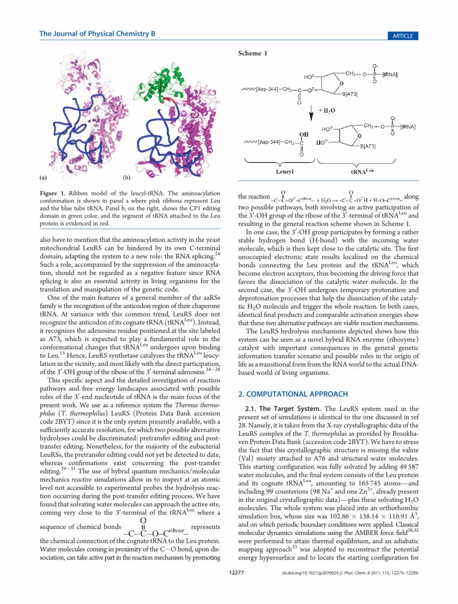
This specific aspect and the detailed investigation of reactionpathways and free energy landscapes associated with possibleroles of the 30-end nucleotide of tRNA is the main focus of thepresent work. We use as a reference system the Thermus thermo-philus (T. thermophilus) LeuRS (Protein Data Bank accessioncode 2BYT) since it is the only system presently available, with asufficiently accurate resolution, for which two possible alternativehydrolyses could be discriminated: pretransfer editing and post-transfer editing. Nonetheless, for the majority of the eubacterialLeuRSs, the pretransfer editing could not yet be detected to date,whereas confirmations exist concerning the post-transferediting.29�31 The use of hybrid quantum mechanics/molecularmechanics reactive simulations allow us to inspect at an atomiclevel not accessible to experimental probes the hydrolysis reac-tion occurring during the post-transfer editing process. We havefound that solvating water molecules can approach the active site,coming very close to the 30-terminal of the tRNALeu where a
sequence of chemical bonds represents
the chemical connection of the cognate tRNA to the Leu protein.Water molecules coming in proximity of the C�O bond, upon dis-sociation, can take active part in the reactionmechanism by promoting
the reaction along
two possible pathways, both involving an active participation ofthe 30-OH group of the ribose of the 30-terminal of tRNALeu andresulting in the general reaction scheme shown in Scheme 1.
In one case, the 30-OH group participates by forming a ratherstable hydrogen bond (H-bond) with the incoming watermolecule, which is then kept close to the catalytic site. The firstunoccupied electronic state results localized on the chemicalbonds connecting the Leu protein and the tRNALeu, whichbecome electron acceptors, thus becoming the driving force thatfavors the dissociation of the catalytic water molecule. In thesecond case, the 30-OH undergoes temporary protonation anddeprotonation processes that help the dissociation of the cataly-tic H2O molecule and trigger the whole reaction. In both cases,identical final products and comparable activation energies showthat these two alternative pathways are viable reaction mechanisms.
The LeuRS hydrolysis mechanisms depicted shows how thissystem can be seen as a novel hybrid RNA enzyme (ribozyme)catalyst with important consequences in the general geneticinformation transfer scenario and possible roles in the origin oflife as a transitional form from the RNAworld to the actual DNA-based world of living organisms.
2. COMPUTATIONAL APPROACH
2.1. The Target System. The LeuRS system used in thepresent set of simulations is identical to the one discussed in ref28. Namely, it is taken from the X-ray crystallographic data of theLeuRS complex of the T. thermophilus as provided by Brookha-ven Protein Data Bank (accession code 2BYT).We have to stressthe fact that this crystallographic structure is missing the valine(Val) moiety attached to A76 and structural water molecules.This starting configuration was fully solvated by adding 49 587water molecules, and the final system consists of the Leu proteinand its cognate tRNALeu, amounting to 165 745 atoms—andincluding 99 counterions (98 Na+ and one Zn2+, already presentin the original crystallographic data)—plus these solvating H2Omolecules. The whole system was placed into an orthorhombicsimulation box, whose size was 102.86 � 138.14 � 116.91 Å3,and on which periodic boundary conditions were applied. Classicalmolecular dynamics simulations using the AMBER force field28,32
were performed to attain thermal equilibrium, and an adiabaticmapping approach33 was adopted to reconstruct the potentialenergy hypersurface and to locate the starting configuration for
Figure 1. Ribbon model of the leucyl-tRNA. The aminoacylationconformation is shown in panel a where pink ribbons represent Leuand the blue tube tRNA. Panel b, on the right, shows the CP1 editingdomain in green color, and the segment of tRNA attached to the Leuprotein is evidenced in red.
Scheme 1

12278 dx.doi.org/10.1021/jp2070024 |J. Phys. Chem. B 2011, 115, 12276–12286
The Journal of Physical Chemistry B ARTICLE
QM/MM metadynamics simulations.28 During this simulationstage, two crucial water molecules, not originally present in thepristine PDB structure and indicated as WAT1 and WAT2 inFigure 2, were shown to approach the catalytic site and identifiedas potentially involved in the editing reaction in ref 28. Followingthese results, the LeuRS equilibrated system used in the presentwork is shown in Figure 2a and presents these two watermolecules, WAT1 and WAT2, included in the quantum me-chanics subsystem in view of their potential activity as nucleo-philic attack catalysts for the hydrolysis reaction as discussedelsewhere.28 In our hybrid quantum mechanics/molecular me-chanics (QM/MM) simulations, the QM subsystem was chosento be the part of the 30-terminal of tRNALeu which includes thelast phosphate, the ribose ring, and its guanine base, along withthe branch representing the leucyl-tRNA connection. Further-more, we included the threonine (Thr-247) which is rather closeto this moiety, forms H-bonds with it, and could be potentiallyinvolved in the editing reaction. As pointed out in ref 28, thenucleophilic water molecules WAT1 and WAT2 approach thecatalytic site and become active through the formation of a low-barrier hydrogen bond (LBHB). Hence, they become part of thequantum subsystem in the present work. This gives a QM systemamounting to 63 atoms, shown in panel b of Figure 2. Sincecovalent bonds are cut in this selection process, the QM valencewas restored by adding five monovalent link atoms.2.2. QM/MMApproach. In all the simulations presented here,
we adopted a hybrid QM/MM approach which has already beenshown to be particularly successful in a wealth of biologicalsystems.34�43 Specifically, starting from the solvated LeuRSstructure described in the previous paragraph and equilibratedat 300 K, we continued for about 8.5 ps the dynamical simulationwithin our QM/MM setup in which the QM driver is based onthe density functional theory44 (DFT) within the HCTH gradient-corrected approximation38 for the exchange and correlationfunctionals. This particular choice has already been extensivelyused in several biomolecules including phosphates,39,40,45�48 andits accuracy is comparable to the hybrid (but computationallymore expensive) B3LYP functional. As a first principles moleculardynamics algorithm, we adopted the Car�Parrinello method49 asimplemented in the CPMD code.50 The interaction betweencore and valence electrons was described by Martins�Troullierpseudopotentials,51 and the Kohn�Sham orbitals, representing
the valence electrons, were expanded in a plane wave (PW) basisset with an energy cutoff of 70 Ry, with the Brillouin zone samplingrestricted to the Γ point. Since the QM subsystem is contained in asubcell of 17 � 15 � 21 Å3, such a cutoff corresponds to 164 759PWs, which translates into amesh of 180� 144� 216 points in realspace. TheQM subsystem is coupled to theMMone according to afull Hamiltonian scheme,52,53 which make use of a restrainedelectrostatic potential (R-ESP) to reduce the computational cost ofthe particle-mesh Coulomb interaction between the embeddedQM subsystem and all the atoms of the outer MM region.All dynamical simulations were performed at 300 K, and the
temperature was controlled via a Nos�e�Hoover54,55 thermostaton the ionic degrees of freedom. A fictitious electron mass of 380au and an integration step of 4 au (0.096 fs) ensured a goodcontrol of the adiabaticity.56
2.3. Reaction Path Sampling.The reaction path was sampledboth by metadynamics57,58 and Blue Moon58 approaches. In thefirst case we added to the Car�Parrinello Lagrangean, LCP, thedegrees of freedom of selected collective variables, sα(t), plus ahistory dependent penalty potential V(sα, t)
L ¼ LCP þ ∑α
12Mα_s
2αðtÞ � ∑
α
12kα½sαðqÞ � sαðtÞ�2 � Vðsα, tÞ
ð1Þwhere q indicates a set of variables (e.g., atomic positions)suitable to describe analytically the reaction coordinates. Thefictitious masses for the kinetic term have been set toMα = 22.0au, and the harmonic coupling constants were set to kα = 0.24 au.Further details on the methods adopted here and their assess-ment have been reported in detail elsewhere and can be found inthe literature.39�42,47,48,60,61 The specific choices of collectivevariables for each simulation will be given in the next paragraphs,as a support to the discussion. The explicit form of the penaltypotential used in this work is a superposition of small Gaussians
Vðsα, tÞ ¼Z
t0 dt
0j_sðt0Þjδ _sðt0Þj_sðt0Þjðs� sðt0ÞÞ
� �Aðt0Þ exp � ðs� sðt0ÞÞ2
2ðΔsÞ2" #
ð2ÞIn each metadynamics run, a new Gaussian contribution was
added to V(sα, t) every 120 steps, corresponding to a time
Figure 2. (a) Equilibrated LeuRS (solvating water not shown). The leucyl protein is shown as a green ribbon, the tRNALeu is shown as a yellow double-stranded tube with the base pairs in blue, and the red square indicates the editing domain where QM atoms were selected. (b) Details of the QM systemsurrounded by the closest residues. QM atoms are shown as thick sticks and balls, where the color code, here and in all the following figures, is black for H,gray for C, blue for N, red for O, and yellow for P. The surrounding MM residues, within a radius of 10 Å, are colored as follows: red for Asp, purple forThr, pink for Glu, brown for Arg, orange for Gly, yellow for Ser, green for Ile, light blue for Val, blue for Tyr, and black for Met.

12279 dx.doi.org/10.1021/jp2070024 |J. Phys. Chem. B 2011, 115, 12276–12286
The Journal of Physical Chemistry B ARTICLE
interval ofΔt = 0.01 ps. The amplitude A(t0) was set toWt = 0.15kcal/mol and the width Δs was fixed to 0.18 (in collectivevariables units). Since, roughly speaking, the free energy land-scapes spanned by the collective variables adopted here are not sodifferent in terms of roughness and barriers, these values are keptunchanged in the various metadynamics simulations unlessotherwise specified in the ongoing discussion. In all the simula-tions presented in the following section, in order to reduce theintrinsic error bar in the free energy surface (FES) reconstructionvia metadynamics, we used the average procedure described inref 62. Namely, after allowing for at least a couple of passagesback and forth of the collective variables (as described in ref 39for the case of QM/MM approaches) from the reactants side tothe products side on the FES, we computed the arithmeticaverage of the penalty potential as
FðsαÞ ¼ � 1tsimul � tdiff
Ztsimul
tdiff
Vðsα, tÞ dt ð3Þ
where tsimul is the total simulation time and tdiff is the time atwhich collective variables start diffusing in all the phase spacespanned by the selected sα(t). The coupling of metadynamicswith the QM/MM hybrid approach has already been discussedelsewhere37�41 and has been shown to be able to study with ap-preciable accuracy complicated reactions in biomolecular systems.In the case of Blue Moon simulations, the (single) reaction
coordinate ξ = ξ(RI) defined by a given subset of atomic coor-dinates RI was assumed to be a specific distance between twoatoms, as will be discussed in the next paragraph, and added tothe Car�Parrinello Lagrangean as a holonomic constraint
L ¼ LCP þ λk½ξðRIÞ � ξk� ð4Þwhere λk is a Lagrange multiplier and ξk is a selected value of thereaction coordinate. Upon equilibration of each constraint value,corresponding in our specific cases to about 3 ps simulations foreach sampled value of ξ(RI), the average constraint force fξ wascomputed as time average
fξ ¼ Æλkæt ¼∂F∂ξ
ð5Þ
and the free energy profile is obtained according to the standardthermodynamic integration technique
ΔF ¼Z
ξf
ξ0
fξ dξ ð6Þ
from the initial value ξ0 to the final one ξf.
3. RESULTS AND DISCUSSION
3.1. Hybrid QM/MM Molecular Dynamics. Before perform-ing any reactive simulation, we first did a pre-equilibration of thesystem starting a QM/MM simulation from the structure ob-tained after the classical AMBER32 simulation. This hybridmolecular dynamics run lasted for 8.5 ps and allowed also forthe newly added electronic degrees of freedom to reach a dynamicalequilibrium within the Car�Parrinello framework.As shown in the upper panel of Figure 3, the thermal equilibrium
of the system was attained rather quickly, in about 1�2 ps.However, the structural equilibration took a longer time, namelyabout 6.5 ps. The root-mean-square displacement (rmsd) wascomputed during the dynamical simulation by considering all the
heavy atoms (i.e., H excluded), these atoms being the only onescrystallographically detected. The result is reported in the lowerpanel of Figure 3. The larger contribution to the rmsd comesfrom the reorganization of the solvent water, the most mobilespecies. It is worthy of note to observe that the final equilibratedrmsd does not exceed the experimental resolution,11,12 thusbeing still compatible with the crystallographic data.The water molecules that in a former simulation were found
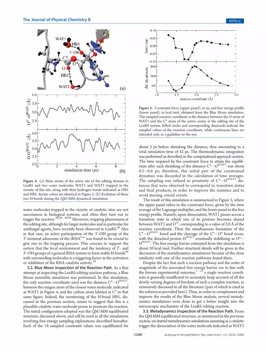
to come close to the catalytic site of the LeuRS editing domain, asa consequence of a LBHB mechanism,28 do not escape awayduring the equilibration phase. On the contrary, they are keptin place by relatively long-living H-bonds. These bonds areindicated as HB1 and HB2 in panel a of Figure 4, and theirevolution in terms of interatomic distances OWAT1�O30 andOWAT2�O is shown in panel b of this same figure, where theatoms labeling adopted hereafter is reported.The quantum approach used in the present study has been
extensively benchmarked over the years to characterize aqueoussolutions; in particular both in pure water61 and in salt solutionsat biological concentrations,63 the oxygen�oxygen radial dis-tribution function shows a first minimum located at about 3.3 Å,which defines the typical H-bond distance. In the present case,the distances OWAT1�O30 and OWAT2�O are most of the timebelow that threshold, indicating that these H-bonds are ratherstable. They can be temporarily broken, as observed during theQM/MM simulation, mostly because the 30-OH group canrotate relatively easily, as will be discussed in the next paragraph,around the C3�O30 bond, where C3� belongs to the ribose ofthe 30-terminal of the tRNALeu. However, their average lifetimesare 3.0 ( 0.7 ps for HB1 and 1.2 ( 0.4 ps for HB2. Despite thepoor statistics due to the rather short equilibration time, thelifetime of HB1 is longer than HB2, which, instead, is character-ized by a lifetime typical of regular H-bonds (∼1 ps) in liquidwater.64 From these considerations, we can infer that a H-bondstrength of 3�4 kcal/mol65 characterizes HB2, and most likelythis value is just a lower bound for HB1.This H-bonding environment keeps these water molecules
trapped in proximity of the active site and seems to suggest thatthey might play an active role in the reaction mechanism. Inparticular, the water molecule labeled as WAT1 can go very closeto the �C*�O*� moiety, where the catalytic reaction is expectedto occur. This was observed also in our classical AMBER simu-lations, where the minimum distance observed was 2.38 Å. Indeed,
Figure 3. Temperature and root-mean-square displacement (rmsd) ofall the heavy atoms of the LeuRS system in solution, as a function of thesimulation time, during the QM/MM equilibration phase.
12280 dx.doi.org/10.1021/jp2070024 |J. Phys. Chem. B 2011, 115, 12276–12286
The Journal of Physical Chemistry B ARTICLE
water molecules trapped in the vicinity of catalytic sites are notuncommon in biological systems, and often they turn out totrigger the reaction.28,38�40,48 Moreover, trapping phenomena atthe editing site, although for larger molecules and in particular forantifungal agents, have recently been observed in LeuRS.66 Alsoin that case, an active participation of the 30-OH group of the30-terminal adenosine of the tRNALeu was found to be crucial togive rise to the trapping process. This concurs to support thenotion that the local environment and the tendency of 20- and30-OH groups of a general RNA system to form stable H-bonds66
with surrounding molecules is a triggering factor in the activationor inhibition of the RNA catalytic activity.28
3.2. Blue Moon Inspection of the Reaction Path. As a firstattempt at inspecting the LeuRS editing reaction pathway, a BlueMoon ensemble simulation was performed. In this simulation,the only reaction coordinate used was the distance C*�OWAT1,between the oxygen atom of the closest watermolecule, indicatedas WAT1 in Figure 4, and the carbon atom labeled as C* in thatsame figure. Indeed, the monitoring of the H-bond HB1, dis-cussed in the previous section, seems to suggest that this is aplausible catalytic water molecule prone to promote the reaction.The initial configuration adopted was the QM/MM equilibratedstructure, discussed above, and will be used in all the simulationsinvolving free energy sampling explorations described hereafter.Each of the 14 sampled constraint values was equilibrated for
about 3 ps before shrinking the distance, thus amounting to atotal simulation time of 42 ps. The thermodynamic integrationwas performed as described in the computational approach section.The time required by the constraint force to attain the equilib-rium after each shrinking of the distance C*�OWAT1 was about0.5�0.6 ps; therefore, this initial part of the constraineddynamics was discarded in the calculation of time averages.The sampling was refined in proximity of C*�OWAT1 dis-tances that were observed to correspond to transition statesand final products, in order to improve the statistics and toavoid missing crucial events.The result of this simulation is summarized in Figure 5, where
the upper panel refers to the constraint force, given by the timeaverage of the Lagrangemultiplier, and the lower panel to the freeenergy profile. Namely, upon dissociation, WAT1 passes across atransition state in which one of its protons becomes sharedbetweenWAT1 and O30, corresponding to a value of 2.0 Å of thereaction coordinate. Then the simultaneous formation of theC*�OWAT1 bond and the cleavage of the C*�O* bond occur,with the detached proton HWAT1 eventually stabilizing as O*�HWAT1. The free energy barrier estimated from this simulation isabout 20 kcal/mol. Further structural details will be given in thediscussion of the metadynamics simulations because of the closesimilarity with one of the reaction pathways found there.Despite the fact that such a reaction pathway and the order of
magnitude of the associated free energy barrier are in line withthe known experimental outcome,1�15 a single reaction coordi-nate is generally insufficient to accurately keep account of all theslowly varying degrees of freedom of such a complex reaction, asextensively discussed in all the literature (part of which is cited inthe references provided here). Thus, in order to complement andimprove the results of the Blue Moon analysis, several metady-namics simulations were done to get a better insight into themicroscopic mechanism of the LeuRS editing reaction.3.3. Metadynamics Inspection of the Reaction Path. From
theQM.MM equilibrated structure, as mentioned in the previoussection, we startedmetadynamics simulations assuming as a catalytictrigger the dissociation of the water molecule indicated as WAT1
Figure 5. Constraint force (upper panel), in au, and free energy profile(lower panel), in kcal/mol, obtained from the Blue Moon simulation.The sampled reaction coordinate is the distance between the O atom ofWAT1 and the C* atom of the active center at the editing site of theLeuRS system. Filled circles and corresponding diamonds indicate thesampled values of the reaction coordinate, while continuous lines areintended only as a guideline to the eye.
Figure 4. (a) Main atoms of the active site of the editing domain inLeuRS and two water molecules WAT1 and WAT2 trapped in thevicinity of this site, along with their hydrogen bonds indicated as HB1and HB2. Atoms colors are identical to Figure 2. (b) Evolution of thesetwo H-bonds during the QM/MM dynamical simulation.

12281 dx.doi.org/10.1021/jp2070024 |J. Phys. Chem. B 2011, 115, 12276–12286
The Journal of Physical Chemistry B ARTICLE
in Figure 4. This choice was driven by structural and electronicconsiderations. On one hand, this H2Omolecule is the closest tothe active site and its dipole moment is, on average, pointingdirectly toward the �C*-O*� bond, the one expected to under-go the cleavage reaction. On the other hand, an accurate analysisof the local electronic structure of the system at the end of theQM/MM equilibration has shown that the electronic states ofthe Kohn�Sham (KS) Hamiltonian37 close to the energy gap(Egap = 2.85 eV), hence the ones directly involved in the reaction,present the features summarized in Figure 6.Namely, the highest occupied KS (HOKS) wave function—
assumed as a reference level—and the one immediately below(HOKS�1), at�0.37 eV aremainly bonding states of the base ofthe 30-terminal adenosine of the tRNALeu, with considerableamplitudes also on the 30-OH group, as shown in panels a and bof Figure 6. Above the HOKS, the lowest unoccupied KS (LUKS)state, energetically located at +2.85 eV above HOKS, shows large(antibonding) amplitudes on top of the�C*�O*� and�C*=Obonds, with nodes of the wave functions passing across bothbonds. This empty state is ready to accept electrons from theWAT1 and to start the reaction, as we observed in the reactivesimulations described in the ongoing discussion. Finally, the stateabove LUKS (LUKS+1) is an antibonding state, localized onthe base of the 30-terminal adenosine of the tRNALeu, butenergetically located above LUKS by about 0.8 eV, thus muchhigher in energy and unlikely to take an active role in the reaction.A first metadynamics simulation was performed by using
two collective variables, s1 = |C*�OWAT1| and s2 = |O*�HWAT1|.
These are the distances between the O atom of the water moleculeWAT1 and the carbon atom indicated as C* in Figure 4 and thedistance of one of the H atoms of WAT1 from the O* atom ofthe�C*-O*�moiety, representing the attachment of the tRNAto the Leu protein.The simulation was actually repeated twice, selecting each
time, alternatively, one of the two H atoms of WAT1, leading toidentical results. The free energy landscape provided by thissimulation is shown in Figure 7.Starting from the initial local minimum, indicated as panel a in
Figure 7 and represented by the equilibrated LeuRS editing siteshown in Figure 8a, along this pathway the dissociation of thewater moleculeWAT1 is promoted by the overlap of the bondingorbitals of this molecule with the LUKS localized on top of the
group. The dissociation of the water molecule into a
proton and a hydroxyl anionHO� occurs in a concerted way withthe cleavage of the C*�O* bond and the subsequent formationof the C*�OHWAT1 bond (panels b and c of Figure 8). Theseprocesses, in fact, occur at a given value of the time-dependentpenalty potential V(sα, t), before any new Gaussian function isadded, and we recall that metadynamics is an energeticallyordered (dynamical) exploration of the free energy surface butdoes not necessarily reproduce time-ordered trajectories.57,58,60,61
According to this reaction pathway, the reactant (Figure 8a)has to overcome a free energy barrier ΔF = 19 kcal/mol to reachthe transition state, represented by the dissociation of theH2O molecule WAT1 and the simultaneous cleavage of the
Figure 6. Electronic KS wave functions of the system around the energy gap: (a) HOKS�1, (b) HOKS, (c) LUKS, and (d) LUKS+1. The firstunoccupied state is localized on the �C*�O*� bond and ready to accept electrons from the nearby WAT1 H2O molecule. Wave functions arerepresented as isosurfaces at 5� 10�2 1/Å3/2, with positive amplitudes in cyan and negative amplitudes in orange. The color code of the main atoms,represented by sticks and balls, is the same as in previous figures with the exception of H (white). For the sake of clarity, main atoms are labeled only inpanel a. Thin gray sticks are the surrounding MM system and dashed blue lines hydrogen bonds.

12282 dx.doi.org/10.1021/jp2070024 |J. Phys. Chem. B 2011, 115, 12276–12286
The Journal of Physical Chemistry B ARTICLE
�C*�O*� bond. During this phase, the proton detached fromWAT1 wanders about, jumping back to WAT1, formingH-bonds with O30, but spending most of the time on O*, onwhich eventually stabilizes as shown in panels b, c, and d ofFigure 8. A short living (about 1 ps) hydrogen bond is formedbetween �O*�HWAT1 and �C*�OWAT1�HWAT1 (Figure 8c)which, however, is not present in the final product (Figure 8d).Such a final product turns out to be more stable than the initialreactant by about 8 kcal/mol, thusmaking at least this stage of theediting reaction a one-directional process along the reaction path.A noticeable feature, at this point, is the close similarity of the free
energy barrier, with respect to the Blue Moon result; however, astructurally different transition state was found, since no sharedproton betweenWAT1 and O30 is realized in the concerted processdescribed above.An identical simulation, performed using WAT2 instead of
WAT1, gave rise to a destabilization and eventually a rupture ofthe system and resulted in an inconsistent reaction pathway withan unreasonably high free energy barrier, larger than 40 kcal/mol,as expected whenever a wrong reaction path is sampled.67 Thedissociation of the water molecule is much more problematic,and no contribution from the 30-OH group comes into play to
Figure 7. Free energy landscape obtained from the metadynamics simulation of the LeuRs system (panel a) and its two-dimensional contour densityversion (panel b), assuming as reaction coordinates the distances C*�OWAT1 and O*�HWAT1, the atom labeling being the one given in Figure 4. Labelsfrom (a) to (d) refer to the configurations reported in Figure 8.
Figure 8. Main steps of the reaction path for the hydrolysis reaction at the editing site of LeuRS. The label of each panel refers to the points on the freeenergy surface indicated in the previous figure. All the main atoms involved in the reaction are labeled. The color code is the same as in the precedingfigures apart from the protons of WAT1 (white circled in blue) and the reactive carbon C* (cyan), evidenced for the sake of clarity.

12283 dx.doi.org/10.1021/jp2070024 |J. Phys. Chem. B 2011, 115, 12276–12286
The Journal of Physical Chemistry B ARTICLE
promote or help the reaction. The whole sequence of chemicalbonds connecting the leucyl protein and the tRNALeu, fromC* tothe ribose ring, undergoes unrealistic distortions, and the finalproduct is just an unrecoverable broken system. This is, however,a clear indication thatWAT1 is the right catalytic water molecule.In the metadynamics inspection of the reaction pathway
involving WAT1, we observed that, during the reaction, the 30-OH group rotates rather freely around its axis represented by theC3�O30 bond, C3 being the carbon atom of the ribose ring oftRNALeu to which O30 is chemically bonded. This seems tosuggest that little or no barrier exists for this specific rotation.Indeed, this is not in contradiction with the trend observedduring the QM/MM equilibration phase and summarized inFigure 4, where the H-bond, labeled as HB1, betweenWAT1 andthe 30-OH group can be temporarily broken and then formedagain. Yet, it is difficult to infer something more precise about therotation barrier on the basis of this simulation. Another ques-tionable point is the protonation of O*, shown in the finalconfiguration (Figure 8d). In fact, since we made use of thedistance O*�HWAT1 as a collective variable a biasing could havebeen introduced, thus forcing one of the protons of WAT1 toapproach O*. In order to verify whether or not our choice ofreaction coordinates biased the reaction, we repeated the simu-lation using as new set of collective variables the distanceC*�OWAT1, as in the former simulations, and the rotationangle of the 30-OH group around its C3�O30 axis, i.e., s1 =|C*�OWAT1| and s2 =Θ{�C3�OH30}. The angle of the secondcollective variable is measured assuming as a reference config-uration s2 = 0ο the initial (equilibrated) structure shown inFigure 4a. No other constraints are imposed on the two H ofWAT1, which are thus free to move around. Also in this case, thesimulation was started from the structure obtained at the end ofthe QM/MM equilibration phase, prior to the addition of anyconstraint. The result of this metadynamics simulation is sum-marized in the free energy landscape of Figure 9. The overall freeenergy barrier for the reaction is about 18 kcal/mol, slightly lowerthan the former result, but similar if we keep into the error bar ofabout 1�2 kcal/mol typical of DFT numerical approaches. Theproduct is more stable than the reactant by about 6�7 kcal/mol,and the labels from a to e in Figure 9 correspond to the mainstages of the reaction represented by the configurations, labeledin the same way, shown in Figure 10.At a first glance, the reactant and the product of this second
simulation look identical to the former ones. In fact, althoughunconstrained, one of the protons of WAT1, upon dissociation
into H+ and OH�, is eventually donated to the O* oxygen,whereas the hydroxyl anion reacts with C* to form a stable�C*�OH bond. Nonetheless, the pathway of the reaction issignificantly different from the previous one and involves moreactively the 30-OH group in a way identical to what was found inthe Blue Moon simulation reported in the former paragraph.Namely, by analyzing the metadynamics trajectory, we noticedfirst that the initial system is able to switch rather freely fromconfigurations a and b shown in Figure 10. These two structuresdiffer in the donor�acceptor configuration of the H-bondbetween the 30-OH group and the water molecule WAT1 andcorrespond to two rotation angles Θ{�C3�OH30} of about 0�and 45�. The difference in the two structures is that in one case(Figure 10a) the H of the 30-OH group points toward the oxygenof WAT1, whereas in the second case (Figure 10b) one of the Hatoms of WAT1 points toward the O30 atom of the 30-OH group.The system can easily take any one of these configurationsby simply rotating the 30-OH group around the C3�O30 axis,which involves the overcoming of a rather small free energybarrier (about 5 kcal/mol), typical of the breaking/formation ofan H-bond.65
Then the reaction proceeds with an active (chemical) partici-pation of the 30-OH group, this time identical to what wasobserved in the case of the Blue Moon ensemble approach. Infact, during the formation of the C*�OWAT1 bond, one of theprotons ofWAT1 becomes temporarily a shared proton betweenOWAT1 and O30 as shown by panel c of Figure 10. The formationof the new bond and the transitory detachment of the protonfrom WAT1 are sufficient to weaken the C*�O* bond, which israpidly cleaved as shown in panel d of the same figure. In theseconditions, the shared proton departs from the water moleculeand O30, going to saturate the broken bond and formingeventually a stable bond with O*. The shared proton stage andthe cleavage of the C*�O* bond can be regarded, also in thiscase, as concerted for the reasons explained above, since theyoccur at a given value of the time-dependent penalty potentialV(sα, t), before any new Gaussian function is added.A first conclusion that can be drawn from these metadynamics
(and Blue Moon) studies is that although reactant and productsare indistinguishable in all the (successful) reaction mechanismsidentified here, at least two alternative reaction pathways exist,both involving the participation of the 30-OH group, but while inone case only H-bonds are involved, in the second case atemporary formation of chemical bonds is possible. During thisphase, one of the protons of the catalytic water becomes a “strongly”
Figure 9. Free energy landscape as obtained from the metadynamics simulation of the LeuRs systems assuming as reaction coordinates the distanceC*�OWAT1 and the rotation angle of the 30-OH group around its C3�O30 axis. Labels from a to e refer to the configurations reported in Figure 10. Panela shows the three-dimensional surface and panel b the corresponding two-dimensional contour density.

12284 dx.doi.org/10.1021/jp2070024 |J. Phys. Chem. B 2011, 115, 12276–12286
The Journal of Physical Chemistry B ARTICLE
shared proton, as in an undercoordinated Zundel complex.68 Atthe same time, the rotation barrier for the 30-OH group is nearlynegligible, and the rate-limiting step of the reaction is repre-sented by the concerted dissociation of the catalytic watermolecule and the cleavage of the C*�O* bond.
4. CONCLUSIONS
By using reactive QM/MM computational approaches, thereaction mechanism and the related energetics in the hydrolysisreaction occurring at the editing domain of the T. termophilusleucyl-tRNA (LeuRS) system has been inspected. On the basis ofthese simulations, we have shown that the reaction occurringduring the enzymatic activity is triggered by a catalytic watermolecule, labeled as WAT1, which approaches the catalyticcenter in proximity of the�C*-O*� bond connecting the leucylprotein and its cognate tRNA.During this stage, the 30-OH groupof the tRNALeu forms a stable hydrogen bond with WAT1 andtriggers the starting phase of the whole reaction. In fact, althoughthe 30-OH group is free to rotate around its axis, either the H30 orthe O30 atom of this group maintains an H-bond with WAT1,which keeps the water molecule close to the active site. Twopossible pathways have been identified, which differ in the roleplayed by the 30-OH group during the reaction, but that give riseto an identical final product and are characterized by nearlyequivalent energy barriers. In the first case the 30-OH group ofthe cognate tRNALeu acts as a Lewis acid, accepting electrons
from WAT1 and helping its dissociation with the resulting OH�
and H+ and attacking the C* and O* atoms, respectively. Thesetwo atoms bridging leucyl and tRNALeu represent the localizationsites of the lowest unoccupied (antibonding) atomic orbital,suitable to accommodate electrons. This promotes the bondcleavage process of the reaction and favors the formation of thenew bonds mentioned above. In the second case, WAT1 shares aproton with the O30 atom of the 30-OH group, forming a tem-porary Zundel-like complex and thus favoring the dissociation ofthe catalytic water molecule. After its dissociation, the reactionproceeds in the same way summarized above, with a concertedcleavage of the C*�O* bond and formation of the C*�OHWAT1
and O*�HWAT1 bonds. This active participation of the 30-OHgroup in both the reaction pathways makes its presence anindispensable element in the realization of the enzymatic activityof LeuRS.
’AUTHOR INFORMATION
Corresponding Author*E-mail: [email protected].
’ACKNOWLEDGMENT
Yohsuke Hagiwara andMasaru Tateno are gratefully acknowl-edged for providing the configurations of ref 28 used as a startingpoint for the present set of simulations. Calculations were
Figure 10. Main steps of the reaction path assuming as reaction coordinates the distance C*�OWAT1 and the rotation of the 30-OH group. The label ofeach panel refers to the points on the free energy surface indicated in Figure 8. All the main atoms involved in the reaction are labeled. The color code isthe same as in Figure 8.

12285 dx.doi.org/10.1021/jp2070024 |J. Phys. Chem. B 2011, 115, 12276–12286
The Journal of Physical Chemistry B ARTICLE
performed on the PACS-CS system of CCS—Tsukuba Univer-sity and partly on theHPC resources of [CCRT/CINES/IDRIS]under the allocation x2010096092 made by GENCI (GrandEquipement National de Calcul Intensif). Insightful discus-sions with Dino Moras, Alberto D. Podjarny, Isabelle M.Billas, Carlo Massobrio, and Kiyoyuki Terakura are alsowarmly acknowledged.
’REFERENCES
(1) Cusack, S.; Hartlein, M.; Leberman, R. Nucleic Acids Res. 1991,19, 3489–3498.(2) Nagel, G. M.; Doolittle, R. F. Proc. Natl. Acad. Sci. U.S.A. 1991,
88, 8121–8125.(3) Delarue, M. Curr. Opin. Struct. Biol. 1995, 5, 48–55.(4) Ibba, M.; Soll, D. Annu. Rev. Biochem. 2000, 69, 617–650.(5) Hendrickson, T. L.; Schimmel, P. In Transfer RNA-Dependent
Amino Acid Discrimination by Aminoacyl-tRNA Synthetase; Lapointe,J. P. D., Brakier-Gingras, L., Eds.; Landes Bioscience/Eurekah.com:Austin, TX, 2003; pp 34�64.(6) Schimmel, P. Annu. Rev. Biochem. 1987, 56, 125–158.(7) Rao, S. T.; Rossmann, M. G. J. Mol. Biol. 1973, 76, 241–250.(8) Xu, M.-G.; Chen, J.-F.; Martin, F.; Zhao, M.-W.; Eriani, G.;
Wang, E.-D. J. Mol. Biol. 2002, 277, 41590–41596.(9) Cavarelli, J.; Delagoutte, B.; Eriani, G.; Gangloff, J.; Moras, D.
EMBO J. 1998, 17, 5438–5448.(10) Silvian, L. F.; Wang, J.; Steitz, T. A. Science 1999, 285,
1074–1077.(11) Fukai, S.; Nureki, O.; Sekine, S.; Shimada, A.; Tao, J.; Vassylyev,
D. G.; Yokoyama, S. Cell 2000, 103, 793–803.(12) Fukunaga, R.; Yokoyama, S. J. Mol. Biol. 2005, 346, 57–71.(13) Fukunaga, R.; Yokoyama, S. Nat. Struct. Chem. Biol. 2005,
12, 915–922.(14) Tukalo, M.; Yaremchuck, A.; Fukunaga, R.; Yokoyama, S.;
Cusack, S. Nat. Struct. Chem. Biol. 2005, 12, 923–930.(15) Fukunaga, R.; Yokoyama, S. Biochemistry 2007, 46, 4985–4996.(16) The anticodon region of tRNA is a sequence of three bases
complementary to the codon region of the mRNA (mRNA); the codonand anticodon bases are bonded together by appropriate hydrogenbonds in a double-stranded RNA structure.(17) Schmidt, E.; Schimmel, P. Science 1994, 264, 265–267.(18) Lin, L.; Hale, S. P.; Schimmel, P. Nature 1996, 384, 33–34.(19) Lin, L.; Schimmel, P. Biochemistry 1996, 35, 5596–5601.(20) Chen, J. F.; Guo, N. N.; Li, T.; Wang, E. D.; Wang, Y. L.
Biochemistry 2000, 39, 6726–6731.(21) Fukunaga, R.; Fukai, S.; Ishitani, R.; Nureki, O.; Yokoyama, S.
J. Biol. Chem. 2004, 279, 8396–8402.(22) Cura, V.; Olieric, N.; Guichard, A.; En-Duo, W.; Moras, D.;
Eriani, G.; Cavarelli, J. Acta Crystallogr. 2005, F61, 899–901.(23) Rho, S. B.; Lincecum, T. L.; Martins, S. A. EMBO J. 2002,
21, 6874–6881.(24) Martinis, S. A.; Hsu, J. L. J. Mol. Biol. 2008, 376, 482–491.(25) Eriani, G.; Delarue, M.; Poch, O.; Gangloff, J.; Moras, D.Nature
1990, 347, 203–206.(26) Hagiwara, Y.; Nureki, O.; Tateno, M. FEBS Lett. 2009, 583,
1901–1908.(27) Hagiwara, Y.; Nureki, O.; Tateno, M. FEBS Lett. 2009, 583,
825–830.(28) Hagiwara, Y.; Field, M. J.; Nureki, O.; Tateno, M. J. Am. Chem.
Soc. 2010, 132, 2751–2758.(29) Lincecum, T. L., Jr.; Tukalo, M.; Yaremchuk, A.; Mursinna,
R. S.; Williams, A. M.; Sproat, B. S.; Van Den Eynde, W.; Link, A.; VanCalenbergh, S.; Grotli, M.; Martinis, S. A.; Cusack, S. Mol. Cell 2003,11, 951–963.(30) Mursinna, R. S.; Lincecum, T. L., Jr.; Martinis, S. A. Biochemistry
2001, 40, 5376–5381.(31) Zhai, Y.; Martinis, S. A. Biochemistry 2005, 44, 15437–15443.
(32) (a) Cornell, W. D.; Cieplack, P.; Bayly, C. I.; Gould, I. R.; Merz,K. M., Jr.; Ferguson, D. M.; Spellmeyer, D. C.; Fox, T.; Caldwell, J. W.;Kollman, P. A. J. Am. Chem. Soc. 1995, 117, 5179–5197. (b) Case, D. A.;Cheatham, T. E.; Darden, T.; Gohlke, H.; Luo, R.; Merz, K. M.;Onufriev, A.; Simmerling, C.; Wang, B.; Woods, R. J. J. Comput. Chem.2005, 26, 1668–1688.
(33) The adiabatic mapping consists of a set of geometry optimiza-tions performed after short molecular dynamics runs on differentconfigurations, sampled along a reactive trajectory for a set of fixedreaction coordinate(s) as explained in Hagiwara, Y.; Ohta, T.; Tateno,M. J. Phys.: Condens. Matter 2009, 21, 064234.
(34) Gervasio, F. L.; Carloni, P.; Parrinello, M. Phys. Rev. Lett. 2002,89, 108102.
(35) Carloni, P.; R€othlisberger, U.; Parrinello, M. Acc. Chem. Res.2002, 35, 455–464.
(36) Colombo, M. C.; Guidoni, L.; Laio, A.; Magistrato, A.; Maurer,P.; Piana, S.; Rohrig, U.; Spiegel, K.; Sulpizi, M.; VandeVondele, J.;Zumstein, M.; R€othlisberger, U. Chimia 2002, 56, 13–19.
(37) Magistrato, A.; De Grado, W. F.; Laio, A.; R€othlisberger, U.;VandeVondele, J.; Klein, M. L. J. Phys. Chem. B 2003, 107, 4182–4188.
(38) von Lilienfeld, O. A.; Tavernelli, I.; R€othlisberger, U.; Sebastiani,D. J. Chem. Phys. 2005, 122, 014133.
(39) Boero, M.; Ikeda, T.; Ito, E.; Terakura, K. J. Am. Chem. Soc.2006, 128, 16798–16807.
(40) Dal Peraro, M.; Ruggerone, P.; Raugei, S.; Gervasio, F. L.;Carloni, P. Curr. Opinion Struct. Biol. 2007, 17, 149–156.
(41) Gervasio, F. L.; Boero, M.; Parrinello, M. Angew. Chem., Int. Ed.2006, 45, 5606–5609.
(42) Boero, M.; Gervasio, F. L.; Parrinello, M. Mol. Simul. 2007,33, 57–60.
(43) Boero, M.; Park, J. M.; Hagiwara, Y.; Tateno, M. J. Phys.:Condens. Matter 2007, 19, 365217.
(44) Kohn, W.; Sham, L. J. Phys. Rev. 1965, 140, A1133–A1138.(45) Hamprecht, F. A.; Cohen, A. J.; Tozer, D. J.; Handy, N. C.
J. Chem. Phys. 1998, 109, 6264–6271.(46) Boese, A. D.; Doltsinis, N. L.; Handy, N. C.; Sprik, M. J. Chem.
Phys. 2000, 112, 1670–1678.(47) Gervasio, F.; Laio, A.; Iannuzzi, M.; Parrinello, M. Chem.—Eur.
J. 2004, 10, 4846–4852.(48) Boero, M.; Tateno, M.; Terakura, K.; Oshiyama, A. J. Chem.
Theory Comput. 2005, 1, 925–934.(49) Car, R.; Parrinello, M. Phys. Rev. Lett. 1985, 55, 2471–2474.(50) CPMD, version 3.13.1; http://www.cpmd.org; Copyright IBM
Corp. 1990�2011; Copyright MPI f€ur Festk€orperforschung Stuttgart,1997�2001.
(51) Troullier, N.; Martins, J. L. Phys. Rev. B 1991, 43, 1993–2006.(52) Laio, A.; VandeVondele, J.; R€othlisberger, U. J. Chem. Phys.
2002, 116, 6941–6947.(53) Laio, A.; VandeVondele, J.; R€othlisberger, U. J. Phys. Chem. B
2002, 106, 7300–7307.(54) (a) Nos�e, S.Mol. Phys. 1984, 52, 255–268. (b) Nos�e, S. J. Chem.
Phys. 1984, 81, 511–519.(55) Hoover, W. G. Phys. Rev. A 1985, 31, 1695–1697.(56) Grossman, J. C.; Schwegeler, E.; Draeger, E. W.; Gygi, F.; Galli,
G. J. Chem. Phys. 2004, 120, 300–311.(57) Laio, A.; Parrinello, M. Proc. Natl. Acad. Sci. U.S.A. 2002,
99, 12562–12566.(58) Iannuzzi, M.; Laio, A.; Parrinello, M. Phys. Rev. Lett. 2003,
90, 238302.(59) Sprik, M.; Ciccotti, G. J. Chem. Phys. 1998, 109, 7737–7744.(60) Laio, A.; Rodriguez-Fortea, A.; Gervasio, F. L.; Ceccarelli, M.;
Parrinello, M. J. Phys. Chem. B 2005, 109, 6714–6721.(61) Laio, A.; Gervasio, F. L. Rep. Prog. Phys. 2008, 71, 126601.(62) Silvestrelli, P. L.; Parrinello, M. J. Chem. Phys. 2002, 111,
3572–3580.(63) Schmidt, D. A.; Scipioni, R.; Boero, M. J. Phys. Chem. A 2009,
113, 7725–7729.(64) Bergman, D. L. Chem. Phys. 2000, 253, 267–282.

12286 dx.doi.org/10.1021/jp2070024 |J. Phys. Chem. B 2011, 115, 12276–12286
The Journal of Physical Chemistry B ARTICLE
(65) Walrafen, G. E.; Fisher, M. R.; Hokmabadi, M. S.; Yang, W.-H.J. Chem. Phys. 1986, 85, 6970–6982.(66) Rock, F. L.; Mao, W.; Yaremchuck, A.; Tukalo, M.; Cr�epin, T.;
Zhou, H.; Zhang, Y.-K.; Hernandez, V.; Akama, T.; Baker, S. J.; et al.Science 2007, 316, 1759–1761.(67) Kamiya, K.; Boero, M.; Tateno, M.; Shiraishi, K.; Oshiyama, A.
J. Am. Chem. Soc. 2007, 129, 9663–9673.(68) Boero, M.; Ikeshoji, T.; Terakura, K. ChemPhysChem 2005,
6, 1775–1779.