leukocyte glucocorticoid receptor expression and immunoregulation in veterans with and without...
TRANSCRIPT
ORIGINAL ARTICLE
Leukocyte glucocorticoid receptor expression andimmunoregulation in veterans with and withoutpost-traumatic stress disorderCS de Kloet1,2, E Vermetten1,2, A Bikker1,3, E Meulman1,3, E Geuze1,2, A Kavelaars3, HGM Westenberg2
and CJ Heijnen3
1Department of Military Psychiatry, Central Military Hospital, Utrecht, The Netherlands; 2Department of Psychiatry, RudolfMagnus Institute of Neurosciences, University Medical Center Utrecht, Utrecht, The Netherlands and 3Laboratory ofPsychoneuroimmunology, University Medical Center Utrecht, Utrecht, The Netherlands
Post-traumatic stress disorder (PTSD) is associated with a dysregulation of the hypothala-mus–pituitary–adrenal axis (HPA axis). In addition, there is evidence for altered glucocorticoidreceptor (GR) expression and function in peripheral blood mononuclear cells. The aim of thepresent study was to differentiate between the effect of trauma exposure and PTSD onleukocyte GR expression and glucocorticoid immune regulation. Leukocyte GR bindingcharacteristics and glucocorticoid sensitivity of immune activity, determined as the effect ofdexamethasone (DEX) on in vitro cytokine release and T-cell proliferation, were comparedbetween veterans with PTSD, traumatized veterans without PTSD and healthy controls.Leukocyte GR density was significantly lower in veterans with and without PTSD compared tohealthy controls. DEX-induced inhibition of T-cell proliferation was significantly lower in PTSDcompared to trauma and healthy controls. DEX-induced increase in lipopolysaccharide-stimulated interleukin-10 was less pronounced in traumatized veterans with and without PTSDcompared to healthy controls. No group differences were observed in the effect of DEX onother cytokines or in baseline immune activity, except for lower tumor necrosis factor-aproduction in PTSD patients compared to healthy controls. The results suggest that traumaexposure is sufficient to induce changes in GR binding characteristics, whereas resistance ofT-cell proliferation to DEX only occurs in PTSD. DEX resistance of in vitro immune activity wasnot a general phenomenon, but was restricted to specific immune functions.Molecular Psychiatry (2007) 12, 443–453. doi:10.1038/sj.mp.4001934; published online 23 January 2007
Keywords: PTSD; glucocorticoid receptor; cortisol; cytokines; immunology
Introduction
There is converging evidence that severe psychologi-cal distress or life-threatening events are importantrisk factors for the development of several psychiatricand non-psychiatric conditions, such as post-trau-matic stress disorder (PTSD), major depressive dis-order (MDD), cardiovascular disease and chronicfatigue.1–4 Several studies have implicated the hy-pothalamic–pituitary–adrenal axis (HPA axis) as keysubstrate in the pathogenic processes underlyingthese disorders.5–7 Chronic stress and PTSD aregenerally thought to cause long-term alterations inthe HPA axis regulation.8–11 Over the last decademany studies have reported on the activity of the HPA
axis in PTSD, but these studies did not always reportconsistent findings.12–21
Preclinical research has shown that the centralnervous system, the endocrine system and theimmune system closely interact.22 Traumatic orchronic stress may therefore have an effect on theregulation of both HPA axis8,11 and immune sys-tem23,24 separately, as well as on their interaction.25
Cortisol, the end product of HPA axis activation, isthought to be an important endogenous modulator ofthe immune response.26 Binding of glucocorticoids(e.g., cortisol) to GRs in immune cells is known toinhibit mitogen-induced lymphocyte proliferationand the production of pro-inflammatory cytokines.The immune modulating effect of glucocorticoids notonly depends on the level of circulating cortisol butalso on the number and subtypes of GRs in immunecells and on molecular interactions downstream ofreceptor activation.27 Some early studies that assessedperipheral blood mononuclear cell (PBMC) GR den-sity and binding affinity in PTSD report higher PBMCGR density in PTSD,28,29 but later studies did not
Received 9 August 2006; revised 3 October 2006; accepted 29October 2006; published online 23 January 2007
Correspondence: Dr CS de Kloet, Central Military Hospital,Research Unit of Military Psychiatry Department, Heidelberglaan100, 3584CX Utrecht, The Netherlands.E-mail: [email protected]
Molecular Psychiatry (2007) 12, 443–453& 2007 Nature Publishing Group All rights reserved 1359-4184/07 $30.00
www.nature.com/mp

replicate these findings.30–32 Flow cytometric deter-mination of GR expression in lymphocyte subpopula-tions even revealed a lower GR expression in alllymphocyte subsets from PTSD patients compared tohealthy individuals.33
To get an indication of GR function in the immunesystem, the capacity of glucocorticoids to inhibitimmune responses in vitro32,34 as well as in vivodexamethasone (DEX)-induced changes in leukocyteGR binding30,31 have been measured in PTSD.
The aim of the present study was to differentiatebetween the effect of trauma exposure and PTSD onleukocyte GR binding characteristics and glucocorti-coid immune regulation. Leukocyte GR bindingcharacteristics were measured in living cells withincreasing doses of radioligand, allowing for accuratedetermination of GR binding affinity and GR density.To investigate whether the effect of glucocorticoids onspecific in vitro activity of the immune system can beused as a general model for GR function, we assessedthe effect of DEX on various aspects of the immuneresponse, including DEX inhibition of T-cell prolif-eration, and T cell and monocyte cytokine secretion invitro. We determined T-cell mitogen-induced produc-tion of IFN-g as a prototype pro-inflammatory Th1-type cytokine and interleukin-10 (IL-10) as an anti-inflammatory cytokine. As a measure of DEX effectson innate immunity, we determined changes inlipopolysaccharide (LPS)-induced tumor necrosisfactor a, a, pro-inflammatory cytokine that is highlysensitive to DEX inhibition and the anti-inflammatorycytokine IL-10, production of which is inhibited athigh doses of DEX and potentiated at low doses ofDEX.
In order to investigate the role of trauma and PTSDon these outcome measures, we compared veteranswith PTSD with age-, region- and year of deployment-matched traumatized veterans without PTSD andnon-traumatized healthy controls.
Methods
ParticipantsPTSD patients were recruited from the Department ofMilitary Psychiatry at the Central Military Hospital,Utrecht, The Netherlands. Between August 2002 andAugust 2005, all new male patients diagnosed withPTSD were asked to participate in this study. Traumacontrols (TC) were selected from a group of 360 maleveterans registered at the Veteran Institute. Theywere, whenever possible, matched with the PTSDgroup for age, year and region of deployment. Healthymale controls (HC) were recruited through advertise-ment and were matched for age. HC were included ifthere was no indication for a psychiatric disorderaccording to the Hopkins symptom checklist (SCL-90)and the Dutch Self Inventory for PTSD (ZIL),35 and noreport of traumatic experiences. All veterans werescreened for psychiatric illness and history of drugabuse using the Structured Clinical Interview forDSM IV axis I disorders.36 The diagnosis of PTSD was
confirmed by the Clinical Administered PTSD Scale(CAPS)37 and after consensus by three clinicians(CdK, EV, EG). Only patients with a CAPS scoreabove 50 were included in this study. TC wereincluded if they met the A1 criteria for PTSD, buthad a CAPS score below 25 and did not meet DSM IVcriteria for PTSD or any other current axis I disorder.None of the included veterans were physicallyinjured during their deployment. At study entry, allparticipants had to be free of any psychotropicmedications and medication that affect the HPA axis,for example glucocorticoids, for at least 4 weeks.Subjects with a serious somatic illness or a psychia-tric illness other than mood and anxiety disorderswere excluded.
Baseline endocrine and immune measurements aswell as GR sensitivity was obtained from all subjectsPTSD (n = 29), TC (n = 29), HC (n = 25). Only PTSDpatients included between August 2004 and August2005 were asked to additionally participate in the GRbinding study (n = 14). For this part of the protocol,trauma controls (n = 13) were also matched for age,region and year of deployment and healthy controls(n = 16) were matched for age.
The study was approved by the Ethical ReviewBoard of the University Medical Centre Utrecht, TheNetherlands. Written informed consent was obtainedfrom all subjects who participated in the study after acomplete written and verbal description of the study.
Blood sampling
Participants were instructed to refrain from coffee,chocolate and fruit intake on the morning of sam-pling. Blood was collected between 0900 and 0930 inheparinized vacutainers for functional assays and inethylenediaminetetraacetic acid vacutainers for corti-sol and blood count. Plasma for cortisol measurementwas stored at �801C.
GR binding characteristics
PBMC were isolated by centrifugation over Ficoll-Paque (Amersham Biosciences, Sweden). A [3H]-DEXradioligand-binding assay was used as describedpreviously to determine GR binding parameters.38,39
Cells were incubated for 30 min in RPMI-1640supplemented with 5% fetal calf serum (Gibco, GrandIsland, NY, USA). Cells were washed and PBMC(106 cells/ml) were incubated for 60 min at 371C with25–1080 nM [3H]-DEX (Amersham Life Science, Buck-inghamshire, UK), in the presence or absence of anexcess of unlabeled DEX (16 mM) (Sigma-Aldrich,Steinheim, Germany). Cell bound [3H]-DEX wasseparated from free [3H]-DEX by centrifugation overFicoll-Paque (r= 1.030 g/cm3). Cell-bound radioactiv-ity was determined using a liquid scintillationcounter. The number of specific DEX-binding sitesper cell (Bmax) and the dissociation constant (Kd) wereanalysed by computer-assisted iterative nonlinearregression curve fitting procedures using Graph PadPrism 4.02.
Leukocyte glucocorticoid receptor expression and immunoregulation in veteransCS de Kloet et al
444
Molecular Psychiatry

ImmunemeasuresCellular composition of peripheral blood. Leukocytesubsets in peripheral blood were assessed using dual-color fluorescence analysis with a Becton DickinsonCalibur flowcytometer. Whole blood was stainedusing monoclonal antibodies labeled with eitherfluorescine isothiocyanate or phyco-erythrin toquantify CD14þ (monocytes), CD3þ (total T cells),CD4þ (T-helper cells), CD8þ (T suppressor, Tcytotoxic) and CD19þ (B cells). Absolute numbersof cells were calculated from a total leukocyte count.
In vitro response of peripheral blood cells to DEX. Theresponse of peripheral blood cells to DEX wasmeasured using methodology described previously.40
Diluted blood was cultured with phytohemagglutinin(PHA) (final concentration 25mg/ml) in the presenceor absence of increasing concentrations of DEX(3–1000 nM DEX). After 72 h of culture, 50ml ofsupernatant was collected for determination ofcytokine levels and 1 mCi (37 kBq) [3H]-thymidinewas added. Sixteen to eighteen hours later, cellswere harvested by the use of an automated cellharvester, and incorporated radioactivity wasdetermined as a measure of the proliferativeresponse using a liquid scintillation counter. Thesupernatants were stored at �801C until measurementof IL-10 and IFN-g levels by standard enzyme-linkedimmunosorbant assay (Sanquin, Amsterdam, TheNetherlands).
The effect of DEX on monocyte-derived pro-inflam-matory (TNF-a) and anti-inflammatory (IL-10) cyto-kine production was measured in LPS-stimulatedwhole-blood cultures in the presence or absence ofsimilarly increasing concentrations of DEX. Super-natants were collected after 18 h of culture and IL-10and TNF-a concentration were determined.
Plasma cortisol
Cortisol levels were measured using a competitiveluminoimmuno-assay (Nichols Advantage). Referencevalues (8–10 am): 0.19–0.7mmol/l. All samples wereanalysed in one batch.
Statistical analysis
The demographics, psychometric scales and baselineimmune and endocrine parameters were comparedbetween the three groups. Parametric tests (ANOVA)and post hoc tests (Scheffe) were used when appro-priate. Where necessary, a logarithmic transformationwas used to normalize the distribution. When normal-ity was not reached, data were analysed using non-parametric tests (Kruskal–Wallis test). Repeated mea-sures analysis was used to measure the effect of DEXon T-cell proliferation and cytokine secretion. Whenthe assumption of sphericity was violated, the degreesof freedom were corrected using Greenhouse–Geisserestimates of sphericity. IC50 and maximal inhibitionwere compared between groups using parametric ornon-parametric tests depending on the distribution.
A biphasic response to DEX does not allowcalculation of IC50. In order to analyse the differencesin response, we then compared the area under thedose–response curve between groups.
The effect of a comorbid depressive disorder wasevaluated by comparing all outcome variables be-tween PTSD patients with and without PTSD. Allstatistical analyses were carried out using SPSS-PCv.11.5. Differences between groups were consid-ered statistically significant at P < 0.05. Data areexpressed as mean and standard deviation (s.d.).
Results
DemographicsThe demographic characteristics of PTSD patients(n = 29), TC (n = 29) and HC (n = 25) are displayed inTable 1. Patients and controls did not differ withrespect to body mass index or race. Patients withPTSD had significantly higher CAPS, Hamilton A andHamilton D scores (see Table 1). The mean CAPSscore in PTSD patients was 76 (range 51–106) and inTC 6 (range 0–18). Lifetime DSM-IV axis I diagnosisare displayed in Table 1 for PTSD patients and TCs.Substance abuse and dependence included cannabisand cocaine. Fourteen PTSD patients also met DSM-IV criteria for a current depressive episode, fivepatients met current diagnostic criteria for somato-form disorder and one patient met current diagnosticcriteria for panic disorder with agoraphobia. Out of 29patients, 24 were naıve for psychotropic medication.All other patients were medication-free for at least 4weeks. Fourteen PTSD patients, 13 TCs and 16 HCswere included in the GR binding study. In thissubsample, nine PTSD patients were diagnosed witha comorbid MDD. Owing to technical problems, notall samples for PHA-stimulated cytokine measure-ments were analysed (21 PTSD, 29 TC and 19 HC).
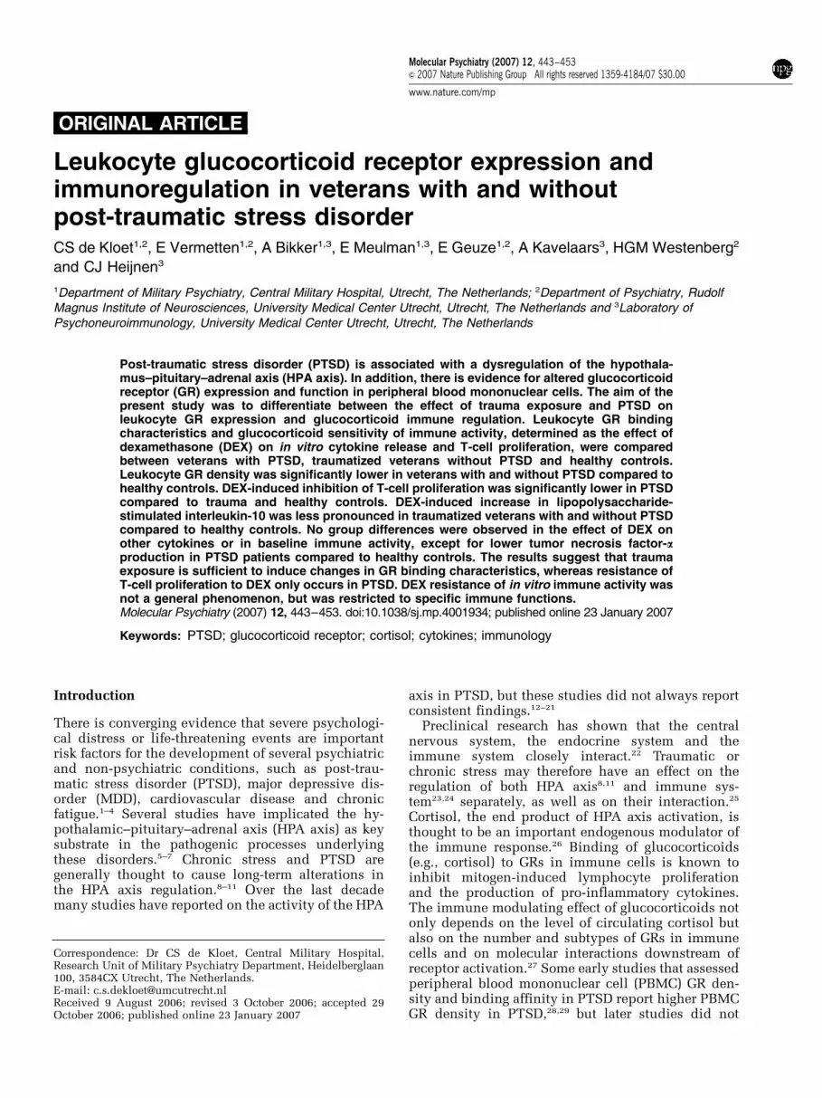
In vitro 3H-DEX binding to peripheral bloodmononuclear cellsNo significant group difference was observed in theaffinity of 3H-DEX binding to PBMC (Kd) (ANOVAF2,44 = 1.76; P = 0.18). A significant difference inglucocorticoid receptor number (Bmax) was observedbetween groups (Figure 1, Kruskal–Wallis testw2 = 11.73; df = 2; P = 0.003). GR number was signifi-cantly lower in PTSD (Bmax: median: 5235, range1101–9502) compared to HC (Bmax: median: 8445,range 5015–23 787; Kruskal–Wallis test P = 0.003). Asignificantly lower GR number was also observed inTC (Bmax: median: 5070, range 2847–8678) comparedto HC (Kruskal–Wallis test P = 0.004) (Figure 1). GRnumber was not significantly different between TCand PTSD (P = 0.75).
Composition of the leukocyte populationTo examine the possibility that the observed groupdifferences in GR binding characteristics were causedby differences in the composition of the leukocytepopulation in the peripheral blood, we analysed the
Leukocyte glucocorticoid receptor expression and immunoregulation in veteransCS de Kloet et al
445
Molecular Psychiatry

subset distribution of these cells. Total numbers ofleukocytes were all within the normal range (4.0–10.0�109/l) and there was no significant differencebetween groups (F2,84 = 0.19; P = 0.19). Moreover, nogroup differences were observed in total number oflymphocytes, monocytes, B-lymphocytes and T-lym-phocytes (total, helper/inducer and T suppressor/cytotoxic) per milliliter nor in the percentage of T-cellsubsets or B cells within the lymphocyte population.
Mitogen-induced cytokine secretionBefore analysing the sensitivity of the immune systemto regulation by DEX, we first determined the capacityof peripheral blood cells to proliferate and to releasepro- and anti-inflammatory cytokines in response tostimulation with the T-cell mitogen PHA. No signi-ficant differences were observed in the proliferativeresponse to PHA stimulation (ANOVA F2,79 = 2.38,P = 0.10), as well as in PHA-stimulated secretion ofIFN-g (ANOVA F2,67 = 0.70, P = 0.50) and IL-10 (ANO-VA F2,67 = 0.56, P = 0.57) (Table 2).
To evaluate the capacity of monocytes to releasecytokines, we stimulated whole-blood cultures withLPS, known to specifically induce cytokine release by
monocytes. We observed a significant group differ-ence in LPS-induced TNF-a secretion (ANOVAF2,84 = 4.21, P = 0.018). Post hoc tests showed thatthis effect could be attributed to a significantlylower level of TNF-a in cultures from PTSD patients(TNF-a: 7327277 pg/ml) compared to HC (TNF-a:9987320 pg/ml; Scheffe: P = 0.027). There was nosignificant difference in LPS-stimulated TNF-a secre-tion between TC and HC or between PTSD and TC(TNF-a: 9197439 pg/ml). No significant group differ-ence was observed in LPS-stimulated IL-10 levels(ANOVA F2,80 = 0.03, P = 0.97) (Table 2).
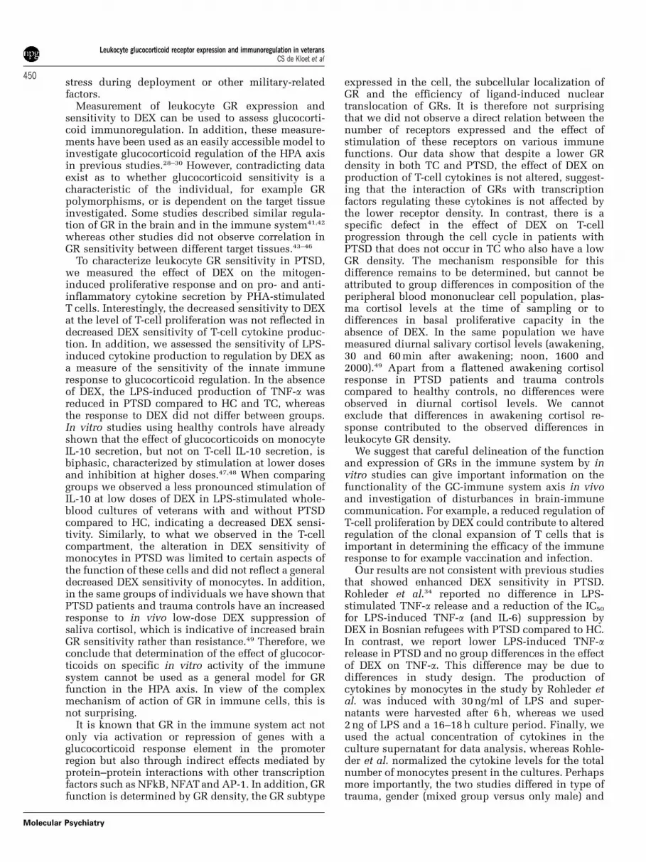
Ex vivo response of peripheral blood cells to DEXTo investigate the functional capacity of the GRs inleukocytes, the effect of DEX on mitogen-inducedT-cell proliferation and cytokine production wasmeasured. Addition of DEX to cultures stimulatedwith PHA resulted in a dose-dependent inhibition ofthe proliferative response (F1,5; 105 = 219; P < 0.001).The inhibitory effect of DEX was less pronouncedin samples from PTSD patients compared to HC(Figure 2, Table 3). There was a significant groupdifference in the dose of DEX resulting in a 50%
Table 1 Demographics characteristics and test variables of PTSD patients, TC and HC
PTSD (n = 29) Trauma controls (n = 29) Healthy controls (n = 25) P
Mean s.d. Range Mean s.d. Range Mean s.d. Range
Age 33.4 5.6 25–44 33.5 5 25–40 32.7 5.4 25–45 0.75Year of deployment 1992 5.8 1980–2002 1992 5.7 1980–2002 0.64Country of deployment Bosnia (n = 16) Bosnia (n = 16)
Lebanon (n = 6) Lebanon (n = 6)Cambodia (n = 5) Cambodia (n = 6)Afghanistan (n = 1) Afghanistan (n = 1)Kosovo (n = 1)
BMI 25.5 4.1 25.4 2.6 25.5 1.9 0.98
RaceCaucasian n = 28 n = 28 n = 24Other n = 1 n = 1 n = 1
CAPS 75.9 14.9 51–106 6.5 5.7 0–18 < 0.001Hamilton D 16.6 5.8 3–29 1 1.5 0–5 < 0.001
Comorbid disorders(lifetime)
MDD (n = 18) MDD (n = 3)
Bipolar II disorder (n = 3) Panic disorder (n = 1)Alcohol dependence (n = 2)Alcohol abuse (n = 3)Substance abuse (n = 2)Substance dependence (n = 2)Panic disorder (n = 3)Somatoform disorder (n = 5)
Abbreviations: BMI, body mass index; CAPS, clinical Administered PTSD Scale; HC, healthy controls; MDD, majordepressive disorder; PTSD, post-traumatic stress disorder; TC, trauma controls.Outcome variables were compared between three groups using parametric (ANOVA) or non-parametric tests (Kruskal–Wallistest) depending on the normality of distribution.Variables are displayed as mean, standard deviation (s.d.) and range.
Leukocyte glucocorticoid receptor expression and immunoregulation in veteransCS de Kloet et al
446
Molecular Psychiatry

inhibition of T-cell proliferation (IC50) (Kruskal–Wallis test w2 = 7.41; df = 2; P = 0.025). No significantdifference was observed in IC50 between PTSD andHC or between TC and HC, but PTSD patients showeda higher IC50 compared to TC (Kruskal–Wallis testw2 = 6.61; df = 1; P = 0.01) (Figure 2, Table 3). We also
observed significant group differences in the inhibi-tion of T-cell proliferation at the highest dose of DEX(1000 nM) (ANOVA: F2,79 = 4.59; P = 0.01. Inhibition ofT-cell proliferation at the highest dose of DEX inPTSD patients was significantly lower in PTSDcompared to HC and TC (P = 0.020). No significantdifferences in maximal effect of DEX (1000 nM)between TC and HC were detected.
Regulation of cytokine secretion by DEX
To investigate whether the alteration in GR functionin PTSD reflects a general alteration in GR function oris specific for certain aspects of the immune response,the effect of DEX on cytokine secretion was measured.
Figure 1 GR binding characteristics of peripheral bloodmononuclear cells. GR binding characteristics were deter-mined by classical 3H-DEX binding assays. The equilibriumdissociation constant (Kd, (a)) and the number of specific3H-DEX-binding sites per cell (Bmax, (b)) was calculated bycomputer-assisted iterative nonlinear regression curve fit-ting procedures using Prism 4.02. Data represent individualvalues and median. No significant group difference wasobserved in Kd (ANOVA). Kruskal–Wallis test showed asignificantly lower Bmax in PTSD (Bmax: median: 5235, range1101–9502) and trauma controls (Bmax: median: 5070, range2847–8678) compared to healthy controls (Bmax: median:8445, range 5015–23787).
Table 2 Cytokine release of PHA-stimulated whole-blood cultures and cytokine release of LPS-stimulated whole-blood culture
Cytokine Stimulator PTSD Trauma controls Healthy controls Subject group effect
Mean s.d. Mean s.d. Mean s.d.
T-cell IFN-g (pg/ml) PHA 8925 15431 6037 5836 8940 70 455 F2,67 = 0.70, P = 0.50IL10 (pg/ml) PHA 212 125 160 82 246 168 F2,67 = 0.56, P = 0.57
Monocyte TNF-a (pg/ml) LPS 732 277 919 439 998 320 F2,84 = 4.21, P = 0.018IL10 (pg/ml) LPS 59 37 59 40.0 56 66 F2,80 = 0.03, P = 0.97
Abbreviations: IFN-g, interferon-g; LPS, lippopolysaccharide; PHA, phytohemagglutinin; PTSD, post-traumatic stressdisorder; TC, trauma controls; TNF-a, tumor necrosis factor-a.Diluted blood was cultured with PHA for 72 h or with LPS for 18 h and cytokine levels were determined in the supernatantby ELISA. Data represent mean, and s.d. For PHA stimulated IL-10 secretion log-transformed values were analyzed. Nosignificant difference was observed in PHA stimulated T-cell proliferation and log transformed IL-10. We observed asignificant group difference in TNF-a level, but no group difference in IL-10 in response to culture with LPS.
Figure 2 DEX inhibition of PHA-stimulated T-cell prolif-eration. Whole-blood cultures were stimulated with PHA inthe presence of increasing concentrations of DEX and theproliferative response was determined. Data are expressedas percentage of proliferation in the absence of DEX andrepresent mean and s.e.m. *P < 0.05, **P < 0.01.
Leukocyte glucocorticoid receptor expression and immunoregulation in veteransCS de Kloet et al
447
Molecular Psychiatry

Figure 3 shows DEX inhibition of cytokine release inPHA-stimulated (Figure 3a and b) and LPS-stimulatedwhole-blood cultures (Figure 3c and d). IncreasingDEX concentrations significantly inhibited PHA-stimulated IFN-g (repeated measures: F1,2;8,14 = 32.8;P < 0.001) and IL-10 (repeated measures: F1,9;130 = 92.4;P < 0.001) production. However, no significant groupdifferences were observed in IC50 and maximal DEXinhibition (at 1000 nM DEX) of PHA-stimulated IFN-gand IL-10 release (Figure 3a and b).
LPS-stimulated TNF-a secretion was significantlyinhibited by DEX in all groups (repeated measures:F1,7;143 = 387; P < 0.001). No significant group differ-ences were observed for IC50 and effect of 1000 nM
DEX on LPS-stimulated TNF-a (Figure 3c). DEX alsosignificantly affected LPS-induced IL-10 secretion.DEX induced an increase in IL-10 secretion at thelower concentrations, with a peak between 20 and50 nM DEX (repeated measures: F4,243 = 5.6; P = 0.009),and decreased IL-10 secretion at higher DEX concen-trations (repeated measures: F3,243 = 144; P < 0.001)(Figure 3d). Repeated measure analysis showed asignificant group by dose interaction (F4,156 = 4.9;P = 0.001). (Figure 3d). This biphasic dose–responsecurve for the effect of DEX on IL-10 secretion did notallow calculation of the IC50, but significant groupdifferences were observed in area under the curve(AUC) (Kruskal–Wallis test w2 = 6.6; df = 2; P = 0.04)with significantly lower AUC in PTSD (P = 0.03) andTC (P = 0.02) compared to HC. When comparing LPS-stimulated IL-10 increase in response to DEX, DEXinduced a maximal increase (33%) at a DEX concen-tration of 50 nM in HC, whereas DEX induced amaximal increase in PTSD (12%) and TC (15%) at aDEX concentration of 10 nM. No significant groupdifferences were observed in the maximal dose ofDEX (1000 nmol/l).
Plasma cortisolTo determine if the observed group differences in GRbinding and function could be related to individualdifferences in plasma cortisol levels, we measured
plasma cortisol in the same blood samples. Plasmacortisol levels were within the normal range (0.20–0.65mmol/l) in all groups. There was no significantgroup difference in plasma cortisol concentrations(PTSD: 0.4170.11mmol/l, TC: 0.4170.07mmol/l, HC:0.3670.09mmol/l) (F2,76 = 2.55; P = 0.08). Analyses ofthe cortisol levels and the GR binding characteristicsshowed no correlation between plasma cortisol levelsand Bmax (Pearson: r =�0.070; P = 0.66;) or Kd (Pear-son: r =�0.23; P = 0.18). In addition, cortisol levelsdid not correlate with DEX inhibition of proliferation(AUC) (Pearson: r =�0.162; P = 0.15) either.
Comorbid major depressive disorder
Fourteen out of the 29 PTSD patients were diagnosedwith a current comorbid depressive disorder. As it hasbeen suggested that depressive disorder may beassociated with altered GR sensitivity, we analysedthe effect of comorbid depressive disorder on alloutcome measures within the PTSD group. Nosignificant differences were observed between PTSDpatients with and without comorbid MDD in leuko-cyte GR density (P = 0.74) and binding affinity(P = 0.19) (Table 4). We did not observe differencesin PHA- and LPS-stimulated cytokine secretion(P > 0.40) and the effect of DEX on cytokine secretion(P > 0.40) and T-cell proliferation (Table 4). Cortisollevels were not significantly higher in PTSD withcomorbid MDD (0.4470.10 mmol/l) compared toPTSD patients without MDD (0.3870.12 mmol/l,P = 0.13).
Discussion
In this study, we observed a lower leukocyte GRdensity in traumatized veterans with and withoutPTSD compared to nontraumatized healthy controls,whereas only veterans with PTSD show a reducedregulation of T-cell proliferation by DEX. The datasuggest that some biological alterations are specificfor PTSD whereas others are related to traumatic
Table 3 Dexamethasone inhibition of PHA-stimulated T-cell proliferation
PTSD(n = 28)
Trauma controls(n = 29)
Healthy controls(n = 25)
Subject groupeffect
Mean s.d. Mean s.d. Mean s.d.
DEX inhibition T-cell proliferationc.p.m. after PHA 44 933 18 167 36 894 22 072 48 308 19 218 F2,79 = 2.38; P = 0.10IC50 (nM) 92.6 121 34.3 40 46.1 41.8 w2 = 7.41; df = 2; P = 0.025Inhibition at 1000 nM DEX 83.2 19.7 93.4 11 93.5 10 F2,79 = 4.59; P = 0.01
Abbreviations: DEX, dexamethasone; PHA, phytohemagglutinin; PTSD, post-traumatic stress disorder.Whole-blood cultures were stimulated with PHA in the presence of increasing concentrations of DEX and the proliferativeresponse was determined by measuring [3H]-thymidine incorporation. IC50 was calculated using PRISM 4.02 software andwas analysed using a non-parametric test (Kruskal–Wallis test). Inhibition at 1000 nM DEX is expressed as % of controlvalues in the absence of DEX (100%). For maximal inhibition (DEX1000 nM) log-transformed values were analysed (ANOVA).Results are displayed as mean and standard deviation.
Leukocyte glucocorticoid receptor expression and immunoregulation in veteransCS de Kloet et al
448
Molecular Psychiatry

Figure 3 DEX inhibition of cytokine release. (a) ZIFN-g and (b) IL-10 levels were determined in the supernatants of wholeblood after culture for 72 h in the presence of the T-cell mitogen PHA and increasing DEX concentrations. (c) TNF-a and (d)IL-10 were determined in supernatants from whole-blood cultures-stimulated with LPS and increasing DEX concentrations.Data are expressed as percentage of cytokine production in the absence of DEX and represent mean and s.e.m. of PTSDpatients, trauma controls (TC) and healthy controls (HC).
Table 4 Effect of comorbid MDD on observed group differences in GR binding and GR sensitivity studies
PTSD-MDD PTSDþMDD Subject group effect
Mean s.d. Mean s.d.
GR bindingcharacteristicsKd (nmol/l) 24.7 7.6 18 9.1 F1,12 = 1.9; P = 0.19Bmax (binding sites/cell) 5714 2450 5152 2510 w2 = 0.11; df = 1; P = 0.74
DEX inhibition T-cell proliferationc.p.m. after PHA 46194 21749 43674 14458 F1.27 = 0.13; P = 0.72IC50 (nM) 125.9 159.2 59.3 52 w2 = 0.54; df = 1; P = 0.74Inhibition at 1000 nM DEX (%) 78.6 23.2 87.5 15.3 F1,27 = 1.40; P = 0.25
LPS-stimulated TNF-a (pg/ml) 761 256 700 305 F1,27 = 0.34; P = 0.56
Cortisol (mmol/l) 0.38 0.12 0.44 0.1 F1,27 = 2,50; P = 0.13
Abbreviations: DEX, dexamethasone; GR, glucocorticoid receptor; LPS, lippopolysaccharide; MDD, major depressivedisorder; PTSD, post-traumatic stress disorder; TNF-a, tumor necrosis factor-a.Differences in outcome measures were compared between PTSD patients with and without comorbid MDD, using parametric(ANOVA) or non-parametric tests (Kruskal–Wallis test) depending on the normality of distribution. Results are displayed asmean and standard deviation.
Leukocyte glucocorticoid receptor expression and immunoregulation in veteransCS de Kloet et al
449
Molecular Psychiatry
stress during deployment or other military-relatedfactors.
Measurement of leukocyte GR expression andsensitivity to DEX can be used to assess glucocorti-coid immunoregulation. In addition, these measure-ments have been used as an easily accessible model toinvestigate glucocorticoid regulation of the HPA axisin previous studies.28–30 However, contradicting dataexist as to whether glucocorticoid sensitivity is acharacteristic of the individual, for example GRpolymorphisms, or is dependent on the target tissueinvestigated. Some studies described similar regula-tion of GR in the brain and in the immune system41,42
whereas other studies did not observe correlation inGR sensitivity between different target tissues.43–46
To characterize leukocyte GR sensitivity in PTSD,we measured the effect of DEX on the mitogen-induced proliferative response and on pro- and anti-inflammatory cytokine secretion by PHA-stimulatedT cells. Interestingly, the decreased sensitivity to DEXat the level of T-cell proliferation was not reflected indecreased DEX sensitivity of T-cell cytokine produc-tion. In addition, we assessed the sensitivity of LPS-induced cytokine production to regulation by DEX asa measure of the sensitivity of the innate immuneresponse to glucocorticoid regulation. In the absenceof DEX, the LPS-induced production of TNF-a wasreduced in PTSD compared to HC and TC, whereasthe response to DEX did not differ between groups.In vitro studies using healthy controls have alreadyshown that the effect of glucocorticoids on monocyteIL-10 secretion, but not on T-cell IL-10 secretion, isbiphasic, characterized by stimulation at lower dosesand inhibition at higher doses.47,48 When comparinggroups we observed a less pronounced stimulation ofIL-10 at low doses of DEX in LPS-stimulated whole-blood cultures of veterans with and without PTSDcompared to HC, indicating a decreased DEX sensi-tivity. Similarly, to what we observed in the T-cellcompartment, the alteration in DEX sensitivity ofmonocytes in PTSD was limited to certain aspects ofthe function of these cells and did not reflect a generaldecreased DEX sensitivity of monocytes. In addition,in the same groups of individuals we have shown thatPTSD patients and trauma controls have an increasedresponse to in vivo low-dose DEX suppression ofsaliva cortisol, which is indicative of increased brainGR sensitivity rather than resistance.49 Therefore, weconclude that determination of the effect of glucocor-ticoids on specific in vitro activity of the immunesystem cannot be used as a general model for GRfunction in the HPA axis. In view of the complexmechanism of action of GR in immune cells, this isnot surprising.
It is known that GR in the immune system act notonly via activation or repression of genes with aglucocorticoid response element in the promoterregion but also through indirect effects mediated byprotein–protein interactions with other transcriptionfactors such as NFkB, NFAT and AP-1. In addition, GRfunction is determined by GR density, the GR subtype
expressed in the cell, the subcellular localization ofGR and the efficiency of ligand-induced nucleartranslocation of GRs. It is therefore not surprisingthat we did not observe a direct relation between thenumber of receptors expressed and the effect ofstimulation of these receptors on various immunefunctions. Our data show that despite a lower GRdensity in both TC and PTSD, the effect of DEX onproduction of T-cell cytokines is not altered, suggest-ing that the interaction of GRs with transcriptionfactors regulating these cytokines is not affected bythe lower receptor density. In contrast, there is aspecific defect in the effect of DEX on T-cellprogression through the cell cycle in patients withPTSD that does not occur in TC who also have a lowGR density. The mechanism responsible for thisdifference remains to be determined, but cannot beattributed to group differences in composition of theperipheral blood mononuclear cell population, plas-ma cortisol levels at the time of sampling or todifferences in basal proliferative capacity in theabsence of DEX. In the same population we havemeasured diurnal salivary cortisol levels (awakening,30 and 60 min after awakening; noon, 1600 and2000).49 Apart from a flattened awakening cortisolresponse in PTSD patients and trauma controlscompared to healthy controls, no differences wereobserved in diurnal cortisol levels. We cannotexclude that differences in awakening cortisol re-sponse contributed to the observed differences inleukocyte GR density.
We suggest that careful delineation of the functionand expression of GRs in the immune system by invitro studies can give important information on thefunctionality of the GC-immune system axis in vivoand investigation of disturbances in brain-immunecommunication. For example, a reduced regulation ofT-cell proliferation by DEX could contribute to alteredregulation of the clonal expansion of T cells that isimportant in determining the efficacy of the immuneresponse to for example vaccination and infection.
Our results are not consistent with previous studiesthat showed enhanced DEX sensitivity in PTSD.Rohleder et al.34 reported no difference in LPS-stimulated TNF-a release and a reduction of the IC50
for LPS-induced TNF-a (and IL-6) suppression byDEX in Bosnian refugees with PTSD compared to HC.In contrast, we report lower LPS-induced TNF-arelease in PTSD and no group differences in the effectof DEX on TNF-a. This difference may be due todifferences in study design. The production ofcytokines by monocytes in the study by Rohleder etal. was induced with 30 ng/ml of LPS and super-natants were harvested after 6 h, whereas we used2 ng of LPS and a 16–18 h culture period. Finally, weused the actual concentration of cytokines in theculture supernatant for data analysis, whereas Rohle-der et al. normalized the cytokine levels for the totalnumber of monocytes present in the cultures. Perhapsmore importantly, the two studies differed in type oftrauma, gender (mixed group versus only male) and
Leukocyte glucocorticoid receptor expression and immunoregulation in veteransCS de Kloet et al
450
Molecular Psychiatry

age. For example, female and male Bosnian warrefugees with PTSD and a control group consistingof Bosnian refugees (n = 5) and laboratory staff (n = 8)were used by Rohleder et al., whereas we includedonly male veterans with PTSD (n = 29), male veteranswithout PTSD (n = 29) and a healthy nonmilitary malecontrol group, recruited by advertisement (n = 25).The veterans in our study were at the time of traumaexposure trained soldiers who were involved inpeacekeeping missions outside The Netherlands.Although no details have been reported on the historyof the Bosnian refugees, it is likely that there are majordifferences in physical and psychosocial status at thetime of trauma exposure between the veterans in ourstudy and the Bosnian refugees in the study byRohleder et al.
Yehuda et al. studied GR function in relation toalterations in HPA axis functioning in PTSD. Theseauthors reported increased sensitivity for DEX ofperipheral blood leukocytes from PTSD patients whenusing inhibition of lysozyme activity as a read-out.32
In contrast to the assays we used to determineglucocorticoid receptor function, in the study byYehuda et al., cells were not stimulated to perform acertain immune activity, isolated cells (in the absenceof endogenous plasma) were used and the cultureperiod was much longer. In another study,30 the in vivoeffect of administration of DEX on the change in GRexpression was studied and the data suggest increasedGR functioning in PTSD, as oral administration ofDEX induced an increased reduction of GR number inperipheral blood mononuclear cells of PTSD pa-tients.30 Obviously, we cannot directly compare thesein vivo effects on receptor availability with in vitrostudies on modulation of specific immune functions.
Our finding of a lower leukocyte GR density is incontrast with other studies that reported higherleukocyte GR density in PTSD compared to healthycontrols28,29 or data by the same group suggesting thatthere are no differences in GR density between PTSDpatients and trauma or healthy controls.29–31 Differ-ences in the method used to assess GR density mayhave contributed to the discrepancy in resultsbetween GR binding studies. Yehuda et al.28–32
determined GR density in a total cell homogenate offrozen cells, using a binding assay with only one doseof DEX to calculate GR density. In our present study,GR density and binding affinity was measured inliving cells using a classical whole-cell binding assay.It may well be possible that during the process offreezing and homogenizing cells, changes in protein–protein interactions, changes in degree of phosphor-ylation, protein degradation, changes in the cellmembrane or loss of specific cell types may influencethe outcome of GR binding studies in specific patientgroups. As mentioned above, the studies of Yehuda etal. and our study also differ with respect to thecomposition of the subject population. For example,our populations consisted completely of Caucasians,whereas in the study of Yehuda et al., 42% of thesubjects were African-American.
It remains to be determined whether the apparentdiscrepancies in the conclusions on the nature of thealterations in leukocyte GR binding and GR functionare due to differences in subject characteristics or todifferences in the functional read-outs used.
Our data suggest that traumatic stress duringdeployment may contribute to the altered expressionof GR in TC and PTSD. However, we cannot excludethat other factors, associated with deployment andmilitary training, including vaccination, change ofenvironment, physical exercise, chronic stress andmilitary selection may also be responsible for thedifference in GR expression levels between veteransand the non-military healthy controls.
Differences in patient characteristics between stu-dies included gender, age, cause of trauma and thetime passed since the traumatic event. Preclinicalstudies have shown that the development of receptoralterations after intense social stress is time-depen-dent.50,51 In a clinical design, significant correlationsbetween the time passed since the traumatic eventand in vivo DEX sensitivity have been reported.52 Inline with these observations, it is important tohighlight that our results are strengthened by theinclusion of a homogeneous population in type oftrauma, moment of trauma in lifespan and the timepassed since the traumatic event.
There are some limiting factors that need to betaken into account when interpreting the data. First ofall, we did not include a military non-traumatizedcontrol group. This would have enabled us todifferentiate between the effects of trauma and othermilitary-related factors on leukocyte GR expressionand sensitivity. Second, we included PTSD patientswith and without a comorbid MDD. Data fromepidemiological surveys indicate that the vast major-ity of individuals with PTSD meet criteria for at leastone other psychiatric disorder.53,54 The most commoncomorbid diagnoses are depressive disorder, sub-stance use disorders and other anxiety disorders. Assubstance disorders are known to influence neuro-biology significantly, we have excluded this group.Because of the high rate of comorbid MDD in PTSD,almost half of the patients with PTSD is diagnosedwith comorbid MDD,55 and the large overlap inclinical symptoms between PTSD and MDD, moststudies on PTSD do not exclude patients withcomorbid MDD, but do analyse the effect of comorbidMDD on outcome measures. Other studies thatassessed GR function or GR binding characteristicsalso included patients with comorbid MDD; therefore,it is not likely that inclusion of patients withcomorbid MDD can account for the observed differ-ences between our data and previous studies. Asseveral studies have reported a decreased effect ofDEX on T-cell proliferation in MDD56,57 it is importantto investigate the effect of a comorbid MDD. However,our additional analyses suggest that a comorbidMDD did not cause the observed differences. Third,although early life trauma has been shown to affectHPA axis functioning significantly,58 and might there-
Leukocyte glucocorticoid receptor expression and immunoregulation in veteransCS de Kloet et al
451
Molecular Psychiatry

fore also have its effect on glucocorticoid immunor-egulation, we did not assess the contribution of earlylife trauma.
In this study, we reported long-lasting alterations inglucocorticoid immune regulation after deployment.Epidemiological research increasingly suggests thatexposure to traumatic events is related to adversehealth outcomes and the onset of specific dis-eases.3,4,59 To gain more insight in human adaptationto traumatic stress and the relation of these processeswith alterations in GR expression, glucocorticoid–immune interaction and psychopathology, furtherresearch in a longitudinal, instead of a cross-sectionaldesign, is preferable.
Acknowledgments
This work was financially supported by the DutchMinistry of Defense. We thank Arthur Rademaker,for clinical assessments, Jos Weerts, for his help inselecting trauma controls, and Mirjam Maas and JitskeZijlstra, Laboratory for Psychoneuroimmunology, forexcellent technical assistance.
References
1 McEwen BS. Protection and damage from acute and chronic stress:allostasis and allostatic overload and relevance to the pathophy-siology of psychiatric disorders. Ann NYAcad Sci 2004; 1032: 1–7.
2 Charney DS, Manji HK. Life stress, genes, and depression:multiple pathways lead to increased risk and new opportunitiesfor intervention. Sci STKE 2004; 2004: re5.
3 Boscarino JA. Posttraumatic stress disorder and physical illness:results from clinical and epidemiologic studies. Ann NY Acad Sci2004; 1032: 141–153.
4 Amir M, Kaplan Z, Neumann L, Sharabani R, Shani N, Buskila D.Posttraumatic stress disorder, tenderness and fibromyalgia.J Psychosom Res 1997; 42: 607–613.
5 Nemeroff CB, Bremner JD, Foa EB, Mayberg HS, North CS, SteinMB. Posttraumatic stress disorder: a state-of-the-science review.J Psychiatr Res 2006; 40: 1–21.
6 Holsboer F. The corticosteroid receptor hypothesis of depression.Neuropsychopharmacology 2000; 23: 477–501.
7 Claes SJ. CRH, stress, and major depression: a psychobiologicalinterplay. Vitam Horm 2004; 69: 117–150.
8 Heim C, Ehlert U, Hellhammer DH. The potential role ofhypocortisolism in the pathophysiology of stress-related bodilydisorders. Psychoneuroendocrinology 2000; 25: 1–35.
9 Newport DJ, Heim C, Bonsall R, Miller AH, Nemeroff CB. Pituitary-adrenal responses to standard and low-dose dexamethasonesuppression tests in adult survivors of child abuse. Biol Psychiatry2004; 55: 10–20.
10 Heim C, Owens MJ, Plotsky PM, Nemeroff CB. Persistent changesin corticotropin-releasing factor systems due to early life stress:relationship to the pathophysiology of major depression andposttraumatic stress disorder. Psychopharmacol Bull 1997; 33:185–192.
11 Yehuda R. Biology of posttraumatic stress disorder. J ClinPsychiatry 2001; 62(Suppl 17): 41–46.
12 Baker DG, West SA, Nicholson WE, Ekhator NN, Kasckow JW, HillKK et al. Serial CSF corticotropin-releasing hormone levels andadrenocortical activity in combat veterans with posttraumaticstress disorder. Am J Psychiatry 1999; 156: 585–588.
13 Bremner JD, Licinio J, Darnell A, Krystal JH, Owens MJ, SouthwickSM et al. Elevated CSF corticotropin-releasing factor concentra-tions in posttraumatic stress disorder. Am J Psychiatry 1997; 154:624–629.
14 Baker DG, Ekhator NN, Kasckow JW, Dashevsky B, Horn PS,Bednarik L et al. Higher levels of basal serial CSF cortisol incombat veterans with posttraumatic stress disorder. Am JPsychiatry 2005; 162: 992–994.
15 Yehuda R, Teicher MH, Levengood RA, Trestman RL, Siever LJ.Circadian regulation of basal cortisol levels in posttraumatic stressdisorder. Ann NY Acad Sci 1994; 746: 378–380.
16 Bremner JD, Vythilingam M, Anderson G, Vermetten E, McGlashanT, Heninger G et al. Assessment of the hypothalamic-pituitary-adrenal axis over a 24-h diurnal period and in response toneuroendocrine challenges in women with and without childhoodsexual abuse and posttraumatic stress disorder. Biol Psychiatry2003; 54: 710–718.
17 Wessa M, Rohleder N, Kirschbaum C, Flor H. Altered cortisolawakening response in posttraumatic stress disorder. Psycho-neuroendocrinology 2006; 31: 209–215.
18 Neylan TC, Brunet A, Pole N, Best SR, Metzler TJ, Yehuda R et al.PTSD symptoms predict waking salivary cortisol levels in policeofficers. Psychoneuroendocrinology 2005; 30: 373–381.
19 Liberzon I, Abelson JL, Flagel SB, Raz J, Young EA. Neuroendo-crine and psychophysiologic responses in PTSD: a symptomprovocation study. Neuropsychopharmacology 1999; 21: 40–50.
20 Bremner JD, Vythilingam M, Vermetten E, Adil J, Khan S, Nazeer Aet al. Cortisol response to a cognitive stress challenge inposttraumatic stress disorder (PTSD) related to childhood abuse.Psychoneuroendocrinology 2003; 28: 733–750.
21 Elzinga BM, Schmahl CG, Vermetten E, van Dyck R, Bremner JD.Higher cortisol levels following exposure to traumatic remindersin abuse-related PTSD. Neuropsychopharmacology 2003; 28:1656–1665.
22 Besedovsky HO, del Rey A. The cytokine-HPA axis feed-backcircuit. Z Rheumatol 2000; 59(Suppl 2): 26–30.
23 Glaser R, Kiecolt-Glaser JK. Stress-induced immune dysfunction:implications for health. Nat Rev Immunol 2005; 5: 243–251.
24 Bonneau RH, Kiecolt-Glaser JK, Glaser R. Stress-induced modula-tion of the immune response. Ann NY Acad Sci 1990; 594:253–269.
25 Raison CL, Miller AH. When not enough is too much: the role ofinsufficient glucocorticoid signaling in the pathophysiology ofstress-related disorders. Am J Psychiatry 2003; 160: 1554–1565.
26 Marshall Jr GD, Agarwal SK. Stress, immune regulation, andimmunity: applications for asthma. Allergy Asthma Proc 2000; 21:241–246.
27 Bamberger CM, Schulte HM, Chrousos GP. Molecular determi-nants of glucocorticoid receptor function and tissue sensitivity toglucocorticoids. Endocr Rev 1996; 17: 245–261.
28 Yehuda R, Lowy MT, Southwick SM, Shaffer D, Giller Jr EL.Lymphocyte glucocorticoid receptor number in posttraumaticstress disorder. Am J Psychiatry 1991; 148: 499–504.
29 Yehuda R, Boisoneau D, Mason JW, Giller EL. Glucocorticoidreceptor number and cortisol excretion in mood, anxiety, andpsychotic disorders. Biol Psychiatry 1993; 34: 18–25.
30 Yehuda R, Boisoneau D, Lowy MT, Giller Jr EL. Dose-responsechanges in plasma cortisol and lymphocyte glucocorticoidreceptors following dexamethasone administration in combatveterans with and without posttraumatic stress disorder. ArchGen Psychiatry 1995; 52: 583–593.
31 Yehuda R, Halligan SL, Grossman R, Golier JA, Wong C. Thecortisol and glucocorticoid receptor response to low dosedexamethasone administration in aging combat veterans andholocaust survivors with and without posttraumatic stressdisorder. Biol Psychiatry 2002; 52: 393–403.
32 Yehuda R, Golier JA, Yang RK, Tischler L. Enhanced sensitivity toglucocorticoids in peripheral mononuclear leukocytes in posttrau-matic stress disorder. Biol Psychiatry 2004; 55: 1110–1116.
33 Gotovac K, Sabioncello A, Rabatic S, Berki T, Dekaris D. Flowcytometric determination of glucocorticoid receptor (GCR) expres-sion in lymphocyte subpopulations: lower quantity of GCR inpatients with posttraumatic stress disorder (PTSD). Clin ExpImmunol 2003; 131: 335–339.
34 Rohleder N, Joksimovic L, Wolf JM, Kirschbaum C. Hypocortiso-lism and increased glucocorticoid sensitivity of pro-inflammatorycytokine production in Bosnian war refugees with posttraumaticstress disorder. Biol Psychiatry 2004; 55: 745–751.
Leukocyte glucocorticoid receptor expression and immunoregulation in veteransCS de Kloet et al
452
Molecular Psychiatry

35 Hovens JE, van der Ploeg HM, Bramsen I, Klaarenbeek MT,Schreuder JN, Rivero VV. The development of the self-ratinginventory for posttraumatic stress disorder. Acta Psychiatr Scand1994; 90: 172–183.
36 First MB, Spitzer RL, Wiliams JBW, Gibbon M. Structured ClinicalInterview for DSM-IV Axis I Disorders. American PsychiatricAssociation: Washington, DC, 1997, pp 49–53.
37 Blake DD, Weathers FW, Nagy LM, Kaloupek DG, Gusman FD,Charney DS et al. The development of a Clinician-AdministeredPTSD Scale. J Trauma Stress 1995; 8: 75–90.
38 Crabtree G. S.K.M.A. Glucocorticoid Receptors. Churchill Living-stone: New York, 1981.
39 Bonnans C, Chanez P, Meziane H, Godard P, Bousquet J, Vachier I.Glucocorticoid receptor-binding characteristics in severe asthma.Eur Respir J 2003; 21: 985–988.
40 Kavelaars A, Kuis W, Knook L, Sinnema G, Heijnen CJ. Disturbedneuroendocrine-immune interactions in chronic fatigue syn-drome. J Clin Endocrinol Metab 2000; 85: 692–696.
41 Lowy MT. Corticosterone regulation of brain and lymphoid corticos-teroid receptors. J Steroid Biochem Mol Biol 1991; 39: 147–154.
42 Brown PH, Teelucksingh S, Matusiewicz SP, Greening AP,Crompton GK, Edwards CR. Cutaneous vasoconstrictor responseto glucocorticoids in asthma. Lancet 1991; 337: 576–580.
43 Ebrecht M, Buske-Kirschbaum A, Hellhammer D, Kern S, RohlederN, Walker B et al. Tissue specificity of glucocorticoid sensitivity inhealthy adults. J Clin Endocrinol Metab 2000; 85: 3733–3739.
44 Chriguer RS, Elias LL, da Silva Jr IM, Vieira JG, Moreira AC, deCastro M. Glucocorticoid sensitivity in young healthy individuals:in vitro and in vivo studies. J Clin Endocrinol Metab 2005; 90:5978–5984.
45 Schuld A, Schmid DA, Haack M, Holsboer F, Friess E, PollmacherT. Hypothalamo-pituitary-adrenal function in patients with de-pressive disorders is correlated with baseline cytokine levels, butnot with cytokine responses to hydrocortisone. J Psychiatr Res2003; 37: 463–470.
46 Lowy MT. Reserpine-induced decrease in type I and II corticoster-oid receptors in neuronal and lymphoid tissues of adrenalecto-mized rats. Neuroendocrinology 1990; 51: 190–196.
47 Franchimont D, Martens H, Hagelstein MT, Louis E, Dewe W,Chrousos GP et al. Tumor necrosis factor alpha decreases, andinterleukin-10 increases, the sensitivity of human monocytes todexamethasone: potential regulation of the glucocorticoid recep-tor. J Clin Endocrinol Metab 1999; 84: 2834–2839.
48 Hodge S, Hodge G, Flower R, Han P. Methyl-prednisolone up-regulates monocyte interleukin-10 production in stimulated wholeblood. Scand J Immunol 1999; 49: 548–553.
49 De Kloet CS, Vermetten E, Heijnen CJ, Geuze E, Lentjes EGWM,Westenberg HGM. Enhanced cortisolsuppression in response todexamethasone administration in veterans with and withoutPTSD. Psychoneuroendocrinology (in press).
50 Buwalda B, Felszeghy K, Horvath KM, Nyakas C, de Boer SF,Bohus B et al. Temporal and spatial dynamics of corticosteroidreceptor down-regulation in rat brain following social defeat.Physiol Behav 2001; 72: 349–354.
51 Buwalda B, de Boer SF, Schmidt ED, Felszeghy K, Nyakas C,Sgoifo A et al. Long-lasting deficient dexamethasone suppressionof HPA activation following peripheral CRF challenge in socialdefeated rats. J Neuroendocrinol 1999; 11: 513–520.
52 Yehuda R, Halligan SL, Golier JA, Grossman R, Bierer LM. Effectsof trauma exposure on the cortisol response to dexamethasoneadministration in PTSD and major depressive disorder. Psycho-neuroendocrinology 2004; 29: 389–404.
53 Brady KT, Killeen TK, Brewerton T, Lucerini S. Comorbidity ofpsychiatric disorders and posttraumatic stress disorder. J ClinPsychiatry 2000; 61(Suppl 7): 22–32.
54 Brunello N, Davidson JR, Deahl M, Kessler RC, Mendlewicz J,Racagni G et al. Posttraumatic stress disorder: diagnosis andepidemiology, comorbidity and social consequences, biology andtreatment. Neuropsychobiology 2001; 43: 150–162.
55 Shalev AY. What is posttraumatic stress disorder? J Clin Psychiatry2001; 62(Suppl 17): 4–10.
56 Calfa G, Kademian S, Ceschin D, Vega G, Rabinovich GA, VolosinM. Characterization and functional significance of glucocorticoidreceptors in patients with major depression: modulation byantidepressant treatment. Psychoneuroendocrinology 2003; 28:687–701.
57 Pariante CM. Glucocorticoid receptor function in vitro in patientswith major depression. Stress 2004; 7: 209–219.
58 Shea A, Walsh C, Macmillan H, Steiner M. Child maltreatment andHPA axis dysregulation: relationship to major depressive disorderand post traumatic stress disorder in females. Psychoneuroendo-crinology 2005; 30: 162–178.
59 Kang HK, Natelson BH, Mahan CM, Lee KY, Murphy FM.Posttraumatic stress disorder and chronic fatigue syndrome-likeillness among Gulf War veterans: a population-based survey of30 000 veterans. Am J Epidemiol 2003; 157: 141–148.
Leukocyte glucocorticoid receptor expression and immunoregulation in veteransCS de Kloet et al
453
Molecular Psychiatry