laura rossetto foschera
TRANSCRIPT
1
UNIVERSIDADE FEDERAL DE SANTA MARIA
CENTRO DE CIÊNCIAS DA SAÚDE
PROGRAMA DE PÓS-GRADUAÇÃO EM REABILITAÇÃO FUNCIONAL
Laura Rossetto Foschera
CINEMÁTICA DE MEMBROS INFERIORES EM ADOLESCENTES
COM E SEM DOR PATELOFEMORAL
Santa Maria, RS, Brasil
2021

2
Laura Rossetto Foschera
CINEMÁTICA DE MEMBROS INFERIORES EM ADOLESCENTES
COM E SEM DOR PATELOFEMORAL
Dissertação apresentada ao Curso de Pós-
Graduação em Reabilitação Funcional do
Centro de Ciências da Saúde da Universidade
Federal de Santa Maria (UFSM) como
requisito para obtenção do título de Mestre em
Reabilitação Funcional.
Orientador: Prof. Dr. Carlos Bolli Mota
Coorientadora: Prof.ª. Dr.ª Michele Forgiarini Saccol
Santa Maria, RS 2021
3
4
Laura Rossetto Foschera
CINEMÁTICA DE MEMBROS INFERIORES EM ADOLESCENTES
COM E SEM DOR PATELOFEMORAL
Dissertação apresentada ao Curso de Pós-
Graduação em Reabilitação Funcional do
Centro de Ciências da Saúde da Universidade
Federal de Santa Maria (UFSM) como requisito
para obtenção do título de Mestre em
Reabilitação Funcional.
Aprovado em 19 de fevereiro de 2021:
____________________________________
Prof. Dr. Carlos Bolli Mota
Presidente/Orientador
_____________________________________
Prof. Dra. Heloyse Uliam Kuriki (UFSC)
_____________________________________
Prof. Dr. Guilherme Silva Nunes (UFSM)
Santa Maria, RS
2021

5
Dedico este trabalho às adolescentes voluntárias dessa pesquisa, que com muito entusiasmo e
solicitude dedicaram seu tempo para contribuir com a ciência.
6
AGRADECIMENTOS
Sempre soube que não chegamos a lugar nenhum sozinhos. Durante este período de mestrado
percebi que o mundo da pesquisa científica também não se faz sozinho. Não teria sido
possível desenvolver este trabalho sem o encorajamento e amparo de muitas pessoas
queridas. Muitos momentos foram árduos e acredito que minhas sinceras palavras não serão
capazes de expressar meu sentimento de gratidão a todos vocês.
Primeiramente, ao meu pai Dario e minha mãe Adriana, que nunca hesitaram para que
continuasse estudando, instigando a necessidade de aprimoramento pessoal e profissional.
Vocês são minha motivação pra tudo.
À minha irmã Emília, que sempre esteve ao meu lado nesse período.
Ao meu companheiro Orlando, sem o qual essa dissertação não teria sido escrita. Você me
manteve firme, acolheu e encorajou, sempre acreditando em meu potencial. Obrigada por
sempre estar ao meu lado.
Aos meus avós, Zilda e Francisco, Luci (in memoriam) e Jandir (in memoriam), que com
certeza estão/estariam muito felizes com minha conquista.
À minha prima Paulina, minhas tias, em especial à Sandra (in memoriam) e Ireni (in
memoriam), que sempre me inspiraram em todos os sentidos.
Ao meu orientador, professor Bolli, por permitir que eu adentrasse no mundo da pesquisa
científica. Nunca esquecerei minha primeira visita ao Laboratório de Biomecânica, no sexto
semestre de graduação, no qual o senhor pacientemente me conduziu e mostrou todas as
infinitas possibilidades daquele ambiente. Agradeço imensamente por todo o convívio, trocas
enriquecedoras tanto no âmbito profissional quanto pessoal.
À minha coorientadora, professora Michele, por sempre ter uma palavra amiga, por estar
disposta a contribuir com meu trabalho, pelas risadas, abraços e amizade! Sua generosidade,
prontidão e intensidade são únicas. Obrigada por acalmar meus momentos de adversidade
com palavras de alento.
7
Ao Programa de Pós-Graduação em Reabilitação Funcional (PPGRF) da Universidade
Federal de Santa Maria (UFSM) – RS, incluindo todo o corpo docente e funcionários que
foram fundamentais em minha caminhada durante o período de mestrado.
Ao meus amigos e professores do Laboratório de Biomecânica (LABIOMEC), por
compartilharem tanta experiência e sempre estarem dispostos a colaborar para que esse
trabalho se concretizasse. Em especial, agradeço à Gabriela, minha parceira no
desenvolvimento de toda parte metodológica e coletas desse estudo. Também à Karine e ao
Gustavo, que estiveram comigo desde meu primeiro contato com o Lab. Não é por acaso que
vocês foram convidados para contribuir, são pessoas que confio e admiro muito. Saibam que
foram imprescindíveis para a finalização desse artigo. É muito bom ter com quem contar. O
LABIOMEC é muito mais que um laboratório, é o lugar no qual me sinto em casa, pois sei
que sempre posso compartilhar as coisas boas e ruins, e que nada se compara aos nossos
momentos de reunião e café passado.
Aos membros da banca examinadora, Prof. Dra. Heloyse Uliam Kuriki (UFSC), Prof. Dr.
Guilherme Silva Nunes (UFSM) e Prof. Dr. Jeam Marcel Geremia (UFRGS), por aceitarem
prontamente e gentilmente o convite para aprimorarem este estudo.
A todos os professores, funcionários e alunos do Centro de Educação Física e Desporto
(CEFD) que tive o prazer de compartilhar ideias.
Às minhas amigas Nubia e Cláudia, com as quais divido os anseios e alegrias desde o
período de graduação até o mestrado. Vocês são mais que especiais.
Ao Colégio Técnico Industrial de Santa Maria (CTISM), por disponibilizarem espaço e tempo
para o recrutamento da maioria das voluntárias deste estudo.
À Universidade Federal de Santa Maria, por proporcionar educação pública de qualidade.
Às meninas voluntárias deste estudo. Vocês tornaram o período de coletas leve, foram nossa
maior motivação. Sem a disponibilidade de vocês, nada teria sido feito. Gratidão eterna!
A todos que de alguma forma fizeram parte da minha formação.
Muito obrigada.

8
“Um dia vamos acordar e ver todas as meninas, no Brasil e no mundo, na escola. Sem medo
de estudar, com educação de qualidade [...] e podendo sonhar com o que quiser – ser
médica, policial ou qualquer outra coisa.”
Malala Yousafzai

9
RESUMO
CINEMÁTICA DE MEMBROS INFERIORES EM ADOLESCENTES COM E SEM
DOR PATELOFEMORAL
AUTORA: Laura Rossetto Foschera
ORIENTADOR: Carlos Bolli Mota
COORIENTADORA: Michele Forgiarini Saccol
A etiologia da dor patelofemoral (DPF) é multifatorial e alguns aspectos biomecânicos de
origem local, distal e proximal são destacados na literatura como potenciais causadores de
estresse articular em adultos, porém, é durante a adolescência que o início dos sintomas são
geralmente observados. A investigação de variáveis cinemáticas na população adolescente
especialmente em tarefas funcionais ainda foi pouco explorada, tornando necessária a análise
da cinemática de membros inferiores de adolescentes durante um teste funcional. O objetivo
deste estudo foi analisar o padrão cinemático de membros inferiores durante o teste de degrau
lateral em adolescentes do sexo feminino com e sem DPF. Foi realizada análise cinemática de
nove adolescentes com DPF e nove adolescentes assintomáticas durante a execução do teste
Lateral Step Down. Os ângulos articulares dos membros inferiores foram comparados durante
a execução do LSD, sendo que os valores médios de ângulo de quadril, joelho e tornozelo foram
interpolados em 101 pontos para as fases descendente e ascendente. Além disso, usamos um
modelo de regressão linear para determinar como os ângulos do quadril e do tornozelo estavam
relacionados aos ângulos frontais do joelho em ambos os grupos. Não houve diferença
estatística no padrão cinemático de membros inferiores durante o teste LSD entre adolescentes
do sexo feminino com e sem DPF. Além disso, durante as fases de agachamento e subida do
teste LSD, a coordenação entre quadril, joelho e tornozelo não apresentou diferenças entre os
grupos.
Palavras-chave: Joelho. Patela. Fenômenos Biomecânicos. Adolescente.
10
ABSTRACT
LOWER LIMB KINEMATICS IN ADOLESCENTS WITH AND WITHOUT
PATELLOFEMORAL PAIN
AUTHOR: Laura Rossetto Foschera
ADVISOR: Carlos Bolli Mota
CO-ADVISOR: Michele Forgiarini Saccol
The etiology of patelofemoral pain (PFP) is multifactorial and some biomechanical aspects of
local, distal and proximal origin are highlighted in the literature as potential causes of joint
stress in adults, however, it is during adolescence that the onset of symptoms is observed. The
investigation of kinematic variables in the adolescent population, especially in functional tasks,
has been little explored, making it necessary to analyze the kinematics of the lower limbs of
adolescents during a functional test. The aim of this study was to analyze the kinematic pattern
of lower limbs during the lateral step test in female adolescents with and without PFP.
Kinematic analysis of nine adolescents with patellofemoral pain and nine asymptomatic
adolescents was performed during the execution of the Lateral Step Down test. The joint angles
of the lower limbs were compared during the execution of the LSD, and the mean values of the
hip, knee and ankle angle were interpolated in 101 points for the squat and lift phases. In
addition, we used a linear regression model to determine how the hip and ankle angles were
related to the frontal knee angles in both groups. There was no statistical difference in the
kinematic pattern of the lower limbs during the LSD test between female adolescents with and
without PFP. In addition, during the squat and climb phases of the LSD test, coordination
between hips, knees and ankles did not differ between groups.
Keywords: Knee. Patella. Biomechanical Phenomena. Adolescent.
11
SUMÁRIO
1 INTRODUÇÃO GERAL .................................................................................................... 11
2 ARTIGO ............................................................................................................................... 13
2.1 INTRODUÇÃO .............................................................................................................. 16
2.2 METODOLOGIA ........................................................................................................... 17
2.3 RESULTADOS .............................................................................................................. 19
2.4 DISCUSSÃO .................................................................................................................. 20
2.5 CONSIDERAÇÕES FINAIS ......................................................................................... 23
2.6 REFERÊNCIAS ............................................................................................................. 32
3. CONCLUSÃO GERAL ..................................................................................................... 36
4. REFERÊNCIAS ................................................................................................................. 37
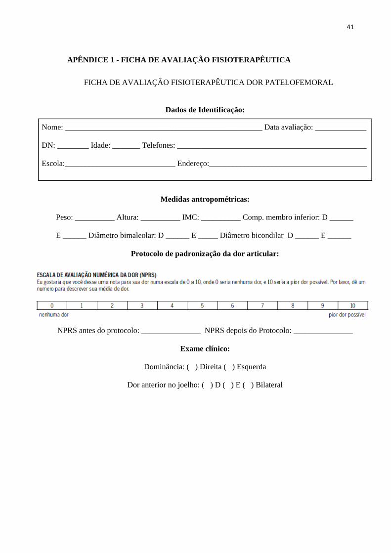
APÊNDICE 1 - FICHA DE AVALIAÇÃO FISIOTERAPÊUTICA.......................................40
ANEXO A - ESCALA EDAJ ................................................................................................... 42
ANEXO B - ESCALA NPRS .................................................................................................. 43
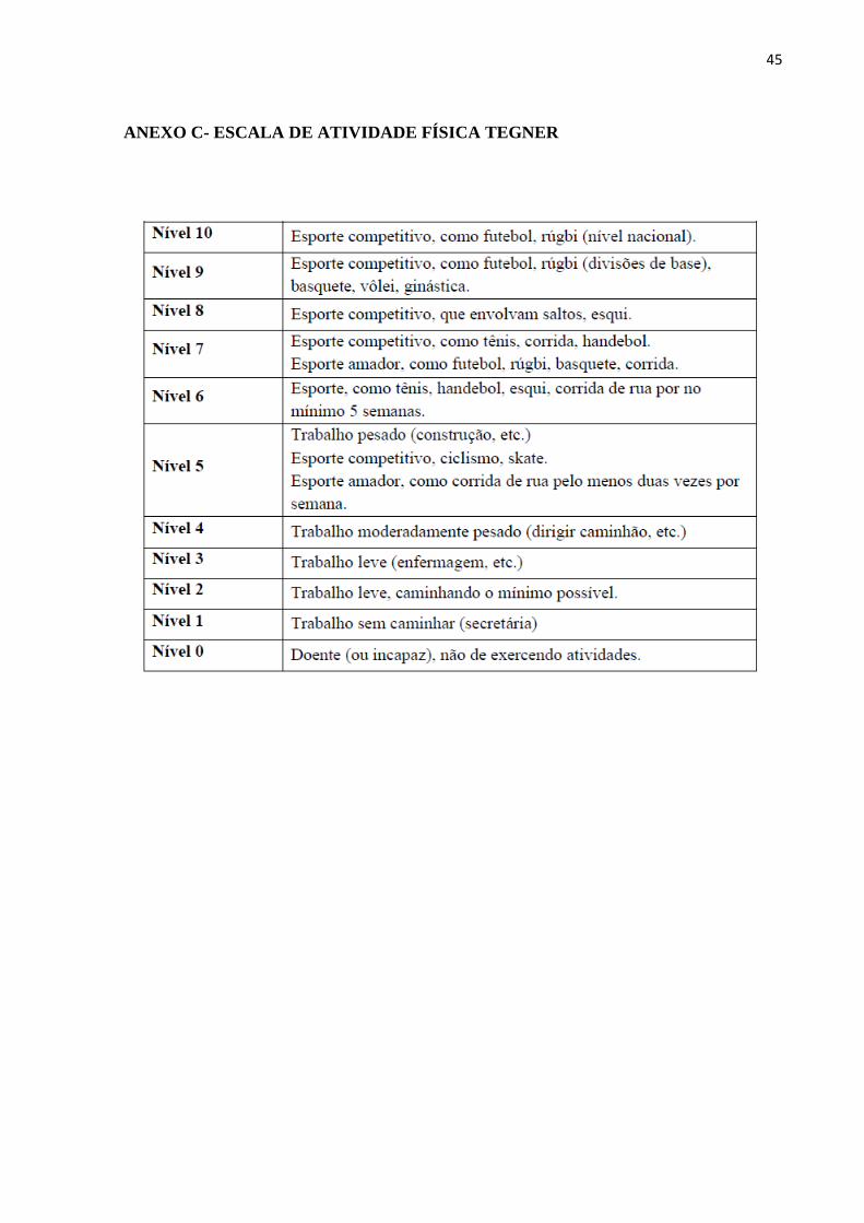
ANEXO C- ESCALA DE ATIVIDADE FÍSICA TEGNER ................................................... 44
ANEXO D - NORMAS DA REVISTA..........................................................................................45
12
1 INTRODUÇÃO GERAL
A dor patelofemoral (DPF) é uma queixa frequente no joelho e afeta 23% da população
geral (SMITH et al., 2018). A DPF é definida como a dor que tem início insidioso na região
anterior e/ou retropatelar, que se agrava por pelo menos uma atividade (CROSSLEY et al.,
2016) como sentar por período prolongado, caminhar, ajoelhar, subir e descer escadas, correr e
agachar (WILLY et al., 2019a).
A etiologia da dor patelofemoral (DPF) é multifatorial e complexa (WILLY et al.,
2019b), entretanto, alguns aspectos biomecânicos de origem local, distal e proximal são
destacados na literatura como potenciais causadores de estresse articular. Como fatores locais
são indicados um mau alinhamento anatômico da patela (POWERS, 2003; SALSICH;
PERMAN, 2013), assim como fraqueza muscular de quadríceps (LANKHORST et al., 2016;
PAPPAS; WONG-TOM, 2012). Em relação aos fatores proximais, destacam-se a alteração de
força muscular de abdutores, extensores, rotadores externos de quadril (PRINS; VAN DER
WURFF, 2009), maior adução e rotação interna do quadril, queda da pelve contralateral e
inclinação ipsilateral do tronco (BLEY et al., 2014; DOS REIS et al., 2015; NAKAGAWA et
al., 2012). Por fim, os fatores distais são identificados como rotação interna do pé em relação à
tíbia, pronação excessiva da articulação subtalar acompanhada de rotação interna da tíbia
(NOVELLO et al., 2018; POWERS, 2003; TIBERIO, 1987).
Além disso, o valgo dinâmico de joelho foi um tema bastante discutido como possível
fator de risco para DPF (BOLING et al., 2009; HOLDEN et al., 2017; NOEHREN; HAMILL;
DAVIS, 2013; SOUZA; POWERS, 2009), pelo aumento do movimento articular do joelho no
plano frontal (NAKAGAWA et al., 2020). Mesmo que a longo prazo, a grande maioria de
pessoas com DPF não apresentem sinais de osteoartrose (LANKHORST et al., 2016), a DPF
aumenta os níveis de incapacidade física (SMITH et al., 2018), restringe atividades físicas,
esportes e trabalho, podendo haver reincidência e persistir por anos (LANKHORST et al.,
2016). Recentemente, um estudo destacou que mais de 50% das pessoas com diagnóstico de
DPF ainda apresentaram os sintomas de 5 a 8 anos após a realização de um programa de
treinamento (LANKHORST et al., 2016).
Embora a maioria dos estudos sejam conduzidos na população adulta, é durante a
adolescência que o início dos sintomas da DPF são observados, com taxas de recuperação mais
baixas em jovens, destacando-se assim a necessidade de melhorar a compreensão dessa
alteração nessa faixa etária (RATHLEFF et al., 2016). Sabe-se que a maior prevalência da
13
condição parece estar entre indivíduos de 12 a 19 anos (WITVROUW et al., 2014) do sexo
feminino (TENFORDE et al., 2011).
Enquanto alguns adolescentes com alta frequência de participação em esportes
desenvolvem a DPF, um terço dos indivíduos que não praticam esportes também apresentam
DPF (RATHLEFF et al., 2015). Em relação a isso, sugere-se que enquanto o adolescente
altamente ativo que participa de esportes em média cinco vezes por semana possivelmente tenha
uma sobrecarga articular, o sedentário pode apresentar falta de controle neuromuscular e/ou
desenvolvimento de habilidades (CARTER; MICHELI, 2011). Em suma, não há evidências
científicas capazes de confirmar que a DPF é uma condição dependente da carga. (WILLY et
al., 2019a).
Observa-se que diferentemente do adulto, os adolescentes não demonstram déficits de
força, especialmente do quadríceps (RATHLEFF et al., 2013). Sendo assim, há uma lacuna na
literatura sobre o mecanismo que leva à alta incidência de DPF em adolescentes, principalmente
quando fala-se na disparidade de nível de exercício físico (altamente ativo X sedentário)
(FILBAY; GRINDEM, 2019). Com relação a qualidade de vida, um estudo de coorte observou
que 40% dos adolescentes com DPF mantém os sintomas após 5 anos e em 15% a dor no joelho
teve impacto na escolha de carreira/trabalho (RATHLEFF et al., 2019). Além das hipóteses
para o aumento no estresse da articulação patelofemoral, que levam em consideração questões
anatômicas, deve ser levada em consideração a influência de medidas de performance física,
que são diretamente influenciadas pelos movimentos de quadril e tornozelo (WILLY et al.,
2019a). Não há evidência de que questões anatômicas isoladas estão relacionadas diretamente
ao desenvolvimento da dor, sendo que vários aspectos como retreinamento, gerenciamento de
carga e educação podem otimizar a adesão ao tratamento e autocuidado dos pacientes.
(CROSSLEY et al., 2019).
Considerando que profissionais de saúde e pesquisadores devem desenvolver e otimizar
estratégias de prevenção da DPF, são necessárias evidências sobre as diferenças cinemáticas
entre os indivíduos com e sem DPF (BARTON et al., 2009; LUZA; LUZA; SANTOS, 2020).
A investigação dessas variáveis na população adolescente especialmente em tarefas funcionais
ainda foi pouco explorada (CARLSON; BODEN; SHEEHAN, 2017; RATHLEFF et al., 2013,
2016), tornando indispensável uma aprofundamento no aspecto biomecânico.
O objetivo dessa dissertação foi analisar o padrão cinemático de membros inferiores
durante o teste de degrau lateral em adolescentes do sexo feminino com e sem DPF. A hipótese

14
é de que o padrão cinemático dos membros inferiores nos três planos de movimento é diferente
em adolescentes com DPF quando comparadas a adolescentes sem dor patelofemoral durante
uma tarefa funcional. Considerando o contexto exposto, a presente dissertação será apresentada
no formato de artigo a ser submetido ao periódico Journal of Sport Rehabilitation (ISSN 1543-
3072 e Qualis Capes A1). As normas para publicação estão apresentadas no Anexo D.
15
Title: Lower limb kinematic analysis during lateral step down in female adolescents with and
without patellofemoral pain
Authors:
Laura Rossetto Foschera1, Karine Josibel Velasques Stoelben1,2, Gabriela dos Santos de Souza1,
Gustavo do Nascimento Petter, Michele Forgiarini Saccol1, Carlos Bolli Mota1
Author affiliations:
1Federal University of Santa Maria, Laboratory of Biomechanics, 1000 Roraima Avenue,
building 51, room 1007, Santa Maria, Rio Grande do Sul, Brazil, CEP: 97105-900.
E-mail address: [email protected].
2 Applied Neuromechanics Research Group, Federal University of Pampa, Uruguaiana, RS,
Brazil
Corresponding author:
Laura Rossetto Foschera
Av. Roraima, 1000, UFSM, University City Building, Laboratory of Biomechanics, Camobi.
ZIP: Code: 97105-900. Santa Maria – RS - Brazil
Tel.: +55 55 997264311.
E-mail address: [email protected]
16
ABSTRACT
Patellofemoral pain (PFP) is a prevalent disorder among female adolescents. Considering that
association between lower limb kinematics and PFP symptoms remains unclear, we conducted
a case-control study comparing female adolescents with and without PFP during Lateral Step
Down. The aim of this study was to investigate differences in kinematic pattern of lower limbs
during the lateral step down task (LSD) and evaluate the association between hip and ankle
angles in the three planes of movement with knee in the frontal plane. We used the squat and
the rising phase of the LSD cycle for analysis. To compare all the angles in the movement cycle
between groups, data were interpolated to 101 points for squat and rising phases. Mean values
of joint angles for sagittal, frontal and transverse plane were plotted graphically with upper and
lower limits of 95% confidence intervals (CI) for each comparison. Significant differences
were established as a consecutive 5% of gait cycle in which 95% confidence interval did not
overlap. A linear regression model was used to determine how hip and ankle angles were related
to frontal knee angles in both groups. There is no difference between joints analyzed in isolation
during the execution of LSD and during squatting and rising phases of LSD test, coordination
between hip, knee and ankle were not different.
17
Introduction
Patellofemoral pain (PFP) has a complex and multifactorial etiology characterized by
an insidious onset of pain localized to the anterior, retropatellar and/or peripatellar region of the
knee.1 PFP can restrict participation in physical activity, sports, work 2 and remain or persist
for years.2 This condition can occur during any period of life 3 but early onset of PFP symptoms
in adolescence are related to lower recovery rates 4. It is known that the highest prevalence of
the condition appears to be in female sex 5 between 12 to 19 years-old 1
Although high participation in sports is associated with PFP development in adolescents
6 one-third of PFP individuals did not practice any sport 7. While high active adolescent who
participates in sports on average five times a week may present an joint overload, the sedentary
one may present a lack of neuromuscular control and/or skills development 8. This finding
highlights the lack of evidence to confirm the PFP as a load-dependent condition.
In addition to the hypotheses for increased stress on the patellofemoral joint, the
influence of physical performance measures must be taken into account 9. Reduced muscle
strength in knee extension is an expected deficit among adults,10 but adolescents with PFF did
not show quadriceps strength deficits 11. Also, strength and movement control training did not
alter the kinematics12 or promoted additional benefits 13 in young adults with PFP. This
evidence challenges the hypothesis of causality of muscular strength and movement disturbance
in patients with PFP.
Altered lower limb kinematics seems to be associated with self-reports of pain and
function14,15. Individuals with PFP have altered segment coordination variability during running
compared to healthy individuals. 16,17 In contrast to adults, adolescents present abnormal patellar
tracking patterns and static alignment. 4,18,19 Considering that association between lower limb
kinematics and PFP symptoms remains unclear in female adolescents 4,18,19, it is indispensable
to evaluate the interaction between joints during functional tasks to understand generate
patterned movement in adolescents with and without PFP.
The aim of this study was to identify differences in kinematic pattern of lower limbs
during the lateral step down task (LSD) in female adolescents with and without PFP. The
secondary aim was to evaluate the association between hip and ankle angles in the three planes
of movement with knee in the frontal plane. It was hypothesized that the PFP group has a
different angle pattern during eccentric and concentric phase on LSD than control group and
greater hip angles on frontal plane would be associated with greater knee angles in PFP group.
18
Methods
Study design and participants
We conducted a case-control study comparing female adolescents with and without
PFP. This report followed STROBE guidelines to case-control studies. This study was approved
by the local Human Research Ethics (registration number: 26928514.6.0000.5346). Data
collection was conducted between January and December 2019. All the participants provided
written informed assent and the respective parents or guardians an informed consent.
Potential participants were recruited through advertisements at local high school and
social media platforms. For PFP group, the following eligibility criteria were used based on the
most recent consensus for clinical assessment of PFP2: (i) female adolescents aged between 13
and 17 years; (ii) presence of retropatelar or peripatelar pain (minimum of three points on the
visual analog scale) in at least two functional tasks (squatting, going up and down stairs,
running, jumping, kneeling and prolonged sitting) for at least three months; (iii) absence of
patellar instability; (iv) absence of hip and ankle injury, (v) start of PFP not related to trauma,
(vi) present sensitivity/apprehension or pain on palpation of the facets of the patella or report
previous knee pain when performing a squat maneuver (viii) refer a minimum pain intensity of
3 on the numerical pain assessment scale (NPRS) during the last month; (ix) present
“reasonable” or “bad” observational classification in the LSD to characterize the dynamic knee
valgus and report a maximum of 87 points on the Anterior Knee Pain Scale (AKPS).
For the pain free control group, the inclusion criteria were aged between 13 and 17 years
and do not have any symptom associated with PFP.
Exclusion criteria for both groups were as follows: (i) history of surgery in any lower
limb joint, (ii) history of patellar subluxation or clinical evidence of meniscal injury or ligament
instability, or joint effusion, (iii) symptomatic patellar tendon pathology, (iv) referred pain
coming from the lumbar spine, hips, ankles or feet, (v) presence of medical conditions and (vi)
do not have gone through menarche and (vii) do not have dynamic knee valgus.
Measurements
Data collection included lower limb kinematic evaluation during LSD and
questionnaires. For control group, the dominant limb was assessed by asking which leg the
participant would use to kick a ball as far as possible.20 For PFP group, the participant´s limb
with PFP was characterized as symptomatic limb (unilateral symptoms) or most symptomatic
limb (bilateral symptoms).
19
The Tegner (physical activity) and Anterior Knee Pain Scale (AKPS, functionality)
questionnaires were completed by both groups. The Tegner physical activity score was
assessed, with score variation between 0 and 10, with higher values indicating higher level of
activity (Briggs et al.). The AKPS score was used to evaluate the reduction in functional
capacity, with higher scores (nearest 100) indicating better knee function.21
Body mass, height, intermaleolar and intercondylar distance were measured before tests.
Then a marker set was placed on participants according to Vicon Plug-In-Gate model by the
same experienced researcher. Sixteen markers were placed bilaterally on the anterior and
posterior superior iliac spines, thigh, lateral epicondyle of femur, shank, calcaneus, lateral
malleolus, second metatarsal head, between the femoral condyles and between the medial and
lateral malleoli. The markers were fixed using double-side adhesive tape. Six infrared cameras
VICON system, 624 model, Oxford, United Kingdom) captured the motion with acquisition
frequency set at 200 Hz and kinematic data was processed using VICON Nexus software
(Version 1.5.3).
A step was used for LSD test and step height was adjusted so that the analyzed leg
reached 60º of knee flexion during the execution, in order to avoid higher joint forces that may
exacerbate knee pain symptoms. This measurement was previously performed with the aid of
a goniometer and speed was self-selected 13. Participants positioned the 2nd toe (tested member)
on a standard line contained in the step, and kept the contralateral member suspended laterally
to the step with the ankle in dorsiflexion and hands on the waist.22 Participants were instructed
to go down the step and flex the knee of the tested leg up to the heel of the contralateral leg,
lightly touching the ground, without the body weight being unloaded. Immediately, the tested
knee was then extended, returning to the initial position in a slow and controlled manner.22
Before testing, participants were able to become familiar with the task, performing a repetition
of the test for each lower limb. Three series of three continuous repetitions of the test were
performed. We used the squat phase and the rising phase of the LSD cycle for analysis, i.e.,
from the beginning of the squat phase until touching the ground and when the tested knee begins
to extend, returning to the initial position in rising phase. The LSD is a widespread and easy-
to-use tool that can be used on clinical environment and research to quantify lower extremity
quality of movement 23 and is one that most differentiates kinematics of women with and
without PFP.24
20
Data Analysis
The lower limb joint angles were compared during the execution of LSD adopting a
method utilized by Stoelben et al, Kuenze et al and McKeon.25–27To compare all the movement
cycle between groups, data were interpolated to 101 points for squat and rising phases. Mean
values of joint angles for sagittal, frontal and transverse plane were plotted graphically with
upper and lower limits of 95% confidence intervals (CI) for each comparison. Statistical
significance was defined as portions of the movement cycle where 95% CIs did not overlap for
a minimum of five consecutive percentages of the LSD cycle.
After data normality agreement, we used a linear regression model to determine how
hip and ankle angles were related to frontal knee angles in both groups. Statistical analysis was
undertaken using GraphPad Prism version 5.00 for Windows (GraphPad Software, San Diego
California USA). For all analyses, the level of statistical significance was p<0.05.
Results
Thirty-four participants were recruited for the study. Of those, 16 potentially eligible for
the PFP group did not include based on the eligibility criteria. So, nine participants with PFP
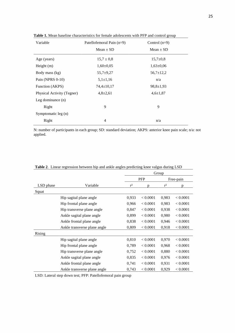
were included in the study and nine in the control group. Participant characteristics are provided
in Table 1. In the PFP group, 4 had pain in their dominant leg.
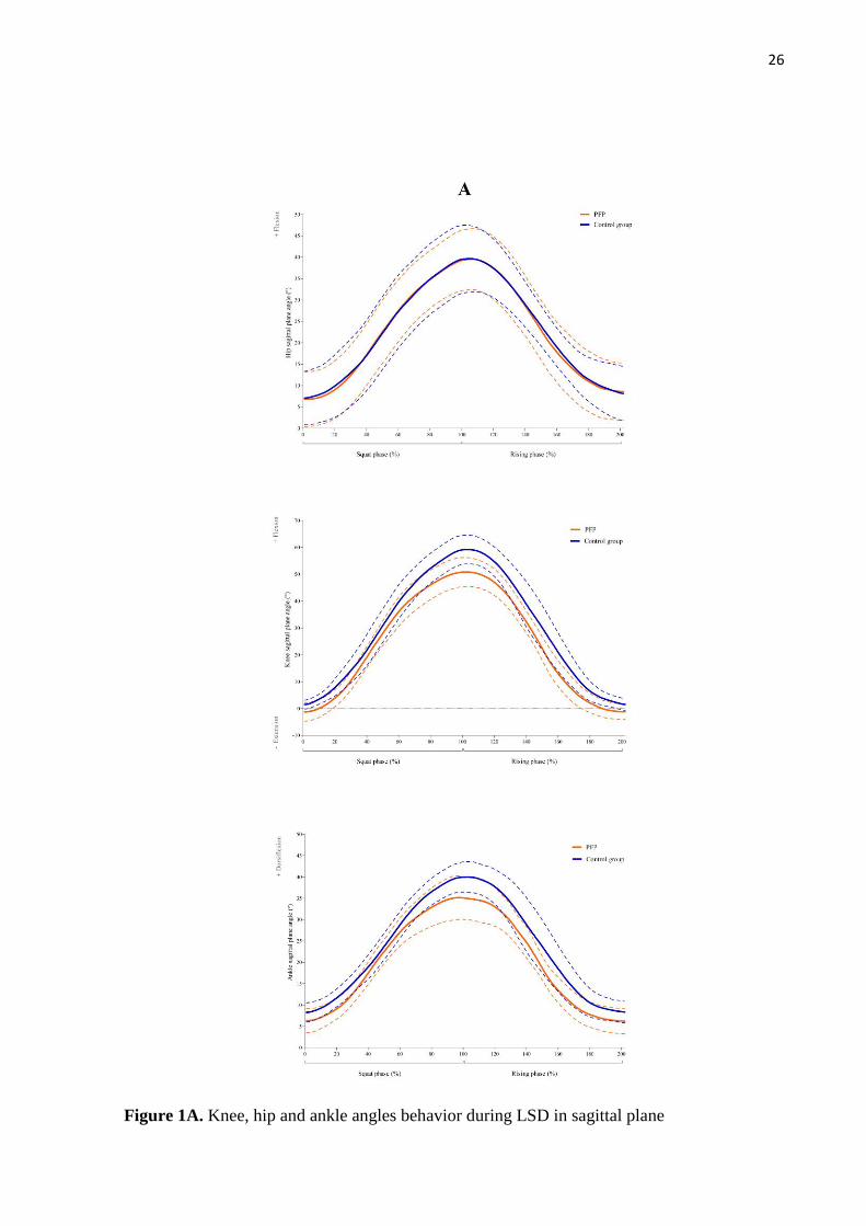
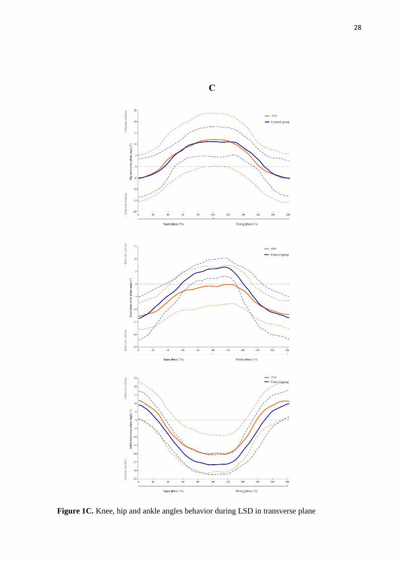
During the squatting and rising phases of the LSD (Fig. 1), there were no differences
between the groups in the excursion throughout the movement for hip, knee and ankle angles
in the sagittal plane (Fig. 1A), frontal plane (Fig. 1B), or transverse plane (Fig. 1C).
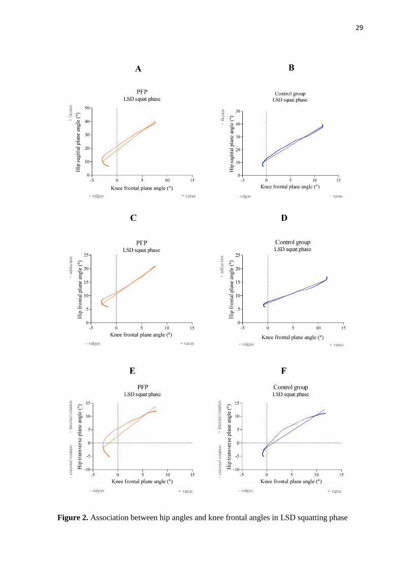
During the LSD squatting, while there is an increase in the hip sagittal plane angle, there
is also an increase in the knee frontal angle. The hip flexion movement explains 93% of the
knee varus/valgus movement for the PFP group (Figure 2A) and 98% for the control group
(Figure 2B). Concerning the hip in the frontal plane, a greater angle also refers to an increase
in the knee frontal angle. The movement of hip abduction throughout squatting explains 96%
of the variation of knee valgus to varus (Figure 2C) for the PFP group and 98% for pain-free
patients (Figure 2D). For the hip transverse plane angle, an increase is related to an increase in
knee frontal angles. Therefore, the hip transverse angle explains 84% of the transition from
valgus to varus movement of the knee to PFP group (Figure 2E) and 93% for pain-free group
(Figure 2F).
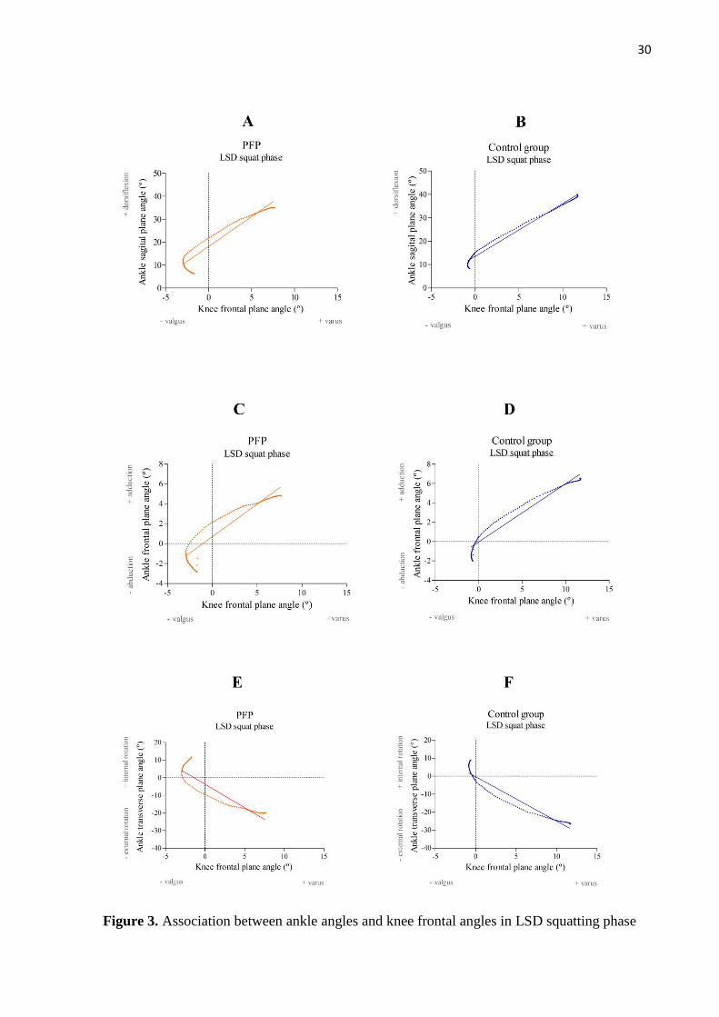
Even during the squat phase, when we talk about greater ankle angles in the sagittal
plane, we can assume greater knee frontal angles. The transition from plantarflexion to
dorsiflexion explains 89% of the valgus to varus movement for the PFP group (Figure 3A) and
21
98% for the controls (Figure 3B). In the frontal plane, the transition from abduction to adduction
movement follows the movement towards the knee varus. In the PFP group (Figure 3C), ankle
frontal angle explained in 83% the knee valgus to varus, and in the controls 94% (Figure 3D).
Lastly, in the transverse plane, there is a transition to external rotation of the ankle, which
explains the movement of knee valgus to varus by 80% for the PFP group (Figure 3E) and 91%
in the controls (Figure 3F).
During the LSD rising phase, there is also a relation between hip and ankle angles and
knee frontal plane. Hip sagittal plane angle explained 81% of the development of varus in the
PFP group (Figure 4A) and 97% in the pain-free group (Figure 4B). For an increase in hip
adduction angle, there is an increase in knee varus. Hip frontal plane angle explains 78% of
knee frontal plane variation in PFP (Figure 4C) and 96% in the control group (Figure 4D). In
the transverse plane, the transition of hip external to internal rotation explains 75% of the knee
valgus to varus in for PFP (Figure 4E) and 88% for controls (Figure 4F).
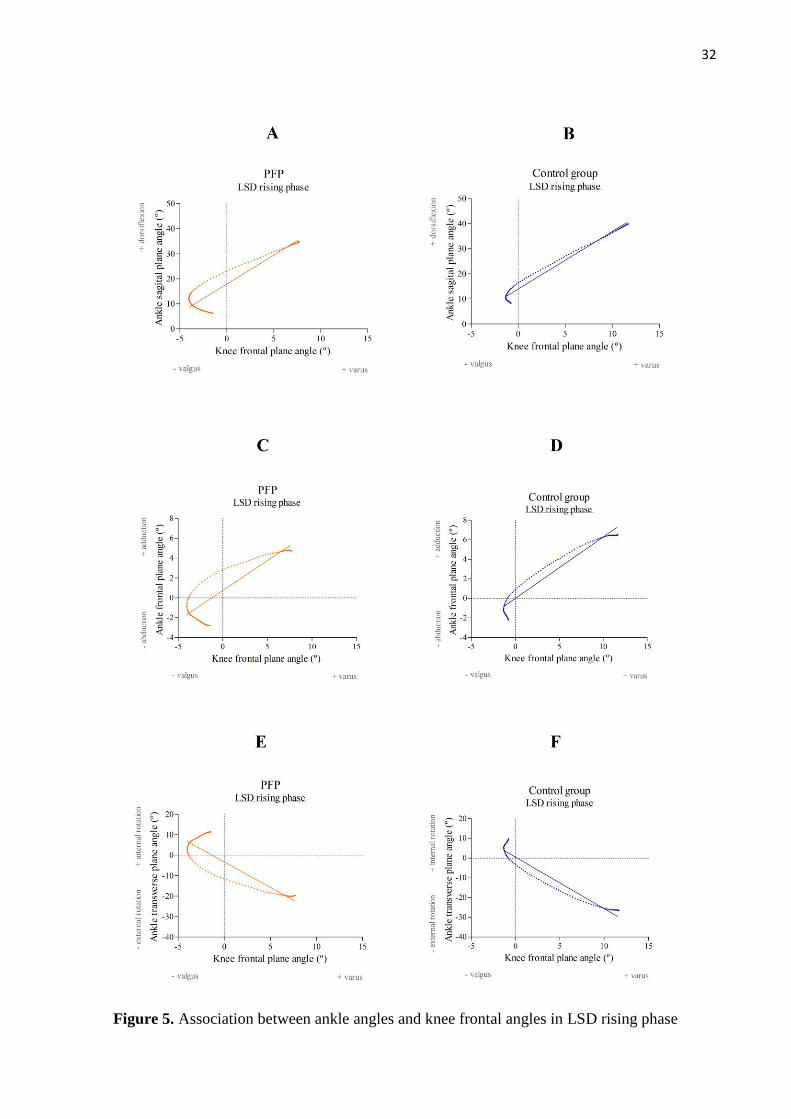
In the relationship between ankle angles and knee frontal plane angles during rising, we
found ankle sagittal plane explains 83% of the knee valgus to varus movement in PFP (Figure
5A) and 97% in the control group (Figure 5B). In the frontal plane, the ankle adduction angle
explains 74% to knee varus in PFP (Figure 5C) and 93% in controls (Figure 5D). While we
have a transition to internal rotation in ankle angle, the knee frontal plane angle is going to
valgus. Ankle transverse angles explain 74% of knee varus to valgus in the PFP group (Figure
5E) and 92% in the pain-free group (Figure 5F).
Discussion
The aim of our study was to identify whether there is a kinematic pattern in the lower
limbs during the LSD test in female adolescents with and without PFP. The secondary objective
was to assess the association between synergic angle movements of hip and ankle in sagittal,
frontal and transverse planes with knee angles in frontal plane during squatting and rising
phases of LSD. The findings of this study suggested that there is no difference in lower limbs
angles when comparing both groups during the execution of the LSD. When analyzing
qualitatively the synergic movement in frontal plane, it is possible to observe that there is a
greater association between the hip and ankle angles with the knee angles in PFP especially in
the rising phase of LSD.
Although anatomical factors such as patellar maltracking is widely accepted as an
underlying mechanism of patellofemoral pain, 28 there is limited evidence that movement
dysfunction is an individual risk factor for women with PFP.29 In adolescents, the only moderate
22
modifiable risk factor identified for PFP was a higher abduction strength 29 and recently
psychological characteristics 30and altered pain modulation 31 have been elicited as contributor
factors for PFP. These recent findings for PFP evidence that joint misalignment does not
necessary leads to pain. In our study, even when groups were paired clinically by dynamic knee
valgus, both showed the same individual angular behavior throughout LSD.
Considering that both the PFP group and the control group have dynamic knee valgus
and both groups showed statistically the same kinematic pattern during the test, our findings
are in line with Rabin et al. (2014), who demonstrated that even individuals with good quality
of movement in LSD, did not show any difference in the intensity of knee pain when compared
to those with worse quality of movement. Thinking from this perspective, the issue of quality
of movement of the lower limb does not seem to be interesting; however, when we analyze the
graphics in a qualitative way, the variability of movement of the knee joint seems to be a
characteristic of DPF, as well as the findings of Ferber et al. (2011) when comparing with
controls.32
Regarding the movement of the ankle joint, especially in the ascent phase, we can
observe qualitative differences in the movement pattern between the groups. Considering that
healthy women with altered lower limb movement pattern have decreased dorsiflexion
amplitude during the lateral descent test,22 we can suggest that this may be related to a lower
variability in ankle movement. Furthermore, the evidence is limited in relation to pronated foot
posture as a risk factor for patellofemoral pain 29 and ankle biomechanics is not consistently
observed in people with PFP.33
The LSD task is one of the functional tasks that most differentiate the kinematics
between PFP and controls.24 Our study showed that female adolescents with PFP have almost
the same slightly increased joint movement in several plane at the knee and hip during the squat
phase. This finding was unexpected and is contrary to previous studies that reported women
with PFP running and landing with excessive knee adduction, hip adduction and internal
rotation.34–36 Landing from a jump and running are tasks with impact loads and movement
velocity higher than LSD,29,37 so differences are expected since more demanding tasks are more
likely to challenge lower limb biomechanics. The results of the study increase
knowledge about the lack of association between lower limb kinematics and PFP symptoms,
highlighting that PFP can be influenced by other factors besides anatomical and functional
deficits. Potential treatment strategies to improve pain in female adolescents with PFP
23
necessarily need to consider psychological 30 and social33 issues that recently had been
considered relevant to pain development especially in female adolescents PFP.30,31,38
This study has some limitations such as the cross-sectional design that does not allow
causal inference. Our results are also limited to female adolescents and cannot be extrapolated
to other populations with PFP. Despite these limitations, to our knowledge, this is the first study
that evaluated the lower limb angles behavior during a functional task in adolescents with PFP.
24
Conclusions
There were no statistical differences in kinematic pattern of lower limbs during the LSD
test between female adolescents with and without PFP. In addition, during squatting and rising
phases of LSD test, coordination between hip, knee and ankle were not different. However,
qualitatively analyzing the synergistic movement in the rising phase of LSD, there was less
association between the hip and ankle angles with the knee angles in the PFP group.
25
Table 1. Mean baseline characteristics for female adolescents with PFP and control group
Variable Patellofemoral Pain (n=9)
Mean ± SD
Control (n=9)
Mean ± SD
Age (years) 15,7 ± 0,8 15,7±0,8
Height (m) 1,60±0,05 1,63±0,06
Body mass (kg) 55,7±9,27 56,7±12,2
Pain (NPRS 0-10) 5,1±1,16 n/a
Function (AKPS) 74,4±10,17 98,8±1,93
Physical Activity (Tegner) 4,8±2,61 4,6±1,87
Leg dominance (n)
Right 9 9
Symptomatic leg (n)
Right 4 n/a
N: number of participants in each group; SD: standard deviation; AKPS: anterior knee pain scale; n/a: not
applied.
Table 2. Linear regression between hip and ankle angles predicting knee valgus during LSD
LSD phase Variable
Group
PFP Free-pain
r² p r² p
Squat
Hip sagital plane angle 0,933 < 0.0001 0,983 < 0.0001
Hip frontal plane angle 0,966 < 0.0001 0,983 < 0.0001
Hip transverse plane angle 0,847 < 0.0001 0,938 < 0.0001
Ankle sagital plane angle 0,899 < 0.0001 0,980 < 0.0001
Ankle frontal plane angle 0,838 < 0.0001 0,946 < 0.0001
Ankle transverse plane angle 0,809 < 0.0001 0,918 < 0.0001
Rising
Hip sagital plane angle 0,810 < 0.0001 0,970 < 0.0001
Hip frontal plane angle 0,789 < 0.0001 0,968 < 0.0001
Hip transverse plane angle 0,752 < 0.0001 0,880 < 0.0001
Ankle sagital plane angle 0,835 < 0.0001 0,976 < 0.0001
Ankle frontal plane angle 0,741 < 0.0001 0,931 < 0.0001
Ankle transverse plane angle 0,743 < 0.0001 0,929 < 0.0001
LSD: Lateral step down test; PFP: Patellofemoral pain group
26
Figure 1A. Knee, hip and ankle angles behavior during LSD in sagittal plane

27
Figure 1B. Knee, hip and ankle angles behavior during LSD in frontal plane
28
Figure 1C. Knee, hip and ankle angles behavior during LSD in transverse plane
29
Figure 2. Association between hip angles and knee frontal angles in LSD squatting phase
30
Figure 3. Association between ankle angles and knee frontal angles in LSD squatting phase

31
Figure 4. Association between hip angles and knee frontal angles in LSD rising phase
32
Figure 5. Association between ankle angles and knee frontal angles in LSD rising phase
33
References
1. Witvrouw E, Callaghan MJ, Stefanik JJ, et al. Patellofemoral pain: consensus
statement from the 3rd International Patellofemoral Pain Research Retreat held in Vancouver,
September 2013. Br J Sports Med. 2014;48(6):411-414. doi:10.1136/bjsports-2014-093450
2. Crossley KM, Stefanik JJ, Selfe J, et al. 2016 Patellofemoral pain consensus statement
from the 4th International Patellofemoral Pain Research Retreat, Manchester. Part 1:
Terminology, definitions, clinical examination, natural history, patellofemoral osteoarthritis
and patient-reported outcome measures. Br J Sports Med. 2016;50(14):839-843.
doi:10.1136/bjsports-2016-096384
3. Callaghan MJ, Selfe J. Has the incidence or prevalence of patellofemoral pain in the
general population in the United Kingdom been properly evaluated? Physical Therapy in
Sport. 2007;8(1):37-43. doi:10.1016/j.ptsp.2006.07.001
4. Rathleff MS, Samani A, Olesen JL, Roos EM, Rasmussen S, Madeleine P. Effect of
exercise therapy on neuromuscular activity and knee strength in female adolescents with
patellofemoral pain—An ancillary analysis of a cluster randomized trial. Clinical
Biomechanics. 2016;34:22-29. doi:10.1016/j.clinbiomech.2016.03.002
5. Tenforde AS, Sayres LC, McCurdy ML, Collado H, Sainani KL, Fredericson M.
Overuse Injuries in High School Runners: Lifetime Prevalence and Prevention Strategies.
PM&R. 2011;3(2):125-131. doi:10.1016/j.pmrj.2010.09.009
6. Hall R, Foss KB, Hewett TE, Myer GD. Sport Specialization’s Association With an
Increased Risk of Developing Anterior Knee Pain in Adolescent Female Athletes. Journal of
Sport Rehabilitation. 2015;24(1):31-35. doi:10.1123/jsr.2013-0101
7. Rathleff MS, Vicenzino B, Middelkoop M, et al. Patellofemoral Pain in Adolescence
and Adulthood: Same Same, but Different? Sports Med. 2015;45(11):1489-1495.
doi:10.1007/s40279-015-0364-1
8. Carter CW, Micheli LJ. Training the child athlete: physical fitness, health and injury.
British Journal of Sports Medicine. 2011;45(11):880-885. doi:10.1136/bjsports-2011-090201
9. Willy RW, Hoglund LT, Barton CJ, et al. Patellofemoral Pain: Clinical Practice
Guidelines Linked to the International Classification of Functioning, Disability and Health
From the Academy of Orthopaedic Physical Therapy of the American Physical Therapy
Association. J Orthop Sports Phys Ther. 2019;49(9):CPG1-CPG95.
doi:10.2519/jospt.2019.0302
10. Lankhorst NE, Bierma-Zeinstra SMA, van Middelkoop M. Factors associated with
patellofemoral pain syndrome: a systematic review. Br J Sports Med. 2013;47(4):193-206.
doi:10.1136/bjsports-2011-090369
11. Rathleff CR, Baird WN, Olesen JL, Roos EM, Rasmussen S, Rathleff MS. Hip and
Knee Strength Is Not Affected in 12-16 Year Old Adolescents with Patellofemoral Pain - A
Cross-Sectional Population-Based Study. Hug F, ed. PLoS ONE. 2013;8(11):e79153.
doi:10.1371/journal.pone.0079153
34
12. Palmer K, Hebron C, Williams JM. A randomised trial into the effect of an isolated
hip abductor strengthening programme and a functional motor control programme on knee
kinematics and hip muscle strength. BMC Musculoskelet Disord. 2015;16(1):105.
doi:10.1186/s12891-015-0563-9
13. Rabelo ND dos A, Costa LOP, Lima BM de, et al. Adding motor control training to
muscle strengthening did not substantially improve the effects on clinical or kinematic
outcomes in women with patellofemoral pain: A randomised controlled trial. Gait & Posture.
2017;58:280-286. doi:10.1016/j.gaitpost.2017.08.018
14. Crossley KM, van Middelkoop M, Barton CJ, Culvenor AG. Rethinking
patellofemoral pain: Prevention, management and long-term consequences. Best Practice &
Research Clinical Rheumatology. 2019;33(1):48-65. doi:10.1016/j.berh.2019.02.004
15. Filbay SR, Grindem H. Evidence-based recommendations for the management of
anterior cruciate ligament (ACL) rupture. Best Practice & Research Clinical Rheumatology.
2019;33(1):33-47. doi:10.1016/j.berh.2019.01.018
16. Cunningham TJ, Mullineaux DR, Noehren B, Shapiro R, Uhl TL. Coupling angle
variability in healthy and patellofemoral pain runners. Clinical Biomechanics.
2014;29(3):317-322. doi:10.1016/j.clinbiomech.2013.12.008
17. Heiderscheit BC, Hamill J, van Emmerik REA. Variability of Stride Characteristics
and Joint Coordination among Individuals with Unilateral Patellofemoral Pain. Journal of
Applied Biomechanics. 2002;18(2):110-121. doi:10.1123/jab.18.2.110
18. Rathleff CR, Baird WN, Olesen JL, Roos EM, Rasmussen S, Rathleff MS. Hip and
Knee Strength Is Not Affected in 12-16 Year Old Adolescents with Patellofemoral Pain - A
Cross-Sectional Population-Based Study. Hug F, ed. PLoS ONE. 2013;8(11):e79153.
doi:10.1371/journal.pone.0079153
19. Carlson VR, Boden BP, Sheehan FT. Patellofemoral Kinematics and Tibial
Tuberosity–Trochlear Groove Distances in Female Adolescents With Patellofemoral Pain. Am
J Sports Med. 2017;45(5):1102-1109. doi:10.1177/0363546516679139
20. Barton CJ, Levinger P, Menz HB, Webster KE. Kinematic gait characteristics
associated with patellofemoral pain syndrome: A systematic review. Gait & Posture.
2009;30(4):405-416. doi:10.1016/j.gaitpost.2009.07.109
21. Kujala UM, Jaakkola LH, Koskinen SK, Taimela S, Hurme M, Nelimarkka O. Scoring
of patellofemoral disorders. Arthroscopy: The Journal of Arthroscopic & Related Surgery.
1993;9(2):159-163. doi:10.1016/S0749-8063(05)80366-4
22. Rabin A, Kozol Z, Moran U, Efergan A, Geffen Y, Finestone AS. Factors Associated
With Visually Assessed Quality of Movement During a Lateral Step-down Test Among
Individuals With Patellofemoral Pain. J Orthop Sports Phys Ther. 2014;44(12):937-946.
doi:10.2519/jospt.2014.5507
23. Silva RL e, Pinheiro YT, Lins CA de A, de Oliveira RR, Scattone Silva R. Assessment
of quality of movement during a lateral step-down test: Narrative review. Journal of
Bodywork and Movement Therapies. 2019;23(4):835-843. doi:10.1016/j.jbmt.2019.05.012
35
24. Lopes Ferreira C, Barton G, Delgado Borges L, dos Anjos Rabelo ND, Politti F,
Garcia Lucareli PR. Step down tests are the tasks that most differentiate the kinematics of
women with patellofemoral pain compared to asymptomatic controls. Gait & Posture.
2019;72:129-134. doi:10.1016/j.gaitpost.2019.05.023
25. Stoelben KJV, Pappas E, Mota CB. Lower extremity joint moments throughout gait at
two speeds more than 4 years after ACL reconstruction. Gait & Posture. 2019;70:347-354.
doi:10.1016/j.gaitpost.2019.02.025
26. Kuenze C, Hertel J, Weltman A, Diduch DR, Saliba S, Hart JM. Jogging
Biomechanics after Exercise in Individuals with ACL-Reconstructed Knees: Medicine &
Science in Sports & Exercise. 2014;46(6):1067-1076. doi:10.1249/MSS.0000000000000217
27. McKeon PO, Paolini G, Ingersoll CD, et al. Effects of balance training on gait
parameters in patients with chronic ankle instability: a randomized controlled trial. Clin
Rehabil. 2009;23(7):609-621. doi:10.1177/0269215509102954
28. Grant C, Fick CN, Welsh J, McConnell J, Sheehan FT. A Word of Caution for Future
Studies in Patellofemoral Pain: A Systematic Review With Meta-analysis. Am J Sports Med.
2021;49(2):538-551. doi:10.1177/0363546520926448
29. Neal BS, Barton CJ, Gallie R, O’Halloran P, Morrissey D. Runners with
patellofemoral pain have altered biomechanics which targeted interventions can modify: A
systematic review and meta-analysis. Gait & Posture. 2016;45:69-82.
doi:10.1016/j.gaitpost.2015.11.018
30. Maclachlan L. Psychological features and somatosensory characteristics of
patellofemoral pain (PhD Academy Award). Br J Sports Med. 2020;54(17):1060-1061.
doi:10.1136/bjsports-2019-101856
31. Bartholomew C, Lack S, Neal B. Altered pain processing and sensitisation is evident
in adults with patellofemoral pain: a systematic review including meta-analysis and meta-
regression. Scandinavian Journal of Pain. 2019;20(1):11-27. doi:10.1515/sjpain-2019-0079
32. Ferber R, Kendall KD, Farr L. Changes in Knee Biomechanics After a Hip-Abductor
Strengthening Protocol for Runners With Patellofemoral Pain Syndrome. Journal of Athletic
Training. 2011;46(2):142-149. doi:10.4085/1062-6050-46.2.142
33. Powers CM, Witvrouw E, Davis IS, Crossley KM. Evidence-based framework for a
pathomechanical model of patellofemoral pain: 2017 patellofemoral pain consensus statement
from the 4th International Patellofemoral Pain Research Retreat, Manchester, UK: part 3. Br J
Sports Med. 2017;51(24):1713-1723. doi:10.1136/bjsports-2017-098717
34. Dierks TA, Manal KT, Hamill J, Davis IS. Proximal and Distal Influences on Hip and
Knee Kinematics in Runners With Patellofemoral Pain During a Prolonged Run. J Orthop
Sports Phys Ther. 2008;38(8):448-456. doi:10.2519/jospt.2008.2490
35. dos Reis AC, Correa JCF, Bley AS, Rabelo ND dos A, Fukuda TY, Lucareli PRG.
Kinematic and Kinetic Analysis of the Single-Leg Triple Hop Test in Women With and
Without Patellofemoral Pain. J Orthop Sports Phys Ther. 2015;45(10):799-807.
doi:10.2519/jospt.2015.5011
36
36. Nakagawa TH, Moriya ÉTU, Maciel CD, SerrãO FV. Trunk, Pelvis, Hip, and Knee
Kinematics, Hip Strength, and Gluteal Muscle Activation During a Single-Leg Squat in Males
and Females With and Without Patellofemoral Pain Syndrome. J Orthop Sports Phys Ther.
2012;42(6):491-501. doi:10.2519/jospt.2012.3987
37. Earl JE, Monteiro SK, Snyder KR. Differences in Lower Extremity Kinematics
Between a Bilateral Drop-Vertical Jump and A Single-Leg Step-down. J Orthop Sports Phys
Ther. 2007;37(5):245-252. doi:10.2519/jospt.2007.2202
38. Sigmund KJ, Hoeger Bement MK, Earl-Boehm JE. Exploring the pain in
patellofemoral pain: A systematic review and meta-analysis examining signs of central
sensitization. Journal of Athletic Training. Published online November 24, 2020.
doi:10.4085/1062-6050-0190.20
37
CONCLUSÃO GERAL
Com base nas investigações científicas apresentadas no artigo, pode-se concluir que,
analisar a cinemática específica de uma articulação de forma isolada implica em perda de
aspectos qualitativos importantes de sinergia de movimento em adolescentes com dor
patelofemoral.
Mesmo com a atual situação de pandemia da Covid-19, que interferiu no andamento
desta pesquisa, reitero que ela cumpriu seu objetivo, que é muito mais do que a obtenção do
título de mestre. Esse estudo pode auxiliar fisioterapeutas a desenvolverem avaliações para que,
baseados em um teste clínico simples, possam observar os aspectos do movimento de uma
forma global e qualitativa, e assim, abordar essa disfunção que contribui e influencia não só
repercussões físicas de dor nessas adolescentes, mas também a qualidade de vida. É essencial
que mais estudos continuem sendo desenvolvidos nessa perspectiva e que os recursos
fisioterapêuticos possam contribuir para a saúde e qualidade de vida de adolescentes com dor
patelofemoral.
38
REFERÊNCIAS
BARTON, C. J. et al. Kinematic gait characteristics associated with patellofemoral pain
syndrome: A systematic review. Gait & Posture, v. 30, n. 4, p. 405–416, nov. 2009.
BLEY, A. S. et al. Propulsion Phase of the Single Leg Triple Hop Test in Women with
Patellofemoral Pain Syndrome: A Biomechanical Study. PLoS ONE, v. 9, n. 5, p. e97606, 15
maio 2014.
BOLING, M. C. et al. A Prospective Investigation of Biomechanical Risk Factors for
Patellofemoral Pain Syndrome: The Joint Undertaking to Monitor and Prevent ACL Injury
(JUMP-ACL) Cohort. The American Journal of Sports Medicine, v. 37, n. 11, p. 2108–
2116, nov. 2009.
CARLSON, V. R.; BODEN, B. P.; SHEEHAN, F. T. Patellofemoral Kinematics and Tibial
Tuberosity–Trochlear Groove Distances in Female Adolescents With Patellofemoral Pain.
The American Journal of Sports Medicine, v. 45, n. 5, p. 1102–1109, abr. 2017.
CARTER, C. W.; MICHELI, L. J. Training the child athlete: physical fitness, health and
injury. British Journal of Sports Medicine, v. 45, n. 11, p. 880–885, 1 set. 2011.
CROSSLEY, K. M. et al. 2016 Patellofemoral pain consensus statement from the 4th
International Patellofemoral Pain Research Retreat, Manchester. Part 1: Terminology,
definitions, clinical examination, natural history, patellofemoral osteoarthritis and patient-
reported outcome measures. British Journal of Sports Medicine, v. 50, n. 14, p. 839–843,
jul. 2016.
CROSSLEY, K. M. et al. Rethinking patellofemoral pain: Prevention, management and long-
term consequences. Best Practice & Research Clinical Rheumatology, v. 33, n. 1, p. 48–65,
fev. 2019.
DOS REIS, A. C. et al. Kinematic and Kinetic Analysis of the Single-Leg Triple Hop Test in
Women With and Without Patellofemoral Pain. Journal of Orthopaedic & Sports Physical
Therapy, v. 45, n. 10, p. 799–807, out. 2015.
FILBAY, S. R.; GRINDEM, H. Evidence-based recommendations for the management of
anterior cruciate ligament (ACL) rupture. Best Practice & Research Clinical
Rheumatology, v. 33, n. 1, p. 33–47, fev. 2019.
HOLDEN, S. et al. Two-dimensional knee valgus displacement as a predictor of
patellofemoral pain in adolescent females. Scandinavian Journal of Medicine & Science in
Sports, v. 27, n. 2, p. 188–194, fev. 2017.
LANKHORST, N. E. et al. Factors that predict a poor outcome 5–8 years after the diagnosis
of patellofemoral pain: a multicentre observational analysis. British Journal of Sports
Medicine, v. 50, n. 14, p. 881–886, jul. 2016.
LOUDON, J. K. BIOMECHANICS AND PATHOMECHANICS OF THE
PATELLOFEMORAL JOINT. p. 11, [s.d.].
39
LUZA, L. P.; LUZA, M.; SANTOS, G. M. A síndrome da dor patelofemoral altera o
movimento do retropé, mas não modifica a distribuição da pressão plantar. Revista Brasileira
de Ortopedia, v. 55, n. 04, p. 419–425, ago. 2020.
NAKAGAWA, T. H. et al. Trunk, Pelvis, Hip, and Knee Kinematics, Hip Strength, and
Gluteal Muscle Activation During a Single-Leg Squat in Males and Females With and
Without Patellofemoral Pain Syndrome. Journal of Orthopaedic & Sports Physical
Therapy, v. 42, n. 6, p. 491–501, jun. 2012.
NAKAGAWA, T. H. et al. Y-Balance Test Asymmetry and Frontal Plane Knee Projection
Angle During Single-leg squat as Predictors of Patellofemoral Pain in Male Military Recruits.
Physical Therapy in Sport, v. 44, p. 121–127, jul. 2020.
NOEHREN, B.; HAMILL, J.; DAVIS, I. Prospective Evidence for a Hip Etiology in
Patellofemoral Pain: Medicine & Science in Sports & Exercise, v. 45, n. 6, p. 1120–1124,
jun. 2013.
NOVELLO, A. DE A. et al. Descending stairs: Good or bad task to discriminate women with
patellofemoral pain? Gait & Posture, v. 65, p. 26–32, set. 2018.
PAPPAS, E.; WONG-TOM, W. M. Prospective Predictors of Patellofemoral Pain Syndrome:
A Systematic Review With Meta-analysis. Sports Health: A Multidisciplinary Approach,
v. 4, n. 2, p. 115–120, mar. 2012.
PATEL, D. R.; VILLALOBOS, A. Evaluation and management of knee pain in young
athletes: overuse injuries of the knee. Translational Pediatrics, v. 6, n. 3, p. 190–198, jul.
2017.
POWERS, C. M. The Influence of Altered Lower-Extremity Kinematics on Patellofemoral
Joint Dysfunction: A Theoretical Perspective. Journal of Orthopaedic & Sports Physical
Therapy, v. 33, n. 11, p. 639–646, nov. 2003.
PRINS, M. R.; VAN DER WURFF, P. Females with patellofemoral pain syndrome have
weak hip muscles: a systematic review. Australian Journal of Physiotherapy, v. 55, n. 1, p.
9–15, 2009.
RATHLEFF, C. R. et al. Hip and Knee Strength Is Not Affected in 12-16 Year Old
Adolescents with Patellofemoral Pain - A Cross-Sectional Population-Based Study. PLoS
ONE, v. 8, n. 11, p. e79153, 13 nov. 2013.
RATHLEFF, M. S. et al. Patellofemoral Pain in Adolescence and Adulthood: Same Same, but
Different? Sports Medicine, v. 45, n. 11, p. 1489–1495, nov. 2015.
RATHLEFF, M. S. et al. Effect of exercise therapy on neuromuscular activity and knee
strength in female adolescents with patellofemoral pain—An ancillary analysis of a cluster
randomized trial. Clinical Biomechanics, v. 34, p. 22–29, maio 2016.
RATHLEFF, M. S. et al. Five-year prognosis and impact of adolescent knee pain: a
prospective population-based cohort study of 504 adolescents in Denmark. BMJ Open, v. 9,
n. 5, p. e024113, maio 2019.
40
SALSICH, G. B.; PERMAN, W. H. Tibiofemoral and patellofemoral mechanics are altered at
small knee flexion angles in people with patellofemoral pain. Journal of Science and
Medicine in Sport, v. 16, n. 1, p. 13–17, jan. 2013.
SMITH, B. E. et al. Incidence and prevalence of patellofemoral pain: A systematic review and
meta-analysis. PLOS ONE, v. 13, n. 1, p. e0190892, 11 jan. 2018.
SOUZA, R. B.; POWERS, C. M. Predictors of Hip Internal Rotation during Running: An
Evaluation of Hip Strength and Femoral Structure in Women with and without Patellofemoral
Pain. The American Journal of Sports Medicine, v. 37, n. 3, p. 579–587, mar. 2009.
TENFORDE, A. S. et al. Overuse Injuries in High School Runners: Lifetime Prevalence and
Prevention Strategies. PM&R, v. 3, n. 2, p. 125–131, fev. 2011.
TIBERIO, D. The Effect of Excessive Subtalar Joint Pronation on Patellofemoral Mechanics:
A Theoretical Model. Journal of Orthopaedic & Sports Physical Therapy, v. 9, n. 4, p.
160–165, out. 1987.
WILLY, R. W. et al. Patellofemoral Pain: Clinical Practice Guidelines Linked to the
International Classification of Functioning, Disability and Health From the Academy of
Orthopaedic Physical Therapy of the American Physical Therapy Association. Journal of
Orthopaedic & Sports Physical Therapy, v. 49, n. 9, p. CPG1–CPG95, set. 2019a.
WILLY, R. W. et al. Patellofemoral Pain: Clinical Practice Guidelines Linked to the
International Classification of Functioning, Disability and Health From the Academy of
Orthopaedic Physical Therapy of the American Physical Therapy Association. Journal of
Orthopaedic & Sports Physical Therapy, v. 49, n. 9, p. CPG1–CPG95, set. 2019b.
WITVROUW, E. et al. Patellofemoral pain: consensus statement from the 3rd International
Patellofemoral Pain Research Retreat held in Vancouver, September 2013. British Journal of
Sports Medicine, v. 48, n. 6, p. 411–414, mar. 2014.
41
APÊNDICE 1 - FICHA DE AVALIAÇÃO FISIOTERAPÊUTICA
FICHA DE AVALIAÇÃO FISIOTERAPÊUTICA DOR PATELOFEMORAL
Dados de Identificação:
Nome: __________________________________________________ Data avaliação: _____________
DN: ________ Idade: _______ Telefones: ________________________________________________
Escola:____________________________ Endereço:________________________________________
Medidas antropométricas:
Peso: __________ Altura: __________ IMC: __________ Comp. membro inferior: D ______
E ______ Diâmetro bimaleolar: D ______ E _____ Diâmetro bicondilar D ______ E ______
Protocolo de padronização da dor articular:
NPRS antes do protocolo: _______________ NPRS depois do Protocolo: _______________
Exame clínico:
Dominância: ( ) Direita ( ) Esquerda
Dor anterior no joelho: ( ) D ( ) E ( ) Bilateral

42
1) Dor retro ou periarticular de início insidioso sem relação com trauma na região anterior do
joelho com duração de pelo menos 3 meses:
Duração dos sintomas (meses): ______________
2) Presença de dor em pelos menos 2 dessas atividades:
( ) Correr
( ) Saltar
( ) Subir e descer escadas
( ) Agachar
( ) Permanecer sentado por tempo prolongado
3) Dor na palpação das facetas patelares: ( ) Não ( ) Sim, onde? ______________________
Apreensão ( ) Não ( ) Sim
4) Ao realizar agachamento, houve reprodução da dor e/ou desconforto? ( ) Não ( ) Sim
5) Classificação observacional para caracterização do VDJ: ( ) Bom ( ) Razoável ( ) Ruim
6) No último mês, que nota (NPRS) você daria para sua dor anterior no joelho? (3 ou >)
___________.
7) Pontuação na EDAJ (87 ou <): Total: _________________________________________________
7) Apresenta dor em outras partes do corpo? ( ) Não ( ) Sim, onde: ___________________
8) Refere histórico de deslocamento/ sub luxação da patela? ( ) Não ( ) Sim
9) Já realizou alguma cirurgia em membros inferiores? ( ) Não ( ) Sim
10) Uso de anti-inflamatório e analgésicos semanalmente? ( ) Não ( ) Sim
11) Utiliza outros medicamentos:________________________________________________
12) Realizou fisioterapia no último ano? __________________________________________
13) Pontuação escala de atividade física Tegner __________

43
ANEXO A - ESCALA EDAJ

44
ANEXO B - ESCALA DE AVALIAÇÃO NUMÉRICA DA DOR (NPRS)
45
ANEXO C- ESCALA DE ATIVIDADE FÍSICA TEGNER
46
ANEXO D – NORMAS DA REVISTA JOURNAL OF SPORT REHABILITATION
Authorship Guidelines The Journals Division at Human Kinetics adheres to the criteria for authorship as outlined by the International Committee of Medical Journal Editors*:
Each author should have participated sufficiently in the work to take public responsibility for the content. Authorship credit should be based only on substantial contributions to:
a. Conception and design, or analysis and interpretation of data; and b. Drafting the article or revising it critically for important intellectual content; and c. Final approval of the version to be published.
Conditions a, b, and c must all be met. Individuals who do not meet the above criteria may be listed in the acknowledgments section of the manuscript. *Uniform requirements for manuscripts submitted to biomedical journals. (1991). New England Journal of Medicine, 324, 424–428.
Open Access Human Kinetics is pleased to allow our authors the option of having their articles published Open Access. In order for an article to be published Open Access, authors must complete and return the Request for Open Access form and provide payment for this option. To learn more and request Open Access, click here.
Manuscript Guidelines The Journal of Sport Rehabilitation publishes peer-reviewed original research, systematic reviews/meta-analyses, critically appraised topics (CATs), case studies/series, and technical reports that directly advance the understanding and application of rehabilitation of injuries incurred during sport-related activities. Authors interested in submitting a letter to the editor or review article must contact the editor for approval prior to submission. Submissions must align with the JSR mission.
Format/Preparation Guidelines It is recommended that authors use the appropriate JSR manuscript preparation checklist when preparing their submission. This document is provided for guidance; it should not be included in the submission.
Text Submissions must be prepared in English as a typed Microsoft Word document. The document must be double-spaced, include page and continuous line numbers, and use margins of 1 in (2.5 cm).
Cover Letter A cover letter must be submitted that details why the submission would be a good fit for JSR's readership and be signed by the corresponding author. The cover letter must also state that the manuscript has not been previously published (except in abstract form), is not presently under consideration by another journal, and will not be submitted to another journal before a final editorial decision from JSR is rendered.
Title Page A separate title page file is required and must include the following:
Title of the manuscript (15 word limit) Name(s) of author(s), institutional affiliation(s), and Twitter handle(s) for all author(s) Contact information (e-mail address and phone numbers) of the corresponding author who
is to receive the proofs. If the study was a registered clinical trial, indicate the registration

47
number. Finally, the title page should include acknowledgements, sources of funding, as well as any potential Conflict of Interest(s) by the authors.
Authors should include 2–3 highlights or take home points that can be used to help promote published manuscripts on social media. Limit each highlight to one sentence.
Manuscript Blinding JSR uses a double-blinded peer review process for all submissions. It is important that authors ensure integrity of blinded peer review by following the guidelines below.
No identifying author or institutional information may be included any place in the main body of the manuscript, figures and tables, or videos (ie, title page, subjects, methods, funding, acknowledgements).
If the study was a registered clinical trial, indicate "registration number XXX" in the body of the manuscript and include the actual registration number in the Title Page file (see below).
Acknowledgements, sources of funding, and conflict of interest statements must be blinded in the body of the manuscript; unblinded acknowledgements may be submitted in the Title Page file (see below).
Remove identifying information created within Microsoft Word settings (File → Info → Inspect Document).
For authors invited to revise and resubmit their manuscript for further peer review: No identifying author or institutional information (signature, letterhead, file name)
may be included in the "Response to Reviewer" portion of the resubmission. Do not re-insert identifying information into the body of the manuscript (subjects,
methods, funding, acknowledgements). Track changes and comments in the body of the manuscript may not be used to
signify revised text. Revised text color should be different (ex: red font) or highlighted.
Body of Manuscript The main body of manuscripts (submit as a separate file) must begin with the title of the manuscript and an abstract of no more than 300 words. Abstract requirements are specific to the manuscript type; refer to specific instructions below. Immediately following the abstract, 3–5 keywords must be included. The keywords cannot replicate words included in the title.
Writing should be concise and direct, and use first person active voice. Avoid using unnecessary jargon and abbreviations. Acronyms or abbreviations may be used if it is more commonly recognized than the spelled-out version of a term. Follow the AMA Manual of Style, 11th edition for formatting and style, including numbers and units of measurement (metric). JSR discourages the use of already printed and copyrighted materials. If necessary, the author must include a letter granting permission to reprint the material. Authors are responsible for all financial costs associated with permission to use such materials. The required structure of each manuscript type is detailed below.
References Referencing style should be consistent with Index Medicus for journal abbreviations and follow the AMA Manual of Style, 11th edition. In brief, each citation in the text must be designated by a superscripted numeral, and full information must appear in the reference list. The reference list is to be double-spaced, arranged in the order the works are first cited, and numbered serially, with only 1 reference per number. Reference information must be accurate. References must be limited to directly pertinent published works or papers that have been accepted for publication. Number of references will vary by manuscript type and specific information can be seen below. For more information, a brief AMA tutorial for formatting references/citations can be downloaded here.
48
Figures and Tables The maximum number of figures and/or tables is six. Additional figures or tables, if necessary, should be provided as supplemental Material. Figures and tables should be professional in appearance and have clean, crisp lines. Figures may not exceed 8 in × 10 in (20.3 cm × 25.4 cm), but may require reduction in size to fit the journal's format. Hand drawing and hand lettering are not acceptable. Use black and white or gray shading only, no color. Submit images as separate files that are either JPEG or TIFF format with a minimum resolution of 300 dots per inch (dpi). Tables should be formatted using the table function of of your word processing program rather than aligning columns in text with tabs and spaces or using text boxes. Authors are urged to submit figures rather than tables, when possible. Each table and figure must be on its own page, at the end of the manuscript. Tables should include a brief title and figures should include a descriptive legend. Both tables and figures should include a footnote section that provides expansions for abbreviated terms as well as notation for symbols used in the table. If P-value significance should be listed, please be sure to include the level of significance (e.g., P < .05). Information presented in figures and tables should not duplicate the text. Each table or figure should be cited in the text with a callout (e.g., "see Table 1").
Video Video clips may be submitted to illustrate your article in the online version of JSR. Videos are encouraged because they allow for elaboration of techniques, testing procedures, or clinical observations. Replication of the figures in the text is not recommended. Files may be submitted for review as part of the manuscript; each digital video file should be designated and uploaded as a “supplementary file.” You also should indicate in the cover letter accompanying your submission that you have submitted a video file. Digital material from a source not original to the author must be accompanied by a statement from the copyright holder giving you permission to publish it; the source and copyright holder must be credited in the article. Video must be submitted in MPEG-4 (.mp4), Quicktime (.m4v, .mov), or Windows Media (.wmv) format with standard frame sizes of 1920 × 1080 pixels (for high definition) or 720 × 480 pixels (for standard definition) and a frame rate of 30 frames/s. Human Kinetics will inspect all video submissions for quality and technical specifications, and we reserve the right to reject any video submission that does not meet quality standards and specifications.
Guidelines for Manuscript Types Original Research Original research manuscripts must be limited to a 300 word abstract, 4,000 words within the main body (excluding the abstract, references, tables, and figure legends), a total of 6 figures/tables, and 30 references. Original research manuscripts must include the following sections: Introduction, Methods, Results, Discussion, and Conclusions. A description of each section is below.
Abstract. The abstract may not exceed 300 words and must include the following headings: Context, Design, Methods, Results, and Conclusions.
Introduction. In this section, build the problem using relevant evidence and specifically state the purpose and hypotheses of the study without comprehensively reviewing the existing literature. Do not label the introduction section.
Methods. This section should include the following subheadings:
Study Design: This section should include the independent and dependent variables. If the study was a registered clinical trial, indicate “registration number XXX” and include the actual registration number in the Title Page file.
Patients or Participants: This section should include pertinent sample demographics, inclusion and exclusion criteria, a statement that institutional ethics review board approval
49
was granted [without indicating author’s affiliation], and a statement that patients or participants provided informed consent in the spirit of the Helsinki Declaration.
Procedures: This section should clearly and succinctly describe the steps taken and/or interventions. Outcome measures and the instrumentation used to capture data should also be described in detail, with associated measurement properties.
Statistical Analyses: This section should clearly outline the analytic approach used to test the a priori hypotheses.
Results. This section should include a presentation of results relevant to the stated objectives. Authors are encouraged to use figures and tables when possible, as opposed to text, to summarize the results. If figures and tables are used, the information should not be repeated in the text. Most importantly, at the conclusion of the results section, including figures and tables, readers should understand the direction and magnitude of changes and/or differences. Thus, authors are encouraged to provide metrics (e.g. means differences with confidence intervals, effect sizes with confidence intervals, etc.) in addition to exact P-values. The text of this section should not explain why the results turned out as they did or justify the use of a specific statistical procedure. Text in this section should also have a focus on the results as opposed to the statistical analysis used.
Discussion. The discussion is a formal consideration and critical examination of the study. The research hypotheses of the study should be addressed and considered in the context of other published works. The study’s limitations and generalizability should also be addressed. The final paragraph should outline the clinical implications of the study results in the context of the overall literature.
Conclusions. This section should summarize the most pertinent findings of the study. Conclusions should be directly supported by the data and should highlight the importance of the work that was performed while avoiding overgeneralizations.
Acknowledgments* (if applicable)
Conflict of interest* (if applicable)
References
Appendices (if applicable)
* Refer to Manuscript Blinding and Title Page sections above.
Systematic Reviews and Meta-Analyses These manuscripts are systematic, critical assessments of literature and data sources pertaining to clinical topics, usually initiated with a specific clinical question(s) that are within the scope of the JSR mission. All articles or data sources should be systematically selected for inclusion and critically evaluated, and the search and selection process should be described in detail in the manuscript, with the number of results obtained at each step (eg. PRISMA diagram). The specific type of study or analysis, population, intervention, exposure, and tests or outcomes should be described for each article or data source. The data sources should be as current as possible, ideally with the updated search having been conducted within 3 months of manuscript initial submission. If a manuscript is invited to be revised and resubmitted for further review, the search must be repeated and results updated prior to the resubmission. A 300 word maximum structured abstract is required and the recommended body of the manuscript is limited to ~4,000 words (excluding the abstract, tables, figures, and references).
Authors of systematic reviews and meta-analyses of randomized trials are required to submit the PRISMA flow diagram and checklist. Please see Moher D, Liberati A, Tetzlaff J, Altman DG, The
50
PRISMA Group (2009). Preferred Reporting Items for Systematic Reviews and Meta-Analyses: The PRISMA Statement. BMJ 2009;339:b2535, doi: 10.1136/bmj.b2535, or visit http://www.prisma-statement.org/ Authors of systematic reviews and meta-analyses of observational studies are required to submit the proposed MOOSE checklist. Please see Stroup DF, Berlin JA, Morton SC, et al. Meta-analysis of observational studies in epidemiology: a proposal for reporting. JAMA. 2000;283:2008–2012, or visit Equator Network. Structured Abstract. The abstract may not exceed 300 words and must include the following headings:
Context: Succinctly explain the importance of the review questions Objective(s): A precise statement of the primary clinical question addressed by the review
followed by any secondary question(s) Evidence Acquisition: Data sources, study selection, quality assessment, and data
extraction Evidence Synthesis: Data synthesis and results of quality assessment Conclusion(s)
Context. In this section, provide background and justify the need for the review by clearly describing the problem for which evidence of effectiveness was sought. This section should conclude with the objectives, which includes the question(s) to be addressed by the review (hypotheses tested).
Methods. This section should include the following subheadings:
Design: Discuss whether prospective registration of the SR/MA was sought (eg. PROSPERO, Cochrane Collaboration) and which reporting guideline was used (eg. PRISMA, MOOSE). Describe the design as a systematic review or systematic review with meta-analysis.
Evidence acquisition: Explain how the research was conducted and should detail data sources, search strategy, study selection (inclusion and exclusion criteria), data extraction, quality assessment, risk of bias, and data analysis. The details of the study selection process should be reported explicitly, preferably using a flow diagram. A list of studies excluded from the review should also be reported, where possible, giving the reasons for each exclusion. Study quality assessment and data extraction should also be addressed.
Evidence synthesis: Detail the included and excluded studies including the results of the review, including results of the quality assessment and risk of bias analysis. Discuss findings of the review, such as inclusion of the point measures and the measures of variability, is encouraged. For example, 95% confidence intervals should be incorporated around the calculation of effect sizes, odds ratios, relative risk, numbers needed to treat, and sensitivity and specificity. Robustness of the results (sensitivity analyses) should be addressed.
Discussion. Interpretation of the results and integration of findings into the body of evidence. Discuss the limitations of the systematic review process.
Conclusions. Provide recommendations for health care implications for further research and strength of recommendation.
Acknowledgments* (if applicable)
Conflict of interest* (if applicable)
References
Appendices (if applicable)
51
* Refer to Manuscript Blinding and Title Page sections above.
Critically Appraised Topics (CATs) These manuscripts are a shorter summary of available evidence focused on a specific clinical question in sport rehabilitation. A critically appraised topic (CAT) is similar to a systematic review in that it summarizes the best evidence in a body of literature; however, it is intended to answer a focused clinical question for topics on which a clinical recommendation is needed, with emerging or limited evidence. CATs provide an excellent mechanism for busy evidence-based practice clinicians to collect and disseminate information they find while searching for answers to important clinical questions. A CAT is typically written when the results of a search include 3-5 high quality studies. If the search results in more than 5 studies, to answer that clinical question, a systematic review is more appropriate. Conversely, a summary of a single paper is referred to as a Critically Appraised Paper (CAP) (Note: JSR is not accepting CAPs for review.) A CAT seeks to find the best available evidence that is more readily available to clinicians, and then critically appraise the papers selected for inclusion using accepted standards for evidence-based practice. Examples of published CATs can be accessed on JSR’s website within each issue. Structured Abstract. Structured abstracts should provide the following key information in one page or less.
Clinical Scenario: A brief description of the clinical scenario leading to the clinical question Clinical Question: A focused clinical question of importance in sport rehabilitation Summary of Key Findings: A bulleted list of the key clinical findings from the search Clinical Bottom Line: The most important take-home message from the available evidence; a
statement regarding the level of available evidence and subsequent strength of recommendations is required
Strength of Recommendation: A brief description of the strength of evidence summarized following the critical appraisal
Clinical Scenario. A brief description of background information that demonstrates need and clinical relevance for the focused clinical question. This section should be approximately 2-4 paragraphs.
Focused Clinical Question. The focused clinical question should be developed and presented using the PICO format [Patient/Client and Condition; Intervention (or Assessment); Comparison; Outcome(s)].
Search Strategy and Criteria. Clearly describe the process used to search for and include evidence to answer the focused clinical question.
Search Strategy: Include the databases and sites searched, the search terms used, search limits, and timeframe of search. The search should ideally be conducted within 3 months of submission for publication and should seek to obtain the best available evidence.
Inclusion and Exclusion Criteria: Describe the criteria for selection - the processes through which studies were selected for inclusion for further analysis.
Evidence of Quality Assessment. Describe the quality assessment scale used to appraise the quality of the evidence in the included studies
Summary of Search and Key Findings. In narrative form, describe the results of your search.
Results of Quality Assessment from Best Available Evidence. Describe the sources of evidence used, including the reasons the studies were selected (level of evidence, strengths and weaknesses related to issues of validity, and quality assessment scale results. Authors are encouraged to use the Center for Evidence-Based Medicine (2011) definitions for level of evidence (https://www.cebm.ox.ac.uk/).
52
Summary of Best Evidence (Table). This summary of included evidence is a comparative presentation that must be presented in a table. The table headings/rows must include citation, study design, participants, inclusion/exclusion criteria, intervention investigated, outcome measures, results, level of evidence, quality assessment score, and contribution to CAT question. The contribution to CAT question should be the CAT authors’ rating of how the study answers the focused clinical question. A numerical score should be used to rate the contribution to answering the question based on results and quality, such that 1 is no contribution, 3 is an inconclusive contribution, and 5 is a conclusive contribution.
Clinical Bottom Line. This section includes the following:
Strength of Recommendation: Provide the strength of recommendation based on the collective findings of the CAT by taking into account the individual studies’ level of evidence and the number, consistency, and coherence of the evidence as a whole. Authors must use the following text and insert the appropriate grade: “Collectively, the body of evidence included to answer the clinical question aligns with the strength of recommendation of [insert: A, B, C].”
Answer to Clinical Question: Describe the clinical bottom line that answers the proposed clinical question using the appraised evidence.
Implications for Practice, Education, and Future Research. Include a practical discussion based on the information provided from the appraisal of current literature. Anecdotal comments regarding whether or not this intervention is commonly used clinically, the cost of this intervention, and so on are appropriate.
Acknowledgments* (if applicable)
Conflict of interest* (if applicable)
References
Appendices (if applicable)
* Refer to Manuscript Blinding and Title Page sections above.
Case Studies / Case Series Case studies/series are descriptions of clinically relevant topics applied in practice that integrate clinical-decision making, available evidence, and outcomes used in the management of a patient case. Case studies/series must be within the scope of the JSR mission. Case study/series manuscripts should be limited to a 300 word abstract, 2,500 words within the main body (excluding the abstract, tables, references, and figure legends), 6 total tables/figures, and 20 references. Please note that Case Studies/Series will be indexed and go through peer-review like all other submissions but will only be published online. Abstract. This section must include the following headings: Context, Case Presentation, Management and Outcomes, and Conclusions.
Introduction. This section should describe background information to support the clinical relevance of the case to sport rehabilitation. Important concepts that relate to the case presentation, management, and outcomes presented in the manuscript can be included in this section.
Case Presentation. This section should be a concise description of the case, including relevant patient history, case presentation, and clinical reasoning only to the extent it relates to and impacts the management of the case. The reader should have sufficient information to understand the clinical reasoning based on final diagnosis for the management presented in the next section.
53
Management and Outcomes. This section should detail the patient care provided. Surgical and non-surgical (pharmacological, therapy/rehabilitation, supportive devices, etc.) interventions should be described and include specific rehabilitation parameters, clinical and patient-reported outcomes must be used to show progress and results, and short- and long-term follow-up. Authors are encouraged to consider including figures and videos to demonstrate unique interventions that will enhance understanding and clinical application by readers.
Discussion. This section should include a brief literature review describe underlying factors of the pathology/injury presented, provide clinical reasoning and integration of practice guidelines and recommendations used in decision-making, and a description of the patient’s response, perspective, and experience related to the care provided and resulting outcomes.
Conclusions. This section should focus on clinically relevant conclusions and recommendations that clinicians can translate into their own practice based on what was learned in this patient case study/series.
Acknowledgments* (if applicable)
Conflict of interest* (if applicable)
References
Appendices (if applicable)
* Refer to Manuscript Blinding and Title Page sections above.
Technical Reports Technical Reports are short articles that present results within the scope of the JSR mission. Results can be related to a new or modified method, instrument, or clinical technique; validity/reliability or methods; or an important experimental observation. This format is focused on providing concise reports of experimental investigations with the intent of providing the readership with innovative original findings in regard to clinical and laboratory techniques or observations. Technical Report manuscripts should be limited to a 300 word abstract, 2,500 words within the main body (excluding the abstract, tables, references, and figure legends), 6 total tables/figures, and 20 references. Please note that Technical Reports will be indexed and go through peer-review like all other submissions but will only be published online. Examples of published Technical Reports can be accessed on JSR’s website within each issue. Technical Reports must include the following parts: Introduction, Methods, Results, Discussion, and Conclusions. Further details on each section are below.
Abstract. This section must include the following headings: Context, Design, Methods, Results, and Conclusions.
Introduction. This section should very concisely build the problem. Given word counts and methodological details needed, the introduction should focus on the larger context and only include the most pertinent details. This section must also specifically state the purpose and hypotheses of the study. Do not label the introduction section.
Methods. This section should be as detailed as possible within the word count constraints and include the following subheadings:
Study Design: This section should include the independent and dependent variables. Patients or Participants: This section should include pertinent demographics, a statement
that institutional ethics review board approval was granted [without indicating author’s affiliation], and a statement that participants/patients provided informed consent in the spirit of the Helsinki Declaration.
54
Procedures: This section should clearly and succinctly describe steps taken and/or interventions. Intervention descriptions can also be provided in tabular format. If such a table is very large, authors will likely be asked to reduce the total number of tables/figures associated with their submission. Outcome measures and the instrumentation used to capture them should also be described in detail.
Statistical Analyses: This section should clearly outline the analytic approach used to test the a priori hypotheses.
Results. This section should include a presentation of results relevant to the stated objectives. Authors are encouraged to use tables and figures when possible, as opposed to text, to summarize the results. If tables or figures are used, the information should not be repeated in the text. Most importantly, at the conclusion of the results section, including tables and figures, readers should understand the direction and magnitude of changes and/or differences. Thus, authors are encouraged to provide metrics (e.g. means differences with confidence intervals, effect sizes with confidence intervals, etc.) in additional to P-values. The text of this section should not explain why the results turned out as they did or justify the use of a specific statistical procedure. Text in this section should also have a focus on the results as opposed to the statistical analysis run.
Discussion. The discussion is a formal consideration and critical examination of the study. The research hypotheses of the study should be addressed and considered in the context of other published works. The study’s limitations and generalizability should also be addressed. The final paragraph should outline the clinical implications of the study results in the context of the overall literature.
Conclusions. This section should summarize the most pertinent findings of the study. Conclusions should be directly supported by the data and should highlight the importance of the work that was performed while avoiding overgeneralizations.
Acknowledgments* (if applicable)
Conflict of interest* (if applicable)
References
Appendices (if applicable)
* Refer to Manuscript Blinding and Title Page sections above.
Submitting a Manuscript The Journal of Sport Rehabilitation uses a web-based system, ScholarOne, for the submission and tracking of manuscripts (see submission button at the top of this page). Submissions undergo a peer-review process; submissions are read by the editor and at least two reviewers through a blind review process. The time between submission and a decision regarding acceptance for publication takes approximately 7–10 weeks, but may be longer. Submitted manuscripts may not be in the review process by any other publication when, or during review, by the Journal of Sport Rehabilitation.
Authors of manuscripts accepted for publication must transfer copyright to Human Kinetics, Inc. To view this form, please visit ScholarOne and select "Instructions & Forms" in the upper right corner. You do not need an account to access this information.