laparoscopic transabdominal preperitoneal repair of spigelian hernia—closure of the fascial defect...
TRANSCRIPT
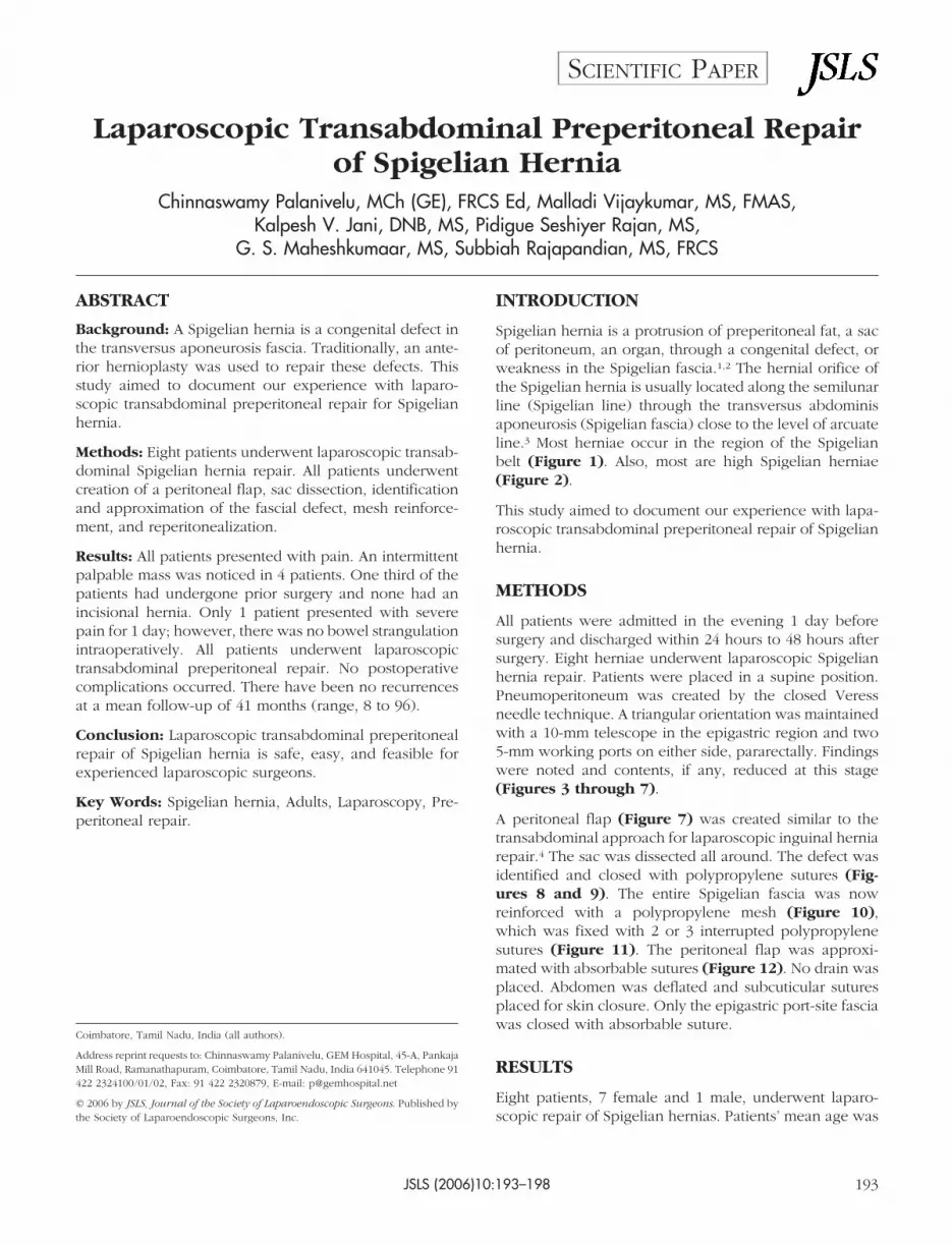
Laparoscopic Transabdominal Preperitoneal Repairof Spigelian Hernia
Chinnaswamy Palanivelu, MCh (GE), FRCS Ed, Malladi Vijaykumar, MS, FMAS,Kalpesh V. Jani, DNB, MS, Pidigue Seshiyer Rajan, MS,
G. S. Maheshkumaar, MS, Subbiah Rajapandian, MS, FRCS
ABSTRACT
Background: A Spigelian hernia is a congenital defect inthe transversus aponeurosis fascia. Traditionally, an ante-rior hernioplasty was used to repair these defects. Thisstudy aimed to document our experience with laparo-scopic transabdominal preperitoneal repair for Spigelianhernia.
Methods: Eight patients underwent laparoscopic transab-dominal Spigelian hernia repair. All patients underwentcreation of a peritoneal flap, sac dissection, identificationand approximation of the fascial defect, mesh reinforce-ment, and reperitonealization.
Results: All patients presented with pain. An intermittentpalpable mass was noticed in 4 patients. One third of thepatients had undergone prior surgery and none had anincisional hernia. Only 1 patient presented with severepain for 1 day; however, there was no bowel strangulationintraoperatively. All patients underwent laparoscopictransabdominal preperitoneal repair. No postoperativecomplications occurred. There have been no recurrencesat a mean follow-up of 41 months (range, 8 to 96).
Conclusion: Laparoscopic transabdominal preperitonealrepair of Spigelian hernia is safe, easy, and feasible forexperienced laparoscopic surgeons.
Key Words: Spigelian hernia, Adults, Laparoscopy, Pre-peritoneal repair.
INTRODUCTION
Spigelian hernia is a protrusion of preperitoneal fat, a sacof peritoneum, an organ, through a congenital defect, orweakness in the Spigelian fascia.1,2 The hernial orifice ofthe Spigelian hernia is usually located along the semilunarline (Spigelian line) through the transversus abdominisaponeurosis (Spigelian fascia) close to the level of arcuateline.3 Most herniae occur in the region of the Spigelianbelt (Figure 1). Also, most are high Spigelian herniae(Figure 2).
This study aimed to document our experience with lapa-roscopic transabdominal preperitoneal repair of Spigelianhernia.
METHODS
All patients were admitted in the evening 1 day beforesurgery and discharged within 24 hours to 48 hours aftersurgery. Eight herniae underwent laparoscopic Spigelianhernia repair. Patients were placed in a supine position.Pneumoperitoneum was created by the closed Veressneedle technique. A triangular orientation was maintainedwith a 10-mm telescope in the epigastric region and two5-mm working ports on either side, pararectally. Findingswere noted and contents, if any, reduced at this stage(Figures 3 through 7).
A peritoneal flap (Figure 7) was created similar to thetransabdominal approach for laparoscopic inguinal herniarepair.4 The sac was dissected all around. The defect wasidentified and closed with polypropylene sutures (Fig-ures 8 and 9). The entire Spigelian fascia was nowreinforced with a polypropylene mesh (Figure 10),which was fixed with 2 or 3 interrupted polypropylenesutures (Figure 11). The peritoneal flap was approxi-mated with absorbable sutures (Figure 12). No drain wasplaced. Abdomen was deflated and subcuticular suturesplaced for skin closure. Only the epigastric port-site fasciawas closed with absorbable suture.
RESULTS
Eight patients, 7 female and 1 male, underwent laparo-scopic repair of Spigelian hernias. Patients’ mean age was
Coimbatore, Tamil Nadu, India (all authors).
Address reprint requests to: Chinnaswamy Palanivelu, GEM Hospital, 45-A, PankajaMill Road, Ramanathapuram, Coimbatore, Tamil Nadu, India 641045. Telephone 91422 2324100/01/02, Fax: 91 422 2320879, E-mail: [email protected]
© 2006 by JSLS, Journal of the Society of Laparoendoscopic Surgeons. Published bythe Society of Laparoendoscopic Surgeons, Inc.
JSLS (2006)10:193–198 193
SCIENTIFIC PAPER

42 years (range, 35 to 60). All patients presented withsymptoms of abdominal pain and an intermittent palpablemass was noted in 4 patients. One patient had pain un-related to the hernia. The pain was described as localizedin 5 patients and diffuse in the other 3 patients. Only 1patient had experienced severe pain for 1 day. Sevenpatients had no significant preoperative morbidity. One(female) patient had chronic obstructive pulmonary dis-ease, controlled with medication. One third of the patientshad undergone prior surgery and none had incisionalhernia. One patient had undergone previous abdominalsurgery followed by ventral hernia repair. Two patientshad undergone surgery for gynecological conditions.None of them were laparoscopic surgeries.
Abdominal ultrasound showed the defect in the echo linefrom the aponeurosis (Figure 13). This defect was notedin all patients, and ultrasound aided the diagnosis in 4 ofthe patients, without the combination of pain and palpa-ble mass. Thus, all patients were diagnosed on the basis ofpatient history, and clinical and ultrasound examination.None underwent herniography or computed tomography.All patients underwent laparoscopic transabdominal pre-peritoneal repair of Spigelian hernia. The patient whopresented with severe pain for 1 day had a small bowelloop as contents. However, the bowel loop showed no
Figure 1. Spigelian aponeurosis. (A) Munros line, (B) imaginaryline 6 cm from interspinal line, (C) linea alba, (D) posteriorrectus sheath, (E) spigelian line (semilunar line), (F) spigelianbelt, (G) spigelian aponeurosis, (H) interspinal line.
Figure 2. Spigelian aponeurosis. (A) Rectus muscle, (B) area ofhigh spigelian hernia, (C) area of low spigelian hernia, (D)transversus abdominis, (E) inferior epigastric vessels.
Figure 3. (A) Spigelian hernial defect without any contents.
Laparoscopic Transabdominal Preperitoneal Repair of Spigelian Hernia, Palanivelu C et al.
JSLS (2006)10:193–198194

signs of strangulation. Two more patients had omentumand sigmoid colon mesentery. In all cases, contents werereduced easily. There was no inadvertent trauma duringreduction. All patients underwent defect closure; meshre-enforcement; and re-peritonealization.
No postoperative complications occurred. All patientswere discharged within 24 hours to 48 hours. There havebeen no recurrences at a mean follow-up of 41 months(range, 8 to 96). The demographic and technical data arementioned in Table 1.5,6
DISCUSSION
Spigelian or lateral ventral hernia is a rare pathology,representing about 2% of all abdominal wall hernias.7 Itsdiagnosis requires a high index of suspicion, given thelack of consistent symptoms and signs. Thus, a physicianneeds a combination of history, physical examination, andpreoperative imaging to secure the diagnosis.
The predominant symptom was pain, which varied in itslocation and severity. A palpable mass was noted in 4
Figure 4. Spigelian hernial defect with sigmoid mesentery ascontents. (A) Rectus abdominis muscle, (B) Spigelian defect.
Figure 5. (A) Reduction of the sigmoid mesentery.
Figure 6. Spigelian defect after reduction of contents. (A) Infe-rior epigastric vessels, (B) defect visualized above the inferiorepigastric vessels, (C) Sigmoid colon.
Figure 7. Dissection of sac after creation of peritoneal flap.
JSLS (2006)10:193–198 195

patients. Thus, a combination of palpable and reduciblemass makes diagnosis easy. Predisposing factors wereobserved in most patients with hernia. In our study, 2patients had undergone surgery for gynecological condi-tions; 1 patient underwent abdominal surgery followed byventral hernia repair. Thus, one third of the patients hadundergone a previous surgery but had no incisional her-nia. This suggests a possibility of an increased risk ofspigelian hernia development. Also, a prior laparoscopicsurgery appears to be a risk factor, due to the creation ofpneumoperitoneum.8 However, none of the patients hadundergone laparoscopic surgery. All patients underwent
an abdominal ultrasound, which aided diagnosis in thosepatients without the combination of pain and palpablemass. Abdominal sonography or computed tomographymay aid diagnosis.9 Abdominal sonography scanning isadvantageous, as it is noninvasive, easily available, inex-pensive, and can detect the hernial orifice in the Spigelianfascia. All the defects were diagnosed by abdominalsonography. None of our patients underwent computedtomography.
Given the high rate of incarceration/strangulation, the
Figure 8. Spigelian defect. (A) Inferior epigastric vessels, (B) defect.
Figure 9. Approximation of the defect by intracorporealpolypropylene suture.
Figure 10. Defect reinforced with polypropylene mesh.
Figure 11. Mesh fixation with intracorporeal polypropylenesuture.
Laparoscopic Transabdominal Preperitoneal Repair of Spigelian Hernia, Palanivelu C et al.
JSLS (2006)10:193–198196

diagnosis of Spigelian hernia is an indication for surgicalrepair. Most repairs are done electively. Laparoscopic re-pairs reported include the preperitoneal approach withpolypropylene mesh,10 onlay mesh repairs using ex-panded polytetrafluoroethylene,11 and composite meshrepairs.12 We performed laparoscopic transabdominalpreperitoneal repair for 8 Spigelian herniae. A similarrepair technique has been reported by Kasirajan et al.13
No postoperative complications occurred.
CONCLUSION
Laparoscopic transabdominal preperitoneal repair ofSpigelian herniae is safe, easy, and feasible for experi-enced laparoscopic surgeons.
References:
1. Spangen L. Spigelian hernia. Surg Clin North Am. 1984;64:351–366.
2. Kalaba Z. Spigelian hernia: a case of typical Spigelian herniain an elderly man. Ugeskr Laeger. 1999;161:2095–2096.
3. Spangen L. Spigelian hernia. World J Surg. 1989;13:573–580.
4. Palanivelu C. Laparoscopic management of rare hernia. In:Operative Manual of Laparoscopic Hernia Surgery. India: GemFoundation; 2004;295–305.
5. Larson DW, Farley DR. Spigelian hernias: repair and out-come for 81 patients. World J Surg. 2002;26(10):1277–1281.
6. Moreno-Egea A, Flores B, Girela E, et al. Spigelian hernia:bibliographical study and presentation of a series of 28 patients.Hernia. 2002;6(4):167–170.
7. Ng WT. Incidence and outcome of surgical repair of spige-lian hernia. Br J Surg. 2004;91:640–644.
8. Slakey DR, Teplitsky S, Cheng SS. Incarcerated Spigelianhernia following laparoscopic living-donor nephrectomy. JSLS.2002;6(3):217–219.
9. Mostard GJ, van Deursen CT. Diagnostic image (74). Awoman with stomach cramps and vomiting. Ned TijdschrGeneeskd. 2002;146(5):209.
10. Tarnoff M, Rosen M, Brody F. Planned totally extraperitoneal
Figure 12. Completion of the peritoneal flap closure with ab-sorbable suture.
Figure 13. Ultrasound image of spigelian hernia. Defect isshown (between the arrows).
Table 1.Demographic and Technical Details
Spangen1 LarsonandFarley5
Moreno-Egeaet al6
Palanivelu
No. of Patients 24 76 28 8
No. of Hernias 25 81 28 8
Mean Age (years) 35 63 60 42
Female (%) 92 51 71 88
Left (%) 25 52 64 75
Pain (%) 100 26 — 100
Palpable Mass (%) 50 38 — 50
Palpable Mass andPain (%)
21 29 — 50
Acute Presentation(%)
10 10 21 12
Strangulation (%) 8 17 — 0
Recurrence (%) — 4 — 0
Mean Follow-up(years)
— 8 3.4 3.4
JSLS (2006)10:193–198 197

laparoscopic Spigelian hernia repair. Surg Endosc. 2002;16(2):359.
11. Appeltans BM, Zeebregts CJ, Cate Hoedemaker HO. Lapa-roscopic repair of a Spigelian hernia using an expanded poly-tetrafluoroethylene (ePTFE) mesh. Surg Endosc. 2000;14(12):1189. Epub 2000 Sep 28.
12. Barie PS, Thompson WA, Mack CA. Planned laparoscopicrepair of a spigelian hernia using a composite prosthesis. JLaparoendosc Surg. 1994;4(5):359–363.
13. Kasirajan K, Lopez J, Lopez R. Laparoscopic technique in themanagement of Spigelian hernia. J Laparoendosc Adv Surg TechA. 1997;7(6):385–388.
Laparoscopic Transabdominal Preperitoneal Repair of Spigelian Hernia, Palanivelu C et al.
JSLS (2006)10:193–198198