la thermorégulation comme cible thérapeutique pour la
TRANSCRIPT
© Marine Tournissac, 2019
La thermorégulation comme cible thérapeutique pour la maladie d’Alzheimer
Thèse
Marine Tournissac
Doctorat en sciences pharmaceutiques
Philosophiæ doctor (Ph. D.)
Québec, Canada
La thermorégulation comme cible thérapeutique pour la maladie d’Alzheimer
Thèse
Marine Tournissac
Sous la direction de :
Frédéric Calon, directeur de recherche
ii
Résumé La maladie d’Alzheimer (MA) est une maladie neurodégénérative qui se manifeste par l’apparition progressive
de troubles de la mémoire. Le nombre de personnes affectées par la maladie est en constante hausse, mais il
n’existe pour l’instant aucun traitement curatif pour la MA. L’âge avancé est le principal facteur de risque de la
MA et est associé à un déficit de thermorégulation. De précédentes études animales montrent d’ailleurs que
l’hypothermie augmente la phosphorylation de la protéine tau, l’un des principaux marqueurs
neuropathologiques de la MA. La souris triple transgénique (3xTg-AD), un modèle murin de la MA, présente
un déficit de thermorégulation qui s’installe progressivement avec le vieillissement et l’apparition de la
neuropathologie. Une exposition aigüe au froid aggrave l’hyperphosphorylation de tau dans le cerveau de ces
souris, tandis que les placer à une température thermoneutre (28°C) diminue leurs déficits de mémoire et leur
pathologie amyloïde. Le tissu adipeux brun (TAB) est le principal siège de la thermogenèse chez les
mammifères. La stimulation de son activité, par des expositions au froid ou l’administration d’agonistes des
récepteurs β3 adrénergiques (Rβ3A), permet de diminuer les altérations métaboliques périphériques. Puisque
les maladies métaboliques sont des facteurs de risques importants pour la MA, et qu’un déficit de
thermorégulation semble aggraver la MA, nous avons émis l’hypothèse que la stimulation du TAB pourrait être
bénéfique pour la MA en corrigeant les déficits de thermorégulation et les troubles métaboliques.
L’objectif de cette thèse était de déterminer si la stimulation de la thermogenèse du TAB permettait de
diminuer la neuropathologie et les déficits comportementaux dans un modèle murin de la MA, la souris 3xTg-
AD.
Dans un premier temps, nous avons montré que l’âge avancé potentialisait la phosphorylation de tau induite
par une exposition aigüe au froid. Dans un second temps, nous avons montré que de courtes expositions
répétées au froid permettaient d’augmenter la capacité de thermogenèse du TAB et de réduire l’intolérance au
glucose des souris 3xTg-AD âgées. Une meilleure thermorégulation conférait une protection contre la
phosphorylation de tau induite par une exposition aigüe au froid. De plus, nous avons observé une corrélation
négative entre les niveaux du facteur de croissance des fibroblastes 21 (FGF21) et la phosphorylation de tau
dans l’hippocampe, suggérant que cette hormone produite par le TAB est impliquée dans la neuroprotection
contre une exposition aigüe au froid. Finalement, nous avons investigué l’effet d’un agoniste des Rβ3A sur la
neuropathologie et la mémoire des souris 3xTg-AD âgées. Cette approche pharmacologique a permis
d’améliorer la tolérance au glucose et d’augmenter la thermogenèse du TAB des souris 3xTg-AD et NonTg
âgées de 16 mois. Le traitement a renversé le déficit de mémoire de reconnaissance et a diminué le ratio de
peptides Aβ42/Aβ40 insolubles dans l’hippocampe des souris transgéniques, sans moduler la phosphorylation
de la protéine tau.

iii
Ces résultats montrent que les interventions visant à stimuler la thermogenèse permettent de diminuer les
désordres métaboliques et d’altérer la neuropathologie et les déficits comportementaux de la MA dans un
modèle murin. Ainsi, nos résultats mettent en lumière la thermorégulation comme une nouvelle cible
thérapeutique et ouvrent la voie à de nouvelles stratégies pour cette maladie.

iv
Abstract Alzheimer's disease (AD) is a neurodegenerative disease characterized by a progressive loss of memory. The
number of people affected by the disease is constantly increasing, but there is currently no cure for AD. Aging,
the main risk factor for AD, is also associated with a thermoregulatory deficit. Previous animal studies have
shown that hypothermia increases the phosphorylation of the protein tau, one of the main neuropathological
markers of AD. The transgenic triple mouse (3xTg-AD), a mouse model of AD neuropathology, develops
thermoregulatory deficits along with the progression of the neuropathology. Acute cold exposure exacerbates
tau hyperphosphorylation in the brain of 3xTg-AD mice, while exposing them to a thermoneutral environment
(28°C) alleviates their memory deficits and amyloid pathology. Brown adipose tissue (BAT) is the main site of
thermogenesis in mammals. The stimulation of its activity, by cold exposures or the administration of β3
adrenergic receptor agonists (β3AR), has been shown to improve peripheral metabolic determinants. Since
metabolic diseases are important risk factors for AD, and thermoregulatory deficits may worsen AD, we
hypothesized that BAT stimulation could be beneficial in AD by correcting both thermoregulatory deficits and
metabolic disorders.
The aim of this thesis was to determine whether the stimulation of BAT thermogenesis reduces
neuropathology and behavioral deficits in a mouse model of AD, the 3xTg-AD mouse.
First, we showed that advanced age potentiates tau phosphorylation induced by acute cold exposure.
Secondly, we observed that repeated short cold exposures increased the thermogenesis capacity of BAT and
reduced glucose intolerance in aged 3xTg-AD mice. Better thermoregulation provided protection against tau
phosphorylation induced by an acute cold exposure. In addition, we reported a negative correlation between
fibroblast growth factor 21 (FGF21) levels and tau phosphorylation in the hippocampus, suggesting that this
BAT-secreted hormone is involved in neuroprotection against acute cold exposure. Finally, we investigated the
effect of a β3AR agonist on the neuropathology and memory of old 3xTg-AD mice. This pharmacological
approach improved glucose tolerance and increased BAT thermogenesis in 16-month-old 3xTg-AD and NonTg
mice. The treatment reversed the recognition memory deficiency and decreased the ratio of insoluble
Aβ42/Aβ40 peptides in the hippocampus of transgenic mice, without modulating tau phosphorylation.
These results show that interventions aiming at stimulating thermogenesis can reduce metabolic disorders and
modulate neuropathology and behavioral deficits of AD in a mouse model. Thus, our results highlight
thermoregulation as a novel therapeutic target for this disease.
v
Table des matières
Résumé ....................................................................................................... ii Abstract ..................................................................................................... iv Table des matières ...................................................................................... v Liste des figures ......................................................................................... ix Liste des tableaux ...................................................................................... xi Liste des abréviations ............................................................................... xii Remerciements .......................................................................................... xv Avant-‐propos ............................................................................................ xvi Introduction ................................................................................................ 1
1 La maladie d’Alzheimer ..................................................................................................... 1 1.1 Définition, chiffres et symptômes ..................................................................................... 1 1.2 Diagnostic .......................................................................................................................... 2
1.2.1 Diagnostic clinique ................................................................................................................... 2 1.2.2 Diagnostic neuropathologique ................................................................................................. 3 1.2.3 Biomarqueurs ........................................................................................................................... 3
1.3 Neuropathologie ............................................................................................................... 4 1.3.1 Plaques amyloïdes .................................................................................................................... 4 1.3.2 Enchevêtrements neurofibrillaires ........................................................................................... 7 1.3.3 Pertes synaptiques ................................................................................................................. 10 1.3.4 Atrophie cérébrale ................................................................................................................. 10 1.3.5 La neuroinflammation ............................................................................................................ 11 1.3.6 Autres neuropathologies et neuropathologies mixtes ........................................................... 12
1.4 Facteurs de risque de la MA ............................................................................................ 13 1.4.1 Formes familiales de la maladie ............................................................................................. 13 1.4.2 Formes sporadiques de la maladie ......................................................................................... 14
1.4.2.1 Les facteurs de risque non modifiables .......................................................................... 14 1.4.2.1.1 L’âge ........................................................................................................................ 14 1.4.2.1.2 La génétique ............................................................................................................ 15 1.4.2.1.3 Le sexe ..................................................................................................................... 17
1.4.2.2 Les facteurs de risque modifiables .................................................................................. 17 1.4.2.2.1 Hypertension et maladies cardiovasculaires ........................................................... 17 1.4.2.2.2 Maladies métaboliques ........................................................................................... 18 1.4.2.2.3 Alimentation ............................................................................................................ 20 1.4.2.2.4 Autres facteurs de risque ........................................................................................ 21
1.5 Les traitements ................................................................................................................ 21 1.5.1 La prévention ......................................................................................................................... 21 1.5.2 Les traitements actuels .......................................................................................................... 22
1.5.2.1 Inhibiteurs de la cholinestérase ...................................................................................... 22 1.5.2.2 Antagoniste des récepteurs NMDA ................................................................................ 23
1.5.3 Les traitements en essai clinique ........................................................................................... 23 1.6 Les modèles animaux de la MA ....................................................................................... 25
1.6.1 Les modèles de pathologie amyloïde ..................................................................................... 25 1.6.2 Les modèles de pathologie tau ............................................................................................... 26
vi
1.6.3 La souris triple transgénique 3xTg-‐AD .................................................................................... 26 1.6.4 Autres modèles animaux ........................................................................................................ 28
2 Les désordres métaboliques observés dans la maladie d’Alzheimer ................................. 29 2.1 Diminution du métabolisme cérébral du glucose ............................................................ 29 2.2 Résistance à l’insuline centrale ....................................................................................... 31 2.3 Repositionnement thérapeutique de médicaments antidiabétiques dans la MA ........... 33
2.3.1 Insuline ................................................................................................................................... 33 2.3.2 Les thiazolidinediones ............................................................................................................ 34 2.3.3 Analogues de GLP-‐1 ................................................................................................................ 35
3 La thermorégulation ........................................................................................................ 37 3.1 Définition ......................................................................................................................... 37 3.2 Régulation centrale de la température corporelle .......................................................... 38
3.2.1 La détection par les thermorécepteurs .................................................................................. 38 3.2.2 Intégration centrale du signal par l’hypothalamus ................................................................ 39
3.3 Régulation périphérique de la température corporelle .................................................. 41 3.3.1 Thermolyse ............................................................................................................................. 41 3.3.2 Thermogenèse et tissu adipeux brun ..................................................................................... 42
3.4 Stimulation de la thermogenèse : cible thérapeutique pour les maladies métaboliques 45
3.4.1 Les expositions au froid .......................................................................................................... 45 3.4.2 Les agonistes des récepteurs β3 adrénergiques .................................................................... 46 3.4.3 Le tissu adipeux brun : un organe endocrinien ...................................................................... 51
4 Thermorégulation et maladie d’Alzheimer ....................................................................... 53 4.1 Déficits de thermorégulation chez les personnes âgées ................................................. 53 4.2 Déficits de thermorégulation dans la maladie d’Alzheimer ............................................ 55 4.3 Thermorégulation et maladie d’Alzheimer : arguments issus d’études animales ........... 56
Hypothèses et objectifs ............................................................................. 59 1 Hypothèses ..................................................................................................................... 59 2 Objectifs .......................................................................................................................... 60
2.1 Objectif spécifique 1 ........................................................................................................ 60 2.2 Objectif spécifique 2 ........................................................................................................ 61 2.3 Objectif spécifique 3 ........................................................................................................ 61
Chapitre 1 : L’âge avancé potentialise la phosphorylation de tau induite par le froid : lien entre déficit de thermorégulation et maladie d’Alzheimer. .. 63
1.1. Résumé ........................................................................................................................... 65 1.2. Abstract .......................................................................................................................... 65 1.3. Introduction .................................................................................................................... 65 1.4. Methods ......................................................................................................................... 66
1.4.1. Animals and cold exposure .................................................................................................... 66 1.4.2. Protein extraction and Western immunoblotting ................................................................. 67 1.4.3. Quantitative real-‐time PCR .................................................................................................... 67 1.4.4. Statistical analysis .................................................................................................................. 68
1.5. Results ............................................................................................................................ 68 1.5.1. Enhanced cold-‐induced phosphorylation of soluble tau in old mice compared to young mice. 68 1.5.2. GSK3β inactivation in young but not in old mice. .................................................................. 69
1.6. Discussion ....................................................................................................................... 69 1.7. Acknowledgements ........................................................................................................ 70
vii
1.8. Figures ............................................................................................................................ 71 1.9. Supplementary data ....................................................................................................... 72
Chapitre 2 : Les expositions répétées au froid protègent un modèle murin de la maladie d’Alzheimer de la phosphorylation de tau induite par le froid. ................................................................................................................. 73
1.1. Résumé ........................................................................................................................... 75 1.2. Abstract .......................................................................................................................... 75 1.3. Introduction .................................................................................................................... 76 1.4. Methods ......................................................................................................................... 78
1.4.1. Animals and diet .................................................................................................................... 78 1.4.2. Cold exposures ...................................................................................................................... 78 1.4.3. Glucose tolerance test ........................................................................................................... 79 1.4.4. Protein extraction .................................................................................................................. 79 1.4.5. Western immunoblotting ...................................................................................................... 79 1.4.6. Aβ40 and Aβ42 quantification ............................................................................................... 80 1.4.7. Quantitative real-‐time PCR .................................................................................................... 80 1.4.8. FGF21 assay ........................................................................................................................... 80 1.4.9. Triglycerides assay ................................................................................................................. 80 1.4.10. Histology and UCP1 immunostaining .................................................................................. 80 1.4.11. Corticosterone ELISA ........................................................................................................... 81 1.4.12. Statistical analysis ................................................................................................................ 81
1.5. Results ............................................................................................................................ 82 1.5.1. Repeated short cold exposures potentiate BAT thermogenic capacity and protect old 3xTg-‐AD mice against cold-‐induced decreases in body temperature .......................................................... 82 1.5.2. Repeated short cold exposures improve glucose tolerance in 16-‐month-‐old 3xTg-‐AD mice 83 1.5.3. Repeated short cold exposures protect old 3xTg-‐AD mice against cold-‐induced tau phosphorylation .................................................................................................................................. 83 1.5.4. Repeated short cold exposures lead to FGF21 accumulation in plasma ............................... 84
1.6. Discussion ....................................................................................................................... 84 1.6.1. Increased thermogenesis capacity following RSCE in old mice ............................................. 85 1.6.2. RSCE improved peripheral metabolic disorders .................................................................... 86 1.6.3. RSCE protected against cold-‐induced tau phosphorylation .................................................. 86 1.6.4. RSCE increased FGF21 production ......................................................................................... 88 1.6.5. Conclusions ............................................................................................................................ 88
1.7. Acknowledgements ........................................................................................................ 89 1.7.1. Author contributions ............................................................................................................. 89 1.7.2. Funding .................................................................................................................................. 89
1.8. Figures ............................................................................................................................ 90 1.9. Supplementary data ....................................................................................................... 95
Chapitre 3 : Un agoniste des récepteurs β3 adrénergiques renverse les déficits mnésiques et réduit la pathologie amyloïde dans un modèle murin de la maladie d’Alzheimer. ...................................................................... 100
1.1. Résumé ......................................................................................................................... 102 1.2. Abstract ........................................................................................................................ 102 1.3. Introduction .................................................................................................................. 103 1.4. Methods ....................................................................................................................... 105
1.4.1. Animals ................................................................................................................................ 105 1.4.2. CL-‐316,243 treatment ......................................................................................................... 105
viii
1.4.3. Body temperature measurement and analysis ................................................................... 106 1.4.4. Glucose tolerance test ......................................................................................................... 106 1.4.5. Behavioral tests ................................................................................................................... 106 1.4.6. Tissue preparation for postmortem analysis ....................................................................... 107 1.4.7. Protein extractions .............................................................................................................. 107 1.4.8. Western immunoblotting .................................................................................................... 108 1.4.9. Aβ40 and Aβ42 peptides quantification .............................................................................. 108 1.4.10. Immunohistochemistry ..................................................................................................... 108 1.4.11. Statistical analysis .............................................................................................................. 108
1.5. Results .......................................................................................................................... 109 1.5.1. β3AR stimulation improves peripheral glucose metabolism in old mice. ........................... 109 1.5.2. β3AR stimulation increases brown adipose tissue thermogenesis. .................................... 110 1.5.3. β3AR stimulation reverses memory deficits in 16-‐month-‐old 3xTg-‐AD mice. ..................... 110 1.5.4. β3AR stimulation reduces insoluble Aβ42/Aβ40 ratio in the hippocampus of 3xTg-‐AD mice but has no effect on tau phosphorylation nor synaptic proteins. ..................................................... 111
1.6. Discussion ..................................................................................................................... 112 1.6.1. β3AR agonist: a two in one strategy to target both metabolic and thermoregulatory defects 113 1.6.2. β3AR stimulation reverses memory deficits in old 3xTg-‐AD mice ....................................... 114 1.6.3. β3AR stimulation decreases insoluble Aβ42/Aβ40 ratio in the hippocampus of 3xTg-‐AD mice 115 1.6.4. Conclusion: potential translation to clinic ........................................................................... 115
1.7. Acknowledgements ...................................................................................................... 116 1.8. Figures .......................................................................................................................... 117 1.9. Supplementary data ..................................................................................................... 122
Conclusion .............................................................................................. 124 1 Retour sur les résultats .................................................................................................. 124
Chapitre 1 : L’âge avancé potentialise la phosphorylation de tau induite par le froid : lien entre déficit de thermorégulation et maladie d’Alzheimer. ................................................... 124 Chapitre 2 : Les expositions répétées au froid protègent un modèle murin de la maladie d’Alzheimer de la phosphorylation de tau induite par le froid. .............................................. 125 Chapitre 3 : Un agoniste des récepteurs β3 adrénergiques renverse les déficits mnésiques et réduit la pathologie amyloïde dans un modèle murin de la maladie d’Alzheimer. ................ 127
2 Discussion générale et perspectives ............................................................................... 130 2.1 Température corporelle et phosphorylation de la protéine tau ................................... 130 2.2 La thermorégulation dans les études chez le rongeur .................................................. 132 2.3 Les altérations métaboliques et la MA .......................................................................... 136 2.4 Mécanismes potentiels par lesquels la thermorégulation peut affecter la MA ............ 138 2.5 Études précliniques dans la MA ..................................................................................... 143
3 Conclusion générale ...................................................................................................... 145
Bibliographie .......................................................................................... 146 Annexe A : Article publié non inclus dans la thèse ................................... 185 Annexe B : Autres articles publiés ........................................................... 209
ix
Liste des figures
Introduction Figure 1. Processus de clivage de la protéine précurseur de l’amyloïde (APP). ................................................. 5 Figure 2. Formation des enchevêtrements neurofibrillaires. ................................................................................ 8 Figure 3. Illustration de l’atrophie cérébrale dans la MA au stade avancé. ........................................................ 11 Figure 4. Les différents facteurs de risques génétiques de la MA. .................................................................... 15 Figure 5. Représentation des différentes études cliniques pour le traitement de la MA en 2018. ..................... 24 Figure 6. Représentation schématique du développement des pathologies amyloïdes et tau dans la souris 3xTg-AD. ............................................................................................................................................................ 27 Figure 7. Diminution de la captation du glucose au niveau cérébral mesuré par FDG-TEP chez un sujet sain et un sujet atteint de la MA. .................................................................................................................................... 30 Figure 8. Signalisation de l’insuline. ................................................................................................................... 32 Figure 9. Schéma de la voie de régulation centrale de la thermogenèse du tissu adipeux brun. ...................... 40 Figure 10. Morphologie des tissus adipeux blancs et bruns. ............................................................................. 43 Figure 11. Cascade d’activation de la thermogenèse du tissu adipeux brun. .................................................... 44 Figure 12. Représentation schématique du récepteur β3 adrénergique. ........................................................... 47 Figure 13. Les différentes « batokines » sécrétées par le tissu adipeux brun et leurs organes cibles. ............. 51 Figure 14. Effets d’expositions au froid et à thermoneutralité chez la souris 3xTg-AD. ..................................... 58 Chapitre 1 : L’âge avancé potentialise la phosphorylation de tau induite par le froid : lien entre déficit de thermorégulation et maladie d’Alzheimer. Figure 1.1. Cold exposure increases phosphorylation of soluble tau more importantly in old mice, while inactivating GSK3β only in young mice. ............................................................................................................. 71 Chapitre 2 : Les expositions répétées au froid protègent un modèle murin de la maladie d’Alzheimer de la phosphorylation de tau induite par le froid. Figure 2.1. Repeated short cold exposures protect old 3xTg-AD mice against cold-induced decrease in body temperature by increasing thermogenesis. ........................................................................................................ 90 Figure 2.2. Repeated short cold exposures improved glucose tolerance in 16-month-old 3xTg-AD mice and decrease plasmatic triglycerides without affecting white adipose depots. ......................................................... 91 Figure 2.3. Repeated short cold exposures protect old 3xTg-AD mice against cold-induced phosphorylation of soluble tau. ......................................................................................................................................................... 92 Figure 2.4. Repeated short cold exposures modify the levels of kinases involved in tau phosphorylation. ....... 93 Figure 2.5. Repeated short cold exposures increase FGF21 production by BAT. ............................................. 94 Supplementary Figure 2.1. No difference between cold-exposed and non-exposed mice on change in plasmatic corticosterone levels. ......................................................................................................................... 96 Supplementary Figure 2.2. Change in body temperature before and after each short cold exposure (raw data of Fig.1B). ........................................................................................................................................................... 97 Supplementary Figure 2.3. Cortical levels of amyloid peptides were not affected by repeated short cold exposures. .......................................................................................................................................................... 98 Chapitre 3 : Un agoniste des récepteurs β3 adrénergiques renverse les déficits mnésiques et réduit la pathologie amyloïde dans un modèle murin de la maladie d’Alzheimer. Figure 3.1. β3AR stimulation improves peripheral glucose metabolism in NonTg and 3xTg-AD mice. ........... 117 Figure 3.2. CL-316,243 treatment increases brown adipose tissue thermogenesis. ....................................... 118 Figure 3.3. β3AR stimulation reverses memory deficits in 16-month-old 3xTg-AD. ......................................... 119
x
Figure 3.4. β3AR stimulation reduces insoluble Aβ42/Aβ40 ratio in the hippocampus of 3xTg-AD mice. ....... 120 Figure 3.5. β3AR stimulation has no effect on tau phosphorylation in the hippocampus of 3xTg-AD mice. .... 121 Conclusion Figure C1. Persistance de l’hyperphosphorylation de tau après une seule exposition au froid de 24 heures à 4°C chez des souris non-transgéniques de 12 mois. ....................................................................................... 131 Figure C2. Effet de l’exposition d’un mois à 28°C sur la température corporelle des souris âgées. ................ 134 Figure C3. Effet de l’antagoniste des récepteurs β3 adrénergiques SR 59230A sur l’expression d’UCP1 dans le tissu adipeux brun chez la 3xTg-AD exposée au froid. ................................................................................ 135 Figure C4. Mécanismes par lesquels l’activité de thermogenèse du tissu adipeux brun peut moduler la MA. 139

xi
Liste des tableaux Introduction Tableau 1. Gènes impliqués dans les formes familiales de la MA. .................................................................... 14 Tableau 2. Différents agonistes des récepteurs β3 adrénergiques. ................................................................... 48 Tableau 3. Effet de la stimulation de la thermogenèse sur les désordres métaboliques dans des modèles animaux et chez l’Homme. ................................................................................................................................. 50 Chapitre 1 : L’âge avancé potentialise la phosphorylation de tau induite par le froid : lien entre déficit de thermorégulation et maladie d’Alzheimer. Supplementary Table 1.1. Antibodies used in the study. ................................................................................... 72 Chapitre 2 : Les expositions répétées au froid protègent les souris 3xTg-AD âgées de la phosphorylation de tau induite par le froid. Supplementary Table 2.1. Composition of the “Western” diet. ........................................................................... 95 Supplementary Table 2.2. Quantification of the optical density of proteins detected by Western Blot in gastrocnemius muscles homogenates. .............................................................................................................. 97 Supplementary Table 2.3. Synaptic proteins and other markers are not affected by repeated cold exposures.99 Supplementary Table 2.4. Antibodies used in the study. ................................................................................... 99 Chapitre 3 : Un agoniste des récepteurs β3 adrénergiques renverse les déficits mnésiques et réduit la pathologie amyloïde dans un modèle murin de la maladie d’Alzheimer. Supplementary Table 3.1. Other AD markers not affected by CL-316,243 treatment. ..................................... 122 Supplementary Table 3.2. Antibodies used in this study. ................................................................................. 123 Conclusion Tableau C1. Effets des différentes approches pour moduler la thermorégulation sur la neuropathologie de la MA chez la souris. ............................................................................................................................................ 129
xii
Liste des abréviations 18F-FDG 18F-Fluorodésoxyglucose 3xTg-AD Souris triple transgénique Aβ β-amyloïde AGL Acide gras libre Agrp Agouti related peptide AICD Amyloid precursor protein intracellular domain AKT Protéine kinase B AMPc Adénosine monophosphate cyclique APOE Apolipoprotéine E APP Amyloid precursor protein AVC Accident vasculaire cérébral BACE-1 Beta-site amyloid precursor protein-cleaving 1 BHE Barrière hémato-encéphalique CERAD Consortium to Establish a Registry for Alzheimer’s Disease DT2 Diabète de type 2 ENF Enchevêtrements neurofibrillaires FGF21 Facteur de croissance des fibroblastes 21 GLP-1 Gucagon-like peptide-1 GLUT1 Glucose transporter type 1 GSK3β Glycogène synthase kinase 3β GWAS Genome wide association studies HPLC Chromatographie liquide à haute performance IGF Insulin growth factor IMC Indice de masse corporelle INSR Récepteur à l’insuline IRM Imagerie par résonnance magnétique LCR Liquide céphalo-rachidien LHS Lipase hormonosensible LRP1 Low density lipoprotein receptor-related protein 1 MA Maladie d’Alzheimer MAPT Microtubule associated protein tau MC4R Melanocortin 4 receptor MMSE Mini-Mental State Examination MoCa Montreal Cognitive Assessment NE Norépinephrine NonTg Souris non-transgénique PGE2 Prostaglandine E2 PKA Protéine kinase A POMC Pro-opiomélanocortine PP2A Protéine phosphatase 2A PPARγ Peroxysome proliferator-activated receptor γ PSEN Préséniline Rβ3A Récepteurs β3 adrénergiques RAGE Receptor for advanced glycation endproducts SAMP8 Senescence accelerated mice-prone 8 SNAP-25 Synaptosomal-associated protein 25 SNC Système nerveux central TAB Tissu adipeux brun

xiii
TDP-43 Transactive response DNA binding protein 43 TEP Tomographie à émission de positrons TG Triglycérides TREM2 Triggering receptor expressed on myloid cells 2 TRP Temperature-activated transient receptor potential TZD Thiazolidinediones UCP1 Uncoupling protein 1 VEGF Facteur de croissance de l’endothélium vasculaire

xiv
« Tout ce que je sais, c’est que je ne sais rien. » Socrate
xv
Remerciements Tout d’abord, je tiens à remercier les membres du jury qui ont accepté d’évaluer ma thèse.
De nombreuses personnes ont contribué de près ou de loin à la réalisation de cette thèse. Les articles présentés dans cette thèse sont le résultat d’un travail d’équipe, et j’ai eu la chance de faire partie d’une équipe incroyable et unie.
Je remercie mon directeur, le Dr Frédéric Calon, de m’avoir accueilli dans son équipe et de m’avoir fait confiance dès le début. Je suis reconnaissante de la liberté que vous m’avez accordée tout au long de ma maîtrise et de mon doctorat. Je vous remercie également de m’avoir donné l’opportunité de présenter mes résultats lors de nombreux congrès internationaux, pour vos conseils et votre support. Peut-être que nous serons amenés à collaborer ensemble dans quelques années !
Milène, je ne te remercierai jamais assez de m’avoir prise sous ton aile lors de mes débuts dans le laboratoire. Tu as joué un rôle crucial dans le succès de ces travaux de recherche. Tu es un modèle de persévérance et tu sais relever tous les défis dans la plus grande sérénité. J’espère avoir la chance de travailler de nouveau avec toi dans le futur. Cyntia, tu es un élément indispensable pour l’équipe Calon. Je te remercie pour ton écoute, tes conseils et ta bienveillance à mon égard. Ce fut un réel plaisir de travailler avec toi. Vincent, tu es le deuxième pilier de l’équipe. Je te remercie pour ton regard critique et toujours constructif. Tu as su calmer mon enthousiasme parfois débordant et me remettre dans le droit chemin. Philippe, je te remercie pour tous les bons moments que nous avons passé ensemble, dans la chambre froide et lors des longues journées de PCIS, avec ou sans Sean Paul. Olivier et Olivier, je vous remercie pour votre humour et surtout les cafés et bons plans nourriture que nous avons partagés et qui m’ont certainement réconfortée dans les moments difficiles. L’alliance franco-québécoise n’a jamais été si bien représentée que par vous deux ! Manon et Vicky, merci pour votre bonne humeur et votre gentillesse. Isabelle, tes conseils précieux m’ont grandement aidé tout au long de mon doctorat. Je te remercie également pour les bons moments hors du laboratoire et j’espère que nos chemins (scientifiques, ou pas), se croiseront de nouveau ! Je remercie également les anciens membres de l’équipe Calon que j’ai eu la chance de côtoyer : Aurélie, Arnaud, Éric, Ariane, Katherine, Sarah, Alexandre, Marie-Thérèse et Jessica. Je tiens aussi à remercier les étudiants qui ont travaillé avec moi durant leur stage : Ruben, Nika, Cyrielle et Tra My. J’ai eu la chance de tomber sur vous, vous avez tous énormément travaillé et avec beaucoup d’enthousiasme. Hortense et Essi, je me sens très chanceuse que la cotutelle vous ait mises sur mon chemin. Je suis infiniment reconnaissante du support scientifique et moral que vous m’avez apporté. Merci d’avoir partagé avec moi votre passion pour la science et l’enthousiasme des nouveaux résultats, même tard le soir !
Il est important de mentionner que les membres du personnel de l’animalerie du CHUL ont également contribué à la réalisation de cette thèse. Je tiens particulièrement à remercier Stéphanie Bernard, Sonia Francoeur et France Duclos qui font un travail remarquable et m’ont accompagnée, conseillée et aidée tout au long de mes protocoles animaux. J’ai beaucoup appris de vous et ce fut un réel plaisir de travailler avec vous.
J’ai eu la chance de bénéficier du support de ma famille malgré l’océan qui nous a séparé pendant toutes ces années. Mamie et maman, vous êtes deux modèles de force pour moi. Maman, le fait que tu sois fière de moi à chaque étape m’a aidé à aller toujours plus loin. Ta force, ta persévérance et ton optimisme m’ont aidée à repousser mes limites. Philippe, tu es toujours là pour ma mère et moi, et je ne te remercierai jamais assez pour cela. Terence, je te remercie d’avoir subi mes humeurs, mes déceptions, ma fatigue et surtout mon stress toutes ces années. Malgré cela, tu as su me soutenir et me pousser à aller toujours plus loin.
Finalement, je tiens sincèrement à remercier les donateurs du Fonds d’Enseignement et de Recherche de la Faculté de pharmacie ainsi que ceux de la Société Alzheimer du Canada, qui m’ont permis de réaliser cette thèse dans les meilleures conditions et, je l’espère, d’avancer les connaissances sur cette terrible maladie.
xvi
Avant-propos Notes introductives et contribution La première partie de cette thèse est une revue de la littérature permettant d’introduire mes travaux de doctorat. Les chapitres 1 à 3 de cette thèse sont présentés sous forme d’articles publiés ou en préparation pour publication. Les autres publications issues de mon doctorat non inclues dans cette thèse sont mentionnés dans les annexes.
Chapitre 1 : L’âge avancé potentialise la phosphorylation de tau induite par le froid : lien entre déficit de thermorégulation et maladie d’Alzheimer. Ce chapitre est constitué à partir de l’insertion de l’article suivant: Tournissac M, Vandal M, Francois A, Planel E, & Calon F. Old age potentiates cold-induced tau phosphorylation: linking thermoregulatory deficit with Alzheimer's disease. Neurobiol. Aging 50, 25–29 (2017). Cet article a été accepté pour publication en septembre 2016 dans le journal Neurobiology of Aging. J’ai effectué les expérimentations animales (prises de température, expositions au froid, sacrifice des animaux), la grande majorité des expériences post-mortem (Western Blot, qPCR, HPLC), les analyses statistiques et l’interprétation des données, la rédaction et la révision du manuscrit. Dre Milène Vandal a réalisé la conceptualisation de l’étude, contribué à l’interprétation des données et la rédaction du manuscrit. Dr Arnaud François a réalisé les expériences de qPCR. Dr Frédéric Calon a participé à la conceptualisation de l’étude ainsi qu’à la rédaction de l’article. Le Dr Emmanuel Planel a participé à l’amélioration et la révision du manuscrit.
Chapitre 2 : Les expositions répétées au froid protègent un modèle murin de la maladie d’Alzheimer de la phosphorylation de tau induite par le froid. Ce chapitre est constitué à partir de l’insertion de l’article suivant: Tournissac M, Bourassa P, Martinez C. RD, Vu TM, Planel E, Hébert S, Calon F. Repeated short cold exposures protect a mouse model of Alzheimer’s disease from cold-induced tau phosphorylation. Mol. Metab. (2019). Cet article a été accepté pour publication en janvier 2019 dans le journal Molecular Metabolism. J’ai élaboré la conceptualisation de l’étude sous la supervision du Dr Frédéric Calon. J’ai effectué les expérimentations animales avec l’aide de Philippe Bourassa (tests métaboliques, expositions au froid, suivi de poids et de consommation des animaux, sacrifice des animaux). J’ai réalisé la moitié des expérimentations post-mortem, dont des Western Blot, la qPCR, les immunohistochimies, les dosages ELISAs. Philippe Bourassa et Ruben Martinez m’ont aidé pour les extractions protéiques, les Western Blot et les dosages des peptides amyloïdes. Philippe a réalisé les dosages de corticostérone et a révisé l’article. Tra My Vu a réalisé des Western Blot, m’a aidé pour les immunohistochimies et les expérimentations additionnelles lors des révisions de l’article. J’ai rédigé le manuscrit avec la supervision du Dr Frédéric Calon. J’ai réalisé les analyses statistiques et
xvii
l’interprétation des résultats. Les Dr Emmanuel Planel et Sébastien Hébert ont participé à l’amélioration du manuscrit.
Chapitre 3 : Un agoniste des récepteurs β3 adrénergiques renverse les déficits mnésiques et réduit la pathologie amyloïde dans un modèle murin de la maladie d’Alzheimer. Ce chapitre est constitué à partir de l’insertion de l’article suivant: Tournissac M, Vrabic N, Vu TM, Hozer C, Tremblay C, Planel E, Pifferi F, Calon F. A beta3-adrenergic receptor agonist reverses memory deficits and reduces insoluble Aβ42/Aβ40 ratio in a mouse model of Alzheimer’s disease. Cet article est en préparation pour soumission prochaine. J’ai élaboré la conceptualisation de l’étude sous la supervision du Dr Frédéric Calon. J’ai réalisé l’intégralité des expérimentations animales (tests métaboliques et comportementaux, administration des traitements, implantation des sondes de télémétrie, suivi de poids, de température et de consommation des animaux, sacrifice des animaux). Nika Vrabic m’a aidé à faire l’extraction protéique de l’hippocampe ainsi que la majorité des Western Blots. Tra My Vu a réalisé des Western Blot, les coupes de cerveau, l’immunohistochimie pour le marquage des plaques et m’a aidé pour quelques injections intrapéritonéales de la dernière partie des animaux. Clara Hozer a réalisé les analyses des données de température par télémétrie sous la supervision du Dr Fabien Pifferi. Cyntia Tremblay a fait le dosage des peptides amyloïdes. Le Dr Emmanuel Planel a participé à l’amélioration de l’article. Le Dr Frédéric Calon a participé à la rédaction de l’article et son amélioration. J’ai fait l’ensemble des analyses statistiques et l’interprétation des résultats. J’ai rédigé le manuscrit.
Annexe 1 : Articles complétés, non inclus dans la thèse Tournissac, M, Vandal, M, Tremblay, C, Bourassa, P, Vancassel, S, Emond, V, Gangloff, A, Calon, F. Dietary intake of branched-chain amino acids in a mouse model of Alzheimer's disease: Effects on survival, behavior, and neuropathology. Alzheimer's & Dementia: Translational Research & Clinical Interventions 4, 677–687 (2018). Cet article a été accepté pour publication dans le journal Alzheimer's & Dementia: Translational Research & Clinical Interventions en novembre 2018. Dre Milène Vandal a fait la conceptualisation de l’étude avec le Dr Frédéric Calon. J’ai réalisé la totalité des expérimentations animales (tests comportementaux, mise sur diète et sacrifice des animaux), la majorité des analyses post-mortem (Western Blot, dosages par HPLC, ELISAs), l’analyse des données. J’ai rédigé le manuscrit. Cyntia Tremblay a fait le dosage des peptides amyloïdes. Philippe Bourassa a réalisé quelques Western Blot et a révisé l’article. Dre Sylvie Vancassel a fait les dosages des catécholamines dans le cortex frontal (HPLC) et a révisé l’article. Dr Vincent Émond a contribué à l’amélioration du manuscrit et m’a aidé pour les dosages de catécholamines dans l’hippocampe (HPLC). Dre Anne Gangloff a fait les dosages biochimiques dans le plasma des animaux et m’a aidé pour l’interprétation des résultats et l’amélioration du manuscrit. Dr Frédéric Calon a participé à la rédaction et l’amélioration du manuscrit.
1
Introduction
1 La maladie d’Alzheimer
1.1 Définition, chiffres et symptômes
Le premier médecin à décrire la maladie d’Alzheimer (MA) est le neurologue allemand Dr Aloïs Alzheimer en
1906 (Alzheimer et al., 1995). Il étudia le cas d’Auguste Deter et analysa son cerveau au microscope à l’aide
de techniques d’imprégnation argentique. Ainsi, il fut également le premier à dessiner les deux principaux
marqueurs neuropathologiques de la maladie, soit les enchevêtrements neurofibrillaires (ENF) et les plaques
séniles, sur lesquels les scientifiques s’appuient encore aujourd’hui pour diagnostiquer la maladie.
La MA est une maladie neurodégénérative qui affecte principalement les fonctions cognitives. Bien que les
symptômes diffèrent d’un sujet à l’autre, la MA est caractérisée par une perte très progressive de ces facultés
(Lyketsos et al., 2011). La mémoire est la fonction principalement affectée dans la maladie, et plus
particulièrement la mémoire épisodique dans les premiers stades. En effet, les sujets ont de la difficulté à se
souvenir de ce qu’ils viennent de faire, tandis que les souvenirs plus lointains sont conservés. Par la suite, on
observe une perte de la mémoire spatiale, qui se caractérise par une désorientation (Alzheimer's association,
2018; Weintraub et al., 2012). Puis, progressivement, le langage et les fonctions exécutives (comme la
planification par exemple) sont touchés. Globalement, les symptômes de la MA sont regroupés sous le terme
des 4A pour amnésie (perte de mémoire), aphasie (trouble du langage), apraxie (difficulté à exécuter des
gestes volontaires) et agnosie (trouble de la reconnaissance). Des troubles de l’humeur et de la personnalité
sont également très fréquents, de même que la dépression et des psychoses dans lesquels les sujets peuvent
être particulièrement agités (Zahodne et al., 2015). Ces symptômes mènent à une perte d’autonomie des
individus malades : il devient impossible pour eux de vivre seul. Finalement, il est estimé qu’un sénior sur trois
décède avec la MA (Alzheimer's association, 2018).
La MA représente plus de 70% des cas de démence. Au Canada en 2016, il est estimé que plus de 700 000
Canadiens sont touchés par la démence (Société Alzheimer du Canada). Puisque la MA affecte
principalement les personnes âgées, la prévalence de la démence augmente avec le vieillissement de la
population : elle devrait passer de 47 millions de personnes dans le monde en 2015 à 131 millions en 2050
(Winblad et al., 2016).
2
En plus d’affecter un très grand nombre de séniors, le fardeau économique de la maladie est énorme. Les
coûts associés à la MA sont estimés à 818 milliards de dollars à l’échelle mondiale (Wimo et al., 2016), et à
10,4 milliards de dollars au Canada seulement (Société Alzheimer du Canada). Ces chiffres sont en constante
augmentation et comprennent notamment les frais directs de santé, les frais d’hébergement et de soins de
longue durée. En outre, il est estimé que plus de 18,4 milliards d’heures de soins sont données par la famille
ou des personnes non rémunérées aux États-Unis (Alzheimer's association, 2018).
1.2 Diagnostic
1.2.1 Diagnostic clinique
Le diagnostic clinique de la MA est seulement « probable » et non définitif (Jack et al., 2018; Moore et al.,
2014; Petersen, 2018). En effet, le véritable diagnostic de la MA se fait après la mort des individus grâce aux
marqueurs neuropathologiques caractéristiques de la maladie (décrits à la section 1.3). En clinique, le
diagnostic est basé principalement sur l’historique familial de la MA, des questions sur le quotidien de la
personne, ses antécédents médicaux et des tests cognitifs (Alzheimer's association, 2018; McKhann et al.,
2011; J. C. Morris et al., 2014). L’entourage du patient sera également consulté pour relever d’éventuelles
dysfonctions que le sujet n’aurait pas mentionnées. L’imagerie par résonnance magnétique (IRM) peut
compléter le diagnostic afin d’observer une éventuelle atrophie cérébrale, ou d’exclure un accident vasculaire
cérébral (Frisoni et al., 2017; 2013; 2010). Ces différentes analyses permettent de distinguer les sujets
cognitivement sains ou atteints de trouble cognitif léger ou modéré de ceux atteints de démence de type
Alzheimer (Knopman et al., 2018b).
Les tests cognitifs les plus utilisés sont le MMSE (Mini-Mental State Examination), le test de l’horloge, ou
encore le MoCA (Montreal Cognitive Assessment). Le test de l’horloge consiste à demander au patient de
représenter l’heure sur un cadrant. Il est le plus rapide et permet d’apprécier des troubles exécutifs et visuo-
spaciaux, mais il ne permet pas de poser un diagnostic seul ; il doit être complété par un autre test (Shulman,
2000). Le MMSE est le test le plus utilisé en clinique. Il contient 30 questions et permet d’évaluer la mémoire,
l’attention, l’orientation spatio-temporelle et le langage en seulement 10 minutes (Arevalo-Rodriguez et al.,
2015). Il donne un score entre 0 et 30 ; un score en dessous de 24 signalant un trouble cognitif. Le MoCA
comprend 30 items et est quant à lui plus sensible pour diagnostiquer les cas de démence légère et permet
d’évaluer en plus les fonctions exécutives.
3
1.2.2 Diagnostic neuropathologique
Le diagnostic définitif de la MA est seulement donné lors de l’évaluation neuropathologique, c’est-à-dire après
la mort du sujet, grâce à un marquage immunohistologique du cerveau (Montine et al., 2016; 2012).
Différentes échelles de diagnostic neuropathologique ont été développées selon les deux principaux
marqueurs de la maladie, que sont les plaques amyloïdes et les ENF (voir section 1.3) (Bennett et al., 2006).
L’échelle de Braak est l’une des plus utilisées. Elle permet de donner un score de I à VI, VI étant le niveau le
plus élevé d’ENF causés par l’agrégation de la protéine tau (H. Braak and E. Braak, 1991). Cette échelle est
basée sur l’observation des Dr Heiko et Eva Braak de la propagation spatio-temporelle des ENF à travers les
différentes zones du cerveau, du cortex entorhinal vers l’hippocampe et l’amygdale pour finir sur les zones
corticales dans les stades plus avancés de la MA (H. Braak et al., 2011). L’évolution de la pathologie selon les
scores de Braak est corrélée avec le développement des symptômes de démence (Nelson et al., 2012;
Tremblay et al., 2017).
Le CERAD pour Consortium to Establish a Registry for Alzheimer’s Disease est quant à lui basé sur une
évaluation semi-quantitative des plaques neuritiques, c’est-à-dire des plaques amyloïdes entourées de
neurites dystrophiques (Mirra et al., 1991). Cette échelle permet de classer les sujets selon quatre stades :
définitif, probable, possible ou pas de MA en fonction du nombre de plaques séniles comptées.
Finalement, le NIA-Reagan a été introduit par le National Institute on Aging et est une combinaison des scores
de Braak et de CERAD. Il présente quatre niveaux de probabilité de MA : élevé, intermédiaire, faible et pas de
MA (“Consensus recommendations for the postmortem diagnosis of Alzheimer‘s disease. The National
Institute on Aging, and Reagan Institute Working Group on Diagnostic Criteria for the Neuropathological
Assessment of Alzheimer’s Disease.,” 1997; Jack et al., 2018; Knopman et al., 2018b; Montine et al., 2016).
1.2.3 Biomarqueurs
Une des plus grandes difficultés dans le domaine de la MA est l’impossibilité de poser un diagnostic précoce
fiable. Ainsi, différents biomarqueurs pour le diagnostic de la MA sont à l’étude (Frisoni et al., 2017; Jack et al.,
2018; Scheltens et al., 2016). Les mieux caractérisés et les plus utilisés dans les études cliniques sont les
niveaux de peptides Aβ42 et de protéine tau, totale et phosphorylée, dans le liquide céphalo-rachidien
(LCR) des sujets (Blennow and Zetterberg, 2018). Plus la quantité de peptides est basse dans le LCR, plus

4
elle reflète un niveau élevé de celle-ci dans le parenchyme cérébral des individus (A. L. Young et al., 2014). La
diminution de peptides β-amyloïdes (Aβ) 42 dans le LCR serait le premier biomarqueur à apparaître dans les
cas familiaux de MA. Selon une étude longitudinale, cette baisse est significative environ 25 ans avant l’âge
attendu d’apparition des premiers symptômes (Bateman et al., 2012). Une récente étude sur plus de 1000
participants a montré que la quantité plasmatique de peptides Aβ40 et Aβ42 est également plus basse chez
les sujets atteints de la MA et de trouble cognitif léger comparativement aux sujets sains (Hanon et al., 2018).
De plus, les niveaux d’Aβ42 dans le plasma sont corrélés avec les scores cognitifs des individus, suggérant
que le dosage plasmatique plutôt que dans le LCR, qui nécessite une ponction lombaire, pourrait être un
biomarqueur valide pour la MA et surtout moins invasif.
1.3 Neuropathologie
Les deux principaux marqueurs neuropathologiques de la MA sont les plaques séniles (ou plaques amyloïdes)
et les ENF. Ils sont connus depuis la description d’Alois Alzheimer en 1906 (Alzheimer et al., 1995). Ils
constituent les éléments qui permettent de confirmer ou non le diagnostic clinique de la maladie et sont les
seuls qui permettent de réellement discriminer un cas de MA d’un autre type de démence. Même si nous ne
savons toujours pas si ces marqueurs sont réellement la cause ou la conséquence de la pathologie, ils
apparaissent plusieurs années avant les premiers symptômes et sont encore aujourd’hui les principales cibles
pour le développement de nouvelles thérapies.
1.3.1 Plaques amyloïdes
Un des marqueurs neuropathologiques principaux de la MA est la présence de plaques amyloïdes (Selkoe
and Hardy, 2016). Les plaques amyloïdes sont formées par l’agrégation des peptides Aβ en plaque (Scheltens
et al., 2016). Ces peptides sont libérés par le clivage de la protéine amyloid precursor protein (APP), une
protéine transmembranaire jouant un rôle dans la neurogenèse et la fonction synaptique (Figure 1) (Kamenetz
et al., 2003; Tyan et al., 2012).
5
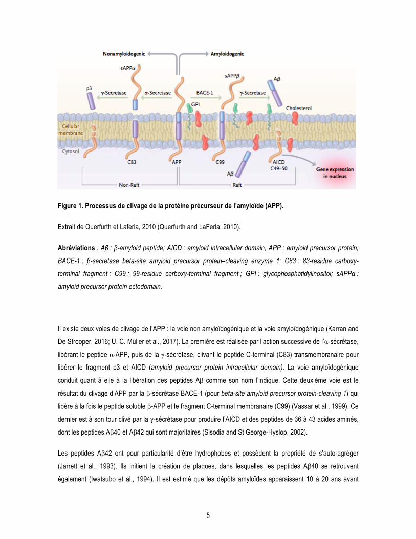
Figure 1. Processus de clivage de la protéine précurseur de l’amyloïde (APP).
Extrait de Querfurth et Laferla, 2010 (Querfurth and LaFerla, 2010).
Abréviations : Aβ : β-amyloid peptide; AICD : amyloid intracellular domain; APP : amyloid precursor protein;
BACE-1 : β-secretase beta-site amyloid precursor protein–cleaving enzyme 1; C83 : 83-residue carboxy-
terminal fragment ; C99 : 99-residue carboxy-terminal fragment ; GPI : glycophosphatidylinositol; sAPPα :
amyloid precursor protein ectodomain.
Il existe deux voies de clivage de l’APP : la voie non amyloïdogénique et la voie amyloïdogénique (Karran and
De Strooper, 2016; U. C. Müller et al., 2017). La première est réalisée par l’action successive de l’α-sécrétase,
libérant le peptide α-APP, puis de la γ-sécrétase, clivant le peptide C-terminal (C83) transmembranaire pour
libérer le fragment p3 et AICD (amyloid precursor protein intracellular domain). La voie amyloïdogénique
conduit quant à elle à la libération des peptides Aβ comme son nom l’indique. Cette deuxième voie est le
résultat du clivage d’APP par la β-sécrétase BACE-1 (pour beta-site amyloid precursor protein-cleaving 1) qui
libère à la fois le peptide soluble β-APP et le fragment C-terminal membranaire (C99) (Vassar et al., 1999). Ce
dernier est à son tour clivé par la γ-sécrétase pour produire l’AICD et des peptides de 36 à 43 acides aminés,
dont les peptides Aβ40 et Aβ42 qui sont majoritaires (Sisodia and St George-Hyslop, 2002).
Les peptides Aβ42 ont pour particularité d’être hydrophobes et possèdent la propriété de s’auto-agréger
(Jarrett et al., 1993). Ils initient la création de plaques, dans lesquelles les peptides Aβ40 se retrouvent
également (Iwatsubo et al., 1994). Il est estimé que les dépôts amyloïdes apparaissent 10 à 20 ans avant
6
l’apparition des symptômes de la MA (Bateman et al., 2012; Tanzi, 2012). Le ratio de production de peptides
Aβ42/Aβ40 est d’environ 10/90, mais ce ratio est augmenté dans la MA ainsi que dans les cas de MA
familiaux, c’est-à-dire chez les porteurs d’une mutation génétique dans le gène APP ou la préséniline (PSEN),
la site catalytique de la γ-sécrétase (voir section 1.4.1) (Tanzi, 2012). Dans le cas de la MA, il y aurait un
débalancement de l’activité des enzymes qui conduirait à la surproduction des peptides Aβ40 et Aβ42 (Selkoe
and Hardy, 2016). Ainsi, plusieurs inhibiteurs de BACE-1 ont été développés comme traitement pour la
maladie, sans succès pour le moment (Cummings et al., 2018; Vassar, 2014). Toutefois, l’hypothèse de la
cascade amyloïde est remise en questions depuis plusieurs années car certains rôles protecteurs de ces
peptides ont été identifiés et l’accumulation des peptides amyloïdes seule ne suffit pas à causer la MA
(Carrillo-Mora et al., 2014; Skaper, 2012).
La majorité des études neuropathologiques ou animales se basent sur la quantité de plaques amyloïdes pour
établir un niveau plus ou moins important de pathologie. Toutefois, une hypothèse selon laquelle les
oligomères seraient plus toxiques que les plaques amyloïdes émerge depuis plusieurs années et est soutenue
par une littérature scientifique abondante (Esparza et al., 2013; Klein et al., 2001; Viola and Klein, 2015).
L’agrégation des peptides Aβ42 présente plusieurs étapes au cours desquelles ceux-ci forment des
oligomères de 2 à 6 peptides, puis des fibrilles, pour finalement former des feuillets plissés β qui constituent
les plaques amyloïdes (Kayed et al., 2009; Ono et al., 2009). Chez le rat, l’injection d’oligomères d’Aβ42
extraits de cerveaux de sujets décédés de la MA dans l’hippocampe perturbe la fonction synaptique et induit
des troubles de la mémoire (Shankar et al., 2008). D’autres études ont montré que 6 injections d’oligomères
(de monomères à tétramères) d’Aβ42 dans l’hippocampe suffisent à induire une perte neuronale et des
déficits cognitifs chez le rat (Brouillette et al., 2012; Sajadi et al., 2016). En effet, ces oligomères perturberaient
la communication synaptique en s’intercalant au sein des fentes synaptiques (Brouillette, 2014).
Un autre mécanisme conduisant à l’accumulation de peptides amyloïdes dans la MA serait la diminution de
leur clairance du cerveau vers le sang. En effet, plusieurs études ont montré une diminution de l’expression du
transporteur LRP1 (low density lipoprotein receptor-related protein 1), qui participe à la clairance des peptides
du cerveau vers le sang, et à l’inverse une augmentation de RAGE (receptor for advanced glycation
endproducts) au niveau des cellules endothéliales de la barrière hémato-encéphalique (BHE) dans des
cerveaux de sujets décédés de la MA (Deane et al., 2004; Donahue et al., 2006; Miller et al., 2008; Wilhelmus
et al., 2007). Au niveau du LCR, les peptides Aβ peuvent être transportés par la transthyrétine (ou
préalbumine), une protéine impliquée principalement dans le transport du rétinol et de la thyroxine qui se
trouve diminuée chez les patients atteints de la MA (Serot et al., 1997). Finalement, une baisse d’activité des
enzymes impliquées dans la dégradation d’Aβ, IDE (insulin degrading enzyme) et neprilysin, contribuerait
également à la formation des plaques amyloïdes (Farris et al., 2003; Kanemitsu et al., 2003).
7
1.3.2 Enchevêtrements neurofibrillaires
La protéine tau est présente au niveau des microtubules des cellules neuronales. Elle est produite à partir de
l’expression du gène MAPT pour microtubule-associated protein tau située sur le chromosome 17 (Buée et al.,
2000). Six isoformes différentes sont trouvées chez l’humain, variant de 352 à 441 acides aminés, se
distinguant entre elles principalement dans la présence de trois ou quatre domaines répétés dans la région C-
terminale de la protéine (Goedert et al., 1989; Kitamura et al., 2005).
Le principal rôle de la protéine tau est de stabiliser les microtubules et de participer au transport axonal. La
protéine tau peut subir plusieurs modifications post-traductionnelles telles que la troncation, l’ubiquitination, la
glycosylation, l’acétylation ou encore la phosphorylation (M. Morris et al., 2015). Toutefois, la phosphorylation
résultant de l’action de différentes kinases et phosphatases est la modification principale que subit la protéine
tau : plus de 80 sites de phosphorylations sur la protéine sont connus (Martin et al., 2011). Cette
phosphorylation physiologique va modifier son degré d’attachement aux microtubules et ainsi contribuer à la
plasticité neuronale. Toutefois, dans le cas de la MA et d’autres tauopathies, la protéine tau est
hyperphosphorylée, ce qui va conduire à son détachement des microtubules et son relargage au niveau
cytosolique (Figure 2) (Iqbal et al., 2016). L’hyperphosphorylation de ces protéines tau « libres » va causer
leur modification de conformation tridimensionnelle et induire leur agrégation (Bretteville and Planel, 2008).
Les protéines tau hyperphosphorylées vont s’agréger en oligomères puis former des ENF à l’intérieur des
neurones (Ren and Sahara, 2013).
Les principales kinases impliquées dans l’hyperphosphorylation pathologique de la protéine tau sont GSK3β,
AKT, MAPK, cdk5 et CaMKII. La kinase GSK3β en particulier est responsable de la phosphorylation de plus
de 30 sites sur la protéine tau, ce qui fait d’elle une kinase majeure pour la pathologie tau (Hooper 2008).
Ainsi, différents inhibiteurs pour cette kinase ont été testés dans des modèles murins de la MA, mais les effets
indésirables conséquents à l’inhibition de cette enzyme, impliquée dans de nombreuses voies de signalisation,
ont compromis le développement de ces thérapies dans la MA (Hooper et al., 2008; Kremer et al., 2011). AKT
peut également phosphoryler tau au niveau de la serine 214, mais son implication dans l’hyperphosphorylation
de tau réside principalement dans le fait qu’elle module l’activité de GSK3β (Gratuze et al., 2018; Griffin et al.,
2005). En effet, AKT phosphoryle GSK3β au niveau de la serine 9, ce qui résulte en l’inactivation de GSK3β.
Ainsi, une plus grande activité d’AKT est associée à une diminution de la phosphorylation de la protéine tau
via l’inactivation de GSK3β. Les phosphatases sont également impliquées dans la régulation de la
phosphorylation de tau. La PP2A est la principale phosphatase impliquée dans la régulation de tau; elle est
également capable de moduler l’activité de GSK3β (Torrent and Ferrer, 2012).
8
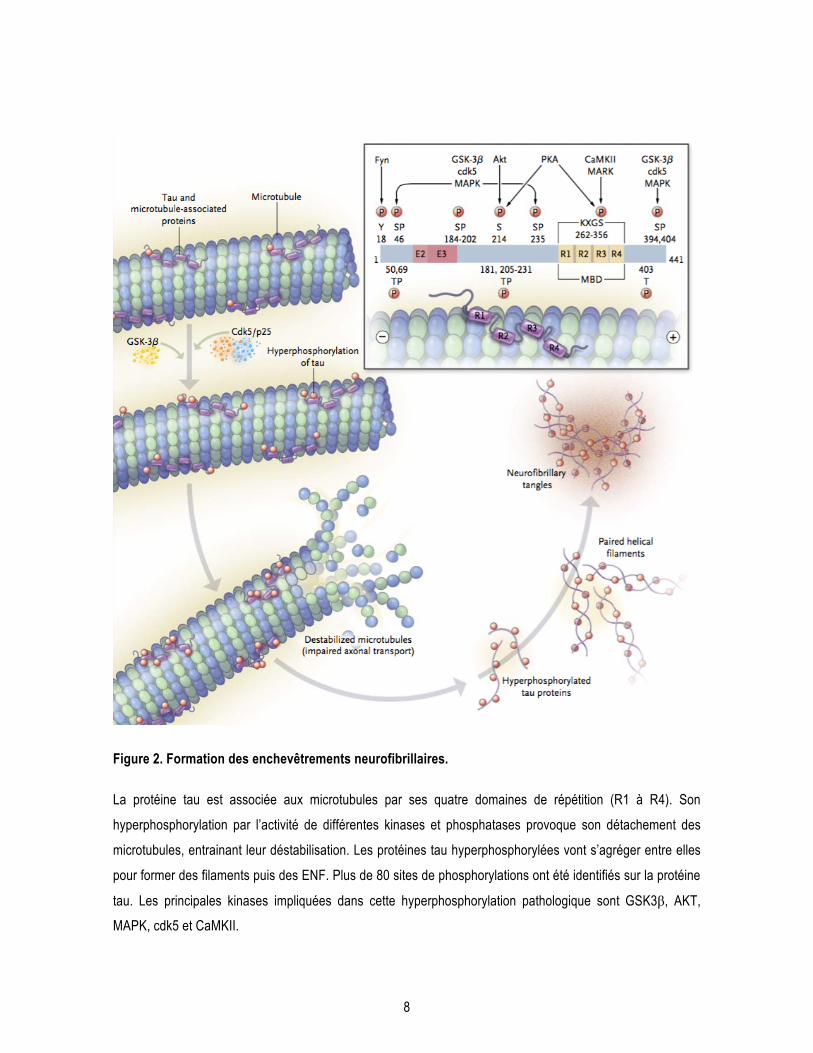
Figure 2. Formation des enchevêtrements neurofibrillaires.
La protéine tau est associée aux microtubules par ses quatre domaines de répétition (R1 à R4). Son
hyperphosphorylation par l’activité de différentes kinases et phosphatases provoque son détachement des
microtubules, entrainant leur déstabilisation. Les protéines tau hyperphosphorylées vont s’agréger entre elles
pour former des filaments puis des ENF. Plus de 80 sites de phosphorylations ont été identifiés sur la protéine
tau. Les principales kinases impliquées dans cette hyperphosphorylation pathologique sont GSK3β, AKT,
MAPK, cdk5 et CaMKII.
9
Extrait de Querfurth et Laferla, 2010 (Querfurth and LaFerla, 2010).
Abréviations : AKT : protéine kinase B; CaMKII : calcium–calmodulin protein kinase 2; cdk5 : cyclin-depen-
dent kinase; GSK3β : glycogen synthase kinase 3β; MAPK : mitogen-activated protein kinase; MARK :
microtubule affinity-regulating kinase; MBD : microtubule-binding domain; P : proline; p25 : cdk5 activator
subunit; PKA : protein kinase A; S: serine; T : threonine.
Il est important de noter que parmi tous les sites de phosphorylation présents sur la protéine tau, la plupart
sont considérés comme physiologiques, alors que d’autres sont considérés comme anormaux.
L’hyperphosphorylation de la protéine tau sur ses sites physiologiques et la phosphorylation des sites
anormaux sont impliqués dans l’agrégation de la protéine tau (M. Morris et al., 2011). Il est estimé que la
protéine tau possède environ 3 groupements phosphates en condition physiologique, tandis que ce nombre
augmente jusqu’à huit fois en condition pathologique (Khatoon et al., 1994). De nombreuses études
neuropathologiques et in vitro ont permis de mettre en évidence les principaux sites de phosphorylation
impliqués dans l’agrégation de la protéine tau et la MA : Ser202, Thr205, Ser396, Ser422, Ser404, Ser262,
Thr212, Thr231, Thr181 (Alonso et al., 2010; Augustinack et al., 2002; Daly et al., 2000; Haase et al., 2004;
Martin et al., 2011).
L’hyperphosphorylation de la protéine tau, ainsi que la quantité de protéine tau insoluble (agrégée) et d’ENF
sont fortement corrélées au diagnostic clinique des sujets décédés de la MA, appuyant son rôle dans le
développement de la maladie (Bennett et al., 2004; Tremblay et al., 2017; 2007). Des stratégies
thérapeutiques visant à diminuer la quantité de tau hyperphosphorylée ou sa capacité à s’agréger ont été
testées chez l’animal et l’humain, mais aucun de ces traitements n’a pour le moment été approuvé pour une
utilisation clinique (Asuni et al., 2007; Cummings et al., 2018; Kontsekova et al., 2014). Toutefois, nous ne
savons toujours pas avec certitude si la pathologie tau est une cause ou une conséquence de la MA. Il faut
noter que la présence d’ENF est fréquente dans le cerveau de personnes âgées cognitivement saines
(Bennett et al., 2006), et que ces agrégats sont retrouvés dans d’autres maladies neurodégénératives,
regroupées sous le terme de tauopathies, telle que la démence fronto-temporale, le syndrôme de Down ou
encore la maladie de Pick (Buée et al., 2000; Sergeant et al., 2005). Les ENF ont récemment été retrouvés
dans la maladie de Huntington, une maladie neurodégénérative purement génétique (St-Amour et al., 2017).
Ces études indiquent que les ENF ne sont pas spécifiques à la MA, et suggèrent que le phénomène
d’agrégation de la protéine tau pourrait être une conséquence de la maladie plutôt qu’une cause.
10
1.3.3 Pertes synaptiques
La synapse correspond à la zone où deux neurones entrent en contact. Les pertes synaptiques sont connues
depuis longtemps dans la MA (Terry et al., 1991). En effet, la diminution de la quantité de protéines
synaptiques est très claire dans le cerveau des sujet décédés de la MA, et apparaît dès les stades précoces
de la maladie (Counts et al., 2014). Les déficits synaptiques quantifiés au niveau post-mortem corrèlent de
façon robuste avec les déficits cognitifs des individus. Une étude a montré que les protéines pré-synaptiques
synaptophysine et syntaxine 3 étaient diminuées dans le cortex pariétal dès le stade de trouble cognitif léger,
et que les protéines pré-synaptiques synaptophysine et septin-3 corrélaient de façon positive avec le score
cognitif global de l’ensemble des sujets déments et sains (Tremblay et al., 2017). Une autre étude a montré
que la quantité de SNAP-25 (Synaptosomal-associated protein 25) dans le cortex préfrontal était corrélée au
taux de déclin cognitif calculé sur les huit années avant la mort du sujet (Bereczki et al., 2016). Également, la
protéine drébrine est diminuée d’environ 25% dans le cortex des sujets atteints de la MA et son expression
corrèle avec la pathologie tau ainsi que la durée des symptômes de la MA avant la mort des individus (Julien
et al., 2008). Finalement, quelques études cliniques ont observé des niveaux augmentés de protéines pré-
synaptiques (SNAP25) et post-synaptiques (neurogranine) dans le LCR dès les stades précoces de la MA,
suggérant que ces protéines pourraient être des biomarqueurs pour la MA (Brinkmalm et al., 2014; Kvartsberg
et al., 2015). Toutefois, ces deux protéines ne sont pas spécifiques à la MA puisqu’elles sont également
augmentées dans le LCR des sujets atteints de Parkinson (Bereczki et al., 2017).
1.3.4 Atrophie cérébrale
La MA étant une maladie neurodégénérative, l’atrophie cérébrale causée par les pertes synaptiques puis une
mort neuronale apparaît dans les stades précoces de la MA et fait partie intégrante du diagnostic comme
discuté plus haut. L’imagerie par résonance magnétique (IRM) permet de détecter ces changements
morphologiques (Frisoni et al., 2010). Il est possible de mesurer l’atrophie du cerveau entier, mais le cortex
entorhinal est la première région touchée, suivie de l’hippocampe et l’élargissement des ventricules puis le
cortex temporal, suivant ainsi l’évolution spatio-temporelle de la pathologie tau dans le cerveau (Thompson et
al., 2003).
Avec le vieillissement normal, une atrophie des régions corticales d’environ 10% est observée (Erten-Lyons et
al., 2013; Fotenos et al., 2005; Freeman et al., 2008). En revanche, dès le stade de trouble cognitif léger, la
perte neuronale dans le cortex entorhinal est de près de 30% et corrèle avec les troubles cognitifs des sujets
(M. C. Evans et al., 2010; Kordower et al., 2001). Plus frappant encore, une étude d’IRM à haute résolution sur
11
près de 400 sujets a montré que le taux d’atrophie cérébrale globale est de 0,45% par année chez un individu
sain, tandis qu’il double pour atteindre 0,98% pour les individus atteints de démence légère de type Alzheimer
(Pakkenberg et al., 2003). Une autre étude sur des données d’IRM de plus de 700 participants issues de
l’étude Alzheimer’s Disease Neuroimaging Initiative a mesuré une accélération de 0,22% du taux d’atrophie
dans l’hippocampe chez les sujets atteints de trouble cognitif léger comparativement aux individus sains
(Leung et al., 2013). Ainsi, l’atrophie cérébrale est attentivement examinée dans les études cliniques afin
d’évaluer un potentiel effet « disease modifying » de l’intervention chez les participants (Frisoni et al., 2010).
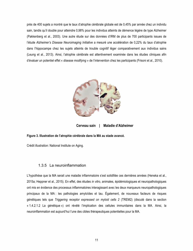
Cerveau sain | Maladie d’Alzheimer
Figure 3. Illustration de l’atrophie cérébrale dans la MA au stade avancé.
Crédit illustration: National Institute on Aging.
1.3.5 La neuroinflammation
L’hypothèse que la MA serait une maladie inflammatoire s’est solidifiée ces dernières années (Heneka et al.,
2015a; Heppner et al., 2015). En effet, des études in vitro, animales, épidémiologiques et neuropathologiques
ont mis en évidence des processus inflammatoires interagissant avec les deux marqueurs neuropathologiques
principaux de la MA : les pathologies amyloïdes et tau. Également, de nouveaux facteurs de risques
génétiques tels que Triggering receptor expressed on myloid cells 2 (TREM2) (discuté dans la section
« 1.4.2.1.2 La génétique ») ont révélé l’implication des cellules immunitaires dans la MA. Ainsi, la
neuroinflammation est aujourd’hui l’une des cibles thérapeutiques potentielles pour la MA.
12
Plusieurs mécanismes inflammatoires « anormaux » ont été identifiés dans la MA, mais seuls les principaux
seront présentés ici. Les microglies sont des macrophages résidents du système nerveux central (SNC) et
constituent la première ligne de défense immunitaire de ce dernier (Ransohoff and Engelhardt, 2012). Ces
cellules sont notamment activées par les peptides Aβ in vitro et dans des modèles murins de la MA (Bisht et
al., 2016; Maezawa et al., 2011; Pan et al., 2011), et seraient impliquées dans la clairance des formes
solubles et des fibrilles d’Aβ (pour revue : (C. Y. D. Lee and Landreth, 2010)). Une étude qualitative
(immunohistochimie) sur 19 sujets montre que les microglies se retrouvent également près des ENF, mais
dans un état dystrophique et dans les premiers stades de Braak, suggérant que leur dégénérescence précoce
est impliquée dans le développement de la MA (Streit et al., 2009). En revanche, il a récemment été montré
que les microglies pouvaient adopter un phénotype trop « actif » en présence de pathologie amyloïde et
phagocyter les synapses dans des modèles murins de la MA (Hong et al., 2016). Ces résultats suggèrent
plutôt que l’activation microgliale conséquente de la neuropathologie Alzheimer deviendrait néfaste et
contribuerait à la perte synaptique caractéristique de la MA (Rajendran and Paolicelli, 2018). De manière
assez similaire, les astrocytes constituent un autre type de cellule immunitaire résidente du SNC ayant une
capacité de clairance des plaques amyloïdes (Pihlaja et al., 2011; Wyss-Coray et al., 2003), parmi d’autres
rôles variés tels que la transmission synaptique, le métabolisme énergétique ou encore le maintien de l’unité
neurovasculaire (Sofroniew and Vinters, 2010). Une astrogliose est observée près des plaques amyloïdes
dans des modèles animaux de MA (Olabarria et al., 2010) mais aussi dans le cerveau de sujets décédés de la
MA (Serrano-Pozo et al., 2011; Webster et al., 2006). Finalement, l’infiltration de macrophages de la
périphérie vers le parenchyme cérébral est observée de façon plus fréquente dans le cerveau d’individus
décédés de la MA comparativement aux individus sains (Togo et al., 2002). Ces études ont conduit à tester
différentes approches thérapeutiques pour la MA telles que les anti-inflammatoires et l’immunisation passive et
active (voir section 1.5.2) (Butchart and Holmes, 2012; St-Amour et al., 2016).
1.3.6 Autres neuropathologies et neuropathologies mixtes
Près de la moitié des cas de diagnostic clinique probable de MA présente en fait réellement une
neuropathologie mixte et non purement de type Alzheimer (Schneider et al., 2009; Schneider and Bennett,
2010; Sweeney et al., 2019).
La concomitance de pathologie vasculaire et de neuropathologie de type Alzheimer est la plus courante et
s’élève à plus de 70% des cas de MA diagnostiqués cliniquement, bien que ces chiffres varient d’une étude à
l’autre en fonction des lésions vasculaires étudiées (Kapasi et al., 2017; Toledo et al., 2013). Les pathologies
vasculaires sont diverses, mais les principales observées dans la MA sont les macro et microinfarctus, les

13
athéroscléroses et les angiopathies amyloïdes (Iadecola, 2017). Les angiopathies amyloïdes cérébrales sont
caractérisées par le dépôt de peptides amyloïdes dans les vaisseaux sanguins cérébraux (Kalaria and Kalimo,
2002). Ces dépositions sont observées dans le cerveau 20 à 40% des personnes âgées et dans près de 90%
des cerveaux de personnes décédées de la MA (Attems et al., 2011).
D’autre part, les lésions de la matières blanche sont très répandues chez les sujets atteints de la MA et sont
corrélées avec l’apparition de troubles cognitifs et de démence (Black et al., 2009). Ces lésions se présentent
de manière diffuse près des ventricules cérébraux et sont visibles en IRM : elles sont caractérisées par des
tâches diffuses plus blanches par rapport au tissu avoisinant et sont appelées « hyper-intensités » (Maillard et
al., 2012).
D’autres protéinopathies sont fréquemment observées dans le cerveau de sujets décédés de la MA.
L’accumulation de la protéine nucléaire TDP-43 (Transactive response DNA binding protein 43) dans le
cytosol est caractéristique de la sclérose latérale amyotrophique (Sreedharan et al., 2008). Plusieurs études
neuropathologiques ont montré la présence de cette protéinopathie dans des cerveaux de sujets
diagnostiqués avec la MA ou un trouble cognitif léger (Ohta et al., 2014; Robinson et al., 2011; Tremblay et al.,
2011). Finalement, les corps de Lewy formés par la protéine α-synucléine, classiquement retrouvés dans la
maladie de Parkinson, sont également retrouvés dans certains cas de MA (Mikolaenko et al., 2005).
1.4 Facteurs de risque de la MA
Il existe deux formes de la MA : la forme familiale, aussi appelée « early-onset AD », qui représente moins de
1% des cas, et la forme sporadique, ou « late-onset AD », la forme majoritaire (Tanzi, 2012).
1.4.1 Formes familiales de la maladie
Les formes familiales de la MA se caractérisent par une transmission génétique de mutations rares à
pénétration complète et une apparition des symptômes de la maladie de façon précoce, c’est-à-dire
généralement avant l’âge de 65 ans (Tanzi, 2012). Trois principaux gènes sont touchés par des mutations
responsables de la forme héréditaire de la MA : APP, PSEN1 et PSEN2 (Tableau 1). Ces mutations
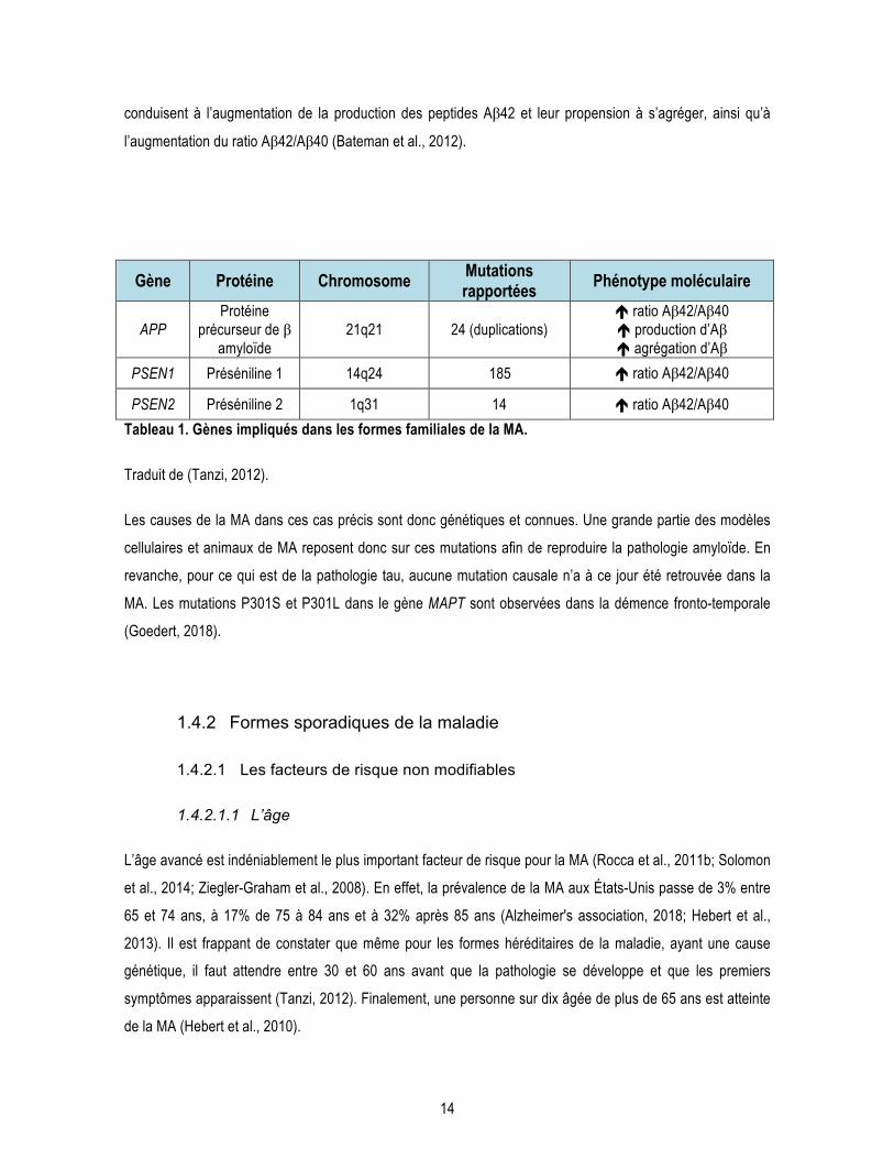
14
conduisent à l’augmentation de la production des peptides Aβ42 et leur propension à s’agréger, ainsi qu’à
l’augmentation du ratio Aβ42/Aβ40 (Bateman et al., 2012).
Gène Protéine Chromosome Mutations rapportées Phénotype moléculaire
APP Protéine
précurseur de β amyloïde
21q21 24 (duplications) ! ratio Aβ42/Aβ40 ! production d’Aβ ! agrégation d’Aβ
PSEN1 Préséniline 1 14q24 185 ! ratio Aβ42/Aβ40
PSEN2 Préséniline 2 1q31 14 ! ratio Aβ42/Aβ40
Tableau 1. Gènes impliqués dans les formes familiales de la MA.
Traduit de (Tanzi, 2012).
Les causes de la MA dans ces cas précis sont donc génétiques et connues. Une grande partie des modèles
cellulaires et animaux de MA reposent donc sur ces mutations afin de reproduire la pathologie amyloïde. En
revanche, pour ce qui est de la pathologie tau, aucune mutation causale n’a à ce jour été retrouvée dans la
MA. Les mutations P301S et P301L dans le gène MAPT sont observées dans la démence fronto-temporale
(Goedert, 2018).
1.4.2 Formes sporadiques de la maladie
1.4.2.1 Les facteurs de risque non modifiables
1.4.2.1.1 L’âge
L’âge avancé est indéniablement le plus important facteur de risque pour la MA (Rocca et al., 2011b; Solomon
et al., 2014; Ziegler-Graham et al., 2008). En effet, la prévalence de la MA aux États-Unis passe de 3% entre
65 et 74 ans, à 17% de 75 à 84 ans et à 32% après 85 ans (Alzheimer's association, 2018; Hebert et al.,
2013). Il est frappant de constater que même pour les formes héréditaires de la maladie, ayant une cause
génétique, il faut attendre entre 30 et 60 ans avant que la pathologie se développe et que les premiers
symptômes apparaissent (Tanzi, 2012). Finalement, une personne sur dix âgée de plus de 65 ans est atteinte
de la MA (Hebert et al., 2010).
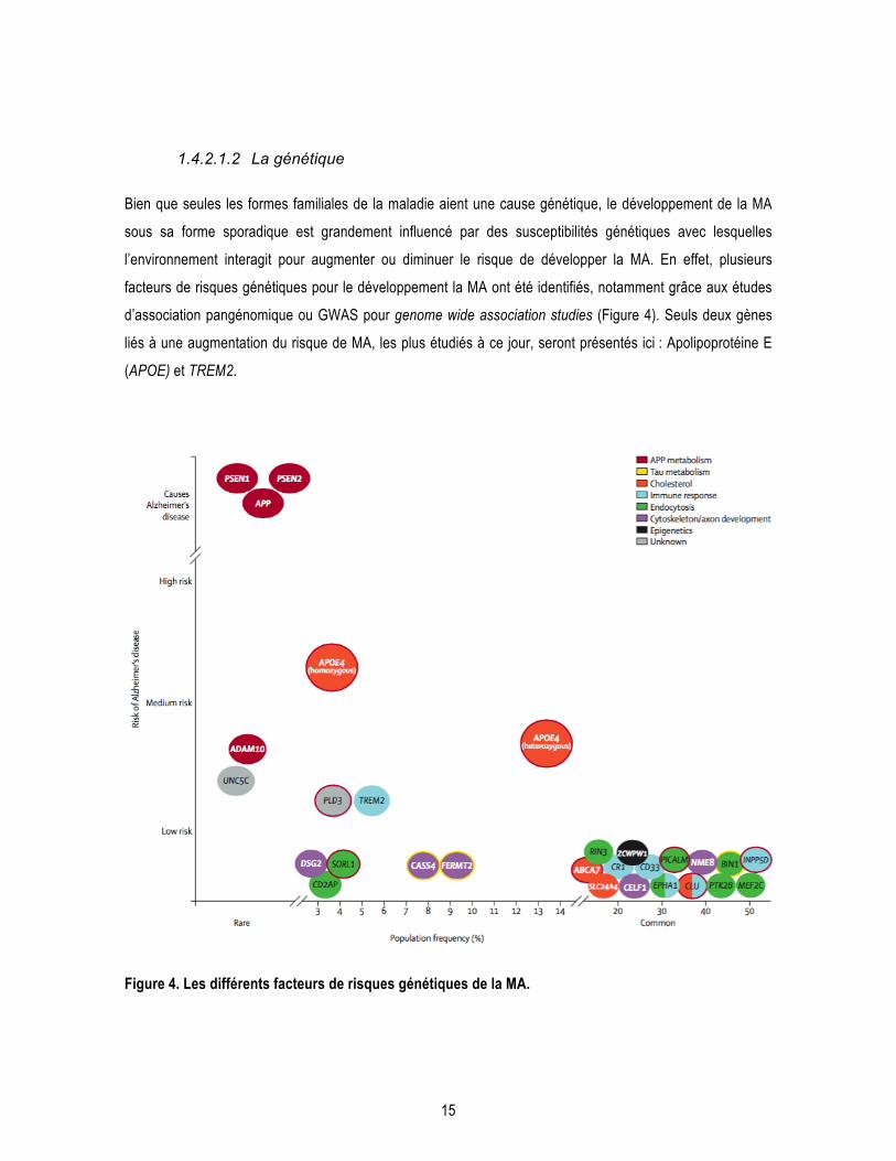
15
1.4.2.1.2 La génétique
Bien que seules les formes familiales de la maladie aient une cause génétique, le développement de la MA
sous sa forme sporadique est grandement influencé par des susceptibilités génétiques avec lesquelles
l’environnement interagit pour augmenter ou diminuer le risque de développer la MA. En effet, plusieurs
facteurs de risques génétiques pour le développement la MA ont été identifiés, notamment grâce aux études
d’association pangénomique ou GWAS pour genome wide association studies (Figure 4). Seuls deux gènes
liés à une augmentation du risque de MA, les plus étudiés à ce jour, seront présentés ici : Apolipoprotéine E
(APOE) et TREM2.
Figure 4. Les différents facteurs de risques génétiques de la MA.
16
Les différents facteurs de risques génétiques identifiés pour la MA sont présentés en fonction de leur
fréquence dans la population (en %, rare ou commun) et du niveau de risque pour le développement de la MA
(faible, moyen, élevé ou cause la MA). Extrait de (Scheltens et al., 2016).
Le principal facteur de risque génétique de la forme sporadique de la MA est l’allèle ε4 du gène de
l’apolipoprotéine E (APOE) (Michaelson, 2014). Les apolipoprotéines sont des glycoprotéines responsables du
transport des lipides dans le corps. Plus spécifiquement, l’apolipoprotéine de type E est responsable du
transport du cholestérol et est principalement produite par le foie (Mahley et al., 2009; Phillips, 2014). Elle est
présente sous trois isoformes : APOE2, E3 et E4. Ces isoformes diffèrent seulement par deux acides aminés
(112 et 158) (Holtzman et al., 2012). Corder et collaborateurs furent les premiers en 1993 à identifier ce risque
génétique dans la MA (Corder et al., 1993). En étudiant 42 familles avec des cas de MA sporadiques, ils ont
montré que les porteurs homozygotes de l’allèle ε4 avaient 12 fois plus de risque de développer la MA, tandis
que les porteurs hétérozygotes avaient trois fois plus de risque. Cette augmentation du risque s’accompagne
d’une apparition plus précoce des symptômes d’environ 15 ans par rapport à la moyenne. Au contraire, les
porteurs de l’allèle ε2 sont peu fréquents dans la population touchée par la MA et ont moins de risque de
développer une démence (Berlau et al., 2009).
Les mécanismes liant APOE4 et la MA sont divers. L’APOE est impliquée dans le processus de production et
de dégradation de l’Aβ (Holtzman et al., 2012; Jiang et al., 2008). Toutefois, l’expression de l’isoforme E4
dans des modèles animaux de la MA augmente la production d’Aβ42 et perturbe sa clairance en interagissant
avec le transporteur LRP1 (J. M. Castellano et al., 2011; Deane et al., 2008; Fryer et al., 2005; Liraz et al.,
2013). Par ailleurs, le génotype APOE4 affecte la vasculature cérébrale et les transporteurs de la BHE dans
des modèles animaux (Alata et al., 2015; Bell et al., 2012; Vandal et al., 2014a) ainsi que chez l’humain
(Halliday et al., 2016). Bien que le rôle d’APOE4 sur la pathologie tau soit moins connu, une récente étude
animale a montré que le génotype APOE4 interagit avec la protéine tau mutée (P301S), indépendamment de
la pathologie amyloïde, pour aggraver la neurodégénérescence et la neuroinflammation (Y. Shi et al., 2017).
Un autre facteur de risque génétique important plus récemment identifié est TREM2 pour triggering receptor
expressed on myeloid cells 2 (Rohn, 2013). Ce récepteur est exprimé sur les microglies et leur permet de
phagocyter des débris cellulaires (Neumann and Takahashi, 2007). Plusieurs variants dans le gène TREM2
ont été associés à un plus grand risque de développer la MA (Guerreiro et al., 2013). L’hypothèse actuelle est
que des variants du gène TREM2 induisent la perte de fonction du récepteur et conduisent à l’incapacité des
microglies à phagocyter les peptides amyloïdes (pour revue : (Ulland and Colonna, 2018)).
17
1.4.2.1.3 Le sexe
Il est bien connu que le nombre de femmes affectées par la MA est plus grand que celui des hommes. Il est
estimé qu’environ 65% des cas de MA sont des femmes (Mielke et al., 2014). Plusieurs raisons expliquent ces
disparités. Tout d’abord, les femmes ont une espérance de vie plus élevée que les hommes (Tom et al.,
2015). Le principal facteur de risque de la MA étant l’âge avancé, il n’est donc pas étonnant que plus de
femmes soient touchées par la maladie. Une autre explication est que lors de la ménopause, les femmes
subissent une chute drastique d’estrogènes, qui sont neuroprotecteurs, tandis que la diminution des hormones
sexuelles est très progressive chez les hommes (Arevalo et al., 2015; Rocca et al., 2011a). Toutefois, ces
différences sont remises en question par de récentes études prospectives : elles seraient probablement
biaisées par le fait que les hommes ont une plus grande mortalité due aux maladies cardiovasculaires en
milieu de vie (Chêne et al., 2015; Ruitenberg et al., 2001). Ainsi, les hommes plus âgés auraient un profil de
risque cardiovasculaire plus faible que les femmes du même âge et donc un moins grand risque de
développer la MA. En effet, l’incidence cumulative de MA est quant à elle similaire entre les hommes et les
femmes dans l’étude prospective de la Framingham Heart Study (Chêne et al., 2015).
1.4.2.2 Les facteurs de risque modifiables
De nombreux facteurs de risque environnementaux ont été associés à la MA, et les principaux seront
présentés dans cette section. Ces facteurs sont considérés comme modifiables, c’est-à-dire qu’il est possible
d’intervenir dessus afin de réduire la probabilité de développer la MA (Winblad et al., 2016). L’alimentation
peut d’ailleurs influencer la majorité de ces facteurs de risque. Ainsi, du point de vue thérapeutique mais aussi
préventif, il est crucial de bien comprendre leur importance dans la pathogenèse de la MA (Biessels, 2014).
1.4.2.2.1 Hypertension et maladies cardiovasculaires
Les maladies cardiovasculaires désignent l’ensemble des maladies du cœur, des maladies vasculaires
cérébrales et des maladies des vaisseaux sanguins (World Health Organization et al., 2011). Elles sont la
principale cause de décès dans le monde (Benjamin et al., 2017). Plusieurs d’entre elles augmentent les
risques de démence. D’une part, la fibrillation auriculaire ou arythmie cardiaque, une des maladies
cardiovasculaires les plus communes, est associée avec une plus grande incidence de démence (Kwok et al.,
18
2011). D’autre part, les accidents vasculaires cérébraux (AVC) conduisent souvent à des déficits cognitifs
(Marshall, 2012). En effet, il est estimé que les personnes ayant subi un AVC ont deux fois plus de risque de
développer une démence, et que 30% de cette population va développer des troubles cognitifs dans les trois
années suivant l’accident (Allan et al., 2011).
L’hypertension est également l’un des plus grand facteur de risque de la MA, mais également la comorbidité la
plus répandue chez les sujets atteints de MA (Reitz et al., 2010; J.-H. Wang et al., 2018). Une tension
artérielle élevée en milieu de vie est associée au développement de trouble cognitif léger et de la MA à un âge
plus avancé (Elias and Davey, 2012; Reitz et al., 2007; Shah et al., 2012). L’hypertension chronique induit des
lésions cérébrovasculaires telles que des microinfarctus ou des microhémorragies cérébrales (Kalaria, 2012).
Plusieurs études ont montré qu’une pression artérielle élevée augmente les marqueurs neuropathologiques de
la MA et induit une apparition plus précoce des déficits cognitifs dans des modèles murins de la MA (Cifuentes
et al., 2017; 2015; Díaz-Ruiz et al., 2009; Shih et al., 2018). Chez l’homme adulte, l’hypertension accroît
l’atrophie cérébrale et les lésions de la matière blanche (Firbank et al., 2007).
De façon intéressante, des études épidémiologiques montrent que les traitements pour les maladies
cardiovasculaires et l’hypertension sont associés à un risque plus faible de démence vasculaire et de MA
(Mogi and Horiuchi, 2009; Zhuang et al., 2016; Zissimopoulos et al., 2017). En effet, les statines et les
antihypertenseurs ont montré des effets bénéfiques pour renverser les déficits cérébrovasculaires, cognitifs et
la pathologie amyloïde dans des modèles animaux de la MA (Hamel et al., 2016; Royea et al., 2017; Tong et
al., 2009; J. Wang et al., 2007). Néanmoins, des études cliniques chez l’humain sont nécessaires afin
d’apporter des preuves solides du bénéfice de ces traitements pour la MA.
1.4.2.2.2 Maladies métaboliques
Les maladies métaboliques les plus fréquentes apparaissent lorsque l’équilibre entre l’apport et la dépense
énergétique est altérée. Elles regroupent par exemple l’obésité, le diabète de type 1 et 2 ou encore le
syndrome métabolique. Ces pathologies sont reconnues depuis un certain temps comme des facteurs de
risque importants pour la MA (Luchsinger, 2008; Luchsinger and Gustafson, 2009; Profenno et al., 2010).
Le diabète de type 2 (DT2) est une maladie chronique caractérisée par une résistance à l’insuline et une
hyperglycémie dont les premiers symptômes apparaissent à l’âge adulte (“Votre guide sur le diabète,” 2011).
Le DT2 est à différencier du diabète de type 1, qui ne concerne que 10% des diabétiques et se déclare très tôt
par l’absence totale de production d’insuline par les cellules β du pancréas. Le DT2 augmente de deux fois le

19
risque de développer la MA selon une étude prospective de 1999 (Ott et al., 1999). Il fait partie des cinq
comorbidités les plus retrouvées dans la population atteinte de la MA (J.-H. Wang et al., 2018). De plus, il est
estimé que le DT2 est responsable d’un cas de démence sur dix, suggérant que les traitements contre le
diabète permettraient également de diminuer la prévalence de la MA (Biessels et al., 2006). Les sujets
diabétiques ont un plus grand risque de démence et développent des altérations cérébrales similaires à celles
observées dans la MA (Daulatzai, 2017) : ils présentent une atrophie cérébrale (Espeland et al., 2013;
Manschot et al., 2006), une plus importante pathologie tau et amyloïde que les sujets sains (Y. Liu et al., 2009;
Peila et al., 2002) ainsi que des micro-infarctus cérébraux et de la neuroinflammation (Sonnen et al., 2009).
Les modèles animaux de diabète présentent également une augmentation de la pathologie tau (El-Khoury et
al., 2016; Gratuze et al., 2017a; Kim et al., 2009), et l’apparition d’un phénotype diabétique par l’administration
de la toxine streptozotocin dans des modèles animaux de la MA aggrave leur neuropathologie (Gratuze et al.,
2017b).
Les désordres métaboliques associés au diabète sont à eux seuls associés à la MA. En effet, d’une part, les
niveaux élevés d’hémoglobine glyquée (HbA1c), un marqueur d’équilibre glycémique sur plusieurs mois, sont
associés à un plus fort taux d’atrophie cérébrale chez des sujets âgés (Enzinger et al., 2005). Une étude
longitudinale sur 7 ans et plus de 2000 participants âgés a montré qu’une glycémie élevée chez des sujets
non diabétiques est associée à un plus grand risque de démence (Crane et al., 2013). Cette association était
également significative pour les individus diabétiques. D’autre part, la glycémie à jeun est inversement
corrélée avec les déficits de mémoire spatiale et le volume hippocampique chez un primate non-humain âgé
(Djelti et al., 2016). Finalement, l’hypercholestérolémie en milieu de vie est associée à un plus grand risque de
démence à un âge avancé (Knopman et al., 2018a). Ainsi, même sans diabète, les altérations métaboliques
ont des répercussions importantes sur le cerveau et le risque de MA.
L’obésité est caractérisée par un surpoids et un excès de masse adipeuse. Elle est définie par un indice de
masse corporelle (IMC = poids (kg) / taille (m)2) égal ou supérieur à 30. Un IMC élevé est associé à une plus
importante perte de matière cérébrale (Enzinger et al., 2005; Jauch-Chara et al., 2015) ainsi qu’à de moins
bonnes performances cognitives chez des sujets cognitivement sains (Gunstad et al., 2010). De plus, l’obésité
induite par une diète riche en gras aggrave les pathologies tau et amyloïde dans des modèles animaux de la
MA (Julien et al., 2010; Leboucher et al., 2013). Chez l’humain, l’obésité à l’âge adulte (en milieu de vie), et
non à un âge avancé, est fortement associée au risque de démence (Emmerzaal et al., 2015; Whitmer et al.,
2008).
Toutefois, il faut être prudent dans l’interprétation des résultats obtenus à l’aide de modèles animaux dans un
contexte de désordre métabolique. En effet, ces résultats ne sont pas toujours consistants d’une publication à
20
l’autre car les études diffèrent beaucoup dans la durée, la dose et la source de gras administrée aux animaux,
rendant la comparaison des travaux difficile (Gratuze et al., 2016). De plus, peu d’études dans le domaine de
la MA réalisent un suivi de la prise de poids et de la prise alimentaire des animaux, ainsi que des tests de
tolérance au glucose et à l’insuline, pourtant nécessaires pour confirmer un phénotype obèse et/ou diabétique
dans les modèles murins. En effet, les animaux, tout comme les humains, peuvent être obèses sans
développer de diabète ou d’intolérance au glucose, et inversement. Il est fréquent d’observer des animaux ou
même un sous-groupe d’animaux résistants à la prise de poids et/ou à l’intolérance au glucose pour diverses
raisons (génétiques, qualité de la diète, prise alimentaire inégale, etc).
1.4.2.2.3 Alimentation
L’alimentation est un facteur environnemental important pour le développement des maladies
cardiovasculaires et métaboliques mentionnées ci-dessus. Il n’est donc pas surprenant que de nombreuses
études épidémiologiques aient associé l’alimentation à la démence (Gustafson et al., 2015; Pasinetti et al.,
2011; van der Lee et al., 2018). L’alimentation riche en gras est l’une des plus étudiée et accroît le risque de
MA (Luchsinger et al., 2002). Toutefois, le suivi et le contrôle stricte de l’alimentation chez l’humain étant très
difficile, la plupart des études d’intervention diététique sont réalisées dans des modèles animaux de la MA.
Ces études permettent également de mieux comprendre les mécanismes impliqués tout en contrôlant un
grand nombre de paramètres (diète identique, environnement, fond génétique, etc). Ainsi, il a été montré
qu’une alimentation riche en gras saturés augmente les pathologies tau et amyloïdes dans des modèles
murins de MA (Ettcheto et al., 2016; Julien et al., 2010; Leboucher et al., 2013; Theriault et al., 2016; Vandal
et al., 2014b). Notre équipe a récemment montré qu’une supplémentation en acides aminés branchés
(leucine, valine et isoleucine) combinée à une diète riche en gras saturés exacerbe la phosphorylation de la
protéine tau et affecte la survie de la souris triple transgénique (3xTg-AD), un modèle murin de la MA, tandis
qu’une restriction de ces acides aminés diminue les déficits cognitifs de ces souris (Tournissac et al., 2018).
À l’inverse, divers aliments protecteurs contre la démence ont été identifiés. Les plus connus et étudiés en
neuroscience sont les aliments riches en omega-3 (poissons, huiles, noix), en polyphénols (petits fruits
rouges), et le curcumin pour leurs propriétés antioxydantes et neuroprotectrices (Arsenault et al., 2011;
Bensalem et al., 2016; Calon et al., 2004; Dal-Pan et al., 2016; Krikorian et al., 2012; G. P. Lim et al., 2001; J.
Wang et al., 2010). De façon plus globale, la diète méditerranéenne comprenant majoritairement des fruits,
légumes, noix et légumineuses (riche en oméga-3 et polyphénols) a été étudiée lors d’études cliniques et
semble préserver les fonctions cognitives des participants (Gu et al., 2010; Valls-Pedret et al., 2015).
21
1.4.2.2.4 Autres facteurs de risque
D’autres facteurs de risque pour le développement de la MA ont été identifiés et seront brièvement abordés ici.
Un des plus important est le niveau d’éducation, souvent mesuré par le nombre d’années de scolarisation
(Sando et al., 2008). Dans le même ordre d’idée, les personnes ayant des activités intellectuelles et sociales
régulières ont un moins grand risque de développer une démence (Norton et al., 2014). D’autre part,
l’inactivité physique est associée à un plus grand risque de MA et des interventions visant à augmenter
l’activité physique des personnes âgées semblent prévenir le déclin cognitif des participants (Laurin et al.,
2001; Ngandu et al., 2015). Finalement, le tabagisme et la consommation d’alcool sont également des
facteurs environnementaux contribuant à élever le risque de MA (Anstey et al., 2007). En effet, une
consommation élevée d’alcool a été associée à un plus fort taux d’atrophie cérébrale dans une cohorte
d’individus âgés d’environ 60 ans (Enzinger et al., 2005).
1.5 Les traitements
Malgré des dizaines d’années de recherche, et plus d’un siècle après la découverte de la MA par Alois
Alzheimer, il n’existe aucun traitement curatif aujourd’hui (Gauthier et al., 2016). Les seuls outils
pharmacologiques à la disposition des cliniciens sont à visée symptomatique, c’est-à-dire qu’ils vont agir sur
les symptômes de la maladie, mais ne permettent pas de freiner ou de modifier l’évolution de la pathologie.
Toutefois, des stratégies préventives visant à mieux contrôler les facteurs de risque de la MA démontrent des
effets intéressants.
1.5.1 La prévention
Il est frappant de constater que de nombreux facteurs de risque pour la MA sont des facteurs
environnementaux liés à notre mode de vie occidental, notamment l’alimentation riche en gras et la
sédentarité (Winblad et al., 2016). La MA étant une maladie multi-factorielle, l’étude FINGER a expérimenté
une intervention multi-domaine de deux ans sur des personnes âgées incluant une diète nordique, de
l’exercice physique, un entrainement cognitif et un suivi du risque cardiovasculaire. Les participants ayant suivi
l’intervention avaient de meilleures performances cognitives que les sujets contrôles (Ngandu et al., 2015).
22
Puisqu’il n’existe aucun traitement curatif pour la MA, les stratégies préventives de ce type restent pour le
moment le meilleur moyen de diminuer les risques de développer la MA (Sindi et al., 2015; Solomon et al.,
2014).
1.5.2 Les traitements actuels
Pour le moment, les seuls médicaments approuvés pour la démence sont les inhibiteurs de la cholinestérase
(Donepezil (Aricept®), Rivastigmine (Exelon®) et Galantamine (Reminyl®)) et un antagoniste des récepteurs
de type acide N-méthyl-D-aspartique (NMDA) (Mémantine (Namenda®)) (Anand et al., 2014; Lleó et al.,
2006).
1.5.2.1 Inhibiteurs de la cholinestérase
L’une des premières hypothèses quant à la pathogenèse de la MA est une dysfonction du système
cholinergique. En effet, une diminution des marqueurs de la voie cholinergique a été observée dans les
cerveaux d’individus décédés de la MA (Davis et al., 1999; Slotkin et al., 1990). Ainsi, plusieurs inhibiteurs de
la cholinestérase, enzyme responsable de la dégradation de l’acétylcholine, ont été développés et ont montré
leurs effets au niveau symptomatique (Anand et al., 2014). Cette classe de médicaments fut la première
approuvée pour la démence par la U.S. Food and Drug Administration (FDA) en 1995. Leurs effets bénéfiques
sont globalement modestes, mais permettent néanmoins de maintenir les fonctions cognitives et parfois même
de réduire les symptômes neuropsychiatriques des patients (Trinh et al., 2003). Bien que des effets
indésirables soient régulièrement observés, ces traitements permettent dans certains cas de ralentir la
progression des symptômes de la MA pour quelques mois, et ainsi de retarder d’environ 20 mois selon
certaines études l’entrée des patients en établissement de santé de longue durée (Birks, 2006; Geldmacher et
al., 2003). Il faut également noter que le fait de prescrire ces médicaments appelés couramment « anti-
Alzheimer » permet une prise de conscience des patients, de leur famille et du personnel médical, ainsi que
l’entrée dans un parcours de soin adapté et une meilleure prise en charge.

23
1.5.2.2 Antagoniste des récepteurs NMDA
L’excitotoxicité glutamatergique est un autre élément faisant partie de la pathophysiologie de la MA (Yan
Zhang et al., 2016). Bloquer l’activité des récepteurs NMDA à l’aide d’un antagoniste semblait donc une
avenue intéressante au niveau thérapeutique. Des essais sur des sujets atteints de démence ont montré une
efficacité significative de la mémantine pour réduire l’évolution des symptômes de démence (Hellweg et al.,
2012). Toutefois, la mémantine est prescrite seulement dans les stades avancés de la MA. Une combinaison
d’inhibiteurs de cholinestérase et de mémantine est également possible dans les cas modérés à sévères de
MA (Gauthier and Molinuevo, 2013).
1.5.3 Les traitements en essai clinique
Actuellement, 112 molécules sont en essai clinique de phase 1, 2 et 3 pour la MA (Figure 5) (Cummings et al.,
2018). Un peu plus de la moitié ont pour but de modifier l’évolution de la maladie, dont un tiers sont des
immunothérapies, tandis que le reste vise plutôt à réduire les symptômes de la MA.
L’immunothérapie prend une place importante dans le développement pharmaceutique aujourd’hui. L’idée est
que les défenses immunitaires vont permettre de diminuer la neuropathologie Alzheimer. Il existe deux types
d’immunothérapie : l’immunothérapie active, qui consiste à stimuler les défenses immunitaires contre un
pathogène donné (les peptides Aβ par exemple), et l’immunothérapie passive, qui consiste à administrer des
anticorps ciblant directement les protéines tau solubles par exemple (St-Amour et al., 2016). Cette méthode
est un succès dans le domaine du cancer (Mellman et al., 2011). Toutefois, dans la MA, de nombreux échecs
d’essais clinique de phase 2 et 3 ont eu lieu ces dernières années. Les causes de ces échecs sont diverses,
mais on peut mentionner principalement une faible distribution au cerveau des molécules, des participants
dans un stade de la MA trop avancé, une hétérogénéité dans la neuropathologie des individus et la nécessité
de faire des études longues, et donc coûteuses (Selkoe and Hardy, 2016). Pour faire face à cela, certains ont
changé le design des études cliniques en attribuant plusieurs doses de médicament par exemple ou en
séparant dès le début les sujets amyloïdes positifs et négatifs (Satlin et al., 2016).
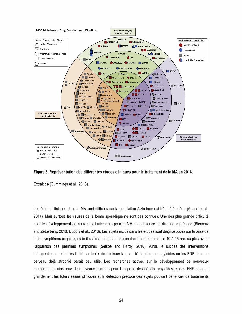
24
Figure 5. Représentation des différentes études cliniques pour le traitement de la MA en 2018.
Extrait de (Cummings et al., 2018).
Les études cliniques dans la MA sont difficiles car la population Alzheimer est très hétérogène (Anand et al.,
2014). Mais surtout, les causes de la forme sporadique ne sont pas connues. Une des plus grande difficulté
pour le développement de nouveaux traitements pour la MA est l’absence de diagnostic précoce (Blennow
and Zetterberg, 2018; Dubois et al., 2016). Les sujets inclus dans les études sont diagnostiqués sur la base de
leurs symptômes cognitifs, mais il est estimé que la neuropathologie a commencé 10 à 15 ans ou plus avant
l’apparition des premiers symptômes (Selkoe and Hardy, 2016). Ainsi, le succès des interventions
thérapeutiques reste très limité car tenter de diminuer la quantité de plaques amyloïdes ou les ENF dans un
cerveau déjà atrophié paraît peu utile. Les recherches actives sur le développement de nouveaux
biomarqueurs ainsi que de nouveaux traceurs pour l’imagerie des dépôts amyloïdes et des ENF aideront
grandement les futurs essais cliniques et la détection précoce des sujets pouvant bénéficier de traitements
25
efficaces. Certains pensent qu’il sera nécessaire d’administrer un traitement personnalisé en fonction des
comorbidités, de la génétique, du sexe et de l’origine ethnique des sujets (Zissimopoulos et al., 2017). La
médecine personnalisée permettra peut-être de mieux cibler les sujets à risque et d’instaurer une stratégie
spécifique pour eux. Finalement, puisque l’âge avancé est le principal facteur de risque de la MA, de nouvelles
cibles thérapeutiques associées au vieillissement doivent être investiguées afin de faire avancer le
développement de nouveaux traitements.
1.6 Les modèles animaux de la MA
Les modèles animaux permettent d’étudier de nombreuses hypothèses dans un organisme vivant complet et
génétiquement proche de l’humain, tout en contrôlant un grand nombre de facteurs. Ils permettent de mettre
en lumière les mécanismes pathogéniques de la MA et d’investiguer des stratégies thérapeutiques diverses.
Toutefois, ces modèles sont loin d’être parfaits, et très peu de résultats obtenus chez la souris ont été
répliqués chez l’humain (Götz et al., 2018). L’interprétation des résultats précliniques doit être faite avec
précaution car la translation vers la clinique n’est pas évidente. Mais surtout, le domaine souffre d’un manque
de modèle de la forme sporadique de la maladie, qui représente la très grande majorité des cas de MA.
De nombreux modèles animaux, principalement des souris, ont été développés afin d’étudier la MA. La base
de donnée Alzforum contient 166 modèles animaux de MA, dont 162 sont des souris, et seulement 1 (la souris
SAMP8) est un modèle spontané (LaFerla and Green, 2012). Les modèles les plus utilisés seront décrits
brièvement ici.
1.6.1 Les modèles de pathologie amyloïde
Un des modèles animaux de pathologie amyloïde le plus agressif est la souris 5xFAD. Elle présente 5
mutations retrouvées dans les formes familiales de la MA exprimées sous le promoteur Thy1.2 : les mutations
Swedish, Florida et London dans le gène APP, ainsi que les mutations M146L et L286V dans le gène PSEN1
(Oakley et al., 2006). Les plaques amyloïdes sont visibles dès l’âge d’un mois et demi, la neuroinflammation
est très importante et des pertes neuronales sont observées à partir de 5 mois. Un des modèles les plus
utilisés pour la pathologie amyloïde est la souris APP/PS1. L’insertion de la mutation APP Swedish combinée
avec PSEN1dE9 permet l’apparition de plaques dès l’âge de 6 mois (Jankowsky et al., 2004). Les déficits
26
comportementaux sont nombreux dans cette souris, mais apparaissent à un âge relativement avancé, autour
de 12 mois dépendamment du test effectué (Janus et al., 2015). Un autre modèle très connu est la souris J20,
qui développe une pathologie étendue de façon rapide ainsi que des troubles de la mémoire précoces, dès
l’âge de 4 mois. Cette souris surexprime les mutations APP Swedish et Indiana, sans mutation dans PSEN1,
sous le promoteur PDGF-β (Mucke et al., 2000).
1.6.2 Les modèles de pathologie tau
Plusieurs modèles de pathologie tau ont été développés à partir des mutations dans le gène MAPT retrouvées
dans les démences fronto-temporales, principalement les mutations P301S et P301L (Terwel et al., 2005). La
souris surexprimant la mutation P301S de la protéine tau humaine sous le promoteur Thy1 est un modèle de
tauopathie sévère, qui développe des ENF dès 4 mois dans le cerveau mais également dans la moelle
épinière, induisant la mort des motoneurones et des troubles moteurs majeurs à 5 mois (Allen et al., 2002).
Une autre souris portant la mutation P301S, mais sous le promoteur Prnp (mouse prion protein) (lignée PS19),
développe quant à elle une mort neuronale plus progressive, apparaissant à 9 mois, démontrant l’importance
du promoteur dans la création de modèles animaux (Yoshiyama et al., 2007). En revanche, la souris human
Tau (hTau) présente l’avantage d’exprimer uniquement la protéine tau humaine et non mutée, étant donné
qu’aucune mutation dans la protéine tau n’a à ce jour été reliée à la MA (Andorfer et al., 2003). Cette souris
est issue du croisement d’une souris tau knock-out et d’une souris 8c exprimant la protéine tau humaine (Duff
et al., 2000).
1.6.3 La souris triple transgénique 3xTg-AD
La souris triple transgénique 3xTg-AD a été créée par le Dr Frank Laferla et est l’un des rares modèles
développant à la fois les pathologies amyloïde et tau (Oddo et al., 2003b). Trois transgènes sont exprimés
sous le promoteur Thy1.2 : les mutations APP Swedish, PSEN1 M146V et MAPT P301L. Cette surproduction
de protéines tau et APP humaines mutées conduit à l’apparition très progressive de plaques amyloïdes
principalement dans le cortex frontal, le subiculum et la région CA1 de l’hippocampe, et d’ENF dans le cortex
pariéto-temporal, l’amygdale et l’hippocampe (Figure 6) (Oddo et al., 2003a). Il faut attendre au moins 12 mois
pour pouvoir bien détecter ces agrégats dans les différentes régions cérébrales des souris 3xTg-AD, ce qui
permet d’inclure le facteur de l’âge, si important dans le développement de la MA, dans les expérimentations

27
avec ce modèle. Toutefois, ce modèle ne présente pas de mort neuronale malgré la présence des deux
marqueurs neuropathologiques principaux de la MA.
Figure 6. Représentation schématique du développement des pathologies amyloïdes et tau dans la souris 3xTg-AD.
Adapté de (Götz and Ittner, 2008).
Cette souris présente différents déficits comportementaux, notamment une importante anxiété, des troubles de
la mémoire de reconnaissance et spatiale (Belfiore et al., 2018; Bories et al., 2012; Filali et al., 2012;
Tournissac et al., 2018; Vandal et al., 2016). La souris 3xTg-AD n’est pas considérée comme un véritable
modèle de neuroinflammation, mais présente tout de même une astrogliose et une microgliose, ainsi qu’une
élévation des niveaux de cytokines pro-inflammatoires cérébrales (Belfiore et al., 2018; Caruso et al., 2013;
St-Amour et al., 2019; 2014). Une diminution de la densité cérébrovasculaire accompagnée d’un
élargissement de la lame basale sont présents dans ce modèle (T. M. Do et al., 2014). En revanche, il faut
noter que les souris femelles présentent une plus grande quantité de peptides Aβ40 et Aβ42, et donc de
plaques amyloïdes (Vandal et al., 2015b). D’autres différences liées au sexe des souris ont été observées au
niveau du comportement (Bories et al., 2012; Clinton et al., 2007). Au niveau métabolique, notre équipe a
observé l’apparition d’une intolérance au glucose spontanée avec l’âge, de manière plus précoce chez les
femelles que les mâles (Vandal et al., 2015b). Quelques équipes ont également rapporté des déficits de
thermorégulation, qui seront présentés dans la section 4.2.
Cortex
Amygdale 6 mois 12 mois
Hippocampe
18 mois
Désordres métaboliques Déficits de thermorégulation
Plaques amyloïdes Enchevêtrements neurofibrillaires
28
1.6.4 Autres modèles animaux
Plusieurs modèles de souris ont été créés à partir des facteurs de risques génétiques identifiés pour la MA,
afin de développer des modèles de la forme sporadique de la MA et non la forme familiale. Les premiers
modèles développés sont les souris exprimant les trois isoformes d’APOE humaines à la place des protéines
murines (Knouff et al., 1999). Il est intéressant de remarquer que la souris APOE4 présente des déficits
cognitifs par rapport aux souris APOE2 et APOE3 (Andrews-Zwilling et al., 2010; Traversy et al., 2016).
D’autre part, il existe une trentaine de souris différentes basées sur le facteur de risque génétique TREM2.
Des souris knock-out pour TREM2 ont été créées afin d’étudier la perte de fonction de ce récepteur, et ont été
croisées avec différents modèles de pathologie amyloïde ou tau ou encore APOE, tandis que d’autres
modèles reposent sur des mutations dans le gène TREM2 (Kleinberger et al., 2017; Turnbull et al., 2006). La
souris SAMP8 (senescence accelerated mice-prone 8) présente quant à elle un vieillissement accéléré de
manière spontanée ainsi que des déficits cognitifs (Yagi et al., 1988).
Il existe également des modèles non-murins de la MA. Récemment, une étude a montré la présence de
neuropathologie de type Alzheimer, c’est-à-dire des ENF en plus des plaques amyloïdes, apparaissant
naturellement chez des macaques âgés (Paspalas et al., 2018). Le microcèbe (ou lémurien) représente un
modèle sporadique intéressant car il a une durée de vie relativement courte et environ 20% d’entre eux
présentent une neurodégénérescence liée à l’âge associée avec des plaques amyloïdes et une pathologie tau
(Bons et al., 2006). Ces primates très proches de l’Homme représentent des modèles de choix davantage
représentatifs de la forme sporadique de la MA, mais il est difficile et coûteux de travailler avec des primates.

29
2 Les désordres métaboliques observés dans la maladie d’Alzheimer
Les liens entre MA et maladies métaboliques, telles que le DT2 et l’obésité, sont maintenant bien établis dans
la littérature (De Felice and Ferreira, 2014). Le fait que ces maladies métaboliques soient d’importants facteurs
de risque pour la MA a été discuté plus haut. De l’autre côté, il est intéressant de constater que les personnes
atteintes de la MA présentent justement des désordres métaboliques au niveau du SNC. D’ailleurs, certains
scientifiques vont même jusqu’à renommer la MA le « diabète de type 3 » (Steen et al., 2005). Ces
observations suggèrent que diminuer les altérations métaboliques pourrait être bénéfique pour la MA, et ont
mené à tester différents antidiabétiques pour le traitement de la MA.
2.1 Diminution du métabolisme cérébral du glucose
Le cerveau représente seulement 2% du poids total d’un être humain, mais consomme pourtant 20% de
l’énergie du corps, principalement sous forme de glucose (Mergenthaler et al., 2013). Comme il n’existe pas
de réserve de glucose dans le cerveau, celui-ci doit le puiser directement dans la circulation sanguine, grâce
au transporteur spécifique GLUT1 (glucose transporter type 1) situé sur les cellules endothéliales de la
vasculature cérébrale et les astrocytes (Simpson et al., 2007). L’importance de ce transporteur pour le
fonctionnement cérébral est illustré par le syndrome de déficit de GLUT1, une maladie génétique rare, dont la
majorité des individus affectés souffrent d’épilepsie et de retard mental (Leen et al., 2010).
L’une des toutes premières dysfonctions observées dans la MA, qui apparaît d’ailleurs plusieurs années avant
les premiers symptômes cognitifs, est la diminution du métabolisme cérébral du glucose (Cunnane et al.,
2011; Mamelak, 2012). L’imagerie par 18F-fluorodésoxyglucose (18F-FDG) couplé à la tomographie par
émission de positron (FDG-TEP) permet de détecter un hypométabolisme du glucose dès le stade de trouble
cognitif léger (Figure 5) (C.-A. Castellano et al., 2014; Mosconi et al., 2005). Cette technique est utilisée dans
plusieurs études cliniques, mais est bien trop coûteuse pour faire partie intégrante du diagnostic de la maladie
en clinique. Pourtant, la réduction du métabolisme du glucose est de l’ordre de 20 à 25% et est corrélée avec
les scores cognitifs des participants (Edison et al., 2007). De plus, le FDG-TEP permet de prédire la
conversion des sujets d’un trouble cognitif léger vers la MA ainsi que leur déclin cognitif (Landau et al., 2010).
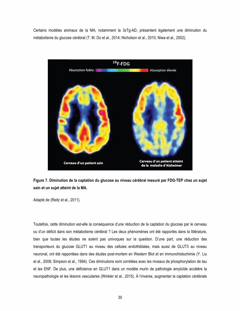
30
Certains modèles animaux de la MA, notamment la 3xTg-AD, présentent également une diminution du
métabolisme du glucose cérébral (T. M. Do et al., 2014; Nicholson et al., 2010; Niwa et al., 2002).
Figure 7. Diminution de la captation du glucose au niveau cérébral mesuré par FDG-TEP chez un sujet sain et un sujet atteint de la MA.
Adapté de (Reitz et al., 2011).
Toutefois, cette diminution est-elle la conséquence d’une réduction de la captation du glucose par le cerveau
ou d’un déficit dans son métabolisme cérébral ? Les deux phénomènes ont été rapportés dans la littérature,
bien que toutes les études ne soient pas univoques sur la question. D’une part, une réduction des
transporteurs du glucose GLUT1 au niveau des cellules endothéliales, mais aussi de GLUT3 au niveau
neuronal, ont été rapportées dans des études post-mortem en Western Blot et en immunohistochimie (Y. Liu
et al., 2008; Simpson et al., 1994). Ces diminutions sont corrélées avec les niveaux de phosphorylation de tau
et les ENF. De plus, une déficience en GLUT1 dans un modèle murin de pathologie amyloïde accélère la
neuropathologie et les lésions vasculaires (Winkler et al., 2015). À l’inverse, augmenter la captation cérébrale
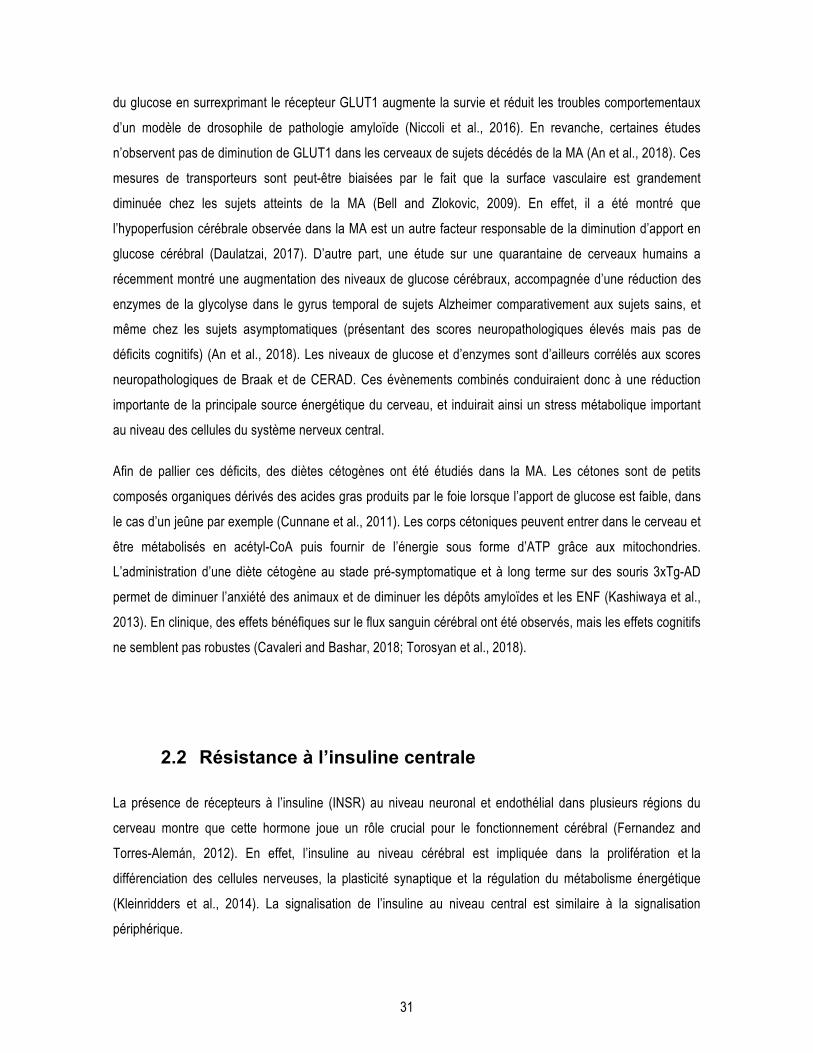
31
du glucose en surrexprimant le récepteur GLUT1 augmente la survie et réduit les troubles comportementaux
d’un modèle de drosophile de pathologie amyloïde (Niccoli et al., 2016). En revanche, certaines études
n’observent pas de diminution de GLUT1 dans les cerveaux de sujets décédés de la MA (An et al., 2018). Ces
mesures de transporteurs sont peut-être biaisées par le fait que la surface vasculaire est grandement
diminuée chez les sujets atteints de la MA (Bell and Zlokovic, 2009). En effet, il a été montré que
l’hypoperfusion cérébrale observée dans la MA est un autre facteur responsable de la diminution d’apport en
glucose cérébral (Daulatzai, 2017). D’autre part, une étude sur une quarantaine de cerveaux humains a
récemment montré une augmentation des niveaux de glucose cérébraux, accompagnée d’une réduction des
enzymes de la glycolyse dans le gyrus temporal de sujets Alzheimer comparativement aux sujets sains, et
même chez les sujets asymptomatiques (présentant des scores neuropathologiques élevés mais pas de
déficits cognitifs) (An et al., 2018). Les niveaux de glucose et d’enzymes sont d’ailleurs corrélés aux scores
neuropathologiques de Braak et de CERAD. Ces évènements combinés conduiraient donc à une réduction
importante de la principale source énergétique du cerveau, et induirait ainsi un stress métabolique important
au niveau des cellules du système nerveux central.
Afin de pallier ces déficits, des diètes cétogènes ont été étudiés dans la MA. Les cétones sont de petits
composés organiques dérivés des acides gras produits par le foie lorsque l’apport de glucose est faible, dans
le cas d’un jeûne par exemple (Cunnane et al., 2011). Les corps cétoniques peuvent entrer dans le cerveau et
être métabolisés en acétyl-CoA puis fournir de l’énergie sous forme d’ATP grâce aux mitochondries.
L’administration d’une diète cétogène au stade pré-symptomatique et à long terme sur des souris 3xTg-AD
permet de diminuer l’anxiété des animaux et de diminuer les dépôts amyloïdes et les ENF (Kashiwaya et al.,
2013). En clinique, des effets bénéfiques sur le flux sanguin cérébral ont été observés, mais les effets cognitifs
ne semblent pas robustes (Cavaleri and Bashar, 2018; Torosyan et al., 2018).
2.2 Résistance à l’insuline centrale
La présence de récepteurs à l’insuline (INSR) au niveau neuronal et endothélial dans plusieurs régions du
cerveau montre que cette hormone joue un rôle crucial pour le fonctionnement cérébral (Fernandez and
Torres-Alemán, 2012). En effet, l’insuline au niveau cérébral est impliquée dans la prolifération et la
différenciation des cellules nerveuses, la plasticité synaptique et la régulation du métabolisme énergétique
(Kleinridders et al., 2014). La signalisation de l’insuline au niveau central est similaire à la signalisation
périphérique.
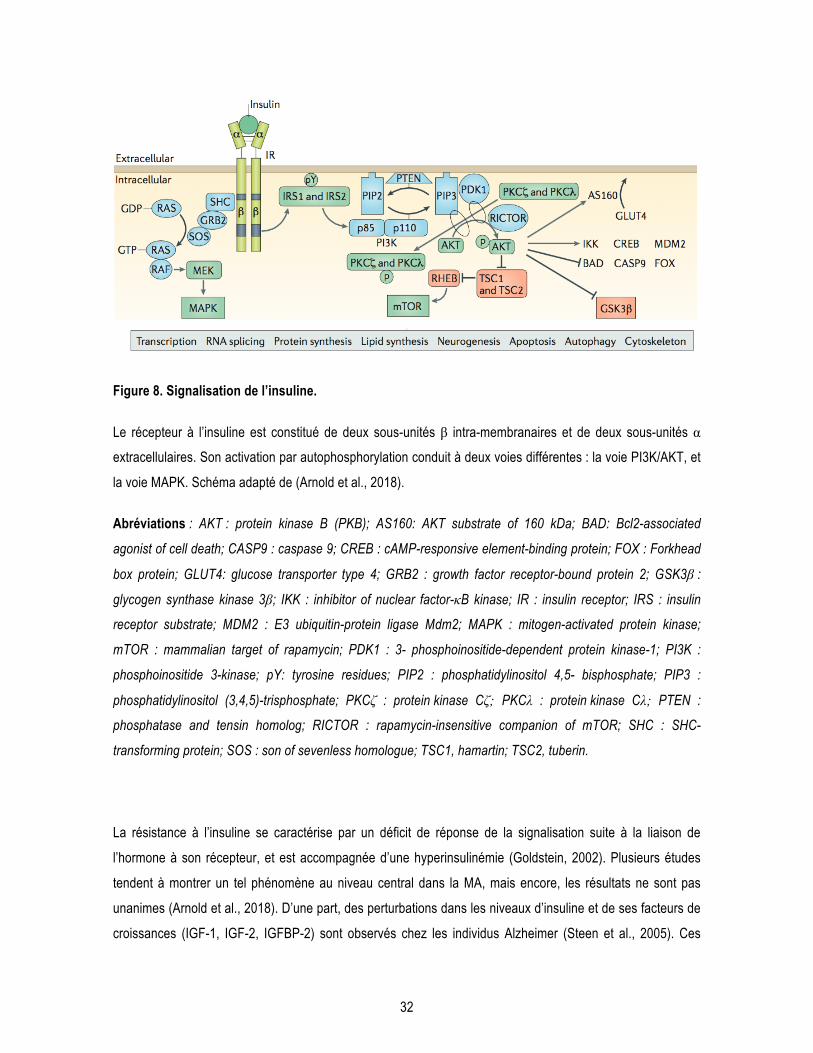
32
Figure 8. Signalisation de l’insuline.
Le récepteur à l’insuline est constitué de deux sous-unités β intra-membranaires et de deux sous-unités α
extracellulaires. Son activation par autophosphorylation conduit à deux voies différentes : la voie PI3K/AKT, et
la voie MAPK. Schéma adapté de (Arnold et al., 2018).
Abréviations : AKT : protein kinase B (PKB); AS160: AKT substrate of 160 kDa; BAD: Bcl2-associated
agonist of cell death; CASP9 : caspase 9; CREB : cAMP-responsive element-binding protein; FOX : Forkhead
box protein; GLUT4: glucose transporter type 4; GRB2 : growth factor receptor-bound protein 2; GSK3β :
glycogen synthase kinase 3β; IKK : inhibitor of nuclear factor-κB kinase; IR : insulin receptor; IRS : insulin
receptor substrate; MDM2 : E3 ubiquitin-protein ligase Mdm2; MAPK : mitogen-activated protein kinase;
mTOR : mammalian target of rapamycin; PDK1 : 3- phosphoinositide-dependent protein kinase-1; PI3K :
phosphoinositide 3-kinase; pY: tyrosine residues; PIP2 : phosphatidylinositol 4,5- bisphosphate; PIP3 :
phosphatidylinositol (3,4,5)-trisphosphate; PKCζ : protein kinase Cζ; PKCλ : protein kinase Cλ; PTEN :
phosphatase and tensin homolog; RICTOR : rapamycin-insensitive companion of mTOR; SHC : SHC-
transforming protein; SOS : son of sevenless homologue; TSC1, hamartin; TSC2, tuberin.
La résistance à l’insuline se caractérise par un déficit de réponse de la signalisation suite à la liaison de
l’hormone à son récepteur, et est accompagnée d’une hyperinsulinémie (Goldstein, 2002). Plusieurs études
tendent à montrer un tel phénomène au niveau central dans la MA, mais encore, les résultats ne sont pas
unanimes (Arnold et al., 2018). D’une part, des perturbations dans les niveaux d’insuline et de ses facteurs de
croissances (IGF-1, IGF-2, IGFBP-2) sont observés chez les individus Alzheimer (Steen et al., 2005). Ces

33
perturbations surviennent avec l’évolution du stade de Braak (Rivera et al., 2005). Une étude sur 39 sujets a
montré une diminution d’insuline dans le LCR conjointement avec une augmentation dans le plasma des
sujets atteints de la MA par rapport aux sujets sains (Craft et al., 1998). En revanche, une étude a observé des
niveaux d’IGF-2 et d’IGFBP-2 plus élevés dans le LCR des hommes dans les stades précoces de MA, mais
pas chez les femmes, qui corrèlent positivement avec les niveaux de tau dans le LCR (Åberg et al., 2015).
D’autre part, la réponse cérébrale à l’insuline mais également à IGF-1 est diminuée dans le cerveau de sujets
décédés de la MA par rapport aux contrôles, de la même façon que dans les sujets diabétiques (Y. Liu et al.,
2011; Talbot et al., 2012). Certaines études ont observé une diminution du récepteur à l’insuline (Steen et al.,
2005), mais pas toutes (Moloney et al., 2010; Talbot et al., 2012). Des perturbations dans les niveaux des
kinases subséquentes à l’activation du récepteur à l’insuline, principalement AKT et GSK3β, ont été observées
à plusieurs reprises chez l’humain (Griffin et al., 2005; Hooper et al., 2008; Rickle et al., 2004; Steen et al.,
2005). La signalisation de l’insuline cérébrale est également perturbée dans certains modèles murins de la MA
(Ruiz et al., 2016). D’ailleurs, des dépôts amyloïdes sont retrouvés dans le pancréas de la souris 3xTg-AD
(Vandal et al., 2014b).
2.3 Repositionnement thérapeutique de médicaments antidiabétiques dans la MA
Les liens entre DT2 et MA sont très forts, et les sujets Alzheimer présentent des troubles métaboliques
importants. Ces observations ont donc mené à tenter de repositionner les médicaments antidiabétiques
disponibles en clinique dans le cadre de la MA (Yarchoan and Arnold, 2014). Ici, seuls l’insuline, les
thiazolidinediones et les analogues de GLP-1 (Gucagon-like peptide-1) seront abordés car ce sont les
médicaments les plus utilisés dans le diabète.
2.3.1 Insuline
L’insuline est testée au niveau préclinique et clinique dans la MA depuis plusieurs années maintenant. D’une
part, les études précliniques dans des modèles animaux de la MA ont montré des effets bénéfiques sur la
mémoire, la neuroinflammation et la pathologie amyloïde (Adzovic et al., 2015; Chen et al., 2014b; Mao et al.,
2016; Rajasekar et al., 2016; Vandal et al., 2014b). Une étude de notre équipe a notamment montré qu’une
injection intrapéritonéale d’insuline (1 UI/kg) améliorait les performances cognitives de la souris 3xTg-AD et
34
qu’une administration intraveineuse à forte dose (3,8 UI/kg) augmentait grandement la clairance des peptides
Aβ du cerveau vers le sang en seulement 5 minutes (Vandal et al., 2014b). De manière plus générale, divers
éléments de la voie de signalisation de l’insuline sont impliqués dans la clairance des peptides Aβ du cerveau
((Swaminathan et al., 2017); pour revue : (Vandal et al., 2015a)).
La voie intranasale a été sélectionnée en clinique afin de permettre une administration centrale plus directe et
éviter que l’hormone atteigne la périphérie, et ainsi les risques d’hypoglycémie (Born et al., 2002; Freiherr et
al., 2013). Toutefois, la présence d’insuline dans la circulation après injection intranasale n’a été que peu
étudiée. Les quelques travaux ayant directement investigué cet aspect montrent que l’injection intranasale de
macromolécules mène tout de même à une exposition systémique (ex : IGF-1, immunoglobuline G, interféron
β) (N. N. Kumar et al., 2018; Lochhead and Thorne, 2012; Thorne et al., 2008; 2004). En clinique, l’insuline
administrée par voie intranasale a montré des effets bénéfiques sur la cognition de sujets atteints de la MA
(Benedict et al., 2004; Claxton et al., 2015; Craft et al., 2012; Shemesh et al., 2012). Toutefois, dans l’étude de
Claxton et collaborateurs, les sujets porteurs de l’APOE4 présentaient au contraire une détérioration de leurs
scores cognitifs suite à l’administration d’insuline. Ceci pourrait s’expliquer par le fait que l’insuline induit une
augmentation de la phosphorylation de tau dans des souris exprimant la forme humaine de l’APOE4 (Traversy
et al., 2016). Des études cliniques avec l’insuline sont toujours en cours pour la MA, mais avec différentes
formulations d’insuline permettant une plus longue demi-vie par exemple (Cummings et al., 2018). Toutefois,
les résultats préliminaires de l’étude clinique placébo-traitement « SNIFF » avec de l’insuline intranasale
pendant un an sur 240 sujets atteints de trouble cognitif léger ou MA ne montrent pas d’effet sur la cognition
(NCT01767909; communiqué de Alzforum sur le Clinical Trials on Alzheimer’s Disease Conference 2018).
2.3.2 Les thiazolidinediones
Les thiazolidinediones (TZD) sont une famille de molécules antidiabétiques qui activent le récepteur
peroxysome proliferator-activated receptor γ (PPARγ) (Rendell and Kirchain, 2000). Les deux TZD
approuvées pour le DT2 et étudiées dans la MA sont le pioglitazone et le rosiglitazone. Une étude prospective
a montré que la consommation de pioglitazone diminuait de 47% le risque de démence chez les sujets
diabétiques (Heneka et al., 2015b).
Au niveau mécanistique, les agonistes de PPARγ augmentent la dégradation des peptides Aβ par une
activation de la phagocytose des microglies et les niveaux d’APOE dans le parenchyme cérébral (Mandrekar-
Colucci et al., 2012). Chez la souris APP/PS1 de 12 mois, neuf jours de gavage avec du pioglitazone
(80mg/kg) suffisent à réduire significativement les plaques amyloïdes et les déficits cognitifs. En revanche, un
35
traitement de 2 mois avec le pioglitazone réduit les déficits cérébrovasculaires chez la souris J20 âgée de 16
mois, mais n’a pas d’effet sur la pathologie amyloïde ni les déficits cognitifs (Nicolakakis et al., 2008). Un autre
agoniste de PPAR, le GFT1803, a montré des effets bénéfiques supérieurs au pioglitazone chez la souris
APP/PS1 de 6 mois au niveau neuropathologique et comportemental (Kummer et al., 2015).
Chez l’humain, les résultats sont plus mitigés. Une étude pilote sur 30 sujets atteints de la MA a montré que
les participants ayant reçu six mois de traitement avec le rosiglitazone avaient de meilleurs scores cognitifs
que le groupe ayant reçu le placébo (Watson et al., 2005). Toutefois, des études cliniques de phase 3 n’ont
pas permis de répliquer un effet significatif du traitement (Yarchoan and Arnold, 2014). Par ailleurs, un
traitement de 6 mois avec du pioglitazone sur 15 individus diabétiques et atteints de trouble cognitif léger a
permis d’améliorer leurs performances cognitives (Hanyu et al., 2009). Une étude de phase 3 sur plus de 3000
sujets appelée « TOMORROW » (NCT01931566) est en cours avec de légères doses de pioglitazone et vise
à déterminer si le traitement sur des sujets à risque de développer la MA permet de retarder le diagnostic de la
maladie (Roses et al., 2014). Finalement, une méta-analyse des études randomisées contrôles suggère que le
pioglitazone serait efficace pour les individus présentant la MA et un diabète, mais les résultats avec le
rosiglitazone ne seraient pas concluants (J. Liu et al., 2015).
2.3.3 Analogues de GLP-1
Le GLP-1 est une incrétine utilisée dans le traitement du DT2 pour réguler la glycémie en stimulant la
production d’insuline du pancréas (Drucker and Nauck, 2006). La demi-vie du GLP-1 étant de quelques
minutes seulement, des analogues de cette hormone ont été développés pour être compatibles avec une
utilisation thérapeutique. Il existe deux analogues sur le marché pour le moment : l’exénatide et le liraglutide.
Depuis environ 20 ans, des études ont commencé à démontrer un potentiel thérapeutique de la voie GLP-1
dans le SNC et la MA (Perry and Greig, 2004). Différents auteurs ont rapporté des effets bénéfiques des
analogues de GLP-1 au niveau de la mémoire, de l’hyperphosphorylation de tau, de la quantité de peptides
Aβ, de la perte synaptique ainsi que de la neuroinflammation dans des modèles animaux (Hansen et al., 2015;
Ma et al., 2015; McClean et al., 2011; L. Shi et al., 2017; Tai et al., 2018). Chez l’Homme, une étude sur 38
sujets Alzheimer a montré que 6 mois de traitement avec le liraglutide augmentait la captation du glucose au
cerveau par rapport au groupe contrôle (Gejl et al., 2017; 2016). Trois études cliniques de phase 1 et 2 sont
en cours, ou les résultats ne sont pas encore publiés, pour tester l’efficacité du liraglutide dans la MA (Étude
ELAD, NCT01843075; NCT02140983 et NCT01469351).

36
Toutefois, aucun de ces médicaments antidiabétiques n’a pour le moment passé les essais cliniques de phase
3 pour la MA. Peut-être qu’il est nécessaire de cibler des sous-populations atteintes de troubles métaboliques
pour observer un effet bénéfique de cette classe de médicament. Considérant les liens très étroits entre
maladies métaboliques et MA, de nouvelles molécules permettant de diminuer les désordres métaboliques
restent à investiguer dans la MA. Des molécules multi-cibles pour toucher les différents paramètres impliqués
dans la pathogenèse de la MA semblent requises pour cette maladie multifactorielle.
37
3 La thermorégulation
La thermorégulation désigne l’ensemble des mécanismes permettant au corps de réguler sa température. Elle
est intimement liée au métabolisme énergétique et est altérée chez les personnes âgées. Les deux prochaines
sections visent à expliquer pourquoi elle pourrait représenter une cible thérapeutique intéressante pour
modifier à la fois les désordres métaboliques et la MA.
3.1 Définition
La température corporelle est un paramètre biologique extrêmement bien régulé chez l’Homme (Mekjavic and
Eiken, 2006). En effet, de trop grandes variations de température ont des effets délétères importants sur le
fonctionnement du corps humain : une hyperthermie, c’est-à-dire une température corporelle anormalement
élevée, peut induire des lésions irréversibles, notamment pour le SNC (Walter and Carraretto, 2016); tandis
qu’une hypothermie (température corporelle en dessous des 35°C) conduit à une diminution de la pression
artérielle causant dans certains cas une hypoperfusion tissulaire et une diminution de la réponse immunitaire
(Doufas, 2003). Le maintien de la température corporelle autour des 37°C a tout de même un coût
métabolique très important : près de la moitié de la dépense énergétique d’un humain en condition d’activité
normale est consacrée à maintenir l’homéothermie (Landsberg, 2012).
La température corporelle varie au cours de la journée selon un cycle circadien. Le rythme circadien du corps
regroupe l’ensemble des mécanismes biologiques cycliques qui varient au cours d’une journée de 24 heures,
comme par exemple l’alternance veille/sommeil, la pression sanguine, l’état de vigilance et le métabolisme
(Hofstra and de Weerd, 2008). Pour ce qui est de la température du corps, celle-ci varie de quelques degrés
au cours de la journée : elle est plus élevée à la fin de la journée, en raison de l’activité musculaire, et elle est
plus basse dans la nuit, car le corps est inactif pour plusieurs heures (Refinetti and Menaker, 1992).
La thermorégulation désigne l’ensemble des mécanismes qui permettent au corps de maintenir une
température autour d’une valeur seuil, environ 37°C pour l’Homme. Les mécanismes comprennent la
thermolyse, c’est-à-dire la diffusion de chaleur, mais aussi la thermogenèse, la production de chaleur (voir
section 3.3) (Cannon and Nedergaard, 2004). Cette capacité à réguler la température interne en produisant
soi-même de la chaleur concerne les organismes dits endothermes, c’est-à-dire les mammifères et les oiseaux
(Silva, 2006). Par opposition, les organismes ectothermes comme les reptiles ou les poissons n’ont pas ou
38
peu de capacité de production de chaleur et vont réguler leur température interne grâce aux changements de
température externe. Le comportement joue également un rôle important dans la thermorégulation : la façon
de s’habiller en fonction de la température externe, le rapprochement des souris dans une même cage ou
encore le fait de se déplacer vers un environnement plus frais ou chaud en font partie (Gordon et al., 1998).
Toutefois, ces aspects ne seront pas abordés dans les sections suivantes afin de se concentrer sur la
régulation physiologique de la température corporelle.
La thermoneutralité (ou zone thermoneutre) correspond à la température externe à laquelle un organisme n’a
pas besoin de produire de chaleur en plus de celle issue du métabolisme basal (Cannon and Nedergaard,
2010). Cette zone varie d’un animal à l’autre, et même au sein d’une même espèce en fonction de l’âge par
exemple. La thermoneutralité chez l’Homme est située entre 22 et 25°C, tandis qu’elle est autour des 28-30°C
pour la souris (Speakman and Keijer, 2013).
3.2 Régulation centrale de la température corporelle
3.2.1 La détection par les thermorécepteurs
Les changements de température externe sont détectés par des thermorécepteurs ou temperature-activated
transient receptor potential (TRP). Ce sont des structures sensibles à la température qui alertent le système
central d’une sensation de froid ou de chaud. Ces signaux sont indispensables pour le confort mais aussi et
surtout pour la survie de l’individu (Patapoutian et al., 2003). Leur activation induit une réponse avant même
que la température centrale ait réellement changée (Morrison et al., 2012). Les thermorécepteurs sont
retrouvés à différents endroits du corps : en périphérie (peau, système nerveux périphérique), au niveau
interne (intestin, foie) et même dans l’hypothalamus (Song et al., 2016). Les fibres nerveuses afférentes qui
conduisent les signaux thermiques sont des fibres Aδ, qui sont légèrement myélinisées, de diamètre moyen et
à conduction rapide; et des fibres C, qui sont non myélinisées, de petit diamètre et à conduction plus lente
(Julius and Basbaum, 2001).
Au niveau cutané, il existe différents types de récepteurs TRP qui sont chacun activés spécifiquement par leur
propre gamme de température; ensemble, ils couvrent la presque totalité des températures que les
mammifères peuvent sentir (Caterina, 2007). En particulier, le récepteur TRPM8 est un récepteur cutané qui
détecte les sensations de froid, c’est-à-dire les températures situées en dessous de 27°C (Dhaka et al., 2007;
McKemy et al., 2002). Les récepteurs TRPV4 et TRPV3 quant à eux participent à la détection de températures
39
chaudes, entre 27 et 42°C au niveau cutané (H. Lee et al., 2005; Moqrich et al., 2005). Le récepteur TRPV4
est également impliqué dans la thermogenèse : il est présent dans les zones préoptique et antérieure de
l’hypothalamus (Abe et al., 2003; Hori et al., 1999). Finalement, les informations des thermorécepteurs vont
être intégrées au niveau central par la zone préoptique de l’hypothalamus pour induire la thermolyse
(évacuation de la chaleur) ou la thermogenèse (production de la chaleur) (Nakamura and Morrison, 2008).
3.2.2 Intégration centrale du signal par l’hypothalamus
L’hypothalamus est situé dans la région ventrale du cerveau, au-dessus de la zone du tronc cérébral et près
du troisième ventricule (D. Richard, 2015). Il est en étroite relation avec l’hypophyse et contribue à faire le lien
entre le système nerveux central et le système endocrinien. L’hypothalamus est également impliqué dans la
régulation de l’appétit et de la dépense énergétique (Coll and Yeo, 2013). Il est composé de plusieurs sous-
régions, mais on peut grossièrement distinguer l’hypothalamus postérieur de l’hypothalamus antérieur. La
partie postérieure est impliquée dans la thermogenèse, alors que la région antérieure de l’hypothalamus
participe à la thermolyse.
De façon plus détaillée, lors d’une sensation de froid, les afférences glutamatergiques des thermorécepteurs
de la peau passent par la corne dorsale de la moelle épinière et stimulent les interneurones GABAergiques du
noyau préoptique médian (MnPO) (voie complète schématisée dans la Figure 9) (Labbé et al., 2015b;
Morrison et al., 2012). Ces derniers inhibent les neurones GABAergiques de la zone préoptique (POA) de
l’hypothalamus, qui projettent vers la zone dorsomédiale de l’hypothalamus (DMH) : leur inhibition sur les
neurones glutamatergiques de la DMH est donc diminuée. Ces derniers vont alors projeter vers la médulla
ventromédiale rostrale (RVM), plus précisément dans le noyau du raphé pallidus rostral (RPar), où les
neurones glutamatergiques vont stimuler les neurones pré-ganglionnaires du système nerveux sympathique
dans le noyau spinal intermédiolatéral (IML). Finalement, les afférences sympathiques post-ganglionnaires
vont relâcher la norépinephrine dans les terminaisons synaptiques situées sur les adipocytes du tissu adipeux
brun (TAB); le neurotransmetteur va alors stimuler les récepteurs β3 adrénergiques et induire en bout de ligne
la production de chaleur par les mitochondries du TAB (décrit plus bas) ainsi que la lipolyse au sein des
adipocytes blancs (Cannon and Nedergaard, 2004). Il faut toutefois noter que l’activation du TAB ne joue pas
un rôle prépondérant dans la thermogenèse chez l’adulte, la quantité de TAB chez l’humain étant limitée
(discuté plus bas).
À l’inverse, la sensation de chaleur active les thermorécepteurs spécifiques de la peau et induit une réponse
opposée à celle de la sensation de froid, c’est-à-dire une inhibition des neurones sympathiques innervant le

40
TAB. Cette réponse se réalise en empruntant la même voie centrale que décrite ci-dessus, mais en stimulant
des neurones différents. En effet, la principale différence est que les afférences périphériques
glutamatergiques stimulent les neurones glutamatergiques du MnPO, qui activent subséquemment les
neurones GABAergiques de la POA, qui vont alors inhiber les voies subséquentes (Figure 8) (Morrison et al.,
2012).
Figure 9. Schéma de la voie de régulation centrale de la thermogenèse du tissu adipeux brun.
41
Abréviations : 5HT : sérotonine; AC : acétylcholine; BAT SC : neurone sensible au chaud; CD : corne
dorsale; DMH : noyau dorsomédian de l’hypothalamus; GABA : Acide γ-aminobutyrique; GLU : glutamate;
iBAT : tissu adipeux brun interscapulaire; IML : colonne intermédiolatérale de la moelle épinière; LHA :
hypothalamus latéral; MnPO : noyau préoptique médian; MPO : noyau préoptique médial; MVL : médulla
ventrolatérale; NA : noradrénaline; NTS : noyau du tractus solitaire; PBLd : noyau parabrachial latéral (dorsal);
PBLle : noyau parabrachial latéral (latéral externe); PVH : noyau paraventriculaire de l’hypothalamus; RPar :
noyau du raphé pallidus rostral; TRPV1/3 et TRPM8 : transient receptor potential V1/3 and M8. Tiré de la
thèse du Dr Monge-Roffarello, adapté et traduit de (Morrison et al., 2012).
L’hypothalamus est également impliqué dans le rythme circadien de la température. En effet, le noyau
suprachiasmatique de l’hypothalamus est synchronisé avec l’environnement : le noyau suprachiasmatique
reçoit des afférences rétiniennes, ce qui lui permet de synchroniser l’activité de ses neurones en fonction de la
lumière perçue par l’individu (Brown and Piggins, 2007; Ralph et al., 1990). Ce noyau joue un rôle dans la
stimulation sympathique de différents organes tels que le TAB, mais aussi la glande thyroïde, les reins, la
vessie et la rate (Bartness et al., 2001).
3.3 Régulation périphérique de la température corporelle
3.3.1 Thermolyse
La thermolyse désigne l’ensemble des mécanismes permettant de dissiper la chaleur et diminuer la
température corporelle (Kenny and Journeay, 2010). La chaleur corporelle s’évacue sous différentes façons :
par radiation (transfert de chaleur sous forme d’ondes), convection (transfert de chaleur vers un gaz ou un
fluide de plus basse température), conduction (transfert de chaleur par contact), ou évaporation (transfert de
chaleur par la transformation d’un liquide en vapeur) (Krau, 2013; N. A. S. Taylor, 2014).
Dans des conditions normales, la température corporelle se dissipe principalement sous le phénomène de
radiation, mais cette perte est compensée par la chaleur produite par le métabolisme, maintenant ainsi un
équilibre (Mekjavic and Eiken, 2006). En revanche, en condition d’hyperthermie, le corps va utiliser en premier
lieu la convection : lorsque la température corporelle augmente, les vaisseaux sanguins périphériques vont se
dilater afin d’augmenter la surface d’échange pour diffuser la chaleur vers l’environnement (Mack et al., 1995).
42
Cette vasodilatation entraîne une augmentation de la quantité de sang au niveau périphérique et une
augmentation de la pression artérielle. Le débit sanguin cutané peut atteindre 6 litres par minute en condition
de stress thermique intense, alors qu’il est de 300 ml par minute en condition normale (Smith and Johnson,
2016). Dans un deuxième temps, c’est l’évaporation sous forme de sueur (transpiration) qui intervient pour
évacuer la chaleur chez l’Homme. Tout ceci est, comme la thermogenèse, régulé par le système nerveux
sympathique (Madden and Morrison, 2018). Toutefois, toutes les voies au niveau du contrôle central ne sont
pas encore connues (Smith and Johnson, 2016).
Chez le rongeur, la vasodilatation au niveau de la queue est le principal mécanisme de thermolyse (Fischer et
al., 2016; Warner et al., 2013; A. A. Young and Dawson, 1982). Une étude récente a montré qu’une mauvaise
régulation de la vasoconstriction de la veine caudale chez la souris déficiente en thermorécepteur TRPM8,
sans déficit de thermogenèse, est responsable de l’hypothermie et d’une diminution de la capacité
d’adaptation à une exposition au froid (Reimúndez et al., 2018).
Dans les premières phases de l’hyperthermie induite par une fièvre, le système immunitaire s’active et conduit
à l’augmentation de la température corporelle nécessaire à l’élimination des pathogènes. Il semblerait que le
TAB participe également à cette augmentation de température au stade précoce de la pyrexie (Cannon and
Nedergaard, 2004). En effet, son activité est augmentée dans les trois premières heures de fièvre (Harris et
al., 1985). Pour expliquer ce phénomène, des études ont montré qu’une injection de la prostaglandine E2
(PGE2), produite par le système immunitaire en réponse à un pathogène, active le TAB en quelques minutes
seulement (Amir and Schiavetto, 1990; Ootsuka et al., 2008; Steiner et al., 2006).
3.3.2 Thermogenèse et tissu adipeux brun
Il existe deux types de thermogenèse : la thermogenèse dite obligatoire et la thermogenèse dite facultative ou
adaptative. La première désigne la production de chaleur issue du métabolisme basal du corps, c’est-à-dire
produite par les réactions métaboliques libérant de la chaleur (Silva, 2006). La thermogenèse facultative
regroupe quant à elle la chaleur produite par les muscles (activité physique ou thermogenèse avec frissons),
par la digestion (thermogenèse induite par l’alimentation) et par le TAB (thermogenèse sans frissons), plus
connu sous le nom de graisse brune (Cannon and Nedergaard, 2004; D. Richard and Picard, 2011). Cette
section va se concentrer sur la thermogenèse sans frissons du TAB.
Le TAB est présent en grande quantité chez les rongeurs et les animaux qui hibernent, principalement au
niveau interscapulaire (Cannon and Nedergaard, 2004). Chez l’Homme, on le retrouve au stade embryonnaire
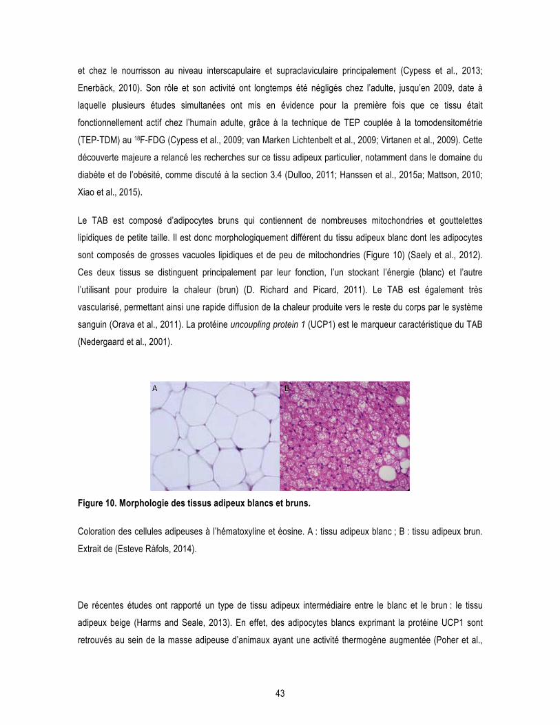
43
et chez le nourrisson au niveau interscapulaire et supraclaviculaire principalement (Cypess et al., 2013;
Enerbäck, 2010). Son rôle et son activité ont longtemps été négligés chez l’adulte, jusqu’en 2009, date à
laquelle plusieurs études simultanées ont mis en évidence pour la première fois que ce tissu était
fonctionnellement actif chez l’humain adulte, grâce à la technique de TEP couplée à la tomodensitométrie
(TEP-TDM) au 18F-FDG (Cypess et al., 2009; van Marken Lichtenbelt et al., 2009; Virtanen et al., 2009). Cette
découverte majeure a relancé les recherches sur ce tissu adipeux particulier, notamment dans le domaine du
diabète et de l’obésité, comme discuté à la section 3.4 (Dulloo, 2011; Hanssen et al., 2015a; Mattson, 2010;
Xiao et al., 2015).
Le TAB est composé d’adipocytes bruns qui contiennent de nombreuses mitochondries et gouttelettes
lipidiques de petite taille. Il est donc morphologiquement différent du tissu adipeux blanc dont les adipocytes
sont composés de grosses vacuoles lipidiques et de peu de mitochondries (Figure 10) (Saely et al., 2012).
Ces deux tissus se distinguent principalement par leur fonction, l’un stockant l’énergie (blanc) et l’autre
l’utilisant pour produire la chaleur (brun) (D. Richard and Picard, 2011). Le TAB est également très
vascularisé, permettant ainsi une rapide diffusion de la chaleur produite vers le reste du corps par le système
sanguin (Orava et al., 2011). La protéine uncoupling protein 1 (UCP1) est le marqueur caractéristique du TAB
(Nedergaard et al., 2001).
Figure 10. Morphologie des tissus adipeux blancs et bruns.
Coloration des cellules adipeuses à l’hématoxyline et éosine. A : tissu adipeux blanc ; B : tissu adipeux brun.
Extrait de (Esteve Ràfols, 2014).
De récentes études ont rapporté un type de tissu adipeux intermédiaire entre le blanc et le brun : le tissu
adipeux beige (Harms and Seale, 2013). En effet, des adipocytes blancs exprimant la protéine UCP1 sont
retrouvés au sein de la masse adipeuse d’animaux ayant une activité thermogène augmentée (Poher et al.,

44
2015). Toutefois, ce tissu intermédiaire semble être peu recruté en condition de stimulation adrénergique chez
le rongeur, ou du moins de manière plus limitée par rapport au TAB pur (Labbé et al., 2016).
Lors de la production de chaleur, la norépinephrine est relâchée par les boutons synaptiques du système
nerveux sympathique provenant de l’hypothalamus (décrit en 3.2.2). Le neurotransmetteur stimule les
récepteurs β3 adrénergiques (Rβ3A) situés à la surface des adipocytes bruns. Par une cascade d’activation, il
se produit alors une lyse des triglycérides contenus dans les adipocytes bruns, une activation d’UCP1 (ou
thermogénine) sur la membrane interne des mitochondries. Cette protéine va créer un découplage de la
pompe à proton des mitochondries conduisant à la production de chaleur (Figure 11) (Cannon and
Nedergaard, 2004; Golozoubova et al., 2001).
Figure 11. Cascade d’activation de la thermogenèse du tissu adipeux brun.
Abréviations : AGL : acide gras libre ; AMPc : adénosine monophosphate cyclique ; LHS : lipase
hormonosensible ; NE : norépinephrine ; PKA : protéine kinase A ; Rβ3A : récepteur β3 adrénergique ; TG :
triglycérides ; UCP1 : uncoupling protein 1.
Le volume et l’activité du TAB sont modulés par différents facteurs. Chez l’Homme, il est plus difficile de
détecter l’activité du TAB par TEP chez les adultes et les personnes âgées (Cypess et al., 2009; Rogers,
2014). Le pourcentage de détection diminue également lorsque le poids de l’individu est élevé (Pfannenberg
et al., 2010; van Marken Lichtenbelt et al., 2009), et lorsque ce dernier est diabétique (Ouellet et al., 2011). À
l’inverse, les femmes présentent un TAB plus actif et plus volumineux que les hommes (Cypess et al., 2009;
Ouellet et al., 2011; Pfannenberg et al., 2010).
NE
vacuole lipidique
AMPc
LHS
TG
AGL
Rβ3A
↑UCP1
mitochondrie
PKA
Adipocyte brun Chaleur
45
3.4 Stimulation de la thermogenèse : cible thérapeutique pour les maladies métaboliques
La quantité de TAB est très hétérogène dans la population humaine. Certaines études distinguent d’ailleurs les
sujets « TAB positifs » et « TAB négatifs » (Nedergaard et al., 2010). Ainsi, plusieurs scientifiques sont
critiques quant à la possibilité de stimuler efficacement ce tissu pour des applications thérapeutiques.
Toutefois, de nombreuses études animales et humaines ont montré des effets bénéfiques dans le cadre de
diabète ou d’obésité (Tableau 3) (Schrauwen and van Marken Lichtenbelt, 2016). En effet, le TAB consomme
beaucoup d’énergie pour produire de la chaleur : il est estimé que 60 g de ce tissu chez l’adulte pourrait brûler
à lui seul environ 4 kg de tissu adipeux en un an (Virtanen et al., 2009). Son activation est donc une cible
intéressante pour rétablir le débalancement énergétique, mais aussi pour diminuer la résistance à l’insuline
(de Souza and Burkey, 2001; Orava et al., 2011; Roberts-Toler et al., 2015). Différentes méthodes permettent
de stimuler l’activité de thermogenèse du TAB : les deux principales sont les expositions au froid et
l’administration d’agonistes des Rβ3A.
3.4.1 Les expositions au froid
Lorsque la température ambiante diminue, la thermogenèse avec frissons, effectuée par contraction rapide
des muscles squelettiques, s’active (Silva, 2006). Celle-ci intervient au début du processus de thermogenèse
adaptative en réponse à une exposition au froid, puis la thermogenèse sans frissons effectuée par le TAB
prend peu à peu le relais (Cannon and Nedergaard, 2010; Cypess et al., 2012; S. Lim et al., 2012). La
captation du glucose par le TAB est grandement augmentée et la perfusion sanguine de ce tissu est doublée
en condition de froid afin de dissiper la chaleur produite à travers le corps (Orava et al., 2011).
L’humain vivant majoritairement dans des conditions de thermoneutralité, par les vêtements et l’habitat, le TAB
n’est que peu souvent mis à contribution, même dans les pays nordiques. Néanmoins, une première étude en
1981 sur 11 individus a montré une plus importante quantité de TAB chez des sujets Finlandais travaillant à
l’extérieur comparativement aux sujets contrôles (Huttunen et al., 1981). Depuis, plusieurs études ont montré
que la température extérieure influence l’activité et donc la détection du TAB par imagerie chez l’humain
(Hanssen et al., 2015a; Ouellet et al., 2011; van Marken Lichtenbelt et al., 2009). Il est estimé que la détection
du TAB chez l’adulte en dessous de 40 ans pourrait atteindre les 100% si le TEP était effectué après
stimulation par une exposition à une basse température (Leitner et al., 2017; Orava et al., 2011; Rogers, 2014;
Virtanen et al., 2009). De récentes études ont montré que les expositions au froid permettaient d’augmenter la
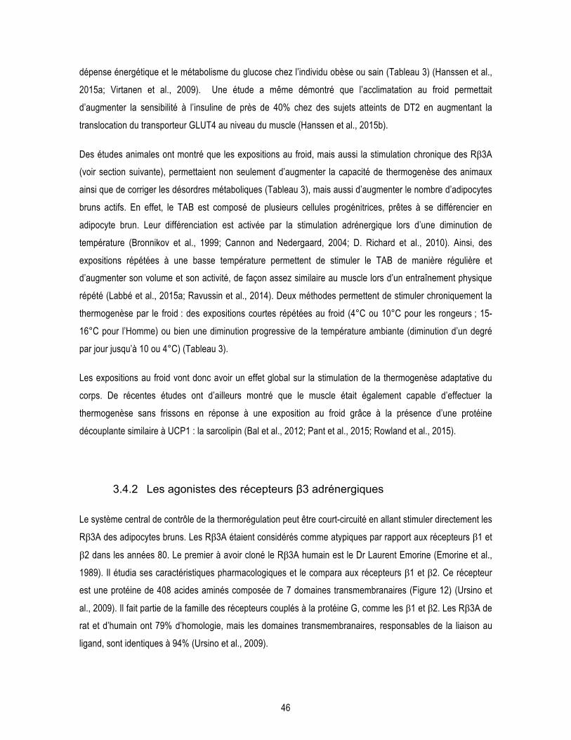
46
dépense énergétique et le métabolisme du glucose chez l’individu obèse ou sain (Tableau 3) (Hanssen et al.,
2015a; Virtanen et al., 2009). Une étude a même démontré que l’acclimatation au froid permettait
d’augmenter la sensibilité à l’insuline de près de 40% chez des sujets atteints de DT2 en augmentant la
translocation du transporteur GLUT4 au niveau du muscle (Hanssen et al., 2015b).
Des études animales ont montré que les expositions au froid, mais aussi la stimulation chronique des Rβ3A
(voir section suivante), permettaient non seulement d’augmenter la capacité de thermogenèse des animaux
ainsi que de corriger les désordres métaboliques (Tableau 3), mais aussi d’augmenter le nombre d’adipocytes
bruns actifs. En effet, le TAB est composé de plusieurs cellules progénitrices, prêtes à se différencier en
adipocyte brun. Leur différenciation est activée par la stimulation adrénergique lors d’une diminution de
température (Bronnikov et al., 1999; Cannon and Nedergaard, 2004; D. Richard et al., 2010). Ainsi, des
expositions répétées à une basse température permettent de stimuler le TAB de manière régulière et
d’augmenter son volume et son activité, de façon assez similaire au muscle lors d’un entraînement physique
répété (Labbé et al., 2015a; Ravussin et al., 2014). Deux méthodes permettent de stimuler chroniquement la
thermogenèse par le froid : des expositions courtes répétées au froid (4°C ou 10°C pour les rongeurs ; 15-
16°C pour l’Homme) ou bien une diminution progressive de la température ambiante (diminution d’un degré
par jour jusqu’à 10 ou 4°C) (Tableau 3).
Les expositions au froid vont donc avoir un effet global sur la stimulation de la thermogenèse adaptative du
corps. De récentes études ont d’ailleurs montré que le muscle était également capable d’effectuer la
thermogenèse sans frissons en réponse à une exposition au froid grâce à la présence d’une protéine
découplante similaire à UCP1 : la sarcolipin (Bal et al., 2012; Pant et al., 2015; Rowland et al., 2015).
3.4.2 Les agonistes des récepteurs β3 adrénergiques
Le système central de contrôle de la thermorégulation peut être court-circuité en allant stimuler directement les
Rβ3A des adipocytes bruns. Les Rβ3A étaient considérés comme atypiques par rapport aux récepteurs β1 et
β2 dans les années 80. Le premier à avoir cloné le Rβ3A humain est le Dr Laurent Emorine (Emorine et al.,
1989). Il étudia ses caractéristiques pharmacologiques et le compara aux récepteurs β1 et β2. Ce récepteur
est une protéine de 408 acides aminés composée de 7 domaines transmembranaires (Figure 12) (Ursino et
al., 2009). Il fait partie de la famille des récepteurs couplés à la protéine G, comme les β1 et β2. Les Rβ3A de
rat et d’humain ont 79% d’homologie, mais les domaines transmembranaires, responsables de la liaison au
ligand, sont identiques à 94% (Ursino et al., 2009).

47
Figure 12. Représentation schématique du récepteur β3 adrénergique.
Extrait de (Ursino et al., 2009).
Ce récepteur est exprimé principalement par les adipocytes bruns, mais également dans la vessie, le système
digestif et dans le cerveau (cortex, l’hippocampe, hypothalamus et tronc cérébral) (B. A. Evans et al., 1996;
Summers et al., 1995). Toutefois, son rôle physiologique dans le SNC est assez peu connu. Une étude a
montré que la stimulation des Rβ3A cérébraux par l’administration intracérébroventriculaire d’un agoniste
reproduisait les effets métaboliques d’une administration périphérique (J. E. Richard et al., 2017). De plus,
certains agonistes des Rβ3A possèdent des effets antidépresseurs en augmentant la quantité de tryptophane
au cerveau ainsi que la synthèse de la sérotonine (Claustre et al., 2008; Consoli et al., 2007). C’est ainsi que
l’agoniste SR58611A (Amibegron®) a été testé en clinique jusqu’en phase 3 pour la dépression, mais n’a
finalement pas été commercialisé (NCT00252330).
Pour étudier les Rβ3A, plusieurs molécules ont été développées dans les années 80 dans le but de mimer les
effets de la norépinephrine sur les Rβ3A (Tableau 2). Ils sont chacun plus ou moins sélectifs des β3 par
rapport aux β1 et β2, situés quant à eux au niveau du cœur, des muscles lisses vasculaires et bronchiques
principalement. Pour plusieurs compagnies pharmaceutiques, l’objectif est donc de développer un agoniste le
plus sélectif possible des β3 chez l’humain afin de stimuler spécifiquement le TAB sans effets secondaires
(tels que l’hypertension) comme traitement pour le diabète et l’obésité. Dans les études précliniques, le plus
couramment utilisé aujourd’hui est le CL-316,243 (Tableau 3) (J. D. Bloom et al., 1992). Il est très spécifique
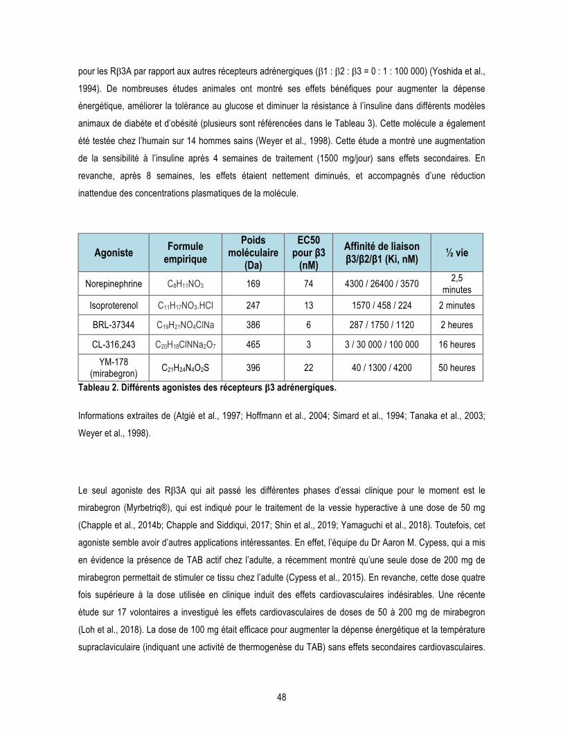
48
pour les Rβ3A par rapport aux autres récepteurs adrénergiques (β1 : β2 : β3 = 0 : 1 : 100 000) (Yoshida et al.,
1994). De nombreuses études animales ont montré ses effets bénéfiques pour augmenter la dépense
énergétique, améliorer la tolérance au glucose et diminuer la résistance à l’insuline dans différents modèles
animaux de diabète et d’obésité (plusieurs sont référencées dans le Tableau 3). Cette molécule a également
été testée chez l’humain sur 14 hommes sains (Weyer et al., 1998). Cette étude a montré une augmentation
de la sensibilité à l’insuline après 4 semaines de traitement (1500 mg/jour) sans effets secondaires. En
revanche, après 8 semaines, les effets étaient nettement diminués, et accompagnés d’une réduction
inattendue des concentrations plasmatiques de la molécule.
Agoniste Formule empirique
Poids moléculaire
(Da)
EC50 pour β3
(nM) Affinité de liaison β3/β2/β1 (Ki, nM) ½ vie
Norepinephrine C8H11NO3 169 74 4300 / 26400 / 3570 2,5
minutes
Isoproterenol C11H17NO3.HCl 247 13 1570 / 458 / 224 2 minutes
BRL-37344 C19H21NO4ClNa 386 6 287 / 1750 / 1120 2 heures
CL-316,243 C20H18ClNNa2O7 465 3 3 / 30 000 / 100 000 16 heures
YM-178 (mirabegron)
C21H24N4O2S 396 22 40 / 1300 / 4200 50 heures
Tableau 2. Différents agonistes des récepteurs β3 adrénergiques.
Informations extraites de (Atgié et al., 1997; Hoffmann et al., 2004; Simard et al., 1994; Tanaka et al., 2003;
Weyer et al., 1998).
Le seul agoniste des Rβ3A qui ait passé les différentes phases d’essai clinique pour le moment est le
mirabegron (Myrbetriq®), qui est indiqué pour le traitement de la vessie hyperactive à une dose de 50 mg
(Chapple et al., 2014b; Chapple and Siddiqui, 2017; Shin et al., 2019; Yamaguchi et al., 2018). Toutefois, cet
agoniste semble avoir d’autres applications intéressantes. En effet, l’équipe du Dr Aaron M. Cypess, qui a mis
en évidence la présence de TAB actif chez l’adulte, a récemment montré qu’une seule dose de 200 mg de
mirabegron permettait de stimuler ce tissu chez l’adulte (Cypess et al., 2015). En revanche, cette dose quatre
fois supérieure à la dose utilisée en clinique induit des effets cardiovasculaires indésirables. Une récente
étude sur 17 volontaires a investigué les effets cardiovasculaires de doses de 50 à 200 mg de mirabegron
(Loh et al., 2018). La dose de 100 mg était efficace pour augmenter la dépense énergétique et la température
supraclaviculaire (indiquant une activité de thermogenèse du TAB) sans effets secondaires cardiovasculaires.

49
Une autre étude sur 12 individus a montré des effets métaboliques pour la dose de 200 mg mais pas de la
dose de 50 mg (Baskin et al., 2018). Toutefois, d’autres études cliniques sont nécessaires pour confirmer ces
effets sur une plus grande population et sur des sujets atteints de maladies métaboliques.
Traitement Modèle Observations Référence Rongeur
CL-316,243 (1 mg/kg/jour, 17 jours)
Souris femelles
! dépense énergétique ! prise alimentaire
! production d’insuline = poids, " masse adipeuse
Garilova 2000
Acclimatation au froid (4°C, 4 h, 3 fois /
semaine, 10 semaines)
Souris C57BL/6J, diète
riche en gras
! activation TAB ! dépense énergétique ! prise alimentaire
!= tolérance au glucose = poids
Ravussin 2014
CL-316,243 (1 mg/kg/jour, 4
semaines)
Souris C57BL/6J
! activation TAB ! dépense énergétique ! prise alimentaire
! tolérance au glucose = poids
Xiao 2015
Acclimatation au froid (10°C, 21 jours)
Rats Wistar
! activation TAB ! dépense énergétique
! métabolisme du glucose ! métabolisme des lipides
Labbé 2015
CL-316,243 (1 mg/kg/j, 14 jours)
Rats Wistar et Lou/C
! activation TAB = poids, " masse adipeuse ! métabolisme du glucose ! métabolisme des lipides
" hyperinsulinémie
Poher 2015
Mirabegron (0,1 mg/kg, aigüe)
Souris C57BL/6J
! activation TAB Roberts-Toler 2015
CL-316,243 (1 mg/kg/jour, 3
semaines)
Souris diabétiques
(MKR)
! métabolisme du glucose ! métabolisme des lipides = poids, " masse adipeuse
" hyperinsulinémie
Kumar 2015
CL-316,243 (1 mg/kg/j, 14 jours)
Souris C57BL/6J
!= activation TAB = prise alimentaire
= poids, " masse adipeuse
Labbé 2016
Acclimatation au froid (10°C, 14 jours)
Souris C57BL/6J
! activation TAB ! prise alimentaire
= poids, " masse adipeuse Labbé 2016
CL-316,243 (10 ug, ICV)
Rats Sprage-Dawley
" prise alimentaire " poids
! libération d’insuline Richard 2017
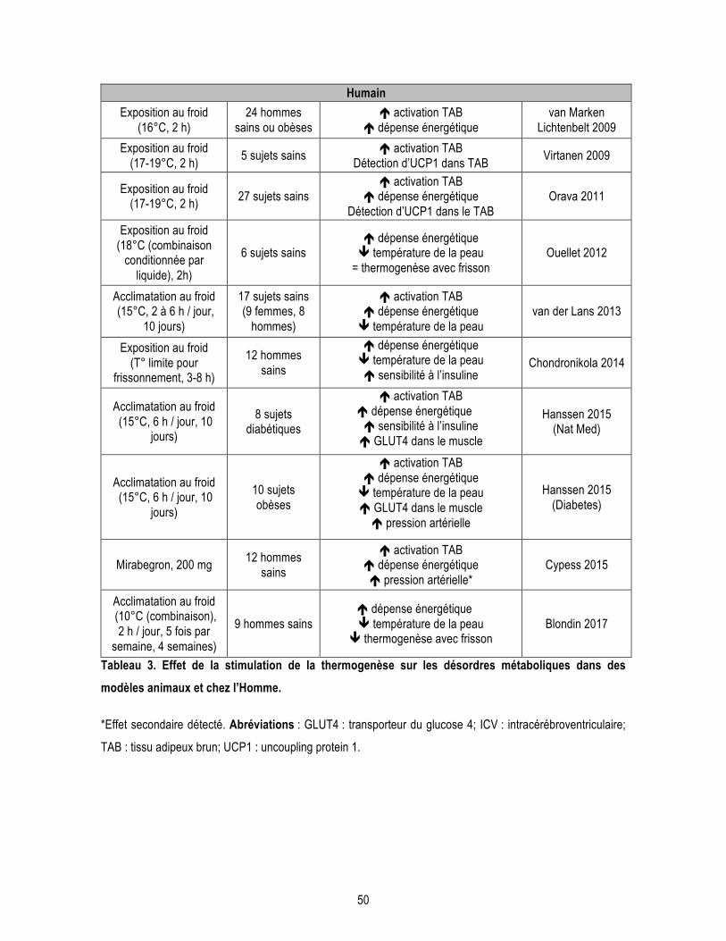
50
Humain Exposition au froid
(16°C, 2 h) 24 hommes
sains ou obèses ! activation TAB
! dépense énergétique van Marken
Lichtenbelt 2009
Exposition au froid (17-19°C, 2 h)
5 sujets sains ! activation TAB
Détection d’UCP1 dans TAB Virtanen 2009
Exposition au froid (17-19°C, 2 h)
27 sujets sains ! activation TAB
! dépense énergétique Détection d’UCP1 dans le TAB
Orava 2011
Exposition au froid (18°C (combinaison
conditionnée par liquide), 2h)
6 sujets sains ! dépense énergétique " température de la peau
= thermogenèse avec frisson Ouellet 2012
Acclimatation au froid (15°C, 2 à 6 h / jour,
10 jours)
17 sujets sains (9 femmes, 8
hommes)
! activation TAB ! dépense énergétique " température de la peau
van der Lans 2013
Exposition au froid (T° limite pour
frissonnement, 3-8 h)
12 hommes sains
! dépense énergétique " température de la peau ! sensibilité à l’insuline
Chondronikola 2014
Acclimatation au froid (15°C, 6 h / jour, 10
jours)
8 sujets diabétiques
! activation TAB ! dépense énergétique ! sensibilité à l’insuline ! GLUT4 dans le muscle
Hanssen 2015 (Nat Med)
Acclimatation au froid (15°C, 6 h / jour, 10
jours)
10 sujets obèses
! activation TAB ! dépense énergétique " température de la peau ! GLUT4 dans le muscle ! pression artérielle
Hanssen 2015 (Diabetes)
Mirabegron, 200 mg 12 hommes
sains
! activation TAB ! dépense énergétique ! pression artérielle*
Cypess 2015
Acclimatation au froid (10°C (combinaison),
2 h / jour, 5 fois par semaine, 4 semaines)
9 hommes sains ! dépense énergétique " température de la peau
" thermogenèse avec frisson Blondin 2017
Tableau 3. Effet de la stimulation de la thermogenèse sur les désordres métaboliques dans des modèles animaux et chez l’Homme.
*Effet secondaire détecté. Abréviations : GLUT4 : transporteur du glucose 4; ICV : intracérébroventriculaire;
TAB : tissu adipeux brun; UCP1 : uncoupling protein 1.
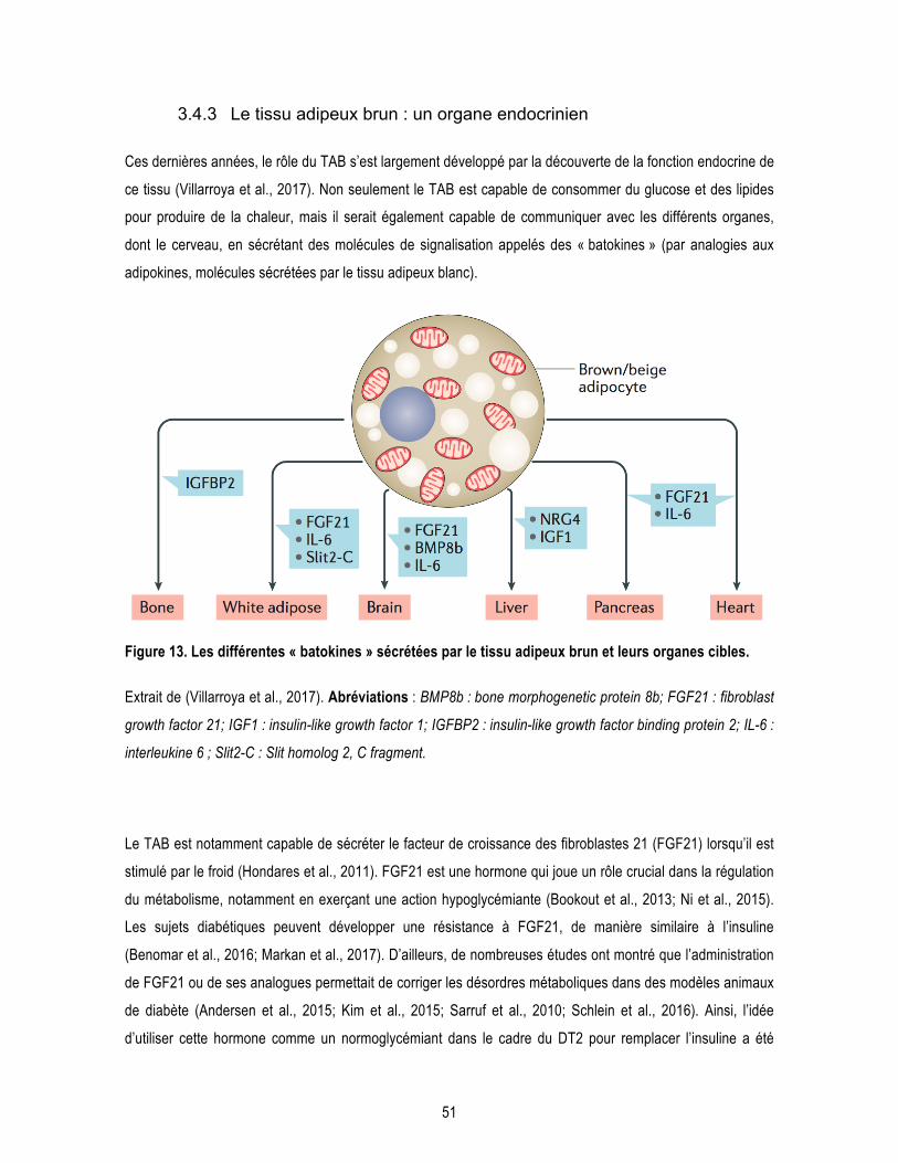
51
3.4.3 Le tissu adipeux brun : un organe endocrinien
Ces dernières années, le rôle du TAB s’est largement développé par la découverte de la fonction endocrine de
ce tissu (Villarroya et al., 2017). Non seulement le TAB est capable de consommer du glucose et des lipides
pour produire de la chaleur, mais il serait également capable de communiquer avec les différents organes,
dont le cerveau, en sécrétant des molécules de signalisation appelés des « batokines » (par analogies aux
adipokines, molécules sécrétées par le tissu adipeux blanc).
Figure 13. Les différentes « batokines » sécrétées par le tissu adipeux brun et leurs organes cibles.
Extrait de (Villarroya et al., 2017). Abréviations : BMP8b : bone morphogenetic protein 8b; FGF21 : fibroblast
growth factor 21; IGF1 : insulin-like growth factor 1; IGFBP2 : insulin-like growth factor binding protein 2; IL-6 :
interleukine 6 ; Slit2-C : Slit homolog 2, C fragment.
Le TAB est notamment capable de sécréter le facteur de croissance des fibroblastes 21 (FGF21) lorsqu’il est
stimulé par le froid (Hondares et al., 2011). FGF21 est une hormone qui joue un rôle crucial dans la régulation
du métabolisme, notamment en exerçant une action hypoglycémiante (Bookout et al., 2013; Ni et al., 2015).
Les sujets diabétiques peuvent développer une résistance à FGF21, de manière similaire à l’insuline
(Benomar et al., 2016; Markan et al., 2017). D’ailleurs, de nombreuses études ont montré que l’administration
de FGF21 ou de ses analogues permettait de corriger les désordres métaboliques dans des modèles animaux
de diabète (Andersen et al., 2015; Kim et al., 2015; Sarruf et al., 2010; Schlein et al., 2016). Ainsi, l’idée
d’utiliser cette hormone comme un normoglycémiant dans le cadre du DT2 pour remplacer l’insuline a été
52
investiguée, mais l’effet n’était pas considéré suffisant chez le primate par rapport aux autres médicaments
pour poursuivre les études cliniques (Kharitonenkov and Adams, 2014).
De façon intéressante, une récente étude cellulaire suggère que FGF21 pourrait avoir un rôle neuroprotecteur
contre l’excitotoxicité glutamatergique et la toxicité d’Aβ (Amiri et al., 2018; Sa-Nguanmoo et al., 2016a). Au
niveau animal, FGF21 permet de renverser les déficits cognitifs induits par une diète riche en gras saturés
chez la souris et le rat (Sa-Nguanmoo et al., 2016b; Q. Wang et al., 2018) et de réduire les lésions de la BHE
(Kuroda et al., 2017; Yu et al., 2018).

53
4 Thermorégulation et maladie d’Alzheimer
Il est bien connu que des déficits métaboliques sont observés dans la MA et sont liés au développement de la
pathologie comme discuté plus haut. En revanche, peu d’études ont investigué le lien entre thermorégulation
et MA. Pourtant, le déficit de thermorégulation chez les personnes âgées est décrit depuis longtemps, la
plupart des études sur le sujet étant relativement anciennes. Mais surtout, il existe maintenant une littérature
scientifique conséquente sur le fait que la température corporelle influence les marqueurs neuropathologiques
de la MA. Ces différents aspects seront présentés dans cette section.
4.1 Déficits de thermorégulation chez les personnes âgées
Le phénomène de vieillissement amène avec lui plusieurs changements conséquents pour le fonctionnement
général du corps. Tout d’abord, avec l’âge, le métabolisme énergétique central et périphérique diminue peu à
peu (Frisard et al., 2007; Rizzo et al., 2005). Ce qui est également frappant est que les personnes âgées
présentent un déficit de thermorégulation : il se caractérise par une difficulté à ressentir les changements
thermiques et à évaluer la température ambiante (Florez-Duquet and McDonald, 1998; Van Someren et al.,
2002). Il a également été rapporté que le cycle circadien était perturbé chez les personnes âgées (Weinert,
2000), et qu’il l’était d’autant plus chez les personnes atteintes de démence (Weldemichael and Grossberg,
2010). De façon plus marquée, le retour à la normale de la température corporelle suite à une exposition au
froid se fait de manière plus lente avec l’âge (Degroot and Kenney, 2007) : les personnes âgées mettent en
moyenne 30% plus de temps pour retrouver leur température orale initiale après ingestion d’eau glacée
(Sugarek, 1986).
Ces déficits pourraient être en partie expliqués par le fait que ces individus auraient de plus importantes pertes
de température corporelle car leur vasoconstriction, permettant de limiter la diffusion de la température à
travers la peau lors d’une exposition au froid, serait moins efficace que les personnes plus jeunes (Florez-
Duquet and McDonald, 1998; Richardson et al., 1992). D’autre part, l’activité du système nerveux sympathique
de la peau en réponse aux variations de la température est diminuée dans cette population : ils auraient donc
plus de difficulté à ressentir les changements de température ambiante (Grassi et al., 2003). Mais surtout,
avec l’âge, l’activité et le volume du TAB diminuent grandement (Cypess et al., 2009; Graja and Schulz, 2015).
54
Les déficits de thermorégulation des personnes âgées limitent leur capacité à réajuster leur température
corporelle à un niveau optimal et pourrait donc expliquer pourquoi les personnes âgées présentent une
diminution de leur température corporelle. En effet, quelques études ont rapporté que la température
corporelle moyenne des personnes âgées était plus basse que les personnes plus jeunes (Hoshino et al.,
2007; Howell, 1975). Une étude sur 100 participants âgés de plus de 65 ans vivant dans le même
environnement a montré que dans plus de 90% des cas, leur température orale en début, milieu et fin de
journée se trouvait sous les 37°C (Gomolin et al., 2005). Une autre étude sur plus de 400 sujets âgés de 15 à
84 ans a montré que la température axillaire des sujets de plus de 55 ans était plus basse que celle des
groupes plus jeunes (Hoshino et al., 2007). D’ailleurs, cette population âgée est plus à risque d’hypothermie :
selon une étude effectuée en Grande-Bretagne, environ 9% des personnes âgées ont une température
corporelle un demi degré sous 35°C, limite clinique de l’hypothermie (Fox et al., 1973). En revanche, une
étude sur 93 sujets âgés de 62 à 96 ans n’a pas mis en évidence de corrélation négative entre l’âge et la
température corporelle (Marion et al., 1991).
Il faut noter que ces études présentent des biais de méthode. En effet, la plupart se basent sur des mesures
de température orale, qui peut être influencée par ce que la personne vient de manger ou boire, et n’est
qu’assez peu corrélée à la température interne (Sund-Levander and Grodzinsky, 2013). Les personnes âgées
ayant fréquemment une température moyenne plus basse que 37°C, comme mentionnée plus haut, la fièvre
devrait être vérifiée en clinique avec d’autres variables comme la présence de la protéine C réactive dans le
sang par exemple et non pas seulement à l’aide d’une prise de la température corporelle (Hoshino et al.,
2007). De plus, la température est souvent prise à un seul moment de la journée (le matin). Comme la
température suit un rythme circadien, suivre la température corporelle en continue tout au long des 24 heures
apporterait beaucoup plus d’informations quant à la fluctuation de la température corporelle chez les
personnes âgées. Pour réellement comprendre le déficit de thermorégulation des personnes âgées, il faudrait
réaliser des études longitudinales avec des mesures de la température en continue et en ambulatoire. Ceci
peut être réalisé par exemple avec des sondes de télémétrie, sur une période de plusieurs jours et dans un
environnement contrôlé du mieux que possible, idéalement sur une population de personnes âgées hébergées
dans le même établissement de santé.
55
4.2 Déficits de thermorégulation dans la maladie d’Alzheimer
L’âge étant le principal facteur de risque de la MA, les perturbations de thermorégulation ont également été
observées dans la population atteinte de la MA. En effet, plusieurs études rapportent un cycle circadien
perturbé chez les sujets atteints de démence, ainsi qu’une plus grande amplitude de variation de la
température corporelle (Harper et al., 2004; Satlin et al., 1995; Weldemichael and Grossberg, 2010). Le
rythme circadien de la pression artérielle est également perturbé chez les sujets Alzheimer comparativement
aux sujets sains (Chen et al., 2013). De plus, une étude sur 237 sujets âgés de plus de 65 ans a montré que le
statut de démence influençait la température rectale et tympanique prise le matin (Sund-Levander and
Wahren, 2002). Toutefois, il n’y a pas vraiment de consensus sur l’effet de la MA sur la température corporelle
moyenne : certaines études rapportent une diminution de la température corporelle chez les sujets atteints de
la MA, tandis que d’autres présentent l’inverse ou aucun changement (“Body temperature as a risk factor for
Alzheimer’s disease,” 2000; Harper et al., 2001; Klegeris et al., 2007; Prinz et al., 1992; Touitou et al., 1986;
Volicer et al., 2001). Cette inconsistance des résultats dépend de nouveau de la méthode et du moment de la
prise de température, mais également du stade de la MA et des symptômes neuropsychologiques associés
(Okawa et al., 1991). En effet, certains patients présentent une grande agitation et des troubles
comportementaux pouvant influencer leur locomotion. D’autre part, les troubles du sommeil sont fréquents
dans cette population et peuvent affecter le rythme circadien (Klaffke and Staedt, 2006; Lushington et al.,
2000; Most et al., 2012).
Récemment, une étude a analysé les résultats de trois grandes études observationnelles, regroupant les
données de plus de 3000 sujets et a montré une variation des scores cognitifs, du diagnostic clinique et de la
quantité d’Aβ42 dans le LCR en fonction des saisons de l’année (A. S. P. Lim et al., 2018). Plus précisément,
les auteurs ont observé que pendant les saisons plus froides de l’année (printemps et hiver), les scores
cognitifs étaient moins bons, il y avait un plus grand nombre de diagnostic de MA et une plus basse quantité
d’Aβ42 dans le LCR que durant l’été et l’automne. Bien qu’une étude longitudinale de grande échelle soit
nécessaire pour confirmer ces résultats, ceci suggère que la MA pourrait être influencée par les saisons et la
température extérieure.
Quelques études animales ont mis en évidence un déficit de thermorégulation dans des modèles murins de la
MA. La souris 3xTg-AD présente des troubles dans le rythme circadien avant même que la neuropathologie
soit significativement détectée dans leur cerveau (Sterniczuk et al., 2010). Ceci est caractérisé notamment par
une température moyenne plus élevée que les souris contrôles à 6 et 10 mois d’âge, accompagnée d’une
augmentation de l’activité locomotrice de ces animaux (Knight et al., 2013). Un autre modèle animal de
56
pathologie amyloïde présente une diminution de la température corporelle de façon âge-dépendant, par
rapport aux animaux contrôles, à l’âge de 20 mois (Huitrón-Reséndiz et al., 2002). De façon intéressante, un
groupe a montré que le poids du TAB était diminué dans la souris 3xTg-AD (García-Mesa et al., 2012). Plus
récemment, notre équipe a montré que la 3xTg-AD présentait une diminution de la température corporelle
avec l’âge et l’avancée de la neuropathologie : la différence était significative à partir de l’âge de 12 mois, âge
où la neuropathologie est bien installée dans le cerveau des animaux (Vandal et al., 2016). Ces études
suggèrent que le développement de la neuropathologie Alzheimer affecte les processus de régulation de la
température corporelle. À l’inverse, les mécanismes impliqués dans la thermorégulation peuvent moduler la
MA comme le décrit la section suivante.
4.3 Thermorégulation et maladie d’Alzheimer : arguments issus d’études animales
Le lien entre diminution de température corporelle et phosphorylation de la protéine tau a été mis en évidence
par plusieurs travaux. En effet, des études sur des modèles animaux (rats et souris) montrent que
l’hypothermie, induite par une anesthésie ou un jeûne prolongé, conduit à une augmentation de
l’hyperphosphorylation de la protéine tau dans le cerveau (Planel et al., 2004; 2007). Selon ces études,
l’hyperphosphorylation est non seulement associée à l’augmentation de l’activité de plusieurs kinases, mais
aussi et surtout à l’inhibition de l’activité de la protéine phosphatase 2A (PP2A). Cette augmentation de la
phosphorylation est très rapide et drastique : il est estimé que pour une diminution de 1°C de température
corporelle, la phosphorylation de la protéine tau est augmentée de 80% (Planel et al., 2007).
Plus récemment, deux études ont montré que la hausse de phosphorylation de la protéine tau dans deux
modèles animaux de diabète (les souris db/db et ob/ob) était induite par l’hypothermie, apparaissant de façon
âge-dépendant dans ces souris (El-Khoury et al., 2016; Gratuze et al., 2017a). En effet, le retour à la
normothermie de ces souris par une exposition à une température de 34°C pendant une heure permet de
réduire l’hyperphosphorylation sur la plupart des sites de la protéine tau. L’hypothermie et les déficits de
thermorégulation étant également fréquents dans la population diabétique (B. W. Foster, 1869; Neil et al.,
1986; Scott et al., 1987), ces études proposent une explication originale au fait que les individus diabétiques
ont plus de risque de développer la MA.
De façon intéressante, cette augmentation de l’hyperphosphorylation de la protéine tau a également été
observée dans le cerveau d'animaux qui hibernent (Arendt et al., 2003; Stieler et al., 2011), et donc diminuent

57
drastiquement leur métabolisme et leur température corporelle pendant un certain temps. Une étude chez
l’écureuil terrestre de l’arctique a montré que différents sites de phosphorylation de la protéine tau sont
impliqués, tandis que la glycogène synthase kinase 3β (GSK3β) est d’avantage présente sous sa forme
inactive (Stieler et al., 2011). Toutefois, dans ces modèles d’hibernation, l’hyperphosphorylation de la protéine
tau n’est que transitoire et revient à la normale dès la sortie de la torpeur (Arendt and Bullmann, 2013). Ce
mécanisme semble plutôt être un phénomène neuroprotecteur en réponse à ce changement physiologique
dans ce cas-ci.
En revanche, il est possible qu’un tel retour à la normal ne soit pas retrouvé chez les personnes âgées. En
effet, les cas de troubles cognitifs et délirium post-opératoire suggèrent que l’anesthésie peut avoir des
répercussions à long terme sur les ainés (Ancelin et al., 2001; Fodale et al., 2010; Papon et al., 2011). En ce
sens, plusieurs études animales ont montré que l’anesthésie augmentait la phosphorylation de la protéine tau
(Planel et al., 2009; 2008; Tan et al., 2010). En effet, l’anesthésie affecte la régulation de la température
corporelle : l’hypothermie induite par l’anesthésie serait donc responsable de ses effets délétères dans le
cerveau (Lenhardt, 2010). Certains ont montré que le maintien de la température corporelle des animaux sous
anesthésie permettait de maintenir des niveaux de phosphorylation de tau normaux et de contrer les effets
néfastes de l’anesthésie (Planel et al., 2007). En revanche, d’autres ont observé que même en condition de
normothermie, l’exposition à l’anesthésie augmentait la phosphorylation de la protéine tau (Le Freche et al.,
2012). De manière intéressante, bien que les effets d’une seule exposition d’une heure soient transitoires,
l’exposition répétée à l’anesthésie induit une hyperphosphorylation persistante à long terme chez la souris. De
plus, quelques études proposent que les anesthésiants aient un effet direct sur l’activité des kinases
impliquées dans la phosphorylation de tau (Run et al., 2010).
Finalement, nous avons récemment montré un déficit de thermorégulation chez la souris 3xTg-AD survenant à
partir de l’âge de 12 mois (Figure 14A) (Vandal et al., 2016). De façon surprenante, une seule exposition aigüe
au froid de 24 heures à 4°C augmente la phosphorylation de tau de façon plus importante chez les souris
3xTg-AD comparativement aux souris non-transgéniques. De plus, l’exposition au froid augmente de deux fois
le ratio Aβ42/Aβ40 et la quantité d’APP dans le cortex des souris 3xTg-AD de 12 mois (Figure 14B,C). À
l’inverse, augmenter leur température corporelle de 0,5°C en les exposant à un environnement thermoneutre
(28°C) pendant une semaine diminue le déficit cognitif, l’anxiété et la pathologie amyloïde de souris 3xTg-AD
de 20 mois (Figure 14D-G) (Vandal et al., 2016). Ces résultats montrent pour la première fois que les
changements de température externe, recrutant ainsi les mécanismes de thermorégulation de l’animal,
affectent à la fois le comportement et la neuropathologie Alzheimer dans un modèle murin de la MA.
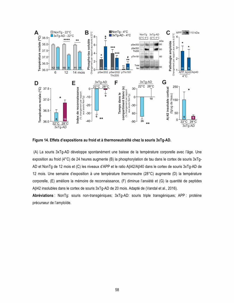
58
Figure 14. Effets d’expositions au froid et à thermoneutralité chez la souris 3xTg-AD.
(A) La souris 3xTg-AD développe spontanément une baisse de la température corporelle avec l’âge. Une
exposition au froid (4°C) de 24 heures augmente (B) la phosphorylation de tau dans le cortex de souris 3xTg-
AD et NonTg de 12 mois et (C) les niveaux d’APP et le ratio Aβ42/Aβ40 dans le cortex de souris 3xTg-AD de
12 mois. Une semaine d’exposition à une température thermoneutre (28°C) augmente (D) la température
corporelle, (E) améliore la mémoire de reconnaissance, (F) diminue l’anxiété et (G) la quantité de peptides
Aβ42 insolubles dans le cortex de souris 3xTg-AD de 20 mois. Adapté de (Vandal et al., 2016).
Abréviations : NonTg: souris non-transgéniques; 3xTg-AD: souris triple transgéniques; APP : protéine
précurseur de l’amyloïde.
BTe
mpé
ratu
re re
ctal
e (º
C)
6 12 14 mois36.0
37.0
38.0
37.5
36.5
38.5A
**** **
NonTg - 22°C3xTg-AD - 22°C
12 13 10 9 11 110
2
4
6
8
pSer202pSer202/
Thr205
pThr181
60kDa
60
60
*
*****
*
***
60Total
tau
22°C 4°CNonTg 3xTg-AD
11 11 11 1111
11
NonTg - 4°C3xTg-AD - 4°C
#
&
36.0
36.5
37.0
37.5
*
Tem
péra
ture
rect
ale
(°C
)
Inde
x de
reco
nnai
ssan
ce(%
cha
ngem
ent p
ar ra
ppor
t au
x so
uris
Non
Tg)
22°C 28°C
-40
-30
-20
-10
0
**6
9
8 10
3xTg-AD
22°C 4°C
D E
pSer202 pSer202/Thr205
pThr181
22°C 28°C3xTg-AD
50
150
250*
9 9Aβ4
2 in
solu
ble
cort
ical
(fg/u
g de
tiss
u)
G
22°C 28°C3xTg-AD
Tem
ps d
ans
leco
mpa
rtim
ent b
lanc
(s)
(% c
hang
emen
t par
rapp
ort
aux
sour
is N
onTg
)
-90
60
-30
0
30
**26
10
F 22°C 28°C
3xTg-AD
Phos
pho-
tau
solu
ble
Cha
ngem
ent p
ar ra
ppor
t aux
sou
ris à
22°
C
1
2
3
4APP 110 kDa
**
11 9
C
Aβ42/Aβ40APP4°C
Path
olog
ie a
myl
oïde
Cha
ngem
ent p
ar ra
ppor
t aux
sou
ris à
22°
C
0

59
Hypothèses et objectifs
1 Hypothèses
La majorité des études précliniques et cliniques sur la MA s’est concentrée sur la diminution des pathologies
amyloïdes et tau. Toutefois, les dogmes proposant que la cascade amyloïde et la pathologie tau sont les
causes du développement de la MA ont été bousculés au cours de ces dernières années. Un retour en arrière
vers l’étude des potentielles causes de la maladie est nécessaire afin de découvrir de nouvelles cibles
thérapeutiques pour la MA.
Puisque que l’âge avancé est extrêmement important pour le déclenchement de la maladie, mieux
comprendre les mécanismes moléculaires associés au vieillissement pourrait nous permettre d’élucider
certains aspects de la pathogenèse de la MA. Bien que peu d’études soient disponibles sur le lien entre
thermorégulation et MA, des études animales ont permis d’établir clairement le lien entre diminution de
température corporelle et hyperphosphorylation de la protéine tau dans le cerveau. D’autre part, le déficit de
thermorégulation survenant chez les personnes âgées est également bien documenté. Ainsi, le fait que les
personnes âgées aient plus de difficulté à réguler leur température corporelle pourrait expliquer pourquoi elles
sont plus à risque de développer la MA.
Intervenir sur la thermorégulation pourrait être une stratégie doublement intéressante : les désordres
métaboliques contribuent à la pathogenèse de la MA, et le déficit de thermorégulation, du moins dans les
modèles animaux, accentue la neuropathologie Alzheimer. La stimulation de la thermogenèse ayant fait ses
preuves comme outil thérapeutique pour les maladies métaboliques, stimuler la thermorégulation pourrait être
bénéfique pour la MA en corrigeant à la fois les désordres métaboliques et les déficits de thermorégulation.
Notre hypothèse générale est que le déficit de thermorégulation des personnes âgées contribue au
développement de la MA et que le corriger permettrait de réduire à la fois les désordres métaboliques, la
neuropathologie et les déficits cognitifs associés à la MA. De plus, étudier le lien entre thermorégulation et MA
nous permettrait peut-être de développer de nouvelles stratégies thérapeutiques pour la MA.
Comme discuté dans la fin de l’introduction, notre équipe a montré que la pathologie Alzheimer induite
génétiquement dans un modèle animal de la MA (la souris 3xTg-AD) aggrave l’hyperphosphorylation de la
protéine tau induite par une exposition au froid. Mais qu’en est-il du facteur de l’âge, qui est le principal facteur

60
de risque de la MA ? Quels seraient les effets d’une intervention visant à améliorer les mécanismes de
thermorégulation, plus précisément la thermogenèse du TAB, sur la pathologie Alzheimer ?
2 Objectifs
Pour répondre à ces questions, nous avons utilisé la souris afin d’intervenir sur la thermorégulation et d’être
capable d’analyser directement les effets sur le cerveau de ces mêmes animaux. Le choix d’utiliser la souris
3xTg-AD s’est basé sur nos précédentes études, montrant que ce modèle murin de la MA présente des
désordres du métabolisme et de la thermorégulation associés à l’évolution des pathologies tau et amyloïde.
Comme l’apparition des déficits cognitifs et de la neuropathologie est très progressive dans cet animal, nous
avons sélectionné des animaux âgés, permettant de prendre en compte le vieillissement dans nos études.
Finalement, ce modèle nous a permis d’évaluer l’effet de nos interventions sur les deux marqueurs
neuropathologiques principaux de la MA.
L’objectif général de cette thèse est de déterminer si la thermorégulation pourrait être une nouvelle cible
thérapeutique pour le traitement de la MA. Plus précisément, nous avons cherché à savoir si l’augmentation
de l’activité de thermogenèse permettait de moduler la pathologie Alzheimer dans un modèle murin de la
maladie.
2.1 Objectif spécifique 1
Nous avons précédemment montré que la neuropathologie de la MA était un facteur aggravant
l’hyperphosphorylation de tau en réponse à une exposition au froid. Notre premier objectif était de déterminer
si l’âge était un facteur influençant le lien entre température corporelle et hyperphosphorylation de la protéine
tau.
Nous avons comparé l’effet d’une exposition aigüe au froid (24 heures, 4°C) sur la phosphorylation de tau
dans le cortex de souris jeunes (6 mois) et âgées (18 mois) non-transgéniques. Nous avons également vérifié
61
l’effet de cette exposition au froid sur la température et l’activité du TAB des animaux. Finalement, nous avons
mesuré les différentes kinases et phosphatases impliquées dans la phosphorylation de la protéine tau.
2.2 Objectif spécifique 2
Par la suite, nous avons réalisé deux études dont le but était de déterminer l’effet de la stimulation de la
thermogenèse du TAB sur la neuropathologie des souris 3xTg-AD. Pour cela, nous avons utilisé deux
approches différentes.
Dans un premier temps, nous avons utilisé des expositions répétées au froid, une méthode globale et non
pharmacologique. Nous voulions déterminer si cette méthode d’acclimatation au froid permettait de protéger
les souris 3xTg-AD âgées des effets délétères d’une exposition aigüe au froid.
Nous avons exposé des souris 3xTg-AD âgées de 15 mois à de courtes expositions au froid de façon répétée
(4 heures à 4°C, 5 fois par semaine) pour une durée totale d’un mois, avant de les présenter à une exposition
aigüe au froid (24 heures à 4°C). Nous avons comparé ce groupe à un groupe de souris contrôles restées à la
température d’hébergement (22°C) tout au long de l’expérience, et à un groupe de souris ayant reçu une
seule exposition aigüe au froid (24 heures à 4°C). Nous avons déterminé l’effet de l’intervention sur la
capacité de thermogenèse et la tolérance au glucose des animaux. Nous avons également mesuré les
niveaux de phosphorylation de la protéine tau et de peptides Aβ40 et Aβ42 dans l’hippocampe des souris
3xTg-AD âgées. Finalement, nous avons évalué la production de FGF21 en réponse aux expositions au froid
répétées ou aigües.
2.3 Objectif spécifique 3
Dans un second temps, nous avons utilisé une approche pharmacologique par l’administration chronique d’un
agoniste des Rβ3A. Cette deuxième méthode vise directement les Rβ3A présents sur le TAB sans stimulation
du système nerveux sympathique. Nous voulions savoir si l’activité de thermogenèse du TAB permettait de
diminuer la neuropathologie et les déficits cognitifs des souris 3xTg-AD âgées, et si les agonistes des Rβ3A
pouvaient être utilisés dans le traitement de la MA.
Nous avons administré le CL-316,243 à une dose de 1 mg/kg par jour ou du salin pour une durée d’un mois à
des souris non-transgéniques et 3xTg-AD. Nous avons utilisé des souris âgées de 16 mois afin d’évaluer

62
l’effet du traitement à un âge avancé. Nous avons d’abord évalué l’effet de l’agoniste sur les paramètres
métaboliques des animaux, puis sur la température corporelle et l’activité du TAB. Nous avons effectué
plusieurs tests comportementaux afin de déterminer si le traitement avait des conséquences sur la locomotion,
l’anxiété et la mémoire de reconnaissance des différents groupes de souris. Finalement, les pathologies tau et
amyloïdes ont été quantifiées dans l’hippocampe des animaux.
63
Chapitre 1 : L’âge avancé potentialise la phosphorylation de tau induite par le froid : lien entre déficit de thermorégulation et maladie d’Alzheimer.

64
Old age potentiates cold-induced tau phosphorylation: linking thermoregulatory deficit with Alzheimer’s disease
Marine Tournissac1,2,3, Milène Vandal1,2,3, Arnaud François1,2, Emmanuel Planel4,2, Frédéric Calon*1,2,3
1Faculté de pharmacie, Université Laval, Québec, Québec, Canada;
2Axe Neurosciences, Centre de recherche du centre Hospitalier de l’Université Laval (CHUL), Québec,
Québec, Canada;
3Institut sur la Nutrition et les Aliments Fonctionnels, Québec, Québec, Canada;
4Faculté de médecine, Département de psychiatrie et de neurosciences, Université Laval, Québec, Québec,
Canada.
65
1.1. Résumé
Les déficits de thermorégulation coïncident avec le pic d’incidence de la maladie d’Alzheimer (MA) à un âge
avancé. Une basse température corporelle augmente la phosphorylation de tau (pTau), un marqueur
neuropathologique de la MA. Pour déterminer si l’âge avancé potentialise pTau induite par le froid, nous avons
comparé l’effet d’une exposition au froid (4°C, 24h) sur des souris de 6 et 18 mois. La température corporelle,
l’activité du tissu adipeux brun et pTau sur la Ser202 après l’exposition étaient similaires entre les souris
jeunes et âgées. En revanche, seules les souris âgées avaient une augmentation de pTau sur les Thr181 et
Thr231, corrélant avec leur température corporelle. L’inactivation de GSK3β chez les souris jeunes suggère
un mécanisme protecteur contre pTau induite par le froid. Ces résultats suggèrent que l’âge avancé confère
une plus grande susceptibilité à pTau suivant un changement de la température, contribuant à augmenter le
risque de MA.
1.2. Abstract
Thermoregulatory deficits coincide with a rise in the incidence of Alzheimer's disease (AD) in old age. Lower
body temperature increases tau phosphorylation, a neuropathological hallmark of AD. To determine whether
old age potentiates cold-induced tau phosphorylation, we compared the effects of cold exposure (4°C, 24h) in
6- and 18-month-old mice. Cold-induced changes in body temperature, brown adipose tissue activity and
phosphorylation of tau at Ser202 were not different between 6- and 18-month-old mice. However, following
cold exposure, only old mice displayed a significant rise in soluble tau pThr181 and pThr231, which was
correlated with body temperature. Inactivation of GSK3β was more prominent in young mice, suggesting a
protective mechanism against cold-induced tau phosphorylation. These results suggest that old age confers
higher susceptibility to tau hyperphosphorylation following a change in body temperature, thereby contributing
to an enhanced risk of developing AD.
1.3. Introduction
Aging is by far the most important risk factor of Alzheimer’s disease (AD) (Querfurth and LaFerla, 2010; Rocca
et al., 2011b; Ziegler-Graham et al., 2008). Even familial forms of the disease are rarely expressed clinically
before the fifth decade of life (Ridge et al., 2013). Thus, a better understanding of how old age contributes to
AD pathogenesis will likely provide critical clues on potential treatments. Strikingly, thermoregulatory deficits
66
appear at the same time in life when the incidence of AD greatly increases (Degroot and Kenney, 2007;
Florez-Duquet and McDonald, 1998). In fact, mean body temperature in the elderly has been long known to be
lower than in young people and below 37°C (Gomolin et al., 2005; Hoshino et al., 2007; Howell, 1975),
increasing their risk of hypothermia (Fox et al., 1973; Whittington et al., 2010). Furthermore, several other
parameters linked to thermoregulation are affected in the elderly: energy metabolism (Cunnane et al., 2011;
Frisard et al., 2007), thermogenic activity of brown adipose tissue (BAT) (Cypess et al., 2009), ability to
recover after cold exposure (Florez-Duquet and McDonald, 1998; Sugarek, 1986) and circadian cycles of body
temperature (Weinert, 2010).
Preclinical investigations in animal models have brought potential mechanisms coupling thermoregulatory
deficits to AD pathogenesis. Most prominently, hypothermia in mice has been repeatedly shown to increase
tau phosphorylation and pathology in the brain (Planel et al., 2009; 2004; Vandal et al., 2016), one of the main
neuropathological markers of AD (Cowan and Mudher, 2013; Frost et al., 2014; Tremblay et al., 2007).
Although the link between lower body temperature and tau phosphorylation was evidenced in multiple
experiments, to our knowledge, no study investigated the role of age in this mechanism.
Since the appearance of thermoregulation deficits coincides with a higher incidence of AD and that a lower
body temperature has been repeatedly shown to increase tau phosphorylation (Arendt, 2003; Planel et al.,
2007; Whittington et al., 2010), we aimed to verify whether this post-translational mechanism was potentiated
in old mice compared to younger mice. Using 6- and 18-month-old mice exposed to 4°C during 24 hours, we
found that cold exposure led to a higher increase in soluble phosphorylated tau in old compared to young
mice. In parallel, only young mice displayed higher levels of the inactivated form of glycogen synthase kinase
3β (GSK3β) in the cortex following cold exposure, suggesting that GSK3β inactivation is a potential protective
mechanism against tau hyperphosphorylation that is lost in old animals.
1.4. Methods
1.4.1. Animals and cold exposure
All experiments were performed in accordance with the Canadian Council on Animal Care and were approved
by the Institutional Committee of the Centre Hospitalier de l’Université Laval (CHUL). 6- and 18-month-old
C57BL6/129SvJ mice produced at our animal facility were used in equal number of males and females in each
group. One animal per cage was housed at 4°C or 22°C during 24 hours. Body temperature was determined
rectally with an electronic thermometer coupled with a rectal probe before and after exposure at the same hour
of the day (8 a.m.). All mice were kept under their housing temperature (4°C or 22°C) until sacrifice by

67
intracardiac perfusion as described elsewhere (Julien et al., 2009). Although it is known that anesthesia has an
effect per se on body temperature (Lenhardt, 2010), all mice were put under deep anesthesia with
ketamine/xylazine injection (100 mg/kg ketamine, 10 mg/kg xylazine) for ethical reasons. Rapidly,
interscapular brown adipose tissue and brain were dissected and frozen at -80°C until processing for Western
blot. In a separate experiment, we found that exposure to 4°C for 24 hours prior to anesthesia significantly
aggravated the ketamine-induced decrease in body temperature (-3.23±0.64°C, n=7) compared to exposure
to room temperature (-1.61±0.59°C, n=7).
1.4.2. Protein extraction and Western immunoblotting
The protein extraction method results in a TBS-soluble intracellular and extracellular fraction (cytosolic
fraction), a detergent-soluble fraction (membrane fraction) and a detergent-insoluble fraction solubilized in
formic acid as previously described (Lebbadi et al., 2011). The detailed method for Western immunoblotting is
described elsewhere (Vandal et al., 2014b). The list of primary antibodies used in our experiments is available
in supplementary Table 1.1. All brain homogenates from the same experiment were put on the same gel but
consecutive bands were not taken for all representative photo examples.
1.4.3. Quantitative real-time PCR
The expression level of uncoupling protein 1 (UCP1) mRNA was determined in brown adipose tissue using a
reverse transcription real-time quantitative polymerase chain reaction (QPCR). Frozen brown adipose tissues
were powderized and 10 mg of each sample was homogenized in 1mL QIAzol lysis reagent (Qiagen, Valencia,
CA, USA) to which was added 200 µL chloroform (J.T. Baker, Center Valley, PA, USA). RNA was extracted
with an RNeasy lipid tissue mini kit (Qiagen, Valencia, CA, USA). The High Capacity cDNA Reverse
Transcription kit with random hexamer primers and MultiScribe Reverse Transcriptase (Life Technologies,
Burlington, ON, Canada) was used to synthesize complementary DNA (cDNA) according to the instructions
from the manufacturer. Then, TaqMan qPCR primers were used to amplify mouse UCP1 (TaqMan Gene
Expression Assays, Mm01244861_m1 from Life Technologies) and glyceraldehyde-3-phosphate
dehydrogenase (GAPDH, Mm99999915_g1, Life Technologies) as the housekeeping gene. Reactions were
prepared in triplicates and cycle threshold (Ct) values were measured using a real-time qPCR cycler (MxPro-
Mx3005P, Agilent Technologies, Mississauga, ON, Canada) in 96-well optical plates covered with optical
adhesive film. Each run contained a “no template” control for both genes. The delta delta Ct (ddCT) method
was used to evaluate differences in relative gene expression between groups using GAPDH as a control gene.
Results are presented as ratios of UCP1/GAPDH cDNA on control group.

68
1.4.4. Statistical analysis
Data are presented as means ± SEM or as relative change observed in cold-exposed mice compared to
control mice. Statistical analysis and number of mice per group are specified in each Figure. Bartlett’s tests
were used to rule out inequality of variances between the groups. One-way (one independent variable) or two-
way ANOVA (two independent variables) were used when more than two groups were compared. ANOVAs
were followed by Tukey’s post-hoc analysis in case of equal variance or by a Dunnett’s post-hoc analysis in
case of unequal variances. An unpaired Student’s t-test was performed when only two groups were compared,
with a Welch correction when variances were not equal. Correlations between variables were investigated
using linear regression analyses. All statistical analyses were performed with Prism 5 (GraphPad software,
San Diego, CA, USA) or JMP (version 12.1.0; SAS Institute Inc., Cary, IL) software and statistical significance
was set at p<0.05.
1.5. Results
1.5.1. Enhanced cold-induced phosphorylation of soluble tau in old mice compared to young mice.
Cold exposure at 4°C is a classical method to study non-shivering thermogenesis in rodents (Cannon and
Nedergaard, 2010; S. Lim et al., 2012). Thus, we first confirmed that cold exposure induced a decrease in
body temperature and an increase in thermogenesis, as indexed by the enhanced ratio of UCP1 on GAPDH
expression in BAT compared to animals maintained at 22°C. However, no significant differences in body
temperature or BAT activation were detected between 6- and 18-month-old mice (Fig. 1.1A-B).
Given the importance of tau phosphorylation in AD pathogenesis (Cowan and Mudher, 2013; Frost et al.,
2014; Querfurth and LaFerla, 2010; Tremblay et al., 2007), we then assessed the extent by which cold
exposure potentiates the phosphorylation of key epitopes. We found that 18-month-old mice displayed a
greater hyperphosphorylation of tau in response to cold exposure. More specifically, although cold exposure
increased the phosphorylation of tau at epitopes pSer396/404 in young mice (+40%) and pSer202 in both 6-
and 18-month-old mice (+101% and +132%, respectively), only old mice displayed an increase in pThr181 and
pThr231 (+41% and +61%, respectively) (Fig. 1.1C-D). Two-way ANOVA further confirmed the upregulating
effect of a lower temperature on phosphorylated tau species in the soluble fraction (P = 0.0001 for pSer202; P
= 0.026 for pThr231 and P = 0.031 for pThr181).
Interestingly, the phosphorylation of soluble tau in response to cold exposure was correlated with body
temperature suggesting that both events are linked. In fact, we found that tau pSer202 was correlated with
69
body temperature in both 6- and 18-month-old mice (r2=0.16 and 0.43, respectively) (Fig. 1.1E). However,
phosphorylation levels of soluble tau at pThr231 and pThr181 were correlated with body temperature only in
old mice (r2= 0.29 and r2=0.47, respectively), not in young ones (Fig. 1.1F-G).
1.5.2. GSK3β inactivation in young but not in old mice.
Since tau phosphorylation is regulated by kinases and phosphatases (J.-Z. Wang et al., 2013), we next
measured several of these key enzymes in cortex homogenates of young and old mice. No significant changes
were found in main phosphatases and kinases (Fig. 1.1I-L) after exposure to 4°C, although two-way ANOVA
revealed that older age was associated with a higher pAKTSer473/AKT ratio (P = 0.038). In particular, the
activated form of GSK3β (pTyr216) was unchanged by cold exposure in both young and old mice. However,
the inactivated form (pSer9) was 49% higher in cortex homogenates of 6-month-old mice exposed to cold
compared to those at room temperature, a finding not observed in 18-month-old mice (Fig. 1.1K-L).
1.6. Discussion
In the present study, we do not only confirm previous observations suggesting that lower body temperature
increases the phosphorylation of soluble tau in the cerebral cortex of cold-exposed mice (Planel et al., 2007;
Vandal et al., 2016), but we more importantly propose that old age worsens this phenomenon. We also
identified that a loss of protective kinase-related mechanisms could underlie the observed higher tau
phosphorylation in old mice. These findings provide a potential mechanistic explanation on how old age
promotes the translation of thermoregulatory deficits into aggravated AD-like neuropathology, thereby
shedding light on why old age is the main risk factor for AD.
The cerebral accumulation of hyperphosphorylated tau is considered as one of the main neuropathological
hallmarks of AD (Frost et al., 2014; Querfurth and LaFerla, 2010). Our first series of investigations focused on
indexing the hyperphosphorylation status of soluble tau, which is thought to precede pathological aggregation
and formation of intraneuronal NFT (Avila et al., 2004; Buée et al., 2000). In particular, pThr231 tau is believed
to initiate the tau hyperphosphorylation cascade (Martin et al., 2011). Further supporting the relevance of the
phosphorylation of soluble tau to AD symptoms, we previously showed that both pSer396/404 and pThr231
tau correlate with ante-mortem cognitive performance in AD, mild cognitive impairment (MCI) and non-
demented subjects from the Rush religious Order study (Bennett et al., 2004; Calon et al., 2015). The present

70
data corroborate previous reports of increased tau phosphorylation following reduction in body temperature:
tau phosphorylation at pSer202, pThr231 and pThr181 are all increased in hibernators (Arendt et al., 2003),
whereas pSer202 and pThr181 tau in murine brain were consistently shown to be increased after an
anesthesia-induced decrease in body temperature (Planel et al., 2007; Run et al., 2009). Interestingly, reduced
body temperature correlated with increased phosphorylation of soluble tau, further strengthening the link
between a lower body temperature and a higher phosphorylation of soluble tau species, especially at older
ages.
The phosphorylation of tau is regulated by several kinases in vivo, such as cdk5, CAMKII and JNK (J.-Z. Wang
et al., 2013), but GSK3β is the most prominent (Kremer et al., 2011; Takashima, 2006). We found that 6-
month-old mice, following cold exposure, not only displayed a smaller increase in soluble tau phosphorylation
compared to old animals, but also showed higher levels of the inactivated form (pSer9) of GSK3β. Similar
signs of GSK3β inactivation are found in the forebrain of hibernating squirrels compared to summer-active
animals (Su et al., 2008). A possible interpretation is that young mice prevent the pathological increase in tau
phosphorylation by reducing the activity of GSK3β.
In summary, the present results suggest that a 2°C decrease in body temperature enhances tau
phosphorylation in the brain cortex more importantly in old mice compared to young ones. From this, it could
be speculated that the 0.5°C reduction of body temperature reported in the elderly (between people 65 to 74
years of age and people over 85 years of age) (Gomolin et al., 2005) could promote the phosphorylation of tau
in the brain over time and contribute to AD pathogenesis (Whittington et al., 2010). If true, this would mean that
old age confers a higher susceptibility to changes in body temperature, thereby enhancing the risk of
developing AD. Finally, a defect in kinase deactivation in old age stands as a potential mechanism linking
thermoregulation and tau phosphorylation. In the light of these results, it is postulated that improving natural
thermoregulatory mechanisms in the elderly could be a novel therapeutic strategy for AD.
1.7. Acknowledgements
This study was made possible by funding from the Alzheimer Society Canada (15-12), the Canadian Institutes
of Health Research (CIHR) (MOP 102532 and IAO 74443) and the Canadian Foundation for Innovation (CFI).
Fonds de Recherche du Québec en Santé (FRQ-S) provided salary support to Frédéric Calon. Marine
Tournissac was supported by scholarships from Fonds d’Enseignement et de la Recherche from the Faculty of
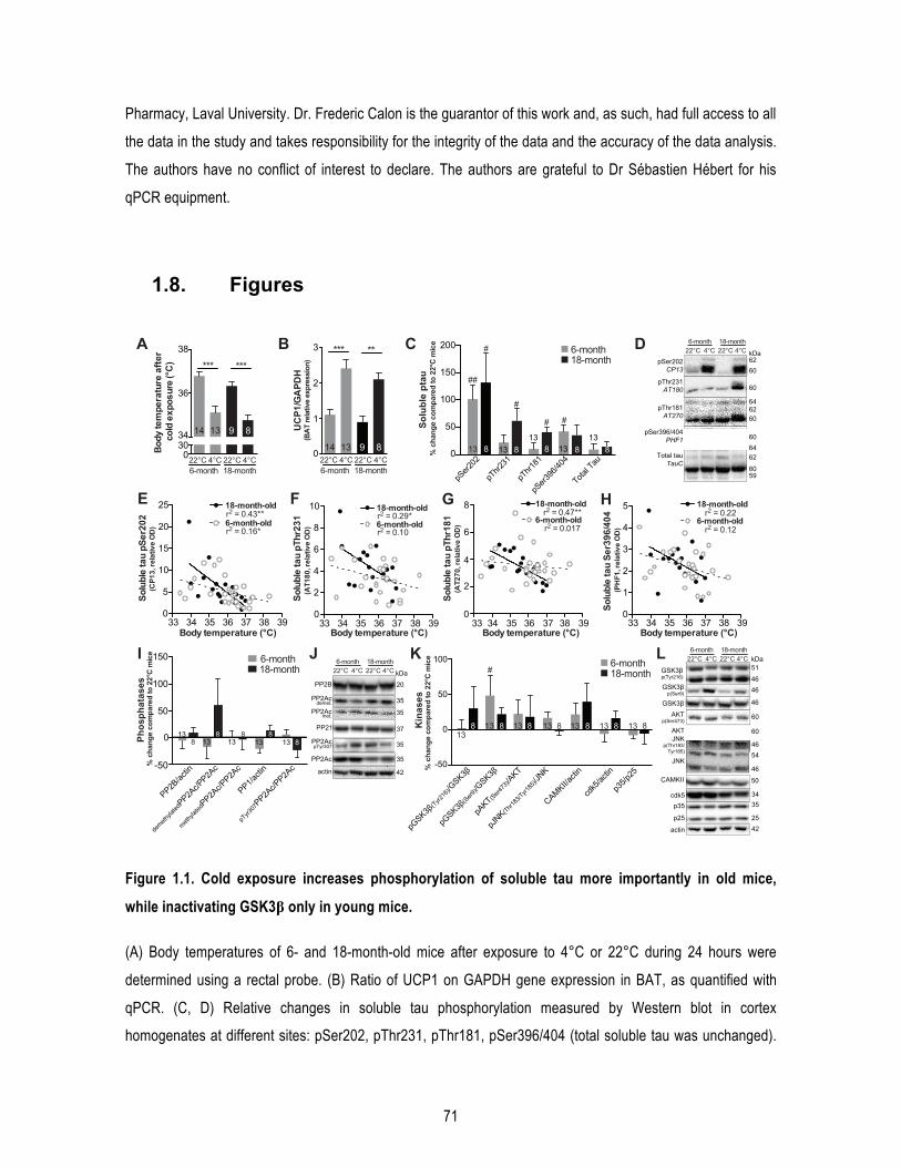
71
Pharmacy, Laval University. Dr. Frederic Calon is the guarantor of this work and, as such, had full access to all
the data in the study and takes responsibility for the integrity of the data and the accuracy of the data analysis.
The authors have no conflict of interest to declare. The authors are grateful to Dr Sébastien Hébert for his
qPCR equipment.
1.8. Figures
Figure 1.1. Cold exposure increases phosphorylation of soluble tau more importantly in old mice,
while inactivating GSK3β only in young mice.
(A) Body temperatures of 6- and 18-month-old mice after exposure to 4°C or 22°C during 24 hours were
determined using a rectal probe. (B) Ratio of UCP1 on GAPDH gene expression in BAT, as quantified with
qPCR. (C, D) Relative changes in soluble tau phosphorylation measured by Western blot in cortex
homogenates at different sites: pSer202, pThr231, pThr181, pSer396/404 (total soluble tau was unchanged).
C
I 6-month18-month
K
60
pSer202CP13
pThr181AT270
60pThr231AT180 60
Total tauTauC
60
DkDa 22°C 22°C 4°C4°C
6-month 18-month
62
6264
6264
59
GSK3β 46
GSK3βp(Ser9) 46
CAMKII
AKTp(Ser473)
GSK3βp(Tyr216)
JNKp(Thr183/
Tyr185)
p35
JNK
AKT
46
kDa 51
50
60
60
54
46
35
46
L 22°C 22°C 4°C4°C6-month 18-month
A
03034
36
38
Body
tem
pera
ture
afte
r co
ld e
xpos
ure
(°C
)
22°C 22°C 4°C4°C6-month 18-month
***
14 13 9 8
***
B
0
1
2
3
UC
P1/G
APD
H(B
AT re
lativ
e ex
pres
sion
)
22°C 22°C 4°C4°C6-month 18-month
14 13 9 8
*** **
J
PP2B
actin
PP2Ac
PP2Ac
PP2Ac
42
20
35
35
35
kDa 22°C 22°C 4°C4°C6-month 18-month
demet.
met.
0
50
100
150
200
Solu
ble
ptau
% c
hang
e co
mpa
red
to 2
2°C
mic
e
6-month18-month
##
#
#
#
813
13813813 813
8
#
pSer2
02
pThr2
31
pThr1
81
pSer3
96/40
4
Total T
au
pSer396/404PHF1
60
Body temperature (°C)
E
33 34 35 36 37 38 390
5
10
15
20
25r2 = 0.43**6-month-old
18-month-old
r2 = 0.16*
Solu
ble
tau
pSer
202
(CP1
3, re
lativ
e O
D)
Body temperature (°C)
G
33 34 35 36 37 38 390
2
4
6
8r2 = 0.47**
r2 = 0.017
18-month-old
6-month-old
Solu
ble
tau
pThr
181
(AT2
70, r
elat
ive
OD
)
F
0
2
4
6
8
10r2 = 0.29*
r2 = 0.106-month-old
18-month-old
Solu
ble
tau
pThr
231
(AT1
80, r
elat
ive
OD
)
33 34 35 36 37 38 39Body temperature (°C)
H
0
1
2
3
4
5r2 = 0.22
r2 = 0.12
18-month-old
6-month-old
Solu
ble
tau
Ser3
96/4
04(P
HF1
, rel
ativ
e O
D)
33 34 35 36 37 38 39Body temperature (°C)
-50
0
50
100
pGSK3β
(Tyr216
)/GSK3β
pGSK3β
(Ser9
)/GSK3β
CAMKII/acti
n
pAKT(S
er473
)/AKT
pJNK(Thr1
83/Tyr1
85)/J
NK
cdk5
/actin
p35/p
25
6-month18-month#
1388 13 8 13 8 813 813 1313 8K
inas
es%
cha
nge
com
pare
d to
22°
C m
ice
138 13
8 813 13
8813
PP2B/ac
tin
demeth
ylated
PP2Ac/P
P2Ac
methyla
tedPP2A
c/PP2A
c
PP1/acti
n
pTyr3
07PP2A
c/PP2A
c-50
50
100
150
Phos
phat
ases
% c
hang
e co
mpa
red
to 2
2°C
mic
e
037 PP21
PP2AcpTyr307 35
25 p25
actin 42
cdk5 34
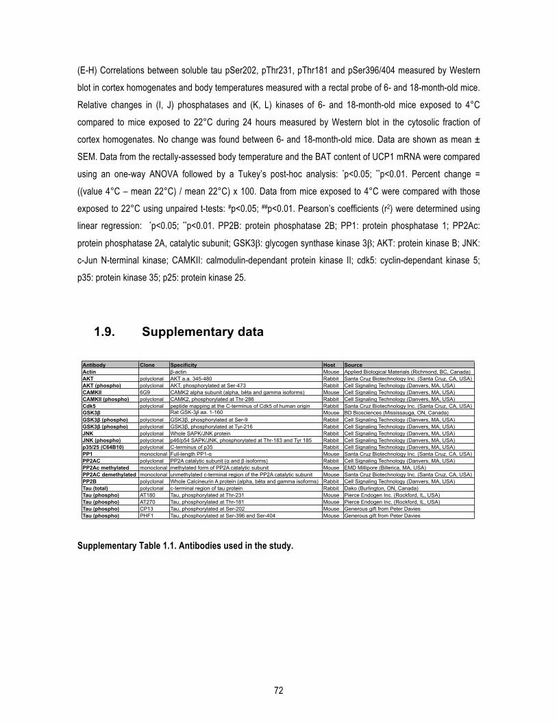
72
(E-H) Correlations between soluble tau pSer202, pThr231, pThr181 and pSer396/404 measured by Western
blot in cortex homogenates and body temperatures measured with a rectal probe of 6- and 18-month-old mice.
Relative changes in (I, J) phosphatases and (K, L) kinases of 6- and 18-month-old mice exposed to 4°C
compared to mice exposed to 22°C during 24 hours measured by Western blot in the cytosolic fraction of
cortex homogenates. No change was found between 6- and 18-month-old mice. Data are shown as mean ±
SEM. Data from the rectally-assessed body temperature and the BAT content of UCP1 mRNA were compared
using an one-way ANOVA followed by a Tukey’s post-hoc analysis: *p<0.05; **p<0.01. Percent change =
((value 4°C – mean 22°C) / mean 22°C) x 100. Data from mice exposed to 4°C were compared with those
exposed to 22°C using unpaired t-tests: #p<0.05; ##p<0.01. Pearson’s coefficients (r2) were determined using
linear regression: *p<0.05; **p<0.01. PP2B: protein phosphatase 2B; PP1: protein phosphatase 1; PP2Ac:
protein phosphatase 2A, catalytic subunit; GSK3β: glycogen synthase kinase 3β; AKT: protein kinase B; JNK:
c-Jun N-terminal kinase; CAMKII: calmodulin-dependant protein kinase II; cdk5: cyclin-dependant kinase 5;
p35: protein kinase 35; p25: protein kinase 25.
1.9. Supplementary data
Supplementary Table 1.1. Antibodies used in the study.
Antibody Clone Specificity Host SourceActin β-actin Mouse Applied Biological Materials (Richmond, BC, Canada)AKT polyclonal AKT a.a. 345-480 Rabbit Santa Cruz Biotechnology Inc. (Santa Cruz, CA, USA)AKT (phospho) polyclonal AKT, phosphorylated at Ser-473 Rabbit Cell Signaling Technology (Danvers, MA, USA)CAMKII 6G9 CAMK2 alpha subunit (alpha, bêta and gamma isoforms) Mouse Cell Signaling Technology (Danvers, MA, USA)CAMKII (phospho) polyclonal CAMK2, phosphorylated at Thr-286 Rabbit Cell Signaling Technology (Danvers, MA, USA)Cdk5 polyclonal peptide mapping at the C-terminus of Cdk5 of human origin Rabbit Santa Cruz Biotechnology Inc. (Santa Cruz, CA, USA)GSK3β Rat GSK-3β aa. 1-160 Mouse BD Biosciences (Mississauga, ON, Canada)GSK3β (phospho) polyclonal GSK3β, phosphorylated at Ser-9 Rabbit Cell Signaling Technology (Danvers, MA, USA)GSK3β (phospho) polyclonal GSK3β, phosphorylated at Tyr-216 Rabbit Cell Signaling Technology (Danvers, MA, USA)JNK polyclonal Whole SAPK/JNK protein Rabbit Cell Signaling Technology (Danvers, MA, USA)JNK (phospho) polyclonal p46/p54 SAPK/JNK, phosphorylated at Thr-183 and Tyr 185 Rabbit Cell Signaling Technology (Danvers, MA, USA)p35/25 (C64B10) polyclonal C-terminus of p35 Rabbit Cell Signaling Technology (Danvers, MA, USA)PP1 monoclonal Full-length PP1-α Mouse Santa Cruz Biotechnology Inc. (Santa Cruz, CA, USA)PP2AC polyclonal PP2A catalytic subunit (α and β isoforms) Rabbit Cell Signaling Technology (Danvers, MA, USA)PP2Ac methylated monoclonal methylated form of PP2A catalytic subunit Mouse EMD Millipore (Billerica, MA, USA)PP2AC demethylated monoclonal unmethylated c-terminal region of the PP2A catalytic subunit Mouse Santa Cruz Biotechnology Inc. (Santa Cruz, CA, USA)PP2B polyclonal Whole Calcineurin A protein (alpha, bêta and gamma isoforms) Rabbit Cell Signaling Technology (Danvers, MA, USA)Tau (total) polyclonal c-terminal region of tau protein Rabbit Dako (Burlington, ON, Canada)Tau (phospho) AT180 Tau, phosphorylated at Thr-231 Mouse Pierce Endogen Inc. (Rockford, IL, USA)Tau (phospho) AT270 Tau, phosphorylated at Thr-181 Mouse Pierce Endogen Inc. (Rockford, IL, USA)Tau (phospho) CP13 Tau, phosphorylated at Ser-202 Mouse Generous gift from Peter DaviesTau (phospho) PHF1 Tau, phosphorylated at Ser-396 and Ser-404 Mouse Generous gift from Peter Davies

73
Chapitre 2 : Les expositions répétées au froid protègent un modèle murin de la maladie d’Alzheimer de la phosphorylation de tau induite par le froid.
74
Repeated cold exposures protect a mouse model of Alzheimer’s disease against cold-induced tau phosphorylation
Marine Tournissac1,2; Philippe Bourassa1,2, Ruben D. Martinez-Cano2, Tra-My Vu2, Sébastien S. Hébert2,3,
Emmanuel Planel2,3, Frédéric Calon1,2*.
1Faculty of Pharmacy, Université Laval, Quebec City, Qc, Canada;
2Neurosciences Axis, CHU de Québec-Université Laval Research Center, Quebec City, Qc, Canada;
3Department of Psychiatry and Neuroscience, Faculty of Medicine, Université Laval, Quebec City, Qc,
Canada.

75
1.1. Résumé
Le pic d’incidence de la maladie d’Alzheimer (MA) survient au même moment qu’un déficit de
thermorégulation à un âge avancé. Suggérant un lien entre les deux, l’hypothermie induit
l’hyperphosphorylation de tau (pTau). La stimulation du tissu adipeux brun (TAB) contrecarre les désordres
métaboliques. Pour vérifier si la stimulation du TAB pouvait interférer avec la neuropathologie Alzheimer, des
3xTg-AD de 15 mois ont reçu des expositions répétées au froid (ERF), consistant à des expositions de 4h à
4°C, 5 fois par semaine pendant 4 semaines, tandis qu’un groupe est resté à 22°C. Les 3xTg-AD acclimatées
au froid présentaient une meilleure thermogenèse et tolérance au glucose. Ces souris étaient complètement
résistantes à pTau induite par une exposition au froid de 24h. L’ERF augmentait les niveaux plasmatiques du
facteur de croissance 21, qui étaient corrélés avec les niveaux de pTau hippocampiques. Ces résultats
suggèrent qu’améliorer la thermogenèse pourrait être bénéfique dans la MA.
1.2. Abstract
Old age is associated with a rise in the incidence of Alzheimer’s disease (AD) but also with thermoregulatory
deficits. Indicative of a link between the two, hypothermia induces tau hyperphosphorylation. The 3xTg-AD
mouse model not only develops tau and amyloid pathologies in the brain, but also metabolic and
thermoregulatory deficits. Brown adipose tissue (BAT) is the main thermogenic driver in mammals and its
stimulation counteracts metabolic deficits in rodents and humans. We thus investigated whether BAT
stimulation impedes AD neuropathology. 15-month-old 3xTg-AD mice were subjected to repeated short cold
exposures (RSCE), consisting of 4-hour sessions of cold exposure (4 °C), five times per week for four weeks,
compared to animals kept at housing temperature. First, we confirmed that 3xTg-AD RSCE-trained mice
exhibited BAT thermogenesis and improved glucose tolerance. RSCE-trained mice were completely resistant
to tau hyperphosphorylation in the hippocampus induced by a 24-hour cold challenge. Finally, RSCE
increased plasmatic levels of fibroblast growth factor 21 (FGF21), a batokine, which correlated with
hippocampal tau phosphorylation. Overall, BAT stimulation through RSCE improved metabolic deficits and
completely blocked cold-induced tau hyperphosphorylation in the 3xTg-AD mouse model of AD
neuropathology. These results suggest that improving thermogenesis could exert a therapeutic effect in AD.

76
1.3. Introduction
Alzheimer’s disease (AD) is a neurodegenerative disease characterized by disrupted cognitive functions and is
diagnosed neuropathologically by the presence of tau-laden neurofibrillary tangles and Aβ-loaded plaques
(Tremblay et al., 2017; 2007). The principal risk factor for developing AD is unquestionably old age (Scheltens
et al., 2016). Therefore, a better understanding of pathological changes triggered by old age could reveal new
clues in AD pathogenesis. Among the changes that come with aging, thermoregulatory deficits are frequently
reported. Indeed, elderly individuals regulate their core temperature less efficiently than younger adults
(Degroot and Kenney, 2007), and many of them have subnormal body temperatures during the day (Collins,
1995; Gomolin et al., 2005), along with decreased basal metabolism (Frisard et al., 2007). Interestingly, the
appearance of defects in maintaining optimal body temperature roughly coincides with a rise in the incidence
of AD in this population.
Animal studies are providing new lines of evidence suggesting that thermoregulatory deficits contribute to AD
pathogenesis. It has been repeatedly shown in the rodent brain that a decrease in body temperature induces
tau hyperphosphorylation (Le Freche et al., 2012; Planel et al., 2009; 2004; Tournissac et al., 2017; Vandal et
al., 2016), one of the main neuropathological markers of the disease (Tremblay et al., 2017; 2007). Our studies
in animal models show that both aging and AD pathology potentiate the rise in tau phosphorylation following
acute exposure to cold (4 °C, 24h) (Tournissac et al., 2017; Vandal et al., 2016). For instance, compared to
non-transgenic mice, the 3xTg-AD mouse model of AD has a lower basal body temperature that worsens with
age and the progression of the neuropathology. Interestingly, cold exposure was shown to aggravate AD
neuropathology, while thermoneutrality (28 °C) improved both AD-related neuropathologies and cognitive
impairment (Tournissac et al., 2017; Vandal et al., 2016).
Thermoregulatory deficits can also be interpreted as part of the general metabolic failure that is believed to
occur in AD. Strong pathogenic links between AD and diabetes have been revealed in numerous reports in
recent years, as type 2 diabetes is now considered a risk factor for developing AD (Baglietto-Vargas et al.,
2016; Chatterjee et al., 2016). Reciprocally, AD patients display lower brain glucose metabolism and impaired
insulin signaling (An et al., 2018; Arnold et al., 2018). In addition, peripheral metabolic deficits have been
observed in animal models expressing AD neuropathology (Vandal et al., 2015b). Interestingly, mouse models
of diabetes and obesity also display thermoregulatory deficits associated with higher tau phosphorylation,
further supporting a close relationship between type 2 diabetes and AD (El-Khoury et al., 2016; Gratuze et al.,
2017a). Not surprisingly, numerous antidiabetic drugs have been tested for AD in preclinical and clinical

77
studies, including insulin, thiazolidinediones, metformin and GLP-1 analogs (Vandal et al., 2014b; Yarchoan
and Arnold, 2014).
One promising strategy for simultaneously correcting thermoregulatory and metabolic deficits is through the
stimulation of brown adipose tissue (BAT) thermogenesis. BAT is the main thermogenic tissue in mammals. It
burns fat and glucose to produce heat that dissipates throughout the body via blood flow (Cannon and
Nedergaard, 2004). Hence, the activation of BAT has been considered as a potential drug target for type 2
diabetes and obesity for a long time (de Souza and Burkey, 2001), with renewed interest following the
demonstration that (i) BAT is functional in adults (Cypess et al., 2009) and (ii) BAT activity can be modulated
through cold exposure or drugs (P. Lee and Greenfield, 2015). In addition, active BAT has the remarkable
ability to improve glucose tolerance and insulin sensitivity (Chondronikola et al., 2014; Stanford et al., 2013).
Since older individuals experience a decrease in BAT activity (Ouellet et al., 2011), they are likely to benefit
the most from BAT stimulation.
BAT is not only a modulator of energy homeostasis, but is also an endocrine organ that secretes metabolic
regulators or adipokines, also called batokines, such as fibroblast growth factor 21 (FGF21), adiponectin, and
interleukin 6 (IL-6) (Villarroya et al., 2017). Interestingly, FGF21 was shown to reverse high-fat diet-induced
cognitive defects in rats (Sa-Nguanmoo et al., 2016b), reduce blood-brain barrier disruptions in db/db mice (Yu
et al., 2018), act as a neuroprotective agent against glutamate-induced excitotoxicity in primary rat brain
neurons (Leng et al., 2015; Z. Wang et al., 2016) and protect against Aβ42 toxicity in vitro (Amiri et al., 2018).
To our knowledge, no study has investigated the stimulation of BAT thermogenesis in the context of AD. Given
that BAT is involved in multiple pathways in AD pathogenesis, we hypothesized that correcting
thermoregulatory impairments through BAT stimulation could exert benefits on metabolic and
neuropathological endpoints in a mouse model of AD. Repeated short cold exposures (RSCE) is a
standardized procedure to stimulate BAT growth and activity that has been shown to increase energy
expenditure and improve glucose tolerance and insulin sensitivity in rodents (Labbé et al., 2016; Ravussin et
al., 2014; T.-Y. Wang et al., 2015) and humans (Blondin et al., 2014; Hanssen et al., 2015b; P. Lee et al.,
2014).
We thus applied RSCE to a mouse model of AD (the 3xTg-AD mouse) that reproduces both metabolic and
thermoregulatory deficits along with the development of tau and amyloid pathologies in the brain (Oddo et al.,
2003b; Vandal et al., 2016; 2015b; 2014b). Our objective was to determine whether stimulating BAT
thermogenesis could be a potential therapeutic strategy in AD.

78
1.4. Methods
1.4.1. Animals and diet
Thirteen-month-old 3xTg-AD (APPswe, PS1M146V, tauP301L) mice produced at our animal facility were used in
equal numbers of males and females in each group. Mice from our colony were fed the same chow (Teklad
2018, Harlan Laboratories, Canada) from breeding to 13 months of age. From 13 to 16 months, mice were fed
a 20% (w/w) of total fat diet to be more representative of a human “Western diet” and to potentiate metabolic
defects previously observed in old 3xTg-AD mice (Vandal et al., 2015b). The diet was manufactured by
Research Diets Inc. (New Brunswick, NJ) and is described in detail in Supplementary table 2.1. We used
purified diet formulations standardized to ensure consistency and eliminate batch-to-batch variations,
containing measured concentrations of macronutrients, vitamins, and minerals. All experiments were
performed in accordance with the Canadian Council on Animal Care and were approved by the Institutional
Committee at the Centre Hospitalier de l’Université Laval (CHUL).
1.4.2. Cold exposures
From 15 to 16 months, animals were housed one per cage to avoid mutual heating. A control group remained
at housing temperature (22 °C) for the duration of the study (Figure 2.1A). The “Acute” group underwent a 24-
hour period of exposure at 4 °C, without adaptation, just before sacrifice to assess thermoregulatory capacity.
A third group (“Repeated”) underwent RSCE consisting of 4-h exposure at 4 °C (from 10 a.m. to 2 p.m.), five
days per week for four weeks. This RSCE protocol sums up to a total of 18 4-h sessions of short exposure to
cold. Then, animals underwent a final 24-hour exposure at 4 °C (from 9 a.m. to 9 a.m., identical to the ‘Acute’
group) at the end of the fourth week (Figure 2.1A) (Labbé et al., 2016; Ravussin et al., 2014; T.-Y. Wang et al.,
2015). To minimize stress variability between the groups, all animals were moved from the housing room to
the room next to the cold chamber. Body temperature was determined using an electronic thermometer
coupled with a rectal probe before and after exposure at the same time of the day. All mice were sacrificed by
intracardiac perfusion at their respective temperatures (4 °C or 22 °C) as described previously (Vandal et al.,
2016). Although it is known that anesthesia affects body temperature (Lenhardt, 2010), all mice were put under
deep anesthesia with ketamine/xylazine i.p. injection (100 mg/kg ketamine, 10 mg/kg xylazine) for ethical
reasons a few minutes before intracardiac perfusion. Interscapular BAT, gastrocnemius muscles and brains
were rapidly dissected and frozen at -80°C until processed.

79
1.4.3. Glucose tolerance test
Glucose tolerance test (GTT) was performed after the fifteenth short cold exposure, or after three weeks of
RSCE, during the few days without short cold exposure (Figure 1A). “Control” and “Acute” groups were pooled
under the “Control” group because at the time of the GTT, “Acute” mice did not yet undergo the acute
exposure to cold (24h, 4 °C) and were still in the same conditions as the “Control” group (22 °C). In addition, it
was not possible to test the mice after the final 24-h acute cold exposure because mice were immediately
sacrificed at 4 °C. Sixteen-month-old 3xTg-AD non-exposed and RSCE mice (after the fifteenth exposure)
were fasted for 6 h. Glucose at 1 g/kg was injected i.p. and blood glucose was measured over a 2-h period
with a glucometer (OneTouch UltraMini; LifeScan, Milpitas, CA) using a blood drop sampled from the
saphenous vein.
1.4.4. Protein extraction
For the parieto-temporal cortex, protein extraction method results in a TBS-soluble intracellular and
extracellular fraction (cytosolic fraction), a detergent-soluble fraction (membrane fraction) and a detergent-
insoluble fraction (resuspended in formic acid) as previously described (St-Amour et al., 2014). For the
hippocampus, frozen samples were directly homogenized in a lysis buffer containing detergents that results in
a detergent-soluble fraction (cytosolic and membrane fraction). The pellets remaining from ultracentrifugation
were resuspended in formic acid (detergent-insoluble fraction). Proteins from the BAT and gastrocnemius
muscles were extracted in the lysis buffer only.
1.4.5. Western immunoblotting
The detailed method for Western immunoblotting is described elsewhere (Vandal et al., 2015b). The list of
primary and secondary antibodies used in our experiments is available in Supplementary Table 2.4.
Homogenates were all run on the same gel for each experiment. Proteins extracted from the hippocampus of
tau KO mice were added in immunoblotting experiments with tau antibodies as a negative control, as
described (Petry et al., 2014).

80
1.4.6. Aβ40 and Aβ42 quantification
For β-Amyloid ELISA, protein extracts from the parieto-temporal cortex were used. Aβ40 and Aβ42 were
measured in TBS-soluble and detergent-insoluble fractions using a human β-Amyloid ELISA (Wako, Osaka,
Japan) according to the manufacturer’s instructions (St-Amour et al., 2014). Plates were read at 450 nm using
a SynergyTM HT multidetection microplate reader (Biotek, Winooski, VT).
1.4.7. Quantitative real-time PCR
The expression levels of uncoupling protein 1 (Ucp1, TaqMan Gene Expression Assays, Mm01244861_m1,
Life Technologies), Ffg21 (TaqMan Gene Expression Assays, Mm00840165_g1, Life Technologies) and β2-
microglobulin (β2mg, Mm00437762_m1, Life Technologies) mRNA were determined in BAT using a reverse
transcription real-time quantitative PCR (RT-qPCR) as previously described (Tournissac et al., 2017). The
delta delta Ct (ddCT) method was used to evaluate differences in relative gene expression between groups
using β2mg as the control gene. Results are presented as ratios of Ucp1/β2mg or Fgf21/β2mg cDNA in
relation to the control group.
1.4.8. FGF21 assay
Levels of FGF21 were determined in plasma sampled before intracardiac perfusion in non-fasted mice and in
BAT protein extracts using the mouse/rat FGF21 Quantikine ELISA kit (MF2100, R&D systems, Minneapolis,
MN).
1.4.9. Triglycerides assay
Levels of triglycerides were measured in plasma sampled before intracardiac perfusion in non-fasted mice
using the Thermo Scientific Triglycerides Reagent assay (TR22421, Thermo Fisher Scientific, Waltham, MA,
USA).
1.4.10. Histology and UCP1 immunostaining
Visceral (perirenal and epididymal) and subcutaneous (inguinal) fat pads were sampled and weighed.
Epididymal fat was post-fixed for 48 h in 4% paraformaldehyde (pH 7.4). Samples were then transferred in

81
PBS until embedding in paraffin. Samples were sectioned in 10-µm-thick slices and mounted. For adipocytes
quantification, slices were stained in hematoxylin and eosin (H&E) as described elsewhere (Vandal et al.,
2014b). Adipocytes area and number were quantified using ImageJ software (version 1.50i, NIH, Bethesda,
MA, USA). For UCP1 immunostaining, slices were first deparaffinized, followed by antigen retrieval with a
boiling 10 mM sodium citrate buffer (pH 6.0) for 30 min and inhibition of endogenous peroxidases with a 3%
hydrogen peroxide in methanol for 30 min. Mounted sections were then washed twice in PBS with 0.4% Triton
X-100 (PBS-T) and 1% horse serum for 10 min before blocking with 5% horse serum in PBS-T solution for 30
min. Slices were incubated overnight at 4 °C in a humid chamber with a rabbit UCP1 antibody (1:200,
Ab10983, Abcam, Cambridge, United Kingdom) in a 1% horse serum PBS-T solution. Sections were then
washed twice in PBS-T for 10 min before a 1-h incubation period with a goat anti-rabbit biotinylated antibody
(1:200, Jackson ImmunoResearch, PA, USA). Mounted sections were then washed twice in PBS-T and an
avidin/horseradish peroxidase complex (ABC Elite Kit; Vector Laboratories, Burlington, ON, Canada) was
added for 35 min following the manufacturer’s guidelines. After two washes, a 0.3 mg/mL 3-amino-9-
ethylcarbazole (AEC) (Sigma-Aldrich, St Louis, MO, USA) and 0.03% hydrogen peroxide in acetate buffer was
added for detection. The reaction was stopped by extensive washings in PBS, and sections were then
coverslipped with Mowiol mounting medium. Images were taken with an EVOS® FL Auto Cell Imaging System
(Thermo Fisher Scientific, Waltham, MA, USA). Optical density was quantified with ImageJ software (NIH,
version 1.50i).
1.4.11. Corticosterone ELISA
Corticosterone levels were measured in plasma sampled from the saphenous vein just before and after
moving to the cold chamber in non-fasted mice following the manufacturer’s instructions (Ab108821, Abcam,
Cambridge, United Kingdom).
1.4.12. Statistical analysis
Data are represented as a mean ± standard error of the mean (SEM), except for Supplementary table 2.2 and
2.3 in which data are presented as mean ± standard deviation (SD). The statistical analysis and the number of
mice per group are specified in each figure. Bartlett’s tests were used to rule out inequality of variances
between groups. One-way (one independent variable) or two-way (two independent variables) ANOVAs were
used when more than two groups were compared. ANOVAs were followed by a Tukey’s post-hoc analysis in
cases of equal variance. In cases of unequal variance, a Kruskal Wallis followed by a Dunn’s post-hoc test
82
was performed. Repeated measures two-way ANOVAs followed by a Bonferroni’s post-test were executed to
compare recurrent measurements in the same animals. An unpaired Student’s t-test was performed when only
two groups were compared. One sample t-test was used to compare means to a theoretical value.
Correlations between variables were investigated using linear regression analyses. All statistical analyses
were performed with Prism 5 (GraphPad software, San Diego, CA, USA) or JMP (version 12.1.0; SAS Institute
Inc., Cary, IL, USA) software and statistical significance was set at p<0.05.
1.5. Results
1.5.1. Repeated short cold exposures potentiate BAT thermogenic capacity and protect old 3xTg-AD mice against cold-induced decreases in body temperature
RSCE is a standardized procedure to stimulate BAT growth and thermogenesis activity (Labbé et al., 2016;
Ravussin et al., 2014; T.-Y. Wang et al., 2015). Corticosterone levels were measured in plasma sampled
before and after the first and the thirteenth RSCE to confirm that stress was equivalent between cold-exposed
and non-exposed mice (Supplementary figure 2.1). We first observed that mice in the RSCE group became
resistant to cold-induced decreases in body temperature from the sixth cold exposure session onward, as
shown by a non-significant change before and after short exposures (Figure 2.1B and Supplementary figure
2.2 for raw data). We confirmed that Ucp1 mRNA expression in BAT was increased by cold exposure, after a
single acute episode (24 hours at 4 °C) or at the end of the RSCE session (Figure 2.1C), while β3 adrenergic
receptors (β3AR) were not affected by RSCE (Figure 2.1E,G). BAT weights were reduced by acute cold
exposure, possibly due to brown adipocyte shrinkage following lipid consumption to produce heat (Figure
2.1D). We also found an increase in SIRT3 in the BAT of repeatedly cold-exposed mice (Figure 2.1F,G), a
protein expressed in mitochondria of BAT after fasting or cold exposure conditions (Ahn et al., 2008; Cheng et
al., 2016; T. Shi et al., 2005), further evidencing that RSCE efficiently stimulated BAT thermogenesis. Since
RSCE may improve whole-body thermogenesis not only through BAT stimulation, we assessed non-shivering
activity in muscles through sarcolipin protein (Supplementary table 2.2) (Bal et al., 2012). However, we did not
observe any significant increase in muscular proteins involved in thermogenesis. Finally, a trend toward a
“browning” phenotype was observed in the white adipose tissue (WAT) of repeatedly cold-exposed mice with
UCP1 staining (Figure 2.1H), although the quantification of the optical density of UCP1 staining was not
statistically different from control mice (Figure 2.1I).
83
1.5.2. Repeated short cold exposures improve glucose tolerance in 16-month-old 3xTg-AD mice
Increased BAT thermogenesis activity leads to improved metabolic outcomes, including glucose tolerance in
rodents (T.-Y. Wang et al., 2015). We took advantage of the fact that 3xTg-AD mice on a Western diet display
metabolic defects with age (Vandal et al., 2015b) to assess the impact of RSCE. All animals gained weight
(approximately 15%) after the introduction of the Western diet and RSCE did not significantly alter the final
body or white adipose tissue weight (Figure 2.2A,B,F). The number and area of white adipocytes were not
significantly modified by the RSCE (Figure 2.2G-I). However, RSCE improved glucose tolerance as RSCE
mice displayed a large decrease in blood glucose 30 minutes post-injection and a smaller area under the
curve, compared to non-exposed 3xTg-AD mice (Figure 2.2C,D), without modification of fasting insulin (Figure
2.2E). Finally, circulating triglycerides levels were halved by both RSCE and acute cold exposure (4 °C, 24h)
(Figure 2.2J).
1.5.3. Repeated short cold exposures protect old 3xTg-AD mice against cold-induced tau phosphorylation
Tau phosphorylation is one of the main neuropathological hallmarks of AD, strongly correlating with
antemortem cognitive deficits (Tremblay et al., 2017; 2007). Cold-induced tau phosphorylation has been
repeatedly observed in non-transgenic and in AD mouse models (Le Freche et al., 2012; Planel et al., 2009;
2004; Tournissac et al., 2017; Vandal et al., 2016). In this study, we first confirmed that acute exposure to cold
(24h, 4 °C) markedly increased the phosphorylation of soluble tau in the hippocampus of old 3xTg-AD mice
(pSer394/404: +87%; pSer202: +211%; pSer202/Thr205: +183%; pThr181: +49% vs. non-exposed mice)
(Figure 2.3A-G). As expected, tau phosphorylation strongly correlated with body temperature recorded at
sacrifice (Figure 2.3H-J). Interestingly, RSCE completely prevented cold-induced tau hyperphosphorylation in
the hippocampus, as the level of tau phosphorylation in RSCE mice was similar to non-exposed mice (Figure
2.3A-G). These results indicate that old 3xTg-AD mice undergoing BAT training with RSCE developed a
sufficient thermogenic capacity to maintain their body temperature over a 24-hour cold challenge and thus
preserved a level of tau phosphorylation similar to non-exposed mice. However, levels of insoluble tau in the
hippocampus remained unchanged between exposed and non-exposed 3xTg-AD mice (Figure 2.3K-O).
We then investigated the impact of RSCE on kinases and phosphatases involved in the phosphorylation of tau
in the hippocampus (Figure 2.4A-I). We found that phospho-GSK3β (Ser9) increased after acute cold
exposure (4 °C, 24h) but remained equivalent to the control level after RSCE, thus following the same pattern
84
as tau phosphorylation (Figure 2.4A). In contrast, phospho-GSK3β (Tyr216) levels remained similar in all
groups (Figure 2.4B). Levels of phospho-JNK and phospho-AKT also increased following acute cold exposure,
but with no significant effect from RSCE (Figure 2.4C,D). No significant changes were found for p25/p35,
pCAMKII, pMAPK and PP2Ac (Figure 2.4A-H).
Amyloid pathology accumulates progressively in the 3xTg-AD mice (Oddo et al., 2003b). Here, RSCE and
acute cold exposure (4 °C, 24h) had no effect on either soluble or insoluble forms of Aβ40 or Aβ42 measured
in the parieto-temporal cortex of the mice (Supplementary figure 2.3).
1.5.4. Repeated short cold exposures lead to FGF21 accumulation in plasma
BAT has been recently presented as a secretory organ (Villarroya et al., 2017), communicating with other
tissues, including the brain, through the secretion of batokines. Here, we focused on FGF21 because recent
studies indicate that this hormone has neuroprotective properties (Amiri et al., 2018; Leng et al., 2015; Sa-
Nguanmoo et al., 2016b). We found that plasmatic levels of FGF21 increased by 66% following RSCE
compared to control (Figure 2.5A). Surprisingly, plasmatic FGF21 concentrations were negatively correlated
with levels of soluble tau pSer231 in the hippocampus of 3xTg-AD mice (Figure 2.5B), suggesting that FGF21
production induced by RSCE could be involved in the observed protection against cold-induced tau
phosphorylation. In addition, plasmatic levels of FGF21 were correlated with body temperature recorded just
before sacrifice (Figure 2.5C). Since BAT can secrete FGF21, we measured its expression in BAT of old 3xTg-
AD mice and found that mice that only underwent the 24-hour cold challenge displayed a non-statistically
significant trend toward higher Fgf21 expression (Figure 2.5D).
1.6. Discussion
Based on previous work suggesting that thermoregulatory deficits contribute to AD pathogenesis (Tournissac
et al., 2017; Vandal et al., 2016), we sought to determine whether stimulation of BAT thermogenesis
counteracts metabolic and thermoregulation deficits as well as AD-like neuropathology in an animal model of
AD. We opted for a non-pharmacological mean to stimulate thermogenesis, namely RSCE, routinely used in
the field of metabolic research, although not specific to BAT (Labbé et al., 2016; Ravussin et al., 2014; T.-Y.
85
Wang et al., 2015). First, we found that RSCE was effective at increasing BAT thermogenic capacity and
improving glucose tolerance in old 3xTg-AD mice. Notably, 3xTg-AD mice trained with RSCE were completely
protected from cold-induced tau hyperphosphorylation, a key correlate of AD symptoms in patients. Finally,
RSCE increased plasmatic FGF21 levels, a batokine involved in both metabolic improvement and
neuroprotection (Sa-Nguanmoo et al., 2016b; Villarroya et al., 2017).
1.6.1. Increased thermogenesis capacity following RSCE in old mice
The present data show that RSCE increased BAT thermogenic capacity, as shown by increased BAT Ucp1
expression and by the capacity of mice to resist cold-induced decreases in body temperature. WAT can also
contribute to thermogenesis, although to a lower magnitude compared to interscapular BAT, by adopting a
“browning” phenotype (Labbé et al., 2016). Here, we observed such a phenotype through the trend to increase
in UCP1 staining in the WAT of repeatedly exposed mice. Since RSCE stimulates whole body thermogenesis
and cold acclimation not only through BAT, we also measured markers of muscular thermogenesis but did not
observe any modification in the mice repeatedly exposed to cold compared to control. RSCE or cold
acclimation in humans have been shown to increase energy expenditure and BAT thermogenic activity
(Blondin et al., 2014; Hanssen et al., 2015b; P. Lee et al., 2014). However, studies in the elderly are lacking,
possibly due to the risks associated with cold exposure in that population. Thermoregulation is impaired with
aging, the BAT is less prominent in the elderly and old mice are more vulnerable to tau hyperphosphorylation
induced by cold challenge compared to younger ones (Cypess et al., 2009; Tournissac et al., 2017). Yet, our
data confirm that RSCE is an appropriate research tool to investigate the effect of BAT stimulation even in old
mice.
It should be noted that animals in our facilities are kept at 22 °C, which is lower than the thermoneutral
temperature of the mouse estimated at 28 °C (Cannon and Nedergaard, 2010). A previous study showed that
thermoneutrality improves behavior and AD pathology in old 3xTg-AD mice after one week (Vandal et al.,
2016). In addition, it is known that, when mice are housed at their thermoneutral temperature, their
thermogenesis and sympathetic tone are halted while their adiposity is increased (Cui et al., 2016). Therefore,
the present results are valid in the context of mice experiencing mild thermogenesis and could be different at
another environmental temperature.
86
1.6.2. RSCE improved peripheral metabolic disorders
Boosting thermogenic capacity could improve AD through different mechanisms. First, improvement in glucose
and lipid metabolism, as well as insulin sensitivity, gained with higher thermogenesis may be therapeutic in
AD. Type 2 diabetes and obesity are now widely listed among risk factors for developing AD, now increasingly
recognized as a metabolic disease, since patients display impaired brain glucose metabolism and insulin
signaling (An et al., 2018; Arnold et al., 2018; Chatterjee et al., 2016). Peripheral metabolic impairments are
also replicated in mouse models of AD neuropathology, which become aggravated following the induction of
experimental diabetes (Gratuze et al., 2017b; Vandal et al., 2014b). RSCE extended over three weeks (15
sessions of 4-hour cold exposure) was sufficient to improve glucose tolerance in 16-month-old 3xTg-AD mice.
Such a result is in agreement with several studies that found that cold acclimation improved glucose
metabolism in rodents and humans (Hanssen et al., 2015b; T.-Y. Wang et al., 2015). Here, glucose intolerance
in the 3xTg-AD mice was likely caused by AD neuropathology generated by the expression of transgenes in
the CNS in combination with the westernized diet (Vandal et al., 2015b; 2014b). This indicates that BAT
stimulation, a procedure with a long history in the field of metabolic diseases (de Souza and Burkey, 2001),
could correct a peripheral metabolic endpoint consequent of central AD neuropathology.
1.6.3. RSCE protected against cold-induced tau phosphorylation
As a second key mechanism, BAT stimulation could exert a central therapeutic effect by preventing body
temperature from dropping below the hypothermic threshold known to trigger tau hyperphosphorylation
(Tournissac et al., 2017; Vandal et al., 2016). Several studies in rodents have shown that tau phosphorylation
is increased following a reduction in body temperature induced by different means, such as external cold
exposure, anesthesia, hibernation or glucose deprivation (Arendt et al., 2003; Le Freche et al., 2012; Planel et
al., 2009; 2004; Tournissac et al., 2017; Vandal et al., 2016). Here, RSCE training before a 24-hour exposure
to 4 °C completely blunted cold-induced tau phosphorylation in old 3xTg-AD mice. This indicates that an
increased thermogenesis capacity confers full protection against this neuropathological insult. To the best of
our knowledge, this is the first time that an intervention is shown to block an in vivo rise in cold-induced tau
phosphorylation in the hippocampus, indicating an important link between thermoregulation and tau
phosphorylation.
Although not all the studies found correlations between tau and cognition in humans, there is strong evidence
that postmortem tau pathology correlates with antemortem memory deficits, and this association is usually
stronger than with Aβ (Arriagada et al., 1992; Bennett et al., 2004; Nelson et al., 2012; Robinson et al., 2011;
Tremblay et al., 2017; 2007). The phospho-epitopes of tau protein assessed here, specifically pSer396/404
87
and pThr231, are important from an AD standpoint, as they correlate with clinical diagnoses and antemortem
cognitive deficits, including episodic memory (Tremblay et al., 2017; 2007). Indeed, we previously observed a
negative correlation between global cognitive score (including episodic memory) and both pThr231/Ser235
and pSer396/404 phosphorylated tau in the same series of clinically characterized volunteers from the
Religious Order study, using extracts from the parietal cortex (Tremblay et al., 2017). These associations
remained significant even after adjustment for total tau, highlighting the importance of soluble tau
phosphorylation at these sites in AD cognitive impairment (Tremblay et al., 2017). In addition, the Ser396/404
epitope also correlates strongly with the spread of neurofibrillary tangles and Braak diagnosis (Tremblay et al.,
2007). In vitro evidence indicates that these phospho-epitopes are also involved in the pathological
aggregation of tau (Martin et al., 2011). Thus, it could be speculated that preventing hyperphosphorylation at
those specific sites could prevent tau protein aggregation and its associated cognitive deficits.
Acute reductions in body temperature by various means, such as anesthesia, has been hypothesized to
induce lasting deleterious rises in tau phosphorylation (Le Freche et al., 2012; Planel et al., 2009).
Hypothermia and more frequent and pronounced fluctuations in body temperature during the day are observed
in the elderly, notably in AD patients (Collins, 1995; Gomolin et al., 2005). We previously showed that old age
potentiates cold-induced tau phosphorylation in mice (Tournissac et al., 2017). It is thus reasonable to think
that tau phosphorylation fluctuates during the day along with body temperature, possibly leading to tau
hyperphosphorylation and aggregation over the long term in older individuals. Here, RSCE over a four-week
period did not increase insoluble tau phosphorylation, even if body temperature decreased during the first
exposures (-0.5 to -1°C). This rather indicates that short exposures to cold do not induce tau aggregation, and
that the stimulation of thermogenesis may counteract the adverse impact of transient reductions in body
temperature.
Tau phosphorylation is regulated by the activity of various kinases and phosphatases. Although we did not find
any change in PP2A levels in the hippocampus of RSCE-exposed mice, PP2A activity has been shown to be
directly inhibited by low temperatures (Planel et al., 2004). Instead, GSK3β pSer9 followed a pattern very
similar to tau phosphorylation, suggesting a possible involvement in the protection induced by RSCE. Such a
view is consistent with the fact that this site of phosphorylation is sensitive to hypothermia (Bretteville et al.,
2012).
88
1.6.4. RSCE increased FGF21 production
Our data suggest that FGF21 could be a third potential therapeutic target linking increased BAT
thermogenesis activity with a potential benefit in AD. The idea that FGF21 is of interest in AD is in agreement
with previous work suggesting that this batokine protects against obesity-induced memory impairment and
Aβ42 toxicity (Amiri et al., 2018; Sa-Nguanmoo et al., 2016b). While the fact that RSCE mice were completely
protected from cold-induced tau phosphorylation is certainly linked to the increased ability of old mice to
maintain their body temperature during a cold challenge, a direct BAT-to-brain crosstalk could be at least partly
involved. Plasmatic levels of FGF21 were inversely correlated with tau phosphorylation in the hippocampus of
3xTg-AD mice, which support such a link but does not prove any causal relationship. Thus, further
investigations on the effects of FGF21 in AD models, for example by directly administering recombinant
FGF21 or drugs targeting the FGF21 receptor, would be of interest.
The present work does not confirm whether BAT training leads to improved cognitive faculties. The RSCE
paradigm used here is not compatible with behavioral testing, as it is not possible to test the mice immediately
after or during cold exposure. Indeed, locomotor activity and exploratory behavior are affected by cold
exposure (Ishii et al., 1996; Jhaveri et al., 2007). Thus, mice will hardly perform as expected in most behavioral
paradigms, introducing insurmountable biases. However, given the previously reported beneficial effects of
RSCE on metabolic disorders, we opted to perform a glucose tolerance test during the few days without short
cold exposure. Nevertheless, the effects observed in the present study following RSCE have been previously
associated with cognitive-enhancing properties. Indeed, increased circulating FGF21 (Sa-Nguanmoo et al.,
2016b) and better glucose homeostasis (Porter et al., 2011) are associated with improved cognitive
performance in diabetic rodents.
1.6.5. Conclusions
Overall, our results indicate for the first time that BAT thermogenesis stimulation through RSCE is efficient in
old mice and results in improved glucose metabolism and protection against cold-induced tau
hyperphosphorylation. Thus, therapeutic strategies aiming to stimulate BAT thermogenesis initially developed
against metabolic disorders could be repurposed for dementia.
89
1.7. Acknowledgements
1.7.1. Author contributions
M.T. developed the experimental design, performed animal experiments, protein extractions, western blots in
the hippocampus, BAT and muscle, Aβ, insulin, and FGF21 ELISAs, qPCR, UCP1 immunostaining, and
triglycerides assays, completed the statistical analysis and wrote the first versions of the manuscript. P.B.
contributed to animal experiments, protein extractions, western blots in the hippocampus and BAT, and Aβ
and corticosterone ELISAs and reviewed the manuscript. R.M. performed western blots in the hippocampus
and contributed to the qPCR experiment and protein extractions. T.V. contributed to the UCP1
immunostaining, adipocytes quantification, and western blots in the muscle. E.P. provided expertise for the tau
analysis and reviewed the manuscript. S.H. provided qPCR equipment and reviewed the manuscript. F.C.
secured funding and contributed to the experimental design and wrote the manuscript. F.C. is the guarantor of
this work and, as such, had full access to all the data in the study and takes responsibility for the integrity of
the data and the accuracy of the data analysis.
1.7.2. Funding
M.T. was funded by a scholarship from the Alzheimer Society of Canada. P.B. received a scholarship from the
Canadian Institutes of Health Research. This study was made possible by funding from the Canadian Institutes
of Health Research (MOP 102532), the Quebec Network for Research on Aging, the Alzheimer Society of
Canada (#1502), and the Canadian Foundation for Innovation (#34480). F.C. and E.P are a Fonds de
Recherche du Québec - Santé (FRQ-S) scholars (#253895, #26936, and #252178).

90
1.8. Figures
Figure 2.1. Repeated short cold exposures protect old 3xTg-AD mice against cold-induced decrease in body temperature by increasing thermogenesis.
A: Study timeline, including RSCE. B: Difference in rectal temperatures of exposed and non-exposed mice
taken before and after each short exposure to cold (4h, 4 °C) between 15 and 16 months of age. The
temperature of the mice was recorded before and after the first, the second, the 4th, the 6th, the 8th, the 13th and
the 18th (last) repeated short cold exposure (RSCE) (4 °C during 4 hours). C: BAT Ucp1 mRNA expression
normalized to β2mg (qPCR). D: BAT weighed just after dissection. E: Levels of β3AR and F: SIRT3 proteins
measured in BAT by Western blot. G: Examples of Western Blots. Homogenates were all run on the same gel,
but consecutive bands were not taken for all representative photo examples. H: Immunohistochemistry of
UCP1 on WAT sections. I: Quantification of UCP1 staining in WAT. Data are represented as mean ± SEM
(n/group indicated in bars). Statistical analyses: One sample t-test versus 0: #p<0.05; ##p<0.01. One-way
ANOVA, Tukey’s post-hoc test: *p<0.05; **p<0.01. Control (C): 22 °C; Acute (A): 24h, 4 °C; Repeated (R): 4h,
4 °C for four weeks + 24h, 4 °C. Abbreviations: β2mg: β2 microglobulin; β3AR: β3 adrenergic receptor; BAT:
brown adipose tissue; eEF2: eukaryotic elongation factor 2; Ucp1: uncoupling protein 1; SIRT3: Sirtuin 3;
WAT: white adipose tissue.

91
Figure 2.2. Repeated short cold exposures improved glucose tolerance in 16-month-old 3xTg-AD mice and decrease plasmatic triglycerides without affecting white adipose depots.
A: Body weights during the dietary treatment and series of repeated short cold exposures. B: Body weight at
the end of the experiment. C,D: Glucose tolerance test performed after the fifteenth short cold exposure
session. E: Levels of insulin measured in plasma sampled after the 6-hour fasting (before GTT). F: Visceral
(epididymal and perirenal fat) and subcutaneous (inguinal) adipose tissue of 16-month-old 3xTg-AD mice
weighed just after dissection. G,H: Number and average surface of adipocytes in epididymal fat. I: Photos of
white adipocytes (H&E stained paraffin sections). J: Levels of triglycerides measured in plasma sampled at the
sacrifice in non-fasted mice. Data are represented as mean ± SEM (n/group indicated in bars). Statistical
analyses: Repeated measures two-way ANOVA, Bonferroni’s post-hoc test: &p<0.05; &&p<0.01. Unpaired
Student t-test: #p<0.05. One-way ANOVA, Tukey’s post-hoc test: **p<0.01; ***p<0.001. Control: 22 °C; Acute:
24h, 4 °C; Repeated: 4h, 4 °C for four weeks + 24h, 4 °C. Note: “Control” and “Acute” groups were pooled
under the “Control” group in A and C because at the time of the GTT and the weighing, “Acute” mice did not
yet undergo the acute exposure to cold (24h, 4 °C) and were still in the same conditions as the “Control” group
(22 °C).

92
Figure 2.3. Repeated short cold exposures protect old 3xTg-AD mice against cold-induced phosphorylation of soluble tau.
A-G: Hippocampal soluble tau phosphorylation of 16-month-old 3xTg-AD mice. H-J: Correlations between
hippocampal soluble tau phosphorylation and body temperature at sacrifice. K-O: Hippocampal insoluble tau
phosphorylation. Homogenates were all run on the same gel, but consecutive bands were not taken for all
representative photo examples. Data are represented as mean ± SEM (n/group indicated in bars). Statistical
analyses: One-way ANOVA, Tukey’s post-hoc test: *p<0.05; **p<0.01. Pearson r correlation: #p<0.05;
##p<0.01. Control (C): 22 °C; Acute (A): 24h, 4 °C; Repeated (R): 4h, 4 °C for four weeks + 24h, 4 °C.
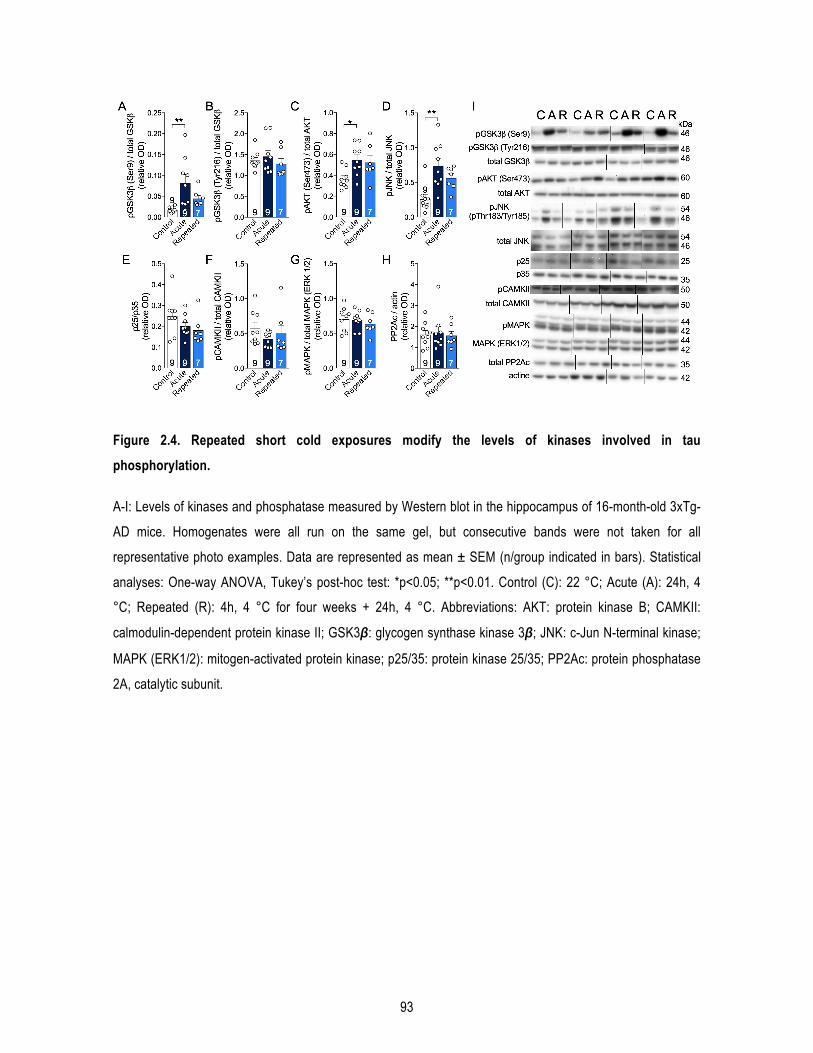
93
Figure 2.4. Repeated short cold exposures modify the levels of kinases involved in tau phosphorylation.
A-I: Levels of kinases and phosphatase measured by Western blot in the hippocampus of 16-month-old 3xTg-
AD mice. Homogenates were all run on the same gel, but consecutive bands were not taken for all
representative photo examples. Data are represented as mean ± SEM (n/group indicated in bars). Statistical
analyses: One-way ANOVA, Tukey’s post-hoc test: *p<0.05; **p<0.01. Control (C): 22 °C; Acute (A): 24h, 4
°C; Repeated (R): 4h, 4 °C for four weeks + 24h, 4 °C. Abbreviations: AKT: protein kinase B; CAMKII:
calmodulin-dependent protein kinase II; GSK3β: glycogen synthase kinase 3β; JNK: c-Jun N-terminal kinase;
MAPK (ERK1/2): mitogen-activated protein kinase; p25/35: protein kinase 25/35; PP2Ac: protein phosphatase
2A, catalytic subunit.
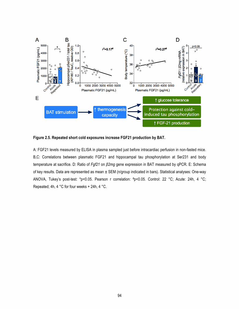
94
Figure 2.5. Repeated short cold exposures increase FGF21 production by BAT.
A: FGF21 levels measured by ELISA in plasma sampled just before intracardiac perfusion in non-fasted mice.
B,C: Correlations between plasmatic FGF21 and hippocampal tau phosphorylation at Ser231 and body
temperature at sacrifice. D: Ratio of Fgf21 on β2mg gene expression in BAT measured by qPCR. E: Schema
of key results. Data are represented as mean ± SEM (n/group indicated in bars). Statistical analyses: One-way
ANOVA, Tukey’s post-test: *p<0.05. Pearson r correlation: #p<0.05. Control: 22 °C; Acute: 24h, 4 °C;
Repeated; 4h, 4 °C for four weeks + 24h, 4 °C.
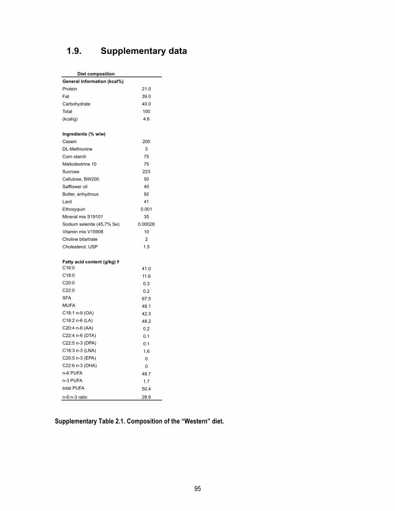
95
1.9. Supplementary data
Supplementary Table 2.1. Composition of the “Western” diet.
Diet composition
General information (kcal%)
Protein 21.0Fat 39.0Carbohydrate 40.0Total 100(kcal/g) 4.6
Ingredients (% w/w)
Casein 200DL-Methionine 3Corn starch 75Maltodextrine 10 75Sucrose 223Cellulose, BW200 50Safflower oil 40Butter, anhydrous 92Lard 41Ethoxyquin 0.001Mineral mix S19101 35Sodium selenite (45,7% Se) 0.00028Vitamin mix V15908 10Choline bitartrate 2Cholesterol, USP 1.5
Fatty acid content (g/kg) Ϯ C16:0 41.0C18:0 11.6C20:0 0.3C22:0 0.2SFA 67.5MUFA 48.1C18:1 n-9 (OA) 42.3C18:2 n-6 (LA) 48.2C20:4 n-6 (AA) 0.2C22:4 n-6 (DTA) 0.1C22:5 n-3 (DPA) 0.1C18:3 n-3 (LNA) 1.6C20:5 n-3 (EPA) 0C22:6 n-3 (DHA) 0n-6 PUFA 48.7n-3 PUFA 1.7total PUFA 50.4
n-6:n-3 ratio 28.6
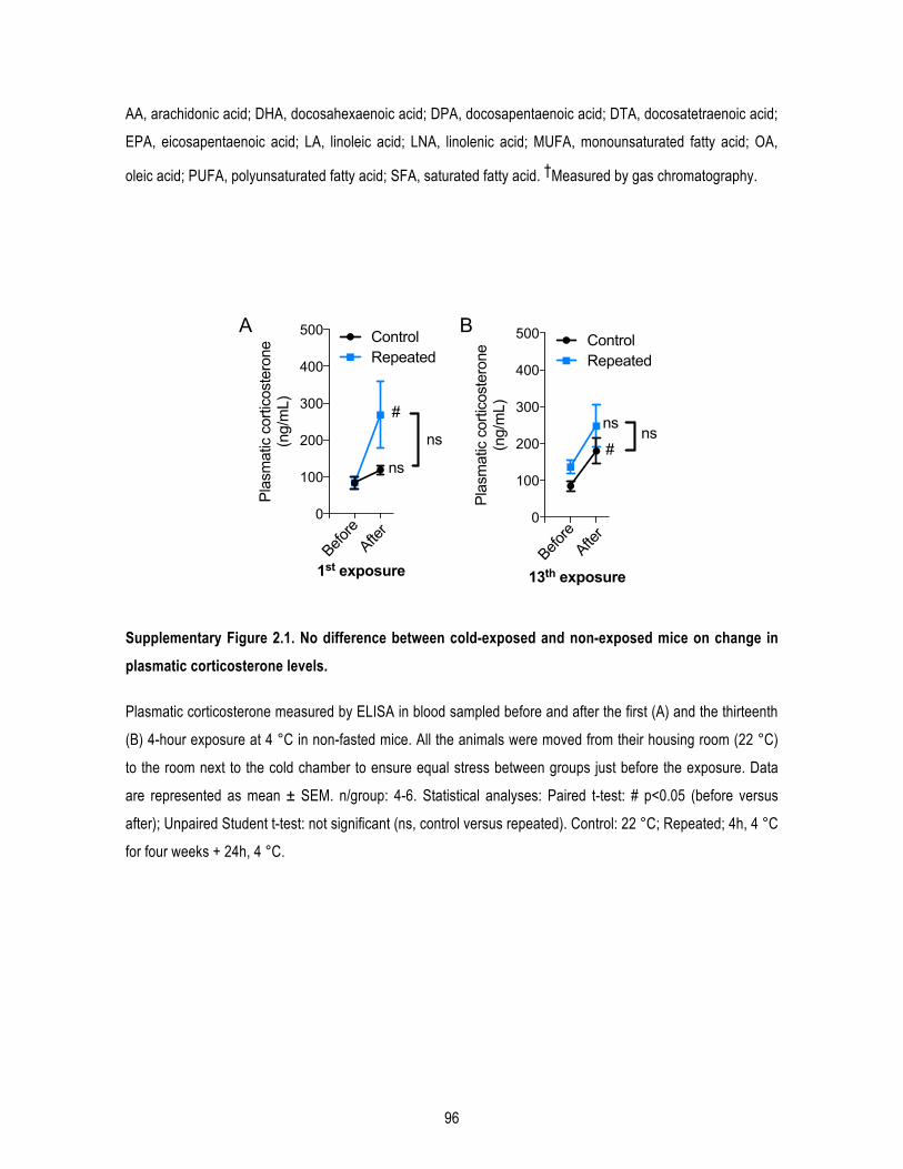
96
AA, arachidonic acid; DHA, docosahexaenoic acid; DPA, docosapentaenoic acid; DTA, docosatetraenoic acid;
EPA, eicosapentaenoic acid; LA, linoleic acid; LNA, linolenic acid; MUFA, monounsaturated fatty acid; OA,
oleic acid; PUFA, polyunsaturated fatty acid; SFA, saturated fatty acid. †Measured by gas chromatography.
Supplementary Figure 2.1. No difference between cold-exposed and non-exposed mice on change in plasmatic corticosterone levels.
Plasmatic corticosterone measured by ELISA in blood sampled before and after the first (A) and the thirteenth
(B) 4-hour exposure at 4 °C in non-fasted mice. All the animals were moved from their housing room (22 °C)
to the room next to the cold chamber to ensure equal stress between groups just before the exposure. Data
are represented as mean ± SEM. n/group: 4-6. Statistical analyses: Paired t-test: # p<0.05 (before versus
after); Unpaired Student t-test: not significant (ns, control versus repeated). Control: 22 °C; Repeated; 4h, 4 °C
for four weeks + 24h, 4 °C.
0
100
200
300
400
500
1st exposure
Pla
smat
ic c
ortic
oste
rone
(ng/
mL)
ControlRepeated
Before
After
#
ns
ns
0
100
200
300
400
500
13th exposure
Pla
smat
ic c
ortic
oste
rone
(ng/
mL)
ControlRepeated
Before
After
#
nsns
A B

97
Control (n=9) Acute (n=9) Repeated (n=8) P-value
Mean ± SD Mean ± SD Mean ± SD
Sarcolipin 48,8 ± 18,2 53,4 ± 16,6 50,1 ± 12,0 ns Serca1 77,9 ± 8,3 79,6 ± 11,0 81,3 ± 10,8 ns Serca2 60,4 ± 13,0 62,4 ± 13,1 58,2 ± 7,6 ns GLUT4 40,0 ± 11,9 50,6 ± 15,8 41,8 ± 16,9 ns Pgc1-α 1,8 ± 1,1 2,2 ± 1,2 1,8 ± 0,8 ns
Supplementary Table 2.2. Quantification of the optical density of proteins detected by Western Blot in gastrocnemius muscles homogenates.
Control: 22 °C; Repeated; 4h, 4 °C for four weeks + 24h, 4 °C.
Supplementary Figure 2.2. Change in body temperature before and after each short cold exposure (raw data of Fig.1B).
A: Description of the timeline for body temperature collection for each short cold exposure (4h, 4 °C). B: Rectal
temperatures of exposed (Repeated) and non-exposed (Control) mice taken before and after short exposures
to cold (4h, 4 °C) at 16 months. Paired t-test: @p<0.05; @@p<0.01.
22°C
A B
22°C 4°C
4 hours
T° before T° after
34
36
38
40
Rec
tal t
empe
ratu
re (°
C)
1st short exposure
@@ BeforeAfter
2nd short exposure
4th short exposure
6th short exposure
8th short exposure
11th short exposure
18th short exposure
@@ @
RepeatedControl
ns ns ns ns
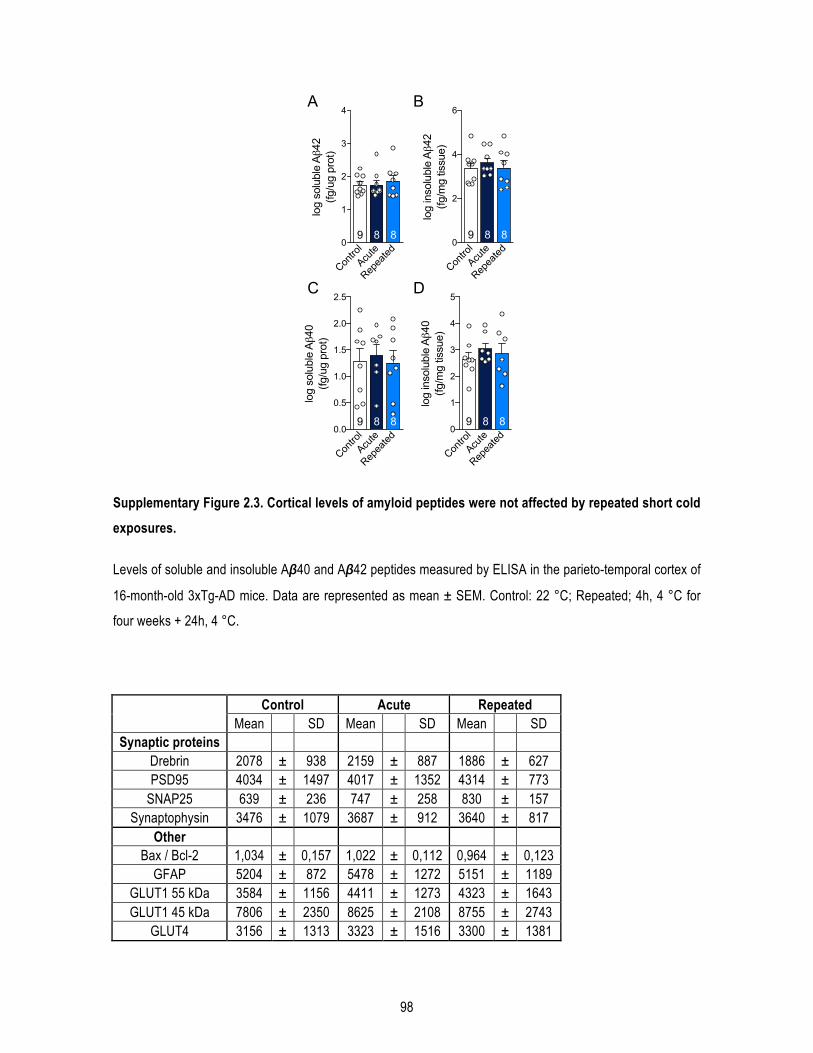
98
Supplementary Figure 2.3. Cortical levels of amyloid peptides were not affected by repeated short cold exposures.
Levels of soluble and insoluble Aβ40 and Aβ42 peptides measured by ELISA in the parieto-temporal cortex of
16-month-old 3xTg-AD mice. Data are represented as mean ± SEM. Control: 22 °C; Repeated; 4h, 4 °C for
four weeks + 24h, 4 °C.
Control Acute Repeated
Mean
SD Mean
SD Mean
SD Synaptic proteins
Drebrin 2078 ± 938 2159 ± 887 1886 ± 627 PSD95 4034 ± 1497 4017 ± 1352 4314 ± 773
SNAP25 639 ± 236 747 ± 258 830 ± 157 Synaptophysin 3476 ± 1079 3687 ± 912 3640 ± 817
Other
Bax / Bcl-2 1,034 ± 0,157 1,022 ± 0,112 0,964 ± 0,123 GFAP 5204 ± 872 5478 ± 1272 5151 ± 1189
GLUT1 55 kDa 3584 ± 1156 4411 ± 1273 4323 ± 1643 GLUT1 45 kDa 7806 ± 2350 8625 ± 2108 8755 ± 2743
GLUT4 3156 ± 1313 3323 ± 1516 3300 ± 1381
Contro
l
Acute
Repea
ted0
2
4
6
log
inso
lubl
e Aβ4
2(fg
/mg
tissu
e)
9 8 8
Contro
l
Acute
Repea
ted0
1
2
3
4
log
solu
ble
Aβ4
2(fg
/ug
prot
)
9 8 8
Contro
l
Acute
Repea
ted0.0
0.5
1.0
1.5
2.0
2.5lo
g so
lubl
e Aβ4
0(fg
/ug
prot
)
9 8 8
Contro
l
Acute
Repea
ted0
1
2
3
4
5
log
inso
lubl
e Aβ4
0(fg
/mg
tissu
e)
9 8 8
A B
C D

99
Supplementary Table 2.3. Synaptic proteins and other markers are not affected by repeated cold exposures.
Data are expressed as relative optical densities. Bax: Bcl-2 associated X protein; GFAP: glial fibrillary acidic
protein; PSD95: postsynaptic density protein 95; SNAP25: synaptosomal-associated protein 25.
Supplementary Table 2.4. Antibodies used in the study.
Antibody Clone Specificity Host Source
Actin monoclonal β-actin Mouse Applied Biological Materials (Richmond, BC, Canada)AKT polyclonal AKT a.a. 345-480 Rabbit Santa Cruz Biotechnology Inc. (Santa Cruz, CA, USA)AKT (phospho) polyclonal AKT, phosphorylated at Ser-473 Rabbit Cell Signaling Technology (Danvers, MA, USA)β3AR polyclonal Mouse beta 3 adrenergic receptor, aa 350 to C-teminus Rabbit Abcam (Cambridge, MA, USA)CAMKII 6G9 CAMK2 alpha subunit (alpha, bêta and gamma isoforms) Mouse Cell Signaling Technology (Danvers, MA, USA)CAMKII (phospho) polyclonal CAMK2, phosphorylated at Thr-286 Rabbit Cell Signaling Technology (Danvers, MA, USA)Cdk5 polyclonal peptide mapping at the C-terminus of Cdk5 of human origin Rabbit Santa Cruz Biotechnology Inc. (Santa Cruz, CA, USA)Drebrin Mx823 c-term peptide (a.a.632-649) Mouse Progen Biotechnik GmbH (Heidelberg, Germany)eEF2 polyclonal total eEF2 protein independant of phosphorylation Rabbit Cell Signaling Technology (Danvers, MA, USA)GFAP GA-5 GFAP Mouse Sigma-Aldrich (St.Louis, MO, USA)GLUT4 polyclonal C-terminus Rabbit EMD Millipore (Billerica, MA, USA)GSK3β monoclonal Rat GSK-3β aa. 1-160 Mouse BD Biosciences (Mississauga, ON, Canada)GSK3β (phospho) polyclonal GSK3β, phosphorylated at Ser-9 Rabbit Cell Signaling Technology (Danvers, MA, USA)GSK3β (phospho) polyclonal GSK3β, phosphorylated at Tyr-216 Rabbit Cell Signaling Technology (Danvers, MA, USA)JNK polyclonal Whole SAPK/JNK protein Rabbit Cell Signaling Technology (Danvers, MA, USA)JNK (phospho) polyclonal p46/p54 SAPK/JNK, phosphorylated at Thr-183 and Tyr 185 Rabbit Cell Signaling Technology (Danvers, MA, USA)ERK polyclonal Whole p44/42 MAPK (Erk1/Erk2) Rabbit Cell Signaling Technology (Danvers, MA, USA)
ERK (phospho) polyclonal MAPK, phosphorylated at Thr-202 and Tyr-204 of Erk1 and Thr-185 and Tyr-187 of Erk2 Rabbit Cell Signaling Technology (Danvers, MA, USA)
p35/25 (C64B10) polyclonal C-terminus of p35 Rabbit Cell Signaling Technology (Danvers, MA, USA)PGC1-α polyclonal aa 777-797 (c-terminus) Rabbit EMD Millipore (Billerica, MA, USA)PP2AC polyclonal PP2A catalytic subunit (α and β isoforms) Rabbit Cell Signaling Technology (Danvers, MA, USA)PSD95 monoclonal aa 77-299 Mouse NeuroMab (Davis, CA, USA)Sarcolipin polyclonal c-terminus of sarcolipin protein Rabbit EMD Millipore (Billerica, MA, USA)Serca1 polyclonal Internal sequence of SERCA1 ATPase Rabbit Abcam (Cambridge, MA, USA)Serca2 polyclonal Residues 1-100 of SERCA2 ATPase Rabbit Abcam (Cambridge, MA, USA)SIRT3 monoclonal total SIRT3 protein Rabbit Cell Signaling Technology (Danvers, MA, USA)SNAP25 SM1 81 Whole Snap25 Mouse Covance, Inc. (Princeton, NJ, USA)Synaptophysin monoclonal Synaptophysin Mouse EMD Millipore (Billerica, MA, USA)Tau (total) TauC polyclonal C-terminal region of tau protein Rabbit Dako (Burlington, ON, Canada)Tau (total) Tau13 Human total tau Rabbit Dako (Burlington, ON, Canada)Tau (normal Tau and PHF-tau) HT7 aa 159-163 Mouse Thermo Scientific (Rockford, IL, USA)
Tau (phospho) AT8 Tau, phosphorylated at Ser-202 and Thr-205 Mouse Pierce Endogen Inc. (Rockford, IL, USA)Tau (phospho) AT180 Tau, phosphorylated at Thr-231 Mouse Pierce Endogen Inc. (Rockford, IL, USA)Tau (phospho) AT270 Tau, phosphorylated at Thr-181 Mouse Pierce Endogen Inc. (Rockford, IL, USA)Tau (phospho) CP13 Tau, phosphorylated at Ser-202 Mouse Generous gift from Peter DaviesTau (phospho) AD2 Tau, phosphorylated at Ser-396 and Ser-404 Mouse BioRad (Hercules, CA, USA)
Goat anti-mouse Peroxidase-conjugated AffiniPure Goat anti-mouse IgG (H+L) Goat Jackson ImmunoResearch (West Grove, PA, USA)Goat anti-rabbit Peroxidase-conjugated AffiniPure Goat anti-rabbit IgG (H+L) Goat Jackson ImmunoResearch (West Grove, PA, USA)
Primary antibodies
Secondary antibodies
100
Chapitre 3 : Un agoniste des récepteurs β3 adrénergiques renverse les déficits mnésiques et réduit la pathologie amyloïde dans un modèle murin de la maladie d’Alzheimer.

101
A beta3-adrenergic receptor agonist reverses memory deficits and reduces insoluble Aβ42/Aβ40 ratio in a mouse model of Alzheimer’s disease
Marine Tournissac1,2 ; Nika Vrabic2; Tra-My Vu1,2; Clara Hozer3; Cyntia Tremblay2, Emmanuel Planel2,4, Fabien
Pifferi3, Frédéric Calon1,2*.
1Faculté de pharmacie, Université Laval, Québec, Qc, Canada;
2Axe Neurosciences, Centre de recherche du CHU de Québec-Université Laval, Québec, Qc, Canada;
3UMR 7179 Centre National de la Recherche Scientifique, Muséum National d'Histoire Naturelle, Brunoy,
France.
4Département de psychiatrie et neurosciences, Faculté de médecine, Université Laval, Québec, Qc, Canada.

102
1.1. Résumé
L’âge avancé, principal facteur de risque de la maladie d’Alzheimer (MA), est associé à des déficits de
thermorégulation. L'acclimatation au froid protège les souris 3xTg-AD de l'hyperphosphorylation de tau induite
par l’hypothermie. Ainsi, la stimulation du tissu adipeux brun (TAB) par un agoniste des récepteurs β3
adrénergiques (Rβ3A) pourrait être bénéfique pour la MA. Des souris 3xTg-AD et non-transgéniques âgées
ont reçu le CL-316,243 ou du salin pendant un mois. Le CL-316,243 a diminué le poids, amélioré le
métabolisme du glucose périphérique et a augmenté la thermogenèse du TAB chez les souris de 16 mois. Le
traitement a augmenté de 19% l'index de reconnaissance des 3xTg-AD par rapport au score avant le
traitement. Enfin, le ratio d’Aβ42/Aβ40 insolubles a été réduit de 27% dans l'hippocampe des 3xTg-AD ayant
reçu le CL-316,243. Les agonistes des Rβ3A, dont l’un est utilisé en clinique, pourraient représenter de
nouveaux outils thérapeutiques pour la MA.
1.2. Abstract
Old age, the most important risk factor for Alzheimer’s disease (AD), is associated with thermoregulatory
deficits. Brown adipose tissue (BAT) is the main thermogenic driver in mammals and its stimulation, through
β3-adrenergic receptor (β3AR) agonists or cold acclimation, counteracts metabolic deficits in rodents and
humans. Studies in animal models show that AD neuropathology leads to thermoregulatory deficits and cold-
induced tau hyperphosphorylation is prevented by cold acclimation. Since metabolic disorders and AD share
strong pathogenic links, we hypothesized that BAT stimulation through β3AR agonist could exert benefits in
AD as well. Non-transgenic and 3xTg-AD mice of 15 months received a β3AR agonist (CL-316,243, 1
mg/kg/day i.p.) or saline for a month. We first confirmed that β3AR agonist administration decreases body
weight and improved peripheral glucose metabolism in both non-transgenic and 3xTg-AD mice. β3AR
stimulation increased BAT thermogenesis as shown by higher uncoupling protein 1 and mitochondrial
oxidative phosphorylation complex levels in BAT. Novel object recognition was performed before (14 months)
and after (16 months) the treatment. One-month treatment with a β3AR agonist increased recognition index
(RI) by 19% in 3xTg-AD mice compared to pre-treatment. Locomotion, anxiety and tau pathology were not
modified. Finally, insoluble Aβ42/Aβ40 ratio was decreased by 27% in the hippocampus of CL-316,243-
injected 3xTg-AD mice. Overall, our results indicate that β3AR stimulation reverses memory deficits and shifts

103
downward the insoluble Aβ42/Aβ40 ratio in 16-month-old 3xTg-AD mice. β3AR agonists, of which one is
currently used in clinic, could represent a valuable therapeutic strategy in AD.
1.3. Introduction
Old age is the main risk factor of Alzheimer’s disease (AD), a neurodegenerative disorder clinically expressed
by memory deficits and cognitive dysfunction (Querfurth and LaFerla, 2010; Scheltens et al., 2016). The
prevalence of AD is growing fast along with the aging population (Winblad et al., 2016). Yet, the exact
pathogenic causes of the sporadic form of the disease are unknown. Despite decades of intense research and
clinical trials, there is still no curative treatment for AD. Since AD is a complex and multifactorial disease, with
frequent age-related comorbidities, multi-target agents might be advantageous over a single-bullet approach.
The undeniable impact of old age on AD incidence indicates that aging triggers etiopathological factors of AD;
identifying these key factors could provide invaluable clues to the development of novel therapeutic
treatments.
Thermoregulation deficits are another documented consequence of old age. Although few studies investigated
thermoregulation in AD individuals, it is well known that thermoregulatory deficits appear in the elderly, the
population primarily affected by AD (Degroot and Kenney, 2007; Gomolin et al., 2005; Grassi et al., 2003;
Hebert et al., 2013). Mounting evidence now supports the hypothesis that thermoregulation deficits contribute
to the development of AD pathology. Spontaneous thermoregulation deficits occur in AD mouse models,
including the 3xTg-AD mice (Huitrón-Reséndiz et al., 2002; Knight et al., 2013; Sterniczuk et al., 2010; Vandal
et al., 2016). Studies in mouse and hibernators repeatedly showed that decreased body temperature leads to
increased tau phosphorylation (Arendt et al., 2003; Planel et al., 2007; Stieler et al., 2011). Accordingly,
manipulation of external temperature leads to strong modulation of AD neuropathology in mice: cold exposure
increases tau phosphorylation and amyloid pathology and decreases synaptic proteins, while higher
temperature reverses memory and anxiety-like behavior and reduced amyloid β (Aβ) 42 peptides in 3xTg-AD
mice (Vandal et al., 2016). More recently, our group provided evidence that enhancement of thermogenesis
through cold acclimation improves metabolic disorders and protects old 3xTg-AD mice from cold-induced tau
phosphorylation (Tournissac et al., 2019). Supporting the link with age, we showed that cold-induced tau
phosphorylation is potentiated in old mice compared to young mice (Tournissac et al., 2017). Altogether, these
observations suggest that thermoregulatory mechanisms could be a potential therapeutic target in AD.
104
Beside thermoregulation, metabolic diseases share strong pathogenic links with AD and have been raising
interest in the field of AD. Indeed, induction of a diabetic phenotype in mice model of AD, such as glucose
intolerance, has been repeatedly shown to increase AD neuropathology (Gratuze et al., 2017b; Julien et al.,
2010; Theriault et al., 2016; Vandal et al., 2014b). Central insulin signaling defects and lower brain glucose
metabolism are observed in AD (An et al., 2018; Arnold et al., 2018). It is estimated that one out of ten cases
of AD is attributable to type 2 diabetes (T2D) (Biessels et al., 2006). These observations logically led to the
idea of repurposing T2D drugs in AD (Yarchoan and Arnold, 2014). Insulin, thiazolidinediones and glucagon-
like peptide-1 (GLP-1) analog are still the subject of clinical trials in dementia, with mitigated results (Craft et
al., 2012; Gejl et al., 2016; J. Liu et al., 2015). Thus, common metabolic targets between both diseases such
as thermoregulatory defects are of interest to develop new therapeutic tools.
Brown adipose tissue (BAT) is the main thermogenic driver in mammals (Cannon and Nedergaard, 2004). The
discovery of functional BAT in adults in 2009 has incredibly revived researches in this tissue (Cypess et al.,
2009; Virtanen et al., 2009). The ability of BAT thermogenesis to improve main metabolic disorders is now
well-established, even in old mice (Hanssen et al., 2015b; Ravussin et al., 2014; Schrauwen and van Marken
Lichtenbelt, 2016; Tournissac et al., 2019). Pharmacological tools have been developed in this direction. In
particular, β3-adrenergic receptors (β3AR) agonists are extensively used since β3AR are mainly present in
brown adipocytes and their stimulation leads to lipolysis and UCP1 expression, the main marker of non-
shivering thermogenesis (Arch, 2011; Nedergaard et al., 2001). CL-316,243 is a highly specific β3AR agonist
frequently used in metabolic diseases studies in rodents. It has been shown to improve blood glucose
metabolism, insulin sensitivity, energy expenditure and to regulate lipids metabolism (Burkey et al., 2000; de
Souza et al., 1997; Kim et al., 2006; A. Kumar et al., 2015; Labbé et al., 2016). Since β3AR agonists can
correct metabolic disorders by enhancing BAT activity, they could tackle both T2D and AD at the same time.
Importantly, β3AR agonists are able to stimulate BAT activity in humans (Baskin et al., 2018; Cypess et al.,
2015), and one (Mirabegron®) is presently in clinical use for overactive bladder (Chapple et al., 2014a).
Therefore, β3AR agonists could rapidly be tested in human for dementia.
We hypothesized that pharmacological stimulation of BAT thermogenesis through β3AR agonist treatment
could stem AD neuropathology and behavior through improved thermoregulatory and metabolic deficits. To
verify whether β3AR agonists could be novel therapeutic tools in AD, 15-month-old non-transgenic and 3xTg-
AD mice received daily CL-316,243 (1 mg/kg) or saline injections for a month.
105
1.4. Methods
1.4.1. Animals
The triple-transgenic mouse model of AD (homozygous 3xTg-AD; APPswe, PS1M146V, tauP301L) developing both
amyloid and tau pathology in the brain while aging was used here (Oddo et al., 2003b). We selected 15-
month-old 3xTg-AD and non-transgenic (NonTg) littermates, at an age when 3xTg-AD mice have extended
plaques and tangles in the brain, as well as cognitive deficits (Belfiore et al., 2018; Bories et al., 2012; St-
Amour et al., 2014; Vandal et al., 2015b). Animals were all on the same genetic background (C57BL6/129SvJ)
and were produced at our animal facility. 42 mice were used for all experiments (n= 9-12 mice per group), and
9 mice were added for behavioral and glucose tolerance tests for a total of 51 mice (n= 9-16 mice per group).
Mice were housed one to five per cage at a housing temperature of 23°C, with a 12:12 hours light-dark cycle
(light phase from 7 a.m. to 7 p.m). Animals had ad libitum access to water and chow (Teklad 2018, Harlan
Laboratories, Canada). Only males were used here to avoid temperature variation induced by the estrous
cycle of female mice (Weinert et al., 2004). Food consumption was evaluated by weighing the diet of each
cage and averaged for each mouse per day per cage every four days during the one-month treatment, and
three weeks before the beginning of the experiment. At the end of the experiment, all mice were put under
deep anesthesia with ketamine/xylazine intraperitoneal (i.p.) injection (100 mg/kg ketamine, 10 mg/kg xylazine)
and immediately placed under a heating pad to maintain body temperature until complete loss of posterior paw
reflex. Then, mice were rapidly sacrificed by intracardiac perfusion with 0.1M phosphate buffer saline (PBS)
solution containing phosphatases (sodium pyrophosphate, 1 mM and sodium fluoride, 50 mM) and proteases
(Sigmafast protease inhibitor tablets, Sigma-Aldrich, St-Louis, USA) inhibitors. All experiments were performed
in accordance with the Canadian Council on Animal Care and were approved by the Institutional Committee of
the Centre Hospitalier de l’Université Laval (CHUL).
1.4.2. CL-316,243 treatment
CL-316,243 was selected to stimulate BAT thermogenesis because it is one of the most selective β3AR
agonists in rodents (β1: β2: β3 = 0:1:100,000) and its safety and efficacy has been confirmed in multiple
studies (J. D. Bloom et al., 1992; Caron et al., 2017; Danysz et al., 2018; Ghorbani et al., 2012; Yoshida et al.,
1994). Two to three weeks of daily injection at a dose of 1 mg/kg per day are necessary to improve metabolic
disorders (Burkey et al., 2000; de Souza et al., 1997; Kim et al., 2006; A. Kumar et al., 2015; Labbé et al.,
2016). Thus, mice were injected i.p. every day for a month with a weight-adjusted bolus of CL-316,243 (1
106
mg/kg) or an equivalent volume of saline (the vehicle) at the same hour of the day (4 p.m.) from 15 to 16
months of age (Fig. 3.1A). Mice were weighed every day before each i.p. injection.
1.4.3. Body temperature measurement and analysis
Telemetric probes (Anipill, Caen, France) were used to record body temperature of the animals every hour
during the one-month experiment without manipulation. Probes were implanted in the intraperitoneal cavity
under isoflurane anesthesia a week before the beginning of the treatment to allow recovery from the surgery
(Fig. 3.1A). Heat pads were used throughout the procedure to avoid hypothermia. The time under anesthesia
was similar between mice and lasted approximately 10 minutes. Then, the animals were kept under heat pads
during the waking period. Body temperature was analyzed during the two first weeks of treatment, before
animals underwent glucose tolerance and behavioral tests, to avoid resulting interference in circadian rhythms.
In order to visualize endogenous rhythmicity and regularity of the mice circadian clock, Clocklab software
(Actimetrics Inc., Evanston, Illinois, USA) provided following parameters for each individual: mean body
temperature during the dark phase (from 7 p.m. to 7 a.m.) and the light phase (from 7 a.m. to 7 p.m.), mean
temperature amplitude during the dark and light phase and during a complete day.
1.4.4. Glucose tolerance test
Glucose tolerance test (GTT) was performed at the end of the third week of treatment (Fig. 3.1A) (Vandal et
al., 2015b). Mice were fasted for 6 hours (from 8 a.m. to 2 p.m.). Then, glucose was injected i.p. at 1 g/kg and
blood glucose was measured regularly during 2 hours with a glucometer (OneTouch UltraMini; LifeScan,
Milpitas, CA) in a blood drop sampled from the saphenous vein.
1.4.5. Behavioral tests
Behavioral tests were performed during the fourth week of treatment with a recovery time of at least 24 hours
between tests (Fig. 3.1A). The novel object recognition (NOR) test was also performed three weeks before the
beginning of the treatment (baseline). Mice were acclimated overnight to the testing room located next to the
housing room. Locomotor activity was assessed with the open field test (Dal-Pan et al., 2016). Mice were
placed in a 40 cm x 40 cm x 40 cm translucent Plexiglas box for an hour. Movements were tracked with
photobeam breaks (San Diego Instruments). The total distance traveled (voluntary horizontal movement) and
the average speed were compared between groups. Anxiety behavior was evaluated with the dark-light box
107
test (Vandal et al., 2016). Mice were put in the center of the dark compartment with an opening to the light
compartment. The time spent in the light compartment and the latency to do the first exploration (nose latency)
of the light compartment were measured during a 5-minute trial. Memory deficits were evaluated with the NOR
test. That test detects behavioral deficits from 12 months in 3xTg-AD mice and is one of the less stressful
behavioral test (Arsenault et al., 2011; Clinton et al., 2007; St-Amour et al., 2014; Vandal et al., 2016). It
evaluates recognition memory and corresponds to episodic memory that is early affected in AD (Antunes and
Biala, 2012; Leger et al., 2013; Wolf et al., 2016). Mice were first placed in a 29.2 cm x 19 cm x 12.7 cm cage
with two identical objects for 5 minutes during the acquisition phase. After an hour in their housing cage, mice
returned in the testing cage containing a familiar and a novel object for the test phase. Recognition index (RI)
corresponds to the time spent exploring the novel object divided by the total time of exploration during the test
phase multiplied by 100. A 50% RI corresponds to an equal exploration between the novel and the familiar
object. Mice exploring less than 6 seconds each object during the acquisition phase or less than 4 seconds
during the test phase were excluded from the RI analysis. Mice were assigned to the treated or control group
at 15 months of age with caution to homogenize memory performance (baseline RI) between groups at the
beginning of the experiment.
1.4.6. Tissue preparation for postmortem analysis
Intracardiac blood sampled just before intracardiac perfusion was centrifuged at 3000 rpm for 5 minutes, and
resulting plasma kept frozen at -80°C until analysis. The first hemisphere of all the mice and interscapular BAT
were rapidly dissected and frozen at -80°C until processing. The second hemisphere was either fixed in 4%
paraformaldehyde for 48 hours, and then transferred in a 20% sucrose solution until sectioning (3-4 mice per
group) or frozen and kept at -80°C.
1.4.7. Protein extractions
For the hippocampus, frozen samples were homogenized in 8 volumes of a lysis buffer (150 mM NaCl, 10 mM
NaH2PO4, 0.5% sodium deoxycholate, 0.5% sodium dodecyl sulfate, 1% Triton X-100) containing a cocktail of
protease and phosphatase inhibitors (Bimake, Houston, TX), sonicated (3 x 45 s in a Sonic Dismembrator
apparatus, Thermo Fisher Scientific, Waltham, MA) and centrifuged (100,000g, 20 min, 4°C), resulting in a
detergent-soluble fraction (cytosolic, extracellular and membrane-bound proteins). The remaining pellets from
ultracentrifugation were resuspended in formic acid, resulting in a detergent-insoluble fraction (insoluble
proteins fraction). The resultant suspension was sonicated and centrifuged (13,000g, 20 min, 4°C), acid formic

108
was evaporated and proteins were either solubilized in Laemmli’s buffer for Western blot or in a 5 M guanidium
solution in Tris-HCl 50mM for Aβ peptides ELISAs as previously described (Tremblay et al., 2007). Proteins
from the BAT were extracted in the lysis buffer only. Protein concentrations were evaluated with a
bicinchoninic acid assay (BCA, Pierce, Rockford, IL, USA).
1.4.8. Western immunoblotting
15 µg and 10 µg of proteins of hippocampus and BAT homogenates, respectively, were loaded and separated
by SDS-PAGE as previously described (Vandal et al., 2014b). The list of antibodies used in this study is
available in Table S2. Homogenates were all run on the same gel for each experiment.
1.4.9. Aβ40 and Aβ42 peptides quantification
Aβ peptides were quantified in protein extracts from the hippocampus. Aβ40 and Aβ42 were measured in
detergent-soluble and detergent-insoluble fractions using a human β-amyloid ELISA (Wako, Osaka, Japan)
according to the manufacturer’s instructions. Plates were read at 450 nm using a SynergyTM HT multi-detection
microplate reader (Biotek, Winooski, VT).
1.4.10. Immunohistochemistry
Amyloid plaques load was performed with MOAB-2 antibody. Briefly, hemispheres were post-fixed in 4%
paraformaldehyde for 48 hours and then transferred into a 20% sucrose solution. Hemispheres were cut in 25-
µm sections and stained as previously described (St-Amour et al., 2014).
1.4.11. Statistical analysis
Data are represented as means ± standard error of the mean (SEM). Statistical analysis and the number of
samples per group are specified in each figure and legend. Bartlett’s tests were used to rule out the inequality
of variances between the groups. Two-way ANOVA (two independent variables: genotype and treatment) was
used when all groups were compared in case of equal variances. In case of unequal variances, a Kruskal-
Wallis followed by a Dunn’s post-hoc test was performed. Repeated measures two-way ANOVA was executed
to compare recurrent measurements in same animals. ANOVAs were followed by Tukey’s post-hoc analysis.
109
An unpaired Student’s t-test was performed when only two groups were compared with independent variables,
with a Welch correction in case of unequal variances, and a paired t-test was executed for the comparison of
two dependent variables. One sample t-test was used to compare means to a theoretical value (for the NOR
test). Correlations between variables were investigated using linear regression analyses. All statistical
analyses were performed with Prism 7 (GraphPad Software Inc., San Diego, CA, USA) or JMP (version 13.2.0;
SAS Institute Inc., Cary, IL, USA) software and statistical significance was set at p<0.05.
1.5. Results
1.5.1. β3AR stimulation improves peripheral glucose metabolism in old mice.
NonTg and 3xTg-AD mice received saline or CL-316,243 i.p. at a dose of 1 mg/kg every day for a month from
15 to 16 months of age (Fig. 3.1A). To verify whether CL-316,243 affects energy balance, mice were weighed
every day before each i.p. injection and food consumption was evaluated three weeks before the beginning of
the experiment and then every four days during the one-month treatment. First, we found that CL-316,243
injections induced persisting weight loss in both NonTg and 3xTg-AD mice (Fig. 3.1B,C). At the beginning of
the experiment, food consumption recorded in the preceding 21 days was higher in 3xTg-AD compared to
NonTg mice (average of 4.7 g/day/mice for 3xTg-AD versus 3.9 g/day/mice for NonTg mice; unpaired t-test
with Welch’s correction: p=0.0105) (Fig. 3.1D). Over the one-month period, β3AR stimulation increased food
consumption in NonTg mice up to levels of transgenic mice (Fig. 3.1D,E).
Previous studies showed improved glucose metabolism with CL-316,243 administration even in mice
displaying a normal response to glucose (Xiao et al., 2015). Here, the GTT revealed that treated mice
displayed a stronger control over glucose levels compared to control, independently of the genotype (Fig.
3.1F,G). Fasting blood glucose was also lower following three weeks of CL-316,243 administration (Fig. 3.1H).
However, levels of plasmatic triglycerides were unchanged (Fig. 3.1I). Overall, our data indicate that β3AR
stimulation led to an improved pattern of metabolic determinants in the periphery in both NonTg and 3xTg-AD
mice.
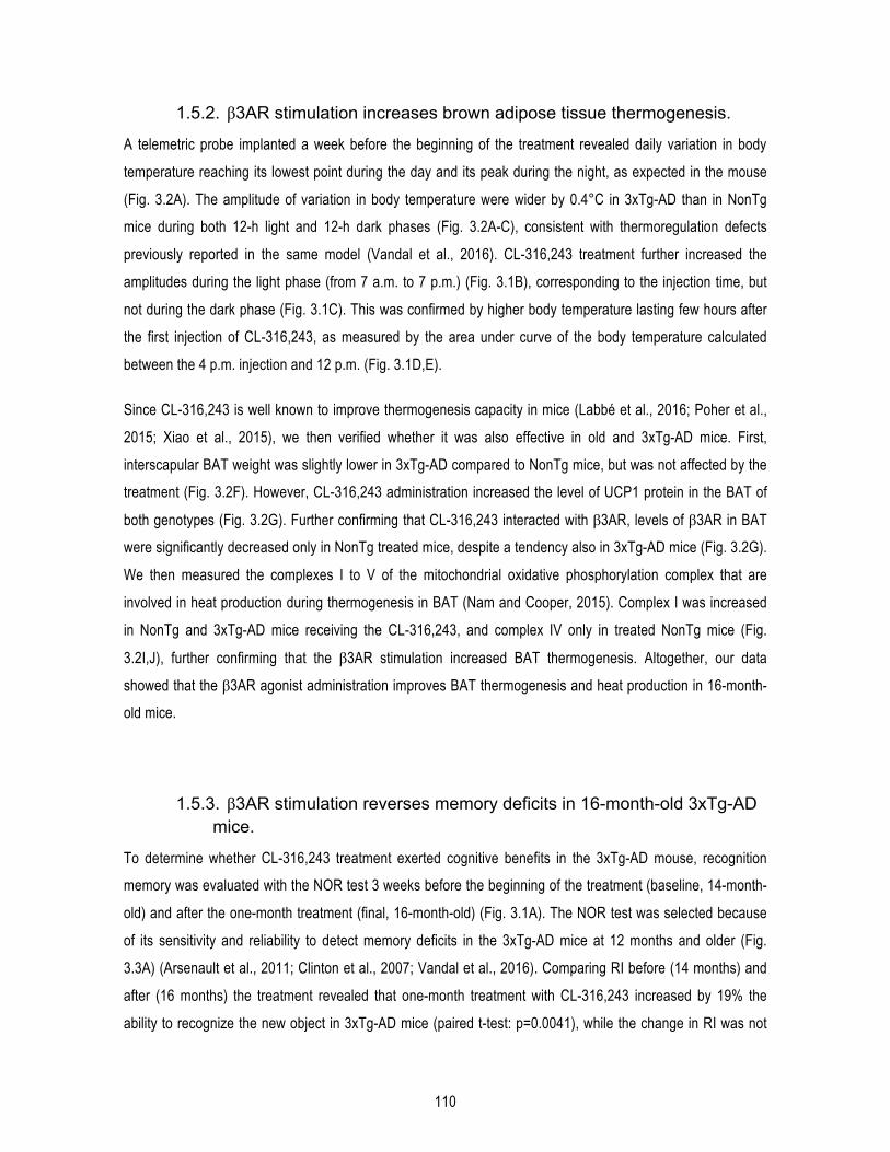
110
1.5.2. β3AR stimulation increases brown adipose tissue thermogenesis.
A telemetric probe implanted a week before the beginning of the treatment revealed daily variation in body
temperature reaching its lowest point during the day and its peak during the night, as expected in the mouse
(Fig. 3.2A). The amplitude of variation in body temperature were wider by 0.4°C in 3xTg-AD than in NonTg
mice during both 12-h light and 12-h dark phases (Fig. 3.2A-C), consistent with thermoregulation defects
previously reported in the same model (Vandal et al., 2016). CL-316,243 treatment further increased the
amplitudes during the light phase (from 7 a.m. to 7 p.m.) (Fig. 3.1B), corresponding to the injection time, but
not during the dark phase (Fig. 3.1C). This was confirmed by higher body temperature lasting few hours after
the first injection of CL-316,243, as measured by the area under curve of the body temperature calculated
between the 4 p.m. injection and 12 p.m. (Fig. 3.1D,E).
Since CL-316,243 is well known to improve thermogenesis capacity in mice (Labbé et al., 2016; Poher et al.,
2015; Xiao et al., 2015), we then verified whether it was also effective in old and 3xTg-AD mice. First,
interscapular BAT weight was slightly lower in 3xTg-AD compared to NonTg mice, but was not affected by the
treatment (Fig. 3.2F). However, CL-316,243 administration increased the level of UCP1 protein in the BAT of
both genotypes (Fig. 3.2G). Further confirming that CL-316,243 interacted with β3AR, levels of β3AR in BAT
were significantly decreased only in NonTg treated mice, despite a tendency also in 3xTg-AD mice (Fig. 3.2G).
We then measured the complexes I to V of the mitochondrial oxidative phosphorylation complex that are
involved in heat production during thermogenesis in BAT (Nam and Cooper, 2015). Complex I was increased
in NonTg and 3xTg-AD mice receiving the CL-316,243, and complex IV only in treated NonTg mice (Fig.
3.2I,J), further confirming that the β3AR stimulation increased BAT thermogenesis. Altogether, our data
showed that the β3AR agonist administration improves BAT thermogenesis and heat production in 16-month-
old mice.
1.5.3. β3AR stimulation reverses memory deficits in 16-month-old 3xTg-AD mice.
To determine whether CL-316,243 treatment exerted cognitive benefits in the 3xTg-AD mouse, recognition
memory was evaluated with the NOR test 3 weeks before the beginning of the treatment (baseline, 14-month-
old) and after the one-month treatment (final, 16-month-old) (Fig. 3.1A). The NOR test was selected because
of its sensitivity and reliability to detect memory deficits in the 3xTg-AD mice at 12 months and older (Fig.
3.3A) (Arsenault et al., 2011; Clinton et al., 2007; Vandal et al., 2016). Comparing RI before (14 months) and
after (16 months) the treatment revealed that one-month treatment with CL-316,243 increased by 19% the
ability to recognize the new object in 3xTg-AD mice (paired t-test: p=0.0041), while the change in RI was not
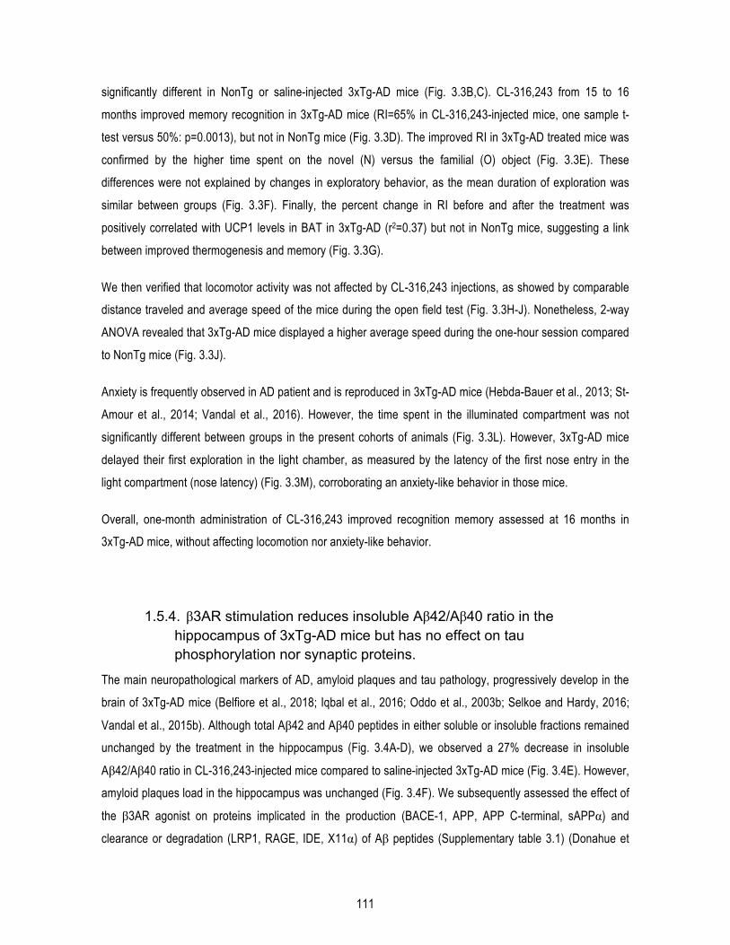
111
significantly different in NonTg or saline-injected 3xTg-AD mice (Fig. 3.3B,C). CL-316,243 from 15 to 16
months improved memory recognition in 3xTg-AD mice (RI=65% in CL-316,243-injected mice, one sample t-
test versus 50%: p=0.0013), but not in NonTg mice (Fig. 3.3D). The improved RI in 3xTg-AD treated mice was
confirmed by the higher time spent on the novel (N) versus the familial (O) object (Fig. 3.3E). These
differences were not explained by changes in exploratory behavior, as the mean duration of exploration was
similar between groups (Fig. 3.3F). Finally, the percent change in RI before and after the treatment was
positively correlated with UCP1 levels in BAT in 3xTg-AD (r2=0.37) but not in NonTg mice, suggesting a link
between improved thermogenesis and memory (Fig. 3.3G).
We then verified that locomotor activity was not affected by CL-316,243 injections, as showed by comparable
distance traveled and average speed of the mice during the open field test (Fig. 3.3H-J). Nonetheless, 2-way
ANOVA revealed that 3xTg-AD mice displayed a higher average speed during the one-hour session compared
to NonTg mice (Fig. 3.3J).
Anxiety is frequently observed in AD patient and is reproduced in 3xTg-AD mice (Hebda-Bauer et al., 2013; St-
Amour et al., 2014; Vandal et al., 2016). However, the time spent in the illuminated compartment was not
significantly different between groups in the present cohorts of animals (Fig. 3.3L). However, 3xTg-AD mice
delayed their first exploration in the light chamber, as measured by the latency of the first nose entry in the
light compartment (nose latency) (Fig. 3.3M), corroborating an anxiety-like behavior in those mice.
Overall, one-month administration of CL-316,243 improved recognition memory assessed at 16 months in
3xTg-AD mice, without affecting locomotion nor anxiety-like behavior.
1.5.4. β3AR stimulation reduces insoluble Aβ42/Aβ40 ratio in the hippocampus of 3xTg-AD mice but has no effect on tau phosphorylation nor synaptic proteins.
The main neuropathological markers of AD, amyloid plaques and tau pathology, progressively develop in the
brain of 3xTg-AD mice (Belfiore et al., 2018; Iqbal et al., 2016; Oddo et al., 2003b; Selkoe and Hardy, 2016;
Vandal et al., 2015b). Although total Aβ42 and Aβ40 peptides in either soluble or insoluble fractions remained
unchanged by the treatment in the hippocampus (Fig. 3.4A-D), we observed a 27% decrease in insoluble
Aβ42/Aβ40 ratio in CL-316,243-injected mice compared to saline-injected 3xTg-AD mice (Fig. 3.4E). However,
amyloid plaques load in the hippocampus was unchanged (Fig. 3.4F). We subsequently assessed the effect of
the β3AR agonist on proteins implicated in the production (BACE-1, APP, APP C-terminal, sAPPα) and
clearance or degradation (LRP1, RAGE, IDE, X11α) of Aβ peptides (Supplementary table 3.1) (Donahue et
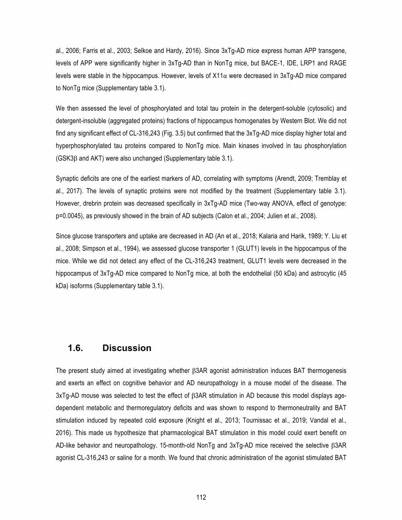
112
al., 2006; Farris et al., 2003; Selkoe and Hardy, 2016). Since 3xTg-AD mice express human APP transgene,
levels of APP were significantly higher in 3xTg-AD than in NonTg mice, but BACE-1, IDE, LRP1 and RAGE
levels were stable in the hippocampus. However, levels of X11α were decreased in 3xTg-AD mice compared
to NonTg mice (Supplementary table 3.1).
We then assessed the level of phosphorylated and total tau protein in the detergent-soluble (cytosolic) and
detergent-insoluble (aggregated proteins) fractions of hippocampus homogenates by Western Blot. We did not
find any significant effect of CL-316,243 (Fig. 3.5) but confirmed that the 3xTg-AD mice display higher total and
hyperphosphorylated tau proteins compared to NonTg mice. Main kinases involved in tau phosphorylation
(GSK3β and AKT) were also unchanged (Supplementary table 3.1).
Synaptic deficits are one of the earliest markers of AD, correlating with symptoms (Arendt, 2009; Tremblay et
al., 2017). The levels of synaptic proteins were not modified by the treatment (Supplementary table 3.1).
However, drebrin protein was decreased specifically in 3xTg-AD mice (Two-way ANOVA, effect of genotype:
p=0.0045), as previously showed in the brain of AD subjects (Calon et al., 2004; Julien et al., 2008).
Since glucose transporters and uptake are decreased in AD (An et al., 2018; Kalaria and Harik, 1989; Y. Liu et
al., 2008; Simpson et al., 1994), we assessed glucose transporter 1 (GLUT1) levels in the hippocampus of the
mice. While we did not detect any effect of the CL-316,243 treatment, GLUT1 levels were decreased in the
hippocampus of 3xTg-AD mice compared to NonTg mice, at both the endothelial (50 kDa) and astrocytic (45
kDa) isoforms (Supplementary table 3.1).
1.6. Discussion
The present study aimed at investigating whether β3AR agonist administration induces BAT thermogenesis
and exerts an effect on cognitive behavior and AD neuropathology in a mouse model of the disease. The
3xTg-AD mouse was selected to test the effect of β3AR stimulation in AD because this model displays age-
dependent metabolic and thermoregulatory deficits and was shown to respond to thermoneutrality and BAT
stimulation induced by repeated cold exposure (Knight et al., 2013; Tournissac et al., 2019; Vandal et al.,
2016). This made us hypothesize that pharmacological BAT stimulation in this model could exert benefit on
AD-like behavior and neuropathology. 15-month-old NonTg and 3xTg-AD mice received the selective β3AR
agonist CL-316,243 or saline for a month. We found that chronic administration of the agonist stimulated BAT
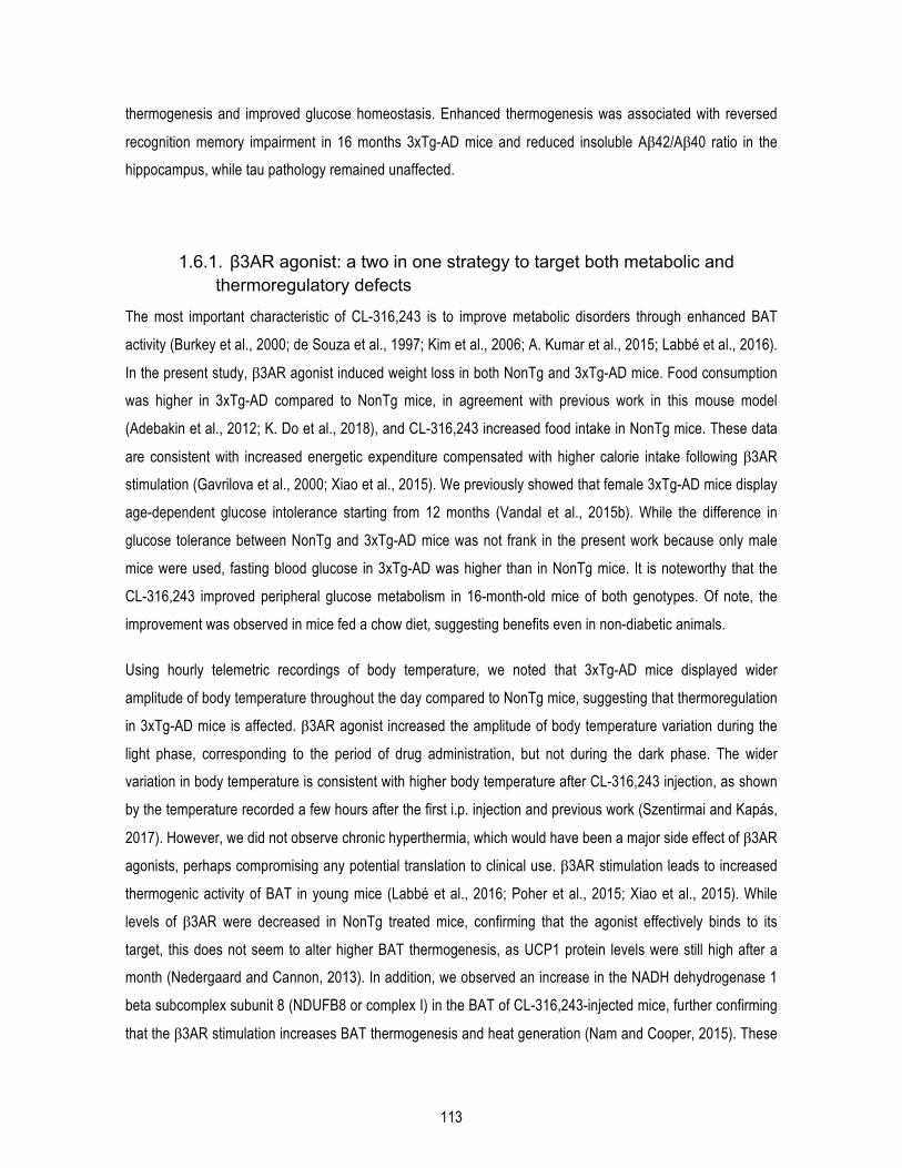
113
thermogenesis and improved glucose homeostasis. Enhanced thermogenesis was associated with reversed
recognition memory impairment in 16 months 3xTg-AD mice and reduced insoluble Aβ42/Aβ40 ratio in the
hippocampus, while tau pathology remained unaffected.
1.6.1. β3AR agonist: a two in one strategy to target both metabolic and thermoregulatory defects
The most important characteristic of CL-316,243 is to improve metabolic disorders through enhanced BAT
activity (Burkey et al., 2000; de Souza et al., 1997; Kim et al., 2006; A. Kumar et al., 2015; Labbé et al., 2016).
In the present study, β3AR agonist induced weight loss in both NonTg and 3xTg-AD mice. Food consumption
was higher in 3xTg-AD compared to NonTg mice, in agreement with previous work in this mouse model
(Adebakin et al., 2012; K. Do et al., 2018), and CL-316,243 increased food intake in NonTg mice. These data
are consistent with increased energetic expenditure compensated with higher calorie intake following β3AR
stimulation (Gavrilova et al., 2000; Xiao et al., 2015). We previously showed that female 3xTg-AD mice display
age-dependent glucose intolerance starting from 12 months (Vandal et al., 2015b). While the difference in
glucose tolerance between NonTg and 3xTg-AD mice was not frank in the present work because only male
mice were used, fasting blood glucose in 3xTg-AD was higher than in NonTg mice. It is noteworthy that the
CL-316,243 improved peripheral glucose metabolism in 16-month-old mice of both genotypes. Of note, the
improvement was observed in mice fed a chow diet, suggesting benefits even in non-diabetic animals.
Using hourly telemetric recordings of body temperature, we noted that 3xTg-AD mice displayed wider
amplitude of body temperature throughout the day compared to NonTg mice, suggesting that thermoregulation
in 3xTg-AD mice is affected. β3AR agonist increased the amplitude of body temperature variation during the
light phase, corresponding to the period of drug administration, but not during the dark phase. The wider
variation in body temperature is consistent with higher body temperature after CL-316,243 injection, as shown
by the temperature recorded a few hours after the first i.p. injection and previous work (Szentirmai and Kapás,
2017). However, we did not observe chronic hyperthermia, which would have been a major side effect of β3AR
agonists, perhaps compromising any potential translation to clinical use. β3AR stimulation leads to increased
thermogenic activity of BAT in young mice (Labbé et al., 2016; Poher et al., 2015; Xiao et al., 2015). While
levels of β3AR were decreased in NonTg treated mice, confirming that the agonist effectively binds to its
target, this does not seem to alter higher BAT thermogenesis, as UCP1 protein levels were still high after a
month (Nedergaard and Cannon, 2013). In addition, we observed an increase in the NADH dehydrogenase 1
beta subcomplex subunit 8 (NDUFB8 or complex I) in the BAT of CL-316,243-injected mice, further confirming
that the β3AR stimulation increases BAT thermogenesis and heat generation (Nam and Cooper, 2015). These
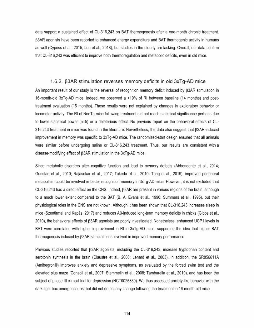
114
data support a sustained effect of CL-316,243 on BAT thermogenesis after a one-month chronic treatment.
β3AR agonists have been reported to enhanced energy expenditure and BAT thermogenic activity in humans
as well (Cypess et al., 2015; Loh et al., 2018), but studies in the elderly are lacking. Overall, our data confirm
that CL-316,243 was efficient to improve both thermoregulation and metabolic deficits, even in old mice.
1.6.2. β3AR stimulation reverses memory deficits in old 3xTg-AD mice
An important result of our study is the reversal of recognition memory deficit induced by β3AR stimulation in
16-month-old 3xTg-AD mice. Indeed, we observed a +19% of RI between baseline (14 months) and post-
treatment evaluation (16 months). These results were not explained by changes in exploratory behavior or
locomotor activity. The RI of NonTg mice following treatment did not reach statistical significance perhaps due
to lower statistical power (n=5) or a deleterious effect. No previous report on the behavioral effects of CL-
316,243 treatment in mice was found in the literature. Nevertheless, the data also suggest that β3AR-induced
improvement in memory was specific to 3xTg-AD mice. The randomized-start design ensured that all animals
were similar before undergoing saline or CL-316,243 treatment. Thus, our results are consistent with a
disease-modifying effect of β3AR stimulation in the 3xTg-AD mice.
Since metabolic disorders alter cognitive function and lead to memory defects (Abbondante et al., 2014;
Gunstad et al., 2010; Rajasekar et al., 2017; Takeda et al., 2010; Tong et al., 2019), improved peripheral
metabolism could be involved in better recognition memory in 3xTg-AD mice. However, it is not excluded that
CL-316,243 has a direct effect on the CNS. Indeed, β3AR are present in various regions of the brain, although
to a much lower extent compared to the BAT (B. A. Evans et al., 1996; Summers et al., 1995), but their
physiological roles in the CNS are not known. Although it has been shown that CL-316,243 increases sleep in
mice (Szentirmai and Kapás, 2017) and reduces Aβ-induced long-term memory deficits in chicks (Gibbs et al.,
2010), the behavioral effects of β3AR agonists are poorly investigated. Nonetheless, enhanced UCP1 levels in
BAT were correlated with higher improvement in RI in 3xTg-AD mice, supporting the idea that higher BAT
thermogenesis induced by β3AR stimulation is involved in improved memory performance.
Previous studies reported that β3AR agonists, including the CL-316,243, increase tryptophan content and
serotonin synthesis in the brain (Claustre et al., 2008; Lenard et al., 2003). In addition, the SR856611A
(Amibegron®) improves anxiety and depressive symptoms, as evaluated by the forced swim test and the
elevated plus maze (Consoli et al., 2007; Stemmelin et al., 2008; Tamburella et al., 2010), and has been the
subject of phase III clinical trial for depression (NCT0025330). We thus assessed anxiety-like behavior with the
dark-light box emergence test but did not detect any change following the treatment in 16-month-old mice.
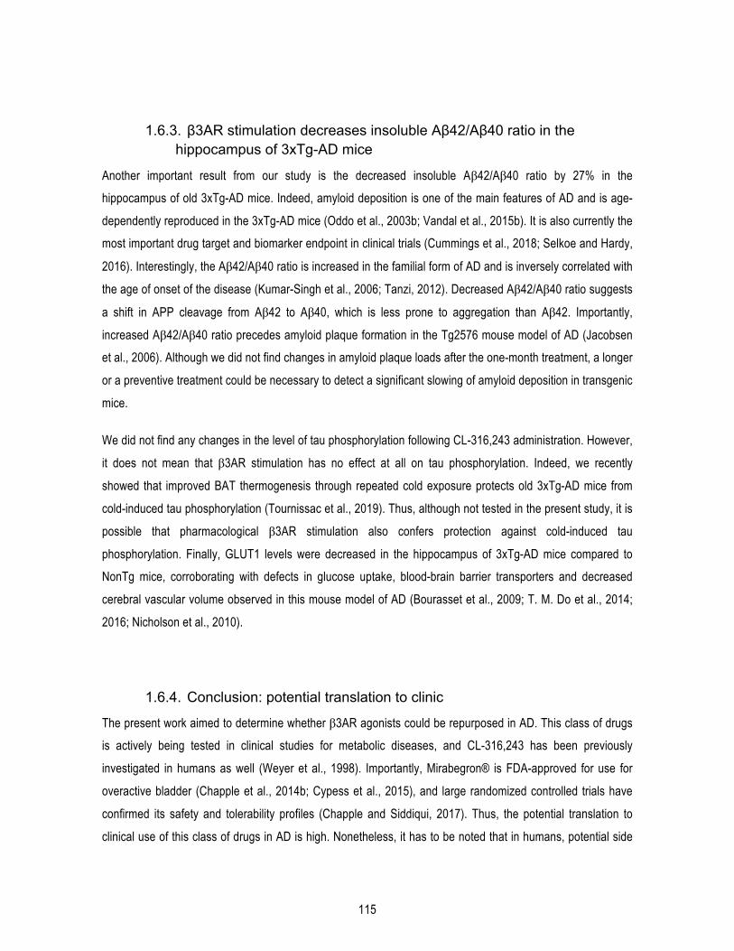
115
1.6.3. β3AR stimulation decreases insoluble Aβ42/Aβ40 ratio in the hippocampus of 3xTg-AD mice
Another important result from our study is the decreased insoluble Aβ42/Aβ40 ratio by 27% in the
hippocampus of old 3xTg-AD mice. Indeed, amyloid deposition is one of the main features of AD and is age-
dependently reproduced in the 3xTg-AD mice (Oddo et al., 2003b; Vandal et al., 2015b). It is also currently the
most important drug target and biomarker endpoint in clinical trials (Cummings et al., 2018; Selkoe and Hardy,
2016). Interestingly, the Aβ42/Aβ40 ratio is increased in the familial form of AD and is inversely correlated with
the age of onset of the disease (Kumar-Singh et al., 2006; Tanzi, 2012). Decreased Aβ42/Aβ40 ratio suggests
a shift in APP cleavage from Aβ42 to Aβ40, which is less prone to aggregation than Aβ42. Importantly,
increased Aβ42/Aβ40 ratio precedes amyloid plaque formation in the Tg2576 mouse model of AD (Jacobsen
et al., 2006). Although we did not find changes in amyloid plaque loads after the one-month treatment, a longer
or a preventive treatment could be necessary to detect a significant slowing of amyloid deposition in transgenic
mice.
We did not find any changes in the level of tau phosphorylation following CL-316,243 administration. However,
it does not mean that β3AR stimulation has no effect at all on tau phosphorylation. Indeed, we recently
showed that improved BAT thermogenesis through repeated cold exposure protects old 3xTg-AD mice from
cold-induced tau phosphorylation (Tournissac et al., 2019). Thus, although not tested in the present study, it is
possible that pharmacological β3AR stimulation also confers protection against cold-induced tau
phosphorylation. Finally, GLUT1 levels were decreased in the hippocampus of 3xTg-AD mice compared to
NonTg mice, corroborating with defects in glucose uptake, blood-brain barrier transporters and decreased
cerebral vascular volume observed in this mouse model of AD (Bourasset et al., 2009; T. M. Do et al., 2014;
2016; Nicholson et al., 2010).
1.6.4. Conclusion: potential translation to clinic
The present work aimed to determine whether β3AR agonists could be repurposed in AD. This class of drugs
is actively being tested in clinical studies for metabolic diseases, and CL-316,243 has been previously
investigated in humans as well (Weyer et al., 1998). Importantly, Mirabegron® is FDA-approved for use for
overactive bladder (Chapple et al., 2014b; Cypess et al., 2015), and large randomized controlled trials have
confirmed its safety and tolerability profiles (Chapple and Siddiqui, 2017). Thus, the potential translation to
clinical use of this class of drugs in AD is high. Nonetheless, it has to be noted that in humans, potential side

116
effects of β3AR agonists include cardiovascular dysfunction induced by non-specific activation of β1 and β2
adrenergic receptors. Indeed, one administration of Mirabegron® at a dose of 200 mg (fourth times the clinical
dose) is able to activate BAT activity in adults, but it also induces cardiac arrhythmia (Cypess et al., 2015).
While the clinical dose of 50 mg does not seem to be efficient to acutely stimulate BAT (Baskin et al., 2018), a
recent study showed that 100 mg of Mirabegron® enhances thermogenesis without any cardiovascular side
effects in adults (Loh et al., 2018). Yet, long-term studies investigating chronic effect of β3AR agonists in BAT
thermogenesis in humans are needed.
Altogether, our results in a mouse model of AD demonstrate for the first time that β3AR agonists are potent
tools to reverse memory deficits and insoluble Aβ42/Aβ40 ratio in the hippocampus. It is the first study to our
knowledge to investigate the potential of this class of drugs in AD neuropathology and behavior.
1.7. Acknowledgements
This study was made possible by funding from the Canadian Institutes of Health Research (MOP 102532), the
Quebec Network for Research on Aging, the Alzheimer Society of Canada (#1502), and the Canadian
Foundation for Innovation (#34480). M.T. was funded by a scholarship from the Alzheimer Society of Canada.
F.C. and E.P are a Fonds de Recherche du Québec - Santé (FRQ-S) scholars (#253895, #26936, and
#252178). F.C. is the guarantor of this work and, as such, had full access to all the data in the study and takes
responsibility for the integrity of the data and the accuracy of the data analysis. The authors have no conflict of
interest to declare.
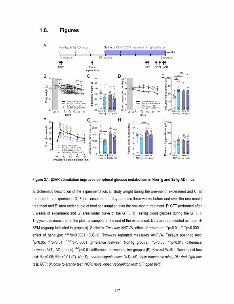
117
1.8. Figures
Figure 3.1. β3AR stimulation improves peripheral glucose metabolism in NonTg and 3xTg-AD mice.
A: Schematic description of the experimentation. B: Body weight during the one-month experiment and C: at
the end of the experiment. D: Food consumed per day per mice three weeks before and over the one-month
treatment and E: area under curve of food consumption over the one-month treatment. F: GTT performed after
3 weeks of experiment and G: area under curve of the GTT. H: Fasting blood glucose during the GTT. I:
Triglycerides measured in the plasma sampled at the end of the experiment. Data are represented as mean ±
SEM (n/group indicated in graphics). Statistics: Two-way ANOVA, effect of treatment: **p<0.01; ****p<0.0001;
effect of genotype: &&&&p<0.0001 (C,G,H). Two-way repeated measures ANOVA, Tukey’s post-hoc test: Δp<0.05; ΔΔp<0.01; ΔΔΔΔp<0.0001 (difference between NonTg groups); ∞p<0.05; ∞∞p<0.01; (difference
between 3xTg-AD groups); ΦΦp<0.01 (difference between saline groups) (F). Kruskal-Wallis, Dunn’s post-hoc
test: @p<0.05; @@p<0.01 (E). NonTg: non-transgenic mice; 3xTg-AD: triple transgenic mice; DL: dark-light box
test; GTT: glucose tolerance test; NOR: novel object recognition test; OF: open field.
0
50
100
150
Plas
mat
ic tr
igly
cerid
es (m
g/dL
)
NonTg 3xTg-ADSaline CL Saline CL
9 9 12 12
5 10 15 20 2520
30
40
50
Days
Body
wei
ght (
g)
NonTg saline (n=10)NonTg CL-316,243 (n=9)3xTg saline (n=16)3xTg CL-316,243 (n=15)
-35 -21
GTT OFsurgery
14 months 15 months 16 months
GTT
0 1 2 3 4 weeks-1-2-3
NOR OF DL NOR
Saline or CL-316,243 treatment (1 mg/kg/day i.p.)
Probeimplantation
NonTg, 3xTg-AD mice
25
30
35
40
45
50
55
Fina
l bod
y w
eigh
t (g)
** **
NonTg 3xTg-ADSaline CL Saline CL10 9 16 15
5 10 15 20 250
1
2
3
4
5
6
Days
Food
con
sum
ptio
n pe
r day
(g)
3xTg saline (n=16)3xTg CL-316,243 (n=15)
-21 0
NonTg saline (n=10)NonTg CL-316,243 (n=9)
0 30 60 90 120
0
5
10
15
20
25
Time after glucose injection (min)
Bloo
d gl
ucos
e (m
M)
3xTg-AD saline (n=16)3xTg-AD CL-316,243 (n=15)
NonTg saline (n=10)NonTg CL-316,243 (n=9)
φφ
ΔΔΔΔ
∞∞
ΔΔ∞
Δ∞
∞∞
ΔΔΔΔ
ΔΔΔΔ
ΔΔΔΔ
0
2
4
6
8
10
12
Fast
ing
bloo
d gl
ucos
e (m
M)
NonTg 3xTg-ADSaline CL Saline CL10 9 16 15
****
&&&&
0
500
1000
1500
2000
2500
Area
und
er c
urve
(mM
• m
in)
********
NonTg 3xTg-ADSaline CL Saline CL
10 9 16 15
A
B C D E
F G H I
20
40
60
80
100
Area
und
er c
urve
(arb
itrar
y un
its)
NonTg 3xTg-ADSaline CL Saline CL10 9 16 15
@@@
@@
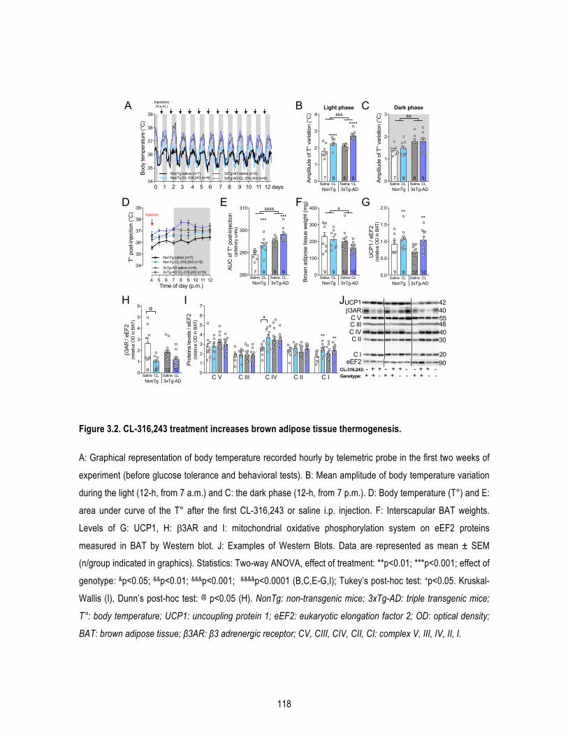
118
Figure 3.2. CL-316,243 treatment increases brown adipose tissue thermogenesis.
A: Graphical representation of body temperature recorded hourly by telemetric probe in the first two weeks of
experiment (before glucose tolerance and behavioral tests). B: Mean amplitude of body temperature variation
during the light (12-h, from 7 a.m.) and C: the dark phase (12-h, from 7 p.m.). D: Body temperature (T°) and E:
area under curve of the T° after the first CL-316,243 or saline i.p. injection. F: Interscapular BAT weights.
Levels of G: UCP1, H: β3AR and I: mitochondrial oxidative phosphorylation system on eEF2 proteins
measured in BAT by Western blot. J: Examples of Western Blots. Data are represented as mean ± SEM
(n/group indicated in graphics). Statistics: Two-way ANOVA, effect of treatment: **p<0.01; ***p<0.001; effect of
genotype: &p<0.05; &&p<0.01; &&&p<0.001; &&&&p<0.0001 (B,C,E-G,I); Tukey’s post-hoc test: +p<0.05. Kruskal-
Wallis (I), Dunn’s post-hoc test: @ p<0.05 (H). NonTg: non-transgenic mice; 3xTg-AD: triple transgenic mice;
T°: body temperature; UCP1: uncoupling protein 1; eEF2: eukaryotic elongation factor 2; OD: optical density;
BAT: brown adipose tissue; β3AR: β3 adrenergic receptor; CV, CIII, CIV, CII, CI: complex V, III, IV, II, I.
34
35
36
37
38
39
Body
tem
pera
ture
(°C
)
0 1 2 3 4 5 6 7 8 9 10 11 12 days
Injections (4 p.m.)
3xTg-AD CL-316,243 (n=9)3xTg-AD saline (n=8)NonTg saline (n=7)
NonTg CL-316,243 (n=9)
BA C
0.0
0.5
1.0
1.5
2.0
UC
P1 /
eEF2
(re
lativ
e O
D in
BAT
)
****
NonTg 3xTg-ADSaline CL Saline CL
9 9 12 12
0
1
2
3
4
5
6
3AR
/ eE
F2
(rela
tive
OD
in B
AT)
NonTg 3xTg-ADSaline CL Saline CL
9 9 12 12
@
GF
H
C V C III C IV C II C I0
1
2
3
4
5
6
7
Prot
eins
leve
ls /
eEF2
(rela
tive
OD
in B
AT)
** **
+
J
0
100
200
300
400
Brow
n ad
ipos
e tis
sue
wei
ght (
mg)
NonTg 3xTg-ADSaline CL Saline CL
9 9 12 12
&
I
D
0
1
2
3
4
Ampl
itude
of T
° var
iatio
n (°
C)
****
****
NonTg 3xTg-ADSaline CL Saline CL
7 9 8 9
&&&
Light phase
0
1
2
3
Ampl
itude
of T
° var
iatio
n (°
C)
NonTg 3xTg-ADSaline CL Saline CL
7 9 8 9
&&
Dark phase
E
280
290
300
310
AUC
of T
° pos
t-inj
ectio
n(a
rbitr
ary
units
)
NonTg 3xTg-ADSaline CL Saline CL
7 9 8 9
*** ***&&&&
CL-316,243: - + + - + + - - + + - Genotype: + + - + + - - + + - -
9020
30404855
4240
C VC IIIC IVC II
C IeEF2
UCP13AR
34
35
36
37
38
39
T° p
ost-i
njec
tion
(°C
)
4 5 6 7 8 9 10 11 12 Time of day (p.m.)
NonTg saline (n=7)NonTg CL-316,243 (n=9)
3xTg-AD CL-316,243 (n=9)3xTg-AD saline (n=8)
Injection
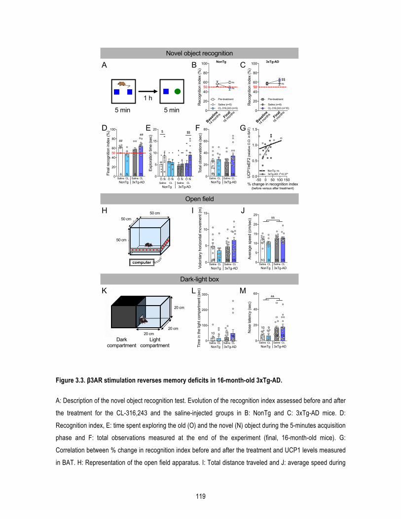
119
Figure 3.3. β3AR stimulation reverses memory deficits in 16-month-old 3xTg-AD.
A: Description of the novel object recognition test. Evolution of the recognition index assessed before and after
the treatment for the CL-316,243 and the saline-injected groups in B: NonTg and C: 3xTg-AD mice. D:
Recognition index, E: time spent exploring the old (O) and the novel (N) object during the 5-minutes acquisition
phase and F: total observations measured at the end of the experiment (final, 16-month-old mice). G:
Correlation between % change in recognition index before and after the treatment and UCP1 levels measured
in BAT. H: Representation of the open field apparatus. I: Total distance traveled and J: average speed during
0
20
40
60
80
Tota
l obs
erva
tions
(sec
)
NonTg 3xTg-ADSaline CL Saline CL
10 9 16 150
20
40
60
80
100
Fina
l rec
ogni
tion
inde
x (%
)
##
##
NonTg 3xTg-ADSaline CL Saline CL
6 5 8 11
50
Light compartment
50 cm
50 cm
5 min
1 h
5 min
Volo
ntar
y ho
rizon
tal m
ovem
ent (
m)
5
10
15
0
NonTg 3xTg-ADSaline CL Saline CL
10 9 16 15 0
5
10
15
20
25
Aver
age
spee
d (c
m/s
ec)
NonTg 3xTg-ADSaline CL Saline CL10 9 16 15
&&
Novel object recognition
0
20
40
60
Nos
e la
tenc
y (s
ec)
NonTg 3xTg-ADSaline CL Saline CL
10 9
16 15
&&
0
100
200
300
Tim
e in
the
light
com
partm
ent (
sec)
NonTg 3xTg-ADSaline CL Saline CL
10 9 1615
Dark compartment
50 cm
20 cm
20 cm20 cm
BA C
ED F
IH J
LK M
O N O N O N O N0
5
10
15
20
Expl
orat
ion
time
(sec
)
$ $$
NonTg 3xTg-ADSaline CL Saline CL
Open field
Dark-light box
-50 0 50 100 1500.0
0.5
1.0
1.5
% change in recognition index (before versus after treatment)
UC
P1/e
EF2
(rela
tive
O.D
. in
BAT)
NonTg: ns3xTg-AD: r2=0.37*
GBaseline
Final0
20
40
60
80
100
Rec
ogni
tion
inde
x (%
)
Saline (n=5)CL-316,243 (n=5)
NonTg
50ns
ns
14 m
onths
16 m
onths
Pre-treatment
Baseline
Final0
20
40
60
80
100
Rec
ogni
tion
inde
x (%
)
Saline (n=8)CL-316,243 (n=10)
$$
14 m
onths
16 m
onths
3xTg-AD
50ns
Pre-treatment
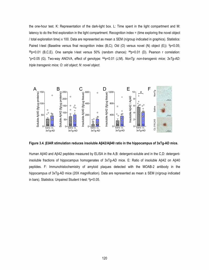
120
the one-hour test. K: Representation of the dark-light box. L: Time spent in the light compartment and M:
latency to do the first exploration in the light compartment. Recognition index = (time exploring the novel object
/ total exploration time) x 100. Data are represented as mean ± SEM (n/group indicated in graphics). Statistics:
Paired t-test (Baseline versus final recognition index (B,C); Old (O) versus novel (N) object (E)): $p<0.05;
$$p<0.01 (B,C,E). One sample t-test versus 50% (random chance): ##p<0.01 (D). Pearson r correlation:
*p<0.05 (G). Two-way ANOVA, effect of genotype: &&p<0.01 (J,M). NonTg: non-transgenic mice; 3xTg-AD:
triple transgenic mice; O: old object; N: novel object.
Figure 3.4. β3AR stimulation reduces insoluble Aβ42/Aβ40 ratio in the hippocampus of 3xTg-AD mice.
Human Aβ40 and Aβ42 peptides measured by ELISA in the A,B: detergent-soluble and in the C,D: detergent-
insoluble fractions of hippocampus homogenates of 3xTg-AD mice. E: Ratio of insoluble Aβ42 on Aβ40
peptides. F: Immunohistochemistry of amyloid plaques detected with the MOAB-2 antibody in the
hippocampus of 3xTg-AD mice (20X magnification). Data are represented as mean ± SEM (n/group indicated
in bars). Statistics: Unpaired Student t-test: #p<0.05.
0
50
100
150
200
250
Solu
ble
A42
(fg/μg
pro
tein
s)
3xTg-ADSaline CL12 12 0
2
4
6
8
10
Inso
lubl
e A
42 /
A40
#
3xTg-ADSaline CL12 12
0
50
100
150
Solu
ble
A40
(fg/μg
pro
tein
s)
3xTg-ADSaline CL12 12
A B C D E
Inso
lubl
e A
42 (f
g/μg
tiss
ue)
3xTg-ADSaline CL12 12
1200
800
400
0Inso
lubl
e A
40 (f
g/μg
tiss
ue)
3xTg-ADSaline CL
1212
600
400
200
0
F
100 µm
100 µm
100 µm
Non
Tg
Sal
ine
CL-
316,
243
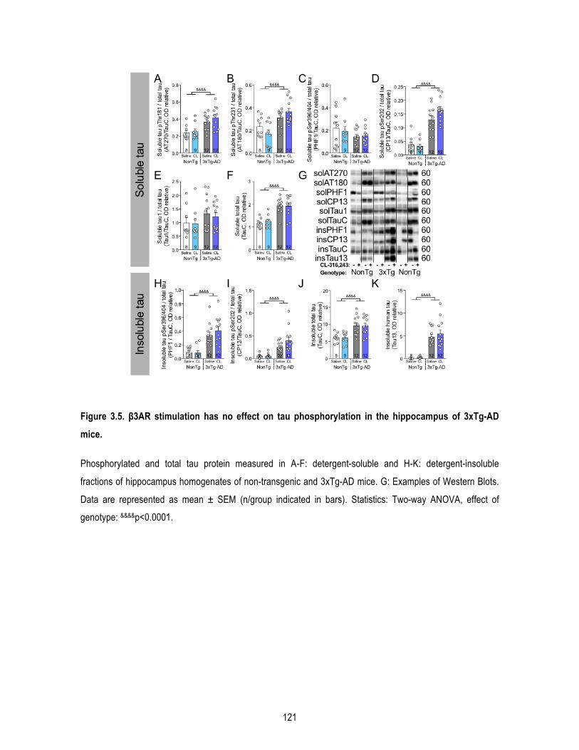
121
Figure 3.5. β3AR stimulation has no effect on tau phosphorylation in the hippocampus of 3xTg-AD mice.
Phosphorylated and total tau protein measured in A-F: detergent-soluble and H-K: detergent-insoluble
fractions of hippocampus homogenates of non-transgenic and 3xTg-AD mice. G: Examples of Western Blots.
Data are represented as mean ± SEM (n/group indicated in bars). Statistics: Two-way ANOVA, effect of
genotype: &&&&p<0.0001.
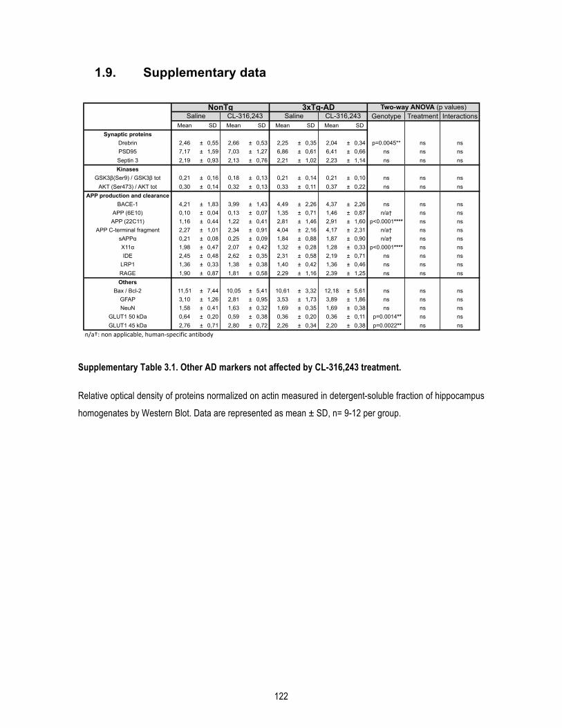
122
1.9. Supplementary data
Supplementary Table 3.1. Other AD markers not affected by CL-316,243 treatment.
Relative optical density of proteins normalized on actin measured in detergent-soluble fraction of hippocampus
homogenates by Western Blot. Data are represented as mean ± SD, n= 9-12 per group.
Genotype Treatment InteractionsMean SD Mean SD Mean SD Mean SD
Synaptic proteinsDrebrin 2,46 ± 0,55 2,66 ± 0,53 2,25 ± 0,35 2,04 ± 0,34 p=0.0045** ns nsPSD95 7,17 ± 1,59 7,03 ± 1,27 6,86 ± 0,61 6,41 ± 0,66 ns ns nsSeptin 3 2,19 ± 0,93 2,13 ± 0,76 2,21 ± 1,02 2,23 ± 1,14 ns ns nsKinases
GSK3β(Ser9) / GSK3β tot 0,21 ± 0,16 0,18 ± 0,13 0,21 ± 0,14 0,21 ± 0,10 ns ns nsAKT (Ser473) / AKT tot 0,30 ± 0,14 0,32 ± 0,13 0,33 ± 0,11 0,37 ± 0,22 ns ns ns
APP production and clearanceBACE-1 4,21 ± 1,83 3,99 ± 1,43 4,49 ± 2,26 4,37 ± 2,26 ns ns ns
APP (6E10) 0,10 ± 0,04 0,13 ± 0,07 1,35 ± 0,71 1,46 ± 0,87 n/a† ns nsAPP (22C11) 1,16 ± 0,44 1,22 ± 0,41 2,81 ± 1,46 2,91 ± 1,60 p<0.0001**** ns ns
APP C-terminal fragment 2,27 ± 1,01 2,34 ± 0,91 4,04 ± 2,16 4,17 ± 2,31 n/a† ns nssAPPα 0,21 ± 0,08 0,25 ± 0,09 1,84 ± 0,88 1,87 ± 0,90 n/a† ns nsX11α 1,98 ± 0,47 2,07 ± 0,42 1,32 ± 0,28 1,28 ± 0,33 p<0.0001**** ns nsIDE 2,45 ± 0,48 2,62 ± 0,35 2,31 ± 0,58 2,19 ± 0,71 ns ns ns
LRP1 1,36 ± 0,33 1,38 ± 0,38 1,40 ± 0,42 1,36 ± 0,46 ns ns nsRAGE 1,90 ± 0,87 1,81 ± 0,58 2,29 ± 1,16 2,39 ± 1,25 ns ns nsOthers
Bax / Bcl-2 11,51 ± 7,44 10,05 ± 5,41 10,61 ± 3,32 12,18 ± 5,61 ns ns nsGFAP 3,10 ± 1,26 2,81 ± 0,95 3,53 ± 1,73 3,89 ± 1,86 ns ns nsNeuN 1,58 ± 0,41 1,63 ± 0,32 1,69 ± 0,35 1,69 ± 0,38 ns ns ns
GLUT1 50 kDa 0,64 ± 0,20 0,59 ± 0,38 0,36 ± 0,20 0,36 ± 0,11 p=0.0014** ns nsGLUT1 45 kDa 2,76 ± 0,71 2,80 ± 0,72 2,26 ± 0,34 2,20 ± 0,38 p=0.0022** ns ns
n/a†:&non&applicable,&human2specific&antibody
Two-way ANOVA (p values)NonTg 3xTg-ADSaline CL-316,243 Saline CL-316,243
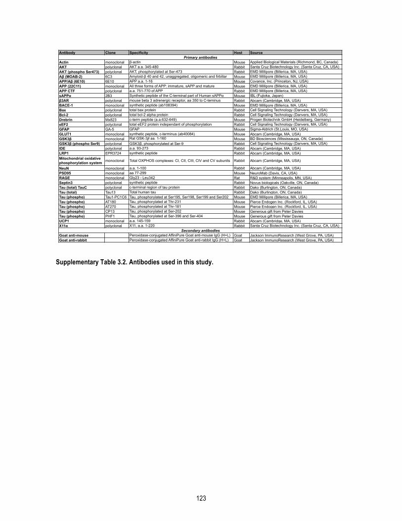
123
Supplementary Table 3.2. Antibodies used in this study.
Antibody Clone Specificity Host Source
Actin monoclonal β-actin Mouse Applied Biological Materials (Richmond, BC, Canada)AKT polyclonal AKT a.a. 345-480 Rabbit Santa Cruz Biotechnology Inc. (Santa Cruz, CA, USA)AKT (phospho Ser473) polyclonal AKT, phosphorylated at Ser-473 Rabbit EMD Millipore (Billerica, MA, USA)Aβ (MOAB-2) 6C3 Amyloid-β 40 and 42, unaggregated, oligomeric and firbillar Mouse EMD Millipore (Billerica, MA, USA)APP/Aβ (6E10) 6E10 APP a.a. 1-16 Mouse Covance, Inc. (Princeton, NJ, USA)APP (22C11) monoclonal All three forms of APP: immature, sAPP and mature Mouse EMD Millipore (Billerica, MA, USA)APP CTF polyclonal a.a. 751-770 of APP Rabbit EMD Millipore (Billerica, MA, USA)sAPPα 2B3 Synthetic peptide of the C-terminal part of Human sAPPα Mouse IBL (Fujioka, Japan)β3AR polyclonal mouse beta 3 adrenergic receptor, aa 350 to C-teminus Rabbit Abcam (Cambridge, MA, USA)BACE-1 monoclonal synthetic peptide (ab108394) Mouse EMD Millipore (Billerica, MA, USA)Bax polyclonal total bax protein Rabbit Cell Signaling Technology (Danvers, MA, USA)Bcl-2 polyclonal total bcl-2 alpha protein Rabbit Cell Signaling Technology (Danvers, MA, USA)Drebrin Mx823 c-term peptide (a.a.632-649) Mouse Progen Biotechnik GmbH (Heidelberg, Germany)eEF2 polyclonal total eEF2 protein independant of phosphorylation Rabbit Cell Signaling Technology (Danvers, MA, USA)GFAP GA-5 GFAP Mouse Sigma-Aldrich (St.Louis, MO, USA)GLUT1 monoclonal synthetic peptide, c-terminus (ab40084) Mouse Abcam (Cambridge, MA, USA)GSK3β monoclonal Rat GSK-3β aa. 1-160 Mouse BD Biosciences (Mississauga, ON, Canada)GSK3β (phospho Ser9) polyclonal GSK3β, phosphorylated at Ser-9 Rabbit Cell Signaling Technology (Danvers, MA, USA)IDE polyclonal a.a. 93-273 Rabbit Abcam (Cambridge, MA, USA)LRP1 EPR3724 synthetic peptide Rabbit Abcam (Cambridge, MA, USA)Mitochondrial oxidative phosphorylation system monoclonal Total OXPHOS complexes: CI, CII, CIII, CIV and CV subunits Rabbit Abcam (Cambridge, MA, USA)
NeuN monoclonal a.a. 1-100 Rabbit Abcam (Cambridge, MA, USA)PSD95 monoclonal aa 77-299 Mouse NeuroMab (Davis, CA, USA)RAGE monoclonal Gly23 - Leu342 Rat R&D system (Minneapolis, MN, USA)Septin3 polyclonal synthetic peptide Rabbit Novus biologicals (Oakville, ON, Canada)Tau (total) TauC polyclonal c-terminal region of tau protein Rabbit Dako (Burlington, ON, Canada)Tau (total) Tau13 Total human tau Rabbit Dako (Burlington, ON, Canada)Tau (phospho) Tau1-PC1C6 Tau, phosphorylated at Ser195, Ser198, Ser199 and Ser202 Mouse EMD Millipore (Billerica, MA, USA)Tau (phospho) AT180 Tau, phosphorylated at Thr-231 Mouse Pierce Endogen Inc. (Rockford, IL, USA)Tau (phospho) AT270 Tau, phosphorylated at Thr-181 Mouse Pierce Endogen Inc. (Rockford, IL, USA)Tau (phospho) CP13 Tau, phosphorylated at Ser-202 Mouse Generous gift from Peter DaviesTau (phospho) PHF1 Tau, phosphorylated at Ser-396 and Ser-404 Mouse Generous gift from Peter DaviesUCP1 monoclonal a.a. 145-159 Rabbit Abcam (Cambridge, MA, USA)X11α polyclonal X11, a.a. 1-220 Rabbit Santa Cruz Biotechnology Inc. (Santa Cruz, CA, USA)
Goat anti-mouse Peroxidase-conjugated AffiniPure Goat anti-mouse IgG (H+L) Goat Jackson ImmunoResearch (West Grove, PA, USA)Goat anti-rabbit Peroxidase-conjugated AffiniPure Goat anti-rabbit IgG (H+L) Goat Jackson ImmunoResearch (West Grove, PA, USA)
Primary antibodies
Secondary antibodies
124
Conclusion
Cette section de la thèse comprend un retour sur les résultats des articles présentés ci-dessus, puis une
discussion sur les limites, les questions soulevées et les perspectives de nos résultats. Enfin, elle se terminera
par une conclusion générale.
1 Retour sur les résultats
Chapitre 1 : L’âge avancé potentialise la phosphorylation de tau induite par le froid : lien entre déficit de thermorégulation et maladie d’Alzheimer.
Cette étude fait suite aux résultats d’une première publication de notre équipe montrant que les souris 3xTg-
AD sont plus susceptibles à une exposition au froid que les souris NonTg, dans le sens où l’augmentation de
la phosphorylation de la protéine tau dans leur cortex est plus importante que les souris contrôles (Figure 14)
(Vandal et al., 2016).
L’objectif de ce travail était de vérifier si l’âge pouvait être un facteur aggravant la phosphorylation de tau en
réponse à une diminution de température corporelle. Nous avons donc exposé des souris jeunes (6 mois) et
âgées (18 mois) à une température de 4°C pendant 24 heures, puis analysé la phosphorylation de tau sur
différents épitopes dans le cortex pariéto-temporal. Tout d’abord, nous avons observé une diminution de la
température corporelle d’environ 2°C et une augmentation de l’expression d’UCP1 dans le TAB de manière
équivalente entre les souris jeunes et âgées. Ensuite, nous avons de nouveau confirmé l’augmentation
drastique de la phosphorylation de la protéine tau en réponse à une diminution de température corporelle. De
façon intéressante, la phosphorylation de tau sur les épitopes Thr231 et Thr181 était significativement
augmentée seulement pour les souris âgées, tandis que les souris jeunes présentaient une plus grande
inactivation de la kinase GSK3β (mesurée par la phosphorylation sur la Ser9). Ces résultats suggèrent que
l’âge est un facteur qui potentialise l’effet de la diminution de la température corporelle sur la phosphorylation
de la protéine tau. Cette différence entre souris jeunes et âgées pourrait s’expliquer par un manque
d’inactivation de la GSK3β chez les souris âgées.

125
Cette étude propose une explication originale quant au fait que les personnes âgées soient les principales
touchées par la MA : l’âge avancé augmente la susceptibilité à la phosphorylation de la protéine tau induite
par une hypothermie. Comme les personnes âgées présentent un déficit de thermorégulation et sont plus à
risque d’hypothermie (Gomolin et al., 2005; Hoshino et al., 2007; Sugarek, 1986), des épisodes plus fréquents
de basse température corporelle pourraient conduire à une hyperphosphorylation de tau répétée. Ces effets
pourraient être additifs pour finalement conduire à la formation d’ENF et éventuellement au développement de
la MA. Toutefois, l’hyperphosphorylation de la protéine tau suite à une hypothermie reste à démontrer chez
l’humain.
En revanche, il ne faudrait pas tirer de nos résultats la simple conclusion que la température ambiante est
dangereuse pour les personnes âgées et qu’il suffirait de les maintenir à des températures plus élevées pour
prévenir la démence. Nous avons utilisé une exposition à une température ambiante froide comme méthode
pour diminuer rapidement la température corporelle des animaux et induire une réponse thermogénique. Bien
qu’une semaine d’exposition à une température thermoneutre soit bénéfique pour les souris 3xTg-AD (Vandal
et al., 2016), il s’agit bien de la modification de la température corporelle, et non simplement de la température
externe, qui induit un changement de la phosphorylation de la protéine tau. En effet, plusieurs études ont
montré que l’hypothermie induite par une anesthésie ou une diminution du métabolisme (hypoglycémie ou
hibernation) provoque également cette hyperphosphorylation de tau, sans modification de la température
ambiante (Planel et al., 2009; 2004; Stieler et al., 2011). De plus, maintenir la température corporelle des
animaux suite à une anesthésie prévient ce phénomène (Planel et al., 2007; 2004).
Ainsi, il est nécessaire de mieux comprendre par quels mécanismes les changements de température
corporelle influencent la phosphorylation de la protéine tau. L’hypothèse découlant de ces deux premières
études est que la modulation (à la hausse ou à la baisse) des mécanismes de thermorégulation influence la
neuropathologie Alzheimer (Tournissac et al., 2017; Vandal et al., 2016). Cette hypothèse a été investiguée
dans les deux chapitres suivants.
Chapitre 2 : Les expositions répétées au froid protègent un modèle murin de la maladie d’Alzheimer de la phosphorylation de tau induite par le froid.
L’objectif de ce deuxième chapitre était de déterminer l’effet d’une stimulation de l’activité du TAB de manière
« naturelle » ou non-pharmacologique sur la neuropathologie Alzheimer chez la souris 3xTg-AD âgée. Nous

126
avons sélectionné la souris 3xTg-AD car elle présente à la fois les pathologies tau et amyloïde, qui sont les
deux marqueurs neuropathologiques principaux de la MA (Oddo et al., 2003b; Querfurth and LaFerla, 2010).
De plus, le développement de la neuropathologie se fait de manière très progressive dans le cerveau de ces
souris (Belfiore et al., 2018; Vandal et al., 2015b), nous permettant d’inclure le facteur de l’âge avancé, si
important dans la MA (Rocca et al., 2011b; Solomon et al., 2014). Nous avons précédemment montré que
cette souris développait également des désordres métaboliques et un déficit de thermorégulation au fur et à
mesure de son vieillissement, faisant d’elle un modèle de choix pour la présente étude (Vandal et al., 2016;
2015b; 2014b).
Nous avons choisi de faire des expositions répétées au froid pour stimuler le TAB, une méthode bien connue
pour réduire les désordres métaboliques en augmenter la thermogenèse, que ce soit chez le rongeur ou chez
l’humain (Hanssen et al., 2015b; Ravussin et al., 2014; van der Lans et al., 2013). Cette approche se voulait
globale : elle ne fait pas intervenir uniquement la thermogenèse du TAB mais aussi la thermogenèse avec
frissons des muscles squelettiques (Blondin et al., 2017; Cannon and Nedergaard, 2004; Silva, 2006). Elle est
donc moins spécifique à l’activité du TAB que l’approche pharmacologique utilisée dans le troisième chapitre.
Nous aurions pu réaliser une acclimatation au froid plus progressive en plaçant les animaux dans une pièce
dont la température diminue de 2°C par jour pour atteindre une température de 4°C afin de stimuler la
thermogenèse de façon plus chronique et non pas intermittente (Mineo et al., 2012). Toutefois, ce protocole
est plus demandant au niveau métabolique pour la souris à long terme, ce qui n’est peut-être pas adapté pour
un animal âgé. Aussi, il aurait été impossible de réaliser le test de tolérance au glucose sur des animaux dans
la chambre froide puisque la combinaison du jeûne de 6 heures et de l’exposition au froid aurait induit un
stress métabolique et une hypoglycémie plus importante, ce qui est trop risqué pour nos animaux âgés de 16
mois.
Nous avons donc exposé de façon quotidienne des souris 3xTg-AD de 16 mois à 4°C pour une durée
relativement courte de 4 heures à raison de cinq fois par semaine. Ce protocole d’acclimatation au froid a duré
quatre semaines pendant lesquelles un groupe contrôle est resté à la température d’hébergement de 22°C. À
la fin de la 4ème semaine, un groupe de souris contrôle a subi une exposition au froid aigüe (4°C, 24 heures)
de même que le groupe ayant reçu les expositions répétées au froid. Cette expérience nous a permis de
constater qu’il était possible d’augmenter la thermogenèse des 3xTg-AD âgées de 16 mois, tout en améliorant
leur métabolisme du glucose périphérique et en diminuant les triglycérides plasmatiques. Mais surtout,
stimuler la thermogenèse de façon répétée a permis de protéger les souris transgéniques de l’augmentation
de la phosphorylation de la protéine tau dans l’hippocampe induite par une exposition au froid aigüe. Nous
avons finalement tenté de mettre en lien ces résultats avec une batokine : FGF21. Nous avons observé une
127
plus grande quantité plasmatique de FGF21 chez les souris acclimatées au froid, quantité corrélée
négativement avec la phosphorylation de tau dans l’hippocampe de ces animaux.
Ainsi, nos résultats suggèrent qu’une meilleure capacité de thermogenèse permet de contrer la
phosphorylation de tau induite par une exposition aigüe au froid. Le changement d’activité des kinases et des
phosphatases, principalement la PP2A, est le principal mécanisme proposé pour la modulation de la
phosphorylation de la protéine tau en réponse à un changement de température corporelle (Planel et al., 2007;
2001). Toutefois, nous n’avons pas observé de diminution significative des principales kinases impliquées
dans la phosphorylation de tau, ni d’augmentation de la quantité de PP2A dans le groupe de souris
acclimatées au froid, mais il n’est pas exclu que son activité soit modulée par notre traitement. En revanche,
nos résultats proposent qu’un lien plus direct entre activité du TAB et phosphorylation de tau existe : ce lien
pourrait se faire via la sécrétion de molécules de signalisation par le TAB (les batokines), et plus
spécifiquement FGF21 (Hondares et al., 2011; Villarroya et al., 2017). En effet, nous avons observé une
corrélation inverse entre les niveaux plasmatiques de FGF21 et la phosphorylation de la protéine tau dans
l’hippocampe, suggérant que des niveaux élevés de cette batokine pourraient être associés à une
préservation des niveaux de phosphorylation de tau. Toutefois, ceci n’est que corrélatif et les effets des
batokines sur le cerveau commencent à peine à être étudiés.
Chapitre 3 : Un agoniste des récepteurs β3 adrénergiques renverse les déficits mnésiques et réduit la pathologie amyloïde dans un modèle murin de la maladie d’Alzheimer.
L’objectif de cette étude était de déterminer l’effet de la stimulation de la thermogenèse du TAB à l’aide d’un
agoniste spécifique des Rβ3A sur la neuropathologie et le comportement des souris 3xTg-AD âgées de 16
mois. Plus largement, nous voulions savoir si cette molécule pourrait être un bon candidat pour le traitement
de la MA. Nous avons sélectionné la souris 3xTg-AD car cette souris présente à la fois les pathologies tau et
amyloïde associées à déficits cognitifs. Des souris 3xTg-AD et non-transgéniques (NonTg) âgées de 15 mois
ont reçu le CL-316,243 à une dose de 1 mg/kg, un agoniste spécifique des Rβ3A, ou du salin, tous les jours
pendant un mois.
Nous avons d’abord confirmé que le CL-316,243 permettait d’améliorer le métabolisme périphérique des
souris de 16 mois, quel que soit leur génotype, en diminuant leur poids et en améliorant leur tolérance au
glucose, même si ces souris n’étaient pas diabétiques. Une amélioration du métabolisme du glucose sur des
128
souris non diabétiques a déjà été observée précédemment, mais sur des souris plus jeunes (Xiao et al., 2015).
D’autre part, la prise alimentaire était augmentée uniquement chez les NonTg en réponse au traitement,
rejoignant le niveau de consommation des souris 3xTg-AD. De plus, l’agoniste des Rβ3A a permis
d’augmenter la thermogenèse des animaux âgés comme l’indiquent la hausse de température corporelle post-
injection ainsi que l’augmentation d’UCP1 et du complexe I de la chaine de phosphorylation oxydative
mitochondrial. Bien que les améliorations métaboliques et l’augmentation de l’activité de thermogenèse du
TAB suite à l’administration du CL-316,243 soient bien connues, il s’agit de la première étude testant cette
molécule sur des animaux âgés à notre connaissance. Les souris âgées montrant une thermorégulation moins
efficace que les souris jeunes (Gonzales and Rikke, 2010; Talan, 1997; Tatelman and Talan, 1990), il aurait
été possible que l’agoniste ait peu ou pas d’effet sur les souris âgées, et d’autant plus pour les souris 3xTg-
AD, qui présentent un déficit de thermorégulation.
Le traitement d’un mois avec l’agoniste a permis de réduire le déficit cognitif des souris 3xTg-AD âgées. À
notre connaissance, il s’agit de la première démonstration d’un effet mnésique d’un agoniste des Rβ3A dans
un modèle murin de la MA. En effet, seulement une étude à notre connaissance a observé une amélioration
mnésique chez des poussins ayant reçu une injection de peptides Aβ42 après administration intracrânienne
de CL-316,243 (Gibbs et al., 2010). De plus, nous avons observé une corrélation positive entre la quantité
d’UCP1 dans le TAB et le pourcentage d’augmentation du score au test de reconnaissance d’objet
spécifiquement chez les souris 3xTg-AD, suggérant que cette meilleure performance était liée à
l’augmentation de thermogenèse du TAB. En revanche, le test de reconnaissance d’objet avec seulement une
heure d’intervalle entre les deux phases, bien qu’adapté pour la souris 3xTg-AD âgée, ne permet pas de
savoir si cet effet est spécifique aux souris présentant un déficit cognitif, ou bien si le CL-316,243 a un effet
global d’amélioration de la mémoire. Pour répondre à cette question, il faudrait réaliser des tests plus
sensibles avec une tâche plus difficile pour détecter une amélioration mnésique sur des souris sans trouble de
la mémoire. Par exemple, il faudrait faire un test de reconnaissance d’objet avec un intervalle de 24 heures
entre les deux phases (correspondant à l’évaluation de la mémoire à long terme), ou encore un test de
localisation d’objet, permettant d’évaluer la mémoire spatiale, sur des souris jeunes et non-transgéniques
(Cohen et al., 2013; Leger et al., 2013; Vogel-Ciernia and Wood, 2014).
Contrairement à nos deux précédentes études, l’agoniste des Rβ3A n’a eu aucun effet sur la phosphorylation
de la protéine tau (Tableau C1). Au vu des améliorations métaboliques des souris ayant reçu le traitement,
nous aurions pu nous attendre à une diminution de la phosphorylation de la protéine tau. Il n’est toutefois pas
exclu que, comme nos résultats obtenus avec les expositions répétées au froid, l’agoniste des Rβ3A
permettent de protéger les souris contre une hyperphosphorylation de tau induite par une exposition aigüe au
froid. Une exposition de 24 heures à 4°C après l’administration du CL-316,243 pendant un mois permettrait de

129
répondre à cette question. En revanche, cette méthode pharmacologique a quant à elle eu des effets
bénéfiques sur la pathologie amyloïde. Plus précisément, le ratio de peptides Aβ42/Aβ40 insolubles était
diminué dans l’hippocampe des souris 3xTg-AD ayant reçu le traitement. À titre de comparaison, nous avions
précédemment montré qu’une exposition à une température thermoneutre de 28°C diminuait les niveaux
d’Aβ42 insolubles dans le cortex des souris 3xTg-AD en augmentant la quantité du transporteur LRP1
(Tableau C1) (Vandal et al., 2016). Toutefois, nous n’avons ici observé aucun changement dans la quantité de
LRP1, RAGE ou encore BACE-1 et APP. Il est possible que les améliorations du métabolisme périphérique
des souris soient en lien avec la diminution du ratio Aβ42/Aβ40. En effet, une précédente étude de notre
équipe montre qu’une injection d’insuline induit une clairance des peptides Aβ du cerveau vers la périphérie
chez la souris 3xTg-AD, sans pour autant modifier la quantité de LRP1 (Vandal et al., 2014b). Les liens entre
altérations métaboliques et neuropathologie de la MA seront discutés plus en détail dans la section « 2.4
Mécanismes potentiels par lesquels la thermogenèse peut affecter la MA ».
Traitement Souris Comportement Neuropathologie Référence
Exposition aigüe (4°C, 24 h)
NonTg et 3xTg-AD 12 mois
ND ! pTau
! APP, soluble Aβ42/Aβ40 " protéines synaptiques
Vandal 2016
Exposition aigüe (4°C, 24 h)
NonTg 6 et 18 mois
ND ! pTau
= protéines synaptiques
Tournissac 2017
(chapitre 1)
Exposition aigüe (4°C, 24 h)
3xTg-AD 16 mois ND
! pTau = Aβ40 et Aβ42
= protéines synaptiques
Tournissac 2019
(chapitre 2) Expositions répétées (4°C, 4h par jour, un
mois)
3xTg-AD 16 mois
ND Protection contre pTau
= Aβ40 et Aβ42 = protéines synaptiques
Tournissac 2019
(chapitre 2)
Thermoneutralité (28°C, une semaine)
3xTg-AD 20 mois
! mémoire de reconnaissance
" anxiété = locomotion
= pTau " insoluble Aβ42; ! LRP1 ! protéines synaptiques
Vandal 2016
Agoniste des Rβ3A (CL-316,243,
1 mg/kg par jour, un mois)
3xTg-AD 16 mois
! mémoire de reconnaissance
= anxiété = locomotion
= pTau " insoluble Aβ42/Aβ40 = protéines synaptiques
Tournissac, en préparation (chapitre 3)
Tableau C1. Effets des différentes approches pour moduler la thermorégulation sur la neuropathologie de la MA chez la souris.
De manière générale, notre étude est la première à avoir testé cette classe de molécule dans la MA. Nos
résultats suggèrent que les agonistes des Rβ3A seraient bénéfiques pour la MA, du moins en ce qui a trait à
la pathologie amyloïde et la mémoire de reconnaissance. Bien que des études plus longues et sur d’autres

130
modèles animaux soient nécessaires pour confirmer nos résultats, il s’agit d’une toute première preuve de
concept qu’agir sur les Rβ3A représente une cible thérapeutique intéressante pour la MA.
2 Discussion générale et perspectives
2.1 Température corporelle et phosphorylation de la protéine tau
Les phospho-épitopes de tau que nous avons analysés dans nos différentes études ont été sélectionnés pour
leur importance quant à la MA car ils corrèlent avec le diagnostic clinique et les déficits cognitifs, incluant la
mémoire épisodique (Tremblay et al., 2007). En effet, une précédente étude de notre équipe a comparé un
grand nombre de marqueurs neuropathologiques dans les cerveaux de volontaires de la Religious Order study
qui ont été cliniquement caractérisés de leur vivant (Tremblay et al., 2017). Les résultats montrent que la
phosphorylation de tau sur l’épitope PHF1 ou AD2 (pSer396/404) est fortement corrélée avec le diagnostic
clinique de la MA, même dans la fraction soluble. Ce même épitope est aussi associé avec le score de Braak
et la propagation des ENF. Par ailleurs, les épitope AT180 (pThr231) et PHF1, que ce soit dans la fraction
soluble ou insoluble, sont négativement corrélés avec le score cognitif global des sujets. Bien qu’une seule
exposition au froid de 24 heures ne semble pas suffisante pour induire une augmentation de tau dans la
fraction insoluble, une hypothermie prolongée ou répétée pourrait augmenter l’agrégation de tau et l’apparition
d’ENF. En effet, les études in vitro montrent que les phospo-épitopes que nous avons étudiés sont impliqués
dans l’agrégation pathologique de la protéine tau (Martin et al., 2011).
Nous avons de nouveau confirmé dans nos travaux chez la souris que la phosphorylation de tau était
augmentée lorsque la température corporelle diminue. En revanche, l’effet d’une diminution de la température
corporelle sur la phosphorylation de la protéine tau chez l’humain demeure inconnu. Cela pourrait
éventuellement être étudié grâce à l’imagerie cérébrale avec des ligands dirigés contre la protéine tau totale et
phosphorylée, actuellement testés chez l’humain, suite à une exposition au froid (Okamura et al., 2018). Une
alternative serait de réaliser un dosage de la protéine tau phosphorylée et totale dans le LCR des participants.
D’autre part, les études présentées dans cette thèse soulèvent la question de savoir si l’augmentation de la
phosphorylation de tau en réponse à l’hypothermie perdure dans le temps ou si elle n’est que transitoire. De
précédentes études apportent des éléments de réponse. Dans le cas d’animaux qui hibernent, celle-ci est
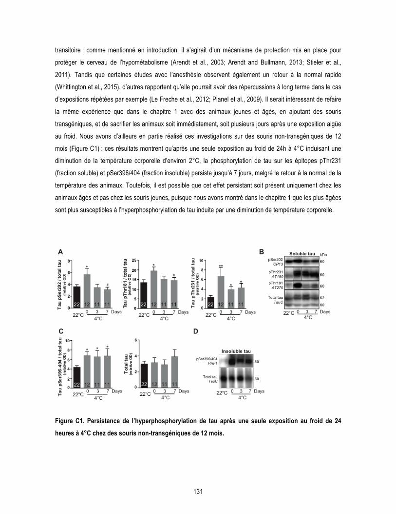
131
transitoire : comme mentionné en introduction, il s’agirait d’un mécanisme de protection mis en place pour
protéger le cerveau de l’hypométabolisme (Arendt et al., 2003; Arendt and Bullmann, 2013; Stieler et al.,
2011). Tandis que certaines études avec l’anesthésie observent également un retour à la normal rapide
(Whittington et al., 2015), d’autres rapportent qu’elle pourrait avoir des répercussions à long terme dans le cas
d’expositions répétées par exemple (Le Freche et al., 2012; Planel et al., 2009). Il serait intéressant de refaire
la même expérience que dans le chapitre 1 avec des animaux jeunes et âgés, en ajoutant des souris
transgéniques, et de sacrifier les animaux soit immédiatement, soit plusieurs jours après une exposition aigüe
au froid. Nous avons d’ailleurs en partie réalisé ces investigations sur des souris non-transgéniques de 12
mois (Figure C1) : ces résultats montrent qu’après une seule exposition au froid de 24h à 4°C induisant une
diminution de la température corporelle d’environ 2°C, la phosphorylation de tau sur les épitopes pThr231
(fraction soluble) et pSer396/404 (fraction insoluble) persiste jusqu’à 7 jours, malgré le retour à la normal de la
température des animaux. Toutefois, il est possible que cet effet persistant soit présent uniquement chez les
animaux âgés et pas chez les souris jeunes, puisque nous avons montré dans le chapitre 1 que les plus âgées
sont plus susceptibles à l’hyperphosphorylation de tau induite par une diminution de température corporelle.
Figure C1. Persistance de l’hyperphosphorylation de tau après une seule exposition au froid de 24 heures à 4°C chez des souris non-transgéniques de 12 mois.

132
Les souris non-transgéniques de 12 mois ont été exposées au froid pendant 24 heures à 4°C, puis ont été
sacrifiées par perfusion intracardiaque immédiatement, 3 ou 7 jours après l’exposition au froid, tandis qu’un
groupe est resté à 22°C. La phosphorylation de tau soluble (A,B) et insoluble (C,D) a été quantifiée par
Western Blot dans des homogénats de cortex pariéto-temporal. Statistiques : test t de Student non apparié :
différence par rapport au groupe 22°C: *p<0.05; **p<0.01; différence par rapport au groupe 4°C, 0 day:
#p<0.05. Données non publiées.
Bien que l’anesthésie ait un effet sur la phosphorylation de la protéine tau, elle est obligatoire pour des raisons
éthiques évidentes lors du sacrifice des animaux par perfusion intracardiaque. Certaines équipes
n’anesthésient pas les animaux mais procèdent à une décapitation ou dislocation rapide afin de sacrifier les
animaux et ne pas affecter la phosphorylation de tau. Nous avons fait le choix d’anesthésier les animaux, tout
en les maintenant sur tapis chauffant (sauf pour les animaux exposés à 4°C), afin de perfuser les animaux et
de retirer le sang des vaisseaux sanguins. En effet, les anticorps de souris peuvent réagir avec les
immunoglobulines de souris restantes dans les tissus des animaux lors de la méthode de Western Blot ou
d’immunohistofluorescence (Petry et al., 2014). Néanmoins, il est possible de s’affranchir de ce risque de non
spécificité des anticorps en utilisant des anticorps secondaires dirigés contre les chaînes légères des
immunoglobulines.
Finalement, une question qui est souvent revenue lors de nos investigations est de savoir si la température du
cerveau diminue lors des expositions au froid ou autre traitement induisant une hypothermie. Pour répondre à
cette question, il faudrait mesurer la température du cerveau en utilisant des sondes de télémétrie implantées
sur le crâne de l’animal (DeBow and Colbourne, 2003). Toutefois, cette méthode très invasive nécessite une
chirurgie, et donc une anesthésie prolongée, et induit une inflammation au niveau de l’insertion de la sonde,
qui pourrait également influencer nos résultats. Néanmoins, puisque la température du cerveau est
extrêmement bien régulée grâce au flux sanguin cérébral, certaines études montrent que la température
cérébrale fluctue de façon très similaire à la température corporelle, et que la mesure abdominale reflète
relativement bien les variations cérébrales (Ootsuka et al., 2009; 2011).
2.2 La thermorégulation dans les études chez le rongeur
La thermorégulation chez le rongeur est différente de celle de l’humain et fait intervenir différents paramètres.
Dans nos études, nous nous sommes limités à des mesures de température corporelle (par sonde rectale

133
dans les deux premiers chapitres, puis par télémétrie dans la dernière étude) et d’analyses biochimiques du
TAB (UCP1 et Rβ3A principalement). Un élément important de la thermorégulation chez le rongeur que nous
n’avons pas exploré ici est la diffusion de chaleur, principalement au niveau de la queue de la souris. Nous
aurions pu mesurer les variations de température à l’aide d’une caméra thermique au niveau de la queue des
animaux afin de déterminer si l’animal a une moins grande perte de chaleur en réponse aux traitements par
exemple (Meyer et al., 2017; Reimúndez et al., 2018). Les variations thermiques peuvent également être
évaluées au niveau du TAB interscapulaire et donnent un indice de l’activité thermogène de celui-ci (T. D.
Müller et al., 2013; Warner et al., 2013). Finalement, la technique d’imagerie TEP-CT après injection de 18F-
FDG est réalisable chez le rongeur et donne une meilleure idée de l’activité du TAB par la mesure de
l’utilisation du glucose dans ce tissu (Coleman et al., 2017; Labbé et al., 2016).
Dans notre troisième étude, nous avons mesuré la température des souris par télémétrie. Cette méthode est
idéale pour obtenir un suivi continu de la température corporelle des animaux à distance, et donc sans biais de
manipulation (Chapon et al., 2012; Clement et al., 1989; Tokizawa et al., 2015). Cela permet également de
visualiser le rythme circadien des animaux et d’obtenir non seulement des informations sur la température
moyenne des animaux, mais également sur l’amplitude de variation de température et la durée des phases
(en grande partie guidée par les cycles de lumière de la salle d’hébergement). Ainsi, toutes ces informations
permettent une comparaison plus fine des différents groupes qu’une seule mesure ponctuelle. Cette mesure
aurait pu être couplée à un suivi de l’activité locomotrice, nous permettant de mieux interpréter les variations
de température en fonction d’une hyperactivité ou hypoactivité des animaux (Grosbellet et al., 2016; Knight et
al., 2013; Tokizawa et al., 2015). Nous avons tout de même réalisé un test d’exploration en champs libre ou
open field, dans lequel nous n’avons observé aucune différence de locomotion entre les groupes sur une
courte période d’une heure. Par ailleurs, la télémétrie nous a permis de constater que les tests
comportementaux réalisés pendant la période diurne, et donc la phase de sommeil des souris, perturbent
grandement les cycles circadiens. On peut considérer que ceci correspond à de la privation de sommeil, un
stress très important pour les souris ayant des répercussions majeures sur le fonctionnement cérébral (Florian
et al., 2011; Tononi and Cirelli, 2014). Ainsi, réaliser les tests comportementaux dans la phase nocturne, c’est-
à-dire d’activité des animaux, devrait être la norme et non l’exception dans les études comportementales.
La question de la température d’hébergement s’est régulièrement posée dans la planification de nos
expériences. En effet, plusieurs études montrent que la température de 22-23°C, la plus répandue dans les
animaleries, est adaptée pour les expérimentateurs mais pas pour la souris : elle serait trop froide pour ces
rongeurs dont la zone de thermoneutralité se situe davantage autour des 28-30°C (Cannon and Nedergaard,
2010; Gordon et al., 1998; Karp, 2012). Toutefois, cette idée ne fait pas l’unanimité auprès de la communauté
scientifique (Speakman and Keijer, 2013). Les résultats de notre équipe montrent qu’une semaine à 28°C
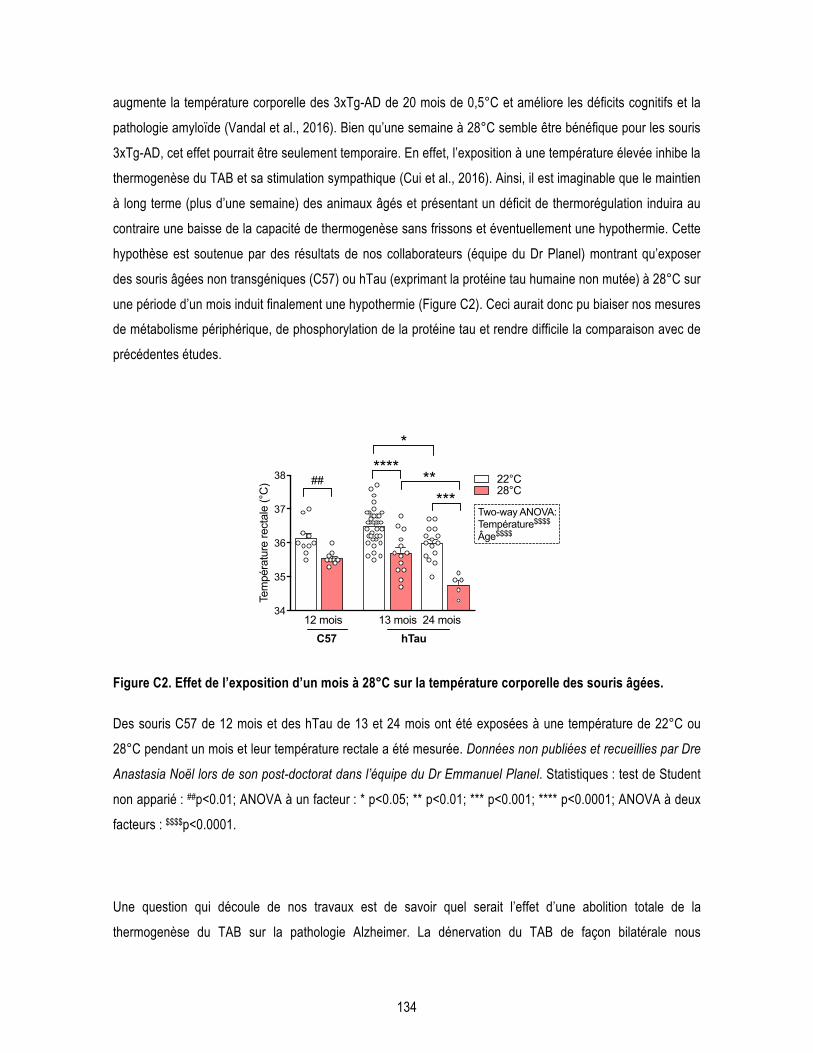
134
augmente la température corporelle des 3xTg-AD de 20 mois de 0,5°C et améliore les déficits cognitifs et la
pathologie amyloïde (Vandal et al., 2016). Bien qu’une semaine à 28°C semble être bénéfique pour les souris
3xTg-AD, cet effet pourrait être seulement temporaire. En effet, l’exposition à une température élevée inhibe la
thermogenèse du TAB et sa stimulation sympathique (Cui et al., 2016). Ainsi, il est imaginable que le maintien
à long terme (plus d’une semaine) des animaux âgés et présentant un déficit de thermorégulation induira au
contraire une baisse de la capacité de thermogenèse sans frissons et éventuellement une hypothermie. Cette
hypothèse est soutenue par des résultats de nos collaborateurs (équipe du Dr Planel) montrant qu’exposer
des souris âgées non transgéniques (C57) ou hTau (exprimant la protéine tau humaine non mutée) à 28°C sur
une période d’un mois induit finalement une hypothermie (Figure C2). Ceci aurait donc pu biaiser nos mesures
de métabolisme périphérique, de phosphorylation de la protéine tau et rendre difficile la comparaison avec de
précédentes études.
Figure C2. Effet de l’exposition d’un mois à 28°C sur la température corporelle des souris âgées.
Des souris C57 de 12 mois et des hTau de 13 et 24 mois ont été exposées à une température de 22°C ou
28°C pendant un mois et leur température rectale a été mesurée. Données non publiées et recueillies par Dre
Anastasia Noël lors de son post-doctorat dans l’équipe du Dr Emmanuel Planel. Statistiques : test de Student
non apparié : ##p<0.01; ANOVA à un facteur : * p<0.05; ** p<0.01; *** p<0.001; **** p<0.0001; ANOVA à deux
facteurs : $$$$p<0.0001.
Une question qui découle de nos travaux est de savoir quel serait l’effet d’une abolition totale de la
thermogenèse du TAB sur la pathologie Alzheimer. La dénervation du TAB de façon bilatérale nous
34
35
36
37
38
Tem
péra
ture
rect
ale
(°C
)
****
***
12 mois 13 mois 24 moishTauC57
22°C28°C
##
*
**
Two-way ANOVA:Température$$$$
Âge$$$$
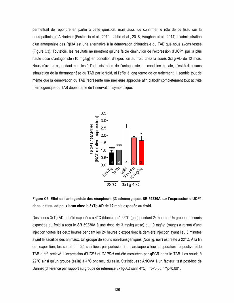
135
permettrait de répondre en partie à cette question, mais aussi de confirmer le rôle de ce tissu sur la
neuropathologie Alzheimer (Festuccia et al., 2010; Labbé et al., 2018; Vaughan et al., 2014). L’administration
d’un antagoniste des Rβ3A est une alternative à la dénervation chirurgicale du TAB que nous avons testée
(Figure C3). Toutefois, les résultats ne montrent qu’une faible diminution de l’expression d’UCP1 par la plus
haute dose d’antagoniste (10 mg/kg) en condition d’exposition au froid chez la souris 3xTg-AD de 12 mois.
Nous n’avons cependant pas testé l’administration de l’antagoniste en condition basale, c’est-à-dire sans
stimulation de la thermogenèse du TAB par le froid, ni l’effet à long terme de ce traitement. Il semble tout de
même que la dénervation du TAB représente une meilleure approche afin d’abolir complètement tout activité
thermogénique du TAB dépendante de l’innervation sympathique.
Figure C3. Effet de l’antagoniste des récepteurs β3 adrénergiques SR 59230A sur l’expression d’UCP1 dans le tissu adipeux brun chez la 3xTg-AD de 12 mois exposée au froid.
Des souris 3xTg-AD ont été exposées à 4°C (blanc) ou à 22°C (gris) pendant 24 heures. Un groupe de souris
exposées au froid a reçu le SR 59230A à une dose de 3 mg/kg (rose) ou 10 mg/kg (rouge) à raison d’une
injection toutes les deux heures pendant les 24 heures d’exposition; la dernière injection ayant lieu 5 minutes
avant le sacrifice des animaux. Un groupe de souris non-transgéniques (NonTg, noir) est resté à 22°C. À la fin
de l’exposition, les souris ont été sacrifiées par perfusion intracardiaque à leur température respective et le
TAB a été prélevé. L’expression d’UCP1 et GAPDH ont été mesurées par qPCR dans le TAB. Les souris à
22°C ainsi qu’un groupe (salin) à 4°C ont reçu du salin. Statistiques : ANOVA à un facteur, test post-hoc de
Dunnet (différence par rapport au groupe de référence 3xTg-AD salin 4°C) : *p<0.05; ***p<0.001.
NonTg
3xTg
salin
3 mg/k
g
10 m
g/kg
0.0
0.5
1.0
1.5
2.0
2.5
3.0
3.5
******
4 4 4 3 2
*
3xTg 4°C22°C
UC
P1 /
GAP
DH
(B
AT re
lativ
e ex
pres
sion
)
136
Nous avons observé une augmentation des marqueurs de thermogenèse du TAB en réponse aux expositions
répétées au froid et aux injections d’agoniste des Rβ3A chez des souris âgées de 16 mois. Bien que des
études montrent qu’il est possible de stimuler l’activité du TAB chez l’humain (Cypess et al., 2015; Hanssen et
al., 2015a), nos résultats ne sont probablement pas entièrement transposables à l’Homme car la
thermorégulation du rongeur et de l’humain sont très différentes. Par exemple, la quantité de TAB chez la
souris représente 0,8% de son poids total (environ 150 mg pour une souris jeune de 20 g), contre seulement
0,1% pour un humain de 80 kg (si l’on estime à 100 g de TAB) (Carpentier et al., 2018; U Din et al., 2016),
bien que cette valeur soit très variable d’une individu à l’autre. D’autre part, le métabolisme basal de la souris
est 10 fois supérieur à celui de l’Homme. Également, le flux sanguin au niveau du TAB en condition
d’exposition au froid chez la souris est près de 100 fois supérieur à celui observé chez l’humain (D. O. Foster
and Frydman, 1979). Finalement, l’amplitude de variation de la température corporelle au cours d’une journée
de 24 heures est de l’ordre de 3°C chez la souris contre 1,5°C chez l’humain (Gordon, 2017). Ainsi, bien que
des expositions au froid répétées ne soient évidemment pas envisageables en clinique, surtout sur des
personnes âgées, des études cliniques supplémentaires sur une population âgée sont nécessaires afin de
déterminer si l’approche pharmacologique serait transposable en clinique. En effet, les études évaluant la
quantité et l’activité du TAB par imagerie chez les personnes âgées de plus de 60 ans manquent. Il serait
également intéressant d’évaluer ces paramètres après l’administration d’un agoniste des Rβ3A.
2.3 Les altérations métaboliques et la MA
La souris 3xTg-AD est un modèle de choix pour étudier le lien entre altérations métaboliques et MA. Bien que
les déficits métaboliques soient moins importants chez les souris 3xTg-AD mâles par rapport aux femelles
(Vandal et al., 2015b), nous avons choisi d’utiliser uniquement des mâles dans la dernière étude afin de
s’affranchir du biais du cycle œstral des femelles sur la température corporelle. Dans le deuxième chapitre,
nous avons exposé les animaux à une diète enrichie en gras à 20% (w/w) afin d’augmenter leurs troubles
métaboliques. Cette diète représente davantage les niveaux de gras consommés par un humain dans les pays
occidentaux, mais n’est pas une diète très riche en gras induisant une importante obésité et un phénotype
diabétique comme celle riche en gras à 35% (w/w) (Vandal et al., 2014b).
Afin de connaître l’effet de nos expérimentations sur la dépense énergétique totale de nos animaux dans les
deux derniers chapitres, nous avons suivi le poids et la consommation de nourriture des animaux de façon
régulière, donnant une indication de la quantité d’énergie consommée en réponse à notre traitement. Des
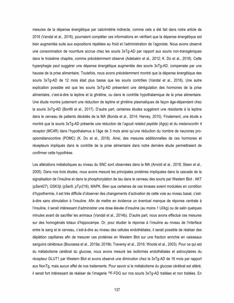
137
mesures de la dépense énergétique par calorimétrie indirecte, comme cela a été fait dans notre article de
2016 (Vandal et al., 2016), pourraient compléter ces informations en vérifiant que la dépense énergétique est
bien augmentée suite aux expositions répétées au froid et l’administration de l’agoniste. Nous avons observé
une consommation de nourriture accrue chez les souris 3xTg-AD par rapport aux souris non-transgéniques
dans le troisième chapitre, comme précédemment observé (Adebakin et al., 2012; K. Do et al., 2018). Cette
hyperphagie peut suggérer une dépense énergétique augmentée des souris 3xTg-AD, compensée par une
hausse de la prise alimentaire. Toutefois, nous avons précédemment montré que la dépense énergétique des
souris 3xTg-AD de 12 mois était plus basse que les souris contrôles (Vandal et al., 2016). Une autre
explication possible est que les souris 3xTg-AD présentent une dérégulation des hormones de la prise
alimentaire, c’est-à-dire la leptine et la ghréline, ou dans le contrôle hypothalamique de la prise alimentaire.
Une étude montre justement une réduction de leptine et ghréline plasmatiques de façon âge-dépendant chez
la souris 3xTg-AD (Bonfili et al., 2017). D’autre part, certaines études suggèrent une résistante à la leptine
dans le cerveau de patients décédés de la MA (Bonda et al., 2014; Harvey, 2010). Finalement, une étude a
montré que la souris 3xTg-AD présente une réduction de l’agouti related peptide (Agrp) et du melanocortin 4
receptor (MC4R) dans l’hypothalamus à l’âge de 3 mois ainsi qu’une réduction du nombre de neurones pro-
opiomélanocortine (POMC) (K. Do et al., 2018). Ainsi, des mesures additionnelles de ces hormones et
récepteurs impliqués dans le contrôle de la prise alimentaire dans notre dernière étude permettraient de
confirmer cette hypothèse.
Les altérations métaboliques au niveau du SNC sont observées dans la MA (Arnold et al., 2018; Steen et al.,
2005). Dans nos trois études, nous avons mesuré les principales protéines impliquées dans la cascade de la
signalisation de l’insuline et dans la phosphorylation de tau dans le cerveau des souris par Western Blot : AKT
(pSer427), GSK3β (pSer9, pTyr216), MAPK. Bien que certaines de ces kinases soient modulées en condition
d’hypothermie, il est très difficile d’observer des changements d’activation de cette voie au niveau basal, c’est-
à-dire sans stimulation à l’insuline. Afin de mettre en évidence un éventuel manque de réponse centrale à
l’insuline, il serait intéressant d’administrer une dose élevée d’insuline (au moins 1 UI/kg) ou de salin quelques
minutes avant de sacrifier les animaux (Vandal et al., 2014b). D’autre part, nous avons effectué ces mesures
sur des homogénats totaux d’hippocampe. Or, pour étudier la réponse à l’insuline au niveau de l’interface
entre le sang et le cerveau, c’est-à-dire au niveau des cellules endothéliales, il serait possible de réaliser des
déplétion capillaires afin de mesurer ces protéines en Western Blot sur une fraction enrichie en vaisseaux
sanguins cérébraux (Bourassa et al., 2019a; 2019b; Traversy et al., 2016; Woods et al., 2003). Pour ce qui est
du métabolisme cérébral du glucose, nous avons mesuré les isoformes endothéliales et astrocytaires du
récepteur GLUT1 par Western Blot et avons observé une diminution chez la 3xTg-AD de 16 mois par rapport
aux NonTg, mais aucun effet de nos traitements. Pour savoir si le métabolisme du glucose cérébral est altéré,
il serait fort intéressant de réaliser de l’imagerie 18F-FDG sur nos souris 3xTg-AD traitées et non traitées. En

138
effet, cette technique a permis de mettre en évidence une diminution du métabolisme du glucose dans
différentes régions cérébrales de la souris 3xTg-AD, qui est surtout bien visible à l’âge de 18 mois (Nicholson
et al., 2010). De plus, une diminution de la captation cérébrale du glucose a été montrée par la technique de
perfusion cérébrale in situ (PCIS) dans des 3xTg-AD de 18 mois (T. M. Do et al., 2014). Cette technique
consiste à perfuser une molécule marquée (ici le glucose) directement dans la carotide interne des souris
anesthésiées afin de quantifier le passage de la molécule à travers la BHE (Alata et al., 2014). Ainsi, cette
méthode permettrait de déterminer si la captation du glucose est modulée par nos traitements dans le cerveau
des souris.
2.4 Mécanismes potentiels par lesquels la thermorégulation peut affecter la MA
Nos travaux montrent pour la première fois que stimuler la thermorégulation du TAB permet de diminuer la
neuropathologie de la MA dans un modèle murin. Toutefois, nous n’avons que peu investigué les mécanismes
sous-jacents les liens entre thermorégulation et MA. Plusieurs pistes, qu’il serait intéressant d’investiguer dans
nos prochains travaux, sont présentées ici (Figure C4).
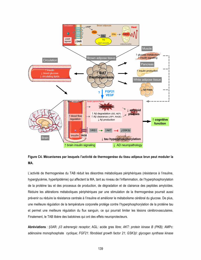
139
Figure C4. Mécanismes par lesquels l’activité de thermogenèse du tissu adipeux brun peut moduler la MA.
L’activité de thermogenèse du TAB réduit les désordres métaboliques périphériques (résistance à l’insuline,
hyperglycémie, hyperlipidémie) qui affectent la MA, tant au niveau de l’inflammation, de l’hyperphosphorylation
de la protéine tau et des processus de production, de dégradation et de clairance des peptides amyloïdes.
Réduire les altérations métaboliques périphériques par une stimulation de la thermogenèse pourrait aussi
prévenir ou réduire la résistance centrale à l’insuline et améliorer le métabolisme cérébral du glucose. De plus,
une meilleure régulation de la température corporelle protège contre l’hyperphosphorylation de la protéine tau
et permet une meilleure régulation du flux sanguin, ce qui pourrait limiter les lésions cérébrovasculaires.
Finalement, le TAB libère des batokines qui ont des effets neuroprotecteurs.
Abréviations : β3AR: β3 adrenergic receptor; AGL: acide gras libre; AKT: protein kinase B (PKB); AMPc:
adénosine monophosphate cyclique; FGF21: fibroblast growth factor 21; GSK3β: glycogen synthase kinase
↑ insulin↓ blood glucose↓ circulating lipids
↑ insulin production
↑ glucose metabolism↑ insulin signaling
↑AKT↑IRS1 ↓GSK3β
↓ tau hyperphosphorylation
↑ Aβ degradation (IDE, NEP)↑ Aβ clearance (LRP1, RAGE)
↓ Aβ production ↑ cognitive function
↑ brain insulin signaling ↓ AD neuropathology
brain parenchyma
↓ fat mass
↑ BAT thermogenesis
White adipose tissue
Pancreas
Muscle
Circulation
Brain
β3ARagonist
lipid vacuole
cAMP
HSL
TG
FFA
INSR
↓ amyloidplaques
β3AR
↑UCP1
mitochondria
PKA
↓inflammation
Brown adipose tissue
Circulation Brown adipocyteHeat
insulin
↑ blood flow regulation
FGF21VEGF
?
brain vasculature
140
3β; INSR: insulin receptor; IDE: insulin degrading enzyme; IRS1: insulin receptor substrate 1; LHS: lipase
hormonosensible; LRP1: low density lipoprotein receptor-related protein 1; NE: norépinephrine; NEP:
neprylisin; PKA: protéine kinase A ; RAGE: receptor of advanced glycated endproduct; TG: triglycérides ;
UCP1: uncoupling protein 1; VEGF: vascular endothelial growth factor.
Nos différentes études montrent clairement qu’une diminution de la température corporelle, induite par des
expositions aigües au froid par exemple, aggrave la neuropathologie de la MA. Une meilleure capacité de
thermorégulation pourrait donc prévenir l’hypothermie et ainsi limiter l’hyperphosphorylation de la protéine tau,
comme le démontre notre deuxième chapitre. Toutefois, le lien direct entre température corporelle,
thermorégulation et mémoire reste à élucider. Nos précédents travaux montrent qu’augmenter la température
corporelle de 0,5°C permet de diminuer les déficits mnésiques des souris 3xTg-AD de 20 mois (Vandal et al.,
2016). De façon surprenante, dans notre dernière étude, nous avons observé une corrélation positive
relativement forte entre la quantité d’UCP1 dans le TAB et le pourcentage d’augmentation du score au test de
reconnaissance d’objet chez les souris transgéniques spécifiquement. Ceci soutient l’hypothèse qu’une
augmentation d’activité de thermogenèse du TAB est impliquée dans l’amélioration mnésique suite à
l’administration d’un agoniste des Rβ3A, et qu’elle est bénéfique spécifiquement pour les souris 3xTg-AD.
Un mécanisme bien connu pour lier température corporelle et phosphorylation de tau est l’activité des kinases
et phosphatases comme mentionné plus haut (Planel et al., 2001). Les altérations métaboliques, qui peuvent
être corrigées par la stimulation du TAB, peuvent également influencer la phosphorylation de tau via l’activité
des kinases. En effet, GSK3β est la principale kinase impliquée dans la phosphorylation de la protéine tau
(Hooper et al., 2008). Plusieurs études proposent que la résistance à l’insuline diminue l’activation de la voie
PI3K/AKT, qui inhibe GSK3β (en augmentant sa phosphorylation sur le site Ser9), et donc finalement conduit
à une plus grande activité de cette dernière et une hyperphosphorylation de tau (Gratuze et al., 2018; Griffin et
al., 2005; Jolivalt et al., 2008; Qu et al., 2011). Ainsi, une meilleure réponse centrale à l’insuline permettrait de
diminuer la phosphorylation de tau. Certains travaux montrent que l’administration d’insuline dans des
modèles animaux de la MA permet de diminuer l’hyperphosphorylation de tau (Chen et al., 2014a; Gratuze et
al., 2017b), même dans la fraction insoluble correspondant aux protéines tau agrégées (résultats
supplémentaires : (Vandal et al., 2014b)). D’ailleurs, l’administration du CL-316,243 induit une importante
sécrétion d’insuline (Atef et al., 1996; MacPherson et al., 2014). Il est donc envisageable que les effets
bénéfiques de l’agoniste des Rβ3A sur la neuropathologie de la MA et la mémoire soient en partie dus à la
libération d’insuline dans la circulation.
141
Les résultats des Chapitres 2 et 3 confirment que la stimulation du TAB, que ce soit par les expositions
répétées au froid ou par l’administration du CL-316,243, permet d’améliorer la tolérance au glucose des souris
âgées. Comme mentionné en introduction, l’hyperglycémie chronique augmente la neuropathologie de la MA
dans des modèles animaux de la maladie (Gratuze et al., 2017b; Guo et al., 2016; Kim et al., 2013). De plus,
le diabète induit des lésions vasculaires importantes (Sonnen et al., 2009). Réduire l’intolérance au glucose
induite par une diète riche en gras par un régime hypocalorique permet de réduire la pathologie amyloïde et la
neuroinflammation chez la souris APP/PS1 (Walker et al., 2017). Ainsi, une plus importante activité de
thermogenèse du TAB pourrait être protectrice contre les effets néfastes de l’hyperglycémie sur le cerveau.
De manière générale, les altérations métaboliques périphériques induisent également une augmentation de la
neuroinflammation et du stress oxydatif, qui sont associés à la MA et principalement à la pathologie amyloïde
(De Felice and Ferreira, 2014; Theriault et al., 2016; Verdile et al., 2015). Bien que nous n’ayons pas étudié
ces aspects dans nos études, il est possible que nos traitements permettent de diminuer la neuroinflammation
et le stress oxydatif dans le cerveau des souris 3xTg-AD âgées. En effet, nous avons observé une diminution
de l’intolérance au glucose et des triglycérides plasmatiques dans le Chapitre 2, et une diminution du poids et
une meilleure tolérance au glucose dans le Chapitre 3.
La régulation de la température corporelle est intimement liée à la pression sanguine. En effet, la
vasodilatation permet de diminuer la température corporelle par diffusion de la chaleur, et à l’inverse, la
vasoconstriction des vaisseaux permet de limiter les pertes de chaleur en condition de froid, et de diffuser plus
rapidement la chaleur produite par la thermogenèse (Smith and Johnson, 2016). De plus, l’activité accrue du
TAB suite à une stimulation par le froid induit une augmentation du flux sanguin au niveau local (Baron et al.,
2012; Orava et al., 2011). Il est bien connu maintenant que le flux sanguin cérébral affecte l’activité neuronale,
le métabolisme et les fonctions cérébrales (Iadecola, 2017). Mais surtout, des dysfonctions dans le couplage
neurovasculaire ont été identifiées dans la MA (Bell and Zlokovic, 2009; Hamel et al., 2016), et sont également
observées chez la souris 3xTg-AD (Lourenço et al., 2017). Afin de vérifier si la stimulation du TAB permet une
meilleure régulation du débit sanguin cérébral, ce paramètre pourrait être mesuré en réponse à une exposition
au froid ou à l’administration de l’agoniste Rβ3A grâce à la méthode du laser doppler (Nicolakakis et al.,
2008). L’inconvénient de cette méthode est qu’elle nécessite un amincissement du crâne des souris et surtout
leur anesthésie, qui peut influencer également la régulation du flux sanguin. En revanche, de nouvelles
méthodes d’imagerie du flux sanguin cérébral par ultrasons (fUS imaging) sur souris vigiles ont été
développées et permettraient de suivre l’évolution du débit sanguin cérébral tout au long de nos traitements et
à différents âges (Deffieux et al., 2018; Tiran et al., 2017).
142
Un autre mécanisme reliant thermogenèse du TAB et MA pourrait être la sécrétion de batokines (Villarroya et
al., 2017). En effet, nous avons observé une augmentation de FGF21 dans le plasma des souris ayant reçu
les expositions répétées au froid, et ces niveaux étaient corrélés négativement à la phosphorylation de tau
dans l’hippocampe de ces souris. Cette deuxième étude soulève la question de savoir si FGF21 peut exercer
directement un effet sur la phosphorylation de tau dans l’hippocampe. Pour appuyer cette hypothèse,
quelques études suggèrent que FGF21 améliore la mémoire de souris obèses, protège contre la toxicité
induite par les peptides Aβ42 in vitro et des lésions de la BHE (Amiri et al., 2018; Chen et al., 2018; Sa-
Nguanmoo et al., 2016b; Q. Wang et al., 2018; Yu et al., 2018). De plus, une étude a montré que FGF21
pouvait traverser la BHE (Hsuchou et al., 2007) alors que son récepteur FGFR1 est présent au niveau
cérébral, bien que son rôle soit peu connu pour le moment (Sun et al., 2007). Ainsi, il serait intéressant
d’administrer FGF21 à des souris transgéniques et d’évaluer l’effet de ce traitement sur la mémoire et la
neuropathologie de la MA.
D’autre part, le TAB sécrète le facteur de croissance de l’endothélium vasculaire (VEGF) en condition de
stimulation afin d’augmenter l’angiogenèse dans le tissu (S. Lim et al., 2012; Villarroya et al., 2017). Ce facteur
de croissance est connu pour avoir des effets neuroprotecteurs (Storkebaum et al., 2004). Les niveaux de
VEGF plasmatiques sont plus bas chez les sujets Alzheimer, et les quantités dans le LCR sont associées à
l’atrophie de l’hippocampe et la mémoire chez des individus atteints de la MA (Hohman et al., 2015; Mateo et
al., 2007). De plus, l’administration de VEGF permet de renverser les déficits cognitifs et d’augmenter les
marqueurs cérébrovasculaires dans des souris diabétiques et un modèle murin de la MA (S. L. Taylor et al.,
2015; P. Wang et al., 2011). Ainsi, la stimulation du TAB pourrait avoir un effet sur la mémoire et la protection
de la vasculature cérébrale via la production de VEGF.
Notre troisième chapitre soulève la question de savoir si l’agoniste des Rβ3A a un effet direct au niveau du
SNC ou si son action est exclusivement restreinte à la périphérie, plus précisément au niveau du TAB. En
effet, il n’est pas exclu que l’agoniste ait un effet direct au niveau du SNC puisque certaines études montrent
que le cerveau exprime, bien que faiblement, des Rβ3A (B. A. Evans et al., 1996; Summers et al., 1995). Les
bases de données de single-cell RNA sequencing dans le cerveau de souris montrent également une
expression très faible du gène Adrb3 (Ye Zhang et al., 2014). L’administration de l’agoniste des Rβ3A à des
souris ayant une délétion des Rβ3A spécifiquement sur les adipocytes ou dans le SNC permettrait de savoir si
son action est exclusivement restreinte à la périphérie (plus précisément au niveau du TAB) ou non. Toutefois,
il faudrait d’abord vérifier la localisation cellulaire de ces récepteurs dans le cerveau : sont-ils présents à la
surface des cellules endothéliales, permettant une atteinte de la molécule présente dans le sang ? Ou bien
sont-ils présents dans le parenchyme cérébral ? Dans ce deuxième cas, l’action de l’agoniste dans le SNC
nécessiterait son passage à travers la BHE, passage restreint à de très petites molécules ou à celles
143
bénéficiant d’un transporteur spécifique (Pardridge, 2012). Bien que certains articles mentionnent que le CL-
316,243 ne traverse pas la BHE, ceci pourrait être vérifié par l’injection intraveineuse de la molécule à des
souris puis son dosage dans le cerveau par chromatographie liquide à haute performance (HPLC) couplé à la
spectrométrie de masse. D’autre part, la méthode de la PCIS permettrait de quantifier son taux de passage en
µL par gramme de cerveau par seconde et de savoir si ce passage se fait grâce à un transporteur en faisant
une expérience de saturation (St-Amour et al., 2013). Toutefois, cette dernière méthode nécessite d’avoir la
molécule sous forme radio-marquée.
2.5 Études précliniques dans la MA
Travailler avec des modèles animaux présente plusieurs limitations pour la translation vers l’humain.
Toutefois, les études précliniques sont obligatoires pour développer de nouveaux traitements et permettent de
mieux comprendre certains aspects de la pathogenèse de la MA ainsi que les mécanismes d’action de
diverses interventions. Travailler avec la souris 3xTg-AD comporte plusieurs limitations, comme tous les
modèles animaux de la MA. La plus importante limitation est que cette souris est un modèle de la forme
familiale et non de la forme sporadique de la MA. Comme il n’existe pour le moment aucun véritable modèle
murin de la forme sporadique de la MA, les études précliniques dans la MA sont confrontées à cette limitation
majeure. Une autre limite de la souris 3xTg-AD est qu’elle ne présente pas de mort neuronale malgré la
présence des marqueurs neuropathologiques de la MA. Finalement, il s’agit d’un modèle de surexpression, qui
présente ses désavantages, mais cette surexpression demeure dans les faits très progressive dans ce
modèle. D’autre part, cette souris présente de nombreux avantages dans le cadre de nos études. Tout
d’abord, elle présente à la fois les pathologies tau et amyloïde, permettant d’analyser l’effet de traitements sur
les deux marqueurs neuropathologiques à la fois et leur interaction (G. S. Bloom, 2014; Oddo et al., 2003b; St-
Amour et al., 2014). Cela nous a d’ailleurs permis de mettre en évidence que nos différentes approches n’ont
pas les mêmes effets sur ces deux marqueurs neuropathologiques au sein d’un même modèle animal. De
plus, il faut attendre au moins 8 à 10 mois avant d’observer des ENF de façon relativement répandu dans
l’hippocampe ainsi que quelques plaques amyloïdes dans le subiculum des animaux (Belfiore et al., 2018).
Ceci nous oblige à travailler avec des animaux âgés, ce qui a une répercussion majeure dans le coût de nos
études ainsi que la planification des expérimentations à long terme, mais nous permet de prendre en compte
le paramètre du vieillissement dans nos expérimentations. Comme le principal facteur de risque de la MA
demeure l’âge avancé, travailler avec des animaux âgés est important dans les études précliniques. D’autre
part, notre équipe a observé plusieurs altérations métaboliques chez la 3xTg-AD (Vandal et al., 2015b;
2014b), mais surtout un déficit de thermorégulation (Vandal et al., 2016), faisant de ce modèle un très bon
144
choix pour nos études. Toutefois, il serait intéressant de reproduire nos résultats dans d’autres modèles de la
MA. Puisque nous avons obtenu des résultats sur le comportement et la pathologie amyloïde, il serait
intéressant de vérifier l’effet de l’agoniste des Rβ3A dans un modèle de pathologie amyloïde pure, et non
induit par des mutations génétiques, comme une souris ayant reçu des injections de peptides Aβ directement
dans l’hippocampe (Brouillette et al., 2012; Shankar et al., 2008).
Nous avons montré pour la première fois qu’un agoniste des Rβ3A exerçait des effets bénéfiques chez la
souris 3xTg-AD de 16 mois après un mois de traitement. Puisque la pathologie et les déficits
comportementaux apparaissent dès l’âge de 12 mois, et que nous avons administré le traitement de 15 à 16
mois, notre étude avait pour but de vérifier si cette molécule pouvait avoir un effet thérapeutique en
ralentissant la progression ou en réduisant la pathologie dans un modèle murin de la MA. Il serait intéressant
de déterminer l’effet de cet agoniste selon différents paradigmes expérimentaux (Shineman et al., 2011). Par
exemple, l’agoniste pourrait être administré dans le stade « pré-symptomatique » chez la souris 3xTg-AD (dès
l’âge de 6 mois par exemple), afin d‘évaluer son potentiel en prévention. D’autre part, les animaux pourraient
être sacrifiés après une période de « wash-out » d’un mois ou plus afin de vérifier si le traitement a des effets
à long terme (Flores et al., 2018).
Au niveau comportemental, nos résultats ont permis de mettre en évidence une amélioration de la mémoire de
reconnaissance induite par le traitement avec l’agoniste grâce au test de reconnaissance d’objet. Ce test est
approprié pour la souris 3xTg-AD âgée car il induit peu de stress, représente une tâche relativement facile
(dans le cas d’un délai d’une heure entre les deux phases de test) et est assez sensible pour détecter des
déficits mnésiques dès l’âge de 12 mois. Toutefois, nous aurions pu vérifier l’effet de notre traitement sur la
mémoire spatiale telle qu’évaluée par le test du Y-Maze, qui est un test induisant peu de stress et relativement
rapide à effectuer également, un critère important dans la mesure où nous avons effectué un traitement
pharmacologique d’un mois seulement (Wolf et al., 2016). Le labyrinthe radial à 8 bras et le test de la piscine
de Morris mesurent la mémoire spatiale à long terme, mais nécessitent plusieurs journées d’expérimentation
(Puzzo et al., 2014). De plus, le test de la piscine de Morris implique de placer les animaux dans l’eau, certes
à température contrôlée, mais aurait interféré avec les mécanismes naturels de thermorégulation de nos
animaux.
Globalement, il existe assez peu de translation des résultats obtenus chez la souris vers l’humain, surtout
dans le domaine de la MA. Dans notre cas, la translation de nos résultats de la souris vers l’Homme pourrait
être relativement rapide car un agoniste des Rβ3A, le mirabegron, est déjà utilisé en clinique. De plus, des
études cliniques pour une utilisation thérapeutique de ce médicament dans le diabète et l’obésité sont en
cours (NCT02811289; NCT03012113). Bien que le mirabegron soit sécuritaire et efficace pour le traitement de
145
la vessie hyperactive chez les personnes âgées (Chapple and Siddiqui, 2017), il reste à déterminer s’il pourrait
être efficace pour activer le TAB dans une population âgée. Des études longitudinales évaluant l’incidence de
MA dans une population recevant le mirabegron pourraient aider à évaluer l’intérêt d’un tel traitement dans la
maladie. Dans le cadre de la MA, ce traitement pourrait bénéficier plus spécifiquement à une sous-population
ayant un déficit de thermorégulation ou des maladies métaboliques.
3 Conclusion générale
La MA est une maladie neurodégénérative qui affecte un nombre considérable de personnes âgées à travers
le monde. Elle représente aujourd’hui un problème de santé publique majeur pour nos sociétés.
Malheureusement, il n’existe pour le moment aucun traitement curatif pour la MA et le développement de
nouvelles stratégies thérapeutiques stagne depuis plusieurs années. Plusieurs raisons expliquent ces
lacunes : l’hétérogénéité des patients, les causes exactes de la maladie demeures inconnues, le manque de
modèles animaux et de biomarqueurs fiables. Pour réellement faire avancer la recherche sur la MA, il est
nécessaire de sortir des sentiers battus et d’explorer de nouvelles pistes.
L’objectif de cette thèse était de déterminer si la thermorégulation pouvait être une cible thérapeutique
intéressante dans la MA. Nous avons investigué une piste originale et très peu étudiée dans le domaine.
Ensemble, nos données démontrent que les interventions visant à stimuler la thermogenèse permettent à la
fois de contrer l’hyperphosphorylation de la protéine tau induite par une hypothermie, de diminuer la
pathologie amyloïde et d’améliorer les déficits cognitifs dans un modèle murin de la MA. De manière plus
générale, les travaux issus de cette thèse montrent que moduler la thermorégulation permet d’atténuer la
neuropathologie et les symptômes de la MA. Ainsi, nos résultats présentent la thermorégulation comme une
nouvelle cible thérapeutique et ouvrent la voie à de nouvelles stratégies pour cette maladie, un enjeu crucial
pour la découverte de traitements innovants.
Ces résultats inciteront peut-être d’autres équipes à étudier les mécanismes reliant la thermorégulation à la
MA, que ce soit chez l’animal ou l’humain. Des études cliniques chez l’Homme sont cruciales pour confirmer
l’efficacité de telles interventions dans la MA. Malgré cela, cette piste semble prometteuse car elle permet de
cibler plusieurs altérations périphériques et centrales survenant dans la MA en même temps.
146
Bibliographie Abbondante, S., Baglietto-Vargas, D., Rodriguez-Ortiz, C.J., Estrada-Hernandez, T., Medeiros, R., LaFerla,
F.M., 2014. Genetic ablation of tau mitigates cognitive impairment induced by type 1 diabetes. The American Journal of Pathology 184, 819–826. doi:10.1016/j.ajpath.2013.11.021
Abe, J., Okazawa, M., Adachi, R., Matsumura, K., Kobayashi, S., 2003. Primary cold-sensitive neurons in acutely dissociated cells of rat hypothalamus. Neurosci. Lett. 342, 29–32.
Adebakin, A., Bradley, J., Gümüsgöz, S., Waters, E.J., Lawrence, C.B., 2012. Impaired satiation and increased feeding behaviour in the triple-transgenic Alzheimer's disease mouse model. PLoS ONE 7, e45179. doi:10.1371/journal.pone.0045179
Adzovic, L., Lynn, A.E., D'Angelo, H.M., Crockett, A.M., Kaercher, R.M., Royer, S.E., Hopp, S.C., Wenk, G.L., 2015. Insulin improves memory and reduces chronic neuroinflammation in the hippocampus of young but not aged brains. J Neuroinflammation 12, 63. doi:10.1186/s12974-015-0282-z
Ahn, B.-H., Kim, H.-S., Song, S., Lee, I.H., Liu, J., Vassilopoulos, A., Deng, C.-X., Finkel, T., 2008. A role for the mitochondrial deacetylase Sirt3 in regulating energy homeostasis. Proc. Natl. Acad. Sci. U.S.A. 105, 14447–14452. doi:10.1073/pnas.0803790105
Alata, W., Paris-Robidas, S., Emond, V., Bourasset, F., Calon, F., 2014. Brain uptake of a fluorescent vector targeting the transferrin receptor: a novel application of in situ brain perfusion. Mol. Pharm. 11, 243–253. doi:10.1021/mp400421a
Alata, W., Ye, Y., St-Amour, I., Vandal, M., Calon, F., 2015. Human apolipoprotein E ɛ4 expression impairs cerebral vascularization and blood-brain barrier function in mice. J. Cereb. Blood Flow Metab. 35, 86–94. doi:10.1038/jcbfm.2014.172
Allan, L.M., Rowan, E.N., Firbank, M.J., Thomas, A.J., Parry, S.W., Polvikoski, T.M., O'Brien, J.T., Kalaria, R.N., 2011. Long term incidence of dementia, predictors of mortality and pathological diagnosis in older stroke survivors. Brain 134, 3716–3727. doi:10.1093/brain/awr273
Allen, B., Ingram, E., Takao, M., Smith, M.J., Jakes, R., Virdee, K., Yoshida, H., Holzer, M., Craxton, M., Emson, P.C., Atzori, C., Migheli, A., Crowther, R.A., Ghetti, B., Spillantini, M.G., Goedert, M., 2002. Abundant tau filaments and nonapoptotic neurodegeneration in transgenic mice expressing human P301S tau protein. J Neurosci 22, 9340–9351.
Alonso, A.D., Di Clerico, J., Li, B., Corbo, C.P., Alaniz, M.E., Grundke-Iqbal, I., Iqbal, K., 2010. Phosphorylation of tau at Thr212, Thr231, and Ser262 combined causes neurodegeneration. J. Biol. Chem. 285, 30851–30860. doi:10.1074/jbc.M110.110957
Alzheimer's association, 2018. 2018 Alzheimer's disease facts and figures. Alzheimer's & Dementia 14, 367–429.
Alzheimer, A., Stelzmann, R.A., Schnitzlein, H.N., Murtagh, F.R., 1995. An English translation of Alzheimer's 1907 paper, "Uber eine eigenartige Erkankung der Hirnrinde"., Clinical anatomy (New York, N.Y.). Wiley-Blackwell. doi:10.1002/ca.980080612
Amir, S., Schiavetto, A., 1990. Injection of prostaglandin E2 into the anterior hypothalamic preoptic area activates brown adipose tissue thermogenesis in the rat. Brain Res. 528, 138–142.
Amiri, M., Braidy, N., Aminzadeh, M., 2018. Protective Effects of Fibroblast Growth Factor 21 Against Amyloid-Beta1-42-Induced Toxicity in SH-SY5Y Cells. Neurotox Res 34, 574–583. doi:10.1007/s12640-018-9914-2
An, Y., Varma, V.R., Varma, S., Casanova, R., Dammer, E., Pletnikova, O., Chia, C.W., Egan, J.M., Ferrucci, L., Troncoso, J., Levey, A.I., Lah, J., Seyfried, N.T., Legido-Quigley, C., O'Brien, R., Thambisetty, M., 2018. Evidence for brain glucose dysregulation in Alzheimer's disease. Alzheimers Dement 14, 318–329. doi:10.1016/j.jalz.2017.09.011
Anand, R., Gill, K.D., Mahdi, A.A., 2014. Therapeutics of Alzheimer's disease: Past, present and future. Neuropharmacology 76 Pt A, 27–50. doi:10.1016/j.neuropharm.2013.07.004
Ancelin, M.L., de Roquefeuil, G., Ledésert, B., Bonnel, F., Cheminal, J.C., Ritchie, K., 2001. Exposure to anaesthetic agents, cognitive functioning and depressive symptomatology in the elderly. Br J Psychiatry

147
178, 360–366. Andersen, B., Omar, B.A., Rakipovski, G., Raun, K., Ahrén, B., 2015. Fibroblast growth factor 21 prevents
glycemic deterioration in insulin deficient mouse models of diabetes. Eur. J. Pharmacol. 764, 189–194. doi:10.1016/j.ejphar.2015.07.003
Andorfer, C., Kress, Y., Espinoza, M., de Silva, R., Tucker, K.L., Barde, Y.-A., Duff, K., Davies, P., 2003. Hyperphosphorylation and aggregation of tau in mice expressing normal human tau isoforms. Journal of Neurochemistry 86, 582–590.
Andrews-Zwilling, Y., Bien-Ly, N., Xu, Q., Li, G., Bernardo, A., Yoon, S.Y., Zwilling, D., Yan, T.X., Chen, L., Huang, Y., 2010. Apolipoprotein E4 causes age- and Tau-dependent impairment of GABAergic interneurons, leading to learning and memory deficits in mice. J Neurosci 30, 13707–13717. doi:10.1523/JNEUROSCI.4040-10.2010
Anstey, K.J., Sanden, von, C., Salim, A., O'Kearney, R., 2007. Smoking as a risk factor for dementia and cognitive decline: a meta-analysis of prospective studies. Am. J. Epidemiol. 166, 367–378. doi:10.1093/aje/kwm116
Antunes, M., Biala, G., 2012. The novel object recognition memory: neurobiology, test procedure, and its modifications. Cogn Process 13, 93–110. doi:10.1007/s10339-011-0430-z
Arch, J.R.S., 2011. Challenges in β(3)-Adrenoceptor Agonist Drug Development. Ther Adv Endocrinol Metab 2, 59–64. doi:10.1177/2042018811398517
Arendt, T., 2009. Synaptic degeneration in Alzheimer’s disease. Acta Neuropathol 118, 167–179. doi:10.1007/s00401-009-0536-x
Arendt, T., 2003. Synaptic plasticity and cell cycle activation in neurons are alternative effector pathways: the 'Dr. Jekyll and Mr. Hyde concept‘ of Alzheimer’s disease or the yin and yang of neuroplasticity. Prog. Neurobiol. 71, 83–248.
Arendt, T., Bullmann, T., 2013. Neuronal plasticity in hibernation and the proposed role of the microtubule-associated protein tau as a “master switch” regulating synaptic gain in neuronal networks. Am. J. Physiol. Regul. Integr. Comp. Physiol. 305, R478–89. doi:10.1152/ajpregu.00117.2013
Arendt, T., Stieler, J., Strijkstra, A.M., Hut, R.A., Rüdiger, J., Van der Zee, E.A., Harkany, T., Holzer, M., Härtig, W., 2003. Reversible paired helical filament-like phosphorylation of tau is an adaptive process associated with neuronal plasticity in hibernating animals. J Neurosci 23, 6972–6981.
Arevalo, M.-A., Azcoitia, I., Garcia-Segura, L.M., 2015. The neuroprotective actions of oestradiol and oestrogen receptors. Nat. Rev. Neurosci. 16, 17–29. doi:10.1038/nrn3856
Arevalo-Rodriguez, I., Smailagic, N., Roqué I Figuls, M., Ciapponi, A., Sanchez-Perez, E., Giannakou, A., Pedraza, O.L., Bonfill Cosp, X., Cullum, S., 2015. Mini-Mental State Examination (MMSE) for the detection of Alzheimer's disease and other dementias in people with mild cognitive impairment (MCI). Cochrane Database Syst Rev 25, CD010783. doi:10.1002/14651858.CD010783.pub2
Arnold, S.E., Arvanitakis, Z., Macauley-Rambach, S.L., Koenig, A.M., Wang, H.-Y., Ahima, R.S., Craft, S., Gandy, S., Buettner, C., Stoeckel, L.E., Holtzman, D.M., Nathan, D.M., 2018. Brain insulin resistance in type 2 diabetes and Alzheimer disease: concepts and conundrums. Nat Rev Neurol 14, 168–181. doi:10.1038/nrneurol.2017.185
Arriagada, P.V., Growdon, J.H., Hedley-Whyte, E.T., Hyman, B.T., 1992. Neurofibrillary tangles but not senile plaques parallel duration and severity of Alzheimer's disease. Neurology 42, 631–639.
Arsenault, D., Julien, C., Tremblay, C., Calon, F., 2011. DHA improves cognition and prevents dysfunction of entorhinal cortex neurons in 3xTg-AD mice. 6, e17397. doi:10.1371/journal.pone.0017397
Asuni, A.A., Boutajangout, A., Quartermain, D., Sigurdsson, E.M., 2007. Immunotherapy targeting pathological tau conformers in a tangle mouse model reduces brain pathology with associated functional improvements. J Neurosci 27, 9115–9129. doi:10.1523/JNEUROSCI.2361-07.2007
Atef, N., Lafontan, M., Doublé, A., Hélary, C., Ktorza, A., Pénicaud, L., 1996. A specific beta 3-adrenoceptor agonist induces increased pancreatic islet blood flow and insulin secretion in rats. Eur. J. Pharmacol. 298, 287–292.
Atgié, C., D'Allaire, F., Bukowiecki, L.J., 1997. Role of beta1- and beta3-adrenoceptors in the regulation of lipolysis and thermogenesis in rat brown adipocytes. Am. J. Physiol. 273, C1136–42.
148
Attems, J., Jellinger, K., Thal, D.R., Van Nostrand, W., 2011. Review: sporadic cerebral amyloid angiopathy. Neuropathol. Appl. Neurobiol. 37, 75–93. doi:10.1111/j.1365-2990.2010.01137.x
Augustinack, J.C., Schneider, A., Mandelkow, E.-M., Hyman, B.T., 2002. Specific tau phosphorylation sites correlate with severity of neuronal cytopathology in Alzheimer's disease. Acta Neuropathol 103, 26–35.
Avila, J., Lucas, J.J., Perez, M., Hernández, F., 2004. Role of tau protein in both physiological and pathological conditions. Physiol. Rev. 84, 361–384. doi:10.1152/physrev.00024.2003
Åberg, D., Johansson, P., Isgaard, J., Wallin, A., Johansson, J.-O., Andreasson, U., Blennow, K., Zetterberg, H., Åberg, N.D., Svensson, J., 2015. Increased Cerebrospinal Fluid Level of Insulin-like Growth Factor-II in Male Patients with Alzheimer's Disease. J. Alzheimers Dis. 48, 637–646. doi:10.3233/JAD-150351
Baglietto-Vargas, D., Shi, J., Yaeger, D.M., Ager, R., LaFerla, F.M., 2016. Diabetes and Alzheimer’s disease crosstalk. Neurosci Biobehav Rev 64, 272–287. doi:10.1016/j.neubiorev.2016.03.005
Bal, N.C., Maurya, S.K., Sopariwala, D.H., Sahoo, S.K., Gupta, S.C., Shaikh, S.A., Pant, M., Rowland, L.A., Bombardier, E., Goonasekera, S.A., Tupling, A.R., Molkentin, J.D., Periasamy, M., 2012. Sarcolipin is a newly identified regulator of muscle-based thermogenesis in mammals. Nat. Med. 18, 1575–1579. doi:10.1038/nm.2897
Baron, D.M., Clerte, M., Brouckaert, P., Raher, M.J., Flynn, A.W., Zhang, H., Carter, E.A., Picard, M.H., Bloch, K.D., Buys, E.S., Scherrer-Crosbie, M., 2012. In vivo noninvasive characterization of brown adipose tissue blood flow by contrast ultrasound in mice. Circ Cardiovasc Imaging 5, 652–659. doi:10.1161/CIRCIMAGING.112.975607
Bartness, T.J., Song, C.K., Demas, G.E., 2001. SCN efferents to peripheral tissues: implications for biological rhythms. J. Biol. Rhythms 16, 196–204.
Baskin, A.S., Linderman, J.D., Brychta, R.J., McGehee, S., Anflick-Chames, E., Cero, C., Johnson, J.W., O'Mara, A.E., Fletcher, L.A., Leitner, B.P., Duckworth, C.J., Huang, S., Cai, H., Garraffo, H.M., Millo, C.M., Dieckmann, W., Tolstikov, V., Chen, E.Y., Gao, F., Narain, N.R., Kiebish, M.A., Walter, P.J., Herscovitch, P., Chen, K.Y., Cypess, A.M., 2018. Regulation of Human Adipose Tissue Activation, Gallbladder Size, and Bile Acid Metabolism by a β3-Adrenergic Receptor Agonist. Diabetes db180462. doi:10.2337/db18-0462
Bateman, R.J., Xiong, C., Benzinger, T.L.-S., Fagan, A.M., Goate, A., Fox, N.C., Marcus, D.S., Cairns, N.J., Xie, X., Blazey, T.M., Holtzman, D.M., Santacruz, A., Buckles, V., Oliver, A., Moulder, K., Aisen, P.S., Ghetti, B., Klunk, W.E., McDade, E., Martins, R.N., Masters, C.L., Mayeux, R., Ringman, J.M., Rossor, M.N., Schofield, P.R., Sperling, R.A., Salloway, S., Morris, J.C., Dominantly Inherited Alzheimer Network, 2012. Clinical and biomarker changes in dominantly inherited Alzheimer's disease. N. Engl. J. Med. 367, 795–804. doi:10.1056/NEJMoa1202753
Belfiore, R., Rodin, A., Ferreira, E., Velazquez, R., Branca, C., Caccamo, A., Oddo, S., 2018. Temporal and regional progression of Alzheimer's disease-like pathology in 3xTg-AD mice. Aging Cell 12, e12873. doi:10.1111/acel.12873
Bell, R.D., Winkler, E.A., Singh, I., Sagare, A.P., Deane, R., Wu, Z., Holtzman, D.M., Betsholtz, C., Armulik, A., Sallstrom, J., Berk, B.C., Zlokovic, B.V., 2012. Apolipoprotein E controls cerebrovascular integrity via cyclophilin A. Nature 485, 512–516. doi:10.1038/nature11087
Bell, R.D., Zlokovic, B.V., 2009. Neurovascular mechanisms and blood-brain barrier disorder in Alzheimer's disease. Acta Neuropathol 118, 103–113. doi:10.1007/s00401-009-0522-3
Benedict, C., Hallschmid, M., Hatke, A., Schultes, B., Fehm, H.L., Born, J., Kern, W., 2004. Intranasal insulin improves memory in humans. Psychoneuroendocrinology 29, 1326–1334. doi:10.1016/j.psyneuen.2004.04.003
Benjamin, E.J., Blaha, M.J., Chiuve, S.E., Cushman, M., Das, S.R., Deo, R., de Ferranti, S.D., Floyd, J., Fornage, M., Gillespie, C., Isasi, C.R., Jiménez, M.C., Jordan, L.C., Judd, S.E., Lackland, D., Lichtman, J.H., Lisabeth, L., Liu, S., Longenecker, C.T., Mackey, R.H., Matsushita, K., Mozaffarian, D., Mussolino, M.E., Nasir, K., Neumar, R.W., Palaniappan, L., Pandey, D.K., Thiagarajan, R.R., Reeves, M.J., Ritchey, M., Rodriguez, C.J., Roth, G.A., Rosamond, W.D., Sasson, C., Towfighi, A., Tsao, C.W., Turner, M.B., Virani, S.S., Voeks, J.H., Willey, J.Z., Wilkins, J.T., Wu, J.H., Alger, H.M., Wong, S.S., Muntner, P., American Heart Association Statistics Committee and Stroke Statistics Subcommittee, 2017. Heart

149
Disease and Stroke Statistics-2017 Update: A Report From the American Heart Association. Circulation 135, e146–e603. doi:10.1161/CIR.0000000000000485
Bennett, D.A., Schneider, J.A., Arvanitakis, Z., Kelly, J.F., Aggarwal, N.T., Shah, R.C., Wilson, R.S., 2006. Neuropathology of older persons without cognitive impairment from two community-based studies. Neurology 66, 1837–1844. doi:10.1212/01.wnl.0000219668.47116.e6
Bennett, Schneider, J.A., Wilson, R.S., Bienias, J.L., Arnold, S.E., 2004. Neurofibrillary tangles mediate the association of amyloid load with clinical Alzheimer disease and level of cognitive function. Arch. Neurol. 61, 378–384. doi:10.1001/archneur.61.3.378
Benomar, Y., Amine, H., Crépin, D., Rifai, Al, S., Riffault, L., Gertler, A., Taouis, M., 2016. Central Resistin/TLR4 Impairs Adiponectin Signaling, Contributing to Insulin and FGF21 Resistance. Diabetes 65, 913–926. doi:10.2337/db15-1029
Bensalem, J., Servant, L., Alfos, S., Gaudout, D., Layé, S., Pallet, V., Lafenetre, P., 2016. Dietary Polyphenol Supplementation Prevents Alterations of Spatial Navigation in Middle-Aged Mice. Front Behav Neurosci 10, 9. doi:10.3389/fnbeh.2016.00009
Bereczki, E., Bogstedt, A., Höglund, K., Tsitsi, P., Brodin, L., Ballard, C., Svenningsson, P., Aarsland, D., 2017. Synaptic proteins in CSF relate to Parkinson's disease stage markers. NPJ Parkinsons Dis 3, 7. doi:10.1038/s41531-017-0008-2
Bereczki, E., Francis, P.T., Howlett, D., Pereira, J.B., Höglund, K., Bogstedt, A., Cedazo-Minguez, A., Baek, J.-H., Hortobágyi, T., Attems, J., Ballard, C., Aarsland, D., 2016. Synaptic proteins predict cognitive decline in Alzheimer's disease and Lewy body dementia. Alzheimers Dement 12, 1149–1158. doi:10.1016/j.jalz.2016.04.005
Berlau, D.J., Corrada, M.M., Head, E., Kawas, C.H., 2009. APOE epsilon2 is associated with intact cognition but increased Alzheimer pathology in the oldest old. Neurology 72, 829–834. doi:10.1212/01.wnl.0000343853.00346.a4
Biessels, G.J., 2014. Capitalising on modifiable risk factors for Alzheimer's disease. Lancet Neurol 13, 752–753. doi:10.1016/S1474-4422(14)70154-1
Biessels, G.J., Staekenborg, S., Brunner, E., Brayne, C., Scheltens, P., 2006. Risk of dementia in diabetes mellitus: a systematic review. Lancet Neurol 5, 64–74. doi:10.1016/S1474-4422(05)70284-2
Birks, J., 2006. Cholinesterase inhibitors for Alzheimer's disease. Cochrane Database Syst Rev CD005593. doi:10.1002/14651858.CD005593
Bisht, K., Sharma, K.P., Lecours, C., Gabriela Sánchez, M., Hajj, El, H., Milior, G., Olmos-Alonso, A., Gómez-Nicola, D., Luheshi, G., Vallières, L., Branchi, I., Maggi, L., Limatola, C., Butovsky, O., Tremblay, M.-È., 2016. Dark microglia: A new phenotype predominantly associated with pathological states. Glia 64, 826–839. doi:10.1002/glia.22966
Black, S., Gao, F., Bilbao, J., 2009. Understanding white matter disease: imaging-pathological correlations in vascular cognitive impairment. Stroke 40, S48–52. doi:10.1161/STROKEAHA.108.537704
Blennow, K., Zetterberg, H., 2018. Biomarkers for Alzheimer's disease: current status and prospects for the future. J. Intern. Med. 284, 643–663. doi:10.1111/joim.12816
Blondin, D.P., Daoud, A., Taylor, T., Tingelstad, H.C., Bézaire, V., Richard, D., Carpentier, A.C., Taylor, A.W., Harper, M.-E., Aguer, C., Haman, F., 2017. Four-week cold acclimation in adult humans shifts uncoupling thermogenesis from skeletal muscles to brown adipose tissue. J Physiol 595, 2099–2113. doi:10.1113/JP273395
Blondin, D.P., Labbé, S.M., Tingelstad, H.C., Noll, C., Kunach, M., Phoenix, S., Guérin, B., Turcotte, E.E., Carpentier, A.C., Richard, D., Haman, F., 2014. Increased brown adipose tissue oxidative capacity in cold-acclimated humans. J. Clin. Endocrinol. Metab. 99, E438–46. doi:10.1210/jc.2013-3901
Bloom, G.S., 2014. Amyloid-β and tau: the trigger and bullet in Alzheimer disease pathogenesis. JAMA Neurol 71, 505–508. doi:10.1001/jamaneurol.2013.5847
Bloom, J.D., Dutia, M.D., Johnson, B.D., Wissner, A., Burns, M.G., Largis, E.E., Dolan, J.A., Claus, T.H., 1992. Disodium (R,R)-5-[2-[[2-(3-chlorophenyl)-2-hydroxyethyl]-amino] propyl]-1,3-benzodioxole-2,2-dicarboxylate (CL 316,243). A potent beta-adrenergic agonist virtually specific for beta 3 receptors. A promising antidiabetic and antiobesity agent. J. Med. Chem. 35, 3081–3084.

150
Body temperature as a risk factor for Alzheimer’s disease, 2000. Body temperature as a risk factor for Alzheimer’s disease 55, 440–444. doi:10.1054/mehy.2000.1085
Bonda, D.J., Stone, J.G., Torres, S.L., Siedlak, S.L., Perry, G., Kryscio, R., Jicha, G., Casadesus, G., Smith, M.A., Zhu, X., Lee, H.-G., 2014. Dysregulation of leptin signaling in Alzheimer disease: evidence for neuronal leptin resistance. Journal of Neurochemistry 128, 162–172. doi:10.1111/jnc.12380
Bonfili, L., Cecarini, V., Berardi, S., Scarpona, S., Suchodolski, J.S., Nasuti, C., Fiorini, D., Boarelli, M.C., Rossi, G., Eleuteri, A.M., 2017. Microbiota modulation counteracts Alzheimer's disease progression influencing neuronal proteolysis and gut hormones plasma levels. Nature Publishing Group 7, 2426. doi:10.1038/s41598-017-02587-2
Bons, N., Rieger, F., Prudhomme, D., Fisher, A., Krause, K.-H., 2006. Microcebus murinus: a useful primate model for human cerebral aging and Alzheimer's disease? Genes Brain Behav. 5, 120–130. doi:10.1111/j.1601-183X.2005.00149.x
Bookout, A.L., de Groot, M.H.M., Owen, B.M., Lee, S., Gautron, L., Lawrence, H.L., Ding, X., Elmquist, J.K., Takahashi, J.S., Mangelsdorf, D.J., Kliewer, S.A., 2013. FGF21 regulates metabolism and circadian behavior by acting on the nervous system. Nat. Med. 19, 1147–1152. doi:10.1038/nm.3249
Bories, C., Guitton, M.J., Julien, C., Tremblay, C., Vandal, M., Msaid, M., De Koninck, Y., Calon, F., 2012. Sex-dependent alterations in social behaviour and cortical synaptic activity coincide at different ages in a model of Alzheimer's disease. 7, e46111. doi:10.1371/journal.pone.0046111
Born, J., Lange, T., Kern, W., McGregor, G.P., Bickel, U., Fehm, H.L., 2002. Sniffing neuropeptides: a transnasal approach to the human brain. Nat. Neurosci. 5, 514–516. doi:10.1038/nn849
Bourassa, P., Alata, W., Tremblay, C., Paris-Robidas, S., Calon, F., 2019a. Transferrin Receptor-Mediated Uptake at the Blood-Brain Barrier Is Not Impaired by Alzheimer's Disease Neuropathology. Mol. Pharm. 16, 583–594. doi:10.1021/acs.molpharmaceut.8b00870
Bourassa, P., Tremblay, C., Schneider, J.A., Bennett, Calon, F., 2019b. Beta-amyloid pathology in human brain microvessel extracts from the parietal cortex: relation with cerebral amyloid angiopathy and Alzheimer’s disease. Acta Neuropathol 76, 1479–24. doi:10.1007/s00401-019-01967-4
Bourasset, F., Ouellet, M., Tremblay, C., Julien, C., Do, T.M., Oddo, S., LaFerla, F., Calon, F., 2009. Reduction of the cerebrovascular volume in a transgenic mouse model of Alzheimer's disease. Neuropharmacology 56, 808–813.
Braak, H., Braak, E., 1991. Neuropathological stageing of Alzheimer-related changes. Acta Neuropathol 82, 239–259.
Braak, H., Thal, D.R., Ghebremedhin, E., Del Tredici, K., 2011. Stages of the pathologic process in Alzheimer disease: age categories from 1 to 100 years. J. Neuropathol. Exp. Neurol. 70, 960–969. doi:10.1097/NEN.0b013e318232a379
Bretteville, A., Marcouiller, F., Julien, C., El-Khoury, N.B., Petry, F.R., Poitras, I., Mouginot, D., Lévesque, G., Hébert, S.S., Planel, E., 2012. Hypothermia-induced hyperphosphorylation: a new model to study tau kinase inhibitors. Nature Publishing Group 2, 480. doi:10.1038/srep00480
Bretteville, A., Planel, E., 2008. Tau aggregates: toxic, inert, or protective species? J. Alzheimers Dis. 14, 431–436.
Brinkmalm, A., Brinkmalm, G., Honer, W.G., Frölich, L., Hausner, L., Minthon, L., Hansson, O., Wallin, A., Zetterberg, H., Blennow, K., Öhrfelt, A., 2014. SNAP-25 is a promising novel cerebrospinal fluid biomarker for synapse degeneration in Alzheimer's disease. Molecular Neurodegeneration 9, 53. doi:10.1186/1750-1326-9-53
Bronnikov, G., Bengtsson, T., Kramarova, L., Golozoubova, V., Cannon, B., Nedergaard, J., 1999. beta1 to beta3 switch in control of cyclic adenosine monophosphate during brown adipocyte development explains distinct beta-adrenoceptor subtype mediation of proliferation and differentiation. Endocrinology 140, 4185–4197. doi:10.1210/endo.140.9.6972
Brouillette, J., 2014. The effects of soluble Aβ oligomers on neurodegeneration in Alzheimer's disease. Curr. Pharm. Des. 20, 2506–2519.
Brouillette, J., Caillierez, R., Zommer, N., Alves-Pires, C., Benilova, I., Blum, D., De Strooper, B., Buée, L., 2012. Neurotoxicity and memory deficits induced by soluble low-molecular-weight amyloid-β1-42
151
oligomers are revealed in vivo by using a novel animal model. J Neurosci 32, 7852–7861. doi:10.1523/JNEUROSCI.5901-11.2012
Brown, T.M., Piggins, H.D., 2007. Electrophysiology of the suprachiasmatic circadian clock. Prog. Neurobiol. 82, 229–255. doi:10.1016/j.pneurobio.2007.05.002
Buée, L., Bussière, T., Buée-Scherrer, V., Delacourte, A., Hof, P.R., 2000. Tau protein isoforms, phosphorylation and role in neurodegenerative disorders. Brain Res. Brain Res. Rev. 33, 95–130.
Burkey, B.F., Dong, M., Gagen, K., Eckhardt, M., Dragonas, N., Chen, W., Grosenstein, P., Argentieri, G., de Souza, C.J., 2000. Effects of pioglitazone on promoting energy storage, not expenditure, in brown adipose tissue of obese fa/fa Zucker rats: comparison to CL 316,243. Metabolism 49, 1301–1308. doi:10.1053/meta.2000.9524
Butchart, J., Holmes, C., 2012. Systemic and central immunity in Alzheimer's disease: therapeutic implications. CNS Neurosci Ther 18, 64–76. doi:10.1111/j.1755-5949.2011.00245.x
Calon, F., Lim, G.P., Yang, F., Morihara, T., Teter, B., Ubeda, O., Rostaing, P., Triller, A., Salem, N., Ashe, K.H., Frautschy, S.A., Cole, G.M., 2004. Docosahexaenoic acid protects from dendritic pathology in an Alzheimer's disease mouse model. Neuron 43, 633–645. doi:10.1016/j.neuron.2004.08.013
Calon, F., Tremblay, C., François, A., Vandal, M., Bennett, 2015. Neuropathological correlates of cognitive impairments in Alzheimer’s disease. J Neurosci 1–1.
Cannon, B., Nedergaard, J., 2010. Nonshivering thermogenesis and its adequate measurement in metabolic studies. Journal of Experimental Biology 214, 242–253. doi:10.1242/jeb.050989
Cannon, B., Nedergaard, J., 2004. Brown adipose tissue: function and physiological significance. Physiol. Rev. 84, 277–359. doi:10.1152/physrev.00015.2003
Caron, A., Labbé, S.M., Carter, S., Roy, M.-C., Lecomte, R., Ricquier, D., Picard, F., Richard, D., 2017. Loss of UCP2 impairs cold-induced non-shivering thermogenesis by promoting a shift toward glucose utilization in brown adipose tissue. Biochimie 134, 118–126. doi:10.1016/j.biochi.2017.01.006
Carpentier, A.C., Blondin, D.P., Virtanen, K.A., Richard, D., Haman, F., Turcotte, E.E., 2018. Brown Adipose Tissue Energy Metabolism in Humans. Front Endocrinol (Lausanne) 9, 447. doi:10.3389/fendo.2018.00447
Carrillo-Mora, P., Luna, R., Colín-Barenque, L., 2014. Amyloid beta: multiple mechanisms of toxicity and only some protective effects? Oxid Med Cell Longev 2014, 795375–15. doi:10.1155/2014/795375
Caruso, D., Barron, A.M., Brown, M.A., Abbiati, F., Carrero, P., Pike, C.J., Garcia-Segura, L.M., Melcangi, R.C., 2013. Age-related changes in neuroactive steroid levels in 3xTg-AD mice. Neurobiol. Aging 34, 1080–1089. doi:10.1016/j.neurobiolaging.2012.10.007
Castellano, C.-A., Nugent, S., Paquet, N., Tremblay, S., Bocti, C., Lacombe, G., Imbeault, H., Turcotte, E., Fulop, T., Cunnane, S.C., 2014. Lower Brain 18F-Fluorodeoxyglucose Uptake But Normal 11C-Acetoacetate Metabolism in Mild Alzheimer's Disease Dementia. J. Alzheimers Dis. 43, 1343–1353. doi:10.3233/JAD-141074
Castellano, J.M., Kim, J., Stewart, F.R., Jiang, H., DeMattos, R.B., Patterson, B.W., Fagan, A.M., Morris, J.C., Mawuenyega, K.G., Cruchaga, C., Goate, A.M., Bales, K.R., Paul, S.M., Bateman, R.J., Holtzman, D.M., 2011. Human apoE isoforms differentially regulate brain amyloid-β peptide clearance. Sci Transl Med 3, 89ra57–89ra57. doi:10.1126/scitranslmed.3002156
Caterina, M.J., 2007. Transient receptor potential ion channels as participants in thermosensation and thermoregulation. AJP: Regulatory, Integrative and Comparative Physiology 292, R64–76. doi:10.1152/ajpregu.00446.2006
Cavaleri, F., Bashar, E., 2018. Potential Synergies of β-Hydroxybutyrate and Butyrate on the Modulation of Metabolism, Inflammation, Cognition, and General Health. J Nutr Metab 2018, 7195760–13. doi:10.1155/2018/7195760
Chapon, P.A., Bessot, N., Gauthier, A., Besnard, S., Moussay, S., 2012. Performance testing of an innovative telemetric temperature sensor in animals. Journal of Thermal Biology 37, 255–259. doi:10.1016/j.jtherbio.2011.08.004
Chapple, C.R., (null), L., Nitti, V.W., Siddiqui, E., Michel, M.C., 2014a. Mirabegron in overactive bladder: a review of efficacy, safety, and tolerability. Neurourol. Urodyn. 33, 17–30. doi:10.1002/nau.22505
152
Chapple, C.R., Kaplan, S.A., Mitcheson, D., Blauwet, M.B., Huang, M., Siddiqui, E., Khullar, V., 2014b. Mirabegron 50 mg once-daily for the treatment of symptoms of overactive bladder: an overview of efficacy and tolerability over 12 weeks and 1 year. Int. J. Urol. 21, 960–967. doi:10.1111/iju.12568
Chapple, C.R., Siddiqui, E., 2017. Mirabegron for the treatment of overactive bladder: a review of efficacy, safety and tolerability with a focus on male, elderly and antimuscarinic poor-responder populations, and patients with OAB in Asia. Expert Rev Clin Pharmacol 10, 131–151. doi:10.1080/17512433.2017.1275570
Chatterjee, S., Peters, S.A.E., Woodward, M., Mejia Arango, S., Batty, G.D., Beckett, N., Beiser, A., Borenstein, A.R., Crane, P.K., Haan, M., Hassing, L.B., Hayden, K.M., Kiyohara, Y., Larson, E.B., Li, C.-Y., Ninomiya, T., Ohara, T., Peters, R., Russ, T.C., Seshadri, S., Strand, B.H., Walker, R., Xu, W., Huxley, R.R., 2016. Type 2 Diabetes as a Risk Factor for Dementia in Women Compared With Men: A Pooled Analysis of 2.3 Million People Comprising More Than 100,000 Cases of Dementia. Diabetes Care 39, 300–307. doi:10.2337/dc15-1588
Chen, H.-F., Chang-Quan, H., You, C., Wang, Z.-R., Hui, W., Liu, Q.-X., Si-Qing, H., 2013. The circadian rhythm of arterial blood pressure in Alzheimer disease (AD) patients without hypertension. Blood Press. 22, 101–105. doi:10.3109/08037051.2012.733508
Chen, J., Hu, J., Liu, H., Xiong, Y., Zou, Y., Huang, W., Shao, M., Wu, J., Yu, L., Wang, X., Wang, X., Lin, L., 2018. FGF21 Protects the Blood-Brain Barrier by Upregulating PPARγ via FGFR1/β-klotho after Traumatic Brain Injury. J. Neurotrauma 35, 2091–2103. doi:10.1089/neu.2017.5271
Chen, Run, X., Liang, Z., Zhao, Y., Dai, C.-L., Iqbal, K., Liu, F., Gong, C.-X., 2014a. Intranasal insulin prevents anesthesia-induced hyperphosphorylation of tau in 3xTg-AD mice. Front Aging Neurosci 6, 100. doi:10.3389/fnagi.2014.00100
Chen, Zhao, Y., Dai, C.-L., Liang, Z., Run, X., Iqbal, K., Liu, F., Gong, C.-X., 2014b. Intranasal insulin restores insulin signaling, increases synaptic proteins, and reduces Aβ level and microglia activation in the brains of 3xTg-AD mice. Experimental Neurology 261, 610–619. doi:10.1016/j.expneurol.2014.06.004
Cheng, A., Yang, Y., Zhou, Y., Maharana, C., Lu, D., Peng, W., Liu, Y., Wan, R., Marosi, K., Misiak, M., Bohr, V.A., Mattson, M.P., 2016. Mitochondrial SIRT3 Mediates Adaptive Responses of Neurons to Exercise and Metabolic and Excitatory Challenges. Cell Metabolism 23, 128–142. doi:10.1016/j.cmet.2015.10.013
Chêne, G., Beiser, A., Au, R., Preis, S.R., Wolf, P.A., Dufouil, C., Seshadri, S., 2015. Gender and incidence of dementia in the Framingham Heart Study from mid-adult life. Alzheimers Dement 11, 310–320. doi:10.1016/j.jalz.2013.10.005
Chondronikola, M., Volpi, E., Børsheim, E., Porter, C., Annamalai, P., Enerbäck, S., Lidell, M.E., Saraf, M.K., Labbé, S.M., Hurren, N.M., Yfanti, C., Chao, T., Andersen, C.R., Cesani, F., Hawkins, H., Sidossis, L.S., 2014. Brown adipose tissue improves whole-body glucose homeostasis and insulin sensitivity in humans. Diabetes 63, 4089–4099. doi:10.2337/db14-0746
Cifuentes, D., Poittevin, M., Bonnin, P., Ngkelo, A., Kubis, N., Merkulova-Rainon, T., Lévy, B.I., 2017. Inactivation of Nitric Oxide Synthesis Exacerbates the Development of Alzheimer Disease Pathology in APPPS1 Mice (Amyloid Precursor Protein/Presenilin-1). Hypertension 70, 613–623. doi:10.1161/HYPERTENSIONAHA.117.09742
Cifuentes, D., Poittevin, M., Dere, E., Broquères-You, D., Bonnin, P., Benessiano, J., Pocard, M., Mariani, J., Kubis, N., Merkulova-Rainon, T., Lévy, B.I., 2015. Hypertension accelerates the progression of Alzheimer-like pathology in a mouse model of the disease. Hypertension 65, 218–224. doi:10.1161/HYPERTENSIONAHA.114.04139
Claustre, Y., Leonetti, M., Santucci, V., Bougault, I., Desvignes, C., Rouquier, L., Aubin, N., Keane, P., Busch, S., Chen, Y., Palejwala, V., Tocci, M., Yamdagni, P., Didier, M., Avenet, P., Le Fur, G., Oury-Donat, F., Scatton, B., Steinberg, R., 2008. Effects of the beta3-adrenoceptor (Adrb3) agonist SR58611A (amibegron) on serotonergic and noradrenergic transmission in the rodent: relevance to its antidepressant/anxiolytic-like profile. Neuroscience 156, 353–364. doi:10.1016/j.neuroscience.2008.07.011
Claxton, A., Baker, L.D., Hanson, A., Trittschuh, E.H., Cholerton, B., Morgan, A., Callaghan, M., Arbuckle, M., Behl, C., Craft, S., 2015. Long-acting intranasal insulin detemir improves cognition for adults with mild

153
cognitive impairment or early-stage Alzheimer's disease dementia. J. Alzheimers Dis. 44, 897–906. doi:10.3233/JAD-141791
Clement, J.G., Mills, P., Brockway, B., 1989. Use of telemetry to record body temperature and activity in mice. J Pharmacol Methods 21, 129–140.
Clinton, L.K., Billings, L.M., Green, K.N., Caccamo, A., Ngo, J., Oddo, S., McGaugh, J.L., LaFerla, F.M., 2007. Age-dependent sexual dimorphism in cognition and stress response in the 3xTg-AD mice. Neurobiology of Disease 28, 76–82. doi:10.1016/j.nbd.2007.06.013
Cohen, S.J., Munchow, A.H., Rios, L.M., Zhang, G., Asgeirsdóttir, H.N., Stackman, R.W., 2013. The rodent hippocampus is essential for nonspatial object memory. Curr. Biol. 23, 1685–1690. doi:10.1016/j.cub.2013.07.002
Coleman, R.A., Liang, C., Patel, R., Ali, S., Mukherjee, J., 2017. Brain and Brown Adipose Tissue Metabolism in Transgenic Tg2576 Mice Models of Alzheimer Disease Assessed Using (18)F-FDG PET Imaging. Mol Imaging 16, 1536012117704557. doi:10.1177/1536012117704557
Coll, A.P., Yeo, G.S.H., 2013. The hypothalamus and metabolism: integrating signals to control energy and glucose homeostasis. Curr Opin Pharmacol 13, 970–976. doi:10.1016/j.coph.2013.09.010
Collins, K., 1995. Hypothermia: the elderly person's enemy. Practitioner 239, 22–26. Consensus recommendations for the postmortem diagnosis of Alzheimer“s disease. The National Institute on
Aging, and Reagan Institute Working Group on Diagnostic Criteria for the Neuropathological Assessment of Alzheimer”s Disease, 1997. Consensus recommendations for the postmortem diagnosis of Alzheimer“s disease. The National Institute on Aging, and Reagan Institute Working Group on Diagnostic Criteria for the Neuropathological Assessment of Alzheimer”s Disease., in:. Presented at the Neurobiology of aging, pp. S1–2.
Consoli, D., Leggio, G.M., Mazzola, C., Micale, V., Drago, F., 2007. Behavioral effects of the beta3 adrenoceptor agonist SR58611A: is it the putative prototype of a new class of antidepressant/anxiolytic drugs? Eur. J. Pharmacol. 573, 139–147. doi:10.1016/j.ejphar.2007.06.048
Corder, E.H., Saunders, A.M., Strittmatter, W.J., Schmechel, D.E., Gaskell, P.C., Small, G.W., Roses, A.D., Haines, J.L., Pericak-Vance, M.A., 1993. Gene dose of apolipoprotein E type 4 allele and the risk of Alzheimer's disease in late onset families. Science 261, 921–923.
Counts, S.E., Alldred, M.J., Che, S., Ginsberg, S.D., Mufson, E.J., 2014. Synaptic gene dysregulation within hippocampal CA1 pyramidal neurons in mild cognitive impairment. Neuropharmacology 79, 172–179. doi:10.1016/j.neuropharm.2013.10.018
Cowan, C.M., Mudher, A., 2013. Are tau aggregates toxic or protective in tauopathies? Front Neurol 4, 114. doi:10.3389/fneur.2013.00114
Craft, S., Baker, L.D., Montine, T.J., Minoshima, S., Watson, G.S., Claxton, A., Arbuckle, M., Callaghan, M., Tsai, E., Plymate, S.R., Green, P.S., Leverenz, J., Cross, D., Gerton, B., 2012. Intranasal insulin therapy for Alzheimer disease and amnestic mild cognitive impairment: a pilot clinical trial. Arch. Neurol. 69, 29–38. doi:10.1001/archneurol.2011.233
Craft, S., Peskind, E., Schwartz, M.W., Schellenberg, G.D., Raskind, M., Porte, D., 1998. Cerebrospinal fluid and plasma insulin levels in Alzheimer's disease: relationship to severity of dementia and apolipoprotein E genotype. Neurology 50, 164–168.
Crane, P.K., Walker, R., Hubbard, R.A., Li, G., Nathan, D.M., Zheng, H., Haneuse, S., Craft, S., Montine, T.J., Kahn, S.E., McCormick, W., McCurry, S.M., Bowen, J.D., Larson, E.B., 2013. Glucose levels and risk of dementia. N. Engl. J. Med. 369, 540–548. doi:10.1056/NEJMoa1215740
Cui, X., Nguyen, N.L.T., Zarebidaki, E., Cao, Q., Li, F., Zha, L., Bartness, T., Shi, H., Xue, B., 2016. Thermoneutrality decreases thermogenic program and promotes adiposity in high-fat diet-fed mice. Physiol Rep 4, e12799. doi:10.14814/phy2.12799
Cummings, J., Lee, G., Ritter, A., Zhong, K., 2018. Alzheimer's disease drug development pipeline: 2018. Alzheimer's & Dementia: Translational Research & Clinical Interventions 4, 195–214. doi:10.1016/j.trci.2018.03.009
Cunnane, S., Nugent, S., Roy, M., Courchesne-Loyer, A., Croteau, E., Tremblay, S., Castellano, A., Pifferi, F., Bocti, C., Paquet, N., Begdouri, H., Bentourkia, M., Turcotte, E., Allard, M., Barberger-Gateau, P., Fulop,
154
T., Rapoport, S.I., 2011. Brain fuel metabolism, aging, and Alzheimer's disease. Nutrition 27, 3–20. doi:10.1016/j.nut.2010.07.021
Cypess, A.M., Chen, Y.-C., Sze, C., Wang, K., English, J., Chan, O., Holman, A.R., Tal, I., Palmer, M.R., Kolodny, G.M., Kahn, C.R., 2012. Cold but not sympathomimetics activates human brown adipose tissue in vivo. Proc. Natl. Acad. Sci. U.S.A. 109, 10001–10005. doi:10.1073/pnas.1207911109
Cypess, A.M., Lehman, S., Williams, G., Tal, I., Rodman, D., Goldfine, A.B., Kuo, F.C., Palmer, E.L., Tseng, Y.-H., Doria, A., Kolodny, G.M., Kahn, C.R., 2009. Identification and importance of brown adipose tissue in adult humans. N. Engl. J. Med. 360, 1509–1517. doi:10.1056/NEJMoa0810780
Cypess, A.M., Weiner, L.S., Roberts-Toler, C., Franquet Elía, E., Kessler, S.H., Kahn, P.A., English, J., Chatman, K., Trauger, S.A., Doria, A., Kolodny, G.M., 2015. Activation of human brown adipose tissue by a β3-adrenergic receptor agonist. Cell Metabolism 21, 33–38. doi:10.1016/j.cmet.2014.12.009
Cypess, A.M., White, A.P., Vernochet, C., Schulz, T.J., Xue, R., Sass, C.A., Huang, T.L., Roberts-Toler, C., Weiner, L.S., Sze, C., Chacko, A.T., Deschamps, L.N., Herder, L.M., Truchan, N., Glasgow, A.L., Holman, A.R., Gavrila, A., Hasselgren, P.-O., Mori, M.A., Molla, M., Tseng, Y.-H., 2013. Anatomical localization, gene expression profiling and functional characterization of adult human neck brown fat. Nat. Med. 19, 635–639. doi:10.1038/nm.3112
Dal-Pan, A., Dudonné, S., Bourassa, P., Bourdoulous, M., Tremblay, C., Desjardins, Y., Calon, F., Neurophenols consortium, 2016. Cognitive-Enhancing Effects of a Polyphenols-Rich Extract from Fruits without Changes in Neuropathology in an Animal Model of Alzheimer's Disease. J. Alzheimers Dis. Preprint, 1–21. doi:10.3233/JAD-160281
Daly, N.L., Hoffmann, R., Otvos, L., Craik, D.J., 2000. Role of phosphorylation in the conformation of tau peptides implicated in Alzheimer's disease. Biochemistry 39, 9039–9046.
Danysz, W., Han, Y., Li, F., Nicoll, J., Buch, P., Hengl, T., Ruitenberg, M., Parsons, C., 2018. Browning of white adipose tissue induced by the ß3 agonist CL-316,243 after local and systemic treatment - PK-PD relationship. Biochim. Biophys. Acta. doi:10.1016/j.bbadis.2018.06.007
Daulatzai, M.A., 2017. Cerebral hypoperfusion and glucose hypometabolism: Key pathophysiological modulators promote neurodegeneration, cognitive impairment, and Alzheimer's disease. J. Neurosci. Res. 95, 943–972. doi:10.1002/jnr.23777
Davis, K.L., Mohs, R.C., Marin, D., Purohit, D.P., Perl, D.P., Lantz, M., Austin, G., Haroutunian, V., 1999. Cholinergic markers in elderly patients with early signs of Alzheimer disease. JAMA 281, 1401–1406.
De Felice, F.G., Ferreira, S.T., 2014. Inflammation, defective insulin signaling, and mitochondrial dysfunction as common molecular denominators connecting type 2 diabetes to Alzheimer disease. Diabetes 63, 2262–2272. doi:10.2337/db13-1954
de Souza, C.J., Burkey, B.F., 2001. Beta 3-adrenoceptor agonists as anti-diabetic and anti-obesity drugs in humans. Curr. Pharm. Des. 7, 1433–1449.
de Souza, C.J., Hirshman, M.F., Horton, E.S., 1997. CL-316,243, a beta3-specific adrenoceptor agonist, enhances insulin-stimulated glucose disposal in nonobese rats. Diabetes 46, 1257–1263.
Deane, R., Sagare, A., Hamm, K., Parisi, M., Lane, S., Finn, M.B., Holtzman, D.M., Zlokovic, B.V., 2008. apoE isoform-specific disruption of amyloid beta peptide clearance from mouse brain. J. Clin. Invest. 118, 4002–4013. doi:10.1172/JCI36663
Deane, R., Wu, Z., Zlokovic, B.V., 2004. RAGE (yin) versus LRP (yang) balance regulates alzheimer amyloid beta-peptide clearance through transport across the blood-brain barrier. Stroke 35, 2628–2631. doi:10.1161/01.STR.0000143452.85382.d1
DeBow, S., Colbourne, F., 2003. Brain temperature measurement and regulation in awake and freely moving rodents. Methods 30, 167–171.
Deffieux, T., Demene, C., Pernot, M., Tanter, M., 2018. Functional ultrasound neuroimaging: a review of the preclinical and clinical state of the art. Curr. Opin. Neurobiol. 50, 128–135. doi:10.1016/j.conb.2018.02.001
Degroot, D.W., Kenney, W.L., 2007. Impaired defense of core temperature in aged humans during mild cold stress. AJP: Regulatory, Integrative and Comparative Physiology 292, R103–8. doi:10.1152/ajpregu.00074.2006
155
Dhaka, A., Murray, A.N., Mathur, J., Earley, T.J., Petrus, M.J., Patapoutian, A., 2007. TRPM8 is required for cold sensation in mice. Neuron 54, 371–378. doi:10.1016/j.neuron.2007.02.024
Díaz-Ruiz, C., Wang, J., Ksiezak-Reding, H., Ho, L., Qian, X., Humala, N., Thomas, S., Martínez-Martín, P., Pasinetti, G.M., 2009. Role of Hypertension in Aggravating Abeta Neuropathology of AD Type and Tau-Mediated Motor Impairment. Cardiovasc Psychiatry Neurol 2009, 107286–9. doi:10.1155/2009/107286
Djelti, F., Dhenain, M., Terrien, J., Picq, J.-L., Hardy, I., Champeval, D., Perret, M., Schenker, E., Epelbaum, J., Aujard, F., 2016. Impaired fasting blood glucose is associated to cognitive impairment and cerebral atrophy in middle-aged non-human primates. Aging (Albany NY) 9, 173–186. doi:10.18632/aging.101148
Do, K., Laing, B.T., Landry, T., Bunner, W., Mersaud, N., Matsubara, T., Li, P., Yuan, Y., Lu, Q., Huang, H., 2018. The effects of exercise on hypothalamic neurodegeneration of Alzheimer's disease mouse model. PLoS ONE 13, e0190205. doi:10.1371/journal.pone.0190205
Do, T.M., Alata, W., Dodacki, A., Traversy, M.-T., Chacun, H., Pradier, L., Scherrmann, J.-M., Farinotti, R., Calon, F., Bourasset, F., 2014. Altered cerebral vascular volumes and solute transport at the blood-brain barriers of two transgenic mouse models of Alzheimer's disease. Neuropharmacology 81, 311–317. doi:10.1016/j.neuropharm.2014.02.010
Do, T.M., Dodacki, A., Alata, W., Calon, F., Nicolic, S., Scherrmann, J.-M., Farinotti, R., Bourasset, F., 2016. Age-Dependent Regulation of the Blood-Brain Barrier Influx/Efflux Equilibrium of Amyloid-β Peptide in a Mouse Model of Alzheimer's Disease (3xTg-AD). J. Alzheimers Dis. 49, 287–300. doi:10.3233/JAD-150350
Donahue, J.E., Flaherty, S.L., Johanson, C.E., Duncan, J.A., Silverberg, G.D., Miller, M.C., Tavares, R., Yang, W., Wu, Q., Sabo, E., Hovanesian, V., Stopa, E.G., 2006. RAGE, LRP-1, and amyloid-beta protein in Alzheimer's disease. Acta Neuropathol 112, 405–415. doi:10.1007/s00401-006-0115-3
Doufas, A.G., 2003. Consequences of inadvertent perioperative hypothermia. Best Pract Res Clin Anaesthesiol 17, 535–549.
Drucker, D.J., Nauck, M.A., 2006. The incretin system: glucagon-like peptide-1 receptor agonists and dipeptidyl peptidase-4 inhibitors in type 2 diabetes. Lancet 368, 1696–1705. doi:10.1016/S0140-6736(06)69705-5
Dubois, B., Hampel, H., Feldman, H.H., Scheltens, P., Aisen, P., Andrieu, S., Bakardjian, H., Benali, H., Bertram, L., Blennow, K., Broich, K., Cavedo, E., Crutch, S., Dartigues, J.-F., Duyckaerts, C., Epelbaum, S., Frisoni, G.B., Gauthier, S., Genthon, R., Gouw, A.A., Habert, M.-O., Holtzman, D.M., Kivipelto, M., Lista, S., Molinuevo, J.-L., O'Bryant, S.E., Rabinovici, G.D., Rowe, C., Salloway, S., Schneider, L.S., Sperling, R., Teichmann, M., Carrillo, M.C., Cummings, J., Jack, C.R., Proceedings of the Meeting of the International Working Group (IWG) and the American Alzheimer's Association on “The Preclinical State of AD;” July 23, 2015; Washington DC, USA, 2016. Preclinical Alzheimer's disease: Definition, natural history, and diagnostic criteria. Alzheimers Dement 12, 292–323. doi:10.1016/j.jalz.2016.02.002
Duff, K., Knight, H., Refolo, L.M., Sanders, S., Yu, X., Picciano, M., Malester, B., Hutton, M., Adamson, J., Goedert, M., Burki, K., Davies, P., 2000. Characterization of pathology in transgenic mice over-expressing human genomic and cDNA tau transgenes. Neurobiology of Disease 7, 87–98. doi:10.1006/nbdi.1999.0279
Dulloo, A.G., 2011. The search for compounds that stimulate thermogenesis in obesity management: from pharmaceuticals to functional food ingredients. Obes Rev 12, 866–883. doi:10.1111/j.1467-789X.2011.00909.x
Edison, P., Archer, H.A., Hinz, R., Hammers, A., Pavese, N., Tai, Y.F., Hotton, G., Cutler, D., Fox, N., Kennedy, A., Rossor, M., Brooks, D.J., 2007. Amyloid, hypometabolism, and cognition in Alzheimer disease: an [11C]PIB and [18F]FDG PET study. Neurology 68, 501–508. doi:10.1212/01.wnl.0000244749.20056.d4
El-Khoury, N.B., Gratuze, M., Petry, F., Papon, M.-A., Julien, C., Marcouiller, F., Morin, F., Nicholls, S.B., Calon, F., Hébert, S.S., Marette, A., Planel, E., 2016. Hypothermia mediates age-dependent increase of tau phosphorylation in db/db mice. Neurobiology of Disease 88, 55–65. doi:10.1016/j.nbd.2016.01.005
Elias, M.F., Davey, A., 2012. Midlife blood pressure, amyloid-β, and risk for Alzheimer disease: one more reason to treat hypertension. Hypertension 59, 771–772. doi:10.1161/HYPERTENSIONAHA.111.189464

156
Emmerzaal, T.L., Kiliaan, A.J., Gustafson, D.R., 2015. 2003-2013: a decade of body mass index, Alzheimer's disease, and dementia. J. Alzheimers Dis. 43, 739–755. doi:10.3233/JAD-141086
Emorine, L.J., Marullo, S., Briend-Sutren, M.M., Patey, G., Tate, K., Delavier-Klutchko, C., Strosberg, A.D., 1989. Molecular characterization of the human beta 3-adrenergic receptor. Science 245, 1118–1121.
Enerbäck, S., 2010. Human brown adipose tissue. Cell Metabolism 11, 248–252. doi:10.1016/j.cmet.2010.03.008
Enzinger, C., Fazekas, F., Matthews, P.M., Ropele, S., Schmidt, H., Smith, S., Schmidt, R., 2005. Risk factors for progression of brain atrophy in aging: six-year follow-up of normal subjects. Neurology 64, 1704–1711. doi:10.1212/01.WNL.0000161871.83614.BB
Erten-Lyons, D., Dodge, H.H., Woltjer, R., Silbert, L.C., Howieson, D.B., Kramer, P., Kaye, J.A., 2013. Neuropathologic basis of age-associated brain atrophy. JAMA Neurol 70, 616–622. doi:10.1001/jamaneurol.2013.1957
Esparza, T.J., Zhao, H., Cirrito, J.R., Cairns, N.J., Bateman, R.J., Holtzman, D.M., Brody, D.L., 2013. Amyloid-β oligomerization in Alzheimer dementia versus high-pathology controls. Ann. Neurol. 73, 104–119. doi:10.1002/ana.23748
Espeland, M.A., Bryan, R.N., Goveas, J.S., Robinson, J.G., Siddiqui, M.S., Liu, S., Hogan, P.E., Casanova, R., Coker, L.H., Yaffe, K., Masaki, K., Rossom, R., Resnick, S.M., WHIMS-MRI Study Group, 2013. Influence of type 2 diabetes on brain volumes and changes in brain volumes: results from the Women's Health Initiative Magnetic Resonance Imaging studies. Diabetes Care 36, 90–97. doi:10.2337/dc12-0555
Esteve Ràfols, M., 2014. Adipose tissue: cell heterogeneity and functional diversity. Endocrinol Nutr 61, 100–112. doi:10.1016/j.endonu.2013.03.011
Ettcheto, M., Petrov, D., Pedrós, I., Alva, N., Carbonell, T., Beas-Zarate, C., Pallas, M., Auladell, C., Folch, J., Camins, A., 2016. Evaluation of Neuropathological Effects of a High-Fat Diet in a Presymptomatic Alzheimer's Disease Stage in APP/PS1 Mice. J. Alzheimers Dis. 54, 233–251. doi:10.3233/JAD-160150
Evans, B.A., Papaioannou, M., Bonazzi, V.R., Summers, R.J., 1996. Expression of beta 3-adrenoceptor mRNA in rat tissues. 117, 210–216.
Evans, M.C., Barnes, J., Nielsen, C., Kim, L.G., Clegg, S.L., Blair, M., Leung, K.K., Douiri, A., Boyes, R.G., Ourselin, S., Fox, N.C., Alzheimer's Disease Neuroimaging Initiative, 2010. Volume changes in Alzheimer's disease and mild cognitive impairment: cognitive associations. Eur Radiol 20, 674–682. doi:10.1007/s00330-009-1581-5
Farris, W., Mansourian, S., Chang, Y., Lindsley, L., Eckman, E.A., Frosch, M.P., Eckman, C.B., Tanzi, R.E., Selkoe, D.J., Guenette, S., 2003. Insulin-degrading enzyme regulates the levels of insulin, amyloid beta-protein, and the beta-amyloid precursor protein intracellular domain in vivo. Proc. Natl. Acad. Sci. U.S.A. 100, 4162–4167. doi:10.1073/pnas.0230450100
Fernandez, A.M., Torres-Alemán, I., 2012. The many faces of insulin-like peptide signalling in the brain. Nat. Rev. Neurosci. 13, 225–239. doi:10.1038/nrn3209
Festuccia, W.T., Blanchard, P.-G., Richard, D., Deshaies, Y., 2010. Basal adrenergic tone is required for maximal stimulation of rat brown adipose tissue UCP1 expression by chronic PPAR-gamma activation. Am. J. Physiol. Regul. Integr. Comp. Physiol. 299, R159–67. doi:10.1152/ajpregu.00821.2009
Filali, M., Lalonde, R., Theriault, P., Julien, C., Calon, F., Planel, E., 2012. Cognitive and non-cognitive behaviors in the triple transgenic mouse model of Alzheimer's disease expressing mutated APP, PS1, and Mapt (3xTg-AD). Behav. Brain Res. 234, 334–342. doi:10.1016/j.bbr.2012.07.004
Firbank, M.J., Wiseman, R.M., Burton, E.J., Saxby, B.K., O'Brien, J.T., Ford, G.A., 2007. Brain atrophy and white matter hyperintensity change in older adults and relationship to blood pressure. Brain atrophy, WMH change and blood pressure. J. Neurol. 254, 713–721. doi:10.1007/s00415-006-0238-4
Fischer, A.W., Hoefig, C.S., Abreu-Vieira, G., de Jong, J.M.A., Petrovic, N., Mittag, J., Cannon, B., Nedergaard, J., 2016. Leptin Raises Defended Body Temperature without Activating Thermogenesis. Cell Rep 14, 1621–1631. doi:10.1016/j.celrep.2016.01.041
Flores, J., Noël, A., Foveau, B., Lynham, J., Lecrux, C., LeBlanc, A.C., 2018. Caspase-1 inhibition alleviates cognitive impairment and neuropathology in an Alzheimer's disease mouse model. Nat Commun 9, 3916. doi:10.1038/s41467-018-06449-x
157
Florez-Duquet, M., McDonald, R.B., 1998. Cold-induced thermoregulation and biological aging. Physiol. Rev. 78, 339–358.
Florian, C., Vecsey, C.G., Halassa, M.M., Haydon, P.G., Abel, T., 2011. Astrocyte-derived adenosine and A1 receptor activity contribute to sleep loss-induced deficits in hippocampal synaptic plasticity and memory in mice. J Neurosci 31, 6956–6962. doi:10.1523/JNEUROSCI.5761-10.2011
Fodale, V., Santamaria, L.B., Schifilliti, D., Mandal, P.K., 2010. Anaesthetics and postoperative cognitive dysfunction: a pathological mechanism mimicking Alzheimer's disease. Anaesthesia 65, 388–395. doi:10.1111/j.1365-2044.2010.06244.x
Foster, B.W., 1869. The Temperature in Diabetes. J Anat Physiol 3, 377–378. Foster, D.O., Frydman, M.L., 1979. Tissue distribution of cold-induced thermogenesis in conscious warm- or
cold-acclimated rats reevaluated from changes in tissue blood flow: the dominant role of brown adipose tissue in the replacement of shivering by nonshivering thermogenesis. Can. J. Physiol. Pharmacol. 57, 257–270.
Fotenos, A.F., Snyder, A.Z., Girton, L.E., Morris, J.C., Buckner, R.L., 2005. Normative estimates of cross-sectional and longitudinal brain volume decline in aging and AD. Neurology 64, 1032–1039. doi:10.1212/01.WNL.0000154530.72969.11
Fox, R.H., Woodward, P.M., Exton-Smith, A.N., Green, M.F., Donnison, D.V., Wicks, M.H., 1973. Body temperatures in the elderly: a national study of physiological, social, and environmental conditions. Br Med J 1, 200–206.
Freeman, S.H., Kandel, R., Cruz, L., Rozkalne, A., Newell, K., Frosch, M.P., Hedley-Whyte, E.T., Locascio, J.J., Lipsitz, L.A., Hyman, B.T., 2008. Preservation of neuronal number despite age-related cortical brain atrophy in elderly subjects without Alzheimer disease. J. Neuropathol. Exp. Neurol. 67, 1205–1212. doi:10.1097/NEN.0b013e31818fc72f
Freiherr, J., Hallschmid, M., Frey, W.H., Brünner, Y.F., Chapman, C.D., Hölscher, C., Craft, S., De Felice, F.G., Benedict, C., 2013. Intranasal insulin as a treatment for Alzheimer's disease: a review of basic research and clinical evidence. CNS Drugs 27, 505–514. doi:10.1007/s40263-013-0076-8
Frisard, M.I., Broussard, A., Davies, S.S., Roberts, L.J., Rood, J., de Jonge, L., Fang, X., Jazwinski, S.M., Deutsch, W.A., Ravussin, E., Louisiana Healthy Aging Study, 2007. Aging, resting metabolic rate, and oxidative damage: results from the Louisiana Healthy Aging Study. J. Gerontol. A Biol. Sci. Med. Sci. 62, 752–759.
Frisoni, G.B., Boccardi, M., Barkhof, F., Blennow, K., Cappa, S., Chiotis, K., Démonet, J.-F., Garibotto, V., Giannakopoulos, P., Gietl, A., Hansson, O., Herholz, K., Jack, C.R., Nobili, F., Nordberg, A., Snyder, H.M., Kate, Ten, M., Varrone, A., Albanese, E., Becker, S., Bossuyt, P., Carrillo, M.C., Cerami, C., Dubois, B., Gallo, V., Giacobini, E., Gold, G., Hurst, S., Lönneborg, A., Lovblad, K.-O., Mattsson, N., Molinuevo, J.-L., Monsch, A.U., Mosimann, U., Padovani, A., Picco, A., Porteri, C., Ratib, O., Saint-Aubert, L., Scerri, C., Scheltens, P., Schott, J.M., Sonni, I., Teipel, S., Vineis, P., Visser, P.J., Yasui, Y., Winblad, B., 2017. Strategic roadmap for an early diagnosis of Alzheimer's disease based on biomarkers. Lancet Neurol 16, 661–676. doi:10.1016/S1474-4422(17)30159-X
Frisoni, G.B., Bocchetta, M., Chételat, G., Rabinovici, G.D., de Leon, M.J., Kaye, J., Reiman, E.M., Scheltens, P., Barkhof, F., Black, S.E., Brooks, D.J., Carrillo, M.C., Fox, N.C., Herholz, K., Nordberg, A., Jack, C.R., Jagust, W.J., Johnson, K.A., Rowe, C.C., Sperling, R.A., Thies, W., Wahlund, L.-O., Weiner, M.W., Pasqualetti, P., Decarli, C., ISTAART's NeuroImaging Professional Interest Area, 2013. Imaging markers for Alzheimer disease: which vs how. Neurology 81, 487–500. doi:10.1212/WNL.0b013e31829d86e8
Frisoni, G.B., Fox, N.C., Jack, C.R., Scheltens, P., Thompson, P.M., 2010. The clinical use of structural MRI in Alzheimer disease. Nat Rev Neurol 6, 67–77. doi:10.1038/nrneurol.2009.215
Frost, B., Hemberg, M., Lewis, J., Feany, M.B., 2014. Tau promotes neurodegeneration through global chromatin relaxation. Nat. Neurosci. 17, 357–366. doi:10.1038/nn.3639
Fryer, J.D., Simmons, K., Parsadanian, M., Bales, K.R., Paul, S.M., Sullivan, P.M., Holtzman, D.M., 2005. Human apolipoprotein E4 alters the amyloid-beta 40:42 ratio and promotes the formation of cerebral amyloid angiopathy in an amyloid precursor protein transgenic model. J Neurosci 25, 2803–2810. doi:10.1523/JNEUROSCI.5170-04.2005
158
García-Mesa, Y., Giménez-Llort, L., López, L.C., Venegas, C., Cristòfol, R., Escames, G., Acuña-Castroviejo, D., Sanfeliu, C., 2012. Melatonin plus physical exercise are highly neuroprotective in the 3xTg-AD mouse. Neurobiol. Aging 33, 1124.e13–29. doi:10.1016/j.neurobiolaging.2011.11.016
Gauthier, S., Albert, M., Fox, N., Goedert, M., Kivipelto, M., Mestre-Ferrandiz, J., Middleton, L.T., 2016. Why has therapy development for dementia failed in the last two decades? Alzheimers Dement 12, 60–64. doi:10.1016/j.jalz.2015.12.003
Gauthier, S., Molinuevo, J.L., 2013. Benefits of combined cholinesterase inhibitor and memantine treatment in moderate-severe Alzheimer's disease. Alzheimers Dement 9, 326–331. doi:10.1016/j.jalz.2011.11.005
Gavrilova, O., Marcus-Samuels, B., Reitman, M.L., 2000. Lack of responses to a beta3-adrenergic agonist in lipoatrophic A-ZIP/F-1 mice. Diabetes 49, 1910–1916.
Gejl, M., Brock, B., Egefjord, L., Vang, K., Rungby, J., Gjedde, A., 2017. Blood-Brain Glucose Transfer in Alzheimer's disease: Effect of GLP-1 Analog Treatment. Nature Publishing Group 7, 17490. doi:10.1038/s41598-017-17718-y
Gejl, M., Gjedde, A., Egefjord, L., Møller, A., Hansen, S.B., Vang, K., Rodell, A., Brændgaard, H., Gottrup, H., Schacht, A., Møller, N., Brock, B., Rungby, J., 2016. In Alzheimer's Disease, 6-Month Treatment with GLP-1 Analog Prevents Decline of Brain Glucose Metabolism: Randomized, Placebo-Controlled, Double-Blind Clinical Trial. Front Aging Neurosci 8, 108. doi:10.3389/fnagi.2016.00108
Geldmacher, D.S., Provenzano, G., McRae, T., Mastey, V., Ieni, J.R., 2003. Donepezil is associated with delayed nursing home placement in patients with Alzheimer's disease. J Am Geriatr Soc 51, 937–944.
Ghorbani, M., Shafiee Ardestani, M., Gigloo, S.H., Cohan, R.A., Inanlou, D.N., Ghorbani, P., 2012. Anti diabetic effect of CL 316,243 (a β3-adrenergic agonist) by down regulation of tumour necrosis factor (TNF-α) expression. 7, e45874. doi:10.1371/journal.pone.0045874
Gibbs, M.E., Maksel, D., Gibbs, Z., Hou, X., Summers, R.J., Small, D.H., 2010. Memory loss caused by beta-amyloid protein is rescued by a beta(3)-adrenoceptor agonist. Neurobiol. Aging 31, 614–624. doi:10.1016/j.neurobiolaging.2008.05.018
Goedert, M., 2018. Tau filaments in neurodegenerative diseases. FEBS Lett. 592, 2383–2391. doi:10.1002/1873-3468.13108
Goedert, M., Spillantini, M.G., Jakes, R., Rutherford, D., Crowther, R.A., 1989. Multiple isoforms of human microtubule-associated protein tau: sequences and localization in neurofibrillary tangles of Alzheimer's disease. Neuron 3, 519–526.
Goldstein, B.J., 2002. Insulin resistance as the core defect in type 2 diabetes mellitus. Am. J. Cardiol. 90, 3G–10G.
Golozoubova, V., Hohtola, E., Matthias, A., Jacobsson, A., Cannon, B., Nedergaard, J., 2001. Only UCP1 can mediate adaptive nonshivering thermogenesis in the cold. FASEB J. 15, 2048–2050. doi:10.1096/fj.00-0536fje
Gomolin, I.H., Aung, M.M., Wolf-Klein, G., Auerbach, C., 2005. Older is colder: temperature range and variation in older people. J Am Geriatr Soc 53, 2170–2172. doi:10.1111/j.1532-5415.2005.00500.x
Gonzales, P., Rikke, B.A., 2010. Thermoregulation in mice exhibits genetic variability early in senescence. Age (Dordr) 32, 31–37. doi:10.1007/s11357-009-9109-0
Gordon, C.J., 2017. The mouse thermoregulatory system: Its impact on translating biomedical data to humans. Physiol. Behav. 179, 55–66. doi:10.1016/j.physbeh.2017.05.026
Gordon, C.J., Becker, P., Ali, J.S., 1998. Behavioral thermoregulatory responses of single- and group-housed mice. Physiol. Behav. 65, 255–262.
Götz, J., Bodea, L.-G., Goedert, M., 2018. Rodent models for Alzheimer disease. Nat. Rev. Neurosci. 19, 583–598. doi:10.1038/s41583-018-0054-8
Götz, J., Ittner, L.M., 2008. Animal models of Alzheimer's disease and frontotemporal dementia. Nat. Rev. Neurosci. 9, 532–544. doi:10.1038/nrn2420
Graja, A., Schulz, T.J., 2015. Mechanisms of aging-related impairment of brown adipocyte development and function. Gerontology 61, 211–217. doi:10.1159/000366557
Grassi, G., Seravalle, G., Turri, C., Bertinieri, G., Dell'Oro, R., Mancia, G., 2003. Impairment of thermoregulatory control of skin sympathetic nerve traffic in the elderly. Circulation 108, 729–735.
159
doi:10.1161/01.CIR.0000081769.02847.A1 Gratuze, M., El-Khoury, N.B., Turgeon, A., Julien, C., Marcouiller, F., Morin, F., Whittington, R.A., Marette, A.,
Calon, F., Planel, E., 2017a. Tau hyperphosphorylation in the brain of ob/ob mice is due to hypothermia: Importance of thermoregulation in linking diabetes and Alzheimer's disease. Neurobiology of Disease 98, 1–8. doi:10.1016/j.nbd.2016.10.004
Gratuze, M., Joly-Amado, A., Vieau, D., Buée, L., Blum, D., 2018. Mutual Relationship between Tau and Central Insulin Signalling: Consequences for AD and Tauopathies? Neuroendocrinology 107, 181–195. doi:10.1159/000487641
Gratuze, M., Julien, J., Morin, F., Calon, F., Hébert, S.S., Marette, A., Planel, E., 2016. High-fat, high-sugar, and high-cholesterol consumption does not impact tau pathogenesis in a mouse model of Alzheimer's disease-like tau pathology. Neurobiol. Aging 47, 71–73. doi:10.1016/j.neurobiolaging.2016.07.016
Gratuze, M., Julien, J., Petry, F.R., Morin, F., Planel, E., 2017b. Insulin deprivation induces PP2A inhibition and tau hyperphosphorylation in hTau mice, a model of Alzheimer's disease-like tau pathology. Nature Publishing Group 7, 46359. doi:10.1038/srep46359
Griffin, R.J., Moloney, A., Kelliher, M., Johnston, J.A., Ravid, R., Dockery, P., O'Connor, R., O'Neill, C., 2005. Activation of Akt/PKB, increased phosphorylation of Akt substrates and loss and altered distribution of Akt and PTEN are features of Alzheimer's disease pathology. Journal of Neurochemistry 93, 105–117. doi:10.1111/j.1471-4159.2004.02949.x
Grosbellet, E., Dumont, S., Schuster-Klein, C., Guardiola-Lemaitre, B., Pevet, P., Criscuolo, F., Challet, E., 2016. Circadian phenotyping of obese and diabetic db/db mice. Biochimie 124, 198–206. doi:10.1016/j.biochi.2015.06.029
Gu, Y., Luchsinger, J.A., Stern, Y., Scarmeas, N., 2010. Mediterranean diet, inflammatory and metabolic biomarkers, and risk of Alzheimer's disease. J. Alzheimers Dis. 22, 483–492. doi:10.3233/JAD-2010-100897
Guerreiro, R., Wojtas, A., Bras, J., Carrasquillo, M., Rogaeva, E., Majounie, E., Cruchaga, C., Sassi, C., Kauwe, J.S.K., Younkin, S., Hazrati, L., Collinge, J., Pocock, J., Lashley, T., Williams, J., Lambert, J.-C., Amouyel, P., Goate, A., Rademakers, R., Morgan, K., Powell, J., St George-Hyslop, P., Singleton, A., Hardy, J., Alzheimer Genetic Analysis Group, 2013. TREM2 variants in Alzheimer's disease. N. Engl. J. Med. 368, 117–127. doi:10.1056/NEJMoa1211851
Gunstad, J., Lhotsky, A., Wendell, C.R., Ferrucci, L., Zonderman, A.B., 2010. Longitudinal examination of obesity and cognitive function: results from the Baltimore longitudinal study of aging. Neuroepidemiology 34, 222–229. doi:10.1159/000297742
Guo, C., Zhang, S., Li, J.-Y., Ding, C., Yang, Z.-H., Chai, R., Wang, X., Wang, Z.-Y., 2016. Chronic hyperglycemia induced via the heterozygous knockout of Pdx1 worsens neuropathological lesion in an Alzheimer mouse model. Nature Publishing Group 6, 29396. doi:10.1038/srep29396
Gustafson, D.R., Clare Morris, M., Scarmeas, N., Shah, R.C., Sijben, J., Yaffe, K., Zhu, X., 2015. New Perspectives on Alzheimer's Disease and Nutrition. J. Alzheimers Dis. 46, 1111–1127. doi:10.3233/JAD-150084
Haase, C., Stieler, J.T., Arendt, T., Holzer, M., 2004. Pseudophosphorylation of tau protein alters its ability for self-aggregation. Journal of Neurochemistry 88, 1509–1520.
Halliday, M.R., Rege, S.V., Ma, Q., Zhao, Z., Miller, C.A., Winkler, E.A., Zlokovic, B.V., 2016. Accelerated pericyte degeneration and blood-brain barrier breakdown in apolipoprotein E4 carriers with Alzheimer's disease. J. Cereb. Blood Flow Metab. 36, 216–227. doi:10.1038/jcbfm.2015.44
Hamel, E., Royea, J., Ongali, B., Tong, X.-K., 2016. Neurovascular and Cognitive failure in Alzheimer's Disease: Benefits of Cardiovascular Therapy. Cell. Mol. Neurobiol. 36, 219–232. doi:10.1007/s10571-015-0285-4
Hanon, O., Vidal, J.-S., Lehmann, S., Bombois, S., Allinquant, B., Tréluyer, J.-M., Gelé, P., Delmaire, C., Blanc, F., Mangin, J.-F., Buée, L., Touchon, J., Hugon, J., Vellas, B., Galbrun, E., Benetos, A., Berrut, G., Paillaud, E., Wallon, D., Castelnovo, G., Volpe-Gillot, L., Paccalin, M., Robert, P.-H., Godefroy, O., Dantoine, T., Camus, V., Belmin, J., Vandel, P., Novella, J.-L., Duron, E., Rigaud, A.-S., Schraen-Maschke, S., Gabelle, A., BALTAZAR study group, 2018. Plasma amyloid levels within the Alzheimer's
160
process and correlations with central biomarkers. Alzheimers Dement 14, 858–868. doi:10.1016/j.jalz.2018.01.004
Hansen, H.H., Fabricius, K., Barkholt, P., Niehoff, M.L., Morley, J.E., Jelsing, J., Pyke, C., Knudsen, L.B., Farr, S.A., Vrang, N., 2015. The GLP-1 Receptor Agonist Liraglutide Improves Memory Function and Increases Hippocampal CA1 Neuronal Numbers in a Senescence-Accelerated Mouse Model of Alzheimer's Disease. J. Alzheimers Dis. 46, 877–888. doi:10.3233/JAD-143090
Hanssen, M.J., van der Lans, A.A., Brans, B., Hoeks, J., Jardon, K.M., Schaart, G., Mottaghy, F.M., Schrauwen, P., van Marken Lichtenbelt, W.D., 2015a. Short-term cold acclimation recruits brown adipose tissue in obese humans. Diabetes db151372. doi:10.2337/db15-1372
Hanssen, M.J.W., Hoeks, J., Brans, B., van der Lans, A.A.J.J., Schaart, G., van den Driessche, J.J., Jörgensen, J.A., Boekschoten, M.V., Hesselink, M.K.C., Havekes, B., Kersten, S., Mottaghy, F.M., van Marken Lichtenbelt, W.D., Schrauwen, P., 2015b. Short-term cold acclimation improves insulin sensitivity in patients with type 2 diabetes mellitus. Nat. Med. 21, 863–865. doi:10.1038/nm.3891
Hanyu, H., Sato, T., Kiuchi, A., Sakurai, H., Iwamoto, T., 2009. Pioglitazone improved cognition in a pilot study on patients with Alzheimer's disease and mild cognitive impairment with diabetes mellitus. J Am Geriatr Soc 57, 177–179. doi:10.1111/j.1532-5415.2009.02067.x
Harms, M., Seale, P., 2013. Brown and beige fat: development, function and therapeutic potential. Nat. Med. 19, 1252–1263. doi:10.1038/nm.3361
Harper, D.G., Stopa, E.G., McKee, A.C., Satlin, A., Fish, D., Volicer, L., 2004. Dementia severity and Lewy bodies affect circadian rhythms in Alzheimer disease. NBA 25, 771–781. doi:10.1016/j.neurobiolaging.2003.04.009
Harper, D.G., Stopa, E.G., McKee, A.C., Satlin, A., Harlan, P.C., Goldstein, R., Volicer, L., 2001. Differential circadian rhythm disturbances in men with Alzheimer disease and frontotemporal degeneration. Arch. Gen. Psychiatry 58, 353–360.
Harris, W.H., Foster, D.O., Nadeau, B.E., 1985. Evidence for a contribution by brown adipose tissue to the development of fever in the young rabbit. Can. J. Physiol. Pharmacol. 63, 595–598.
Harvey, J., 2010. Leptin: the missing link in Alzheimer disease? Clin. Chem. 56, 696–697. doi:10.1373/clinchem.2010.144006
Hebda-Bauer, E.K., Simmons, T.A., Sugg, A., Ural, E., Stewart, J.A., Beals, J.L., Wei, Q., Watson, S.J., Akil, H., 2013. 3xTg-AD mice exhibit an activated central stress axis during early-stage pathology. J. Alzheimers Dis. 33, 407–422. doi:10.3233/JAD-2012-121438
Hebert, L.E., Bienias, J.L., Aggarwal, N.T., Wilson, R.S., Bennett, D.A., Shah, R.C., Evans, D.A., 2010. Change in risk of Alzheimer disease over time. Neurology 75, 786–791. doi:10.1212/WNL.0b013e3181f0754f
Hebert, L.E., Weuve, J., Scherr, P.A., Evans, D.A., 2013. Alzheimer disease in the United States (2010-2050) estimated using the 2010 census. Neurology 80, 1778–1783. doi:10.1212/WNL.0b013e31828726f5
Hellweg, R., Wirth, Y., Janetzky, W., Hartmann, S., 2012. Efficacy of memantine in delaying clinical worsening in Alzheimer's disease (AD): responder analyses of nine clinical trials with patients with moderate to severe AD. Int J Geriatr Psychiatry 27, 651–656. doi:10.1002/gps.2766
Heneka, M.T., Carson, M.J., Khoury, El, J., Landreth, G.E., Brosseron, F., Feinstein, D.L., Jacobs, A.H., Wyss-Coray, T., Vitorica, J., Ransohoff, R.M., Herrup, K., Frautschy, S.A., Finsen, B., Brown, G.C., Verkhratsky, A., Yamanaka, K., Koistinaho, J., Latz, E., Halle, A., Petzold, G.C., Town, T., Morgan, D., Shinohara, M.L., Perry, V.H., Holmes, C., Bazan, N.G., Brooks, D.J., Hunot, S., Joseph, B., Deigendesch, N., Garaschuk, O., Boddeke, E., Dinarello, C.A., Breitner, J.C., Cole, G.M., Golenbock, D.T., Kummer, M.P., 2015a. Neuroinflammation in Alzheimer's disease. Lancet Neurol 14, 388–405. doi:10.1016/S1474-4422(15)70016-5
Heneka, M.T., Fink, A., Doblhammer, G., 2015b. Effect of pioglitazone medication on the incidence of dementia. Ann. Neurol. 78, 284–294. doi:10.1002/ana.24439
Heppner, F.L., Ransohoff, R.M., Becher, B., 2015. Immune attack: the role of inflammation in Alzheimer disease. Nat. Rev. Neurosci. 16, 358–372. doi:10.1038/nrn3880
Hoffmann, C., Leitz, M.R., Oberdorf-Maass, S., Lohse, M.J., Klotz, K.-N., 2004. Comparative pharmacology of
161
human beta-adrenergic receptor subtypes--characterization of stably transfected receptors in CHO cells. Naunyn Schmiedebergs Arch. Pharmacol. 369, 151–159. doi:10.1007/s00210-003-0860-y
Hofstra, W.A., de Weerd, A.W., 2008. How to assess circadian rhythm in humans: a review of literature. Epilepsy Behav 13, 438–444. doi:10.1016/j.yebeh.2008.06.002
Hohman, T.J., Bell, S.P., Jefferson, A.L., Alzheimer's Disease Neuroimaging Initiative, 2015. The role of vascular endothelial growth factor in neurodegeneration and cognitive decline: exploring interactions with biomarkers of Alzheimer disease. JAMA Neurol 72, 520–529. doi:10.1001/jamaneurol.2014.4761
Holtzman, D.M., Herz, J., Bu, G., 2012. Apolipoprotein E and apolipoprotein E receptors: normal biology and roles in Alzheimer disease. Cold Spring Harb Perspect Med 2, a006312–a006312. doi:10.1101/cshperspect.a006312
Hondares, E., Iglesias, R., Giralt, A., Gonzalez, F.J., Giralt, M., Mampel, T., Villarroya, F., 2011. Thermogenic activation induces FGF21 expression and release in brown adipose tissue. J. Biol. Chem. 286, 12983–12990. doi:10.1074/jbc.M110.215889
Hong, S., Beja-Glasser, V.F., Nfonoyim, B.M., Frouin, A., Li, S., Ramakrishnan, S., Merry, K.M., Shi, Q., Rosenthal, A., Barres, B.A., Lemere, C.A., Selkoe, D.J., Stevens, B., 2016. Complement and microglia mediate early synapse loss in Alzheimer mouse models. Science 352, 712–716. doi:10.1126/science.aad8373
Hooper, C., Killick, R., Lovestone, S., 2008. The GSK3 hypothesis of Alzheimer's disease. Journal of Neurochemistry 104, 1433–1439. doi:10.1111/j.1471-4159.2007.05194.x
Hori, A., Minato, K., Kobayashi, S., 1999. Warming-activated channels of warm-sensitive neurons in rat hypothalamic slices. Neurosci. Lett. 275, 93–96.
Hoshino, A., Tamura, J., Nakazawa, M., Koyama, H., 2007. Middle-aged and elderly outpatients show lower body temperature responses than the young, even with the same C-reactive protein levels. J. Int. Med. Res. 35, 329–337.
Howell, T.H., 1975. Oral temperature range in old age. Gerontol Clin (Basel) 17, 133–136. Hsuchou, H., Pan, W., Kastin, A.J., 2007. The fasting polypeptide FGF21 can enter brain from blood. Peptides
28, 2382–2386. doi:10.1016/j.peptides.2007.10.007 Huitrón-Reséndiz, S., Sánchez-Alavez, M., Gallegos, R., Berg, G., Crawford, E., Giacchino, J.L., Games, D.,
Henriksen, S.J., Criado, J.R., 2002. Age-independent and age-related deficits in visuospatial learning, sleep-wake states, thermoregulation and motor activity in PDAPP mice. Brain Res. 928, 126–137.
Huttunen, P., Hirvonen, J., Kinnula, V., 1981. The occurrence of brown adipose tissue in outdoor workers. Eur J Appl Physiol Occup Physiol 46, 339–345.
Iadecola, C., 2017. The Neurovascular Unit Coming of Age: A Journey through Neurovascular Coupling in Health and Disease. Neuron 96, 17–42. doi:10.1016/j.neuron.2017.07.030
Iqbal, K., Liu, F., Gong, C.-X., 2016. Tau and neurodegenerative disease: the story so far. Nat Rev Neurol 12, 15–27. doi:10.1038/nrneurol.2015.225
Ishii, K., Kuwahara, M., Tsubone, H., Sugano, S., 1996. The telemetric monitoring of heart rate, locomotor activity, and body temperature in mice and voles (Microtus arvalis) during ambient temperature changes. Lab. Anim. 30, 7–12. doi:10.1258/002367796780744992
Iwatsubo, T., Odaka, A., Suzuki, N., Mizusawa, H., Nukina, N., Ihara, Y., 1994. Visualization of A beta 42(43) and A beta 40 in senile plaques with end-specific A beta monoclonals: evidence that an initially deposited species is A beta 42(43). Neuron 13, 45–53.
Jack, C.R., Bennett, Blennow, K., Carrillo, M.C., Dunn, B., Haeberlein, S.B., Holtzman, D.M., Jagust, W., Jessen, F., Karlawish, J., Liu, E., Molinuevo, J.-L., Montine, T., Phelps, C., Rankin, K.P., Rowe, C.C., Scheltens, P., Siemers, E., Snyder, H.M., Sperling, R., Contributors, 2018. NIA-AA Research Framework: Toward a biological definition of Alzheimer's disease. Alzheimers Dement 14, 535–562. doi:10.1016/j.jalz.2018.02.018
Jacobsen, J.S., Wu, C.-C., Redwine, J.M., Comery, T.A., Arias, R., Bowlby, M., Martone, R., Morrison, J.H., Pangalos, M.N., Reinhart, P.H., Bloom, F.E., 2006. Early-onset behavioral and synaptic deficits in a mouse model of Alzheimer's disease. Proc. Natl. Acad. Sci. U.S.A. 103, 5161–5166. doi:10.1073/pnas.0600948103
162
Jankowsky, J.L., Fadale, D.J., Anderson, J., Xu, G.M., Gonzales, V., Jenkins, N.A., Copeland, N.G., Lee, M.K., Younkin, L.H., Wagner, S.L., Younkin, S.G., Borchelt, D.R., 2004. Mutant presenilins specifically elevate the levels of the 42 residue beta-amyloid peptide in vivo: evidence for augmentation of a 42-specific gamma secretase. Hum. Mol. Genet. 13, 159–170. doi:10.1093/hmg/ddh019
Janus, C., Flores, A.Y., Xu, G., Borchelt, D.R., 2015. Behavioral abnormalities in APPSwe/PS1dE9 mouse model of AD-like pathology: comparative analysis across multiple behavioral domains. Neurobiol. Aging 36, 2519–2532. doi:10.1016/j.neurobiolaging.2015.05.010
Jarrett, J.T., Berger, E.P., Lansbury, P.T., 1993. The C-terminus of the beta protein is critical in amyloidogenesis. Ann. N. Y. Acad. Sci. 695, 144–148.
Jauch-Chara, K., Binkofski, F., Loebig, M., Reetz, K., Jahn, G., Melchert, U.H., Schweiger, U., Oltmanns, K.M., 2015. Blunted brain energy consumption relates to insula atrophy and impaired glucose tolerance in obesity. Diabetes 64, 2082–2091. doi:10.2337/db14-0421
Jhaveri, K.A., Trammell, R.A., Toth, L.A., 2007. Effect of environmental temperature on sleep, locomotor activity, core body temperature and immune responses of C57BL/6J mice. Brain Behav. Immun. 21, 975–987. doi:10.1016/j.bbi.2007.03.007
Jiang, Q., Lee, C.Y.D., Mandrekar, S., Wilkinson, B., Cramer, P., Zelcer, N., Mann, K., Lamb, B., Willson, T.M., Collins, J.L., Richardson, J.C., Smith, J.D., Comery, T.A., Riddell, D., Holtzman, D.M., Tontonoz, P., Landreth, G.E., 2008. ApoE promotes the proteolytic degradation of Abeta. Neuron 58, 681–693. doi:10.1016/j.neuron.2008.04.010
Jolivalt, C.G., Lee, C.A., Beiswenger, K.K., Smith, J.L., Orlov, M., Torrance, M.A., Masliah, E., 2008. Defective insulin signaling pathway and increased glycogen synthase kinase-3 activity in the brain of diabetic mice: parallels with Alzheimer's disease and correction by insulin. J. Neurosci. Res. 86, 3265–3274. doi:10.1002/jnr.21787
Julien, C., Tremblay, C., Bendjelloul, F., Phivilay, A., Coulombe, M.-A., Emond, V., Calon, F., 2008. Decreased drebrin mRNA expression in Alzheimer disease: correlation with tau pathology. J. Neurosci. Res. 86, 2292–2302. doi:10.1002/jnr.21667
Julien, C., Tremblay, C., Emond, V., Lebbadi, M., Salem, N., Bennett, Calon, F., 2009. Sirtuin 1 reduction parallels the accumulation of tau in Alzheimer disease. J. Neuropathol. Exp. Neurol. 68, 48–58. doi:10.1097/NEN.0b013e3181922348
Julien, C., Tremblay, C., Phivilay, A., Berthiaume, L., Emond, V., Julien, P., Calon, F., 2010. High-fat diet aggravates amyloid-beta and tau pathologies in the 3xTg-AD mouse model. Neurobiol. Aging 31, 1516–1531. doi:10.1016/j.neurobiolaging.2008.08.022
Julius, D., Basbaum, A.I., 2001. Molecular mechanisms of nociception. Nature 413, 203–210. doi:10.1038/35093019
Kalaria, R.N., 2012. Cerebrovascular disease and mechanisms of cognitive impairment: evidence from clinicopathological studies in humans. Stroke 43, 2526–2534. doi:10.1161/STROKEAHA.112.655803
Kalaria, R.N., Harik, S.I., 1989. Reduced glucose transporter at the blood-brain barrier and in cerebral cortex in Alzheimer disease. Journal of Neurochemistry 53, 1083–1088.
Kalaria, R.N., Kalimo, H., 2002. Introduction: Non-atherosclerotic cerebrovascular disorders. Brain Pathol. 12, 337–342.
Kamenetz, F., Tomita, T., Hsieh, H., Seabrook, G., Borchelt, D., Iwatsubo, T., Sisodia, S., Malinow, R., 2003. APP processing and synaptic function. Neuron 37, 925–937.
Kanemitsu, H., Tomiyama, T., Mori, H., 2003. Human neprilysin is capable of degrading amyloid beta peptide not only in the monomeric form but also the pathological oligomeric form. Neurosci. Lett. 350, 113–116.
Kapasi, A., Decarli, C., Schneider, J.A., 2017. Impact of multiple pathologies on the threshold for clinically overt dementia. Acta Neuropathol 134, 171–186. doi:10.1007/s00401-017-1717-7
Karp, C.L., 2012. Unstressing intemperate models: how cold stress undermines mouse modeling. J. Exp. Med. 209, 1069–1074. doi:10.1084/jem.20120988
Karran, E., De Strooper, B., 2016. The amyloid cascade hypothesis: are we poised for success or failure? Journal of Neurochemistry 139 Suppl 2, 237–252. doi:10.1111/jnc.13632
Kashiwaya, Y., Bergman, C., Lee, J.-H., Wan, R., King, M.T., Mughal, M.R., Okun, E., Clarke, K., Mattson,
163
M.P., Veech, R.L., 2013. A ketone ester diet exhibits anxiolytic and cognition-sparing properties, and lessens amyloid and tau pathologies in a mouse model of Alzheimer's disease. Neurobiol. Aging 34, 1530–1539. doi:10.1016/j.neurobiolaging.2012.11.023
Kayed, R., Pensalfini, A., Margol, L., Sokolov, Y., Sarsoza, F., Head, E., Hall, J., Glabe, C., 2009. Annular protofibrils are a structurally and functionally distinct type of amyloid oligomer. J. Biol. Chem. 284, 4230–4237. doi:10.1074/jbc.M808591200
Kenny, G.P., Journeay, W.S., 2010. Human thermoregulation: separating thermal and nonthermal effects on heat loss. Front Biosci (Landmark Ed) 15, 259–290.
Kharitonenkov, A., Adams, A.C., 2014. Inventing new medicines: The FGF21 story. Molecular Metabolism 3, 221–229. doi:10.1016/j.molmet.2013.12.003
Khatoon, S., Grundke-Iqbal, I., Iqbal, K., 1994. Levels of normal and abnormally phosphorylated tau in different cellular and regional compartments of Alzheimer disease and control brains. FEBS Lett. 351, 80–84.
Kim, Backus, C., Oh, S., Feldman, E.L., 2013. Hyperglycemia-induced tau cleavage in vitro and in vivo: a possible link between diabetes and Alzheimer's disease. J. Alzheimers Dis. 34, 727–739. doi:10.3233/JAD-121669
Kim, Backus, C., Oh, S., Hayes, J.M., Feldman, E.L., 2009. Increased tau phosphorylation and cleavage in mouse models of type 1 and type 2 diabetes. Endocrinology 150, 5294–5301. doi:10.1210/en.2009-0695
Kim, H., Pennisi, P.A., Gavrilova, O., Pack, S., Jou, W., Setser-Portas, J., East-Palmer, J., Tang, Y., Manganiello, V.C., Leroith, D., 2006. Effect of adipocyte beta3-adrenergic receptor activation on the type 2 diabetic MKR mice. Am. J. Physiol. Endocrinol. Metab. 290, E1227–36. doi:10.1152/ajpendo.00344.2005
Kim, J.-H., Bae, K.-H., Choi, Y.-K., Go, Y., Choe, M., Jeon, Y.-H., Lee, H.-W., Koo, S.-H., Perfield, J.W., Harris, R.A., Lee, I.-K., Park, K.-G., 2015. Fibroblast growth factor 21 analogue LY2405319 lowers blood glucose in streptozotocin-induced insulin-deficient diabetic mice by restoring brown adipose tissue function. Diabetes Obes Metab 17, 161–169. doi:10.1111/dom.12408
Kitamura, T., Sugimori, K., Sudo, S., Kobayashi, K., 2005. Relationship between microtubule-binding repeats and morphology of neurofibrillary tangle in Alzheimer's disease. Acta Neurol. Scand. 112, 327–334. doi:10.1111/j.1600-0404.2005.00488.x
Klaffke, S., Staedt, J., 2006. Sundowning and circadian rhythm disorders in dementia. Acta Neurol Belg 106, 168–175.
Klegeris, A., Schulzer, M., Harper, D.G., McGeer, P.L., 2007. Increase in core body temperature of Alzheimer's disease patients as a possible indicator of chronic neuroinflammation: a meta-analysis. Gerontology 53, 7–11. doi:10.1159/000095386
Klein, W.L., Krafft, G.A., Finch, C.E., 2001. Targeting small Abeta oligomers: the solution to an Alzheimer's disease conundrum? Trends Neurosci. 24, 219–224.
Kleinberger, G., Brendel, M., Mracsko, E., Wefers, B., Groeneweg, L., Xiang, X., Focke, C., Deußing, M., Suárez-Calvet, M., Mazaheri, F., Parhizkar, S., Pettkus, N., Wurst, W., Feederle, R., Bartenstein, P., Mueggler, T., Arzberger, T., Knuesel, I., Rominger, A., Haass, C., 2017. The FTD-like syndrome causing TREM2 T66M mutation impairs microglia function, brain perfusion, and glucose metabolism. EMBO J. 36, 1837–1853. doi:10.15252/embj.201796516
Kleinridders, A., Ferris, H.A., Cai, W., Kahn, C.R., 2014. Insulin action in brain regulates systemic metabolism and brain function. Diabetes 63, 2232–2243. doi:10.2337/db14-0568
Knight, E.M., Brown, T.M., Gümüsgöz, S., Smith, J.C.M., Waters, E.J., Allan, S.M., Lawrence, C.B., 2013. Age-related changes in core body temperature and activity in triple-transgenic Alzheimer's disease (3xTgAD) mice. Dis Model Mech 6, 160–170. doi:10.1242/dmm.010173
Knopman, D.S., Gottesman, R.F., Sharrett, A.R., Tapia, A.L., DavisThomas, S., Windham, B.G., Coker, L., Schneider, A.L.C., Alonso, A., Coresh, J., Albert, M.S., Mosley, T.H., 2018a. Midlife vascular risk factors and midlife cognitive status in relation to prevalence of mild cognitive impairment and dementia in later life: The Atherosclerosis Risk in Communities Study. Alzheimers Dement 14, 1406–1415. doi:10.1016/j.jalz.2018.03.011
Knopman, D.S., Haeberlein, S.B., Carrillo, M.C., Hendrix, J.A., Kerchner, G., Margolin, R., Maruff, P., Miller,
164
D.S., Tong, G., Tome, M.B., Murray, M.E., Nelson, P.T., Sano, M., Mattsson, N., Sultzer, D.L., Montine, T.J., Jack, C.R., Kolb, H., Petersen, R.C., Vemuri, P., Canniere, M.Z., Schneider, J.A., Resnick, S.M., Romano, G., van Harten, A.C., Wolk, D.A., Bain, L.J., Siemers, E., 2018b. The National Institute on Aging and the Alzheimer“s Association Research Framework for Alzheimer”s disease: Perspectives from the Research Roundtable. Alzheimers Dement 14, 563–575. doi:10.1016/j.jalz.2018.03.002
Knouff, C., Hinsdale, M.E., Mezdour, H., Altenburg, M.K., Watanabe, M., Quarfordt, S.H., Sullivan, P.M., Maeda, N., 1999. Apo E structure determines VLDL clearance and atherosclerosis risk in mice. J. Clin. Invest. 103, 1579–1586. doi:10.1172/JCI6172
Kontsekova, E., Zilka, N., Kovacech, B., Novak, P., Novak, M., 2014. First-in-man tau vaccine targeting structural determinants essential for pathological tau-tau interaction reduces tau oligomerisation and neurofibrillary degeneration in an Alzheimer's disease model. Alzheimers Res Ther 6, 44. doi:10.1186/alzrt278
Kordower, J.H., Chu, Y., Stebbins, G.T., DeKosky, S.T., Cochran, E.J., Bennett, D., Mufson, E.J., 2001. Loss and atrophy of layer II entorhinal cortex neurons in elderly people with mild cognitive impairment. Ann. Neurol. 49, 202–213.
Krau, S.D., 2013. Heat-related illness: a hot topic in critical care. Crit Care Nurs Clin North Am 25, 251–262. doi:10.1016/j.ccell.2013.02.012
Kremer, A., Louis, J.V., Jaworski, T., Van Leuven, F., 2011. GSK3 and Alzheimer's Disease: Facts and Fiction…. Front Mol Neurosci 4, 17. doi:10.3389/fnmol.2011.00017
Krikorian, R., Boespflug, E.L., Fleck, D.E., Stein, A.L., Wightman, J.D., Shidler, M.D., Sadat-Hossieny, S., 2012. Concord grape juice supplementation and neurocognitive function in human aging. J. Agric. Food Chem. 60, 5736–5742. doi:10.1021/jf300277g
Kumar, A., Shiloach, J., Betenbaugh, M.J., Gallagher, E.J., 2015. The beta-3 adrenergic agonist (CL-316,243) restores the expression of down-regulated fatty acid oxidation genes in type 2 diabetic mice. Nutr Metab (Lond) 12, 8. doi:10.1186/s12986-015-0003-8
Kumar, N.N., Lochhead, J.J., Pizzo, M.E., Nehra, G., Boroumand, S., Greene, G., Thorne, R.G., 2018. Delivery of immunoglobulin G antibodies to the rat nervous system following intranasal administration: Distribution, dose-response, and mechanisms of delivery. J Control Release 286, 467–484. doi:10.1016/j.jconrel.2018.08.006
Kumar-Singh, S., Theuns, J., Van Broeck, B., Pirici, D., Vennekens, K., Corsmit, E., Cruts, M., Dermaut, B., Wang, R., Van Broeckhoven, C., 2006. Mean age-of-onset of familial alzheimer disease caused by presenilin mutations correlates with both increased Abeta42 and decreased Abeta40. Hum. Mutat. 27, 686–695. doi:10.1002/humu.20336
Kummer, M.P., Schwarzenberger, R., Sayah-Jeanne, S., Dubernet, M., Walczak, R., Hum, D.W., Schwartz, S., Axt, D., Heneka, M.T., 2015. Pan-PPAR modulation effectively protects APP/PS1 mice from amyloid deposition and cognitive deficits. Mol. Neurobiol. 51, 661–671. doi:10.1007/s12035-014-8743-4
Kuroda, M., Muramatsu, R., Maedera, N., Koyama, Y., Hamaguchi, M., Fujimura, H., Yoshida, M., Konishi, M., Itoh, N., Mochizuki, H., Yamashita, T., 2017. Peripherally derived FGF21 promotes remyelination in the central nervous system. J. Clin. Invest. 127, 3496–3509. doi:10.1172/JCI94337
Kvartsberg, H., Duits, F.H., Ingelsson, M., Andreasen, N., Öhrfelt, A., Andersson, K., Brinkmalm, G., Lannfelt, L., Minthon, L., Hansson, O., Andreasson, U., Teunissen, C.E., Scheltens, P., van der Flier, W.M., Zetterberg, H., Portelius, E., Blennow, K., 2015. Cerebrospinal fluid levels of the synaptic protein neurogranin correlates with cognitive decline in prodromal Alzheimer's disease. Alzheimers Dement 11, 1180–1190. doi:10.1016/j.jalz.2014.10.009
Kwok, C.S., Loke, Y.K., Hale, R., Potter, J.F., Myint, P.K., 2011. Atrial fibrillation and incidence of dementia: a systematic review and meta-analysis. Neurology 76, 914–922. doi:10.1212/WNL.0b013e31820f2e38
Labbé, S.M., Caron, A., Bakan, I., Laplante, M., Carpentier, A.C., Lecomte, R., Richard, D., 2015a. In vivo measurement of energy substrate contribution to cold-induced brown adipose tissue thermogenesis. FASEB J. 29, 2046–2058. doi:10.1096/fj.14-266247
Labbé, S.M., Caron, A., Chechi, K., Laplante, M., Lecomte, R., Richard, D., 2016. Metabolic activity of brown, “beige,” and white adipose tissues in response to chronic adrenergic stimulation in male mice. Am. J.
165
Physiol. Endocrinol. Metab. 311, E260–8. doi:10.1152/ajpendo.00545.2015 Labbé, S.M., Caron, A., Festuccia, W.T., Lecomte, R., Richard, D., 2018. Interscapular brown adipose tissue
denervation does not promote the oxidative activity of inguinal white adipose tissue in male mice. Am. J. Physiol. Endocrinol. Metab. doi:10.1152/ajpendo.00210.2018
Labbé, S.M., Caron, A., Lanfray, D., Monge-Rofarello, B., Bartness, T.J., Richard, D., 2015b. Hypothalamic control of brown adipose tissue thermogenesis. Front Syst Neurosci 9, 150. doi:10.3389/fnsys.2015.00150
LaFerla, F.M., Green, K.N., 2012. Animal models of Alzheimer disease. Cold Spring Harb Perspect Med 2, a006320–a006320. doi:10.1101/cshperspect.a006320
Landau, S.M., Harvey, D., Madison, C.M., Reiman, E.M., Foster, N.L., Aisen, P.S., Petersen, R.C., Shaw, L.M., Trojanowski, J.Q., Jack, C.R., Weiner, M.W., Jagust, W.J., Alzheimer's Disease Neuroimaging Initiative, 2010. Comparing predictors of conversion and decline in mild cognitive impairment. Neurology 75, 230–238. doi:10.1212/WNL.0b013e3181e8e8b8
Landsberg, L., 2012. Core temperature: a forgotten variable in energy expenditure and obesity? Obes Rev 13 Suppl 2, 97–104. doi:10.1111/j.1467-789X.2012.01040.x
Laurin, D., Verreault, R., Lindsay, J., MacPherson, K., Rockwood, K., 2001. Physical activity and risk of cognitive impairment and dementia in elderly persons. Arch. Neurol. 58, 498–504.
Le Freche, H., Brouillette, J., Fernandez-Gomez, F.-J., Patin, P., Caillierez, R., Zommer, N., Sergeant, N., Buée-Scherrer, V., Lebuffe, G., Blum, D., Buée, L., 2012. Tau phosphorylation and sevoflurane anesthesia: an association to postoperative cognitive impairment. Anesthesiology 116, 779–787. doi:10.1097/ALN.0b013e31824be8c7
Lebbadi, M., Julien, C., Phivilay, A., Tremblay, C., Emond, V., Kang, J.X., Calon, F., 2011. Endogenous conversion of omega-6 into omega-3 fatty acids improves neuropathology in an animal model of Alzheimer's disease. J. Alzheimers Dis. 27, 853–869. doi:10.3233/JAD-2011-111010
Leboucher, A., Laurent, C., Fernandez-Gomez, F.-J., Burnouf, S., Troquier, L., Eddarkaoui, S., Demeyer, D., Caillierez, R., Zommer, N., Vallez, E., Bantubungi, K., Breton, C., Pigny, P., Buée-Scherrer, V., Staels, B., Hamdane, M., Tailleux, A., Buée, L., Blum, D., 2013. Detrimental effects of diet-induced obesity on τ pathology are independent of insulin resistance in τ transgenic mice. Diabetes 62, 1681–1688. doi:10.2337/db12-0866
Lee, C.Y.D., Landreth, G.E., 2010. The role of microglia in amyloid clearance from the AD brain. J Neural Transm (Vienna) 117, 949–960. doi:10.1007/s00702-010-0433-4
Lee, H., Iida, T., Mizuno, A., Suzuki, M., Caterina, M.J., 2005. Altered thermal selection behavior in mice lacking transient receptor potential vanilloid 4. J Neurosci 25, 1304–1310. doi:10.1523/JNEUROSCI.4745.04.2005
Lee, P., Greenfield, J.R., 2015. Non-pharmacological and pharmacological strategies of brown adipose tissue recruitment in humans. Mol. Cell. Endocrinol. 418 Pt 2, 184–190. doi:10.1016/j.mce.2015.05.025
Lee, P., Smith, S., Linderman, J., Courville, A.B., Brychta, R.J., Dieckmann, W., Werner, C.D., Chen, K.Y., Celi, F.S., 2014. Temperature-acclimated brown adipose tissue modulates insulin sensitivity in humans. Diabetes 63, 3686–3698. doi:10.2337/db14-0513
Leen, W.G., Klepper, J., Verbeek, M.M., Leferink, M., Hofste, T., van Engelen, B.G., Wevers, R.A., Arthur, T., Bahi-Buisson, N., Ballhausen, D., Bekhof, J., van Bogaert, P., Carrilho, I., Chabrol, B., Champion, M.P., Coldwell, J., Clayton, P., Donner, E., Evangeliou, A., Ebinger, F., Farrell, K., Forsyth, R.J., de Goede, C.G.E.L., Gross, S., Grunewald, S., Holthausen, H., Jayawant, S., Lachlan, K., Laugel, V., Leppig, K., Lim, M.J., Mancini, G., Marina, A.D., Martorell, L., McMenamin, J., Meuwissen, M.E.C., Mundy, H., Nilsson, N.O., Panzer, A., Poll-The, B.T., Rauscher, C., Rouselle, C.M.R., Sandvig, I., Scheffner, T., Sheridan, E., Simpson, N., Sykora, P., Tomlinson, R., Trounce, J., Webb, D., Weschke, B., Scheffer, H., Willemsen, M.A., 2010. Glucose transporter-1 deficiency syndrome: the expanding clinical and genetic spectrum of a treatable disorder. Brain 133, 655–670. doi:10.1093/brain/awp336
Leger, M., Quiedeville, A., Bouet, V., Haelewyn, B., Boulouard, M., Schumann-Bard, P., Freret, T., 2013. Object recognition test in mice. Nat Protoc 8, 2531–2537. doi:10.1038/nprot.2013.155
Leitner, B.P., Huang, S., Brychta, R.J., Duckworth, C.J., Baskin, A.S., McGehee, S., Tal, I., Dieckmann, W.,

166
Gupta, G., Kolodny, G.M., Pacak, K., Herscovitch, P., Cypess, A.M., Chen, K.Y., 2017. Mapping of human brown adipose tissue in lean and obese young men. Proc. Natl. Acad. Sci. U.S.A. 114, 8649–8654. doi:10.1073/pnas.1705287114
Lenard, N.R., Gettys, T.W., Dunn, A.J., 2003. Activation of beta2- and beta3-adrenergic receptors increases brain tryptophan. J. Pharmacol. Exp. Ther. 305, 653–659. doi:10.1124/jpet.102.048249
Leng, Y., Wang, Z., Tsai, L.-K., Leeds, P., Fessler, E.B., Wang, J., Chuang, D.-M., 2015. FGF-21, a novel metabolic regulator, has a robust neuroprotective role and is markedly elevated in neurons by mood stabilizers. Mol. Psychiatry 20, 215–223. doi:10.1038/mp.2013.192
Lenhardt, R., 2010. The effect of anesthesia on body temperature control. Front Biosci (Schol Ed) 2, 1145–1154.
Leung, K.K., Bartlett, J.W., Barnes, J., Manning, E.N., Ourselin, S., Fox, N.C., Alzheimer's Disease Neuroimaging Initiative, 2013. Cerebral atrophy in mild cognitive impairment and Alzheimer disease: rates and acceleration. Neurology 80, 648–654. doi:10.1212/WNL.0b013e318281ccd3
Lim, A.S.P., Gaiteri, C., Yu, L., Sohail, S., Swardfager, W., Tasaki, S., Schneider, J.A., Paquet, C., Stuss, D.T., Masellis, M., Black, S.E., Hugon, J., Buchman, A.S., Barnes, L.L., Bennett, De Jager, P.L., 2018. Seasonal plasticity of cognition and related biological measures in adults with and without Alzheimer disease: Analysis of multiple cohorts. PLoS Med. 15, e1002647. doi:10.1371/journal.pmed.1002647
Lim, G.P., Chu, T., Yang, F., Beech, W., Frautschy, S.A., Cole, G.M., 2001. The curry spice curcumin reduces oxidative damage and amyloid pathology in an Alzheimer transgenic mouse. J Neurosci 21, 8370–8377.
Lim, S., Honek, J., Xue, Y., Seki, T., Cao, Z., Andersson, P., Yang, X., Hosaka, K., Cao, Y., 2012. Cold-induced activation of brown adipose tissue and adipose angiogenesis in mice. Nat Protoc 7, 606–615. doi:10.1038/nprot.2012.013
Liraz, O., Boehm-Cagan, A., Michaelson, D.M., 2013. ApoE4 induces Aβ42, tau, and neuronal pathology in the hippocampus of young targeted replacement apoE4 mice. Molecular Neurodegeneration 8, 16. doi:10.1186/1750-1326-8-16
Liu, J., Wang, L.-N., Jia, J.-P., 2015. Peroxisome proliferator-activated receptor-gamma agonists for Alzheimer's disease and amnestic mild cognitive impairment: a systematic review and meta-analysis. Drugs Aging 32, 57–65. doi:10.1007/s40266-014-0228-7
Liu, Y., Liu, F., Grundke-Iqbal, I., Iqbal, K., Gong, C.-X., 2011. Deficient brain insulin signalling pathway in Alzheimer's disease and diabetes. J. Pathol. 225, 54–62. doi:10.1002/path.2912
Liu, Y., Liu, F., Grundke-Iqbal, I., Iqbal, K., Gong, C.-X., 2009. Brain glucose transporters, O-GlcNAcylation and phosphorylation of tau in diabetes and Alzheimer's disease. Journal of Neurochemistry 111, 242–249. doi:10.1111/j.1471-4159.2009.06320.x
Liu, Y., Liu, F., Iqbal, K., Grundke-Iqbal, I., Gong, C.-X., 2008. Decreased glucose transporters correlate to abnormal hyperphosphorylation of tau in Alzheimer disease. FEBS Lett. 582, 359–364. doi:10.1016/j.febslet.2007.12.035
Lleó, A., Greenberg, S.M., Growdon, J.H., 2006. Current pharmacotherapy for Alzheimer's disease. Annu. Rev. Med. 57, 513–533. doi:10.1146/annurev.med.57.121304.131442
Lochhead, J.J., Thorne, R.G., 2012. Intranasal delivery of biologics to the central nervous system. Adv. Drug Deliv. Rev. 64, 614–628. doi:10.1016/j.addr.2011.11.002
Loh, R.K.C., Formosa, M.F., La Gerche, A., Reutens, A.T., Kingwell, B.A., Carey, A.L., 2018. The acute metabolic and cardiovascular effects of mirabegron in healthy individuals. Diabetes Obes Metab. doi:10.1111/dom.13516
Lourenço, C.F., Ledo, A., Barbosa, R.M., Laranjinha, J., 2017. Neurovascular uncoupling in the triple transgenic model of Alzheimer's disease: Impaired cerebral blood flow response to neuronal-derived nitric oxide signaling. Experimental Neurology 291, 36–43. doi:10.1016/j.expneurol.2017.01.013
Luchsinger, J.A., 2008. Adiposity, hyperinsulinemia, diabetes and Alzheimer's disease: an epidemiological perspective. Eur. J. Pharmacol. 585, 119–129. doi:10.1016/j.ejphar.2008.02.048
Luchsinger, J.A., Gustafson, D.R., 2009. Adiposity, type 2 diabetes, and Alzheimer's disease. J. Alzheimers Dis. 16, 693–704. doi:10.3233/JAD-2009-1022
Luchsinger, J.A., Tang, M.-X., Shea, S., Mayeux, R., 2002. Caloric intake and the risk of Alzheimer disease.

167
Arch. Neurol. 59, 1258–1263. Lushington, K., Dawson, D., Lack, L., 2000. Core body temperature is elevated during constant wakefulness in
elderly poor sleepers. Sleep 23, 504–510. Lyketsos, C.G., Carrillo, M.C., Ryan, J.M., Khachaturian, A.S., Trzepacz, P., Amatniek, J., Cedarbaum, J.,
Brashear, R., Miller, D.S., 2011. Neuropsychiatric symptoms in Alzheimer's disease., in:. Presented at the Alzheimer“s & dementia : the journal of the Alzheimer”s Association, pp. 532–539. doi:10.1016/j.jalz.2011.05.2410
Ma, D.-L., Chen, F.-Q., Xu, W.-J., Yue, W.-Z., Yuan, G., Yang, Y., 2015. Early intervention with glucagon-like peptide 1 analog liraglutide prevents tau hyperphosphorylation in diabetic db/db mice. Journal of Neurochemistry 135, 301–308. doi:10.1111/jnc.13248
Mack, G., Nishiyasu, T., Shi, X., 1995. Baroreceptor modulation of cutaneous vasodilator and sudomotor responses to thermal stress in humans. J Physiol 483 ( Pt 2), 537–547.
MacPherson, R.E.K., Castellani, L., Beaudoin, M.-S., Wright, D.C., 2014. Evidence for fatty acids mediating CL 316,243-induced reductions in blood glucose in mice. Am. J. Physiol. Endocrinol. Metab. 307, E563–70. doi:10.1152/ajpendo.00287.2014
Madden, C.J., Morrison, S.F., 2018. Central nervous system circuits that control body temperature. Neurosci. Lett. doi:10.1016/j.neulet.2018.11.027
Maezawa, I., Zimin, P.I., Wulff, H., Jin, L.-W., 2011. Amyloid-beta protein oligomer at low nanomolar concentrations activates microglia and induces microglial neurotoxicity. J. Biol. Chem. 286, 3693–3706. doi:10.1074/jbc.M110.135244
Mahley, R.W., Weisgraber, K.H., Huang, Y., 2009. Apolipoprotein E: structure determines function, from atherosclerosis to Alzheimer's disease to AIDS. J. Lipid Res. 50 Suppl, S183–8. doi:10.1194/jlr.R800069-JLR200
Maillard, P., Carmichael, O., Fletcher, E., Reed, B., Mungas, D., Decarli, C., 2012. Coevolution of white matter hyperintensities and cognition in the elderly. Neurology 79, 442–448. doi:10.1212/WNL.0b013e3182617136
Mamelak, M., 2012. Sporadic Alzheimer's disease: the starving brain. J. Alzheimers Dis. 31, 459–474. doi:10.3233/JAD-2012-120370
Mandrekar-Colucci, S., Karlo, J.C., Landreth, G.E., 2012. Mechanisms underlying the rapid peroxisome proliferator-activated receptor-γ-mediated amyloid clearance and reversal of cognitive deficits in a murine model of Alzheimer's disease. J Neurosci 32, 10117–10128. doi:10.1523/JNEUROSCI.5268-11.2012
Manschot, S.M., Brands, A.M.A., van der Grond, J., Kessels, R.P.C., Algra, A., Kappelle, L.J., Biessels, G.J., Utrecht Diabetic Encephalopathy Study Group, 2006. Brain magnetic resonance imaging correlates of impaired cognition in patients with type 2 diabetes. Diabetes 55, 1106–1113.
Mao, Y.-F., Guo, Z., Zheng, T., Jiang, Y., Yan, Y., Yin, X., Chen, Zhang, B., 2016. Intranasal insulin alleviates cognitive deficits and amyloid pathology in young adult APPswe/PS1dE9 mice. Aging Cell. doi:10.1111/acel.12498
Marion, G.S., McGann, K.P., Camp, D.L., 1991. Core body temperature in the elderly and factors which influence its measurement. Gerontology 37, 225–232. doi:10.1159/000213265
Markan, K.R., Naber, M.C., Small, S.M., Peltekian, L., Kessler, R.L., Potthoff, M.J., 2017. FGF21 resistance is not mediated by downregulation of beta-klotho expression in white adipose tissue. Molecular Metabolism 6, 602–610. doi:10.1016/j.molmet.2017.03.009
Marshall, R.S., 2012. Effects of altered cerebral hemodynamics on cognitive function. J. Alzheimers Dis. 32, 633–642. doi:10.3233/JAD-2012-120949
Martin, L., Latypova, X., Terro, F., 2011. Post-translational modifications of tau protein: Implications for Alzheimer's disease. Neurochemistry International 58, 458–471. doi:10.1016/j.neuint.2010.12.023
Mateo, I., Llorca, J., Infante, J., Rodríguez-Rodríguez, E., Fernández-Viadero, C., Peña, N., Berciano, J., Combarros, O., 2007. Low serum VEGF levels are associated with Alzheimer's disease. Acta Neurol. Scand. 116, 56–58. doi:10.1111/j.1600-0404.2006.00775.x
Mattson, M.P., 2010. Perspective: Does brown fat protect against diseases of aging? Ageing Research Reviews 9, 69–76. doi:10.1016/j.arr.2009.11.004

168
McClean, P.L., Parthsarathy, V., Faivre, E., Hölscher, C., 2011. The diabetes drug liraglutide prevents degenerative processes in a mouse model of Alzheimer's disease. Neurobiology of Disease 31, 6587–6594. doi:10.1523/JNEUROSCI.0529-11.2011
McKemy, D.D., Neuhausser, W.M., Julius, D., 2002. Identification of a cold receptor reveals a general role for TRP channels in thermosensation. Nature 416, 52–58. doi:10.1038/nature719
McKhann, G.M., Knopman, D.S., Chertkow, H., Hyman, B.T., Jack, C.R., Kawas, C.H., Klunk, W.E., Koroshetz, W.J., Manly, J.J., Mayeux, R., Mohs, R.C., Morris, J.C., Rossor, M.N., Scheltens, P., Carrillo, M.C., Thies, B., Weintraub, S., Phelps, C.H., 2011. The diagnosis of dementia due to Alzheimer's disease: recommendations from the National Institute on Aging-Alzheimer‘s Association workgroups on diagnostic guidelines for Alzheimer’s disease., in:. Presented at the Alzheimer“s & dementia : the journal of the Alzheimer”s Association, pp. 263–269. doi:10.1016/j.jalz.2011.03.005
Mekjavic, I.B., Eiken, O., 2006. Contribution of thermal and nonthermal factors to the regulation of body temperature in humans. J. Appl. Physiol. 100, 2065–2072. doi:10.1152/japplphysiol.01118.2005
Mellman, I., Coukos, G., Dranoff, G., 2011. Cancer immunotherapy comes of age. Nature 480, 480–489. doi:10.1038/nature10673
Mergenthaler, P., Lindauer, U., Dienel, G.A., Meisel, A., 2013. Sugar for the brain: the role of glucose in physiological and pathological brain function. Trends Neurosci. 36, 587–597. doi:10.1016/j.tins.2013.07.001
Meyer, C.W., Ootsuka, Y., Romanovsky, A.A., 2017. Body Temperature Measurements for Metabolic Phenotyping in Mice. Front. Physiol. 8, 520. doi:10.3389/fphys.2017.00520
Michaelson, D.M., 2014. APOE ε4: the most prevalent yet understudied risk factor for Alzheimer's disease. Alzheimers Dement 10, 861–868. doi:10.1016/j.jalz.2014.06.015
Mielke, M.M., Vemuri, P., Rocca, W.A., 2014. Clinical epidemiology of Alzheimer's disease: assessing sex and gender differences. Clin Epidemiol 6, 37–48. doi:10.2147/CLEP.S37929
Mikolaenko, I., Pletnikova, O., Kawas, C.H., O'Brien, R., Resnick, S.M., Crain, B., Troncoso, J.C., 2005. Alpha-synuclein lesions in normal aging, Parkinson disease, and Alzheimer disease: evidence from the Baltimore Longitudinal Study of Aging (BLSA). J. Neuropathol. Exp. Neurol. 64, 156–162.
Miller, M.C., Tavares, R., Johanson, C.E., Hovanesian, V., Donahue, J.E., Gonzalez, L., Silverberg, G.D., Stopa, E.G., 2008. Hippocampal RAGE immunoreactivity in early and advanced Alzheimer's disease. Brain Res. 1230, 273–280. doi:10.1016/j.brainres.2008.06.124
Mineo, P.M., Cassell, E.A., Roberts, M.E., Schaeffer, P.J., 2012. Chronic cold acclimation increases thermogenic capacity, non-shivering thermogenesis and muscle citrate synthase activity in both wild-type and brown adipose tissue deficient mice. Comp. Biochem. Physiol., Part A Mol. Integr. Physiol. 161, 395–400. doi:10.1016/j.cbpa.2011.12.012
Mirra, S.S., Heyman, A., McKeel, D., Sumi, S.M., Crain, B.J., Brownlee, L.M., Vogel, F.S., Hughes, J.P., van Belle, G., Berg, L., 1991. The Consortium to Establish a Registry for Alzheimer“s Disease (CERAD). Part II. Standardization of the neuropathologic assessment of Alzheimer”s disease. Neurology 41, 479–486.
Mogi, M., Horiuchi, M., 2009. Effects of angiotensin II receptor blockers on dementia. Hypertens. Res. 32, 738–740. doi:10.1038/hr.2009.110
Moloney, A.M., Griffin, R.J., Timmons, S., O'Connor, R., Ravid, R., O'Neill, C., 2010. Defects in IGF-1 receptor, insulin receptor and IRS-1/2 in Alzheimer's disease indicate possible resistance to IGF-1 and insulin signalling. Neurobiol. Aging 31, 224–243. doi:10.1016/j.neurobiolaging.2008.04.002
Montine, T.J., Monsell, S.E., Beach, T.G., Bigio, E.H., Bu, Y., Cairns, N.J., Frosch, M., Henriksen, J., Kofler, J., Kukull, W.A., Lee, E.B., Nelson, P.T., Schantz, A.M., Schneider, J.A., Sonnen, J.A., Trojanowski, J.Q., Vinters, H.V., Zhou, X.-H., Hyman, B.T., 2016. Multisite assessment of NIA-AA guidelines for the neuropathologic evaluation of Alzheimer's disease. Alzheimers Dement 12, 164–169. doi:10.1016/j.jalz.2015.07.492
Montine, T.J., Phelps, C.H., Beach, T.G., Bigio, E.H., Cairns, N.J., Dickson, D.W., Duyckaerts, C., Frosch, M.P., Masliah, E., Mirra, S.S., Nelson, P.T., Schneider, J.A., Thal, D.R., Trojanowski, J.Q., Vinters, H.V., Hyman, B.T., National Institute on Aging, Alzheimer’s Association, 2012. National Institute on Aging-Alzheimer“s Association guidelines for the neuropathologic assessment of Alzheimer”s disease: a
169
practical approach. Acta Neuropathol. doi:10.1007/s00401-011-0910-3 Moore, A., Patterson, C., Lee, L., Vedel, I., Bergman, H., Canadian Consensus Conference on the Diagnosis
and Treatment of Dementia, 2014. Fourth Canadian Consensus Conference on the Diagnosis and Treatment of Dementia: recommendations for family physicians., in:. Presented at the Canadian family physician Medecin de famille canadien, College of Family Physicians of Canada, pp. 433–438.
Moqrich, A., Hwang, S.W., Earley, T.J., Petrus, M.J., Murray, A.N., Spencer, K.S.R., Andahazy, M., Story, G.M., Patapoutian, A., 2005. Impaired thermosensation in mice lacking TRPV3, a heat and camphor sensor in the skin. Science 307, 1468–1472. doi:10.1126/science.1108609
Morris, J.C., Blennow, K., Froelich, L., Nordberg, A., Soininen, H., Waldemar, G., Wahlund, L.-O., Dubois, B., 2014. Harmonized diagnostic criteria for Alzheimer's disease: recommendations., in:. Presented at the Journal of internal medicine, John Wiley & Sons, Ltd (10.1111), pp. 204–213. doi:10.1111/joim.12199
Morris, M., Knudsen, G.M., Maeda, S., Trinidad, J.C., Ioanoviciu, A., Burlingame, A.L., Mucke, L., 2015. Tau post-translational modifications in wild-type and human amyloid precursor protein transgenic mice. Nat. Neurosci. 18, 1183–1189. doi:10.1038/nn.4067
Morris, M., Maeda, S., Vossel, K., Mucke, L., 2011. The many faces of tau. Neuron 70, 410–426. doi:10.1016/j.neuron.2011.04.009
Morrison, S.F., Madden, C.J., Tupone, D., 2012. Central control of brown adipose tissue thermogenesis. Front Endocrinol (Lausanne) 3. doi:10.3389/fendo.2012.00005
Mosconi, L., Tsui, W.-H., De Santi, S., Li, J., Rusinek, H., Convit, A., Li, Y., Boppana, M., de Leon, M.J., 2005. Reduced hippocampal metabolism in MCI and AD: automated FDG-PET image analysis. Neurology 64, 1860–1867. doi:10.1212/01.WNL.0000163856.13524.08
Most, E.I.S., Scheltens, P., Van Someren, E.J.W., 2012. Increased skin temperature in Alzheimer's disease is associated with sleepiness. J Neural Transm (Vienna) 119, 1185–1194. doi:10.1007/s00702-012-0864-1
Mucke, L., Masliah, E., Yu, G.Q., Mallory, M., Rockenstein, E.M., Tatsuno, G., Hu, K., Kholodenko, D., Johnson-Wood, K., McConlogue, L., 2000. High-level neuronal expression of abeta 1-42 in wild-type human amyloid protein precursor transgenic mice: synaptotoxicity without plaque formation. J Neurosci 20, 4050–4058.
Müller, T.D., Lee, S.J., Jastroch, M., Kabra, D., Stemmer, K., Aichler, M., Abplanalp, B., Ananthakrishnan, G., Bhardwaj, N., Collins, S., Divanovic, S., Endele, M., Finan, B., Gao, Y., Habegger, K.M., Hembree, J., Heppner, K.M., Hofmann, S., Holland, J., Küchler, D., Kutschke, M., Krishna, R., Lehti, M., Oelkrug, R., Ottaway, N., Perez-Tilve, D., Raver, C., Walch, A.K., Schriever, S.C., Speakman, J., Tseng, Y.-H., Diaz-Meco, M., Pfluger, P.T., Moscat, J., Tschöp, M.H., 2013. p62 links β-adrenergic input to mitochondrial function and thermogenesis. J. Clin. Invest. 123, 469–478. doi:10.1172/JCI64209
Müller, U.C., Deller, T., Korte, M., 2017. Not just amyloid: physiological functions of the amyloid precursor protein family. Nat. Rev. Neurosci. 18, 281–298. doi:10.1038/nrn.2017.29
Nakamura, K., Morrison, S.F., 2008. A thermosensory pathway that controls body temperature. Nat. Neurosci. 11, 62–71. doi:10.1038/nn2027
Nam, M., Cooper, M.P., 2015. Role of Energy Metabolism in the Brown Fat Gene Program. Front Endocrinol (Lausanne) 6, 104. doi:10.3389/fendo.2015.00104
Nedergaard, J., Bengtsson, T., Cannon, B., 2010. Three years with adult human brown adipose tissue. Ann. N. Y. Acad. Sci. 1212, E20–36. doi:10.1111/j.1749-6632.2010.05905.x
Nedergaard, J., Cannon, B., 2013. UCP1 mRNA does not produce heat. Biochim. Biophys. Acta 1831, 943–949. doi:10.1016/j.bbalip.2013.01.009
Nedergaard, J., Golozoubova, V., Matthias, A., Asadi, A., Jacobsson, A., Cannon, B., 2001. UCP1: the only protein able to mediate adaptive non-shivering thermogenesis and metabolic inefficiency. Biochim. Biophys. Acta 1504, 82–106.
Neil, H.A., Dawson, J.A., Baker, J.E., 1986. Risk of hypothermia in elderly patients with diabetes. Br Med J (Clin Res Ed) 293, 416–418.
Nelson, P.T., Alafuzoff, I., Bigio, E.H., Bouras, C., Braak, H., Cairns, N.J., Castellani, R.J., Crain, B.J., Davies, P., Del Tredici, K., Duyckaerts, C., Frosch, M.P., Haroutunian, V., Hof, P.R., Hulette, C.M., Hyman, B.T., Iwatsubo, T., Jellinger, K.A., Jicha, G.A., Kövari, E., Kukull, W.A., Leverenz, J.B., Love, S., Mackenzie,

170
I.R., Mann, D.M., Masliah, E., McKee, A.C., Montine, T.J., Morris, J.C., Schneider, J.A., Sonnen, J.A., Thal, D.R., Trojanowski, J.Q., Troncoso, J.C., Wisniewski, T., Woltjer, R.L., Beach, T.G., 2012. Correlation of Alzheimer disease neuropathologic changes with cognitive status: a review of the literature. J. Neuropathol. Exp. Neurol. 71, 362–381. doi:10.1097/NEN.0b013e31825018f7
Neumann, H., Takahashi, K., 2007. Essential role of the microglial triggering receptor expressed on myeloid cells-2 (TREM2) for central nervous tissue immune homeostasis. J. Neuroimmunol. 184, 92–99. doi:10.1016/j.jneuroim.2006.11.032
Ngandu, T., Lehtisalo, J., Solomon, A., Levälahti, E., Ahtiluoto, S., Antikainen, R., Bäckman, L., Hänninen, T., Jula, A., Laatikainen, T., Lindström, J., Mangialasche, F., Paajanen, T., Pajala, S., Peltonen, M., Rauramaa, R., Stigsdotter-Neely, A., Strandberg, T., Tuomilehto, J., Soininen, H., Kivipelto, M., 2015. A 2 year multidomain intervention of diet, exercise, cognitive training, and vascular risk monitoring versus control to prevent cognitive decline in at-risk elderly people (FINGER): a randomised controlled trial. Lancet 385, 2255–2263. doi:10.1016/S0140-6736(15)60461-5
Ni, B., Farrar, J.S., Vaitkus, J.A., Celi, F.S., 2015. Metabolic Effects of FGF-21: Thermoregulation and Beyond. Front Endocrinol (Lausanne) 6, 148. doi:10.3389/fendo.2015.00148
Niccoli, T., Cabecinha, M., Tillmann, A., Kerr, F., Wong, C.T., Cardenes, D., Vincent, A.J., Bettedi, L., Li, L., Grönke, S., Dols, J., Partridge, L., 2016. Increased Glucose Transport into Neurons Rescues Aβ Toxicity in Drosophila. Curr. Biol. 26, 2291–2300. doi:10.1016/j.cub.2016.07.017
Nicholson, R.M., Kusne, Y., Nowak, L.A., LaFerla, F.M., Reiman, E.M., Valla, J., 2010. Regional cerebral glucose uptake in the 3xTG model of Alzheimer's disease highlights common regional vulnerability across AD mouse models. Brain Res. 1347, 179–185. doi:10.1016/j.brainres.2010.05.084
Nicolakakis, N., Aboulkassim, T., Ongali, B., Lecrux, C., Fernandes, P., Rosa-Neto, P., Tong, X.-K., Hamel, E., 2008. Complete rescue of cerebrovascular function in aged Alzheimer's disease transgenic mice by antioxidants and pioglitazone, a peroxisome proliferator-activated receptor gamma agonist. Neurobiology of Disease 28, 9287–9296. doi:10.1523/JNEUROSCI.3348-08.2008
Niwa, K., Kazama, K., Younkin, S.G., Carlson, G.A., Iadecola, C., 2002. Alterations in cerebral blood flow and glucose utilization in mice overexpressing the amyloid precursor protein. Neurobiology of Disease 9, 61–68. doi:10.1006/nbdi.2001.0460
Norton, S., Matthews, F.E., Barnes, D.E., Yaffe, K., Brayne, C., 2014. Potential for primary prevention of Alzheimer's disease: an analysis of population-based data. Lancet Neurol 13, 788–794. doi:10.1016/S1474-4422(14)70136-X
Oakley, H., Cole, S.L., Logan, S., Maus, E., Shao, P., Craft, J., Guillozet-Bongaarts, A., Ohno, M., Disterhoft, J., Van Eldik, L., Berry, R., Vassar, R., 2006. Intraneuronal beta-amyloid aggregates, neurodegeneration, and neuron loss in transgenic mice with five familial Alzheimer's disease mutations: potential factors in amyloid plaque formation. J Neurosci 26, 10129–10140. doi:10.1523/JNEUROSCI.1202-06.2006
Oddo, S., Caccamo, A., Kitazawa, M., Tseng, B.P., LaFerla, F.M., 2003a. Amyloid deposition precedes tangle formation in a triple transgenic model of Alzheimer's disease. NBA 24, 1063–1070.
Oddo, S., Caccamo, A., Shepherd, J.D., Murphy, M.P., Golde, T.E., Kayed, R., Metherate, R., Mattson, M.P., Akbari, Y., LaFerla, F.M., 2003b. Triple-Transgenic Model of Alzheimer's Disease with Plaques and Tangles. Neuron 39, 409–421. doi:10.1016/S0896-6273(03)00434-3
Ohta, Y., Tremblay, C., Schneider, J.A., Bennett, Calon, F., Julien, J.-P., 2014. Interaction of transactive response DNA binding protein 43 with nuclear factor κB in mild cognitive impairment with episodic memory deficits. Acta Neuropathol Commun 2, 37. doi:10.1186/2051-5960-2-37
Okamura, N., Harada, R., Ishiki, A., Kikuchi, A., Nakamura, T., Kudo, Y., 2018. The development and validation of tau PET tracers: current status and future directions. Clin Transl Imaging 6, 305–316. doi:10.1007/s40336-018-0290-y
Okawa, M., Mishima, K., Hishikawa, Y., Hozumi, S., Hori, H., Takahashi, K., 1991. Circadian rhythm disorders in sleep-waking and body temperature in elderly patients with dementia and their treatment. Sleep 14, 478–485.
Olabarria, M., Noristani, H.N., Verkhratsky, A., Rodríguez, J.J., 2010. Concomitant astroglial atrophy and astrogliosis in a triple transgenic animal model of Alzheimer's disease. Glia 58, 831–838.
171
doi:10.1002/glia.20967 Ono, K., Condron, M.M., Teplow, D.B., 2009. Structure-neurotoxicity relationships of amyloid beta-protein
oligomers. Proc. Natl. Acad. Sci. U.S.A. 106, 14745–14750. doi:10.1073/pnas.0905127106 Ootsuka, Y., Blessing, W.W., Steiner, A.A., Romanovsky, A.A., 2008. Fever response to intravenous
prostaglandin E2 is mediated by the brain but does not require afferent vagal signaling. AJP: Regulatory, Integrative and Comparative Physiology 294, R1294–303. doi:10.1152/ajpregu.00709.2007
Ootsuka, Y., de Menezes, R.C., Zaretsky, D.V., Alimoradian, A., Hunt, J., Stefanidis, A., Oldfield, B.J., Blessing, W.W., 2009. Brown adipose tissue thermogenesis heats brain and body as part of the brain-coordinated ultradian basic rest-activity cycle. Neuroscience 164, 849–861. doi:10.1016/j.neuroscience.2009.08.013
Ootsuka, Y., Kulasekara, K., de Menezes, R.C., Blessing, W.W., 2011. SR59230A, a beta-3 adrenoceptor antagonist, inhibits ultradian brown adipose tissue thermogenesis and interrupts associated episodic brain and body heating. Am. J. Physiol. Regul. Integr. Comp. Physiol. 301, R987–94. doi:10.1152/ajpregu.00085.2011
Orava, J., Nuutila, P., Lidell, M.E., Oikonen, V., Noponen, T., Viljanen, T., Scheinin, M., Taittonen, M., Niemi, T., Enerbäck, S., Virtanen, K.A., 2011. Different metabolic responses of human brown adipose tissue to activation by cold and insulin. Cell Metabolism 14, 272–279. doi:10.1016/j.cmet.2011.06.012
Ott, A., Stolk, R.P., van Harskamp, F., Pols, H.A., Hofman, A., Breteler, M.M., 1999. Diabetes mellitus and the risk of dementia: The Rotterdam Study. Neurology 53, 1937–1942.
Ouellet, V., Routhier-Labadie, A., Bellemare, W., Lakhal-Chaieb, L., Turcotte, E., Carpentier, A.C., Richard, D., 2011. Outdoor temperature, age, sex, body mass index, and diabetic status determine the prevalence, mass, and glucose-uptake activity of 18F-FDG-detected BAT in humans. J. Clin. Endocrinol. Metab. 96, 192–199. doi:10.1210/jc.2010-0989
Pakkenberg, B., Pelvig, D., Marner, L., Bundgaard, M.J., Gundersen, H.J.G., Nyengaard, J.R., Regeur, L., 2003. Aging and the human neocortex. Exp. Gerontol. 38, 95–99.
Pan, X.-D., Zhu, Y.-G., Lin, N., Zhang, J., Ye, Q.-Y., Huang, H.-P., Chen, X.-C., 2011. Microglial phagocytosis induced by fibrillar β-amyloid is attenuated by oligomeric β-amyloid: implications for Alzheimer's disease. Molecular Neurodegeneration 6, 45. doi:10.1186/1750-1326-6-45
Pant, M., Bal, N.C., Periasamy, M., 2015. Cold adaptation overrides developmental regulation of sarcolipin expression in mice skeletal muscle: SOS for muscle-based thermogenesis? J. Exp. Biol. 218, 2321–2325. doi:10.1242/jeb.119164
Papon, M.-A., Whittington, R.A., El-Khoury, N.B., Planel, E., 2011. Alzheimer's disease and anesthesia. Front Neurosci 4, 272. doi:10.3389/fnins.2010.00272
Pardridge, W.M., 2012. Drug transport across the blood-brain barrier. J. Cereb. Blood Flow Metab. 32, 1959–1972. doi:10.1038/jcbfm.2012.126
Pasinetti, G.M., Wang, J., Porter, S., Ho, L., 2011. Caloric intake, dietary lifestyles, macronutrient composition, and alzheimer' disease dementia. International Journal of Alzheimer's Disease 2011, 806293–12. doi:10.4061/2011/806293
Paspalas, C.D., Carlyle, B.C., Leslie, S., Preuss, T.M., Crimins, J.L., Huttner, A.J., van Dyck, C.H., Rosene, D.L., Nairn, A.C., Arnsten, A.F.T., 2018. The aged rhesus macaque manifests Braak stage III/IV Alzheimer's-like pathology. Alzheimers Dement 14, 680–691. doi:10.1016/j.jalz.2017.11.005
Patapoutian, A., Peier, A.M., Story, G.M., Viswanath, V., 2003. Sensory systems: ThermoTRP channels and beyond: mechanisms of temperature sensation. Nat. Rev. Neurosci. 4, 529–539. doi:10.1038/nrn1141
Peila, R., Rodriguez, B.L., Launer, L.J., Honolulu-Asia Aging Study, 2002. Type 2 diabetes, APOE gene, and the risk for dementia and related pathologies: The Honolulu-Asia Aging Study. Diabetes 51, 1256–1262.
Perry, T.A., Greig, N.H., 2004. A new Alzheimer's disease interventive strategy: GLP-1. Curr Drug Targets 5, 565–571.
Petersen, R.C., 2018. How early can we diagnose Alzheimer disease (and is it sufficient)? The 2017 Wartenberg lecture. Neurology 91, 395–402. doi:10.1212/WNL.0000000000006088
Petry, F.R., Pelletier, J., Bretteville, A., Morin, F., Calon, F., Hébert, S.S., Whittington, R.A., Planel, E., 2014. Specificity of anti-tau antibodies when analyzing mice models of Alzheimer's disease: problems and

172
solutions. 9, e94251. doi:10.1371/journal.pone.0094251 Pfannenberg, C., Werner, M.K., Ripkens, S., Stef, I., Deckert, A., Schmadl, M., Reimold, M., Häring, H.-U.,
Claussen, C.D., Stefan, N., 2010. Impact of age on the relationships of brown adipose tissue with sex and adiposity in humans. Diabetes 59, 1789–1793. doi:10.2337/db10-0004
Phillips, M.C., 2014. Apolipoprotein E isoforms and lipoprotein metabolism. IUBMB Life 66, 616–623. doi:10.1002/iub.1314
Pihlaja, R., Koistinaho, J., Kauppinen, R., Sandholm, J., Tanila, H., Koistinaho, M., 2011. Multiple cellular and molecular mechanisms are involved in human Aβ clearance by transplanted adult astrocytes. Glia 59, 1643–1657. doi:10.1002/glia.21212
Planel, E., Bretteville, A., Liu, L., Virag, L., Du, A.L., Yu, W.H., Dickson, D.W., Whittington, R.A., Duff, K.E., 2009. Acceleration and persistence of neurofibrillary pathology in a mouse model of tauopathy following anesthesia. FASEB J. 23, 2595–2604. doi:10.1096/fj.08-122424
Planel, E., Krishnamurthy, P., Miyasaka, T., Liu, L., Herman, M., Kumar, A., Bretteville, A., Figueroa, H.Y., Yu, W.H., Whittington, R.A., Davies, P., Takashima, A., Nixon, R.A., Duff, K.E., 2008. Anesthesia-induced hyperphosphorylation detaches 3-repeat tau from microtubules without affecting their stability in vivo. J Neurosci 28, 12798–12807. doi:10.1523/JNEUROSCI.4101-08.2008
Planel, E., Miyasaka, T., Launey, T., Chui, D.-H., Tanemura, K., Sato, S., Murayama, O., Ishiguro, K., Tatebayashi, Y., Takashima, A., 2004. Alterations in glucose metabolism induce hypothermia leading to tau hyperphosphorylation through differential inhibition of kinase and phosphatase activities: implications for Alzheimer's disease. J Neurosci 24, 2401–2411. doi:10.1523/JNEUROSCI.5561-03.2004
Planel, E., Richter, K.E.G., Nolan, C.E., Finley, J.E., Liu, L., Wen, Y., Krishnamurthy, P., Herman, M., Wang, L., Schachter, J.B., Nelson, R.B., Lau, L.-F., Duff, K.E., 2007. Anesthesia leads to tau hyperphosphorylation through inhibition of phosphatase activity by hypothermia. J Neurosci 27, 3090–3097. doi:10.1523/JNEUROSCI.4854-06.2007
Planel, E., Yasutake, K., Fujita, S.C., Ishiguro, K., 2001. Inhibition of protein phosphatase 2A overrides tau protein kinase I/glycogen synthase kinase 3 beta and cyclin-dependent kinase 5 inhibition and results in tau hyperphosphorylation in the hippocampus of starved mouse. J. Biol. Chem. 276, 34298–34306. doi:10.1074/jbc.M102780200
Poher, A.-L., Veyrat-Durebex, C., Altirriba, J., Montet, X., Colin, D.J., Caillon, A., Lyautey, J., Rohner-Jeanrenaud, F., 2015. Ectopic UCP1 Overexpression in White Adipose Tissue Improves Insulin Sensitivity in Lou/C Rats, a Model of Obesity Resistance. Diabetes 64, 3700–3712. doi:10.2337/db15-0210
Porter, D.W., Irwin, N., Flatt, P.R., Hölscher, C., Gault, V.A., 2011. Prolonged GIP receptor activation improves cognitive function, hippocampal synaptic plasticity and glucose homeostasis in high-fat fed mice. Eur. J. Pharmacol. 650, 688–693. doi:10.1016/j.ejphar.2010.10.059
Prinz, P.N., Moe, K.E., Vitiello, M.V., Marks, A.L., Larsen, L.H., 1992. Entrained body temperature rhythms are similar in mild Alzheimer's disease, geriatric onset depression, and normal aging. J Geriatr Psychiatry Neurol 5, 65–71.
Profenno, L.A., Porsteinsson, A.P., Faraone, S.V., 2010. Meta-analysis of Alzheimer's disease risk with obesity, diabetes, and related disorders. Biol. Psychiatry 67, 505–512. doi:10.1016/j.biopsych.2009.02.013
Puzzo, D., Lee, L., Palmeri, A., Calabrese, G., Arancio, O., 2014. Behavioral assays with mouse models of Alzheimer's disease: practical considerations and guidelines. Biochem. Pharmacol. 88, 450–467. doi:10.1016/j.bcp.2014.01.011
Qu, Z., Jiao, Z., Sun, X., Zhao, Y., Ren, J., Xu, G., 2011. Effects of streptozotocin-induced diabetes on tau phosphorylation in the rat brain. Brain Res. 1383, 300–306. doi:10.1016/j.brainres.2011.01.084
Querfurth, H.W., LaFerla, F.M., 2010. Alzheimer's disease. N. Engl. J. Med. 362, 329–344. doi:10.1056/NEJMra0909142
Rajasekar, N., Nath, C., Hanif, K., Shukla, R., 2017. Intranasal insulin improves cerebral blood flow, Nrf-2 expression and BDNF in STZ (ICV)-induced memory impaired rats. Life Sci. 173, 1–10. doi:10.1016/j.lfs.2016.09.020

173
Rajasekar, N., Nath, C., Hanif, K., Shukla, R., 2016. Intranasal Insulin Administration Ameliorates Streptozotocin (ICV)-Induced Insulin Receptor Dysfunction, Neuroinflammation, Amyloidogenesis, and Memory Impairment in Rats. Mol. Neurobiol. 52, 975–16. doi:10.1007/s12035-016-0169-8
Rajendran, L., Paolicelli, R.C., 2018. Microglia-Mediated Synapse Loss in Alzheimer's Disease. J Neurosci 38, 2911–2919. doi:10.1523/JNEUROSCI.1136-17.2017
Ralph, M.R., Foster, R.G., Davis, F.C., Menaker, M., 1990. Transplanted suprachiasmatic nucleus determines circadian period. Science 247, 975–978.
Ransohoff, R.M., Engelhardt, B., 2012. The anatomical and cellular basis of immune surveillance in the central nervous system. Nat. Rev. Immunol. 12, 623–635. doi:10.1038/nri3265
Ravussin, Y., Xiao, C., Gavrilova, O., Reitman, M.L., 2014. Effect of intermittent cold exposure on brown fat activation, obesity, and energy homeostasis in mice. 9, e85876. doi:10.1371/journal.pone.0085876
Refinetti, R., Menaker, M., 1992. The circadian rhythm of body temperature. Physiol. Behav. 51, 613–637. Reimúndez, A., Fernández-Peña, C., García, G., Fernández, R., Ordás, P., Gallego, R., Pardo-Vazquez, J.L.,
Arce, V., Viana, F., Señarís, R., 2018. Deletion of the Cold Thermoreceptor TRPM8 Increases Heat Loss and Food Intake Leading to Reduced Body Temperature and Obesity in Mice. J Neurosci 38, 3643–3656. doi:10.1523/JNEUROSCI.3002-17.2018
Reitz, C., Brayne, C., Mayeux, R., 2011. Epidemiology of Alzheimer disease. Nat Rev Neurol 7, 137–152. doi:10.1038/nrneurol.2011.2
Reitz, C., Tang, M.-X., Manly, J., Mayeux, R., Luchsinger, J.A., 2007. Hypertension and the risk of mild cognitive impairment. Arch. Neurol. 64, 1734–1740. doi:10.1001/archneur.64.12.1734
Reitz, C., Tang, M.-X., Schupf, N., Manly, J.J., Mayeux, R., Luchsinger, J.A., 2010. A summary risk score for the prediction of Alzheimer disease in elderly persons. Arch. Neurol. 67, 835–841. doi:10.1001/archneurol.2010.136
Ren, Y., Sahara, N., 2013. Characteristics of tau oligomers. Front Neurol 4, 102. doi:10.3389/fneur.2013.00102
Rendell, M.S., Kirchain, W.R., 2000. Pharmacotherapy of type 2 diabetes mellitus. Ann Pharmacother 34, 878–895.
Richard, D., 2015. Cognitive and autonomic determinants of energy homeostasis in obesity. Nat Rev Endocrinol 11, 489–501. doi:10.1038/nrendo.2015.103
Richard, D., Carpentier, A.C., Doré, G., Ouellet, V., Picard, F., 2010. Determinants of brown adipocyte development and thermogenesis. Int J Obes (Lond) 34 Suppl 2, S59–66. doi:10.1038/ijo.2010.241
Richard, D., Picard, F., 2011. Brown fat biology and thermogenesis. Front Biosci (Landmark Ed) 16, 1233–1260.
Richard, J.E., López-Ferreras, L., Chanclón, B., Eerola, K., Micallef, P., Skibicka, K.P., Wernstedt Asterholm, I., 2017. CNS β3-adrenergic receptor activation regulates feeding behavior, white fat browning, and body weight. Am. J. Physiol. Endocrinol. Metab. 313, E344–E358. doi:10.1152/ajpendo.00418.2016
Richardson, D., Tyra, J., McCray, A., 1992. Attenuation of the cutaneous vasoconstrictor response to cold in elderly men. J Gerontol 47, M211–4.
Rickle, A., Bogdanovic, N., Volkman, I., Winblad, B., Ravid, R., Cowburn, R.F., 2004. Akt activity in Alzheimer's disease and other neurodegenerative disorders. Neuroreport 15, 955–959.
Ridge, P.G., Ebbert, M.T.W., Kauwe, J.S.K., 2013. Genetics of Alzheimer's disease. Biomed Res Int 2013, 254954. doi:10.1155/2013/254954
Rivera, E.J., Goldin, A., Fulmer, N., Tavares, R., Wands, J.R., la Monte, de, S.M., 2005. Insulin and insulin-like growth factor expression and function deteriorate with progression of Alzheimer's disease: link to brain reductions in acetylcholine. J. Alzheimers Dis. 8, 247–268.
Rizzo, M.R., Mari, D., Barbieri, M., Ragno, E., Grella, R., Provenzano, R., Villa, I., Esposito, K., Giugliano, D., Paolisso, G., 2005. Resting metabolic rate and respiratory quotient in human longevity. J. Clin. Endocrinol. Metab. 90, 409–413. doi:10.1210/jc.2004-0390
Roberts-Toler, C., O'Neill, B.T., Cypess, A.M., 2015. Diet-induced obesity causes insulin resistance in mouse brown adipose tissue. Obesity (Silver Spring) 23, 1765–1770. doi:10.1002/oby.21134
Robinson, J.L., Geser, F., Corrada, M.M., Berlau, D.J., Arnold, S.E., Lee, V.M.Y., Kawas, C.H., Trojanowski,
174
J.Q., 2011. Neocortical and hippocampal amyloid-β and tau measures associate with dementia in the oldest-old. Brain 134, 3708–3715. doi:10.1093/brain/awr308
Rocca, W.A., Grossardt, B.R., Shuster, L.T., 2011a. Oophorectomy, menopause, estrogen treatment, and cognitive aging: clinical evidence for a window of opportunity. Brain Res. 1379, 188–198. doi:10.1016/j.brainres.2010.10.031
Rocca, W.A., Petersen, R.C., Knopman, D.S., Hebert, L.E., Evans, D.A., Hall, K.S., Gao, S., Unverzagt, F.W., Langa, K.M., Larson, E.B., White, L.R., 2011b. Trends in the incidence and prevalence of Alzheimer's disease, dementia, and cognitive impairment in the United States. Alzheimers Dement 7, 80–93. doi:10.1016/j.jalz.2010.11.002
Rogers, N.H., 2014. Brown adipose tissue during puberty and with aging. Ann. Med. 1–8. doi:10.3109/07853890.2014.914807
Rohn, T.T., 2013. The triggering receptor expressed on myeloid cells 2: “TREM-ming” the inflammatory component associated with Alzheimer's disease. Oxid Med Cell Longev 2013, 860959–8. doi:10.1155/2013/860959
Roses, A.D., Saunders, A.M., Lutz, M.W., Zhang, N., Hariri, A.R., Asin, K.E., Crenshaw, D.G., Budur, K., Burns, D.K., Brannan, S.K., 2014. New applications of disease genetics and pharmacogenetics to drug development. Curr Opin Pharmacol 14, 81–89. doi:10.1016/j.coph.2013.12.002
Rowland, L.A., Bal, N.C., Kozak, L.P., Periasamy, M., 2015. Uncoupling Protein 1 and Sarcolipin Are Required to Maintain Optimal Thermogenesis, and Loss of Both Systems Compromises Survival of Mice under Cold Stress. J. Biol. Chem. 290, 12282–12289. doi:10.1074/jbc.M115.637603
Royea, J., Zhang, L., Tong, X.-K., Hamel, E., 2017. Angiotensin IV Receptors Mediate the Cognitive and Cerebrovascular Benefits of Losartan in a Mouse Model of Alzheimer's Disease. J Neurosci 37, 5562–5573. doi:10.1523/JNEUROSCI.0329-17.2017
Ruitenberg, A., Ott, A., van Swieten, J.C., Hofman, A., Breteler, M.M., 2001. Incidence of dementia: does gender make a difference? NBA 22, 575–580.
Ruiz, H.H., Chi, T., Shin, A.C., Lindtner, C., Hsieh, W., Ehrlich, M., Gandy, S., Buettner, C., 2016. Increased susceptibility to metabolic dysregulation in a mouse model of Alzheimer's disease is associated with impaired hypothalamic insulin signaling and elevated BCAA levels. Alzheimers Dement 12, 851–861. doi:10.1016/j.jalz.2016.01.008
Run, X., Liang, Z., Gong, C.-X., 2010. Anesthetics and tau protein: animal model studies. J. Alzheimers Dis. 22 Suppl 3, 49–55. doi:10.3233/JAD-2010-100813
Run, X., Liang, Z., Zhang, L., Iqbal, K., Grundke-Iqbal, I., Gong, C.-X., 2009. Anesthesia induces phosphorylation of tau. J. Alzheimers Dis. 16, 619–626. doi:10.3233/JAD-2009-1003
Sa-Nguanmoo, P., Chattipakorn, N., Chattipakorn, S.C., 2016a. Potential roles of fibroblast growth factor 21 in the brain. Metab Brain Dis 31, 239–248. doi:10.1007/s11011-015-9789-3
Sa-Nguanmoo, P., Tanajak, P., Kerdphoo, S., Satjaritanun, P., Wang, X., Liang, G., Li, X., Jiang, C., Pratchayasakul, W., Chattipakorn, N., Chattipakorn, S.C., 2016b. FGF21 improves cognition by restored synaptic plasticity, dendritic spine density, brain mitochondrial function and cell apoptosis in obese-insulin resistant male rats. Horm Behav 85, 86–95. doi:10.1016/j.yhbeh.2016.08.006
Saely, C.H., Geiger, K., Drexel, H., 2012. Brown versus white adipose tissue: a mini-review. Gerontology 58, 15–23. doi:10.1159/000321319
Sajadi, A., Provost, C., Pham, B., Brouillette, J., 2016. Neurodegeneration in an Animal Model of Chronic Amyloid-beta Oligomer Infusion Is Counteracted by Antibody Treatment Infused with Osmotic Pumps. J Vis Exp e54215. doi:10.3791/54215
Sando, S.B., Melquist, S., Cannon, A., Hutton, M., Sletvold, O., Saltvedt, I., White, L.R., Lydersen, S., Aasly, J., 2008. Risk-reducing effect of education in Alzheimer's disease. Int J Geriatr Psychiatry 23, 1156–1162. doi:10.1002/gps.2043
Sarruf, D.A., Thaler, J.P., Morton, G.J., German, J., Fischer, J.D., Ogimoto, K., Schwartz, M.W., 2010. Fibroblast growth factor 21 action in the brain increases energy expenditure and insulin sensitivity in obese rats. Diabetes 59, 1817–1824. doi:10.2337/db09-1878
Satlin, A., Volicer, L., Stopa, E.G., Harper, D., 1995. Circadian locomotor activity and core-body temperature
175
rhythms in Alzheimer's disease. NBA 16, 765–771. Satlin, A., Wang, J., Logovinsky, V., Berry, S., Swanson, C., Dhadda, S., Berry, D.A., 2016. Design of a
Bayesian adaptive phase 2 proof-of-concept trial for BAN2401, a putative disease-modifying monoclonal antibody for the treatment of Alzheimer's disease. Alzheimer's & Dementia: Translational Research & Clinical Interventions 2, 1–12. doi:10.1016/j.trci.2016.01.001
Scheltens, P., Blennow, K., Breteler, M.M.B., De Strooper, B., Frisoni, G.B., Salloway, S., Van der Flier, W.M., 2016. Alzheimer's disease. Lancet 388, 505–517. doi:10.1016/S0140-6736(15)01124-1
Schlein, C., Talukdar, S., Heine, M., Fischer, A.W., Krott, L.M., Nilsson, S.K., Brenner, M.B., Heeren, J., Scheja, L., 2016. FGF21 Lowers Plasma Triglycerides by Accelerating Lipoprotein Catabolism in White and Brown Adipose Tissues. Cell Metabolism 23, 441–453. doi:10.1016/j.cmet.2016.01.006
Schneider, J.A., Arvanitakis, Z., Leurgans, S.E., Bennett, 2009. The neuropathology of probable Alzheimer disease and mild cognitive impairment. Ann. Neurol. 66, 200–208. doi:10.1002/ana.21706
Schneider, J.A., Bennett, 2010. Where vascular meets neurodegenerative disease. Stroke 41, S144–6. doi:10.1161/STROKEAHA.110.598326
Schrauwen, P., van Marken Lichtenbelt, W.D., 2016. Combatting type 2 diabetes by turning up the heat. Diabetologia 59, 2269–2279. doi:10.1007/s00125-016-4068-3
Scott, A.R., Bennett, T., Macdonald, I.A., 1987. Diabetes mellitus and thermoregulation. Can. J. Physiol. Pharmacol. 65, 1365–1376.
Selkoe, D.J., Hardy, J., 2016. The amyloid hypothesis of Alzheimer's disease at 25 years. EMBO Mol Med 8, 595–608. doi:10.15252/emmm.201606210
Sergeant, N., Delacourte, A., Buée, L., 2005. Tau protein as a differential biomarker of tauopathies. Biochim. Biophys. Acta 1739, 179–197. doi:10.1016/j.bbadis.2004.06.020
Serot, J.M., Christmann, D., Dubost, T., Couturier, M., 1997. Cerebrospinal fluid transthyretin: aging and late onset Alzheimer's disease. J. Neurol. Neurosurg. Psychiatr. 63, 506–508. doi:10.1136/jnnp.63.4.506
Serrano-Pozo, A., Mielke, M.L., Gómez-Isla, T., Betensky, R.A., Growdon, J.H., Frosch, M.P., Hyman, B.T., 2011. Reactive glia not only associates with plaques but also parallels tangles in Alzheimer's disease. The American Journal of Pathology 179, 1373–1384. doi:10.1016/j.ajpath.2011.05.047
Shah, N.S., Vidal, J.-S., Masaki, K., Petrovitch, H., Ross, G.W., Tilley, C., DeMattos, R.B., Tracy, R.P., White, L.R., Launer, L.J., 2012. Midlife blood pressure, plasma β-amyloid, and the risk for Alzheimer disease: the Honolulu Asia Aging Study. Hypertension 59, 780–786. doi:10.1161/HYPERTENSIONAHA.111.178962
Shankar, G.M., Li, S., Mehta, T.H., Garcia-Munoz, A., Shepardson, N.E., Smith, I., Brett, F.M., Farrell, M.A., Rowan, M.J., Lemere, C.A., Regan, C.M., Walsh, D.M., Sabatini, B.L., Selkoe, D.J., 2008. Amyloid-beta protein dimers isolated directly from Alzheimer's brains impair synaptic plasticity and memory. Nat. Med. 14, 837–842. doi:10.1038/nm1782
Shemesh, E., Rudich, A., Harman-Boehm, I., Cukierman-Yaffe, T., 2012. Effect of intranasal insulin on cognitive function: a systematic review. J. Clin. Endocrinol. Metab. 97, 366–376. doi:10.1210/jc.2011-1802
Shi, L., Zhang, Z., Li, L., Hölscher, C., 2017. A novel dual GLP-1/GIP receptor agonist alleviates cognitive decline by re-sensitizing insulin signaling in the Alzheimer icv. STZ rat model. Behav. Brain Res. 327, 65–74. doi:10.1016/j.bbr.2017.03.032
Shi, T., Wang, F., Stieren, E., Tong, Q., 2005. SIRT3, a mitochondrial sirtuin deacetylase, regulates mitochondrial function and thermogenesis in brown adipocytes. J. Biol. Chem. 280, 13560–13567. doi:10.1074/jbc.M414670200
Shi, Y., Yamada, K., Liddelow, S.A., Smith, S.T., Zhao, L., Luo, W., Tsai, R.M., Spina, S., Grinberg, L.T., Rojas, J.C., Gallardo, G., Wang, K., Roh, J., Robinson, G., Finn, M.B., Jiang, H., Sullivan, P.M., Baufeld, C., Wood, M.W., Sutphen, C., McCue, L., Xiong, C., Del-Aguila, J.L., Morris, J.C., Cruchaga, C., Alzheimer's Disease Neuroimaging Initiative, Fagan, A.M., Miller, B.L., Boxer, A.L., Seeley, W.W., Butovsky, O., Barres, B.A., Paul, S.M., Holtzman, D.M., 2017. ApoE4 markedly exacerbates tau-mediated neurodegeneration in a mouse model of tauopathy. Nature 549, 523–527. doi:10.1038/nature24016
Shih, Y.-H., Wu, S.-Y., Yu, M., Huang, S.-H., Lee, C.-W., Jiang, M.-J., Lin, P.-Y., Yang, T.-T., Kuo, Y.-M.,
176
2018. Hypertension Accelerates Alzheimer's Disease-Related Pathologies in Pigs and 3xTg Mice. Front Aging Neurosci 10, 73. doi:10.3389/fnagi.2018.00073
Shin, D.G., Kim, H.W., Yoon, S.J., Song, S.H., Kim, Y.H., Lee, Y.G., Joo, K.J., Bae, J.H., Kang, T.W., Jeong, S.J., Woo, S.H., Yoo, E.S., Son, H., Koo, K.C., Kim, S.W., 2019. Mirabegron as a treatment for overactive bladder symptoms in men (MIRACLE study): Efficacy and safety results from a multicenter, randomized, double-blind, placebo-controlled, parallel comparison phase IV study. Neurourol. Urodyn. 38, 295–304. doi:10.1002/nau.23852
Shineman, D.W., Basi, G.S., Bizon, J.L., Colton, C.A., Greenberg, B.D., Hollister, B.A., Lincecum, J., Leblanc, G.G., Lee, L.B.H., Luo, F., Morgan, D., Morse, I., Refolo, L.M., Riddell, D.R., Scearce-Levie, K., Sweeney, P., Yrjänheikki, J., Fillit, H.M., 2011. Accelerating drug discovery for Alzheimer's disease: best practices for preclinical animal studies. Alzheimers Res Ther 3, 28. doi:10.1186/alzrt90
Shulman, K.I., 2000. Clock-drawing: is it the ideal cognitive screening test? Int J Geriatr Psychiatry 15, 548–561.
Silva, J.E., 2006. Thermogenic Mechanisms and Their Hormonal Regulation. Physiol. Rev. 86, 435–464. doi:10.1152/physrev.00009.2005
Simard, P.M., Atgié, C., Mauriège, P., D'Allaire, F., Bukowiecki, L.J., 1994. Comparison of the lipolytic effects of norepinephrine and BRL 37344 in rat brown and white adipocytes. Obes. Res. 2, 424–431.
Simpson, I.A., Carruthers, A., Vannucci, S.J., 2007. Supply and demand in cerebral energy metabolism: the role of nutrient transporters. J. Cereb. Blood Flow Metab. 27, 1766–1791. doi:10.1038/sj.jcbfm.9600521
Simpson, I.A., Chundu, K.R., Davies-Hill, T., Honer, W.G., Davies, P., 1994. Decreased concentrations of GLUT1 and GLUT3 glucose transporters in the brains of patients with Alzheimer's disease. Ann. Neurol. 35, 546–551. doi:10.1002/ana.410350507
Sindi, S., Mangialasche, F., Kivipelto, M., 2015. Advances in the prevention of Alzheimer's Disease. F1000Prime Rep 7, 50. doi:10.12703/P7-50
Sisodia, S.S., St George-Hyslop, P.H., 2002. gamma-Secretase, Notch, Abeta and Alzheimer's disease: where do the presenilins fit in? Nat. Rev. Neurosci. 3, 281–290. doi:10.1038/nrn785
Skaper, S.D., 2012. Alzheimer's disease and amyloid: culprit or coincidence? Int. Rev. Neurobiol. 102, 277–316. doi:10.1016/B978-0-12-386986-9.00011-9
Slotkin, T.A., Seidler, F.J., Crain, B.J., Bell, J.M., Bissette, G., Nemeroff, C.B., 1990. Regulatory changes in presynaptic cholinergic function assessed in rapid autopsy material from patients with Alzheimer disease: implications for etiology and therapy. Proc. Natl. Acad. Sci. U.S.A. 87, 2452–2455.
Smith, C.J., Johnson, J.M., 2016. Responses to hyperthermia. Optimizing heat dissipation by convection and evaporation: Neural control of skin blood flow and sweating in humans. Auton Neurosci 196, 25–36. doi:10.1016/j.autneu.2016.01.002
Sofroniew, M.V., Vinters, H.V., 2010. Astrocytes: biology and pathology. Acta Neuropathol 119, 7–35. doi:10.1007/s00401-009-0619-8
Solomon, A., Mangialasche, F., Richard, E., Andrieu, S., Bennett, D.A., Breteler, M., Fratiglioni, L., Hooshmand, B., Khachaturian, A.S., Schneider, L.S., Skoog, I., Kivipelto, M., 2014. Advances in the prevention of Alzheimer's disease and dementia. J. Intern. Med. 275, 229–250. doi:10.1111/joim.12178
Song, K., Wang, H., Kamm, G.B., Pohle, J., Reis, F. de C., Heppenstall, P., Wende, H., Siemens, J., 2016. The TRPM2 channel is a hypothalamic heat sensor that limits fever and can drive hypothermia. Science 353, 1393–1398. doi:10.1126/science.aaf7537
Sonnen, J.A., Larson, E.B., Brickell, K., Crane, P.K., Woltjer, R., Montine, T.J., Craft, S., 2009. Different patterns of cerebral injury in dementia with or without diabetes. Arch. Neurol. 66, 315–322. doi:10.1001/archneurol.2008.579
Speakman, J.R., Keijer, J., 2013. Not so hot: Optimal housing temperatures for mice to mimic the thermal environment of humans. Molecular Metabolism 2, 5–9. doi:10.1016/j.molmet.2012.10.002
Sreedharan, J., Blair, I.P., Tripathi, V.B., Hu, X., Vance, C., Rogelj, B., Ackerley, S., Durnall, J.C., Williams, K.L., Buratti, E., Baralle, F., de Belleroche, J., Mitchell, J.D., Leigh, P.N., Al-Chalabi, A., Miller, C.C., Nicholson, G., Shaw, C.E., 2008. TDP-43 mutations in familial and sporadic amyotrophic lateral sclerosis. Science 319, 1668–1672. doi:10.1126/science.1154584
177
St-Amour, I., Bosoi, C.R., Paré, I., Doss, P.M.I.A., Rangachari, M., Hébert, S.S., Bazin, R., Calon, F., 2019. Peripheral adaptive immunity of the triple transgenic mouse model of Alzheimer’s disease 1–12. doi:10.1186/s12974-018-1380-5
St-Amour, I., Cicchetti, F., Calon, F., 2016. Immunotherapies in Alzheimer's disease: Too much, too little, too late or off-target? Acta Neuropathol 131, 481–504. doi:10.1007/s00401-015-1518-9
St-Amour, I., Paré, I., Alata, W., Coulombe, K., Ringuette-Goulet, C., Drouin-Ouellet, J., Vandal, M., Soulet, D., Bazin, R., Calon, F., 2013. Brain bioavailability of human intravenous immunoglobulin and its transport through the murine blood-brain barrier. J. Cereb. Blood Flow Metab. 33, 1983–1992. doi:10.1038/jcbfm.2013.160
St-Amour, I., Paré, I., Tremblay, C., Coulombe, K., Bazin, R., Calon, F., 2014. IVIg protects the 3xTg-AD mouse model of Alzheimer's disease from memory deficit and Aβ pathology. J Neuroinflammation 11, 54. doi:10.1186/1742-2094-11-54
St-Amour, I., Turgeon, A., Goupil, C., Planel, E., Hébert, S.S., 2017. Co-occurrence of mixed proteinopathies in late-stage Huntington’s disease. Acta Neuropathol 1–17. doi:10.1007/s00401-017-1786-7
Stanford, K.I., Middelbeek, R.J.W., Townsend, K.L., An, D., Nygaard, E.B., Hitchcox, K.M., Markan, K.R., Nakano, K., Hirshman, M.F., Tseng, Y.-H., Goodyear, L.J., 2013. Brown adipose tissue regulates glucose homeostasis and insulin sensitivity. J. Clin. Invest. 123, 215–223. doi:10.1172/JCI62308
Steen, E., Terry, B.M., Rivera, E.J., Cannon, J.L., Neely, T.R., Tavares, R., Xu, X.J., Wands, J.R., la Monte, de, S.M., 2005. Impaired insulin and insulin-like growth factor expression and signaling mechanisms in Alzheimer's disease--is this type 3 diabetes? J. Alzheimers Dis. 7, 63–80.
Steiner, A.A., Ivanov, A.I., Serrats, J., Hosokawa, H., Phayre, A.N., Robbins, J.R., Roberts, J.L., Kobayashi, S., Matsumura, K., Sawchenko, P.E., Romanovsky, A.A., 2006. Cellular and molecular bases of the initiation of fever. PLoS Biol. 4, e284. doi:10.1371/journal.pbio.0040284
Stemmelin, J., Cohen, C., Terranova, J.-P., Lopez-Grancha, M., Pichat, P., Bergis, O., Decobert, M., Santucci, V., Françon, D., Alonso, R., Stahl, S.M., Keane, P., Avenet, P., Scatton, B., Le Fur, G., Griebel, G., 2008. Stimulation of the beta3-Adrenoceptor as a novel treatment strategy for anxiety and depressive disorders. Neuropsychopharmacology 33, 574–587. doi:10.1038/sj.npp.1301424
Sterniczuk, R., Dyck, R.H., LaFerla, F.M., Antle, M.C., 2010. Characterization of the 3xTg-AD mouse model of Alzheimer's disease: part 1. Circadian changes. Brain Res. 1348, 139–148. doi:10.1016/j.brainres.2010.05.013
Stieler, J.T., Bullmann, T., Kohl, F., Tøien, Ø., Brückner, M.K., Härtig, W., Barnes, B.M., Arendt, T., 2011. The physiological link between metabolic rate depression and tau phosphorylation in mammalian hibernation. 6, e14530. doi:10.1371/journal.pone.0014530
Storkebaum, E., Lambrechts, D., Carmeliet, P., 2004. VEGF: once regarded as a specific angiogenic factor, now implicated in neuroprotection. Bioessays 26, 943–954. doi:10.1002/bies.20092
Streit, W.J., Braak, H., Xue, Q.-S., Bechmann, I., 2009. Dystrophic (senescent) rather than activated microglial cells are associated with tau pathology and likely precede neurodegeneration in Alzheimer's disease. Acta Neuropathol 118, 475–485. doi:10.1007/s00401-009-0556-6
Sugarek, N.J., 1986. Temperature lowering after iced water. Enhanced effects in the elderly. J Am Geriatr Soc 34, 526–529.
Summers, R.J., Papaioannou, M., Harris, S., Evans, B.A., 1995. Expression of beta 3-adrenoceptor mRNA in rat brain. 116, 2547–2548.
Sun, H.D., Malabunga, M., Tonra, J.R., DiRenzo, R., Carrick, F.E., Zheng, H., Berthoud, H.-R., McGuinness, O.P., Shen, J., Bohlen, P., Leibel, R.L., Kussie, P., 2007. Monoclonal antibody antagonists of hypothalamic FGFR1 cause potent but reversible hypophagia and weight loss in rodents and monkeys. Am. J. Physiol. Endocrinol. Metab. 292, E964–76. doi:10.1152/ajpendo.00089.2006
Sund-Levander, M., Grodzinsky, E., 2013. Assessment of body temperature measurement options. Br J Nurs 22, 942–944–50. doi:10.12968/bjon.2013.22.16.942
Sund-Levander, M., Wahren, L.K., 2002. The impact of ADL status, dementia and body mass index on normal body temperature in elderly nursing home residents. Arch Gerontol Geriatr 35, 161–169.
Swaminathan, S.K., Ahlschwede, K.M., Sarma, V., Curran, G.L., Omtri, R.S., Decklever, T., Lowe, V.J.,
178
Poduslo, J.F., Kandimalla, K.K., 2017. Insulin differentially affects the distribution kinetics of amyloid beta 40 and 42 in plasma and brain. J. Cereb. Blood Flow Metab. 271678X17709709. doi:10.1177/0271678X17709709
Sweeney, M.D., Montagne, A., Sagare, A.P., Nation, D.A., Schneider, L.S., Chui, H.C., Harrington, M.G., Pa, J., Law, M., Wang, D.J.J., Jacobs, R.E., Doubal, F.N., Ramirez, J., Black, S.E., Nedergaard, M., Benveniste, H., Dichgans, M., Iadecola, C., Love, S., Bath, P.M., Markus, H.S., Salman, R.A., Allan, S.M., Quinn, T.J., Kalaria, R.N., Werring, D.J., Carare, R.O., Touyz, R.M., Williams, S.C.R., Moskowitz, M.A., Katusic, Z.S., Lutz, S.E., Lazarov, O., Minshall, R.D., Rehman, J., Davis, T.P., Wellington, C.L., González, H.M., Yuan, C., Lockhart, S.N., Hughes, T.M., Chen, C.L.H., Sachdev, P., O'Brien, J.T., Skoog, I., Pantoni, L., Gustafson, D.R., Biessels, G.J., Wallin, A., Smith, E.E., Mok, V., Wong, A., Passmore, P., Barkof, F., Muller, M., Breteler, M.M.B., Roman, G.C., Hamel, E., Seshadri, S., Gottesman, R.F., van Buchem, M.A., Arvanitakis, Z., Schneider, J.A., Drewes, L.R., Hachinski, V., Finch, C.E., Toga, A.W., Wardlaw, J.M., Zlokovic, B.V., 2019. Vascular dysfunction-The disregarded partner of Alzheimer's disease. Alzheimers Dement 15, 158–167. doi:10.1016/j.jalz.2018.07.222
Szentirmai, É., Kapás, L., 2017. The role of the brown adipose tissue in β3-adrenergic receptor activation-induced sleep, metabolic and feeding responses. Nature Publishing Group 7, 958. doi:10.1038/s41598-017-01047-1
Tai, J., Liu, W., Li, Y., Li, L., Hölscher, C., 2018. Neuroprotective effects of a triple GLP-1/GIP/glucagon receptor agonist in the APP/PS1 transgenic mouse model of Alzheimer's disease. Brain Res. 1678, 64–74. doi:10.1016/j.brainres.2017.10.012
Takashima, A., 2006. GSK-3 is essential in the pathogenesis of Alzheimer's disease. J. Alzheimers Dis. 9, 309–317.
Takeda, S., Sato, N., Uchio-Yamada, K., Sawada, K., Kunieda, T., Takeuchi, D., Kurinami, H., Shinohara, M., Rakugi, H., Morishita, R., 2010. Diabetes-accelerated memory dysfunction via cerebrovascular inflammation and Abeta deposition in an Alzheimer mouse model with diabetes. Proc. Natl. Acad. Sci. U.S.A. 107, 7036–7041. doi:10.1073/pnas.1000645107
Talan, M., 1997. Age-related changes in thermoregulation of mice. Ann. N. Y. Acad. Sci. 813, 95–100. Talbot, K., Wang, H.-Y., Kazi, H., Han, L.-Y., Bakshi, K.P., Stucky, A., Fuino, R.L., Kawaguchi, K.R.,
Samoyedny, A.J., Wilson, R.S., Arvanitakis, Z., Schneider, J.A., Wolf, B.A., Bennett, Trojanowski, J.Q., Arnold, S.E., 2012. Demonstrated brain insulin resistance in Alzheimer's disease patients is associated with IGF-1 resistance, IRS-1 dysregulation, and cognitive decline. J. Clin. Invest. 122, 1316–1338. doi:10.1172/JCI59903
Tamburella, A., Micale, V., Leggio, G.M., Drago, F., 2010. The beta3 adrenoceptor agonist, amibegron (SR58611A) counteracts stress-induced behavioral and neurochemical changes. Eur Neuropsychopharmacol 20, 704–713. doi:10.1016/j.euroneuro.2010.04.006
Tan, W., Cao, X., Wang, J., Lv, H., Wu, B., Ma, H., 2010. Tau hyperphosphorylation is associated with memory impairment after exposure to 1.5% isoflurane without temperature maintenance in rats. Eur J Anaesthesiol 27, 835–841. doi:10.1097/EJA.0b013e32833a6561
Tanaka, N., Tamai, T., Mukaiyama, H., Hirabayashi, A., Muranaka, H., Ishikawa, T., Kobayashi, J., Akahane, S., Akahane, M., 2003. Relationship between stereochemistry and the beta3-adrenoceptor agonistic activity of 4'-hydroxynorephedrine derivative as an agent for treatment of frequent urination and urinary incontinence. J. Med. Chem. 46, 105–112. doi:10.1021/jm020177z
Tanzi, R.E., 2012. The genetics of Alzheimer disease. Cold Spring Harb Perspect Med 2, a006296–a006296. doi:10.1101/cshperspect.a006296
Tatelman, H.M., Talan, M.I., 1990. Metabolic heat production during repeated cold stress in adult and aged male C57BL/6J mice. J Gerontol 45, B215–9.
Taylor, N.A.S., 2014. Human heat adaptation. Compr Physiol 4, 325–365. doi:10.1002/cphy.c130022 Taylor, S.L., Trudeau, D., Arnold, B., Wang, J., Gerrow, K., Summerfeldt, K., Holmes, A., Zamani, A.,
Brocardo, P.S., Brown, C.E., 2015. VEGF can protect against blood brain barrier dysfunction, dendritic spine loss and spatial memory impairment in an experimental model of diabetes. Neurobiology of Disease 78, 1–11. doi:10.1016/j.nbd.2015.03.022
179
Terry, R.D., Masliah, E., Salmon, D.P., Butters, N., DeTeresa, R., Hill, R., Hansen, L.A., Katzman, R., 1991. Physical basis of cognitive alterations in Alzheimer's disease: synapse loss is the major correlate of cognitive impairment. Ann. Neurol. 30, 572–580. doi:10.1002/ana.410300410
Terwel, D., Lasrado, R., Snauwaert, J., Vandeweert, E., Van Haesendonck, C., Borghgraef, P., Van Leuven, F., 2005. Changed conformation of mutant Tau-P301L underlies the moribund tauopathy, absent in progressive, nonlethal axonopathy of Tau-4R/2N transgenic mice. J. Biol. Chem. 280, 3963–3973. doi:10.1074/jbc.M409876200
Theriault, P., ElAli, A., Rivest, S., 2016. High fat diet exacerbates Alzheimer's disease-related pathology in APPswe/PS1 mice. Oncotarget 5. doi:10.18632/oncotarget.12179
Thompson, P.M., Hayashi, K.M., de Zubicaray, G., Janke, A.L., Rose, S.E., Semple, J., Herman, D., Hong, M.S., Dittmer, S.S., Doddrell, D.M., Toga, A.W., 2003. Dynamics of gray matter loss in Alzheimer's disease. J Neurosci 23, 994–1005.
Thorne, R.G., Hanson, L.R., Ross, T.M., Tung, D., Frey, W.H., 2008. Delivery of interferon-beta to the monkey nervous system following intranasal administration. Neuroscience 152, 785–797. doi:10.1016/j.neuroscience.2008.01.013
Thorne, R.G., Pronk, G.J., Padmanabhan, V., Frey, W.H., 2004. Delivery of insulin-like growth factor-I to the rat brain and spinal cord along olfactory and trigeminal pathways following intranasal administration. Neuroscience 127, 481–496. doi:10.1016/j.neuroscience.2004.05.029
Tiran, E., Ferrier, J., Deffieux, T., Gennisson, J.-L., Pezet, S., Lenkei, Z., Tanter, M., 2017. Transcranial Functional Ultrasound Imaging in Freely Moving Awake Mice and Anesthetized Young Rats without Contrast Agent. Ultrasound Med Biol 43, 1679–1689. doi:10.1016/j.ultrasmedbio.2017.03.011
Togo, T., Akiyama, H., Iseki, E., Kondo, H., Ikeda, K., Kato, M., Oda, T., Tsuchiya, K., Kosaka, K., 2002. Occurrence of T cells in the brain of Alzheimer's disease and other neurological diseases. J. Neuroimmunol. 124, 83–92.
Tokizawa, K., Yoda, T., Uchida, Y., Kanosue, K., Nagashima, K., 2015. Estimation of the core temperature control during ambient temperature changes and the influence of circadian rhythm and metabolic conditions in mice. Journal of Thermal Biology 51, 47–54. doi:10.1016/j.jtherbio.2015.03.005
Toledo, J.B., Arnold, S.E., Raible, K., Brettschneider, J., Xie, S.X., Grossman, M., Monsell, S.E., Kukull, W.A., Trojanowski, J.Q., 2013. Contribution of cerebrovascular disease in autopsy confirmed neurodegenerative disease cases in the National Alzheimer's Coordinating Centre. Brain 136, 2697–2706. doi:10.1093/brain/awt188
Tom, S.E., Hubbard, R.A., Crane, P.K., Haneuse, S.J., Bowen, J., McCormick, W.C., McCurry, S., Larson, E.B., 2015. Characterization of dementia and Alzheimer's disease in an older population: updated incidence and life expectancy with and without dementia. Am J Public Health 105, 408–413. doi:10.2105/AJPH.2014.301935
Tong, X.-K., Nicolakakis, N., Fernandes, P., Ongali, B., Brouillette, J., Quirion, R., Hamel, E., 2009. Simvastatin improves cerebrovascular function and counters soluble amyloid-beta, inflammation and oxidative stress in aged APP mice. Neurobiology of Disease 35, 406–414. doi:10.1016/j.nbd.2009.06.003
Tong, X.-K., Trigiani, L.J., Hamel, E., 2019. High cholesterol triggers white matter alterations and cognitive deficits in a mouse model of cerebrovascular disease: benefits of simvastatin. Cell Death Dis 10, 89. doi:10.1038/s41419-018-1199-0
Tononi, G., Cirelli, C., 2014. Sleep and the price of plasticity: from synaptic and cellular homeostasis to memory consolidation and integration. Neuron 81, 12–34. doi:10.1016/j.neuron.2013.12.025
Torosyan, N., Sethanandha, C., Grill, J.D., Dilley, M.L., Lee, J., Cummings, J.L., Ossinalde, C., Silverman, D.H., 2018. Changes in regional cerebral blood flow associated with a 45 day course of the ketogenic agent, caprylidene, in patients with mild to moderate Alzheimer's disease: Results of a randomized, double-blinded, pilot study. Exp. Gerontol. 111, 118–121. doi:10.1016/j.exger.2018.07.009
Torrent, L., Ferrer, I., 2012. PP2A and Alzheimer disease. Curr Alzheimer Res 9, 248–256. Touitou, Y., Reinberg, A., Bogdan, A., Auzéby, A., Beck, H., Touitou, C., 1986. Age-related changes in both
circadian and seasonal rhythms of rectal temperature with special reference to senile dementia of Alzheimer type. Gerontology 32, 110–118.
180
Tournissac, M., Bourassa, P., Martinez-Cano, R.D., Vu, T.-M., Hébert, S.S., Planel, E., Calon, F., 2019. Repeated cold exposures protect a mouse model of Alzheimer's disease against cold-induced tau phosphorylation. Molecular Metabolism 22, 110–120. doi:10.1016/j.molmet.2019.01.008
Tournissac, M., Vandal, M., Francois, A., Planel, E., Calon, F., 2017. Old age potentiates cold-induced tau phosphorylation: linking thermoregulatory deficit with Alzheimer's disease. Neurobiol. Aging 50, 25–29. doi:10.1016/j.neurobiolaging.2016.09.024
Tournissac, M., Vandal, M., Tremblay, C., Bourassa, P., Vancassel, S., Emond, V., Gangloff, A., Calon, F., 2018. Dietary intake of branched-chain amino acids in a mouse model of Alzheimer's disease: Effects on survival, behavior, and neuropathology. Alzheimer's & Dementia: Translational Research & Clinical Interventions 4, 677–687. doi:10.1016/j.trci.2018.10.005
Traversy, M.-T., Vandal, M., Tremblay, C., Tournissac, M., Giguère-Rancourt, A., Bennett, A.D., Calon, F., 2016. Altered cerebral insulin response in transgenic mice expressing the epsilon-4 allele of the human apolipoprotein E gene. Psychoneuroendocrinology 77, 203–210. doi:10.1016/j.psyneuen.2016.11.028
Tremblay, C., Francois, A., Delay, C., Freland, L., Vandal, M., Bennett, Calon, F., 2017. Association of Neuropathological Markers in the Parietal Cortex With Antemortem Cognitive Function in Persons With Mild Cognitive Impairment and Alzheimer Disease. J. Neuropathol. Exp. Neurol. doi:10.1093/jnen/nlw109
Tremblay, C., Pilote, M., Phivilay, A., Emond, V., Bennett, Calon, F., 2007. Biochemical characterization of Abeta and tau pathologies in mild cognitive impairment and Alzheimer's disease. J. Alzheimers Dis. 12, 377–390.
Tremblay, C., St-Amour, I., Schneider, J., Bennett, Calon, F., 2011. Accumulation of transactive response DNA binding protein 43 in mild cognitive impairment and Alzheimer disease. J. Neuropathol. Exp. Neurol. 70, 788–798. doi:10.1097/NEN.0b013e31822c62cf
Trinh, N.-H., Hoblyn, J., Mohanty, S., Yaffe, K., 2003. Efficacy of cholinesterase inhibitors in the treatment of neuropsychiatric symptoms and functional impairment in Alzheimer disease: a meta-analysis. JAMA 289, 210–216.
Turnbull, I.R., Gilfillan, S., Cella, M., Aoshi, T., Miller, M., Piccio, L., Hernandez, M., Colonna, M., 2006. Cutting edge: TREM-2 attenuates macrophage activation. J. Immunol. 177, 3520–3524.
Tyan, S.-H., Shih, A.Y.-J., Walsh, J.J., Maruyama, H., Sarsoza, F., Ku, L., Eggert, S., Hof, P.R., Koo, E.H., Dickstein, D.L., 2012. Amyloid precursor protein (APP) regulates synaptic structure and function. Mol. Cell. Neurosci. 51, 43–52. doi:10.1016/j.mcn.2012.07.009
U Din, M., Raiko, J., Saari, T., Kudomi, N., Tolvanen, T., Oikonen, V., Teuho, J., Sipilä, H.T., Savisto, N., Parkkola, R., Nuutila, P., Virtanen, K.A., 2016. Human brown adipose tissue [(15)O]O2 PET imaging in the presence and absence of cold stimulus. Eur. J. Nucl. Med. Mol. Imaging 43, 1878–1886. doi:10.1007/s00259-016-3364-y
Ulland, T.K., Colonna, M., 2018. TREM2 - a key player in microglial biology and Alzheimer disease. Nat Rev Neurol 14, 667–675. doi:10.1038/s41582-018-0072-1
Ursino, M.G., Vasina, V., Raschi, E., Crema, F., De Ponti, F., 2009. The beta3-adrenoceptor as a therapeutic target: current perspectives. Pharmacol. Res. 59, 221–234. doi:10.1016/j.phrs.2009.01.002
Valls-Pedret, C., Sala-Vila, A., Serra-Mir, M., Corella, D., la Torre, de, R., Martínez-González, M.Á., Martínez-Lapiscina, E.H., Fitó, M., Pérez-Heras, A., Salas-Salvadó, J., Estruch, R., Ros, E., 2015. Mediterranean Diet and Age-Related Cognitive Decline: A Randomized Clinical Trial. JAMA Intern Med 175, 1094–1103. doi:10.1001/jamainternmed.2015.1668
van der Lans, A.A.J.J., Hoeks, J., Brans, B., Vijgen, G.H.E.J., Visser, M.G.W., Vosselman, M.J., Hansen, J., Jörgensen, J.A., Wu, J., Mottaghy, F.M., Schrauwen, P., van Marken Lichtenbelt, W.D., 2013. Cold acclimation recruits human brown fat and increases nonshivering thermogenesis. J. Clin. Invest. 123, 3395–3403. doi:10.1172/JCI68993
van der Lee, S.J., Teunissen, C.E., Pool, R., Shipley, M.J., Teumer, A., Chouraki, V., van Lent, D.M., Tynkkynen, J., Fischer, K., Hernesniemi, J., Haller, T., Singh-Manoux, A., Verhoeven, A., Willemsen, G., de Leeuw, F.A., Wagner, H., van Dongen, J., Hertel, J., Budde, K., van Dijk, K.W., Weinhold, L., Ikram, M.A., Pietzner, M., Perola, M., Wagner, M., Friedrich, N., Slagboom, P.E., Scheltens, P., Yang, Q., Gertzen, R.E., Egert, S., Li, S., Hankemeier, T., van Beijsterveldt, C.E.M., Vasan, R.S., Maier, W.,
181
Peeters, C.F.W., Grabe, H.J., Ramirez, A., Seshadri, S., Metspalu, A., Kivimäki, M., Salomaa, V., Demirkan, A., Boomsma, D.I., van der Flier, W.M., Amin, N., van Duijn, C.M., 2018. Circulating metabolites and general cognitive ability and dementia: Evidence from 11 cohort studies. Alzheimer's & Dementia 14, 707–722. doi:10.1016/j.jalz.2017.11.012
van Marken Lichtenbelt, W.D., Vanhommerig, J.W., Smulders, N.M., Drossaerts, J.M.A.F.L., Kemerink, G.J., Bouvy, N.D., Schrauwen, P., Teule, G.J.J., 2009. Cold-activated brown adipose tissue in healthy men. N. Engl. J. Med. 360, 1500–1508. doi:10.1056/NEJMoa0808718
Van Someren, E.J.W., Raymann, R.J.E.M., Scherder, E.J.A., Daanen, H.A.M., Swaab, D.F., 2002. Circadian and age-related modulation of thermoreception and temperature regulation: mechanisms and functional implications. Ageing Research Reviews 1, 721–778.
Vandal, M., Alata, W., Tremblay, C., Rioux-Perreault, C., Salem, N., Calon, F., Plourde, M., 2014a. Reduction in DHA transport to the brain of mice expressing human APOE4 compared to APOE2. Journal of Neurochemistry 129, 516–526. doi:10.1111/jnc.12640
Vandal, M., Bourassa, P., Calon, F., 2015a. Can insulin signaling pathways be targeted to transport Aβ out of the brain? Front Aging Neurosci 7, 114. doi:10.3389/fnagi.2015.00114
Vandal, M., White, P.J., Chevrier, G., Tremblay, C., St-Amour, I., Planel, E., Marette, A., Calon, F., 2015b. Age-dependent impairment of glucose tolerance in the 3xTg-AD mouse model of Alzheimer's disease. FASEB J. 29, 4273–4284. doi:10.1096/fj.14-268482
Vandal, M., White, P.J., Tournissac, M., Tremblay, C., St-Amour, I., Drouin-Ouellet, J., Bousquet, M., Traversy, M.-T., Planel, E., Marette, A., Calon, F., 2016. Impaired thermoregulation and beneficial effects of thermoneutrality in the 3×Tg-AD model of Alzheimer's disease. Neurobiol. Aging 43, 47–57. doi:10.1016/j.neurobiolaging.2016.03.024
Vandal, M., White, P.J., Tremblay, C., St-Amour, I., Chevrier, G., Emond, V., Lefrançois, D., Virgili, J., Planel, E., Giguere, Y., Marette, A., Calon, F., 2014b. Insulin reverses the high-fat diet-induced increase in brain aβ and improves memory in an animal model of Alzheimer disease. Diabetes 63, 4291–4301. doi:10.2337/db14-0375
Vassar, R., 2014. BACE1 inhibitor drugs in clinical trials for Alzheimer's disease. Alzheimers Res Ther 6, 89. doi:10.1186/s13195-014-0089-7
Vassar, R., Bennett, B.D., Babu-Khan, S., Kahn, S., Mendiaz, E.A., Denis, P., Teplow, D.B., Ross, S., Amarante, P., Loeloff, R., Luo, Y., Fisher, S., Fuller, J., Edenson, S., Lile, J., Jarosinski, M.A., Biere, A.L., Curran, E., Burgess, T., Louis, J.C., Collins, F., Treanor, J., Rogers, G., Citron, M., 1999. Beta-secretase cleavage of Alzheimer's amyloid precursor protein by the transmembrane aspartic protease BACE. Science 286, 735–741.
Vaughan, C.H., Zarebidaki, E., Ehlen, J.C., Bartness, T.J., 2014. Analysis and measurement of the sympathetic and sensory innervation of white and brown adipose tissue. Meth. Enzymol. 537, 199–225. doi:10.1016/B978-0-12-411619-1.00011-2
Verdile, G., Keane, K.N., Cruzat, V.F., Medic, S., Sabale, M., Rowles, J., Wijesekara, N., Martins, R.N., Fraser, P.E., Newsholme, P., 2015. Inflammation and Oxidative Stress: The Molecular Connectivity between Insulin Resistance, Obesity, and Alzheimer's Disease. Mediators Inflamm. 2015, 105828–17. doi:10.1155/2015/105828
Villarroya, F., Cereijo, R., Villarroya, J., Giralt, M., 2017. Brown adipose tissue as a secretory organ. Nat Rev Endocrinol 13, 26–35. doi:10.1038/nrendo.2016.136
Viola, K.L., Klein, W.L., 2015. Amyloid β oligomers in Alzheimer's disease pathogenesis, treatment, and diagnosis. Acta Neuropathol 129, 183–206. doi:10.1007/s00401-015-1386-3
Virtanen, K.A., Lidell, M.E., Orava, J., Heglind, M., Westergren, R., Niemi, T., Taittonen, M., Laine, J., Savisto, N.-J., Enerbäck, S., Nuutila, P., 2009. Functional brown adipose tissue in healthy adults. N. Engl. J. Med. 360, 1518–1525. doi:10.1056/NEJMoa0808949
Vogel-Ciernia, A., Wood, M.A., 2014. Examining object location and object recognition memory in mice. Curr Protoc Neurosci 69, 8.31.1–17. doi:10.1002/0471142301.ns0831s69
Volicer, L., Harper, D.G., Manning, B.C., Goldstein, R., Satlin, A., 2001. Sundowning and circadian rhythms in Alzheimer's disease. Am J Psychiatry 158, 704–711. doi:10.1176/appi.ajp.158.5.704
182
Walker, J.M., Dixit, S., Saulsberry, A.C., May, J.M., Harrison, F.E., 2017. Reversal of high fat diet-induced obesity improves glucose tolerance, inflammatory response, β-amyloid accumulation and cognitive decline in the APP/PSEN1 mouse model of Alzheimer's disease. Neurobiology of Disease 100, 87–98. doi:10.1016/j.nbd.2017.01.004
Walter, E.J., Carraretto, M., 2016. The neurological and cognitive consequences of hyperthermia. Crit Care 20, 199. doi:10.1186/s13054-016-1376-4
Wang, J., Ho, L., Chen, L., Zhao, Z., Zhao, W., Qian, X., Humala, N., Seror, I., Bartholomew, S., Rosendorff, C., Pasinetti, G.M., 2007. Valsartan lowers brain beta-amyloid protein levels and improves spatial learning in a mouse model of Alzheimer disease. J. Clin. Invest. 117, 3393–3402. doi:10.1172/JCI31547
Wang, J., Santa-Maria, I., Ho, L., Ksiezak-Reding, H., Ono, K., Teplow, D.B., Pasinetti, G.M., 2010. Grape derived polyphenols attenuate tau neuropathology in a mouse model of Alzheimer's disease. J. Alzheimers Dis. 22, 653–661. doi:10.3233/JAD-2010-101074
Wang, J.-H., Wu, Y.-J., Tee, B.L., Lo, R.Y., 2018. Medical Comorbidity in Alzheimer's Disease: A Nested Case-Control Study. J. Alzheimers Dis. 63, 773–781. doi:10.3233/JAD-170786
Wang, J.-Z., Xia, Y.-Y., Grundke-Iqbal, I., Iqbal, K., 2013. Abnormal hyperphosphorylation of tau: sites, regulation, and molecular mechanism of neurofibrillary degeneration. J. Alzheimers Dis. 33 Suppl 1, S123–39. doi:10.3233/JAD-2012-129031
Wang, P., Xie, Z.-H., Guo, Y.-J., Zhao, C.-P., Jiang, H., Song, Y., Zhu, Z.-Y., Lai, C., Xu, S.-L., Bi, J.-Z., 2011. VEGF-induced angiogenesis ameliorates the memory impairment in APP transgenic mouse model of Alzheimer's disease. Biochem. Biophys. Res. Commun. 411, 620–626. doi:10.1016/j.bbrc.2011.07.003
Wang, Q., Yuan, J., Yu, Z., Lin, L., Jiang, Y., Cao, Z., Zhuang, P., Whalen, M.J., Song, B., Wang, X.-J., Li, X., Lo, E.H., Xu, Y., Wang, X., 2018. FGF21 Attenuates High-Fat Diet-Induced Cognitive Impairment via Metabolic Regulation and Anti-inflammation of Obese Mice. Mol. Neurobiol. 55, 4702–4717. doi:10.1007/s12035-017-0663-7
Wang, T.-Y., Liu, C., Wang, A., Sun, Q., 2015. Intermittent cold exposure improves glucose homeostasis associated with brown and white adipose tissues in mice. Life Sci. 139, 153–159. doi:10.1016/j.lfs.2015.07.030
Wang, Z., Leng, Y., Wang, J., Liao, H.-M., Bergman, J., Leeds, P., Kozikowski, A., Chuang, D.-M., 2016. Tubastatin A, an HDAC6 inhibitor, alleviates stroke-induced brain infarction and functional deficits: potential roles of α-tubulin acetylation and FGF-21 up-regulation. Scientific Reports 6, 19626 EP –. doi:10.1038/srep19626
Warner, A., Rahman, A., Solsjö, P., Gottschling, K., Davis, B., Vennström, B., Arner, A., Mittag, J., 2013. Inappropriate heat dissipation ignites brown fat thermogenesis in mice with a mutant thyroid hormone receptor α1. Proc. Natl. Acad. Sci. U.S.A. 110, 16241–16246. doi:10.1073/pnas.1310300110
Watson, G.S., Cholerton, B.A., Reger, M.A., Baker, L.D., Plymate, S.R., Asthana, S., Fishel, M.A., Kulstad, J.J., Green, P.S., Cook, D.G., Kahn, S.E., Keeling, M.L., Craft, S., 2005. Preserved cognition in patients with early Alzheimer disease and amnestic mild cognitive impairment during treatment with rosiglitazone: a preliminary study. Am J Geriatr Psychiatry 13, 950–958. doi:10.1176/appi.ajgp.13.11.950
Webster, B., Hansen, L., Adame, A., Crews, L., Torrance, M., Thal, L., Masliah, E., 2006. Astroglial activation of extracellular-regulated kinase in early stages of Alzheimer disease. J. Neuropathol. Exp. Neurol. 65, 142–151. doi:10.1097/01.jnen.0000199599.63204.6f
Weinert, D., 2010. Circadian temperature variation and ageing. 9, 51–60. doi:10.1016/j.arr.2009.07.003 Weinert, D., 2000. Age-dependent changes of the circadian system. Chronobiol Int 17, 261–283. Weinert, D., Waterhouse, J., Nevill, A., 2004. Changes of Body Temperature and Thermoregulation in the
Course of the Ovarian Cycle in Laboratory Mice. Biological Rhythm Research 35, 171–185. doi:10.1080/09291010412331335724
Weintraub, S., Wicklund, A.H., Salmon, D.P., 2012. The neuropsychological profile of Alzheimer disease. Cold Spring Harb Perspect Med 2, a006171–a006171. doi:10.1101/cshperspect.a006171
Weldemichael, D.A., Grossberg, G.T., 2010. Circadian rhythm disturbances in patients with Alzheimer's disease: a review. International Journal of Alzheimer's Disease 2010, 1–9. doi:10.4061/2010/716453
Weyer, C., Tataranni, P.A., Snitker, S., Danforth, E., Ravussin, E., 1998. Increase in insulin action and fat
183
oxidation after treatment with CL 316,243, a highly selective beta3-adrenoceptor agonist in humans. Diabetes 47, 1555–1561.
Whitmer, R.A., Gustafson, D.R., Barrett-Connor, E., Haan, M.N., Gunderson, E.P., Yaffe, K., 2008. Central obesity and increased risk of dementia more than three decades later. Neurology 71, 1057–1064. doi:10.1212/01.wnl.0000306313.89165.ef
Whittington, R.A., Papon, M.-A., Chouinard, F., Planel, E., 2010. Hypothermia and Alzheimer's disease neuropathogenic pathways. Curr Alzheimer Res 7, 717–725.
Whittington, R.A., Virag, L., Gratuze, M., Petry, F.R., Noël, A., Poitras, I., Truchetti, G., Marcouiller, F., Papon, M.-A., Khoury, El, N., Wong, K., Bretteville, A., Morin, F., Planel, E., 2015. Dexmedetomidine increases tau phosphorylation under normothermic conditions in vivo and in vitro. Neurobiol. Aging 36, 2414–2428. doi:10.1016/j.neurobiolaging.2015.05.002
Wilhelmus, M.M.M., Otte-Höller, I., van Triel, J.J.J., Veerhuis, R., Maat-Schieman, M.L.C., Bu, G., de Waal, R.M.W., Verbeek, M.M., 2007. Lipoprotein receptor-related protein-1 mediates amyloid-beta-mediated cell death of cerebrovascular cells. The American Journal of Pathology 171, 1989–1999. doi:10.2353/ajpath.2007.070050
Wimo, A., Guerchet, M., Ali, G.-C., Wu, Y.-T., Prina, A.M., Winblad, B., Jönsson, L., Liu, Z., Prince, M., 2016. The worldwide costs of dementia 2015 and comparisons with 2010. Alzheimers Dement. doi:10.1016/j.jalz.2016.07.150
Winblad, B., Amouyel, P., Andrieu, S., Ballard, C., Brayne, C., Brodaty, H., Cedazo-Minguez, A., Dubois, B., Edvardsson, D., Feldman, H., Fratiglioni, L., Frisoni, G.B., Gauthier, S., Georges, J., Graff, C., Iqbal, K., Jessen, F., Johansson, G., Jönsson, L., Kivipelto, M., Knapp, M., Mangialasche, F., Melis, R., Nordberg, A., Rikkert, M.O., Qiu, C., Sakmar, T.P., Scheltens, P., Schneider, L.S., Sperling, R., Tjernberg, L.O., Waldemar, G., Wimo, A., Zetterberg, H., 2016. Defeating Alzheimer's disease and other dementias: a priority for European science and society. Lancet Neurol 15, 455–532. doi:10.1016/S1474-4422(16)00062-4
Winkler, E.A., Nishida, Y., Sagare, A.P., Rege, S.V., Bell, R.D., Perlmutter, D., Sengillo, J.D., Hillman, S., Kong, P., Nelson, A.R., Sullivan, J.S., Zhao, Z., Meiselman, H.J., Wendy, R.B., Soto, J., Abel, E.D., Makshanoff, J., Zuniga, E., De Vivo, D.C., Zlokovic, B.V., 2015. GLUT1 reductions exacerbate Alzheimer's disease vasculo-neuronal dysfunction and degeneration. Nat. Neurosci. 18, 521–530. doi:10.1038/nn.3966
Wolf, A., Bauer, B., Abner, E.L., Ashkenazy-Frolinger, T., Hartz, A.M.S., 2016. A Comprehensive Behavioral Test Battery to Assess Learning and Memory in 129S6/Tg2576 Mice. PLoS ONE 11, e0147733. doi:10.1371/journal.pone.0147733
Woods, S.C., Seeley, R.J., Baskin, D.G., Schwartz, M.W., 2003. Insulin and the blood-brain barrier. Curr. Pharm. Des. 9, 795–800.
World Health Organization, Federation, W.H., World Stroke Organization, 2011. Global Atlas on cardiovascular disease prevention and control 1–164.
Wyss-Coray, T., Loike, J.D., Brionne, T.C., Lu, E., Anankov, R., Yan, F., Silverstein, S.C., Husemann, J., 2003. Adult mouse astrocytes degrade amyloid-beta in vitro and in situ. Nat. Med. 9, 453–457. doi:10.1038/nm838
Xiao, C., Goldgof, M., Gavrilova, O., Reitman, M.L., 2015. Anti-obesity and metabolic efficacy of the β3-adrenergic agonist, CL316243, in mice at thermoneutrality compared to 22°C. Obesity (Silver Spring) 23, 1450–1459. doi:10.1002/oby.21124
Yagi, H., Katoh, S., Akiguchi, I., Takeda, T., 1988. Age-related deterioration of ability of acquisition in memory and learning in senescence accelerated mouse: SAM-P/8 as an animal model of disturbances in recent memory. Brain Res. 474, 86–93.
Yamaguchi, O., Kakizaki, H., Homma, Y., Igawa, Y., Takeda, M., Nishizawa, O., Gotoh, M., Yoshida, M., Yokoyama, O., Seki, N., Okitsu, A., Hamada, T., Kobayashi, A., Kuroishi, K., 2018. Long-term safety and efficacy of antimuscarinic add-on therapy in patients with overactive bladder who had a suboptimal response to mirabegron monotherapy: A multicenter, randomized study in Japan (MILAI II study). Int. J. Urol. 34, S69. doi:10.1111/iju.13868

184
Yarchoan, M., Arnold, S.E., 2014. Repurposing diabetes drugs for brain insulin resistance in Alzheimer disease. Diabetes 63, 2253–2261. doi:10.2337/db14-0287
Yoshida, T., Sakane, N., Wakabayashi, Y., Umekawa, T., Kondo, M., 1994. Anti-obesity and anti-diabetic effects of CL 316,243, a highly specific beta 3-adrenoceptor agonist, in yellow KK mice. Life Sci. 54, 491–498.
Yoshiyama, Y., Higuchi, M., Zhang, B., Huang, S.-M., Iwata, N., Saido, T.C., Maeda, J., Suhara, T., Trojanowski, J.Q., Lee, V.M.Y., 2007. Synapse loss and microglial activation precede tangles in a P301S tauopathy mouse model. Neuron 53, 337–351. doi:10.1016/j.neuron.2007.01.010
Young, A.A., Dawson, N.J., 1982. Evidence for on-off control of heat dissipation from the tail of the rat. Can. J. Physiol. Pharmacol. 60, 392–398.
Young, A.L., Oxtoby, N.P., Daga, P., Cash, D.M., Fox, N.C., Ourselin, S., Schott, J.M., Alexander, D.C., Alzheimer's Disease Neuroimaging Initiative, 2014. A data-driven model of biomarker changes in sporadic Alzheimer's disease. Brain 137, 2564–2577. doi:10.1093/brain/awu176
Yu, Z., Lin, L., Jiang, Y., Chin, I., Wang, X., Li, X., Lo, E.H., Wang, X., 2018. Recombinant FGF21 Protects Against Blood-Brain Barrier Leakage Through Nrf2 Upregulation in Type 2 Diabetes Mice. Mol. Neurobiol. 1–14. doi:10.1007/s12035-018-1234-2
Zahodne, L.B., Ornstein, K., Cosentino, S., Devanand, D.P., Stern, Y., 2015. Longitudinal relationships between Alzheimer disease progression and psychosis, depressed mood, and agitation/aggression. Am J Geriatr Psychiatry 23, 130–140. doi:10.1016/j.jagp.2013.03.014
Zhang, Yan, Li, P., Feng, J., Wu, M., 2016. Dysfunction of NMDA receptors in Alzheimer's disease. Neurol. Sci. 37, 1039–1047. doi:10.1007/s10072-016-2546-5
Zhang, Ye, Chen, K., Sloan, S.A., Bennett, M.L., Scholze, A.R., O'Keeffe, S., Phatnani, H.P., Guarnieri, P., Caneda, C., Ruderisch, N., Deng, S., Liddelow, S.A., Zhang, C., Daneman, R., Maniatis, T., Barres, B.A., Wu, J.Q., 2014. An RNA-sequencing transcriptome and splicing database of glia, neurons, and vascular cells of the cerebral cortex. J Neurosci 34, 11929–11947. doi:10.1523/JNEUROSCI.1860-14.2014
Zhuang, S., Li, J., Wang, X., Wang, H.-F., Zhang, W.-J., Wang, H.-Y., Xing, C.-M., 2016. Renin-angiotensin system-targeting antihypertensive drugs and risk of vascular cognitive impairment: A meta-analysis. Neurosci. Lett. 615, 1–8. doi:10.1016/j.neulet.2016.01.011
Ziegler-Graham, K., Brookmeyer, R., Johnson, E., Arrighi, H.M., 2008. Worldwide variation in the doubling time of Alzheimer's disease incidence rates. Alzheimers Dement 4, 316–323. doi:10.1016/j.jalz.2008.05.2479
Zissimopoulos, J.M., Barthold, D., Brinton, R.D., Joyce, G., 2017. Sex and Race Differences in the Association Between Statin Use and the Incidence of Alzheimer Disease. JAMA Neurol 74, 225–232. doi:10.1001/jamaneurol.2016.3783
185
Annexe A : Article publié non inclus dans la thèse
Tournissac M, Vandal M, Tremblay C, Bourassa P, Vancassel S, Émond V, Gangloff A, Calon F. Dietary
intake of branched-chain amino acids in a mouse model of Alzheimer's disease: Effects on survival, behavior,
and neuropathology. Alzheimer's & Dementia: Translational Research & Clinical Interventions 4, 677–687
(2018).

186
Dietary intake of branched-chain amino acids in a mouse model of Alzheimer’s disease: effects on survival, behavior and neuropathology Tournissac Marine1,2,3,4; Vandal Milene1,2,3,4; Tremblay Cyntia2; Bourassa Philippe1,2,3,4, Vancassel Sylvie3,5; Emond Vincent 2,3, Gangloff Anne 6; Calon Frederic1,2,3,4*. 1Faculty of pharmacy, Laval University, Quebec City, Qc, Canada; 2Neuroscience axis, CHU de Québec-Université Laval Research Center, Quebec City, Qc, Canada; 3OptiNutriBrain International Associated Laboratory (NutriNeuro France-INAF Canada); 4Institute of Nutrition and Functional Foods, Université Laval, Quebec City, Qc, Canada; 5INRA, NutriNeuro UMR INRA 1286, Université Victor Segalen Bordeaux 2, 146 Rue Léo Saignat, 33076, Bordeaux, Cedex, France; 6 Endocrinology and Nephrology Unit, CHU de Québec Research Centre, Québec, Canada Key words: Alzheimer’s disease; Branched-chain amino acids; 3xTg-AD mice; Dietary protein; Tau pathology.

187
Abstract Introduction: High levels of plasmatic branched-chain amino acids (BCAAs), commonly used as dietary supplements, are linked to metabolic risk factors for Alzheimer’s disease (AD). BCAAs directly influence amino acid transport to the brain and, therefore, neurotransmitter levels. We thus investigated the impact of BCAAs on AD neuropathology in a mouse model. Methods: 3xTg-AD mice were fed either a control diet or a high-fat diet (HFD) from 6 to 18 months of age. For the last 2 months, dietary BCAA content was adjusted to high (+50%), normal (+0%) or low (-50%). Results: Mice fed BCAA-supplemented HFD displayed higher tau neuropathology and only 4 out of 13 survived. Mice on the low-BCAA diet showed higher threonine and tryptophan cortical levels while performing better on the novel object recognition task. Discussion: These preclinical data underscore a potential risk of combining high-fat and high-BCAA consumption, and possible benefits from BCAA restriction in AD.
Introduction Alzheimer’s disease (AD) is the most prevalent neurodegenerative disease, and the number of demented patients in the world is predicted to double within the next 15 years because of the aging population [1]. Given the lack of disease-modifying treatments, controlling environmental risk factors remains an attractive opportunity to reduce the incidence of AD [2]. Changing dietary habits has been proposed as a means of delaying the onset of the disease either through direct effects on brain function or indirectly by improving peripheral metabolic determinants [3]. Indeed, common nutrients such as lipids, amino acids, and polyphenols may have significant impact on cognitive performance [4-6]. However, the optimal diet to be used in prevention trials remains to be determined. Branched-chain amino acids (BCAAs) (leucine, isoleucine, and valine) are essential amino acids, comprising approximately 20% of protein intake. They are commonly used as dietary supplements, doubling the daily intake of those amino acids, to increase muscular mass and recovery after exercise [7,8]. However, a BCAA-related signature is identifiable in a context of metabolic disorders: circulating BCAAs are increased in people suffering from obesity and have been shown to predict insulin resistance and other complications related to metabolic diseases [9,10]. Evidence from metabolomics studies indicate that HFD acts in synergy with BCAA to induce metabolic disorders, at least in animal models [11]. BCAAs enter the CNS via the large neutral amino acids transporter (LAT1) at the blood-brain barrier, where they compete with tryptophan and tyrosine, which are direct precursors to serotonin and dopamine, respectively [12]. Thus, circulating BCAAs can modulate levels of these key neurotransmitters in the brain, thereby altering brain function and behavior [13-16]. Since cognitive impairments observed in AD have been associated with a neurotransmitter imbalance [17], we hypothesize that BCAA intake can modulate AD-like behavior. Indeed, the maple syrup urine disease is a neurometabolic disorder characterized by a toxic accumulation of BCAAs and their metabolites, which leads to severe mental retardation and encephalopathy [18,19]. It is only recently that studies have provided clues to a potential link between BCAAs and AD pathogenesis. A metabolomic analysis from the ADNI cohort recently reported inverse correlations between plasmatic valine concentrations and cognitive deficits, as well as ventricle volume in symptomatic stages [20]. Another
188
metabolomic study in large prospective cohorts relates higher levels of plasma BCAAs with a lower risk of dementia [21]. Conversely, a genome-wide association study suggests that single nucleotide polymorphisms (SNPs) associated with higher plasma isoleucine levels are positively associated with the risk for developing AD [22]. However, no studies have focused yet on the effect of BCAA consumption on AD pathology. Thus, we investigated the impact of both BCAA supplementation and restriction in old 3xTg-AD mice, an animal model
of age-related behavioral impairments and Aβ/tau neuropathologies.
Methods Animals and diets The 3xTg-AD (APPswe, PS1M146V, tauP301L) mouse model of genetically induced AD-like neuropathology was used in an equal number of males and females in each group. Mice produced at our animal facility received either a control diet (CD; 5% kcal fat) or a high-fat diet (HFD; 60% kcal fat) for a 10-month period starting at the age of 6 months. At 16 months of age, groups of mice were exposed until 18 months to the same diets modified to include high (+50%), normal (+0%) or low (-50%) BCAA content. Table 1 provides a detailed description of the amino acids measured in the diets using gas chromatography with flame ionization detector (GC-FID, see methods below), while Table S1 contains the list of amino acids added to the formulations. Table S2 shows the description of fatty acids measured in the diets using gas chromatography. Diets were formulated and custom-made by Research Diets, Inc. (New Brunswick, NJ) to ensure uniformity between groups and consistency with our previous studies with the 3xTg-AD mouse [23,24]. All mice were put under deep anesthesia with a ketamine/xylazine injection (100 mg/kg ketamine, 10 mg/kg xylazine) and sacrificed by intracardiac perfusion with 50 ml of ice-cold 0.1 M phosphate buffer saline (PBS) with a cocktail of inhibitors of phosphatases (sodium pyrophosphate 1 mM and sodium fluoride 50 mM) and proteases (SigmaFast protease inhibitor tablets, Sigma-Aldrich, St-Louis, US) as described elsewhere [23]. Rapidly, one hemisphere of the brain was dissected and frozen at -80°C until processing for Western blot, ELISA, or measurements of amino acids and neurotransmitters. All experiments were performed in accordance with the Canadian Council on Animal Care and were approved by the Institutional Committee of the Centre Hospitalier de l’Université Laval (CHUL). Biochemical analyses Aspartate aminotransferase (AST), alanine aminotransferase (ALT), creatine kinase (CK), triglycerides and cholesterol were analyzed in plasma from intracardiac blood (centrifuged 5 min, 3000 rpm) sampled just before intracardiac perfusion at sacrifice with a Modular analyzer (Roche). Only samples with more than 100 uL of plasma could be analyzed with that method. When the hemolysis level reached more than 50 g/dL, samples were eliminated from the analysis. Creatinine was determined with an ELISA kit (B-Bridge International Inc, Santa Clara, CA, US) according to the manufacturer’s instructions. Behavioral assessment Recognition memory and locomotor activity were evaluated with the novel object recognition test and the open field test, respectively, as previously described (see the Supplementary Material) [25]. Amino acid quantification in diet, plasma and brain
189
Amino acids in the diets, plasma, and cortex homogenates were analyzed by GC-FID as described in the Supplementary Material [26]. Quantification of monoamines in prefrontal cortex and hippocampus Dopamine (DA), serotonin (5-HT), 5-hydroxyindolacétic acid (5-HIAA), homovanillic acid (HVA) and 3,4-dihydroxyphenylacetic acid (DOPAC) were quantified in the prefrontal cortex and hippocampus of mice by electrochemical high performance liquid chromatography (HPLC) following the detailed method described in the Supplementary Material [27]. Protein extraction and Western immunoblotting For Western immunoblotting and β-Amyloid ELISA, proteins were extracted from the parieto-temporal cortex. The protein extraction method results in a TBS-soluble fraction (intracellular and extracellular fraction), a detergent-soluble fraction (membrane fraction) and a detergent-insoluble fraction (insoluble proteins resuspended in formic acid) as previously described [25]. The detailed method for Western immunoblotting is described elsewhere [24]. The list of primary antibodies used in our experiments is available in Supplementary Table S5. Homogenates were generated from the parieto-temporal cortex and were all run on the same gel for each experiment. Aβ40 and Aβ42 quantification Aβ40 and Aβ42 were measured in TBS-soluble and detergent-insoluble fractions from the parieto-temporal cortex using sensitive human β-Amyloid ELISA kits (Wako, Osaka, Japan) according to the manufacturer’s instructions, as shown [24]. Plates were read at 450 nm using a SynergyTM HT multidetection microplate reader (Biotek, Winooski, VT, US). Statistics Data are presented as the mean ± SEM. Statistical analysis used and the numbers of mice per group are specified in each Figure or legend. Briefly, one-way (one independent variable) or two-way (two independent variables) ANOVA were used when more than two groups were compared, and were followed by Tukey’s post hoc (equal variance). In case of unequal variance, a Dunnett’s post hoc was used or a Kruskal-Wallis followed by a Dunn’s. The log-rank Mantel-Cox test was used for the analysis of the survival curves. Correlations between variables were investigated using linear regression analyses. All statistical analyses were performed with Prism 5 (GraphPad software, San Diego, CA, USA) or JMP (version 12.1.0; SAS Institute Inc., Cary, IL, US) software and statistical significance was set at p<0.05.
Results To evaluate the impact of BCAAs on AD pathogenesis, 3xTg-AD mice were fed a CD or HFD from 6 to 16 months of age and then either supplemented or restricted in BCAAs by 50% for 2 months (Table 1; Fig. 1A). Because age-related factors are essential to the development of AD [28,29], old 3xTg-AD mice were selected. Experiments were performed in the 3xTg-AD mouse model of AD, which develops behavioral impairments as
well as Aβ and tau neuropathologies with age. Animals were exposed to a HFD more representative of
190
prevalent dietary habits in Western societies [30], and because HFD have been shown to accelerate AD-like pathology [23,24,31,32] and to synergize with BCAAs to induce metabolic disorders [11] in animal models. BCAA supplementation combined with HFD led to the premature death of two thirds of 3xTg-AD mice At the end of the study, only 4 out of 13 mice (30.8%) in the group fed the HFD survived the 2-month supplementation with BCAAs, which is significantly less than the group fed HFD only, in which 10 mice out of 12 (83.3%) survived (Fig. 1B). Interestingly, no mice restricted in BCAAs for the 2-month period died prematurely, regardless of the base diet. While a total of 15 animals from all groups died before the end of the planned study, we were able to sacrifice a few (n=7) in the high-BCAA HFD group while they were in critical states, allowing collection of intracardiac blood and brains as described in the method. These moribund mice fed the combination of HFD and BCAAs (identified with red circles in Figure 1) displayed higher AST, ALT and CK plasmatic concentrations (Fig. 1C, D and E), suggesting major liver, muscular or cardiac damage [33]. A two-way ANOVA revealed that mice fed the HFD, independently of BCAA intake, had lower plasmatic creatinine concentrations, possibly due to reduced hepatic activity, muscle mass, or increased renal function (Fig. 1F) [34]. No differences were found in plasma triglycerides and cholesterol levels between each group (Table S4). Titrating BCAA supply modifies plasma and brain amino acids and monoamines levels in 18-month-old 3xTg-AD mice High BCAA consumption was reported to directly modify levels of plasmatic levels BCAAs and aromatic amino acids in humans [14]. Since tryptophan, threonine, tyrosine, valine, leucine and isoleucine all compete for LAT1 for brain entry, specific changes in circulating amino acids may impact their balance in the brain, as well as downstream monoaminergic neurotransmitters [13,14]. We thus measured levels of amino acids in the plasma and parieto-temporal cortex (Fig. 2). We first confirmed that BCAA restriction decreased valine, leucine and isoleucine concentrations in the plasma in both CD and HFD groups, but not in the cerebral cortex (Fig. 2A,D). Lower BCAA intake also increased the ratio of circulating aromatic amino acids to BCAAs and levels of glycine and tyrosine under both types of diets (Fig. 2B,C). Although a 50% higher consumption of BCAAs was not reflected in significantly higher plasmatic or cortical concentrations, increased asparagine levels were noted in the plasma of mice supplemented with BCAA in either the CD or HFD (Fig. 2C). Moreover, threonine levels were markedly modulated in the brain with higher and lower concentrations following low and high BCAA intake, respectively, whether in CD or HFD (Fig. 2F). Changes in cortical tyrosine levels were not significant, probably due to interindividual variations (Fig. 2F), while cortical concentrations of tryptophan, but not phenylalanine, were inversely associated with levels of BCAAs in the plasma in both CD and HFD (Fig. 2F). To confirm that modulations in plasmatic and brain amino acids impact neurotransmitter synthesis, we measured catecholamines (dopamine) and indolamines (serotonin) and their metabolites in the prefrontal cortex and hippocampus of the 3xTg-AD mice (Fig. 3). Whereas BCAA intake did not affect dopamine concentrations in 3xTg-AD mice, a trend toward higher serotonin (5-HT) content was observed in both the prefrontal cortex and hippocampus of mice fed a HFD reduced in BCAAs compared with mice fed a HFD only (Fig. 3A,F). In addition, a low BCAA intake led to higher levels of metabolites 5-HIAA and DOPAC in both the prefrontal cortex and hippocampus, compared to the HFD supplemented with BCAAs (Fig. 3B,D,G). Low BCAA dietary intake improves object recognition memory in old 3xTg-AD mice

191
To assess the effect of BCAA dietary intake on memory function, all groups of mice underwent the novel object recognition task at 18 months (Fig. 4A). This test requires minimal movement and induces no anxiety, which makes it a suitable choice for old HFD-fed 3xTg-AD mice [24]. The test first confirmed that 18-month-old 3xTg-AD mice, fed either a CD or a HFD, did not discriminate the novel from the old object (Fig. 4B, C), as previously shown [25]. Similarly, 3xTg-AD mice fed the high-BCAA diet failed to recognize the novel object. In contrast, 3xTg-AD mice fed the low BCAA diets, either CD-low or HFD-low, spent significantly more time exploring the novel than the familiar object, suggesting memory improvement after BCAA restriction. The open field test confirmed that no group of mice demonstrated impaired voluntary locomotor activity, which could have affected exploratory behavior (Fig. 4D). The time the mice spent on a wire confirms that muscular strength was not affected by dietary treatments (Fig. S1). Interestingly, cortical threonine levels were correlated with the recognition index (Fig. 4E). BCAA supplementation in a HFD context worsens tau hyperphosphorylation but not amyloid pathology in old 3xTg-AD mice Tau hyperphosphorylation is one of the main neuropathological hallmarks of AD, correlating with cognitive deficits in AD patients [28,35]. Here we found that a 2-month BCAA supplementation combined with HFD was sufficient to increase soluble tau pSer202 (+94%), pSer202/Thr205 (+271%), pThr231 (+680%), and pThr181 (+93%) compared to control mice, without changing the level of total tau (Fig. 5A-D). Strikingly, phosphorylation of detergent-insoluble tau at pSer202 was also increased in the HFD-high group (+151%), while tau pSer396/404 and both total tau and total human tau were unchanged (Fig. 5E-H). In contrast, reducing the BCAAs resulted in a tau phosphorylation status similar to mice fed the CD. However, we did not find any changes in protein levels of main kinases involved in tau phosphorylation (Table S5). Accumulations of brain Aβ peptides and extracellular plaques is another key marker of AD replicated in the 3xTg-AD mouse [25,28]. However, we did not observe any effect of BCAA consumption on Aβ40 or Aβ42 peptides in soluble or detergent-insoluble fractions (Fig. 5I,J). Finally, we found that the cytochrome oxidase protein, a marker of cellular activity, was greatly increased in the cortex of mice fed the HFD supplemented with BCAAs (Fig. 5K). No changes in drebrin (Fig. 5L) or other AD-relevant markers were noted (Table S5).
Discussion The consumption of high levels of BCAAs is prevalent in our contemporary society, but no study to our knowledge has directly probed for the impact of high or low BCAA intake in a context of AD and old age [8,36]. Correlative data linking BCAA and AD exist [20-22] but conclusions on causality require controlled intervention difficult to achieve in a clinical setting. In this study, 3xTg-AD mice were either supplemented or restricted in BCAA over a CD or HFD from 16 to 18 months of age. We found that BCAA supplementation combined with a HFD resulted in a significant mortality rate and strongly increased tau neuropathology. In contrast, reducing BCAAs in both the CD and HFD did not result in any premature death, improved cognitive performance and led to increased levels of threonine, tryptophan, 5-HT, 5-HIAA and DOPAC in the brain. BCAA toxicity A first striking consequence of BCAA supplementation when combined with a HFD was a shortening of the lifespan of 3xTg-AD mice. Rises in plasmatic ALT, AST, and CK were observed in moribund animals and
192
represent a possible signature of hepatic and muscular toxicity, corroborating previous studies reporting BCAA-induced liver injuries in HFD-fed obese mice, inflammation and oxidative stress [33,37]. Although the cause of death for all the animals included in our study is not definitive, major brain damages (atrophy, edema…) were not observed, suggesting that hepatic and muscular toxicity played a more direct role in their premature death. Although little is known about BCAA metabolism in the elderly, some studies have revealed that the response to BCAA supplementation is different in old age [38]. Thus, 18-month-old 3xTg-AD mice could display a failure in catabolizing BCAAs, disturbing amino acids homeostasis in the brain and peripheral tissues. Modulation of amino acids by BCAA intake: plasma and brain levels Our results further confirm that BCAA dietary supply modulates plasma and brain amino acids and catecholamines. Perhaps surprisingly, BCAA supplementation was not accompanied by a significant increase in plasmatic and cortical concentrations in the 3xTg-AD mouse, but rather by higher levels of asparagine in the plasma and lower cortical threonine. It should be kept in mind that the present measurement reflect an equilibrium reached following two-month of altered BCAA intake, which is also affected by other factors such as distribution of amino acids in tissue where they integrate into proteins and elimination (metabolism and renal clearance). Still, BCAA restriction lowered BCAA and increased tyrosine concentrations in the plasma, while in the cortex, levels of threonine and tryptophan were upregulated, suggesting a complex interactions in amino acid homeostasis. Previous studies have shown that BCAA administration decreases cortical tyrosine and tryptophan in the hippocampus of rats [39]. The likely mechanism is competition for amino acid transporters at the BBB [12]. However, despite obvious trends in the means, no significant change in brain tyrosine content was detected here due to interindividual variation. More clearly, increases and decreases in BCAA intake induced a strong modulation in cortical threonine concentrations, possibly due to the fact that threonine brain uptake is highly sensitive to competition from BCAAs [40]. Finally, although we did not observe a decrease in serotonin following BCAA supplementation as previously reported [15,39], a 2-month reduction in dietary BCAAs combined to a HFD led to a trend toward increased serotonin and dopamine levels. This tendency was supported by significant increases in serotonin metabolite 5-HIAA and dopamine metabolite DOPAC, changes typically interpreted as signs of increased secretion and metabolism of these neurotransmitters [41]. Overall, our results confirm that BCAA dietary supply modulates plasmatic and cerebral amino acids and neurotransmitters, thus providing a means by which they can modify brain homeostasis and function. BCAAs and memory The observed failure of 18-month-old 3xTg-AD mice to recognize the novel object is consistent with previous work in the same model at similar ages [5,24,25]. As old transgenic mice already did not recognize the novel object, it was predictable that further deterioration of memory could not be detected at this age even after BCAA supplementation. However, a reduction in BCAA intake improved recognition memory in mice fed either the CD or HFD. These results were intriguing since HFD is known to worsen cognitive defects in 3xTg-AD mice [24]. The observed increase in cortical amino acids and monoamines may underlie these memory improvements, as suggested by the fact that the animals with the higher levels of brain threonine performed better at the novel object recognition task. In particular, threonine, tryptophan, and serotonin are known to be involved in behavioral changes [15,42]. BCAAs and AD neuropathology
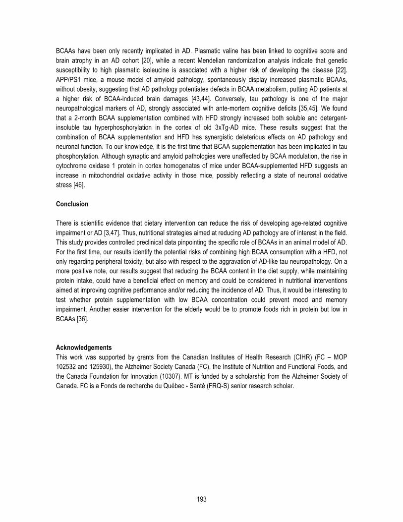
193
BCAAs have been only recently implicated in AD. Plasmatic valine has been linked to cognitive score and brain atrophy in an AD cohort [20], while a recent Mendelian randomization analysis indicate that genetic susceptibility to high plasmatic isoleucine is associated with a higher risk of developing the disease [22]. APP/PS1 mice, a mouse model of amyloid pathology, spontaneously display increased plasmatic BCAAs, without obesity, suggesting that AD pathology potentiates defects in BCAA metabolism, putting AD patients at a higher risk of BCAA-induced brain damages [43,44]. Conversely, tau pathology is one of the major neuropathological markers of AD, strongly associated with ante-mortem cognitive deficits [35,45]. We found that a 2-month BCAA supplementation combined with HFD strongly increased both soluble and detergent-insoluble tau hyperphosphorylation in the cortex of old 3xTg-AD mice. These results suggest that the combination of BCAA supplementation and HFD has synergistic deleterious effects on AD pathology and neuronal function. To our knowledge, it is the first time that BCAA supplementation has been implicated in tau phosphorylation. Although synaptic and amyloid pathologies were unaffected by BCAA modulation, the rise in cytochrome oxidase 1 protein in cortex homogenates of mice under BCAA-supplemented HFD suggests an increase in mitochondrial oxidative activity in those mice, possibly reflecting a state of neuronal oxidative stress [46]. Conclusion There is scientific evidence that dietary intervention can reduce the risk of developing age-related cognitive impairment or AD [3,47]. Thus, nutritional strategies aimed at reducing AD pathology are of interest in the field. This study provides controlled preclinical data pinpointing the specific role of BCAAs in an animal model of AD. For the first time, our results identify the potential risks of combining high BCAA consumption with a HFD, not only regarding peripheral toxicity, but also with respect to the aggravation of AD-like tau neuropathology. On a more positive note, our results suggest that reducing the BCAA content in the diet supply, while maintaining protein intake, could have a beneficial effect on memory and could be considered in nutritional interventions aimed at improving cognitive performance and/or reducing the incidence of AD. Thus, it would be interesting to test whether protein supplementation with low BCAA concentration could prevent mood and memory impairment. Another easier intervention for the elderly would be to promote foods rich in protein but low in BCAAs [36]. Acknowledgements This work was supported by grants from the Canadian Institutes of Health Research (CIHR) (FC – MOP 102532 and 125930), the Alzheimer Society Canada (FC), the Institute of Nutrition and Functional Foods, and the Canada Foundation for Innovation (10307). MT is funded by a scholarship from the Alzheimer Society of Canada. FC is a Fonds de recherche du Québec - Santé (FRQ-S) senior research scholar.

194
Figures
Table 1: Description of dietary treatments. Amino acid content of the diets, accounting for casein and amino acid supplementation, was determined by GC-FID. &Arginine cannot be determined with the technique used and tryptophan degrades quickly during acid hydrolysis.
Normal High-BCAA Low-BCAA Normal High-BCAA Low-BCAAProtein (% w/w) 20.3 23.3 18.4 27.4 31 25Carbohydrate (% w/w) 66 63.5 67.5 25.3 24.1 26.1Fat (% w/w) 5 4.8 5.1 35.1 33.4 36.3Calorie per diet weight (kcal/g) 3.9 3.9 3.9 5.3 5.2 5.3
Ingredient (g/kg)Amino Acids
L-Cystine 0.2 1.8 2.5 0.4 3.4 5.2Isoleucine 6.4 16.2 2.9 8.9 22.2 5.3Leucine 16.3 38.4 7.3 23.3 49.9 14.2Lysine 15.2 13.2 13.7 22.9 20.2 24.9Methionine 7.0 4.1 4.5 9.9 6.6 8.4Phenylalanine 10.3 10.2 11.2 14.8 14.1 21.1Threonine 5.2 5.0 5.3 7.5 6.8 9.0Tryptophan& 0 1.1 1.3 0 1.1 1.3Valine 7.4 20.6 3.2 11.6 26.8 6.4Histidine 4.4 3.8 4.3 7.3 5.7 7.2Alanine 4.9 5.3 5.2 6.6 5.6 6.7Arginine& 0 3.2 3.9 0 3.2 3.9Aspartic Acid 9.9 9.0 9.9 14.5 13.3 15.9Glutamic Acid 40.9 37.1 39.0 55.9 58.8 61.8Glycine 2.5 3.1 3.3 3.7 4.6 4.9Proline 16.6 17.2 18.8 23.8 23.8 30.7Serine 7.4 7.5 7.4 10.7 15.4 12.2Tyrosine 10.9 10.3 10.3 13.2 12.8 18.3
Total 166 207 154 235 294 257Corn Starch 150 150 150 25 25 25Maltodextrin 10 0 0 0 100 100 100Sucrose 500 500 500 52.5 52.5 52.5Cellulose, BW200 50 50 50 50 50 50Corn Oil 30 30 30 0 0 0Safflower Oil 0 0 0 125 125 125Lard 0 0 0 135 135 135Soybean Oil 10 10 10 0 0 0Canola Oil 10 10 10 0 0 0Mineral (S19101) 35 35 35 35 35 35Vitamins (V15908) 10 10 10 10 10 10Choline Bitartrate 2 2 2 2 2 2Cholesterol, USP 0,6 0,6 0,6 3 3 3
Control 5% (w/w) fat High fat 35% (w/w) fat
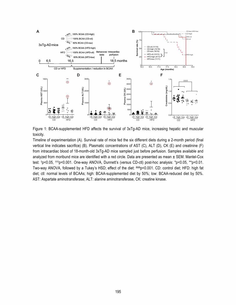
195
Figure 1: BCAA-supplemented HFD affects the survival of 3xTg-AD mice, increasing hepatic and muscular toxicity. Timeline of experimentation (A). Survival rate of mice fed the six different diets during a 2-month period (final vertical line indicates sacrifice) (B). Plasmatic concentrations of AST (C), ALT (D), CK (E) and creatinine (F) from intracardiac blood of 18-month-old 3xTg-AD mice sampled just before perfusion. Samples available and analyzed from moribund mice are identified with a red circle. Data are presented as mean ± SEM. Mantel-Cox test: †p<0.05, †††p<0.001. One-way ANOVA, Dunnett’s (versus CD-ctl) post-hoc analysis: *p<0.05, **p<0.01. Two-way ANOVA, followed by a Tukey’s HSD; effect of the diet: &&&p<0.001. CD: control diet; HFD: high fat diet; ctl: normal levels of BCAAs; high: BCAA-supplemented diet by 50%; low: BCAA-reduced diet by 50%. AST: Aspartate aminotransferase; ALT: alanine aminotransferase, CK: creatine kinase.
0
500
1000
1500 **
CD
lowctl
Plas
ma
AST
(U/L
)
0
1000
2000
3000
Plas
ma
ALT
(U/L
)
0
500
1000
1500
2000
2500
3000
3500
Plas
ma
CK
(U/L
)
B
C D E
A
high lowctl high
HFD CD
lowctl high lowctl high
HFD CD
lowctl high lowctl high
HFD
F
0.0
0.5
1.0
1.5
Cre
atni
nine
(mg/
dL)
CD
lowctl high lowctl high
HFD
&&& * *
3xTg-AD mice
0
CD or HFD
Behavioral
tests
18.5 months16.5
50% BCAA (HFD-low)
100% BCAA (HFD-ctl)
150% BCAA (HFD-high)
- 50%
+ 50%
6.5
Intracardiac
perfusion
Supplementation / reduction in BCAA
50% BCAA (CD-low)
100% BCAA (CD-ctl)
150% BCAA (CD-high)
- 50%
+ 50%
CD
HFD
16.0 16.5 17.0 17.5 18.0 18.5 19.0
0
25
50
75
100
Age (months)
Surv
ival
rate
(%)
CD-high (15/16)
CD-low (16/16)
CD-ctl (11/14)
HFD-high (4/13)
HFD-low (11/11)
HFD-ctl (10/12)†††
† HFD-high
CD-low; HFD-lowCD-high
HFD-ctl CD-ctl
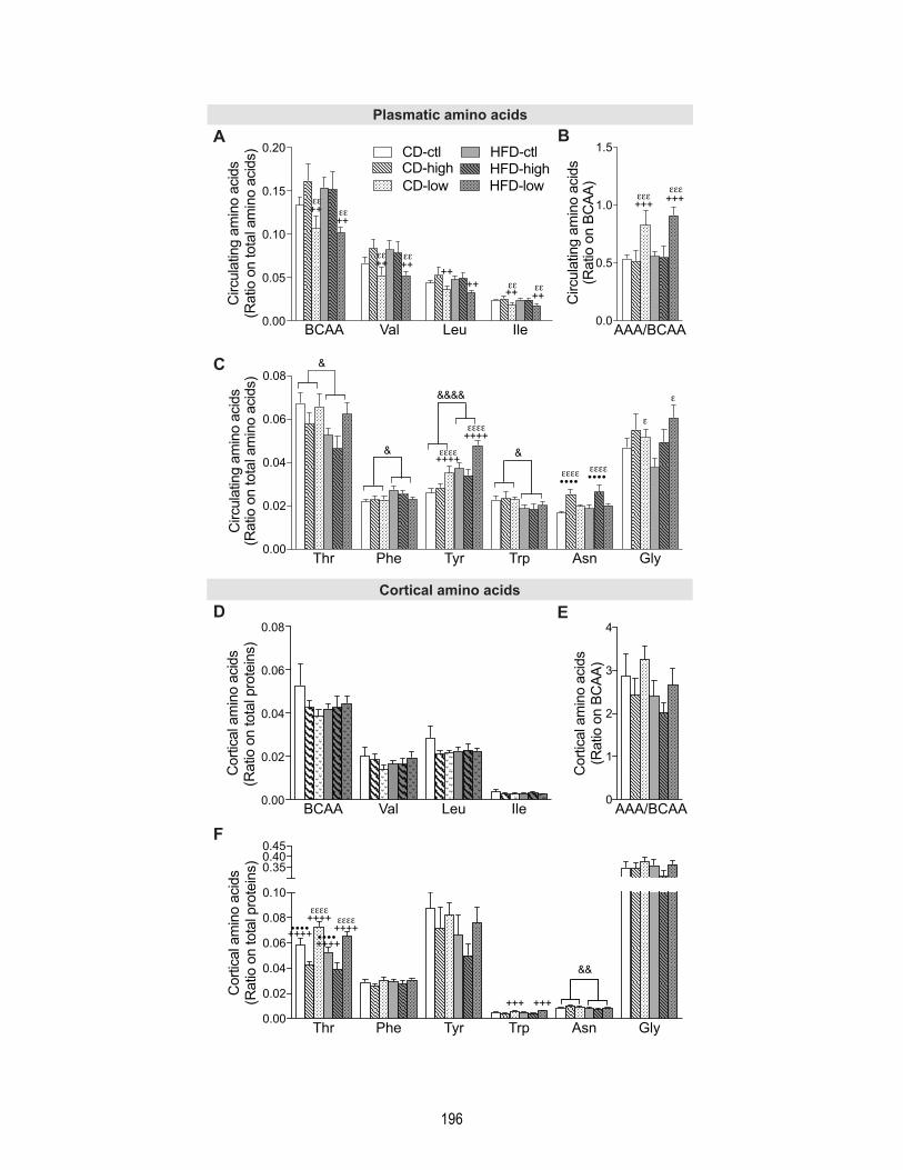
196
C
D
A B
BCAA Val Leu Ile0.00
0.05
0.10
0.15
0.20 CD-ctl
CD-high
CD-low
HFD-ctl
HFD-high
HFD-low
++
++
++
Circula
ting a
min
o a
cid
s
(Ratio o
n tota
l am
ino a
cid
s)
++
++++
++++
AAA/BCAA0.0
0.5
1.0
1.5
+++
Circula
ting a
min
o a
cid
s
(Ratio o
n B
CA
A)
+++
Thr Phe Tyr Trp Asn Gly0.00
0.02
0.04
0.06
0.08
&
&
&&&&
&
••••
Circula
ting a
min
o a
cid
s
(Ratio o
n tota
l am
ino a
cid
s)
••••
++++
++++
Plasmatic amino acids
Cortical amino acids
Thr Phe Tyr Trp Asn Gly0.00
0.02
0.04
0.06
0.08
0.10
0.350.400.45
Cort
ical am
ino a
cid
s
(Ratio o
n tota
l pro
tein
s)
&&
+++ +++
++++
++++
++++
++++
••••••••
0
1
2
3
4
Cort
ical am
ino a
cid
s
(Ratio o
n B
CA
A)
AAA/BCAABCAA Val Leu Ile0.00
0.02
0.04
0.06
0.08
Cort
ical am
ino a
cid
s
(Ratio o
n tota
l pro
tein
s)
E
F

197
Figure 2: High and low BCAA supplies modify plasmatic and cerebral amino acids levels in 18-month-old 3xTg-AD mice. BCAA (Val, Leu, Ile) and aromatic (Phe, Trp, Tyr, His) amino acids in intracardiac blood sampled just before perfusion, expressed as a ratio on total plasma amino acids (n= 4-6 per group) (A-C). BCAA and aromatic amino acids in TBS-soluble fraction of cortex homogenates, expressed as a ratio on total protein content (n= 10-16 per group) (D-F). Data are presented as mean ± SEM. Two-way ANOVA, followed by a Tukey’s HSD; effect of the diet: &p<0.05, &&p<0.01, &&&&p<0.0001; effect of BCAA: ε different from ctl, + different from high, � different from low. CD: control diet; HFD: high-fat diet; ctl: normal levels of BCAA; high: BCAA-supplemented diet by 50%; low: BCAA-reduced diet by 50%. AAA: aromatic amino acids.
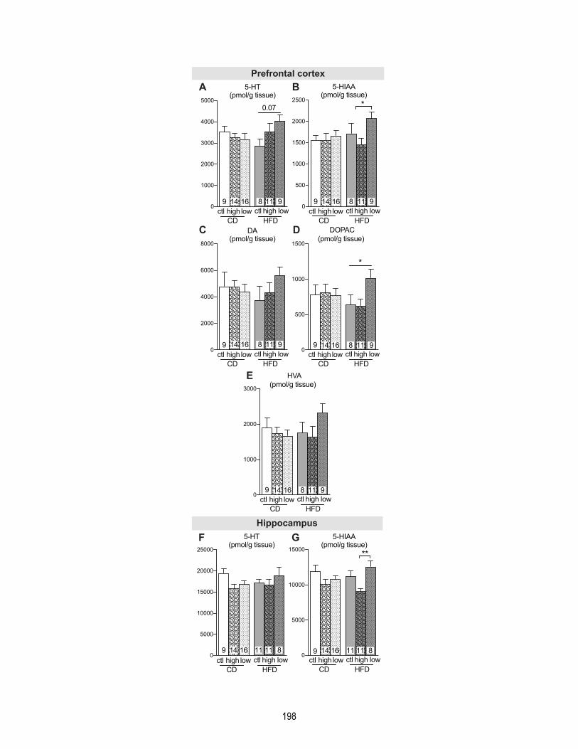
198
A B
E
C D
5-HT(pmol/g tissue)
0
1000
2000
3000
4000
5000
9 16 8 11 9
CDlowhighctl
0.07
lowhighctlHFD
5-HIAA(pmol/g tissue)
0
500
1000
1500
2000
2500 *
9 16 8 11 9
CDlowhighctl lowhighctl
HFDDA
(pmol/g tissue)
0
2000
4000
6000
8000
9 16 8 11 9
CDlowhighctl lowhighctl
HFD
DOPAC(pmol/g tissue)
0
500
1000
1500
*
9 14 16 8 11 9
CDlowhighctl lowhighctl
HFDHVA
(pmol/g tissue)
0
1000
2000
3000
9 14 16 8 11 9
CDlowhighctl lowhighctl
HFD
Prefrontal cortex
Hippocampus
F G5-HT(pmol/g tissue)
CDlowhighctl lowhighctl
HFD
5-HIAA(pmol/g tissue)
CDlowhighctl lowhighctl
HFD
14
11
14
14 14
14
16 16
16 16
16
11 119 98 8
11 118 8
8
9 9
9
0
5000
10000
15000
20000
25000
14 16 10 11 99 14 16 11 11 814 16 811 110
5000
10000
15000
9 14 16 10 11 9
**
14 16 11 11 814 16 811 11
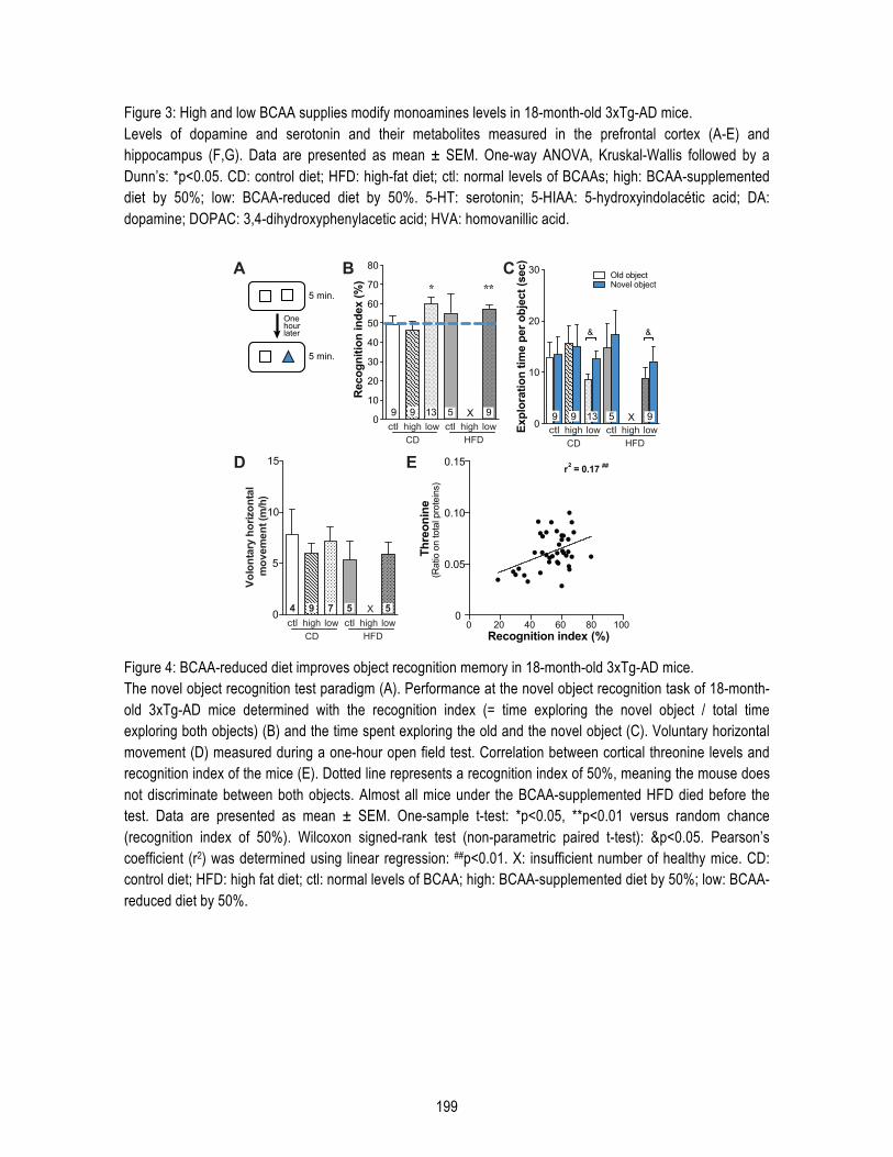
199
Figure 3: High and low BCAA supplies modify monoamines levels in 18-month-old 3xTg-AD mice. Levels of dopamine and serotonin and their metabolites measured in the prefrontal cortex (A-E) and hippocampus (F,G). Data are presented as mean ± SEM. One-way ANOVA, Kruskal-Wallis followed by a Dunn’s: *p<0.05. CD: control diet; HFD: high-fat diet; ctl: normal levels of BCAAs; high: BCAA-supplemented diet by 50%; low: BCAA-reduced diet by 50%. 5-HT: serotonin; 5-HIAA: 5-hydroxyindolacétic acid; DA: dopamine; DOPAC: 3,4-dihydroxyphenylacetic acid; HVA: homovanillic acid.
Figure 4: BCAA-reduced diet improves object recognition memory in 18-month-old 3xTg-AD mice. The novel object recognition test paradigm (A). Performance at the novel object recognition task of 18-month-old 3xTg-AD mice determined with the recognition index (= time exploring the novel object / total time exploring both objects) (B) and the time spent exploring the old and the novel object (C). Voluntary horizontal movement (D) measured during a one-hour open field test. Correlation between cortical threonine levels and recognition index of the mice (E). Dotted line represents a recognition index of 50%, meaning the mouse does not discriminate between both objects. Almost all mice under the BCAA-supplemented HFD died before the test. Data are presented as mean ± SEM. One-sample t-test: *p<0.05, **p<0.01 versus random chance (recognition index of 50%). Wilcoxon signed-rank test (non-parametric paired t-test): &p<0.05. Pearson’s coefficient (r2) was determined using linear regression: ##p<0.01. X: insufficient number of healthy mice. CD: control diet; HFD: high fat diet; ctl: normal levels of BCAA; high: BCAA-supplemented diet by 50%; low: BCAA-reduced diet by 50%.
A B
One hour later
5 min.
5 min.
D E
0
5
10
15
Volo
ntar
y ho
rizon
tal
mov
emen
t (m
/h)
4 9 5 5X7
CDlowctl high lowctl high
HFD0 20 40 60 80 100
0
0.05
0.10
0.15
Recognition index (%)
Thre
onin
e (R
atio
on
tota
l pro
tein
s)
r2 = 0.17 ##
5 59 7
0
10
20
30
40
50
60
70
80
* **
Rec
ogni
tion
inde
x (%
)
9 X9 13 5 9
CDlowctl high lowctl high
HFD
0
10
20
30
Expl
orat
ion
time
per o
bjec
t (se
c)
9 X9 13 5 9
& &
Old objectNovel object
CDlowctl high lowctl high
HFD
C
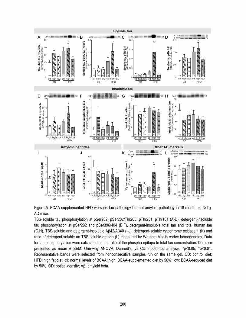
200
Figure 5: BCAA-supplemented HFD worsens tau pathology but not amyloid pathology in 18-month-old 3xTg-AD mice. TBS-soluble tau phosphorylation at pSer202, pSer202/Thr205, pThr231, pThr181 (A-D), detergent-insoluble tau phosphorylation at pSer202 and pSer396/404 (E,F), detergent-insoluble total tau and total human tau (G,H), TBS-soluble and detergent-insoluble Aβ42/Aβ40 (I-J), detergent-soluble cytochrome oxidase 1 (K) and ratio of detergent-soluble on TBS-soluble drebrin (L) measured by Western blot in cortex homogenates. Data for tau phosphorylation were calculated as the ratio of the phospho-epitope to total tau concentration. Data are presented as mean ± SEM. One-way ANOVA, Dunnett’s (vs CDn) post-hoc analysis: *p<0.05, **p<0.01. Representative bands were selected from nonconsecutive samples run on the same gel. CD: control diet; HFD: high fat diet; ctl: normal levels of BCAA; high: BCAA-supplemented diet by 50%; low: BCAA-reduced diet by 50%. OD: optical density; Aβ: amyloid beta.
A B C
0
5
10
15
20
10 15 16 12 11 11
*
Inso
lubl
e ta
u pS
er20
2(C
P13/
TauC
, rel
ativ
e O
D)
D
E F G H
0
0.1
0.2
0.3
10 15 16 12 11 11
**
Solu
ble
tau
pSer
202/
Thr2
05 (A
T8/T
auC
rela
tive
OD
)
0
0.05
0.10
0.15
0.20
10 15 16 12 11 11
**
Solu
ble
tau
pThr
231
(A
T180
/Tau
C re
lativ
e O
D)
0
0.05
0.10
0.15
10 15 16 12 11 11
*
Solu
ble
tau
pThr
181
(A
T270
/Tau
C re
lativ
e O
D)
0
0.1
0.2
0.3
0.4
0.5
10
*So
lubl
e ta
u pS
er20
2 (C
P13/
TauC
rela
tive
OD
)
Soluble tau
Insoluble tau
Amyloid peptides
0
1
2
3
4
5
Solu
ble
A42
/ A
40
9 12 15 10 10 100
0.1
0.2
0.3
10 13 15 12 10 8
Inso
lubl
e A
42 /
A40
I J K L
CP13 60 AT8 60 AT180 60 AT270 60
0
2
4
6
8
10 15 16 12 11 11
Inso
lubl
e to
tal t
au(T
auC
, rel
ativ
e O
D /
mg
tissu
e)
0
2
4
6
8
10
10 15 16 12 11 11
Inso
lubl
e to
tal h
uman
tau
(Tau
13, r
elat
ive
OD
/ m
g tis
sue)
0
1
2
3
4
5
10 15 16 12 11 11
*
Cyt
ochr
ome
oxid
ase
1 (C
ytox
1, r
elat
ive
OD
)
Other AD markers
Inso
lubl
e ta
u pS
er39
6/40
4(P
HF1
/Tau
C, r
elat
ive
OD
)
CP13 60 TauC 60 Tau13 60
CDlowctl high lowctl high
HFD CDlowctl high lowctl high
HFD CDlowctl high lowctl high
HFD CDlowctl high lowctl high
HFD
CDlowctl high lowctl high
HFD CDlowctl high lowctl high
HFD CDlowctl high lowctl high
HFD
CDlowctl high lowctl high
HFD CDlowctl high lowctl high
HFD CDlowctl high lowctl high
HFD
0
1
2
3
4
10 15 16 12 11 11
PHF1 60
35Cytox1
0.0
0.2
0.4
0.6
0.8
10 15 16 12 11 11
CDlowctl high lowctl high
HFD
Mem
bran
e / s
olub
le d
rebr
in(R
elat
ive
OD
)
120120
mDrebrinsDrebrin
CDlowctl high lowctl high
HFD
1615 12 1111
1513 12 810
1615 12 1111 1615 12 1111 1615 12 1111
1615 12 1111 1615 12 1111 1615 12 1111
1615 12 1111 1615 12 11111512 10 1010
1615 12 1111
β-tubulin 50
42β-actin
201
Supplementary data Methods Behavioral assessment Recognition memory and locomotor activity were evaluated with the novel object recognition test and the open field test respectively, as previously described [1]. Behavioral tests were performed one week before sacrifice with a recovery time of 16 to 24 hours. For the novel object recognition test, mice were put in a clear box (29.2 cm x 19 cm x 12.7 cm) with two similar objects for 5 minutes and the time exploring or smelling the objects was counted. One hour later, mice were put in the same box changing one (defined as the novel object) of the two objects from the first phase during another 5 minutes. Mice spending less than 7 seconds exploring one of the two objects during the first phase were excluded from the analysis. For the open field test, mice were placed in Plexiglas cages with translucent walls (40 cm x 40 cm) and their voluntary horizontal movements were tracked for one hour by the automated recording of photobeam breaks (San Diego Instruments, San Diego, CA, USA). Amino acid quantification in brain, plasma and diet All reagents used for cleaning samples and derivatization were included in the EZ:faast kit for free amino acid analysis by GC-FID (Phenomenex, Torrance, CA, USA). This kit also provided the amino acid standard mixtures and internal standard (Norvaline) solution (200 nM each). Prior to diet hydrolysis, about 3 g of each diet were defatted three times with hexane, according to the protocol described previously [2]. This step was made in duplicate. To hydrolyse proteins and peptides, 50 mg of defatted diet was weighed into 18 mL vacuum hydrolysis tubes and 5 mL of HCl 6M was added. Tubes were flushed with N2 before being sealed under vacuum. Samples were heated at 110°C in a bloc heater for 24 hours. After hydrolysis, 5 mL of NaOH 6 M were added before centrifugation (10 minutes, 4°C, 5100 rpm). An aliquot of the supernatant was diluted 10 times with HCl 0.1 M. Amino acids of plasma, cortex homogenates (TBS-soluble fraction) and diet samples were derivatized using EZ:faast kit for GC-FID (Phenomenex, Torrance, CA, USA) as described in the EZ:faast user’s manual. Briefly, 50 µL (plasma) or 100 µL (cortex homogenates and diet) were mixed with 100 µL of Norvaline. This solution was passed through sorbent tips. A washing solution (200 µL) was then pipetted to clean up the samples. Eluting medium (200 µL) permitted to collect the sorbent and derivatized amino acids with 50 µL of Reagent 4 (propyl chloroformate solution). The products of derivatization were then extracted with 100 µL of Reagent 5 (Iso-octane) and 100 µL of Reagent 6 (HCl 1N) were added. Finally, the organic layer was transferred to a GC vial with insert. Derivatized samples were then analyzed with a GC-FID system (Shimadzu, Kyoto, Japan), constituted of a GC-2010 Plus gas chromatograph equipped with an AOC-20s auto-sampler, an AOC-20i auto-injector and a flame ionization detector. The GC solution software controlled the system. A 2 µL sample was injected to the ZB-AAA 10 m x 0.25 mm id GC column (provided by the EZ:faast kit) with a split ratio of 1:10 (v/v). The column flow was constant at 1.7 mL/minute of hydrogen. Nitrogen (30 mL/minute), hydrogen (40 mL/minute) and air (400 mL/minute) were used as auxiliary gasses for flame ionization detector. The injector was set at 300°C and the detector at 320°C. The oven temperature was initially 110°C, then increased to 320°C at 32°C/min and maintained for 1 minute at this temperature (total run time 7.56 minutes and 2 minutes equilibration). Repeatability in plasma and cortex samples was assessed for each amino acid calculating a mean RSD from 5 samples injected three times. Quantification of monoamines
202
Prefrontal cortex Dopamine (DA), 3,4-dihydroxyphenylacetic acid (DOPAC), homovanillic acid (HVA), serotonin (5-HT), 5-hydroxyindoleacetic acid (5-HIAA) were quantified by electrochemical high performance liquid chromatography (HPLC) as previously described [3,4]. Prefrontal cortex were dissected and kept at -80°C until homogenization in 300 µL of an aqueous solution of 12 mM HCLO4, 0.22 mM Na2S2O5, 3 mM octanesulfonic acid and 56 µM EDTA. After centrifugation (30,000 g, 20 minutes, 4°C), the supernatant was stored at -80°C until analysis. Mobile phase (pH 3.4; 0.1 M sodium acetate, 0.1 M citric acid monohydrate, 1 mM sodium octyl sulfate, 0.1 mM disodium EDTA, 1 mM diethylamine and 125 mL/L acétonitrile) was continuously pumped with a rate of 0.5 mL/minute (Ultimate 3000 pump, Thermo Fisher Scientific, Waltham, MA, USA). An analytical column C18, 5 µm particle size, 100x3mm (ACE-121-1003, ACE) was employed to separate the samples and monoamines were detected by an amperometric detector (Decade II) maintained at a potential of +0.75V with an amperage range of 1 nA. Peaks areas were integrated using Chromeleon 6.80 software. Hippocampus Half hippocampi (10 mg) were weighed, homogenized in 180 µL of perchloric acid 0.1 N (Mallinckrodt Baker, Phillipsburg, NJ, USA), centrifuged at 4°C for 20 minutes at 16,000 g and kept at -80°C. Twenty µL of the supernatant were injected in duplicate using a Waters 717 plus Autosampler Automatic Injector (Waters, Lachine, QC, Canada) and submitted to HPLC through an Atlantis dC18 column (Waters). The electrochemical detector was set at 5nA and the mobile phase was delivered at 1.4 mL/minute and composed of 47.8 mM NaH2PO4, 0.9 mM octanesulfonic acid, 0.4 mM ethylenediaminetetra-acetic acid, 2 mM NaCl, and 8% MeOH
(Sigma Aldrich) at pH 2.90. References [1] St-Amour I, Paré I, Tremblay C, Coulombe K, Bazin R, Calon F. IVIg protects the 3xTg-AD mouse model of Alzheimer's disease from memory deficit and Aβ pathology. J Neuroinflammation 2014;11:54. [2] Robbins RJ, Leonczak J, Johnson JC, Li J, Kwik-Uribe C, Prior RL, et al. Method performance and multi-laboratory assessment of a normal phase high pressure liquid chromatography-fluorescence detection method for the quantitation of flavanols and procyanidins in cocoa and chocolate containing samples. J Chromatogr A 2009;1216:4831–40. [3] Crumeyrolle-Arias M, Jaglin M, Bruneau A, Vancassel S, Cardona A, Daugé V, et al. Absence of the gut microbiota enhances anxiety-like behavior and neuroendocrine response to acute stress in rats. Psychoneuroendocrinology 2014;42:207–17. [4] Calon F, Morissette M, Rajput AH, Hornykiewicz O, Bédard PJ, Di Paolo T. Changes of GABA receptors and dopamine turnover in the postmortem brains of parkinsonians with levodopa-induced motor complications. Mov Disord 2003;18:241–53.
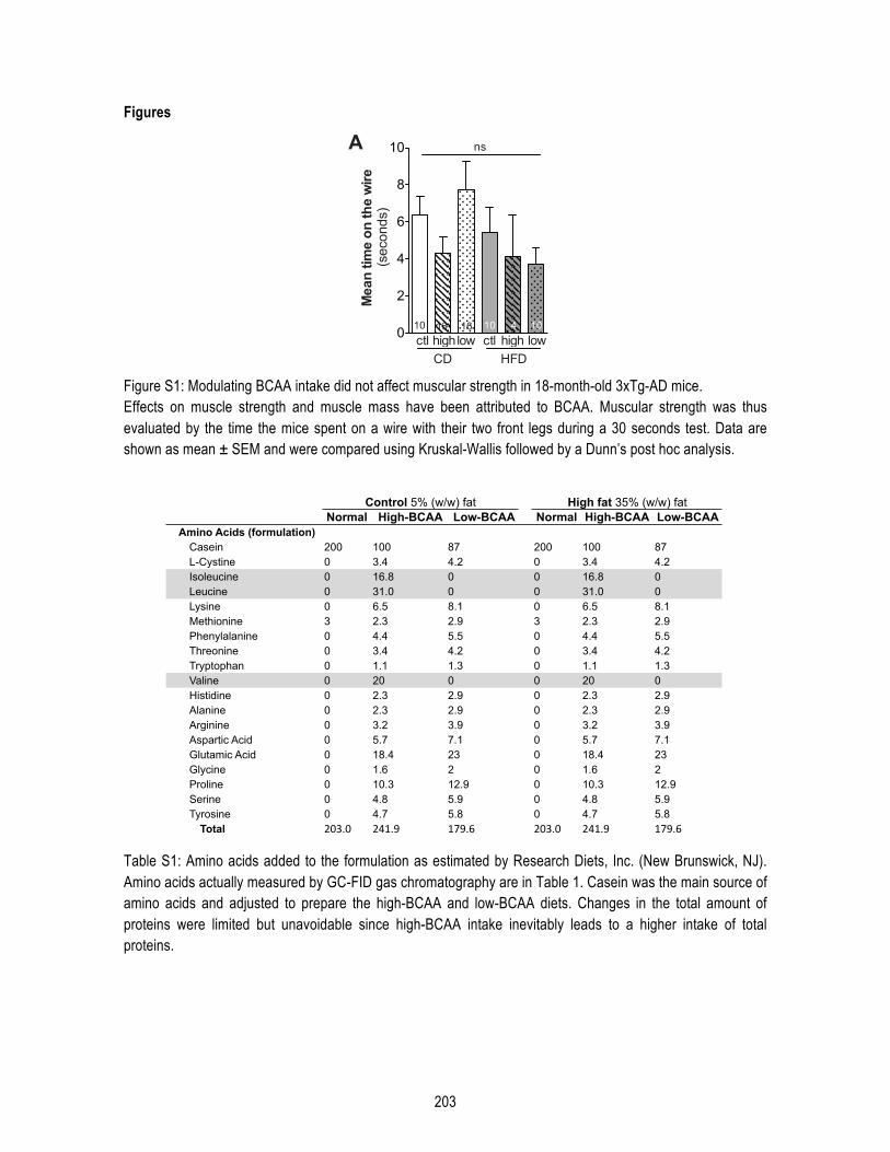
203
Figures
Figure S1: Modulating BCAA intake did not affect muscular strength in 18-month-old 3xTg-AD mice. Effects on muscle strength and muscle mass have been attributed to BCAA. Muscular strength was thus evaluated by the time the mice spent on a wire with their two front legs during a 30 seconds test. Data are shown as mean ± SEM and were compared using Kruskal-Wallis followed by a Dunn’s post hoc analysis.
Table S1: Amino acids added to the formulation as estimated by Research Diets, Inc. (New Brunswick, NJ). Amino acids actually measured by GC-FID gas chromatography are in Table 1. Casein was the main source of amino acids and adjusted to prepare the high-BCAA and low-BCAA diets. Changes in the total amount of proteins were limited but unavoidable since high-BCAA intake inevitably leads to a higher intake of total proteins.
0
2
4
6
8
10
Mea
n tim
e on
the
wire
(seconds)
10 13 16 10 4 10
CD
lowhighctl
HFD
lowhighctl
A ns
Normal High-BCAA Low-BCAA Normal High-BCAA Low-BCAAAmino Acids (formulation)
Casein 200 100 87 200 100 87L-Cystine 0 3.4 4.2 0 3.4 4.2Isoleucine 0 16.8 0 0 16.8 0Leucine 0 31.0 0 0 31.0 0Lysine 0 6.5 8.1 0 6.5 8.1Methionine 3 2.3 2.9 3 2.3 2.9Phenylalanine 0 4.4 5.5 0 4.4 5.5Threonine 0 3.4 4.2 0 3.4 4.2Tryptophan 0 1.1 1.3 0 1.1 1.3Valine 0 20 0 0 20 0Histidine 0 2.3 2.9 0 2.3 2.9Alanine 0 2.3 2.9 0 2.3 2.9Arginine 0 3.2 3.9 0 3.2 3.9Aspartic Acid 0 5.7 7.1 0 5.7 7.1Glutamic Acid 0 18.4 23 0 18.4 23Glycine 0 1.6 2 0 1.6 2Proline 0 10.3 12.9 0 10.3 12.9Serine 0 4.8 5.9 0 4.8 5.9Tyrosine 0 4.7 5.8 0 4.7 5.8
Total 203.0 241.9 179.6 203.0 241.9 179.6
Control 5% (w/w) fat High fat 35% (w/w) fat
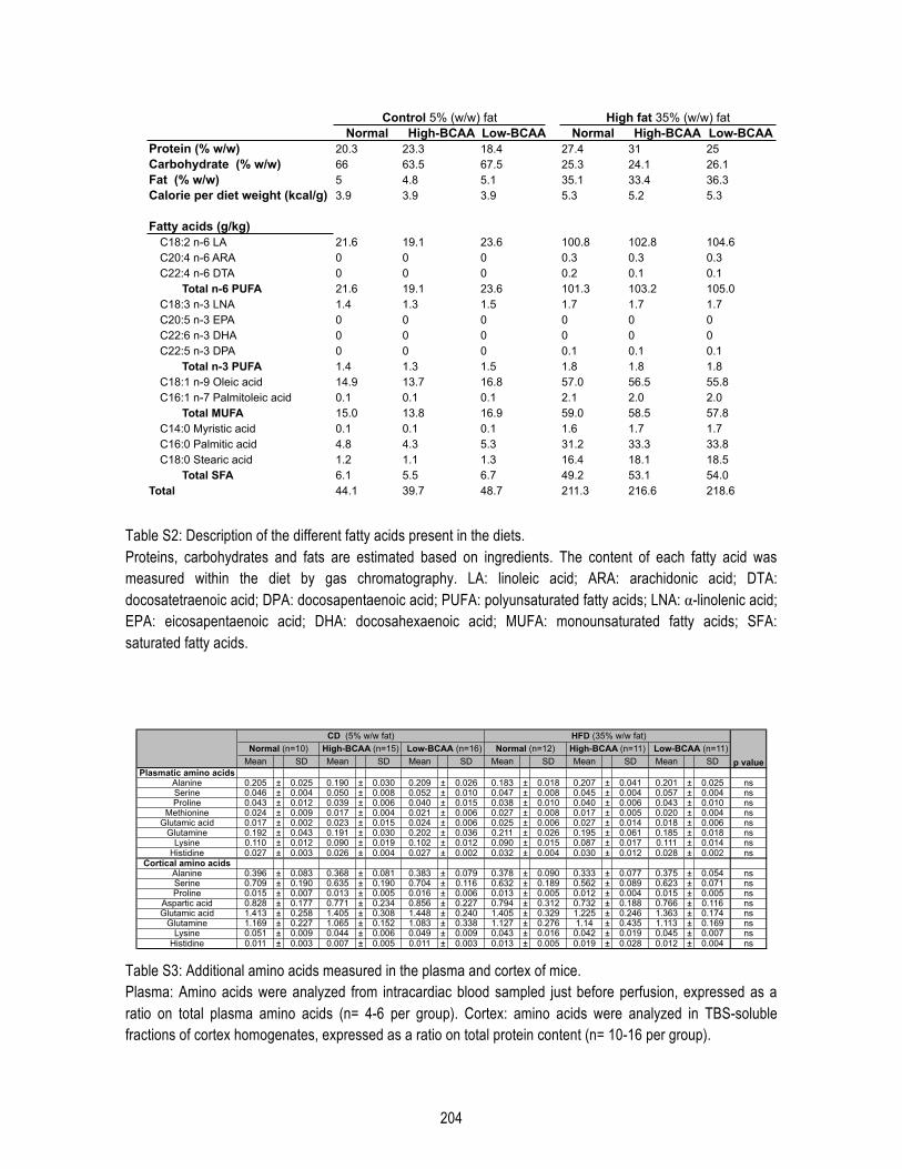
204
Table S2: Description of the different fatty acids present in the diets. Proteins, carbohydrates and fats are estimated based on ingredients. The content of each fatty acid was measured within the diet by gas chromatography. LA: linoleic acid; ARA: arachidonic acid; DTA: docosatetraenoic acid; DPA: docosapentaenoic acid; PUFA: polyunsaturated fatty acids; LNA: α-linolenic acid; EPA: eicosapentaenoic acid; DHA: docosahexaenoic acid; MUFA: monounsaturated fatty acids; SFA: saturated fatty acids.
Table S3: Additional amino acids measured in the plasma and cortex of mice. Plasma: Amino acids were analyzed from intracardiac blood sampled just before perfusion, expressed as a ratio on total plasma amino acids (n= 4-6 per group). Cortex: amino acids were analyzed in TBS-soluble fractions of cortex homogenates, expressed as a ratio on total protein content (n= 10-16 per group).
Normal High-BCAA Low-BCAA Normal High-BCAA Low-BCAAProtein (% w/w) 20.3 23.3 18.4 27.4 31 25Carbohydrate (% w/w) 66 63.5 67.5 25.3 24.1 26.1Fat (% w/w) 5 4.8 5.1 35.1 33.4 36.3Calorie per diet weight (kcal/g) 3.9 3.9 3.9 5.3 5.2 5.3
Fatty acids (g/kg)C18:2 n-6 LA 21.6 19.1 23.6 100.8 102.8 104.6C20:4 n-6 ARA 0 0 0 0.3 0.3 0.3C22:4 n-6 DTA 0 0 0 0.2 0.1 0.1
Total n-6 PUFA 21.6 19.1 23.6 101.3 103.2 105.0C18:3 n-3 LNA 1.4 1.3 1.5 1.7 1.7 1.7C20:5 n-3 EPA 0 0 0 0 0 0C22:6 n-3 DHA 0 0 0 0 0 0C22:5 n-3 DPA 0 0 0 0.1 0.1 0.1
Total n-3 PUFA 1.4 1.3 1.5 1.8 1.8 1.8C18:1 n-9 Oleic acid 14.9 13.7 16.8 57.0 56.5 55.8C16:1 n-7 Palmitoleic acid 0.1 0.1 0.1 2.1 2.0 2.0
Total MUFA 15.0 13.8 16.9 59.0 58.5 57.8C14:0 Myristic acid 0.1 0.1 0.1 1.6 1.7 1.7C16:0 Palmitic acid 4.8 4.3 5.3 31.2 33.3 33.8C18:0 Stearic acid 1.2 1.1 1.3 16.4 18.1 18.5
Total SFA 6.1 5.5 6.7 49.2 53.1 54.0Total 44.1 39.7 48.7 211.3 216.6 218.6
Control 5% (w/w) fat High fat 35% (w/w) fat
Mean SD Mean SD Mean SD Mean SD Mean SD Mean SDPlasmatic amino acids
Alanine 0.205 ± 0.025 0.190 ± 0.030 0.209 ± 0.026 0.183 ± 0.018 0.207 ± 0.041 0.201 ± 0.025 nsSerine 0.046 ± 0.004 0.050 ± 0.008 0.052 ± 0.010 0.047 ± 0.008 0.045 ± 0.004 0.057 ± 0.004 nsProline 0.043 ± 0.012 0.039 ± 0.006 0.040 ± 0.015 0.038 ± 0.010 0.040 ± 0.006 0.043 ± 0.010 ns
Methionine 0.024 ± 0.009 0.017 ± 0.004 0.021 ± 0.006 0.027 ± 0.008 0.017 ± 0.005 0.020 ± 0.004 nsGlutamic acid 0.017 ± 0.002 0.023 ± 0.015 0.024 ± 0.006 0.025 ± 0.006 0.027 ± 0.014 0.018 ± 0.006 ns
Glutamine 0.192 ± 0.043 0.191 ± 0.030 0.202 ± 0.036 0.211 ± 0.026 0.195 ± 0.061 0.185 ± 0.018 nsLysine 0.110 ± 0.012 0.090 ± 0.019 0.102 ± 0.012 0.090 ± 0.015 0.087 ± 0.017 0.111 ± 0.014 ns
Histidine 0.027 ± 0.003 0.026 ± 0.004 0.027 ± 0.002 0.032 ± 0.004 0.030 ± 0.012 0.028 ± 0.002 nsCortical amino acids
Alanine 0.396 ± 0.083 0.368 ± 0.081 0.383 ± 0.079 0.378 ± 0.090 0.333 ± 0.077 0.375 ± 0.054 nsSerine 0.709 ± 0.190 0.635 ± 0.190 0.704 ± 0.116 0.632 ± 0.189 0.562 ± 0.089 0.623 ± 0.071 nsProline 0.015 ± 0.007 0.013 ± 0.005 0.016 ± 0.006 0.013 ± 0.005 0.012 ± 0.004 0.015 ± 0.005 ns
Aspartic acid 0.828 ± 0.177 0.771 ± 0.234 0.856 ± 0.227 0.794 ± 0.312 0.732 ± 0.188 0.766 ± 0.116 nsGlutamic acid 1.413 ± 0.258 1.405 ± 0.308 1.448 ± 0.240 1.405 ± 0.329 1.225 ± 0.246 1.363 ± 0.174 ns
Glutamine 1.169 ± 0.227 1.065 ± 0.152 1.083 ± 0.338 1.127 ± 0.276 1.14 ± 0.435 1.113 ± 0.169 nsLysine 0.051 ± 0.009 0.044 ± 0.006 0.049 ± 0.009 0.043 ± 0.016 0.042 ± 0.019 0.045 ± 0.007 ns
Histidine 0.011 ± 0.003 0.007 ± 0.005 0.011 ± 0.003 0.013 ± 0.005 0.019 ± 0.028 0.012 ± 0.004 ns
CD (5% w/w fat) HFD (35% w/w fat)
p valueNormal (n=10) High-BCAA (n=15) Low-BCAA (n=16) Normal (n=12) High-BCAA (n=11) Low-BCAA (n=11)
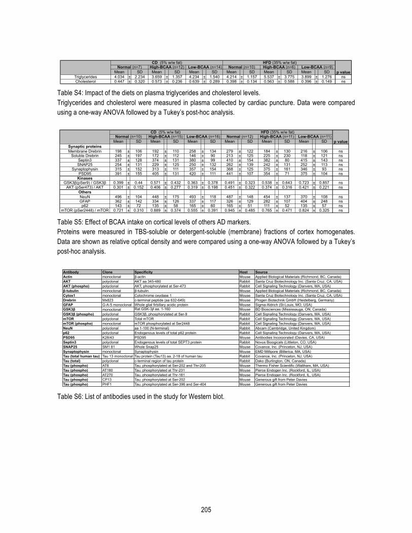
205
Table S4: Impact of the diets on plasma triglycerides and cholesterol levels. Triglycerides and cholesterol were measured in plasma collected by cardiac puncture. Data were compared using a one-way ANOVA followed by a Tukey’s post-hoc analysis.
Table S5: Effect of BCAA intake on cortical levels of others AD markers. Proteins were measured in TBS-soluble or detergent-soluble (membrane) fractions of cortex homogenates. Data are shown as relative optical density and were compared using a one-way ANOVA followed by a Tukey’s post-hoc analysis.
Table S6: List of antibodies used in the study for Western blot.
Mean SD Mean SD Mean SD Mean SD Mean SD Mean SDTriglycerides 4.034 ± 2.234 3.659 ± 1.357 4.234 ± 1.540 4.214 ± 1.157 5.537 ± 3.775 3.899 ± 1.276 nsCholesterol 0.447 ± 0.320 0.573 ± 0.236 0.639 ± 0.289 0.398 ± 0.134 0.563 ± 0.588 0.396 ± 0.149 ns
p value
CD (5% w/w fat) HFD (35% w/w fat)Normal (n=7) High-BCAA (n=12) Low-BCAA (n=14) Normal (n=10) High-BCAA (n=6) Low-BCAA (n=9)
Mean SD Mean SD Mean SD Mean SD Mean SD Mean SDSynaptic proteinsMembrane Drebrin 198 ± 106 192 ± 110 258 ± 134 279 ± 122 184 ± 130 216 ± 106 ns
Soluble Drebrin 245 ± 197 172 ± 112 146 ± 90 213 ± 125 225 ± 230 118 ± 121 nsSeptin3 337 ± 128 374 ± 131 380 ± 99 410 ± 154 382 ± 80 415 ± 143 nsSNAP25 254 ± 131 229 ± 125 250 ± 132 262 ± 139 242 ± 131 252 ± 113 ns
Synaptophysin 315 ± 152 313 ± 111 357 ± 154 368 ± 125 375 ± 161 346 ± 93 nsPSD95 391 ± 155 405 ± 131 420 ± 111 441 ± 107 354 ± 71 375 ± 104 ns
KinasesGSK3β(pSer9) / GSK3β 0.399 ± 0.44 0.571 ± 0.432 0.363 ± 0.378 0.491 ± 0.323 0.538 ± 0.643 0.723 ± 0.857 ns
AKT (pSer473) / AKT 0.301 ± 0.152 0.406 ± 0.277 0.319 ± 0.198 0.451 ± 0.322 0.374 ± 0.316 0.421 ± 0.221 nsOthersNeuN 496 ± 104 448 ± 175 493 ± 118 487 ± 148 454 ± 137 370 ± 108 nsGFAP 362 ± 142 334 ± 126 337 ± 117 326 ± 129 282 ± 107 404 ± 248 nsp62 143 ± 72 135 ± 58 165 ± 60 165 ± 51 111 ± 52 135 ± 57 ns
mTOR (pSer2448) / mTOR 0.721 ± 0.310 0.889 ± 0.374 0.555 ± 0.391 0.945 ± 0.485 0.765 ± 0.471 0.824 ± 0.325 ns
HFD (35% w/w fat)CD (5% w/w fat)High-BCAA (n=15) High-BCAA (n=11)
p valueLow-BCAA (n=11)Normal (n=12)Low-BCAA (n=16)Normal (n=10)
Antibody Clone Specificity Host SourceActin monoclonal β-actin Mouse Applied Biological Materials (Richmond, BC, Canada)AKT polyclonal AKT aa 345-480 Rabbit Santa Cruz Biotechnology Inc. (Santa Cruz, CA, USA)AKT (phospho) polyclonal AKT, phosphorylated at Ser-473 Rabbit Cell Signaling Technology (Danvers, MA, USA)β-tubulin monoclonal β-tubulin Mouse Applied Biological Materials (Richmond, BC, Canada)Cytox1 monoclonal Cytochrome oxydase 1 Mouse Santa Cruz Biotechnology Inc. (Santa Cruz, CA, USA)Drebrin Mx823 c-terminal peptide (aa 632-649) Mouse Progen Biotechnik GmbH (Heidelberg, Germany)GFAP G-A-5 monoclonal Whole glial firbillary acidic protein Mouse Sigma-Aldrich (St-Louis, MO, USA)GSK3β monoclonal Rat GSK-3β aa. 1-160 Mouse BD Biosciences (Mississauga, ON, Canada)GSK3β (phospho) polyclonal GSK3β, phosphorylated at Ser-9 Rabbit Cell Signaling Technology (Danvers, MA, USA)mTOR polyclonal Total mTOR Rabbit Cell Signaling Technology (Danvers, MA, USA)mTOR (phospho) monoclonal mTOR phosphorylated at Ser2448 Rabbit Cell Signaling Technology (Danvers, MA, USA)NeuN polyclonal aa 1-100 (N-terminal) Rabbit Abcam (Cambridge, United Kingdom)p62 polyclonal Endogenous levels of total p62 protein Rabbit Cell Signaling Technology (Danvers, MA, USA)PSD95 K28/43 PSD95 Mouse Antibodies Incorporated (Davies, CA, USA)Septin3 polyclonal Endogenous levels of total SEPT3 protein Rabbit Novus Bioogicals (Littleton, CO, USA)SNAP25 SM1 81 Whole Snap25 Mouse Covance, Inc. (Princeton, NJ, USA)Synaptophysin monoclonal Synaptophysin Mouse EMD Millipore (Billerica, MA, USA)Tau (total human tau) Tau 13 monoclonal Tau protein (Tau13) aa. 2-18 of human tau Rabbit Covance, Inc. (Princeton, NJ, USA)Tau (total) polyclonal c-terminal region of tau protein Rabbit Dako (Burlington, ON, Canada)Tau (phospho) AT8 Tau, phosphorylated at Ser-202 and Thr-205 Mouse Thermo Fisher Scientific (Waltham, MA, USA)Tau (phospho) AT180 Tau, phosphorylated at Thr-231 Mouse Pierce Endogen Inc. (Rockford, IL, USA)Tau (phospho) AT270 Tau, phosphorylated at Thr-181 Mouse Pierce Endogen Inc. (Rockford, IL, USA)Tau (phospho) CP13 Tau, phosphorylated at Ser-202 Mouse Generous gift from Peter DaviesTau (phospho) PHF1 Tau, phosphorylated at Ser-396 and Ser-404 Mouse Generous gift from Peter Davies
206
References [1] Prince M, Bryce R, Albanese E, Wimo A, Ribeiro W, Ferri CP. The global prevalence of dementia: a systematic review and metaanalysis. Alzheimers Dement 2013;9:63–75.e2. [2] Norton S, Matthews FE, Barnes DE, Yaffe K, Brayne C. Potential for primary prevention of Alzheimer's disease: an analysis of population-based data. Lancet Neurol 2014;13:788–94. [3] Ngandu T, Lehtisalo J, Solomon A, Levälahti E, Ahtiluoto S, Antikainen R, et al. A 2 year multidomain intervention of diet, exercise, cognitive training, and vascular risk monitoring versus control to prevent cognitive decline in at-risk elderly people (FINGER): a randomised controlled trial. Lancet 2015;385:2255–63. [4] Kerdiles O, Layé S, Calon F. Omega-3 polyunsaturated fatty acids and brain health: Preclinical evidence for the prevention of neurodegenerative diseases. Trends in Food Science & Technology 2017;69:203–13. [5] Dal-Pan A, Dudonné S, Bourassa P, Bourdoulous M, Tremblay C, Desjardins Y, et al. Cognitive-Enhancing Effects of a Polyphenols-Rich Extract from Fruits without Changes in Neuropathology in an Animal Model of Alzheimer's Disease. J Alzheimers Dis 2016;Preprint:1–21. [6] van de Rest O, van der Zwaluw NL, de Groot LCPGM. Literature review on the role of dietary protein and amino acids in cognitive functioning and cognitive decline. Amino Acids 2013;45:1035–45. [7] Ohtani M, Sugita M, Maruyama K. Amino acid mixture improves training efficiency in athletes. J Nutr 2006;136:538S–543S. [8] Maughan RJ, Depiesse F, Geyer H, International Association of Athletics Federations. The use of dietary supplements by athletes. J Sports Sci, vol. 25 Suppl 1, 2007, pp. S103–13. [9] Newgard CB, An J, Bain JR, Muehlbauer MJ, Stevens RD, Lien LF, et al. A Branched-Chain Amino Acid-Related Metabolic Signature that Differentiates Obese and Lean Humans and Contributes to Insulin Resistance. Cell Metabolism 2009;9:311–26. [10] Wang TJ, Larson MG, Vasan RS, Cheng S, Rhee EP, McCabe E, et al. Metabolite profiles and the risk of developing diabetes. Nat Med 2011;17:448–53. [11] Newgard CB. Interplay between lipids and branched-chain amino acids in development of insulin resistance. Cell Metabolism 2012;15:606–14. [12] Pardridge WM. Kinetics of competitive inhibition of neutral amino acid transport across the blood-brain barrier. Journal of Neurochemistry 1977;28:103–8. [13] Fernstrom JD. Large neutral amino acids: dietary effects on brain neurochemistry and function. Amino Acids 2013;45:419–30. [14] Gijsman HJ, Scarnà A, Harmer CJ, McTavish SB, Odontiadis J, Cowen PJ, et al. A dose-finding study on the effects of branch chain amino acids on surrogate markers of brain dopamine function. Psychopharmacology (Berl) 2002;160:192–7. [15] Coppola A, Wenner BR, Ilkayeva O, Stevens RD, Maggioni M, Slotkin TA, et al. Branched-chain amino acids alter neurobehavioral function in rats. Am J Physiol Endocrinol Metab 2013;304:E405–13. [16] Scaini G, Jeremias GC, Furlanetto CB, Dominguini D, Comim CM, Quevedo J, et al. Behavioral responses in rats submitted to chronic administration of branched-chain amino acids. JIMD Rep 2014;13:159–67. [17] Xu Y, Yan J, Zhou P, Li J, Gao H, Xia Y, et al. Neurotransmitter receptors and cognitive dysfunction in Alzheimer“s disease and Parkinson”s disease. Prog Neurobiol 2012;97:1–13. [18] Zinnanti WJ, Lazovic J. Interrupting the mechanisms of brain injury in a model of maple syrup urine disease encephalopathy. J Inherit Metab Dis 2012;35:71–9.
207
[19] Sitta A, Ribas GS, Mescka CP, Barschak AG, Wajner M, Vargas CR. Neurological damage in MSUD: the role of oxidative stress. Cell Mol Neurobiol 2014;34:157–65. [20] Toledo JB, Arnold M, Kastenmüller G, Chang R, Baillie RA, Han X, et al. Metabolic network failures in Alzheimer's disease: A biochemical road map. Alzheimers Dement 2017;13:965–84. [21] Tynkkynen J, Chouraki V, van der Lee SJ, Hernesniemi J, Yang Q, Li S, et al. Association of branched-chain amino acids and other circulating metabolites with risk of incident dementia and Alzheimer's disease: A prospective study in eight cohorts. Alzheimers Dement 2018. [22] Larsson SC, Markus HS. Branched-chain amino acids and Alzheimer's disease: a Mendelian randomization analysis. Nature Publishing Group 2017;7:13604. [23] Julien C, Tremblay C, Phivilay A, Berthiaume L, Emond V, Julien P, et al. High-fat diet aggravates amyloid-beta and tau pathologies in the 3xTg-AD mouse model. Neurobiol Aging 2010;31:1516–31. [24] Vandal M, White PJ, Tremblay C, St-Amour I, Chevrier G, Emond V, et al. Insulin reverses the high-fat diet-induced increase in brain aβ and improves memory in an animal model of Alzheimer disease. Diabetes 2014;63:4291–301. [25] St-Amour I, Paré I, Tremblay C, Coulombe K, Bazin R, Calon F. IVIg protects the 3xTg-AD mouse model of Alzheimer's disease from memory deficit and Aβ pathology. J Neuroinflammation 2014;11:54. [26] Robbins RJ, Leonczak J, Johnson JC, Li J, Kwik-Uribe C, Prior RL, et al. Method performance and multi-laboratory assessment of a normal phase high pressure liquid chromatography-fluorescence detection method for the quantitation of flavanols and procyanidins in cocoa and chocolate containing samples. J Chromatogr A 2009;1216:4831–40. [27] Crumeyrolle-Arias M, Jaglin M, Bruneau A, Vancassel S, Cardona A, Daugé V, et al. Absence of the gut microbiota enhances anxiety-like behavior and neuroendocrine response to acute stress in rats. Psychoneuroendocrinology 2014;42:207–17. [28] Querfurth HW, LaFerla FM. Alzheimer's disease. N Engl J Med 2010;362:329–44. [29] Rocca WA, Petersen RC, Knopman DS, Hebert LE, Evans DA, Hall KS, et al. Trends in the incidence and prevalence of Alzheimer's disease, dementia, and cognitive impairment in the United States. Alzheimers Dement 2011;7:80–93. [30] Wang DD, Leung CW, Li Y, Ding EL, Chiuve SE, Hu FB, et al. Trends in dietary quality among adults in the United States, 1999 through 2010. JAMA Intern Med 2014;174:1587–95. [31] Reitz C, Tang M-X, Schupf N, Manly JJ, Mayeux R, Luchsinger JA. A summary risk score for the prediction of Alzheimer disease in elderly persons. Arch Neurol 2010;67:835–41. [32] Theriault P, ElAli A, Rivest S. High fat diet exacerbates Alzheimer's disease-related pathology in APPswe/PS1 mice. Oncotarget 2016;5. [33] Zhang F, Zhao S, Yan W, Xia Y, Chen X, Wang W, et al. Branched Chain Amino Acids Cause Liver Injury in Obese/Diabetic Mice by Promoting Adipocyte Lipolysis and Inhibiting Hepatic Autophagy. EBioMedicine 2016. [34] Harita N, Hayashi T, Sato KK, Nakamura Y, Yoneda T, Endo G, et al. Lower serum creatinine is a new risk factor of type 2 diabetes: the Kansai healthcare study. Diabetes Care 2009;32:424–6. [35] Tremblay C, Francois A, Delay C, Freland L, Vandal M, Bennett, et al. Association of Neuropathological Markers in the Parietal Cortex With Antemortem Cognitive Function in Persons With Mild Cognitive Impairment and Alzheimer Disease. J Neuropathol Exp Neurol 2017. [36] Rajendram R, Preedy VR, Patel VB. Branched Chain Amino Acids in Clinical Nutrition. Springer; 2014.
208
[37] Zhenyukh O, Civantos E, Ruiz-Ortega M, Sánchez MS, Vázquez C, Peiró C, et al. High concentration of branched-chain amino acids promotes oxidative stress, inflammation and migration of human peripheral blood mononuclear cells via mTORC1 activation. Free Radic Biol Med 2017;104:165–77. [38] Tatpati LL, Irving BA, Tom A, Bigelow ML, Klaus K, Short KR, et al. The effect of branched chain amino acids on skeletal muscle mitochondrial function in young and elderly adults. J Clin Endocrinol Metab 2010;95:894–902. [39] Choi S, Disilvio B, Fernstrom MH, Fernstrom JD. Oral branched-chain amino acid supplements that reduce brain serotonin during exercise in rats also lower brain catecholamines. Amino Acids 2013;45:1133–42. [40] Tovar A, Tews JK, Torres N, Harper AE. Some characteristics of threonine transport across the blood-brain barrier of the rat. Journal of Neurochemistry 1988;51:1285–93. [41] Burchinsky SG, Kuznetsova SM. Brain monoamine oxidase and aging: a review. Arch Gerontol Geriatr 1992;14:1–15. [42] Markus CR. Dietary amino acids and brain serotonin function; implications for stress-related affective changes. Neuromolecular Med 2008;10:247–58. [43] Ruiz HH, Chi T, Shin AC, Lindtner C, Hsieh W, Ehrlich M, et al. Increased susceptibility to metabolic dysregulation in a mouse model of Alzheimer's disease is associated with impaired hypothalamic insulin signaling and elevated BCAA levels. Alzheimers Dement 2016;12:851–61. [44] González-Domínguez R, García-Barrera T, Vitorica J, Gómez-Ariza JL. Metabolomic investigation of systemic manifestations associated with Alzheimer's disease in the APP/PS1 transgenic mouse model. Mol Biosyst 2015;11:2429–40. [45] Tremblay C, Pilote M, Phivilay A, Emond V, Bennett, Calon F. Biochemical characterization of Abeta and tau pathologies in mild cognitive impairment and Alzheimer's disease. J Alzheimers Dis 2007;12:377–90. [46] Zhu X, Perry G, Moreira PI, Aliev G, Cash AD, Hirai K, et al. Mitochondrial abnormalities and oxidative imbalance in Alzheimer disease. J Alzheimers Dis 2006;9:147–53. [47] Yurko-Mauro K, McCarthy D, Rom D, Nelson EB, Ryan AS, Blackwell A, et al. Beneficial effects of docosahexaenoic acid on cognition in age-related cognitive decline. Alzheimers Dement 2010;6:456–64.

209
Annexe B : Autres articles publiés
1. Vandal M, White PJ, Tournissac M, Tremblay C, St-Amour I, Drouin-Ouellet J, Bousquet M, Traversy MT,
Planel E, Marette A, Calon F. Impaired thermoregulation and beneficial effects of thermoneutrality in the 3×Tg-
AD model of Alzheimer's disease. Neurobiol. Aging 43, 47–57 (2016).
2. Traversy M-T, Vandal M, Tremblay C, Tournissac M, Giguère-Rancourt A, Bennett D, Calon F. Altered
cerebral insulin response in transgenic mice expressing the epsilon-4 allele of the human apolipoprotein E
gene. Psychoneuroendocrinology 77, 203–210 (2016).