kaempferol induces autophagic cell death via ire1 ... - nature
TRANSCRIPT
Kim et al. Cell Death and Disease (2018) 9:875
DOI 10.1038/s41419-018-0930-1 Cell Death & Disease
ART ICLE Open Ac ce s s
Kaempferol induces autophagic cell deathvia IRE1-JNK-CHOP pathway and inhibitionof G9a in gastric cancer cellsTae Woo Kim1, Seon Young Lee1, Mia Kim2, Chunhoo Cheon1 and Seong-Gyu Ko1
AbstractKaempferol, a flavonoid, found in traditional medicine, fruits, and vegetables, and an HDAC inhibitor, is a powerful anti-cancer reagent against various cancer cell lines. However, detailed mechanisms involved in the treatment of gastriccancer (GC) using kaempferol are not fully understood. In our study, we investigated the biological activity andmolecular mechanism involved in kaempferol-mediated treatment of GC. Kaempferol promoted autophagy and celldeath, and increased LC3-I to LC3-II conversion and the downregulation of p62 in GC. Furthermore, our results showedthat kaempferol induces autophagic cell death via the activation of the IRE1-JNK-CHOP signaling, indicating ER stressresponse. Indeed, the inhibition of ER stress suppressed kaempferol-induced autophagy and conferred prolonged cellsurvival, indicating autophagic cell death. We further showed that kaempferol mediates epigenetic change via theinhibition of G9a (HDAC/G9a axis) and also activates autophagic cell death. Taken together, our findings indicate thatkaempferol activates the IRE1-JNK-CHOP signaling from cytosol to nucleus, and G9a inhibition activates autophagiccell death in GC cells.
IntroductionKaempferol is a natural flavonoid that is widely found in
many fruits, vegetables, and traditional herbal medicine1.Kaempferol was recently reported to have anti-cancerproperties against several cancers, including gastric,breast, lung, and renal cancer2–5. Flavonoids includingkaempferol, quercetin, luteonin, and apigenin potentiallyfunction as HDAC inhibitors6,7. HDAC inhibitors inducecell death via diverse mechanisms, such as apoptosis,endoplasmic reticulum (ER) stress, autophagy, and epi-genetic modification, and they have recently been sug-gested to be powerful cancer therapeutic agents8–11.Research for anti-cancer effect by kaempferol indicates
that it may inhibit the proliferation and expression of
vascular endothelial growth factor (VEGF) in ovariancancer cells12. Kaempferol induced cell cycle arrest andapoptosis via downregulation of cyclin B1, Cdk1, NF-κBand Bcl-2, and upregulation of Bax in HeLa cells and GCcells, implying that it has a therapeutic potential via anti-tumor effect2,13. On the basis of the reported molecularmechanisms, kaempferol, owing to its tumor-inhibitingproperties, may be a potential chemotherapeutic strategy.ER stress pathway is known as one of the apoptosis
signaling in several diseases14. The sensors includingpancreatic ER kinase (PERK), inositol-requiring-1 (IRE1),and activating transcription factor-6 (ATF6) are located inthe ER membrane for stimulating ER stress15. Under ERstress response, PERK leads to eukaryotic translationinitiation factor-2α (eIF2α) phosphorylation that causesinduction of activating transcription factor-4 (ATF4) and-CCAAT-enhancer-binding protein homologous protein(CHOP)16. Active IRE1 removes a small intron from X-box-binding protein1 (XBP-1) mRNA and phosphorylatesc-Jun N-terminal protein kinase-1 (JNK1)16. For instance,
© The Author(s) 2018OpenAccessThis article is licensedunder aCreativeCommonsAttribution 4.0 International License,whichpermits use, sharing, adaptation, distribution and reproductionin any medium or format, as long as you give appropriate credit to the original author(s) and the source, provide a link to the Creative Commons license, and indicate if
changesweremade. The images or other third partymaterial in this article are included in the article’s Creative Commons license, unless indicated otherwise in a credit line to thematerial. Ifmaterial is not included in the article’s Creative Commons license and your intended use is not permitted by statutory regulation or exceeds the permitted use, you will need to obtainpermission directly from the copyright holder. To view a copy of this license, visit http://creativecommons.org/licenses/by/4.0/.
Correspondence: Chunhoo Cheon ([email protected]) or S.-G. Ko([email protected])1Department of Preventive Medicine, College of Korean Medicine, Kyung HeeUniversity, Seoul, Korea2Department of Cardiovascular and Neurologic disease (Stroke center), Collegeof Korean Medicine, Kyung Hee University, Seoul, KoreaEdited by: B. Zhivotovsky
Official journal of the Cell Death Differentiation Association
1234
5678
90():,;
1234
5678
90():,;
1234567890():,;
1234
5678
90():,;
quercetin, a well-known flavonoid, induces cell death viaactivation of IRE1-JNK signaling and downregulation ofBcl-2 in colorectal cancer17. Apigenin causes cell deaththrough PERK-eIF2α-ATF4-CHOP pathway in PC12cells18. Caspase-12 is located in the ER and is activatedduring ER stress-induced cell death; however, caspase-12-deficient mice are resistant to ER stress-mediated celldeath19. Recently, it has been demonstrated that a widevariety of flavonoids are able to regulate autophagic celldeath via ER stress in many diseases20. Autophagy is aprocess wherein the cell digests cytoplasmic materialswithin lysosomes21. There are accumulating reports thatautophagy has a dual role, including a tumor suppressiveor promoting role22. Previous reports have demonstratedthat ER stress-induced IRE1/JNK pathway results in Bcl-2/Beclin-1 inhibitory interactions leading to autophagy23.Beclin-1 is an important factor in autophagic cell deathand interacts through its BH3 domain with anti-apoptoticBcl-224. The JNK1 mediates the dissociation between Bcl-2/Beclin-1 complex and causes phosphorylation of Bcl-225. Accumulating reports indicated that IRE1-mediatedJNK activation is required for vacuole or autophagosomeformation26. Autophagy is inhibited by the mammaliantarget of rapamycin (mTOR) and AMP-activated proteinkinase (AMPK) binds to UNC-51-like kinase (ULK1), andthis interaction contributes to autophagy activation27,28.The autophagy process is highly regulated by two kinases,ULK1 via AMPK/mTOR pathway and the class IIIphosphatidylinositol 3-kinse (VPS34) by regulatingFIP200, Beclin-1, and autophagy-related (ATG) pro-teins29. From microtubule-associated protein light chain 3I (LC3-I) to LC3-II translocated to the autophagosomemembrane and it formed autolysosome by fusing withlysosomes and subsequently degraded30.Emerging reports have indicated that many flavonoids
mediate autophagy in cancer and that kaempferol med-iates autophagy via AMPK/mTOR signaling in cancercells31. Recent reports suggest that inhibition of histonemethyltransferase, including G9a, induces autophagy viaAMPK/mTOR pathway32. For example, depsipeptide, anHDAC inhibitor, decreases H3K9me2 expression viainhibition of G9a33. A previous report found that G9a wasupregulated in human cancers and that G9a knockdowninhibited cell growth and metastasis by inducing apoptosisand autophagy34. G9a inhibition-mediated autophagic celldeath was regulated by mTOR/AMPK/ULK1 axis35. Fur-thermore, inhibition of HDAC/G9a pathway hasanti-tumor effect and may have a critical role in thechemotherapeutic efficacy of cancer36. Epigenetic com-pounds, including HDAC and DNMT inhibitors, are usedfor more effective cancer treatment strategies in con-junction with various chemotherapies37. However,detailed research on whether kaempferol regulates
autophagic cell death via epigenetic modification in GC isstill not clear.In the present study, we sought to examine whether
kaempferol induces autophagic cell death via ER stressand epigenetic modification in GC. We demonstratethat kaempferol induces autophagic cell death via IRE1-JNK1 axis and HDAC/G9a pathway in GC, thusbroadening our understanding for kaempferol as ananti-cancer agent.
ResultsEffects of kaempferol in GC cell linesTo investigate the cytotoxic effect of kaempferol in GC,
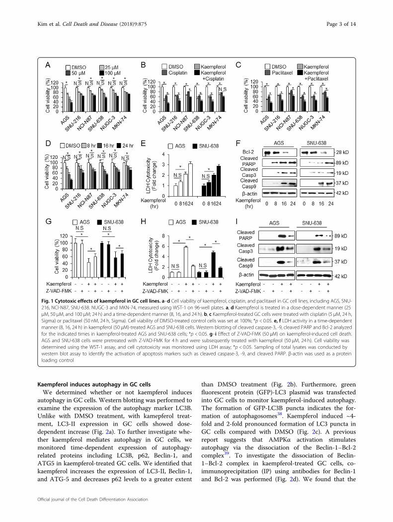
we measured the changes of cell viability by kaempferolusing WST-1 assay on indicated concentration (Fig. 1a).Kaempferol causes a significant decrease of cell viabilitycompared with DMSO (Fig. 1a). Next, we examined theeffect of cisplatin (5 µM, 24 h) or paclitaxel (50 nM, 24 h)in combination with kaempferol (Fig. 1b, c). GC cellstreated with cisplatin or paclitaxel in combination withkaempferol showed lower cell viability than those treatedwith only cisplatin or paclitaxel. To investigate the time-dependent effects of kaempferol, kaempferol treated inGC cells in indicated times (Fig. 1d). Time courseexperiments showed that kaempferol (50 μM) decreasedcell viability in GC cells compared with control. Thesefindings suggest that kaempferol significantly decreasescell viability in GC cells and cisplatin or paclitaxel incombination with kaempferol have a powerful multi-drugcytotoxic effect on GC cells.
Kaempferol induces cell death in GC cellsWe examined kaempferol’s biological effect to identify
whether this induces cell death in GC cells. After the cellswere exposed to kaempferol (50 μM) at indicated timepoints, cell death rate was determined by LDH assay(Fig. 1e). The data showed that kaempferol induces ~3times higher LDH release than DMSO in a time-dependent manner. Western blotting demonstrated thatkaempferol (50 μM) increased cleaved caspase-3 and -9and reduced Bcl-2 levels to a greater extent than DMSOin a time-dependent manner (Fig. 1f). To better char-acterize whether the kaempferol-treated cell death isapoptotic, we treated the cells pretreated with pan-caspase inhibitor, Z-VAD-FMK (50 µM) for 4 h, withkaempferol (Fig. 1g). Consequently, Z-VAD-FMK+kaempferol sufficiently inhibited cell viability and LDHrelease compared to kaempferol alone (Fig. 1g, h). Wes-tern blotting demonstrates that Z-VAD-FMK+ kaemp-ferol decreased cleaved caspase-3 and -9 and cleavedPARP to a greater extent than kaempferol alone (Fig. 1i).Taken together, these findings suggest that kaempferolinduces caspase-dependent cell death in GC.
Kim et al. Cell Death and Disease (2018) 9:875 Page 2 of 14
Official journal of the Cell Death Differentiation Association
Kaempferol induces autophagy in GC cellsWe determined whether or not kaempferol induces
autophagy in GC cells. Western blotting was performed toexamine the expression of the autophagy marker LC3B.Unlike with DMSO treatment, with kamepferol treat-ment, LC3-II expression in GC cells showed dose-dependent increase (Fig. 2a). To further investigate whe-ther kaempferol mediates autophagy in GC cells, wemonitored time-dependent expression of autophagy-related proteins including LC3B, p62, Beclin-1, andATG5 in kaempferol-treated GC cells. We identified thatkaempferol increases the expression of LC3-II, Beclin-1,and ATG-5 and decreases p62 levels to a greater extent
than DMSO treatment (Fig. 2b). Furthermore, greenfluorescent protein (GFP)-LC3 plasmid was transfectedinto GC cells to monitor kaempferol-induced autophagy.The formation of GFP-LC3B puncta indicates the for-mation of autophagosomes38. Kaempferol induced ~4-fold and 2-fold pronounced formation of LC3 puncta inGC cells compared with DMSO (Fig. 2c). A previousreport suggests that AMPKα activation stimulatesautophagy via the dissociation of the Beclin-1–Bcl-2complex39. To investigate the dissociation of Beclin-1–Bcl-2 complex in kaempferol-treated GC cells, co-immunoprecipitation (IP) using antibodies for Beclin-1and Bcl-2 was performed (Fig. 2d). We found that the
Fig. 1 Cytotoxic effects of kaempferol in GC cell lines. a–d Cell viability of kaempferol, cisplatin, and paclitaxel in GC cell lines, including AGS, SNU-216, NCI-N87, SNU-638. NUGC-3 and MKN-74, measured using WST-1 on 96-well plates. a, d Kaempferol is treated in a dose-dependent manner (25μM, 50 μM, and 100 μM; 24 h) and a time-dependent manner (8, 16, and 24 h). b, c Kaempferol-treated GC cells were treated with cisplatin (5 µM, 24 h,Sigma) or paclitaxel (50 nM, 24 h, Sigma). Cell viability of DMSO-treated control cells was set at 100%; *p < 0.05. e, f LDH activity in a time-dependentmanner (8, 16, 24 h) in kaempferol (50 μM)-treated AGS and SNU-638 cells. Western blotting of cleaved caspase-3, -9, cleaved PARP and Bcl-2 analyzedfor the indicated times in kaempferol-treated AGS and SNU-638 cells; *p < 0.05. g–i Effect of Z-VAD-FMK (50 μM) on kaempferol-induced cell death.AGS and SNU-638 cells were pretreated with Z-VAD-FMK for 4 h and were subsequently treated with kaempferol (50 μM, 24 h). Cell viability wasdetermined using the WST-1 assay, and cell cytotoxicity was monitored using LDH assay; *p < 0.05. Sampling of total lysates was conducted bywestern blot assay to identify the activation of apoptosis markers such as cleaved caspase-3, -9, and cleaved PARP. β-actin was used as a proteinloading control
Kim et al. Cell Death and Disease (2018) 9:875 Page 3 of 14
Official journal of the Cell Death Differentiation Association
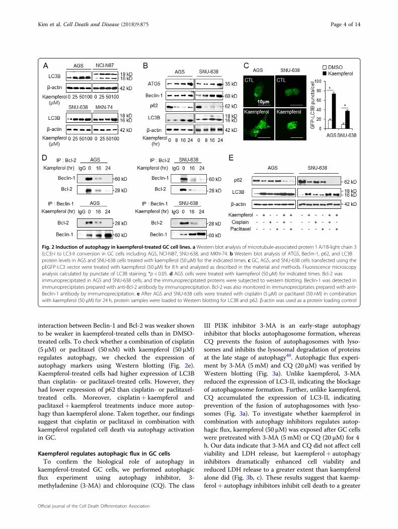
interaction between Beclin-1 and Bcl-2 was weaker shownto be weaker in kaempferol-treated cells than in DMSO-treated cells. To check whether a combination of cisplatin(5 µM) or paclitaxel (50 nM) with kaempferol (50 µM)regulates autophagy, we checked the expression ofautophagy markers using Western blotting (Fig. 2e).Kaempferol-treated cells had higher expression of LC3Bthan cisplatin- or paclitaxel-treated cells. However, theyhad lower expression of p62 than cisplatin- or paclitaxel-treated cells. Moreover, cisplatin+ kaempferol andpaclitaxel+ kaempferol treatments induce more autop-hagy than kaempferol alone. Taken together, our findingssuggest that cisplatin or paclitaxel in combination withkaempferol regulated cell death via autophagy activationin GC.
Kaempferol regulates autophagic flux in GC cellsTo confirm the biological role of autophagy in
kaempferol-treated GC cells, we performed autophagicflux experiment using autophagy inhibitor, 3-methyladenine (3-MA) and chloroquine (CQ). The class
III PI3K inhibitor 3-MA is an early-stage autophagyinhibitor that blocks autophagosome formation, whereasCQ prevents the fusion of autophagosomes with lyso-somes and inhibits the lysosomal degradation of proteinsat the late stage of autophagy40. Autophagic flux experi-ment by 3-MA (5mM) and CQ (20 µM) was verified byWestern blotting (Fig. 3a). Unlike kaempferol, 3-MAreduced the expression of LC3-II, indicating the blockageof autophagosome formation. Further, unlike kaempferol,CQ accumulated the expression of LC3-II, indicatingprevention of the fusion of autophagosomes with lyso-somes (Fig. 3a). To investigate whether kaempferol incombination with autophagy inhibitors regulates autop-hagic flux, kaempferol (50 µM) was exposed after GC cellswere pretreated with 3-MA (5mM) or CQ (20 µM) for 4h. Our data indicate that 3-MA and CQ did not affect cellviability and LDH release, but kaempferol+ autophagyinhibitors dramatically enhanced cell viability andreduced LDH release to a greater extent than kaempferolalone did (Fig. 3b, c). These results suggest that kaemp-ferol+ autophagy inhibitors inhibit cell death to a greater
Fig. 2 Induction of autophagy in kaempferol-treated GC cell lines. aWestern blot analysis of microtubule-associated protein 1 A/1B-light chain 3(LC3)-I to LC3-II conversion in GC cells including AGS, NCI-N87, SNU-638, and MKN-74. b Western blot analysis of ATG5, Beclin-1, p62, and LC3Bprotein levels in AGS and SNU-638 cells treated with kaempferol (50 μM) for the indicated times. c GC, AGS, and SNU-638 cells transfected using thepEGFP-LC3 vector were treated with kaempferol (50 μM) for 8 h and analyzed as described in the material and methods. Fluorescence microscopyanalysis calculated by punctate of LC3B staining; *p < 0.05. d AGS cells were treated with kaempferol (50 μM) for indicated times. Bcl-2 wasimmunoprecipitated in AGS and SNU-638 cells, and the immunoprecipitated proteins were subjected to western blotting. Beclin-1 was detected inimmunoprecipitates prepared with anti-Bcl-2 antibody by immunoprecipitation. Bcl-2 was also monitored in immunoprecipitates prepared with anti-Beclin-1 antibody by immumoprecipitation. e After AGS and SNU-638 cells were treated with cisplatin (5 μM) or paclitaxel (50 nM) in combinationwith kaempferol (50 µM) for 24 h, protein samples were loaded to Western blotting for LC3B and p62. β-actin was used as a protein loading control
Kim et al. Cell Death and Disease (2018) 9:875 Page 4 of 14
Official journal of the Cell Death Differentiation Association
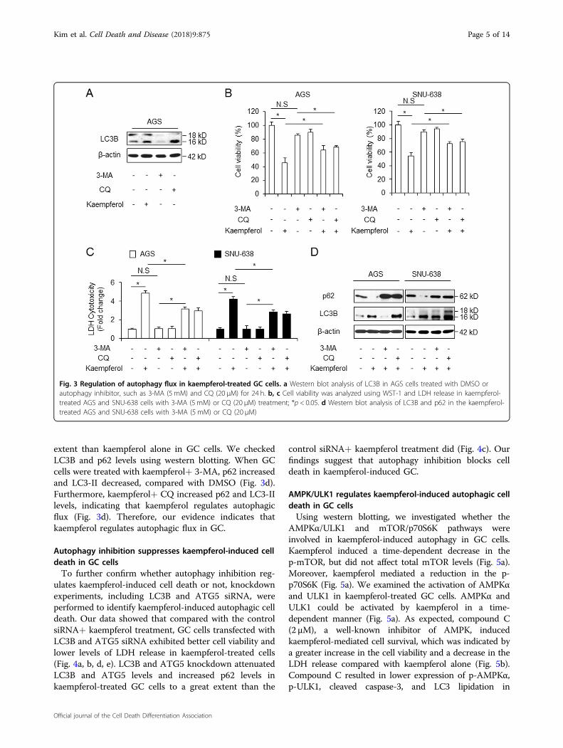
extent than kaempferol alone in GC cells. We checkedLC3B and p62 levels using western blotting. When GCcells were treated with kaempferol+ 3-MA, p62 increasedand LC3-II decreased, compared with DMSO (Fig. 3d).Furthermore, kaempferol+ CQ increased p62 and LC3-IIlevels, indicating that kaempferol regulates autophagicflux (Fig. 3d). Therefore, our evidence indicates thatkaempferol regulates autophagic flux in GC.
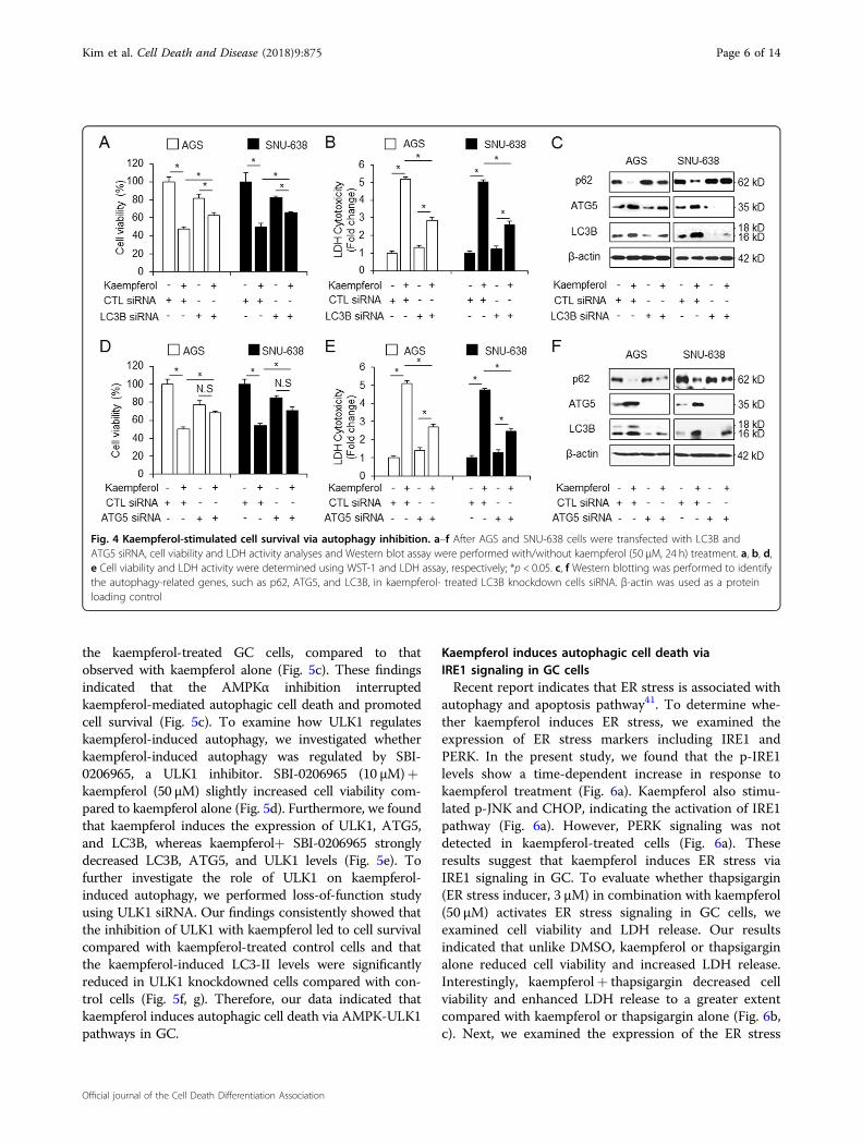
Autophagy inhibition suppresses kaempferol-induced celldeath in GC cellsTo further confirm whether autophagy inhibition reg-
ulates kaempferol-induced cell death or not, knockdownexperiments, including LC3B and ATG5 siRNA, wereperformed to identify kaempferol-induced autophagic celldeath. Our data showed that compared with the controlsiRNA+ kaempferol treatment, GC cells transfected withLC3B and ATG5 siRNA exhibited better cell viability andlower levels of LDH release in kaempferol-treated cells(Fig. 4a, b, d, e). LC3B and ATG5 knockdown attenuatedLC3B and ATG5 levels and increased p62 levels inkaempferol-treated GC cells to a great extent than the
control siRNA+ kaempferol treatment did (Fig. 4c). Ourfindings suggest that autophagy inhibition blocks celldeath in kaempferol-induced GC.
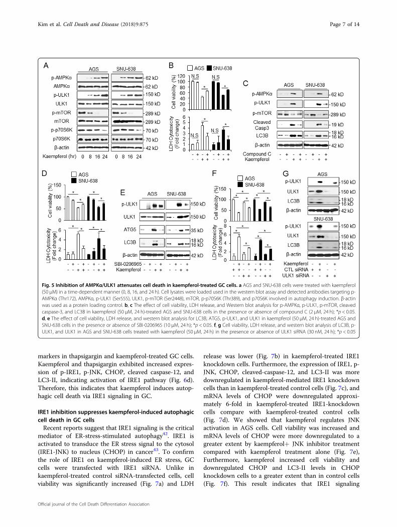
AMPK/ULK1 regulates kaempferol-induced autophagic celldeath in GC cellsUsing western blotting, we investigated whether the
AMPKα/ULK1 and mTOR/p70S6K pathways wereinvolved in kaempferol-induced autophagy in GC cells.Kaempferol induced a time-dependent decrease in thep-mTOR, but did not affect total mTOR levels (Fig. 5a).Moreover, kaempferol mediated a reduction in the p-p70S6K (Fig. 5a). We examined the activation of AMPKαand ULK1 in kaempferol-treated GC cells. AMPKα andULK1 could be activated by kaempferol in a time-dependent manner (Fig. 5a). As expected, compound C(2 µM), a well-known inhibitor of AMPK, inducedkaempferol-mediated cell survival, which was indicated bya greater increase in the cell viability and a decrease in theLDH release compared with kaempferol alone (Fig. 5b).Compound C resulted in lower expression of p-AMPKα,p-ULK1, cleaved caspase-3, and LC3 lipidation in
Fig. 3 Regulation of autophagy flux in kaempferol-treated GC cells. a Western blot analysis of LC3B in AGS cells treated with DMSO orautophagy inhibitor, such as 3-MA (5 mM) and CQ (20 μM) for 24 h. b, c Cell viability was analyzed using WST-1 and LDH release in kaempferol-treated AGS and SNU-638 cells with 3-MA (5 mM) or CQ (20 μM) treatment; *p < 0.05. d Western blot analysis of LC3B and p62 in the kaempferol-treated AGS and SNU-638 cells with 3-MA (5 mM) or CQ (20 μM)
Kim et al. Cell Death and Disease (2018) 9:875 Page 5 of 14
Official journal of the Cell Death Differentiation Association
the kaempferol-treated GC cells, compared to thatobserved with kaempferol alone (Fig. 5c). These findingsindicated that the AMPKα inhibition interruptedkaempferol-mediated autophagic cell death and promotedcell survival (Fig. 5c). To examine how ULK1 regulateskaempferol-induced autophagy, we investigated whetherkaempferol-induced autophagy was regulated by SBI-0206965, a ULK1 inhibitor. SBI-0206965 (10 µM)+kaempferol (50 µM) slightly increased cell viability com-pared to kaempferol alone (Fig. 5d). Furthermore, we foundthat kaempferol induces the expression of ULK1, ATG5,and LC3B, whereas kaempferol+ SBI-0206965 stronglydecreased LC3B, ATG5, and ULK1 levels (Fig. 5e). Tofurther investigate the role of ULK1 on kaempferol-induced autophagy, we performed loss-of-function studyusing ULK1 siRNA. Our findings consistently showed thatthe inhibition of ULK1 with kaempferol led to cell survivalcompared with kaempferol-treated control cells and thatthe kaempferol-induced LC3-II levels were significantlyreduced in ULK1 knockdowned cells compared with con-trol cells (Fig. 5f, g). Therefore, our data indicated thatkaempferol induces autophagic cell death via AMPK-ULK1pathways in GC.
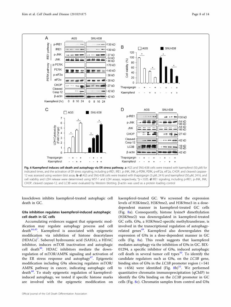
Kaempferol induces autophagic cell death viaIRE1 signaling in GC cellsRecent report indicates that ER stress is associated with
autophagy and apoptosis pathway41. To determine whe-ther kaempferol induces ER stress, we examined theexpression of ER stress markers including IRE1 andPERK. In the present study, we found that the p-IRE1levels show a time-dependent increase in response tokaempferol treatment (Fig. 6a). Kaempferol also stimu-lated p-JNK and CHOP, indicating the activation of IRE1pathway (Fig. 6a). However, PERK signaling was notdetected in kaempferol-treated cells (Fig. 6a). Theseresults suggest that kaempferol induces ER stress viaIRE1 signaling in GC. To evaluate whether thapsigargin(ER stress inducer, 3 µM) in combination with kaempferol(50 µM) activates ER stress signaling in GC cells, weexamined cell viability and LDH release. Our resultsindicated that unlike DMSO, kaempferol or thapsigarginalone reduced cell viability and increased LDH release.Interestingly, kaempferol+ thapsigargin decreased cellviability and enhanced LDH release to a greater extentcompared with kaempferol or thapsigargin alone (Fig. 6b,c). Next, we examined the expression of the ER stress
Fig. 4 Kaempferol-stimulated cell survival via autophagy inhibition. a–f After AGS and SNU-638 cells were transfected with LC3B andATG5 siRNA, cell viability and LDH activity analyses and Western blot assay were performed with/without kaempferol (50 μM, 24 h) treatment. a, b, d,e Cell viability and LDH activity were determined using WST-1 and LDH assay, respectively; *p < 0.05. c, f Western blotting was performed to identifythe autophagy-related genes, such as p62, ATG5, and LC3B, in kaempferol- treated LC3B knockdown cells siRNA. β-actin was used as a proteinloading control
Kim et al. Cell Death and Disease (2018) 9:875 Page 6 of 14
Official journal of the Cell Death Differentiation Association
markers in thapsigargin and kaempferol-treated GC cells.Kaempferol and thapsigargin exhibited increased expres-sion of p-IRE1, p-JNK, CHOP, cleaved caspase-12, andLC3-II, indicating activation of IRE1 pathway (Fig. 6d).Therefore, this indicates that kaempferol induces autop-hagic cell death via IRE1 signaling in GC.
IRE1 inhibition suppresses kaempferol-induced autophagiccell death in GC cellsRecent reports suggest that IRE1 signaling is the critical
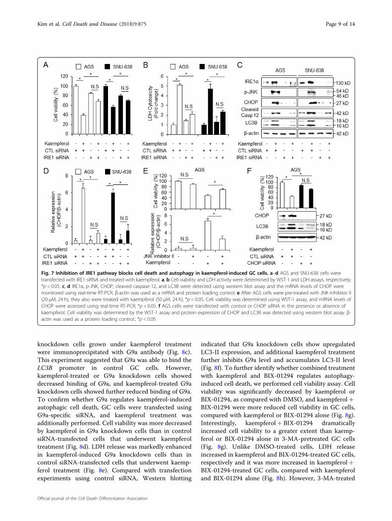
mediator of ER-stress-stimulated autophagy42. IRE1 isactivated to transduce the ER stress signal to the cytosol(IRE1-JNK) to nucleus (CHOP) in cancer43. To confirmthe role of IRE1 on kaempferol-induced ER stress, GCcells were transfected with IRE1 siRNA. Unlike inkaempferol-treated control siRNA-transfected cells, cellviability was significantly increased (Fig. 7a) and LDH
release was lower (Fig. 7b) in kaempferol-treated IRE1knockdown cells. Furthermore, the expression of IRE1, p-JNK, CHOP, cleaved-caspase-12, and LC3-II was moredownregulated in kaempferol-mediated IRE1 knockdowncells than in kaempferol-treated control cells (Fig. 7c), andmRNA levels of CHOP were downregulated approxi-mately 6-fold in kaempferol-treated IRE1-knockdowncells compare with kaempferol-treated control cells(Fig. 7d). We showed that kaempferol regulates JNKactivation in AGS cells. Cell viability was increased andmRNA levels of CHOP were more downregulated to agreater extent by kaempferol+ JNK inhibitor treatmentcompared with kaempferol treatment alone (Fig. 7e),Furthermore, kaempferol increased cell viability anddownregulated CHOP and LC3-II levels in CHOPknockdown cells to a greater extent than in control cells(Fig. 7f). This result indicates that IRE1 signaling
Fig. 5 Inhibition of AMPKα/ULK1 attenuates cell death in kaempferol-treated GC cells. a AGS and SNU-638 cells were treated with kaempferol(50 μM) in a time-dependent manner (0, 8, 16, and 24 h). Cell lysates were loaded used in the western blot assay and detected antibodies targeting p-AMPKα (Thr172), AMPKα, p-ULK1 (Ser555), ULK1, p-mTOR (Ser2448), mTOR, p-p70S6K (Thr389), and p70S6K involved in autophagy induction. β-actinwas used as a protein loading control. b, c The effect of cell viability, LDH release, and Western blot analysis for p-AMPKα, p-ULK1, p-mTOR, cleavedcaspase-3, and LC3B in kaempferol (50 μM, 24 h)-treated AGS and SNU-638 cells in the presence or absence of compound C (2 μM, 24 h); *p < 0.05.d, e The effect of cell viability, LDH release, and western blot analysis for LC3B, ATG5, p-ULK1, and ULK1 in kaempferol (50 μM, 24 h)-treated AGS andSNU-638 cells in the presence or absence of SBI-0206965 (10 μM, 24 h); *p < 0.05. f, g Cell viability, LDH release, and western blot analysis of LC3B, p-ULK1, and ULK1 in AGS and SNU-638 cells treated with kaempferol (50 μM, 24 h) in the presence or absence of ULK1 siRNA (30 nM, 24 h); *p < 0.05
Kim et al. Cell Death and Disease (2018) 9:875 Page 7 of 14
Official journal of the Cell Death Differentiation Association
knockdown inhibits kaempferol-treated autophagic celldeath in GC.
G9a inhibition regulates kaempferol-induced autophagiccell death in GC cellsAccumulating evidences suggest that epigenetic mod-
ification may regulate autophagy process and celldeath44,45. Kaempferol is associated with epigeneticmodification via inhibition of histone deacetylases(HDACs)7. Suberoyl hydroxamic acid (SAHA), a HDACinhibitor, induces mTOR inactivation and autophagiccell death46. HDAC inhibitors mediate the down-regulation of mTOR/AMPK signaling and activation ofthe ER stress response and autophagy47. Epigeneticmodification including G9a silencing regulates mTOR/AMPK pathway in cancer, indicating autophagic celldeath48. To study epigenetic regulation of kaempferol-induced autophagy, we tested if specific histone marksare involved with the epigenetic modification on
kaempferol-treated GC. We screened the expressionlevels of H3K4me2, H3K9me2, and H3K9me3 in a dose-dependent manner in kaempferol-treated GC cells(Fig. 8a). Consequently, histone lysine9 dimethylation(H3K9me2) was downregulated in kaempferol-treatedGC cells. G9a, a H3K9me2-specific methyltransferase, isinvolved in the transcriptional regulation of autophagy-related genes49. Kaempferol also downregulates theexpression of G9a in a dose-dependent manner in GCcells (Fig. 8a). This result suggests that kaempferolmediates autophagy via the inhibition of G9a in GC. BIX-01294, a specific inhibitor of G9a, induced autophagiccell death in several tumor cell types50. To identify thecandidate regulators such as G9a, on the LC3B gene,binding sites of G9a in the LC3B promoter region (+541to +656) were identified (Fig. 8b)51. We performedquantitative chromatin immunoprecipitation (qChIP) toidentify the G9a binding on the LC3B promoter in GCcells (Fig. 8c). Chromatin samples from control and G9a
Fig. 6 Kaempferol induces cell death and autophagy via ER stress pathway. a AGS and SNU-638 cells were treated with kaempferol (50 μM) forindicated times, and the activation of ER stress signaling, including p-IRE1, IRE1, p-JNK, JNK, p-PERK, PERK, p-eIF2α, eIF2α, CHOP, and cleaved caspase-12 was assessed using western blot assay. b–d AGS and SNU-638 cells were treated with thapsigargin (3 μM, 24 h) and kaempferol (50 μM, 24 h), andcell viability and LDH release were determined using WST-1 and LDH assays, respectively; *p < 0.05. d IRE1 signaling, including p-IRE1, p-JNK, JNK,CHOP, cleaved caspase-12, and LC3B were evaluated by Western blotting. β-actin was used as a protein loading control
Kim et al. Cell Death and Disease (2018) 9:875 Page 8 of 14
Official journal of the Cell Death Differentiation Association
knockdown cells grown under kaempferol treatmentwere immunoprecipitated with G9a antibody (Fig. 8c).This experiment suggested that G9a was able to bind theLC3B promoter in control GC cells. However,kaempferol-treated or G9a knockdown cells showeddecreased binding of G9a, and kaempferol-treated G9aknockdown cells showed further reduced binding of G9a.To confirm whether G9a regulates kaempferol-inducedautophagic cell death, GC cells were transfected usingG9a-specific siRNA, and kaempferol treatment wasadditionally performed. Cell viability was more decreasedby kaempferol in G9a knockdown cells than in controlsiRNA-transfected cells that underwent kaempferoltreatment (Fig. 8d). LDH release was markedly enhancedin kaempferol-induced G9a knockdown cells than incontrol siRNA-transfected cells that underwent kaemp-ferol treatment (Fig. 8e). Compared with transfectionexperiments using control siRNA, Western blotting
indicated that G9a knockdown cells show upregulatedLC3-II expression, and additional kaempferol treatmentfurther inhibits G9a level and accumulates LC3-II level(Fig. 8f). To further identify whether combined treatmentwith kaempferol and BIX-01294 regulates autophagy-induced cell death, we performed cell viability assay. Cellviability was significantly decreased by kaempferol orBIX-01294, as compared with DMSO, and kaempferol+BIX-01294 were more reduced cell viability in GC cells,compared with kaempferol or BIX-01294 alone (Fig. 8g).Interestingly, kaempferol+ BIX-01294 dramaticallyincreased cell viability to a greater extent than kaemp-ferol or BIX-01294 alone in 3-MA-pretreated GC cells(Fig. 8g). Unlike DMSO-treated cells, LDH releaseincreased in kaempferol and BIX-01294-treated GC cells,respectively and it was more increased in kaempferol+BIX-01294-treated GC cells, compared with kaempferoland BIX-01294 alone (Fig. 8h). However, 3-MA-treated
Fig. 7 Inhibition of IRE1 pathway blocks cell death and autophagy in kaempferol-induced GC cells. a–d AGS and SNU-638 cells weretransfected with IRE1 siRNA and treated with kaempferol. a, b Cell viability and LDH activity were determined by WST-1 and LDH assays, respectively;*p < 0.05. c, d IRE1α, p-JNK, CHOP, cleaved caspase-12, and LC3B were detected using western blot assay and the mRNA levels of CHOP weremonitored using real-time RT-PCR. β-actin was used as a mRNA and protein loading control. e After AGS cells were pre-treated with JNK inhibitor II(20 μM, 24 h), they also were treated with kaempferol (50 μM, 24 h); *p < 0.05. Cell viability was determined using WST-1 assay, and mRNA levels ofCHOP were assessed using real-time RT-PCR; *p < 0.05. f AGS cells were transfected with control or CHOP siRNA in the presence or absence ofkaempferol. Cell viability was determined by the WST-1 assay and protein expression of CHOP and LC3B was detected using western blot assay. β-actin was used as a protein loading control.; *p < 0.05
Kim et al. Cell Death and Disease (2018) 9:875 Page 9 of 14
Official journal of the Cell Death Differentiation Association
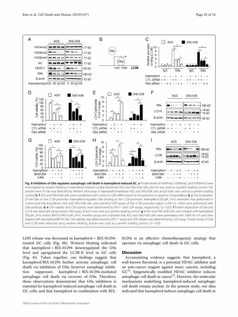
LDH release was decreased in kaempferol+ BIX-01294-treated GC cells (Fig. 8h). Western blotting indicatedthat kaempferol+ BIX-01294 downregulated the G9alevel and upregulated the LC3B-II level in GC cells(Fig. 8i). Taken together, our findings suggest thatkaempferol/BIX-01294 further activate autophagic celldeath via inhibition of G9a; however autophagy inhibi-tion suppresses kaempferol+ BIX-01294-mediatedautophagic cell death via recovery of G9a. Therefore,these observations demonstrate that G9a inhibition isessential for kaempferol-induced autophagic cell death inGC cells, and that kaempferol in combination with BIX-
01294 is an effective chemotherapeutic strategy thatoperates via autophagic cell death in GC cells.
DiscussionAccumulating evidence suggests that kaempferol, a
well-known flavonoid, is a potential HDAC inhibitor andan anti-cancer reagent against many cancers, includingGC52. Epigenetically modified HDAC inhibitor inducesautophagic cell death in cancer53. However, the molecularmechanisms underlying kaempferol-induced autophagiccell death remain unclear. In the present study, our dataindicated that kaempferol induces autophagic cell death in
Fig. 8 Inhibition of G9a regulates autophagic cell death in kaempferol-induced GC. a Protein levels of H3K4me2, H3K9me2, and H3K9me3 wereinvestigated by western blotting in kaempferol-treated nuclear-fractioned AGS and SNU-638 cells, and H3 was used as a protein loading control. Theprotein level of G9a was detected by Western blot assay in kaemprefol-mediated AGS and SNU-638 cells and β-actin was used as a protein loadingcontrol. b–f AGS and SNU-638 cells were transfected with control or G9a siRNA based on the presence or absence of kaempferol. b, c The localizationof the G9a on the LC3B promoter. Kaempferol regulates G9a binding at the LC3B promoter. Kaempferol (50 μM, 24 h) treatment was performed incontrol and G9a knockdown AGS and SNU-638 cells, and real-time ChIP assays of the LC3B promoter region (+541 to +656) were performed withG9a antibody. d, e Cell viability and LDH activity were determined by WST-1 and LDH assays, respectively; *p < 0.05. f Protein expression of G9a andLC3-II was detected using western blot assay. β-actin was used as a protein loading control. g–i AGS and SNU-638 cells were treated with kaempferol(50 μM, 24 h) and/or BIX-01294(10 μM, 24 h). Another group also indicated that AGS and SNU-638 cells were pretreated with 3-MA for 4 h and thentreated with kaempferol/BIX-01294. Cell viability was determined by WST-1 assay and LDH release was determined by LDH assay. Protein levels of G9aand LC3B were detected using western blotting. β-actin was used as a protein loading control; *p < 0.05
Kim et al. Cell Death and Disease (2018) 9:875 Page 10 of 14
Official journal of the Cell Death Differentiation Association
GC, and to our knowledge, this is the first research toidentify that kaempferol promotes cell death via theIRE1–JNK–CHOP pathway. Furthermore, kaempferolcauses autophagic cell death via an epigenetic modifica-tion involving G9a inhibition. Autophagy inhibitionincluding inhibitor and siRNA regulate autophagic flux inkaempferol-induced cells, and autophagy inhibitiondecreased cell death by increasing cell viability andreducing LDH activity in kaempferol-treated GC cells.These results indicate that kaempferol induces autophagiccell death, but autophagy inhibition plays a functional rolefor cell survival, implying kaempferol-induced autophagiccell death.On the basis of our findings, we hypothesized that
kaempferol promotes autophagic cell death via ER stressin GC. IRE1 may initiate cell death, and the accumulationof JNK and CHOP also induced cell death54. In this study,we screened two ER stress signaling pathways:PERK–eIF2α–CHOP and IRE1–JNK–CHOP pathway.When we monitored PERK pathway in kaempferol-treated GC cells, Western blotting did not detectedphosphorylation of PERK and eIF2α, indicating inactiva-tion of PERK signaling. However, it found upregulation ofIRE1, p-IRE1, p-JNK, CHOP, and cleaved caspase-12,indicating the activation of IRE-mediated ER stress.CHOP is an important factor of ER stress-induced celldeath via PERK and IRE1 signaling, and JNK also inducescell death via the induction of CHOP and C-jun55,56. IRE1induces cell death via the activation of JNK and CHOPand the inhibition of Bcl-257. JNK1 induces the phos-phorylation of Bcl-2 and promotes autophagic cell deathby disrupting the Bcl-2–Beclin-1 complex58. Interestingly,the inhibition of IRE1 and JNK, indicating the upstreamsignal of CHOP, reduces autophagic cell death via thedownregulation of CHOP. These data demonstrated thatkaempferol induces autophagic cell death via theIRE1–JNK–CHOP signaling in GC, whereas IRE1knockdown blocks kaempferol-treated autophagic celldeath.Recent reports indicate that BIX-01294 reduces
H3K9me2 through the inhibition of G9a and inducesautophagic cell death in cancer59. Furthermore, G9arepresses the expression of LC3B by directly binding withthe promoter H3K9me2, indicating repressive mark, andthe inhibition of G9a activates LC3B60. Our data indicatedthat kaempferol and BIX-01294 accumulate LC3-II andreduce cell viability via G9a inhibition, and a loss-of-function study using specific siRNA for G9a suggests thatkaempferol reduces the binding of G9a on LC3B pro-moter in G9a knockdown cells. However, 3-MA sig-nificantly blocks LC3-II and cell death and recovers G9ain kaempferol/BIX-01294-treated GC cells. Therefore, ourfindings suggest that kaempferol induces autophagic cell
death via the IRE1–JNK–CHOP pathway and the HDAC/G9a pathway in GC.Taken together, these findings support that kaempferol
induces autophagic cell death via IRE1–JNK1-mediatedBcl-2–Beclin-1 dissociation in GC cells. Moreover,kaempferol epigenetically mediates autophagic cell deathvia HDAC/G9a pathway. A deeper understanding of themolecular mechanism of kaempferol may contribute touseful cancer therapeutic approaches.
Materials and methodsCell cultureThe human GC cell lines (AGS, SNU-216, NCI-N87,
SNU-638, and MKN-74) were purchased from the KoreanCell Line Bank (Cancer Research Center, Seoul NationalUniversity, Seoul, Korea). Cells were cultured inRPMI1640 medium (Welgene) supplemented with 10%fetal bovine serum (JR Scientific) and 100 μg/mL anti-biotics (100 U/ mL penicillin and 100 μg/ mL streptomy-cin, Welgene) in a 5% CO2 humidified incubator at 37 °C.
Cell viability assayThe WST-1 assay was performed according to the
manufacturer’s instructions (Roche, Mannheim,Germany) with 10 μL of WST-1 reagent was added toeach well of a 96-well plate (1 × 10⁴ cell/well). After 1 h ofincubation using CO2 incubator, the conversion of WST-1 reagent into chromogenic formazan was evaluated witha spectrophotometer (Molecular devices, USA). On day 1after cell seeding, cells were treated with various doses ofkaempferol (Sigma) (25, 50, and 100 µM) at various timepoints (8, 16, and 24 h). Autophagy inhibitor, 3-MA(Sigma, 5 mM), chloroquine (Sigma, 20 µM), compound C(Sigma, 2 µM) and SBI-0206965 (Sigma, 10 μM) wereadded sequentially to FBS-free medium for 24 h to inhibitautophagy. A pan-caspase inhibitor, Z-VAD-FMK (R&DSystems, 50 μM), was added to FBS-free medium for 24 hto inhibition of apoptosis. Cells were treated with an ERstress inducer, thapsigargin (Sigma, 3 μM, 24 h), alongwith FBS-free medium to activate ER stress and JNKinhibitor II (Calbiochem, 20 μM, 24 h) was added toinhibit JNK signaling. In addition, a G9a inhibitor, BIX-01294(Sigma, 10 μM, 24 h), was added to activate autop-hagy via G9a inhibition.
LDH assayAGS and SNU-638 cells (1 × 10⁴ cells/well) were seeded
into a 96-well plate with growth medium. To determinethe LDH (Thermo Scientific Pierce) activity in super-natants, 100 μL of Reaction mixture was added andincubated for 30min in a dark room. The LDH activitywas measured by the absorbance of the samples at 490 or492 nm using ELISA reader.
Kim et al. Cell Death and Disease (2018) 9:875 Page 11 of 14
Official journal of the Cell Death Differentiation Association
TransfectionAGS and SNU-638 cells (3 × 10⁵ cell/well) were trans-
fected with double-stranded siRNAs (30 nmol/mL) ofLC3B, ATG5, ULK1, IRE1 (Santacruz), and CHOP (Bio-neer) in 6-well plate for 24 h by the Lipofectamine 2000(Invitrogen) method according to the manufacturer’sprotocol and were then recovered in RPMI1640 medium(Welgene) containing 5% fetal bovine serum (Gibco) and100 μg/mL antibiotics (100 U/mL penicillin and 100 μg/mL streptomycin, Gibco) for 24 h. After recovering, viablecells were calculated by WST-1.
Isolation of total RNA and ProteinTotal RNA (approximately 50–100mg) from GC cells
(2 × 10⁶ cell/well) in 100mm cell culture dish was pre-pared using Trizol according to the manufacturer’s pro-tocols (invitrogen, Carlsbad, CA, USA). Protein celllysates were collected in RIPA buffer containing a pro-tease inhibitor cocktail (Sigma) on ice for 30min and werepassed through an 18-gauge needle and spin down. Thesupernatant was analyzed for protein content using theBCA method (Thermo scientific, Pierce BCA ProteinAssay Kit, USA).
Real-time PCR and western analysisCHOP expression level was measured by real-time PCR
using cDNA synthesized from 5 ug of total RNA and areverse transcription kit (Promega, Madison, WI). Tri-plicate reactions were performed for each sample using anABI Power SYBR green PCR Master Mix (Applied Bio-systems) with CHOP-specific primers [5′-ATGAG-GACCTGCAAGAGGTCC-3′ (sense) and 5′-TCCTCCTCAGTCAGCCAAGC-3′ (antisense)] on a RocheLightCycler 96 (Roche). RNA quantity was normalized toβ-actin primers [5′-AAGGCCAAC CGCGAGAAGAT-3′(sense) and 5′-TGATGACCTGGCCGTCAGG-3′ (anti-sense)]. Gene expression was quantified according to the2-ΔCt method. To conduct the Western blot assay, GC celllines were solubilized in radioimmunoprecipitation assay(RIPA) lysis buffer [50 mM/L Tris-HCl (pH 7.4), 150mM/L NaCl, 1% NP40, 0.25% sodium deoxycholate, 1 mM/Lphenylmethylsulfonylfluoride (PMSF), 1 mM/L sodiumorthovanadate, and 1 × sigma protease inhibitor cocktail]and protein content was measured using a standardbicinchoninic acid assay. Equal amounts of protein (20 μg)were size-fractionated by 8–15% SDS-PAGE and thentransferred onto an NC membrane (Millipore Corpora-tion, Billerica, MA, USA). Membranes were blocked byincubation for 30min with 5% skim milk/PBS-T [PBSwith 5% powdered milk (BD) and 1% Tween20 (Sigma)],and incubated overnight at 4 °C with primary antibodiesdiluted in 1 × PBST buffer. The following primary anti-bodies were used: β-actin, Bcl-2, Beclin-1, ULK1, eIF2α,JNK, and ATG5 (Santa Cruz, 1:1000), LC3B (Sigma,
1:1000), p62 (Sigma, 1:1000), p-IRE1 (Abcam, 1:1000), andG9a (Abcam, 1:1000), cleaved caspase-3, cleaved caspase-9, cleaved PARP, p62, p-AMPKα (Thr172), AMPKα, p-mTOR (Ser2448), mTOR, p-ULK1 (Ser555), p-P70S6K(Thr389), P70S6K, IRE1, PERK, p-PERK (Thr980), p-eIF2α (Ser51), p-JNK, H3, H3K4me2, H3K9me2,H3K9me3, and CHOP (CellSignaling, 1:1000). Themembranes were washed three times with PBST buffer. Asecondary antibody diluted in PBST or TBST buffer wasadded, and incubation was done for 40min at roomtemperature. The following secondary antibodies wereused: anti-rabbit IgG HRP-linked antibody and anti-mouse IgG HRP-linked antibody (KPL, 1:6000). Themembranes were washed six times with PBST buffer for1 h. The blots were visualized using Western chemilu-minescent HRP substrate (Millipore).
Quantification of pEGFP-LC3 punctaAGS and SNU-638 cells (2 × 10⁵ cells per well) in a 6-
well plate were transfected with pEGFP-LC3 using lipo-fectamin 2000 (Invitrogen), and then treated with 50 μMkaempferol for 8 h. A pEGFP-LC3B-positive punctatepattern was observed by confocal microscopy. Confocalmicroscopy was conducted using a ZEISS LSM5 PASCALconfocal microscope with 405- and 488-nm excitationlasers.
Nuclear fractionNuclear fraction was carried out using Nuclear Extract
kit (Active motif) according to manufacturer’s instruc-tions. After the cytoplasmic fraction was extracted assupernatants, cell pellets were resuspended in 50ml ofcomplete lysis buffer and supernatants were used asnuclear fractions by centrifugation at 14,000 × g for10 min at 4 °C.
Immunoprecipitation (IP) assayWe extracted cell lysates from AGS cells (2 × 10⁶ per
well) on 100mm cell culture plate in a IP buffer (pH 7.5)containing 50mM Tris-HCl, 250mM NaCl, 5 mM EDTA,0.5%(v/v) NP-40, and protease inhibitor cocktail (Sigma).We incubated anti-Bcl-2 (Santa Cruz) and anti-BECN-1(Santa Cruz) with lysate at 4 °C for 16 h. We used proteinA/G PLUS agarose (Santa Cruz) to pull down immuno-complexes. We washed precipitates three times with IPbuffer. We resolved the immunoprecipitated proteins by12% SDS-PAGE and analyzed them.
Chromatin immunoprecipitation(ChIP) assayChIP assays were performed using an EZ ChIP Chro-
matin Immunoprecipitation kit (Millipore, Billerica, MA,USA) as described in the supplier’s protocol. Briefly, thecross-linked chromatin was sonicated after cell lysis andthen incubated overnight at 4 °C with antibodies against
Kim et al. Cell Death and Disease (2018) 9:875 Page 12 of 14
Official journal of the Cell Death Differentiation Association
G9a (Abcam). The immunocomplex was precipitated withprotein A–agarose (Millipore), and the beads werewashed, sequentially treated with 10 µl of RNase A (37 °Cfor 30min) and 75 µl of proteinase K (45 °C for 4 h), andincubated at 65 °C overnight to reverse cross-link thechromatin. The DNA was recovered byphenol–chloroform extraction and co-precipitation withglycogen and was then dissolved in 50 µl of Tris-EDTA(TE) buffer. DNA associated with the ER was amplified byPCR using 1 µl of precipitated DNA. PCR primers [5′-GAAGTGGCTATCGCCAGAGT-3′ (sense) and 5′-GCTGCTTGAAGGTCTTCTCC -3′ (antisense)] weredesigned to amplify the G9a binding site at the LC3B genepromoter. Quantitative PCR conditions were 40 cycles at94 °C for 40 s, 60 °C for 1 min, and 72 °C for 40 s.
Statistical AnalysisAll results were confirmed in at least three independent
experiments; Student’s t-tests were used for between-groups comparisons of the means of quantitative data,and p < 0.05 was considered statistically significant.
AcknowledgementsThis work was supported by a grant from Korean Medicine R&D project of theMinistry of Health and Welfare (HI11C2110 and HI12C1889).
Author details1Department of Preventive Medicine, College of Korean Medicine, Kyung HeeUniversity, Seoul, Korea. 2Department of Cardiovascular and Neurologic disease(Stroke center), College of Korean Medicine, Kyung Hee University, Seoul, Korea
Conflict of interestThe authors declare that they have no conflict of interest.
Publisher's noteSpringer Nature remains neutral with regard to jurisdictional claims inpublished maps and institutional affiliations.
Received: 19 March 2018 Revised: 11 July 2018 Accepted: 25 July 2018
References1. Bajpai, M., Pande, A., Tewari, S. K. & Prakash, D. Phenolic contents and anti-
oxidant activity of some food and medicinal plants. Int. J. Food Sci. Nutr. 56,287–291 (2005).
2. Song, H. et al. Kaempferol inhibits gastric cancer tumor growth: an in vitro andin vivo study. Oncol. Rep. 33, 868–874 (2015).
3. Wilsher, N. E. et al. Cytochrome P450 CYP1 metabolism of hydroxylated fla-vones and flavonols: selective bioactivation of luteolin in breast cancer cells.Food Chem. Toxicol. 110, 383–394 (2017).
4. Jeong, H., Phan, A. N. H. & Choi, J. W. Anti-cancer effects of polyphenoliccompounds in epidermal growth factor receptor tyrosine kinase inhibitor-resistant non-small cell lung cancer. Pharmacogn. Mag. 13, 595–599 (2017).
5. Hung, T. W. et al. Kaempferol inhibits the invasion and migration of renalcancer cells through the downregulation of AKT and FAK pathways. Int. J.Med. Sci. 14, 984–993 (2017).
6. Shing, P., Tomar, R. S. & Rath, S. K. Anticancer potential of the histone dea-cetylase inhibitor-like effects of flavones, a subclass of polyphenolic com-pounds: a review. Mol. Biol. Rep. 42, 1515–1531 (2015).
7. Berger, A. et al. Kaempferol, a new nutrition-derived pan-inhibitor of humanhistone deacetylases. J. Nurt. Biochem. 24, 977–985 (2013).
8. Kim, H. J. & Bae, S. C. Histone deacetylase inhibitors: molecular mechanisms ofaction and clinical trials as anti-cancer drugs. Am. J. Transl. Res. 3, 166–179(2011).
9. Hrzenjak, A. et al. SAHA induces caspase-independent autophagic cell deathof endometrial stromal sarcoma cells by influencing the mTOR pathway. J.Pathol. 216, 495–504 (2008).
10. Ryu, C. H. et al. Valproic acid downregulates the expression of MGMT andsensitizes temozolomide-resistant glioma cells. J. Biomed. Biotechnol. 2012,987495 (2012).
11. Chen, Y., Tsai, Y. H. & Tseng, S. H. HDAC inhibitors and RECK modulateendoplasmic reticulum stress in tumor cells. Int. J. Mol. Sci. 18, pii:E258 (2017).
12. Luo, H. et al. Kaempferol inhibits angiogenesis and VEGF expression throughboth HIF dependent and independent pathways in human ovarian cancercells. Nutr. Cancer 61, 554–563 (2009).
13. Xu, W., Liu, J., Wu, H. Z. & Liu, Y. W. Kamepferol-7-O-beta-D-glucoside (KG)isolated from smilax china L. rhizome induces G2/M phase arrest and apop-tosis on HeLa cells in a p53-independent manner. Cancer Lett. 264, 229–240(2008).
14. Sano, R. & Reed, J. C. ER stress-induced cell death mechanisms. Biochim.Biophys. Acta 1832, 2191–2203 (2013).
15. Bertolotti, A., Zhang, Y., Hendershot, L. M., Harding, H. P. & Ron, D. Dynamicinteraction of bip and ER stress tranducers in the unfolded-protein response.Nat. Cell Biol. 2, 326–332 (2000).
16. Cubillos-Ruiz, J. R., Bettigole, S. E. & Glimcher, L. H. Tumorigenic and immu-nosuppressive effects of endoplasmic reticulum stress in cancer. Cell 168,692–706 (2017).
17. Khan, I. et al. Novel quercetin derivative TEF induces ER stress andmitochondria-mediated apoptosis in human colon cancer HCT-116 cells.Biomed. Pharmacother. 84, 789–799 (2016).
18. Wu, P. S., Yen, J. H., Kou, M. C. & Wu, M. J. Luteolin and apigenin attenuate 4-hydroxy-2-nonenal-mediated cell death through modulation of UPR, Nrf2-AREand MAPK pathways in PC12 cells. PLoS ONE 10, e0130599 (2015).
19. Nakagawa, T. et al. Caspase-12 mediates endoplasmic-reticulum-specificapoptosis and cytotoxicity by amyloid-beta. Nature 403, 98–103 (2000).
20. Prieto-Dominquez N., Garcia-Mediavilla M. V., Sanchez-Campos S., Mauriz J. L. &Gonzalez-Gallego J. Autophagy as a molecular target of flavonoids underlyingtheir protective effects in human disease. Curr. Med. Chem. 24, https://doi.org/10.2174/0929867324666170918125155 (2017).
21. De Duve, C. & Wattiaux, R. Functions of lysosomes. Annu. Rev. Physiol. 28,435–492 (1966).
22. Lorin, S., Hamai, A., Mehrpour, M. & Codogno, P. Autophagy regulation and itsrole in cancer. Semin. Cancer Biol. 23, 361–379 (2013).
23. Corazzari, M. et al. Oncogenic BRAF induces chronic ER stress conditionresulting in increased basal autophagy and apoptotic resistance of cutaneousmelanoma. Cell Death Differ. 22, 946–958 (2015).
24. Liang, X. H. et al. Protection against fatal Sindbis virus encephalitis by beclin, anovel Bcl-2-interacting protein. J. Virol. 72, 8586–8596 (1998).
25. Wei, Y., Pattingre, S., Sinha, S., Bassik, M. & Levine, B. JNK1-mediated phos-phorylation of Bcl-2 regulates starvation-induced autophagy. Mol. Cell 30,678–688 (2008).
26. Ogata, M. et al. Autophagy is activated for cell survival after endoplasmicreticulum stress. Mol. Cell Biol. 26, 9220–9231 (2006).
27. Egan, D. F. et al. Phosphorylation of ULK1 (hATG1) by AMP-activatedprotein kinase connects energy sensing to mitophagy. Science 331, 456–461(2011).
28. Cicchini, M., Karantza, V. & Xia, B. Molecular pathways: autophagy in cancer--amatter of timing and context. Clin. Cancer Res. 21, 498–504 (2015).
29. Russell, R. C. et al. ULK1 induces autophagy by phosphorylating Beclin-1 andactivating VPS34 lipid kinase. Nat. Cell Biol. 15, 741–750 (2013).
30. Maiuri, M. C., Zalckvar, E., Kimchi, A. & Kroemer, G. Self-eating and self-killing:crosstalk between autophagy and apoptosis. Nat. Rev. Mol. Cell Biol. 8, 741–752(2007).
31. Filomeni, G. et al. Carcinoma cells activate AMP-activated protein kinase-dependent autophagy as survival response to kaempferol-mediated energeticimpairment. Autophagy 6, 202–216 (2010).
32. Zhang, J. et al. Synthesis and biological evaluation of benzimidazole deriva-tives as the G9a Histone Methyltransferase inhibitors that induce autophagyand apoptosis of breast cancer cells. Bioorg. Chem. 72, 168–181 (2017).
33. Wu, L. P. et al. Histone deacetylase inhibitor depsipeptide activates silencedgenes through decreasing both CpG and H3K9 methylation on the promoter.Mol. Cell Biol. 28, 3219–3235 (2008).
Kim et al. Cell Death and Disease (2018) 9:875 Page 13 of 14
Official journal of the Cell Death Differentiation Association
34. Li, K. C. et al. Inhibition of G9a induces DUSP4-dependent autophagic celldeath in head and neck squamous cell carcinoma. Mol. Cancer 13, 172 (2014).
35. Li, F. et al. G9a inhibition induces autophagic cell death via AMPK/mTORpathway in bladder transitional cell carcinoma. PLoS ONE 10, e0138390 (2015).
36. Nakajima, N. I. et al. Inhibition of the HDAC/Suv39/G9a pathway restores theexpression of DNA damage-dependent major histocompatibility complexclass I-related chain A and B in cancer cells. Oncol. Rep. 38, 693–702 (2017).
37. Juergens, R. A. et al. Combination epigenetic therapy has efficacy in patientswith refractory advanced non-small cell lung cancer. Cancer Discov. 1, 598–607(2011).
38. Shen, S. et al. Detective autophagy associated with LC3 puncta in epothilone-resistant cancer cells. Cell Cycle 9, 377–383 (2010).
39. He, C., Zhu, H., Li, H., Zou, M. H. & Xie, Z. Dissociation of Bcl-2-Beclin-1 complexby activated AMPK enhances cardiac autophagy and protects against cardi-omyocyte apoptosis in diabetes. Diabetes 62, 1270–1281 (2013).
40. Choi, K. S. Autophagy and cancer. Exp. Mol. Med. 44, 109–120 (2012).41. Fernández, A., Ordóñez, R., Reiter, R. J., González-Gallego, J. & Mauriz, J. L.
Melatonin and endoplasmic reticulum stress: relation to autophagy andapoptosis. J. Pineal Res. 59, 292–307 (2015).
42. Szegezdi, E., Logue, S. E., Gorman, A. M. & Samali, A. Mediators of endoplasmicreticulum stress-induced apoptosis. EMBO Rep. 7, 880–885 (2006).
43. Li, Y., Guo, Y., Tang, J., Jiang, J. & Chen, Z. New insights into the roles of CHOP-induced apoptosis in ER stress. Acta Biochim. Biophys. Sin. 47, 146–147 (2015).
44. Wei, F. Z. et al. Epigenetic regulation of autophagy by the methyltransferaseEZH2 through an MTOR-dependent pathway. Autophagy 11, 2309–2322(2015).
45. Artal-Martinez de Narvajas, A. et al. Epigenetic regulation of autophagy by themethyltransferase G9a. Mol. Cell Biol. 33, 3983–3993 (2013).
46. Gammoh, N. et al. Role of autophagy in histone deacetylase inhibitor-inducedapoptotic and nonapoptotic cell death. Proc. Natl Acad. Sci. USA 109,6561–6565 (2012).
47. Liu, Y. L. et al. Autophagy potentiates the anti-cancer effects of the histonedeacetylase inhibitors in hepatocellular carcinoma. Autophagy 6, 1057–1065(2010).
48. Casciello, F., Windloch, K., Gannon, F. & Lee, J. S. Functional role of G9a histonemethyltransferase in cancer. Front. Immunol. 6, 487 (2015).
49. Kim, Y. et al. BIX-01294 induces autophagy-associated cell death via EHMT2/G9a dysfunction and intracellular reactive oxygen species production.Autophagy 9, 2126–2139 (2013).
50. Savickiene, J., Treigyte, G., Stirblyte, I., Valiuliene, G. & Navakauskiene, R.Euchromatic histone methyltransferase 2 inhibitor, BIX-12594, sensitizeshuman promyelocytic leukemia HL-60 and NB4 cells to growth inhibition anddifferentiation. Leuk. Res. 38, 822–839 (2014).
51. Ciechomska, I. A., Przanowski, P., Jackl, J., Wojtas, B. & Kaminska, B. BIX01294, aninhibitor of histone methyltransferase, induces autophagy-dependent differ-entiation of glioma stem-like cells. Sci. Rep. 6, 38723 (2016).
52. Liao, W. et al. Protective effects of kaempferol against reactive oxygen species-induced hemolysis and its antiproliferative activity on human cancer cells. Eur.J. Med. Chem. 114, 24–32 (2016).
53. Zhang, J. et al. Histone deacetylase inhibitors induce autophagy throughFOXO1-dependent pathways. Autophagy 11, 629–642 (2015).
54. Chen, L. et al. Cab45S inhibits the ER stress-induced IRE1-JNK pathway andapoptosis via GRP78/BiP. Cell Death Dis. 5, e1219 (2014).
55. Marhfour, I. et al. Expression of endoplasmic reticulum stress markers in theislets of patients with type 1 diabetes. Diabetologia 55, 2417–2420(2012).
56. Xu, Z. et al. miR-216b regulation of c-jun mediates GADD153/CHOP-depen-dent apoptosis. Nat. Commun. 7, 11422 (2016).
57. Chen, K. et al. Overexpression of Insig-1 protects β cell against glucolipo-toxicity via SREBP-1c. J. Biomed. Sci. 18, 57 (2011).
58. Marquez, R. T. & Xu, L. Bcl-2:Beclin 1 complex: multiple, mechanisms regulatingautophagy/apoptosis toggle switch. Am. J. Cancer Res. 2, 214–221(2012).
59. Ren, A., Qiu, Y., Cui, H. & Fu, G. Inhibition of H3K9 methyltransferase G9ainduces and apoptosis in oral squamous cell carcinoma. Biochem. Biophys. Res.Commun. 459, 10–17 (2015).
60. Collins, P. L. & Oltz, E. M. Histone methylation keeps the brakes on autophagy.Mol. Cell Biol. 33, 3974–3975 (2013).
Kim et al. Cell Death and Disease (2018) 9:875 Page 14 of 14
Official journal of the Cell Death Differentiation Association