journal pre-proof - binasss
TRANSCRIPT
Journal Pre-proof
Patterns of Genital Examination and Vulvovaginal Graft-Versus-Host Disease in aPediatric Post-Hematopoietic Stem Cell Transplant Population
Stephanie M. Allen, MD MSc, Cynthia S. Liang, BS, Arina E. Chesnokova, MD MPH,Krista J. Childress, MD, Kristin F. Pascoe, MD, Jennifer E. Dietrich, MD MSc
PII: S1083-3188(20)30298-9
DOI: https://doi.org/10.1016/j.jpag.2020.08.004
Reference: PEDADO 2535
To appear in: Journal of Pediatric and Adolescent Gynecology
Received Date: 3 March 2020
Revised Date: 29 June 2020
Accepted Date: 2 August 2020
Please cite this article as: Allen SM, Liang CS, Chesnokova AE, Childress KJ, Pascoe KF, DietrichJE, Patterns of Genital Examination and Vulvovaginal Graft-Versus-Host Disease in a Pediatric Post-Hematopoietic Stem Cell Transplant Population, Journal of Pediatric and Adolescent Gynecology(2020), doi: https://doi.org/10.1016/j.jpag.2020.08.004.
This is a PDF file of an article that has undergone enhancements after acceptance, such as the additionof a cover page and metadata, and formatting for readability, but it is not yet the definitive version ofrecord. This version will undergo additional copyediting, typesetting and review before it is publishedin its final form, but we are providing this version to give early visibility of the article. Please note that,during the production process, errors may be discovered which could affect the content, and all legaldisclaimers that apply to the journal pertain.
© 2020 Published by Elsevier Inc. on behalf of North American Society for Pediatric and AdolescentGynecology.
1
Patterns of Genital Examination and Vulvovaginal Graft-Versus-Host Disease in a 1
Pediatric Post-Hematopoietic Stem Cell Transplant Population 2
3
Stephanie M. Allen, MD MSc1,2* 4
Cynthia S. Liang, BS1 5
Arina E. Chesnokova, MD MPH1,3 6
Krista J. Childress, MD1,4 7
Kristin F. Pascoe, MD1, 5 8
Jennifer E. Dietrich, MD MSc1,6 9
10
1 Division of Pediatric and Adolescent Gynecology, Departments of Obstetrics and Gynecology 11
and Pediatrics, Baylor College of Medicine, Houston, TX 12
2 Department of Obstetrics, Gynecology & Women’s Health, University of Minnesota, 13
Minneapolis, MN 14
3 Department of Obstetrics and Gynecology, University of Pennsylvania Health System, 15
Philadelphia, PA 16
4 Division of Gynecologic Specialties, Department of Gynecology and Obstetrics, Emory 17
University, Atlanta, GA 18
5 Department of Obstetrics & Gynecology, University of Chicago, Chicago, IL 19
6 Texas Children’s Hospital, Houston, TX 20
21 Study was undertaken in Houston, TX 22 23 *Corresponding Author’s contact information: 24
Stephanie M. Allen, MD MSc 25 Phone: 979-299-8853; Email: [email protected] 26 Department of Obstetrics, Gynecology & Women’s Health 27 University of Minnesota 28 420 Delaware St. SE, MMC 395 29 Minneapolis, MN 55455 30
31
Journ
al Pre-
proof
2
No funding was received for this project 32 33 Different aspects of this project have been previously presented as a poster at: 34 35
• North American Society for Pediatric and Adolescent Gynecology Annual Clinical & 36 Research Meeting: April 20-22, 2017 in Chicago, IL 37
• North American Society for Pediatric and Adolescent Gynecology Annual Clinical & 38 Research Meeting: April 11-13, 2019 in New Orleans, LA 39
40 Abstracts of the posters presented were published in the Journal of Pediatric & Adolescent 41 Gynecology as follows: 42 43
• Chesnokova AE, Childress KJ, Dietrich JE: Gynecologic care and evaluation of 44 vulvovaginal graft versus host disease after stem cell transplant in the pediatric and 45 adolescent population: an initial assessment. J Pediatr Adolesc Gynecol 2017; 30:2. 46 DOI: https://doi.org/10.1016/j.jpag.2017.03.044 47
• Liang CS, Allen SM, Pascoe KF, Chesnokova AE, Childress KJ, Dietrich JE: Prevalence 48 and surveillance of vulvovaginal graft versus host disease after hematopoietic stem cell 49 transplant in the pediatric population: an analysis of 84 patients. J Pediatr Adolesc 50 Gynecol 2019; 32:2. DOI: https://doi.org/10.1016/j.jpag.2019.02.096 51
52 Word count of abstract: 250 53 54 Word count of manuscript: 4176 55 56 Permission from publisher to use information previously published are enclosed in PDF forms 57
Journ
al Pre-
proof
3
ABSTRACT 58 59 Study Objective: 60 To determine vulvovaginal (vv) GVHD incidence among pediatric patients who are post-61 hematopoietic stem cell transplant (HSCT) and who already have GVHD involving any organ 62 system and characterize patterns of genital examination and referral to pediatric and adolescent 63 gynecology (PAG) in the post-HSCT population 64 65 Design: 66 Retrospective chart review 67 68 Setting: 69 Large tertiary children’s hospital in Texas 70 71 Participants: 72 86 post-HSCT female patients ≤21 years old with GVHD involving any organ system 73 74 Interventions: 75 None 76 77 Main Outcome Measures: 78 vvGVHD among post-HSCT children, referrals to PAG, genital examinations documented by 79 any clinician 80 81 Results: 82 86 patients met inclusion criteria. Most HSCTs were bone marrow transplants, typically for 83 leukemia. Median ages of indication diagnosis and HSCT were 5.1 and 7.5 years, respectively. 84 Median time from HSCT to first GVHD diagnosis (e.g. skin, intestine) was 96 days. Nearly all 85 patients had at least 1 genital exam documented in the first 2 years post-HSCT, with a median 86 of 17 exams. 28 patients were seen by PAG post-HSCT, with 7 of these patients seen within the 87 first 2 years post-HSCT. Four symptomatic patients were diagnosed with vvGVHD. Median time 88 from HSCT to vvGVHD was 398 days. 89 90 Conclusion: 91 The small number of vvGVHD cases in our study population is likely due to lack of symptom 92 reporting from patients and families and difficulty with vvGVHD diagnosis. Further training for 93 non-PAG physicians, including pediatricians and oncologists, in identifying and managing 94 vvGVHD may prevent delayed diagnosis and severe sequelae. Earlier referral to PAG or a 95 gynecologist versed in post-HSCT survivorship is also recommended. 96 97 Keywords: Graft vs Host Disease; Hematopoietic Stem Cell Transplantation; Vulvar diseases; 98 Cancer survivors; Transplant recipients 99 100
Journ
al Pre-
proof
4
Introduction 101
Chronic graft-versus-host disease (GVHD) is the most common cause of poor quality of 102
life following hematopoietic stem cell transplantation (HSCT).1 As HSCT continues to improve 103
survivorship among patients with hematopoietic malignancies and nonmalignant conditions of 104
the bone marrow and immune system, there is increased focus on identification and treatment 105
of long-term sequelae, namely GVHD. 2,3 Areas most commonly affected are the skin, oral 106
mucosa, eyes, liver, and intestine.4,5 107
Chronic GVHD is the most common cause of vulvovaginal symptoms after HSCT in 108
adult and pediatric females.6 The reported incidence ranges from 3% to 49%, but the true 109
incidence has not been established.7–9 A 2019 case series by Cizek and colleagues found that 110
5.9% of all post-HSCT female children followed in one pediatric hospital system developed 111
vulvovaginal GVHD (vvGVHD).10 Symptoms of vvGVHD in general include: vulvar irritation, 112
burning, dysuria, and dyspareunia.6,11 Clinical exam findings from the adult literature may range 113
from vulvar erythema, lichen planus-like features (including, but not limited to, reticular white 114
lines on genital mucosa)12, tenderness to palpation of the Bartholin’s or Skene’s glands 115
openings, labial adhesions/agglutination, erosions, and fissures to introital stenosis and vaginal 116
synechiae.3,6 Among pediatric patients with vvGVHD, the most common exam findings are 117
vulvar adhesions/agglutination, vulvar atrophy, labial erosions, and vestibular pain on exam.10 118
Severity scoring for vvGVHD has been detailed by Stratton et al.6 Cizek et al.10 have suggested 119
severity scoring specific to vulvar GVHD in the pediatric population. From the wider literature, 120
some including pediatric patients, isolated vvGVHD is rare; typically it occurs in the context of 121
current or past GVHD involving another organ system, most commonly skin, oral mucosa, or 122
eyes.6,8 While systemic therapy for chronic GVHD has not been found to prevent or effectively 123
treat vvGVHD, localized treatment with topical steroids, estrogen replacement and management 124
with vaginal dilators and/or surgical intervention have been found to be effective in treating 125
vvGVHD in the adult population.6,9 Concomitant diagnosis of primary ovarian insufficiency (POI) 126
Journ
al Pre-
proof
5
also influences the treatment of vvGVHD, with the addition of systemic and topical hormone 127
replacement therapy to the regimen.10 128
While there are case reports on vvGVHD among pediatric, adolescent and young adult 129
females, most describe the advanced cases needing surgical management, such as vaginal 130
stenosis, hematocolpos, and labial fusion.13–16 While there are screening recommendations for 131
the adult post-HSCT population (gynecologic examination recommended annually, and every 132
three months in the setting of severe GVHD involving any organ system)17, there are currently 133
no vvGVHD screening guidelines for post-HSCT children. Furthermore, to our knowledge, there 134
are no studies detailing vulvovaginal complaints and genital examination patterns of post-HSCT 135
children who already have a diagnosis of GVHD of any organ system, and are thus at high risk 136
of vvGVHD. 137
In this study, we characterize post-HSCT female pediatric patients at a large pediatric 138
hospital who had a clinical history of GVHD involving any organ system, with a focus on: 1) 139
referrals to pediatric and adolescent gynecology (PAG), 2) frequency of genital exams 140
documented by any clinician and those performed by PAG within two years of HSCT, 3) 141
incidence of vvGVHD among post-HSCT children with a history of GVHD of any organ system, 142
and 4) clinical histories of patients diagnosed with vvGVHD. 143
144
Materials and Methods 145
Setting and participants. We conducted a retrospective chart review of female patients 146
≤21 years of age who had a history of HSCT, any subsequent GVHD diagnosis, and were seen 147
at Texas Children’s Hospital (TCH) between 2007 and 2018. Inclusion criteria were as follows: 148
female, history of HSCT, ≤21 years of age at the time they were seen at TCH, and clinical 149
diagnosis of GVHD involving any organ system. We included patients who had HSCT for 150
malignant and nonmalignant conditions. We included patients who were deceased at the time of 151
data collection. While all patients had at least one HSCT that took place at TCH, some 152
Journ
al Pre-
proof
6
underwent previous HSCTs at other institutions. Patients were excluded if they underwent solely 153
autologous HSCT, as these patients are not at risk for GVHD. 154
Data sources and management. Data collected included basic demographics, clinical 155
characteristics of indication disease necessitating HSCT, HSCT type, human leukocyte antigen 156
(HLA)-matching, clinical and histological characteristics of GVHD diagnosis, clinical data from 157
inpatient consults or office visits with PAG, and documentation and number of genital exams by 158
any clinician involved in the patient’s care in the TCH system. Female pediatric patients who 159
underwent HSCT, and developed GVHD of any organ system were identified using ICD10 160
diagnosis codes (e.g. D89.81 for GVHD). While there is no ICD10 code for vvGVHD, the 161
following codes were also used to identify any possible cases of isolated vvGVHD: vulvovaginal 162
discomfort (N94.89), disease (N90.89), dryness (N94.89), itching/pruritus (L29.2), pain 163
(N94.89), vulvovaginitis/vaginitis (N76.0), and vulvovaginitis associated with another disease 164
(N77.1). Data were entered into an Excel spreadsheet and stored in a HIPAA-compliant cloud 165
software program through Baylor College of Medicine. The project received approval from the 166
Institutional Review Board for Baylor College of Medicine and Affiliated Hospitals. 167
Variables and diagnostic criteria. Patients who received a bone marrow transplant (BMT) 168
or peripheral blood stem cell transplant (PBSCT) fell into one of several categories based on 169
their relation to the donor and the degree of HLA matching at five loci (A, B, C, DR, DQ, on both 170
sets of chromosomes, for a total of 10): matched unrelated donor (10/10 match), matched 171
related donor (i.e. sibling, 10/10 match), haploidentical donor (i.e. parent, 5/10 match), 172
mismatched unrelated donor (9/10 match), or mismatched related donor (9/10 match). Patients 173
who received cord blood transplants were categorized based on their relation to donor and the 174
degree of HLA matching at three loci (A, B, DR, on both sets of chromosomes, for a total of 6): 175
matched unrelated donor (6/6 match), matched related donor (i.e. sibling, 6/6 match), 176
mismatched unrelated donor (4/6 or 5/6 match), or mismatched related donor (4/6 or 5/6 177
match). 178
Journ
al Pre-
proof
7
Patients were also categorized by the organ systems where they developed GVHD, 179
including skin, lung, liver, eyes, intestine, oral mucosa, and vulva and/or vagina. 180
Furthermore, data were collected on the frequency of genital exams by any clinician in 181
the two years following HSCT by reviewing the physical exam in each progress note post-182
transplant. The range of what was considered a genital exam by a clinician was wide and 183
varied, including anything from Tanner staging to a vulvovaginal exam under anesthesia and/or 184
vaginoscopy. Genital exams performed by non-PAG clinicians as well as the number performed 185
by PAG clinicians in the first two years following a patient’s first HSCT were included. 186
Data were also collected on whether patients were evaluated by PAG, reason for referral 187
and if any gynecologic exam was performed by PAG. Both clinic visits and inpatient consults 188
were considered evaluations by PAG. 189
Lastly, vvGVHD was defined as vulvar erythema, lichen planus-like features, tenderness 190
to palpation of the Bartholin’s or Skene’s glands openings, labial adhesions/agglutination, 191
erosions, fissures, introital stenosis and/or vaginal synechiae.6,11 As vvGVHD is a clinical 192
diagnosis, biopsy was not necessary. Clinical history of events leading to vvGVHD diagnoses 193
were documented. 194
Statistical methods. Descriptive statistics including median, interquartile range (IQR) for 195
continuous variables, and frequency and proportion for categorical variables were calculated. 196
Χ2 test was utilized for categorical variables and Wilcoxon rank-sum test for continuous 197
variables. Data were analyzed using Stata 15 (StataCorp, College Station, TX). 198
199
Results 200
Demographics 201
Of the approximately 380 female patients who underwent HSCT at TCH during the study 202
period, 86 met criteria for inclusion in our study. More than half (55%) were Latina, one-third 203
were white/non-Latina, and the remaining 12% were Black, Asian or other. Half (49%) of 204
Journ
al Pre-
proof
8
patients had insurance coverage through Medicaid or other state-funded programs. Median age 205
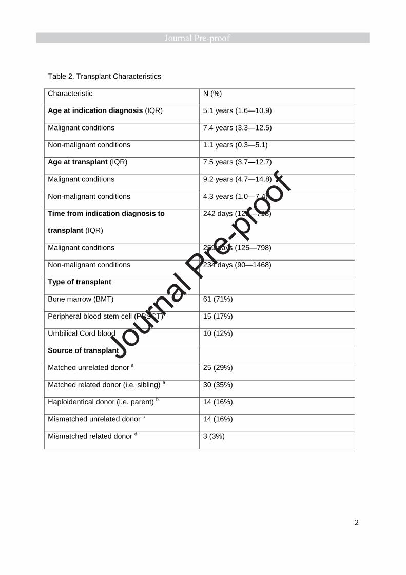
at diagnosis for indication disease was 5.1 years (IQR: 1.6—10.9), but children who were 206
diagnosed with a malignant condition tended to be older at diagnosis than children who were 207
diagnosed with a nonmalignant condition (7.4 years vs. 1.1 years, p<0.001). 208
HSCT indications and characteristics 209
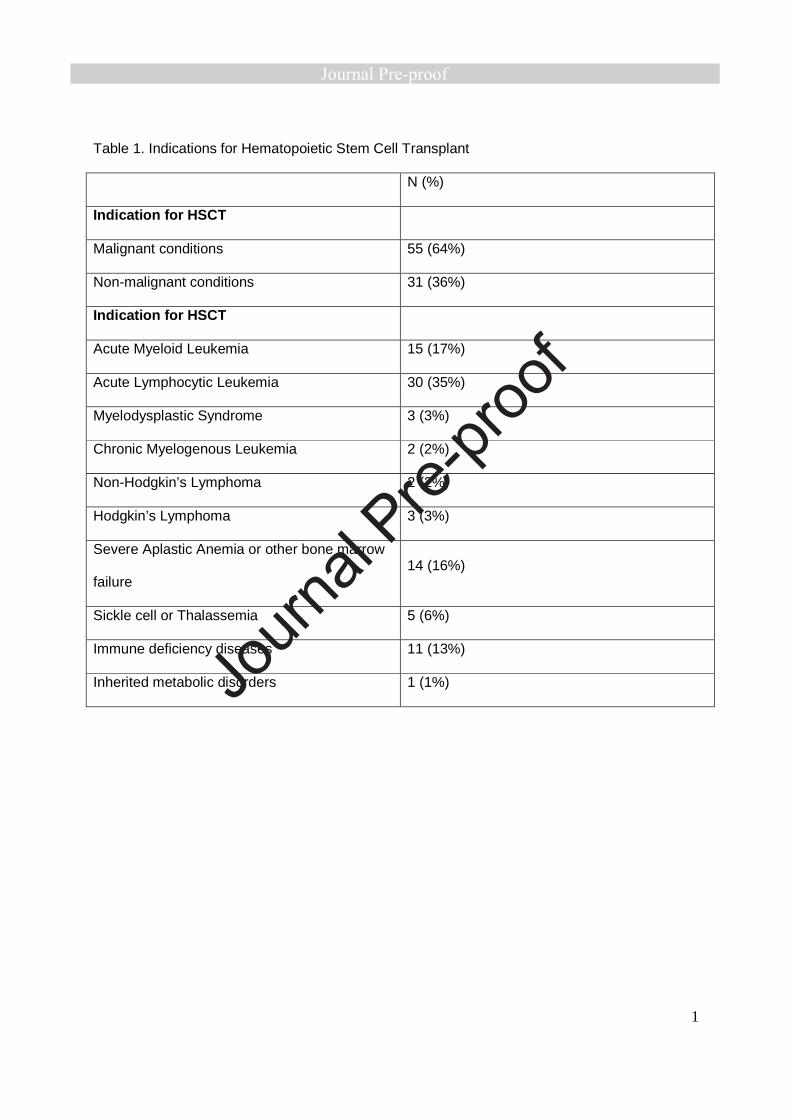
By definition, all patients in the study underwent HSCT: nearly two-thirds (64%) for a 210
malignant diagnosis, such as leukemia or lymphoma, the remaining 36% for a nonmalignant 211
diagnosis, such as bone marrow failure or an immune deficiency disease (Table 1). 212
Patients underwent HSCT at a median of 7.5 years of age (IQR: 3.7—12.7). Median time 213
from indication diagnosis to HSCT was 247 days (IQR: 121—798); this did not differ between 214
those who had a malignant indication diagnosis and those with a nonmalignant diagnosis 215
(p=0.500). Sixty-three percent of patients received a transplant from an HLA-matched source 216
(either related, 29%, or unrelated, 35%). The remaining patients received a transplant from 217
sources that were either haploidentical (16%), such as a parent, or a mismatched donor, either 218
related (3%) or unrelated (16%). While most children in the study had a BMT (Table 2), those 219
with a malignant diagnosis were more likely to have had a PBSCT (23% vs. 6%) whereas those 220
with a nonmalignant diagnosis were more likely to have had an umbilical cord blood transplant 221
(19% vs 7%) (p=0.05). 222
GVHD Characteristics 223
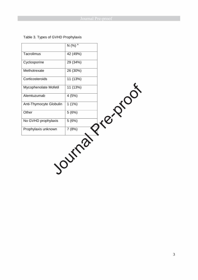
Most (n=74, 86%) patients received one or more medications for GVHD prophylaxis. The 224
most common agents included in prophylactic regimen were tacrolimus (49%), cyclosporine 225
(34%), and methotrexate (30%) (Table 3). Only 6% received no GVHD prophylaxis and an 226
additional 8% had missing information for GVHD prophylaxis. 227
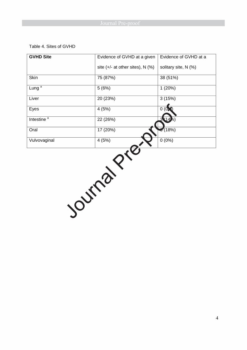
By definition, all patients in our study went on to develop GVHD in one or more organ 228
systems (Table 4). Median time from transplant to first GVHD diagnosis was 96 days (IQR: 35—229
210). For three patients the date of first diagnosis of GVHD could not be determined, due to 230
Journ
al Pre-
proof
9
conflicting dates in the chart. Their GVHD diagnosis dates were thus said to be missing. Skin 231
was the most common site of GVHD (87%), followed by intestine (26%), liver (23%), and oral 232
mucosa (20%). Over half of those who had skin GVHD had no evidence of GVHD at other sites. 233
This is in contrast to every other site of GVHD, which were much more likely to appear in the 234
setting of GVHD at another site, most commonly skin. No cases of ocular or vvGVHD appeared 235
in isolation. Nearly half (44%) of patients had evidence of GVHD in more than one organ 236
system. 237
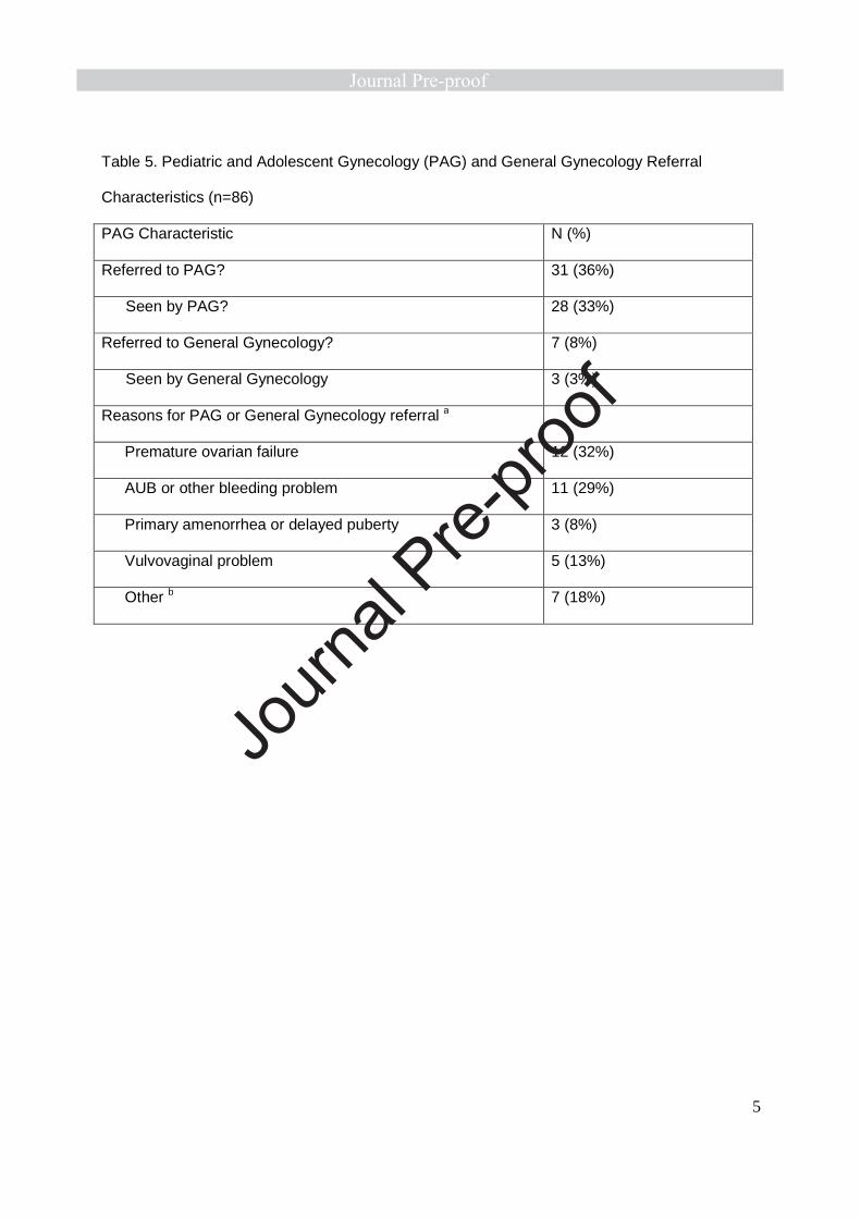
Genital exams and Pediatric and Adolescent Gynecology evaluations 238
In the first two years post-HSCT, 86% of patients had at least one genital exam 239
documented in the medical record. Seven patients were seen by PAG within the first two years 240
post-HSCT and underwent a median of three exams (IQR: 2—4). Additionally, most patients 241
(n=76, 85%) had a genital exam by a non-PAG clinician in the first two years post-HSCT, with a 242
median of 17 exams (IQR: 6—33). Of the 1712 genital exams performed in the first two years 243
following transplant, 1% were performed by PAG clinicians. 244
While only seven patients saw PAG within the first two years post-HSCT, an additional 245
21 were seen by PAG later post-HSCT (median number of PAG visits/consultations: 3, IQR: 2—246
7). Three more patients were referred to PAG but did not keep the clinic appointment. Another 247
seven patients were referred to outside general gynecologists instead of PAG, three of which 248
were seen. For the three outside gynecologist visits, little information was available. In what was 249
available, there was no mention of a concern for GVHD or vulvar complaint consistent with 250
vvGVHD. The most common reasons for referral to a gynecology provider included diagnosis of 251
or concern for POI, and abnormal uterine bleeding. Four patients were referred to PAG for a 252
vulvovaginal problem, including vulvar lesions, vulvovaginitis, vaginal atresia/hematocolpos, and 253
concern for vvGVHD (Table 5). 254
Relatively few (n=13, 15%) patients were menarchal at the time of their indication 255
diagnosis. Median age at menarche in the study was 13 years (IQR: 12—15). Of note, 13 256
Journ
al Pre-
proof
10
patients passed away before menarche and another 26 had not reached menarche at the time 257
of data collection. The prevalence of any diagnosis of POI was 43%. 258
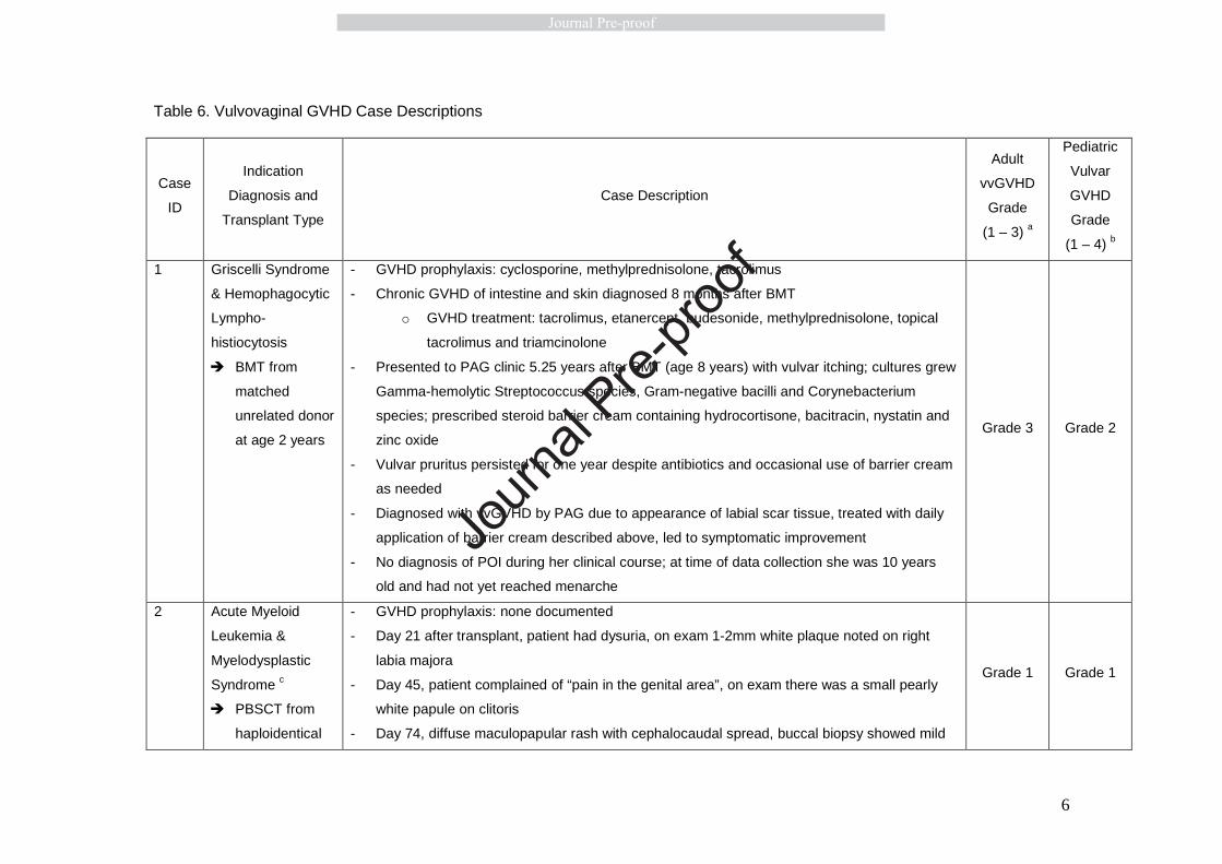
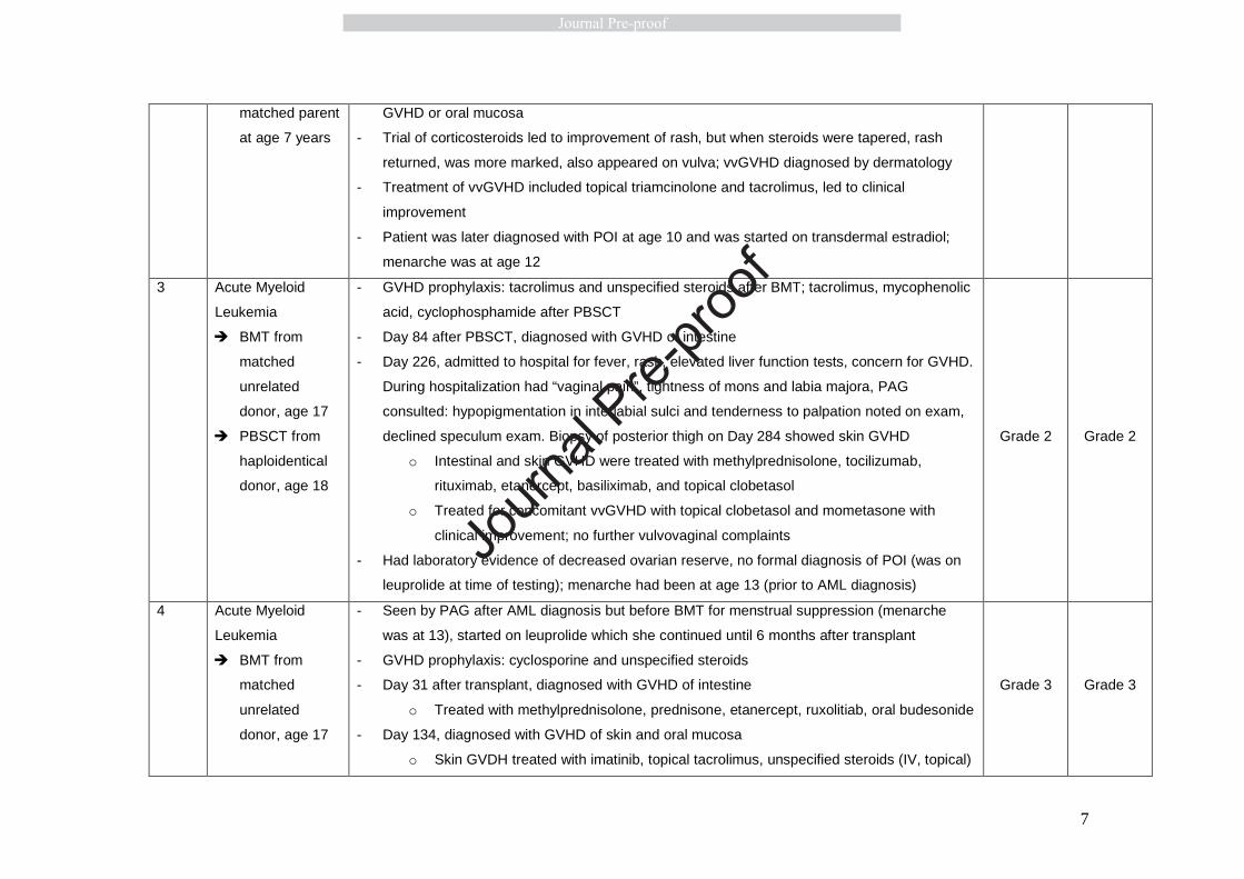
Vulvovaginal GVHD Cases 259
There were four cases of vvGVHD in this group of patients (5% incidence among 260
patients who already had a diagnosis of GVHD, 1.2% incidence among all female pediatric 261
patients post-HSCT). Each case is described and graded according to both the Adult and 262
Pediatric vvGVHD scales in Table 6. 6,10 Two of four patients were older adolescents (18-19 263
years of age), and the remaining two were prepubertal when they developed symptoms of 264
vvGVHD. Three of four cases were seen and diagnosed by PAG, the remaining case was 265
diagnosed and managed by dermatology. One case had a nonmalignant indication diagnosis 266
(Griscelli Syndrome with Hemophagocytic Lymphohistiocytosis), while the remaining three had 267
indication diagnosis of AML. Two of the four patients with vvGHVD had undergone BMT; one 268
had undergone PBSCT; one underwent both BMT and PBSCT. Only one of the three had a 269
vaginal exam; the extent of vaginal disease is unknown in the other cases. Case 1 was 270
diagnosed after several treatments for vulvovaginitis. Ultimately, she was found to have scarring 271
consistent with vvGVHD. Case 2 had vulvar pain and a diffuse rash that included the vulva, was 272
diagnosed with vvGVHD by dermatology, who also initiated treatment. Case 3 was an 273
adolescent who had vulvar GVHD in the setting of GVHD of the skin. Case 4 developed 274
significant labial agglutination secondary to vvGVHD. She was treated with estrogen creams 275
with some improvement, but ultimately needed surgical intervention. Timelines of indication 276
diagnosis, transplant and development of GVHD for each case of vvGVHD are illustrated in 277
Figures 1a-d. 278
279
Discussion 280
In this group of 86 children who received HSCT for malignant or nonmalignant conditions 281
and went on to develop GVHD involving any organ system, incidence of vvGVHD was low (5%). 282
Journ
al Pre-
proof
11
Most patients had at least one genital exam documented during their first two years post-HSCT. 283
One-third saw PAG at any point post-HSCT, typically for POI concerns, abnormal bleeding, or 284
vulvovaginal complaints. vvGVHD cases ranged from those that were mild and treated 285
successfully with topical creams to one case that required surgical management. 286
While non-PAG physicians certainly can identify genital abnormalities in this population, 287
it has been well established that, when examining prepubertal girls, many providers, including 288
pediatricians, are unable to distinguish between normal anatomical variants and genital 289
pathology.18–20 Pediatric residency programs have instituted more formal training in PAG over 290
the past two decades,21 however, the degree of exposure to PAG training varies. The North 291
American Society for Pediatric and Adolescent Gynecology has created a variety of curricula for 292
trainees from centers with limited formal PAG experience.22 Wider use of such materials by 293
providers caring for pediatric and adolescent post-HSCT patients could minimize 294
underdiagnosis of vvGVHD. There is also a need to decrease provider bias in asking about 295
vulvovaginal symptoms in this young population. Considering the above points and that children 296
and families may underreport genital symptoms, there is a significant risk of underdiagnosis of 297
this debilitating condition.23 298
In addition to poor detection of genital pathology in female pediatric patients overall, 299
there is further evidence that, among children younger than ten years, girls have historically 300
received genital examinations almost half as frequently as boys, with only a third of girls ages 5-301
10 years being examined by their primary care provider.24 A more recent study from the child 302
abuse literature found that 90% of pediatric chief residents examined the genitalia of a 303
prepubescent girl in at least half of annual visits.18 This demonstrates an improvement, but the 304
American Academy of Pediatrics recommends “at a minimum, examination of the external 305
genitalia should be included as part of the annual comprehensive physical examination of 306
children and adolescents of all ages”.25 In this study of female pediatric patients with a history of 307
Journ
al Pre-
proof
12
HSCT and GVHD, most patients had at least one genital exam documented in the first two 308
years post-HSCT. 309
Only 1% of genital exams these patients received in the first two years post-HSCT were 310
performed by PAG clinicians. While 33% of patients were seen in PAG clinic or were evaluated 311
by PAG while inpatient at any point post-HSCT, an additional three were referred, but did not 312
attend outpatient clinic appointments. Three additional patients were seen by general 313
gynecology, although it is unclear why they were not instead seen by PAG. Since one of the 314
most common reasons for referral to PAG or general gynecology was POI, a concern with 315
mainly long-term health implications such as infertility or bone loss, it is plausible that attending 316
appointments addressing more acute matters was priority for these families. While the 317
prevalence of POI in the post-HSCT population is relatively high due to gonadotoxic therapies, it 318
is crucial to note that the symptoms of vvGVHD can overlap with those of the reduced estrogen 319
state in POI, including vulvovaginal pain and irritation.9 320
Four (5%) patients were diagnosed with vvGVHD. As only one-third of patients were 321
seen by PAG at any point post-HSCT, it is likely that this is an underestimate of vvGVHD in this 322
population. Five of the patients who were seen by PAG presented for a vulvovaginal complaint. 323
One was an adolescent who presented for vulvovaginal lesions found on biopsy to be lichen 324
simplex chronicus and an HPV-associated condyloma. While this patient was not found to have 325
vvGVHD, her early HPV disease is important to note, as reactivation of HPV and other viruses 326
is common after transplant, given these patients typically have a long period of 327
immunosuppression. A recent case report highlights the dramatic presentation and complicated 328
management of concomitant severe vvGVHD and florid HPV disease in an adult female who 329
had undergone HSCT.26 Another patient who presented to PAG for a vulvovaginal complaint 330
was determined to have vaginal atresia and subsequent hematocolpos. Although hematocolpos 331
due to vulvovaginal adhesions and vaginal obstruction can be a severe manifestation of 332
vvGVHD,6,12,13 the findings in this patient were determined to be a congenital lower reproductive 333
Journ
al Pre-
proof
13
tract anomaly, not attributed to vvGVHD. Lastly, one patient who was ultimately diagnosed with 334
vvGVHD (Case 1) was evaluated by PAG multiple times for vulvovaginitis prior to her vvGVHD 335
diagnosis. Her initial presentation is important because young girls are especially vulnerable to 336
vulvovaginal irritation due to non-estrogenized genital tissue, improper wiping and frequent 337
contact with irritants such as bubble bath, soaps and wet wipes.23,27,28 While this patient’s 338
treatment with topical corticosteroid creams improved her symptoms, had her disease gone 339
undiagnosed, she may have developed more severe structural vvGVHD manifestations such as 340
vaginal stenosis. This case highlights the need for vulvovaginitis in a pre-pubertal post-HSCT 341
child to prompt evaluation for vvGVHD. 342
While all four cases in our study were symptomatic, a recent case series of female 343
children developing vvGVHD post-HSCT found that a higher proportion of pediatric patients in 344
the series were asymptomatic compared to women in the adult literature.10 This potential for 345
insidious onset of disease emphasizes the need for regular genital exams for children and 346
adolescents post-HSCT. 347
All cases of vvGVHD occurred in patients with a history of or current GVHD of the skin 348
elsewhere on the body; three of the four cases occurred in the setting of past or current 349
intestinal GVHD. This is consistent with the adult literature: a cohort of post-HSCT adult women 350
found that women with vvGVHD had a high rate of chronic GVHD in other skin and mucosal 351
surfaces.6 This also suggests that, while the vulva and/or vagina are rarely the initial site of 352
GVHD, the likelihood of vulvovaginal involvement increases when the disease is present on 353
other skin or mucosal areas. 354
The median time from transplant to development of vvGVHD in our study was 398 days 355
(IQR: 88—2207). This was similar to the median time to development of vvGVHD of 452 days in 356
a case series of 19 pediatric post-HSCT patients10 and longer than the median time of 267 days 357
in a case series of 33 adult female post-HSCT patients.6 While the median time to vvGVHD in 358
Journ
al Pre-
proof

14
our study was just over one year after HSCT, it must be noted that Case 1 was diagnosed six 359
years after HSCT. 360
The incidence of vvGVHD among all female patients who underwent HSCT during the 361
study period was lower than that in the Cizek study (1.2% vs 6.3%), possibly due to under-362
reporting of symptoms and under-referral in our setting. 10 Symptoms and presentation of 363
vvGVHD were similar in character but less in severity when compared to the Cizek study. While 364
one of our cases was Grade 1 by the Stratton Scale (a three-point scale), one was Grade 2, and 365
two were Grade 3, 89% of patients in the Cizek study were Grade 3 by the Stratton Scale. 6,10 366
While the clinical presentation of three of our cases were similar to those in adult women, Case 367
1 had a more classic pediatric vulvovaginitis presentation with development of labial scar tissue, 368
which is more specific for vvGVHD. There are a variety of challenges unique to pediatric post-369
HSCT populations that could lead to a higher rate of under-diagnoses of vvGVHD when 370
compared to adult women. In addition to the challenges discussed above, no cohesive 371
guidelines currently exist for vvGVHD surveillance in pediatric populations as they do for more 372
common sequelae of HSCT. In a 2015 review on gynecologic care after HSCT, some pediatric 373
screening recommendations are provided, including clinical assessment by a pediatric 374
gynecologist and/or endocrinologist with Tanner staging, inspection of external genitalia and 375
reevaluation every 3-6 months.23 Cizek and colleagues recommend that female pediatric HSCT 376
patients should receive frequent screening for vvGVHD starting at routine 100 days post-377
transplant visits, including patients who are asymptomatic.10 378
To our knowledge this is the first study of genital examination patterns for post-HSCT 379
patients at-risk for vvGVHD, furthermore, this is the second to attempt to establish incidence of 380
vvGVHD in a pediatric post-HSCT population. The study was conducted in a pediatric hospital 381
population with good access to PAG specialists and a variety of other sub-specialists involved in 382
the interdisciplinary care of post-HSCT patients. There were several limitations to this study. 383
While we did identify four cases of vvGVHD in the study population, our sample size was 384
Journ
al Pre-
proof
15
relatively small. It is possible that the true incidence of vvGVHD in this population is much 385
higher than our estimates, especially less symptomatic forms of the disease. While vvGVHD 386
typically occurs in the context of GVHD involving another organ system,6,8 there are reported 387
cases of isolated vvGVHD.8 Because we included only patients who had GVHD involving 388
another organ system, we may have missed cases of new-onset vvGVHD occurring in isolation. 389
Patients who underwent HSCT as children are surviving much longer than in past 390
decades and are therefore developing long-term sequelae that were previously rarely seen. 391
Furthermore, PAG is a relatively new specialty within obstetrics and gynecology and not all 392
pediatric specialties are aware of PAG as a resource for post-HSCT patients. While we did 393
categorize our four vvGVHD cases according to the Stratton and Cizek criteria6,10 for vvGVHD 394
and vulvar GVHD, respectively (Table 8), clinical information available in the charts was limited 395
and case descriptions may be incomplete. Furthermore, diagnoses of vvGVHD relied on 396
interpretation of documentation from clinical encounters: no patients had photos of vulvovaginal 397
lesions available in their electronic medical record. Similarly, we relied on documentation of 398
genital exams usually performed by non-PAG providers. While it does appear that these 399
providers performed frequent genital examinations, it is possible that the genital exam was part 400
of progress note templates in the electronic medical record, and thus this may be an over-401
estimate of the number of exams performed. In-depth knowledge of providers on distinguishing 402
normal variants from pathological findings in pediatric vulvovaginal exams may also be limited. 403
It is likely that both our study and that of Cizek et al.10 have underestimated the true 404
incidence of vvGVHD in pediatric post-HSCT patients. Thus, larger prospective studies are 405
needed to both determine the true incidence and to elucidate the effectiveness of screening 406
regimens. Many institutions provide post-HSCT “day 100” visits to screen for complications, 407
including GVHD, and these visits should include vulvar exams, even in asymptomatic patients. 408
Furthermore, future research should attempt to clinically differentiate vvGVHD from POI, as 409
signs and symptoms of these conditions can overlap. 410
Journ
al Pre-
proof
16
Pediatric cancer survivors require interdisciplinary care teams to provide surveillance 411
and management for the multitude of conditions for which they are at increased risk. Given the 412
rarity but severity of vvGVHD, girls who are post-HSCT should have surveillance for vvGVHD by 413
a provider who is trained in identifying and treating vvGVHD, such as gynecologists, 414
pediatricians, oncologists and/or family physicians. Surveillance for vvGVHD should be 415
increased if a patient develops GVHD of the skin and/or a mucosal surface (e.g. oral mucosa). 416
Patients with symptoms consistent with POI must receive a thorough gynecologic exam prior to 417
symptoms being attributed solely to POI. In addition to performing regular exams, providers 418
should frequently inquire about vulvovaginal symptoms. Such screening and surveillance should 419
continue indefinitely, as post-HSCT patients can develop GVHD years after their transplant. 420
Referral to PAG, when accessible, is important at some point in the post-HSCT period to 421
discuss prevention and management of gynecologic sequelae, including vvGVHD, POI, and 422
fertility and sexual health effects. As PAG physicians are few in number and typically located in 423
academic centers, training of and partnership with other practitioners who see these patients is 424
crucial for early detection and treatment of vvGVHD. 425
Journ
al Pre-
proof
17
Table Legends 426 Table 2. Transplant Characteristics 427 a Includes the following degrees of HLA matching: 10/10, 6/6 428 b Includes the following degrees of HLA matching and any transplant from a parent, regardless 429 of degree of HLA match: 3/6, 5/10 430 c includes the following degrees of HLA matching: 4/6, 5/6, 7/10, 9/10 431 d Includes the following degrees of HLA matching: 5/6, 9/10 432 433 Table 3. Types of GVHD Prophylaxis 434 a Proportions do not add up to 100%, as many patients received >1 prophylactic medication 435 436 Table 4. Sites of GVHD 437 a Includes one that was probable but not confirmed (patient died before case could be confirmed 438 on biopsy) 439 440 Table 5. Pediatric and Adolescent Gynecology (PAG) and General Gynecology Referral 441 Characteristics 442 a only includes those that were referred to PAG or General Gyn (n=38) 443 b Included those presenting with other laboratory or imaging abnormalities, well woman exams 444 (no complaints) or those for whom reason for referral was missing445 446 Table 6. Vulvovaginal GVHD Case Descriptions 447 a Adult vvGVHD grading scale by Stratton 6: GRADE 1 (Minimal): Generalized erythema and 448 edema of vulvar structures; patchy erythema of mucosa and glandular structures of vulvar 449 vestibule; erythema around openings of vestibular (Bartholin’s & Skene’s) glands; vulvar 450 redness, pain on touching the labia, small areas of vulvar denudation (plaques); GRADE 2 451 (Moderate): Grade I findings plus erosions of mucosal surfaces of the vulva fissures in vulvar 452 folds (e.g., interlabial sulci; fourchette); extensive areas of vulvar denudation with or without 453 leukokeratosis and introital stenosis; Grade I findings plus erosions of mucosal surfaces of the 454 vulva, fissures in vulvar folds (eg, interlabial sulci; fourchette); extensive areas of vulvar 455 denudation with or without leukokeratosis and introital stenosis; GRADE 3 (Severe): Vaginal 456 adhesions or complete vaginal closure; Grade II findings plus agglutination of clitoral hood, 457 introital stenosis, vaginal synechiae, hematocolpos, or complete vaginal closure; Fasciitis or 458 spasticity of levator sling 459 b Pediatric vulvar GVHD grading scale by Cizek 10: GRADE 1: Erythema of vulvar structures, 460 with or without symptoms; GRADE 2: Mild adhesive disease (thin adhesions); presence of 461 scattered skin erosions/fissures; GRADE 3: Moderate adhesive disease (thick/diffuse 462 adhesions, distorting architecture); scattered skin erosions or fissures; GRADE 4: Severe 463 adhesive disease (partial or complete occlusion of urethra and/or vaginal opening); diffuse skin 464 erosions or fissures; loss of architecture of vulvar structures 465 c Case 2 first had a diagnosis of Ewing Sarcoma at age 4, had chemotherapy, later developed 466 Acute Myeloid Leukemia and Myelodysplastic Syndrome at age 7 years 467 d Also had CD34 stem cell top-off three months after PBSCT 468
469
Journ
al Pre-
proof
18
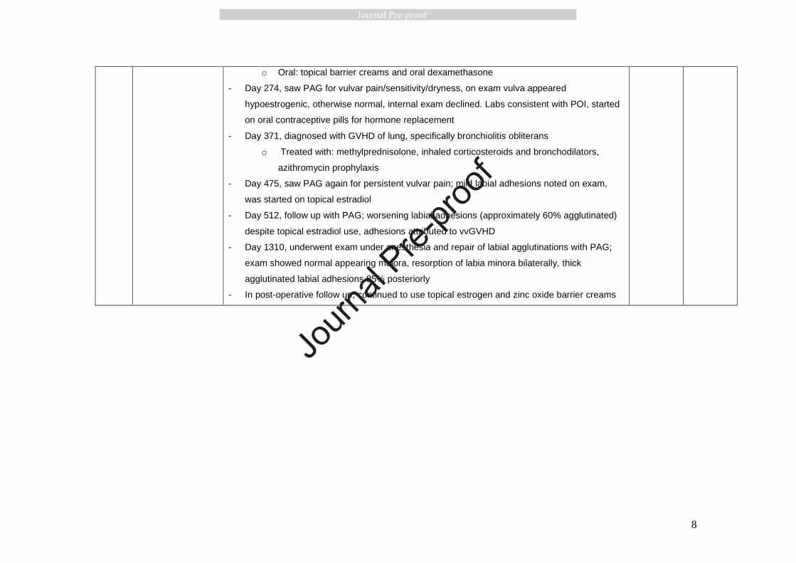
Figure Legends 470 471 Figure 1a: GVHD Timeline for vvGVHD Case 1 472 a Hemophagocytic lymphohistiocytosis syndrome 473 474 Figure 1b: GVHD Timeline for vvGVHD Case 2 475 a Acute myeloid leukemia and myelodysplastic syndrome 476 477 Figure 1c: GVHD Timeline for vvGVHD Case 3 478 a Acute myeloid leukemia 479 480 Figure 1d: GVHD Timeline for vvGVHD Case 4 481 a Acute myeloid leukemia 482 b Had a GVHD/engraftment syndrome phenomenon on the skin the day after transplant; 483 resolved quickly with steroids 484 485 There are no conflicts of interest to declare. 486
Journ
al Pre-
proof
19
REFERENCES 487
1. Bhushan V, Collins R: Chronic graft-vs-host disease. JAMA 2003; 290:2599–603 488
2. Armitage JO: Bone marrow transplantation. NEJM 1994; 330:827–38 489
3. Baird K, Cooke K, Schultz KR: Chronic graft-versus-host disease in children. Pediatr Clin 490
North Am 2010; 57:297–322 491
4. Sullivan KM, Agura E, Anasetti C, et al: Chronic graft-versus-host disease and other late 492
complications of bone marrow transplantation. Semin Hematol 1991; 28:250–9 493
5. Lee SJ, Vogelsang G, Flowers ME. Chronic graft-versus-host disease. Biol Blood 494
Marrow Transplant 2003; 9:215–33 495
6. Stratton P, Turner ML, Childs R. et al: Vulvovaginal chronic graft-versus-host disease 496
with allogeneic hematopoietic stem cell transplantation. Obstet Gynecol 2007; 497
110:1041–9 498
7. Ciavattini A, Clemente N: Female genital tract chronic graft-versus-host disease: Review 499
of the literature. Anticancer Res 2015; 35:13–7 500
8. Hirsch P, Leclerc M, Rybojad M, et al: Female genital chronic graft-versus-host disease: 501
Importance of early diagnosis to avoid severe complications. Transplantation 2012; 502
93:1265–9 503
9. Zantomio D, Grigg AP, MacGregor L, et al: Female genital tract graft-versus-host 504
disease: Incidence, risk factors and recommendations for management. Bone Marrow 505
Transplant 2006; 38:567–72 506
10. Cizek SM, El-Bietar J, Rubinstein J, et al: Pediatric and young adult vulvovaginal graft-507
versus-host disease. Biol Blood Marrow Transplant 2019; 25:2408–15 508
11. Spinelli S, Chiodi S, Costantini S, et al: Female genital tract graft-versus-host disease 509
following allogeneic bone marrow transplantation. Haematologica 2003; 88:1163–8 510
Journ
al Pre-
proof
20
12. Smith Knutsson E, Björk Y, Broman A-KK, et al. Genital chronic graft-versus-host 511
disease in females: A cross-sectional study. Biol Blood Marrow Transplant 512
2014;20(6):806-811. 513
13. Michala L, Vlachopapadopoulou E, Tsimaris P, et al: Resolution of hematocolpos in 514
adolescents affected with graft vs host disease. J Pediatr Adolesc Gynecol 2018; 31: 515
536–9 516
14. Riera C, Deroover Y, Marechal, M: Severe vaginal chronic graft-versus-host disease: 517
Two cases with late onset and literature review. Eur J Gynaecol Oncol 2010; 31:703–4 518
15. Norian JM, Stratton P: Labial fusion: a rare complication of chronic graft-versus-host 519
disease. Obstet Gynecol 2008; 112:437–9 520
16. Childress K, Swanson K, Gossett D: Vulvovaginal graft-vs-host disease. J Pediatr 521
Adolesc Gynecol 2015; 28:e61 522
17. Shanis D, Merideth M, Pulanic TK, Savani BN, Battiwalla M, Stratton P. Female long-523
term survivors after allogeneic hematopoietic stem cell transplantation: Evaluation and 524
management. Semin Hematol 2012;49(1):83-93 525
18. Dubow SR, Giardino AP, Christian CW, et al: Do pediatric chief residents recognize 526
details of prepubertal female genital anatomy: a national survey. Child Abuse Negl 2005; 527
29:195–205 528
19. Muram D, Jones CE, Hostetler BR, et al: Teaching pediatric and adolescent gynecology: 529
a pilot study at one institution. J Pediatr Adolesc Gynecol 1996; 9:12–5 530
20. Muram D, Simmons KJ: Pattern recognition in pediatric and adolescent gynecology—a 531
case for formal education. J Pediatr Adolesc Gynecol 2008; 21:103–8 532
21. Dietrich JE: Comments for “Pattern recognition in pediatric and adolescent gynecology, 533
a case for formal education”. J Pediatr Adolesc Gynecol 2008; 21:109–10 534
Journ
al Pre-
proof
21
22. Talib HJ, Karjane N, Teelin K et al: Resident education curriculum in pediatric and 535
adolescent gynecology: The Short Curriculum 2.0. J Pediatr Adolesc Gynecol 2018; 536
31:71–6 537
23. Frey Tirri B, Häusermann P, Bertz H, et al: Clinical guidelines for gynecologic care after 538
hematopoietic SCT: Report from the international consensus project on clinical practice 539
in chronic GVHD. Bone Marrow Transplant 2015; 50:3–9 540
24. Balk SJ, Dreyfus NG, Harris P: Examination of genitalia in children: ‘The remaining 541
taboo’. Pediatrics 1982; 70:751–3 542
25. Braverman PK, Breech L & American Academy of Pediatrics Committee on 543
Adolescence: Clinical report––Gynecologic examination for adolescents in the pediatric 544
office setting. Pediatrics 2010; 126:583–90 545
26. Buchan A, Merideth MA, Childs RW, et al: Novel management of vaginal chronic graft-546
versus-host disease causing haematometra and haematocolpos. BMJ Case Rep 2018; 547
doi:10.1136/bcr-2017-222720 548
27. Arsenault PS, Gerbie AB: Vulvovaginitis in the preadolescent girl. Pediatr Ann 1986; 549
15:577–9, 583–5 550
28. Paradise JE, Campos JM, Friedman HM, et al: Vulvovaginitis in premenarcheal girls: 551
Clinical features and diagnostic evaluation. Pediatrics 1982; 70:193–8 552
553
Journ
al Pre-
proof
1
Table 1. Indications for Hematopoietic Stem Cell Transplant
N (%)
Indication for HSCT
Malignant conditions 55 (64%)
Non-malignant conditions 31 (36%)
Indication for HSCT
Acute Myeloid Leukemia 15 (17%)
Acute Lymphocytic Leukemia 30 (35%)
Myelodysplastic Syndrome 3 (3%)
Chronic Myelogenous Leukemia 2 (2%)
Non-Hodgkin’s Lymphoma 2 (2%)
Hodgkin’s Lymphoma 3 (3%)
Severe Aplastic Anemia or other bone marrow
failure 14 (16%)
Sickle cell or Thalassemia 5 (6%)
Immune deficiency diseases 11 (13%)
Inherited metabolic disorders 1 (1%)
Journ
al Pre-
proof
2
Table 2. Transplant Characteristics
Characteristic N (%)
Age at indication diagnosis (IQR) 5.1 years (1.6—10.9)
Malignant conditions 7.4 years (3.3—12.5)
Non-malignant conditions 1.1 years (0.3—5.1)
Age at transplant (IQR) 7.5 years (3.7—12.7)
Malignant conditions 9.2 years (4.7—14.8)
Non-malignant conditions 4.3 years (1.0—7.4)
Time from indication diagnosis to
transplant (IQR)
242 days (121—798)
Malignant conditions 259 days (125—798)
Non-malignant conditions 234 days (90—1468)
Type of transplant
Bone marrow (BMT) 61 (71%)
Peripheral blood stem cell (PBSCT) 15 (17%)
Umbilical Cord blood 10 (12%)
Source of transplant
Matched unrelated donor a 25 (29%)
Matched related donor (i.e. sibling) a 30 (35%)
Haploidentical donor (i.e. parent) b 14 (16%)
Mismatched unrelated donor c 14 (16%)
Mismatched related donor d 3 (3%)
Journ
al Pre-
proof
3
Table 3. Types of GVHD Prophylaxis
N (%) a
Tacrolimus 42 (49%)
Cyclosporine 29 (34%)
Methotrexate 26 (30%)
Corticosteroids 11 (13%)
Mycophenolate Mofetil 11 (13%)
Alemtuzumab 4 (5%)
Anti-Thymocyte Globulin 1 (1%)
Other 5 (6%)
No GVHD prophylaxis 5 (6%)
Prophylaxis unknown 7 (8%)
Journ
al Pre-
proof
4
Table 4. Sites of GVHD
GVHD Site Evidence of GVHD at a given
site (+/- at other sites), N (%)
Evidence of GVHD at a
solitary site, N (%)
Skin 75 (87%) 38 (51%)
Lung a 5 (6%) 1 (20%)
Liver 20 (23%) 3 (15%)
Eyes 4 (5%) 0 (0%)
Intestine a 22 (26%) 3 (14%)
Oral 17 (20%) 3 (18%)
Vulvovaginal 4 (5%) 0 (0%)
Journ
al Pre-
proof
5
Table 5. Pediatric and Adolescent Gynecology (PAG) and General Gynecology Referral
Characteristics (n=86)
PAG Characteristic N (%)
Referred to PAG? 31 (36%)
Seen by PAG? 28 (33%)
Referred to General Gynecology? 7 (8%)
Seen by General Gynecology 3 (3%)
Reasons for PAG or General Gynecology referral a
Premature ovarian failure 12 (32%)
AUB or other bleeding problem 11 (29%)
Primary amenorrhea or delayed puberty 3 (8%)
Vulvovaginal problem 5 (13%)
Other b 7 (18%)
Journ
al Pre-
proof
6
Table 6. Vulvovaginal GVHD Case Descriptions
Case
ID
Indication
Diagnosis and
Transplant Type
Case Description
Adult
vvGVHD
Grade
(1 – 3) a
Pediatric
Vulvar
GVHD
Grade
(1 – 4) b
1 Griscelli Syndrome
& Hemophagocytic
Lympho-
histiocytosis
� BMT from
matched
unrelated donor
at age 2 years
- GVHD prophylaxis: cyclosporine, methylprednisolone, tacrolimus
- Chronic GVHD of intestine and skin diagnosed 8 months after BMT
o GVHD treatment: tacrolimus, etanercept, budesonide, methylprednisolone, topical
tacrolimus and triamcinolone
- Presented to PAG clinic 5.25 years after BMT (age 8 years) with vulvar itching; cultures grew
Gamma-hemolytic Streptococcus species, Gram-negative bacilli and Corynebacterium
species; prescribed steroid barrier cream containing hydrocortisone, bacitracin, nystatin and
zinc oxide
- Vulvar pruritus persisted for one year despite antibiotics and occasional use of barrier cream
as needed
- Diagnosed with vvGVHD by PAG due to appearance of labial scar tissue, treated with daily
application of barrier cream described above, led to symptomatic improvement
- No diagnosis of POI during her clinical course; at time of data collection she was 10 years
old and had not yet reached menarche
Grade 3 Grade 2
2 Acute Myeloid
Leukemia &
Myelodysplastic
Syndrome c
� PBSCT from
haploidentical
- GVHD prophylaxis: none documented
- Day 21 after transplant, patient had dysuria, on exam 1-2mm white plaque noted on right
labia majora
- Day 45, patient complained of “pain in the genital area”, on exam there was a small pearly
white papule on clitoris
- Day 74, diffuse maculopapular rash with cephalocaudal spread, buccal biopsy showed mild
Grade 1 Grade 1
Journ
al Pre-
proof
7
matched parent
at age 7 years
GVHD or oral mucosa
- Trial of corticosteroids led to improvement of rash, but when steroids were tapered, rash
returned, was more marked, also appeared on vulva; vvGVHD diagnosed by dermatology
- Treatment of vvGVHD included topical triamcinolone and tacrolimus, led to clinical
improvement
- Patient was later diagnosed with POI at age 10 and was started on transdermal estradiol;
menarche was at age 12
3 Acute Myeloid
Leukemia
� BMT from
matched
unrelated
donor, age 17
� PBSCT from
haploidentical
donor, age 18
- GVHD prophylaxis: tacrolimus and unspecified steroids after BMT; tacrolimus, mycophenolic
acid, cyclophosphamide after PBSCT
- Day 84 after PBSCT, diagnosed with GVHD of intestine
- Day 226, admitted to hospital for fever, rash, elevated liver function tests, concern for GVHD.
During hospitalization had “vaginal pain”, tightness of mons and labia majora, PAG
consulted: hypopigmentation in interlabial sulci and tenderness to palpation noted on exam,
declined speculum exam. Biopsy of posterior thigh on Day 284 showed skin GVHD
o Intestinal and skin GVHD were treated with methylprednisolone, tocilizumab,
rituximab, etanercept, basiliximab, and topical clobetasol
o Treated for concomitant vvGVHD with topical clobetasol and mometasone with
clinical improvement; no further vulvovaginal complaints
- Had laboratory evidence of decreased ovarian reserve, no formal diagnosis of POI (was on
leuprolide at time of testing); menarche had been at age 13 (prior to AML diagnosis)
Grade 2 Grade 2
4 Acute Myeloid
Leukemia
� BMT from
matched
unrelated
donor, age 17
- Seen by PAG after AML diagnosis but before BMT for menstrual suppression (menarche
was at 13), started on leuprolide which she continued until 6 months after transplant
- GVHD prophylaxis: cyclosporine and unspecified steroids
- Day 31 after transplant, diagnosed with GVHD of intestine
o Treated with methylprednisolone, prednisone, etanercept, ruxolitiab, oral budesonide
- Day 134, diagnosed with GVHD of skin and oral mucosa
o Skin GVDH treated with imatinib, topical tacrolimus, unspecified steroids (IV, topical)
Grade 3 Grade 3
Journ
al Pre-
proof
8
o Oral: topical barrier creams and oral dexamethasone
- Day 274, saw PAG for vulvar pain/sensitivity/dryness, on exam vulva appeared
hypoestrogenic, otherwise normal, internal exam declined. Labs consistent with POI, started
on oral contraceptive pills for hormone replacement
- Day 371, diagnosed with GVHD of lung, specifically bronchiolitis obliterans
o Treated with: methylprednisolone, inhaled corticosteroids and bronchodilators,
azithromycin prophylaxis
- Day 475, saw PAG again for persistent vulvar pain; mild labial adhesions noted on exam,
was started on topical estradiol
- Day 512, follow up with PAG; worsening labial adhesions (approximately 60% agglutinated)
despite topical estradiol use, adhesions attributed to vvGVHD
- Day 1310, underwent exam under anesthesia and repair of labial agglutinations with PAG;
exam showed normal appearing majora, resorption of labia minora bilaterally, thick
agglutinated labial adhesions 85% posteriorly
- In post-operative follow up, continued to use topical estrogen and zinc oxide barrier creams
Journ
al Pre-
proof

2012 20182013
Dx with GriscelliSyndrome and HLHa
Age 2 yearsJune 2012
Received Bone Marrow Transplant
Age 2 years137 days post dx
Dx Skin GVHD245 days post-
transplant
Dx Vulvovaginal GVHDAge 8 years
2,207 days post-transplant
Dx Intestine GVHD51 days post-transplant
2014 2015 2016 2017Jo
urnal
Pre-pro
of
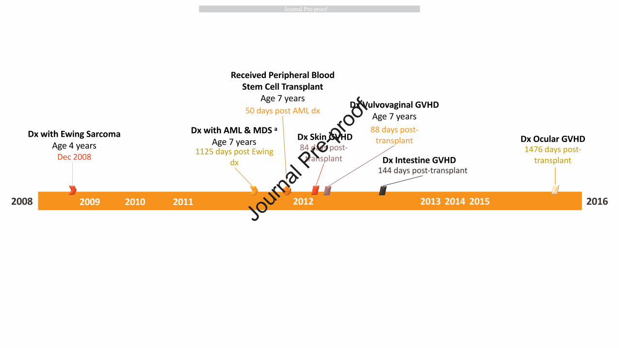
2008 20162009 2010 2011
Dx with Ewing SarcomaAge 4 years
Dec 2008
Dx with AML & MDS aAge 7 years
1125 days post Ewing dx
Received Peripheral Blood Stem Cell Transplant
Age 7 years50 days post AML dx
Dx Skin GVHD84 days post-
transplant
Dx Vulvovaginal GVHDAge 7 years88 days post-
transplant Dx Ocular GVHD1476 days post-
transplantDx Intestine GVHD144 days post-transplant
2012 2013 2014 2015
Journ
al Pre-
proof
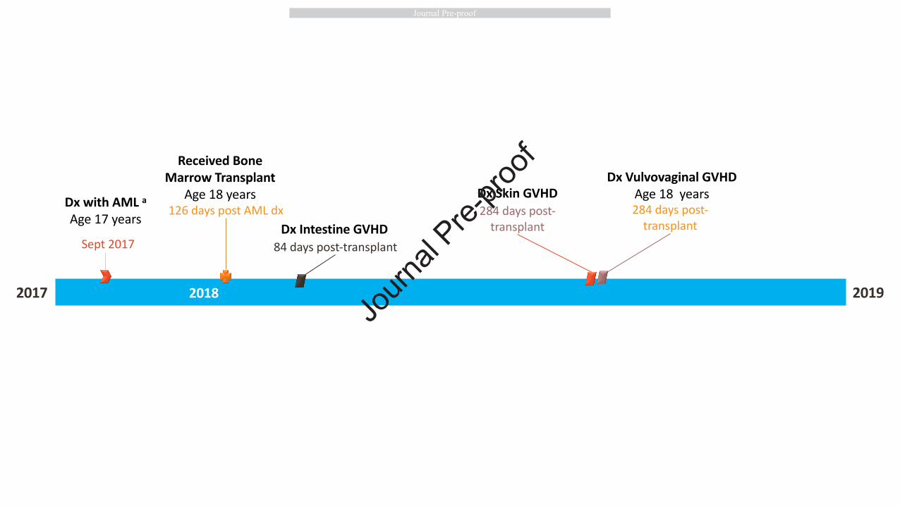
2017 20192018
Dx with AML aAge 17 years
Sept 2017
Received Bone Marrow Transplant
Age 18 years126 days post AML dx
Dx Skin GVHD284 days post-
transplant
Dx Vulvovaginal GVHDAge 18 years284 days post-
transplantDx Intestine GVHD84 days post-transplant
Journ
al Pre-
proof
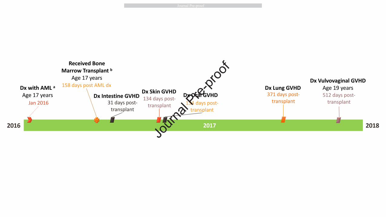
2016 2018
Dx with AML aAge 17 years
Jan 2016
Received Bone Marrow Transplant b
Age 17 years158 days post AML dx
Dx Skin GVHD134 days post-
transplant
Dx Oral GVHD134 days post-
transplant
Dx Lung GVHD371 days post-
transplantDx Intestine GVHD
31 days post-transplant
2017
512 days post-transplant
Dx Vulvovaginal GVHDAge 19 years
Journ
al Pre-
proof