drug hypersensitivity reactions - binasss
TRANSCRIPT
Drug HypersensitivityReactions
R. Gentry Wilkerson, MD
KEYWORDS
� Drug hypersensitivity � Drug allergy � Adverse drug reaction� Hypersensitivity reactions � Anaphylaxis � Severe cutaneous adverse reactions
KEY POINTS
� Drug hypersensitivity reactions result from various immune system-mediated responsesto exposure to a drug.
� The Gell and Coombs classification divides immunologic drug hypersensitivity reactionsinto 4 major categories based on immunologic mechanism.
� Dermatologic manifestations are the most common clinical finding of a drug allergy.
� Type IV hypersensitivity reactions include severe cutaneous adverse reactions (SCARs)such as drug reaction with eosinophilia and systemic symptom (DRESS) syndrome,Stevens–Johnson Syndrome (SJS), toxic epidermal necrolysis (TEN), and acute general-ized exanthematous pustulosis (AGEP).
� Epinephrine is the first-line treatment of anaphylaxis. Antihistamines may be given to alle-viate cutaneous manifestations but, they do not treat the underlying process ofanaphylaxis.
INTRODUCTION
Drug hypersensitivity reactions (DHRs) are a diverse group of reactions mediated bythe immune system after exposure to a drug. The mechanisms underlying the devel-opment of a hypersensitivity reaction are complex and not always fully characterized.Anaphylaxis is a DHR that requires immediate recognition and treatment. Other typesof reactions are slow to develop and do not always require rapid treatment. Emer-gency physicians should have a good understanding of these various types ofDHRs and how to approach the patient regarding evaluation and treatment.
Department of Emergency Medicine, University of Maryland School of Medicine, 110 SouthPaca Street, 6th Floor, Suite 200, Baltimore, MD 21201, USAE-mail address: [email protected]: @gentrywmd (R.G.W.)
Emerg Med Clin N Am 40 (2022) 39–55https://doi.org/10.1016/j.emc.2021.09.001 emed.theclinics.com0733-8627/22/ª 2021 Elsevier Inc. All rights reserved.
Descargado para BINASSS BINASSS ([email protected]) en National Library of Health and Social Security de ClinicalKey.es por Elsevier en febrero 15, 2022. Para uso personal exclusivamente. No se permiten otros usos sin autorización. Copyright ©2022. Elsevier Inc. Todos los derechos reservados.
Wilkerson40
EPIDEMIOLOGY
The true burden of disease due to allergic reactions is difficult to determine becauseepidemiologic data are limited in quality due to variations in terminology used, differentmethodological approaches for determining the prevalence of disease, and differentoutcomes used to determine the presence of an allergy. Overall, adverse drug reac-tions (ADRs) have been estimated to affect up to approximately 15% of hospitalizedpatients.1 In a 2013 study using random digit dialing to survey members of the generalpublic, the prevalence of anaphylaxis using the most stringent criteria was at least1.6%, whereas the prevalence was 7.7% using the least stringent criteria. Respon-dents in the survey attributed episodes of anaphylaxis to drugs in 35% of cases.2
From 2001 to 2012, there was an increase in the percentage of emergency department(ED) visits due to allergic drug reactions—from 0.49% to 0.94%.3 In New York City be-tween 2004 and 2008, anaphylaxis accounted for 0.18% of pediatric ED visits.4 Over-all, medications are the leading cause of anaphylaxis that results in death.5 In children,however, exposure to food causes the greatest number of anaphylaxis fatalities.6 Incontrast to anaphylaxis in general, whereby there has been a rise in hospital admis-sions without a rise in fatalities, for drug-induced anaphylaxis, one study of an Austra-lian database found a threefold increase in deaths due to anaphylaxis but only a 1.5xincrease in the number of hospital admissions between 1997 and 2005. In this study,over half of all the fatalities due to anaphylaxis were likely caused by drug allergies.7
The risk of anaphylaxis to drugs increases with age.8 The United States Food andDrug Administration (FDA) Adverse Event Reporting System (FAERS) is a web-based system used to compile adverse event reports to assist with postmarketing sur-veillance of drugs to identify potential safety concerns. Analysis of FAERS datademonstrated that the rate of anaphylaxis due to monoclonal antibodies (mAbs) is ris-ing faster than any other class of drug. In 1999, mAbs accounted for 2% of all reportedcases of anaphylaxis, but this had risen to 17.37% in 2019.9
RISK FACTORS
Most ADRs are an extension of the usual pharmacologic effect of the drug. Factorsthat increase the risk of ADRs include the type of drug, the dose of the drug, specificpharmacokinetic properties of the drug, and other factors that play a role in the meta-bolism and action of the drug. A study by Gurwitz and colleagues in 2003 found thatADRs were common in the elderly population and that as many as one-fourth werepreventable.10 The elderly experience age-related changes in drug metabolism butalso are subject to polypharmacy and inappropriate prescribing.11 At the other endof the age spectrum, Clavenna and Bonati found that the incidence of ADRs in pedi-atric patients was 10.9% for in-hospital patients and 1.0% for outpatients.The risk of having an allergic reaction to a drug is greatest when there is a history of
allergic relation to the same or closely related compounds. Drug-specific factors influ-ence the likelihood of developing an allergy. Large molecular weight compounds suchas proteins and polysaccharides have increased rates of allergic reactions. The routeof administration of a drug may influence the likelihood of developing an allergic reac-tion although the data supporting these statements is weak. Some polymorphisms ofhuman leukocyte antigen (HLA) region carry a higher risk of certain forms of allergicreaction.12
The risk of anaphylaxis increases with age, presence of comorbid conditions, andthe use of angiotensin-converting enzyme (ACE) inhibitors.13,14 A retrospective anal-ysis of a European registry of anaphylaxis cases found that age was the greatestrisk factor for having severe cardiovascular complications from anaphylaxis (adjusted
Descargado para BINASSS BINASSS ([email protected]) en National Library of Health and Social Security de ClinicalKey.es por Elsevier en febrero 15, 2022. Para uso personal exclusivamente. No se permiten otros usos sin autorización. Copyright ©2022. Elsevier Inc. Todos los derechos reservados.
Drug Hypersensitivity 41
odds ratio 6.08).15 Asthma and other respiratory conditions have been associated withgreater severity of anaphylactic reactions.14,16,17
CLASSIFICATION & MECHANISMS
Multiple systems have been developed to characterize and classify different reactionsto drugs. These reactions may occur as the result of a multitude of different pathwayswith an immunologic basis being just one. In 1955, Brown wrote that the use of theterm drug allergy was used “as a sort of wastepaper basket into which are castmany unexplained phenomena.”18 The FDA defines an adverse event as “any unto-ward medical occurrence associated with the use of a drug in humans, whether ornot considered drug related.”19 In the report published in 1972, International DrugMonitoring: The Role of National Centers, the World Health Organization (WHO)defined an ADR as “one that is noxious, is unintended, and occurs at doses normallyused in man.”20
The Rawlins–Thompson classification of ADRs was proposed in 1977.21 The systembroke ADRs into Type A, which are dose-dependent and predictable and Type B,which are not dose-dependent or predictable. Type A reactions make up 85% to90% of all ADRs and have been referred to as “augmented” as these reactions arean extension of the normal pharmacologic properties of the drug. Prolongation ofthe QRS complex in tricyclic antidepressant overdose is an example of a Type A reac-tion. Type B reactions comprise 10% to 15% of ADRs and have been referred to as“bizarre” because they are not a normal, expected property of the drug. Anaphylaxisresulting from exposure to penicillin is an example of a Type B reaction. Subsequently,additional categories have been added by some to further characterize different typesof ADRs. These include: Type C (dose-related and time-related), Type D (time-related),Type E (withdrawal), and Type F (unexpected failure of efficacy).22
A DHR is a response to a drug that results in symptoms or signs due to exposure to adrug at a dose normally tolerated by nonhypersensitive people and is induced byimmunologic or inflammatory pathways. The term DHR is preferred in cases of sus-pected drug allergy because clinically it is difficult to distinguish between a truedrug allergy and nonallergic DHR. In its International Consensus on Drug Allergy,the World Allergy Organization classified DHRs based on the timing of onset of symp-toms after exposure. Immediate DHRs such as urticaria, anaphylaxis, and broncho-spasm, typically occur within 1 to 6 hours of exposure although usually within1 hour. Nonimmediate or delayed DHRs occur after 1 hour of exposure and frequentlymany days later.23
Gell and Coombs Classification of Hypersensitivity Reactions
The Gell and Coombs classification divides immunologic DHRs into 4 major patho-physiologic categories based on the immunologic mechanism (Table 1). In this clas-sification which was first proposed in 1963, each reaction has a distinct and mutuallyexclusive mechanism. In the following years, advances in the understanding of variousimmunologic effectors and pathways have exploded and it is now known that theremay be overlap across different Gell and Coombs reaction types.24
Type I, or immediate-type hypersensitivity reactions occur when exposure to a pre-viously encountered antigen causes crosslinking of IgE bound to high-affinity recep-tors (FcεRI) on the surface of sensitized mast cells and basophils leading to releaseof preformed vasoactive mediators such as histamine, tryptase, and chymase.25,26
These mediators cause vasodilation and increased capillary permeability. The initialreaction is followed 4 to 8 hours later by a late phase release of cytokines such as
Descargado para BINASSS BINASSS ([email protected]) en National Library of Health and Social Security de ClinicalKey.es por Elsevier en febrero 15, 2022. Para uso personal exclusivamente. No se permiten otros usos sin autorización. Copyright ©2022. Elsevier Inc. Todos los derechos reservados.
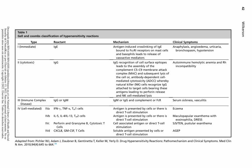
Table 1Gell and coombs classification of hypersensitivity reactions
Type Reactant Mechanism Clinical Symptoms
I (Immediate) IgE Antigen-induced crosslinking of IgEbound to FcεRI receptors on mast cellsand basophils leads to release ofvasoactive mediators
Anaphylaxis, angioedema, urticaria,bronchospasm, hypotension
II (cytotoxic) IgG IgG recognition of cell surface epitopesleads to the assembly of thecomplement C5–C9 membrane attackcomplex (MAC) and subsequent lysis ofthe cell or, antibody-dependent cell-mediated cytotoxicity (ADCC) wherebynatural killer (NK) cells recognize IgGattached to target cells bearing theseantigens leading to perforin releaseand NK cell-mediated lysis
Autoimmune hemolytic anemia and Rhincompatibility
III (Immune ComplexDisease)
IgG or IgM IgM or IgG and complement or FcR Serum sickness, vasculitis
IV (cell-mediated) IVa IFN-g, TNF-a, TH1 cells Antigen is presented by cells or there isdirect T-cell stimulation
Eczema
IVb IL-5, IL-4/IL-13, TH2 cells Antigen is presented by cells or there isdirect T-cell stimulation
Maculopapular exanthema witheosinophilia, DRESS
IVc Perforin and Granzyme B, Cytotoxic TCells
Cell associated antigen or direct T-cellstimulation
SJS/TEN, pustular exanthema
IVd CXCL8, GM-CSF, T Cells Soluble antigen presented by cells ordirect T-cell stimulation
AGEP
Adapted from: Pichler WJ, Adam J, Daubner B, Gentinetta T, Keller M, Yerly D. Drug Hypersensitivity Reactions: Pathomechanism and Clinical Symptoms. Med ClinN Am. 2010;94(4):645 to 664.34
Wilk
erso
n42
Descargado para B
INA
SSS BIN
ASSS (pedidos@
binasss.sa.cr) en National Library of H
ealth and Social Security de C
linicalKey.es por Elsevier en febrero 15, 2022. Para uso personal exclusivam
ente. No se
permiten otros usos sin autorización. C
opyright ©2022. Elsevier Inc. Todos los derechos reservados.
Drug Hypersensitivity 43
IL-1, IL-4, IL-5, granulocyte monocyte colony-stimulating factor (GM-CSF), and tumornecrosis factor (TNF)-a. Type I hypersensitivity reactions lead to the development ofurticaria, angioedema, bronchospasm, and hypotension.27
Type II hypersensitivity reactions are delayed cytotoxic reactions in which host cellsare destroyed through complement-mediated reactions, antibody-dependent cell-mediated cytotoxicity, or antibody-mediated cellular dysfunction. Host cells coatedwith antigen bind to IgG, or less commonly, IgM antibodies. This can lead to the acti-vation of the classic complement pathway leading to the assembly of the membraneattack complex (C5–C9) and subsequent lysis of the host cell. Natural killer cells andmacrophages can also be activated by binding antibodies to FcgRIIb receptorsexpressed on their surface. Examples of Type II hypersensitivity reactions includeautoimmune hemolytic anemia, Rh-incompatibility, and Goodpasture syndrome (anti-glomerular basement membrane disease).28
In Type III hypersensitivity reactions, IgG or IgM form immune complexes with an-tigens and activate the complement system. This leads to inflammation and tissueinjury by activated neutrophils. The clinical manifestations of this process resultfrom the site whereby the immune complexes deposit rather than the specific antigenor antibody and usually take at least a week to appear.29 Serum sickness and Arthusreactions are examples of Type III hypersensitivity reactions.30,31
Type IV hypersensitivity reactions are distinct from Types I through III in that Type IVreactions are not mediated by antibodies but instead involve the activation and expan-sion of T cells. This process is not immediate and sometimes takes days to weeks todevelop. Since the original classification by Gell and Coombs, Type IV reactions havebeen further characterized into 4 subclasses based on the cytokines produced andthe cells involved.32 There is a strong link to T cell-mediated hypersensitivity reactionsand specific HLA risk alleles.33 Stevens–Johnson syndrome/toxic epidermal necroly-sis (SJS/TEN), acute generalized exanthema pustulosis (AGEP), and drug reactionwith eosinophilia and systemic symptoms (DRESS) are examples of Type IV hypersen-sitivity reactions.DHRs have also been classified based on the mode of action of the drug with im-
mune/inflammatory cells. In this system, there are 3 types of reactions-allergic/immune, pseudoallergic, and pharmacologic stimulation of immune receptors (p-iconcept). Large molecular weight drugs can be recognized directly by immune cellsand antibodies. However, most drugs act as haptens in that they are too small(<1000 Da) to elicit an immune response and must bind covalently to a protein toform an antigen.26 In the pseudoallergic class, drugs cause the release of mediatorsfrom mast cells, basophils, and other effector cells without the involvement of immu-noglobulins or T cells. In the p-i concept, some drugs may bind noncovalently to non-active sites of HLA molecules or T cell receptors to cause activation. The drugs arethus not acting as antigens.35
CLINICAL MANIFESTATIONS
Patients experiencing an allergic reaction to a drug may have a wide variety of clinicalpresentations based on the immunologic mechanism underlying the drug allergy.Within the same mechanism, there may be substantial differences in presentationand organ systems involved from patient to patient. Dermatologic manifestationsare the most commonly seen presentation in allergic reactions to drugs.36,37
The manifestations of Type I (immediate) hypersensitivity reactions are a directresult of the actions of the vasoactive mediators that are released from mast cellsand basophils. Common dermatologic manifestations include urticaria and
Descargado para BINASSS BINASSS ([email protected]) en National Library of Health and Social Security de ClinicalKey.es por Elsevier en febrero 15, 2022. Para uso personal exclusivamente. No se permiten otros usos sin autorización. Copyright ©2022. Elsevier Inc. Todos los derechos reservados.
Wilkerson44
angioedema associated with flushing and pruritus. The classic description of thisswelling associated with vasodilation-induced erythema is the wheal-and-flareresponse.38 The respiratory system may be involved resulting in wheezing due tobronchoconstriction and stridor due to edema of the upper airway including the vocalcords. Death due to asphyxiation may occur in severe cases.39 Gastrointestinalinvolvement may present with crampy abdominal pain, nausea, and vomiting, aswell as diarrhea although these may also be attributable to a non–immune-mediatedADR. Vasoplegia and third-spacing of fluids may result in hypotension and loss of con-sciousness. Anaphylaxis is the most severe presentation of an IgE-mediated allergicreaction. The clinical presentation of Type I hypersensitivity reactions usually occurswithin minutes to hours of the exposure.The clinical presentation of Type II (cytotoxic) hypersensitivity reactions is usually
the result of anemia, thrombocytopenia, or neutropenia, as these are the most com-mon cell types involved. Symptoms most commonly occur within days of exposure.When red blood cells are targeted, drug-induced immune hemolytic anemia (DIIHA)occurs. The drugs most frequently associated with the development of DIIHA are an-timicrobials (mostly penicillin and cephalosporins), anti-inflammatories, and antineo-plastic agents.40 Patients will present with typical signs and symptoms of anemiaincluding fatigue, pallor, jaundice, darkened urine due to bilirubinuria, tachycardia,tachypnea, and hypotension. Destruction of platelets via this mechanism leads todrug-induced immune thrombocytopenia (DIITP). This is a secondary form of immunethrombocytopenia (ITP). In this condition, low platelet counts lead to easy bruising andbleeding. In one review of 309 cases, the median time between exposure to theoffending drug and development of DIITP was 21 days and the median minimumplatelet count was 11,000/mL.41 Drug-induced immune neutropenia (DIIN) occurswhen exposure to a drug results in the development of antibodies that cross-reactwith glycoproteins on neutrophil cell walls leading to their destruction and placingthe patient at risk for infection.42
In Type III (immune complex) hypersensitivity reactions, there is an abnormal forma-tion of antigen–antibody complexes that are deposited in tissues and result in the acti-vation of the complement system. Diseases that are the result of Type IIIhypersensitivity reactions include poststreptococcal glomerulonephritis, serum sick-ness, hypersensitivity pneumonitis (also called extrinsic allergic alveolitis), and sys-temic lupus erythematosus (SLE). The clinical presentation depends on the disease.SLE is a prototypical Type III hypersensitivity reaction whereby antibodies developto components of the cellular nucleus—antinuclear antibodies (ANA). The type ofANA that develops often has a strong association with the patient’s clinical presenta-tion. For example, anti-Smith antibodies are frequently associated with kidney dis-ease.43 Drug-induced lupus (DIL) occurs when exposure to a drug leads to thedevelopment of autoantibodies and loss of self-tolerance. The use of procainamideand hydralazine is associated with a high risk of the development of DIL. DIL maynot develop until after years of use of the associated drug. Patients with DIL mostcommonly present with fatigue, low-grade fever, and other systemic symptoms.Generally, DIL tends to present with more mild symptoms than SLE. Developmentof major organ system involvement is less frequent in DIL than in SLE.44
Type IV hypersensitivity reactions occur as a result of T cell response to an antigenleading to an inflammatory response. These reactions are further subdivided (IVathrough IVd) based on the type of T cells involved. The clinical presentation is basedon the distinct condition that develops. The skin is a depository for a large number ofT cells so dermatologic involvement is common in Type IV hypersensitivity reactions.Contact dermatitis is a very common Type IV hypersensitivity reaction. During the
Descargado para BINASSS BINASSS ([email protected]) en National Library of Health and Social Security de ClinicalKey.es por Elsevier en febrero 15, 2022. Para uso personal exclusivamente. No se permiten otros usos sin autorización. Copyright ©2022. Elsevier Inc. Todos los derechos reservados.
Drug Hypersensitivity 45
sensitization (afferent) stage, a hapten contacts the skin and leads to the formation ofhapten-specific T cells. During the elicitation (efferent) phase, re-exposure to the samehapten causes the release of mediators that are responsible for the clinical presenta-tion including the development of an erythematous, pruritic rash with swelling. Severecutaneous adverse reactions (SCARs) are a group of dermatologic diseases that resultfrom a Type IV hypersensitivity process.
Drug Reaction with Eosinophilia and Systemic Symptoms Syndrome
DRESS syndrome, also known as drug-induced hypersensitivity syndrome (DIHS), is aSCAR that has a long latency period before the development of clinical symptomswhich include fever, adenopathy, hematologic abnormalities, and multiorgan systeminvolvement. The onset of disease usually occurs within 3 weeks of exposure to thedrug but may be delayed by as much as 3 months.45 Reactions to the medicationphenytoin were described soon after its introduction in the 1930s. Over time variousterms were used to describe similar reactions including anticonvulsant hypersensitiv-ity syndrome and drug-induced pseudolymphoma. In 1996, Bocquet and colleaguesintroduced the term drug rash with eosinophilia and systemic symptoms.46 Due to var-iations in dermatologic involvement the word “rash” in the name was subsequentlyreplaced with “reaction.” Different diagnostic criteria have been proposed to definedisease patterns that are likely a continuum of DRESS (Table 2). A Japaneseconsensus group proposed a set of diagnostic criteria in 2006 and later developeda scoring system.47,48 In 2007, the RegiSCAR group, a multinational effort that collectsdata on cases of SCAR, proposed a similar set of diagnostic criteria and a scoring sys-tem to help classify cases as definite, probable, or not DRESS.49 DRESS is associatedwith the reactivation of human herpes virus (HHV), especially HHV-6, HHV-7, Epstein–Barr virus (EBV), varicella-zoster virus (VZV), and cytomegalovirus (CMV).50 Aromaticanticonvulsant medications such as phenytoin, carbamazepine, and phenobarbitalhave classically been associated with DRESS. Several other drug classes have nowbeen implicated as causative agents including antidepressants, sulfonamides and sul-fones, nonsteroidal anti-inflammatories (NSAIDs), antibiotics, ACE inhibitors, andbeta-blockers.51 The overall mortality of DRESS is approximately 5% to 10%.52 Incases with cardiac involvement, one retrospective analysis demonstrated the mortal-ity increases to 37.5%.53
Stevens–Johnson Syndrome and Toxic Epidermal Necrolysis
SJS and TEN are SCARs with skin necrosis and detachment that represent differentpoints on a continuum of severity based on the percentage involvement of body sur-face area (BSA). SJS involves less than 10% BSA, whereas TEN involves more than30%. SJS/TEN overlap describes cases whereby there is between 10% and 30%BSA involved.54 Previously considered to be on the continuum of the same disease,erythema multiforme is now thought to be a distinct entity. Drugs are the most com-mon triggers for the development of SJS/TEN with aromatic antiepileptics, NSAIDs,and antibacterial sulfonamides frequently implicated. Infections are also implicatedin the development of SJS/TEN. Cases associated with Mycoplasma pneumoniaeoften have a less severe presentation.55
Patients with SJS/TEN initially present with an influenza-like prodromal phase whichmay include fever and burning sensation. This prodrome precedes the development ofskin findings by 1 to 3 days.56 The rash of SJS/TEN typically begins as erythematousmacules with purpuric centers and ill-defined borders. Lesions are first present on theface and thorax before spreading to other areas. The distribution is symmetric andusually spares the scalp, palms, and soles. Over time, sometimes within hours,
Descargado para BINASSS BINASSS ([email protected]) en National Library of Health and Social Security de ClinicalKey.es por Elsevier en febrero 15, 2022. Para uso personal exclusivamente. No se permiten otros usos sin autorización. Copyright ©2022. Elsevier Inc. Todos los derechos reservados.
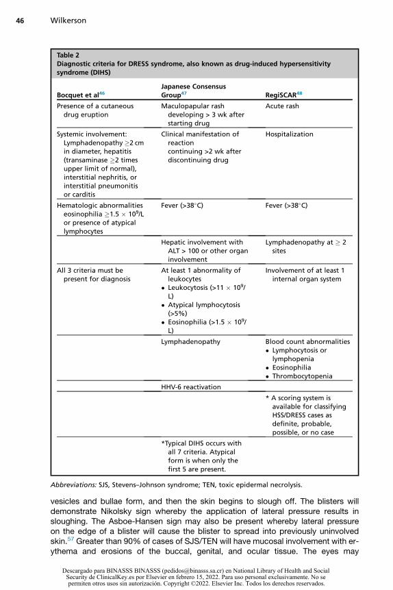
Table 2Diagnostic criteria for DRESS syndrome, also known as drug-induced hypersensitivitysyndrome (DIHS)
Bocquet et al46Japanese ConsensusGroup47 RegiSCAR48
Presence of a cutaneousdrug eruption
Maculopapular rashdeveloping > 3 wk afterstarting drug
Acute rash
Systemic involvement:Lymphadenopathy�2 cmin diameter, hepatitis(transaminase �2 timesupper limit of normal),interstitial nephritis, orinterstitial pneumonitisor carditis
Clinical manifestation ofreactioncontinuing >2 wk afterdiscontinuing drug
Hospitalization
Hematologic abnormalitieseosinophilia �1.5 � 109/Lor presence of atypicallymphocytes
Fever (>38�C) Fever (>38�C)
Hepatic involvement withALT > 100 or other organinvolvement
Lymphadenopathy at � 2sites
All 3 criteria must bepresent for diagnosis
At least 1 abnormality ofleukocytes
� Leukocytosis (>11 � 109/L)
� Atypical lymphocytosis(>5%)
� Eosinophilia (>1.5 � 109/L)
Involvement of at least 1internal organ system
Lymphadenopathy Blood count abnormalities� Lymphocytosis or
lymphopenia� Eosinophilia� Thrombocytopenia
HHV-6 reactivation
* A scoring system isavailable for classifyingHSS/DRESS cases asdefinite, probable,possible, or no case
*Typical DIHS occurs withall 7 criteria. Atypicalform is when only thefirst 5 are present.
Abbreviations: SJS, Stevens–Johnson syndrome; TEN, toxic epidermal necrolysis.
Wilkerson46
vesicles and bullae form, and then the skin begins to slough off. The blisters willdemonstrate Nikolsky sign whereby the application of lateral pressure results insloughing. The Asboe-Hansen sign may also be present whereby lateral pressureon the edge of a blister will cause the blister to spread into previously uninvolvedskin.57 Greater than 90% of cases of SJS/TEN will have mucosal involvement with er-ythema and erosions of the buccal, genital, and ocular tissue. The eyes may
Descargado para BINASSS BINASSS ([email protected]) en National Library of Health and Social Security de ClinicalKey.es por Elsevier en febrero 15, 2022. Para uso personal exclusivamente. No se permiten otros usos sin autorización. Copyright ©2022. Elsevier Inc. Todos los derechos reservados.
Drug Hypersensitivity 47
demonstrate conjunctival erythema, periorbital edema, discharge, crusting, anddevelopment of a pseudomembrane.58
The severity-of-illness score for toxic epidermal necrolysis (SCORTEN) was devel-oped to assess the severity and predict prognosis. Using logistic regression tech-niques, 7 independent variables were identified and assigned a value of either 1 or0 based on the presence or absence of the variable. These variables included age�40, associated cancer, heart rate �120 beats per minute, serum blood urea nitrogengreater than 28 mg/dL, BSA �10%, serum bicarbonate less than 20 mEq/L, andserum glucose greater than 250 mg/dL. With increasing scores, the mortality rate in-creases. A score of 5 or more is associated with a greater than 90% mortality.59
Recently, another scoring system was derived from an international dataset, theABCD-10 Score, named for age, bicarbonate, cancer, dialysis, and 10% BSA.60
Recent comparisons of the 2 scores have demonstrated better performance ofSCORTEN than ABCD-10.61,62
Acute Generalized Exanthematous Pustulosis
Acute generalized exanthematous pustulosis (AGEP) is a SCAR that is almost exclu-sively caused by exposure to a drug with a very short latency period, frequently lessthan 2 days.63 It presents with numerous nonfollicular pustules on an erythematousbase. The multinational EuroSCAR group found that the medications most often impli-cated in the development of AGEPwere pristinamycin, ampicillin and amoxicillin, quin-olones, chloroquine and hydroxychloroquine, anti-infective sulfonamides, terbinafine,and diltiazem.64 The rash tends to first appear in the axillary, submammary, andinguinal intertriginous regions. Mucosal involvement is limited and only seen in aboutone-fourth of patients.62 Evidence of systemic inflammation includes the developmentof fever, leukocytosis with elevated neutrophils, and elevated C-reactive protein. Thelesions of AGEP typically spontaneously regress after 2 weeks with the developmentof collarette desquamation in previously affected areas. The mortality rate of AGEP isabout 5% and death usually occurs in patients with significant comorbidities.65
EVALUATION
In the ED, the initial evaluation of a patient with a possible DHR focuses on the clinicalstability of the patient by assessing the airway, breathing, and circulation. Once thepatient is stable, clinical evaluation of a patient with a possible DHR focuses on thedrug and on the patient. Information to gather include the name of the medication,the timing from drug exposure to the development of symptoms, a history of similarreactions especially in the absence of the suspected drug, and the signs and symp-toms of the reaction. Clearly delineating the timing of all symptoms and the timingof drug exposure can help to avoid protopathic bias. In this form of bias, a symptomoccurs for which the patient takes a drug which is followed by the full development ofthe disease. The disease is erroneously thought to be caused by the drug even thoughthe exposure actually occurred after the onset of disease.66
Type I hypersensitivity reactions are acute in onset after exposure to the offendingagent. Evaluation of patients in the ED is often conducted without the aid of laboratoryor radiographic testing. Clinical evaluation is what is used to differentiate a simpleallergic reaction from life-threatening anaphylaxis. The National Institutes of Allergyand Infectious Disease/Food Allergy and Anaphylaxis Network (NIAID/FAAN) criteriaare used to determine the presence of anaphylaxis based on the presence of anyone of the 3 clinical scenarios. The first criterion requires the presence of mucocuta-neous findings coupled with either respiratory or cardiovascular involvement. In the
Descargado para BINASSS BINASSS ([email protected]) en National Library of Health and Social Security de ClinicalKey.es por Elsevier en febrero 15, 2022. Para uso personal exclusivamente. No se permiten otros usos sin autorización. Copyright ©2022. Elsevier Inc. Todos los derechos reservados.
Wilkerson48
second criterion, there is the involvement of any 2 of the following 4 organ systemsafter exposure to a likely allergen—mucocutaneous, respiratory, cardiovascular, andgastrointestinal. For the final criterion, hypotension develops after exposure to aknown allergen for the patient.67
Evaluating a patient with a Type II hypersensitivity reaction requires laboratory eval-uation with a complete blood count. Considering DIIHA, DIITP, and DIIN as a diagnosisrequires a high degree of suspicion and is made by the demonstration of reduced redblood cells, platelets, or neutrophils in the setting of drug administration. Similarly,when patients present with a Type III hypersensitivity reaction, the signs and symp-toms are nonspecific and require a high degree of suspicion. The diagnosis is usuallymade during an admission whereby other possible etiologies can be ruled out.The SCARs that develop as a result of a Type IV hypersensitivity reaction carry a
high risk of mortality and thus rapid evaluation is paramount to ensure that the pa-tient receives proper treatment. Usually, patients that present with DRESS, SJS/TEN, or AGEP have such profound skin findings that suspicion is easily raised forthese diagnoses. The patient may present early whereby the full clinical picturehas not yet evolved making the diagnosis that much harder to make. Early involve-ment of a dermatologist to facilitate histopathologic analysis is recommended. Lab-oratory studies are used to assess the severity of illness and to help guide supportivecare.
TREATMENT
The first step in the treatment of any DHR is discontinuing the offending agent. Furthertreatment is dictated by the acuity and severity of the reaction. All patients should beassessed for clinical stability by first evaluating the ABCs—patency of the airway,ensuring breathing is adequate, and assessing for the effectiveness of cardiac output.For cases of anaphylaxis, epinephrine is the first-line medication.68 For patients not
in cardiac arrest, epinephrine should be administered intramuscularly in the anterolat-eral thigh—at the location of the vastus lateralis muscle, a large, highly vascularizedmuscle. Administration in the thigh leads to better absorption than either subcutane-ous injection or intramuscular injection into the deltoid muscle.69 The concentration ofepinephrine used for intramuscular injection is 1:1000 (1 mg/mL). The dose is 0.01 mg/kg to a maximum of 0.5 mg for adults and 0.3 mg for children. This can be repeatedevery 5 to 15 minutes as needed for persistent symptoms of anaphylaxis.70 Epineph-rine can be given as a continuous infusion using a 1:10,000 (0.1 mg/mL) concentrationfor patients that fail to respond to intramuscular doses. In cases of severe anaphylaxis,patients can lose up to one-third of their intravascular volume through plasma extrav-asation into surrounding tissue leading to cardiovascular collapse.71 Patients shouldhave adequate intravenous access established with 2 large-bore IV catheters. In theanticipation of intravascular volume loss, crystalloids should be administered. Supple-mental oxygen should be administered to all patients in respiratory distress, thoserequiring multiple doses of epinephrine, and patients with chronic cardiac or respira-tory diseases.72 Antihistamines may be given for the treatment of pruritus and cuta-neous signs in anaphylaxis. It is important to realize the limits of antihistaminetreatment, specifically that it lacks the bronchodilatory, inotropic, vasoconstrictive,and mast cell stabilization properties of epinephrine. Glucocorticoid steroids arealso frequently given in cases of anaphylaxis. These have a slow onset of actionand there is no compelling evidence that their use reduces the occurrence of biphasicreactions.73 Some guidelines now recommend against the routine use of steroids forthe treatment of anaphylaxis.74
Descargado para BINASSS BINASSS ([email protected]) en National Library of Health and Social Security de ClinicalKey.es por Elsevier en febrero 15, 2022. Para uso personal exclusivamente. No se permiten otros usos sin autorización. Copyright ©2022. Elsevier Inc. Todos los derechos reservados.
Drug Hypersensitivity 49
Patients with drug-induced Type II hypersensitivity reactions will need treatmenttailored to the abnormalities that are specific to the reaction. In severe cases of DIIHA,transfusion of packed red blood cells may be required. In cases of DIITP, there islimited evidence for the use of immunosuppressive therapy; however, because DIITPmay not be distinguished from ITP, intravenous immune globulin (IVIG) may be admin-istered. Transfusion with platelets should be given in cases of severe thrombocyto-penia.75 Patients with DIIN who develop infections should be treated aggressivelywith broad-spectrum antibiotics and possibly antifungal agents. Administration of re-combinant granulocyte colony-stimulating factor (G-CSF) may shorten the time to re-covery of normal neutrophil counts. Transfusion of granulocyte concentrates aregenerally reserved for cases of severe, life-threatening infection.76
The treatment of drug-induced Type III hypersensitivity reactions is generally morelong-term management options. Acute presentations due to infections or organ dam-age (eg, acute kidney injury) may occur. The treatment will need to be directed to thepresenting problem.Patients with a Type IV hypersensitivity reaction will be managed based on the
severity of the presentation. For minor reactions such as contact dermatitis, theonly treatment required may be the removal of the offending agent. More severe pre-sentations such as a SCAR like SJS or TEN will need aggressive resuscitation andoften transfer to a specialty center that cares for burn patients as many of the princi-ples of therapy are similar to that patient population.77 Other than the initial resuscita-tion, most treatment decisions will be made by the specialist. There is no clearconsensus on the use of debridement or treatment with either steroids or IVIG.78
DISPOSITION
The disposition of patients who present to the ED for a DHR depends on the severity ofthe reaction and the response to treatment. For mild cases such as contact dermatitis,patients can be discharged once they have been evaluated and a treatment plan hasbeen developed and explained to the patient. For cases of anaphylaxis, patients canbe discharged home if they have a rapid response to treatment and complete resolu-tion of symptoms. There should be some period of observation in the ED after anepisode of anaphylaxis; however, the duration of this observation is based on limitedevidence. The Resuscitation Council UK updated guidelines for anaphylaxis releasedin 2021 suggests a 2 hour observation for patients who responded to epinephrinetreatment within 5 to 10 minutes, had complete resolution of symptoms, and whohave adequate outpatient resources including an epinephrine autoinjector. A longerobservation period of 6 hours is recommended if more than one dose of epinephrineis administered or if there is a history of a previous biphasic reaction. More severecases require longer periods of observation.73 The Joint Task Force on Practice Pa-rameters comprised of members from the American Academy of Allergy, Asthma &Immunology and the American College of Allergy, Asthma and Immunology statethat it may be reasonable to discharge low-risk patients after a 1 hour period ofasymptomatic observation.69 All patients with anaphylaxis should receive educationabout the avoidance of triggers, indications for return to the ED, and the use ofepinephrine auto-injectors. Patients should be discharged with a prescription for anappropriate epinephrine auto-injector and a referral to an allergist.69
For other types of DHRs, the disposition will be determined by the presenting signsand symptoms, the clinical status of the patient, and the treatment needs of the pa-tient. As mentioned previously, patients with SJS or TEN should be considered fortransfer to a burn center for specialized treatment. A delay of greater than 7 days in
Descargado para BINASSS BINASSS ([email protected]) en National Library of Health and Social Security de ClinicalKey.es por Elsevier en febrero 15, 2022. Para uso personal exclusivamente. No se permiten otros usos sin autorización. Copyright ©2022. Elsevier Inc. Todos los derechos reservados.
Wilkerson50
the transfer of care of patients with TEN to a burn center has been associated withincreased mortality.79
DELABELING OF DRUG ALLERGIES
Many patients are given a label of having a drug allergy despite not actually having anepisode of a DHR. This can lead to substandard care due to the withholding of optimaltreatments. Many of these reactions are patient reported and do not meet the clinicalcriteria for an allergic reaction.80 Since 2013, there has been an increased focus on theproblems of misattributed drug allergies with a push to “de-label” these patients.81
This issue is commonly encountered with patients who are identified as being allergicto penicillin. In the US, approximately 8% of the population or 25 million individualscarry the label of being allergic to penicillin. In one study of 500 patients who wereidentified as being allergic to penicillin, only 4 patients (0.8%, 95% confidence interval(CI): 0.32% to 2.03%) had a positive reaction on gold standard testing.82 A penicillinand cephalosporin testing pathway was implemented at a large academic hospitalin Boston whereby patients identified as having an allergy to these antibiotics couldundergo test dosing in the ED. Of the 310 test doses given, hypersensitivity reactionsoccurred in only 10 patients (3.2%; 95% CI: 1.6%–5.9%). In 5 of those cases, thepathway was not followed correctly. This led to a change in allergy labeling for 146(47%) of the patients.83 Programs to perform confirmatory testing for patients labeledas having a penicillin allergy may have substantial cost-benefit through improved uti-lization of resources and selection of treatment options.84 There should also be aneffort to ensure greater accuracy of allergy labeling in the first place.
SUMMARY
The immune system, the body’s defense against foreign substances which may beharmful, can respond to the administration of drugs leading to the development of awide variety of DHRs. These are a form of unpredictable events that have been clas-sified as Type B ADRs. Clinical presentations are heterogeneous, and the exact diag-nosis is often beyond the scope of the ED. Care of patients who present to the EDfocuses on stabilization, providing supportive care, and, in cases of anaphylaxis,administering epinephrine, the first-line treatment.
CLINICS CARE POINTS
� Patients with new clinical symptoms after the administration of a drug must be carefullyevaluated for timing, associated symptoms, and type of drug to help determine if thepatient is having a DHR.
� When the possibility of a DHR is being considered, all possible causes of the reaction shouldbe discontinued.
� Initially focus on the tenets of good resuscitation, such as airway, breathing, and circulationas anaphylaxis is a potentially fatal reaction.
� Cutaneous signs and symptoms are themost commonmanifestation of an allergic reaction toa drug, but one should carefully assess for the involvement of the respiratory andgastrointestinal systems as well as signs of poor circulation resulting in hypotension or loss ofconsciousness.
� Patients presenting with severe rashes should be queried about the use of drugs, even if thedrug was not started recently. These rashes could be a manifestation of SCARs, which has a
Descargado para BINASSS BINASSS ([email protected]) en National Library of Health and Social Security de ClinicalKey.es por Elsevier en febrero 15, 2022. Para uso personal exclusivamente. No se permiten otros usos sin autorización. Copyright ©2022. Elsevier Inc. Todos los derechos reservados.
Drug Hypersensitivity 51
high mortality rate.
DISCLOSURE
The authors have nothing to disclose.
REFERENCES
1. Lazarou J, Pomeranz BH, Corey PN. Incidence of adverse drug reactions in hos-pitalized patients: a meta-analysis of prospective studies. JAMA 1998;279:1200–5.
2. Wood RA, Camargo CA Jr, Lieberman P, et al. Anaphylaxis in America: the prev-alence and characteristics of anaphylaxis in the United States. J Allergy Clin Im-munol 2014;133(2):461–7.
3. Saff R, Camargo C, Rudders SA, et al. Utility of ICD-9-CM codes for identificationof allergic drug reactions. J Allergy Clin Immunol Pract 2014;133:AB271.
4. Huang F, Chawla K, Jarvinen KM, et al. Anaphylaxis in a New York City pediatricemergency department: Triggers, treatments, and outcomes. J Allergy Clin Im-munol 2012;129:162–8.e3.
5. Jerschow E, Lin RY, Scaperotti MM, et al. Fatal anaphylaxis in the United States,1999-2010: Temporal patterns and demographic associations. J Allergy Clin Im-mun 2014;134:1318–28.e7.
6. Tanno LK, Demoly P. Epidemiology of anaphylaxis. Curr Opin Allergy Clin Immu-nol 2021;21:168–74.
7. Liew WK, Williamson E, Tang MLK. Anaphylaxis fatalities and admissions inAustralia. J Allergy Clin Immunol 2009;123:434–42.
8. Turner PJ, Gowland MH, Sharma V, et al. Increase in anaphylaxis-related hospi-talizations but no increase in fatalities: An analysis of United Kingdom nationalanaphylaxis data, 1992-2012. J Allergy Clin Immunol 2014. https://doi.org/10.1016/j.jaci.2014.10.021.
9. Yu RJ, Krantz MS, Phillips EJ, et al. Emerging causes of drug-induced anaphy-laxis: a review of anaphylaxis-associated reports in the FDA adverse event re-porting system (FAERS). J Allergy Clin Immunol Pract 2021;9:819–29.e2.
10. Gurwitz JH, Field TS, Harrold LR, et al. Incidence and preventability of adversedrug events among older persons in the ambulatory setting. JAMA 2003;289:1107–16.
11. Lavan AH, Gallagher P. Predicting risk of adverse drug reactions in older adults.Ther Adv Drug Saf 2016;7:11–22.
12. Alfirevic A, Pirmohamed M. Drug induced hypersensitivity and the HLA Complex.Pharmacogenomics 2010;4:69–90.
13. Clark S, Wei W, Rudders SA, et al. Risk factors for severe anaphylaxis in patientsreceiving anaphylaxis treatment in US emergency departments and hospitals.J Allergy Clin Immunol 2014;134:1125–30.
14. Motosue MS, Bellolio MF, Houten HKV, et al. Risk factors for severe anaphylaxis inthe United States. Ann Allergy Asthma Immunol 2017;119:356–61.e2.
15. Worm M, Francuzik W, Renaudin J-M, et al. Factors increasing the risk for a se-vere reaction in anaphylaxis: An analysis of data from The European AnaphylaxisRegistry. Allergy 2018;73(6):1322–30.
16. Calvani M, Cardinale F, Martelli A, et al. Risk factors for severe pediatric foodanaphylaxis in Italy. Pediatr Allergy Immu 2011;22(8):813–9.
Descargado para BINASSS BINASSS ([email protected]) en National Library of Health and Social Security de ClinicalKey.es por Elsevier en febrero 15, 2022. Para uso personal exclusivamente. No se permiten otros usos sin autorización. Copyright ©2022. Elsevier Inc. Todos los derechos reservados.
Wilkerson52
17. Iribarren C, Tolstykh IV, Miller MK, et al. Asthma and the prospective risk ofanaphylactic shock and other allergy diagnoses in a large integrated healthcare delivery system. Ann Allergy Asthma Immunol 2010;104:371–7.e2.
18. Brown EA. Problems of Drug Allergy. J Am Med Assoc 1955;157:814–9.19. Available at: https://www.accessdata.fda.gov/scripts/cdrh/cfdocs/cfcfr/
CFRSearch.cfm?CFRPart5312&showFR51. Accessed July 23, 2021.20. WHO. International drug monitoring: the role of national centres. World Health Or-
gan Tech Rep Ser 1972;498:1–25.21. Rawlins MD, Thompson JW. Pathogenesis of adverse drug reactions. In:
Davies DM, editor. Textbook of adverse drug reactions. Oxford: Oxford UniversityPress; 1977. p. 10.
22. Edwards IR, Aronson JK. Adverse drug reactions: definitions, diagnosis, andmanagement. Lancet 2000;356:1255–9.
23. Demoly P, Adkinson NF, Brockow K, et al. International Consensus on drug al-lergy. Allergy 2014;69:420–37.
24. Gell PGH, Coombs RRA. The classification of allergic reactions underlying dis-ease. In: Coombs RRA, Gells PGH, editors. Clinical aspects of immunology. Ox-ford: Blackwell; 1963.
25. Siraganian RP. Mast cell signal transduction from the high-affinity IgE receptor.Curr Opin Immunol 2003;15:639–46.
26. Dispenza MC. Classification of hypersensitivity reactions. Allergy Asthma Proc2019;40:470–3.
27. Limsuwan T, Demoly P. Acute symptoms of drug hypersensitivity (urticaria, an-gioedema, anaphylaxis, anaphylactic shock). Med Clin North Am 2010;94(4):691–710.
28. Dimenstein IB. The road from cytotoxins to immunohistochemistry. J Histotechnol2020;1–9. https://doi.org/10.1080/01478885.2020.1804234.
29. Uzzaman A, Cho SH. Chapter 28: classification of hypersensitivity reactions. Al-lergy Asthma Proc 2012;33:96–9.
30. Karmacharya P, Poudel DR, Pathak R, et al. Rituximab-induced serum sickness:A systematic review. Semin Arthritis Rheum 2015;45:334–40.
31. Peng B, Wei M, Zhu F-C, et al. The vaccines-associated Arthus reaction. HumVaccin Immunother 2019;15:2769–77.
32. Pavlos R, Mallal S, Ostrov D, et al. T Cell–Mediated Hypersensitivity Reactions toDrugs. Annu Rev Med 2015;66:1–16.
33. Redwood AJ, Pavlos RK, White KD, et al. HLAs: Key regulators of T-cell-mediateddrug hypersensitivity. HLA 2018;91:3–16.
34. Pichler WJ, Adam J, Daubner B, et al. Drug Hypersensitivity Reactions: Pathome-chanism and Clinical Symptoms. Med Clin North Am 2010;94(4):645–64.
35. Pichler WJ, Hausmann O. Classification of Drug Hypersensitivity into Allergic, p-i,and Pseudo-Allergic Forms. Int Arch Allergy Immunol 2017;171:166–79.
36. Riedl MA, Casillas AM. Adverse drug reactions: types and treatment options. AmFam Physician 2003;68:1781–90.
37. Khan DA, Solensky R. Drug allergy. J Allergy Clin Immun 2010;125:126–37.e1.38. Monroe EW, Daly AF, Shalhoub RF. Appraisal of the validity of histamine-induced
wheal and flare to predict the clinical efficacy of antihistamines. J Allergy Clin Im-munol 1997;99:S798–806.
39. Greenberger PA, Ditto AM. Chapter 24: Anaphylaxis. Allergy Asthma Proc 2012;33:80–3.
40. Garratty G. Drug-induced immune hemolytic anemia. Hematology Am Soc Hem-atol Educ Program 2009;73–9.
Descargado para BINASSS BINASSS ([email protected]) en National Library of Health and Social Security de ClinicalKey.es por Elsevier en febrero 15, 2022. Para uso personal exclusivamente. No se permiten otros usos sin autorización. Copyright ©2022. Elsevier Inc. Todos los derechos reservados.
Drug Hypersensitivity 53
41. Pedersen-Bjergaard U, Andersen M, Hansen PB. Drug-induced thrombocyto-penia: clinical data on 309 cases and the effect of corticosteroid therapy. Eur JClin Pharmacol 1997;52:183–9.
42. Curtis BR. Drug-induced immune neutropenia/agranulocytosis. Immunohema-tology 2014;30:95–101.
43. Maidhof W, Hilas O. Lupus: an overview of the disease and management options.P T 2012;37(4):240–9.
44. Vaglio A, Grayson PC, Fenaroli P, et al. Drug-induced lupus: Traditional and newconcepts. Autoimmun Rev 2018;17:912–8.
45. Miyagawa F, Asada H. Current Perspective Regarding the Immunopathogenesisof Drug-Induced Hypersensitivity Syndrome/Drug Reaction with Eosinophilia andSystemic Symptoms (DIHS/DRESS). Int J Mol Sci 2021;22:2147.
46. Bocquet H, Bagot M, Roujeau JC. Drug-induced pseudolymphoma and drug hy-persensitivity syndrome (drug rash with eosinophilia and systemic symptoms:DRESS). Semin Cutan Med Surg 1996;15:250–7.
47. Shiohara T, Inaoka M, Kano Y. Drug-induced Hypersensitivity Syndrome(DIHS): AReaction Induced by a Complex Interplay among Herpesviruses and Antiviraland Antidrug Immune Responses. Allergol Int 2006;55:1–8.
48. Mizukawa Y, Hirahara K, Kano Y, et al. Drug-induced hypersensitivity syndrome/drug reaction with eosinophilia and systemic symptoms severity score: A usefultool for assessing disease severity and predicting fatal cytomegalovirus disease.J Am Acad Dermatol 2019;80:670–8.e2.
49. Kardaun SH, Sidoroff A, Valeyrie-Allanore L, et al. Variability in the clinical patternof cutaneous side-effects of drugs with systemic symptoms: does a DRESS syn-drome really exist? Br J Dermatol 2007;156:609–11.
50. Shiohara T, Mizukawa Y. Drug-induced hypersensitivity syndrome (DiHS)/drug re-action with eosinophilia and systemic symptoms (DRESS): An update in 2019. Al-lergol Int 2019;68:301–8.
51. Criado PR, Avancini J, Santi CG, et al. Drug reaction with eosinophilia and sys-temic symptoms (DRESS): a complex interaction of drugs, viruses and the im-mune system. Isr Med Assoc J 2012;14(9):577–82.
52. Cacoub P, Musette P, Descamps V, et al. The DRESS syndrome: a literature re-view. Am J Med 2011;124(7):588–97.
53. Intarasupht J, Kanchanomai A, Leelasattakul W, et al. Prevalence, risk factors,and mortality outcome in the drug reaction with eosinophilia and systemic symp-toms patients with cardiac involvement. Int J Dermatol 2018;57(10):1187–91.
54. Bastuji-Garin S, Rzany B, Stern RS, et al. Clinical classification of cases of toxicepidermal necrolysis, Stevens-Johnson syndrome, and erythema multiforme.Arch Dermatol 1993;129(1):92–6.
55. Wetter DA, Camilleri MJ. Clinical, etiologic, and histopathologic features ofStevens-Johnson syndrome during an 8-year period at Mayo Clinic. Mayo ClinProc 2010;85:131–8.
56. Roujeau JC, Stern RS. Severe Adverse Cutaneous Reactions to Drugs. N Engl JMed 1994;331:1272–85.
57. Dowling JR, Anderson KL, Huang WW. Asboe-Hansen sign in toxic epidermalnecrolysis. Cutis 2019;103:E6–8.
58. Schwering MS, Kayange P, Rothe C. Ocular manifestations in patients withStevens–Johnson syndrome in Malawi—review of the literature illustrated by clin-ical cases. Graefes Arch Clin Exp Ophthalmol 2019;257:2343–8.
59. Bastuji-Garin S, Fouchard N, Bertocchi M, et al. SCORTEN: A Severity-of-IllnessScore for Toxic Epidermal Necrolysis. J Invest Dermatol 2000;115:149–53.
Descargado para BINASSS BINASSS ([email protected]) en National Library of Health and Social Security de ClinicalKey.es por Elsevier en febrero 15, 2022. Para uso personal exclusivamente. No se permiten otros usos sin autorización. Copyright ©2022. Elsevier Inc. Todos los derechos reservados.
Wilkerson54
60. Noe MH, Rosenbach M, Hubbard RA, et al. Development and validation of a riskprediction model for in-hospital mortality among patients with stevens-johnsonsyndrome/toxic epidermal necrolysis—ABCD-10. JAMA Dermatol 2019;155:448–54.
61. Koh HK, Fook-Chong S, Lee HY. Assessment and Comparison of Performance ofABCD-10 and SCORTEN in Prognostication of Epidermal Necrolysis. JAMA Der-matol 2020;156:1294–9.
62. Duplisea MJ, Roberson ML, Chrisco L, et al. Performance of ABCD-10 andSCORTEN mortality prediction models in a cohort of patients with Stevens-John-son syndrome/toxic epidermal necrolysis. J Am Acad Dermatol 2021;85(4):873–7.
63. Roujeau JC, Bioulac-Sage P, Bourseau C, et al. Acute generalized exanthema-tous pustulosis. Analysis of 63 cases. Arch Dermatol 1991;127:1333–8.
64. Sidoroff A, Dunant A, Viboud C, et al. Risk factors for acute generalized exan-thematous pustulosis (AGEP)—results of a multinational case–control study(EuroSCAR). Br J Dermatol 2007;157:989–96.
65. Feldmeyer L, Heidemeyer K, Yawalkar N. Acute generalized exanthematous pus-tulosis: pathogenesis, genetic background, clinical variants and therapy. Int J MolSci 2016;17:1214.
66. Horwitz RI, Feinstein AR. The problem of “protopathic bias” in case-controlstudies. Am J Med 1980;68:255–8.
67. Sampson HA, Munoz-Furlong A, Campbell RL, et al. Second symposium on thedefinition and management of anaphylaxis: summary report–second NationalInstitute of Allergy and Infectious Disease/Food Allergy and Anaphylaxis Networksymposium. Ann Emerg Med 2006;47(4):373–80.
68. Kemp SF, Lockey RF, Simons FE. World Allergy Organization ad hoc Committeeon Epinephrine in A. Epinephrine: the drug of choice for anaphylaxis. A statementof the World Allergy Organization. Allergy 2008;63(8):1061–70.
69. Simons FER, Gu X, Simons KJ. Epinephrine absorption in adults: Intramuscularversus subcutaneous injection. J Allergy Clin Immunol 2001;108(5):871–3.
70. Shaker MS, Wallace DV, Golden DBK, et al. Anaphylaxis—a 2020 practiceparameter update, systematic review, and Grading of Recommendations,Assessment, Development and Evaluation (GRADE) analysis. J Allergy Clin Im-munol 2020;145(4):1082–123.
71. Fisher MMcD. Clinical Observations on the Pathophysiology and Treatment ofAnaphylactic Cardiovascular Collapse. Anaesth Intensive Care 1986;14(1):17–21.
72. Simons FER, Ardusso LRF, Bilo MB, et al. World allergy organization guidelinesfor the assessment and management of anaphylaxis. World Allergy Organ J2011;4(2):13–37.
73. Alqurashi W, Ellis AK. Do Corticosteroids Prevent Biphasic Anaphylaxis? J AllergyClin Immunol Pract 2017;5(5):1194–205.
74. Dodd A, Hughes A, Sargant N, et al. Evidence update for the treatment ofanaphylaxis. Resuscitation 2021;163:86–96.
75. Aster RH, Bougie DW. Drug-Induced Immune Thrombocytopenia. N Engl J Med2007;357(6):580–7.
76. Andres E, Federici L, Weitten T, et al. Recognition and management of drug-induced blood cytopenias: the example of drug-induced acute neutropeniaand agranulocytosis. Expert Opin Drug Saf 2008;7(4):481–9.
Descargado para BINASSS BINASSS ([email protected]) en National Library of Health and Social Security de ClinicalKey.es por Elsevier en febrero 15, 2022. Para uso personal exclusivamente. No se permiten otros usos sin autorización. Copyright ©2022. Elsevier Inc. Todos los derechos reservados.
Drug Hypersensitivity 55
77. Lerch M, Mainetti C, Beretta-Piccoli BT, et al. Current Perspectives on Stevens-Johnson Syndrome and Toxic Epidermal Necrolysis. Clin Rev Allergy Immunol2018;54(1):147–76.
78. Curtis JA, Christensen L-C, Paine AR, et al. Stevens-Johnson syndrome and toxicepidermal necrolysis treatments: An Internet survey. J Am Acad Dermatol 2016;74(2):379–80.
79. Palmieri TL, Greenhalgh DG, Saffle JR, et al. A Multicenter Review of ToxicEpidermal Necrolysis Treated in U.S. Burn Centers at the End of the TwentiethCentury. J Burn Care Rehabil 2002;23(2):87–96.
80. Vyles D, Antoon JW, Norton A, et al. Children with reported penicillin allergy: Pub-lic health impact and safety of delabeling. Ann Allergy Asthma Immunol 2020;124(6):558–65.
81. Trubiano J, Phillips E. Antimicrobial stewardship’s new weapon? A review of anti-biotic allergy and pathways to ‘de-labeling. Curr Opin Infect Dis 2013;26(6):526.
82. Macy E, Ngor EW. Safely Diagnosing Clinically Significant Penicillin Allergy UsingOnly Penicilloyl-Poly-Lysine, Penicillin, and Oral Amoxicillin. J Allergy Clin Immu-nol Pract 2013;1(3):258–63.
83. Maguire M, Hayes BD, Fuh L, et al. Beta-lactam antibiotic test doses in the emer-gency department. World Allergy Organ J 2020;13(1):100093.
84. Sousa-Pinto B, Blumenthal KG, Macy E, et al. Penicillin Allergy Testing Is Cost-Saving: An Economic Evaluation Study. Clin Infect Dis 2020;72(6):924–38.
Descargado para BINASSS BINASSS ([email protected]) en National Library of Health and Social Security de ClinicalKey.es por Elsevier en febrero 15, 2022. Para uso personal exclusivamente. No se permiten otros usos sin autorización. Copyright ©2022. Elsevier Inc. Todos los derechos reservados.