issn 2675-5009 - cerem-goiÁs
TRANSCRIPT
SCIENTIFIC JOURNAL CEREM-GO
ISSN 2675-5009
Jornalista: Dário Álvares Diagramação: Lethicia Serrano
D&D COMUNICAÇÃORUA 27-A Nº 142 - SETOR AEROPORTOFONE: (62) 3941-7676
EDITORIAL BOARD
EDITORES CHEFESTárik Kassem Saidah
Waldemar Naves do Amaral
CONSELHO EDITORIALAntônio Fernando CarneiroJoão Alves de Araújo FilhoJuarez Antônio de Souza
Leonardo CaixetaLuciene Barbosa de SousaLuiz Fernando Jubé RibeiroLuiza Emylce Pelá Rosado
Melissa A. G. AvelinoRégis Resende Paulinelli
Rui Gilberto Ferreira
CONSELHO HONORÍFICO CIENTÍFICO Bruno Air Machado da Silva
Carlos Hassel Mendes da SilvaEvandro das Merces Bittencourt Resque Junior
Guillermo Sócrates Pinheiro de Lemos Kassem Saidah
Sandro Dultra e SilvaSérgio Mota da Silva Júnior
Ernei de Oliveira PinaVinícius Stival Veneziano Sobrinho
ASSOCIAÇÃO GOIANA DE RESIDÊNCIA MÉDICA- AGRMEnd. Rua 95, nº. 159, sala 05, Setor Sul, Goiânia Goiás CEP 74.083-100.Presidente: Tárik Kassem Saidah
Copyright © 2021 by: Scientific Journal CEREM- GOEditora : D&D Comunicação Ltda
CIP - Brasil - Catalogação na FonteDartony Diocen T. Santos CRB-1 (1° Região)3294
R454
CDU: 611(52)
CDU: 611(52)
Scientific Journal CEREM-GO: State Medical ResidencyCommission from Goiás. / Goiana Medical ResidencyAssociation. vol.01 n.03 - february 2021 - Goiânia:.D&D Comunicação Ltda.
45p. : il. ( Editions February)
1. Magazine. 2. Pregnancy. 3.English. 4. Gynecology .I.Título.
Printed in Brazil – 2020 Index for systematic catalog:
APOIO
SCIENTIFIC JOURNAL CEREM-GO | 3
PUBLICATION RULES
A revista publicará: 1. Artigos originais completos sejam prospectivos, experimentais
ou retrospectivos. 2. Relatos de casos de grande interesse desde que bem documen-
tados clínica e laboratorialmente. 3. Números especiais com anais, coletâneas de trabalhos apresen-
tados em congressos e suplementos com trabalhos versando sobre tema de grande interesse.
4. Artigos de revisão, inclusive meta-análises e comentários edito-riais, a convite, quando solicitados a membros do conselho editorial.
PROCESSAMENTO Todo material enviado será analisado pelo Corpo Editorial da re-
vista composto pelo: editores da revista, conselho editorial, editores associados, colaboradores e adjuntos; sendo vetado a identificação aos revisores dos autores ou do serviço onde os trabalhos foram de-senvolvidos, assim como os revisores não serão identificados pelos autores, exceto quando solicitados por aqueles. Ao recebimento os artigos serão datados e codificados sendo seus autores comunicados do recebimento. Os artigos que não preencherem as normas edito-riais serão rejeitados neste estágio. Aqueles que estiverem de acordo serão enviados a dois revisores indicados pelo Editor. Os autores se-rão informados sobre a aceitação e das modificações eventualmente sugeridas pelo Corpo Editorial. Quando modificações forem solici-tadas os autores deverão retornar o trabalho corrigido dentro de 15 dias, devendo justificar se alguma sugestão não for aceita.
DIREITOS AUTORAIS (COPYRIGHT)É uma condição de publicação em que os autores transferem os
direitos autorais de seus artigos à Comissão Estadual de Residência Médica de Goiás. A transferência dos direitos autorais à revista não afeta os direitos de patente ou acordos relacionados aos autores. As figuras, fotos ou tabelas de outras publicações podem ser reproduzi-das, desde que autorizadas pelo proprietário. O material publicado passa a ser propriedade da CEREM-GOIÁS, podendo ser reproduzido com sua anuência.
ASPECTOS ÉTICOS O Corpo Editorial segue os princípios da Declaração de Helsinki
e recomendamos que os autores dos artigos enviados obedeçam à comissão ética e preencham os requerimentos reguladores e legais para experiências em seres humanos com drogas, incluindo consen-timento informado, de acordo com os procedimentos necessários em sua instituição ou país. Toda informação do paciente deve ser anô-nima, em particular, checar se o número de identificação e o nome da paciente foram retirados das fotos. Para maiores detalhes acessar o site da comissão de ética e pesquisa (http://www.datasus. gov.br/conselho/comissões/ética/conep.htm).
AUTORIDADE E RESPONSABILIDADE O conteúdo intelectual dos trabalhos é de total responsabilidade
de seus autores. O Corpo Editorial não assumirá qualquer responsa-bilidade sobre as opiniões ou afirmações dos autores. Todo esforço será feito pelo Corpo Editorial para evitar dados incorretos ou impre-cisos. O número de autores deve ser limitado em seis.
SUBMISSÃO DOS ARTIGOSOs autores enviarão cópias juntamente com jogos de figuras, fotos
ou tabelas e manter uma cópia para referência. O texto deve identifi-car um autor como correspondente para onde serão enviadas as noti-ficações da revista. Deverá conter nome completo, instituição, unida-de, departamento, cidade, estado, País, link para CV Lattes, número ORCID de todos os autores e endereço completo, telefone e email do responsável pelo trabalho. Os trabalhos devem ser enviados para o e-mail [email protected] .
APRESENTAÇÃO Os artigos devem ser digitados em espaço duplo e devem conter
os seguintes tópicos: Título (português e inglês), resumo (português e inglês), introdu-
ção, métodos, resultados, discussão, agradecimentos e referências. Cada tópico deve ser iniciado em uma nova página. Os relatos de ca-sos devem ser estruturados em: resumo, introdução, relato de caso, discussão, conclusão e referências. A primeira página deve incluir: título, primeiro e último nome dos autores e sua filiação, títulos (não mais que 20 letras), palavras chaves (5-8) e o endereço de email. A segunda página deve conter o título do manuscrito no cabeçalho e cuidado deve ser tomado no restante do texto para que o serviço ou os autores não possam ser identificados (suprimi-los).
RESUMO O resumo dos artigos originais deve ser dividido em seções con-
tendo informações que permita ao leitor ter uma ideia geral do artigo, sendo divididos nos seguintes tópicos: objetivos, métodos, resulta-dos e conclusões. Não deve exceder 250 palavras. O resumo dos rela-tos de casos deve ser em um único parágrafo. Uma versão em inglês do resumo e das palavras chaves deve ser fornecido.
ESTILO As abreviaturas devem ser em letras maiúsculas e não utilizar
ponto após as letras, ex: US e não U.S.. As análises estatísticas devem ser pormenorizadas no tópico referente aos métodos. O uso de roda-pé não será permitido, exceto em tabelas. O Corpo Editorial reserva o direito de alterar os manuscritos sempre que necessário para adaptá--los ao estilo bibliográfico da revista.
LITERATURA CITADA As referências devem ser numeradas consecutivamente à medida
que aparecem no texto e depois nas figuras e tabelas se necessárias, citadas em numeral sobrescrito, ex: “Trabalho recente sobre o efeito do ultrassom 22 mostra que....”. Todas as referências devem ser cita-das no fim do artigo seguindo as informações abaixo: 1. et al. não é usado. Todos os autores do artigo devem ser citados. 2. As abreviações dos jornais médicos devem seguir o formato do Index Meddicus. 3. Trabalhos não publicados, artigos em preparação ou comunicações pessoais não devem ser usadas como referências Quando absoluta-mente necessárias, somente citá-las no texto. 4. Não usar artigos de acesso difícil ou restrito aos leitores, selecionando os mais relevantes ou recentes. Nos artigos originais o número de referência deve ser limitado em 25 e os relatos de casos e cartas em 10. 5. A exatidão dos dados da referência é de responsabilidade dos autores. As referências devem seguir o estilo Vancouver como nos exemplos abaixo: Artigos de jornais: Cook CM, Ellwood DA. A longitudinal studyofthecervix in pregnancyusing transvaginal ultrasound. Br J ObstetGynaecol 1966; 103:16-8. In press: Wyon DP. Thermalcomfortduringsurgicalopera-tions. J HygCamb 20-;in press (colocar o ano atual). Artigo em livro editado: Speroff L, Glass RH, Kase NG. In Mitchell C, ed. ClinicalGy-necologicEndocrinologyandInfertility. Baltimore, USA: Willliams& Wilkins, 1994:1-967.
AGRADECIMENTOS Dirigidos às contribuições científicas ou materiais de outros que
não justificam coautoria.
ILUSTRAÇÕES Todas as ilustrações devem ser identificadas com o nome do autor
principal e número da figura. Todas as ilustrações devem ser citadas no texto e numeradas de acordo com aparecimento, ex: figura 3.

SCIENTIFIC JOURNAL CEREM-GO|4
INDEX
SOCIODEMOGRAPHIC AND OBSTETRIC CHARACTERIZATION OF WOMEN UNDERGOING INTRAPARTUM CESAREAN SECTION IN A PUBLIC MATERNITY HOSPITAL IN GOIÂNIA
6
11
15
19
23
28
32
36
40
43
PROFILE OF NEWBORNS SUBMITTED TO SURGERY IN THE INTENSIVE CARE UNIT
EVALUATION OF NEONATAL MORTALITY RISK IN THE CRIB SCORE APPLICATION
LEFT ATRIAL APPENDAGE CLOSURE: CASE REPORT
SUPERIOR MESENTERIC ARTERY SYNDROME - WILKIE SYNDROME: REPORT OF TWO CASES
INTERMEDIATE UVEITIS SECONDARY TO COVID-19 INFECTION: A CASE REPORT
SYPHILIS IN PREGNANCY AND TRANSMISSION: EPIDEMIOLOGICAL PROFILE
PLACENTAL ACCRETISM: CESAREAN - HISTERECTOMY A SERIES OF CASES
SARCOMAS AND OTHER NON-EPITHELIAL BREAST TUMORS: A LITERATURE REVIEW
MASTALGIA - LITERATURE REVIEW
ANA LYDIA MELO DE GODOY OLIVEIRA , WALDEMAR NAVES DO AMARAL
PATRÍCIA DE PAULA MIGUEL, SANDRA MARCIA RAMOS PIMENTEL AFIUNE, DANIELA CARVALHO PORTAL, CARLA AMARAL VIEIRA, TÁRIK KASSEM SAIDAH
CARLA AMARAL VIEIRA, SANDRA MARCIA RAMOS PIMENTEL AFIUNE, DANIELA CARVALHO PORTAL, PATRÍCIA DE PAULA MIGUEL, TÁRIK KASSEM SAIDAH
DÉBORA FREIRE RIBEIRO ROCHA , LEONARDO VELOSO DO AMARAL , MAURÍCIO LOPES PRUDENTE , HENRIQUE LIMA GUIMARÃES , FERNANDO HENRIQUE FERNANDES , ADRIANO GONÇALVES DE ARAÚJO , JOÃO BATISTA MASSON SILVA , GIULLIANO GARDENGHI
RÔMULO MENDES SILVA , LOHANA MENDONÇA LINHARES , NATÁLLIA ROSA EDUARDO , ISABELA COUTO MENDONÇA, RAPHAEL SALES NOGUEIRA AMORIM CANEDO
VINICIUS STIVAL VENEZIANO SOBRINHO, AUGUSTO PEREIRA, FRANCISCO DIAS LUCENA NETO
MARIANA CARVALHO DE LIMA GOMES, WALDEMAR NAVES DO AMARAL
GABRIELLA DE OLIVEIRA FERREIRA, WALDEMAR NAVES DO AMARAL, PATRÍCIA GONÇALVES EVANGELISTA
ANDRÉ MAROCCOLO DE SOUSA , ANA LUÍZA FLEURY LUCIANO, LEANDRO GONÇALVES OLIVEIRA, RAFAEL MAZON CORANDIN , MARCELLA SEGATO DE SOUSA MELO, SEBASTIÃO ALVES PINTO, JUAREZ ANTÔNIO DE SOUSA
SAYRA RAYANE TITOTO LABRE, ANITA CÉLIA NAVES DA SILVA, MIGUEL PEREIRA DE QUEIROZ JÚNIOR,ANDRÉ MAROCCOLO DE SOUSA, ANA LUIZA FLEURY LUCIANO, JUAREZ ANTÔNIO DE SOUSA
SCIENTIFIC JOURNAL CEREM-GO | 5
MESSAGE
COMMITMENT TO GOOD TRAINING
The State Medical Residency Commission of Goiás - CEREM Goiás has the mission of assisting the 24 Coremes in the state of Goiás, from an administrative and management point of view with our resident physicians.
Understanding that it is necessary to take care of the scientific questions that guide Medicine, we idealized the Scientific Journal CEREM Goiás, in addition to carrying out Continuing Education activities with the Regional Council of Medicine of the State of Goiás - CREMEGO.
Now headquartered at the Hospital das Clínicas de Goiás - HC-UFG, we are moving towards helping and favoring the good training of this lato sensu graduate program, through good practices aiming at the best result with the population we assist.
TÁRIK KASSEM SAIDAH WALDEMAR NAVES DO AMARAL
CHIEF EDITORS
SCIENTIFIC JOURNAL CEREM-GO|6
ORIGINAL ARTICLE
SOCIODEMOGRAPHIC AND OBSTETRIC CHARACTERIZATION OF WOMEN UNDERGOING INTRAPARTUM CESAREAN
SECTION IN A PUBLIC MATERNITY HOSPITAL IN GOIÂNIA
ANA LYDIA MELO DE GODOY OLIVEIRA 1, WALDEMAR NAVES DO AMARAL 2
1- Hospital e Maternidade Dona Iris 2- Universidade Federal de Goiás
ENDEREÇO PATRÍCIA GONÇALVES EVANGELISTAAlameda Emílio Póvoa, 165 - Vila RedençãoGoiânia - GO, 74845-250E-mail [email protected]
DOI - 10.37951/CEREM-2021-V2I1-6-10
ABSTRACTINTRODUCTION: In Brazil, delivery and birth assistance is permeated by excesses of obstetric and neonatal interventions in a routine and indiscriminate manner, resulting in unfavorable perinatal outcomes. For example, cesarean section or obstetric delivery, considered an intervention procedure that aims to ensure the safety of the mother and the fetus. It consists of a medical surgical act, through an incision of the abdominal and uterine wall followed by the removal of the fetus and placenta. However, changes have been observed in terms of objectives, indications and complications. OBJECTIVES: To characterize the sociodemographic and obstetric profile of parturients who underwent intrapartum cesarean section. METHODS: This is a cross-sectional, exploratory, descriptive, retrospective and quantitative study, with secondary data collection. RESULTS: This study revealed a prevalence of women who underwent intrapartum cesarean section with a mean age of 24.1 years, with a predominance of the age group of 19 to 34 years, which represents 83.1% of the studied population and a small portion (12.4 %) were 18 years old or younger. Most were non-white (48.7%), with an average of 10.2 years of study, without formal work (76%), with low income (57.3%) and who lived without a partner (76.4%) , as stated in the registration form and Declaration of Live Birth attached to the physical record. It is worth noting that 48.3% of women belonged to the surrounding cities, given that the maternity in question is a reference for the State of Goiás in maternal and child care. Regarding the obstetric profile, most women were between 37 weeks and 40 weeks and 6 days old, characterizing term pregnancy. Also, 84% of them had prenatal care, in which 66.6% attended 6 times or more. Regarding parity, there was a prevalence of primiparous women, that is, women experiencing their first pregnancy. Although 86.5% of women received some non-pharmacological method that facilitates labor, such as bathing in warm water, Swiss ball, and freedom of deambulation, 46.1% were exposed to intravenous oxytocin. In the study, 78 (87.6%) of the newborns were born with an Apgar of 1 minute of life greater than or equal to 7 and 11 (12.4%) with Apgar less than 7. It is noteworthy that the majority of women in this study did not present comorbidities, totaling 71.9% of the studied sample. CONCLUSION: There was a prevalence of women aged between 19 and 34 years old, the majority being non-white, with an average of 10.2 years of study, without formal work, low income and who lived without partner, concluding that the unfavorable socioeconomic level, low education and marital instability appear related to cesarean indications. Regarding the obstetric profile, it was possible to observe that most were in term pregnancy, attended prenatal care, had no comorbidities and that there was a greater indication in primiparous women. The prevalence of indications for intrapartum cesarean section was the progression arrest.
KEYWORDS: INDICATION. CESAREAN SECTION. INTRAPARTUM.
INTRODUCTIONIn Brazil, delivery and birth assistance is permeated
by excesses of obstetric and neonatal interventions on a routine and indiscriminate basis, resulting in unfavorable perinatal outcomes1,2. For example, cesarean section or obstetric delivery, considered an intervention procedure that aims to ensure the safety of the mother and the fetus. It consists of a medical surgical act, through an incision of the abdominal and uterine wall followed by the remov-
al of the fetus and placenta. However, changes have been observed in terms of objectives, indications and compli-cations3.
Despite obstetric progress, there has been a signif-icant increase in cesarean rates in the world in recent decades, especially in Brazil. Cesarean sections have be-come the most frequent delivery life, reaching 85% of de-liveries performed in private health services and 40% in the Unified Health System (SUS) 10. The World Health Or-

SCIENTIFIC JOURNAL CEREM-GO | 7
SOCIODEMOGRAPHIC AND OBSTETRIC CHARACTERIZATION OF WOMEN UNDERGOING INTRAPARTUM CESAREAN SECTION IN A PUBLIC MATERNITY HOSPITAL IN GOIÂNIA
ganization has developed an acceptable rate of cesarean deliveries of 10% to 15%, based on the results of cesarean deliveries in countries with lower rates of maternal and neonatal mortality. Considering that the countries stud-ied were developed, the recommendation of up to 15% of cesarean sections was applied for countries with a low degree of development, due to the greater probability of pregnant women with higher obstetric risks5,6.
The Brazilian scenario regarding obstetric delivery is on the rise, with high growth in all regions. In 2018, the national index of cesarean deliveries reached 55%, oc-cupying the second position in the ranking of countries with the highest rates of cesarean sections in the world, alarming data especially when compared to developed countries such as Sweden (17%) and the United States (26%) 7,9.
The justifications for the increasing rate are varied, the advancement of medicine added to surgical practices and access to more complex assistance are factors that contribute to the increase in cesarean sections8. The rise of this surgical procedure requires the attention of health professionals in charge of perinatal care, since maternal morbidity increases twice in women undergoing intra-partum cesarean section when compared to vaginal de-livery. In the case of elective cesarean section, this mor-bidity increases three times, leading to a longer hospital stay and greater chances of mortality after discharge10.
The indications for cesarean sections are broad and all include the impossibility of vaginal delivery or a high risk for the mother or the fetus. They may indicate con-version of the delivery route in parturients: pelvic cepha-lopathic disproportion, including poor fetal position and anomalous presentation, premature placenta, diagnosed vasa previa, cord prolapse, failure to progress3,11.
It is also known that cesarean sections without indica-tions are related to higher chances of puerperal infection, maternal morbidity and mortality, neonatal mortality, and higher costs for the health system. Investigating the fac-tors related to this growing practice of cesarean deliver-ies is important so that strategies can be developed and implemented12,13.
Given the above, it was considered relevant to ana-lyze the clinical indications pointed for the need of in-trapartum cesarean, as well as the associated sociode-mographic and obstetric factors, in a public maternity of reference of the State of Goiás, located in Goiânia.
METHODSThis is a cross-sectional, exploratory, descriptive, ret-
rospective and quantitative study, with secondary data collection. The research was carried out at the Obstetric Center of the Hospital and Maternidade Dona Iris, of local public nature, located in Goiânia, Goiás.
The variables used were: age (in years); years of study, occupation (has a job versus no work), self-declared race (white versus non-white), marital status (lives with a
partner versus lives without a partner), income (in reais), religious practice (no versus yes), type of housing (own or non-own), physical activity (yes or no), previous co-morbidities (yes versus no), use of medications (yes ver-sus no), use of psychoactive substances (yes versus no). Regarding the clinical obstetric aspects, the variables analyzed will be: performed prenatal care (yes or no), number of prenatal consultations, gestational age when prenatal care started, pregnancy planning (yes or no), gestational age (in weeks), parity, medical indication for intrapartum cesarean section, high-risk pregnancy (yes versus no), postpartum complications (yes or no), neona-tal complications (yes or no) and what the indication for cesarean section was.
Initially, the data were inserted in statistical software, software Statistical Package for the Social Sciences (SPSS) version 22.0, for analysis, description and inter-pretation of the results. Subsequently, simple statistics will frequently be performed. Confidence Interval 95% (CI: 95%), mean and Standard Deviation (SD±).
RESULTS In this investigation, 97 physical records were an-
alyzed between the months of January to March 2020, there were 8 losses due to inconclusive information, re-sulting in 89 records. The average age of women under-going intrapartum cesarean section was 24.17 years (95% CI 4.736 - 6.053, SD 5. 451), minimum 15 and maximum 38 years, mean schooling was 10.2 years (95% CI 1.636 – 2.704, SD 2, 252), most lived without a partner 68 (76.4%, 95% CI 67.4-85.4) and had an income below two salaries 51 (57.3%, 95% CI 57.3 - 78.7).
Of the medical records analyzed regarding the cities of origin, the highest prevalences referred to those based in the capital 46 (51.7%, 95% CI 41.6-61.8), and surround-ing cities 43 (48.3%, CI 95% 48.2-58.4).
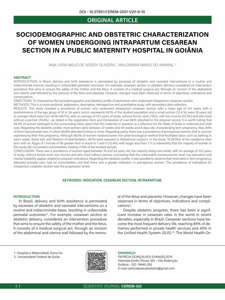
The sociodemographic characterization of women sub-mitted to intrapartum cesarean section is shown in Table 1.
Table 1. Sociodemographic characterization of women undergoing intrapartum cesarean section, Goiânia, Brazil, 2019 (n = 89).
SCIENTIFIC JOURNAL CEREM-GO|8
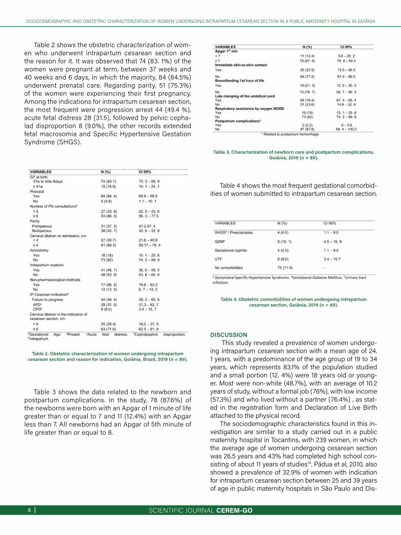
Table 3. Characterization of newborn care and postpartum complications, Goiânia, 2019 (n = 89).
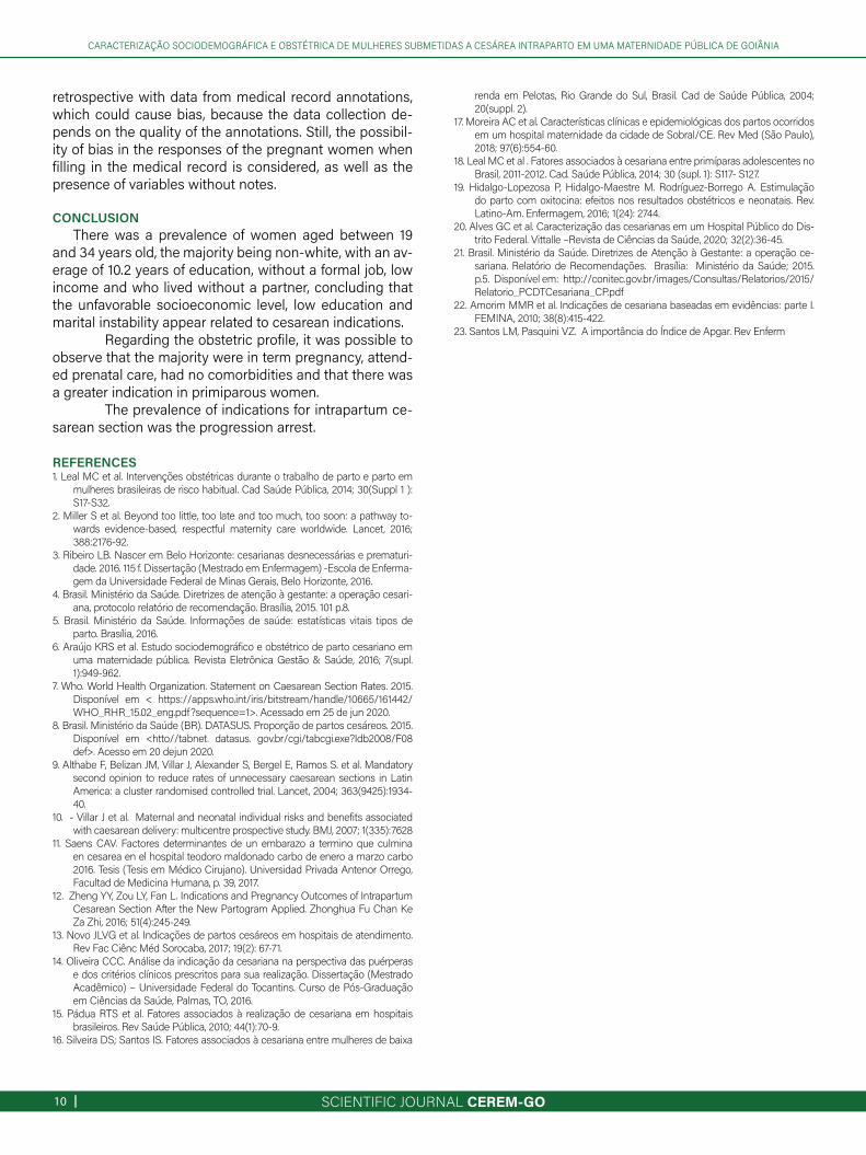
Table 4. Obstetric comorbidities of women undergoing intrapartum cesarean section, Goiânia, 2019 (n = 89).
SOCIODEMOGRAPHIC AND OBSTETRIC CHARACTERIZATION OF WOMEN UNDERGOING INTRAPARTUM CESAREAN SECTION IN A PUBLIC MATERNITY HOSPITAL IN GOIÂNIA
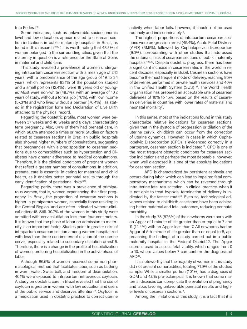
Table 2. Obstetric characterization of women undergoing intrapartum cesarean section and reason for indication, Goiânia, Brazil, 2019 (n = 89).
Table 2 shows the obstetric characterization of wom-en who underwent intrapartum cesarean section and the reason for it. It was observed that 74 (83. 1%) of the women were pregnant at term, between 37 weeks and 40 weeks and 6 days, in which the majority, 84 (84.5%) underwent prenatal care. Regarding parity, 51 (75.3%) of the women were experiencing their first pregnancy. Among the indications for intrapartum cesarean section, the most frequent were progression arrest 44 (49.4 %), acute fetal distress 28 (31.5), followed by pelvic cepha-lad disproportion 8 (9.0%), the other records extended fetal macrosomia and Specific Hypertensive Gestation Syndrome (SHGS).
Table 3 shows the data related to the newborn and postpartum complications. In the study, 78 (87.6%) of the newborns were born with an Apgar of 1 minute of life greater than or equal to 7 and 11 (12.4%) with an Apgar less than 7. All newborns had an Apgar of 5th minute of life greater than or equal to 8.
Table 4 shows the most frequent gestational comorbid-ities of women submitted to intrapartum cesarean section.
DISCUSSION This study revealed a prevalence of women undergo-
ing intrapartum cesarean section with a mean age of 24. 1 years, with a predominance of the age group of 19 to 34 years, which represents 83.1% of the population studied and a small portion (12. 4%) were 18 years old or young-er. Most were non-white (48.7%), with an average of 10.2 years of study, without a formal job (76%), with low income (57.3%) and who lived without a partner (76.4%) , as stat-ed in the registration form and Declaration of Live Birth attached to the physical record.
The sociodemographic characteristics found in this in-vestigation are similar to a study carried out in a public maternity hospital in Tocantins, with 239 women, in which the average age of women undergoing cesarean section was 26.5 years and 43% had completed high school con-sisting of about 11 years of studies14. Pádua et al, 2010, also showed a prevalence of 32.9% of women with indication for intrapartum cesarean section between 25 and 39 years of age in public maternity hospitals in São Paulo and Dis-
SCIENTIFIC JOURNAL CEREM-GO | 9
SOCIODEMOGRAPHIC AND OBSTETRIC CHARACTERIZATION OF WOMEN UNDERGOING INTRAPARTUM CESAREAN SECTION IN A PUBLIC MATERNITY HOSPITAL IN GOIÂNIA
trito Federal15.Some indicators, such as unfavorable socioeconomic
level and low education, appear related to cesarean sec-tion indications in public maternity hospitals in Brazil, as found in this research14,16,17. It is worth noting that 48.3% of women belonged to the surrounding cities, given that the maternity in question is a reference for the State of Goiás in maternal and child care.
This study revealed a prevalence of women undergo-ing intrapartum cesarean section with a mean age of 24.1 years, with a predominance of the age group of 19 to 34 years, which represents 83.1% of the population studied and a small portion (12.4%) , were 18 years old or young-er. Most were non-white (48.7%), with an average of 10.2 years of study, without a formal job (76%), with low income (57.3%) and who lived without a partner (76.4%) , as stat-ed in the registration form and Declaration of Live Birth attached to the physical record.
Regarding the obstetric profile, most women were be-tween 37 weeks and 40 weeks and 6 days, characterizing term pregnancy. Also, 84% of them had prenatal care, in which 66.6% attended 6 times or more. Studies on factors related to cesarean sections in Brazilian public hospitals also showed higher numbers of consultations, suggesting that pregnancies with a predisposition to cesarean sec-tions due to comorbidities such as hypertension and Di-abetes have greater adherence to medical consultations. Therefore, it is the clinical conditions of pregnant women that reflect a greater number of consultations. In addition, prenatal care is essential in caring for maternal and child health, as it enables better perinatal results through the early identification of gestational risks15,17.
Regarding parity, there was a prevalence of primipa-rous women, that is, women experiencing their first preg-nancy. In Brazil, the proportion of cesarean sections is higher in primiparous women, especially those residing in the Central Region, and are often indicated without clini-cal criteria18. Still, 30.7% of the women in this study were admitted with cervical dilation less than four centimeters. It is known that the phase of labor on admission to mater-nity is an important factor. Studies point to greater risks of intrapartum cesarean section among women hospitalized with less than three centimeters of dilation of the uterine cervix, especially related to secondary dilatation arrest16. Therefore, there is a change in the profile of hospitalization of women, preferring hospitalization in the active phase of labor.
Although 86.5% of women received some non-phar-macological method that facilitates labor, such as bathing in warm water, Swiss ball, and freedom of deambulation, 46.1% were exposed to intrapartum intravenous oxytocin. A study on obstetric care in Brazil revealed that the use of oxytocin is greater in women with low education and users of the public service and deserves attention18. Oxytocin is a medication used in obstetric practice to correct uterine
activity when labor fails, however, it should not be used routinely and indiscriminately19.
The highest proportions of intrapartum cesarean sec-tions were progression arrest (49.4%), Acute Fetal Distress (AFD) (31.5%), followed by Cephalopelvic disproportion (9.0%), corroborating with other studies that addressed the criteria clinics of cesarean sections of public maternity hospitals14,19,20. Despite obstetric progress, there has been a significant increase in cesarean rates in the world in re-cent decades, especially in Brazil. Cesarean sections have become the most frequent mode of delivery, reaching 85% of deliveries performed in private health services and 40% in the Unified Health System (SUS) 21. The World Health Organization has prepared an acceptable rate of cesarean deliveries of 10% to 15%, based on the results of cesare-an deliveries in countries with lower rates of maternal and neonatal mortality6.
In this sense, most of the indications found in this study characterize relative indications for cesarean sections, given that in the dystocia of progression or dilation of the uterine cervix, childbirth can occur from the correction of uterine dynamics. However, in cases in which Cepha-lopelvic Disproportion (CPD) is evidenced correctly in a partogram, cesarean section is indicated22. CPD is one of the most frequent obstetric conditions for cesarean sec-tion indications and perhaps the most debatable, however, when well diagnosed it is one of the absolute indications for cesarean section.
AFD is characterized by persistent asphyxia and occurs during labor, which can lead to impaired fetal com-pensatory mechanisms, which can be reversed through intrauterine fetal resuscitation. In clinical practice, when it is not able to treat hypoxia, termination of delivery is in-dicated by the fastest route13. Even so, technological ad-vances related to childbirth assistance have been achiev-ing better maternal and fetal outcomes, reducing perinatal morbidity.
In the study, 78 (87.6%) of the newborns were born with an Apgar of 1 minute of life greater than or equal to 7 and 11 (12.4%) with an Apgar less than 7. All newborns had an Apgar of 5th minute of life greater than or equal to 8, ap-proaching the findings of a study carried out in a public maternity hospital in the Federal District22. The Apgar score is used to assess fetal vitality, which ranges from 0 to 10, where values below 7 can confirm the diagnosis of AFD23.
It is noteworthy that the majority of women in this study did not present comorbidities, totaling 71.9% of the studied sample. While a smaller portion (10.1%) had a diagnosis of GDM and 4.5% pre-eclampsia. It is known that some ma-ternal diseases can complicate the evolution of pregnancy and labor, favoring unfavorable perinatal results and high-er levels of cesarean sections15.
Among the limitations of this study, it is a fact that it is
SCIENTIFIC JOURNAL CEREM-GO|10
CARACTERIZAÇÃO SOCIODEMOGRÁFICA E OBSTÉTRICA DE MULHERES SUBMETIDAS A CESÁREA INTRAPARTO EM UMA MATERNIDADE PÚBLICA DE GOIÂNIA
retrospective with data from medical record annotations, which could cause bias, because the data collection de-pends on the quality of the annotations. Still, the possibil-ity of bias in the responses of the pregnant women when filling in the medical record is considered, as well as the presence of variables without notes.
CONCLUSIONThere was a prevalence of women aged between 19
and 34 years old, the majority being non-white, with an av-erage of 10.2 years of education, without a formal job, low income and who lived without a partner, concluding that the unfavorable socioeconomic level, low education and marital instability appear related to cesarean indications.
Regarding the obstetric profile, it was possible to observe that the majority were in term pregnancy, attend-ed prenatal care, had no comorbidities and that there was a greater indication in primiparous women.
The prevalence of indications for intrapartum ce-sarean section was the progression arrest.
REFERENCES1. Leal MC et al. Intervenções obstétricas durante o trabalho de parto e parto em
mulheres brasileiras de risco habitual. Cad Saúde Pública, 2014; 30(Suppl 1 ): S17-S32.
2. Miller S et al. Beyond too little, too late and too much, too soon: a pathway to-wards evidence-based, respectful maternity care worldwide. Lancet, 2016; 388:2176-92.
3. Ribeiro LB. Nascer em Belo Horizonte: cesarianas desnecessárias e prematuri-dade. 2016. 115 f. Dissertação (Mestrado em Enfermagem) -Escola de Enferma-gem da Universidade Federal de Minas Gerais, Belo Horizonte, 2016.
4. Brasil. Ministério da Saúde. Diretrizes de atenção à gestante: a operação cesari-ana, protocolo relatório de recomendação. Brasília, 2015. 101 p.8.
5. Brasil. Ministério da Saúde. Informações de saúde: estatísticas vitais tipos de parto. Brasília, 2016.
6. Araújo KRS et al. Estudo sociodemográfico e obstétrico de parto cesariano em uma maternidade pública. Revista Eletrônica Gestão & Saúde, 2016; 7(supl. 1):949-962.
7. Who. World Health Organization. Statement on Caesarean Section Rates. 2015. Disponível em < https://apps.who.int/iris/bitstream/handle/10665/161442/WHO_RHR_15.02_eng.pdf?sequence=1>. Acessado em 25 de jun 2020.
8. Brasil. Ministério da Saúde (BR). DATASUS. Proporção de partos cesáreos. 2015. Disponível em <htto//tabnet. datasus. gov.br/cgi/tabcgi.exe?Idb2008/F08 def>. Acesso em 20 dejun 2020.
9. Althabe F, Belizan JM, Villar J, Alexander S, Bergel E, Ramos S. et al. Mandatory second opinion to reduce rates of unnecessary caesarean sections in Latin America: a cluster randomised controlled trial. Lancet, 2004; 363(9425):1934-40.
10. - Villar J et al. Maternal and neonatal individual risks and benefits associated with caesarean delivery: multicentre prospective study. BMJ, 2007; 1(335):7628
11. Saens CAV. Factores determinantes de un embarazo a termino que culmina en cesarea en el hospital teodoro maldonado carbo de enero a marzo carbo 2016. Tesis (Tesis em Médico Cirujano). Universidad Privada Antenor Orrego, Facultad de Medicina Humana, p. 39, 2017.
12. Zheng YY, Zou LY, Fan L. Indications and Pregnancy Outcomes of Intrapartum Cesarean Section After the New Partogram Applied. Zhonghua Fu Chan Ke Za Zhi, 2016; 51(4):245-249.
13. Novo JLVG et al. Indicações de partos cesáreos em hospitais de atendimento. Rev Fac Ciênc Méd Sorocaba, 2017; 19(2): 67-71.
14. Oliveira CCC. Análise da indicação da cesariana na perspectiva das puérperas e dos critérios clínicos prescritos para sua realização. Dissertação (Mestrado Acadêmico) – Universidade Federal do Tocantins. Curso de Pós-Graduação em Ciências da Saúde, Palmas, TO, 2016.
15. Pádua RTS et al. Fatores associados à realização de cesariana em hospitais brasileiros. Rev Saúde Pública, 2010; 44(1):70-9.
16. Silveira DS; Santos IS. Fatores associados à cesariana entre mulheres de baixa
renda em Pelotas, Rio Grande do Sul, Brasil. Cad de Saúde Pública, 2004; 20(suppl. 2).
17. Moreira AC et al. Características clínicas e epidemiológicas dos partos ocorridos em um hospital maternidade da cidade de Sobral/CE. Rev Med (São Paulo), 2018; 97(6):554-60.
18. Leal MC et al . Fatores associados à cesariana entre primíparas adolescentes no Brasil, 2011-2012. Cad. Saúde Pública, 2014; 30 (supl. 1): S117- S127.
19. Hidalgo-Lopezosa P, Hidalgo-Maestre M. Rodríguez-Borrego A. Estimulação do parto com oxitocina: efeitos nos resultados obstétricos e neonatais. Rev. Latino-Am. Enfermagem, 2016; 1(24): 2744.
20. Alves GC et al. Caracterização das cesarianas em um Hospital Público do Dis-trito Federal. Vittalle –Revista de Ciências da Saúde, 2020; 32(2):36-45.
21. Brasil. Ministério da Saúde. Diretrizes de Atenção à Gestante: a operação ce-sariana. Relatório de Recomendações. Brasília: Ministério da Saúde; 2015. p.5. Disponível em: http://conitec.gov.br/images/Consultas/Relatorios/2015/Relatorio_PCDTCesariana_CP.pdf
22. Amorim MMR et al. Indicações de cesariana baseadas em evidências: parte I. FEMINA, 2010; 38(8):415-422.
23. Santos LM, Pasquini VZ. A importância do Índice de Apgar. Rev Enferm
SCIENTIFIC JOURNAL CEREM-GO | 11
ORIGINAL ARTICLE
PROFILE OF NEWBORNS SUBMITTED TO SURGERY IN THE INTENSIVE CARE UNIT
PATRÍCIA DE PAULA MIGUEL 1, SANDRA MARCIA RAMOS PIMENTEL AFIUNE 1, DANIELA CARVALHO PORTAL 1,CARLA AMARAL VIEIRA 1, TÁRIK KASSEM SAIDAH2
ABSTRACTIntroduction: Improvements in pediatric surgical outcomes are partly attributable to major advances in better understanding of neonatal physiology, specialized pediatric anesthesia, neonatal intensive care, including sophisticated cardiopulmonary support, use of parenteral nutrition and adjustments in fluid management, refinement of surgical technique and advances in surgical technology, including minimally invasive options, which further reduced operative mortality in neonates. Objective: To analyze the profile of patients undergoing surgery in a neonatal intensive care unit. Method: Retrospective analytical cross-sectional study with survey of all cases of surgery performed on newborns admitted to the Neonatal Intensive Care Unit of the Hospital and Maternidade Dona Íris (HMDI). Results: We analyzed 523 medical records that correspond to the number of patients admitted to the Neonatal ICU of HMDI in 2018 and 2019 and of these 78 underwent some type of surgery corresponding to 14.9% of NBs. The profile of these patients is of gestational age between 33 to <37 weeks 31 (40%), weighing> 2500g 35 (45%), male 45 (58%), born by cesarean section 49 (63%), without post complications - surgical 41 (53%). The predominant type of surgery was a thoracostomy with a drain 21 (27%) followed by a gastroise 11 (14%). In the comparison between gestational age and type of surgery, we found: <28 weeks thoracostomy with drain, 29 to <32 weeks thoracostomy with drain and herniorrhaphy, 33 to <37 weeks gastroschisis,> 38 weeks thoracostomy with drain. The main complication found was sepsis 17 (42%) and death 16 (40%). It is worth noting that there was a higher occurrence of deaths in NBs with gestational age <28 weeks 8 (50%). Of the patients who underwent surgery, 20.5% died. Conclusions: The profile of patients undergoing surgery in the neonatal ICU was male NB, gestational age between 33 to <37 weeks, weighing> 2500g, born by cesarean section and without post-surgical complications. The rate of surgeries performed in the neonatal ICU was 14.9%. The main complication found was sepsis 42%. Post-surgical death rate was 20.5%.
1. HMDI2. Unievangélica
ADDRESS PATRÍCIA GONÇALVES EVANGELISTAAlameda Emílio Póvoa, 165 - Vila RedençãoGoiânia - GO, 74845-250E-mail [email protected]
DOI - 10.37951/CEREM-2021-V2I1-11-14
KEYWORDS: NEWBORN, NEONATAL ICU, SURGERIES.
INTRODUCTIONThe Neonatal Intensive Care Unit is an inpatient ser-
vice responsible for comprehensive care for seriously or potentially serious newborns, endowed with assistance structures that have adequate technical conditions to pro-vide specialized assistance, including physical facilities, equipment and human resources. With the advancement of medicine and technical-scientific development, the pro-file of children hospitalized in intensive care units (ICU) can change, demanding from professionals more complex care and invasive procedures that can effectively guaran-tee the survival of these patients1,2.
The neonatal period is extremely vulnerable and con-stitutes a major component of infant mortality. It is esti-mated that about 25.0% of deaths occur in the first twen-ty-four hours of life and most of these neonatal deaths are related to prematurity, asphyxia and infections3.
Neonatal surgery emerged as an incipient in the 1930s and 1940s in restricted regional centers around the world where pioneering pediatric surgeons were located. It be-came a genuine pediatric surgical subspecialty during the 1950s, led by those children's hospitals that developed neonatal surgical units. Technological developments such as ultrasound, computed tomography (CT), sophisticated ventilators and advances in parenteral nutrition have rev-olutionized diagnosis and treatment. Magnetic resonance imaging, ECMO increased the scope and expanded the horizons of neonatal care in the 1980s, improving treat-ment performance and reducing morbidity and mortality.
The improvements in pediatric surgical outcomes are partly attributable to major advances in better under-standing of neonatal physiology, specialized pediatric an-esthesia, neonatal intensive care, including sophisticated cardiopulmonary support, use of parenteral nutrition and
SCIENTIFIC JOURNAL CEREM-GO|12
PROFILE OF NEWBORNS SUBMITTED TO SURGERY IN THE INTENSIVE CARE UNIT
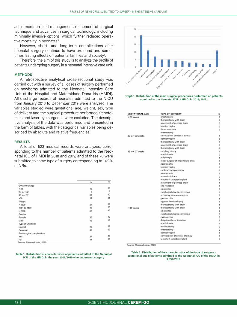
Table 1: Distribution of characteristics of patients admitted to the Neonatal ICU of the HMDI in the year 2018/2019 who underwent surgery
Graph 1: Distribution of the main surgical procedures performed on patients admitted to the Neonatal ICU of HMDI in 2018/2019.
Table 2: Distribution of the characteristics of the type of surgery x gestational age of patients admitted to the Neonatal ICU of the HMDI in
2018/2019
adjustments in fluid management, refinement of surgical technique and advances in surgical technology, including minimally invasive options, which further reduced opera-tive mortality in neonates5.
However, short- and long-term complications after neonatal surgery continue to have profound and some-times lasting effects on patients, families and society6.
Therefore, the aim of this study is to analyze the profile of patients undergoing surgery in a neonatal intensive care unit.
METHODSA retrospective analytical cross-sectional study was
carried out with a survey of all cases of surgery performed on newborns admitted to the Neonatal Intensive Care Unit of the Hospital and Maternidade Dona Íris (HMDI). All discharge records of neonates admitted to the NICU from January 2018 to December 2019 were analyzed. The variables studied were gestational age, weight, sex, type of delivery and the surgical procedure performed, frenoto-mies and laser eye surgeries were excluded. The descrip-tive analysis of the data was performed and presented in the form of tables, with the categorical variables being de-scribed by absolute and relative frequencies.
RESULTSA total of 523 medical records were analyzed, corre-
sponding to the number of patients admitted to the Neo-natal ICU of HMDI in 2018 and 2019, and of these 78 were submitted to some type of surgery corresponding to 14.9% of NBs.
thoracostom
y with drain
Gastroschisis
Herniograp
hy
Penrose drain placement
Esophageal atresia and correction
Omphalocele
Ileum
resection
Enterectom
yAbdominal drain
Others
Tenckhoff cathe
ter implantation
Colostom
y
SCIENTIFIC JOURNAL CEREM-GO | 13
PROFILE OF NEWBORNS SUBMITTED TO SURGERY IN THE INTENSIVE CARE UNIT
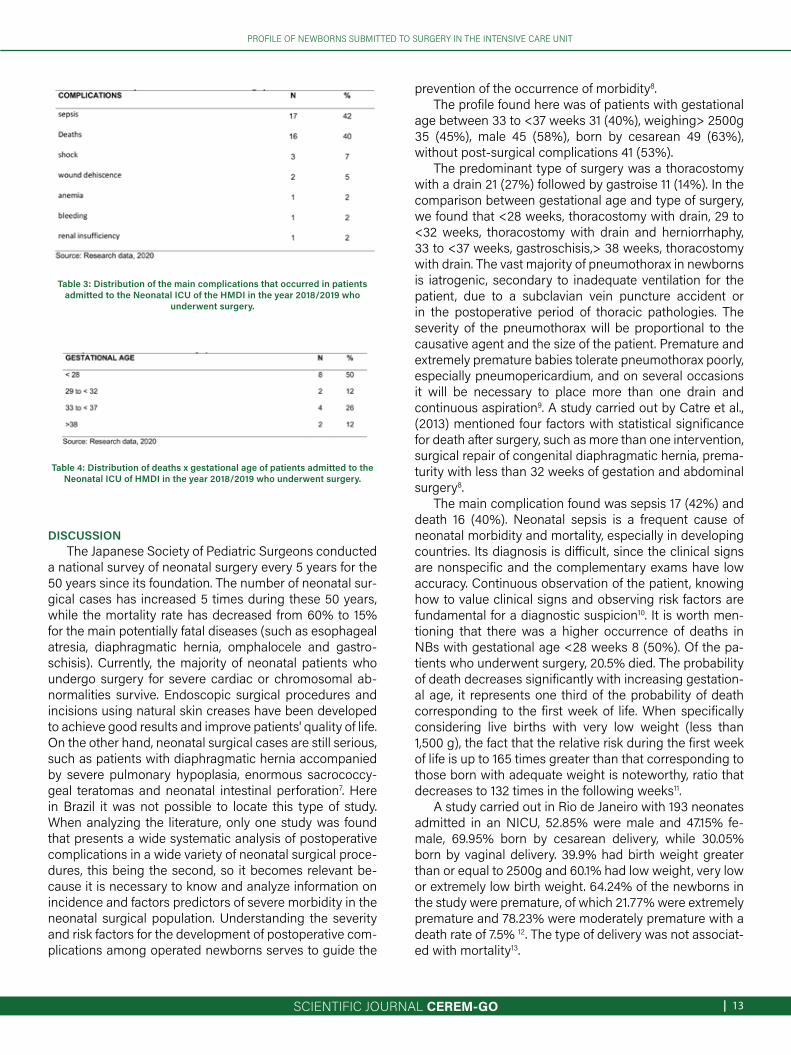
Table 3: Distribution of the main complications that occurred in patients admitted to the Neonatal ICU of the HMDI in the year 2018/2019 who
underwent surgery.
Table 4: Distribution of deaths x gestational age of patients admitted to the Neonatal ICU of HMDI in the year 2018/2019 who underwent surgery.
DISCUSSIONThe Japanese Society of Pediatric Surgeons conducted
a national survey of neonatal surgery every 5 years for the 50 years since its foundation. The number of neonatal sur-gical cases has increased 5 times during these 50 years, while the mortality rate has decreased from 60% to 15% for the main potentially fatal diseases (such as esophageal atresia, diaphragmatic hernia, omphalocele and gastro-schisis). Currently, the majority of neonatal patients who undergo surgery for severe cardiac or chromosomal ab-normalities survive. Endoscopic surgical procedures and incisions using natural skin creases have been developed to achieve good results and improve patients' quality of life. On the other hand, neonatal surgical cases are still serious, such as patients with diaphragmatic hernia accompanied by severe pulmonary hypoplasia, enormous sacrococcy-geal teratomas and neonatal intestinal perforation7. Here in Brazil it was not possible to locate this type of study. When analyzing the literature, only one study was found that presents a wide systematic analysis of postoperative complications in a wide variety of neonatal surgical proce-dures, this being the second, so it becomes relevant be-cause it is necessary to know and analyze information on incidence and factors predictors of severe morbidity in the neonatal surgical population. Understanding the severity and risk factors for the development of postoperative com-plications among operated newborns serves to guide the
prevention of the occurrence of morbidity8.The profile found here was of patients with gestational
age between 33 to <37 weeks 31 (40%), weighing> 2500g 35 (45%), male 45 (58%), born by cesarean 49 (63%), without post-surgical complications 41 (53%).
The predominant type of surgery was a thoracostomy with a drain 21 (27%) followed by gastroise 11 (14%). In the comparison between gestational age and type of surgery, we found that <28 weeks, thoracostomy with drain, 29 to <32 weeks, thoracostomy with drain and herniorrhaphy, 33 to <37 weeks, gastroschisis,> 38 weeks, thoracostomy with drain. The vast majority of pneumothorax in newborns is iatrogenic, secondary to inadequate ventilation for the patient, due to a subclavian vein puncture accident or in the postoperative period of thoracic pathologies. The severity of the pneumothorax will be proportional to the causative agent and the size of the patient. Premature and extremely premature babies tolerate pneumothorax poorly, especially pneumopericardium, and on several occasions it will be necessary to place more than one drain and continuous aspiration9. A study carried out by Catre et al., (2013) mentioned four factors with statistical significance for death after surgery, such as more than one intervention, surgical repair of congenital diaphragmatic hernia, prema-turity with less than 32 weeks of gestation and abdominal surgery8.
The main complication found was sepsis 17 (42%) and death 16 (40%). Neonatal sepsis is a frequent cause of neonatal morbidity and mortality, especially in developing countries. Its diagnosis is difficult, since the clinical signs are nonspecific and the complementary exams have low accuracy. Continuous observation of the patient, knowing how to value clinical signs and observing risk factors are fundamental for a diagnostic suspicion10. It is worth men-tioning that there was a higher occurrence of deaths in NBs with gestational age <28 weeks 8 (50%). Of the pa-tients who underwent surgery, 20.5% died. The probability of death decreases significantly with increasing gestation-al age, it represents one third of the probability of death corresponding to the first week of life. When specifically considering live births with very low weight (less than 1,500 g), the fact that the relative risk during the first week of life is up to 165 times greater than that corresponding to those born with adequate weight is noteworthy, ratio that decreases to 132 times in the following weeks11.
A study carried out in Rio de Janeiro with 193 neonates admitted in an NICU, 52.85% were male and 47.15% fe-male, 69.95% born by cesarean delivery, while 30.05% born by vaginal delivery. 39.9% had birth weight greater than or equal to 2500g and 60.1% had low weight, very low or extremely low birth weight. 64.24% of the newborns in the study were premature, of which 21.77% were extremely premature and 78.23% were moderately premature with a death rate of 7.5% 12. The type of delivery was not associat-ed with mortality13.
SCIENTIFIC JOURNAL CEREM-GO|14
PROFILE OF NEWBORNS SUBMITTED TO SURGERY IN THE INTENSIVE CARE UNIT
Knowledge of the characteristics of birth and death of newborns, the biological conditions of pregnancy and childbirth, as well as the neonates admitted to Neonatal Intensive Care Units (NICU), made available through ap-propriate epidemiological studies, can support health care actions, minimizing the occurrence of their injuries and planning a more appropriate treatment14.
Understanding the severity and risk factors for the de-velopment of postoperative complications among operat-ed newborns serves to guide the prevention of the occur-rence of morbidity8.
CONCLUSION The profile of patients undergoing surgery in the
neonatal ICU was male NB, gestational age between 33 to <37 weeks, weighing> 2500g, born by cesarean section and without post-surgical complications. The rate of surgeries performed in the neonatal
ICU was 14.9%. The main complication found was sepsis 42%. Post-surgical death rate was 20.5%.
REFERENCES1. Brasil. Portaria nº 930, de 10 de maio de 2012. Brasília: Ministério da Saúde; 2012.2. Gomes AVO, Nascimento MAL. O processo do cateterismo venoso central em
Unidade de Terapia Intensiva Neonatal e Pediátrica. Rev esc enferm USP, 2013; 47(4):794-800.
3. Ferraresi MF, Arrais AR. Perfil epidemiológico de mães de recém-nascidos ad-mitidos em uma unidade neonatal pública. Rev Rene, 2016;17(6):733-40.
4. Soper RT, Kimura K. Overview of neonatal surgery. Clin Perinatol., 1989; 16(1):1-12. 5. Rickham PP. Into the limits of neonatal surgery. Z Kinderchir, 1992;35(2):46-50.6. Escobar MA, Caty MG. Complications in neonatal surgery. Semin Pediatr Surg,
2016;25(6):347-370. 7. Taguchi T et al. Progress in and outcomes of neonatal surgery over the past 50
years. Nihon Geka Gakkai Zasshi, 2014; 115(6):306-11. 8. Catre D et al. Fatores preditivos de complicações graves em cirurgia neonatal.
Rev Col Bras Cir, 2013; 40(5).9. Boëchat, PR. Patologia cirúrgica do recém-nascido. In: Moreira MEL, Lopes JMA,
Caralho M. O recém-nascido de alto risco: teoria e prática do cuidar. Rio de Janeiro: Editora FIOCRUZ, 2004. 564 p.
10. Procianoy RS, Silveira RC. Os desafios no manejo da sepse neonatal. J. Pediatr. (Rio J.), 2020;96(supl. 1):80-86.
11. Ortiz LP, Oushiro DA. Perfil da mortalidade neonatal no Estado de São Paulo. São Paulo em Perspectiva, São Paulo, Fundação Seade, 2008; 22(1):19-29.
12. Silva EJ et al. Perfil Epidemiológico de UTI Neonatal de Maternidade Pública do Interior do RJ. Revista de Pediatria SOPERJ,2015;1(1).
13. Carvalho PI et al . Fatores de risco para mortalidade neonatal em coorte hospita-lar de nascidos vivos. Epidemiol. Serv. Saúde, 2007;16(3):185-194.
14. Lima SS. Perfil epidemiológico dos recém-nascidos admitidos na Unidade de Terapia Intensiva Neonatal de um hospital de referência em atenção materno infantil. Tese em Português. Belém-Pará; s.n; 2015. 67 p.
SCIENTIFIC JOURNAL CEREM-GO | 15
ORIGINAL ARTICLE
EVALUATION OF NEONATAL MORTALITY RISKIN THE CRIB SCORE APPLICATION
CARLA AMARAL VIEIRA1, SANDRA MARCIA RAMOS PIMENTEL AFIUNE1, DANIELA CARVALHO PORTAL1,PATRÍCIA DE PAULA MIGUEL1, TÁRIK KASSEM SAIDAH2
ABSTRACTIntroduction: In neonatology, several disease severity scores have been developed to predict the risk of mortality and morbidity in neonates. Among the scores based on physiological changes, some are simpler, with few variables and are fast to apply; others are more complete, as they include more variables, but take longer to be applied. The most studied and most used scoring systems in newborns are the Clinical Risk Index for Babies (CRIB) and the Neonatal Acute Physiology Score (SNAP). These scores were validated and reapplied in different studies in different ones. The neonatal scoring system CRIB (clinical risk index for babies) uses birth weight, gestational age, maximum and minimum fraction of inspired oxygen and maximum excess of base in the first 12 hours and presence of congenital malformationsObjective: To determine the mortality rate of newborns with CRIB variations.Results: Of the 283 hospitalized newborns, 62 met the inclusion criteria. The analyzed cohort had an average birth weight of 834.84g and a range of 500 to 1415 g. The average gestational age was 27 weeks, ranging from 23.3 to 31 weeks. The average CRIB score was 6.8 and ranged from 1 to 14. 29 newborns (46.7%) died. The analyzed cohort had an average birth weight of 834.84g and a range of 500 to 1415 g. The average gestational age was 27 weeks, ranging from 23.3 to 31 weeks. The mean CRIB score was 6.8 and ranged from 1 to 14. 29 newborns (46.7%) died, and the mortality rate was observed more frequently in newborns weighing less than 751g at 999 grams, gestational age less than 28 weeks and CRIB score above 6 to 10. The survival rate was observed most frequently in newborns weighing less than 751g at 999 grams, gestational age less than 28 weeks and CRIB score above from 0 to 5. Conclusions: The mean CRIB score was 6.8 and the range was 1 to 14. 29 newborns (46.7%) died. The mortality rate was observed more frequently in newborns weighing less than 751g at 999 grams, gestational age less than 28 weeks and CRIB score above 6 to 10. The survival rate was observed more frequently in newborns weighing less than 751g to 999 grams, gestational age less than 28 weeks and CRIB score above 0 to 5.
1 – Hospital e Maternidade Dona Iris 2 – Unievangélica
ADDRESS PATRÍCIA GONÇALVES EVANGELISTAAlameda Emílio Póvoa, 165 - Vila RedençãoGoiânia - GO, 74845-250E-mail [email protected]
DOI - 10.37951/CEREM-2021-V2I1-15-18
KEYWORDS: NEWBORN, NEONATAL ICU, CRIB.
INTRODUCTIONNeonatal mortality (0 to 27 days of life) has become
the main component of infant mortality in proportion-al terms since the late 1980s, and currently represents between 60% and 70% of infant mortality in all regions of Brazil . The perinatal period begins at 22 complete weeks (or 154 days) of gestation and ends at seven com-plete days after birth, that is, from 0 to 6 days of life (early neonatal period). Total births include live births and fetal deaths. For the purpose of international com-parison, WHO / ICD-10 uses the late fetal mortality rate, which considers fetuses above 28 weeks of gestation.
Neonatal mortality is also linked to preventable causes, related to access and use of health services, in addition to the quality of prenatal care, childbirth and the new-born, so it is important to know them1.
In recent years, with advances and improvements in neonatal care, the chance of survival for these chil-dren has increased, but, consequently, the risk of com-plications, including retinopathy of prematurity, hearing problems, neural tube defects and increased bactere-mia. Considering the importance of these diseases and the need for their prevention, an instrument to identify critically ill infants on admission to help the treatment
SCIENTIFIC JOURNAL CEREM-GO|16
EVALUATION OF NEONATAL MORTALITY RISK IN THE CRIB SCORE APPLICATION
team is highly necessary. More than a decade ago, “clin-ical risk scoring systems for babies” - that is, CRIB and CRIBII were used to assess health status and predict mortality in babies admitted to neonatal intensive care units (NICU) 2.
In neonatology, several disease severity scores have been developed to predict the risk of mortality and mor-bidity in neonates. Among the scores based on physi-ological changes, some are simpler, with few variables and are fast to apply; others are more complete, as they include more variables, but take longer to be applied. The most studied and most used scoring systems in newborns are the Clinical Risk Index for Babies (CRIB) and the Score for Neonatal Acute Physiology (SNAP). These scores have been validated and reapplied in dif-ferent studies in different countries3,4.
The neonatal scoring system CRIB (clinical risk index for babies) uses birth weight, gestational age, maximum and minimum fraction of inspired oxygen and maximum excess of base in the first 12 hours and presence of con-genital malformations5.
This study aims to determine the mortality rate of newborns with variations in the CRIB.
METHODOLOGYCross-sectional quantitative and retrospective study.
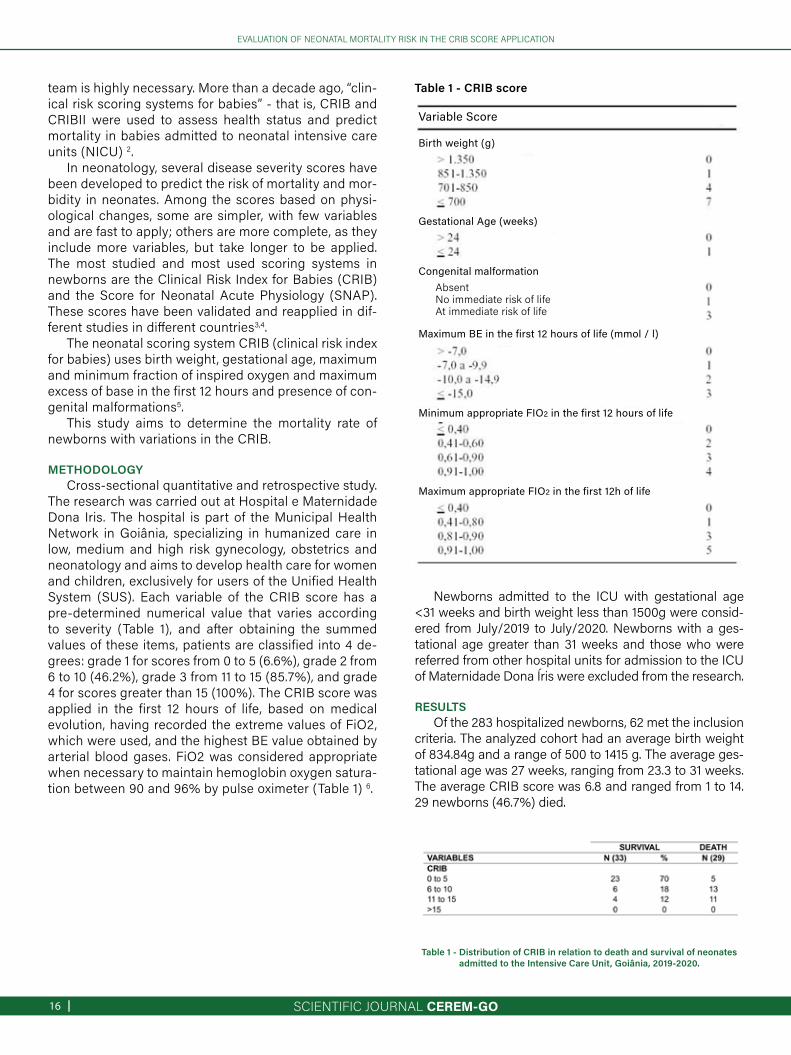
The research was carried out at Hospital e Maternidade Dona Iris. The hospital is part of the Municipal Health Network in Goiânia, specializing in humanized care in low, medium and high risk gynecology, obstetrics and neonatology and aims to develop health care for women and children, exclusively for users of the Unified Health System (SUS). Each variable of the CRIB score has a pre-determined numerical value that varies according to severity (Table 1), and after obtaining the summed values of these items, patients are classified into 4 de-grees: grade 1 for scores from 0 to 5 (6.6%), grade 2 from 6 to 10 (46.2%), grade 3 from 11 to 15 (85.7%), and grade 4 for scores greater than 15 (100%). The CRIB score was applied in the first 12 hours of life, based on medical evolution, having recorded the extreme values of FiO2, which were used, and the highest BE value obtained by arterial blood gases. FiO2 was considered appropriate when necessary to maintain hemoglobin oxygen satura-tion between 90 and 96% by pulse oximeter (Table 1) 6.
Newborns admitted to the ICU with gestational age <31 weeks and birth weight less than 1500g were consid-ered from July/2019 to July/2020. Newborns with a ges-tational age greater than 31 weeks and those who were referred from other hospital units for admission to the ICU of Maternidade Dona Íris were excluded from the research.
RESULTSOf the 283 hospitalized newborns, 62 met the inclusion
criteria. The analyzed cohort had an average birth weight of 834.84g and a range of 500 to 1415 g. The average ges-tational age was 27 weeks, ranging from 23.3 to 31 weeks. The average CRIB score was 6.8 and ranged from 1 to 14. 29 newborns (46.7%) died.
Table 1 - Distribution of CRIB in relation to death and survival of neonates admitted to the Intensive Care Unit , Goiânia, 2019-2020.
Table 1 - CRIB score
Birth weight (g)
Variable Score
Gestational Age (weeks)
Congenital malformation
Maximum BE in the first 12 hours of life (mmol / l)
Minimum appropriate FIO2 in the first 12 hours of life
Maximum appropriate FIO2 in the first 12h of life
AbsentNo immediate risk of lifeAt immediate risk of life
SCIENTIFIC JOURNAL CEREM-GO | 17
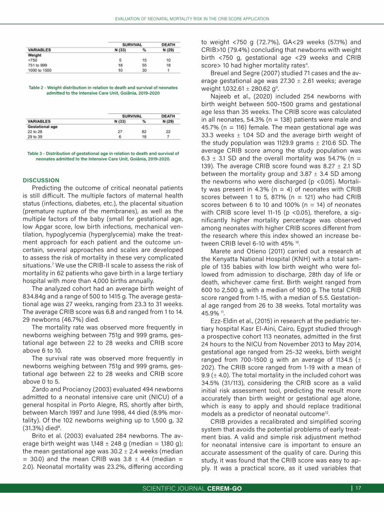
Table 2 - Weight distribution in relation to death and survival of neonates admitted to the Intensive Care Unit , Goiânia, 2019-2020
Table 3 - Distribution of gestational age in relation to death and survival of neonates admitted to the Intensive Care Unit , Goiânia, 2019-2020.
EVALUATION OF NEONATAL MORTALITY RISK IN THE CRIB SCORE APPLICATION
DISCUSSIONPredicting the outcome of critical neonatal patients
is still difficult. The multiple factors of maternal health status (infections, diabetes, etc.), the placental situation (premature rupture of the membranes), as well as the multiple factors of the baby (small for gestational age, low Apgar score, low birth infections, mechanical ven-tilation, hypoglycemia (hyperglycemia) make the treat-ment approach for each patient and the outcome un-certain, several approaches and scales are developed to assess the risk of mortality in these very complicated situations.7 We use the CRIB-II scale to assess the risk of mortality in 62 patients who gave birth in a large tertiary hospital with more than 4,000 births annually.
The analyzed cohort had an average birth weight of 834.84g and a range of 500 to 1415 g. The average gesta-tional age was 27 weeks, ranging from 23.3 to 31 weeks. The average CRIB score was 6.8 and ranged from 1 to 14. 29 newborns (46.7%) died.
The mortality rate was observed more frequently in newborns weighing between 751g and 999 grams, ges-tational age between 22 to 28 weeks and CRIB score above 6 to 10.
The survival rate was observed more frequently in newborns weighing between 751g and 999 grams, ges-tational age between 22 to 28 weeks and CRIB score above 0 to 5.
Zardo and Procianoy (2003) evaluated 494 newborns admitted to a neonatal intensive care unit (NICU) of a general hospital in Porto Alegre, RS, shortly after birth, between March 1997 and June 1998, 44 died (8.9% mor-tality). Of the 102 newborns weighing up to 1,500 g, 32 (31.3%) died8.
Brito et al. (2003) evaluated 284 newborns. The av-erage birth weight was 1,148 ± 248 g (median = 1,180 g); the mean gestational age was 30.2 ± 2.4 weeks (median = 30.0) and the mean CRIB was 3.8 ± 4.4 (median = 2.0). Neonatal mortality was 23.2%, differing according
to weight <750 g (72.7%), GA<29 weeks (57.1%) and CRIB>10 (79.4%) concluding that newborns with weight birth <750 g, gestational age <29 weeks and CRIB score> 10 had higher mortality rates4.
Breuel and Segre (2007) studied 71 cases and the av-erage gestational age was 27.30 ± 2.61 weeks; average weight 1,032.61 ± 280.62 g9.
Najeeb et al., (2020) included 254 newborns with birth weight between 500-1500 grams and gestational age less than 35 weeks. The CRIB score was calculated in all neonates, 54.3% (n = 138) patients were male and 45.7% (n = 116) female. The mean gestational age was 33.3 weeks ± 1.04 SD and the average birth weight of the study population was 1129.9 grams ± 210.6 SD. The average CRIB score among the study population was 6.3 ± 3.1 SD and the overall mortality was 54.7% (n = 139). The average CRIB score found was 8.27 ± 2.1 SD between the mortality group and 3.87 ± 3.4 SD among the newborns who were discharged (p <0.05). Mortali-ty was present in 4.3% (n = 4) of neonates with CRIB scores between 1 to 5, 87.1% (n = 121) who had CRIB scores between 6 to 10 and 100% (n = 14) of neonates with CRIB score level 11-15 (p <0.05), therefore, a sig-nificantly higher mortality percentage was observed among neonates with higher CRIB scores different from the research where this index showed an increase be-tween CRIB level 6-10 with 45% 10.
Marete and Otieno (2011) carried out a research at the Kenyatta National Hospital (KNH) with a total sam-ple of 135 babies with low birth weight who were fol-lowed from admission to discharge, 28th day of life or death, whichever came first. Birth weight ranged from 600 to 2,500 g, with a median of 1600 g. The total CRIB score ranged from 1-15, with a median of 5.5. Gestation-al age ranged from 26 to 38 weeks. Total mortality was 45.9% 11.
Ezz-Eldin et al., (2015) in research at the pediatric ter-tiary hospital Kasr El-Aini, Cairo, Egypt studied through a prospective cohort 113 neonates, admitted in the first 24 hours to the NICU from November 2013 to May 2014, gestational age ranged from 25-32 weeks, birth weight ranged from 700-1500 g with an average of 1134.5 (± 202). The CRIB score ranged from 1-19 with a mean of 9.9 (± 4.0). The total mortality in the included cohort was 34.5% (31/113), considering the CRIB score as a valid initial risk assessment tool, predicting the result more accurately than birth weight or gestational age alone, which is easy to apply and should replace traditional models as a predictor of neonatal outcome12.
CRIB provides a recalibrated and simplified scoring system that avoids the potential problems of early treat-ment bias. A valid and simple risk adjustment method for neonatal intensive care is important to ensure an accurate assessment of the quality of care. During this study, it was found that the CRIB score was easy to ap-ply. It was a practical score, as it used variables that
SCIENTIFIC JOURNAL CEREM-GO|18
EVALUATION OF NEONATAL MORTALITY RISK IN THE CRIB SCORE APPLICATION
were part of the routine of care for premature newborns, being obtained quickly. Due to its simplicity, the CRIB score was also considered to be easy to reproduce, giv-ing no scope for interpretation errors due to individual subjectivity6,13.
CONCLUSIONThe average CRIB score was 6.8 and ranged from 1
to 14. 29 newborns (46.7%) died.The mortality rate was observed more frequently in
newborns weighing between 751g and 999 grams, ges-tational age between 22 to 28 weeks and CRIB score above 6 to 10.
The survival rate was observed more frequently in newborns weighing between 751g and 999 grams, ges-tational age between 22 to 28 weeks and CRIB score above 0 to 5.
REFERENCES1. Brasil. Ministério da Saúde (BR). Manual de vigilância do óbito infantil e fetal e
do comitê de prevenção do óbito infantil e fetal. Brasília (DF): Ministério da Saúde; 2009.
2. Motlagh AJ. et al. Evaluation of Clinical Risk Index for Babies to Predict Mortality and Morbidity in Neonates Admitted to Neonatal Intensive Care Unit. Electron J Gen Med, 2020; 17(5).
3. Jašić M et al. CRIB II score versus gestational age and birth weight in preterm infant mortality prediction: who will win the bet? Signa Vitae, 2016;11(1):172-81.
4. Brito AS, Matsuo T, Gonzalez MR, de Carvalho AB, Ferrari LS. Escore CRIB, peso ao nascer e idade gestacional na avaliação do risco de mortalidade neonatal [CRIB score, birth weight and gestational age in neonatal mortality risk evalu-ation]. Rev Saude Publica. 2003 Oct;37(5):597-602.
5. The CRIB (clinical risk index for babies) score: a tool for assessing initial neonatal risk and comparing performance of neonatal intensive care units. The Inter-national Neonatal Network. Lancet. 1993 Jul 24;342(8865):193-8. Erratum in: Lancet 1993 Sep 4;342(8871):626.
6. Sarquis ALF, Miyaki M, Cat MNL. Aplicação do escore CRIB para avaliar o risco de mortalidade neonatal. J. Pediatr. (Rio J.), 2002;78(3):225-229.
7. Stomnaroska O, Danilovski D. The CRIB II (Clinical Risk Index for Babies II) Score in Prediction of Neonatal Mortality. Pril (Makedon Akad Nauk Umet Odd Med Nauki). 2020 Dec 8;41(3):59-64.
8. Zardo MS, Procianoy R S. Comparação entre diferentes escores de risco de mortalidade em unidade de tratamento intensivo neonatal. Rev Saúde Pública, 2003; 37(5):591-6.
9. Breuel PA, Segre CAM. Avaliação do índice de risco clínico em recém-nascidos de muito baixo peso em maternidade pública terciária da cidade de São Paulo. Einstein, 2007;5(4):326-332.
10. Najeeb S, Ejaz E, Raza MA, Sarwar S, Gillani S, Afridi RU, Ali H, Khan IM. Impor-tance Of Clinical Risk Index For Babies Score For Predicting Mortality Among Neonates. J Ayub Med Coll Abbottabad. 2020 Oct-Dec;32(4):502-506.
11. Marete IK, Wasunna AO, Otieno PA. Clinical risk index for babies (CRIB) II score as a predictor of neonatal mortality among low birth weight babies at Kenyatta National Hospital. East Afr Med J. 2011 Jan;88(1):18-23.
12. Ezz-Eldin ZM, Hamid TA, Youssef MR, Nabil Hel-D. Clinical Risk Index for Babies (CRIB II) Scoring System in Prediction of Mortality in Premature Babies. J Clin Diagn Res. 2015 Jun;9(6):SC08-11.
13. Parry G, Tucker J, Tarnow-Mordi W; UK Neonatal Staffing Study Collaborative Group. CRIB II: an update of the clinical risk index for babies score. Lancet. 2003 May 24;361(9371):1789-91.
SCIENTIFIC JOURNAL CEREM-GO | 19
CASE REPORT
LEFT ATRIAL APPENDAGE CLOSURE: CASE REPORT
DÉBORA FREIRE RIBEIRO ROCHA 1, LEONARDO VELOSO DO AMARAL 1, MAURÍCIO LOPES PRUDENTE 1,HENRIQUE LIMA GUIMARÃES 1, FERNANDO HENRIQUE FERNANDES 1, ADRIANO GONÇALVES DE ARAÚJO 1,
JOÃO BATISTA MASSON SILVA 1, GIULLIANO GARDENGHI 1
KEYWORDS: ATRIAL FIBRILLATION, CORONARY DISEASE, ANTICOAGULANTS, ATRIAL APPENDAGE.
DOI - 10.37951/CEREM-2021-V2I1-19-22
ABSTRACTPeople with atrial fibrillation (AF) have five times greater risk of having a stroke than people who do not respond to this problem. Stroke secondary to AF has been associated with mortality rates and high permanent disability, since its effective prevention is important. Mechanical methods for the occlusion of the LAA have been developed as an alternative to oral anticoagulation for patients with contraindications or complications derived from anticoagulation. The case is a male patient, 86 years old, hypertensive and with AF who was admitted to our service on 06/07/2020 with a picture of lipothymia, dyspnea and chest pain associated with bradycardia (HR of 32bpm) and rhythm of total atrioventricular block with AF, he was admitted to the ICU, a transvenous transient pacemaker was passed, atenolol was suspended and full anticoagulation with enoxaparin was started. However, he developed an important melena condition on 12/06/2020 with a hematimetric fall and the need for blood transfusion, with anticoagulation and investigation with EDA and colonoscopy being suspended. He underwent a transesophageal echocardiogram and an electrophysiological study to assess cardioversion and AF ablation. Two protections from electrical cardioversion were performed without success. Patient is discharged from the hospital on 06/21/2020 using Eliquis 5mg twice a day associated with clopidogrel. However, on 06/07/2020, the patient evolved with a hematoma contained in a retropeitorial right hemithorax, a dose of Eliquis® was reduced to 2.5 mg twice a day, the patient maintained a persistent hematoma and the anticoagulant was then suspended and scheduled to close the LAA.
1. Hospital ENCORE ADDRESSGIULLIANO GARDENGHIRua Gurupi, R. 109, 06/08 - S/N - Quadra 25 - Vila BrasiliaAparecida de Goiânia - GO, 74905-350E-mail: [email protected]
INTRODUCTIONAtrial fibrillation (AF) is prevalent in developed coun-
tries from 1% to 2.5%, being the fifth leading cause of death in Brazil and the fifth leading cause of hospitalization in the SUS. And the incidence of stroke increases substantially with age, being attributable to AF in about 1.5% of patients aged <60 years and in more than 20% of patients aged> 80 years 1,2.
In patients with AF, most thrombi are formed inside the left appendage, which, in the presence of AF, reduces blood flow velocities within it, which favors the formation of the clot. The left atrial appendage (LAA) is a structure that presents a great anatomical variability, with the pos-sibility of having at least four different morphologies that imply different risks of thrombosis, even after adjustment for different covariates, such as the CHADSVASC score 3.
This scenario determined the possibility of intervening on the LAA, aiming to eliminate the main site of thrombus location. The closure of LAA as a prophylaxis strategy for
thromboembolic events in patients with AF has been car-ried out for decades; initially during mitral valve repair sur-geries and, more recently, in patients with non-valvular AF who are at high risk of embolism and who cannot tolerate the use of oral anticoagulants 4,5.
Considering that more than 90% of the thrombi iden-tified in patients with non-valvular AF originate from the LAA, several techniques have been developed to dry or exclude this appendix from circulation: surgical resection, isolation with direct suture or closure with clips (in patients who must undergo concomitant cardiac surgery) or exclu-sion through endovascular implantable devices4,6.
There are several devices for excluding the catheter ap-pendage: PLAATO™ system (the first device developed), Amplatzer™ cardiac plug, WATCHMAN™, ACP / AMU-LET™, Wavecrest™ system, LAMbre™. They are implanted by venipuncture and approaching the left atrium through the transeptal route, controlled with transesophageal echocardiogram (TEE). Before implantation, it is necessary
SCIENTIFIC JOURNAL CEREM-GO|20
EFT ATRIAL APPENDAGE CLOSURE: CASE REPORT.
to know the anatomy of the appendage, which is achieved with magnetic resonance imaging (MRI) or multi-section tomography (CT) to decide whether the patient is a candi-date for the procedure and thus choose the type and size of the device 7.
In patients who cannot tolerate the chronic use of oral anticoagulants, the occlusion of the LAA through the placement of a prosthesis by percutaneous route has been shown to be an interesting strategy for the prevention of stroke, and has been evaluated by several observational and randomized clinical studies 8.
The aim of this study is to report a case of percutane-ous closure of LAA in a patient with AF and coronary heart disease with contraindication to full anticoagulation.
The Research Ethics Committee of the Hospi-tal de Urgências de Goiânia approved this study (CAAE: 94882318.7.0000.0033).
CASE REPORTMale patient, 86 years old, hypertensive, ex-smoker
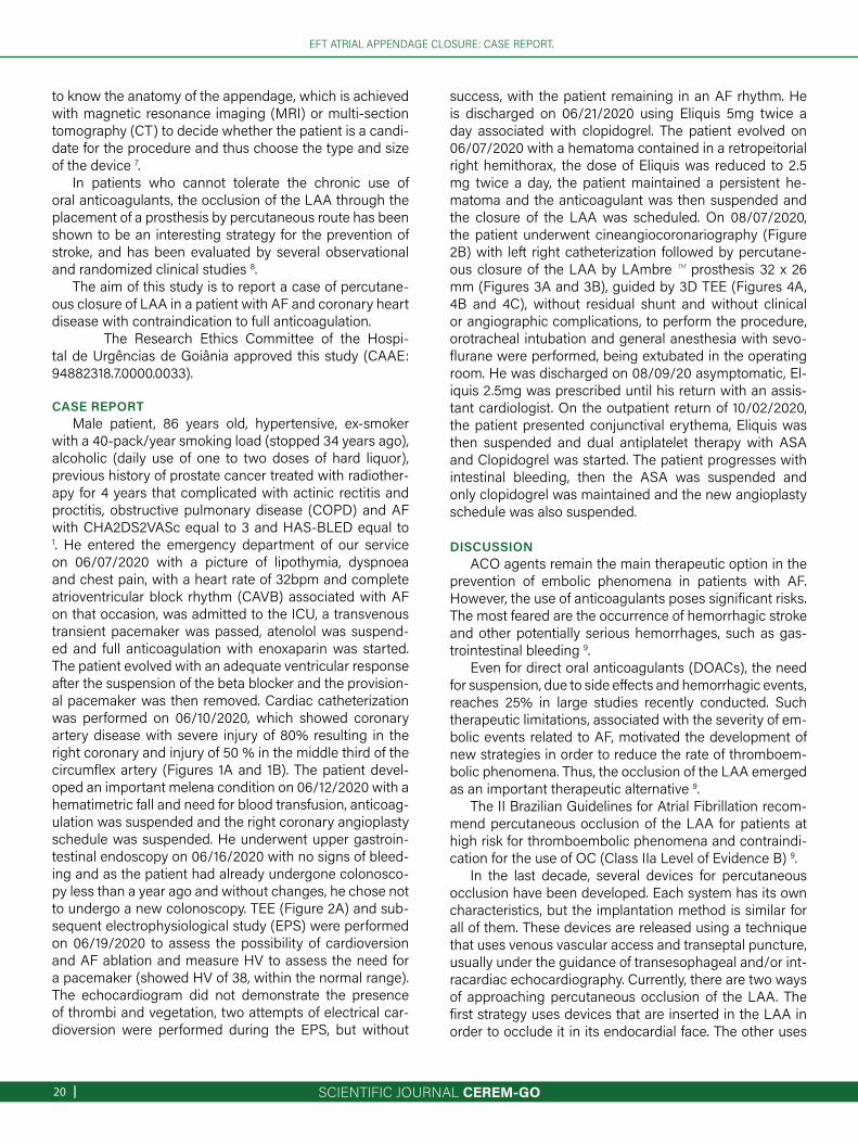
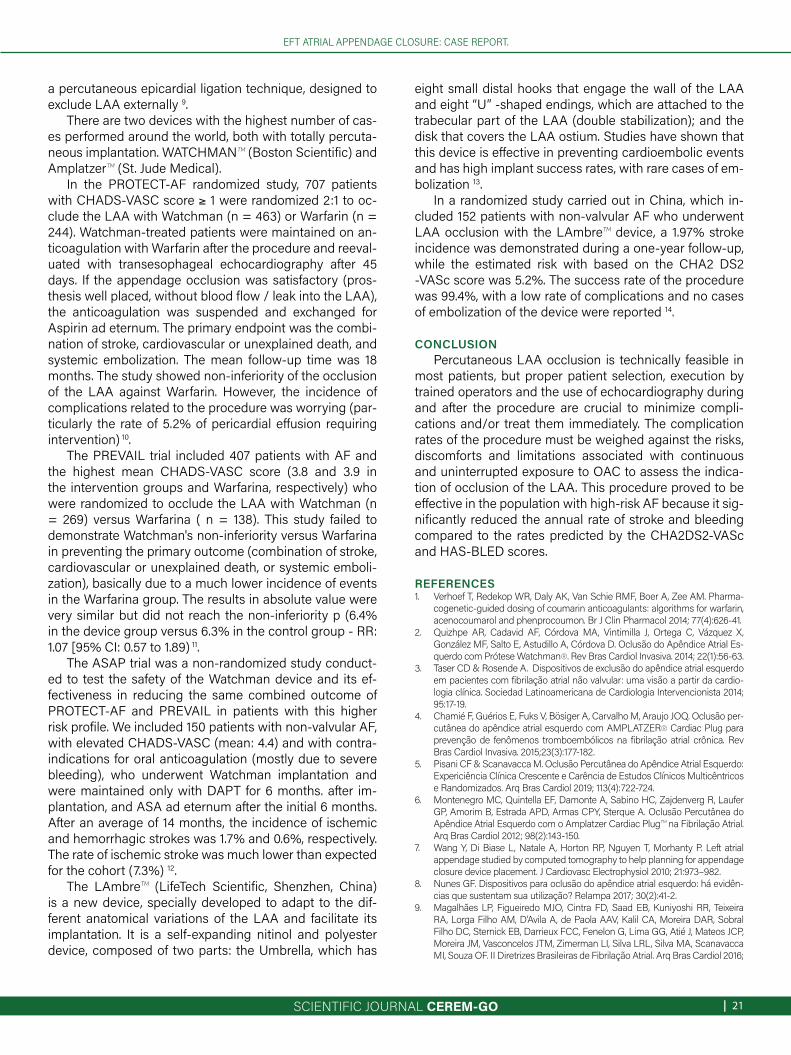
with a 40-pack/year smoking load (stopped 34 years ago), alcoholic (daily use of one to two doses of hard liquor), previous history of prostate cancer treated with radiother-apy for 4 years that complicated with actinic rectitis and proctitis, obstructive pulmonary disease (COPD) and AF with CHA2DS2VASc equal to 3 and HAS-BLED equal to 1. He entered the emergency department of our service on 06/07/2020 with a picture of lipothymia, dyspnoea and chest pain, with a heart rate of 32bpm and complete atrioventricular block rhythm (CAVB) associated with AF on that occasion, was admitted to the ICU, a transvenous transient pacemaker was passed, atenolol was suspend-ed and full anticoagulation with enoxaparin was started. The patient evolved with an adequate ventricular response after the suspension of the beta blocker and the provision-al pacemaker was then removed. Cardiac catheterization was performed on 06/10/2020, which showed coronary artery disease with severe injury of 80% resulting in the right coronary and injury of 50 % in the middle third of the circumflex artery (Figures 1A and 1B). The patient devel-oped an important melena condition on 06/12/2020 with a hematimetric fall and need for blood transfusion, anticoag-ulation was suspended and the right coronary angioplasty schedule was suspended. He underwent upper gastroin-testinal endoscopy on 06/16/2020 with no signs of bleed-ing and as the patient had already undergone colonosco-py less than a year ago and without changes, he chose not to undergo a new colonoscopy. TEE (Figure 2A) and sub-sequent electrophysiological study (EPS) were performed on 06/19/2020 to assess the possibility of cardioversion and AF ablation and measure HV to assess the need for a pacemaker (showed HV of 38, within the normal range). The echocardiogram did not demonstrate the presence of thrombi and vegetation, two attempts of electrical car-dioversion were performed during the EPS, but without
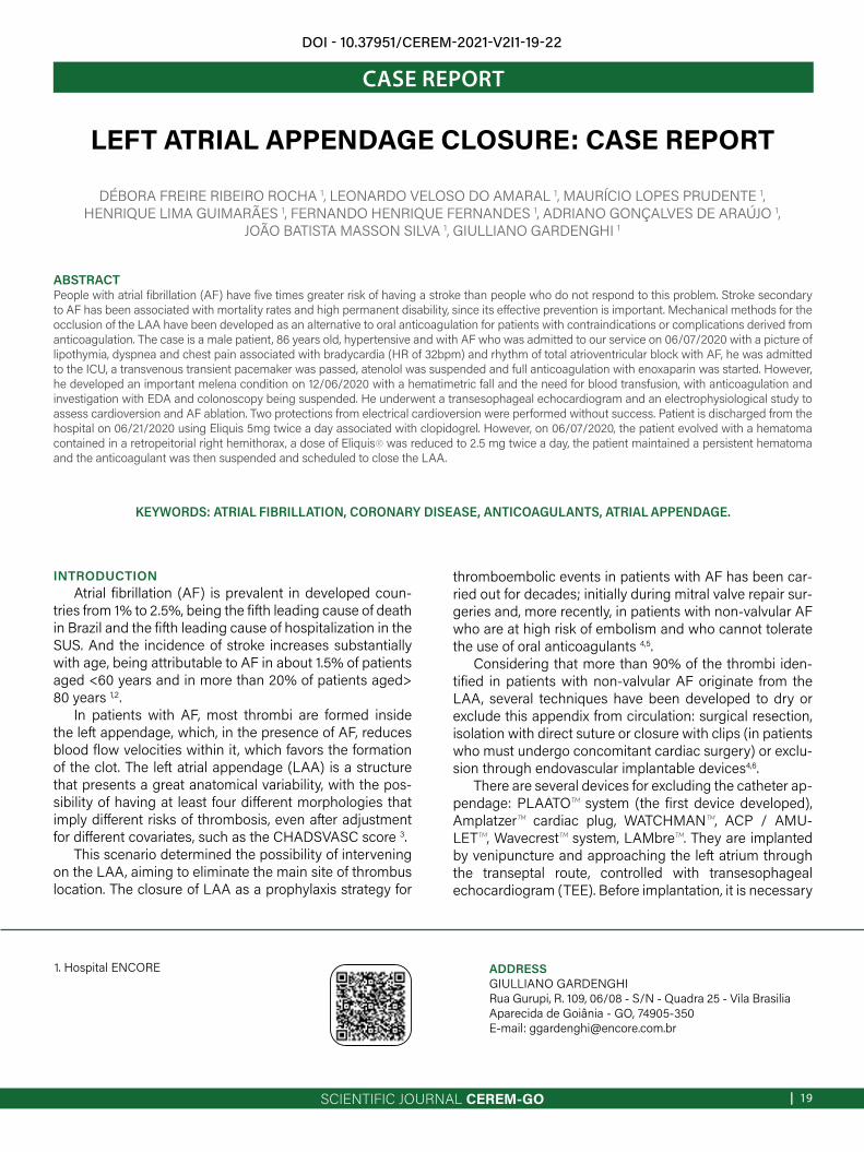
success, with the patient remaining in an AF rhythm. He is discharged on 06/21/2020 using Eliquis 5mg twice a day associated with clopidogrel. The patient evolved on 06/07/2020 with a hematoma contained in a retropeitorial right hemithorax, the dose of Eliquis was reduced to 2.5 mg twice a day, the patient maintained a persistent he-matoma and the anticoagulant was then suspended and the closure of the LAA was scheduled. On 08/07/2020, the patient underwent cineangiocoronariography (Figure 2B) with left right catheterization followed by percutane-ous closure of the LAA by LAmbre ™ prosthesis 32 x 26 mm (Figures 3A and 3B), guided by 3D TEE (Figures 4A, 4B and 4C), without residual shunt and without clinical or angiographic complications, to perform the procedure, orotracheal intubation and general anesthesia with sevo-flurane were performed, being extubated in the operating room. He was discharged on 08/09/20 asymptomatic, El-iquis 2.5mg was prescribed until his return with an assis-tant cardiologist. On the outpatient return of 10/02/2020, the patient presented conjunctival erythema, Eliquis was then suspended and dual antiplatelet therapy with ASA and Clopidogrel was started. The patient progresses with intestinal bleeding, then the ASA was suspended and only clopidogrel was maintained and the new angioplasty schedule was also suspended.
DISCUSSIONACO agents remain the main therapeutic option in the
prevention of embolic phenomena in patients with AF. However, the use of anticoagulants poses significant risks. The most feared are the occurrence of hemorrhagic stroke and other potentially serious hemorrhages, such as gas-trointestinal bleeding 9.
Even for direct oral anticoagulants (DOACs), the need for suspension, due to side effects and hemorrhagic events, reaches 25% in large studies recently conducted. Such therapeutic limitations, associated with the severity of em-bolic events related to AF, motivated the development of new strategies in order to reduce the rate of thromboem-bolic phenomena. Thus, the occlusion of the LAA emerged as an important therapeutic alternative 9.
The II Brazilian Guidelines for Atrial Fibrillation recom-mend percutaneous occlusion of the LAA for patients at high risk for thromboembolic phenomena and contraindi-cation for the use of OC (Class IIa Level of Evidence B) 9.
In the last decade, several devices for percutaneous occlusion have been developed. Each system has its own characteristics, but the implantation method is similar for all of them. These devices are released using a technique that uses venous vascular access and transeptal puncture, usually under the guidance of transesophageal and/or int-racardiac echocardiography. Currently, there are two ways of approaching percutaneous occlusion of the LAA. The first strategy uses devices that are inserted in the LAA in order to occlude it in its endocardial face. The other uses
SCIENTIFIC JOURNAL CEREM-GO | 21
EFT ATRIAL APPENDAGE CLOSURE: CASE REPORT.
a percutaneous epicardial ligation technique, designed to exclude LAA externally 9.
There are two devices with the highest number of cas-es performed around the world, both with totally percuta-neous implantation. WATCHMAN™ (Boston Scientific) and Amplatzer™ (St. Jude Medical).
In the PROTECT-AF randomized study, 707 patients with CHADS-VASC score ≥ 1 were randomized 2:1 to oc-clude the LAA with Watchman (n = 463) or Warfarin (n = 244). Watchman-treated patients were maintained on an-ticoagulation with Warfarin after the procedure and reeval-uated with transesophageal echocardiography after 45 days. If the appendage occlusion was satisfactory (pros-thesis well placed, without blood flow / leak into the LAA), the anticoagulation was suspended and exchanged for Aspirin ad eternum. The primary endpoint was the combi-nation of stroke, cardiovascular or unexplained death, and systemic embolization. The mean follow-up time was 18 months. The study showed non-inferiority of the occlusion of the LAA against Warfarin. However, the incidence of complications related to the procedure was worrying (par-ticularly the rate of 5.2% of pericardial effusion requiring intervention) 10.
The PREVAIL trial included 407 patients with AF and the highest mean CHADS-VASC score (3.8 and 3.9 in the intervention groups and Warfarina, respectively) who were randomized to occlude the LAA with Watchman (n = 269) versus Warfarina ( n = 138). This study failed to demonstrate Watchman's non-inferiority versus Warfarina in preventing the primary outcome (combination of stroke, cardiovascular or unexplained death, or systemic emboli-zation), basically due to a much lower incidence of events in the Warfarina group. The results in absolute value were very similar but did not reach the non-inferiority p (6.4% in the device group versus 6.3% in the control group - RR: 1.07 [95% CI: 0.57 to 1.89) 11.
The ASAP trial was a non-randomized study conduct-ed to test the safety of the Watchman device and its ef-fectiveness in reducing the same combined outcome of PROTECT-AF and PREVAIL in patients with this higher risk profile. We included 150 patients with non-valvular AF, with elevated CHADS-VASC (mean: 4.4) and with contra-indications for oral anticoagulation (mostly due to severe bleeding), who underwent Watchman implantation and were maintained only with DAPT for 6 months. after im-plantation, and ASA ad eternum after the initial 6 months. After an average of 14 months, the incidence of ischemic and hemorrhagic strokes was 1.7% and 0.6%, respectively. The rate of ischemic stroke was much lower than expected for the cohort (7.3%) 12.
The LAmbre™ (LifeTech Scientific, Shenzhen, China) is a new device, specially developed to adapt to the dif-ferent anatomical variations of the LAA and facilitate its implantation. It is a self-expanding nitinol and polyester device, composed of two parts: the Umbrella, which has
eight small distal hooks that engage the wall of the LAA and eight “U” -shaped endings, which are attached to the trabecular part of the LAA (double stabilization); and the disk that covers the LAA ostium. Studies have shown that this device is effective in preventing cardioembolic events and has high implant success rates, with rare cases of em-bolization 13.
In a randomized study carried out in China, which in-cluded 152 patients with non-valvular AF who underwent LAA occlusion with the LAmbre™ device, a 1.97% stroke incidence was demonstrated during a one-year follow-up, while the estimated risk with based on the CHA2 DS2 -VASc score was 5.2%. The success rate of the procedure was 99.4%, with a low rate of complications and no cases of embolization of the device were reported 14.
CONCLUSIONPercutaneous LAA occlusion is technically feasible in
most patients, but proper patient selection, execution by trained operators and the use of echocardiography during and after the procedure are crucial to minimize compli-cations and/or treat them immediately. The complication rates of the procedure must be weighed against the risks, discomforts and limitations associated with continuous and uninterrupted exposure to OAC to assess the indica-tion of occlusion of the LAA. This procedure proved to be effective in the population with high-risk AF because it sig-nificantly reduced the annual rate of stroke and bleeding compared to the rates predicted by the CHA2DS2-VASc and HAS-BLED scores.
REFERENCES1. Verhoef T, Redekop WR, Daly AK, Van Schie RMF, Boer A, Zee AM. Pharma-
cogenetic-guided dosing of coumarin anticoagulants: algorithms for warfarin, acenocoumarol and phenprocoumon. Br J Clin Pharmacol 2014; 77(4):626-41.
2. Quizhpe AR, Cadavid AF, Córdova MA, Vintimilla J, Ortega C, Vázquez X, González MF, Salto E, Astudillo A, Córdova D. Oclusão do Apêndice Atrial Es-querdo com Prótese Watchman®. Rev Bras Cardiol Invasiva. 2014; 22(1):56-63.
3. Taser CD & Rosende A. Dispositivos de exclusão do apêndice atrial esquerdo em pacientes com fibrilação atrial não valvular: uma visão a partir da cardio-logia clínica. Sociedad Latinoamericana de Cardiologia Intervencionista 2014; 95:17-19.
4. Chamié F, Guérios E, Fuks V, Bösiger A, Carvalho M, Araujo JOQ. Oclusão per-cutânea do apêndice atrial esquerdo com AMPLATZER® Cardiac Plug para prevenção de fenômenos tromboembólicos na fibrilação atrial crônica. Rev Bras Cardiol Invasiva. 2015;23(3):177-182.
5. Pisani CF & Scanavacca M. Oclusão Percutânea do Apêndice Atrial Esquerdo: Expericiência Clínica Crescente e Carência de Estudos Clínicos Multicêntricos e Randomizados. Arq Bras Cardiol 2019; 113(4):722-724.
6. Montenegro MC, Quintella EF, Damonte A, Sabino HC, Zajdenverg R, Laufer GP, Amorim B, Estrada APD, Armas CPY, Sterque A. Oclusão Percutânea do Apêndice Atrial Esquerdo com o Amplatzer Cardiac Plug™ na Fibrilação Atrial. Arq Bras Cardiol 2012; 98(2):143-150.
7. Wang Y, Di Biase L, Natale A, Horton RP, Nguyen T, Morhanty P. Left atrial appendage studied by computed tomography to help planning for appendage closure device placement. J Cardiovasc Electrophysiol 2010; 21:973–982.
8. Nunes GF. Dispositivos para oclusão do apêndice atrial esquerdo: há evidên-cias que sustentam sua utilização? Relampa 2017; 30(2):41-2.
9. Magalhães LP, Figueiredo MJO, Cintra FD, Saad EB, Kuniyoshi RR, Teixeira RA, Lorga Filho AM, D’Avila A, de Paola AAV, Kalil CA, Moreira DAR, Sobral Filho DC, Sternick EB, Darrieux FCC, Fenelon G, Lima GG, Atié J, Mateos JCP, Moreira JM, Vasconcelos JTM, Zimerman LI, Silva LRL, Silva MA, Scanavacca MI, Souza OF. II Diretrizes Brasileiras de Fibrilação Atrial. Arq Bras Cardiol 2016;
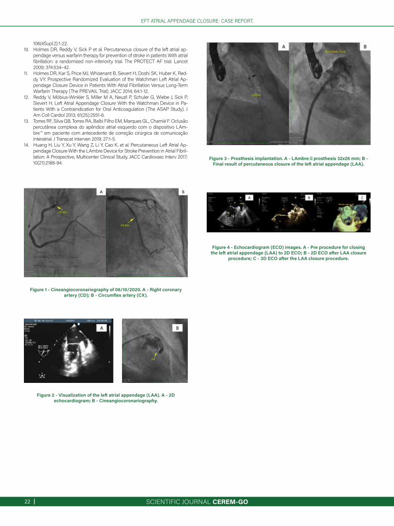
SCIENTIFIC JOURNAL CEREM-GO|22
EFT ATRIAL APPENDAGE CLOSURE: CASE REPORT.
Figure 1 - Cineangiocoronariography of 06/10/2020. A - Right coronary artery (CD); B - Circumflex artery (CX).
Figure 2 - Visualization of the left atrial appendage (LAA). A - 2D echocardiogram; B - Cineangiocoronariography.
Figure 3 - Prosthesis implantation. A - LAmbre ™ prosthesis 32x26 mm; B - Final result of percutaneous closure of the left atrial appendage (LAA).
106(4Supl.2):1-22.10. Holmes DR, Reddy V, Sick P et al. Percutaneous closure of the left atrial ap-
pendage versus warfarin therapy for prevention of stroke in patients With atrial fibrillation: a randomised non-inferiority trial. The PROTECT AF trial. Lancet 2009; 374:534–42.
11. Holmes DR, Kar S, Price MJ, Whisenant B, Sievert H, Doshi SK, Huber K, Red-dy VY. Prospective Randomized Evaluation of the Watchman Left Atrial Ap-pendage Closure Device in Patients With Atrial Fibrillation Versus Long-Term Warfarin Therapy (The PREVAIL Trial). JACC 2014; 64:1-12.
12. Reddy V, Möbius-Winkler S, Miller M A, Neuzil P, Schuler G, Wiebe J, Sick P, Sievert H. Left Atrial Appendage Closure With the Watchman Device in Pa-tients With a Contraindication for Oral Anticoagulation (The ASAP Study). J Am Coll Cardiol 2013; 61(25):2551-6.
13. Torres RF, Silva GB, Torres RA, Balbi Filho EM, Marques GL, Chamié F. Oclusão percutânea complexa do apêndice atrial esquerdo com o dispositivo LAm-bre™ em paciente com antecedente de correção cirúrgica de comunicação interatrial. J Transcat Interven 2019; 27:1-5.
14. Huang H, Liu Y, Xu Y, Wang Z, Li Y, Cao K, et al. Percutaneous Left Atrial Ap-pendage Closure With the LAmbre Device for Stroke Prevention in Atrial Fibril-lation: A Prospective, Multicenter Clinical Study. JACC Cardiovasc Interv 2017; 10(21):2188-94.
Figure 4 - Echocardiogram (ECO) images. A - Pre procedure for closing the left atrial appendage (LAA) to 2D ECO; B - 2D ECO after LAA closure
procedure; C - 3D ECO after the LAA closure procedure.
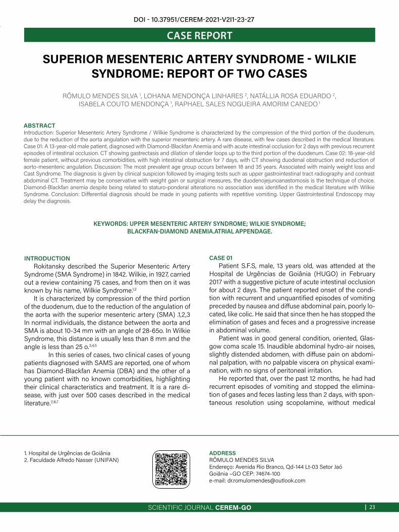
SCIENTIFIC JOURNAL CEREM-GO | 23
CASE REPORT
SUPERIOR MESENTERIC ARTERY SYNDROME - WILKIE SYNDROME: REPORT OF TWO CASES
RÔMULO MENDES SILVA 1, LOHANA MENDONÇA LINHARES 2, NATÁLLIA ROSA EDUARDO 2,ISABELA COUTO MENDONÇA 1, RAPHAEL SALES NOGUEIRA AMORIM CANEDO 1
KEYWORDS: UPPER MESENTERIC ARTERY SYNDROME; WILKIE SYNDROME; BLACKFAN-DIAMOND ANEMIA.ATRIAL APPENDAGE.
ABSTRACT Introduction: Superior Mesenteric Artery Syndrome / Wilkie Syndrome is characterized by the compression of the third portion of the duodenum, due to the reduction of the aorta angulation with the superior mesenteric artery. A rare disease, with few cases described in the medical literature. Case 01: A 13-year-old male patient, diagnosed with Diamond-Blackfan Anemia and with acute intestinal occlusion for 2 days with previous recurrent episodes of intestinal occlusion. CT showing gastrectasis and dilation of slender loops up to the third portion of the duodenum. Case 02: 18-year-old female patient, without previous comorbidities, with high intestinal obstruction for 7 days, with CT showing duodenal obstruction and reduction of aorto-mesenteric angulation. Discussion: The most prevalent age group occurs between 18 and 35 years. Associated with mainly weight loss and Cast Syndrome. The diagnosis is given by clinical suspicion followed by imaging tests such as upper gastrointestinal tract radiography and contrast abdominal CT. Treatment may be conservative with weight gain or surgical measures, the duodenojejunoanastomosis is the technique of choice. Diamond-Blackfan anemia despite being related to staturo-ponderal alterations no association was identified in the medical literature with Wilkie Syndrome. Conclusion: Differential diagnosis should be made in young patients with repetitive vomiting. Upper Gastrointestinal Endoscopy may delay the diagnosis.
1. Hospital de Urgências de Goiânia2. Faculdade Alfredo Nasser (UNIFAN)
ADDRESS RÔMULO MENDES SILVAEndereço: Avenida Rio Branco, Qd-144 Lt-03 Setor JaóGoiânia –GO CEP: 74674-100e-mail: [email protected]
DOI - 10.37951/CEREM-2021-V2I1-23-27
INTRODUCTIONRokitansky described the Superior Mesenteric Artery
Syndrome (SMA Syndrome) in 1842. Wilkie, in 1927, carried out a review containing 75 cases, and from then on it was known by his name, Wilkie Syndrome.1,2
It is characterized by compression of the third portion of the duodenum, due to the reduction of the angulation of the aorta with the superior mesenteric artery (SMA) .1,2,3 In normal individuals, the distance between the aorta and SMA is about 10-34 mm with an angle of 28-65o. In Wilkie Syndrome, this distance is usually less than 8 mm and the angle is less than 25 o.3,4,5
In this series of cases, two clinical cases of young patients diagnosed with SAMS are reported, one of whom has Diamond-Blackfan Anemia (DBA) and the other of a young patient with no known comorbidities, highlighting their clinical characteristics and treatment. It is a rare di-sease, with just over 500 cases described in the medical literature.2,6,7
CASE 01Patient S.F.S, male, 13 years old, was attended at the
Hospital de Urgências de Goiânia (HUGO) in February 2017 with a suggestive picture of acute intestinal occlusion for about 2 days. The patient reported onset of the condi-tion with recurrent and unquantified episodes of vomiting preceded by nausea and diffuse abdominal pain, poorly lo-cated, like colic. He said that since then he has stopped the elimination of gases and feces and a progressive increase in abdominal volume.
Patient was in good general condition, oriented, Glas-gow coma scale 15. Inaudible abdominal hydro-air noises, slightly distended abdomen, with diffuse pain on abdomi-nal palpation, with no palpable viscera on physical exami-nation, with no signs of peritoneal irritation.
He reported that, over the past 12 months, he had had recurrent episodes of vomiting and stopped the elimina-tion of gases and feces lasting less than 2 days, with spon-taneous resolution using scopolamine, without medical
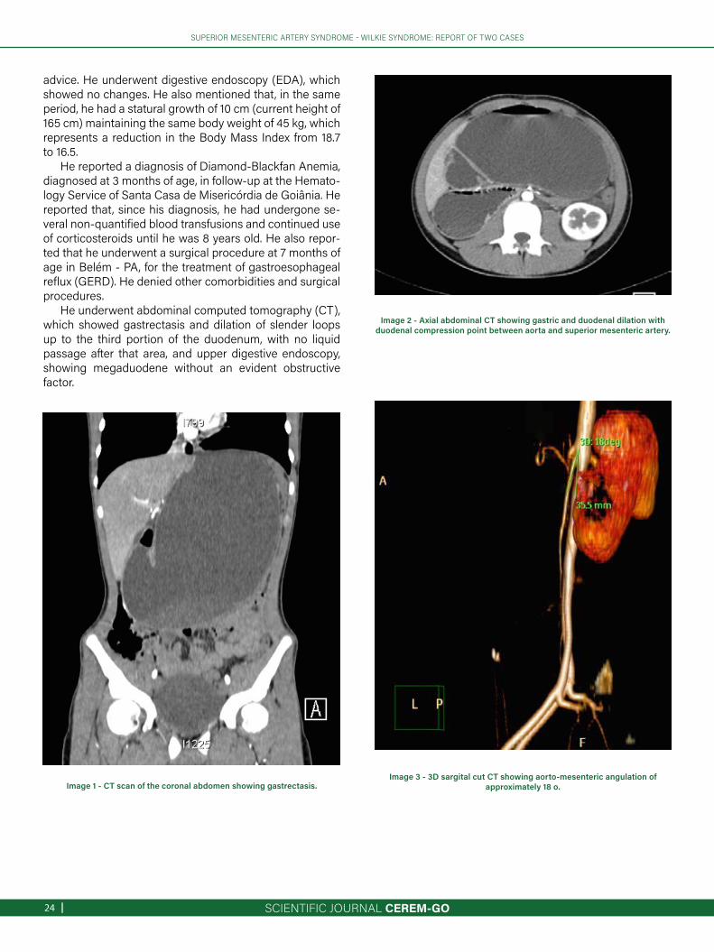
SCIENTIFIC JOURNAL CEREM-GO|24
Image 3 - 3D sargital cut CT showing aorto-mesenteric angulation of approximately 18 o.
SUPERIOR MESENTERIC ARTERY SYNDROME - WILKIE SYNDROME: REPORT OF TWO CASES
Image 1 - CT scan of the coronal abdomen showing gastrectasis.
Image 2 - Axial abdominal CT showing gastric and duodenal dilation with duodenal compression point between aorta and superior mesenteric artery.
advice. He underwent digestive endoscopy (EDA), which showed no changes. He also mentioned that, in the same period, he had a statural growth of 10 cm (current height of 165 cm) maintaining the same body weight of 45 kg, which represents a reduction in the Body Mass Index from 18.7 to 16.5.
He reported a diagnosis of Diamond-Blackfan Anemia, diagnosed at 3 months of age, in follow-up at the Hemato-logy Service of Santa Casa de Misericórdia de Goiânia. He reported that, since his diagnosis, he had undergone se-veral non-quantified blood transfusions and continued use of corticosteroids until he was 8 years old. He also repor-ted that he underwent a surgical procedure at 7 months of age in Belém - PA, for the treatment of gastroesophageal reflux (GERD). He denied other comorbidities and surgical procedures.
He underwent abdominal computed tomography (CT), which showed gastrectasis and dilation of slender loops up to the third portion of the duodenum, with no liquid passage after that area, and upper digestive endoscopy, showing megaduodene without an evident obstructive factor.
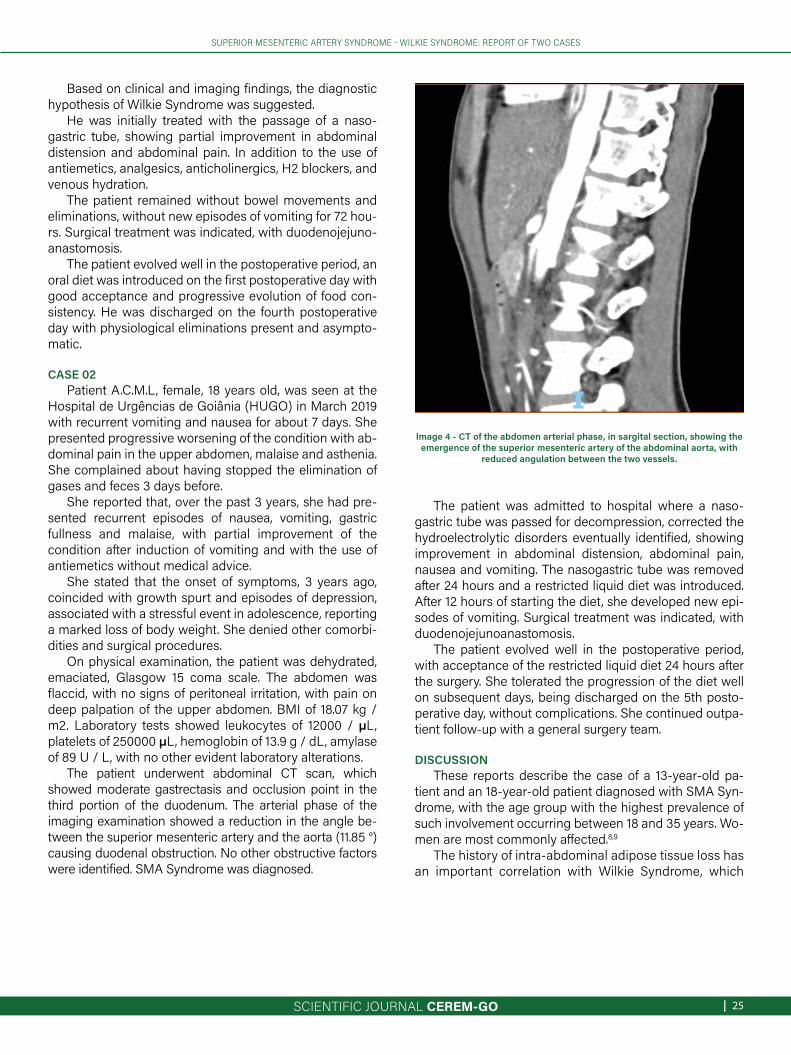
SCIENTIFIC JOURNAL CEREM-GO | 25
SUPERIOR MESENTERIC ARTERY SYNDROME - WILKIE SYNDROME: REPORT OF TWO CASES
Image 4 - CT of the abdomen arterial phase, in sargital section, showing the emergence of the superior mesenteric artery of the abdominal aorta, with
reduced angulation between the two vessels.
Based on clinical and imaging findings, the diagnostic hypothesis of Wilkie Syndrome was suggested.
He was initially treated with the passage of a naso-gastric tube, showing partial improvement in abdominal distension and abdominal pain. In addition to the use of antiemetics, analgesics, anticholinergics, H2 blockers, and venous hydration.
The patient remained without bowel movements and eliminations, without new episodes of vomiting for 72 hou-rs. Surgical treatment was indicated, with duodenojejuno-anastomosis.
The patient evolved well in the postoperative period, an oral diet was introduced on the first postoperative day with good acceptance and progressive evolution of food con-sistency. He was discharged on the fourth postoperative day with physiological eliminations present and asympto-matic.
CASE 02Patient A.C.M.L, female, 18 years old, was seen at the
Hospital de Urgências de Goiânia (HUGO) in March 2019 with recurrent vomiting and nausea for about 7 days. She presented progressive worsening of the condition with ab-dominal pain in the upper abdomen, malaise and asthenia. She complained about having stopped the elimination of gases and feces 3 days before.
She reported that, over the past 3 years, she had pre-sented recurrent episodes of nausea, vomiting, gastric fullness and malaise, with partial improvement of the condition after induction of vomiting and with the use of antiemetics without medical advice.
She stated that the onset of symptoms, 3 years ago, coincided with growth spurt and episodes of depression, associated with a stressful event in adolescence, reporting a marked loss of body weight. She denied other comorbi-dities and surgical procedures.
On physical examination, the patient was dehydrated, emaciated, Glasgow 15 coma scale. The abdomen was flaccid, with no signs of peritoneal irritation, with pain on deep palpation of the upper abdomen. BMI of 18.07 kg / m2. Laboratory tests showed leukocytes of 12000 / µL, platelets of 250000 µL, hemoglobin of 13.9 g / dL, amylase of 89 U / L, with no other evident laboratory alterations.
The patient underwent abdominal CT scan, which showed moderate gastrectasis and occlusion point in the third portion of the duodenum. The arterial phase of the imaging examination showed a reduction in the angle be-tween the superior mesenteric artery and the aorta (11.85 °) causing duodenal obstruction. No other obstructive factors were identified. SMA Syndrome was diagnosed.
The patient was admitted to hospital where a naso-gastric tube was passed for decompression, corrected the hydroelectrolytic disorders eventually identified, showing improvement in abdominal distension, abdominal pain, nausea and vomiting. The nasogastric tube was removed after 24 hours and a restricted liquid diet was introduced. After 12 hours of starting the diet, she developed new epi-sodes of vomiting. Surgical treatment was indicated, with duodenojejunoanastomosis.
The patient evolved well in the postoperative period, with acceptance of the restricted liquid diet 24 hours after the surgery. She tolerated the progression of the diet well on subsequent days, being discharged on the 5th posto-perative day, without complications. She continued outpa-tient follow-up with a general surgery team.
DISCUSSIONThese reports describe the case of a 13-year-old pa-
tient and an 18-year-old patient diagnosed with SMA Syn-drome, with the age group with the highest prevalence of such involvement occurring between 18 and 35 years. Wo-men are most commonly affected.8,9
The history of intra-abdominal adipose tissue loss has an important correlation with Wilkie Syndrome, which
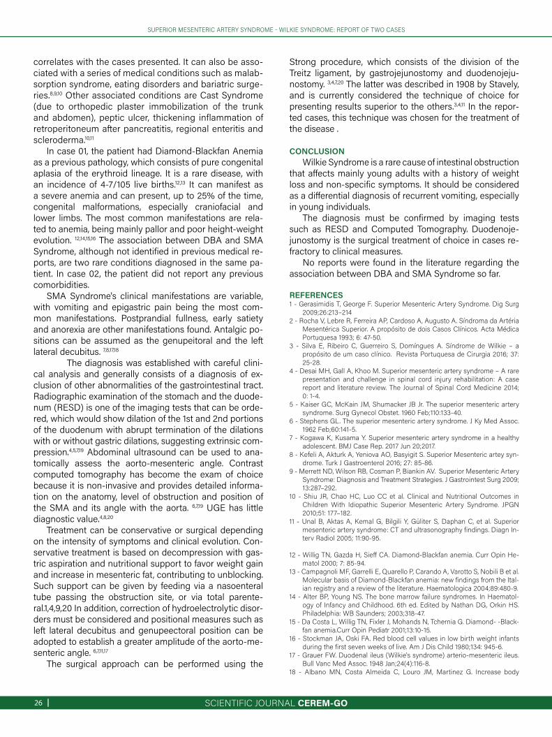
SCIENTIFIC JOURNAL CEREM-GO|26
SUPERIOR MESENTERIC ARTERY SYNDROME - WILKIE SYNDROME: REPORT OF TWO CASES
correlates with the cases presented. It can also be asso-ciated with a series of medical conditions such as malab-sorption syndrome, eating disorders and bariatric surge-ries.8,9,10 Other associated conditions are Cast Syndrome (due to orthopedic plaster immobilization of the trunk and abdomen), peptic ulcer, thickening inflammation of retroperitoneum after pancreatitis, regional enteritis and scleroderma.10,11
In case 01, the patient had Diamond-Blackfan Anemia as a previous pathology, which consists of pure congenital aplasia of the erythroid lineage. It is a rare disease, with an incidence of 4-7/105 live births.12,13 It can manifest as a severe anemia and can present, up to 25% of the time, congenital malformations, especially craniofacial and lower limbs. The most common manifestations are rela-ted to anemia, being mainly pallor and poor height-weight evolution. 12,14,15,16 The association between DBA and SMA Syndrome, although not identified in previous medical re-ports, are two rare conditions diagnosed in the same pa-tient. In case 02, the patient did not report any previous comorbidities.
SMA Syndrome's clinical manifestations are variable, with vomiting and epigastric pain being the most com-mon manifestations. Postprandial fullness, early satiety and anorexia are other manifestations found. Antalgic po-sitions can be assumed as the genupeitoral and the left lateral decubitus. 7,8,17,18
The diagnosis was established with careful clini-cal analysis and generally consists of a diagnosis of ex-clusion of other abnormalities of the gastrointestinal tract. Radiographic examination of the stomach and the duode-num (RESD) is one of the imaging tests that can be orde-red, which would show dilation of the 1st and 2nd portions of the duodenum with abrupt termination of the dilations with or without gastric dilations, suggesting extrinsic com-pression.4,5,7,19 Abdominal ultrasound can be used to ana-tomically assess the aorto-mesenteric angle. Contrast computed tomography has become the exam of choice because it is non-invasive and provides detailed informa-tion on the anatomy, level of obstruction and position of the SMA and its angle with the aorta. 6,7,19 UGE has little diagnostic value.4,8,20
Treatment can be conservative or surgical depending on the intensity of symptoms and clinical evolution. Con-servative treatment is based on decompression with gas-tric aspiration and nutritional support to favor weight gain and increase in mesenteric fat, contributing to unblocking. Such support can be given by feeding via a nasoenteral tube passing the obstruction site, or via total parente-ral.1,4,9,20 In addition, correction of hydroelectrolytic disor-ders must be considered and positional measures such as left lateral decubitus and genupeectoral position can be adopted to establish a greater amplitude of the aorto-me-senteric angle. 6,7,11,17
The surgical approach can be performed using the
Strong procedure, which consists of the division of the Treitz ligament, by gastrojejunostomy and duodenojeju-nostomy. 3,4,7,20 The latter was described in 1908 by Stavely, and is currently considered the technique of choice for presenting results superior to the others.3,4,11 In the repor-ted cases, this technique was chosen for the treatment of the disease .
CONCLUSIONWilkie Syndrome is a rare cause of intestinal obstruction
that affects mainly young adults with a history of weight loss and non-specific symptoms. It should be considered as a differential diagnosis of recurrent vomiting, especially in young individuals.
The diagnosis must be confirmed by imaging tests such as RESD and Computed Tomography. Duodenoje-junostomy is the surgical treatment of choice in cases re-fractory to clinical measures.
No reports were found in the literature regarding the association between DBA and SMA Syndrome so far.
REFERENCES1 - Gerasimidis T, George F. Superior Mesenteric Artery Syndrome. Dig Surg
2009;26:213–214 2 - Rocha V, Lebre R, Ferreira AP, Cardoso A, Augusto A. Síndroma da Artéria
Mesentérica Superior. A propósito de dois Casos Clínicos. Acta Médica Portuquesa 1993; 6: 47-50.
3 - Silva E, Ribeiro C, Guerreiro S, Domíngues A. Síndrome de Wilkie – a propósito de um caso clínico. Revista Portuquesa de Cirurgia 2016; 37: 25-28.
4 - Desai MH, Gall A, Khoo M. Superior mesenteric artery syndrome – A rare presentation and challenge in spinal cord injury rehabilitation: A case report and literature review. The Journal of Spinal Cord Medicine 2014; 0: 1-4.
5 - Kaiser GC, McKain JM, Shumacker JB Jr. The superior mesenteric artery syndrome. Surg Gynecol Obstet. 1960 Feb;110:133-40.
6 - Stephens GL. The superior mesenteric artery syndrome. J Ky Med Assoc. 1962 Feb;60:141-5.
7 - Kogawa K, Kusama Y. Superior mesenteric artery syndrome in a healthy adolescent. BMJ Case Rep. 2017 Jun 20;2017.
8 - Kefeli A, Akturk A, Yeniova AO, Basyigit S. Superior Mesenteric artey syn-drome. Turk J Gastroenterol 2016; 27: 85-86.
9 - Merrett ND, Wilson RB, Cosman P, Biankin AV. Superior Mesenteric Artery Syndrome: Diagnosis and Treatment Strategies. J Gastrointest Surg 2009; 13:287–292.
10 - Shiu JR, Chao HC, Luo CC et al. Clinical and Nutritional Outcomes in Children With Idiopathic Superior Mesenteric Artery Syndrome. JPGN 2010;51: 177–182.
11 - Unal B, Aktas A, Kemal G, Bilgili Y, Güliter S, Daphan C, et al. Superior mesenteric artery syndrome: CT and ultrasonography findings. Diagn In-terv Radiol 2005; 11:90-95.
12 - Willig TN, Gazda H, Sieff CA. Diamond-Blackfan anemia. Curr Opin He-matol 2000; 7: 85-94.
13 - Campagnoli MF, Garrelli E, Quarello P, Carando A, Varotto S, Nobili B et al. Molecular basis of Diamond-Blackfan anemia: new findings from the Ital-ian registry and a review of the literature. Haematologica 2004;89:480-9.
14 - Alter BP, Young NS. The bone marrow failure syndromes. In Haematol-ogy of Infancy and Childhood. 6th ed. Edited by Nathan DG, Orkin HS. Philadelphia: WB Saunders; 2003;318-47.
15 - Da Costa L, Willig TN, Fixler J, Mohands N, Tchernia G. Diamond- -Black-fan anemia.Curr Opin Pediatr 2001;13:10-15.
16 - Stockman JA, Oski FA. Red blood cell values in low birth weight infants during the first seven weeks of live. Am J Dis Child 1980;134: 945-6.
17 - Grauer FW. Duodenal ileus (Wilkie's syndrome) arterio-mesenteric ileus. Bull Vanc Med Assoc. 1948 Jan;24(4):116-8.
18 - Albano MN, Costa Almeida C, Louro JM, Martinez G. Increase body

SCIENTIFIC JOURNAL CEREM-GO | 27
SUPERIOR MESENTERIC ARTERY SYNDROME - WILKIE SYNDROME: REPORT OF TWO CASES
weight to treat superior mesenteric artery syndrome. BMJ Case Rep. 2017 Jun 2;2017.
19 - Ruiz Padilla FJ, Mostazo Torres J, Vílchez Jaimez M. Significant gastric distension caused by superior mesenteric artery syndrome or Wilkie's syndrome. Gastroenterol Hepatol. 2017 Apr 20.
20 - Salem A, Al Ozaibi L, Nassif SMM, Osman RAGS, Al Abed NM, Badri FM. Superior mesenteric artery syndrome: A diagnosis to be kept in mind (Case report and literature review). Int J Surg Case Rep. 2017;34:84-86.
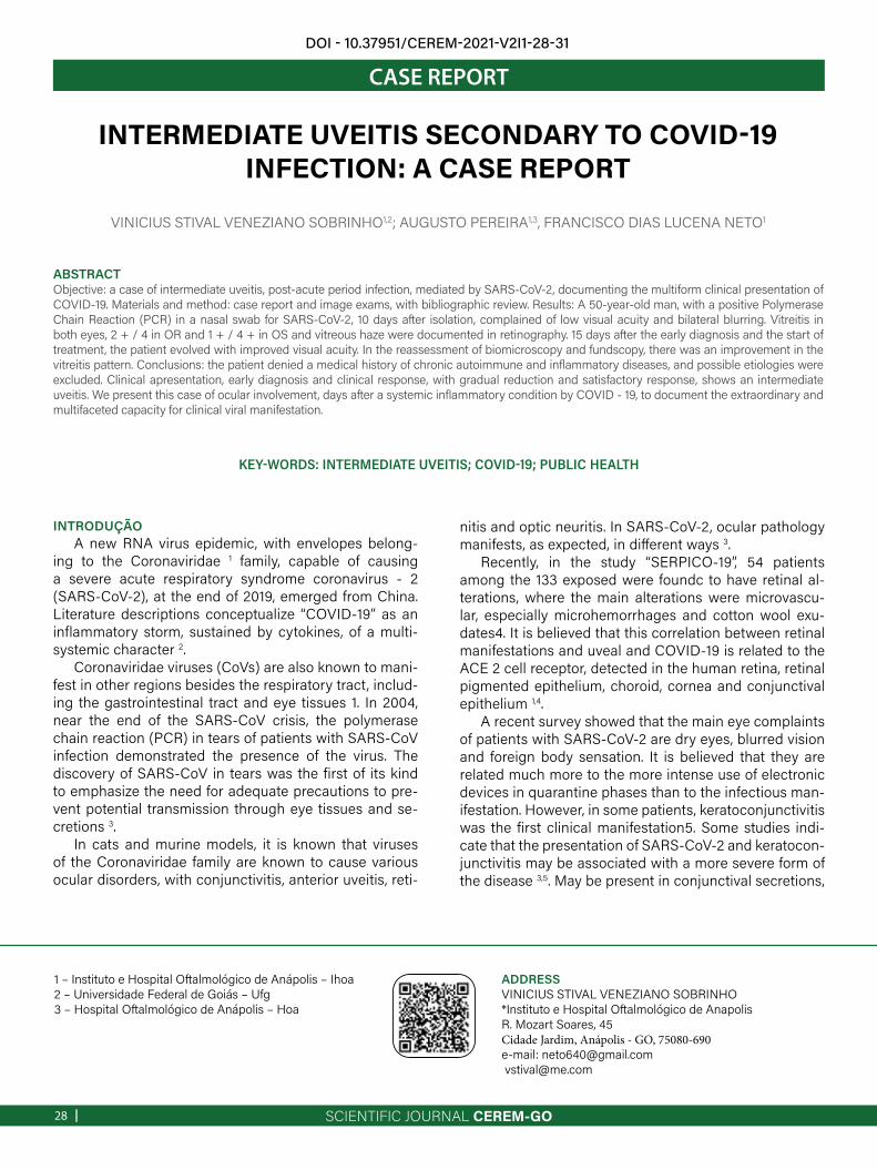
SCIENTIFIC JOURNAL CEREM-GO|28
CASE REPORT
INTERMEDIATE UVEITIS SECONDARY TO COVID-19 INFECTION: A CASE REPORT
VINICIUS STIVAL VENEZIANO SOBRINHO1,2; AUGUSTO PEREIRA1,3, FRANCISCO DIAS LUCENA NETO1
KEY-WORDS: INTERMEDIATE UVEITIS; COVID-19; PUBLIC HEALTH
ABSTRACTObjective: a case of intermediate uveitis, post-acute period infection, mediated by SARS-CoV-2, documenting the multiform clinical presentation of COVID-19. Materials and method: case report and image exams, with bibliographic review. Results: A 50-year-old man, with a positive Polymerase Chain Reaction (PCR) in a nasal swab for SARS-CoV-2, 10 days after isolation, complained of low visual acuity and bilateral blurring. Vitreitis in both eyes, 2 + / 4 in OR and 1 + / 4 + in OS and vitreous haze were documented in retinography. 15 days after the early diagnosis and the start of treatment, the patient evolved with improved visual acuity. In the reassessment of biomicroscopy and fundscopy, there was an improvement in the vitreitis pattern. Conclusions: the patient denied a medical history of chronic autoimmune and inflammatory diseases, and possible etiologies were excluded. Clinical apresentation, early diagnosis and clinical response, with gradual reduction and satisfactory response, shows an intermediate uveitis. We present this case of ocular involvement, days after a systemic inflammatory condition by COVID - 19, to document the extraordinary and multifaceted capacity for clinical viral manifestation.
1 – Instituto e Hospital Oftalmológico de Anápolis – Ihoa2 – Universidade Federal de Goiás – Ufg3 – Hospital Oftalmológico de Anápolis – Hoa
ADDRESSVINICIUS STIVAL VENEZIANO SOBRINHO*Instituto e Hospital Oftalmológico de AnapolisR. Mozart Soares, 45Cidade Jardim, Anápolis - GO, 75080-690e-mail: [email protected] [email protected]
INTRODUÇÃOA new RNA virus epidemic, with envelopes belong-
ing to the Coronaviridae 1 family, capable of causing a severe acute respiratory syndrome coronavirus - 2 (SARS-CoV-2), at the end of 2019, emerged from China. Literature descriptions conceptualize “COVID-19” as an inflammatory storm, sustained by cytokines, of a multi-systemic character 2.
Coronaviridae viruses (CoVs) are also known to mani-fest in other regions besides the respiratory tract, includ-ing the gastrointestinal tract and eye tissues 1. In 2004, near the end of the SARS-CoV crisis, the polymerase chain reaction (PCR) in tears of patients with SARS-CoV infection demonstrated the presence of the virus. The discovery of SARS-CoV in tears was the first of its kind to emphasize the need for adequate precautions to pre-vent potential transmission through eye tissues and se-cretions 3.
In cats and murine models, it is known that viruses of the Coronaviridae family are known to cause various ocular disorders, with conjunctivitis, anterior uveitis, reti-
nitis and optic neuritis. In SARS-CoV-2, ocular pathology manifests, as expected, in different ways 3.
Recently, in the study “SERPICO-19”, 54 patients among the 133 exposed were foundc to have retinal al-terations, where the main alterations were microvascu-lar, especially microhemorrhages and cotton wool exu-dates4. It is believed that this correlation between retinal manifestations and uveal and COVID-19 is related to the ACE 2 cell receptor, detected in the human retina, retinal pigmented epithelium, choroid, cornea and conjunctival epithelium 1,4.
A recent survey showed that the main eye complaints of patients with SARS-CoV-2 are dry eyes, blurred vision and foreign body sensation. It is believed that they are related much more to the more intense use of electronic devices in quarantine phases than to the infectious man-ifestation. However, in some patients, keratoconjunctivitis was the first clinical manifestation5. Some studies indi-cate that the presentation of SARS-CoV-2 and keratocon-junctivitis may be associated with a more severe form of the disease 3,5. May be present in conjunctival secretions,
DOI - 10.37951/CEREM-2021-V2I1-28-31
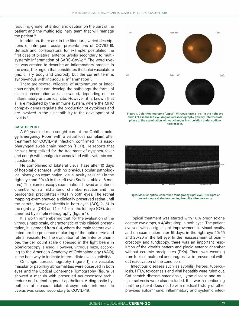
SCIENTIFIC JOURNAL CEREM-GO | 29
INTERMEDIATE UVEITIS SECONDARY TO COVID-19 INFECTION: A CASE REPORT
requiring greater attention and caution on the part of the patient and the multidisciplinary team that will manage the patient 3.
In addition, there are, in the literature, varied descrip-tions of infrequent ocular presentations of COVID-19. Bettach and collaborators, for example, postulated the first case of bilateral anterior uveitis secondary to multi-systemic inflammation of SARS-CoV-2 6. The word uve-itis was created to describe an inflammatory process in the uvea, the region that constitutes the bulbi vasculature (iris, ciliary body and choroid), but the current term is synonymous with intraocular inflammation 7.
There are several etilogies, of autoimmune or infec-tious origin, that can develop the pathology, the forms of clinical presentation are also varied, depending on the inflammatory anatomical site. However, it is known that all are mediated by the immune system, where the MHC complex genes regulate the production of cytokines and are involved in the susceptibility to the development of uveitis 7.
CASE REPORTA 50-year-old man sought care at the Ophthalmolo-
gy Emergency Room with a visual loss complaint after treatment for COVID-19 infection, confirmed in a naso-pharyngeal swab chain reaction (PCR). He reports that he was hospitalized for the treatment of dyspnea, fever and cough with analgesics associated with systemic cor-ticosteroids.
He complained of bilateral visual haze after 10 days of hospital discharge, with no previous ocular patholog-ical history, on examination: visual acuity at 20/50 in the right eye and 20/40 in the left eye (Snellen table at 6 me-ters). The biomicroscopy examination showed an anterior chamber with a mild anterior chamber reaction and fine paracentral precipitates (PKs) in both eyes. The retinal mapping exam showed a clinically preserved retina until the serrata, however vitreitis in both eyes (AO), 2+/4 in the right eye (OD) and 1 + / 4 + in the left eye (OE), doc-umented by simple retinography (figure 1).
It is worth remembering that, for the evaluation of the vitreous haze scale, characteristic of this clinical presen-tation, it is graded from 0-4, where the main factors eval-uated are the presence of blurring of the optic nerve and retinal vessels. For the evaluation of the anterior cham-ber, the cell count scale dispersed in the light beam in biomicroscopy is used. However, vitreous haze, accord-ing to the American Academy of Ophthalmology (AAO), is the best way to indicate intermediate uveitis activity7.
On angiofluoresceinography (figure 1), no vascular, macular or papillary abnormalities were observed in both eyes and the Optical Coherence Tomography (figure 3) showed a macula with preserved neurosensory archi-tecture and retinal pigment epithelium. A diagnostic hy-pothesis of subacute, bilateral, asymmetric intermediate uveitis was raised, secondary to COVID-19.
Figure 1. Color Retinography (upper): Vitreous haze 2+/4+ in the right eye and 1+/4+ in the left eye. Angiofluoresceinography (lower): Intermediate phase of the examination without changes in circulation under sodium
fluorescein.
Fig 2. Macular optical coherence tomography right eye (OD): Spot of posterior optical shadow coming from the vitreous cavity.
Topical treatment was started with 1.0% prednisolone acetate eye drops, a 4/4hrs drop in both eyes. The patient evolved with a significant improvement in visual acuity, and on examination after 15 days: in the right eye 20/25 and 20/20 in the left eye. In the reassessment of biomi-croscopy and fundscopy, there was an important reso-lution of the vitreitis pattern and placid anterior chamber without ceramic precipitates (PKs). There was weaning from topical treatment and progressive improvement with-out reactivation of the condition.
Infectious diseases such as syphilis, herpes, tubercu-losis, HTLV, toxocariasis and viral hepatitis were ruled out. Cat scratch disease, sarcoidosis, Lyme disease and mul-tiple sclerosis were also excluded. It is worth mentioning that the patient does not have a medical history of other previous autoimmune, inflammatory and systemic infec-
SCIENTIFIC JOURNAL CEREM-GO|30
INTERMEDIATE UVEITIS SECONDARY TO COVID-19 INFECTION: A CASE REPORT
tious eye or chronic diseases.
DISCUSSIONIt is known that SRAS-CoV-19 resembles a hyperferrit-
inemic syndrome, in its main stages, with: lymphopenia, reduction in the number and activity of NK lymphocytes, coagulopathy and hyperferritinemia, which demonstrates the great proinflammatory capacity, which induces the ex-pression of different inflammatory mediators, mainly IL-1β 1.
According to Colanfresco et al., despite the numerous etiologies that can develop hyperferritinemic syndrome, they can converge in at least two mechanisms that cause hyperferritinemia: hyperactivation of T lymphocytes and hyperactivity of IFN-γ 1,2. However, recent evidence has described the direct role of the ferritin H chain in the acti-vation of macrophages to increase the secretion of inflam-matory cytokines, evolving with macrophage activation syndrome (MAS), catastrophic antiphospholipid syndrome (cAPS) and septic shock 1.
This pro-inflammatory condition can be found in sever-al observational studies, where the number of autoimmune conditions, such as Kawasaki syndrome, has increased. In children in cities like Paris8, in France, and Bergamo, in It-aly9, the SARS-CoV-2 epidemic has been associated with a high incidence of a severe form of Kawasaki disease, such as children's multisystemic inflammatory syndrome (KDSS) and macrophage activation syndrome (MAS) 8,9.
In a peculiar way, the Kawasaki syndrome is an acute vasculitis of medium-caliber vessels, with systemic de-compensation, with an immunomediated trigger, which often courses with anterior uveitis8,9. The correlation be-tween intraocular inflammations and Kawasaki syndrome is believed to be in the major inflammatory storm present in the pathology, with high levels of IL-6, C-reactive protein and procalcitonin8,9.
There are reports of bilateral acute anterior uveitis (iri-docyclitis), associated with visual haze, associated with a multisystemic inflammatory condition secondary to COVID-19, leading to corneal edema, diffuse descemet folds and keratic precipitates (PKs) in both eyes, with good prognosis after topical and systemic therapeutic follow-up of corticosteroids described in the literature 1,3,5.
Incidentally, intermediate uveitis is a subgroup of uve-itis, where the main site of inflammation is the vitreous, peripheral retina and pars plana, epidemiologically it is not usually associated with sex or race and the involvement tends to be bilateral in 70% of cases . The most frequent initial symptom is the perception of floaters and decreased visual acuity 7.
The eye usually has a lower inflammatory pattern when compared to presentations of anterior uveitis, with mild hyperemia and moderate anterior chamber reaction. Also part of the clinical presentation are small, white, fine ker-atic precipitates, usually in the lower half of the cornea. Vitreitis is the marker of the disease, ranging from mild to
severe, becoming more condensed and classically focal, such as snowballs, are observed during progression 7.
Snowballs are peculiar vitreous infiltrations, containing mononuclear leukocytes and cells similar to fibrocytes, Müller cells and fibrous astrocytes. Apparently, the patho-physiology is related to a disease mediated by T cells, which due to immunotaxis initiated by an unknown antigen, leads to a picture of vasculitis and vitreous inflammation 7.
It is possible that the antigen is infectious because intermediate uveitis is seen in infectious diseases like Lyme, syphilis and cat scratch fever. The disease can be autoimmune, as the pathology is also seen in non-infec-tious diseases, such as multiple sclerosis and sarcoidosis. Collagen type II in the vitreous can be an autoantigen in some patients 7.
HLA associations have been reported in intermediate uveitis, in which HLA-DR is the most significant, occurring in 67-72%. Promising studies correlate Human Leukocyte Antigen (HLA), which are proteins encoded in the main histocompatibility complex, for recognition and immune defenses to COVID-19, which may condition an individu-al more susceptible or more resistant to the inflammatory storm typical of the acute phase of the disease, such as HLA-B*46: 01 and HLA-B*15: 03 10.
In general, the picture of intermediate uveitis is usual-ly benign, where its complications are due to chronicity. Glaucoma, cataracts, macular edema and maculopathy, secondary to intraocular inflammation, are possible com-plications 7. The diagnosis and early therapeutic interven-tion can avoid these aggravations, therefore, it is of funda-mental importance to discuss the clinical and inflammatory presentations, as well as the therapeutic approach of this multisystemic viral condition, in this ongoing Pandemic, and consequently being an important public health issue.
CONCLUSIONIn relation to this COVID – 19 case, it was not possible
to perform the PCR of lacrimal swab or the PCR of vitreous humor, so we cannot say that the uveitis presented was caused by the coronavirus. The good response to early clinical treatment speaks for self-limited subacute inter-mediate uveitis. After excluding other causes and possible etiologies, we considered the presumed diagnosis of inter-mediate uveitis secondary to coronavirus.
The manifestation of intermediate uveitis, in this case reported, occurred shortly after the treatment of acute sys-temic disease by COVID-19. One hypothesis raised is the post-infectious immune-mediated presentation. Another hypothesis raised is that uveitis did not manifest early due to the concomitant use of systemic corticosteroids and that after its suspension, intraocular inflammation was installed.
We report this case of ocular involvement, days after the systemic inflammatory condition by SARS-CoV-2, to document the extraordinary and multifaceted capacity for clinical viral manifestation, as a cause of low visual acuity, in an alarming pandemic scenario.
SCIENTIFIC JOURNAL CEREM-GO | 31
INTERMEDIATE UVEITIS SECONDARY TO COVID-19 INFECTION: A CASE REPORT
REFERENCES1. Huang C, Wang Y, Li X, et al. Clinical features of patients infected with
2019 novel coronavirus in Wuhan, China. Lancet. 2020;395 (10223):497–506.doi:10.1016/S0140-6736(20)30183-5.
2. Colafrancesco S, Alessandri C, Conti F, Priori R. COVID-19 gone bad:a new character in the spectrum of the hyperferritinemic syndrome?. Auto-immun Rev. 2020;19:102573.
3. Dawn Ho, Rebecca Low, Louis Tong, Vishali Gupta, Aravamudan Veer-araghavan & Rupesh Agrawal (2020) COVID-19 and the Ocular Surface: A Review of Transmission and Manifestations, Ocular Immunology and Inflammation, 28:5, 726-734.
4. Invernizzi A, Torre A, Parrulli S, Zicarelli F, Schiuma M, Colombo V, Gia-comelli A, Cigada M, Milazzo L, Ridolfo A, Faggion I, Cordier L, Oldani M, Marini S, Villa P, Rizzardini G, Galli M, Antinori S, Staurenghi G, Meroni L. Retinal findings in patients with COVID-19: Results from the SERPICO-19 study. EClinicalMedicine. 2020 Oct;27:100550.
5. Nuzzi R, Carucci LL, Tripoli F. COVID-19 and ocular implications: an up-date. J Ophthalmic Inflamm Infect. 2020 Sep 4;10(1):20.
6. Bettach, E., Zadok, D., Weill, Y., Brosh, K. and Hanhart, J. (2021), Bilateral anterior uveitis as a part of a multisystem inflammatory syndrome sec-ondary to COVID‐19 infection. J Med Virol, 93: 139-140.
7. Nida Sen H, Albini TA, Burkholder BM, Dahr SS, Dodds EM, Leveque TK, Smith WM, Vasconcelos-Santos AV. Uveitis and ocular inflammation – American Academy of ophthalmology. Basic and Clinical Science Course 2019-2020.
8. Toubiana J, Poirault C, Corsia A, Bajolle F, Fourgeaud J, Angoulvant F, Debray A, Basmaci R, Salvador E, Biscardi S, Frange P, Chalumeau M, Casanova JL, Cohen JF, Allali S. Kawasaki-like multisystem inflammatory syndrome in children during the covid-19 pandemic in Paris, France: pro-spective observational study. BMJ. 2020 Jun 3;369:m2094. doi: 10.1136/bmj.m2094.
9. Verdoni L, Mazza A, Gervasoni A, Martelli L, Ruggeri M, Ciuffreda M, Bonanomi E, D'Antiga L. An outbreak of severe Kawasaki-like disease at the Italian epicentre of the SARS-CoV-2 epidemic: an observational cohort study. Lancet. 2020 Jun 6;395(10239):1771-1778.
10. Iturrieta-Zuazo I, Rita CG, García-Soidán A, et al. Possible role of HLA class-I genotype in SARS-CoV-2 infection and progression: A pilot study in a cohort of Covid-19 Spanish patients. Clin Immunol. 2020;219:108572. doi:10.1016/j.clim.2020.108572.
SCIENTIFIC JOURNAL CEREM-GO|32
CASE REPORT
SYPHILIS IN PREGNANCY AND TRANSMISSION:EPIDEMIOLOGICAL PROFILE
MARIANA CARVALHO DE LIMA GOMES, PROF. LIVRE-DOCENTE, DR. WALDEMAR NAVES DO AMARAL
ABSTRACTINTRODUCTION: Syphilis is a disease caused by the bacterium Treponema Pallidum. In pregnant women, if the disease is not diagnosed and adequately treated, it can reach the fetus, contaminating it and causing repercussions on fetal development. This hematogenous spread is called congenital syphilis. Maternal-fetal transmission occurs more frequently with pregnant women with primary and secondary syphilis. OBJECTIVES: To identify the growth rate of Syphilis in the period from 2017 to 2019. METHODS: Retrospective cross-sectional study RESULTS: In the present study, we found 188 patients reported with syphilis between the years 2017 to 2019, these patients were aged between 15 and 43 years, with a mean 24.6 ± 5.9 years. The number of syphilis cases between the years 2017 to 2019 grew by 85%. Despite this high growth, it was not statistically significant; p = 0411. In the present study, 73.4% of pregnant women with syphilis were brown, 1.6% were black and 7.4% were white. Of the notified patients, 82.3% live in an urban area, 60.6% are single and 47.9% have had prenatal care, 34.6% of the notified pregnant women were in the 3rd trimester. There was no information on the use of alcohol, smoking and drugs to be studied. Regarding the clinical classification, 18.6% were found in primary syphilis and 6.9% in latent syphilis. 78.7% of patients had a reagent treponemic test. 38.3% of pregnant women had VDRL ≥ 1/8. The diagnosis of congenital syphilis was found in 34.6% of cases, abortion in 9% of cases and fetal death in 8% of the entire sample. On the treatment of pregnant women: Penicillin 2.4 million in 17.6%, Penicillin 4.8 million in 27.1% and Penicillin 7.2 million in 12.2%. 7.4% of patients received another treatment regimen, therefore, it is considered inappropriate treatment. Regarding the treatment of the partner, only 12.8% of the partners were proven to be treated. CONCLUSION: Of the patients notified in the HMDI, the majority were young (mean age 24.6 years), brown, single and living in an urban area. Regarding the period of pregnancy, 17 patients were in the first trimester, 5 patients in the second trimester and 65 patients in the third trimester. did prenatal care, there was no information about the presence or absence of prenatal care. The number of syphilis cases between the years 2017 to 2019 grew by 85%, 65 cases of congenital syphilis were diagnosed, 17 cases of abortion and 15 cases of fetal death. The treatment was carried out in 12.8% of the partners, 87.2% of the forms did not contain information about the treatment or not of the partners. 107 pregnant women received treatment with Penicillin G Benzatin.
1 – Hospital e Maternidade Dona Iris 2 – Universidade Federal de Goiás
ADDRESSPATRÍCIA GONÇALVES EVANGELISTAAlameda Emílio Póvoa, 165Vila Redenção, Goiânia - GO, 74845-250 E-mail [email protected]
DOI - 10.37951/CEREM-2021-V2I1-32-35
KEYWORDS: SYPHILIS, PREGNANCY, TRANSMISSION.
INTRODUCTIONSyphilis is a disease caused by the bacterium Trepo-
nema Pallidum. In pregnant women, if the disease is not diagnosed and adequately treated, it can reach the fe-tus, contaminating it and causing repercussions on fetal development. This hematogenous spread is called con-genital syphilis. Maternal-fetal transmission occurs more frequently with pregnant women with primary and sec-ondary syphilis.
Among vertical transmission diseases, syphilis is the one with the highest rates of contamination of the fetus during pregnancy. The prevention of congenital syphilis is done with early diagnosis and treatment during prenatal care. The late diagnosis of syphilis in pregnant women is consid-
ered a risk factor for congenital syphilis, because it can lead to delayed treatment or even failure to undergo treatment during pregnancy. It is known that the use of Penicillin is highly effective in preventing vertical transmission1.
According to the WHO, each year there are 2 million cases of syphilis in pregnant women, and of this total, 25% have not been treated or have not received adequate treat-ment, resulting in spontaneous or stillborn abortions. From the second week of gestational age, Treponema pallidum can infect the fetus and cause spontaneous abruption, as from the sixteenth week of gestation, the bacterium dam-ages the placenta, umbilical cord and several fetal organs, causing prematurity, stillbirth and malformation in new-borns2.
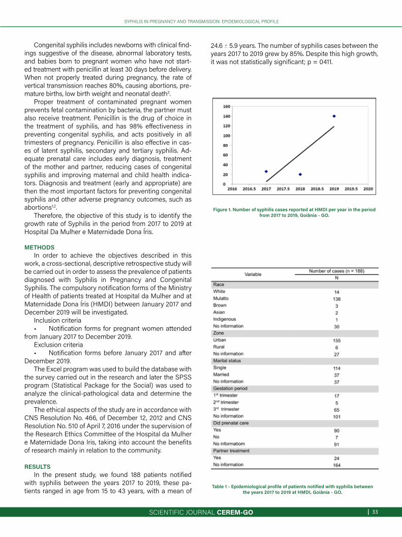
SCIENTIFIC JOURNAL CEREM-GO | 33
SYPHILIS IN PREGNANCY AND TRANSMISSION: EPIDEMIOLOGICAL PROFILE
Congenital syphilis includes newborns with clinical find-ings suggestive of the disease, abnormal laboratory tests, and babies born to pregnant women who have not start-ed treatment with penicillin at least 30 days before delivery. When not properly treated during pregnancy, the rate of vertical transmission reaches 80%, causing abortions, pre-mature births, low birth weight and neonatal death2.
Proper treatment of contaminated pregnant women prevents fetal contamination by bacteria, the partner must also receive treatment. Penicillin is the drug of choice in the treatment of syphilis, and has 98% effectiveness in preventing congenital syphilis, and acts positively in all trimesters of pregnancy. Penicillin is also effective in cas-es of latent syphilis, secondary and tertiary syphilis. Ad-equate prenatal care includes early diagnosis, treatment of the mother and partner, reducing cases of congenital syphilis and improving maternal and child health indica-tors. Diagnosis and treatment (early and appropriate) are then the most important factors for preventing congenital syphilis and other adverse pregnancy outcomes, such as abortions1,2.
Therefore, the objective of this study is to identify the growth rate of Syphilis in the period from 2017 to 2019 at Hospital Da Mulher e Maternidade Dona Íris.
METHODSIn order to achieve the objectives described in this
work, a cross-sectional, descriptive retrospective study will be carried out in order to assess the prevalence of patients diagnosed with Syphilis in Pregnancy and Congenital Syphilis. The compulsory notification forms of the Ministry of Health of patients treated at Hospital da Mulher and at Maternidade Dona Íris (HMDI) between January 2017 and December 2019 will be investigated.
Inclusion criteria• Notification forms for pregnant women attended
from January 2017 to December 2019.Exclusion criteria• Notification forms before January 2017 and after
December 2019.The Excel program was used to build the database with
the survey carried out in the research and later the SPSS program (Statistical Package for the Social) was used to analyze the clinical-pathological data and determine the prevalence.
The ethical aspects of the study are in accordance with CNS Resolution No. 466, of December 12, 2012 and CNS Resolution No. 510 of April 7, 2016 under the supervision of the Research Ethics Committee of the Hospital da Mulher e Maternidade Dona Iris, taking into account the benefits of research mainly in relation to the community.
RESULTS
In the present study, we found 188 patients notified with syphilis between the years 2017 to 2019, these pa-tients ranged in age from 15 to 43 years, with a mean of
24.6 ± 5.9 years. The number of syphilis cases between the years 2017 to 2019 grew by 85%. Despite this high growth, it was not statistically significant; p = 0411.
Figure 1. Number of syphilis cases reported at HMDI per year in the period from 2017 to 2019, Goiânia - GO.
Table 1 - Epidemiological profile of patients notified with syphilis between the years 2017 to 2019 at HMDI, Goiânia - GO.
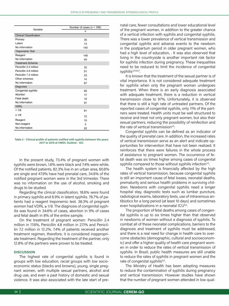
SCIENTIFIC JOURNAL CEREM-GO|34
SYPHILIS IN PREGNANCY AND TRANSMISSION: EPIDEMIOLOGICAL PROFILE
Table 2 - Clinical profile of patients notified with syphilis between the years 2017 to 2019 at HMDI, Goiânia - GO.
In the present study, 73.4% of pregnant women with syphilis were brown, 1.6% were black and 7.4% were white. Of the notified patients, 82.3% live in an urban area, 60.6% are single and 47.9% have had prenatal care, 34.6% of the notified pregnant women were in the 3rd trimester. There was no information on the use of alcohol, smoking and drugs to be studied.
Regarding the clinical classification, 18.6% were found in primary syphilis and 6.9% in latent syphilis. 78.7% of pa-tients had a reagent treponemic test. 38.3% of pregnant women had VDRL ≥ 1/8. The diagnosis of congenital syph-ilis was found in 34.6% of cases, abortion in 9% of cases and fetal death in 8% of the entire sample.
On the treatment of pregnant women: Penicillin 2.4 million in 17.6%, Penicillin 4.8 million in 27.1% and Penicil-lin 7.2 million in 12.2%. 7.4% of patients received another treatment regimen, therefore, it is considered inappropri-ate treatment. Regarding the treatment of the partner, only 12.8% of the partners were proven to be treated.
DISCUSSIONThe highest rate of congenital syphilis is found in
groups with low education, racial groups with low socio-economic status (blacks and browns), young, single preg-nant women, with multiple sexual partners, alcohol and drug use, and even a past history of domestic and sexual violence. It was also associated with the late start of pre-
natal care, fewer consultations and lower educational level of the pregnant woman, in addition to the greater chance of a vertical infection with syphilis and congenital syphilis. There was a lower prevalence of vertical transmission and congenital syphilis and adverse events to the newborn in the postpartum period in older pregnant women, who had a high level of education, . It was also observed that living in the countryside is another important risk factor for syphilis infection during pregnancy. These inequalities need to be reduced to limit the incidence of congenital syphilis3,4,5,6,7.
It is known that the treatment of the sexual partner is of vital importance. It is not considered adequate treatment for syphilis when only the pregnant woman undergoes treatment. When there is an early diagnosis associated with adequate treatment, there is a reduction in vertical transmission close to 97%. Unfortunately, it is observed that there is still a high rate of untreated partners. Of the reported cases of congenital syphilis, only 11% of the part-ners were treated. Health units must be well structured to receive and treat not only pregnant women, but also their sexual partners, reducing the possibility of reinfection and the rate of vertical transmission3,5.
Congenital syphilis can be defined as an indicator of the quality of prenatal care. In addition, the increased rates of vertical transmission serve as an alert and indicate op-portunities for intervention that have not been realized. It reinforces that there were failures in the whole process of assistance to pregnant women. The occurrence of fe-tal death was six times higher among cases of congenital syphilis compared to those without syphilis infection3,5.
The health system is financially affected by the high rates of vertical transmission, because congenital syphilis is still an important cause of fetal losses, neonatal deaths, prematurity and serious health problems in surviving chil-dren. Newborns with congenital syphilis need a longer hospital stay, diagnostic tests such as lumbar puncture, radiological exams, laboratory tests, use of intravenous an-tibiotics for a long period (at least 10 days) and sometimes even hospitalizations in a neonatal ICU5,8.
The proportion of fetal deaths among cases of congen-ital syphilis is up to six times higher than that observed in newborns of women without a diagnosis of syphilis. To combat all of these neonatal complications, barriers to the diagnosis and treatment of syphilis must be addressed, and there is a real need for change in health care to over-come obstacles (demographic, cultural and socioeconom-ic) and offer a higher quality of health care pregnant wom-en in order to reduce the rates of vertical transmission of syphilis. In Brazil, public health measures are still unable to reduce the rates of syphilis in pregnant women and the rate of congenital syphilis5,8.
The Ministry of Health has been adopting measures to reduce the contamination of syphilis during pregnancy and vertical transmission. However studies have shown that the number of pregnant women attended in low qual-
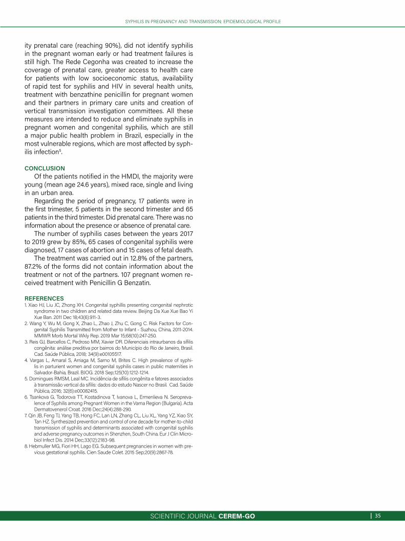
SCIENTIFIC JOURNAL CEREM-GO | 35
SYPHILIS IN PREGNANCY AND TRANSMISSION: EPIDEMIOLOGICAL PROFILE
ity prenatal care (reaching 90%), did not identify syphilis in the pregnant woman early or had treatment failures is still high. The Rede Cegonha was created to increase the coverage of prenatal care, greater access to health care for patients with low socioeconomic status, availability of rapid test for syphilis and HIV in several health units, treatment with benzathine penicillin for pregnant women and their partners in primary care units and creation of vertical transmission investigation committees. All these measures are intended to reduce and eliminate syphilis in pregnant women and congenital syphilis, which are still a major public health problem in Brazil, especially in the most vulnerable regions, which are most affected by syph-ilis infection5.
CONCLUSIONOf the patients notified in the HMDI, the majority were
young (mean age 24.6 years), mixed race, single and living in an urban area.
Regarding the period of pregnancy, 17 patients were in the first trimester, 5 patients in the second trimester and 65 patients in the third trimester. Did prenatal care. There was no information about the presence or absence of prenatal care.
The number of syphilis cases between the years 2017 to 2019 grew by 85%, 65 cases of congenital syphilis were diagnosed, 17 cases of abortion and 15 cases of fetal death.
The treatment was carried out in 12.8% of the partners, 87.2% of the forms did not contain information about the treatment or not of the partners. 107 pregnant women re-ceived treatment with Penicillin G Benzatin.
REFERENCES1. Xiao HJ, Liu JC, Zhong XH. Congenital syphillis presenting congenital nephrotic
syndrome in two children and related data review. Beijing Da Xue Xue Bao Yi Xue Ban. 2011 Dec 18;43(6):911-3.
2. Wang Y, Wu M, Gong X, Zhao L, Zhao J, Zhu C, Gong C. Risk Factors for Con-genital Syphilis Transmitted from Mother to Infant - Suzhou, China, 2011-2014. MMWR Morb Mortal Wkly Rep. 2019 Mar 15;68(10):247-250.
3. Reis GJ, Barcellos C, Pedroso MM, Xavier DR. Diferenciais intraurbanos da sífilis congênita: análise preditiva por bairros do Município do Rio de Janeiro, Brasil. Cad. Saúde Pública, 2018; 34(9):e00105517.
4. Vargas L, Amaral S, Arriaga M, Sarno M, Brites C. High prevalence of syphi-lis in parturient women and congenital syphilis cases in public maternities in Salvador-Bahia, Brazil. BJOG. 2018 Sep;125(10):1212-1214.
5. Domingues RMSM, Leal MC. Incidência de sífilis congênita e fatores associados à transmissão vertical da sífilis: dados do estudo Nascer no Brasil. Cad. Saúde Pública, 2016; 32(6):e00082415.
6. Tsankova G, Todorova TT, Kostadinova T, Ivanova L, Ermenlieva N. Seropreva-lence of Syphilis among Pregnant Women in the Varna Region (Bulgaria). Acta Dermatovenerol Croat. 2016 Dec;24(4):288-290.
7. Qin JB, Feng TJ, Yang TB, Hong FC, Lan LN, Zhang CL, Liu XL, Yang YZ, Xiao SY, Tan HZ. Synthesized prevention and control of one decade for mother-to-child transmission of syphilis and determinants associated with congenital syphilis and adverse pregnancy outcomes in Shenzhen, South China. Eur J Clin Micro-biol Infect Dis. 2014 Dec;33(12):2183-98.
8. Hebmuller MG, Fiori HH, Lago EG. Subsequent pregnancies in women with pre-vious gestational syphilis. Cien Saude Colet. 2015 Sep;20(9):2867-78.
SCIENTIFIC JOURNAL CEREM-GO|36
CASE REPORT
PLACENTAL ACCRETISM: CESAREAN - HISTERECTOMYA SERIES OF CASES
GABRIELLA DE OLIVEIRA FERREIRA, PROF. LIVRE DOCENTE DR. WALDEMAR NAVES DO AMARAL,PATRÍCIA GONÇALVES EVANGELISTA
KEYWORDS: PLACENTAL ACCRETISM, CESAREAN SECTION, HYSTERECTOMY.
ABSTRACTIntroduction: Acretism is the implantation of the abnormal placenta in the uterine wall, it is classified according to the degree of depth. The incidence Introduction: Acretism is the implantation of the abnormal placenta in the uterine wall, it is classified according to the degree of depth. The incidence of accretion increased worldwide in parallel with the increase in cesarean sections, with 1 case for every 533 births. Objective: To evaluate cesarean surgery / hysterectomy (placenta in loco) as a healthy maternal-fetal binomial resolution. Method: case series. Discussion: The best therapeutic proposal in cases of accretism is the planning of cesarean delivery followed by total abdominal hysterectomy (THA). Conservative treatment (maintenance of the uterus leaving the placenta in situ) due to the associated high morbidity and mortality should be considered exceptionally. The patient profiles of the cases fit the risk factors mentioned in the studies. All cases had previous cesarean section and diagnosis of placenta previa; average age: 36.8 years (32-41 years); average parity (gestation): 2.8 (G4-G2). Thus, we are going to meet what the literature cites as the main risk factors. Case 2 was scheduled for cesarean delivery and hypertension. However, during cesarean section, the uterus was preserved and evolved to hemorrhagic shock 4 hours after the end of the procedure, requiring THA in the 2nd period. In cases 1, 3 and 4, cesarean delivery and hypertension were planned without complications. In all cases, the final treatment evolved with hysterectomy, meeting the literature as the best therapy. Final considerations: Good conduct in the face of accretism with prior diagnosis through USG and Doppler, delivery planning in a referral center (reserve of hemoconcentrates and ICU) with an experienced and multidisciplinary team has the power to change the prognosis.
1 – Hospital e Maternidade Dona Iris 2 – Universidade Federal de Goiás
ADDRESSPATRÍCIA GONÇALVES EVANGELISTAAlameda Emílio Póvoa, 165Vila Redenção, Goiânia - GO, 74845-250 E-mail [email protected]
DOI - 10.37951/CEREM-2021-V2I1-36-39
1 INTRODUCTIONThe placenta accreta is defined when the implantation
occurs abnormally in the uterine wall, passing the endometri-um, invading the myometrium, which may become serous or invade other organs1,2.
Normally the chorionic villi penetrate the compact and su-perficial portion of the decidua, and do not reach the spongy layer. This allows the cleavage of the placenta to be detached. Endometrial and myometrial damage are responsible for ab-normal placental implantation, with a thin or absent basal decidua (spongy layer) and imperfect development of the fibrinoid layer (Nitabuch layer) 1,3. The penetration into the spongy layer and the myometrium prevents dequitation and is characteristic of placental accretism1,4.
The ACOG (American Congress of Obstetricians and Gynecologists) reported, in 2012, that the incidence of accretism increased worldwide in parallel with the increase in cesarean sections, with 1 case for every 533 deliveries5. In
1950, the occurrence was very rare, 1 for every 30,000 births6.Early diagnosis is of fundamental importance in this pa-
thology. Pregnant women with previous history of previous cesarean section, placenta previa, multiparity, maternal age greater than 35 years, endometrial defects have an increased risk for accretism. Thus, ultrasonography (USG) should be requested to assess the placenta, as it is a great diagnostic method. When the USG is not clear, magnetic resonance im-aging (MRI) 1.5 can be requested.
Accretism has a high mortality rate of 6 to 7%, the main complication of which is hemorrhagic shock, which can ag-gravate the clinical picture and develop with disseminated in-travascular coagulation (DIC), adult respiratory distress syn-drome, renal failure and even maternal-fetal death1, 5.7.8.
The incidence of placental accretism is on the rise and birth planning with cesarean section and total abdominal hysterectomy (TAH) through previous diagnosis has the power to change this disease's prognosis.
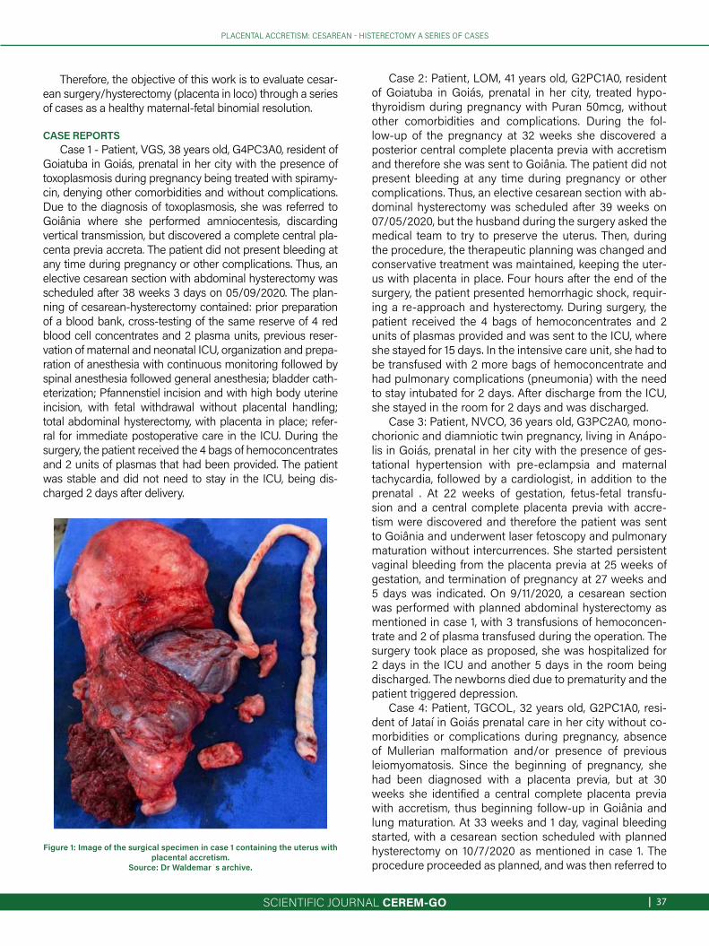
SCIENTIFIC JOURNAL CEREM-GO | 37
PLACENTAL ACCRETISM: CESAREAN - HISTERECTOMY A SERIES OF CASES
Figure 1: Image of the surgical specimen in case 1 containing the uterus with placental accretism.
Source: Dr Waldemar´s archive.
Therefore, the objective of this work is to evaluate cesar-ean surgery/hysterectomy (placenta in loco) through a series of cases as a healthy maternal-fetal binomial resolution.
CASE REPORTSCase 1 - Patient, VGS, 38 years old, G4PC3A0, resident of
Goiatuba in Goiás, prenatal in her city with the presence of toxoplasmosis during pregnancy being treated with spiramy-cin, denying other comorbidities and without complications. Due to the diagnosis of toxoplasmosis, she was referred to Goiânia where she performed amniocentesis, discarding vertical transmission, but discovered a complete central pla-centa previa accreta. The patient did not present bleeding at any time during pregnancy or other complications. Thus, an elective cesarean section with abdominal hysterectomy was scheduled after 38 weeks 3 days on 05/09/2020. The plan-ning of cesarean-hysterectomy contained: prior preparation of a blood bank, cross-testing of the same reserve of 4 red blood cell concentrates and 2 plasma units, previous reser-vation of maternal and neonatal ICU, organization and prepa-ration of anesthesia with continuous monitoring followed by spinal anesthesia followed general anesthesia; bladder cath-eterization; Pfannenstiel incision and with high body uterine incision, with fetal withdrawal without placental handling; total abdominal hysterectomy, with placenta in place; refer-ral for immediate postoperative care in the ICU. During the surgery, the patient received the 4 bags of hemoconcentrates and 2 units of plasmas that had been provided. The patient was stable and did not need to stay in the ICU, being dis-charged 2 days after delivery.
Case 2: Patient, LOM, 41 years old, G2PC1A0, resident of Goiatuba in Goiás, prenatal in her city, treated hypo-thyroidism during pregnancy with Puran 50mcg, without other comorbidities and complications. During the fol-low-up of the pregnancy at 32 weeks she discovered a posterior central complete placenta previa with accretism and therefore she was sent to Goiânia. The patient did not present bleeding at any time during pregnancy or other complications. Thus, an elective cesarean section with ab-dominal hysterectomy was scheduled after 39 weeks on 07/05/2020, but the husband during the surgery asked the medical team to try to preserve the uterus. Then, during the procedure, the therapeutic planning was changed and conservative treatment was maintained, keeping the uter-us with placenta in place. Four hours after the end of the surgery, the patient presented hemorrhagic shock, requir-ing a re-approach and hysterectomy. During surgery, the patient received the 4 bags of hemoconcentrates and 2 units of plasmas provided and was sent to the ICU, where she stayed for 15 days. In the intensive care unit, she had to be transfused with 2 more bags of hemoconcentrate and had pulmonary complications (pneumonia) with the need to stay intubated for 2 days. After discharge from the ICU, she stayed in the room for 2 days and was discharged.
Case 3: Patient, NVCO, 36 years old, G3PC2A0, mono-chorionic and diamniotic twin pregnancy, living in Anápo-lis in Goiás, prenatal in her city with the presence of ges-tational hypertension with pre-eclampsia and maternal tachycardia, followed by a cardiologist, in addition to the prenatal . At 22 weeks of gestation, fetus-fetal transfu-sion and a central complete placenta previa with accre-tism were discovered and therefore the patient was sent to Goiânia and underwent laser fetoscopy and pulmonary maturation without intercurrences. She started persistent vaginal bleeding from the placenta previa at 25 weeks of gestation, and termination of pregnancy at 27 weeks and 5 days was indicated. On 9/11/2020, a cesarean section was performed with planned abdominal hysterectomy as mentioned in case 1, with 3 transfusions of hemoconcen-trate and 2 of plasma transfused during the operation. The surgery took place as proposed, she was hospitalized for 2 days in the ICU and another 5 days in the room being discharged. The newborns died due to prematurity and the patient triggered depression.
Case 4: Patient, TGCOL, 32 years old, G2PC1A0, resi-dent of Jataí in Goiás prenatal care in her city without co-morbidities or complications during pregnancy, absence of Mullerian malformation and/or presence of previous leiomyomatosis. Since the beginning of pregnancy, she had been diagnosed with a placenta previa, but at 30 weeks she identified a central complete placenta previa with accretism, thus beginning follow-up in Goiânia and lung maturation. At 33 weeks and 1 day, vaginal bleeding started, with a cesarean section scheduled with planned hysterectomy on 10/7/2020 as mentioned in case 1. The procedure proceeded as planned, and was then referred to
SCIENTIFIC JOURNAL CEREM-GO|38
PLACENTAL ACCRETISM: CESAREAN - HISTERECTOMY A SERIES OF CASES
the ICU. She was admitted to the ICU for 2 days and was discharged 1 day later in good general condition.
DISCUSSIONThe obstetrician identifying the risk factors, making the
preoperative diagnosis (ultrasound) and treating the intra-partum appropriately changes the prognosis of placental accretism.
The patient profiles of the cases fit the risk factors men-tioned in the literature. All cases had previous cesarean section and diagnosis of placenta previa; average age: 36.8 years (32-41 years); average parity (gestation): 2.8 (G4-G2). Thus, we are in agreement with what the literature says as risk factors: anterior cesarean section, placenta previa, mul-tiparity and maternal age greater than 35 years.
Ultrasonography (USG) with Doppler favors visualiza-tion by turbulent flow, in addition to the disappearance of the retroplacental hypoechogenic space anterior to the my-ometrium and the appearance of dilated vessels in the my-ometrium itself. USG associated with Doppler has a sensi-tivity of 81.1% and specificity of 98.9%. However, if analyzing the anterior and posterior placentas separately, 89.7% and 50% detection rate are observed respectively8,9.
In this work, in all presented cases, the patients under-went Doppler ultrasonography, having previously been di-agnosed with placental accretism, enabling birth planning.
The best therapeutic proposal in suspected and con-firmed cases of accretion is the planning of cesarean deliv-ery followed by total abdominal hysterectomy1,10,11.
Total abdominal hysterectomy is the ideal treatment for cases of placental accretism; after extraction of the fetus, it must be performed with the placenta in situ, as attempts at detachment often result in severe hemorrhage1.
Peripartum hysterectomy is the best option for those who have no desire to gestate10,11.
The doctor has to advise pregnant women and their families about the pre, intra and postoperative risks (blood transfusion, organ damage, ICU, infection and risk of death). The procedure with a well-trained team in a place of reference (blood components reserve, ICU) is of ex-treme importance5.
In cases 1, 3 and 4, cesarean deliveries were planned followed by abdominal hysterectomy, in no case did we have complications and intercurrences. Reaffirming that the planning (pre, intra and postoperative) of cesarean section with TAH improves the prognosis of placental ac-cretism and goes against the studies.
Conservative conduct in accretism (leaving the placen-ta in situ) can be chosen in rare situations with a view of preserving fertility; however, these patients must remain under strict surveillance and receive information about a significant risk of serious complications1.
Conservative treatment (maintenance of the uterus leav-ing the placenta in situ) due to the associated high morbidi-ty and mortality should be considered exceptionally12.
In case 2, a cesarean delivery was scheduled with ab-
dominal hysterectomy (TAH), but during the procedure it was decided to preserve the uterus with placenta in locu. The patient evolved with hemorrhagic shock 4 hours after the end of the surgery, requiring an emergency hysterecto-my in the second stage. Conservative treatment should be left as an exception, as it has a high risk of complications.
In all cases reported, the final treatment evolved with TAH, in line with the literature as the best treatment. Con-servative treatment (maintenance of the uterus leaving the placenta in situ) exposes the patient to many complica-tions and should be chosen in rare cases after exhaustive medical guidance for pregnant women and their families.
The ideal gestational age (GA) for the intervention is still controversial. There is an agreement that should be between 34 to 37 weeks due to pulmonary maturation10. For Zugaib (2016) an elective cesarean section with 36/37 weeks is recommended for patients with an early diagno-sis to reduce the complication rate1.
The average gestational age of termination of preg-nancy in all cases was 34weeks (w) 6 days (d) (27w6d - 39w0d). In case 3, it was a monorionic and diamniotic twin pregnancy with fetus-fetal transfusion. It was interrupted with 27 weeks and 5 days due to vaginal bleeding, the fe-tuses were born alive, but did not survive due to prematu-rity. In all other cases, all newborns survived.
FINAL CONSIDERATIONS:Placental accretism is a pathology with high mortality,
but good conduct with early diagnosis and delivery plan-ning has the power to improve the prognosis.
Prior diagnosis is of fundamental importance in this pathology. Therefore, pregnant women with history of pre-vious cesarean section, placenta previa, multiparity, ma-ternal age greater than 35 years, endometrial defects have an increased risk for accretism. Anterior cesarean section is considered the most relevant risk factor, associating the greater the number of surgeries, the greater the risk of pla-centa accreta.
Ultrasonography is a great tool for assessing placental pathologies, so if requested during prenatal care for pa-tients with risk factors, early diagnosis will help to sched-ule appropriate treatment.
The planning (pre, intra and postoperative) of delivery with pulmonary maturation, reserve of hemoconcentrates, in a tertiary hospital with maternal and neonatal ICU and an experienced multidisciplinary team changes the prog-nosis of placental accretism, improving maternal-fetal sur-vival.
REFERENCES 1. Zugaib M. Zugaib Obstetrícia. 3a Ed. São Paulo: Manole, 2016.2. Benirschke K, Kaufmann P. Pathology of the human placenta. 4a Ed. New York:
Springer, 2000.3. Linhares L et al. Placenta acreta: Artigo de revisão. Revista Médica de Minas
Gerais, 2010; 20(1):57-59.4. Zugaib M, Bittar R. Placenta prévia: Protocolos Assistenciais da Clínica Obsté-
trica da FMUSP. 4ª Ed. São Paulo: Atheneu; 2014.5. Fernandes C, Sá M. Tratado de Obstetrícia da FEBRASGO. São Paulo: Elsevier,

SCIENTIFIC JOURNAL CEREM-GO | 39
PLACENTAL ACCRETISM: CESAREAN - HISTERECTOMY A SERIES OF CASES
2019.6. Read JA; Cotton, D. B; Miller, F. C. Placenta accreta: changing clinical aspects
and outcome. The American College of Obstetricians and Gynecologists, 1980;56(1): 31-4.
7. Rezende J, Montenegro C. Rezende Obstetrícia. 13ª Ed. Rio de Janeiro: Guanabara Koogan, 2017.
8. Falone V et al.; Acretismo placentário – predição ecográfica: relato de caso. Re-vista Brasileira de Ultrassonografia, 2019;27(1):30-32.
9. Freitas F, Costa S, Ramos J et al. Rotinas em Obstetrícia. 6ª Ed. Porto Alegre: Artmed, 2011.
10. Freitas R, Ayres-De-Campos D. Management of placenta percreta. Acta Obstet Ginecol Port., 2015;9(3):250-255.
11. Baggieri R et al. Hemorragia pós-parto: prevenção e tratamento. Arquivos Médi-cos dos Hospitais e da Faculdade de Ciências Médicas da Santa Casa São Paulo, 2011; 56(2): 96-101.
12. Clode N. Acretismo Placentário: uma realidade que não podemos ignorar. Acta Obstet Ginecol Port, 2019;13(3):146-147.
SCIENTIFIC JOURNAL CEREM-GO|40
REVIEW ARTICLE
SARCOMAS AND OTHER NON-EPITHELIAL BREAST TUMORS: A LITERATURE REVIEW
ANDRÉ MAROCCOLO DE SOUSA1 , ANA LUÍZA FLEURY LUCIANO1, LEANDRO GONÇALVES OLIVEIRA2,RAFAEL MAZON CORANDIN3 , MARCELLA SEGATO DE SOUSA MELO3, SEBASTIÃO ALVES PINTO 2,4,
JUAREZ ANTÔNIO DE SOUSA3,4
KEY-WORDS: BREAST CANCER; SARCOMA; MALIGNANT PHYLLODES TUMOR
ABSTRACTNon-epithelial malignant breast diseases are responsible for about 1% of all breast tumors. The most common primary non-epithelial breast cancers are sarcomas and lymphomas. Among sarcomas, the malignant phyllodes tumor is the most common. The macroscopic aspect of sarcomas shows tissue with a firm consistency, ranging from bronze to gray, with soft, cystic and other hemorrhagic areas. Histologically, these tumors are hypercellular, proliferation in fibroblastic spindles, atypical cells and highly anaplastic of intermediate to high degree. Histological grading is important for treatment and prognosis. Hematogenous dissemination is the most common and the involvement of axillary lymph nodes is not frequent among sarcomas. The most frequent sites of metastasis are the lungs, bones and liver. The treatment for primary breast sarcomas is wide excision with adequate and free margins. Lymphadenectomy is not recommended unless there are clinically suspect lymph nodes. Radiotherapy and chemotherapy can be considered, but they have an uncertain role.
1. PUC Goiás.2. INGOH.3. Maternidade Municipal Aristina Cândida.4. Universidade Federal de Goiás.
ADDRESSJUAREZ ANTÔNIO DE SOUSARua 95, 159 setor sul Goiânia GoiásE-mail - [email protected]
DOI - 10.37951/CEREM-2021-V2I1-40-42
INTRODUCTIONMesenchymal lesions of the breast are a rare group of
benign and malignant lesions. They may appear primarily or be iatrogenically induced. The clinical presentation and epidemiology are confused with that of breast carcinomas, compromising patients of both sexes and in a large age group. The formation of a palpable mass or breast asym-metry is commonly the most frequent presentation. The prognosis and treatment vary extraordinarily depending on the histopathological diagnosis 1.
Mass-forming lesions with favorable biological behav-ior of the breast are represented by nodular fasciitis, benign vascular lesions, pseudoangiomatous stromal hyperplasia, myofibroblastoma, desmoid fibromatosis, inflammatory myofibroblastic tumor and lipoma 2.
Malignant mesenchymal neoplasms of the breast can be primary or secondary. Primary neoplasms are those that appear sporadically and spontaneously in the breast parenchyma, the most frequent of which is Phyllodes Tu-mor of the breast. Secondary neoplasms are those that originate after some type of treatment already instituted in
the mammary gland. The most frequent histological type is angiosarcoma, which may be secondary to radiotherapy or late consequence of lymphedema of the arm or breast 3.
The Phyllodes Tumor of the breast is historically called Cystossarcoma Phyllodes for presenting morphological characteristics of leaf-like growth and with the formation of cysts due to rapid growth with necrosis and cystic degener-ation. It is a neoplasm with variable biological behavior, char-acterized by a mass-forming mesenchymal proliferation with a biphasic element - mesenchyme and epithelium 4.
Despite the infrequent presentation in the breast, there is a group of non-epithelial lesions that include hemato-linfoid lesions and melanoma. These are rare primitive le-sions of the breast, that is, the majority of cases diagnosed in the breast represent systemic diseases with secondary involvement of this organ. Mass-forming breast lesions and metastases from other organs to the breast paren-chyma should be remembered in the differential diagno-sis. The main organs involved in this situation are cancers of the contralateral breast, stomach, colorectal tract, lung and ovary (Table 1) 1.
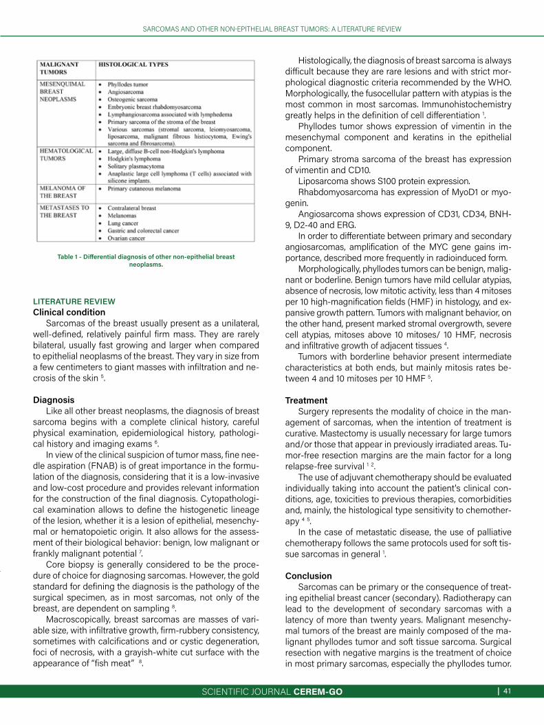
SCIENTIFIC JOURNAL CEREM-GO | 41
SARCOMAS AND OTHER NON-EPITHELIAL BREAST TUMORS: A LITERATURE REVIEW
Table 1 - Differential diagnosis of other non-epithelial breast neoplasms.
LITERATURE REVIEWClinical condition
Sarcomas of the breast usually present as a unilateral, well-defined, relatively painful firm mass. They are rarely bilateral, usually fast growing and larger when compared to epithelial neoplasms of the breast. They vary in size from a few centimeters to giant masses with infiltration and ne-crosis of the skin 5.
DiagnosisLike all other breast neoplasms, the diagnosis of breast
sarcoma begins with a complete clinical history, careful physical examination, epidemiological history, pathologi-cal history and imaging exams 6.
In view of the clinical suspicion of tumor mass, fine nee-dle aspiration (FNAB) is of great importance in the formu-lation of the diagnosis, considering that it is a low-invasive and low-cost procedure and provides relevant information for the construction of the final diagnosis. Cytopathologi-cal examination allows to define the histogenetic lineage of the lesion, whether it is a lesion of epithelial, mesenchy-mal or hematopoietic origin. It also allows for the assess-ment of their biological behavior: benign, low malignant or frankly malignant potential 7.
Core biopsy is generally considered to be the proce-dure of choice for diagnosing sarcomas. However, the gold standard for defining the diagnosis is the pathology of the surgical specimen, as in most sarcomas, not only of the breast, are dependent on sampling 8.
Macroscopically, breast sarcomas are masses of vari-able size, with infiltrative growth, firm-rubbery consistency, sometimes with calcifications and or cystic degeneration, foci of necrosis, with a grayish-white cut surface with the appearance of “fish meat” 8.
Histologically, the diagnosis of breast sarcoma is always difficult because they are rare lesions and with strict mor-phological diagnostic criteria recommended by the WHO. Morphologically, the fusocellular pattern with atypias is the most common in most sarcomas. Immunohistochemistry greatly helps in the definition of cell differentiation 1.
Phyllodes tumor shows expression of vimentin in the mesenchymal component and keratins in the epithelial component.
Primary stroma sarcoma of the breast has expression of vimentin and CD10.
Liposarcoma shows S100 protein expression.Rhabdomyosarcoma has expression of MyoD1 or myo-
genin.Angiosarcoma shows expression of CD31, CD34, BNH-
9, D2-40 and ERG.In order to differentiate between primary and secondary
angiosarcomas, amplification of the MYC gene gains im-portance, described more frequently in radioinduced form.
Morphologically, phyllodes tumors can be benign, malig-nant or boderline. Benign tumors have mild cellular atypias, absence of necrosis, low mitotic activity, less than 4 mitoses per 10 high-magnification fields (HMF) in histology, and ex-pansive growth pattern. Tumors with malignant behavior, on the other hand, present marked stromal overgrowth, severe cell atypias, mitoses above 10 mitoses/ 10 HMF, necrosis and infiltrative growth of adjacent tissues 4.
Tumors with borderline behavior present intermediate characteristics at both ends, but mainly mitosis rates be-tween 4 and 10 mitoses per 10 HMF 5.
TreatmentSurgery represents the modality of choice in the man-
agement of sarcomas, when the intention of treatment is curative. Mastectomy is usually necessary for large tumors and/or those that appear in previously irradiated areas. Tu-mor-free resection margins are the main factor for a long relapse-free survival 1 2.
The use of adjuvant chemotherapy should be evaluated individually taking into account the patient's clinical con-ditions, age, toxicities to previous therapies, comorbidities and, mainly, the histological type sensitivity to chemother-apy 4 5.
In the case of metastatic disease, the use of palliative chemotherapy follows the same protocols used for soft tis-sue sarcomas in general 1.
ConclusionSarcomas can be primary or the consequence of treat-
ing epithelial breast cancer (secondary). Radiotherapy can lead to the development of secondary sarcomas with a latency of more than twenty years. Malignant mesenchy-mal tumors of the breast are mainly composed of the ma-lignant phyllodes tumor and soft tissue sarcoma. Surgical resection with negative margins is the treatment of choice in most primary sarcomas, especially the phyllodes tumor.
SCIENTIFIC JOURNAL CEREM-GO|42
SARCOMAS AND OTHER NON-EPITHELIAL BREAST TUMORS: A LITERATURE REVIEW
REFERENCES 1. Jay R Harris et al. Doenças da Mama. 5. ed. Rio de Janeiro. Di Livros, 2016.2. Bagnoli F et al. Mastologia: do diagnóstico ao tratamento [livro eletrônico]
Goiânia: Conexão Propaganda e Editora, 2017.3. Chen WH, Cheng SP, Tzen CY, Yang TL, Jeng KS, Liu CL, Liu TP. Surgical Treat-
ment of Phyllodes Tumors of the Breast: Retrospective Review of 172 Cases. Taiwan. J Surg Oncol. 2010; vol.102(4), 301-307. ISSN:1096-9098. 2005.
4. Zhao W, Tian Q, Zhao A, Wang B, Yang J, Wang L, Zhang L, Dong D, Chen L, Yang J. The role of adjuvant radiotherapy in patients with malignant phyllodes tumor of the breast: a propensity-score matching analysis. Breast Cancer; 2020; DOI: 10.1007/s12282-020-01135-7. 2020 Aug 03.
5. Rosenberger LH, Thomas SM, Nimbkar SN, Hieken TJ, Ludwig KK, Jacobs LK, Miller ME, Gallagher KK, Wong J, Neuman HB, Tseng J, Hassinger TE, King TA, Jakub JW. Contemporary Multi-Institutional Cohort of 550 Cases of Phyl-lodes Tumors (2007-2017) Demonstrates a Need for More Individualized Mar-gin Guidelines. Journal of Clinical Oncology 2021 39:3, 178-189.
6. Porto e Porto. Semiologia Médica, 8. ed. Rio de Janeiro. Guanabara Koogan, 2019.
7. Boff RA, Carli AC, Brenelli H, Brenelli FP, Carli LS, Reiriz AB, Coelho CP, Coelho GP. Compêndio de Mastologia. Abordagem multidisciplinar. Caxias do Sul: Lorigraf, 2015.
8. Chagas CR, Menke CH, Vieira RJS, Boff RA. Tratado de Mastologia da SBM. Rio de Janeiro: Revinter, 2011.
9. Girão MJBC, Baracat EC, Rodrigues de Lima G. Tratado de Ginecologia. Rio de Janeiro: Atheneu, 2017.
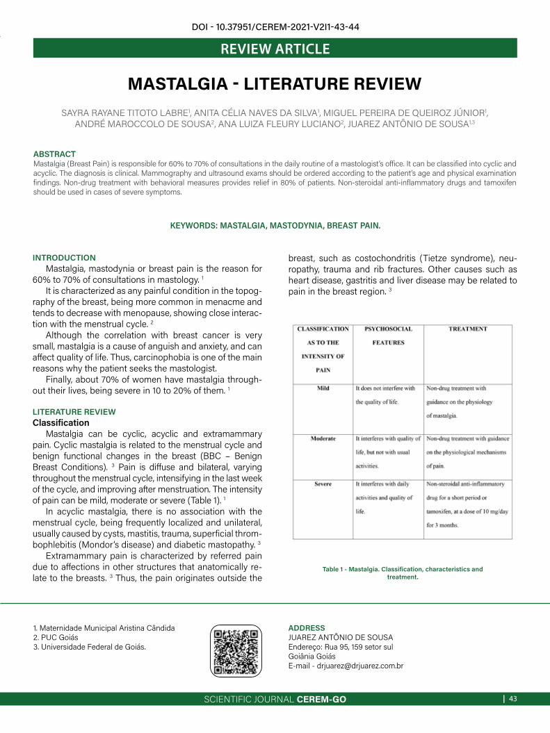
SCIENTIFIC JOURNAL CEREM-GO | 43
REVIEW ARTICLE
MASTALGIA - LITERATURE REVIEWSAYRA RAYANE TITOTO LABRE1, ANITA CÉLIA NAVES DA SILVA1, MIGUEL PEREIRA DE QUEIROZ JÚNIOR1,
ANDRÉ MAROCCOLO DE SOUSA2, ANA LUIZA FLEURY LUCIANO2, JUAREZ ANTÔNIO DE SOUSA1,3
KEYWORDS: MASTALGIA, MASTODYNIA, BREAST PAIN.
ABSTRACT Mastalgia (Breast Pain) is responsible for 60% to 70% of consultations in the daily routine of a mastologist’s office. It can be classified into cyclic and acyclic. The diagnosis is clinical. Mammography and ultrasound exams should be ordered according to the patient’s age and physical examination findings. Non-drug treatment with behavioral measures provides relief in 80% of patients. Non-steroidal anti-inflammatory drugs and tamoxifen should be used in cases of severe symptoms.
1. Maternidade Municipal Aristina Cândida2. PUC Goiás3. Universidade Federal de Goiás.
ADDRESSJUAREZ ANTÔNIO DE SOUSAEndereço: Rua 95, 159 setor sul Goiânia GoiásE-mail - [email protected]
DOI - 10.37951/CEREM-2021-V2I1-43-44
INTRODUCTIONMastalgia, mastodynia or breast pain is the reason for
60% to 70% of consultations in mastology. 1It is characterized as any painful condition in the topog-
raphy of the breast, being more common in menacme and tends to decrease with menopause, showing close interac-tion with the menstrual cycle. 2
Although the correlation with breast cancer is very small, mastalgia is a cause of anguish and anxiety, and can affect quality of life. Thus, carcinophobia is one of the main reasons why the patient seeks the mastologist.
Finally, about 70% of women have mastalgia through-out their lives, being severe in 10 to 20% of them. 1
LITERATURE REVIEWClassification
Mastalgia can be cyclic, acyclic and extramammary pain. Cyclic mastalgia is related to the menstrual cycle and benign functional changes in the breast (BBC – Benign Breast Conditions). 3 Pain is diffuse and bilateral, varying throughout the menstrual cycle, intensifying in the last week of the cycle, and improving after menstruation. The intensity of pain can be mild, moderate or severe (Table 1). 1
In acyclic mastalgia, there is no association with the menstrual cycle, being frequently localized and unilateral, usually caused by cysts, mastitis, trauma, superficial throm-bophlebitis (Mondor’s disease) and diabetic mastopathy. 3
Extramammary pain is characterized by referred pain due to affections in other structures that anatomically re-late to the breasts. 3 Thus, the pain originates outside the
breast, such as costochondritis (Tietze syndrome), neu-ropathy, trauma and rib fractures. Other causes such as heart disease, gastritis and liver disease may be related to pain in the breast region. 3
Table 1 - Mastalgia. Classification, characteristics and treatment.
SCIENTIFIC JOURNAL CEREM-GO|44
MASTALGIA - LITERATURE REVIEW
DIAGNOSISThe diagnosis of mastalgia is made by anamnesis and de-
tailed physical examination, the first step being the differen-tiation between pain originating in the chest wall and breast pain. In the anamnesis, the patient's lifestyle, use of hormonal and non-hormonal medication, work and sports activities, history of trauma, presence of musculoskeletal diseases and psychosocial problems, as well as family history for breast cancer should be assessed. 4
COMPLEMENTARY EXAMSMammography and ultrasound should be requested in
cases of physical examination findings (nodules, suspected papillary effusion and skin changes), especially in patients over 40, family history of breast cancer or if there is any doubt in the physical examination. 5
TREATMENTNon-drug treatment, which is based on guidance on the
physiological mechanisms of breast pain, promotes symptom relief in about 80% of patients (Table 1). Its overriding principle is to listen and reassure the patient. 3 Behavioral measures such as physical activity, a low-lipid diet, weight reduction, anxiety control, abolishing smoking and other habits are im-portant. 2
The use of a correct size bra, with adequate support, has good results in pain relief. In addition, the use of tight bras or metal rods should be avoided, as they compress the chest or the ribs. 2
The initial drug treatment can be done with non-steroidal anti-inflammatory drugs for a period of three to five days, es-pecially in cases of musculoskeletal pain that radiate to the breasts. Tamoxifen can be used at a dose of 10 mg/day, for three months, in cases of severe mastalgia. 6
Other drugs such as gamma linoleic acid, evening primro-se oil, vitamin E, and diuretics have no scientific evidence of effectiveness. 7 In addition, drugs, such as bromoergocriptine, lisuride, danazol, GnRH analogs, are cited in the literature as effective in the treatment of mastalgia, however, due to their side effects, they are in disuse in the medical practice. 6
CONCLUSIONMastalgia is the most common complaint in the daily rou-
tine of a mastologist. It is often motivated by carcinophobia, since it generates a lot of anxiety in the patient. However, the patient should not be overlooked, but should be properly re-assured.
There is a consensus in the literature that the most efficient measures for mastalgia are general guidelines and behavioral measures, as they improve 80% of cases.
When these measures are not sufficient, the use of non--steroidal anti-inflammatory drugs in patients with localized pain is considered the first-line treatment and tamoxifen can be used in refractory cases.
Gamalinoleic acid, evening primrose oil, vitamin E and diuretics do not have scientific proof of effectiveness in the
treatment of mastalgia, however, they are widely used in cli-nical practice.
REFERENCES1. Fernandes CE, de Sá MFS, Filho AL da S. Tratado de Ginecologia Febrasgo. 1st
ed. Elsevier; 2018. 1024 p. 2. Menke CH, Chagas CR, Vieira RJS. Tratado de Mastologia da SBM. Rio de
Janeiro: Revinter; 2015. 1632 p. 3. Boff RA, Carli AC De, Brenelli FP, Brenelli H, de Carli LS, Sauer FZ, et al.
Compêndio de Mastologia: Abordagem multidisciplinar. 1st ed. Lemar, editor. Caxias do Sul; 2015. 754 p.
4. Porto CC. Semiologia Médica. 5th ed. Rio de Janeiro: Guanabara Koogan; 2005.
5. Girão MJBC, Baracat EC, Lima GR de, Nazário ACP, Facina G, Sartori MGF, et al. Tratado de ginecologia. Rio de Janeiro: Atheneu; 2017.
6. Bagnoli F, Brenelli FP, Pedrini JL, Júnior R de F, de Oliveira VM. Mastologia: do diagnóstico ao tratamento. Goiânia: Conexão Propaganda e Editora; 2017.
7. Harris JR, Lippman ME, Morrow M, Osborne CK. Doenças da mama. 5th ed. Rio de Janeiro: Di Livros; 2016.

SCIENTIFIC JOURNAL CEREM-GO | 45
ASSOCIAÇÃO GOIANA DE RESIDÊNCIA MÉDICA - AGRM