issn 0102–3616
TRANSCRIPT

ISSN 0102–3616
VOLUME 56 • N° 3 • MAY/JUNE 2021
Pitcher Shoulder: Update Article
Current Options in Tendon Transfers for Irreparable Posterosuperior Rotator Cuff Tears
Rotator Cuff Healing
Shoulder Injury after Vaccination: A Systematic Review
Incidence and Risk Factors of the Complications Related to the Latarjet Surgery
Return to Sports After High Tibial Osteotomy Using the Opening Wedge Technique
Functional Outcome of Patients Undergoing Knee Arthrodesis after Infected Total Arthroplasty
Evaluation of the CTX-II Biomarker in Patients with Anterior Cruciate Ligament Tear: Pilot Study
Larger Chondral Lesions Treated with Collagen Membrane – Matrix-Induced Autologous Chondrogenesis – Show Larger Increase in Clinical Scores
Diagnostic Failure Rate in Detecting Perilunate Carpal Fractures and Dislocations Using Plain Wrist X-Rays
Onset of Trigger Finger after Carpal Tunnel Syndrome Surgery: Assessment of Open and Endoscopic Techniques
Serum Levels of Vitamin D in Children with or without Isolated Distal Radius Fractures: A Prospective Clinical Study
Electrodiagnostic Testing Characteristics of Diabetic People with Carpal Tunnel Syndrome
Unmet Needs of Surgical Care for Children: A Case Study in the Brazilian Publicly-Financed Health System
Modifi able Risk Factors of Plantar Fasciitis in Non-Athletic Patients and Proposal of a New Objective Assessment System – RKISP
Evaluation of the Reproducibility of Lauge-Hansen, Danis-Weber and AO Classifi cations for Ankle Fractures
Surgical Gloves in Orthopedic Trauma Procedures: How Many Lose Their Integrity?
Treatment of Distal Radio Vicious Consolidation: Corrective Osteotomy Through 3D Printing Prototyping
Candida Parapsilosis Infection after Lumbosacral Arthrodesisa with a TLIF Intersomatic Fusion Device in PEEK: Case Report
Osteonecrosis of the Intermediate Cuneiform: A Case Report
Missed Tillaux Fracture and Syndesmosis Injury in Adult: Arthroscopic Assisted Reduction and Fixation
“Obstetric Paralysis: Who is to blame? A systematic literature review” – Galbiatti JA, Cardoso FL, GalbiattiMGP. Rev Bras Ortop 2020;55(2):139-146
Answer to the Letter to the Editor Regarding the Article “Obstetric Paralysis: Who is to Blame? A Systematic Literature Review”
Márcio Ibrahim de Carvalho

Brazilian Orthopaedic Journal
ISSN 0102-3616
Editor in Chief
Sergio Luiz Checchia Faculdade de Ciências Médicas da Santa Casa de Misericórdia, São Paulo, SP, Brazil
Emeritus Publishers
Márcio Ibrahim de CarvalhoBelo Horizonte, MG, Brazil
Donato D´AngeloRio de Janeiro, RJ, Brazil
Carlos GiestaRio de Janeiro, RJ, Brazil
Gilberto Luís CamanhoSão Paulo, SP, Brazil
Associated Editors
João Carlos BellotiDepto. Ortop. Traumatol. Univ. Federalde São Paulo - UNIFESP, São Paulo, SP, Brazil
Marco Antônio Percope de AndradeFac. Med. Univ. Federal de Minas Gerais – UFMG, Belo Horizonte/MG, Brazil
Nilson Roberto SeverinoDepto. Ortop. Traumatol. Sta. Casa de Misericórdia de São Paulo São Paulo, SP, Brazil
Junior Editor
Mauro Emilio Conforto GracitelliDepto. Ortop. Traumatol. FMUSPSão Paulo, SP, Brazil
Editorial Board
Akira IshidaDepto. Ortop. Traumatol. Univ. Federal de São Paulo - UNIFESPSão Paulo, SP, Brazil
Caio Augusto de Souza NeryDepto. Ortop. Traumatol. Univ. Federal de São Paulo - UNIFESPSão Paulo, SP, Brazil
Carlos Roberto SchwartsmannUniversidade Federal de Ciências da Saúde de Porto Alegre, Porto Alegre, RS, Brazil
Cláudio SantiliDepto. Ortop. Traumatol. Sta. Casa Misericórdia de São PauloSão Paulo, SP, Brazil
Fernando BaldyDepto. Ortop. Traumatol. Univ. Federal deSão Paulo - UNIFESPSão Paulo, SP, Brazil
Geraldo Rocha Motta FilhoInstituto Nacional de Traumatologia e Ortopedia - INTO, Rio de Janeiro, RJ, Brazil
Giancarlo PoleselloDepartamento de Ortopedia e Traumatologia da Santa Casa de São Paulo, São Paulo, SP, Brazil
Gildásio de Cerqueira DaltroUniversidade Federal da Bahia (UFBA), Salvador, BA, Brazil
Helton Defi noDepto. Biomec. Med. Ap. Locom. Fac. Med. Rib. Preto, Ribeirão Preto, SP, Brazil
João Antonio Matheus GuimarãesInstituto Nacional de Traumatologia e Ortopedia - INTORio de Janeiro, RJ, Brazil
João Maurício BarrettoDepto. Ortop. Traumatol. Sta. Casa Misericórdia do Rio de Janeiro Rio de Janeiro, RJ, Brazil
José Batista VolponDepto. Biomec. Med. Ap. Locom. Fac. Med. Rib. Preto, Ribeirão Preto, SP, Brazil
José Maurício de Moraes CarmoHospital Universitário Pedro Ernesto da Univ do Est. do Rio de Janeiro, Rio de Janeiro, RJ, Brazil
José Sérgio FrancoDepto. Ortop. Traumatol. Fac. Med. Univ. Federal do Rio de Janeiro, Rj, Brazil
José Soares Hungria NetoFac. Ciências Méd. Sta. Casa de Misericórdia de São PauloSão Paulo, SP, Brazil
Karlos Celso de MesquitaUniversidade do Estado do Rio de Janeiro, Rio de Janeiro, RJ, Brazil
Luis Roberto VialleUniversidade Católica do Paraná, Curitiba, PR, Brazil
Luiz Antonio M. da CunhaUniversidade Federal do Paraná, Curitiba, PR, Brazil
Marcelo Tomanik MercadanteDepartamento de Ortopedia e Traumatologia da Santa Casa de São Paulo, São Paulo, SP, Brazil
Marcos Antonio Almeida MatosEscola Baiana de Medicina e Saúde Pública, Salvador, BA, Brazil
Moisés CohenDepto. Ortop. Traumatol. Univ. Federal de São Paulo - UNIFESP, São Paulo, SP, Brazil
Olavo Pires de CamargoDepto. Ortop. Traumatol. FMUSPSão Paulo, SP, Brazil
Osmar AvanziDepto. Ortop. Traumatol. Sta. Casa de Misericórdia de São Paulo, São Paulo, SP, Brazil
Osmar Pedro Arbix CamargoFac. Ciências Méd. Sta. Casa de Misericórdia de São PauloSão Paulo, SP, Brazil
Osvandré Luiz Canfi eld LechInstituto de Ortopedia e Traumatologia de Passo Fundo, Passo Fundo, RS, Brazil
Patricia Maria de Moraes Barros FucsDepto. Ortop. Traumatol. Sta. Casa de Misericórdia de São Paulo, São Paulo, SP, Brazil
Pedro José LabroniciDepto. Ortop. Traumatol. Prof. Dr. Donato D'Ângelo - Hospital Santa TeresaPetrópolis, RJ, Brazil
Rames Mattar JuniorDepto. Ortop. Traumatol. FMUSP São Paulo, SP, Brazil
Roberto Sérgio Tavares CantoUniversidade Federal de Uberlândia, Uberlândia, MG, Brazil
Sergio ZylbersztejnUniversidade Federal de Ciências da Saúde de Porto Alegre, Porto Alegre, RS, Brazil
Tarcísio Eloy P. de Barros FilhoDepto. Ortop. Traumatol. FMUSPSão Paulo, SP, Brazil
Willian Dias BelangeroUniversidade Estadual de Campinas, Campinas, SP, Brazil
Indexed in PubMed/PubMed Central (2015), SciELO (2007), ScopusTM (2011), LILACS (1992) and affi liated to Associação Brasileira de Editores Científi cos (ABEC).

Brazilian Orthopaedic Journal
ISSN 0102-3616
Board of Directors SBOT – 2021
President
Adalberto Visco (BA)
1st Vice-President
Jorge dos Santos Silva (SP)
2st Vice-President
João Antônio Matheus Guimarães (RJ)
General Secretary
André Pedrinelli (SP)
1st Secretary
Sérgio Luiz Checchia (SP)
2st Secretary
João Antônio Matheus Guimarães (RJ)
1st Treasurer
Reynaldo Jesus Garcia Filho (SP)
2st Treasurer
Marcus Vinicius Galvão do Amaral (RJ)
Communications and Marketing Director
José Antonio Veiga Sanhudo (RS)
Regional Director
Cristiano Magalhães Menezes (MG)
Committee Director
Ricardo Esperidião (GO)

RBOBrazilian Orthopaedic Journal
Volume 56, Number 3/2021
online www.thieme-connect.com/products
Update Articles
Shoulder and Elbow
275 Pitcher Shoulder: Update ArticleBenno Ejnisman, Paulo Henrique Schmidt Lara, Leandro Masini Ribeiro, and Paulo Santoro Belangero
281 Current Options in Tendon Transfers for Irreparable Posterosuperior Rotator Cuff TearsCaio Santos Checchia, Luciana Andrade da Silva, Guilherme do Val Sella, Marcelo Fregoneze, and Alberto Naoki Miyazaki
291 Rotator Cuff HealingIldeu Afonso de Almeida Filho, and Daniel Andrade Coelho
Systematic Review
Shoulder and Elbow
299 Shoulder Injury after Vaccination: A Systematic ReviewPaul J. Cagle Jr
Original Articles
Shoulder and Elbow
307 Incidence and Risk Factors of the Complications Related to the Latarjet SurgeryMarcio Cohen, Raphael Fonseca, Bernardo Gribel, Marcus Vinicius Galvão, Martim Monteiro, and Geraldo Motta Filho
Sport Arthroscopy and Traumatology
313 Return to Sports After High Tibial Osteotomy Using the Opening Wedge TechniqueAlexandre Pedro Nicolini, Eduardo Suñe Christiano, Rene Jorge Abdalla, Moises Cohen, and Rogério Teixeira de Carvalho
Knee
320 Functional Outcome of Patients Undergoing Knee Arthrodesis after Infected Total ArthroplastyThiago Vivacqua, Rui Moraes, João Barretto, Naasson Cavanelas, Rodrigo Albuquerque, and Alan Mozella
326 Evaluation of the CTX-II Biomarker in Patients with Anterior Cruciate Ligament Tear: Pilot StudyAlexandre Pedro Nicolini, Nacime Salomão Barbachan Mansur, Juliana Luporini Dreyfuss, Benno Ejnisman, Moises Cohen, and Diego Costa Astur
333 Larger Chondral Lesions Treated with Collagen Membrane – Matrix-Induced Autologous Chondrogenesis – Show Larger Increase in Clinical ScoresMateus Kenji Christo Miyahira, João Victor Novaretti, Diego Costa Astur, Camila Cohen Kaleka, Joicemar Tarouco Amaro, and Moisés Cohen
Thieme Revinter Publicações Ltda

Hand
340 Diagnostic Failure Rate in Detecting Perilunate Carpal Fractures and Dislocations Using Plain Wrist X-RaysAleixo Abreu Tanure, Fernanda Ruiz de Andrade, Luis Guilherme Rosifini Alves Rezende, Amanda Favaro Cagnolati,Luiz Garcia Mandarano-Filho, and Nilton Mazzer
346 Onset of Trigger Finger after Carpal Tunnel Syndrome Surgery: Assessment of Open and Endoscopic TechniquesMarcela Fernandes, João Carlos Belloti, Aldo Okamura, Jorge Raduan Neto, Rafael Tajiri, Flávio Faloppa, andVinícius Ynoe de Moraes
351 Serum Levels of Vitamin D in Children with or without Isolated Distal Radius Fractures: A Prospective Clinical StudyMehmet Ali Talmac, Mehmer Akif Gorgel, Ali Varol, Semih Ak, Bekir Eray Kilinc, and Hacı Mustafa Ozdemir
356 Electrodiagnostic Testing Characteristics of Diabetic People with Carpal Tunnel SyndromeHenver Ribeiro Paiva Filho, Alex Timóteo Rodrigues Reis, Gabriel Antonio Matos, Valdênia Graças Nascimento Paiva,Elias Felix Oliveira, and Murilo Antônio Rocha
Pediatric Orthopaedics
360 Unmet Needs of Surgical Care for Children: A Case Study in the Brazilian Publicly-Financed Health SystemMario Bressan-Neto, Marina Rosa Filezio, Fabio Ferri-de-Barros, and Helton Luiz Aparecido Defino
Foot and Ankle
368 Modifi able Risk Factors of Plantar Fasciitis in Non-Athletic Patients and Proposal of a New Objective Assessment System – RKISPRanjeet Choudhary and Kishor Kunal
Trauma
372 Evaluation of the Reproducibility of Lauge-Hansen, Danis-Weber and AO Classifi cations for Ankle FracturesLucas Sacramento Ramos, Henrique Mansur Gonçalves, Anderson Freitas, Marcio de Paiva Oliveira, Diogo Marcelino Santos Lima, and Welvis Soares Carmargo
379 Surgical Gloves in Orthopedic Trauma Procedures: How Many Lose Their Integrity?Maurício Pandini Monteiro de Barros, Thales Thiago Ferreira Godoi, Mario Ferretti Filho, Helio Jorge Alchavian Fernandes, and Fernando Baldy dos Reis
Technical Note
Hand
384 Treatment of Distal Radio Vicious Consolidation: Corrective Osteotomy Through 3D Printing PrototypingJoão Carlos Belloti, Bernardo Vaz Peres Alves, Nicola Archetti, Luis Renato Nakachima, Flavio Faloppa, andMarcel Jun Sugawara Tamaoki
Brazilian Orthopaedic Journal Volume 56, Number 3/2021

Case Reports
Column
390 Candida Parapsilosis Infection after Lumbosacral Arthrodesisa with a TLIF Intersomatic Fusion Device in PEEK: Case ReportMarcelo Wajchenberg, Nelson Astur, Michel Kanas, Thiago Zinsly Sampaio Camargo, Sérgio Barsanti Wey, andDélio Eulalio Martins
Oncology
394 Osteonecrosis of the Intermediate Cuneiform: A Case ReportGustavo Araujo Nunes, Matheus Levy Almeida Taveira de Souza, Bruno Maciel Braga, Mateus Martins Marcatti,Fabrício Melo Bertolini, and Otaviano de Oliveira Junior
Foot and Ankle
399 Missed Tillaux Fracture and Syndesmosis Injury in Adult: Arthroscopic Assisted Reduction and FixationNuno Oliveira, Pedro Pinho, Mário Baptista, Daniel Freitas, Pedro Varanda, and Bruno S. Pereira
Letters to the Editor
Pediatric Orthopaedics
403 “Obstetric Paralysis: Who is to blame? A systematic literature review” – Galbiatti JA, Cardoso FL, GalbiattiMGP. Rev Bras Ortop 2020;55(2):139-146Andreas Rehm and Azeem Thahir
405 Answer to the Letter to the Editor Regarding the Article “Obstetric Paralysis: Who is to Blame? A Systematic Literature Review”José Antonio Galbiatti, Fabrício Luz Cardoso, and Marília Gabriela Palacio Galbiatti
Obituary
407 Márcio Ibrahim de CarvalhoLeonardo Silluzio Ferreira and Marcelo Back Sternick
Brazilian Orthopaedic Journal Volume 56, Number 3/2021
Some of the product names, patents, and registered designs referred to in this publication are in fact registered trade marks or proprietary names even though specifi c reference to this fact is not always made in the text. Therefore, the appearance of a name without designation as proprietary is not to be construed as a representation by the Publisher that it is in the public domain.
All rights, including the rights of publication, distribution, and sales, as well as the right to translation, are reserved. No part of this work covered by the copyrights hereon may be reproduced or copied in any form or by any means—graphic, electronic, or mechanical, including photocopying, recording, taping, or information and retrieval systems—without written permission of the Publisher.
Important Note: Medical knowledge is ever-changing. As new research and clinical experience broaden our knowledge, changes in treatment and drug therapy may be required. The authors and editors of the material here-in have consulted sources believed to be reliable in their efforts to provide information that is complete and in accord with the standards accepted at the time of publication. However, in view of the possibility of human er-ror by the authors, editors, or publisher of the work herein, or changes in
medical knowledge, neither the authors, editors, or publisher, nor any other party who has been involved in the preparation of this work, warrants that the information contained here in is in every respect accurate or complete, and they are not responsible for any errors or omissions or for the results obtained from use of such information. Because of rapid advances in the medical sciences, independent verification of diagnoses and drug dosages should be made. Readers are encouraged to confirm the information con-tained herein with other sources. For example, readers are advised to check the product information sheet included in the package of each drug they plan to administer to be certain that the information contained in this publi-cation is accurate and that changes have not been made in the recommended dose or in the contraindications for administration. This recommendation is of particular importance in connection with new or infrequently used drugs.
Although all advertising material is expected to conform to ethical (medical) standards, inclusion in this journal does not constitute a guar-antee or endorsement of the quality or value of such product or of claims made by its manufacturer.
© 2021. Sociedade Brasileira de Ortopedia e Traumatologia. All rights reserved. Revista Brasileira de Ortopedia is published bimonthly by Thieme-Revinter Publicações Ltda., Rua do Matoso, 170, Rio de Janeiro 20270-135, Brazil.
Editorial comments should be sent to [email protected]. Articles may be submitted to this journal on an open-access basis. For further informa-tion, please send an e-mail to [email protected]. The content of this journal is available online at www.thieme-connect.com/products. Visit our Web site at www.thieme.com and the direct link to this journal at www.thieme.com/rbo.
Revista Brasileira de Ortopedia is an official publication of the Brazilian Society of Orthopedics and Traumatology (Sociedade Brasileira de Ortopedia e Traumatologia, SBOT), It is listed in PubMed, PubMed Central, Scopus, SciELO, and LILACS (Literatura Latino-Americana e do Caribe em Ciências da Saúde). Thieme Medical Publishers is a member of the CrossRef initiative.
ISSN 0102-3616
Cover design: © Thieme

Pitcher Shoulder: Update Article�
Ombro do arremessador: Artigo de atualizaçãoBenno Ejnisman1 Paulo Henrique Schmidt Lara1 Leandro Masini Ribeiro1
Paulo Santoro Belangero1
1Shoulder and Elbow Group, Centro de Traumatologia do Esporte,Escola Paulista de Medicina, Universidade Federal de São Paulo, SãoPaulo, SP, Brazil
Rev Bras Ortop 2021;56(3):275–280.
Address for correspondence Paulo Henrique Schmidt Lara, MD,Centro de Traumatologia do Esporte, Escola Paulista de Medicina,Universidade Federal de São Paulo, Rua Estado de Israel, 636, VilaClementino, São Paulo, SP, 04022-001, Brazil(e-mail: [email protected]; [email protected]).
Introduction
Pitchers tend to have shoulder injuries as a result of the highforces to which this joint is submitted during the pitch. Thedynamic stabilizers of the glenohumeral joint include therotator cuff, the scapulothoracic muscles and the long headof the biceps tendon. The static stabilizers static include thebone anatomy, thefibrocartilaginous lip, and the joint capsule.
A single traumatic event can cause an injury; however, it ismore common that the repetitive overload leads to failure ofone or more structures. The act of pitching requires a coordi-nate action that progresses from the tip of the toes to thefingers of the hand. This string of events was describedconceptually as kinetic chain.1 In order for this to workeffectively, a sequential muscular activity is necessary sothat the energy generated in the lower part of the body istransmitted to theupper part and, lastly, to theball.2Thespeedof the ball is determined by the efficiency of this process. Body
Keywords
► shoulder► joint instability► athletic injuries
Abstract Most shoulder injuries occur due to repetitive overhead movements. Before studying thetreatment of these shoulder injuries, it is paramount that health professionals have anunderstanding of the etiology of and the underlyingmechanisms for shoulder pathologies.The act of overhead throwing is an eloquent full-body motion that requires tremendouscoordination from the time of force generation to the end of the pitch. The shoulder is acrucial componentof theupper-bodykinetic chain, as it transmits force created in the lowerbody to the arm and hand to provide velocity and accuracy to the pitch.
Palavras-chave
► ombro► instabilidade articular► traumatismos em
atletas
Resumo A maioria das lesões do ombro ocorre devido aos movimentos repetitivos acima donível da cabeça. Antes de estudar o tratamento dessas lesões, é fundamental que osprofissionais de saúde tenham um entendimento da etiologia e dos mecanismos quecausam essas patologias. O ato do arremesso acima do nível da cabeça exigeconsiderável coordenação de todo o corpo, desde o momento de geração de forçaaté o final do arremesso. O ombro é um componente crucial da cadeia cinética daextremidade superior, por transmitir a força gerada na extremidade inferior para obraço e mão para produzir velocidade e precisão no lançamento da bola.
� Work developed in the Shoulder and Elbow Group, Centro deTraumatologia do Esporte, Escola Paulista de Medicina, Universi-dade Federal de São Paulo, São Paulo, SP, Brazil.
receivedJuly 5, 2019acceptedDecember 5, 2019published onlineMarch 23, 2020
DOI https://doi.org/10.1055/s-0040-1702958.ISSN 0102-3616.
© 2021. Sociedade Brasileira de Ortopedia e Traumatologia. Allrights reserved.This is an open access article published by Thieme under the terms of the
Creative Commons Attribution-NonDerivative-NonCommercial-License,
permitting copying and reproduction so long as the original work is given
appropriate credit. Contents may not be used for commercial purposes, or
adapted, remixed, transformed or built upon. (https://creativecommons.org/
licenses/by-nc-nd/4.0/)
Thieme Revinter Publicações Ltda., Rua do Matoso 170, Rio deJaneiro, RJ, CEP 20270-135, Brazil
THIEME
Update Article 275

rotation and the position of the scapula are key elements inthekinematic chain. Inprofessional pitchers, there is a delicatebalance between mobility of the shoulder and stability. Theshoulderneeds tobemobileenoughsothat theextremepointsof the rotation are achieved and the speed is transmitted to theball; however, at the same time, the shoulder must stay stableso that the humeral head stays within the glenoidal cavity,creatinga stablehub for rotation,which isknownas “thrower’sparadox.”3 At each pitch, the soft tissue envelope that circlesthe shoulder is submitted to a load that is very close to themaximum load supported, which leads to the possibility ofinjury.3 Even though the standards of the injuries in cases ofpitcher shoulder are common and predictable, there still iscontroversy about the exact mechanisms that lead to theseinjuries. Recent biomechanical studies have helped improvethe understanding of the pathogenesis of the injuries inpitchers.4–6 Moreover, quantitative information about thenormal or pathological biomechanics and kinematics havehelped the development of strategies for the prevention andtreatment of injuries, as well as for rehabilitation.7–9
Pitch Kinematics
The pitch was divided into six phases, which usually take lessthan two seconds to occur.10,11 The first 3 phases consist ofpreparation, stride and arm elevation, and take approximately1.5 seconds in total. Although the fourth phase, acceleration,lasts about 0.05 seconds, the highest angular speeds and thegreatest change in rotation occur during this phase.12 The lasttwo phases are deceleration and execution, and, together, theylast approximately 0.35 seconds12 (►Figure 1). As certainlesions occur at certain stages, it is important to determinewhen pain or a problem occur.
The speed of the ball depends on a variety of biomechanicalfactors, but is more directly related to the amount of lateralrotation that the shoulder performs.13 To generate maximumpitch speed in the most efficient way, the lower and upperextremities must work in a synchronized and coordinatedway.Professional pitchers can generate ball speeds that exceed144.8km/h; tocreatesuchaspeed, theshoulder reachesangularspeeds of up to 7 thousand degrees/s.13 After the release of theball, the shoulder of a professional pitcher can be exposed todistracting forces of up to 950N.14 In the deceleration phase,there are compression forces created by the rotator cuff and
deltoid muscles in the 1,090N range, as well as posterior shearforces of up to 400N.14 The anterior part of the capsule resistsapproximately 800N to 1200N in individuals aged 20 to30years.15 Therefore, if compressive forces do not counterbal-ance the high forces of distraction, injuries will occur.15 ThestudybyKibleret al1 largelycontributed to theunderstandingofscapular dynamics, injury prevention and treatment. It isestimated that only half of the kinetic energy transmitted tothe ball comes from the arm and shoulder. The other half isgenerated by the rotation of the trunk and lower limbs, and istransferred to the upper extremity through the scapular joint,making this joint an important, but often neglected, part of thekinetic chain.16 A dynamic analysis of the shoulders during thepitch added to our current knowledge of normal and abnormalfunction and, by demonstratingwhichmuscle groups are activeduring the pitch in each phase, it helpedguide the developmentof prevention and rehabilitation programs.17
Pathogenesis of Lesions
The pitcher’s shoulder is susceptible to injury due to theconvergence of the following factors: attenuation of the con-strictors of the anterior capsule, contracture of the posteriorcapsule, development of scapular dyskinesia, kinetic chainbreakage and repetitive contact of the greater tuberosityand the posterosuperior lip. Each of these factors was evaluat-ed and strategies were suggested for injury prevention.
Previous Capsule LaxityBiomechanical studies have shown that the anterior capsule,particularly the anterior band of the lower glenohumeralligament, is the main restrictor of the anterior humerustranslationwith thearminabductionand lateral rotation.18–20
Therefore, repetitive stress in this area and the pitcher’s desireto reach increasing levels of lateral rotation lead to a laxity ofthe anterior capsule.18–20 Although the assigned causes arecontroversial, pitchers in fact have more passive lateral rota-tion than rotation of the contralateral shoulder.21,22 If the gainin lateral rotation is greater than the loss of medial rotation,there is laxity of the restrictors.21,22 In support of this, a workby Jobe et al23 describes the tensioning of the anterior capsuleas a means for the athlete to resume pitching. Although in thestudy by Jobe et al23 this procedurewas successful for patients(68% of the patients presented excellent results and returned
Fig. 1 Phases of the pitch: (1) preparation; (2) stride; (3) arm elevation; (4) acceleration; (5) deceleration; and (6) execution/finish. Source:Drawing by the author.
Rev Bras Ortop Vol. 56 No. 3/2021 © 2021. Sociedade Brasileira de Ortopedia e Traumatologia. All rights reserved.
Pitcher Shoulder Ejnisman et al.276

to their preinjury levels, and 96% were satisfied with thesurgery), the violation of the subscapularis muscle and theexcessive tension explain why not all patients were able toreturn to their pre-injury levels after the reconstruction.Withthe progression of the anterior laxity, there is increased lateralrotation and increased contact between the back of the cuffand the lip, which facilitates the occurrence of injuries.24
Subsequent Capsule ContractureOver time, pitchers develop decreased medial rotation,especially when measured during abduction.25 It is believedthat this decrease in medial rotation occurs for two reasons.First, the increase in the retroversion of the humerusobserved in pitchers manifests itself with a loss of medialrotation. However, this loss, due tobone remodeling, is accom-panied by a symmetrical gain in lateral rotation.25 Anothermeans of medial-rotation loss is the contracture of the poste-rior capsule. It is believed that the median-rotation deficit ofthe glenohumeral joint occurs as a scar process in response tochronic distracting forces applied to the posterior capsuleduring the performance of the pitch.25 Rotational loss due tocapsular contracture is evident when the median-rotationdeficit of the glenohumeral joint exceeds the one that can beexplainedonlybybone remodeling (more than12°), andwhenthe loss of medial rotation exceeds the increase in lateralrotation compared to the contralateral side.25
Biomechanical Consequences of the Medial RotationDeficit of the Glenohumeral JointCurrent clinical and biomechanical studies26,27 have shownthat themedian-rotationdeficit of theglenohumeral jointmaybe the sentinel event in the pathological cascade that manypitchers go through. The authors found that pitchers who hadsuperior labial lesions had a median rotation deficit of theglenohumeral jointgreater than25°.26,27Evensmalldegreesofmedial-rotation deficit of the glenohumeral joint (such as 5°,for example) put the shoulder at risk of injury and eventualneed for surgery.26,27 The posterosuperior displacement thatoccurs with the median rotation deficit of the glenohumeraljoint is due to posterior and lower capsular contracture, whichdoes not enable the total lateral rotation of the humerus.Therefore, the athlete begins to rotate around a new centerof rotation,which ismoreposteriorandproximal. Essentially, acontracted posteroinferior capsule displaces the humerusmore posteriorly and proximally (►Figure 2).6
Scapular DyskinesiaDyskinesia is a static or dynamic abnormality of the scapularposition. Shoulder pain leads to an inhibition of the lowertrapezius and anterior serratusmuscles, and to a contractureof the upper trapezius and smaller pectoral muscles.28–31
This muscle imbalance leads to a prostration of the scapula.Pitchers with loss of medial rotation due to capsular contrac-ture end up using medial scapular rotation to perform thepitch. Over time, the scapula loses the static restrictors, andprobably overloads the dynamic restrictors, and the scapuladeviates from the midline and moves anteriorly.32 Thomaset al33 demonstrated that the greater the median rotation
deficit, the greater the changes in the position and mobilityof the scapula. They evaluated 43 professional baseballplayers, and, in 22 athletes, deficits greater than 15° werefound, in which there was higher scapular dyskinesia, withstatistical significance. In another study by Thomas et al,34 atemporal relationship was demonstrated between scapulardyskinesia and the medial-rotation deficit of the glenohum-eral joint, in which more experienced baseball players hadgreater deficits, with statistical significance.
Effects of Excessive Scapular ProtractionThere are several biomechanical consequences of a scapulawith excessivemedial protraction or rotation. First, there is aweakness of the rotator cuff. As the rotator cuff complexessentially originates from the scapula, if there is an unstableplatform, these muscles will not function properly.35 Inaddition, increased protraction increases the version of thescapula, leading to anterior destabilization and increasedoverload in the anterior ligaments.36 Excessive protractionalso increases the degree of impact between the posteriorrotator cuff and the posterior region of the glenoid duringabduction and lateral rotation.26 The study by Laudner et al37
evaluated that pitchers diagnosed with pathological internalimpact showed a statistical significant increase in the eleva-tion of the sternoclavicular joint and scapular deviationduring shoulder elevation in the plane of the scapula.
Fig. 2 Posterior and proximal displacement of the humeral head.Source: Drawing by the author.
Rev Bras Ortop Vol. 56 No. 3/2021 © 2021. Sociedade Brasileira de Ortopedia e Traumatologia. All rights reserved.
Pitcher Shoulder Ejnisman et al. 277

Common Pathological Conditions andTreatment Options
Mobility and InstabilityMobility is defined as passivemovement of a joint in a specialdirection or rotation.38,39 Hyperelasticity can be physiologi-cal or pathological, and may predispose to lesions. The termshoulder instability is reserved for the feeling of excessivehumeral head movement in relation to the glenoid, which isusually associatedwith pain or discomfort. Fewpitchers havesymptoms of instability, although the term instability hasbeen used in many studies to describe the syndrome thatoccurs in pitchers. While some degree of hyperelasticity canhelp the athlete compete at a high level in sports involvingpitching, the excess may be responsible for the developmentof certain pathological conditions of the shoulder. This hasbeen called atraumatic instability, which is believed to bedue to the repetitive stress that occurs during pitches.40
Kuhn et al41 coined the term pathological hyperelasticity,which we also believe is a more accurate description of whatis actually happening.
SLAP InjuriesThe superior labral tear from anterior to posterior (SLAP)lesion is an important clinical cause of shoulder pain. Bur-khart and Morgan42 proposed that SLAP injuries in pitchersoccur by the peel-back mechanism, which is defined as anincrease in tension at the origin of the biceps during maxi-mum lateral rotation during the pitch. Laboratory studieshave shown that the long head of the biceps is an importantdynamic restrictor of lateral rotation when the arm isabducted.43 Conservative treatment is recommended ini-tially, and its main objectives are to provide a decrease inpain, a gain in motion arc and a focus on dynamic strength-ening, with an emphasis on the stabilizers of the scapula androtator cuff.42 If this fails, the surgical treatment is indicated,which usually is arthroscopic and varies according tothe degree of the injury.42
Rotator-Cuff InjuriesAbout 62% of the injuries to the pitcher’s rotator cuff are partial-joint injuries.6 These injuries in pitchers are usually foundposterosuperiorly at the junction of the adrenal and infraspinalinsertions.44,45 Physiotherapy should be considered the initialtreatment for partial cuff injuries in pitchers. Simple debride-menthasnot showngood results inpitchers. ThestudybyPayneet al46evaluatedathletes submitted to simpledebridementwhowere divided into two groups (pitchers with traumatic injuriesandnon-traumatic injuries). Inpatientswith traumatic injuries,there was a satisfactory result in 86% of the cases, and 64%returned to the sport. In the pitchers with non-traumaticinjuries, there were satisfactory results in 66% of the cases,and return to the sport in 45% of the cases.
ImpactDifferent types of impact havebeendescribed in the literature,including the classic, subacromial, secondary and internalimpacts.47–52 The internal impact is a pathological phenome-
non inwhich the rotator cuffmeets theposterosuperior aspectof the lip with the shoulder at the maximum degree ofabductionand lateral rotation.53,54Several studieshaveshownthat this type of impact is most likely caused by fatigue of thescapular waist muscles due to lack of conditioning or over-training.55,56 These studies have shown that, during accelera-tion phase of the pitch, the humerusmust be alignedwith thescapular plane. From the moment the muscles becomefatigued, the humerus comes out of the plane of the scapula,which is called hyperangulation, leading to an overload of theanterior capsule.57
General Treatment GuidelinesThe treatment begins with conservative measures. The con-tracture of the posterior capsule should be addressed, and astretching and mobilization program must be carried out.The stretching should isolate the glenohumeral joint so thatscapular compensation isminimized.26 The evaluation of thekinetic chain is essential. Lumbar contracture, weakness ofthe hip abductors and decreased medial-leg rotation shouldbe investigated.26 Scapular dyskinesia, which is commonlypresent, can usually be treated with exercises that helprestore normal scapular mobility. The first step in scapularrehabilitation should focus on neuromuscular reeducation ofthe escaping stabilizing muscles. Strengthening should beinitiated after this phase.26 Strengthening the muscles of therotator cuff should be performed, especially of the infra-spinatus muscle, through lateral rotation exercises withresistance, which protects the rotator cuff from injury.26
The surgical treatment is indicated in cases of failure ofthe conservative treatment. Three to four months of physio-therapy are usually attempted, and the therapy should beprolonged if the athlete presents progressive improvementof the condition.26 Most pitchers, especially younger ones,are able to recover from the moment there is resolution ofthe scapular dyskinesia and the medial-rotation deficit.
Final Considerations
The performance of pitchers is often limited by shoulderinjuries. These problems are complex and, therefore, difficulttomanage. The problems occur as a result of a combination ofmuscle imbalance,muscle fatigue, hyperlaxity of the anteriorcapsule, contracture of the posterior capsule, alteredmechanics of the pitch, scapular dyskinesia, increasedhumeral retroversion, and repetitive microtraumas. As aresult, in pitchers we have observed lesions involving thelip, the joint side of the back of the rotator cuff, and theproximal insertion of the long head of the biceps.
The mechanisms and etiologies of the injuries in pitchersare becoming more well-defined. Although there is contro-versy over what would be the initial event, the typical injurypatterns remain the same.
Before we think about treatment options, it is essential toget a detailed history, and to perform a physical examinationand additional imaging studies to get to the correct diagno-sis. The treatment of shoulder injuries should be initiatedwith a protocol that focuses on restoring the arc of motion,
Rev Bras Ortop Vol. 56 No. 3/2021 © 2021. Sociedade Brasileira de Ortopedia e Traumatologia. All rights reserved.
Pitcher Shoulder Ejnisman et al.278

strengthening and specific stretching to promote stability ofthe scapula, shoulder and core muscles (deep muscles of theabdominal, lumbar and pelvic regions that aim to maintainthe stability of this region). In addition, physicians, physi-otherapists and trainers involved with pitchers should haveextensive understanding of the entire pathophysiologicalcascade that leads to injuries in these athletes.
Conflict of InterestsThe authors have no conflict of interests to declare.
References1 Kibler WB. The role of the scapula in athletic shoulder function.
Am J Sports Med 1998;26(02):325–3372 Hirashima M, Kadota H, Sakurai S, Kudo K, Ohtsuki T. Sequential
muscle activity and its functional role in the upper extremity andtrunk during overarm throwing. J Sports Sci 2002;20(04):301–310
3 Wilk KE, Meister K, Andrews JR. Current concepts in the rehabili-tation of the overhead throwing athlete. Am J SportsMed 2002;30(01):136–151
4 Werner SL, Guido JA Jr, Stewart GW,McNeice RP, VanDyke T, JonesDG. Relationships between throwing mechanics and shoulderdistraction in collegiate baseball pitchers. J Shoulder Elbow Surg2007;16(01):37–42
5 Bakshi N, Freehill MT. The Overhead Athletes Shoulder. SportsMed Arthrosc Rev 2018;26(03):88–94
6 Burkhart SS, Morgan CD, Kibler WB. The disabled throwingshoulder: spectrum of pathology Part I: pathoanatomy andbiomechanics. Arthroscopy 2003;19(04):404–420
7 Mlynarek RA, Lee S, Bedi A. Shoulder Injuries in the OverheadThrowing Athlete. Hand Clin 2017;33(01):19–34
8 Kuhn JE. Throwing, the Shoulder, and Human Evolution. Am JOrthop 2016;45(03):110–114
9 Zaremski JL, Wasser JG, Vincent HK. Mechanisms and Treatmentsfor Shoulder Injuries in Overhead Throwing Athletes. Curr SportsMed Rep 2017;16(03):179–188
10 Meister K. Injuries to the shoulder in the throwing athlete. Partone: Biomechanics/pathophysiology/classification of injury. Am JSports Med 2000;28(02):265–275
11 Lin DJ, Wong TT, Kazam JK. Shoulder Injuries in the Overhead-Throwing Athlete: Epidemiology, Mechanisms of Injury, andImaging Findings. Radiology 2018;286(02):370–387
12 Pappas AM, Zawacki RM, Sullivan TJ. Biomechanics of baseballpitching. A preliminary report. Am J Sports Med 1985;13(04):216–222
13 Dillman CJ, Fleisig GS, Andrews JR. Biomechanics of pitching withemphasis upon shoulder kinematics. J Orthop Sports Phys Ther1993;18(02):402–408
14 Kuhn JE, Lindholm SR, Huston LJ. Failure of the biceps-superiorlabral complex (SLAP lesion) in the throwing athlete: a bio-mechanical model comparing maximal cocking to early deceler-ation. . [abstract]J Shoulder Elbow Surg 2000;9:463
15 Reeves B. Experiments on the tensile strength of the anteriorcapsular structures of the shoulder in man. J Bone Joint Surg Br1968;50(04):858–865
16 Chu SK, Jayabalan P, Kibler WB, Press J. The Kinetic ChainRevisited: New Concepts on Throwing Mechanics and Injury.PM R 2016;8(3, Suppl)S69–S77
17 David G, MagareyME, JonesMA, Dvir Z, Türker KS, SharpeM. EMGand strength correlates of selected shoulder muscles duringrotations of the glenohumeral joint. Clin Biomech (Bristol,Avon) 2000;15(02):95–102
18 O’Connell PW, Nuber GW, Mileski RA, Lautenschlager E. Thecontribution of the glenohumeral ligaments to anterior stabilityof the shoulder joint. Am J Sports Med 1990;18(06):579–584
19 O’Brien SJ, Schwartz RS,Warren RF, Torzilli PA. Capsular restraintsto anterior-posterior motion of the abducted shoulder: a bio-mechanical study. J Shoulder Elbow Surg 1995;4(04):298–308
20 McMahon PJ, Tibone JE, Cawley PW, et al. The anterior band of theinferior glenohumeral ligament: biomechanical properties fromtensile testing in the position of apprehension. J Shoulder ElbowSurg 1998;7(05):467–471
21 Bigliani LU, Codd TP, Connor PM, Levine WN, Littlefield MA,Hershon SJ. Shoulder motion and laxity in the professionalbaseball player. Am J Sports Med 1997;25(05):609–613
22 Brown LP, Niehues SL, Harrah A, Yavorsky P, Hirshman HP. Upperextremity range of motion and isokinetic strength of the internaland external shoulder rotators in major league baseball players.Am J Sports Med 1988;16(06):577–585
23 Jobe FW, Giangarra CE, Kvitne RS, Glousman RE. Anterior capsu-lolabral reconstruction of the shoulder in athletes in overhandsports. Am J Sports Med 1991;19(05):428–434
24 Karduna AR, McClure PW, Michener LA, Sennett B. Dynamicmeasurements of three-dimensional scapular kinematics: a vali-dation study. J Biomech Eng 2001;123(02):184–190
25 Kay J, Kirsch JM, Bakshi N, et al. Humeral Retroversion andCapsuleThickening in the Overhead Throwing Athlete: A SystematicReview. Arthroscopy 2018;34(04):1308–1318
26 Burkhart SS, Morgan CD, KiblerWB. Shoulder injuries in overheadathletes. The “dead arm” revisited. Clin Sports Med 2000;19(01):125–158
27 Johnson JE, Fullmer JA, Nielsen CM, Johnson JK, Moorman CT 3rd.Glenohumeral Internal Rotation Deficit and Injuries: A SystematicReview and Meta-analysis. Orthop J Sports Med 2018;6(05):2325967118773322
28 Kibler WB, McMullen J. Scapular dyskinesis and its relation toshoulder pain. J Am Acad Orthop Surg 2003;11(02):142–151
29 Pink MM, Perry J. Biomechanics of the shoulder. St Louis: Mosby-Year Book; 1996
30 McClure PW, Michener LA, Sennett BJ, Karduna AR. Direct 3-dimensional measurement of scapular kinematics during dynamicmovements in vivo. J Shoulder Elbow Surg 2001;10(03):269–277
31 McQuade KJ, Dawson J, Smidt GL. Scapulothoracic muscle fatigueassociatedwith alterations in scapulohumeral rhythmkinematicsduring maximum resistive shoulder elevation. J Orthop SportsPhys Ther 1998;28(02):74–80
32 PinkMM. Understanding the linkage system of the upper extrem-ity. Sports Med Arthrosc Rev 2001;9(01):52–60
33 Thomas SJ, Swanik KA, Swanik CB, Kelly JD 4th. Internal rotationdeficits affect scapular positioning in baseball players. ClinOrthop Relat Res 2010;468(06):1551–1557
34 Thomas SJ, Swanik KA, Swanik CB, Kelly JD. Internal rotation andscapular position differences: a comparison of collegiate and highschool baseball players. J Athl Train 2010;45(01):44–50
35 Kibler WB, Sciascia A, Dome D. Evaluation of apparent andabsolute supraspinatus strength in patients with shoulder injuryusing the scapular retraction test. Am J Sports Med 2006;34(10):1643–1647
36 Weiser WM, Lee TQ, McMaster WC, McMahon PJ. Effects of simu-lated scapular protraction on anterior glenohumeral stability. Am JSports Med 1999;27(06):801–805
37 LaudnerKG,Myers JB, PasqualeMR,Bradley JP, Lephart SM.Scapulardysfunction in throwers with pathologic internal impingement.J Orthop Sports Phys Ther 2006;36(07):485–494
38 Wilk KE, Andrews JR, Arrigo CA, Keirns MA, Erber DJ. The strengthcharacteristics of internal and external rotator muscles in profes-sional baseball pitchers. Am J Sports Med 1993;21(01):61–66
39 Ryu RKN, Dunbar WH V, Kuhn JE, McFarland EG, Chronopoulos E,Kim TK. Comprehensive evaluation and treatment of the shoulderin the throwing athlete. Arthroscopy 2002;18(09, Suppl 2):70–89
40 DeFroda SF, Goyal D, Patel N, Gupta N, Mulcahey MK. ShoulderInstability in the Overhead Athlete. Curr Sports Med Rep 2018;17(09):308–314
Rev Bras Ortop Vol. 56 No. 3/2021 © 2021. Sociedade Brasileira de Ortopedia e Traumatologia. All rights reserved.
Pitcher Shoulder Ejnisman et al. 279

41 Kuhn JE, Lindholm SR, Huston LJ, Soslowsky LJ, Blasier RB. Failureof the biceps superior labral complex: a cadaveric biomechanicalinvestigation comparing the late cocking and early decelerationpositions of throwing. Arthroscopy 2003;19(04):373–379
42 Burkhart SS, Morgan CD. The peel-back mechanism: its role inproducing and extending posterior type II SLAP lesions and itseffect on SLAP repair rehabilitation. Arthroscopy 1998;14(06):637–640
43 Kuhn JE, Huston LJ, Soslowsky LJ, Shyr Y, Blasier RB. Externalrotation of the glenohumeral joint: ligament restraints andmuscle effects in the neutral and abducted positions. J ShoulderElbow Surg 2005;14(1, Suppl S)39S–48S
44 Walch G, Boileau P, Noel E, Donell ST. Impingement of the deepsurface of the supraspinatus tendon on the posterosuperiorglenoid rim: An arthroscopic study. J Shoulder Elbow Surg1992;1(05):238–245
45 Miniaci A, Mascia AT, Salonen DC, Becker EJ. Magnetic resonanceimaging of the shoulder in asymptomatic professional baseballpitchers. Am J Sports Med 2002;30(01):66–73
46 Payne LZ, Altchek DW, Craig EV, Warren RF. Arthroscopic treat-ment of partial rotator cuff tears in young athletes. A preliminaryreport. Am J Sports Med 1997;25(03):299–305
47 Jobe FW, Kvitne RS, Giangarra CE. Shoulder pain in the overhandor throwing athlete. The relationship of anterior instability androtator cuff impingement. Orthop Rev 1989;18(09):963–975
48 Jobe FW, Jobe CM. Painful athletic injuries of the shoulder. ClinOrthop Relat Res 1983;(173):117–124
49 Warner JJ, Micheli LJ, Arslanian LE, Kennedy J, Kennedy R. Scapulo-thoracicmotion innormal shoulders and shoulderswith glenohum-eral instability and impingement syndrome. A study using Moirétopographic analysis. Clin Orthop Relat Res 1992;(285):191–199
50 Morris AD, Kemp GJ, Frostick SP. Shoulder electromyography inmultidirectional instability. J Shoulder Elbow Surg 2004;13(01):24–29
51 Crockett HC, Gross LB, Wilk KE, et al. Osseous adaptation andrange ofmotion at the glenohumeral joint in professional baseballpitchers. Am J Sports Med 2002;30(01):20–26
52 Bach HG, Goldberg BA. Posterior capsular contracture of theshoulder. J Am Acad Orthop Surg 2006;14(05):265–277
53 Jobe CM. Posterior superior glenoid impingement: expandedspectrum. Arthroscopy 1995;11(05):530–536
54 Walch G, Liotard JP, Boileau P, Noël E. [Postero-superior glenoidimpingement. Another shoulder impingement]. Rev Chir OrthopRepar Appar Mot 1991;77(08):571–574
55 Jobe CM. Superior glenoid impingement. Orthop Clin North Am1997;28(02):137–143
56 Paley KJ, Jobe FW, Pink MM, Kvitne RS, ElAttrache NS. Arthro-scopic findings in the overhand throwing athlete: evidence forposterior internal impingement of the rotator cuff. Arthroscopy2000;16(01):35–40
57 Jobe CM, Pink MM, Jobe FW, Shaffer B. Anterior shoulder insta-bility, impingement, and rotator cuff tear: theories and concepts.In: Jobe FW, editor. Operative techniques in upper extremitysports injuries. St. Louis, MO: Mosby; 1996:164–176
Rev Bras Ortop Vol. 56 No. 3/2021 © 2021. Sociedade Brasileira de Ortopedia e Traumatologia. All rights reserved.
Pitcher Shoulder Ejnisman et al.280

Current Options in Tendon Transfers forIrreparable Posterosuperior Rotator Cuff Tears�
Opções atuais de transferências tendíneas para lesõesposterossuperiores irreparáveis do manguito rotadorCaio Santos Checchia1 Luciana Andrade da Silva1 Guilherme do Val Sella1 Marcelo Fregoneze1
Alberto Naoki Miyazaki1
1Department of Orthopedics and Traumatology, Faculdade deCiências Médicas da Santa Casa de São Paulo, São Paulo, Brazil
Rev Bras Ortop 2021;56(3):281–290.
Address for correspondence Caio Santos Checchia, Grupo de Ombroe Cotovelo, Departamento de Ortopedia e Traumatologia, Faculdadede Ciências Médicas da Santa Casa de São Paulo, Rua Dr. Cesario MotaJunior, 112, Consolação, São Paulo, SP, 01221-020, Brazil(e-mail: [email protected]).
Keywords
► shoulder► rotator cuff injuries► tendon transfers► review
Abstract Massive irreparable posterosuperior rotator-cuff tears are debilitating lesions that usuallyrequire surgical treatment. Even though there is no consensus regarding the best surgicaltechnique, tendinous transfers around the shoulder are the most commonly performedprocedures. The latissimus dorsi tendon remains the most commonly used, but differentmodifications to the original technique have been shown to minimize complications and toimprove functional results and satisfaction. Other techniques, such as the transfer of thelower trapezius tendon, are promising and should be considered, especially for patients withisolated loss of external rotation. The present paper is a literary review regarding tendontransfers for irreparable posterosuperior rotator-cuff tears.
Palavras-chave
► ombro► lesões do manguito
rotador► transferências de
tendão► revisão
Resumo As grandes lesões posterossuperiores irreparáveis do manguito rotador são debilitan-tes e, de modo geral, requerem tratamento cirúrgico. Embora não haja consenso sobreamelhor técnica cirúrgica, as transferências tendíneas no ombro são os procedimentosmais realizados. O tendão do grande dorsal continua a ser o mais utilizado, masdiferentes modificações na técnica original têm minimizado as complicações emelhorado os resultados funcionais e a satisfação com o procedimento. Outrastécnicas, como a transferência do tendão do trapézio inferior, são promissoras edevem ser consideradas, principalmente em pacientes com perda isolada da rotaçãoexterna. Este artigo é uma revisão da literatura a respeito da transferência de tendõespara tratamento das lesões posterossuperiores irreparáveis do manguito rotador.
� Work developed at the Department of Orthopedics and Traumatol-ogy, Faculdade de Ciências Médicas, Santa Casa de São Paulo,Pavilhão “Fernandinho Simonsen”. Director: professor doctor IvanChakkour – São Paulo (SP), Brazil.
receivedJuly 7, 2019acceptedJanuary 10, 2020published onlineMay 29, 2020
DOI https://doi.org/10.1055/s-0040-1709988.ISSN 0102-3616.
© 2021. Sociedade Brasileira de Ortopedia e Traumatologia. Allrights reserved.This is an open access article published by Thieme under the terms of the
Creative Commons Attribution-NonDerivative-NonCommercial-License,
permitting copying and reproduction so long as the original work is given
appropriate credit. Contents may not be used for commercial purposes, or
adapted, remixed, transformed or built upon. (https://creativecommons.org/
licenses/by-nc-nd/4.0/)
Thieme Revinter Publicações Ltda., Rua do Matoso 170, Rio deJaneiro, RJ, CEP 20270-135, Brazil
THIEME
Update Article 281

Introduction
Rotator-cuff-tear (RCT) repairs are among the most commonshoulder surgeries.1However, tendon-to-bone healing is notalways successful or predictable,2 because it depends on avariety of factors, including biomechanical and biologicalones, the latter being influenced by the patient’s age.3 As thepopulation is rapidly aging, the prevalence of failed rotator-cuff repairs is also increasing.1,3 The failure rates of massiveposterosuperior rotator-cuff repairs are reported to rangefrom 21% to as high as 91%.4–6 Failures and complicationsfollowing revisions are reported to be even significantlyhigher.7,8
There is no consensus regarding the definition of irrepa-rable RCTs. That being said, whenever a lesion is determinedto be irreparable, there are many treatment options, startingwith nonsurgical treatments.9 Regarding the surgicaloptions, there is controversy about the best treatment avail-able. Boileau el al10 and Walch et al11 showed satisfactoryresults after tenotomy of the long head of the biceps (LHB)alone in elderly patients. Other procedures have beenreported, including rotator-cuff debridement (with or with-out concomitant suprascapular nerve release),12 partial cuffrepair,13–15 tendon transfers,16–19 and reverse shoulderarthroplasty (RSA).20 The latter, however, is probably notthe best option in younger and physically-active patients,because the longevity of these implants in this population isyet unknown, and also because RSAs can still be used as asalvage procedure after the failure of other techniques. Morerecently, superior shoulder capsular reconstruction21–28 hasbeen advocated, but its reported functional results are intheir infancy,23,29 and longer follow-up periods are neededto confirm its long-term efficacy.30
Despite being relatively difficult surgical procedures andrequiring accurate patient selection, tendon transfers cansignificantly improve the quality of life of the patients.16,18,31
This is especially important for young and physically-activepatients with lesions graded as Hamada et al32 stages 1 and 2(that is, without both glenohumeral arthritis and static proxi-mal humeral head migration), because they may be the onlyfeasible definitive (that is, long-term) treatments available.30
The aim of the present paper is to review the literatureregarding tendon transfers for irreparable posterosuperiorRCTs (that is, the ones involving the supra- and infraspinatustendons), as the posterosuperior is by far the most commontype of RCT.1 In this context, the latissimus dorsi tendon (LDT)–accompanied or not by the teres major tendon (TMT) – is themostcommonly transferred tendon.33Thealternatives includeisolated TMT transfer34–37 and the more recently describedtransfer of the lower trapezius tendon (LTT).38,39
Latissimus-Dorsi-Tendon (LDT) Transfer
The earliest and also most studied transfer is that of the LDT,which was originally described by Gerber et al,17 and isperformed using a double open-approach technique (a pos-terior approach and a superior transdeltoid approach). Thetransferred tendon is attached superolaterally to the greatertuberosity (by transosseous sutures) and anteriorly to thesubscapularis tendon, therefore changing the latissimusdorsi into a shoulder abductor and external rotator (asopposed to its original role as an internal rotator an adduc-tor) (►Figure 1). The rationale behind this procedure is: torestore humeral-head centering (as the intact anterior sub-scapularis force is now joined by the new posterior force40);and to improve active external rotation (ER) of the shoulder.
Fig. 1 (A-C) Latissimus dorsi transfer using a double-incision technique. Representation of a right shoulder. (A) Posterior view. A posteriorincision is made over the lateral border of the palpable latissimus dorsi belly, which is then separated from the more superior and medial teresmajor muscle. (B) Posterior view. The tendon is detached from the humerus and then is transported to the subacromial space with a clamp that isbrought from the superior to the posterior incision, placed between the posterior deltoid and the long head of the triceps muscle. (C) Superiorview. The transferred tendon is then anchored to an osseous trough in the superolateral humeral head (over-the-top placement), and anyremaining cuff is sutured to the transfer.
Rev Bras Ortop Vol. 56 No. 3/2021 © 2021. Sociedade Brasileira de Ortopedia e Traumatologia. All rights reserved.
Irreparable Posterosuperior Rotator Cuff Tears Checchia et al.282

In 1992, Gerber16 published his postoperative results. Atotal of 17patientswere followed for an average of 33monthspostoperatively, showing that for the subgroup of patientswith normal subscapularis function (12 of the total 17),around 80% of normal shoulder function was reestablished.Therefore, this was the first study indicating that the LDTcould be a safe and valuable alternative for these patients.
Many other subsequent papers18,19,31,33,41–48 have alsoshown that satisfactory results following LDT transfer couldbe achieved. However, these satisfactory results could not beeasily predicted. The probable reason for that, besides theproper surgical technique, is that proper patient selectionmay also crucial in order to obtain satisfactory and predict-able results, as it will be discussed subsequently.
While no upper age limit has been defined, a recentsystematic review46 noted a mean age of 59 years for LDTtransfer, and showed that the indication could be evenextended to elderly patients with Hamada et al32 stage-3arthropathies.
Preoperative integrity from both the deltoid and thesubscapularis have been shown19,44,45,49 to be really impor-tant in order to achieve good outcomes, as forward elevationand shoulder stability respectively drastically decrease withtheir insufficiency.16,42,50–52However, we should notice thatpatientswith concomitant subscapularis lesions limited onlyto its proximal insertion (but without subscapular dysfunc-tion – that is negative lift-off sign) have been successfullytreated since Gerber’s initial publication.16,18,47,51
Another likely predictor of worse postoperative outcomesand decreased active ER is preoperative atrophy and fattyinfiltration (Goutallier 3 or higher) of the teres minor mus-cle.47,52,53 Despite that, Moursy et al52 noticed that eventhough patients with thesefindings faredworse overall, theywere the ones with greater gain in postoperative ER; theyconcluded that this happened because preoperative degen-eration of the teres minor muscle in all of their casescorrelated with lesser preoperative active ER.52
Good preoperative shoulder movement is also essential,specifically passive forward flexion (FF) greater than 80°.41
Furthermore, pseudoparetic shoulders (defined as activeFF lower than 90°54) and previous surgical procedureshave also been demonstrated to correlate with pooreroutcomes.18,55,56
Different techniques for LDT transfer were also developed.Habermeyer et al43 described a single-posterior-incisionapproach with a more posterior attachment site into thehumeral head. Hertzberg et al57 reported a similar tech-nique, but instead, with a correction on the original site ofinsertion of the infraspinatus. However, the authors pointedthat occasional proximal subscapularis lesions cannot beadequately treated through this approach. Nonetheless, Hab-ermeyer et al43 showed clinical results comparable to thoseof Gerber’s two-incision method, with mean Constant-Mur-ley scores improving from 46.5 points (29.3 to 66.7) preop-eratively to 74.6 points (64.5 to 81.5) postoperatively.
Recently, there have been reports of arthroscopically-assisted transfers of this tendon.31,48,58,59 Grimberg andKany31 reported outcomes equivalent to those of historical
open approaches, and this one-incision arthroscopically-assisted approach provided better mechanical resistance totraction due to the tubularized tendon fixation using twosuture threads, resulting in a total of four suture ends forfixation.60
In 2019, Miyazaki et al61 described a new technique foropen LDT transfer. The authors claimed that the originaltechnique has two main drawbacks that could predispose tocomplications and unsatisfactory functional results: 1) post-operative rupture of the origin of the deltoid, as its detach-ment from the acromion is necessary during the superiorapproach; and 2) postoperative rupture of the transfer. In anattempt to avoid these problems, they developed the follow-ingmodifications. Through a deltopectoral approach, the LDTis detached from the humerus shaft, and is then reinforcedand elongated with a tendinous allograft (calcaneus orquadricipital grafts), and finally transferred around thehumerus and fixed to the superolateral aspect of the greatertuberosity. (►Fig. 4) Nonetheless, no clinical results havebeen published so far.
We agree with the findings of Moursy et al52 that it isdifficult to assess the integrity of the transfer throughmagnetic resonance imaging (MRI). Regarding this matter,Kany et al62 published in 2018 an interesting and insightfulstudy. They evaluated 60 patients (after losing 6 patients tofollow-up), with a mean postoperative follow-up of 35.2months. The surgical technique in all of the patients involvedplacing three metallic markers distanced 2 cm, 4 cm and6 cm from the tip of the tendon, therefore enabling an easydiagnosis of transfer ruptures with simple X-rays(►Figure 2). The results showed 23 (38.6%) ruptures(►Figure 3), which in itself was a major factor for worsepostoperative functional results and satisfaction using 7different functional and satisfaction scores (p< 0.05 in allof them).
Checchia et al30 have hypothesized that the outcomes ofLDT transfers would be determined not only by patientselection or concomitant subscapularis insufficiency, asmentioned by Gerber16 and other authors,19,44,45 but alsoby respecting five specific principles of tendon transfers:
1. Accurate positioning of the transferred tendon;2. Physiological tensioning of the transferred muscle-ten-
don unit;3. Strong bone fixation of the reimplanted tendon;4. Minimally-invasive surgery to reduce muscle scarring
(without hindering the excursion of the transfer);5. A synergistic transfer.
1. The ideal fixation site probably varies according to thepreoperative clinical presentation. According to Walchet al,63 the clinical presentation of these patients isvariable, especially regarding the drop sign (also knownas Hornblower sign), which indicates deficiency of theexternal rotators. Painful, pseudoparetic shoulders withnegative drop signs (described by Boileau et al64 as a‘painful loss of active elevation’), should be treated,according to Gerber et al,17 with an over-the-top transfer.
Rev Bras Ortop Vol. 56 No. 3/2021 © 2021. Sociedade Brasileira de Ortopedia e Traumatologia. All rights reserved.
Irreparable Posterosuperior Rotator Cuff Tears Checchia et al. 283

This means the tendon should be transferred above thejoint’s center of rotation in order to act mainly as ahumeral-head depressor. For patients with isolated posi-tive drop signs (described by Boileau et al64 as ‘isolatedloss of external rotation’), the aim should be the fixationlateral to the joint’s center of rotation.43,57
2. The second principle is to control the muscle-belly ten-sion. One can reason that insufficient tension would leadto a non-effective transfer. Alternatively, excessive tensioncould damage the transferred muscle-tendon unit. It hasbeen proposed30 that, in order to obtain appropriatetension, before releasing the latissimus dorsi from itsoriginal attachment, one should place two landmarkson the muscle while the shoulder is in maximum abduc-tion and ER (which is the position of maximal physiologi-cal tension of the latissimus dorsi). At the time of tendonreattachment, the muscle should be retensioned tomatchthe prerelease distance between these two markers.
3. The third principle regards the mechanical resistance oftendon-to-bone fixation. Since the metaphyseal bone isfragile, a resistant fixation method should be used.65 Foropen approaches, it is believed that transosseous suturesare strong enough, especially if augmentedwith some sortof cortical reinforcement, as suggested in an article byGerber et al.66
Arthroscopic techniques, however, require implants toachieve proper tendon-to-bone attachment. Diop et al67
published a cadaveric biomechanical study that compareda standard anchor-fixation method (in a simple double-
rowmanner) to a technique of fixation of the tubularizedLDT to the greater tuberosity with an interference screw.They have shown that the latter presents higher bio-mechanical performance – in terms of stiffness, cyclicdisplacements and load to failure – despite resulting inmore complications (especially greater tuberosity frac-ture during bone drilling).67 For this reason, someauthors68 prefer fixation of the tubularized tendon witha cortical button positioned onto the bicipital groove,thereby avoiding drilling through the greater tuberosity.Another technique that improves tendon-to-bone healinginvolves removing cortical bony chips along with theLDT (by performing a delicate superficial osteotomy ofthe humeral cortex rather than a simple tenotomy). Infact, Moursy et al52 have shown that this modificationstatistically results in fewer postoperative ruptures andbetter functional outcomes. Another possible alternative,as previously mentioned, is tendon reinforcement withthe use of a tendinous allograft, as proposed by Miyazakiet al61 (►Fig. 4).
4. The fourth principle aims to limit the surgical trauma tothe tissues. As far as we are aware, this is not supported byany empirical data, except for the findings by Warner andParsons56 of suboptimal results regarding revisions ormultiple simultaneous tendon transfers. However, wecan reason that less trauma may improve healing becauseit minimizes iatrogenic devascularization, and it may alsoenable greater tendon excursion, as less scarring of thesurrounding soft tissues is generated. Furthermore,
Fig. 2 (A, B) Postoperative radiographs after a latissimus dorsi transfer with 3 metal markers (placed at 2 cm, 4 cm and 6 cm from the tip of thetendon). The asterisks indicate themetallic anchor from a previous (failed) rotator-cuff repair. (A) Anteroposterior view. (B) Scapular profile view.
Rev Bras Ortop Vol. 56 No. 3/2021 © 2021. Sociedade Brasileira de Ortopedia e Traumatologia. All rights reserved.
Irreparable Posterosuperior Rotator Cuff Tears Checchia et al.284
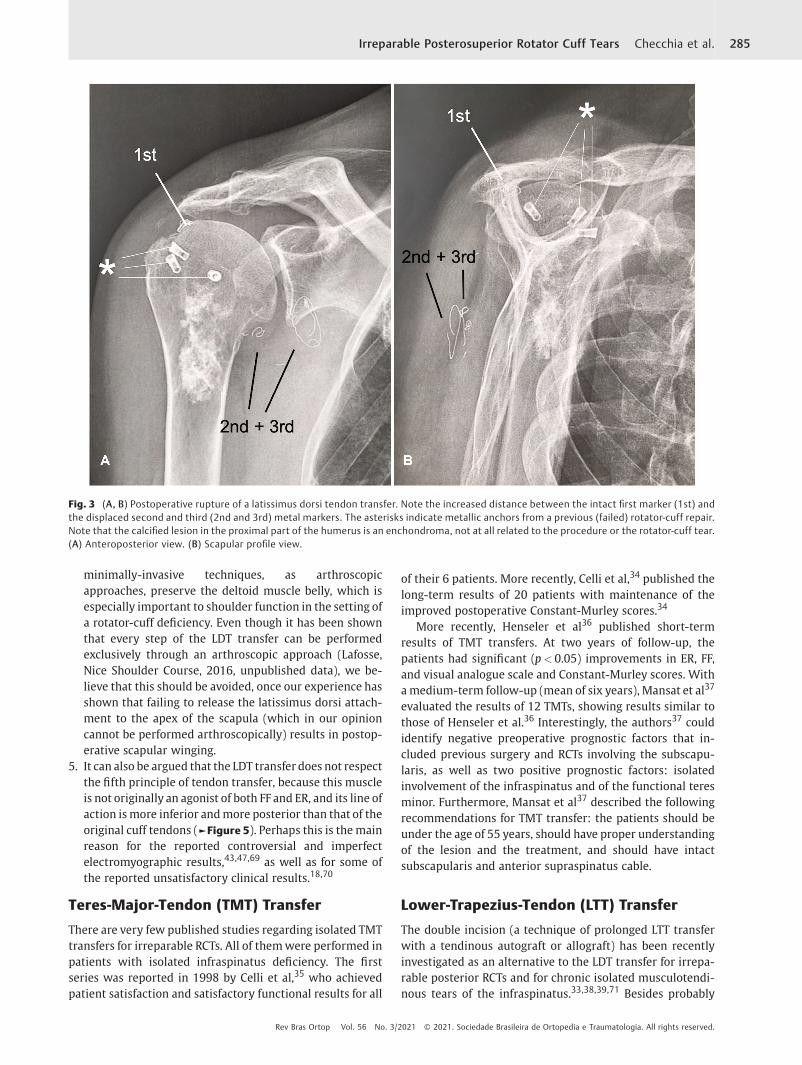
minimally-invasive techniques, as arthroscopicapproaches, preserve the deltoid muscle belly, which isespecially important to shoulder function in the setting ofa rotator-cuff deficiency. Even though it has been shownthat every step of the LDT transfer can be performedexclusively through an arthroscopic approach (Lafosse,Nice Shoulder Course, 2016, unpublished data), we be-lieve that this should be avoided, once our experience hasshown that failing to release the latissimus dorsi attach-ment to the apex of the scapula (which in our opinioncannot be performed arthroscopically) results in postop-erative scapular winging.
5. It can also be argued that the LDT transfer does not respectthe fifth principle of tendon transfer, because this muscleis not originally an agonist of both FF and ER, and its line ofaction ismore inferior andmore posterior than that of theoriginal cuff tendons (►Figure 5). Perhaps this is themainreason for the reported controversial and imperfectelectromyographic results,43,47,69 as well as for some ofthe reported unsatisfactory clinical results.18,70
Teres-Major-Tendon (TMT) Transfer
There are very few published studies regarding isolated TMTtransfers for irreparable RCTs. All of themwere performed inpatients with isolated infraspinatus deficiency. The firstseries was reported in 1998 by Celli et al,35 who achievedpatient satisfaction and satisfactory functional results for all
of their 6 patients. More recently, Celli et al,34 published thelong-term results of 20 patients with maintenance of theimproved postoperative Constant-Murley scores.34
More recently, Henseler et al36 published short-termresults of TMT transfers. At two years of follow-up, thepatients had significant (p< 0.05) improvements in ER, FF,and visual analogue scale and Constant-Murley scores. Withamedium-term follow-up (mean of six years), Mansat et al37
evaluated the results of 12 TMTs, showing results similar tothose of Henseler et al.36 Interestingly, the authors37 couldidentify negative preoperative prognostic factors that in-cluded previous surgery and RCTs involving the subscapu-laris, as well as two positive prognostic factors: isolatedinvolvement of the infraspinatus and of the functional teresminor. Furthermore, Mansat et al37 described the followingrecommendations for TMT transfer: the patients should beunder the age of 55 years, should have proper understandingof the lesion and the treatment, and should have intactsubscapularis and anterior supraspinatus cable.
Lower-Trapezius-Tendon (LTT) Transfer
The double incision (a technique of prolonged LTT transferwith a tendinous autograft or allograft) has been recentlyinvestigated as an alternative to the LDT transfer for irrepa-rable posterior RCTs and for chronic isolated musculotendi-nous tears of the infraspinatus.33,38,39,71 Besides probably
Fig. 3 (A, B) Postoperative rupture of a latissimus dorsi tendon transfer. Note the increased distance between the intact first marker (1st) andthe displaced second and third (2nd and 3rd) metal markers. The asterisks indicate metallic anchors from a previous (failed) rotator-cuff repair.Note that the calcified lesion in the proximal part of the humerus is an enchondroma, not at all related to the procedure or the rotator-cuff tear.(A) Anteroposterior view. (B) Scapular profile view.
Rev Bras Ortop Vol. 56 No. 3/2021 © 2021. Sociedade Brasileira de Ortopedia e Traumatologia. All rights reserved.
Irreparable Posterosuperior Rotator Cuff Tears Checchia et al. 285

being technically easier than the LDT transfer, the line of pullof the lower-trapezius-muscle fibers more closely replicatesthose of the infraspinatus (►Figure 5). Furthermore, it hasbeen shown that tension and excursion forces of the trape-zius are very similar to those of the infraspinatus.38,39
In a cadaveric study, Omid et al39 concluded that the LTTtransfer was biomechanically superior to the LDT transfer,providing greater ER forces. Hartzler et al38 also foundimproved ER with the arm at the side compared to LDTtransfer, but the LDT transfer was better at restoring the FF
as well as the ER at 90° of abduction. More recently, in 2019,Reddy et al,72 showed through a three-dimensional (3D)biomechanical virtual study (performing virtual LDT andLTT transfers in a computer software), that the LTT providedbetter abduction and ER moment arms when transferred tothe infraspinatus insertion. However, LDT performed betterwhen transferred to the supraspinatus insertion. Overall,the LTT transfer showed a biomechanical advantage com-pared with the LDT transfer because of stronger abductionmoment arms.
Fig. 4 (A-D) Latissimus dorsi tendon transfer elongated and reinforced with a tendinous allograft and performed through an isolateddeltopectoral approach, as described by Miyazaki et al.61 (A) Figure depicting tendon preparation before transfer. (B) Allograft preparation. (C)After passing the transfer behind the humerus. (D) Figure depicting final configuration of the latissimus dorsi tendon. Abbreviations: AG,allograft; TRI, triceps, LD, latissimus dorsi; RCT, rotator-Cuff Tear; LHB, long head of the biceps; TM, teres major; sub, subscapularis.
Rev Bras Ortop Vol. 56 No. 3/2021 © 2021. Sociedade Brasileira de Ortopedia e Traumatologia. All rights reserved.
Irreparable Posterosuperior Rotator Cuff Tears Checchia et al.286

Fig. 5 (A-C) Drawings of the right shoulder. The line of pull from the lower trapezius (LT) more closely replicates the one from the infraspinatus(IS) than the one generated by the latissimus dorsi (LD). The line of pull from the LD closely replicates the line of pull from the subscapularis(SSCP). (A) Posterior view. (B) Anterior view. (C) Medial view.
Rev Bras Ortop Vol. 56 No. 3/2021 © 2021. Sociedade Brasileira de Ortopedia e Traumatologia. All rights reserved.
Irreparable Posterosuperior Rotator Cuff Tears Checchia et al. 287

In 2016, Elhassan et al,71 were the first to report theoutcomes following this technique. They evaluated 33patients (26 men, with an average of 53 years of age; range:31to 66 years) at an average follow-up of 47 months (range:24 to 73 months). Except for one patient, who had a bonemass index of 36 kg/m2, all achieved statistically significantimprovements in pain, subjective shoulder value, and dis-abilities of the arm, shoulder and hand (DASH) score, as wellas statistically significant improvements to all active shoul-der motions. Their results were, therefore, the first to showthat this mode of treatment may be a good alternative, atleast at early and medium follow-up.
In 2016, Elhassan et al73 described a modification to theiroriginal technique in which, instead of creating a secondlateral open approach, they would instead fix the transfer tothe greater tuberosity under arthroscopic visualization.Nonetheless, they haven’t published the results followingthis modification. However, in 2018, Valenti and Werthel74
published the results following almost the same technique asthe one published by Elhassan et al,73 the only differencebeing the use of a semitendinosus tendon autograft insteadof an Achilles allograft. They evaluated 14 patients after amean follow-up of 24 months (range 12 to 36months). Theirresults have shown gain in external rotation with the arm atthe side of 24° and, in 90° of abduction, of 40°. The lag signand the Hornblower sign have disappeared from everypatient in whom they were present preoperatively. TheConstant-Murley score improved from 35 (preoperatively)to 60 points (postoperatively), the SST, from 3.5 to 7.5, theSSV, from 30 to 60%, and the pain decreased from 7 to 2(visual analogue scale). There were two cases of hematomas,and one was revised because of infection.
We are unaware of anyother published study showing theresults of lower trapezius transfer for irreparable RCTs.Despite all that, to date there are no clinical studies thatsuggest the superiority of latissimus dorsi transfer or lowertrapezius transfer over the other.
Conclusion
Irreparable posterosuperior RCTs can be debilitating, andfailed cuff repairs are still challenging conditions to treat.Different techniques have been developed, and the pro-posed benefits of tendon transfers are pain relief and someincreased range of motion with potential increase toshoulder strength. The LDT remains the most commonlyused method, and different modifications to the originaltechnique have been shown to minimize complicationsand to improve functional results and satisfaction. None-theless, LTT transfers are promising and should be consid-ered, especially for patients with isolated loss of externalrotation. Its results, however, are limited to two series ofcases, both of which have reported only short to midtermfollow-up.
Conflict of interestsThe authors declare to have no conflict of interests.
References1 Tashjian RZ. Epidemiology, natural history, and indications for
treatment of rotator cuff tears. Clin Sports Med 2012;31(04):589–604
2 Galatz LM, Ball CM, Teefey SA, Middleton WD, Yamaguchi K. Theoutcome and repair integrity of completely arthroscopicallyrepaired large and massive rotator cuff tears. J Bone Joint SurgAm 2004;86(02):219–224
3 Longo UG, Berton A, Papapietro N, Maffulli N, Denaro V. Epide-miology, genetics and biological factors of rotator cuff tears. MedSport Sci 2012;57:1–9
4 Bartl C, Kouloumentas P, Holzapfel K, et al. Long-term outcomeand structural integrity following open repair of massive rotatorcuff tears. Int J Shoulder Surg 2012;6(01):1–8
5 Jo CH, Shin JS, Lee YG, et al. Platelet-rich plasma for arthroscopicrepair of large tomassive rotator cuff tears: a randomized, single-blind, parallel-group trial. Am J Sports Med 2013;41(10):2240–2248
6 Kim SJ, Kim SH, Lee SK, Seo JW, Chun YM. Arthroscopic repair ofmassive contracted rotator cuff tears: aggressive release withanterior and posterior interval slides do not improve cuff healingand integrity. J Bone Joint Surg Am 2013;95(16):1482–1488
7 Shamsudin A, Lam PH, Peters K, Rubenis I, Hackett L, Murrell GA.Revision versus primary arthroscopic rotator cuff repair: a 2-yearanalysis of outcomes in 360 patients. Am J Sports Med 2015;43(03):557–564
8 Parnes N, DeFranco M, Wells JH, Higgins LD, Warner JJ. Compli-cations after arthroscopic revision rotator cuff repair. Arthrosco-py 2013;29(09):1479–1486
9 Bokor DJ, Hawkins RJ, Huckell GH, Angelo RL, Schickendantz MS.Results of nonoperativemanagement of full-thickness tears of therotator cuff. Clin Orthop Relat Res 1993;(294):103–110
10 Boileau P, Baqué F, Valerio L, Ahrens P, Chuinard C, Trojani C.Isolated arthroscopic biceps tenotomy or tenodesis improvessymptoms in patients with massive irreparable rotator cuff tears.J Bone Joint Surg Am 2007;89(04):747–757
11 Walch G, Edwards TB, Boulahia A, Nové-Josserand L, Neyton L,Szabo I. Arthroscopic tenotomy of the long head of the biceps inthe treatment of rotator cuff tears: clinical and radiographicresults of 307 cases. J Shoulder Elbow Surg 2005;14(03):238–246
12 Gartsman GM. Massive, irreparable tears of the rotator cuff.Results of operative debridement and subacromial decompres-sion. J Bone Joint Surg Am 1997;79(05):715–721
13 Kim SJ, Lee IS, Kim SH, Lee WY, Chun YM. Arthroscopic partialrepair of irreparable large to massive rotator cuff tears. Arthros-copy 2012;28(06):761–768
14 Iagulli ND, Field LD, Hobgood ER, Ramsey JR, Savoie FH 3rd. Compar-ison of partial versus complete arthroscopic repair ofmassive rotatorcuff tears. Am J Sports Med 2012;40(05):1022–1026
15 Berth A, NeumannW, Awiszus F, Pap G. Massive rotator cuff tears:functional outcome after debridement or arthroscopic partialrepair. J Orthop Traumatol 2010;11(01):13–20
16 Gerber C. Latissimus dorsi transfer for the treatment of irrepara-ble tears of the rotator cuff. Clin Orthop Relat Res 1992;(275):152–160
17 Gerber C, Vinh TS, Hertel R, Hess CW. Latissimus dorsi transfer forthe treatment of massive tears of the rotator cuff. A preliminaryreport. Clin Orthop Relat Res 1988;(232):51–61
18 Iannotti JP, Hennigan S, Herzog R, et al. Latissimus dorsi tendontransfer for irreparable posterosuperior rotator cuff tears. Factorsaffecting outcome. J Bone Joint Surg Am 2006;88(02):342–348
19 Warner JJ. Management of massive irreparable rotator cuff tears:the role of tendon transfer. Instr Course Lect 2001;50:63–71
20 Mulieri P, Dunning P, Klein S, Pupello D, Frankle M. Reverseshoulder arthroplasty for the treatment of irreparable rotatorcuff tear without glenohumeral arthritis. J Bone Joint Surg Am2010;92(15):2544–2556
Rev Bras Ortop Vol. 56 No. 3/2021 © 2021. Sociedade Brasileira de Ortopedia e Traumatologia. All rights reserved.
Irreparable Posterosuperior Rotator Cuff Tears Checchia et al.288

21 Mihata T, Watanabe C, Fukunishi K, Ohue M, Tsujimura T,Kinoshita M. Arthroscopic Superior Capsular ReconstructionRestores Shoulder Stability and Function in Patients with Irrepa-rable Rotator Cuff Tears: A Prospective Study (SS-15). Arthroscopy2011;27(05):e36–e37
22 Mihata T, McGarry MH, Pirolo JM, Kinoshita M, Lee TQ. Superiorcapsule reconstruction to restore superior stability in irreparablerotator cuff tears: a biomechanical cadaveric study. Am J SportsMed 2012;40(10):2248–2255
23 Mihata T, Lee TQ,Watanabe C, et al. Clinical results of arthroscopicsuperior capsule reconstruction for irreparable rotator cuff tears.Arthroscopy 2013;29(03):459–470
24 Gracitelli ME, Beraldo RA, Malavolta EA, Assunção JH, de OliveiraDR, Ferreira Neto AA. Reconstrução da cápsula superior comaloenxerto de fáscia lata para roturas irreparáveis do tendão domúsculo supraespinhal. Rev Bras Ortop 2019;54(05):591–596
25 Burkhart SS, Denard PJ, Adams CR, Brady PC, Hartzler RU. Arthro-scopic Superior Capsular Reconstruction for Massive IrreparableRotator Cuff Repair. Arthrosc Tech 2016;5(06):e1407–e1418
26 Hartzler RU, Burkhart SS. Superior Capsular Reconstruction.Orthopedics 2017;40(05):271–280
27 Hirahara AM, Adams CR. Arthroscopic Superior Capsular Recon-struction for Treatment of Massive Irreparable Rotator Cuff Tears.Arthrosc Tech 2015;4(06):e637–e641
28 Tokish JM, Beicker C. Superior Capsule Reconstruction TechniqueUsing an Acellular Dermal Allograft. Arthrosc Tech 2015;4(06):e833–e839
29 Denard PJ, Brady PC, Adams CR, Tokish JM, Burkhart SS. Prelimi-nary Results of Arthroscopic Superior Capsule Reconstructionwith Dermal Allograft. Arthroscopy 2018;34(01):93–99
30 Checchia C, Domos P, Grimberg J, Kany J. Current Options inTendonTransfers for Irreparable Rotator Cuff Tears. JBJS Rev 2019;7(02):e6
31 Grimberg J, Kany J. Latissimus dorsi tendon transfer for irrepara-ble postero-superior cuff tears: current concepts, indications, andrecent advances. Curr Rev Musculoskelet Med 2014;7(01):22–32
32 Hamada K, Fukuda H, Mikasa M, Kobayashi Y. Roentgenographicfindings in massive rotator cuff tears. A long-term observation.Clin Orthop Relat Res 1990;(254):92–96
33 Omid R, Lee B. Tendon transfers for irreparable rotator cuff tears. JAm Acad Orthop Surg 2013;21(08):492–501
34 CelliA,MarongiuMC,RovestaC,Celli L. Transplantof the teresmajorin thetreatmentof irreparable injuriesof the rotatorcuff (long-termanalysis of results). Chir Organi Mov 2005;90(02):121–132
35 Celli L, Rovesta C, Marongiu MC, Manzieri S. Transplantation ofteresmajor muscle for infraspinatusmuscle in irreparable rotatorcuff tears. J Shoulder Elbow Surg 1998;7(05):485–490
36 Henseler JF, Nagels J, van der Zwaal P, Nelissen RGHH. Teres majortendon transfer for patients with massive irreparable poster-osuperior rotator cuff tears: Short-term clinical results. BoneJoint J 2013;95-B(04):523–529
37 Mansat P, Dotziz A, Bellumore Y, Mansat M. Teres major flap:surgical anatomy, technique of harvesting, methods of fixation,postoperative management. In: Tendon transfer for irreparablecuff tear [Internet]. SpringerParis2011 [cited 2017 Sep 23]. p.49–64. (Collection GECO). Available from: https://link.springer.-com/chapter/10.1007/978-2-8178-0049-3_5
38 Hartzler RU, Barlow JD, An KN, Elhassan BT. Biomechanical effec-tiveness of different types of tendon transfers to the shoulder forexternal rotation. J Shoulder Elbow Surg 2012;21(10):1370–1376
39 Omid R, Heckmann N, Wang L, McGarry MH, Vangsness CT Jr, LeeTQ. Biomechanical comparison between the trapezius transferand latissimus transfer for irreparable posterosuperior rotatorcuff tears. J Shoulder Elbow Surg 2015;24(10):1635–1643
40 Burkhart SS, Athanasiou KA, Wirth MA. Margin convergence: amethod of reducing strain in massive rotator cuff tears. Arthros-copy 1996;12(03):335–338
41 Buchmann S, Plath JE, Imhoff AB. [Latissimus dorsi transfer for thetreatment of irreparable rotator tears: indication, surgical tech-nique, and modifications]. Oper Orthop Traumatol 2012;24(06):502–512
42 El-Azab HM, Rott O, Irlenbusch U. Long-term follow-up afterlatissimus dorsi transfer for irreparable posterosuperior rotatorcuff tears. J Bone Joint Surg Am 2015;97(06):462–469
43 Habermeyer P, Magosch P, Rudolph T, Lichtenberg S, Liem D.Transfer of the tendon of latissimus dorsi for the treatment ofmassive tears of the rotator cuff: a new single-incision technique.J Bone Joint Surg Br 2006;88(02):208–212
44 Irlenbusch U, Bracht M, Gansen HK, Lorenz U, Thiel J. Latissimusdorsi transfer for irreparable rotator cuff tears: a longitudinalstudy. J Shoulder Elbow Surg 2008;17(04):527–534
45 Miniaci A, MacLeod M. Transfer of the latissimus dorsi muscleafter failed repair of a massive tear of the rotator cuff. A two tofive-year review. J Bone Joint Surg Am 1999;81(08):1120–1127
46 Namdari S, Voleti P, Baldwin K, Glaser D, Huffman GR. Latissimusdorsi tendon transfer for irreparable rotator cuff tears: a system-atic review. J Bone Joint Surg Am 2012;94(10):891–898
47 Nové-Josserand L, Costa P, Liotard JP, Safar JF, Walch G, Zilber S.Results of latissimus dorsi tendon transfer for irreparable cufftears. Orthop Traumatol Surg Res 2009;95(02):108–113
48 Villacis D, Merriman J, Wong K, Rick Hatch GF III. Latissimus dorsitendon transfer for irreparable rotator cuff tears: a modifiedtechnique using arthroscopy. Arthrosc Tech 2013;2(01):e27–e30
49 Irlenbusch U, Bensdorf M, Gansen HK, Lorenz U. [Latissimus dorsitransfer in case of irreparable rotator cuff tear–a comparativeanalysis of primary and failed rotator cuff surgery, in dependenceof deficiency grade and additional lesions]. Z Orthop Ihre Grenz-geb 2003;141(06):650–656
50 Glanzmann MC, Goldhahn J, Flury M, Schwyzer HK, Simmen BR.Deltoid flap reconstruction for massive rotator cuff tears: mid-and long-term functional and structural results. J Shoulder ElbowSurg 2010;19(03):439–445
51 Werner CML, Zingg PO, Lie D, Jacob HAC, Gerber C. The bio-mechanical role of the subscapularis in latissimus dorsi transferfor the treatment of irreparable rotator cuff tears. J ShoulderElbow Surg 2006;15(06):736–742
52 Moursy M, Forstner R, Koller H, Resch H, Tauber M. Latissimusdorsi tendon transfer for irreparable rotator cuff tears: amodifiedtechnique to improve tendon transfer integrity. J Bone Joint SurgAm 2009;91(08):1924–1931
53 Costouros JG,EspinosaN,SchmidMR,GerberC.Teresminor integritypredicts outcome of latissimus dorsi tendon transfer for irreparablerotator cuff tears. J Shoulder Elbow Surg 2007;16(06):727–734
54 Tokish JM, Alexander TC, Kissenberth MJ, Hawkins RJ. Pseudopar-alysis: a systematic review of term definitions, treatmentapproaches, and outcomes ofmanagement techniques. J ShoulderElbow Surg 2017;26(06):e177–e187
55 Gerber C, Maquieira G, Espinosa N. Transfert du grand dorsal pourle traitement des ruptures massives de la coiffe des rotateurs :quels facteurs déterminent le résultat final? Rev Chir OrthopRepar Appar Mot 2004;90(05, Suppl 1):158–162
56 Warner JJ, Parsons IM IV. Latissimus dorsi tendon transfer: acomparative analysis of primary and salvage reconstruction ofmassive, irreparable rotator cuff tears. J Shoulder Elbow Surg2001;10(06):514–521
57 Herzberg G, Urien JP, Dimnet J. Potential excursion and relativetension of muscles in the shoulder girdle: relevance to tendontransfers. J Shoulder Elbow Surg 1999;8(05):430–437
58 Castricini R, Longo UG, De Benedetto M, et al. Arthroscopic-Assisted Latissimus Dorsi Transfer for the Management of Irrepa-rable Rotator Cuff Tears: Short-Term Results. J Bone Joint Surg Am2014;96(14):e119
59 Grimberg J, Kany J, Valenti P, Amaravathi R, Ramalingam AT.Arthroscopic-assisted latissimus dorsi tendon transfer for
Rev Bras Ortop Vol. 56 No. 3/2021 © 2021. Sociedade Brasileira de Ortopedia e Traumatologia. All rights reserved.
Irreparable Posterosuperior Rotator Cuff Tears Checchia et al. 289

irreparable posterosuperior cuff tears. Arthroscopy 2015;31(04):599–607.e1
60 Sirveaux F, Favard L, Oudet D, Huquet D, Walch G, Molé D.Grammont inverted total shoulder arthroplasty in the treatmentof glenohumeral osteoarthritis with massive rupture of the cuff.Results of amulticentre studyof 80 shoulders. J Bone Joint Surg Br2004;86(03):388–395
61 Miyazaki AN, Checchia CS, LopesWC, Fonseca Filho JM, do Val SellaG, Silva LA. Transferência tendínea do grande dorsal com enxertotendíneo homólogo para as lesões irreparáveis do manguito rota-dor: técnica cirúrgica�. Rev Bras Ortop 2019;54(01):99–103
62 Kany J, Grimberg J, Amaravathi RS, Sekaran P, Scorpie D, WerthelJD. Arthroscopically-Assisted Latissimus Dorsi Transfer for Irrep-arable Rotator Cuff Insufficiency: Modes of Failure and ClinicalCorrelation. Arthroscopy 2018;34(04):1139–1150
63 Walch G, Boulahia A, Calderone S, Robinson AH. The ‘dropping’and ‘hornblower’s’ signs in evaluation of rotator-cuff tears. J BoneJoint Surg Br 1998;80(04):624–628
64 Boileau P, Chuinard C, Roussanne Y, Neyton L, Trojani C. Modifiedlatissimus dorsi and teres major transfer through a single delto-pectoral approach for external rotation deficit of the shoulder: asan isolated procedure or with a reverse arthroplasty. J ShoulderElbow Surg 2007;16(06):671–682
65 Clavert P, Bouchaïb J, Sommaire C, Flurin PH, Hardy P. Observe-t-on une modification de la densité osseuse du tubercule majeuraprès 70ans? Rev Chir Orthop Traumatol 2014;100(01):94–96
66 Gerber C, Schneeberger AG, Beck M, Schlegel U. Mechanicalstrength of repairs of the rotator cuff. J Bone Joint Surg Br1994;76(03):371–380
67 Diop A, Maurel N, Chang VK, Kany J, Duranthon LD, Grimberg J.Tendon fixation in arthroscopic latissimus dorsi transfer forirreparable posterosuperior cuff tears: an in vitro biomechanicalcomparison of interference screw and suture anchors. Clin Bio-mech (Bristol, Avon) 2011;26(09):904–909
68 Goldstein Y, Grimberg J, Valenti P, Chechik O, Drexler M, Kany J.Arthroscopic fixationwith aminimally invasive axillary approachfor latissimus dorsi transfer using an endobutton in massive andirreparable postero-superior cuff tears. Int J Shoulder Surg 2013;7(02):79–82
69 AokiM, Okamura K, Fukushima S, Takahashi T, OginoT. Transfer oflatissimus dorsi for irreparable rotator-cuff tears. J Bone Joint SurgBr 1996;78(05):761–766
70 Gerber C, Maquieira G, Espinosa N. Latissimus dorsi transfer forthe treatment of irreparable rotator cuff tears. J Bone Joint SurgAm 2006;88(01):113–120
71 Elhassan BT, Wagner ER, Werthel JD. Outcome of lower trapeziustransfer to reconstruct massive irreparable posterior-superiorrotator cuff tear. J Shoulder Elbow Surg 2016;25(08):1346–1353
72 Reddy A, Gulotta LV, Chen X, et al. Biomechanics of lowertrapezius and latissimus dorsi transfers in rotator cuff-deficientshoulders. J Shoulder Elbow Surg 2019;28(07):1257–1264
73 Elhassan BT, Alentorn-Geli E, Assenmacher AT, Wagner ER. Ar-throscopic-Assisted Lower Trapezius Tendon Transfer for MassiveIrreparable Posterior-Superior Rotator Cuff Tears: Surgical Tech-nique. Arthrosc Tech 2016;5(05):e981–e988
74 Valenti P, Werthel JD. Lower trapezius transfer with semite-ndinosus tendon augmentation: Indication, technique, results.Obere Extrem 2018;13(04):261–268
Rev Bras Ortop Vol. 56 No. 3/2021 © 2021. Sociedade Brasileira de Ortopedia e Traumatologia. All rights reserved.
Irreparable Posterosuperior Rotator Cuff Tears Checchia et al.290

Rotator Cuff Healing
A cicatrização do manguito rotadorIldeu Afonso de Almeida Filho1,2 Daniel Andrade Coelho2,3
1 Department of Orthopedics, Hospital Felício Rocho, Belo Horizonte,MG, Brazil
2Hospital Vera Cruz, Belo Horizonte, MG, Brazil3Hospital Semper, Belo Horizonte, MG, Brazil
Rev Bras Ortop 2021;56(3):291–298.
Address for correspondence Ildeu Afonso de Almeida Filho,Department of Orthopedics, Hospital Felício Rocho, Av. do Contorno,9530, Barro Preto, Belo Horizonte, MG, 30190-130, Brazil(e-mail: [email protected]).
Introduction
The incidence of rotator cuff injuries has grown exponen-tially as the life expectancy of the population increases.Several technical explanations have been given to justify
this increase. The issue revolves around the degenerativeaspects related to the aging process of the organism, associ-atedwith some lifehabits, such as sports practice, profession,diet, use of medications, and the presence of associateddiseases in addition to genetic issues, which has not received
Keywords
► rotator cuff injuries/diagnosis
► rotator cuff injuries/epidemiology
► rotator cuff injuries/surgery
► wound healing
Abstract The present article broadly addresses the aspects that interfere with the healingprocess of the rotator cuff. Life habits, such as smoking and alcoholism, are considered,systemic factors such as diabetes mellitus, hypertension, and obesity, as well as localfactors, among which are those related to the pre, peri, and postoperative periods.From an extensive literature review, with the citation of 60 scientific articles from bothWestern and Eastern literature, the authors intend to deepen the theme by bringing tomedical practice conducts based on new established concepts.
Palavras-chave
► lesões do manguitorotador/diagnóstico
► lesões do manguitorotador/epidemiologia
► lesões do manguitorotador/cirurgia
► cicatrização
Resumo O presente artigo aborda de forma ampla os aspectos que interferem no processo decicatrização do manguito rotador. São considerados hábitos de vida como tabagismo ealcoolismo, fatores sistêmicos como diabetes mellitus, hipertensão arterial e obesi-dade bem como fatores locais, dentre os quais aqueles relacionados ao pré, per e pósoperatório. A partir de uma extensa revisão da literatura, com a citação de 60 artigoscientíficos tanto da literatura ocidental como oriental, os autores pretendem apro-fundar no tema trazendo para a prática médica condutas embasadas em novosconceitos estabelecidos.
receivedFebruary 11, 2020acceptedApril 15, 2020published onlineSeptember 24, 2020
DOI https://doi.org/10.1055/s-0040-1713764.ISSN 0102-3616.
© 2021. Sociedade Brasileira de Ortopedia e Traumatologia. Allrights reserved.This is an open access article published by Thieme under the terms of the
Creative Commons Attribution-NonDerivative-NonCommercial-License,
permitting copying and reproduction so long as the original work is given
appropriate credit. Contents may not be used for commercial purposes, or
adapted, remixed, transformed or built upon. (https://creativecommons.org/
licenses/by-nc-nd/4.0/)
Thieme Revinter Publicações Ltda., Rua do Matoso 170, Rio deJaneiro, RJ, CEP 20270-135, Brazil
THIEME
Update Article 291

much attention in publications to date. Anatomical varia-tions also participate in this process. Therefore, there aremany factors that may influence the development of rotatorcuff tendon injuries.
Hamada et al.1 studied the histology of the human supra-spinatus tendon, subdividing it into portions and observedthat, in theyouth, the tendon is organized intowavy layers andthat in the older patients, this pattern changes to homoge-neous, emaciated, andwith hyaline degeneration (►Figure 1).
Uthoff and Ishii2 analyzed the histology of tendinousinsertion in the larger tubercle and observed, in the youth,the existence of a layered organization pattern. In theelderly, a disorganization occurs that is considered a de-generative process (►Figure 2). The author described theinflammatory reaction in the partial lesion of the rotatorcuff as an attempt by the organism itself to heal it. However,Yamaguchi et al.3 histologically studied the natural evolu-tion of these lesions in a series of 58 cases and, in none ofthem, it was possible to observe the reduction of the size ofthe lesion. This fact shows that spontaneous healing of therotator cuff is something that should not be expected bypatients, let alone by surgeons.
Does the progressive increase in the incidence of injuries inthe general population lead us to ask aboutwhat is considerednormal for a determined age?Howmanycarriers of cuff injuryare asymptomatic?Milgron et al.4 stated that the prevalence oflesions increases significantly in the population after 70 years
of age, and it ismore frequent on the dominant side, a fact thathas been proven by other authors.5 In the seventh decade oflife, this incidence increases to more than 80%.
During the last two decades, methods of treatment of cuffinjuries have evolved substantially. The surgeries, which havepreviously been done openly, have started, in most referencecenters, to be done arthroscopicly. Different types of suturesemerged, andhigh-strength threads began to be used routinely.
Local Factors
Themain factors associatedwith rotator cuff healing are injurysize, muscle quality (atrophy and fatty degeneration), tendonquality, presence of delamination, and shoulder morphology.
Fig. 1 In the young, the tendon insertion is organized in distinctlayers (a) in the elderly, there is disorganization (b).
Fig. 2 Organization of the fibers of the supraspinatus tendon in layersin the young, and its progressive disorganization in the adult.
Rev Bras Ortop Vol. 56 No. 3/2021 © 2021. Sociedade Brasileira de Ortopedia e Traumatologia. All rights reserved.
Rotator Cuff Healing Almeida Filho, Coelho292

Injury SizeThefirst characteristic evaluated bymost surgeons is the sizeof the lesion. The analysis, however, should be performed byevaluating the size of the lesion in the sagittal plane andcoronal plane (medial retraction or tendon shortening), sincethey are independent characteristics with respect to thehealing rate. From a general point of view, the increase inlesion size negatively influences healing rates, ranging from97%, for small lesions, to only 6%, for large or massive lesions,in some series. 6–10
Size Evaluated in the Sagittal PlanWylie et al.,11 when evaluating the healing rate in this plan,showed that lesions� 2.2 cmhad a healing rate of 49%, and inlesions � 2.2 cm the rate rose to 71%. Considering this cutoffvalue, the hypothesis was postulated that an injury exceed-ing 2 cm already presents significant involvement of theinfraspinal,12 which alters the kinematics of the humeanhead, generating unbalance in the forces.13 This fact wouldput the repair at risk, thus justifying the increased risk ofhealing failure in lesions larger than 2.0 cm.
Evaluated Size in Coronal Plan (Shortening)Myotendinous shortening is considered a direct result ofmuscle retraction. However, Meyer et al.14 showed that,although the shortening is predominantly of muscle mass,retraction is also caused by shortening of the tendon itself,both due to the remaining substance in the tubercule after theinitial injury and, in advanced stages, the resorption of thetendon itself. In a subsequent analysis, Tashjian et al.,6 showedthat patientswith lesions inwhich themyotendinous junction(MTJ) is lateral to the face of the glenoid, the healing rate was93%, while in patients with preoperative MTJ medial to theglenoid face, the healing rate was 55%. The analysis thenconcluded that the position of MTJ is an independent factorin healing. From the point of view of the size of the retractionitself, Wylie et al.11 showed that lesions with shortening inrelation to the footprint�2 cmpresentedahealing rateof 47%,as opposed to 76% healing in lesions with retraction � 2 cm.
Muscle Quality (Fatty Atrophy and Degeneration)When one thinks of rotator cuff healing, one goal to beachieved is the healing of the tendon-bone interface. Giventhis fact, most studies focus on methods to strengthen orpositively interfere with this interface. However, analyzinglarge lesions, Jeong et al.15 observed that muscle quality ismore important than tendon quality in the new ruptureindex. According to the authors, the occupancy rate of thesupraspinatus below 43% in relation to the totality of thesupraspinatus fossa and a degree of fatty infiltration � 2according to Goutallier are independent risk factors forfurther rupture of the rotator cuff, with sensitivity of 98%and specificity of 83.6%. A systematic review by Khair et al.,16
corroborates this fact, indicating that the quality of thepreoperative muscle plays an important role in the rate oftendon healing. Low stages of Goutallier (0–1) are associatedwith low rates of new rupture (25%); in contrast, high stages(2–4) have high rates of rupture (59%).
Histological and Macroscopic Tendon QualityRegarding the analysis of the histological and macroscopicquality of the tendon, it has been suggested that a worsequality tendon would lead to worse healing rates. However,Mazzoca et al.17 showed that there is no correlation betweenthe macroscopic appearance of the tendon in the arthrosco-py, or in the histological analysis, and healing. Furthermore,they showed that the macroscopic appearance is not corre-lated with the histological quality of the tendon, and there isno association between the degree of histological tendinop-athy, the macroscopic appearance and the healing index orclinical results of the repair. Therefore, the abnormal appear-ance should not influence negatively the stress or techniqueof repair.
DelaminationA chronic complete lesion is considered delaminated whenthere is a horizontal cleavage area in the ruptured tendon.The rotator cuff is composed of five layers, namely: the mostsuperficial is composed of coracoumeral ligament fibers;the second layer is composed of parallel fibers between thesupra and infraspinal; the third is composed of smallfascicles that intersect the cuff tendons at an angle of45 degrees; the fourth is composed of extracapsular looseconnective tissue that previously connects with the deepportion of the coracoumeral ligament; and, finally, the fifthlayer is basically composed of a joint capsule. The secondand third layers are thicker, presenting different directionsof collagen fibers.18 It is precisely between these layers thattendon delamination occurs. In a recent study, Boileauet al.19 showed a prevalence of 32% delamination, all ofwhich involved a posterior component of the deep layer.This study showed that, in general, delamination has adeleterious effect on the healing rate, especially in largelesions. Kwon et al.,20 in a study with 1,043 patients,confirmed the significantly lower healing rate in delami-nated tendons, if the analysis is performed using a singlevariable. However, when performing multivariate analysis,it was noticed that delamination is not an independent riskfactor for healing failure.
Shoulder MorphologyHeuberer et al.21 analyzed the morphology of the acronymand correlated it with the chance of developing subacromialimpact, impacto rotator cuff injury and arthropathy. Therewere three ways to evaluate acromial morphology, namely:critical shoulder angle (CSA), acromial index (AI) and lateralacromion angle (LAA). The authors concluded that the bestway to predict the appearance of cuff-related lesions or thedevelopment of osteoarthrosis is through the CSA. This angle,which was described by Moor et al.,22 has great benefit forcombining the lateral extension of the acromionwith glenoidangulation. It is calculated on anteroposterior radiographyby the angle between the line of the lower edge to the upperedge of the glenoid and the line of the lower edge of theglenoid to the most inferolateral point of the acromion(►Figure 3). In the referred study, the authors demonstratedan association between rotator cuff injury for CSA> 35° and
Rev Bras Ortop Vol. 56 No. 3/2021 © 2021. Sociedade Brasileira de Ortopedia e Traumatologia. All rights reserved.
Rotator Cuff Healing Almeida Filho, Coelho 293

glenohumeral arthrosis in CSA< 30°. This fact has beendemonstrated by other authors.23
A biomechanical study conducted by Gerber et al.24 deep-ened the subject, demonstrating that CSA> 38° requires anincrease of up to 33% in supraspinatus activity for shoulderstabilization during 6°-61° abduction. This fact could explainthefindings ofMoor et al.22 andpredict probable change in thehealing rates of these patients. In fact, Garcia et al.25 found a14-fold vezes higher risk of rerupture ruptura in patients withCSA> 38°. A recent systematic review on the subject alsoconfirmed the hypothesis, showing that CSA> 38° is associat-edwith lower healing rate.26However, due to the heterogene-ity of the relevant literature in the review in question, thestrength of this finding is considered limited.
Systemic Factors
We will discuss here the various systemic factors that caninfluence tendon-bone or tendon-tendon healing.
Chronic alcohol use is an important factor regarding theincreased incidence of injury as well as the severity of rotatorcuff injury in both genders. The association between alcoholconsumption and rotator cuff injuries showed that, in winedrinkers, therewasmoremassive lesions than small lesions.27
In relation to smoking, it is known that nicotine acts in theexpression of the so-called MMP-9, which is an enzymeinvolved in the degradation of the extracellular matrix, intenocytes. This factor leads to the modification of the modu-
lus of elasticity of the tendon and, according to Park et al.,28 isan important risk factor for the spread of the lesion.
Regarding the influence of diabetes mellitus on the prog-nosis of cuff repair, it is known that the persistence ofhyperglycemia in the postoperative period increases thepossibility of non-healing of reconstructed the tendons.Therefore, blood glucose control after surgery becomesfundamental.29
The association between arterial hypertension and cuffinjuries showed that, in relation to normotensive patients,hypertensive patients have twice as many large lesions andfour times as many massive lesions.30
Cholesterol also influences the prognosis by increasingtendon stiffness through changes in itsmodulus of elasticity.31
Obese patients, those with increased body mass index,also present increased incidence and severity of rotator cuffinjuries.32
Immunobiological factors, by interfering with the mech-anisms associated with muscle homeostasis, aggravate theprocess of fatty degeneration, which, in sua turn, determinesthe worsening of the prognosis of tendon healing.33
Finally, age, which is an inexorable factor, also influencesthrough the natural process of tendon degeneration, themodulus of elasticity in the rotator cuff.
Chung et al.34 analyzed the main prognostic factorsassociated with poor results after surgical reconstructionof the rotator cuff. The mean age of the patients evaluatedwas 63.7 years. The authors observed the incidence of 39.8%of failure in the repair of rotator cuff lesions. The worstprognostic factors consideredwere fatty degeneration of theinfraspinal muscle and reduction of subacromial space bycephalic migration of the humeral head.
Cho et al.35 studied the factors that affect the integrity ofrotator cuff reconstruction. The patients were divided intothree groups, namely:< 50 years of age group I), between 51and 60 years of age (group II), and> 60 years of age (groupIII). They observed that the older the age, the lower theincidence of healing (►Table 1). Nevertheless, the studyrecommends the reconstruction of lesions in elderly patientsdue to clinical improvement.
Deer et al.36 evaluated the integrity of the repair and itsrelationship with the function on the shoulders of patientssubmitted to arthroscopic reconstruction after 65 years ofage. For the study, the authors used the same sonographer toperform all imaging tests and found an incidence of 75% oftendon integritywith the University of California Los AngelesShoulder Score (UCLA) improvement from 17 points to 32points, which represents 85% of good and excellent results.There was also a significant reduction in the level of painwhen the visual analog scale was used.
Fig. 3 Critical shoulder angle.
Table 1 Increased healing in younger patients
Groups: < 50 51–60 > 60 years
N 49 68 52 patients
Healing 87.8% 79.4% 65.4%
N-healing 12.2% 20.6% 34.6%
Rev Bras Ortop Vol. 56 No. 3/2021 © 2021. Sociedade Brasileira de Ortopedia e Traumatologia. All rights reserved.
Rotator Cuff Healing Almeida Filho, Coelho294

Dezaly et al.37 conducted a prospective, comparative,randomized study of two groups of patients. Group I under-went rotator cuff reconstruction, acromioplasty, and tenot-omy of the long portion of the brachial biceps. Group IIunderwent biceps tenotomy and acromioplasty, without cuffreconstruction. There were 103 patients evaluated and,although the postoperative recovery time was shorter ingroup II, as expected, both the functional result by theConstant score (82 in group I and 73 in group II) and thesubacromial distance measured in the imaging exams werefavorable to group I. Interestingly, the best results wereobserved in the reconstructions of the most retractedlesions, suggesting that elderly patients with small lesionsshould be treated conservatively.
Many studies38 have suggested that increasing age isrelated to a reduction in the potential for tendon healing,while biomechanical studies suggest that the reason for thisshould be due to an unfavorable environment, such as low-tissue perfusion and the reduction in the number of undif-ferentiated cells.
Perioperative Factors
The systemic and local factors mentioned above are inherentto the patient, and, therefore, in general, difficult to controlon the part of the surgeon. For this reason, factors related tosurgical techniques are of crucial importance in the results,since at this point the surgeon can actively act in theimprovement of healing. Here, a good choice, focused ontechnical knowledge, can increase healing rates.
Single Row x Double RowApreleva et al.39 showed that rotator cuff repair with singlerow (SR) anchors can restore only 67% of normal footprint,which could be an explanation for healing failures. Consid-ering this, Lo and Burkhart40 created the double row (DR) ofanchors technique. This technique consists of using a row ofmedial anchors, next to the joint margin, and another row inthe lateral aspect of the footprint. In this way, thewidth frommedial to lateral is reestablished in an attempt to recreate theinsertion of the rotator cuff and thus optimize the healingpotential. From a biomechanical point of view, the additionof a second row of anchors increases the number of attach-ment points, increasing the initial strength of the construc-tion, decreasing the load that each knot/anchor needs toresist and decreasing stress at the point of contact betweeneach suture in the cuff. Biomechanical studies supported thetechnique, demonstrating a significant decrease in gap in-terval formation and strain deformation, associated withincreased strength and initial stiffness, when compared tothe single row technique.41
Despite demonstrating a biomechanical advantage, theclinical value of the technique is controversial, with animportant part of the trials stating that there are no improve-ments in quality of life or clinical scores, despite an advan-tage in the healing rate of DR over SR.42 A recent Brazilianstudy,with results of 1 to 4 years of the double row techniquecorroborates this fact, demonstrating no statistically signifi-
cant difference in UCLA and American Shoulder and ElbowScore (ASES) scores between the two techniques.43
Given the above, to decide about the real advantage ofthe technique, and which patient would benefit from it,Xu et al.44 performed a meta-analysis, with randomizedclinical trials of levels 1 and 2 evidence, comparing thetwo methods. They concluded that, although the DR tech-nique presented a lower rate of new rupture (23.8% DR x40.2% SR), better ASES and improvement in medial rotationamplitude, there is no difference in the Constant score, UCLAscore, anterior elevation, lateral rotation andmuscle strengthwhen compared to the SR technique in the overall analysis ofthe results. However, when analyzing subgroups by lesionsize, in large lesions> 3 cm, DR repair showed statisticallysignificant improvement in healing, UCLA and ASES. Thisresult is corroborated by other authors, such as the summaryof meta-analyses performed by Spiegl et al.45 and the meta-analysis produced only with level 1 trials of evidence, con-ducted by Sheibani-Rad et al.46
Arthroscopic x OpenThe most used methods for rotator cuff reconstruction areopen, mini-open and arthroscopic. Both the mini-open andarthroscopic techniques maintain the integrity of the deltoidorigin. In contrast, in the open technique, part of the deltoidis deserted from the edge of the acromion. Theoretically, thiswould be the main disadvantage of open reconstruction;however, a study by Cho et al.,47 in postoperative magneticresonance imaging (MRI) analysis, showed no significantdifference between open and arthroscopic techniques re-garding complications such as detachment of deltoid originor muscle changes. Another disadvantage of the open tech-nique would be increased pain in the initial postoperativephase, since it requires greater mobilization of soft tissues.This fact was also not confirmed, as demonstrated in meta-analysis with level 1 randomized controlled studies con-ducted by Ji et al.48
Regarding healing rates, a recentmeta-analysiswith high-quality studies and 770 patients showed similar healing ratesbetween mini-open and arthroscopic techniques.49 More-over, there was no significant difference in clinical scoresbetween the techniques.
Despite the similarity between the clinical results andhealing rates, it is worth remembering that the arthroscopictechnique offers advantage by providing easy access toglenohumeral joint associated with better cosmetic results.
Postoperative Factors
Chemical Factors: Anti-inflammatoryHealing is a gradual process, composed of overlappingphases, and the inflammatory phase ismandatory. Any factorthat slows or alters its progress can affect it. Most studies onthe subject are in vitro or with animal models and, as foundby Constantinescu et al.,50 in a systematic review, the currentliterature does not provide enough evidence for or againstthe use of nonsteroidal antiinflammatory drugs (NSAIDs). Itis worth noting that this review found only one level 1
Rev Bras Ortop Vol. 56 No. 3/2021 © 2021. Sociedade Brasileira de Ortopedia e Traumatologia. All rights reserved.
Rotator Cuff Healing Almeida Filho, Coelho 295

randomized clinical trial of evidence. The study in question,conducted by Oh et al.,51 compared the analgesic side effectsin the use of celecoxib (selective inhibitor COX-2), ibuprofen(non-selective NSAID), and tramadol (opioid). No significantdifferences were found between medications. In a secondtime, retrospectively, the healing rates were evaluated byMRI and ultrasound (US), and an important negative effectwas observed with the use of selective COX-2 inhibitor (newrupture: 37% celecoxib� 7% ibuprofen� 4% tramadol).Therefore, despite presenting similar analgesia in the post-operative period, when compared with other NSAIDs andopioids, selective COX-2 inhibitors should not be used, sincethey can act negatively on healing after rotator cuff repair.
Mechanical FactorsThe way to conduct the postoperative period may interferewith the healing process of the rotator cuff after a surgicalreconstruction. Rehabilitation protocols consider the needfor initial protection of reconstruction, associated with theintention of restoring shoulder function, preventing jointstiffness andmuscle atrophy. Shoulder joint stiffness still hasunclear etiology, but may be related to prolonged immobili-zation or conservative rehabilitation programs.52 Bio-mechanical studies show that early loading is harmful tothe organization of collagen fibers, and can generate micro-damage at the bone/tendon interface, thus preventing theintegration of collagen fibers into the bone or even completehealing failure.53
Early Mobilization vs. Late MobilizationThe protocols can be divided basically into two categories:early passivemotion (EPM) and immobilizationwith delayedrangemotion (DRM).54 The EPMprotocol consists ofminimalimmobilization with sling, in which commuting movementsare allowed plus passive movements of range of motion suchas lateral rotation and elevation from the first postoperativeday. On the other hand, the DRM protocol imposes the use ofsling without passive movements, and only commutingexercises up to 2 to 4 weeks postoperatively are allowed.Performing analysis and review of published meta-analyses,Houck et al.,55 concluded that the EPMprotocol improves therange of motion, but increases the risk of further rupture ofthe cuff. It is noteworthy that, although shoulder stiffness isan important problem in the postoperative period of rotatorcuff surgery, it is considered a complication, different fromthe recurrence of rupture, which is considered a treatmentfailure.
Sling TimeOne of the first questions of the patients in the preoperativeconsultation concerns the time of use of the sling. There is alot of divergence in the literature regarding the minimumtime of use associated with better rotator cuff healing. Kohet al.56 showed, in a level 1 study of evidence, that there areno benefits in healing when comparing immobilization for4 weeks and immobilization for 8 weeks. Therefore, theliterature does not support the use of sling for more than4 weeks.
Velpeau Sling vs. Abduction SlingBasically, anti-rotational sling keeps the arm close to the bodyinmedial rotation. The abduction slingmaintains the shoulderin various abduction and lateral rotation configurations. In thebiomechanical analysis, Jackson et al.57 found the optimalposition to create the lowest level of supraspinatus and infra-spinatus tension. Thispositionwouldbewithelevationof 21 to45° and lateral rotation of 18 to 23°. However, in practice, theresults donot prove thebenefits of this position. Some authorsshow that therewasnosignificantdifference inclinical aspectsor in the rate of new rupture. Other studies favorable to theuseof abduction slings show advantages from the clinical point ofviewofpain and function,withoutevaluationof the the resultsin healing rates.58 It is worth mentioning that, in general,studies on sling immobilization are based on the responses ofpatients’ self-assessment in relation to the amount of time ofuse of the type; however, an important question is whetherthese patients actually use the sling as reported. Based on thisquestioning, Grubhofer et al.59 showed, evaluating the use ofabduction slingswith digital sensors, that only 48% of patientsused the sling in a way they considered satisfactory (> 80% ofthe time).Moreover,whencomparing thedataobtainedby thesensors and the patient information, the discrepancy wasfrightening, especially in patients who did not use the slingas recommended. The authors, therefore, suggest cautionwiththe results of studies on type and time of use of slings.
Genetic FactorsFigueiredo et al.,60 identified genetic factors associated withsusceptibility of rotator cuff injury, reinforcing the role ofextracellular matrix homeostasis in this context.
Final Considerations
The present review article allowed us to broaden the under-standing of the factors that interfere in the healing process ofrotator cuff tendons. It was clear that there are manyvariables, being local, systemic, mechanical, chemical, ge-netic or associated with life habits. Pre, peri, and postopera-tive factors have a decisive influence on this process. As afinal message, it is suggested that the treatment of rotatorcuff lesions be done individually, considering all the factorspreviously described and, in elderly patients, before thesurgical option, that the attempt of conservative treatmentshould be considered.
Conflict of InterestsThe authors declare that there is no conflict of interests.
References1 Yamanaka K, Fukuda H, Hamada K, Nakajima T. Histology of the
supraspinatus tendon with reference to rotator cuff tears. In:Gazielly DF, Gleyze P, Thomas T, editors. The Cuff. New York:Elsevier; 1997:15–18
2 Uthoff HK, Ishii H. Histology of the cuff and pathogenesis of thedegenerative tendinopathies. In: Gazielly DF, Gleyze P, Thomas T,editors. The Cuff. New York: Elsevier; 1997:19–23
3 Yamaguchi K, Tetro AM, Blam O, Evanoff BA, Teefey SA, MiddletonWD. Natural history of asymptomatic rotator cuff tears: a
Rev Bras Ortop Vol. 56 No. 3/2021 © 2021. Sociedade Brasileira de Ortopedia e Traumatologia. All rights reserved.
Rotator Cuff Healing Almeida Filho, Coelho296

longitudinal analysis of asymptomatic tears detected sonograph-ically. J Shoulder Elbow Surg 2001;10(03):199–203
4 Milgrom C, Schaffler M, Gilbert S, van Holsbeeck M. Rotator-cuffchanges in asymptomatic adults. The effect of age, hand domi-nance and gender. J Bone Joint Surg Br 1995;77(02):296–298
5 Carvalho AL, Martinelli F, Tramujas L, Baggio M, Crocetta MS,Martins RO. Lesões do manguito rotador e fatores associados àreoperação. Rev Bras Ortop 2016;51(03):298–302
6 Tashjian RZ, HungM, Burks RT, Greis PE. Influence of preoperativemusculotendinous junction position on rotator cuff healing usingsingle-row technique. Arthroscopy 2013;29(11):1748–1754
7 Boileau P, Brassart N, Watkinson DJ, Carles M, Hatzidakis AM,Krishnan SG. Arthroscopic repair of full-thickness tears of thesupraspinatus: does the tendon really heal? J Bone Joint Surg Am2005;87(06):1229–1240
8 Galatz LM, Ball CM, Teefey SA, Middleton WD, Yamaguchi K. Theoutcome and repair integrity of completely arthroscopicallyrepaired large and massive rotator cuff tears. J Bone Joint SurgAm 2004;86(02):219–224
9 Ikemoto RY, Murachovsky J, Nascimento LG, et al. ArthroscopicRepair Of Small And Medium Tears Of The Supraspinatus MuscleTendon: Evaluation Of The Clinical And Functional Outcomes AfterTwo Years Of Follow-Up. Rev Bras Ortop 2012;47(04):436–440
10 Godinho GG, França FO, Freitas JM, et al. Evaluation of anatomicalintegrity using ultrasound examination, and functional integrityusing the constant & murley score, of the rotator cuff followingarthroscopic repair. Rev Bras Ortop 2010;45(02):174–180
11 Wylie JD, Baran S, Granger EK, Tashjian RZ. A ComprehensiveEvaluation of Factors Affecting Healing, Range of Motion, Strength,and Patient-Reported Outcomes After Arthroscopic Rotator CuffRepair. Orthop J Sports Med 2018;6(01):2325967117750104
12 Mochizuki T, Sugaya H, Uomizu M, et al. Humeral insertion of thesupraspinatus and infraspinatus. New anatomical findings re-garding the footprint of the rotator cuff. J Bone Joint Surg Am2008;90(05):962–969
13 Burkhart SS, Esch JC, Jolson RS. The rotator crescent and rotatorcable: an anatomic description of the shoulder’s “suspensionbridge”. Arthroscopy 1993;9(06):611–616
14 Meyer DC, Farshad M, Amacker NA, Gerber C, Wieser K. Quanti-tative analysis of muscle and tendon retraction in chronic rotatorcuff tears. Am J Sports Med 2012;40(03):606–610
15 Jeong HY, Kim HJ, Jeon YS, Rhee YG. Factors Predictive of Healingin Large Rotator Cuff Tears: Is It Possible to Predict RetearPreoperatively? Am J Sports Med 2018;46(07):1693–1700
16 KhairMM, Lehman J, Tsouris N, Gulotta LV. A Systematic ReviewofPreoperative Fatty Infiltration and Rotator Cuff Outcomes. HSS J2016;12(02):170–176
17 Sethi PM, Sheth CD, Pauzenberger L, et al. Macroscopic RotatorCuff Tendinopathy and Histopathology Do Not Predict RepairOutcomes of Rotator Cuff Tears. Am J Sports Med 2018;46(04):779–785
18 Clark JM, Harryman DT II. Tendons, ligaments, and capsule of therotator cuff. Gross andmicroscopic anatomy. J Bone Joint Surg Am1992;74(05):713–725
19 Boileau P, Andreani O, SchrammM, BabaM, Barret H, ChelliM. TheEffect of Tendon Delamination on Rotator Cuff Healing. Am JSports Med 2019;47(05):1074–1081
20 Kwon J, Lee YH, Kim SH, Ko JH, Park BK, Oh JH. Delamination DoesNot Affect Outcomes After Arthroscopic Rotator Cuff Repair asCompared With Nondelaminated Rotator Cuff Tears: A Study of1043 Consecutive Cases. Am J Sports Med 2019;47(03):674–681
21 Heuberer PR, Plachel F, Willinger L, et al. Critical shoulder anglecombined with age predict five shoulder pathologies: a retro-spective analysis of 1000 cases. BMC Musculoskelet Disord 2017;18(01):1–9
22 Moor BK, Bouaicha S, Rothenfluh DA, Sukthankar A, Gerber C. Isthere an association between the individual anatomy of thescapula and the development of rotator cuff tears or osteoarthritis
of the glenohumeral joint?: A radiological study of the criticalshoulder angle Bone Joint J 2013;95-B(07):935–941
23 Gomide LC, Carmo TC, Bergo GH, Oliveira GA, Macedo IS. Asso-ciação entre o ângulo crítico do ombro e lesão do manguitorotador: um estudo epidemiológico retrospectivo. Rev Bras Ortop2017;52(04):423–427
24 Gerber C, Snedeker JG, Baumgartner D, Viehöfer AF. Supraspinatustendon load during abduction is dependent on the size of thecritical shoulder angle: A biomechanical analysis. J Orthop Res2014;32(07):952–957
25 Garcia GH, Liu JN, Degen RM, et al. Higher critical shoulder angleincreases the risk of retear after rotator cuff repair. J ShoulderElbow Surg 2017;26(02):241–245
26 Scheiderer B, Imhoff FB, Johnson JD, et al. Higher Critical ShoulderAngle and Acromion Index Are Associated With Increased RetearRisk After Isolated Supraspinatus Tendon Repair at Short-TermFollow Up. Arthroscopy 2018;34(10):2748–2754
27 Passaretti D, Candela V, Venditto T, Giannicola G, Gumina S.Association between alcohol consumption and rotator cuff tear.Acta Orthop 2016;87(02):165–168
28 Park JH, Oh KS, Kim TM, et al. Effect of Smoking on Healing FailureAfter Rotator Cuff Repair. Am J SportsMed2018;46(12):2960–2968
29 Cho NS, Moon SC, Jeon JW, Rhee YG. The influence of diabetesmellitus on clinical and structural outcomes after arthroscopicrotator cuff repair. Am J Sports Med 2015;43(04):991–997
30 Gumina S, Arceri V, Carbone S, et al. The association betweenarterial hypertension and rotator cuff tear: the influence on rotatorcuff tear sizes. J Shoulder Elbow Surg 2013;22(02):229–232
31 Beason DP, Hsu JE, Marshall SM, et al. Hypercholesterolemiaincreasessupraspinatus tendonstiffness andelasticmodulusacrossmultiple species. J Shoulder Elbow Surg 2013;22(05):681–686
32 Gumina S, Candela V, Passaretti D, et al. The association betweenbody fat and rotator cuff tear: the influence on rotator cuff tearsizes. J Shoulder Elbow Surg 2014;23(11):1669–1674
33 Thankam FG, Dilisio MF, Agrawal DK. Immunobiological factorsaggravating the fatty infiltration on tendons and muscles inrotator cuff lesions. Mol Cell Biochem 2016;417(1-2):17–33
34 Chung SW, Kim JY, KimMH, Kim SH, Oh JH. Arthroscopic repair ofmassive rotator cuff tears: outcome and analysis of factorsassociated with healing failure or poor postoperative function.Am J Sports Med 2013;41(07):1674–1683
35 Cho NS, Rhee YG. The factors affecting the clinical outcome andintegrity of arthroscopically repaired rotator cuff tears of theshoulder. Clin Orthop Surg 2009;1(02):96–104
36 Veado MA, Prata EF, Gomes DC. Lesão do manguito rotador empacientes maiores de 65 anos: avaliação da função, integridade eforça. Rev Bras Ortop 2015;50(03):318–323
37 Dezaly C, Sirveaux F, Philippe R, et al. Arthroscopic treatment ofrotator cuff tear in the over-60s: repair is preferable to isolatedacromioplasty-tenotomy in the short term. Orthop TraumatolSurg Res 2011;97(6, Suppl)S125–S130
38 Mall NA, Tanaka MJ, Choi LS, Paletta GA Jr. Factors affectingrotator cuff healing. J Bone Joint Surg Am 2014;96(09):778–788
39 Apreleva M, Ozbaydar M, Fitzgibbons PG, Warner JJ. Rotator cufftears: the effect of the reconstruction method on three-dimen-sional repair site area. Arthroscopy 2002;18(05):519–526
40 Lo IK, Burkhart SS. Double-row arthroscopic rotator cuff repair:re-establishing the footprint of the rotator cuff. Arthroscopy2003;19(09):1035–1042
41 Smith CD, Alexander S, Hill AM, et al. A biomechanical comparisonof single and double-row fixation in arthroscopic rotator cuffrepair. J Bone Joint Surg Am 2006;88(11):2425–2431
42 Charousset C, Grimberg J, Duranthon LD, Bellaiche L, Petrover D.Can adouble-rowanchorage technique improve tendonhealing inarthroscopic rotator cuff repair?: A prospective, nonrandomized,comparative study of double-row and single-row anchoragetechniques with computed tomographic arthrography tendonhealing assessment Am J Sports Med 2007;35(08):1247–1253
Rev Bras Ortop Vol. 56 No. 3/2021 © 2021. Sociedade Brasileira de Ortopedia e Traumatologia. All rights reserved.
Rotator Cuff Healing Almeida Filho, Coelho 297

43 Senna LF, Ramos MR, Bergamaschi RF. Reparo artroscópico domanguito rotador: fileira simples versus fileira dupla – Resultadosclínicosapósumaquatroanos.RevBrasOrtop2018;53(04):448–453
44 Xu C, Zhao J, Li D. Meta-analysis comparing single-row anddouble-row repair techniques in the arthroscopic treatment ofrotator cuff tears. J Shoulder Elbow Surg 2014;23(02):182–188
45 Spiegl UJ, Euler SA, Millett PJ, Hepp P. Summary of Meta-AnalysesDealing with Single-Row versus Double-Row Repair Techniquesfor Rotator Cuff Tears. Open Orthop J 2016;10:330–338
46 Sheibani-Rad S, Giveans MR, Arnoczky SP, Bedi A. Arthroscopicsingle-rowversusdouble-rowrotatorcuff repair: ameta-analysis ofthe randomized clinical trials. Arthroscopy 2013;29(02):343–348
47 Cho NS, Cha SW, Rhee YG. Alterations of the Deltoid Muscle AfterOpen Versus Arthroscopic Rotator Cuff Repair. Am J Sports Med2015;43(12):2927–2934
48 Ji X, Bi C, Wang F, Wang Q. Arthroscopic versus mini-open rotatorcuff repair: an up-to-date meta-analysis of randomized con-trolled trials. Arthroscopy 2015;31(01):118–124
49 Shan L, FuD, ChenK, Cai Z, Li G. All-arthroscopic versusmini-openrepair of small to large sized rotator cuff tears: a meta-analysis ofclinical outcomes. PLoS One 2014;9(04):e94421
50 Constantinescu DS, Campbell MP, Moatshe G, Vap AR. Effects ofPerioperative Nonsteroidal Anti-inflammatory Drug Administra-tion on Soft Tissue Healing: A Systematic Review of ClinicalOutcomes After Sports Medicine Orthopaedic Surgery Proce-dures. Orthop J Sports Med 2019;7(04):2325967119838873
51 Oh JH, Seo HJ, Lee YH, Choi HY, Joung HY, Kim SH. Do SelectiveCOX-2 Inhibitors Affect Pain Control and Healing After Arthro-scopic Rotator Cuff Repair? A Preliminary Study. Am J Sports Med2018;46(03):679–686
52 Koo SS, Burkhart SS. Rehabilitation following arthroscopic rotatorcuff repair. Clin Sports Med 2010;29(02):203–211, vii
53 Gerber C, Schneeberger AG, Perren SM, Nyffeler RW. Experimen-tal rotator cuff repair. A preliminary study. J Bone Joint Surg Am1999;81(09):1281–1290
54 Bakti N, Antonios T, Phadke A, Singh B. Early versus delayedmobilization following rotator cuff repair. J Clin Orthop Trauma2019;10(02):257–260
55 Houck DA, Kraeutler MJ, Schuette HB, McCarty EC, Bravman JT.Early Versus DelayedMotion After Rotator Cuff Repair: A System-atic Reviewof OverlappingMeta-analyses. Am J SportsMed 2017;45(12):2911–2915
56 Koh KH, Lim TK, Shon MS, Park YE, Lee SW, Yoo JC. Effect ofimmobilization without passive exercise after rotator cuff repair:randomized clinical trial comparing four and eight weeks ofimmobilization. J Bone Joint Surg Am 2014;96(06):e44
57 JacksonM,Sylvestre É,Bleau J,AllardP, BegonM.Estimatingoptimalshoulder immobilization postures following surgical repair ofmassive rotator cuff tears. J Biomech 2013;46(01):179–182
58 Conti M, Garofalo R, Castagna A. Does a brace influence clinicaloutcomes after arthroscopic rotator cuff repair? MusculoskeletSurg 2015;99(Suppl 1):S31–S35
59 Grubhofer F, Gerber C, Meyer DC, et al. Compliance with wearingan abduction brace after arthroscopic rotator cuff repair: Aprospective, sensor-controlled study. Prosthet Orthot Int 2019;43(04):440–446
60 Figueiredo EA, Loyola LC, Belangero PS, et al. Rotator Cuff TearSusceptibility Is Associated With Variants in Genes Involved inTendon Extracellular Matrix Homeostasis. J Orthop Res 2020;38(01):192–201
Rev Bras Ortop Vol. 56 No. 3/2021 © 2021. Sociedade Brasileira de Ortopedia e Traumatologia. All rights reserved.
Rotator Cuff Healing Almeida Filho, Coelho298

Shoulder Injury after Vaccination: A SystematicReview
Lesão de ombro após a vacinação: Uma revisão sistemáticaPaul J. Cagle Jr1
1Department of Orthopaedic Surgery, Icahn School of Medicine atMount Sinai, New York, New York, United States
Rev Bras Ortop 2021;56(3):299–306.
Address for correspondence Paul J. Cagle Jr, MD, Assistant Professor,Associate Program Director Department of Orthopaedic Surgery,Icahn School of Medicine at Mount Sinai, New York, NY, United State(e-mail: [email protected]).
Keywords
► bursitis► impingement,
shoulder► shoulder pain► influenza vaccines
Abstract Adverse reactions to vaccine injections are usually mild and incredibly rare in nature,but multiple cases of shoulder events including bursitis, generalized pain or decreasedrange of motion have been reported following routine vaccine administrations. Theseevents are known as Shoulder Injury Related to Vaccine Administration or SIRVA.A systematic review of literature was performed to identify all published accounts ofSIRVA. Twenty-seven papers reporting one or more accounts of SIRVA were identified.The most common vaccination involved was the Influenza vaccine. The most commonsymptoms were pain that began in 48 hours or less and loss of shoulder range ofmotion. The most common treatment modalities were physical therapy, corticosteroidinjections and anti-inflammatory medication; but in some patients, surgery wasrequired. Regardless of intervention, the vast majority of outcomes demonstratedimproved pain and functional except in the occasions of nerve injury.The etiology of SIRVA injuries has multiple possibilities including needle length,mechanical injury from needle overpenetration and the possibility of an immuneinflammatory response from the vaccine components, but a unique definitive test orquantifiably result does not yet exist.
Resumo As reações adversas às injeções de vacina tendem a ser brandas e são incrivelmenteraras. No entanto, vários casos de eventos em ombros, como bursite, dor generalizadaou diminuição da amplitude demovimento, foram relatados após vacinações de rotina.Esses eventos são conhecidos como lesões em ombro relacionadas à administração devacina (SIRVA, do inglês shoulder injury related to vaccine administration).Uma revisão sistemática da literatura foi realizada para identificar todos os relatospublicados de SIRVA. Vinte e sete artigos que relataram um ou mais casos de SIRVAforam encontrados. A vacina mais comumente citada foi a vacina contra influenza. Ossintomas mais comuns foram dor com início em até 48 horas e perda da amplitude demovimento do ombro. As modalidades de tratamento mais comuns foram fisioterapia,
receivedMay 20, 2020acceptedSeptember 16, 2020published onlineDecember 16, 2020
DOI https://doi.org/10.1055/s-0040-1719086.ISSN 0102-3616.
© 2021. Sociedade Brasileira de Ortopedia e Traumatologia. Allrights reserved.This is an open access article published by Thieme under the terms of the
Creative Commons Attribution-NonDerivative-NonCommercial-License,
permitting copying and reproduction so long as the original work is given
appropriate credit. Contents may not be used for commercial purposes, or
adapted, remixed, transformed or built upon. (https://creativecommons.org/
licenses/by-nc-nd/4.0/)
Thieme Revinter Publicações Ltda., Rua do Matoso 170, Rio deJaneiro, RJ, CEP 20270-135, Brazil
THIEME
Systematic Review and Meta-analysis 299

Introduction
Shoulder pain is a common finding in the primary caresetting, and the prevalence in United States has beenreported from 6.7% to 26%.1,2 After the establishment ofthe Vaccine Adverse Event Reporting System in 1990,accounts of prolonged shoulder symptoms after vaccinationshavebeen documented.3,4 The Injection-RelatedWorkGroupof the U.S. Department of Health and Human Services HealthResources and Services Administration Centers for DiseaseControl published the 2011 Institute of Medicine Report,which generated “Proposals for Updates to the Vaccine InjuryTable.” This report suggests Shoulder Injury Related toVaccination Administration (SIRVA) applies when the vac-cine recipient had a shoulder without prior pain or dysfunc-tion, and subsequently within 48 hours of vaccination hadshoulder pain with limited range of motion.5
SIRVA represents a complex series of reported injuries,onset of symptoms, treatments and outcomes, and SIRVAwasadded to the Vaccine Injury Compensation Table publishedby the Health Resources and Services Administration.6 Thestructures reportedly involved have included the rotator cuff,labrum, capsule, bursa, deltoid muscle, and this includeddiagnoses of bursitis, rotator cuff tears, adhesive capsulitis,chondral injury, nerve injury and infection.7–33 The mostcommon mechanism proposed is overpenetration of thedeltoid muscle leading to injury either from a mechanicalinjury and/or from an immune response to the vaccineand/or adjuvants, and these events have frequently beencorrelated with an incorrect injection technique.7–33 Thus,the primary outcome of this review was to identify uniquefeatures of SIRVA and the clinical results. The secondaryoutcomewas to evaluated the etiologyof the proposed injurymechanism with regard to the most commonly suggestedreasons for a SIRVA (needle length, vaccination techniqueand autoimmune response).7–33Wehypothesize that uniquediagnostic findings will be identified and generalizableclinical results will be demonstrated, andwe further hypoth-esize that a critical analysis of the factors associatedwith theproposed mechanism will provide guidance for avoidingadditional shoulder injuries.
Methods
A systematic review of PubMed and Ovid MEDLINE wasperformed on February 1, 2020. The search terms “shoulder”
and “vaccination” in were utilized in combination. Searchresults were completed according to the Preferred ReportingItems for Systematic Reviews and Meta-Analyses guidelines34
(PRISMA), andaPRISMAchecklistwasemployed foranalysis ofthe search results. In addition, a search of all citations presentin the articles was performed. Level I to V studies published inEnglish were considered under the inclusion criteria,35 andany clinical outcomes including but not limited to pain,reducedrangeofmotion, infection, tendon injuryandchondralinjury diagnoses. Exclusion criteria included biomechanicalstudies, non-human related publications, non-English publi-cations, review articles without new cases reported or tumorevents following vaccination.36
Results
Seventy-five unique studies were identified (►Fig. 1). Afterselecting for studies that included shoulder vaccination inhumans, forty-four remained. Excluding papers that were notwritten in English left thirty-nine. Selecting out cadaveric,biomechanical, incomplete, or studies without clinical dataexcluded an additional twelve publications. The remainingtwenty-seven studies were closely examined and reviewed.
A total of 56 reported accounts of shoulder pain, injury orinfection were identified following a reported vaccinationevent. Data demonstrating vaccination type, time to onset ofsymptoms, time to presentation, and age are demonstratedin►Table 1. The age rangewas 21months to 90 years old. Themost common type of vaccination reported was Influenzarepresenting 61% of the cases (34/56). The second mostcommon vaccination reported was the Pneumococcal Poly-valent Vaccination (PPV) representing 14% (8/56). The exactonset of symptoms was not reported in five cases, but 3 ofthose cases presented within two weeks of vaccination. Inthe remaining 51 accounts, the onset of painwas reported tohave occurred in two days (48 hours) or less in 84% of thecases (43/51). Time to clinical presentationwas not reportedin 38% (21/56) of cases. In papers including clinical presen-tation time, time at clinical presentation was three weeks orless for 63% (22/35). Clinical findings, treatments andreported outcomes are demonstrated in ►Table 2. Clinicaltreatment methods were reported in all but two accounts.The two most common treatment modalities were physicaltherapy 41% (22/54) and CSI 33% (18/54). 12 cases (22%)weretreated with surgery. Follow up clinical results were notavailable in 12 accounts (21%). In accounts reporting clinical
injeções de corticosteroides e administração de medicamentos anti-inflamatórios;alguns pacientes, porém, precisaram de cirurgia. Independentemente da intervenção,a grande maioria dos casos apresentou melhora da dor e da função, à exceção dospacientes com lesão nervosa.A SIRVA tem múltiplas possíveis etiologias, inclusive comprimento da agulha, lesãomecânica por penetração excessiva da agulha e resposta inflamatória aos componentesda vacina; no entanto, ainda não há um exame definitivo ou resultado quantificável.
Palavras-chave
► bursite► impacto no ombro► dor em ombro► vacinas contra
influenza
Rev Bras Ortop Vol. 56 No. 3/2021 © 2021. Sociedade Brasileira de Ortopedia e Traumatologia. All rights reserved.
Vaccine Related Shoulder Pain Cagle Jr.300

outcomes, 30% (13/44) were reported to have persistentsymptoms beyond the follow up period. Nine of the 12were part of one case series in which the presentationtime for treatment was not reported.7 The remaining 70%(31/44) of reports were noted to have improved functionsand/or symptoms.
Discussion
The collection of data regarding vaccine-related shoulderdysfunction is relatively newwith only 56 published reports.According to the Vaccine Injury Table,6 the onset of symp-toms needs to occur within 48 hours of the vaccination. Thisreview demonstrated that 84% of the published accounts,with time to onset reported, actuallymet the 48 hours or lesscriteria, and this suggests a portion of the published litera-turewould not fall under the Vaccine Injury table descriptionof a SIRVA. In addition, the onset reporting symptoms wasvariable. Several of the presentations were reported greaterthan three months from the vaccination event, with thelongest reported presentation event occurring 2 yearslater.14,16,21,22,29 Multiple studies also referenced patholo-gies such as rotator cuff tears, andmanyof the accounts werein people over the age of 60. Several studies have demon-strated MRI findings such as rotator cuff tears may be foundin asymptomatic individuals with rates of 50% progression tosymptomatic tears in an average of 2.8 years.7,37–39 Thusdelays in initial presentation compoundedwith the potentialfor other underlying conditions does not allow for trend to bedemonstrated, but themajority of studies did conform to theless than 48 hour definition.
The most common physical exam findings were consis-tent with impingement and loss of range of motion. Despitethis, there was not one unique physical examination findingfor SIRVA. Therewas also no clear correlation between a typeof vaccine and severity of symptompresentation or duration.There was variability in the time before treatment wasinitiated. These treatments included physical therapy, corti-costeroid injections, anti-inflammatory medications and/orsurgical interventions, and patients who began a physiciandirected treatment pathways within three weeks of paindemonstrated a trend towards good to excellent outcomes.This was with the exception of patients who sustained anerve injury or patients who ultimately required surgery. Inthe nerve injury patients, persistent symptoms were noted,and the surgical cases had a more prolonged course. Thoughmany of the cases treated surgically were also noted to makean excellent recovery. Overall this demonstrates that therewas not one particular physical exam findings unique toSIRVA patients, but in patients who do not sustain anneurologic injury, near or full recovery is the most commonoutcome. In addition, patients who begin treatment withinthree weeks of symptoms onset had overall good reportedoutcomes.
Imaging analysis with MRI did demonstrate atrend.8,10,12,13,21,24,26,32 Salmon et al.8 describes a MRI per-formed two days after the vaccination demonstrating aglenohumeral effusion, subacromial bursitis, subdeltoid bur-sitis and subscapular bursitis. A subsequent MRI 5 monthslater demonstrated regression of the joint effusion anddecreased bursitis. Kuether et al.13 illustrated an initialMRI with minor effusions in the subacromial and subdeltoidbursa. Subsequent MRIs at 4 months and 12 months demon-strated decreasing bursitis. Barnes et al.10 demonstrated anMRI 8 weeks after a vaccination with an effusion in thesubacromial bursa. Uchida et al.26 also demonstrated anMRIwith subacromial bursitis after a vaccination. Atanasoffreviewed 13 cases when MRI findings were available, and69% of MRIs demonstrated fluid collections in the bursa orrotator cuff tendinitis. Thus, early MRI findings after a SIRVAevent correlated with inflammatory changes such as in-creased fluid, bursitis and tendinitis, but MRIs taken monthslater may not be an accurate method of assessment.
As a secondary outcome of this review, the mechanismassociated with SIRVA was evaluated. This is suggested toinvolve an overpenetration of the deltoidmuscle allowing fora mechanical injury from the needle and/or an immuneresponse from the injected material. One possible cause isutilization of a long needle. The Centers for Disease Controland Prevention guidelines recommend a 1-inch needle forpatients in all but two categories.40 The first is for femalesover 200 pounds and males over 260 pounds. In thosesettings, a 1.5 inch needle is recommended. The secondexception is for newborns, where 5/8th inch needle is rec-ommended.38 Poland et al.41 evaluated deltoid fat pad thick-ness with ultrasound and suggested a 1 inch needle for menbut stratified the recommendation for women for 5/8thsinch needle for less than 60 kg, a 1 inch needle for 60-90 kgand a 1.5 inch needle for over 90 kg. A similar study was
Fig. 1 Demonstration of Systematic Review Progression.
Rev Bras Ortop Vol. 56 No. 3/2021 © 2021. Sociedade Brasileira de Ortopedia e Traumatologia. All rights reserved.
Vaccine Related Shoulder Pain Cagle Jr. 301

performed by Lippert et al.42 using 250 imaging series butfocused on overpenetration. This study suggested needleoverpenetration would have been experienced by 11% ofpatients with a 5/8th inch needle, 55% of patients with a 7/8th
inch needle and 61% of patients with a 1 inch needle. Theysuggested aweight-based scale that could possibly eliminateoverpenetration rates with a 10% risk of under penetration.Cook et al.43 discussed the importance of understandingbody mass index (BMI) demonstrating that in all males
and females with a BMI less than 35 a 25mm long needlecould be safely utilized, but in females with a BMI greaterthan 35, a 32mm needle would be required for adequatepenetration. Atanasoffa et al.7 examined thirteen patientswith persistent shoulder pain without a history of shoulderinjury and supported the possible correlation of a SIRVAevent and needle size. Overall these studies have demon-strated that a one sizefits all approach is not appropriate, andthis has been supported by other authors analyzes.44 Thus, it
Table 1 Reports of Vaccination Related Shoulder Injuries
AUTHOR Onset Time Presentation Time N Vaccination Age
Barnes et al. < 24 hours 3 weeks 1 Influenza 22
Shaikh et al. < 1 week 1 month 1 Influenza 46
Messerschmitt et al. < 24 hours 3 weeks 1 Influenza 46
Floyd et al. < 24 hours 3 days 1 PPV 59
Kuether et al. < 24 hours 4 weeks 1 Influenza 48
Terreri et al. Unknown 1 week 1 BCG 21 months
Bodor et al. 2 days 5 months2 months
2 PPV and Influenza 71 and 89
Cross et al. < 24 hours 3 days 2 PPV and dTpa 82 and 23
Saleh et al. < 24 hours 6 weeks3 months2 years
3 PPV and 2 Influenza 67, 30, 69
Hexter et al. < 24 hours Immediate 1 Influenza 50
Salmon et al. < 24 hours 2 days 1 Revaxis® 26
Okur et al. < 24 hours: 1/4Unknown: 3/4
1-2 week: 3/42 months: 1/4
4 All Influenza 66, 59, 39, 36
Cook et al. < 24 hours 3 days 1 Influenza 76
Arias et al. < 24 hours: 3/8< 1 week: 3/81-2 months: 2/8
Unknown 8 6/8 Influenza1/8 PPV1/8 Diptheria, Tetanus toxoid
22–89
Anasoff et al. < 24 hours: 12/134 days: 1/13
Unknown 13 8 Influenza, 2 Td,2 Tdap, 1 HPV
22–83
Degreef I and Debeer P < 24 hours: 2/3< 1 week: 1/3
2 months 2/36 months 1/3
3 Hep A, Influenza, Tetatus 36, 54, 73
McColgan BP andBorschke FA
< 24 hours < 24 hours 1 PPV 73
Bathia NA and Stitik T < 24 hours 3 weeks 1 Influenza 34
Shafer B and Burroughs K < 24 hours 3 weeks 1 Influenza 25
Uchida et al. < 24 hours 3 weeks 1 HPV 45
DeRogatis et al. < 24 hours 1 week 1 PPV 90
Jotwani et al. < 24 hours 2 weeks 1 Influenza 61
Imran et al. < 24 hours Unknown 1 Influenza 73
Meirelles et al. < 24 hours 1 day 1 Influenza, Diphtheria, Tetanus 67
Erickson et al. Unknown 2 weeks 1 Influenza 51
Shahbaz et al. < 24 hours 1 hour 1 Influenza 34
Macomb et al. < 24 hours < 24 hours4 days
2 PPV, Zoster 69, 84
Pneumococcal Polyvalent Vaccination (PPV); Bacillus Calmette-Guerin (BCG); Diphtheria, Tetanus, and Pertussis (dTpa); Diphtheria, Tetanus andPoliomyelitis (Revaxis®).Human Papillomavirus (HPV); Tetanus, Diphtheria and Pertussis (Tdap).
Rev Bras Ortop Vol. 56 No. 3/2021 © 2021. Sociedade Brasileira de Ortopedia e Traumatologia. All rights reserved.
Vaccine Related Shoulder Pain Cagle Jr.302

Table 2 Clinical Findings, Treatments, and Outcomes
AUTHOR Findings Treatment Outcome
Barnes et al. Shoulder pain, loss of ROM PT Improvement in pain at 11 weeksResolution of symptoms 16 months
Shaikh et al. EMG – axonal denervation ofdeltoid and supraspinatus
Oral Prednisolone Resolved pain but persistent “mild”weakness 8 months
Messerschmittet al.
Shoulder Pain, loss of ROM,cartilage lesion
Surgery - hemiarthroplasty Resolution of pain and symptoms at 3 years
Floyd et al. Shoulder Pain, loss of ROM Surgery – arthroscopicdebridement
Resolution of pain and symptoms at 12 weeks
Kuether et al. Shoulder Pain, osteonecrosisof humeral head
PT, Oral NSAIDs Resolution of pain and symptoms at 6 months
Terreri et al. Shoulder Pain, fever, osteitis Antibiotics Improved symptoms at 19 days after antibiotics
Bodor et al. Shoulder Pain, loss ofROM, tendinitis
PT and CSI Resolution of pain and symptomsat 5 and 6 months
Cross et al. Shoulder Pain both, infectionmarkers for one patient
1) Surgery – Debridement 2)PT and CSI
Resolution of pain and symptoms1 month after surgery and 3 monthsafter PT and CSI
Saleh et al. All 3 Shoulder Painand loss of ROM
PT 3/3CSI 2/3
Resolution of pain and symptoms50 days for one patient, unknown forsecond, no results for 3rd patient
Hexter et al. Shoulder Pain Surgical Debridement Resolution of pain and symptoms
Salmon et al. Shoulder Pain and effusion NSAIDs and CSI Resolution of pain and symptoms at 5 months
Okur et al. Shoulder Pain NSAIDs 3/4No treatment 1/4
Resolution of pain and symptoms at5 months 33 days, 5.5 months,2 years and 2.5 years
Cook et al. Shoulder Pain CSI Resolution of pain and symptoms at 1 months
Arias et al. Shoulder Pain Unknown Unknown
Anasoff et al. Shoulder Pain: 13/13Loss of ROM: 11/13Weakness: 4/13
NSAIDS: 8/13CSI: 8/13PT: 6/13Surgery: 4/13
Full Recovery 4/13Residual Symptoms: 9/13Symptoms for at least 6 months 13/13
Degreef I andDebeer P
All 3 Shoulder Painand loss of ROM
CSI: 1/3PT: 3/3
Resolution of pain and symptoms at1 month 6 weeks and 3 months
McColgan BPand Borschke FA
Shoulder Pain and Swelling c Improvement at 2 weeks postop
Bathia NAand Stitik T
Shoulder pain Unknown Unknown
Shafer B andBurroughs K
Shoulder Pain and loss of ROM Unknown Unknown
Uchida et al. Shoulder pain Surgery – arthroscopicdebridement
Resolution of pain and symptomsat 1 year postop
DeRogatis et al. Shoulder pain and infection Surgical Debridement Improvement at 2 weeks postop
Jotwani et al. Shoulder pain CSI Improvement but no time frame noted
Imran et al. Shoulder pain and weakness PT ROM limitations at 6 weeks follow up
Meirelles et al Shoulder pain and weakness PT Significant recovery at 1 year, returnof sensation and function at 31 months
Erickson et al Shoulder pain PT and CSISurgical Debridement
Resolution of pain and symptoms at 1 year
Shahbaz et al. Shoulder Pain and loss of ROM PT and NSAIDs 8 month improvement with continued pain
Macomb et al. Shoulder Pain and loss of ROM NSAIDs, PT, CSI Resolution of pain and symptoms at 1 month
Abbreviations: ROM, Range of Motion; PT, Physical Therapy; EMG, Electromyography; NSAIDs, Nonsteroidal anti-inflammatory medications; CSI,Corticosteroid Injection.
Rev Bras Ortop Vol. 56 No. 3/2021 © 2021. Sociedade Brasileira de Ortopedia e Traumatologia. All rights reserved.
Vaccine Related Shoulder Pain Cagle Jr. 303

is conceivable that overpenetration is possible with lowerweight, lower BMI, longer needles or a combination of needlelength and lower body weight/BMI, but an appropriateneedle length should significantly decrease the risk ofoverpenetration.
Vaccination technique is also commonly discussed withmany of SIRVA cases reporting the vaccination was placed“Too High” (less than 3 cm from the lateral edge of theacromion).7,8,12,14,15,19,20,26 One account attempted to mea-sure the bursa of two patients demonstrating it to extend3.5 cm from the acromion in a female patient and 4 cm in amale patient.14 Beals et al.45 examined the bursa of 17cadaveric shoulders. They noted the average distance fromthe anterolateral corner of the acromion to the posteriorbursal curtain was 2.8 cm and that the bursa margins werealways 2 cm or more from the from the anterolateral cornerof the bursal acromial surface. Avoidance of the bursa canpotentially be obtained by a more distal placement of theinjection. The national injection technique recommenda-tions suggest the injection should be placed 2-3 fingerbreadths (2 inches) below the acromion and recommends“to avoid causing an injury, do not inject too high (near theacromion process) or too low”,46 but increasingly distalplacement increases risk to the axillary nerve. Meirelleset al.30 in fact illustrated a case of a 67 year old male whounderwent a vaccination and experienced immediate painand dysfunction. A nerve conduction study revealed axillarynerve compromise and return of function was not until31 months. Imran et al.29 described a case of a 73 year oldmale with acute pain following a vaccination. Physical ex-amination and manual muscle testing demonstrated poordeltoid function, and the authors suggested a direct injury tothe axillary nerve as the cause. This patient had follow up of6 weeks demonstrating improvements in shoulder functionbut continued range of motion deficits. Thus shoulder vac-cinations with overpenetration risks injury to the bursa witha superior location and risks injury to the axillary nervewithan inferior location.
Finally, penetration of the vaccination needle past thedeltoid muscle also risks injection of the vaccine contentsinto the shoulder tissues. The capacity for an immuneresponse from the injection material has been proposedby several authors.7,8,12,13 Dumonde and Glynn47 demon-strated the capacity to cause an intraarticular reactionusing an animal model.48,49 Jasin50 also utilized a rabbitmodel to examine the mechanism of trapping of immunecomplexes in collagen tissues of joints and foundthe trapping depended on the presence of antibody in theextra-vascular space and the diffusion of antigen or solublecomplexes into this space. Trollmo et al.51 evaluatedperipheral blood of six healthy adults before and 14 daysafter antigen exposure. They demonstrated the influenzavirus antigen induces a strong systemic antibody response,but no significant systemic level difference was detectedbetween subjects injected in the intra-articular space whencompared with a subcutaneous injection.
Several accounts of suspected inflammatory reactionhave been reported in the literature. Anasoff et al.7 suggested
that an injection into the subacromial space would have thepotential to cause a reaction. Salmon et al.8 report on anevent following a vaccination where an MRI demonstrated abony reaction. Kuether et al.13 reported on a 48 year oldwoman who had demonstrated signs of osteonecrosis in thehumeral head in MRI scan immediately, at 4 months and at12 months after a vaccination. They state that a direct causalrelationship cannot be confirmed but propose an immuneresponse to the injection as a possible cause of the osteonec-rosis. Messerschmitt et al.12 discussed a 46 year old malewith immediate shoulder pain following a vaccination. Thepatient was ultimately taken for surgery, and the biopsiesobtained demonstrated inflammatory cells and granulationtissue.
Although the capacity to cause an immune response hasbeen supported by animal data, a definitive clinical studydemonstrating a quantitative link between a vaccine antigenand/or vaccine adjuvant and an immune mediate shoulderinflammation causing prolonged clinical symptomatology isstill lacking. This is supported by the statements at the end ofseveral of the SIRVA accounts. Kuether et al.13 statedmultipletimes that a causal link could not be drawn. Messerschmittet al.12 suggested that they were uncertain if the chondro-lytic changes predated the event. Uchida et at.26 go furtherand stated that the consequences of improper injectiontechnique are not currently known and the biopsy samplesthey obtained seven months after the vaccination cannotprovide conclusive evidence. Furthermore, the diagnoses,duration and treatment following the cases reported inthis review are heterogenous as were the types of vaccineswhich were reported. Thus, quantitative support for animmune response was not found in reported cases.
Conclusion
Overall this reviewdemonstrated that in patientswho do notsustain an neurologic injury, near or full recovery is the mostcommon outcome. No unique physical exam feature wasidentified, but early MRI utilization may assist by demon-strating an increased fluid signal and bursitis. Because of theheterogenous treatments utilized, treatments such as physi-cal therapy, CSIs, NSAIDs or surgery cannot be recommendedcannot be individually recommended. Instead, a recommen-dation for treating the resulting pathology based on evidencebased guidelines for the appropriate diagnoses would beappropriate. As patients who presented for treatment withinthree weeks of symptoms onset had overall good reportedoutcomes, a recommendation can be made that all patientswho experience shoulder pain for greater than 14 days after avaccination injection should seek immediate medical evalu-ation. In regards to needle length, a weight/BMI based scaleshould be utilized, and vaccination techniques must balancethe need to avoid superior locations while minimizing axil-lary nerve risk. Finally it is still unclear as to whether or notshoulder injury related to vaccine administration “SIRVA” is aunique event. It would seem for SIRVA to remain a descriptiveterm these events would have to be unique to vaccinationsand not simply an event that could happen with any over
Rev Bras Ortop Vol. 56 No. 3/2021 © 2021. Sociedade Brasileira de Ortopedia e Traumatologia. All rights reserved.
Vaccine Related Shoulder Pain Cagle Jr.304

penetrated injected material. Thus more data is needed toseparate out a mechanical injury from an immune response.
Conflict of InterestsThe authors have no conflict of interests to declare.
References1 Cunningham LS, Kelsey JL. Epidemiology of musculoskeletal
impairments and associated disability. Am J Public Health1984;74(06):574–579
2 Luime JJ, Koes BW,Hendriksen IJM, et al. Prevalence and incidenceof shoulder pain in the general population; a systematic review.Scand J Rheumatol 2004;33(02):73–81
3 Current Trends Vaccine Adverse Event Reporting System–UnitedStates. Available at: https://www.cdc.gov/mmwr/preview/mmwrhtml/00001804.htm. [Accessed March 1, 2019]
4 The National Childhood Vaccine Injury Act of 1986. Public HealthService Act ��� 2125. (42 U.S.C. ��� 300aa-25 (Supp. 1987))
5 Ryan T. 2011 Institute of Medicine (IOM) Report generatedProposals for Updates to the Vaccine Injury Table
6 Vaccine Injury Table. Available at: https://www.hrsa.gov/sites/default/files/hrsa/vaccine-compensation/vaccine-injury-table.pdf.[Accessed March 1, 2019]
7 Atanasoff S, Ryan T, Lightfoot R, Johann-Liang R. Shoulder injuryrelated to vaccine administration (SIRVA). Vaccine 2010;28(51):8049–8052
8 Salmon JH, Geoffroy M, Eschard JP, Ohl X. Bone erosion andsubacromial bursitis caused by diphtheria-tetanus-poliomyelitisvaccine. Vaccine 2015;33(46):6152–6155
9 Terreri MT, Yamada AF. Osteitis caused by BCG vaccination.Pediatr Radiol 2008;38(04):481
10 Barnes MG, Ledford C, Hogan K. A “needling” problem: shoulderinjury related to vaccine administration. J Am Board Fam Med2012;25(06):919–922
11 Shaikh MF, Baqai TJ, Tahir H. Acute brachial neuritis followinginfluenza vaccination. BMJ Case Rep 2012;2012:1–2
12 Messerschmitt PJ, Abdul-Karim FW, Iannotti JP, Gobezie RG.Progressive osteolysis and surface chondrolysis of the proximalhumerus following influenza vaccination. Orthopedics 2012;35(02):e283–e286
13 Kuether G, Dietrich B, Smith T, Peter C, Gruessner S. Atraumaticosteonecrosis of the humeral head after influenza A-(H1N1) v-2009 vaccination. Vaccine 2011;29(40):6830–6833
14 Bodor M, Montalvo E. Vaccination-related shoulder dysfunction.Vaccine 2007;25(04):585–587
15 Cook IF. Subdeltoid/subacromial bursitis associated with influen-za vaccination. Hum Vaccin Immunother 2014;10(03):605–606
16 Saleh ZM, Faruqui S, Foad A. Onset of frozen shoulder followingpneumococcal and influenza vaccinations. J ChiroprMed 2015;14(04):285–289
17 Hexter AT, Gee E, Sandher D. Management of glenohumeralsynovitis secondary to influenza vaccination. Shoulder Elbow2015;7(02):100–103
18 Martín Arias LH, Sanz Fadrique R, Sáinz Gil M, Salgueiro-VazquezME. Risk of bursitis and other injuries and dysfunctions of theshoulder following vaccinations. Vaccine 2017;35(37):4870–4876
19 FloydMW, Boyce BM, Castellan RM,McDonough EB. Pseudosepticarthritis of the shoulder following pneumococcal vaccination.Orthopedics 2012;35(01):e101–e103
20 Cross GB, Moghaddas J, Buttery J, Ayoub S, Korman TM. Don’t aimtoo high: Avoiding shoulder injury related to vaccine administra-tion. Aust Fam Physician 2016;45(05):303–306
21 Okur G, Chaney KA, Lomasney LM.Magnetic resonance imaging ofabnormal shoulder pain following influenza vaccination. SkeletalRadiol 2014;43(09):1325–1331
22 Degreef I, Debeer P. Post-vaccination frozen shoulder syndrome.Report of 3 cases. Acta Chir Belg 2012;112(06):447–449
23 McColgan BP, Borschke FA. Pseudoseptic arthritis after acciden-tal intra-articular deposition of the pneumococcal polyvalentvaccine: a case report. Am J Emerg Med 2007;25(07):864.e1–864.e3
24 Bathia NA, Stitik T. “Influenza vaccine shoulder”–vaccinationrelated traumatic injury to the infraspinatus: a case report [Posterpresentation]. Assoc Acad Physiat Ann Meet 2010;. PM R 20091:S118
25 Shafer B, Burroughs K. Shoulder pain in a 25-year-old femalefollowing an influenza vaccination. AmMed Soc Sport Med 2010.Available at: https://www.amssm.org/shoulder_pain_in_a_25_year-csa
26 Uchida S, Sakai A, Nakamura T. Subacromial bursitis followinghuman papilloma virus vaccine misinjection. Vaccine 2012;31(01):27–30
27 DeRogatis MJ, Parameswaran L, Lee P, Mayer TG, Issack PS. SepticShoulder Joint After Pneumococcal Vaccination Requiring Surgi-cal Debridement. HSS J 2018;14(03):299–301
28 Jotwani V, Narducci DM. Pain in right shoulder · recent influenzavaccination · history of hypertension and myocardial infarction ·Dx? J Fam Pract 2019;68(01):44–46
29 ImranM, Hayley D. Injection-induced axillary nerve injury after adrive- through Flu shot. Clin Geriatr 2013;21(12):
30 Meirelles H, Filho GRM. Axillary nerve injury caused by deltoidmuscle intramuscular injection: case report. Rev Bras Ortop2004;39(10):615–619
31 Erickson BJ, DiCarlo EF, Brause B, Callahan L, Hannafin J. LyticLesion in the Proximal Humerus After a Flu Shot: A Case Report.JBJS Case Connect 2019;9(03):e0248
32 Shahbaz M, Blanc PD, Domeracki SJ, Guntur S. Shoulder InjuryRelated to Vaccine Administration (SIRVA): An Occupational CaseReport. Workplace Health Saf 2019;67(10):501–505
33 MacombCV, EvansMO,Dockstader JE,Montgomery JR, BeakesDE.Treating SIRVA EarlyWith Corticosteroid Injections: ACase Series.Mil Med 2020;185(1-2):e298–e300
34 Moher D, Liberati A, Tetzlaff J, Altman DG; Prisma Group. PRISMAGroup. Preferred reporting items for systematic reviews andmeta-analyses: the PRISMA statement. PLoS Med 2009;6(07):e1000097
35 Obremskey WT, Pappas N, Attallah-Wasif E, Tornetta P III, Bhan-dariM. Level of evidence in orthopaedic journals. J Bone Joint SurgAm 2005;87(12):2632–2638
36 Bilgili SG, Karadag AS, Ceylan MF, Calka O, Turktas U, Bulut G. Thedevelopment of giant lipoma on the BCG vaccine caused scar.Cutan Ocul Toxicol 2012;31(01):70–73
37 Tempelhof S, Rupp S, Seil R. Age-related prevalence of rotator cufftears in asymptomatic shoulders. J Shoulder Elbow Surg 1999;8(04):296–299
38 Minagawa H, Yamamoto N, Abe H, et al. Prevalence of symptom-atic and asymptomatic rotator cuff tears in the general popula-tion: From mass-screening in one village. J Orthop 2013;10(01):8–12
39 Yamaguchi K, Tetro AM, Blam O, Evanoff BA, Teefey SA, MiddletonWD. Natural history of asymptomatic rotator cuff tears: a longi-tudinal analysis of asymptomatic tears detected sonographically.J Shoulder Elbow Surg 2001;10(03):199–203
40 Centers for Disease Control and Prevention. Dose, route, site, andneedle size. Available at: www.immunize.org/catg.d/p3085.pdf.[Accessed March 22, 2019]
41 Poland GA, Borrud A, Jacobson RM, et al. Determination of deltoidfat pad thickness. Implications for needle length in adult immu-nization. JAMA 1997;277(21):1709–1711
42 Lippert WC, Wall EJ. Optimal intramuscular needle-penetrationdepth. Pediatrics 2008;122(03):e556–e563
Rev Bras Ortop Vol. 56 No. 3/2021 © 2021. Sociedade Brasileira de Ortopedia e Traumatologia. All rights reserved.
Vaccine Related Shoulder Pain Cagle Jr. 305

43 Cook IF, Williamson M, Pond D. Definition of needle lengthrequired for intramuscular deltoid injection in elderly adults:an ultrasonographic study. Vaccine 2006;24(07):937–940
44 Zuckerman JN. The importance of injecting vaccines into muscle.Different patients need different needle sizes. BMJ 2000;321(7271):1237–1238
45 Beals TC, Harryman DT 2nd, LazarusMD. Useful boundaries of thesubacromial bursa. Arthroscopy 1998;14(05):465–470
46 How to Administer Intramuscular and Subcutaneous VaccineInjections to Adults. Available at. http://www.immunize.org/catg.d/p2020A.pdf. [Accessed March 15, 2019]
47 Dumonde DC, Glynn LE. The production of arthritis in rabbits byan immunological reaction to fibrin. Br J Exp Pathol 1962;43(04):373–383
48 Cooke TD, Jasin HE. The pathogenesis of chronic inflammation inexperimental antigen-induced arthritis. I. The role of antigen onthe local immune response. Arthritis Rheum 1972;15(04):327–337
49 Cooke TD, Hurd ER, Ziff M, Jasin HE. The pathogenesis of chronicinflammation in experimental antigen-induced arthritis. II. Pref-erential localization of antigen-antibody complexes to collage-nous tissues. J Exp Med 1972;135(02):323–338
50 Jasin HE. Mechanism of trapping of immune complexes injoint collagenous tissues. Clin Exp Immunol 1975;22(03):473–485
51 Trollmo C, Carlsten H, Tarkowski A. Intra-articular immunizationinduces strong systemic immune response in humans. Clin ExpImmunol 1990;82(02):384–389
Rev Bras Ortop Vol. 56 No. 3/2021 © 2021. Sociedade Brasileira de Ortopedia e Traumatologia. All rights reserved.
Vaccine Related Shoulder Pain Cagle Jr.306

Incidence and Risk Factors of the ComplicationsRelated to the Latarjet Surgery�
Incidência e fatores de risco das complicações da cirurgiade LatarjetMarcio Cohen1 Raphael Fonseca1 Bernardo Gribel1 Marcus Vinicius Galvão1
Martim Monteiro1 Geraldo Motta Filho1
1Shoulder and Elbow Surgery Center, Instituto Nacional deTraumatologia e Ortopedia Jamil Haddad, Rio de Janeiro, RJ, Brazil
Rev Bras Ortop 2021;56(3):307–312.
Address for correspondence Marcio Cohen, MD, Rua AlmiranteGuilhem, 127, Leblon, Rio de Janeiro, RJ, 22440-000, Brazil(e-mail: [email protected]).
Keywords
► articular instability/complications
► risk factors► glenohumeral
dislocation
Abstract Objective The Latarjet procedure is a well-accepted treatment of shoulder instability.This technique is associated with a unique set of complications with overall rates of upto 30%. The purpose of the present study was to investigate the incidence and riskfactors associated with complications after open Latarjet procedure.Methods We retrospectively reviewed 102 patients submitted to open Latarjetprocedure. Complications were divided into three types: clinical; graft-related; andscrew-related. All of the patients were submitted to radiography and computedtomography (CT). The risk factors analyzed were gender, age, previous surgery,epilepsy, experience of the surgeon.Results A total of 102 consecutive patients (108 cases) underwent the Latarjetprocedure. The mean age was 33.7 years old (18 to 61 years old), with 88 males and 14females. The overall complication rate was 21.2%, being 12% clinical-related, 7.4%graft-related, and 2.7% screw-related. The most frequent were anterior apprehension(eight cases) and lateral overhang of the graft in six patients. Computed tomographyscan at a minimum of 6 months was performed in 79 cases (73%), and graft unionoccurred in 75 patients (94.9%). There were no cases of instability in the remaining fourcases of nonunion. Ten patients (9.2%) required revision surgery. The risk factorsassociated with complications were epilepsy (p¼ 0.0325), experience of the surgeon(p¼ 0.0499) and patients� 40 years old at the time of the surgery (p¼ 0.0151). Therewas no correlation with gender and previous surgery.
� Study developed at the Shoulder and Elbow Surgery Center,Instituto Nacional de Traumatologia e Ortopedia Jamil Haddad,Rio de Janeiro, RJ, Brazil.
receivedJune 5, 2019acceptedMarch 2, 2020published onlineSeptember 30, 2020
DOI https://doi.org/10.1055/s-0040-1712987.ISSN 0102-3616.
© 2021. Sociedade Brasileira de Ortopedia e Traumatologia. Allrights reserved.This is an open access article published by Thieme under the terms of the
Creative Commons Attribution-NonDerivative-NonCommercial-License,
permitting copying and reproduction so long as the original work is given
appropriate credit. Contents may not be used for commercial purposes, or
adapted, remixed, transformed or built upon. (https://creativecommons.org/
licenses/by-nc-nd/4.0/)
Thieme Revinter Publicações Ltda., Rua do Matoso 170, Rio deJaneiro, RJ, CEP 20270-135, Brazil
THIEME
Original Article 307

Introduction
Latarjet surgeryhasbecomean increasingly frequentoption forthe treatment of anterior glenohumeral instability.1 Its mainindications are glenoid bone injury, surgical revisions and incontact sports athletes. There are numerous described techni-cal variations, but the basic principle of the procedure is thetransfer of the coracoid process along conjoint tendon to theanterior glenoid border through anopen or arthroscopic route.
Although the literature is clear in showing consistent jointstabilization results, few studies report the risk factors andincidence of Latarjet complications, which ranges from 0 to30%.2–12 Themost frequently reported complications includeneurological injury, infection, instability recurrence, fractureandgraft pseudoarthrosis. In addition, the treatment of thesecomplications often requires a surgical revision.
Shah et al.5 observed 10% of neurological complications; themost common complicationwas the involvement of the axillaryand musculocutaneous nerves. Athwal et al.6 showed graftfracture in 7%of patients,with 2 cases requiring surgical revision.
In addition to the surgical technique knowledge, someauthors have identified risk factors associated with a higherchance of complications. For Gartsman et al.,7 advanced agewas a risk factor, while Dauzère et al.8 correlated theexperience of the surgeon with the rate of complications.
The present study aimed to highlight the incidence ofcomplications from Latarjet surgery and to correlate it withpossible risk factors.
Methods
Patients undergoing Latarjet surgery for anterior glenohum-eral instability treatment from January 2012 to June 2018 ina single institution were retrospectively evaluated to high-light any occurred complication. This technique was indicat-ed in cases of glenoid or bipolar bone defect, failure ofprevious surgery and in contact sports athletes. The inclusioncriteria for the present study were patients undergoing theclassic Latarjet surgery with the graft positioned lying downand submitted to outpatient clinical and radiological post-operative follow-up for a minimum period of 6 months.
Complications were divided into three types: clinical,graft-related and implant-related complications. Clinicalcomplications included hematoma, infection (both superfi-cial or deep), neurological injury, subscapular tendon lesionand instability recurrence (anterior apprehension or dislo-cation). Graft-related complications included lateral or me-dial positioning, intra- or postoperative fracture andnonconsolidation, whereas implant-related complicationsincluded malposition or breaking of one or two screws.
Conclusion The complication rate following the Latarjet procedure was 21.2%, with9% requiring revision surgery. Epilepsy, age> 40 years old and experience of thesurgeon were risk factors.
Resumo Objetivo A cirurgia de Latarjet é bem estabelecida para o tratamento da instabilidadeanterior do ombro. Apresenta complicações específicas com taxas de até 30%. Nossoobjetivo é avaliar a incidência e os fatores de risco associados às complicações após acirurgia de Latarjet.Métodos Analisamos retrospectivamente 102 pacientes submetidos ao procedi-mento. Dividimos as complicações em três tipos: clínicas, relacionadas ao enxerto erelacionadas aos implantes. Todos os pacientes foram submetidos a radiografias etomografia computadorizada (TC). Os fatores de risco analisados foram gênero, idade,cirurgia prévia, epilepsia e experiência do cirurgião.Resultados Um total de 102 pacientes consecutivos (108 casos) foram avaliados. Amédia de idade foi 33,7 anos (18 a 61 anos), com 88 homens e 14 mulheres. A taxa decomplicações foi de 21,2%, sendo 12% clínicas, 7,4% relacionadas ao enxerto e 2,7%relacionadas ao implante. As mais frequentes foram apreensão anterior (oito casos) eposicionamento lateral do enxerto, em seis casos. ATC foi realizada com omínimo de 6meses em 79 casos (73%), evidenciando a consolidação do enxerto em 75 pacientes(94.9%). Nenhum caso de não união apresentou instabilidade. Dez pacientes (9.2%)precisaram de cirurgia de revisão. Os fatores de risco relacionados às complicaçõesforam epilepsia (p¼ 0.0325), experiência do cirurgião (p¼ 0.0499) e pacientes � 40anos (p¼ 0.0151). Não houve correlação com gênero e cirurgia prévia.Conclusão A taxa de complicações após a cirurgia de Latarjet foi de 21,2%, com 9%necessitando de revisão cirúrgica. Epilepsia, idade> 40 anos e experiência do cirurgiãoforam fatores de risco.
Palavras-chave
► instabilidadearticular/complicações
► fatores de risco► luxação
glenoumeral
Rev Bras Ortop Vol. 56 No. 3/2021 © 2021. Sociedade Brasileira de Ortopedia e Traumatologia. All rights reserved.
Incidence and Risk Factors of Latarjet Surgery Complications Cohen et al.308

Subscapularis tendon integrity was assessed through aphysical examination using the Gerber and lift off tests.
Radiological examination consisted of simple x-rays in trueanteroposterior (AP) view of the shoulder, scapular view andaxillary lateral view, as well as computed tomography (CT)scans.
Positioning was considered a complication when CTshowed that the graft was> 1mm lateral to the joint surface;the graft> 1mm medial to the surface was deemed acomplication only in case of instability recurrence. Graftpseudoarthrosis was defined as the absence of a bone beambetween the graft and the glenoid at CT examination.
Evaluated risk factors included gender, age younger orolder than 40 years old, epilepsy, history of previous shoul-der surgery, and experience of the surgeon. To assess theexperience of the surgeon, patients were divided in twogroups: the first group was operated on during the first halfof the study,whereas the secondgroupwas operated on later.
Surgical TechniqueAll of the patientswere placed in thebeach chair position andsubmitted to general anesthesia with brachial plexus block.Antimicrobial prophylaxis was performed during anestheticinduction with intravenous administration of first-genera-tion cephalosporin for 24 hours.
A deltopectoral approach with � 7 cm in length from thecoracoid process towards the axillary fold was used. Thecoracoid process was exposed and individualized after re-leasing the pectoralis minor muscle and the coracoacromialligament. Osteotomy was performed immediately distal tothe coracoclavicular ligaments attachments using a curvedosteotome to obtain a graft with at least 20mm in size. Theinferior face of the coracoid process was then decorticatedand prepared with two holes at a 1-cm distance from eachother. The joint was accessed by means of horizontal divul-sion of the subscapularis muscle, between its middle andlower thirds, followed by a vertical capsulotomy. The ante-roinferior border of the glenoid was prepared with labralresection and decortication. The coracoid process was posi-tioned lying down and fixed with two screws. In all cases,screw sizes ranged from 30 to 36mm. There was no capsularrepair in 59 cases. In 37 patients, the coracoacromial liga-ment was preserved and sutured in the joint capsule, result-ing in an intra-articular graft; in 12 patients, the capsule wassutured with an anchor (Gryphon; DePuy Synthes, Warsaw,IN, USA), resulting in an extra-articular graft.
Postoperatively, patients were immobilized with a slingfor 4weeks. Passivemobilization began after 2weeks. Formalphysical therapy started 4 weeks after the procedure togain range of motion. Strengthening exercises started after2 months. Activities were totally resumed after 4 to6 months.
Statistical AnalysisThe chi-squared and Fisher tests were used for categoricalvariables, while paired and unpaired T tests were used forcontinuous variables. Statistical significance was defined asp< 0.05.
Results
During the analyzed period, Latarjet surgery was performed in142 patients (148 shoulders). Of these, 19 patients who did notperform the required minimum follow-up and 21 who under-went thetechniquedescribedascongruentarcwereexcluded.10
Atotalof102patients (108shoulders)met the inclusioncriteria.The mean agewas 33.7 years old (range, 18–61 years old),
with 88 (86.3%) men (93 cases) and 14 (13.7%) women (15cases). Latarjet surgery was the primary treatment forinstability in 94 shoulders (87.1%). In 14 cases (12.9%), theprocedure was performed as a revision due to previoussurgery failure. A total of 11 patients (10.2%) had epilepsy.A total of 79 (73.1%) of the 108 cases underwent a CT scanafter a minimum period of 6 months after surgery.
The mean follow-up period was 16.2 months (6–52months). A total of 23 cases (21.2%) presented at least 1complication, with 16 (14.8%) clinical, 8 (7.4%) graft-related,and 2 (1.8%) implant-related complications. Of this total, 10revision procedures (9%) were required (►Table 1).
Clinical complications included 4 (3.7%) axillary neurop-athies that resolved spontaneously in up to 6 months. Therewere 2 cases of infection; 1 was superficial (0.9%) and wastreated with oral antibiotics, whereas the other was deep(0.9%) and was treated with surgical debridement and intra-venous antibiotic therapy. Eight cases (7.4%) presented posi-tive apprehension during the physical exam. One patient(0.9%), who had epilepsy, presented dislocation recurrence,and the deformity resulted in axial loss in both screws. Ahematoma required drainage (0.9%), and 1 patient presentedpositive clinical tests indicating subscapularis injury (0.9%).One patient with positive apprehension had refractory painand underwent an arthroscopic biopsy to diagnose infection,but no changes were found.
Graft-related complications included 6 cases (5.5%) oflateral positioning. Of these, one underwent lateral extremi-ty regularization, one was repositioned, two were resolvedwith postconsolidation synthetic material removal and tworequired no surgical revision. There were three cases ofmedial positioning, but none of these patients presentedinstability, so they were not counted as complications. Inaddition, there were 2 cases (1.8%) of graft fracture detectedat the first outpatient reassessment (►Figures 1 and 2); 1was treated conservatively, whereas the other was submit-ted to resection of the fractured lateral fragment. Among the79 cases submitted to CT, 75 (94.9%) presented graft consoli-dation and 4 (5.1%) had pseudoarthrosis. None of thepatients with graft fractures or pseudoarthrosis developedinstability recurrence.
Among implant-related complications, there were 2 cases(1.8%) of intraarticular screws; in the first one, the synthesismaterial was removed, but the other one evolved with earlyarthrosis and required a hemiarthroplasty.
Risk factors that significantly affected the complication ratewereepilepsy, age> 40yearsoldandexperienceof thesurgeon.Patients with epilepsy had 45.4% of complications comparedwith 18.5% in cases of traumatic instability (p¼ 0.0325).Patients> 40 years old presented 35.7% of complications
Rev Bras Ortop Vol. 56 No. 3/2021 © 2021. Sociedade Brasileira de Ortopedia e Traumatologia. All rights reserved.
Incidence and Risk Factors of Latarjet Surgery Complications Cohen et al. 309

compared with 19.4% of those< 40 years old (p¼ 0.0151).Likewise, thefirst half of the surgeries had a 27.7% complicationrate versus 14.8% in the second half of the procedures(p¼ 0.0499). No statistical significancewas observed regardinggender and history of previous surgeries (►Table 2).
Discussion
Latarjet surgery has become more and more indicated totreat anterior glenohumeral instability, resulting in an in-crease in reported complications. Unlike the arthroscopicsurgery for capsulolabral repair, in which the main compli-cation is dislocation recurrence, complications from Latarjetsurgery are specific and their outcomes can be disastrous.13
Studies reporting Latarjet complications present someconfusion regarding the definition of postoperative complica-tions. Some authors differentiate problems from complica-tions,while others divide complications asminor ormajor.5,11
Wedifferentiate complications into three types: clinical, graft-related and implant-related complications. With a mean fol-low-up time of 16.2 months, we observed a complication rateof 21.2%, with only 9% of the cases requiring surgical revision.In a recent study, Domos et al.14 presented similar rates, with21% of complications and 9% of surgical revision inpatients> 40 years old submitted to the Latarjet technique.
Our series alsopresented results consistentwith the literatureregarding instability recurrence, with only 1 dislocation recur-rence (0.9%) in a patientwith epilepsy, and 8 (7.4%) subjectswithpositive apprehension at the physical examination.1,10,15–17 Un-like Griesser et al.,9 who identified the musculocutaneous nerveas themost frequently affected nerve in a systematic review, themost affected nerve in our series was the axillary nerve, with 4cases (3.6%), as observed byGartsman et al.7All of the caseswereneuropathies with complete recovery within 4 months.
There were 4 cases (5%) of pseudoarthrosis, one of whichrequiring removal of the screws. None of these cases evolved
Table 1 Complications and respective surgical revisions
Complications n % Intervention
Clinical Complications
Hematoma 1 0.9 Surgical drainage
Superficial Infection 1 0.9 Oral antibiotic treatment
Deep Infection 1 0.9 Debridement and intravenous antibiotic treatment
Anterior Apprehension 8 7.4 No intervention required
Anterior Dislocation 1 0.9 No intervention required
Axillary Neuropathy 4 3.7 No intervention required
Subscapularis Injury 1 0.9 No intervention required
Refractive Pain 1 0.9 Diagnostic arthroscopy
Implant-Related Complication
Intraarticular screw 2 1.8 1 RMS and 1 hemiarthroplasty
Graft-Related Complications
Lateral Positioning 6 5.5 2 RMS, 1 regularization and 1 repositioning
Medial Positioning� 3 2.8 No intervention required
Fracture 2 1.8 1 lateral fragment resection
Pseudoarthrosis 4�� 5.1 No intervention required
Abbreviations: n, absolute number of cases; RMS, synthetic material removal.Source: hospital medical records.�Not considered as complications since there was no development of anterior instability.��from a total number of 79 patients undergoing computed tomography scans.
Fig. 1 Three-dimensional computed tomography scan in theimmediate postoperative period showing a well-positioned graft.
Rev Bras Ortop Vol. 56 No. 3/2021 © 2021. Sociedade Brasileira de Ortopedia e Traumatologia. All rights reserved.
Incidence and Risk Factors of Latarjet Surgery Complications Cohen et al.310

to instability. Shah et al.5 observed that some patients withnonunion of the graft were asymptomatic and did notconsider them as complications.
Lateral graft positioning and poor positioning of thescrews are risk factors for secondary arthrosis.18–20 Therewere 6 (5.5%) cases of laterally positioned graft, and 4 ofthem underwent surgical revision; in addition, there were 2cases of intraarticular screws, with 1 rapidly evolvingto secondary arthrosis with subsequent need for surgicalrevision and hemiarthroplasty.
Due to the technical complexity, the experience ofthe surgeon is associated with complications, as evidenced
by Dauzère et al.8We found that patients operated on duringthe second half of the evaluated period had fewer compli-cations. Other identified risk factors included epilepsy andage> 40 years old.14,21,22 Although some authors report anincreased complication rate in patients with a history ofprevious surgeries, we did not observe such correlation
The strengths of the present study include populationalhomogeneity and the relatively large sample. Since this is aretrospective study, its limitations are the complicationsinherent to this type of study and new complications mayarise with a greater follow-up period.
Conclusion
The complication rate after Latarjet surgery was 21.2%, withonly 9% of the patients requiring surgical revision. Epilepsy,age> 40 years old and the experience of the surgeon wererisk factors for complications.
Conflict of InterestsThe authors have no conflicts of interest to declare. Therewas no financial support frompublic, commercial, or non-profit sources.
References1 Allain J, Goutallier D, Glorion C. Long-term results of the Latarjet
procedure for the treatment of anterior instability of the shoulder.J Bone Joint Surg Am 1998;80(06):841–852
2 Silva J, Damas C, Sá M, Torres J. Análise morfológica da escápula esuas implicações no procedimento de Bristow-Latarjet. ActaOrtop Bras 2017;25(01):34–37
3 Moura DL, Reis ARE, Ferreira J, Capelão M, Cardoso JB. ModifiedBristow-Latarjet procedure for treatment of recurrent traumaticanterior glenohumeral dislocation. Rev Bras Ortop 2018;53(02):176–183
4 Ikemoto R, Murachovisky J, Nascimento L, et al. Resultados dacirurgia de Latarjet no tratamento da instabilidade anterior
Fig. 2 Radiography (A) and three-dimensional computed tomography scan (B) 3 weeks after surgery showing graft fracture.
Table 2 Risk factors and p-value
Risk Factor Withoutcomplications
Withcomplications
p-value
Etiology
Epilepsy 6 5 0.0325
Trauma 79 18
Previous Surgery
Previoussurgery
10 4 0.2435
First surgery 75 19
Gender
Female 13 2 0.2271
Male 72 21
Experience of the surgeon
First Half 39 15 0.0499
Second Half 46 8
Age
< 40 years old 67 13 0.0151
> 40 years old 18 10
Source: hospital medical records.
Rev Bras Ortop Vol. 56 No. 3/2021 © 2021. Sociedade Brasileira de Ortopedia e Traumatologia. All rights reserved.
Incidence and Risk Factors of Latarjet Surgery Complications Cohen et al. 311

traumática do ombro associada à erosão óssea da cavidadeglenoidal - seguimento mínimo de um ano. Rev Bras Ortop2011;46(05):553–560
5 Shah AA, Butler RB, Romanowski J, Goel D, Karadagli D, Warner JJ.Short-term complications of the Latarjet procedure. J Bone JointSurg Am 2012;94(06):495–501
6 Athwal GS, Meislin R, Getz C, Weinstein D, Favorito P. Short-termComplications of the Arthroscopic Latarjet Procedure: A NorthAmerican Experience. Arthroscopy 2016;32(10):1965–1970
7 Gartsman GM, Waggenspack WN Jr, O’Connor DP, Elkousy HA,Edwards TB. Immediate and early complications of the openLatarjet procedure: a retrospective review of a large consecutivecase series. J Shoulder Elbow Surg 2017;26(01):68–72
8 Dauzère F, Faraud A, Lebon J, FaruchM,Mansat P, Bonnevialle N. Isthe Latarjet procedure risky? Analysis of complications andlearning curve. Knee Surg Sports Traumatol Arthrosc 2016;24(02):557–563
9 Griesser MJ, Harris JD, McCoy BW, et al. Complications and re-operations after Bristow-Latarjet shoulder stabilization: a sys-tematic review. J Shoulder Elbow Surg 2013;22(02):286–292
10 Burkhart SS, De Beer JF, Barth JR, Cresswell T, Roberts C, RichardsDP. Results of modified Latarjet reconstruction in patients withanteroinferior instability and significant bone loss. Arthroscopy2007;23(10):1033–1041
11 Frank RM, Gregory B, O’Brien M, et al. Ninety-day complicationsfollowing the Latarjet procedure. J Shoulder Elbow Surg 2019;28(01):88–94
12 Da Silva LA, Lima AGC, Kautsky RM, Santos PD, Val Sella G,Checchia SL. Avaliação dos resultados e das complicações empacientes com instabilidade anterior do ombro tratados pelatécnica de Latarjet. Rev Bras Ortop 2015;50(06):652–659
13 An VV, Sivakumar BS, Phan K, Trantalis J. A systematic review andmeta-analysis of clinical and patient-reported outcomes follow-ing two procedures for recurrent traumatic anterior instability of
the shoulder: Latarjet procedure vs. Bankart repair. J ShoulderElbow Surg 2016;25(05):853–863
14 Domos P, Lunini E, Ascione F, et al. Clinical and radiographicoutcomes of open Latarjet procedure in patients aged 40 years orolder. J Shoulder Elbow Surg 2019;28(09):e304–e312
15 Hovelius L, Vikerfors O, Olofsson A, Svensson O, Rahme H.Bristow-Latarjet and Bankart: a comparative study of shoulderstabilization in 185 shoulders during a seventeen-year follow-up.J Shoulder Elbow Surg 2011;20(07):1095–1101
16 Huguet D, Pietu G, Bresson C, Potaux F, Letenneur J. [Anteriorinstability of the shoulder in athletes: apropos of 51 cases ofstabilization using the Latarjet-Patte intervention]. Acta OrthopBelg 1996;62(04):200–206
17 Matton D, Van Looy F, Geens S. Recurrent anterior dislocations ofthe shoulder joint treated by the Bristow-Latarjet procedure.Historical review, operative technique and results. Acta OrthopBelg 1992;58(01):16–22
18 Mizuno N, Denard PJ, Raiss P, Melis B, Walch G. Long-term resultsof the Latarjet procedure for anterior instability of the shoulder.J Shoulder Elbow Surg 2014;23(11):1691–1699
19 Hovelius L, Sandström B, Saebö M. One hundred eighteen Bristow-Latarjet repairs for recurrent anterior dislocation of the shoulderprospectively followed for fifteen years: study II-the evolution ofdislocationarthropathy. J ShoulderElbowSurg2006;15(03):279–289
20 Lädermann A, Lubbeke A, Stern R, Cunningham G, Bellotti V,Gazielly DF. Risk factors for dislocation arthropathy after Latarjetprocedure: a long-term study. Int Orthop 2013;37(06):1093–1098
21 Raiss P, Lin A, Mizuno N, Melis B, Walch G. Results of the Latarjetprocedure for recurrent anterior dislocation of the shoulder inpatientswith epilepsy. J Bone Joint SurgBr2012;94(09):1260–1264
22 Checchia SL, Doneux PS, Miyazaki AN, Leite AFM, Simmer Filho J,Menezes MVC. Tratamento cirúrgico da luxação recidivante an-terior do ombro em pacientes convulsivos. Rev Bras Ortop 2000;35(09):340–346
Rev Bras Ortop Vol. 56 No. 3/2021 © 2021. Sociedade Brasileira de Ortopedia e Traumatologia. All rights reserved.
Incidence and Risk Factors of Latarjet Surgery Complications Cohen et al.312

Return to Sports After High Tibial OsteotomyUsing the Opening Wedge Technique�
Retorno ao esporte após osteotomia tibial alta commétodo de cunha de aberturaAlexandre Pedro Nicolini1 Eduardo Suñe Christiano1 Rene Jorge Abdalla2 Moises Cohen2
Rogério Teixeira de Carvalho3
1Sports Traumatology Center, Escola Paulista de Medicina,Universidade Federal de São Paulo, São Paulo, SP, Brazil
2 Escola Paulista de Medicina, Universidade Federal de São Paulo, SãoPaulo, SP, Brazil
3Department of Orthopedics and Traumatology, Instituto deAssistência Médica ao Servidor Público Estadual de São Paulo,São Paulo, SP, Brazil
Rev Bras Ortop 2021;56(3):313–319.
Address for correspondence Alexandre Pedro Nicolini, Centro deTraumatologia do Esporte, Rua Estado de Israel, 636, Vila Clementino,São Paulo, SP, 04022-001, Brazil (e-mail: [email protected]).
Keywords
► osteoarthrosis► osteotomy► sports► tibia
Abstract Objective The present paper evaluates the resuming of physical activities by young,active patients who practiced some sport modality and underwent a high tibialosteotomy (HTO) using the opening wedge technique.Methods A total of 12 patients submitted to HTO using the openingwedge techniquewere prospectively analyzed. All patients were not playing sports at that time. Pre- andpostoperative Lysholm and International Knee Documentation Committee (IKDC)scores, visual analog scale for pain and performance level were compared. The averagefollow-up time was of 12 months.Results One patient resumed sporting activities at a performance level significantlylower compared to the preoperative level, while eight patients returned at a slightlybelow level, two returned at the same level and one patient returned at a higher level incomparison with the preoperative period.Conclusion For isolated medial osteoarthrosis treatment, HTO using the openingwedge technique has favorable clinical and functional results, allowing patients toresume their sporting activities.
� Study performed at the Sports Traumatology Center, EscolaPaulista de Medicina, Universidade Federal de São Paulo, SãoPaulo, SP, Brazil.
receivedMarch 7, 2020acceptedJune 1, 2020published onlineSeptember 25, 2020
DOI https://doi.org/10.1055/s-0040-1715514.ISSN 0102-3616.
© 2021. Sociedade Brasileira de Ortopedia e Traumatologia. Allrights reserved.This is an open access article published by Thieme under the terms of the
Creative Commons Attribution-NonDerivative-NonCommercial-License,
permitting copying and reproduction so long as the original work is given
appropriate credit. Contents may not be used for commercial purposes, or
adapted, remixed, transformed or built upon. (https://creativecommons.org/
licenses/by-nc-nd/4.0/)
Thieme Revinter Publicações Ltda., Rua do Matoso 170, Rio deJaneiro, RJ, CEP 20270-135, Brazil
THIEME
Original Article 313

Introduction
The number of young adults with knee osteoarthritis(OA) has been increasing; however, their age and func-tional activity are incompatible with a total knee arthro-plasty (TKA).1–4 Over 25% of people< 70 years old haveknee OA, and this figure is expected to exponentiallyincrease in the future. Sports and recreational activitiesare very important for these subjects and OA limits suchpractice.5
Unicompartmental OA (UCOA)mainly involves themedialfemorotibial compartment in varying degrees, according tothe radiographic criteria defined by Ahlback. Predominantsymptoms are pain that worsens under load and a progres-sive angular deformity.
The different therapeutic methods for UCOA include hightibial osteotomy (HTO),which is indicated for physiologicallyyoung patients (< 60 years old) with isolated medial OA,preserved range of motion (ROM), no ligament instability,minimum patellofemoral symptoms, and failed conservativetreatment. Relative contraindications for HTO includeage> 65 years old, advanced OA, three-compartment OA,inflammatory arthritis, decreased ROM (< 120°), smoking,obesity (body mass index [BMI]> 30), and contracture inflexion> 5°.5
There are three main types of HTO: with an openingwedge, with a subtraction wedge and the cupuliform proce-dure. The opening wedge technique has several advantagesover the closingwedgemethod, such as greater precision andease of correction in both the coronal and sagittal planes; inaddition, it does not require a fibular osteotomy or anapproach to the proximal tibiofibular joint (which protectsthe fibular nerve), sparing the bone stock and facilitating aconversion to TKA. The disadvantages of the opening wedgetechnique include the creation of a bone defect (which,depending on its size, may need a bone graft), risk of
pseudarthrosis, potential loss of correction due to syntheticcollapse, and a longer period of time in which load is notallowed;moreover, this procedure requires greater attentionto the tibial slope.3,4,6–8
The technique was introduced by Jackson et al.9 in1961, but it only became popular in 1965, when Coven-try10 promoted it for treating medial OA with varusdeformity. Since then, there have been countless advan-ces in the technique, fixation, and selection of patients,leading to reduced complications and betteroutcomes.3,4,6–8
High tibial osteotomy aims to decrease the load in theinvolved compartment and transfer it to the healthycompartment, correcting the angular deformity and pro-moting symptomatic improvement of the affectedknee.1–3,7,8
Because of the excellent HTO survival rates11–19 and thepotential TKA risks, such as loosening, material wear orperiprosthetic fractures in subjects practicing high impactactivities,20 HTO is recommended for young adults. John-stone suggested that young patients submitted to osteotomymay resume their work and sports activities. On the otherhand, the rate of return to sports in patients undergoing TKAis of only 20%.5
Even though the HTO technique is well-described, fewstudies analyze the level of return to sports and the clinicaloutcome after the opening wedge procedure in young adultswith UCOA.2,3,22
The present study aims to evaluate the resuming ofphysical activities by young, active patients who practicedsome sport modality and underwent an HTO procedureusing the opening wedge technique. Subjects were assessedusing the Lysholm and International Knee DocumentationCommittee (IKDC) scores, visual analog scale for pain (VAS)and a comparison between pre- and postoperative sportsperformance level.
Resumo Objetivo Avaliar o retorno ao esporte em pacientes jovens e ativos praticantes dealgumamodalidade esportiva submetidos a osteotomia tibial alta (OTA) com ométodode cunha de abertura.Métodos Foram analisados prospectivamente 12 pacientes submetidos ao procedi-mento de OTA utilizando-se método de cunha de abertura. Todos os pacientes estavamafastados do esporte. Foram utilizados os escores Lysholm, questionário InternationalKnee Documentation Committee (IKDC, na sigla em inglês), escala analógica de dor enível de retorno em comparação ao período pré-operatório. O tempo médio deseguimento foi de 12 meses.Resultados Um paciente retornou ao esporte em nível muito abaixo do pré-opera-tório, oito pacientes retornaram em nível pouco abaixo, dois pacientes retornaram nomesmo nível e um paciente retornou em nível acima.Conclusão AOTA com uso dométodo de cunha de adição como forma de tratamentopara osteoartrose medial isolada demonstra resultados clínicos e funcionais favoráveise permite o retorno ao esporte.
Palavras-chave
► osteoartrose► osteotomia► esportes► tíbia
Rev Bras Ortop Vol. 56 No. 3/2021 © 2021. Sociedade Brasileira de Ortopedia e Traumatologia. All rights reserved.
Return to Sports After High Tibial Osteotomy Nicolini et al.314

Material and Methods
The present studywasperformed at the Sports TraumatologyCenter from July 2017 to January 2018, under the approval ofthe institutional Research Ethics Committee
Sample DescriptionA total of 12 patients were assessed, including 2 women and10 men, with a mean age of 34.3 years old (range, 30 to44 years old) and an average BMI of 28.8 kg/m2 (range, 24.4 to33.7 kg/m2). Preoperative ROM ranged from 90° to 132°(mean value, 108.3°) and the mechanical axis of the affectedlower limb ranged from 6.4° in varus to 2° in valgus, with anaverage value of 3.4� 2.2° in varus.
Six subjects were street runners (lengths ranging from 5to 21 kilometers), four were field soccer players, one was ahandball player and one practicedmixedmartial arts (MMA).
Adult patients aged 20 to 55 years old, with OA grade II toIII according to theAhlbackcriteria, BMI< 35, ROM> 90° andwho were not practicing sports for at least 3 months wereincluded in the study.
The following patients were excluded: those with ahistory of surgery on the affected knee; subjects withbicompartmental or tricompartmental arthrosis, varus de-formity> 15°, symptomatic ligament instability, diabetes,inflammatory arthritis, and smokers.
Description of the ProceduresAll patients underwent a previous clinical treatment for atleast 6months before surgery. All of themunderwent anHTOprocedure according to thewedge opening technique, using awedge plate Puddu,23 and they were submitted to the samerehabilitation protocol.
Preoperative planning employed long panoramic radio-graphs, analyzing mechanical and anatomical axes to calcu-late the size of the requiredwedge according to the noyes andDugdale method.24,25
High tibial osteotomy was performed following the con-cept, in which the load axis of the lower limb is kept in aneutral position and an hypercorrection of � 2° to 5° invalgus is performed; as such, the mechanical axis passeslaterally to the center of the knee joint, ideally between themiddle and lateral portion of the lateral condyle (62%), andslightly lateral to the lateral intercondylar eminence.3,24
Opening wedges of up to 15mmweremade. Up to 10mm,a plate spacer was used alone; between 10 and 15mm, anautologous tricortical graft from the ipsilateral iliac crest wasadded.
Load bearing was allowed only after radiographic consol-idation of the fracture. All subjects answered questionnairesbefore surgery and at 6 weeks, 3 months, 6 months, and12 months postoperatively. The IKDC and Lysholm scoresassessed the return to sports, daily activities, clinical param-eters, and personal satisfaction from each patient. In addi-tion, VASwas analyzed before surgery and 6months after theprocedure.
Using long panoramic anteroposterior radiographs, theanatomical axis (femorotibial axis) and the mechanical axis
of the lower limbwere recorded pre- and postoperatively, aswell as the arthrosis degree according to the Ahlbackcriteria.
Patients were asked about their performance level whenresuming sports, with the following answers: they did notreturn to sports; they returned at a significant lower level incomparisonwith the preoperative period; they returned at aslightly lower level compared with the preoperative period;they returned at the same level compared with the preoper-ative period; and they returned at a higher level in compari-son with the preoperative period.
Statistical analysis used the Spearman correlation coeffi-cient to measure the degree of relationship between allquantitative variables at all times: ROM, anatomical axis,VAS, healing time, Lysholm score, IKDC score and BMI.
The Mann-Whitney test compared the degree of arthrosisfor some quantitative variables: preoperative anatomicalaxis, VAS gain, Lysholm score, IKDC score, and healingtime. Results were expressed as mean and standard devia-tion (SD).
Results
Radiographic healing time ranged from 7 to 11weeks (mean,9.16 weeks). This value was correlatedwith the symptoms ofthe patients and the time in which partial progressive loadbearing was allowed.
The mean Lysholm score was 83.8� 8.2 points, rangingfrom 70 to 94 points (p< 0.05). In this evaluation, 7 patients(58.33%) had good results and 5 subjects (41.66%) presentedregular results.
After standardization in percentage, the average IKDCquestionnaire score was 75.8� 9.8, with values rangingfrom 62 to 84% (p< 0.05).
One patient presented delayed healing, requiring a newprocedure to place an autologous graft 6 months after theindex surgery. This patient progressed satisfactorily 1 yearafter the first procedure.
One patient resumed sporting activities at a perfor-mance level significantly lower compared with the preop-erative level, while eight patients returned at a slightlybelow level, two returned at the same level and one patientreturned at a higher level in comparison with the preop-erative period.
Both ROM and VAS presented significant statistical differ-ences between pre- and postoperative values. The mean VASdecreased from 6.83 to 3.53, whereas the mean ROM in-creased from 108.33° to 123.92° (p< 0.001) (►Table 1).
The Spearman correlation assessed the degree of rela-tionship between all quantitative variables at all times. Acorrelation test validated these findings, which wereexpressed as a percentage. In this scale, values between 0and 20% indicate a bad correlation; from 20 to 40%, a verybad correlation; from 40 to 60%, a regular correlation; from60 to 80%, a good correlation; and from 80 to 100%, anexcellent correlation.
This analysis (►Table 2) showed some statisticallysignificant correlations: between the preoperative
Rev Bras Ortop Vol. 56 No. 3/2021 © 2021. Sociedade Brasileira de Ortopedia e Traumatologia. All rights reserved.
Return to Sports After High Tibial Osteotomy Nicolini et al. 315

anatomical axis and the Lysholm score ofþ 65.4%, indi-cating that the greater the preoperative deformity, thebetter the subjective Lysholm score; between the preop-erative anatomical axis and a VAS score gain ofþ 48.5%;between the preoperative anatomical axis and an IKDCscore ofþ 48.5%. In addition, there was an inverselyproportional correlation between the preoperative ana-tomical axis and ROM gain value of 42.4%, indicating thatthe greater the preoperative deformity, the lower theROM gain (p< 0.001).
Other correlations that deserve being highlighted werebetween BMI and an IKDC score of - 58.8% and betweenpreoperative ROM and an IKDC score ofþ 60.8.
The Mann-Whitney test concluded that there was nostatistically significant difference between the degrees ofarthrosis for the variables analyzed: preoperative anatomicalaxis, VAS gain, Lysholm score, IKDC score and healing time(►Table 3).
Discussion
There are several questions about outcomes and the effectiveability of patients undergoing HTO to resume sporting, dailyliving and recreational activities with no limitations or pain.
The literature reports excellent to good outcomes fromHTO with an opening wedge for UCOA and poor alignment
Table 1 Comparison between pre- and postoperative findings
Finding Mean Median Standard Deviation Q1 Q3 n CI p value
Range of motion Pre- 108.33 105.0 12.64 101.5 115.0 12 7.15 <0.001
Post- 123.92 124.0 7.49 119.5 130.0 12 4.24
Anatomical axis Pre- �3.32 �3.9 2.75 �5.4 �2.0 12 1.56 <0.001
Post- 6.38 6.5 1.89 4.8 8.0 12 1.07
Visual analog scale for pain Pre- 6.83 6.4 1.12 6.0 7.9 12 0.64 <0.001
Post- 3.53 3.4 0.77 3.1 4.1 12 0.44
Abbreviations: CI, Confidence interval; post-, postoperative; pre-, preoperative.
Table 2 Variables correlation
Range of Motion Anatomical Axis Analog Visual Scale for Pain Healingtime
LysholmScore
IKDCScore
Pre- Post- Gain Pre- Post- Gain Pre- Post- Gain
Range ofmotion
Post- Corr 76.50%
p-value 0.004
Gain Corr �67.00% �19.70%
p-value 0.017 0.539
Pre- Corr 57.90% 58.00% �42.40%
p-value 0.049 0.048 0.169
Anatomicalaxis
Post- Corr 21.40% 11.30% �8.70% 35%
p-value 0.503 0.727 0.789 0.264
Gain Corr �56.10% �74.50% 32.70% �87.90% �4.60%
p-value 0.058 0.005 0.299 <0.001 0.888
Pre- Corr 7.80% �3.40% �19.80% �32.50% 4.40% 21.20%
p 0.810 0.917 0.538 0.303 0.892 0.506
Visualanalogscale forpain
Post- Corr 7.20% �7.80% �20.90% 3.20% �43.80% �1.20% 17.20%
p-value 0.824 0.811 0.514 0.922 0.155 0.97 0.594
Gain Corr 0.90% �6.10% �5.30% 48.50% �3.90% �27.50% �87.90% 15.70%
p-value 0.978 0.874 0.869 0.110 0.905 0.388 < 0.001 0.626
Healing time Corr 50.80% 61.20% �37.60% 34.40% �47.50% �58.30% �19.90% 24.60% 19.60%
p-value 0.092 0.035 0.228 0.274 0.119 0.047 0.536 0.441 0.541
Lysholmscore
Corr 64.60% 52.80% �52.90% 65.40% 11.10% �67.40% 9.80% 33.70% 9.00% 30.80%
p-value 0.023 0.077 0.077 0.021 0.731 0.016 0.763 0.284 0.781 0.331
IKDC score Corr 60.80% 53.40% �23.80% 43.90% 45.60% �36.40% 44.90% �3.70% �32.00% �6.50% 54.90%
p-value 0.036 0.074 0.456 0.153 0.136 0.244 0.144 0.909 0.311 0.842 0.064
Body mass index Corr �39.20% �45.20% �12.00% �52.20% �51.90% 34.60% 7.10% 14.20% �5.10% 12.70% �12.00% �68.80%
p-value 0.208 0.140 0.709 0.082 0.084 0.271 0.827 0.659 0.875 0.693 0.711 0.044
Abbreviations: Corr, Correlation; IKDC, International Knee Documentation Committee; post-, postoperative; pre-, preoperative.
Rev Bras Ortop Vol. 56 No. 3/2021 © 2021. Sociedade Brasileira de Ortopedia e Traumatologia. All rights reserved.
Return to Sports After High Tibial Osteotomy Nicolini et al.316

treatment: Hernigou et al.13 described 81% of excellent orgood results after 10 years of follow-up with 53 patients.Aglietti et al.26 performed a clinical follow-up of 61 patientsfor up to 21 years after HTO and observed that 79% of themhad no pain or presented mild pain in the operated knee.
Regarding post-HTO sports practice, some previous stud-ies have shown good outcomes and high rates of return.Salzmann et al.22 noted that 90.9% of patients were partici-pating in sports and recreational activities, compared with87.9% before surgery. The Lysholm score and VAS increasedsignificantly, from 42.4 to 69.6 and from 6.9 to 2.9, respec-tively (p< 0.01).
Faschingbauer et al.21 analyzed the rate of return to workand sports in 51 patients submitted to HTO. According tothese authors, 92.3% of the patients returned to sports insimilar performance levels compared with the preoperativeperiod; in addition, they observed a shift from high impact tolow impact activities, and they noticed a decrease in theduration and amount of sports activity.
Hoorntje et al.27 conducted a systematic review on thissubject, which revealed an 82% rate of return to sport instudies with sound methodology and low risk of bias (total-ing 11 studies). They also reported a survival rate of 87 to 99%at 5 years and of 66 to 84% at 10 years after HTO. The studiesdiffered considerably in terms of sports activity assessment(level of practice).
In another systematic review, Ekhtiari et al.28 analyze thereturn to work and sports activities after HTO. This reviewincluded 11 studies, totaling 250 patients with a mean age of46.2 years old. The opening wedge was the most used tech-nique. After the procedure, 87.2% of the patients returned tosports, with 78.6% resuming their activities in equal or higherlevels. Among competitive athletes, 54% returned to compet-itions. Approximately 90% of the patients who returned towork and sports activities did sowithin 1 year. In addition, 7%underwent TKA after an average period of 6.7 years. Severalmethods were used to measure the level of physical activity.
Bastard et al.29 retrospectively analyzed 30 patients for amean follow-up period of 1.3 years and observed that allsubjects returned to sports within 1 year, including 73.3% at
the same preoperative performance level and 23.3% at higherlevels.
W-Dahl et al.30 followed-up 79 HTO patients for 10 years.After this period, 25 subjects underwent TKA. These authorsconcluded that HTO is an excellent solution for youngpatients with OA who present moderate degenerationover time, providing the possibility of physical activityand quality of life. Two years after HTO, patients increasedtheir physical activity, and more than half of them partici-pated in sports such as golf, dance, walking and wateraerobics. After 10 years, almost half were still involved inthe same activities.
Our study corroborates these findings and demonstratesthat opening wedge HTO has good functional outcomes.The anatomical axis was satisfactorily corrected from3.31� 1.2° in varus for an average value of 6.38� 1.8degrees in valgus. One patient did not present overcorrec-tion, with an alignment to 3.6 degrees in valgus. This patienthad the worst functional outcomes, with a Lysholm score of70 and an IKDC score of 62%. The importance of properpreoperative planning, anatomical and mechanical axiscalculation and wedge size determination must behighlighted, since these factors will have a direct impacton the outcome.
In a specific analysis, the Lysholm and IKDC scores weresignificant, with 7 patients (58.33%) presenting a goodLysholm score and a mean percentual IKDC 75.8� 9.8(p< 0.05) at an 84% index.
There was a significant improvement in ROM gain andVAS. The present study reinforces the use of HTO with anopening wedge with good outcomes and potentially resum-ing activities in levels which are close to the preoperativeones.
The present study has some limitations: small sample;lack of control group; short follow-up time, possibly inter-fering with the level of return to sports; and lack of trainingperiodicity, intensity, and volume assessment. These arepreliminary findings and we plan to add more patients toour research for further evaluation to achieve more signifi-cant statistical results.
Table 3 Arthrosis grade comparison
Arthrosis Grade (Ahlback) Mean Median Standard Deviation Q1 Q3 n CI p-value
Preoperative anatomical axis Grade II �2.80 �2.2 3.55 �6.2 �1.2 5 3.11 0.808
Grade III �3.69 �4.2 2.25 �4.8 �3.5 7 1.67
Pain gain Grade II �3.48 �4.0 0.98 �4.0 �3.0 5 0.86 0.935
Grade III �3.17 �3.2 1.34 �4.2 �2.5 7 0.99
Lysholm score Grade II 82.00 82.0 9.27 76.0 90.0 5 8.13 0.624
Grade III 85.14 88.0 8.07 80.0 90.0 7 5.98
IKDC score Grade II 78.40 80.0 5.18 74.0 82.0 5 4.54 0.414
Grade III 74.00 76.0 8.16 69.0 79.0 7 6.05
Healing time Grade II 9.40 10.0 1.34 8.0 10.0 5 1.18 0.618
Grade III 9.00 9.0 1.29 8.5 9.5 7 0.96
Abbreviations: CI, Confidence interval; IKDC, International Knee Documentation Committee.
Rev Bras Ortop Vol. 56 No. 3/2021 © 2021. Sociedade Brasileira de Ortopedia e Traumatologia. All rights reserved.
Return to Sports After High Tibial Osteotomy Nicolini et al. 317

Conclusion
Open wedge HTO as treatment for isolated medial osteo-arthrosis demonstrates favorable clinical and functionaloutcomes, allowing the patient to resume sportsactivities.
Declarations– The present study was approved by the Ethics Commit-tee from UNIFESP.
– All study participants signed an informed consent form.– All analyzed material/data is included in the article.– Financial support.There was no financial support. All costs for collection,analysis, data interpretation, and manuscript writingwere provided exclusively by the authors.
Authors ContributionNicolini A. P. was responsible for conception and design,acquisition of study patients, data analysis and interpre-tation, manuscript writing, and final approval of thesubmitted version.Christiano E. S. was responsible for conception anddesign, logistical support, manuscript writing, and finalapproval of the submitted version.Cohen M. was responsible for conception and design,writing of the manuscript, statistical expertise, techni-cal support, and final approval of the submittedversion.Abdallah R. J. was responsible for conception and design,manuscript writing, and final approval of the submittedversion.Carvalho R. T.was responsible for conception and design,data analysis and interpretation, critical review, acquisi-tion of the study patients, manuscript writing, and finalapproval of the submitted version.
Conflict of InterestsThe authors have no conflict of interests to declare.
References1 Shim JS, Lee SH, JungHJ, Lee HI. High tibial openwedge osteotomy
below the tibial tubercle: clinical and radiographic results. KneeSurg Sports Traumatol Arthrosc 2013;21(01):57–63
2 Wolcott M, Traub S, Efird C. High tibial osteotomies in the youngactive patient. Int Orthop 2010;34(02):161–166
3 - Avakian R, Severino NR, Cury RPL, Oliveira VM, Aihara T,Camargo OPA. High Tibial Osteotomy In Patients With KneeArthrosis. Acta Ortop Bras 2008;16(03):152–156
4 Gomoll AH. High tibial osteotomy for the treatment of uni-compartmental knee osteoarthritis: a review of the litera-ture, indications, and technique. Phys Sportsmed 2011;39(03):45–54
5 Johnstone SF, TranovichMJ, Vyas D,Wright VJ. Unicompartmentalarthritis in the aging athlete: osteotomy and beyond. Curr RevMusculoskelet Med 2013;6(03):264–272
6 Day M, Wolf BR. Medial Opening-Wedge High Tibial Osteotomyfor Medial Compartment Arthrosis/Overload. Clin Sports Med2019;38(03):331–349
7 Mello JuniorWA, Arruda LRP, Coluccini AM, et al. Complicações daosteotomia em cunha de abertura medial do joelho: estudoretrospectivo. Rev Bras Ortop 2011;46(01):64–68
8 - Saggin JI, Severo A, Borges JLP. Osteotomia valgizante proximalda tíbia no tratamento da osteoartrose. Rev Bras Ortop 1996;31(05):383–388
9 Jackson JP, Waugh W, Green JP. High tibial osteotomy for osteoar-thritis of the knee. J Bone Joint Surg Br 1969;51(01):88–94
10 Coventry MB. Osteotomy about the knee for degenerative andrheumatoid arthritis. J Bone Joint Surg Am 1973;55(01):23–48
11 Bouharras M, Hoet F, Watillon M, et al. [Results of tibialvalgus osteotomy for internal femoro-tibial arthritis withan average 8-year follow-up]. Acta Orthop Belg 1994;60(02):163–169
12 Flecher X, Parratte S, Aubaniac JM, Argenson JN. A 12-28-yearfollowup study of closing wedge high tibial osteotomy. ClinOrthop Relat Res 2006;452(452):91–96
13 Hernigou P, Medevielle D, Debeyre J, Goutallier D. Proximal tibialosteotomy for osteoarthritis with varus deformity. A ten tothirteen-year follow-up study. J Bone Joint Surg Am 1987;69(03):332–354
14 Ivarsson I, Myrnerts R, Gillquist J. High tibial osteotomy formedialosteoarthritis of the knee. A 5 to 7 and 11 year follow-up. J BoneJoint Surg Br 1990;72(02):238–244
15 Lootvoet L, Massinon A, Rossillon R, Himmer O, Lambert K,Ghosez JP. [Upper tibial osteotomy for gonarthrosis in genuvarum. Apropos of a series of 193 cases reviewed 6 to 10 yearslater]. Rev Chir Orthop Repar Appar Mot 1993;79(05):375–384
16 Majima T, Yasuda K, Katsuragi R, Kaneda K. Progression of jointarthrosis 10 to 15 years after high tibial osteotomy. Clin OrthopRelat Res 2000;(381):177–184
17 Rinonapoli E, Mancini GB, Corvaglia A, Musiello S. Tibial osteot-omy for varus gonarthrosis. A 10- to 21-year followup study. ClinOrthop Relat Res 1998;(353):185–193
18 Rudan JF, Simurda MA. Valgus high tibial osteotomy. A long-termfollow-up study. Clin Orthop Relat Res 1991;(268):157–160
19 Yasuda K, Majima T, Tsuchida T, Kaneda K. A ten- to 15-yearfollow-up observation of high tibial osteotomy in medial com-partment osteoarthrosis. Clin Orthop Relat Res 1992;(282):186–195
20 Healy WL, Sharma S, Schwartz B, Iorio R. Athletic activity aftertotal joint arthroplasty. J Bone Joint Surg Am 2008;90(10):2245–2252
21 Faschingbauer M, Nelitz M, Urlaub S, Reichel H, Dornacher D.Return towork and sporting activities after high tibial osteotomy.Int Orthop 2015;39(08):1527–1534
22 Salzmann GM, Ahrens P, Naal FD, et al. Sporting activity afterhigh tibial osteotomy for the treatment of medial compart-ment knee osteoarthritis. Am J Sports Med 2009;37(02):312–318
23 - Gomes JLE, Ruthner RP, Marczyk LRS. Osteotomia valgizante detíbia com placa “calço” de Puddu: apresentação de técnica. ActaOrtop Bras 2000;8(03):134–139
24 Dugdale TW, Noyes FR, Styer D. Pre-operative planning for hightibial osteotomy The effect of lateral tibiofemoral separation andtibiofemoral length. Clin Orthop 1992;2(47):248–264
25 Olin MD, Vail TP. High tibial osteotomy: will new techniquesprovide better results? Curr Opin Orthop 2001;12(01):8–12
26 Aglietti P, Rinonapoli E, Stringa G, Taviani A. Tibial osteotomy forthe varus osteoarthritic knee. Clin Orthop Relat Res 1983;176:239–251
27 Hoorntje A, Witjes S, Kuijer PPFM, et al. High Rates of Return toSports Activities andWork After Osteotomies Around the Knee: ASystematic Review and Meta-Analysis. Sports Med 2017;47(11):2219–2244
Rev Bras Ortop Vol. 56 No. 3/2021 © 2021. Sociedade Brasileira de Ortopedia e Traumatologia. All rights reserved.
Return to Sports After High Tibial Osteotomy Nicolini et al.318

28 Ekhtiari S, Haldane CE, de Sa D, Simunovic N, Musahl V, AyeniOR. Return to Work and Sport Following High Tibial Osteotomy:A Systematic Review. J Bone Joint Surg Am 2016;98(18):1568–1577
29 Bastard C, Mirouse G, Potage D, et al. Return to sports andquality of life after high tibial osteotomy in patients under
60 years of age. Orthop Traumatol Surg Res 2017;103(08):1189–1191
30 W-Dahl A, Toksvig-Larsen S, Lindstrand A. Ten-year results ofphysical activity after high tibial osteotomy in patients with kneeosteoarthritis. Knee Surg Sports Traumatol Arthrosc 2017;25(03):902–909
Rev Bras Ortop Vol. 56 No. 3/2021 © 2021. Sociedade Brasileira de Ortopedia e Traumatologia. All rights reserved.
Return to Sports After High Tibial Osteotomy Nicolini et al. 319

Functional Outcome of Patients Undergoing KneeArthrodesis after Infected Total Arthroplasty�
Resultado funcional dos pacientes submetidos aartrodese de joelho após artroplastia total infectadaThiago Vivacqua1 Rui Moraes1 João Barretto1 Naasson Cavanelas1 Rodrigo Albuquerque1
Alan Mozella1
1 Instituto Nacional de Traumatologia e Ortopedia Jamill Haddad, Riode Janeiro, RJ, Brazil
Rev Bras Ortop 2021;56(3):320–325.
Address for correspondence Thiago Vivacqua, MD, Instituto Nacionalde Traumatologia e Ortopedia Jamil Haddad, Av. Brasil, 500, Caju,Centro de Cirurgia do Joelho, Rio de Janeiro, RJ, 20940-070, Brazil(e-mail: [email protected]).
Keywords
► arthrodesis► knee► arthroplasty,
replacement, knee► infection
Abstract Objective Retrospectively assess the functional outcome of patients undergoingarthrodesis after septic failure of total knee arthroplasty.Methods Eighteen patients were evaluated, with a mean time of 3.7 years of follow-up. Arthrodesis surgery was performed using a narrow anterior dynamic compressionplate, and medial or external fixator in two planes of the joint (Orthofix Bone GrowthTherapy, Lewisville, TS, USA), at the surgeon’s discretion.Results The most frequent pathogen found was Staphylococcus aureus methicillinsensitive (38.9%). The mean lower limb discrepancy was 3.63 cm. The mean kneesociety score was 68 points. According to the visual analog scale for pain, 44% of thepatients had a 0 score at the time of assessment, and 22.2% were very dissatisfied,despite the bone healing achieved. Patients were assessed for the ability to walk with orwithout assistance.Conclusion Knee arthrodesis surgery was effective in controlling the infectiousprocess and decreasing pain complaints, but satisfaction with the procedure was low.
Resumo Objetivo Avaliar de modo retrospectivo o resultado funcional dos pacientes subme-tidos a artrodese após falha séptica da artroplastia total de joelho.Métodos Foram avaliados 18 pacientes com tempo médio de 3,7 anos de segui-mento. A cirurgia de artrodese foi realizada com placa do tipo dynamic compression
� Work developed at the Instituto Nacional de Traumatologia eOrtopedia Jamill Haddad, Rio de Janeiro, RJ, Brazil.
receivedOctober 23, 2019acceptedJanuary 27, 2020published onlineJune 10, 2020
DOI https://doi.org/10.1055/s-0040-1709198.ISSN 0102-3616.
© 2021. Sociedade Brasileira de Ortopedia e Traumatologia. Allrights reserved.This is an open access article published by Thieme under the terms of the
Creative Commons Attribution-NonDerivative-NonCommercial-License,
permitting copying and reproduction so long as the original work is given
appropriate credit. Contents may not be used for commercial purposes, or
adapted, remixed, transformed or built upon. (https://creativecommons.org/
licenses/by-nc-nd/4.0/)
Thieme Revinter Publicações Ltda., Rua do Matoso 170, Rio deJaneiro, RJ, CEP 20270-135, Brazil
Original ArticleTHIEME
320

Introduction
Infection after total knee arthroplasty (TKA) represents aserious complication with an incidence ranging from 0.5 to3%. Such complication can have severe functional and psy-chological consequences for patients. Adequate treatmentremains controversial in the literature, even today, repre-senting a huge challenge for the orthopedic surgeon.1,2
Patients with inflammatory symptoms less than 3 weeksare classified as having acute infection after TKA and areoften treated with surgical debridement associated withvenous antibiotic therapy. Success rates are variable, andimplant retention occurs in between 44 and 84% of cases.3–6
Two-stage revision surgery is described as the gold standardfor the treatment of patients with chronic infection afterTKA.7 The success rate of the procedure varies between 80and 90% in most series in short-term follow-up.2,7,8 Currentstudies question these results. Mahmud et al9 found arecurrence of infection of 22% in 10 years of follow-up ofpatients treatedwith a two-stage revision. The identificationof a methicillin-resistant microbial agent may be associatedwith reinfection in up to 24% of cases.10 Despite the goodresults describedwith the two-stage revisionprocedure afterseptic failure of the TKA, functional changes in the limb,residual pain, and joint stiffness are described.11
In patients with refractory infection after TKA, the treat-ment options described are amputation above the knee,resection arthroplasty, suppressive antibiotic therapy, orjoint arthrodesis. Suppressive antibiotic therapy is restrictedto patients infected with low-virulence bacteria sensitive tooral antibiotic therapy and high surgical risk.12,13 Patientsundergoing knee resection arthroplasty after infected TKAevolve with healing of the infectious process in 92 to 100% ofcases, but the maintenance of mild-to-moderate joint painhas been described in 64% of the cases in the largest seriesdescribed in the literature.14
Knee arthrodesis surgery or amputation above the kneeare the methods most used by the orthopedic surgeon in thecontext of refractory infection after TKA. Consideringthe functional limitations, and the low percentage of successin prosthesis and assisted walking in patients undergoing
amputation above the knee, arthrodesis surgery should beattempted in patients with permissive surgical risk.15 Otherindications for knee arthrodesis surgery are: massive injury tothekneeextensormechanism, inadequatesoft-tissuecoverageafter TKA failure, and severe ligament instability.15,16 Youngpatients with high functional demand with secondary orinflammatory osteoarthritis can be considered suitable forknee arthrodesis. To stabilize arthrodesis, external fixator inone or two planes, circular external fixator, double compres-sion plate, or intramedullary nail can be used.16,17
The main objective of our study was to retrospectivelyassess the functional outcome of patients undergoing kneearthrodesis after infected TKA using an external fixator or adouble compression plate. As secondary objectives, satisfac-tionwith the procedure, the healing capacity of the infectiousprocess, the presence of residual pain at the end of the follow-up, and the leg length discrepancy (LLD) were evaluated.
Material and Methods
After approval by the teaching and research committee of theNational Institute of Traumatology and Orthopedics (CAAE71750317.8.0000.5273), 23 patients were selected from thehospital database; the patients undergoing knee arthrodesisafter septic failure of the TKA in the period from January 2010to December 2016. Two patients who refused to attend theevaluation visit, and three patients who underwent ampu-tation after arthrodesis failure, were excluded from thestudy. The patients were operated via anteromedial accessto the knee using a narrow anterior and medial dynamiccompression plate (DCP) plate (6 cases) or external fixator intwo planes of the joint (Orthofix Bone Growth Therapy,Lewisville, TS, USA) at the surgeon’s discretion (12 cases).
The visual analog scale for pain was used to assess thepresenceof residualpainafterconsolidationofarthrodesis. Forfunctional evaluation, the knee society score (KSS) scores,validated for Portuguese, were used.18 Patients were askedabout their satisfactionwith theprocedure, ranging from:verysatisfied, satisfied, dissatisfied and very dissatisfied, accordingto themethodproposedbyMhomedet al.19Theability towalkwas evaluated between: community walker with support
plate (DCP) estreita, anterior e medial, ou fixador externo em dois planos da articulação(Orthofix Bone Growth Therapy, Lewisville, TS, EUA), a critério do cirurgião.Resultados O patógenomais frequentemente encontrado foi o Staphylococcus aureussensível a meticilina (38,9%). A discrepância de membros inferiores média foi de3,63 cm. O da knee society score (KSS) médio foi de 68 pontos. Segundo a escalaanalógica visual de dor, 44% dos pacientes apresentaram pontuação zero no momentoda avaliação, e 22,2% estavam muito insatisfeitos a despeito da consolidação ósseaobtida. Os pacientes foram avaliados quanto à capacidade de deambular com ou semauxílio de apoio.Conclusão A cirurgia de artrodese do joelho se mostrou eficiente quanto ao controledo processo infeccioso e diminuição das queixas álgicas, porém a satisfação com oprocedimento é baixa.
Palavras-chave
► artrodese► joelho► artroplastia do joelho► infecção
Rev Bras Ortop Vol. 56 No. 3/2021 © 2021. Sociedade Brasileira de Ortopedia e Traumatologia. All rights reserved.
Patients Undergoing Knee Arthrodesis after Infected Total Arthroplasty Vivacqua et al. 321

assistance (crutch or cane), community walker without sup-port assistance, household walker with support assistance(crutch, cane), and non-walker (wheelchair users).
The discrepancy of the lower limbs was assessed by thecomfort in the block test method.20
The germs identified in the bone fragments submitted toculture for aerobic and anaerobic bacteria were recorded aswell as the number of surgeries before the knee arthrodesisprocedure. Control of the infectious process was defined bythe absence of local fistula or joint effusion associated withlocal inflammatory signs.
Statistical AnalysisThe data collected from the research instruments weredisplayed in an electronic spreadsheet of the Microsoft Excel2013 software (Microsoft Corp., Redmond, WA, USA), thusbuilding the research database. The Microsoft Excel software(Microsoft Corp.) was also used to build some descriptivegraphics. Any other statistical analysis of the data was donethrough the IBM SPSS version 22.0 ( IBM Corp. Armonk, NY,USA) software.
The descriptive analysis of the data aimed to describe thecharacteristic profiles of the patients, and the distributions ofthe measures of interest. Descriptive analysis was performedbased on the construction of graphs, frequency distributions,andcalculationofdescriptive statistics (proportionsof interestfor all variables and calculationofminimum,maximum,mean,median, standard deviation, coefficient of variation (CV) forquantitative variables). The variability in the distribution of aquantitative variable was considered low if CV< 0.20, moder-ate if 0.20 � CV< 0.40, and high if CV � 0.40.
The correlation between two quantitative variables wasassessed using the Spearman rank-order correlation coeffi-cient. A correlationwas considered strong only if its absolutevalue was greater than 0.7.
Results
Eighteen patients with a mean of 3.7 years of follow-up afterknee arthrodesis surgery were evaluated. The frequencydistributions of patients’ epidemiological variables areshown in ►Table 1.
Pathogens identified through periprosthetic tissue cul-ture after the primary TKA procedure are describedin ►Table 2. The most frequently pathogen found was theStaphylococcus aureus methicillin-sensitive (7 cases, 38.9%),the 2nd most frequent was the Enterobacter cloacae (3 cases,16.7%), and Escherichia coli was found in 2 cases (11.1%).
►Table 3 brings the LLD frequency distribution with thecomfort in the block test. The discrepancy varied from 1.5 to12 cm,beingmore frequent in the rangeof 1.5 to 3.5 cm (55.6%).The averagewas 3.63 cm. In 33.3% of the patients, a discrepancybetween 3.5 and 5.5 cm was identified. The values describedrepresent the limb shortening after the joint fusion procedure.
►Table 4 brings the frequency distribution of the KSSscore. The most frequent score range was from 70 to 80(55.6%), but the KSS scores ranged from43 to 76,with ameanof 68.
►Table 5 brings the frequency distribution of the painscore assessed by the patient using the pain VAS. No patienthad a VAS scale greater than 5, considering its variationbetween score 0 for complete absence of pain and 10 as themaximum score.
In all 18 cases evaluated, bone consolidation of arthrode-sis occurred. Our group considers as consolidated the ab-sence of mobility in dynamic examination by fluoroscopyand the presence of consolidation in the anterior, posterior,medial, and lateral cortical areas seen on radiograph of theknee. Three patients (16.7%) presented an activefistula at thetime of the assessment, characterizing the maintenance of
Table 1 Frequency distributions of epidemiological variables
Variable Frequency
F %
Sex
Female 9 50.0%
Male 9 50.0%
Knee operated side
Left 11 61.1%
Right 7 38.9%
Age (years)
46–53 2 11.1%
53–60 4 22.2%
60–65 6 33.3%
65–72 3 16.7%
74–81 3 16.7%
TKA indication
Primary OA 10 55.6%
Posttraumatic 3 16.7%
RA 3 16.7%
SA sequel 1 5.6%
Posttraumatic OA 1 5.6%
Surgery prior to TKA
None 15 83.3%
One surgery 1 5.6%
Two surgeries 2 11.1%
Comorbidities
SAH 17 94.4%
Obesity 10 55.6%
DM 6 33.3%
RA 3 16.7%
Hypothyroidism 2 11.1%
SLE 1 5.6%
COPD 1 5.6%
Abbreviations: RA rheumatoid arthritis; SA, septic arthritis; COPD,chronic obstructive pulmonary disease; DM, diabetes mellitus; OA,osteoarthritis; SAH, systemic arterial hypertension; SLE, systemic lupuserythematosus; TKA, total knee arthroplasty.
Rev Bras Ortop Vol. 56 No. 3/2021 © 2021. Sociedade Brasileira de Ortopedia e Traumatologia. All rights reserved.
Patients Undergoing Knee Arthrodesis after Infected Total Arthroplasty Vivacqua et al.322

the infectious process despite the consolidation of the ar-throdesis focus. Seven patients evaluated underwent sometype of microsurgical procedure for the treatment of cuta-neous complications in the surgical wound: total free skin
graft or pedicled flap of the gastrocnemius muscle. Amongthese patients, five underwent arthrodesis surgery at thetime of removal of primary prosthetic implants in viewof theextensive damage to the local skin cover (mentioned above).
Regarding the final walking condition, the following wereobserved: 10 community walking patients with supportassistance (crutch or cane), one community walking patientwithout support assistance, five homewalking patients withsupport assistance (crutch, cane orwalker), two non-walkingpatients, restricted to bed.
When assessing the patients’ satisfaction with the treat-ment, two very dissatisfied patients were found, three weresomewhatdissatisfied,nineweresomewhat satisfied, and fourwere very satisfied, as shown by the distribution of►Figure 1.
Discussion
Refractory infection after TKA represents the main indicationfor knee arthrodesis. The most used fixation methods are theuse of nail, external fixator, and double plate fixation. Stabili-zationwith intramedullary nail reduces thediscrepancy in thelength of the lower limb, presents a higher percentage ofconsolidation, but has a higher rate of recurrence of theinfectious process. The use of an external fixator avoids themaintenance of a metallic implant in the infected joint andallows joint stabilization in multiple planes.21,22
Amputation above the knee represents a treatment optionin the event of arthrodesis failure or in patients not candidatesfor the arthrodesis procedure after refractory infection afterTKA—ipsilateral hip or ankle joint disease, extensive bone loss,contralateral lower limb amputation and disease in the otherknee joint.13 Sierra et al23 identified only 20% of patients ableto walk with assistance after amputation above the knee forthe treatmentof refractory infection after TKA. DePaula et al24
evaluated the outcomeof patients amputated after TKA failureand identified that only 37.5% were able to walk with assis-tance for a distance greater than a block. Such results makeamputation above the knee an option in case of failure of boneconsolidation with the knee arthrodesis procedure or inpatients with infection refractory to arthrodesis surgery.
Table 2 Results of culture tests for infections after primarytotal knee arthroplasty
Germ isolated Frequency %
Escherichia coli 2 11.1
Enterobacter cloacae 3 16.7
K. pneumoniae 1 5.6
Morganella morgani 1 5.6
Proteus mirabilis 1 5.6
Pseudomonas aeruginosa 1 5.6
S. aureus� 1 5.6
S. aureus�� 7 38.9
Negative culture 1 5.6
Total 1 100.0
�(methicillin-resistant).����(methicillin-sensitive).
Table 3 Frequency distribution of the lower limb discrepancywith the block test
Discrepancy F %
1.5–3.5 cm 10 55.6%
3.5–5.5 cm 6 33.3%
6.5 cm 1 5.6%
12 cm 1 5.6%
Table 4 Frequency distribution of the knee society score
KSS F %
40–50 1 5.6%
50–60 1 5.6%
60–70 6 33.3%
70–80 10 55.6%
Abbreviation: KSS, knee society score.
Table 5 Frequency distribution of the pain score assessed bythe patient using the visual analog scale
VAS F %
0 8 44.4
1 2 11.1
2 5 27.8
3 1 5.6
4 1 5.6
5 1 5.6
Abbreviation: VAS, visual analog scale.
Fig. 1 Declaration of patient satisfaction with the treatment.
Rev Bras Ortop Vol. 56 No. 3/2021 © 2021. Sociedade Brasileira de Ortopedia e Traumatologia. All rights reserved.
Patients Undergoing Knee Arthrodesis after Infected Total Arthroplasty Vivacqua et al. 323

Complete joint fusion represents a method of controllingthe infectious process as well as the patient’s pain com-plaints. In the group of patients evaluated, despite boneconsolidation of arthrodesis, 83.3% had pain between 0and 2 in the evaluation using the VAS scale. Three patientshad an active low-output fistula at the time of assessment.When the patients were asked about their satisfaction at thetime of assessment, 27.8% were dissatisfied. Despite this fact,61.2% of patients reported being able to walk in the commu-nity at the time of assessment. There was no correlationbetween the control of pain symptoms and the degree ofsatisfaction after consolidation of arthrodesis.
Shortening of the limb represents a common concern forthe patient and the surgeon after the arthrodesis procedure,after septic failure of the TKA. However, in the studied group,the LLDassessed by the comfort in theblock test was 3.63 cm.Balci et al25 evaluated the result of knee arthrodesis in thetreatment of refractory infection after TKA using an externalfixator in 17 patients. The authors obtained a mean discrep-ancy between the lower limbs of 2.9 cm. Robinson et al26
evaluated 23 patients who underwent knee arthrodesis aftertwo-stage review after septic failure of the TKA. After bonefusion, the mean lower limb discrepancy (LLD) was 4.85 cmand the KSS obtained was 44 points. Conway et al27 considerperforming femoral bone elongation during the period ofbone healing of arthrodesis in cases with lower limb dis-crepancies greater than 5.0 cm. The authors argue that thetime taken to consolidate the regenerated bone is less thanthe time to consolidate arthrodesis.
Balato et al,28 in a literature review and meta-analysis,compared the results of knee arthrodesis with an externalfixator or intramedullary nail in the treatment of septicfailure of TKA. Patients treated with an external fixatorhad a shortening of the larger limb, but a lower percentageof reinfection (5.4%) than patients treated with an intra-medullary nail (10.6%). The analysis of the VAS scale of 49patients in 3 studies identified an average score of 2.9 in thepatients treated with an external fixator, and the discrepan-cy between the lower limbs presented a mean of 4.04 cmafter evaluating seven studies and 108 patients.
Rohner et al29 retrospectively assessed the functionaloutcome of patients undergoing arthrodesis with an intra-medullary nail covered with antibiotic cement after septicTKA failure. In the evaluated group, reinfection was diag-nosed in 50% of the patients. The average KSS score was 40,and 73% of patients described pain above 3 on the VAS scale.The authors do not recommend the use of an intramedullarydevice to perform knee arthrodesis in the context of septicfailure after TKA. In our study group, the average scoreobtained by the KSS assessment was 68 out of 100 possiblepoints.
In our case series, the most frequently identified germwasthe S. aureus sensitive tomethicillin, corresponding to 38.9% ofcases. The need to perform a skin flap is associated with ahigher incidence of reinfection after the two-stage review inthe treatment of septic failure after TKA.30 In our institute,such complications after TKA surgery are associated withworse functional results in patients with infection after
primary TKA or revision and prolongation of hospital stay.The choice between using a double plate or external fixatorwas based on the patient’s skin condition, implant availability,and surgeon’s expertise.
The limitations of the present study are related to thesmall number of cases evaluated, the retrospective charac-teristic of the study and the absence of comparative analysis.The blocks discrepancy test performed represents a subjec-tive method of assessment but used in clinical practice. Thefollow-up time is considered short considering the bio-mechanical changes to which the hip and ankle joints weresubjected after knee arthrodesis surgery. The distance thatpatients were able to walk after surgery was not objectivelyassessed. Finally, the KSS score used has the limitation ofhaving been designed to assess patients with joint mobility;however, there is no description in the literature of a specificscore for patients undergoing knee arthrodesis, with noconsensus on which would be the ideal method for thefunctional assessment of these patients.
Conclusion
Knee arthrodesis surgery was effective in controllingthe infectious process and reducing pain complaints in theoperated limb. Most patients were able towalk at home afterthe evaluated follow-up, but satisfaction with the procedureis low. Arthrodesis surgery provides a functional limb, beingan option in cases of refractory infection after TKA.
Financial SupportThere was no financial support from public, commercial,or non-profit sources.
Conflict of interestsThe authors declare that have no conflict of interests.
References1 Wu CH, Gray CF, Lee GC. Arthrodesis should be strongly consid-
ered after failed two-stage reimplantation TKA. Clin Orthop RelatRes 2014;472(11):3295–3304
2 Kuzyk PR, Dhotar HS, Sternheim A, Gross AE, Safir O, Backstein D.Two-stage revision arthroplasty for management of chronic peri-prosthetic hip and knee infection: techniques, controversies, andoutcomes. J Am Acad Orthop Surg 2014;22(03):153–164
3 de Carvalho Júnior LH, Temponi EF, Badet R. Infection after totalknee replacement: diagnosis and treatment. Rev BrasOrtop 2013;48(05):389–396
4 Peel TN, Buising KL, Dowsey MM, et al. Outcome of debridementand retention in prosthetic joint infections by methicillin-resis-tant staphylococci, with special reference to rifampin and fusidicacid combination therapy. Antimicrob Agents Chemother 2013;57(01):350–355
5 Holmberg A, Thórhallsdóttir VG, Robertsson O. W-Dahl A, Ste-fánsdóttir A. 75% success rate after open debridement, exchangeof tibial insert, and antibiotics nin knee prosthetic joint infec-tions. Acta Orthop 2015;86(04):457–462
6 Ottesen CS, Troelsen A, Sandholdt H, Jacobsen S, Husted H,Gromov K. Acceptable Success Rate in Patients With Peripros-thetic Knee Joint Infection Treated With Debridement, Antibiot-ics, and Implant Retention. J Arthroplasty 2019;34(02):365–368
Rev Bras Ortop Vol. 56 No. 3/2021 © 2021. Sociedade Brasileira de Ortopedia e Traumatologia. All rights reserved.
Patients Undergoing Knee Arthrodesis after Infected Total Arthroplasty Vivacqua et al.324

7 Matar WY, Jafari SM, Restrepo C, Austin M, Purtill JJ, Parvizi J.Preventing infection in total joint arthroplasty. J Bone Joint SurgAm 2010;92(Suppl 2):36–46
8 Martín-Hervás C, Rodríguez-Merchán EC. Diagnosis by Imaging ofthe Infected Total Knee Arthroplasty. In: Rodríguez-Merchán EC,Oussedik S, editors. The Infected Total Knee Arthroplasty. Cham:Springer; 2018:61–70
9 Mahmud T, Lyons MC, Naudie DD, Macdonald SJ, McCalden RW.Assessing the gold standard: a review of 253 two-stage revisionsfor infected TKA. Clin Orthop Relat Res 2012;470(10):2730–2736
10 Mittal Y, Fehring TK, Hanssen A, Marculescu C, Odum SM, Osmon D.Two-stage reimplantation for periprosthetic knee infection involvingresistant organisms. J Bone Joint Surg Am 2007;89(06):1227–1231
11 Kim TW, Makani A, Choudhury R, Kamath AF, Lee GC. Patient-reported activity levels after successful treatment of infected totalknee arthroplasty. J Arthroplasty 2012;27(08):81–85
12 Parvizi J, Zmistowski B, Adeli B. Periprosthetic joint infection:treatment options. Orthopedics 2010;33(09):659
13 Jones RE, Russell RD, Huo MH. Alternatives to revision total kneearthroplasty. J Bone Joint Surg Br 2012;94(11 Suppl A):137–140
14 Falahee MH, Matthews LS, Kaufer H. Resection arthroplasty as asalvage procedure for a knee with infection after a total arthro-plasty. J Bone Joint Surg Am 1987;69(07):1013–1021
15 Somayaji HS, Tsaggerides P,Ware HE, DowdGS. Knee arthrodesis–a review. Knee 2008;15(04):247–254
16 Gottfriedsen TB, Schrøder HM, Odgaard A. Knee Arthrodesis AfterFailure of Knee Arthroplasty: A Nationwide Register-Based Study.J Bone Joint Surg Am 2016;98(16):1370–1377
17 VanRensch PJ, Van de Pol GJ, Goosen JH,Wymenga AB, DeMan FH.Arthrodesis of the knee following failed arthroplasty. Knee SurgSports Traumatol Arthrosc 2014;22(08):1940–1948
18 Silva AL, Demange MK, Gobbi RG, da Silva TF, Pécora JR, Croci AT.Translation and Validation of the Knee Society Score - KSS forBrazilian Portuguese. Acta Ortop Bras 2012;20(01):25–30
19 Mahomed N, Gandhi R, Daltroy L, Katz JN. The self-administeredpatient satisfaction scale for primary hip and knee arthroplasty.Arthritis (Egypt) 2011;2011:591253
20 Gofton JP. Studies in osteoarthritis of the hip. IV. Biomechanics andclinical considerations. Can Med Assoc J 1971;104(11):1007–1011
21 SpinaM, Gualdrini G, FoscoM, Giunti A. Knee arthrodesiswith theIlizarov external fixator as treatment for septic failure of kneearthroplasty. J Orthop Traumatol 2010;11(02):81–88
22 Bruno AA, Kirienko A, Peccati A, et al. Knee arthrodesis by theIlizarov method in the treatment of total knee arthroplastyfailure. Knee 2017;24(01):91–99
23 Sierra RJ, Trousdale RT, Pagnano MW. Above-the-knee amputationafter a total knee replacement: prevalence, etiology, and functionaloutcome. J Bone Joint Surg Am 2003;85(06):1000–1004
24 Mozella AP, Palma IM, Souza AF, Gouget GO, Cobra HA. Amputaçãoapós falha ou complicação de artroplastia total de joelho: inci-dência, etiologia e resultados funcionais. Rev Bras Ortop 2013;48(05):406–411
25 Balci HI, Saglam Y, Pehlivanoglu T, Sen C, Eralp L, Kocaoglu M.Knee Arthrodesis in Persistently Infected Total Knee Arthroplasty.J Knee Surg 2016;29(07):580–588
26 Robinson M, Piponov HI, Ormseth A, Helder CW, Schwartz B,Gonzalez MH. Knee Arthrodesis Outcomes After Infected TotalKnee Arthroplasty and Failure of Two-stage Revision With anAntibiotic Cement Spacer. J Am Acad Orthop Surg Glob Res Rev2018;2(01):e077
27 Conway JD,MontMA, BezwadaHP. Arthrodesis of the knee. J BoneJoint Surg Am 2004;86(04):835–848
28 BalatoG,RizzoM,AscioneT, Smeraglia F,MaricondaM.Re-infectionrates and clinical outcomes following arthrodesis with intramedul-lary nail and external fixator for infected knee prosthesis: asystematic review and meta-analysis. BMC Musculoskelet Disord2018;19(01):361
29 Röhner E,Windisch C, Nuetzmann K, RauM, ArnholdM, MatziolisG. Unsatisfactory outcome of arthrodesis performed after septicfailure of revision total knee arthroplasty. J Bone Joint Surg Am2015;97(04):298–301
30 Vadiee I, Backstein DJ. The Effectiveness of Repeat Two-StageRevision for the Treatment of Recalcitrant Total Knee ArthroplastyInfection. J Arthroplasty 2019;34(02):369–374
Rev Bras Ortop Vol. 56 No. 3/2021 © 2021. Sociedade Brasileira de Ortopedia e Traumatologia. All rights reserved.
Patients Undergoing Knee Arthrodesis after Infected Total Arthroplasty Vivacqua et al. 325

Evaluation of the CTX-II Biomarker in Patients withAnterior Cruciate Ligament Tear: Pilot Study�
Avaliação do biomarcador CTX-II em pacientes comruptura do ligamento cruzado anterior: Estudo pilotoAlexandre Pedro Nicolini1 Nacime Salomão Barbachan Mansur1 Juliana Luporini Dreyfuss2
Benno Ejnisman1 Moises Cohen1 Diego Costa Astur1
1Department of Orthopedics and Traumatology, Centro deTraumatologia do Esporte, Escola Paulista de Medicina, UniversidadeFederal de São Paulo, São Paulo, SP, Brazil
2Division of Molecular Biology, Departamento de Bioquímica,Universidade Federal de São Paulo, Escola Paulista de Medicina,São Paulo, SP, Brazil
Rev Bras Ortop 2021;56(3):326–332.
Address for correspondence Alexandre Pedro Nicolini, MD, RuaEstado de Israel, 636, São Paulo, SP, 04022-001, Brazil(e-mail: [email protected]).
Keywords
► osteoarthritis► biomarkers► anterior cruciate
ligament injuries
Abstract Objective The aim of the present study was to quantify the urinary concentration ofthe C-terminal cross-linked telopeptide of type-II collagen (CTX-II) biomarker in patientswho suffered an isolated ACL injury, and to compare the concentrations found in thispopulation with a control group of patients with no metabolic changes in the knee thatcould lead to cartilage degeneration.Methods A cross-sectional pilot study was performed in two groups: patients withACL tears and a control group (each group with 10 male subjects, with an age rangebetween 18 and 35 years, and body mass index below 30 kg/m2). In both groups, urineconcentrations of a biomarker related to the degradation of type-II collagen (CTX-II)was measured. For the group with ACL tears, a temporal relationship between the timeafter the injury and the amount of the biomarker was also examined.Results There were significant differences in the concentrations of urinary CTX-IIbetween the ACL group and the control group (p¼ 0.009). No significant relationshipwas observed between the time after the injury and the quantity of the biomarker.Conclusions Patients with ACL injury had higher concentrations of urinary CTX-II bio-marker than those with no ACL injury (p¼ 0.009). However, there was no correlationbetween theconcentrationof this biomarker and theelapsed timeafter the injury (p>0.05).
� Work developed at the Orthopedics and Traumatology Depart-ment, Centro de Traumatologia do Esporte, Escola Paulista deMedicina, Universidade Federal de São Paulo, São Paulo, SP, Brazil.
receivedNovember 12, 2019acceptedMarch 2, 2020published onlineJuly 22, 2020
DOI https://doi.org/10.1055/s-0040-1712139.ISSN 0102-3616.
© 2021. Sociedade Brasileira de Ortopedia e Traumatologia. Allrights reserved.This is an open access article published by Thieme under the terms of the
Creative Commons Attribution-NonDerivative-NonCommercial-License,
permitting copying and reproduction so long as the original work is given
appropriate credit. Contents may not be used for commercial purposes, or
adapted, remixed, transformed or built upon. (https://creativecommons.org/
licenses/by-nc-nd/4.0/)
Thieme Revinter Publicações Ltda., Rua do Matoso 170, Rio deJaneiro, RJ, CEP 20270-135, Brazil
Original ArticleTHIEME
326

Introduction
Anterior cruciate ligament (ACL) injury causes knee instability.Other intra-articular lesions commonly accompany ACL tears,especially those of the cartilage andmenisci. The treatment ofthese lesions involves surgical reconstruction to reestablishthe anatomy and biomechanics of the native ligament, miti-gating symptoms and enabling the return to activities.1
One of themain postoperative sequelae of ACL injury is notcompletely eliminated after ligament reconstruction: the on-set of osteoarthritis (OA) of theknee. On average, the signs andsymptoms appear 10 to 15 years following ligament recon-struction,withan incidenceranging from0%to86%ofcases.2–6
The diagnosis of OA is clinical. The imaging tests have lowsensitivity and specificity to detect early changes and moni-tor the progression of the disease during a short-term follow-up. Visible radiographic changes occur on average two yearsafter the onset of the disease.1,6 The lack of a universalmeasurement standard with adequate sensitivity and speci-ficity makes it difficult to measure the early degenerativeprocesses after injury or ACL reconstruction, making a moreaccurate, short-term screening modality desirable.4 The useof biomarkers enables an early, non-invasive measurementof the processes of cartilage degeneration. These biochemicalmarkers of connective tissue are released into the systemiccirculation, and can be measured in the blood, the urine orthe synovial fluid. One of the main biomarkers for thediagnosis and prognosis of OA is C-terminal cross-linkedtelopeptide of type-II collagen (CTX-II).7–11 This biomarkeris released during the dynamic process of type-II collagendegeneration, and, consequently, it correlates with the de-struction and formation of cartilage.8,12–18 Mouritzen et al19
showed that CTX-II has a specificity for OA of the knee.Rotational trauma and intra-articular bleeding associated
with ACL tear are thought to cause an acute metabolicalteration of the cartilage and subchondral bone, resultingin the onset of the long-term degeneration of the articularcartilage. One of the biomarkers of this process is CTX-II.8
In the literature, no study has evaluated the correlation ofthe CTX-II biomarker in a homogeneous samplewith isolatedACL injury, what makes the study unique and innovative. Theaim of the present study was to quantify the urinary concen-tration of the CTX-II biomarker in patients who suffered anisolated ACL injury and to compare the concentrations foundin this population with those of a control group of patientswith no knee injury. Our hypothesis was that the urineconcentrations of CTX-II would be higher in patients withACL rupture and, therefore, the biomarker would be useful asa prognostic indicator of the development of OA.
Methods
Study DesignThe present studywas evaluated and approvedby the Ethics inResearch Committee of the author’s institution. The presentwas a cross-sectional, observational, single-center, pilot studycomparing the presence of a type-II collagen degradationurinary biomarker in patients with ACL injury of the kneeand healthy patients without knee injury (control group).
Between June 2017 and February 2018, 10 male subjectswith a history of isolated ACL lesion (group 1–treatment) and10 males with no history of knee ligament injury (group 2–control) were evaluated and included in the study.
The inclusion criteria were: male gender; age between 18and 35 years; body mass index (BMI) lower than 30 kg/m2;isolated ACL lesions or absence of knee injuries; and patientswhodidnotpracticesports, sincesomepreviousstudiessuggest
Resumo Objetivo Quantificar a concentração urinária do biomarcador telopeptídeo C decolágeno de tipo II (C-terminal cross-linked telopeptide of type-II collagen, CTX-II) em casosde lesão isolada do ligamento cruzado anterior (LCA), e comparar as concentraçõesobservadas nessa população com um grupo controle composto por pacientes semalterações metabólicas no joelho que possam levar à degeneração da cartilagem.Métodos Este é um estudo piloto transversal com dois grupos: pacientes com rupturado LCA e grupo controle (cada grupo era composto por 10 indivíduos do sexomasculino, com 18 a 35 anos de idade, e índice de massa corporal inferior a 30 kg/m2).Nos dois grupos, as concentrações urinárias de um biomarcador relacionado àdegradação do colágeno de tipo II (CTX-II) foram medidas. No grupo com rupturado LCA, a relação entre o tempo pós-lesão e a quantidade do biomarcador também foianalisada.Resultados Houve diferenças significativas nas concentrações urinárias de CTX-IIentre o grupo LCA e o grupo controle (p¼ 0,009). Não foi observada relaçãosignificativa entre o tempo de lesão e a quantidade do biomarcador.Conclusões Pacientes com lesão do LCA apresentaram maiores concentraçõesurinárias do biomarcador CTX-II do que aqueles sem lesão do LCA (p¼ 0,009). Noentanto, não houve correlação entre a concentração desse biomarcador e o tempodecorrido após a lesão (p> 0,05).
Palavras-chave
► osteoartrite► biomarcadores► lesões do ligamento
cruzado anterior
Rev Bras Ortop Vol. 56 No. 3/2021 © 2021. Sociedade Brasileira de Ortopedia e Traumatologia. All rights reserved.
Evaluation of the CTX-II Biomarker in Patients with Anterior Cruciate Ligament Tear Nicolini et al. 327

an increase in the levels of CTX-II after the practice of sports incertain conditions.20,21 The exclusion criteria were: femalegender; presence of degenerative knee or other joint disease;systemic, autoimmune or infectious diseases; other knee liga-ment injuries; history of knee surgery; lesions of the meniscusor of the associated cartilage (bone bruises on the magneticresonance imaging [MRI] scans were not considered cartilagelesions, and were included in the study); any previous ACLsurgical treatment; and use of nonsteroidal anti-inflammatorydrugs for at least 1 month before the evaluation. With theseselection criteria,we tried tomake our sample as homogeneousaspossible and toobtain the lowest risk topresent idiopathicOAor other pathologies such as osteoporosis.
The patients were selected from the outpatient carepopulation with clinical and radiologic findings of the ACLtear. The anterior drawer, Lachman, and pivot-shift maneu-vers were used during the physical examination, along withMRI diagnostic confirmation of ACL rupture for Group-1subjects. These evaluations were performed just before theurine collection, excluding all pathologies that could inter-fere in the analysis (meniscal lesions, cartilage lesions,osteoarthritis). The maximum time postinjury was stab-lished as 2 years.
Urine Sample CollectionA single, clean-catch urine collection of all participants wasperformed, following the same aseptic protocol: after thegenital regionwas topically sterilized, collection of either thefirst urination of the day or urination two hours after theprevious urination (exactly as described in the CTX-II kitinstruction manual), during the middle urination stream(neglecting the initial and final phases), using a sterile flaskwas completed. These urine samples were kept in a refriger-ated environment for a maximum duration of 12 hoursbefore centrifugation and frozen at -20°C for the period (1week to 7months) prior to the analysis. In the ACL group, theurine samples were obtained in the patients’ first visit to theoffice before any treatment.
Urinalysis and Presence of CTX-IIThe urine samples were thawed simultaneously at roomtemperature for thirty minutes prior to the quantitativemeasurement of CTX-II. An enzyme-linked immunosorbentassay (ELISA, Elabscience, Houston, TX, US) was performed oneach sample. This ELISA kit used the ELISA-sandwich principlefor sample analyses. Using this methodology, the suppliedELISA plate is precoated with an antibody specific for humanCTX-II. The collected samples (urine) were poured into thewells of the ELISA plate and homogenized with the specificantibody, forming a conjugate (antigen–antibody complex).
Then, a biotinylated detection antibody specific for avi-din-horseradish peroxidase (HRP) conjugate was added tothe plate and incubated. The free components were removedduring awash. The substrate solutionwas added to eachwell,and only thewells containing humanCTX-II/conjugatewouldappear blue. The enzyme-substrate reaction was terminatedby the addition of a stop solution, and the color then becameyellow. Immediately following this, the optical density (OD)
was measured spectrophotometrically at a wavelength of450 nm� 2 nm (EZ Read 400 Biochrom spectrometer, Cam-bourne, Cambridge, UK). The value of the OD is proportionalto the concentration of human CTX-II present in the sample.The calculation of the concentration of CTX-II in the sampleswas then performed by comparing the values calculatedbased on a standard curve.
The specifications of the ELISA test for the detection of thedegradation of type-II collagen (CTX-II kit) according to themanufacturer were as follows: sensitivity: 0.10 ng/mL; de-tection range: 0.16 ng/mL to 10 ng/mL; reproducibility: co-efficient of variation< 10%
All analyses were performed simultaneously in the samelaboratory (at the Molecular Biology Division) on the sameequipment. The results were evaluated and compared be-tween groups.
Statistical AnalysisSummary statistics (mean, standard deviation, median, min-imum, and maximum) were used to describe the character-istics of the patients and the biomarker concentrationswithin each group. The Mann-Whitney test was used tocompare the groups in relation to the concentration ofCTX-II. The Spearman correlation was used to evaluate therelationship between the time after the injury and thepresence of urinary CTX-II. Values of p lower than 5%(0.05) were defined as statistically significant differences.
Results
The patients in the ACL group had a mean age of 20.8 yearsand a mean BMI of 25 kg/m2. The patients in control grouphad a mean age of 28.2 years and a mean BMI of 24.5 kg/m2.The individuals in the ACL groupwere younger than those ofthe control group (p< 0.001), and there was no significantdifference between the groups in terms of BMI (p> 0.05)(►Table 1).
The mean concentration of CTX-II in the ACL groupwas of8.9� 0.7 ng/mL (range: 7.7 ng/mL to 9.8 ng/mL), which washigher than that of the control group: 6.7� 2.6mg/mL (range0.7 ng/mL to 9.4 ng/mL; p¼ 0.009) (►Table 2 and ►Fig. 1).The time postinjury varied between 2 and 18 months, andtherewas no differencebetween the timepost-injury and theCTX-II levels for the ACL group (p¼ 0.521; r¼ -0.231) (►Fig. 2
and ►Table 1).
Discussion
Our hypothesis was supported by the results of this study.Subjects with ACL rupture had significantly higher concen-trations of urinary CTX-II than the subjects without injury(p¼ 0.009), regardless of the time postinjury. This findingsupports the notion that metabolic changes in the articularcartilage occurring soon after the initial ACL rupture appearto predispose patients to degenerative knee pathology, asseen in other previous studies.4,14,15,17,22–25
Interleukins (IL-6, IL-8) and matrix metalloproteases(MMP-3 and MMP-13) are some of the cytokines thought
Rev Bras Ortop Vol. 56 No. 3/2021 © 2021. Sociedade Brasileira de Ortopedia e Traumatologia. All rights reserved.
Evaluation of the CTX-II Biomarker in Patients with Anterior Cruciate Ligament Tear Nicolini et al.328

Table 1 Descriptive characteristics evaluated according to each group and results of the comparative testing
Variable Groups Total p-value
Age (years) Control (n¼ 10) Anterior cruciate ligament (n¼ 10) (n¼ 20) < 0.001
Mean� standard deviation 28.2� 3.8 20.8� 2.6 24.5� 5
Median (minimum; maximum) 28.5 (22; 35) 20 (18; 25) 24 (18; 35)
Body mass index (Kg/m2) 0.853
Mean� standard deviation 24.5� 3.4 25� 2.3 24.8� 2.8
Median (minimum; maximum) 24.5 (18; 29) 24 (22; 29) 24 (18; 29)
Time postinjury (months)
Mean� standard deviation 6.8� 4.7 6.8� 4.7
Median (minimum; maximum) 5.5 (2; 18) 5.5 (2; 18)
Table 2 Quantitative analysis of the CTX-II urine biomarker according to each group and results of the comparative testing
CTX-II Urine Concentration (ng/mL)
Groups n Mean SD Median Minimum Maximum p-value
ACL (1) 10 8.9 0.7 9 7.7 9.8
Control (2) 10 6.7 2.6 7.2 0.7 9.4 p¼ 0.009
Total 20 7.8 2.1 8.4 0.7 9.8
Abbreviations: ACL, anterior cruciate ligament; CTX-II, C-terminal cross-linked telopeptide of type-II collagen; SD, standard deviation.
Fig. 1 Mean value of the C-terminal cross-linked telopeptide of type-II collagen (CTX-II): anterior cruciate ligament (ACL) group versus controlgroup. The mean value of the CTX-II in the ACL group was of 8.9� 0.7 ng/mL (range: 7.7 ng/mL to 9.8 ng/mL), which was higher than that of thecontrol group: 6.7� 2.6 ng/mL (range 0.7 ng/mL to 9.4 ng/mL) (p¼ 0.009).
Rev Bras Ortop Vol. 56 No. 3/2021 © 2021. Sociedade Brasileira de Ortopedia e Traumatologia. All rights reserved.
Evaluation of the CTX-II Biomarker in Patients with Anterior Cruciate Ligament Tear Nicolini et al. 329

to be intimately related to the degradation of type-II colla-gen.1,5 On average, OA becomes symptomatic and hindersthe patients from performing activities 10 to 15 years afterthe initial traumatic event.7,9,12 However, this degenerativeprocess does not affect all patients with a history of ACL tear,as evidenced by the wide range of incidence in this popula-tion (0% to 86%).2–6
Until now, there has not been a reliable, early prognosticmarker for this process. As an established marker of type-IIcartilage breakdown, CTX-II can be measured in the blood,the synovial fluid, and the urine because the molecule is notaltered after renal filtration. The advantage of the urinarymeasurement is the ease of collection. Moreover, because itis considered a burden-of-disease type biomarker accordingto the burden of disease, investigative, prognostic, efficacy ofintervention and diagnostic (BIPED) classification,10 thecorrelation between the severity of the degenerative changesof the OA and the CTX-II biomarker concentrations supportits use as a diagnostic and prognostic tool.10,25–27
Because of the intrinsic characteristics of each method ofbiomarker quantification (manufacturer, as well as gender,age, BMI and articular joint of the studied patients) there areno uniform or reference values described in the literature,and even the units of measurement differ according to the kitused.19 Several biomarkers of inflammation quantificationsuch as the cartilage oligomeric matrix protein (COMP),aggrecan degradation products (ARGS) and even inflamma-tory cytokines have been associated with degenerative pro-cesses in the knee cartilage.5 Despite this variability in theanalysis of biomarkers, previous studies in the literatureconcluded that there is a quantitative increase in biomarkersafter anACL injury.3,24However, these studies did not control
for isolated ACL injury, the stage of the disease, or use of thesame biomarker.
Comparing with previous studies involving CTX-II and OA,Garnero et al28 compared 67 patients with knee OA and 67healthy subjects, and a found significant difference betweenboth groups concerning the concentration of urinary CTX-II(mean value: 431 ng/mmol Creatinine and 345 ng/mmol Crrespectively). In another study comparing 17 healthy indi-viduals and 329 patients with any kind of knee injury(ACL/ACL combined with another ligament or meniscustear, or an isolated meniscus tear), Lohmander et al18 founda significant difference in the serum CTX-II measurement(mean: 1.4 ng/mL and 9.5 ng/mL respectively). Saberi Hosni-jeh et al,29 in a large cohort study, evaluated 1,335 partic-ipantswith andwithout OA and the relationshipwith severalbiomarkers, including CTX-II, and showed as result values of2.4 ng/mmol Cr for the CTX-II in the group that developedOA,and of 2.27 ng/mmol Cr in the groupwithout OA (a small butsignificant difference).
The present study measured only the preoperative urineCTX-II concentrations; therefore, our study is unique in itsaim to identify an early prognostic OA biomarker. Two otherstudies quantified CTX-II following surgical ligament recon-struction. Larsson et al26 found no difference in CTX-IIbiomarker concentrations in the serum, urine, and synovialfluid when comparing the values before and after surgery.Chmielewski et al8 performed serial urine CTX-II measure-ments after surgery and found that the concentrationdecreased over time.
Nevertheless, to demonstrate the specificity of a biomarkerand its prognostic power, the homogeneity of the sample iscrucial. Furthermore, the markers must be measured
Fig. 2 Relationship between time postinjury and CTX-II concentration. There was no difference between the time postinjury and the CTX-IIconcentration in the ACL group (p¼ 0.521; r¼ -0.231).
Rev Bras Ortop Vol. 56 No. 3/2021 © 2021. Sociedade Brasileira de Ortopedia e Traumatologia. All rights reserved.
Evaluation of the CTX-II Biomarker in Patients with Anterior Cruciate Ligament Tear Nicolini et al.330

accurately and reproducibly, with coefficients of variationlower than10%.Becausepatientcharacteristics suchasgender,age and BMI vary, it is necessary to minimize the variabilitybetween the compared groups, or to stratify the study accord-ing to the analyzed variable. The present study is unique in itsmethodology, because carewas taken to evaluateonlypatientswith isolated ACL injuries, establishing the specificity of thisbiomarker for post-ACL injury cartilage degeneration in ahomogeneous group. According to Deshpande et al,30 therisk of OA in male, non-obese patients, under 35 years of ageis lower than 1%.
The temporal trend of the concentration of the CTX-IIbiomarker was also evaluated. However, no statisticallysignificant trend was found (p> 0.05). One of the inclusioncriteria for the study was an injury sustained at a maximumof 2 years prior to enrollment, and the postinjury timeranged from 2 to 18 months. The absence of a temporaltrend can be explained in two ways: the small samplepopulation, and the possibility of type-II collagen degrada-tion occurring after the trauma sooner than our analysis.These levels could remain chronically elevated, unless a newevent occurs, including surgical treatment or some othernon-surgical treatment.26
Despite the fact that our inclusion criteria were designedto identify demographically similar patients, there was asignificant difference between the mean ages of the groups.The control group had a higher average age (28.2� 3.8 years)than the ACL group (20.8� 2.6 years). If the result were theopposite, we might consider this an important bias, onceCTX-II values are increased in older patients.
We acknowledge the limitations of the present study. Thiscross-sectional pilot study provided a statistical evaluation ofbiomarker concentration in both injured and control popula-tions. Biomarker concentrations are known to change withtime and stage of cartilage degeneration. In addition, biomark-er concentrations are known to vary with interventions, bothsurgical and/or clinical.8,26 Prospective studies tend to providebetter information regarding the temporal trends of biomarkerconcentrations and their relatedpathological severity. Anotherlimitation of the present study is the small sample size(n¼ 20). While this limitation may reduce the robustness ofthe conclusions, the sample size provided ample power toprovide statistically significant results. A study with a largersample will ultimately provide more conclusive data and maybetter elucidate the changes in CTX-II concentrations overtime. The final limitation is the exclusion of female patients,because the main objective of the selection criteria was tohomogenize the sample. This analysis will be necessary infuture studies with larger samples of patients of both genders.
Further studies may better elucidate how to inhibit theprocess of cartilage degeneration after an ACL injury orother joint conditions. Biomarker measurement may playan important role in achieving this goal, because they helpin the early diagnosis of metabolic alterations both qualita-tively and quantitatively, in terms of disease severity. Final-ly, they may be used in therapeutic studies to evaluatetreatment efficacy.
PerspectivePrevious studies have shown that inflammatory cytokinelevels increase after ACL rupture, and that this phenomenonmay be associated with OA.2–4,17,24 Another way to show analteration in cartilagemetabolism after knee trauma is usingbiomarkers, such as COMP and CTX-II.5,15 The importance ofthe present study is to show the increase in CTX-II concen-trations after an ACL tear, and other studies are beingconducted after these results. This also could be useful forother pathologies, such as meniscal or cartilaginous injuriesand other ligament injuries. It may also be useful to test orcompare surgical techniques, medications or interventionsto prevent OA. C-terminal cross-linked C-telopeptide of type-II collagen, among other biomarkers, has the advantage ofbeingmeasured in the urine; therefore, it is an easy and non-invasive way to show and follow cartilage formation anddegradation.
Conclusion
Patients with ACL injury had higher concentrations of uri-nary CTX-II than those with no ACL injury (p¼ 0.009).Nevertheless, there was no correlation between the concen-tration of this biomarker and the elapsed time postinjury (p>0.05).
Financial SupportThere was no financial support from public, commercial,or non-profit sources.
Conflict of InterestsThe authors have no conflict interests to declare.
References1 Sanders TL,MaraditKremersH, BryanAJ, et al. Incidence ofAnterior
Cruciate LigamentTears andReconstruction:A21-Year Population-Based Study. Am J Sports Med 2016;44(06):1502–1507
2 Poole AR, Kobayashi M, Yasuda T, et al. Type II collagen degrada-tion and its regulation in articular cartilage in osteoarthritis. AnnRheum Dis 2002;61(Suppl 2):ii78–ii81
3 Lohmander LS, Atley LM, Pietka TA, Eyre DR. The release ofcrosslinked peptides from type II collagen into human synovialfluid is increased soon after joint injury and in osteoarthritis.Arthritis Rheum 2003;48(11):3130–3139
4 Bigoni M, Sacerdote P, Turati M, et al. Acute and late changes inintraarticular cytokine levels following anterior cruciate ligamentinjury. J Orthop Res 2013;31(02):315–321
5 Palmieri-Smith RM, Wojtys EM, Potter HG. Early CartilageChanges After Anterior Cruciate Ligament Injury: EvaluationWith Imaging and Serum Biomarkers-A Pilot Study. Arthroscopy2016;32(07):1309–1318
6 Wang X, Bennell KL, Wang Y, et al. Tibiofemoral joint structuralchange from 2.5 to 4.5 years following ACL reconstruction withand without combined meniscal pathology. BMC MusculoskeletDisord 2019;20(01):312
7 Cohen M, Amaro JT, Ejnisman B, et al. Anterior cruciate ligamentreconstruction after 10 to 15 years: association between menis-cectomy and osteoarthrosis. Arthroscopy 2007;23(06):629–634
8 Chmielewski TL, Trumble TN, Joseph AM, et al. Urinary CTX-IIconcentrations are elevated and associated with knee pain and
Rev Bras Ortop Vol. 56 No. 3/2021 © 2021. Sociedade Brasileira de Ortopedia e Traumatologia. All rights reserved.
Evaluation of the CTX-II Biomarker in Patients with Anterior Cruciate Ligament Tear Nicolini et al. 331

function in subjects with ACL reconstruction. OsteoarthritisCartilage 2012;20(11):1294–1301
9 Oiestad BE, Holm I, Aune AK, et al. Knee function and prevalence ofknee osteoarthritis after anterior cruciate ligament reconstruction:a prospective study with 10 to 15 years of follow-up. Am J SportsMed 2010;38(11):2201–2210
10 Bauer DC, Hunter DJ, Abramson SB, et al. Osteoarthritis Biomark-ers Network. Classification of osteoarthritis biomarkers: a pro-posed approach. Osteoarthritis Cartilage 2006;14(08):723–727
11 Fosang AJ, Stanton H, Little CB, Atley LM. Neoepitopes as biomark-ers of cartilage catabolism. Inflamm Res 2003;52(07):277–282
12 Øiestad BE, Engebretsen L, Storheim K, Risberg MA. Knee osteo-arthritis after anterior cruciate ligament injury: a systematicreview. Am J Sports Med 2009;37(07):1434–1443
13 Mobasheri A, Henrotin Y. Biomarkers of (osteo)arthritis. Biomark-ers 2015;20(08):513–518
14 CameronM,BuchgraberA, PasslerH, et al. Thenatural historyof theanterior cruciate ligament-deficient knee. Changes in synovialfluidcytokineandkeratansulfate concentrations. AmJ SportsMed1997;25(06):751–754
15 Harkey MS, Luc BA, Golightly YM, et al. Osteoarthritis-relatedbiomarkers following anterior cruciate ligament injury and re-construction: a systematic review. Osteoarthritis Cartilage 2015;23(01):1–12
16 AjuiedA,WongF, SmithC,et al.Anteriorcruciate ligament injuryandradiologic progression of knee osteoarthritis: a systematic reviewand meta-analysis. Am J Sports Med 2014;42(09):2242–2252
17 Higuchi H, Shirakura K, Kimura M, et al. Changes in biochemicalparameters after anterior cruciate ligament injury. Int Orthop2006;30(01):43–47
18 Lohmander LS, Englund PM, Dahl LL, Roos EM. The long-termconsequence of anterior cruciate ligament andmeniscus injuries:osteoarthritis. Am J Sports Med 2007;35(10):1756–1769
19 Mouritzen U, Christgau S, Lehmann HJ, Tankó LB, Christiansen C.Cartilage turnover assessed with a newly developed assay mea-suring collagen type II degradation products: influence of age,sex, menopause, hormone replacement therapy, and body massindex. Ann Rheum Dis 2003;62(04):332–336
20 Jorge PB, Sprey JW, Runco GM, Lima MV, Severino NR, Santili C.Diferença na degeneração articular de acordo com o tipo deesporte. Rev Bras Ortop 2019;54(05):509–515
21 Severino RM, Jorge PB, Martinelli MO, de Lima MV, Severino NR,Duarte Junior A. Análise dos níveis séricos do biomarcador CTX-IIem atletas profissionais como fator preditivo de degradaçãoarticular. Rev Bras Ortop 2015;50(03):331–335
22 WuW, Billinghurst RC, Pidoux I, et al. Sites of collagenase cleavageand denaturation of type II collagen in aging and osteoarthriticarticular cartilage and their relationship to the distribution ofmatrix metalloproteinase 1 and matrix metalloproteinase 13.Arthritis Rheum 2002;46(08):2087–2094
23 Nelson F, Billinghurst RC, Pidoux I, et al. Early post-traumaticosteoarthritis-like changes in human articular cartilage followingrupture of the anterior cruciate ligament. Osteoarthritis Cartilage2006;14(02):114–119
24 Irie K, Uchiyama E, Iwaso H. Intraarticular inflammatory cyto-kines in acute anterior cruciate ligament injured knee. Knee 2003;10(01):93–96
25 Davis HC, Spang JT, Loeser RF, et al. Time between anterior cruciateligament injury and reconstruction and cartilage metabolism six-months following reconstruction. Knee 2018;25(02):296–305
26 Larsson S, Struglics A, Lohmander LS, Frobell R. Surgical recon-struction of ruptured anterior cruciate ligament prolongs trau-ma-induced increase of inflammatory cytokines in synovial fluid:an exploratory analysis in the KANON trial. Osteoarthritis Carti-lage 2017;25(09):1443–1451
27 Kraus VB, Hargrove DE, Hunter DJ, Renner JB, Jordan JM. Estab-lishment of reference intervals for osteoarthritis-related solublebiomarkers: the FNIH/OARSI OA Biomarkers Consortium. AnnRheum Dis 2017;76(01):179–185
28 Garnero P, PipernoM, Gineyts E, Christgau S, Delmas PD, Vignon E.Cross sectional evaluation of biochemical markers of bone, carti-lage, and synovial tissue metabolism in patients with kneeosteoarthritis: relations with disease activity and joint damage.Ann Rheum Dis 2001;60(06):619–626
29 Saberi Hosnijeh F, Siebuhr AS, Uitterlinden AG, et al. Associationbetween biomarkers of tissue inflammation and progression ofosteoarthritis: evidence from the Rotterdam study cohort. Arthri-tis Res Ther 2016;18:81
30 Deshpande BR, Katz JN, Solomon DH, et al. Number of PersonsWith Symptomatic Knee Osteoarthritis in the US: Impact of Raceand Ethnicity, Age, Sex, and Obesity. Arthritis Care Res (Hoboken)2016;68(12):1743–1750
Rev Bras Ortop Vol. 56 No. 3/2021 © 2021. Sociedade Brasileira de Ortopedia e Traumatologia. All rights reserved.
Evaluation of the CTX-II Biomarker in Patients with Anterior Cruciate Ligament Tear Nicolini et al.332

Larger Chondral Lesions Treated with CollagenMembrane – Matrix-Induced AutologousChondrogenesis – Show Larger Increase inClinical Scores
Lesões condrais maiores tratadas com uso de membranade colágeno – condrogênese autóloga induzida pormatriz – apresentammaior aumento nos escores clínicosMateus Kenji Christo Miyahira1 João Victor Novaretti2 Diego Costa Astur2
Camila Cohen Kaleka1 Joicemar Tarouco Amaro1 Moisés Cohen2
1Cohen Institute of Orthopedics, Reabilitation and Sports Medicine,São Paulo, SP, Brazil
2Department of Orthopedics and Traumatology, Escola Paulista deMedicina, Universidade Federal de São Paulo, São Paulo, SP, Brazil
Rev Bras Ortop 2021;56(3):333–339.
Address for correspondence Mateus Kenji Christo Miyahira, InstitutoCohen, Av Lineu de Paula Machado, 660, Cidade Jardim, São Paulo, SP,05601-000, Brazil (e-mail: [email protected]).
Keywords
► articular cartilage► chondrogenesis► collagen► knee injuries
Abstract Objective To evaluate clinically and radiologically the results of the treatment of chondrallesions using collagen membrane - autologous matrix-induced chondrogenesis (AMIC).Methods This is a series of observational cases, in which 15 patients undergoing AMICwere analyzed. The clinical evaluation was made by comparing the Lysholm and Interna-tional Knee Document Commitee (IKDC) scores in the pre- and postoperative period of12 months, and radiological evaluation using the Magnetic Resonance Observation ofCartilage Repair Tissue (MOCART) score in the same postoperative period.Results The mean age of the patients was 39.2 years old, and the mean size of thechondral lesions was 1.55cm2. There was a significant improvement in clinical scores,with amean increase of 24.6 points on Lysholm and of 24.3 on IKDC after 12months. Inthe radiological evaluation, MOCART had a mean of 65 points. It was observed that thelarger the size of the lesion, the greater the improvement in scores.Conclusion Evaluating subjective clinical scores, the treatment of chondral lesionswith the collagen membrane showed good results, as well as the evaluation ofMOCART, with greater benefit in larger lesions.
� Work developed at the Department of Orthopedics and Trauma-tology, Universidade Federal de São Paulo (UNIFESP), São Paulo,SP, Brazil.
receivedDecember 11, 2019acceptedMarch 2, 2020published onlineSeptember 25, 2020
DOI https://doi.org/10.1055/s-0040-1712493.ISSN 0102-3616.
© 2021. Sociedade Brasileira de Ortopedia e Traumatologia. Allrights reserved.This is an open access article published by Thieme under the terms of the
Creative Commons Attribution-NonDerivative-NonCommercial-License,
permitting copying and reproduction so long as the original work is given
appropriate credit. Contents may not be used for commercial purposes, or
adapted, remixed, transformed or built upon. (https://creativecommons.org/
licenses/by-nc-nd/4.0/)
Thieme Revinter Publicações Ltda., Rua do Matoso 170, Rio deJaneiro, RJ, CEP 20270-135, Brazil
THIEME
Original Article 333

Introduction
Thebiomechanical functionof thematrixofproteoglycans andcollagen fibers of the cartilage is to absorb compressive andtension loads that act on the joint.1 Cartilage injuries are seeninupto11%of arthroscopies, halfofwhich are> 2 cm2 in size.2
Most are related to trauma or osteochondritis dissecans.The healing capacity of cartilage is limited3,4 and may lead
to osteoarthritis.5 One of the greatest challenges for theorthopedic surgeon remains the treatment of chondral inju-ries.6 Due to the low healing potential, and the degree ofdiscomfort that these lesions cause, surgical intervention hasbeen widely used in an attempt to fill cartilage defects.7,8
Mesenchymal cells are the source for regeneration.9
Pridie,10 in 1959, was the first to stimulate repair usingblood from the bone marrow. Steadman et al.,11 in 2001,introduced the microfracture technique. Microfracture is atechnique for recruiting mesenchymal cells, as the clotprovides a favorable medium for cartilage repair tissue.12
The “fibrocartilage-like” scar tissue13 prevents osteoarthritisand improves patient symptoms.14
As the formed clot does not have good mechanical resis-tance, the use of the collagen membrane has been pro-posed.15–17 Autologous matrix-induced chondrogenesis(AMIC) is a technique that combines microfractures with thecollagen membrane.18 While microperforations are indicatedfor defects< 2 cm2, AMIC is indicated for larger defects.19
The objective of the present study is to evaluate clinicallyand radiologically the results of the treatment for chondrallesions of the knee using the collagen membrane – AMIC.
Methodology
The present study is an observational case series of patientswith chondral injuries of the knee, operated between 2015and 2018 using collagen membrane by the AMIC technique,and approved by the ethics committee of the institution.
All of the patients participating in the present study wereinformed about the surgical technique indicated and othertreatment options, its advantages and disadvantages, andagreed with the performance of the chosen procedure.
The indications were patients between 15 and 55 years ofage,with chondral lesionsof0.5 to5.0 centimeters indiameter,in the patella, trochlea or femoral condyles, grades III or IV ofICRS (Internacional cartilage repair society), active and symp-tomatic. Patients with a history of previous surgery for carti-lage, poor alignment of the affected lower limb or ligamentinstability that were not corrected previously or concurrentlywith the procedure, were not included. The exclusion criteriawere patientswho abandoned outpatient follow-up or did notagree to participate.
A total of 15 patients were included in the study, 12 menand 3 women, all operated by the same surgeon.
Preoperative evaluationPatients who had suspected chondral lesions in theknee, according to anamnesis and physical examination,underwent complementary imaging tests for diagnosticcompletion, with radiographs and magnetic resonanceimaging (MRI), being able to characterize and measurechondral lesion and to identify ligament injuries or pooralignment of the lower limbs. Thus, patients who met thecriteria for inclusion in the study were identified.
Surgical techniqueFirst, arthroscopy of the affected knee was performed, tolocate the chondral lesion, and to evaluate the ligaments andmenisci. Then, afterwards, a longitudinal parapatellararthrotomy was performed according to the joint region tobe operated.When therewas any other concomitant surgicalprocedure, such as reconstruction of the anterior cruciateligament or valgus osteotomy of the tibia, chondroplastywasalways performed last.
Resumo Objetivo Avaliar clínica e radiologicamente os resultados do tratamento das lesõescondrais com a membrana de colágeno – condrogênese autóloga induzida por matriz.Métodos Trata-se de uma série de casos observacional, na qual foram analisados 15pacientes submetidos a condrogênese autóloga induzida pormatriz. A avaliação clínicafoi feita comparando os escores de Lysholm e International Knee Document Commitee(IKDC, na sigla em inglês) no pré- e pós-operatório de 12meses, e avaliação radiológicaatravés do escore de Magnetic Resonance Observation of Cartilage Repair Tissue(MOCART, na sigla em inglês) no mesmo período de pós-operatório.Resultados A média de idade dos pacientes foi 39,2 anos, e a média do tamanho daslesões condrais foi de 1,55cm2. Houve uma melhora significativa nos escores clínicos,com média de aumento de 24,6 pontos no Lysholm e de 24,3 no IKDC, após 12 meses.Na avaliação radiológica, o MOCART tevemédia de 65 pontos. Observou-se que quantomaior o tamanho da lesão, maior foi a melhora nos escores.Conclusão Avaliando escores clínicos subjetivos, o tratamento das lesões condraiscom a membrana de colágeno mostrou bons resultados, assim como a avaliação deMOCART, com maior benefício em lesões maiores.
Palavras-chave
► cartilagem articular► condrogênese► colágeno► traumatismos do
joelho
Rev Bras Ortop Vol. 56 No. 3/2021 © 2021. Sociedade Brasileira de Ortopedia e Traumatologia. All rights reserved.
Larger Chondral Lesions Treated with Collagen Membrane Miyahira et al.334

After debridement of the chondral defect, removal ofdegenerate and unviable cartilaginous tissue, curettage ofthe lesion bed was performed, removing the whole calcifiedlayer and preserving the subchondral bone (►Figure 1).
Using a metallic template, the size and shape of the lesionwas precisely defined, and the porcine type I/III collagenmembrane (Chondrogide; Geistlich Pharma AG, Wolhusen,Switzerland) that would cover the defect was cut out.Then, microperforations were made in the subchondralbone, freehand, with 2 to 4mm of distance between them.The membrane was then placed over the defect and provi-sionally fixedwith two needles. The definitive fixation of themembrane was made with absorbable monocryl 5.0 threadand complementedwith fibrin glue at the edges of the lesion.
RehabilitationDespite the different surgical procedures associatedwith theuse of the collagen membrane, the rehabilitation protocolstarted with three weeks of immobilization with joint braceand without weight unloading on the operated limb.
In order to reduce the inflammatory condition, until the5th week, an increase in the range ofmotion and activation ofthe quadriceps muscle was initiated. After that, until the 8th
week, the patient was encouraged to walk without using anorthosis, progressively, until the gait normalized. Between 6and 8months, the patient was allowed to play contact sports.
Data CollectionDemographic data such as age, gender, laterality, size andlocation of the chondral lesion, and associated procedureswere collected from the database. The preoperative clinicalevaluation was performed using the Lysholm20 and theInternational Knee Document Commitee (IKDC)21 scores,and the 1-year postoperative follow-up, through Lysholm,IKDC, and the Magnetic Resonance Observation of CartilageRepair Tissue (MOCART) radiological score.22 This score is arating system that seeks to assess the repair tissue in itsextent, signal strength, defect filling, integration with theadjacent cartilage, among others.
Statistical analysisInitially, all variables were analyzed descriptively. For quan-titative variables, this analysis was performed by observing
the minimum and maximum values, and calculating means,standard deviations (SDs) and quartiles. For qualitativevariables, absolute and relative frequencies were calculated.For the comparison ofmeans of twoevaluationmoments, thepaired Student t test was used.23 To study the correlationsbetween the deltas of the scores and variables evaluated inthe study, the Pearson correlation coefficient was used. Thesoftware used for the calculations was SPSS Statistics forWindows, Version 17.0 (SPSS Inc., Chicago, IL, USA). The levelof significance used for the tests was 5%.
Results
A total of 15 patients aged between 15 and 54 years old(mean of 39.2 years old) were evaluated, 3 women and 12men. The lesions affected the femoral trochlea in six cases,the patella in five cases and the femoral condyles in fourcases. ►Table 1 presents the frequency distribution of thelesion site.
The size of the lesions ranged from 0.6 cm2 to 2.34 cm2,measured using preoperative MRIs, and the bodymass index(BMI) ranged from 21.6 kg/m2 to 32.5 kg/m2, as shownin ►Table 2, with the descriptive values of these variables.
The Lysholm and IKDC scores were assessed before andafter surgery, at 12 months. There was a significant increasein the means of the Lysholm score (55.9 versus 80.5) and ofthe IKDC score (51.6 versus 75.9) from pre- to postoperative(p< 0.001) (►Figures 2 and 3).
The Tegner andMOCARTscores were evaluated in a singlemoment and are described in ►Table 3. Through the resultsobtained with MRI scans after 1 year, it was shown that allpatients maintained the filling of the chondral lesion withrepair tissue, with good integration of the edges. The averageMOCART score was 65 points, ranging from 50 to 75.
For the study of possible correlations between variables,and thepre-andpostoperativevariationof thescores, thedeltaof variationof the scorespresented in►Table 4was calculated.
In ►Table 5, the correlation coefficients between age,lesion size, BMI and the deltas of variation of the Lysholmand IKDC scores are presented. There is a positive andsignificant correlation between the size of the lesion andthe delta of variation of the IKDC score. Therefore, the greaterthe lesion size, the greater the delta of variation of the IKDC
Fig. 1 A, debrided and microperforated chondral lesion area. B, metallic mold used to measure dimensions of the lesion. C, final aspect showingthe collagen membrane fixed by fibrin glue.
Rev Bras Ortop Vol. 56 No. 3/2021 © 2021. Sociedade Brasileira de Ortopedia e Traumatologia. All rights reserved.
Larger Chondral Lesions Treated with Collagen Membrane Miyahira et al. 335

score (►Figure 4). No relationship was found between theobserved improvement, age and BMI.
Discussion
The most important finding of the present study was thatpatients with larger chondral lesions had a greater increasein clinical scores, therefore a greater benefit with the
treatment. These results show agreement with the previousstudy published by our group.24 There was no significantimpactofageandBMIontheresults found in thepresent study.
The AMIC technique has been used by several surgeons,being established as an option in the treatment of cartilagedefects. Previous studies have shown the ability of micro-fractures in the subchondral bone to take mesenchymal cellsfrom the bonemarrow to the cartilage region, promoting thesupply of growth factors and cytokines. In vitro tests havealready shown that the collagen membrane can retain mes-enchymal cells, which can improve the regenerative capacityof the microfracture technique.15
A 2008 study compared the results of treating chondrallesions with microfractures, using or not the collagen mem-brane in acetabular lesions. It was shown that a moresustained improvement in clinical scores was achievedwith the use of the membrane.25
Compared with other surgical procedures to treat carti-lage defects, the combination of microfractures with thecollagenmembrane is a good option, with a low-cost surgicaltime, without morbidity of a healthy donor area or need forcell proliferation in vitro, as in autologous osteochondraltransplantation and autologous chondrocyte transplanta-tion, respectively. The same technique of the present study,performed arthroscopically, has already been described byPiontek et al., presenting promising results.26 Schagemannet al.,27 in 2018, compared the AMIC arthroscopic techniquewith the mini open technique, similar to our work, conclud-ing that there is no difference in the results in the mediumterm, with 2 years of follow-up.
Dhollander et al.28 have already reported satisfactoryimprovements 2 years after the operation, but with atendency to deterioration of the repair tissue, analyzed
Table 1 Frequency distribution of the lesion site for the 15patients evaluated
Lesion site n %
MFCþ LFC 1 6.7
MFC R 3 20.0
Patella R 3 20.0
Patella L 2 13.3
Trochlea R 4 26.7
Trochlea L 2 13.3
Total 15 100.0
Abbreviations: L, left; LFC, lateral femoral condyle; MFC, medial femoralcondyle; R, right.
Table 2 Descriptive values of lesion size and body mass indexof the 15 patients
Variable n Mean SD Minimum Maximum
Size 15 1.55 0.58 0.60 2.34
BMI 15 27.6 2.6 21.6 32.5
Abbreviations: BMI, body mass index; SD, standard deviation.
Fig. 2 Mean and standard deviation of the Lysholm score in the pre- and postoperative period.
Rev Bras Ortop Vol. 56 No. 3/2021 © 2021. Sociedade Brasileira de Ortopedia e Traumatologia. All rights reserved.
Larger Chondral Lesions Treated with Collagen Membrane Miyahira et al.336

by MRI. In our series of cases, the improvements weregood or excellent. Through imaging exams, no repair tissuethinning or overgrowth was observed. Other studies havealready shown that there is no clinical improvement at thesame pace as the repair tissue evolution observed by MRIscans.22 We believe that the radiological evaluation can beindicative of treatment failure, but not an indication ofsuccess.
There are limitations that must be considered whenevaluating the present work. The small number of patientsreflects a reality for the surgeons of the country, due to theaccess to the necessary material for the procedure. Becauseof this, the lesions are described in different parts of the joint,with various concurrent procedures, making the samplemore heterogeneous.
Conclusion
Evaluating subjective clinical scores, the treatment of chon-dral lesions with the collagen membrane showed good
Fig. 3 Mean and standard deviation of the International Knee Documentation Committee (IKDC) score in the pre- and postoperative period.
Table 3 Descriptive values of the Tegner and Mocart scores ofthe 15 patients
Variable n Mean SD Minimum Maximum
Tegner 15 3.9 1.1 2.0 6.0
MOCART 15 65.0 7.8 50.0 75.0
Abbreviations: MOCART, Magnetic Resonance Observation of CartilageRepair Tissue; SD, standard deviation.
Table 4 Descriptive values of the delta scores of variations ofthe Lysholm and International Knee DocumentationCommittee scores of the 15 patients
Variable n Mean SD Minimum Minimum
Lysholm 15 24.6 9.0 11.0 41.0
IKDC 15 24.3 7.1 12.6 33.4
Abbreviations: IKDC, International Knee Documentation Committee;SD, standard deviation.
Table 5 Pearson correlation coefficients
Lysholm Delta IKDC Delta
Age r 0.266 -0.188
p-value 0.338 0.502
n 15 15
Size r -0.143 0.593
p-value 0.610 0.020
n 15 15
BMI r 0.450 -0.017
p-value 0.092 0.953
n 15 15
Abbreviation: BMI, body mass index; IKDC, International Knee Docu-mentation Committee.
Rev Bras Ortop Vol. 56 No. 3/2021 © 2021. Sociedade Brasileira de Ortopedia e Traumatologia. All rights reserved.
Larger Chondral Lesions Treated with Collagen Membrane Miyahira et al. 337

results, as well as theMOCART evaluation, and that there is agreater benefit in larger lesions.
Financial SupportThere was no financial support from public, commercial,or non-profit sources.
Conflict of InterestsThe authors have no conflict of interests to declare.
References1 Gottardi R, Hansen U, Raiteri R, et al. Supramolecular Organiza-
tion of Collagen Fibrils in Healthy and Osteoarthritic Human Kneeand Hip Joint Cartilage. PLoS One 2016;11(10):e0163552
2 Arøen A, Løken S, Heir S, et al. Articular cartilage lesions in 993consecutive knee arthroscopies. Am J Sports Med 2004;32(01):211–215
3 Buckwalter JA, Mankin HJ. Articular cartilage: degeneration andosteoarthritis, repair, regeneration, and transplantation. InstrCourse Lect 1998;47:487–504
4 Raimondi MT, Bonacina E, Candiani G, et al. Comparative chon-drogenesis of human cells in a 3D integrated experimental-computational mechanobiology model. Biomech Model Mecha-nobiol 2011;10(02):259–268
5 Heir S, Nerhus TK, Røtterud JH, et al. Focal cartilage defects in theknee impair quality of life as much as severe osteoarthritis: acomparison of knee injury and osteoarthritis outcome score in 4patient categories scheduled for knee surgery. Am J Sports Med2010;38(02):231–237
6 Farr J, Gomoll AH. 2016 barriers to cartilage restoration. J ClinOrthop Trauma 2016;7(03):183–186
7 Kubosch EJ, Erdle B, Izadpanah K, et al. Clinical outcome and T2assessment following autologous matrix-induced chondrogene-sis in osteochondral lesions of the talus. Int Orthop 2016;40(01):65–71
8 Steinwachs M. New technique for cell-seeded collagen-matrix-supported autologous chondrocyte transplantation. Arthroscopy2009;25(02):208–211
9 Kon E, Filardo G, Di Matteo B, Perdisa F, Marcacci M. Matrixassisted autologous chondrocyte transplantation for cartilagetreatment: A systematic review. Bone Joint Res 2013;2(02):18–25
10 Pridie K. Amethod of resurfacing osteoarthritic knee joints. J BoneJoint Surg Br 1959;41:618–619
11 Steadman JR, Rodkey WG, Rodrigo JJ. Microfracture: surgicaltechnique and rehabilitation to treat chondral defects. ClinOrthop Relat Res 2001;(391, Suppl)S362–S369
12 Richter W. Mesenchymal stem cells and cartilage in situ regener-ation. J Intern Med 2009;266(04):390–405
13 Hunziker EB. Articular cartilage repair: basic science and clinicalprogress. A review of the current status and prospects. Osteoar-thritis Cartilage 2002;10(06):432–463
14 KusanoT, Jakob RP, Gautier E, Magnussen RA, Hoogewoud H, JacobiM. Treatment of isolated chondral and osteochondral defects in theknee by autologous matrix-induced chondrogenesis (AMIC). KneeSurg Sports Traumatol Arthrosc 2012;20(10):2109–2115
15 Bark S, Piontek T, Behrens P, Mkalaluh S, Varoga D, Gille J.Enhanced microfracture techniques in cartilage knee surgery:Fact or fiction? World J Orthop 2014;5(04):444–449
16 Gille J, Behrens P, Volpi P, et al. Outcome of Autologous MatrixInduced Chondrogenesis (AMIC) in cartilage knee surgery: data ofthe AMIC Registry. Arch Orthop Trauma Surg 2013;133(01):87–93
17 Benthien JP, Behrens P. Autologous matrix-induced chondrogen-esis (AMIC). A one-step procedure for retropatellar articularresurfacing. Acta Orthop Belg 2010;76(02):260–263
18 Behrens P. Matrixgekoppelte Mikrofrakturierung. Arthroskopie2005;18:193–197
19 Anders S, Martin Volz, Frick H, Gellissen J. A randomized,con-trolled trial comparing autologus matrix inducedchondrogenesis(AMIC) to microfracture: analysis of 1- and2-year follow-up dataof 2 centers. Open Orthop J 2013;7(Suppl 1):M4:133–143
20 Peccin MS, Ciconneli R, Cohen M. Questionário específico parasintomas do joelho Lysholm Knee Scoring Scale –Tradução e
Fig. 4 Scatter plot of lesion size and delta variation of the International Knee Documentation Committee (IKDC) score.
Rev Bras Ortop Vol. 56 No. 3/2021 © 2021. Sociedade Brasileira de Ortopedia e Traumatologia. All rights reserved.
Larger Chondral Lesions Treated with Collagen Membrane Miyahira et al.338

validação para a língua Portuguesa. Acta Ortop Bras 2006;14(05):268–272
21 Hefti F, Müller W, Jakob RP, Stäubli HU. Evaluation of kneeligament injurieswith the IKDC form. Knee Surg Sports TraumatolArthrosc 1993;1(3-4):226–234
22 Marlovits S, Striessnig G, Resinger CT, et al. Definition of pertinentparameters for the evaluation of articular cartilage repair tissuewith high-resolution magnetic resonance imaging. Eur J Radiol2004;52(03):310–319
23 Rosner B. Fundamentals of Biostatistics. 2nd. Boston: PWS Pub-lishers; 1986
24 Astur DC, Lopes JC, Santos MA, Kaleka CC, Amaro JT, Cohen M.Surgical treatment of chondral knee defects using a collagenmembrane - autologus matrix-induced chondrogenesis. RevBras Ortop 2018;53(06):733–739
25 Fontana A, de Girolamo L. Sustained five-year benefit of autologousmatrix-induced chondrogenesis for femoral acetabular impinge-ment-inducedchondral lesionscomparedwithmicrofracture treat-ment. Bone Joint J 2015;97-B(05):628–635
26 PiontekT, Ciemniewska-GorzelaK, SzulcA,Naczk J, SłomczykowskiM.All-arthroscopic AMIC procedure for repair of cartilage defects of theknee. Knee Surg Sports Traumatol Arthrosc 2012;20(05):922–925
27 Schagemann J, Behrens P, Paech A, et al. Mid-term outcome ofarthroscopic AMIC for the treatment of articular cartilage defectsin the knee joint is equivalent to mini-open procedures. ArchOrthop Trauma Surg 2018;138(06):819–825
28 Dhollander A, Moens K, Van der Maas J, Verdonk P, Almqvist KF,Victor J. Treatment of patellofemoral cartilage defects in the kneeby autologous matrix-induced chondrogenesis (AMIC. ActaOrthop Belg 2014;80(02):251–259
Rev Bras Ortop Vol. 56 No. 3/2021 © 2021. Sociedade Brasileira de Ortopedia e Traumatologia. All rights reserved.
Larger Chondral Lesions Treated with Collagen Membrane Miyahira et al. 339

Diagnostic Failure Rate in Detecting PerilunateCarpal Fractures and Dislocations Using PlainWrist X-Rays�
Índice de falha diagnóstica na detecção de fraturas eluxações perilunares do carpo utilizando radiografiassimples do punhoAleixo Abreu Tanure1 Fernanda Ruiz de Andrade1 Luis Guilherme Rosifini Alves Rezende1
Amanda Favaro Cagnolati1 Luiz Garcia Mandarano-Filho1 Nilton Mazzer2
1Hand Surgery Program, Hospital das Clínicas, Faculdade de Medicinade Ribeirão Preto, Universidade de São Paulo (HC-FMRP-USP),Ribeirão Preto, SP, Brazil
2Hand Surgery Division, Hospital das Clínicas, Faculdade de Medicinade Ribeirão Preto, Universidade de São Paulo (HC-FMRP-USP),Ribeirão Preto, SP, Brazil
Rev Bras Ortop 2021;56(3):340–345.
Address for correspondence Luis Guilherme Rosifini Alves Rezende,MD, Av. Bandeirantes, 3900, Vila Monte Alegre, Ribeirão Preto,SP, 14049-900, Brazil (e-mail: [email protected]).
Keywords
► carpal bones/injuries► wrist injuries► joint dislocations► fractures, bone
Abstract Objectives The present study aimed to evaluate the diagnostic failure rate indetecting perilunate fractures and dislocations using plain wrist radiographs byorthopedists and orthopedic residents. A secondary objective was to identify possiblegroups with a greater or lesser chance of establishing a correct diagnosis.Methods An online questionnaire was sent to several orthopedists through e-mail, socialnetworks, and smartphone-based communication applications to assess the rate ofdiagnostic failure indetectingperilunate fractures anddislocations usingplain radiographs.Results A total of 511 responses was obtained, with a diagnostic error rate of 8.81% forsimple dislocations and 1.76% for trans-scaphoid perilunate fractures. Group stratificationshowed that residents presented the highest error rates in simple perilunate dislocations(23.91%), whereas hand surgeons presented the lowest error rates (1.74%).Conclusion Comparedwith the literature, the failure rates found were lower, suggestingthat plain radiography is effective and that the error rate may not be as high as reported.
� Study performed at Hospital das Clínicas da Faculdade deMedicina de Ribeirão Preto da Universidade de São Paulo, RibeirãoPreto, SP, Brazil.
receivedFebruary 5, 2020acceptedMay 5, 2020published onlineSeptember 25, 2020
DOI https://doi.org/10.1055/s-0040-1714227.ISSN 0102-3616.
© 2021. Sociedade Brasileira de Ortopedia e Traumatologia. Allrights reserved.This is an open access article published by Thieme under the terms of the
Creative Commons Attribution-NonDerivative-NonCommercial-License,
permitting copying and reproduction so long as the original work is given
appropriate credit. Contents may not be used for commercial purposes, or
adapted, remixed, transformed or built upon. (https://creativecommons.org/
licenses/by-nc-nd/4.0/)
Thieme Revinter Publicações Ltda., Rua do Matoso 170, Rio deJaneiro, RJ, CEP 20270-135, Brazil
Original ArticleTHIEME
340

Introduction
Wrist perilunate fractures and dislocations are uncommonconditions, corresponding to approximately 7% of all carpalinjuries.1 These lesions have serious repercussions for affect-ed patients if not properly diagnosed and treated. Perilunatefractures and dislocations are caused by high-energy trau-mas, such as car accidents, falls from height and contactsports, and they are often associated with other traumaticinjuries. Patients present with diffusewrist pain, edema, lossof range of motion and fingers in a semi-flexed position.Subjects may also complaint of paresthesia in the mediannerve territory and acute carpal tunnel syndrome.2
Radiographic evaluation is essential to manage thesepatients, and posteroanterior (PA) and lateral (L) views aresufficient for diagnosis. Posteroanterior radiography withulnar deviation of the wrist helps to assess trans-scaphoidperilunate fracture-dislocations.2 On PA radiography, it isimportant to observe Gilula lines, which are imaginary linesdrawn through the proximal and distal aspects of the proxi-mal row and the proximal aspect of the distal row. Thesethree lines should be smooth, parallel arches, and breakssuggest carpal incongruity.3 Lateral radiographies show thealignment of the capitate, lunate and radius bones. Thesebones must be properly aligned, and any alignment changestrongly suggests a perilunate dislocation. Computed tomog-raphy scans can be usefulwhen there are associated complexfractures, such as scaphoid and pyramidal fractures, and theymust be performed after dislocation reduction.4
In 1980, Mayfield et al.5 performed a cadaveric study andclassified this condition in four progressive stages. An axialforcewas appliedwith wrist hyperextension associatedwithulnar deviation and intercarpal supination to reproduce theinjury. In stage I, they observed scapholunate ligamentrupture or scaphoid fracture. In stage II, a lunate-capitatesubluxation was observed, and some cases may present
capitate fractures. Stage III was characterized by lunate-pyramidal ligament injury or pyramidal fracture, with adorsal perilunate dislocation of the entire carpus. Stage IVpresented palmar lunate dislocation towards the carpaltunnel when the capitate is reduced to the lunate fossa.5
Due to the low frequency of these injuries and lowfamiliarity of most orthopedists with the complex anatomyof carpal bones, perilunate dislocations often are not diag-nosed at the first visit.1,2 In a multicenter study with 166patients, Herzberg et al.6 demonstrated that diagnosis wasnot made at the initial evaluation in 25% of the cases ofsimple dislocations and trans-scaphoid lunate fracture-dis-locations. This data is troublesome, since early diagnosis andtreatment are critical to minimize serious complicationssuch as stiffness, chronic pain and posttraumatic arthro-sis.2,4,7,8 In addition, treatment delay was shown to nega-tively impact the final outcome.6,9
This reality reported by international studies does not seemto be different from the Brazilian reality. Our service, whichspecializes inhandsurgery, receivesmanypatientswithchronicperilunate dislocations who were not properly diagnosed at afirst orthopedic visit, affecting the treatment outcome.
The present study aimed to evaluate the failure rate inperilunate fractures and dislocations diagnosis using plainwrist radiographs by orthopedists and orthopedic residents.In addition, this study attempted to identify possible groupspresenting a greater or lesser chance of a correct diagnosis.
Materials and Methods
An online questionnaire was prepared and applied using theGoogleFormsplatform(GoogleLLC,MenloPark,CA,USA) 10andsent to orthopedists and orthopedic residents through e-mail,social networks (Workplace from Facebook [Facebook Inc.,Menlo Park, CA, USA] from Sociedade Brasileira de Ortopedia e
Resumo Objetivos O presente estude teve como objetivo avaliar o índice de falha diagnósticana detecção de fraturas e luxações perilunares do carpo utilizando radiografias simplesdo punho por ortopedistas e residentes de ortopedia. Secundariamente, identificarpossíveis grupos que apresentem maior ou menor chance de acerto diagnóstico.Métodos Foi aplicado um questionário online a diversos ortopedistas através de e-mail, redes sociais e aplicativos de comunicação via smartphone, para avaliar o índice defalha diagnóstica na detecção de fraturas e luxações perilunares utilizando radiografiassimples.Resultados Foram obtidas 511 respostas e observado um índice de erro diagnósticode 8,81% para as luxações simples e 1,76% para fratura transescafoperilunar. Aoestratificar por grupos, os médicos residentes obtiveram os maiores índices de erro nasluxações perilunares simples (23,91%), já os cirurgiões de mão obtiveram os índicesmais baixos (1,74%).Conclusão Ao comparar com a literatura, os índices de falha encontrados forammenores, sugerindo que a radiografia simples é eficaz e que o índice de erro pode nãoser tão elevado quanto o relatado na literatura.
Palavras-chave
► ossos docarpo/lesões
► traumatismos dopunho
► luxações articulares► fraturas ósseas
Rev Bras Ortop Vol. 56 No. 3/2021 © 2021. Sociedade Brasileira de Ortopedia e Traumatologia. All rights reserved.
Failure Rate in Perilunate Fractures and Dislocations Diagnosis Tanure et al. 341

Traumatologia [SBOT]), and smartphone-based communicationapplications (WhatsApp [Facebook Inc.]). These platformswerechosen because they can provide awide reach for the question-naire and provide a convenient way for orthopedists andorthopedics residents to answer it. The questionnaire consistedof four initial questions to analyze the profile of the studysubjects. These questions were related to time (in years) sincemedical residency, area of activity, SBOT accreditation, andwork in urgency/emergency units for patients with upperlimb trauma. In the second stage of the questionnaire, eightPA and lateral radiographs of thewrist were shown, with threenormal images andfivewithpathologicalfindings. Pathologicalradiographs include a simple perilunate dislocation, a perilu-nate fracture-dislocation, a scaphoid fracture, and two imagesshowing a distal radial fracture. After analysis, professionalsansweredwhether the radiographwas normal or if therewas afracture and/or dislocation. Radiographs were presented in arandom order. Questionnaires answered in an incomplete orcontradictorywaywere excluded. A chi-square test was used toverify theassociationbetweenqualitativevariables. In addition,this association was quantified using logistic regression mod-els11 to calculate the gross odds ratio (OR) and their respective95% confidence intervals.
This work was approved by the institutional ethics com-mittee under CAAE number 84365318.3.0000.5440.
Results
A total of 511 responses were obtained. Of these, 194 (38%)orthopedists had more than 10 years of experience, 225(44%) had less than 10 years of experience, and 92 (18%)wereresidents. Of the 419 trained orthopedists, 352 (84%) were
accredited by the SBOT, and 67 (16%) did not have a specialisttitle recognized by SBOT. In addition, among these 419orthopedists, 172 (41%) work in hand surgery, 90 (21.5%)are general orthopedists, and 157 (37.5%) work as specialistsin other areas, such as spine, knee, and shoulder. Of the 511professionals, 436 currently work in an emergency unittreating upper limb trauma (85.3%).
►Figure 1 shows a radiograph from a simple, Mayfieldstage III perilunate dislocation. The diagnostic error rate was8.81%. This error rate was 23.91% among residents and 5.49%among trained orthopedists (►Figure 2). The chance of aresident missing a diagnosis (OR) was 4.3 times highercompared to orthopedists with more than 10 years ofexperience and 6.7 times higher compared to orthopedistswith less than 10 years of experience. When compared bysurgical area, hand surgeons had the lowest error rate, of1.74%. The chance of a general orthopedist and a specializedorthopedist making a mistake was about 5 times highercompared to hand surgeons (OR 0.211 and 0.197, respective-ly). Comparing residents with hand surgeons, the chance oferror was about 17.5 times higher (OR 0.057). P values werelower than 0.001 (►Table 1).
The radiograph from ►Figure 3 shows a stage III trans-scaphoid perilunate fracture. The global diagnostic error ratewas1.76%.Errorratewas7.61%amongresidentsand0.48%amongorthopedists (►Figure 2). This represents an ORof 9.1 for ortho-pedists with less than 10 years of experience and 3.6 for generalorthopedists, with a p-value< 0.001. All orthopedists withmorethan 10 years of experience, hand surgeons, and specializedorthopedists correctly diagnosed this radiography (►Table 2).
There were no relevant differences between trainedorthopedists with SBOT, accreditated or not. As for the
Fig. 1 Perilunate dislocation, Mayfield stage III.
Rev Bras Ortop Vol. 56 No. 3/2021 © 2021. Sociedade Brasileira de Ortopedia e Traumatologia. All rights reserved.
Failure Rate in Perilunate Fractures and Dislocations Diagnosis Tanure et al.342

normal radiographs presented in the questionnaire, 38.49%of the answers classified them as pathological.
Discussion
There was a very significant number of responses, with goodparticipation from the orthopedic community. The 511responses obtained correspond to approximately 3% of Bra-zilian orthopedists.12 However, this was a convenience sam-ple, which may limit the external validity of the study andmay not necessarily be representative of all geographicregions of the country.
Most articles regarding the diagnostic failure rate in simpleor complex perilunate dislocation cite a study from Herzberget al.,6 performed in Europe in 1993. These authors reported a25% rate, with no discrimination between simple and complexdislocations. In a more recent study published in Turkey in2018, therewas a 22.7% rate of diagnostic failure in 44 patientswith perilunate dislocation or fracture-dislocation. The onlyrisk factor found was the orthopedist inexperience with thecondition.Among thesurgeonsmissing thediagnosis, 70% saidit was thefirst time they encountered this condition.13Anoth-er recent study, published in 2018, evaluated perilunatedislocation and fracture-dislocation in apopulationofmilitarypersonnel in theUnited States and found a diagnostic failure atFig. 2 Correct diagnoses percentage: Residents versus Nonresidents.
Table 1 Frequencies and logistical regression for X-ray 1
X-ray 1
Miss (0) Hit (1)
n (%) n (%) Total p-value� Gross odds ratio Confidence interval (95%)
Question 1: What is your current degree?
1 13 (2.54) 181 (35.42) 194 (37.96) < 0.001 4.376 (2.090–9.163)
2 10 (1.96) 215 (42.07) 225 (44.03) 6.757 (3.052–14.958)
3 22 (4.31) 70 (13.70) 92 (18.00) 1.000 Reference
Question 2: What is your area of expertise?
1 3 (0.59) 169 (33.07) 172 (33.66) < 0.001 1.000 Reference
2 7 (1.37) 83 (16.24) 90 (17.61) 0.211 (0.053–0.835)
3 13 (2.54) 144 (28.18) 157 (30.72) 0.197 (0.055–0.704)
4 22 (4.31) 70 (13.70) 92 (18.00) 0.057 (0.016–0.195)
Question 3: Are you an orthopedist/traumatologist accredited by Sociedade Brasileira de Ortopedia e Traumatologia (SBOT)?
1 19 (3.72) 333 (65.17) 352 (68.88) < 0.001 5.508 (2.831–10.719)
2 4 (0.78) 63 (12.33) 67 (13.11) 4.950 (1.618–15.147)
3 22 (4.31) 70 (13.70) 92 (18.00) 1.000 Reference
Question 4: Do you currently work in an urgency/emergency unit for upper limb trauma orthopedic treatment?
1 37 (7.24) 399 (78.08) 436 (85.32) 0.5382 1.288 (0.575–2.886)
2 8 (1.57) 67 (13.11) 75 (14.68) 1.000 Reference
�p-value for Chi-square test. Modeled probability 1¼ hit.Question 1. 1: I completedmy residencymore than 10 years ago; 2: I completedmy residency less than 10 years ago; 3: I am a resident in orthopedicsand traumatology.Question 2. 1: Hand surgery; 2: General orthopedics; 3: Other areas (e.g., knee, hip, spine surgery); 4: I am a resident physician in orthopedics andtraumatology.Question 3. 1: Yes; 2: No; 3: I am a resident physician in orthopedics and traumatology.Question 4. 1: Yes; 2: No.
Rev Bras Ortop Vol. 56 No. 3/2021 © 2021. Sociedade Brasileira de Ortopedia e Traumatologia. All rights reserved.
Failure Rate in Perilunate Fractures and Dislocations Diagnosis Tanure et al. 343

Fig. 3 Trans-scaphoid lunate fracture-dislocation, Mayfield stage III.
Table 2 Frequencies and logistical regression for X-ray 2.
X-ray 2
Miss (0) Hit (1)
n (%) n (%) Total p-value� Gross odds ratio Confidence interval (95%)
Question 1: What is your current degree?
1 0 (0.00) 194 (37.96) 194 (37.96) < 0.001 – –
2 2 (0.39) 223 (43.64) 225 (44.03) 9.182 (1.870–45.082)
3 7 (1.37) 85 (16.63) 92 (18.00) 1.000 Reference
Question 2: What is your area of expertise?
1 0 (0.00) 172 (33.66) 172 (33.66) < 0.001 – –
2 2 (0.39) 88 (17.22) 90 (17.61) 3.624 (0.732–17.938)
3 0 (0.00) 157 (30.72) 157 (30.72) – –
4 7 (1.37) 85 (16.63) 92 (18.00) 1.000 Reference
Question 3: Are you an orthopedist/traumatologist accredited by Sociedade Brasileira de Ortopedia e Traumatologia (SBOT)?
1 1 (0.20) 351 (68.69) 352 (68.88) < 0.001 28.906 (3.509–238.094)
2 1 (0.20) 66 (12.92) 67 (13.11) 5.435 (0.653–45.274)
3 7 (1.37) 85 (16.63) 92 (18.00) 1.000 Reference
Question 4: Do you currently work in an urgency/emergency unit for upper limb trauma orthopedic treatment?
1 7 (1.37) 429 (83.95) 436 (85.32) 0.5187 1.679 (0.342–8.241)
2 2 (0.39) 73 (14.29) 75 (14.68) 1.000 Reference
�p-value for Chi-square test. Modeled probability 1¼ hit.Question 1. 1: I completedmy residencymore than 10 years ago; 2: I completedmy residency less than 10 years ago; 3: I am a resident in orthopedicsand traumatology.Question 2. 1: Hand surgery; 2: General orthopedics; 3: Other areas (e.g., knee, hip, spine surgery); 4: I am a resident physician in orthopedics andtraumatology.Question 3. 1: Yes; 2: No; 3: I am a resident physician in orthopedics and traumatology.Question 4. 1: Yes; 2: No.
Rev Bras Ortop Vol. 56 No. 3/2021 © 2021. Sociedade Brasileira de Ortopedia e Traumatologia. All rights reserved.
Failure Rate in Perilunate Fractures and Dislocations Diagnosis Tanure et al.344

initial care in 27.5% of cases.14 We found significantly lowererror rates, of 8.81% for simple perilunate dislocations and1.76% for perilunate fractures-dislocations. Such results maybe due to a high suspicion rate among orthopedists, who,feeling observed and tested, may have changed their behaviorfor fear of making mistakes. This is indicated by findings offractures or dislocations in 38.49% of the answers related tonormal radiographs. Another aspect interfering with thediagnostic accuracy rate is the time the patient was seen inthe emergency room and the amount of rest the doctorhad.13,15 In Turkey, Çolak et al.13 reported that 70% of patientsdiagnosed incorrectly were admitted to the emergency roomat night. The design of our study did not allow an analysis ofthis variable, which is a particular feature of the clinicalpractice.
It should also be considered that the questionnaire doesnot correspond to the clinical practice, in which it is possibleto take the history and perform a physical examination of thepatient, generating a greater or lesser index of suspicion for agiven condition depending on the clinical findings. However,our study may suggest that the diagnostic failure rate is notas high as previously thought and that the European studyfromHerzberg et al.6 does not represent the Brazilian reality.Another hypothesis is that errors may be decreasing due tothe improved training of physicians in residency programssince 1993, when Herzberg’s study was carried out. Oneaspect that corroborates this last hypothesis is that therewasa significant difference between resident doctors and trainedorthopedists (23.9% versus 5.49%), thus demonstrating thatthe orthopedics and traumatology residency is effective ininstructing residents to identify this condition on plainradiographs.
Hand surgeons had the lowest error rates, as expected,due to their greater familiarity with the condition. In con-trast, SBOT accreditation as a specialist was not related todifferences in the correct diagnosis rate.
Our study indicates that the diagnostic error rate forperilunate dislocations may be lower than that described inthe classical literature. However, this statement requires stud-ies with a higher level of scientific evidence in order to beconfirmed. A suitable option would be a multicenter case-control study, as it reflects clinical practice with greaterreliability and provides a satisfactory number of cases, sinceperilunatedislocationsarenotcommonandonehospital alonecould bias the study due to its specific demographic features.
Conclusion
The diagnostic error rate of plain radiographs was 8.91% forisolated perilunate dislocations and 1.76% for perilunatefractures-dislocations, suggesting that this is an effectivemethod and that the error rate may not be as high as
expected. Hand surgeons and orthopedic residents, respec-tively, presented the lowest and highest diagnostic errorrates.
Financial SupportThere was no financial support from public, commercial,or non-profit sources.
Conflict of InterestsThe authors declare that there is no conflict of interests.
References1 Chiappini A, Alessandri M. Considerations on the bloodless re-
duction of a case of perilunar luxation of the carpus with fractureof the scaphoid and of the styloid apophysis of the radius. AnnMed Nav (Roma) 1964;69:399–410
2 Pappas ND III, Lee DH. Perilunate Injuries. Am J Orthop 2015;44(09):E300–E302
3 Gilula LA. Carpal injuries: analytic approach and case exercises.AJR Am J Roentgenol 1979;133(03):503–517
4 Hildebrand KA, Ross DC, Patterson SD, Roth JH, MacDermid JC,King GJ. Dorsal perilunate dislocations and fracture-dislocations:questionnaire, clinical, and radiographic evaluation. J Hand SurgAm 2000;25(06):1069–1079
5 Mayfield JK, Johnson RP, Kilcoyne RK. Carpal dislocations: path-omechanics and progressive perilunar instability. J Hand Surg Am1980;5(03):226–241
6 Herzberg G, Comtet JJ, Linscheid RL, Amadio PC, Cooney WP,Stalder J. Perilunate dislocations and fracture-dislocations: amulticenter study. J Hand Surg Am 1993;18(05):768–779
7 Herzberg G, Forissier D. Acute dorsal trans-scaphoid perilunatefracture-dislocations: medium-term results. J Hand Surg [Br]2002;27(06):498–502
8 Meszaros T, Vögelin E, Mathys L, Leclère FM. Perilunate fracture-dislocations: clinical and radiological results of 21 cases. ArchOrthop Trauma Surg 2018;138(02):287–297
9 Garcia-Elias M, Irisarri C, Henriquez A, et al. Perilunar dislocationof the carpus. A diagnosis still oftenmissed. Ann ChirMain 1986;5(04):281–287
10 Google. Google Forms. Disponível em: https://www.google.-com/forms/about/. [Acesso em 1 de Julho de 2018]
11 HosmerDW,LemeshowS.Assessing thefit of themodel. In:AppliedLogistic Regression. New York: JohnWiley and Sons; 1989:135–173
12 Scheffer M, Cassenote A, Guilloux AG, Biancarelli A, Bruno, MiottoA, Mainardi GM. Demografia Médica no Brasil 2018. São Paulo:Departamento de Medicina Preventiva da Faculdade de Medicinada USP; Conselho Regional de Medicina do Estado de São Paulo;Conselho Federal de Medicina; 2018
13 Çolak I, Bekler HI, Bulut G, Eceviz E, Gülabi D, Çeçen GS. Lack ofexperience is a significant factor in the missed diagnosis ofperilunate fracture dislocation or isolated dislocation. ActaOrthop Traumatol Turc 2018;52(01):32–36
14 Dunn JC, Koehler LR, Kusnezov NA, et al. Perilunate Dislocationsand Perilunate Fracture Dislocations in the U.S. Military. J WristSurg 2018;7(01):57–65
15 Orton DI, Gruzelier JH. Adverse changes in mood and cognitiveperformance of house officers after night duty. BMJ 1989;298(6665):21–23
Rev Bras Ortop Vol. 56 No. 3/2021 © 2021. Sociedade Brasileira de Ortopedia e Traumatologia. All rights reserved.
Failure Rate in Perilunate Fractures and Dislocations Diagnosis Tanure et al. 345

Onset of Trigger Finger after Carpal TunnelSyndrome Surgery: Assessment of Open andEndoscopic Techniques�
Frequência do aparecimento de dedo em gatilho no pósoperatório da síndrome do túnel do carpo em duastécnicas cirúrgicas: Aberta e endoscópicaMarcela Fernandes1,2 João Carlos Belloti1,2 Aldo Okamura1,2 Jorge Raduan Neto1,2
Rafael Tajiri2 Flávio Faloppa1 Vinícius Ynoe de Moraes1–3
1Orthopedics and Traumatology Department, Universidade Federalde São Paulo, São Paulo, SP, Brazil
2Hand Surgery Service, Hospital Alvorada, Moema, São Paulo, SP,Brazil
3Ortocity Serviços Médicos, São Paulo, SP, Brazil
Rev Bras Ortop 2021;56(3):346–350.
Address for correspondence Vinícius Ynoe de Moraes, PhD, RuaBorges Lagoa, 786, São Paulo, SP, 04041050, Brazil(e-mail: [email protected]).
Keywords
► carpal tunnelsyndrome
► paresthesia► comparative study► endoscopy► trigger finger disorder
Abstract Objective The present study aimed to determine the frequency of trigger finger (TF)onset after surgery for carpal tunnel syndrome (CTS) using an open (OT) or anendoscopic technique (ET). As a secondary endpoint, the present study also comparedparesthesia remission and residual pain rates in patients submitted to both techniques.Methods Trigger finger onset and remission rates of paresthesia and pain at themedian nerve territory was verified prospectively in a series of adult patients submittedto an OT procedure (n¼ 34). These findings were compared with a retrospective cohortsubmitted to ET (n¼ 33) by the same surgical team. Patients were evaluated with astructured questionnaire in a return visit at least 6 months after surgery.Results Sixty-seven patients were evaluated. There was no difference regardingtrigger finger onset (OT, 26.5% versus ET, 27.3%; p¼ 0.94) and pain (OT, 76.5% versusET, 84.8%; p¼ 0.38). Patients submitted to OT had fewer paresthesia complaintscompared with those operated using ET (OT, 5.9% versus ET, 24.2%; p¼ 0.03).Conclusions In our series, the surgical technique did not influence trigger fingeronset and residual pain rates. Patients submitted to OT had less complaints of residualpostoperative paresthesia.
� Study developed at the Medical Residence Service in Hand Surgeryand Microsurgery, Hospital Alvorada, São Paulo, SP, Brazil.
receivedMarch 8, 2020acceptedSeptember 16, 2020published onlineSeptember 25, 2020
DOI https://doi.org/10.1055/s-0040-1721834.ISSN 0102-3616.
© 2021. Sociedade Brasileira de Ortopedia e Traumatologia. Allrights reserved.This is an open access article published by Thieme under the terms of the
Creative Commons Attribution-NonDerivative-NonCommercial-License,
permitting copying and reproduction so long as the original work is given
appropriate credit. Contents may not be used for commercial purposes, or
adapted, remixed, transformed or built upon. (https://creativecommons.org/
licenses/by-nc-nd/4.0/)
Thieme Revinter Publicações Ltda., Rua do Matoso 170, Rio deJaneiro, RJ, CEP 20270-135, Brazil
Original ArticleTHIEME
346

Introduction
Carpal tunnel syndrome (CTS) is defined as a set of signs andsymptoms resulting from median nerve compression at thewrist level.1 This compression may be associated with teno-synovitis or anomalous structures within the carpal tunnel.2
Carpal tunnel syndrome is the most prevalent compressiveneuropathy, affecting � 2.7% of the general population3 It ismore common in specific populations, such as diabeticpatients and manual workers.3
The diagnosis of CTS is based on clinical history, symp-toms, and specific maneuvers.1 Common symptoms includenumbness, paresthesia, pain, and loss of strength in the handand the wrist. When indicated, surgical treatment is basedon carpal tunnel decompression, using either an open or anendoscopic technique.4–6
Stenosing tenosynovitis at theflexor tendons of thefingers,that is, “trigger finger” (TF), is characterized by an inflamma-tory process involving the sheath of the flexor tendons at thefirst annular pulley (A1) level. Triggerfinger is related tosheaththickening and to the potential development of a nodule at theflexor tendon. As a result, tendon diameter increases, leadingto frictionwith theA1pulley.7 Some conditions arebelieved topredispose TF development, such as rheumatoid arthritis,diabetes, hypothyroidism, and amyloidosis.8
Trigger finger onset after CTS surgical treatment is com-mon. Studies reported a frequency range from 10 to 13% inJordanian, Brazilian, and American series.9–11 Meanwhile,this cause-effect relationship has been discussed by severalauthors,9–12 but details remain unclear.
Surgical treatment options include decompression by atraditional open approach, consisting in wide access to theskin and palmar fascia, or by an endoscopic approach.2 Wehypothesized, based on some previous studies,10,11 that aminimally invasive technique, such as endoscopy, results in alower frequency of TF because it spares structures such as thepalmar fascia and the skin immediately anterior to the carpal
transverse ligament, potentially mitigating the arc effectresulting from carpal tunnel decompression.9–11
The present study aimed to determine the frequency of TFonset after surgical CTS decompression using an open (OT) oran endoscopic technique (ET). As a secondary endpoint, thepresent study also compared paresthesia remission andresidual pain rates in patients submitted to both techniques.
Materials and Methods
The present study was approved by the research ethicscommittee under the number 17597019.7.0000.5533. Itwas developed at thehand surgerymedical residency serviceof the Hospital Alvorada-Moema (São Paulo, SP, Brazil) andOrtocity orthopedic clinics (São Paulo, SP, Brazil). This is acase series studywith a prospective sample comparedwith aretrospective historical cohort.
The sample consisted of adult patients,> 18 years old,from both genders, who were operated on by the team ofhand surgeons (Moraes V. Y., Belloti J. C., Fernandes M., AO,Raduan Neto J.) using either the open or endoscopic techni-ques in São Paulo, SP, Brazil, and who completed the mini-mum postoperative follow-up of 6 months.
Patients with other hand conditions or deformities werenot included in the study.
Inclusion criteria
– Patients undergoing open or endoscopic surgical treat-ment for CTS;
– Minimum postoperative follow-up of 6 months;– No intraoperative complications
Exclusion criteria
– Patientswhodidnotcomplywithparticipating in thestudy;– Patients with TF prior to the surgical procedure for CTS
(previous clinical diagnosis informed by the patient)
Resumo Objetivo Determinar a frequência do aparecimento de dedo em gatilho (DG) no pós-operatório da síndrome do túnel do carpo (STC) em duas técnicas: aberta (TA) eendoscópica (TE). Como desfecho secundário, comparar as taxas de remissão daparestesia e dor residual entre as duas técnicas.Métodos De forma prospectiva, verificamos o aparecimento de dedo em gatilho e taxa deremissão da parestesia e dor no território do nervo mediano em série de pacientes adultosoperados pela TA (n¼ 34). Comparamos com coorte retrospectiva operada pela TE (n¼ 33),pelamesmaequipede cirurgiões. A avaliaçãodospacientes ocorreupormeiodequestionárioestruturado em consulta de retorno, com mínimo de 6 meses de pós-operatório.Resultados Sessenta e sete pacientes foram avaliados. Não houve diferença quantoao aparecimento de dedo em gatilho (TA, 26,5% versus TE, 27,3%; p¼ 0,94) e dor (TA,76,5% versus TE, 84.8%; p¼ 0,38). Os pacientes operados pela TA apresentarammenosqueixas de parestesia do que os operados pela TE (TA 5,9% versus TE 24,2%; p¼ 0,03).Conclusões Em nossa série, a técnica cirúrgica não influenciou o aparecimento dededos em gatilho e dor residual. Os pacientes operados pela técnica aberta apresen-taram menos queixa de parestesia residual pós-operatória.
Palavras-chave
► síndrome do túneldo carpo
► parestesia► estudo comparativo► endoscopia► dedo em gatilho
Rev Bras Ortop Vol. 56 No. 3/2021 © 2021. Sociedade Brasileira de Ortopedia e Traumatologia. All rights reserved.
Trigger Finger Onset after Surgery for Carpal Tunnel Syndrome Fernandes et al. 347

Data collection method
Patients operated using the open techniqueThis is a convenience sample from the specialized outpatientclinic. Initially, these patients were treated conservatively,with corticosteroids and nighttime orthosis, for a minimumperiod of 4 weeks. If the conservative treatment was unsuc-cessful, patients were operated on according to the followingdiagnostic criteria: 1) presence of nocturnal paresthesia atthe median nerve territory; 2) loss of two-point discrimina-tion capacity; 3) positive Phalen test; 4) positive Tinel sign atthe carpal tunnel level; 5) paresthesia at the median territo-ry; 6) loss of hand strength. Surgical treatment was indicatedto patients with at least three of the aforementioned criteria.
Patients operated using the endoscopic technique(retrospective control)We carried out a survey of patients operated using theendoscopic technique with the help of the surgical controlrecords of the team. These patients were operated on from2016 to 2019. They were identified and demographic data,including age, gender, occupation, dominant hand, pre-existing conditions, and disease/intervention data, werecollected (Annex 1). Using these data, patients submittedto the ET were paired with those submitted to the OT forgroup comparison. Data collectionwas performed preferablyin a face-to-face return visit. Alternatively, some contactswere made by telephone using a structured questionnaire.
Collection determination of pertinent variablesThe diagnosis of TF was based on the following questions: 1)Is any finger stuck or presenting difficulty to “bend” or“stretch”?; 2) do you feel pain at the “base” or “root” of thefingers? Paresthesia and pain were diagnosed with thefollowing questions: 1) Do you present a tingling sensationin your hands?; 2) do you present any hand discomfortdisturbing your sleep?; 3) do you feel any pain or discomfortin the hands? One or more “yes” answers were considered“events” for the purposes of the present study.
Statistical AnalysisAt the descriptive statistical analysis, continuous data wereshown as mean values and their standard deviations (SDs),while categorical data were shown as absolute values andpercentages. Inferential statistical analyses used nonpara-metric tests, namely the U Mann-Whitney test for continu-ous variables and the Fisher F test for categorical variables.Statistical significance was defined at p< 0.05 to determinedifferences between groups.
Results
Group characterization: open versus endoscopictechniqueSixty-sevenpatientswere included. All subjects had a clinicaldiagnosis consistent with CTS. Thirty-three patients wereoperated using ET, and 34, OT. Their demographics are shownin ►Table 1.
Postoperative variables: trigger presence, painimprovement and paresthesia remissionTrigger finger occurred in 26.9% of the patients. Therewas nodifference in the frequency of TF onset after different tech-niques: OT, 26.5% versus ET, 27.3%; p¼ 0.94. Regarding painimprovement, there was no difference between groups: OT,76.5% versus ET, 84.8%; p¼ 0.38. For paresthesia improve-ment, the OT proved to be more effective than the ET (94.1%versus 75.8%; p¼ 0.03).
Discussion
It is known that patientswith CTS are predisposed toTF, with areported incidence of � 0.2 to 31.7% in some studies.9,11,13,14
However, few studies correlated TF onset to the postoperativeperiod in CTS. A study11 analyzing 132 operated hands found a22% frequency of TF. In another prospective study with 164hands, Hayashi et al.14 found a TF frequencyof 31.7%. Our seriesis consistent with the literature, with a TF frequency rangingfrom 26.5 to 27.3%. This finding strengthens the representa-tiveness of our sample, despite its relatively small size. In thisscenario, it seemed important to control comorbidities, espe-ciallydiabetes, as there isa consensus thatTF incidence ishigherin diabetic patients, with a three-fold increase compared tonondiabetic subjects.15 In our series, both groups were compa-rable in terms of comorbidities and diabetes frequency, corrob-orating the robustness of our results. Studies10,11 suggest thatcarpal tunnel releasewould be a risk factor for TF developmentdue to anatomical and mechanical changes. Transverse carpalligament release is hypothesized to alter the biomechanics ofthe action of the flexor tendons, increasing its distal “attackangle” and resulting in greater friction at the osteofibroustunnel entrance level. This event may contribute to TF onsetor worsen a previous condition. As such, endoscopic releasewould represent less tissue damage due to the smaller accessroute and because it spares structures anterior to the carpaltransverse ligament, such as the skin and palmar fascia.
Table 1 Sample characteristics and clinical outcomes: Openand Endoscopic Techniques
OpenTechnique
EndoscopicTechnique
p-value
Age (mean,standard deviation)
58.7 (3.5) 52.2 (3.7) 0.02
Female gender (n, %) 28 (82.4) 27 (81.8) 0.95
Right-sideddominance (n, %)
30 (88.2) 28 (84.8) 0.68
Comorbidities (n, %) 21 (61.8) 14 (42.4) 0.11
Diabetes (n, %) 9 (26.5) 8 (24.2) 0.83
Trigger finger (n, %) 9 (26.5) 9 (27.3) 0.94
Paresthesiaimprovement (n, %)
32(94.1) 25 (75.8) 0.03
Pain improvement(n, %)
26 (76.5) 28 (84.8) 0.38
Rev Bras Ortop Vol. 56 No. 3/2021 © 2021. Sociedade Brasileira de Ortopedia e Traumatologia. All rights reserved.
Trigger Finger Onset after Surgery for Carpal Tunnel Syndrome Fernandes et al.348

However, our results did not prove this hypothesis, since the TFfrequency was quite similar between groups.
We observed a statistical difference between groupsregarding paresthesia improvement, indicating a better per-formance for the OT. Although this difference is robust, wemust keep in mind that it can result from a measurementbias, since these cohorts are not simultaneous and presentdifferent disease evolution times. Several studies16–19 com-pare the same interventions and outcomes. These authorsreport a similar rate of paresthesia or sensitivity improve-ment 6 to 12months after surgery.With a population similarto ours, Okamura et al.20 reported consistent, excellentresults from the ET in objective functional outcomes and inthe Boston questionnaire. Although this study includes arelevant number of patients, it lacks a comparison group.Regarding another relevant outcome, pain, we did not iden-tify any benefit for any of the techniques. It must be consid-ered that part of the literature refers to benefits of the ET,especially within the 1st month.2
Some observations must be made about the internalvalidity21 of our findings: 1) this is a small sample, whichmay have limited the representativeness of our data; 2) thereis a retrospective component regarding the initial diagnosisand conservative treatment; and 3) the nature of our studydoes not allow the attribution of a cause-effect relationship,but it establishes an association between both conditions.
Conclusion
In this sample, we demonstrate that the surgical techniquedoes not seem to influence postoperative TF onset and pain.Patients submitted to the OT showed greater paresthesiaremission when compared to the ET.
Financial SupportThere was no financial support from public, commercial,or non-profit sources.
Conflict of InterestsThe authors have no conflict of interests to declare.
AcknowledgmentsWe are grateful to Giovanna Arcaro de Lima for her kindsupport.
References1 Chammas M, Boretto J, Burmann LM, Ramos RM, Santos Neto FC,
Silva JB. Síndromedo túnel do carpo - Parte I (Anatomia, Fisiologia,Etiologia e Diagnóstico). Rev Bras Ortop 2014;49(05):429–436
2 Chammas M, Boretto J, Burmann LM, Ramos RM, Santos Neto FC,Silva JB. Síndrome do túnel do Carpo - Parte II (Tratamento). RevBras Ortop 2014;49(05):437–445
3 Atroshi I, Gummesson C, Johnsson R, Ornstein E, Ranstam J, RosénI. Prevalence of carpal tunnel syndrome in a general population.JAMA 1999;282(02):153–158
4 Verdugo RJ, Salinas RA, Castillo JL, Cea JG. Surgical versus non-surgical treatment for carpal tunnel syndrome. Cochrane Data-base Syst Rev 2008;(04):CD001552
5 Okamura A, Guidetti BC, Caselli R, Borracini JA, Moraes VY,Belloti JC. How doboard-certified hand surgeons manage carpaltunnel syndrome? a national survey. Acta Ortop Bras 2018;26(01):48–53
6 Phalen GS. The carpal-tunnel syndrome. Seventeen years’ experi-ence in diagnosis and treatment of six hundred fifty-four hands. JBone Joint Surg Am 1966;48(02):211–228
7 RyzewiczM,Wolf JM. Trigger digits: principles, management, andcomplications. J Hand Surg Am 2006;31(01):135–146
8 Buch-Jaeger N, Foucher G, Ehrler S, Sammut D. The results ofconservative management of trigger finger. A series of 169patients. Ann Chir Main Memb Super 1992;11(03):189–193
9 El-Hadidi S. Is there a relation between carpal tunnel syndromeand trigger finger? BMC Proc 2015;9(Suppl 3):A65
10 Pires FA, Fernandes CH, do Santos JBG. Does the presence oftrigger finger affect the outcome of surgical treatment of carpaltunnel syndrome? A preliminary prognostic study. J Hand SurgEur Vol 2018;43(09):999–1001
11 Zhang D, Collins J, Earp BE, Blazar P. Relationship of Carpal TunnelRelease and New Onset Trigger Finger. J Hand Surg Am 2019;44(01):28–34
12 Assmus H. [Tendovaginitis stenosans: a frequent complication ofcarpal tunnel syndrome]. Nervenarzt 2000;71(06):474–476
13 Shapiro S. Microsurgical carpal tunnel release. Neurosurgery1995;37(01):66–70
14 Hayashi M, Uchiyama S, Toriumi H, Nakagawa H, Kamimura M,Miyasaka T. Carpal tunnel syndrome and development of triggerdigit. J Clin Neurosci 2005;12(01):39–41
15 Fahey JJ, Bollinger JA. Trigger-finger in adults and children. J BoneJoint Surg Am 1954;36-A(06):1200–1218
16 Agee JM, McCarroll HR Jr, Tortosa RD, Berry DA, Szabo RM, PeimerCA. Endoscopic release of the carpal tunnel: a randomized prospec-tive multicenter study. J Hand Surg Am 1992;17(06):987–995
17 Flores LP. [Endoscopic carpal tunnel release: a comparative studyto the conventional open technique]. Arq Neuropsiquiatr 2005;63(3A):637–642
18 Palmer DH, Paulson JC, Lane-Larsen CL, Peulen VK, Olson JD.Endoscopic carpal tunnel release: a comparison of two techniqueswith open release. Arthroscopy 1993;9(05):498–508
19 Urbaniak JR. Complications of treatment of carpal tunnel syn-drome. In: Gelberman RH. Operative nerve repair and reconstruc-tion. Philadelphia: Lippincott Williams &Wilkins; 1991:967–979
20 Okamura A, Meirelles LM, Fernandes CH, Raduan Neto J, DosSantos JB, Faloppa F. Evaluation of patients with carpal tunnelsyndrome treated by endoscopic technique. Acta Ortop Bras2014;22(01):29–33
21 de Moraes VY, Ferrari PM, Gracitelli GC, Faloppa F, Belloti JC.Outcomes in orthopedics and traumatology: translating researchinto practice. Acta Ortop Bras 2014;22(06):330–333
Rev Bras Ortop Vol. 56 No. 3/2021 © 2021. Sociedade Brasileira de Ortopedia e Traumatologia. All rights reserved.
Trigger Finger Onset after Surgery for Carpal Tunnel Syndrome Fernandes et al. 349

Annex 1 Study questionnaire - Carpal tunnel syndrome and trigger finger onset
1–FULL NAME____________________________________________________________2–AGE( ) YEARS OLD3–GENDER( ) MALE ( ) FEMALE4–OPERATED HAND( ) RIGHT ( ) LEFT5–DOMINANT HAND( ) RIGHT ( ) LEFT6–PREVIOUS CONDITIONS____________________________________________________________7–WAS THERE TRIGGER FINGER ONSET?( ) YES ( ) NO8–HOW LONG AFTER SURGERY?( ) MONTHS9–DID TRIGGER FINGER OCCUR IN THE OPERATED HAND?( ) YES ( ) NO10 – WHICH FINGER WAS AFFECTED?( ) 1st ( ) 2nd ( ) 3rd ( ) 4th ( ) 5th
10–SURGERY WAS PERFORMED USING THE OPEN OR ENDOSCOPIC APPROACH?( ) OPEN ( ) ENDOSCOPIC11–DID CARPAL TUNNEL SYNDROME SYMPTOMS IMPROVE?Pain( ) YES ( ) NOParesthesia( ) YES ( ) NONocturnal Paresthesia( ) YES ( ) NO
Rev Bras Ortop Vol. 56 No. 3/2021 © 2021. Sociedade Brasileira de Ortopedia e Traumatologia. All rights reserved.
Trigger Finger Onset after Surgery for Carpal Tunnel Syndrome Fernandes et al.350

Serum Levels of Vitamin D in Children with orwithout Isolated Distal Radius Fractures: AProspective Clinical Study�
Níveis séricos de vitamina D de crianças com ou sumfraturas isoladas da extremidade distal do rádio: Umestudo clínico prospectivoMehmet Ali Talmac1 Mehmer Akif Gorgel1 Ali Varol2 Semih Ak3 Bekir Eray Kilinc3
Hacı Mustafa Ozdemir1
1Department of Orthopedic Surgery and Traumatology, SisliHamidiye Etfal Training and Research Hospital, Istanbul, Turkey
2Health Ministry, Department of Orthopedic Surgery andTraumatology, Silopi State Hospital, Sirnak, Turkey
3Department of Orthopedic Surgery and Traumatology, Fatih SultanMehmet Education and Research Hospital, Istanbul, Turkey
Rev Bras Ortop 2021;56(3):351–355.
Address for correspondence Bekir Eray Kilinc, MD, İçerenköyMahallesi Zübeyde Hanım Sok., Şekerevler Sitesi No:1 D:B43,Ataşehir/Istanbul (e-mail: [email protected]).
Keywords
► alkaline phosphatase► children► parathyroid hormone► radius fractures► vitamin D
Abstract Objective To compare the serum levels of vitamin D and minerals in children with orwithout isolated distal radius fractures.Methods The present prospective clinical study included 50 children (aged between5 and 15 years) with isolated distal radius fractures who were admitted to ouremergency unit between February and May 2018 as the study group (group A), and50 healthy children with no history of fracture as the control group (group B).Peripheral venous blood samples were obtained and analyzed for measurements of25-hydroxyvitamin D (25(OH)D), calcium (Ca), magnesium (Mg), phosphorus (P),alkaline phosphatase (ALP), and parathyroid hormone (PTH) in both groups. Patientcharacteristics and peripheral venous blood samples were compared between thegroups.Results Themean age, height, weight, bodymass index (BMI) and gender distributionwere similar in both groups. There were no statistical differences in the blood analyses,including Ca, Mg, P, ALP, and PTH. However, the serum levels of 25(OH)D werestatistically lower in group Awhen compared to group B (p< 0.001), and the number of
� Work developed at the Department of Orthopedic Surgery andTraumatology, Sisli Hamidiye Etfal Training and Research Hospital,Istanbul, Turkey.
receivedMarch 31, 2020acceptedSeptember 17, 2020published onlineMarch 30, 2021
DOI https://doi.org/10.1055/s-0040-1721362.ISSN 0102-3616.
© 2021. Sociedade Brasileira de Ortopedia e Traumatologia. Allrights reserved.This is an open access article published by Thieme under the terms of the
Creative Commons Attribution-NonDerivative-NonCommercial-License,
permitting copying and reproduction so long as the original work is given
appropriate credit. Contents may not be used for commercial purposes, or
adapted, remixed, transformed or built upon. (https://creativecommons.org/
licenses/by-nc-nd/4.0/)
Thieme Revinter Publicações Ltda., Rua do Matoso 170, Rio deJaneiro, RJ, CEP 20270-135, Brazil
THIEME
Original Article 351

Introduction
Distal radius fractures are one of the most common types offractures, with an incidence of 25% in the pediatric popula-tion.1 With puberty, the incidence of these fractures rises,with a peak in the age group between 8 and 11 years in girls,and between 11 and 14 years in boys.2 Although the exactcause has not yet been discovered, the incidence has dem-onstrated a steady increase over the past 40 years.2,3 Severalstudies have been conducted to find out the source of theincreased rates, trying to carefully elucidate the potentialfactors. A general increase in the participation in sports-related activities, gender, ethnic and racial differences, andnutritional status are some of the factors which have beeninvestigated in the literature related with the occurrence ofdistal radius fractures in the pediatric population.4,5
The positive effects of vitamin D on bone density and theassociationbetweenvitaminDdeficiencyand lowbonedensityhave been established with several studies.4–6 Low dietaryintakeofminerals, suchascalcium,magnesium,andphosphate,also causes decreased bonemineral density and increased riskof osteoporotic fracture.7–9 Primary or secondary osteoporosiscaused by various underlying chronic illnessesmay lead to lowserum levels of minerals and vitamin D in the pediatricpopulation, as well as to an increase in the risk of pediatricfracture.10,11 Although there is a consensus in the literature
about thedirectcorrelationbetween lowdietarymineral intakeor low levels of vitamin D and osteoporotic fractures in adults,there is no adequate data to find any correlation between theserum levels of vitamin D and minerals and isolated distalradius fractures in children. Therefore, we aimed to comparethe serum levels of vitaminDandminerals in childrenwith andwithout isolated distal radius fractures.
Materials and Methods
The present prospective clinical studywasperformedwith theapproval of the Insitutional Review Board (approval number:19/12/2017-2427) and in linewith the ethical principles of theDeclaration of Helsinki. After the approval, informed consentwas obtained from the guardians of all participants.
The inclusion criteria were: children aged between 5 and15 years, with similar socioeconomic background, no historyof malnutrition or undernutrition, those needing to examinethe bone mineral density, with adequate sunlight exposure,otherwise healthy apart from the distal radius fracture aftermild trauma, with no history of upper-extremity surgery,and no neurological diseases. The exclusion criteria were:children with concurrent ulnar fractures, history of majortrauma, with mineralization disorders (osteopenia, osteo-genesis imperfecta, etc.), undergoing steroid treatment, andwith chronic diseases or cerebral palsy.
patients with 25(OH)D insufficiency was statistically higher in group A than in group B(p¼ 0.012).Conclusion Children with isolated distal radius fracture should be informed aboutvitamin D deficiency, and, in children with low levels of vitamin D, supplementationmay be considered.
Resumo Objetivo Comparar os níveis séricos de vitamina D eminerais de crianças com ou semfraturas isoladas da extremidade distal do rádio.Métodos Este estudo clínico prospectivo incluiu 50 crianças (com idade entre 5 e 15anos) com fratura isolada distal do rádio que deram entrada em nossa unidade deemergência entre fevereiro e maio de 2018 como grupo de estudo (grupo A), e 50crianças saudáveis sem histórico de fratura como grupo controle (grupo B). Foramobtidas e analisadas amostras de sangue venoso periférico para medições de 25-hidroxivitamina D (25(OH)D), Cálcio (Ca), Magnésio (Mg), Fósforo (P), fosfatasealcalina (FA) e hormônio da paratireoide (HPT) em ambos os grupos. As característicasdos pacientes e as amostras de sangue venoso periférico foram comparadas entre osgrupos.Resultados A média de idade, altura, peso, índice de massa corporal (IMC) edistribuição de gênero foram semelhantes em ambos os grupos. Não houve diferençasestatísticas nas análises sanguíneas, incluindo Ca, Mg, P, FA e HPT. No entanto, os níveisséricos de 25(OH)D foram estatisticamente menores no grupo A do que no grupo B(p< 0,001), e o número de pacientes com insuficiência de 25(OH)D foi estatistica-mente maior no grupo A do que no grupo B (p¼ 0,012).Conclusão Crianças com fratura isolada distal do rádio devem ser informadas sobredeficiência de vitamina D, e, em crianças com baixos níveis de vitamina D, asuplementação pode ser considerada.
Palavras-chave
► fosfatase alcalina► crianças► hormônio da
paratireoide► fraturas do rádio► vitamina D
Rev Bras Ortop Vol. 56 No. 3/2021 © 2021. Sociedade Brasileira de Ortopedia e Traumatologia. All rights reserved.
Vitamin D in Fractures Talmac et al.352

In total, 50 childrenwhowere admitted to our emergencyunit between February and May 2018 with isolated distalradius fractures (►Fig. 1) were included in the study group(group A), and 50 healthy childrenwith no history of fracturewere included as the control group (group B). The patients ingroup B were randomly recruited from pediatric outpatientunits; they had upper respiratory tract infection or werebeing submitted to routine examinations.
The following characteristics of the study sample wereincluded: age (years), gender distribution, height (cm),weight (kg), and body mass index (BMI; kg/m2). Peripheralvenous blood samples were obtained and analyzed formeasurements of 25-hydroxyvitamin D (25(OH)D), calcium(Ca), magnesium (Mg), phosphorus (P), alkaline phosphatase(ALP), and parathyroid hormone (PTH).
Statistical AnalysisThe Statistical Package for the Social Sciences (SPSS, IBMCorp., Armonk, NY, US) software, version 22.0 forWindows 7,was used for all statistical analyses. The data was presentedas means and standard deviations, medians, minimum andmaximum ranges, frequencies and rates. The distributions of
the parametric data were analyzed with the aid of theStudent t-test, and the Mann-Whitney U test was used forthe non-parametric data. The Chi-squared testwas employedto compare the rates between the two groups. Values ofp< 0.05 were considered statistically significant.
Results
The sample was composed of 100 patients, with 50 patientsin the control group and 50 in the study group. All patients inthe study group had isolated distal radius fractures, andthose in the control group had no history of fracture.
The mean age, height, weight, BMI and gender distribu-tion were similar in both groups (►Table 1). In addition,therewere no obese patients (BMI> 30) in the sample. Therewere no statistical differences in the blood analyses, includ-ing Ca, Mg, P, ALP, and PTH (►Table 2). However, the serumlevels of 25(OH)D were statistically lower in the group Awhen compared to group B (p< 0.001), and the number ofpatients with 25(OH)D insufficiency was statistically higherin group A than in group B (p¼ 0.012).
Discussion
Fractures compose around 10% to 25% of all pediatric inju-ries, and one of themost common in the pediatric populationis distal radius fracture.12,13 Approximately 1 in 100 childrenare hospitalized for orthopedic surgery after distal radiusfractures yearly. Thewide spectrum of fracture presentation,the variety of surgical and non-surgical techniques, thegrowth potential of the skeletal structure of the children,and family expectations are some of the challenging factorsin the treatment of distal radius fractures, especially in thepediatric population.14 Due to accelerated skeletal matura-tion, children and adolescents are at a high risk for thesefractures. Ryan et al.15 revealed a significant higher rate offractures caused by minor trauma in the age group between10 and 14 years in comparisonwith the age group between 5and 9 years. This finding implies that rapid skeletal develop-ment may jeopardize bone mineralization, which is dispro-portional to the growth rate, and may lead to a distal radiusfracture even after a minor trauma.
Solar ultraviolet B (UV-B) radiation is the primary source ofvitamin D (other than dietary intake). Vitamin D3, which isproduced by the skin with the action of sunlight, and dietaryvitaminDundergotwoconsecutivehydroxylations: in the liver,
Fig. 1 Anteroposterior and lateral radiograps of an isolated distalradius fracture.
Table 1 Comparison of the patient characteristics
Group A (n¼ 50) Group B (n¼ 50) p-value
Age (years) 8.3� 2.8 (5–15) 7.9� 3.4 (5–15) 0.352
Gender (female); n (%) 18 (36.0) 16 (32.0) 0.765
Height (cm) 118.8� 22.3 (90–165) 116.4� 24.9 (76–160) 0.716
Weight (kg) 29.8� 9.8 (15–56) 27.8� 11.9 (13–55) 0.268
Body mass index (kg/m2) 21.4� 4.9 (13.6–29.6) 20.5� 3.2 (12.2–28.1) 0.412
Note: The values are expressed as means� standard deviations, minimum–maximum ranges, or as numbers of patients and percentages.
Rev Bras Ortop Vol. 56 No. 3/2021 © 2021. Sociedade Brasileira de Ortopedia e Traumatologia. All rights reserved.
Vitamin D in Fractures Talmac et al. 353

the former becomes 25(OH)D, and, in the kidneys, the lattertakes on its biologically-active form, 1,25-dihydroxyvitamin D(1,25(OH)2D); 1,25(OH)2D enhances the serum levels of calci-um and phosphorus by increasing the intestinal absorption ofthese minerals, which are involved in the processes of boneformation,mineralization, and resorption.16 Serum25(OH)D isa major circulating metabolite of vitamin D, and it clinicallyreflects the status of this vitamin, which may be used as anindicator of the lifestyle and dietary habits of the patients. Theserum concentrations of 25(OH)D that indicate deficiency orinsufficiency of vitamin D are controversial, and the cut-offvalues are not well–established. The American Academy ofPediatrics (AAP) and the Institute of Medicine (IOM) bothdefine vitamin D insufficiency as concentrations of 25(OH)D< 20ng/mL in the pediatric population.10 We used theEndocrine Society guideline, which defines the deficiency of25(OH)D as levels< 20ng/mL, and the insufficiency aslevels< 30 ng/mL.17
Vitamin D deficiency affects the intestinal absorption ofcalcium and phosphorus, which results in decreased serumlevels of those minerals. The parathyroid gland releaseshormone to increase the serum calcium back into the ade-quate range. The PTH increases the calcium reabsorption inthe kidneys and the excretion of phosphorus. Meanwhile, ithas also negative effects on bone mineralization, with theaim of increasing the serum levels of calcium.
Over time, chronic severe vitamin D deficiency in infantsand children causes stunted growth, osteomalacia, and rick-ets.10 Moore et al.18 reported that vitamin D deficiency has ahigher incidence among obese children and those withdarker skin. Similarly, vitamin D deficiency was found tobe more frequent in girls than in boys.
Vitamin D, PTH and calcium levels have been correlatedwith bonemineral density.19 The association between 25(OH)D and bone mineral density has been examined in severalstudies.20–23 Most of these studies showed a connectionbetween the adequate intake of vitamin D and high bonemineral density. Low bone mineral density may increase therate of fracture both in the adult and pediatric popula-tions.23–25 A study21 that assessed the status of vitamin D inadolescent girls during winter found that low levels of vitaminD have a negative effect on bone mineral density. Anotherstudy,24 which analyzed calcium supplementation and bone
mineral density, showed a positive effect of vitamin D supple-mentation on high bone mineral density, which may reducethe rate of pediatric fracture. In the present study, we foundthat childrenwithdistal radius fracturehave statistically lowerlevels of vitamin D than the healthy group. On the other hand,between the two groups therewere no statistically significantdifferences regarding the levels of Ca, Mg, P, ALP, and PTH.
Adolescents are prone to vitamin D insufficiency due tothe mineral demands of their growing skeleton. A study25
conducted in northern India found a high prevalence ofclinical and biochemical hypovitaminosis D in healthyschoolchildren. Vitamin D deficiency in healthy childrenhas been subjected to several studies,26–29 which showedthat sunlight exposure alone is not enough to preventhypovitaminosis D, and that nutritional habits (highdietary phytate intake) or genetic tendency (Asian) maylead to vitamin D deficiency. In the present study, weobserved that 12% of the healthy children had vitamin Dlevels< 20 ng/mL, which are considered deficient.
One of the limitations of the present study is that theblood samples were collected from the children regardless ofthe season; thus, some children may not have had enoughtime to produce vitamin D, especially at the end of winter.Another limitation of the study is the lack of informationabout the dietary habits of the children in the sample. Moredetailed studies with larger samples are needed in order tofindmore convincing data about the correlation of vitamin Dand pediatric fractures.
Conclusion
Although optimal vitamin D levels have not been well-estab-lished in the literature, they havebeen used to determinebonehealth.29Besides vitaminD levels,minerals such as Ca,Mg andP or endocrine hormones (PTH) have also been used asdeterminants of bone turnover. In adults, an associationbetween hypovitaminosis D and osteoporotic fractures hasbeen examined in several studies.26,28However, in the pediat-ric population, there is no adequate study to determine anycorrelation between pediatric fractures and vitamin D defi-ciency. With the present study, we have shown that the statusof vitamin Dmay be used as a predictor of pediatric fractures.Especially in spring and summer, families with children with
Table 2 Comparison of the peripheral venous blood analyses of the patients
Group A (n¼ 50) Group B (n¼ 50) p-value
Calcium (mg/dL) 10.0� 0.4 (9.4–10.7) 10.1� 0.6 (9.1–11.4) 0.542
Magnesium (mg/dL) 2.11� 0.14 (1.8–2.4) 2.10� 0.15 (1.9–2.4) 0.830
Phosphorus (mg/dL) 5.0� 0.40 (4.3–5.8) 5.12� 0.48 (4–6.3) 0.350
Alkaline phosphatase (IU/L) 276.4� 69.4 (200–482) 286.6� 72.6 (167–447) 0.478
Parathyroid hormone (pg/mL) 41.7� 22.3 (10.9–97.1) 40.1� 19.1 (18.6–77.2) 0.676
25-hydroxyvitamin D (ng/mL) 19.4� 4.2 (10.7–28.3) 29.4� 11.1 (13–48.7) < 0.001
25-hydroxyvitamin D insufficiency; n (%) 22 (44.0) 6 (12.0) 0.012
Note: The values are expressed as means� standard deviations, minimum–maximum ranges, or as numbers of patients and percentages.
Rev Bras Ortop Vol. 56 No. 3/2021 © 2021. Sociedade Brasileira de Ortopedia e Traumatologia. All rights reserved.
Vitamin D in Fractures Talmac et al.354

isolated distal radius fractures should be informed aboutvitamin D deficiency, and, in children with low levels ofvitamin D, supplementation may be considered.
Conflict of InterestsThe authors have no conflict of interests to declare.
References1 Nellans KW, Kowalski E, Chung KC. The epidemiology of distal
radius fractures. Hand Clin 2012;28(02):113–1252 Khosla S, Melton LJ 3rd, Dekutoski MB, Achenbach SJ, Oberg AL,
Riggs BL. Incidence of childhood distal forearm fractures over 30years: a population-based study. JAMA 2003;290(11):1479–1485
3 Hagino H, Yamamoto K, Ohshiro H, Nose T. Increasing incidence ofdistal radius fractures in Japanese childrenandadolescents. J OrthopSci 2000;5(04):356–360
4 MathisonDJ,AgrawalD.Anupdateon theepidemiologyofpediatricfractures. Pediatr Emerg Care 2010;26(08):594–603, quiz 604–606
5 Wren TA, Shepherd JA, Kalkwarf HJ, et al. Racial disparity infracture risk between white and nonwhite children in the UnitedStates. J Pediatr 2012;161(06):1035–1040
6 Bischoff-Ferrari HA, Dietrich T, Orav EJ, Dawson-Hughes B. Posi-tive association between 25-hydroxy vitamin D levels and bonemineral density: a population-based study of younger and olderadults. Am J Med 2004;116(09):634–639
7 Myburgh KH, Hutchins J, Fataar AB, Hough SF, Noakes TD. Lowbone density is an etiologic factor for stress fractures in athletes.Ann Intern Med 1990;113(10):754–759
8 Goulding A, Jones IE, Taylor RW, Williams SM, Manning PJ. Bonemineral density and bodycomposition in boyswith distal forearmfractures: a dual-energy x-ray absorptiometry study. J Pediatr2001;139(04):509–515
9 Tucker KL, Hannan MT, Chen H, Cupples LA, Wilson PW, Kiel DP.Potassium, magnesium, and fruit and vegetable intakes areassociated with greater bone mineral density in elderly menand women. Am J Clin Nutr 1999;69(04):727–736
10 MisraM, Pacaud D, Petryk A, Collett-Solberg PF, KappyMDrug andTherapeutics Committee of the Lawson Wilkins Pediatric Endo-crine Society. Vitamin D deficiency in children and its manage-ment: review of current knowledge and recommendations.Pediatrics 2008;122(02):398–417
11 Bowden SA, Robinson RF, Carr R, Mahan JD. Prevalence of vitaminD deficiency and insufficiency in children with osteopenia orosteoporosis referred to a pediatric metabolic bone clinic. Pediat-rics 2008;121(06):e1585–e1590
12 Flynn JM, Jones KJ, Garner MR, Goebel J. Eleven years experience inthe operative management of pediatric forearm fractures. J PediatrOrthop 2010;30(04):313–319
13 Landin LA. Epidemiologyof children’s fractures. J Pediatr Orthop B1997;6(02):79–83
14 Chung KC, Spilson SV. The frequency and epidemiology of handand forearm fractures in the United States. J Hand Surg Am 2001;26(05):908–915
15 Ryan LM, Teach SJ, Searcy K, et al. Epidemiology of pediatricforearm fractures inWashington, DC. J Trauma 2010;69(4, Suppl):S200–S205
16 Holick MF. High prevalence of vitamin D inadequacy and impli-cations for health. Mayo Clin Proc 2006;81(03):353–373
17 HolickMF, Binkley NC, Bischoff-Ferrari HA, et al. Endocrine Society.Evaluation, treatment, and prevention of vitamin D deficiency: anEndocrine Society clinical practice guideline. J Clin EndocrinolMetab 2011;96(07):1911–1930
18 Moore CE, Murphy MM, Holick MF. Vitamin D intakes by childrenand adults in the United States differ among ethnic groups. J Nutr2005;135(10):2478–2485
19 Goulding A, Cannan R, Williams SM, Gold EJ, Taylor RW, Lewis-Barned NJ. Bone mineral density in girls with forearm fractures.J Bone Miner Res 1998;13(01):143–148
20 Arya V, Bhambri R, Godbole MM, Mithal A. Vitamin D status andits relationship with bone mineral density in healthy AsianIndians. Osteoporos Int 2004;15(01):56–61
21 Outila TA, Kärkkäinen MU, Lamberg-Allardt CJ. Vitamin D statusaffects serumparathyroid hormone concentrations during winterin female adolescents: associations with forearm bone mineraldensity. Am J Clin Nutr 2001;74(02):206–210
22 Liberman UA, Weiss SR, Bröll J, et al. The Alendronate Phase IIIOsteoporosis Treatment Study Group. Effect of oral alendronateon bone mineral density and the incidence of fractures inpostmenopausal osteoporosis. N Engl J Med 1995;333(22):1437–1443
23 Schuit SC, van der Klift M, Weel AE, et al. Fracture incidence andassociation with bone mineral density in elderly men and women:the Rotterdam Study. Bone 2004;34(01):195–202
24 Johnston CC Jr, Miller JZ, Slemenda CW, et al. Calcium supplemen-tation and increases in bone mineral density in children. N Engl JMed 1992;327(02):82–87
25 Marwaha RK, Tandon N, Reddy DR, et al. Vitamin D and bonemineral density status of healthy schoolchildren in northernIndia. Am J Clin Nutr 2005;82(02):477–482
26 Guillemant J, Taupin P, Le HT, et al. Vitamin D status duringpuberty in French healthymale adolescents. Osteoporos Int 1999;10(03):222–225
27 Ford JA, Colhoun EM, McIntosh WB, Dunnigan MG. Rickets andosteomalacia in the Glasgow Pakistani community, 1961-71. BMJ1972;2(5815):677–680
28 Goswami R, Gupta N, Goswami D, Marwaha RK, Tandon N,Kochupillai N. Prevalence and significance of low 25-hydroxyvi-tamin D concentrations in healthy subjects in Delhi. Am J ClinNutr 2000;72(02):472–475
29 Dawson-Hughes B, Heaney RP, HolickMF, Lips P, Meunier PJ, ViethR. Estimates of optimal vitamin D status. Osteoporos Int 2005;16(07):713–716
Rev Bras Ortop Vol. 56 No. 3/2021 © 2021. Sociedade Brasileira de Ortopedia e Traumatologia. All rights reserved.
Vitamin D in Fractures Talmac et al. 355

Electrodiagnostic Testing Characteristics ofDiabetic People with Carpal Tunnel Syndrome�
Características eletrofisiológicas das pessoas diabéticascom síndrome do túnel do carpoHenver Ribeiro Paiva Filho1 Alex Timóteo Rodrigues Reis2 Gabriel Antonio Matos2
Valdênia Graças Nascimento Paiva3 Elias Felix Oliveira4 Murilo Antônio Rocha5
1Universidade Federal do Triângulo Mineiro, Uberaba, MG, Brazil2Orthopedics and Traumatology Service, Universidade Federal doTriângulo Mineiro, Uberaba, MG, Brazil
3Hand Surgery, Orthopedics and Traumatology Service, UniversidadeFederal do Triângulo Mineiro, Uberaba, MG, Brazil
4Orthopedics and Traumatology Service, Universidade Federal doTriângulo Mineiro, Uberaba, MG, Brazil
5 Foot Surgery, Orthopedics and Traumatology Service, UniversidadeFederal do Triângulo Mineiro, Uberaba, MG, Brazil
Rev Bras Ortop 2021;56(3):356–359.
Address for correspondence Valdênia das Graças Nascimento Paiva,MD, Ambulatório Maria da Glória – Ortopedia e Traumatologia,Hospital de Clínicas da Universidade Federal do Triângulo Mineiro,Uberaba, MG, Brazil (e-mail: [email protected]).
Keywords
► diabetes► electromyography► median neuropathy► paresthesia► carpal tunnel
syndrome
Abstract Objective The present study aimed to correlate electroneuromyography (ENMG)findings in diabetic and nondiabetic subjects with carpal tunnel syndrome (CTS).Methods In total, 154 patients were evaluated in a hand surgery outpatient clinic. AllENMG tests were bilaterally performed by a single neurologist. Qualitative variableswere described for all patients with CTS according to their diabetic status, and the chi-squared test was used to reveal any association. A joint model was adjusted todetermine the influence of diabetes on ENMG severity in CTS patients.Results The sample consisted of 117 women and 37men, with an average age of 56.9years old. Electroneuromyography demonstrated bilateral CTS in 82.5% of the patients.Diabetes was identified in 21.4% of the cases. Severe ENMG was prevalent.Conclusion There was no association between diabetes and ENMG severity inpatients with CTS. Level of evidence IV, case series.
Resumo Objetivo O presente trabalho teve por objetivo verificar se existe correlação entre asíndrome do túnel do carpo (STC) e eletroneuromiografia (ENMG) de pacientesdiabéticos e não diabéticos.
� Study developed at the Orthopedics and Traumatology Depart-ment, Hand Surgery, Universidade Federal do Triângulo Mineiro,Uberaba, MG,, Brazil.
receivedMay 6, 2020acceptedSeptember 16, 2020published onlineFebruary 10, 2021
DOI https://doi.org/10.1055/s-0040-1721841.ISSN 0102-3616.
© 2021. Sociedade Brasileira de Ortopedia e Traumatologia. Allrights reserved.This is an open access article published by Thieme under the terms of the
Creative Commons Attribution-NonDerivative-NonCommercial-License,
permitting copying and reproduction so long as the original work is given
appropriate credit. Contents may not be used for commercial purposes, or
adapted, remixed, transformed or built upon. (https://creativecommons.org/
licenses/by-nc-nd/4.0/)
Thieme Revinter Publicações Ltda., Rua do Matoso 170, Rio deJaneiro, RJ, CEP 20270-135, Brazil
Original ArticleTHIEME
356

Introduction
Carpal tunnel syndrome (CTS) is a frequent neuropathy indiabetic patients, affecting 14% of diabetics with no neurop-athy and 30% of subjects with diabetic neuropathy.1,2 Carpaltunnel syndrome is common in diabetic patients because ofsurrounding synovial tissue changes and secondary nervedamage due to elevated blood glucose levels.3
The most common neurological complications in diabeticsubjects include symmetric sensorimotor polyneuropathiesand focal neuropathies; compressive neuropathies of theupper extremity are the most frequent complications inany stage of diabetes.4
The potential pathogenesis of diabetic CTS involvesincreased circulating levels of inflammatory cytokinesresulting from the final glycation product, which causesdemyelination and peripheral axonal loss, and increasesthe susceptibility of several nerves, including the mediannerve, to compression.5
Although the literature reports characteristics of diabeticsubjects with and without CTS,3 few recent publicationsdemonstrate clinical, epidemiological and electroneuromyo-graphic findings in CTS patients with diabetes or not. Thepresent study aimed to describe any differences in clinicaland electroneuromyography (ENMG) findings in both dia-betic and nondiabetic subjects with CTS.
Casuistry and Methods
Cross-sectional study performed at a hand surgery outpa-tient clinic and evaluating 154 patients. The procedurescompliedwith the Research Ethics Committee, authorizationnumber 3.640.789, andwith the Declaration of Helsinki from1964. All participants signed an informed consent form.
Both male and female subjects, aged> 18 years old, withpositive upper limb ENMG for CTSwere included in the study.Pregnantwomen,patientswith type Idiabetesorwithahistoryof previous wrist surgery were excluded from the sample.
All ENMGs were bilaterally performed by a single neurol-ogist, unrelated to this study, using a Neuropack EMGelectroneuromyograph (S1, MEB-9400K, Nihon Kohden Cor-poration, Tokyo, Japan).
The electroneuromyographic findings were classifiedaccording to the Stevens system as mild (sensory conduc-tion changes alone), moderate (sensory and motor conduc-tion changes) and severe (altered sensory and motorconduction in addition to denervation signs on needleelectromyography).6
Age, as a quantitative variable, was described asmean andstandard deviation (SD) values. Qualitative variables weredescribed for all patients with CTS and positive ENMGfindings according to their diabetic status, and the chi-squared test was used to reveal any association.7
In total, 159 subjects with positive ENMG findings for CTSwere treated; since 5 subjects were excluded due to incom-plete reports, the final study sample consisted of 154participants.
Regarding gender, 117 (76.0%) women and 37 men(24.0%)were included. The average agewas 56.9� 10.9 yearsold (mean and SD). Body mass index (BMI) was within thenormal range in 71 (46.1%) subjects, with 51 (33.1%) over-weight and 32 (20.8%) obese patients. The ENMG demon-strated bilateral CTS in 127 (82.5%) subjects and unilateralCTS in 27 (17.5%) subjects. Thirty-three (21.4%) patientswerediagnosedwith diabetes, and 121 (78.6%) were not diabetics.Fifty-two (33.8%) subjects had only one systemic disease inaddition to diabetes, whereas 52 (33.8%) had � 2 diseases,and 50 (32.4%) presented no comorbidities.
For statistical analysis, a joint model was adjusted to deter-mine the influence of diabetes on ENMG severity in CTSpatients. Variables with a descriptive level< 0.20 in bivariatetests (p< 0.20)were inserted in themodel. Significancewassetat a 5% level.
Results
Eighteen (11.7%) subjects presented a mild ENMG pattern,whereas 64 (41.6%) had a moderate pattern and 72 (46.7%)had a severe pattern.
►Table 1 shows that diabetes was more frequent amongwomen with CTS (p¼ 0.023). In addition, the frequency ofassociated systemic diseases was higher in diabetic patientscompared with nondiabetic patients (p< 0.001).
Métodos Foram avaliados 154 pacientes em um ambulatório de cirurgia da mão.Todas as ENMGs avaliadas foram realizadas por um único neurologista, bilateralmente.As variáveis qualitativas foram descritas para todas as pessoas em acompanhamentodevido à STC segundo a presença de diabetes e foi verificada a associação com uso doteste qui-quadrado. Foi ajustado o modelo conjunto para verificar a influência dadiabetes na gravidade da ENMG em pacientes com STC.Resultados Foram incluídos no presente estudo 117 mulheres e 37 homens, commédia de idade de 56,9 anos. Eletroneuromiografia demonstrando STC bilateral foiobservada em 82,5% das pessoas. Pessoas diabéticas foram identificadas em 21,4% doscasos. Eletroneuromiografia com padrão grave foi prevalente.Conclusão Não houve associação entre a presença de diabetes e a gravidade daENMG em pessoas com STC. Nível de evidência IV, série de casos.
Palavras-chave
► diabetes► eletromiografia► neuropatia mediana► parestesia► síndrome do túnel
do carpo
Rev Bras Ortop Vol. 56 No. 3/2021 © 2021. Sociedade Brasileira de Ortopedia e Traumatologia. All rights reserved.
Electrophysiology in Diabetics with Carpal Tunnel Syndrome Paiva Filho et al. 357

►Table 2 shows that only CTS laterality was associatedwith CTS severity (p< 0.001), and patients with bilateral CTShad more severe ENMG findings.
Diabetes was not statistically associated with ENMGseverity (p¼ 0.466).
►Table 3 shows that CTS laterality in ENMG was influ-enced in a statistically significant way (p¼ 0.023). BilateralCTS patients had ENMG grades 44% more severe comparedwith patients with unilateral positive findings.
Diabetes did not influence the ENMG grade for CTS(p¼ 0.927).
Discussion
In population-based studies, the prevalence of CTS is higherin women and increases with age;4 it is estimated that theincidence in females is up to three-fold higher when com-pared with males.8 According to Papanas et al.,9 the preva-lence of CTS in diabetics ranges from 11 to 25%, and thecondition is more common in women. Our results wereconsistent with the literature, with an increased prevalenceof CTS in women with a mean age of 56 years old innondiabetics and 58 years old in diabetic subjects.
Phalen10 reported that the median nerve from diabeticsubjects may be more susceptible to compression within thecarpal tunnel when compared with nondiabetics. In the
Table 1 Characteristics of subjects with carpal tunnelsyndrome and diabetes
Variable Diabetes p-value
No(n¼ 121)
Yes(n¼ 33)
Age (years old) 0.244�
Mean� standarddeviation
56.4� 10.9 58.9� 11
Median (minimum;maximum value)
56 (30; 84) 58 (33; 89)
Gender,n (%) 0.023
Female 87 (71.9) 30 (90.9)
Male 34 (28.1) 3 (9.1)
BMIclassification,n (%)
0.121
Normal 61 (50.4) 10 (30.3)
Overweight 37 (30.6) 14 (42.4)
Obesity 23 (19) 9 (27.3)
ENMGlaterality,n (%)
0.150
Unilateral 24 (19.8) 3 (9.1)
Bilateral 97 (80.2) 30 (90.9)
Systemicconditions,n (%)
<0.001
1 50 (41.3) 2 (6.1)
� 2 21 (17.4) 31 (93.9)
None 50 (41.3) 0 (0)
Abbreviations: BMI, Body mass index; ENMG, electroneuromyography.Chi-squared test.�T-Student test.
Table 2 Description of carpal tunnel syndrome grades accordingto the characteristics evaluated and statistical tests results
Variable ENMG Classification p-value
Mild Moderate Severe
Age (years old) 0.081�
Mean�standarddeviation
56.3� 10.6 54.8� 10.8 58.9� 10.8
Median(minimum;maximumvalue)
54.5(42; 83)
55(30; 81)
58 (41; 89)
Gender,n (%) 0.373
Female 14 (12) 45 (38.5) 58 (49.6)
Male 4 (10.8) 19 (51.4) 14 (37.8)
BMIclassification,n (%)
0.821
Normal 8 (11.3) 31 (43.7) 32 (45.1)
Overweight 6 (11.8) 18 (35.3) 27 (52.9)
Obesity 4 (12.5) 15 (46.9) 13 (40.6)
ENMGlaterality,n (%)
< 0.001
Unilateral 10 (37) 15 (55.6) 2 (7.4)
Bilateral 8 (6.3) 49 (38.6) 70 (55.1)
Systemicconditions,n (%)
0.516
1 7 (13.5) 19 (36.5) 26 (50)
� 2 4 (7.7) 21 (40.4) 27 (51.9)
None 7 (14) 24 (48) 19 (38)
Diabetes 0.466
No 16 (13.2) 50 (41.3) 55 (45.5)
Yes 2 (6.1) 14 (42.4) 17 (51.5)
Abbreviations: BMI, Body mass index; ENMG, electroneuromyography.Chi-squared test.�ANOVA.
Table 3 Model explaining the electroneuromyography gradeand carpal tunnel syndrome relationship according toevaluated characteristics
Variable Coefficient 95% Confidenceinterval
p-value
Inferiorvalue
Superiorvalue
ENMG (bilateral) 1.44 1.05 1.98 0.023
Diabetes 1.01 0.79 1.30 0.927
Age (years old) 1.001 0.991 1.011 0.836
Abbreviation: ENMG, electroneuromyography.Generalized linear model with Poisson distribution and logarithmic linkfunction.
Rev Bras Ortop Vol. 56 No. 3/2021 © 2021. Sociedade Brasileira de Ortopedia e Traumatologia. All rights reserved.
Electrophysiology in Diabetics with Carpal Tunnel Syndrome Paiva Filho et al.358

1960s, Mulder et al.11 found a 9% prevalence of diabetes inpeople with CTS, whereas Blodgett et al.12 reported a 6.4%prevalence. In 1985, Comi et al.13 reported a 7.7% prevalence,and Kouyoumdjian,14 4.4%. The number of diabetics in oursample (21.4%) was considerably higher compared with theliterature. We believe this is due to our service being aregional reference, with a high number of cases.
According to Becker et al.,15 a high BMI constitutes a riskfactor for CTS; other risk factors include female gender, ageranging from 40 to 60 years old and diabetes. For Bland,16
diabetic patients with CTS have a higher frequency of over-weight and obesity compared to those with normal BMI. Ourresults are consistent with those from the aforementionedauthors, with a higher prevalence inwomen aged 56 years old.Although 69.7% of the diabetics with CTS were overweight orobese, there was no difference compared with nondiabetics(49.6%).
Spahn et al.17 found bilateral CTS in 50 to 60% of cases. Inour sample, 82.46% of the patients had bilateral electro-neuromyographic changes, with 44% of them presentingsevere ENMG. However, there was no correlation betweenlaterality and ENMG severity (p¼ 0.023).
We observed a considerable number of people withcommon systemic conditions, including hypertension, rheu-matological and cardiological diseases (67.6%), with signifi-cant differences between diabetics and nondiabetics. On theother hand, no statistical difference was found in ENMGbetween diabetic and nondiabetic CTS patients (p¼ 0.927).This may be related to the heterogeneity of the evaluatedgroup, since age, female gender, high BMI, time of evolution,and adequate or inadequate clinical controlwere shown to beindependent risk factors for CTS.
The evaluation of CTS patients at a regional referenceservice to assess the relationship between diabetes andENMG severity is a positive point of our study.
A limitation of the present studymay be related to the factthat CTS patients may not represent the real influence ofdiabetes on the severity of ENMG for assessing this condition,since many diabetics are followed-up at the endocrinologyservice.
It is also not possible to affirm peremptorily that there is arelationship between the greater severity of electroneuro-myographic findings and CTS bilaterality. Further studies,with a larger sample and more comprehensive inclusioncriteria, are required to assess this relationship.
Conclusion
Weconclude that CTS is prevalent inwomen at the 5th decadeof life, both diabetics and nondiabetics, and that there is noassociation between diabetes and ENMG severity in subjectswith CTS.
Financial SupportThere was no financial support from public, commercial,or non-profit sources.
Conflict of InterestsThe authors have no conflict of interests to declare.
References1 Ibrahim I, Khan WS, Goddard N, Smitham P. Carpal tunnel
syndrome: a review of the recent literature. Open Orthop J2012;6:69–76
2 Perkins BA, Olaleye D, Bril V. Carpal tunnel syndrome in patientswithdiabetic polyneuropathy. Diabetes Care 2002;25(03):565–569
3 Marciano LHC, Leite VM, Araujo PM, Garbino JA. Avaliação docomprometimento neurológico e da prevalência da síndrome dotúnel do carpo em pacientes portadores de diabetes mellitus tipo2. Acta Fisiatr 2007;14(03):134–141
4 Mondelli M, Aretini A, Rossi S. Ulnar neuropathy at the elbow indiabetes. Am J Phys Med Rehabil 2009;88(04):278–285
5 Mojaddidi MA, AhmedMS, Ali R, et al. Molecular and pathologicalstudies in the posterior interosseous nerve of diabetic and non-diabetic patients with carpal tunnel syndrome. Diabetologia2014;57(08):1711–1719
6 Stevens JCAmerican Association of Electrodiagnostic Medicine.AAEMminimonograph #26: the electrodiagnosis of carpal tunnelsyndrome. Muscle Nerve 1997;20(12):1477–1486
7 Kirkwood BR, Sterne JA. Essential medical statistics. 2nd ed.Blackwell Science: Massachusetts; 2006
8 Sassi SA, Giddins G. Gender differences in carpal tunnel relativecross-sectional area: a possible causative factor in idiopathic carpaltunnel syndrome. J Hand Surg Eur Vol 2016;41(06):638–642
9 Papanas N, Maltezos E. The diabetic hand: a forgotten complica-tion? J Diabetes Complications 2010;24(03):154–162
10 PhalenGS. The carpal-tunnel syndrome. Clinical evaluation of 598hands. Clin Orthop Relat Res 1972;83(83):29–40
11 Mulder DW, Lambert EH, Bastron JA, Sprague RG. The neuropa-thies associated with diabetes mellitus. A clinical and electro-myographic study of 103 unselected diabetic patients. Neurology1961;11(04):275–284
12 Blodgett RC Jr, Lipscomb PR, Hill RW. Incidence of hematologicdisease in patients with carpal tunnel syndrome. JAMA 1962;182:814–815
13 Comi G, Lozza L, Galardi G, Ghilardi MF, Medaglini S, Canal N.Presence of carpal tunnel syndrome in diabetics: effect of age, sex,diabetes duration and polyneuropathy. Acta Diabetol Lat 1985;22(03):259–262
14 Kouyoumdjian JA. Síndrome do túnel do carpo: aspectos clínico-epidemiológico em 668 casos. Arq Neuropsiquiatr 1999;57(02):504–512
15 Becker J, Nora DB, Gomes I, et al. An evaluation of gender, obesity,age and diabetes mellitus as risk factors for carpal tunnel syn-drome. Clin Neurophysiol 2002;113(09):1429–1434
16 Bland JD. The relationship of obesity, age, and carpal tunnelsyndrome: more complex than was thought? Muscle Nerve2005;32(04):527–532
17 Spahn G, Wollny J, Hartmann B, Schiele R, Hofmann GO. [Meta-analysis for the evaluation of risk factors for carpal tunnelsyndrome (CTS) Part I. General factors]. Z Orthop Unfall 2012;150(05):503–515
Rev Bras Ortop Vol. 56 No. 3/2021 © 2021. Sociedade Brasileira de Ortopedia e Traumatologia. All rights reserved.
Electrophysiology in Diabetics with Carpal Tunnel Syndrome Paiva Filho et al. 359

Unmet Needs of Surgical Care for Children: A Case Studyin the Brazilian Publicly-Financed Health System�
Necessidades não atendidas de cuidados cirúrgicos paracrianças: Estudo de caso no Sistema Único de Saúdefinanciado pelo governo no BrasilMario Bressan-Neto1 Marina Rosa Filezio2 Fabio Ferri-de-Barros2 Helton Luiz Aparecido Defino3
1Department of Orthopedics and Anesthesiology, Faculdade deMedicina de Ribeirão Preto, Universidade de São Paulo, RibeirãoPreto, São Paulo, Brazil
2Division of Pediatric Orthopadics, Cumming School of Medicine,University of Calgary, Calgary, AB, Canada
3Department of Biomechanics, Medicine and Rehabilitation,Faculdade de Medicina de Ribeirão Preto, Universidade de São Paulo,Ribeirão Preto, São Paulo, Brazil
Rev Bras Ortop 2021;56(3):360–367.
Address for correspondence Mario Bressan-Neto, MSc,Departamento de Ortopedia e Anestesiologia, Faculdade de Medicinade Ribeirão Preto, Universidade de São Paulo, Avenida dosBandeirantes 3.900, 11° andar, Ribeirão Preto, SP, 14048-900, Brazil(e-mail: [email protected]).
Keywords
► spine► scoliosis► waiting list► vertebral curvatures► health policy
Abstract Objective To measure and document the clinical impact of the waiting time forsurgical treatment of patients with spinal deformities in a quaternary center in Brazil.Methods In total, 59 patients with spinal deformity waiting for surgery on ourhospital’s list were evaluated to observe the impact of the waiting time on theprogression of the deformity. Patient evaluation was performed using the SRS-22rquestionnaire for health-related quality of life (HRQL) and radiographic images toevaluate the deformity of the spine at the time the patients were included in thewaiting list and at themost recent appointment. The radiographic parameters selectedfor comparison were: Cobb angle of the primary and secondary curves, coronalalignment, apical vertebral translation, pelvic obliquity, sagittal vertebral axis, kyphosis(T5-T12), and lordosis (L1-S1).Results Low HRQL scores according to the SRS-22r questionnaire were observed inpatients waiting for surgery. The radiographic parameters showed progression of thedeformity on the initial evaluation when compared with the most recent follow-upevaluation.Conclusion The patients waiting for surgical treatment of spinal deformities in our centershowed relatively low HRQL scores and radiographic progression of the deformity.
� Work developed at the Department of Biomechanics, Medicine andRehabilitation, Faculdade de Medicina de Ribeirão Preto, Universi-dade de São Paulo, Ribeirão Preto, São Paulo, Brazil.
receivedMay 27, 2020acceptedSeptember 16, 2020published onlineMarch 30, 2021
DOI https://doi.org/10.1055/s-0040-1721836.ISSN 0102-3616.
© 2021. Sociedade Brasileira de Ortopedia e Traumatologia. Allrights reserved.This is an open access article published by Thieme under the terms of the
Creative Commons Attribution-NonDerivative-NonCommercial-License,
permitting copying and reproduction so long as the original work is given
appropriate credit. Contents may not be used for commercial purposes, or
adapted, remixed, transformed or built upon. (https://creativecommons.org/
licenses/by-nc-nd/4.0/)
Thieme Revinter Publicações Ltda., Rua do Matoso 170, Rio deJaneiro, RJ, CEP 20270-135, Brazil
Original ArticleTHIEME
360

Introduction
The organization of the Brazilian Unified Health System(Sistema ùnico de Saúde, SUS, in Portuguese) determinesthat the surgical treatment of spinal deformities should beperformed in specialized tertiary centers.1 The patientsreferred to tertiary centers are then placed on a waiting list.
The surgical treatment of spinal deformities has specialfeatures (long duration of surgeries, the requirement ofspecialized human resources, high cost of the implants andtechnical resources). This, associated with the underfundingof SUS, has led to a steadily increase in the surgical waitinglist.2,3
Emerging evidence suggest that the treatment for scolio-sis is time-sensitive, as scoliosis worsens with spinal growthand over time.4–7 As patients wait for treatment, particularlychildren and youths, their spine deformities deteriorate,becoming more complex and morbid, thus causing undueemotional distress for the patients and their families.8
Moreover, the risk of complications of the surgical treatmentof larger spinal deformities is substantially higher,9 and so isthe cost of the treatment.10,11 A few studies have shown theimpact of long waiting times for the surgical treatment ofscoliosis in Canada4,7,12 and in Brazil.3,13,14
Despite improvements in primary health care, the SUShasfaced challenges in delivering universal and equitable healthcare to 209 million Brazilians.15 Allocation decisions andplanning occur at National Health Conferences, which areheld every four years in accordance with a federal law.16 Thecurrent decision-making process for the allocation of healthresources for the SUS has systematically failed to account forunmet needs of surgical care for children and youthswho aredisproportionally burdened with the lack of access to hospi-tal care in Brazil.17–19
In one of the largest quaternary academic hospitals inBrazil, one of the senior authors noticed over the last tenyears a dramatic impact of the growing burden of scoliosis asa result of the current public health policy, or the lackthereof, to allocate surgical resources for children and youthswith spinal deformities. The purpose of the present casestudy is to measure and to document the clinical impact ofsurgical waiting times for the treatment of patients withcomplex spinal deformities in a quaternary center in Brazil.
Materials and Methods
The present retrospective case series was approved by theethics and research committee under number 833.475. Weevaluated a cohort of 59 patients with spinal deformities onthe surgical waiting list as of December 2013 in a quaternarycenter in Brazil. Only pediatric deformities, defined by theage and etiology of the diagnosis, were considered in thestudy. Adult or degenerative deformities were excluded, aswell as one patient who was on the waiting list, but hadalready undergone surgery in another hospital.
Themedical records and spine radiographs of the patientswere reviewed. The main outcome measures included thewaiting time for the surgery (how long the patients had beenwaiting for the surgical treatment until December 2013) andhealth-related quality of life (HRQL) using the SRS-22r®questionnaire validated in Portuguese.20 The questionnairewas applied to patients aged more than 10 years with fullcognitive function.
The radiographic images were evaluated at the time thesurgical treatment was recommended (inclusion in thewaiting list) and at the most recent follow-up appointment.The radiographic measurements were performed manually
Resumo Objetivo Medir e documentar o impacto clínico do tempo de espera para tratamentocirúrgico de pacientes com deformidades na coluna vertebral em um centro quater-nário no Brasil.Métodos No total, 59 pacientes com deformidade espinhal à espera de cirurgia nalista do nosso hospital foram avaliados para observar o impacto dos tempos de esperana progressão da deformidade. A avaliação do paciente foi realizada utilizando oquestionário SRS-22r para qualidade de vida relacionada à saúde (QLRS), e imagensradiográficas para avaliar a deformidade da coluna vertebral quando os pacientes foramincluídos na lista de espera e na consulta mais recente. Os parâmetros radiográficosselecionados para comparação foram: ângulo de Cobb de curvas primárias e secundá-rias, alinhamento coronal, translação de vértebra apical, obliquidade pélvica, eixover-tebral sagital, cifose (T5-T12), e lordose (L1-S1).Resultados Baixos escores de QLRS segundo o questionário SRS-22r foram observa-dos em pacientes que aguardavam cirurgia. Os parâmetros radiográficos mostraramprogressão da deformidade na avaliação inicial em comparação com a avaliação deseguimento mais recente.Conclusão Os pacientes que aguardavam tratamento cirúrgico de deformidadeespinhal em nosso centro apresentaram escores de QLRS e progressão radiográficade deformidade relativamente baixos.
Palavras-chave
► coluna► escoliose► listas de espera► curvaturas vertebrais► política de saúde
Rev Bras Ortop Vol. 56(3) No. 3/2021 © 2021. Sociedade Brasileira de Ortopedia e Traumatologia. All rights reserved.
Unmet Needs of Surgical Care for Children Bressan-Neto et al. 361

on printed and digital radiographic images21 using the Osirixsoftware (Pixmeo Sarl, Bernex, Switzerland). The radio-graphic parameters selected for comparison were: Cobbangle of the primary and secondary curves, coronal align-ment, apical vertebral translation, pelvic obliquity, sagittalvertebral axis, kyphosis (T5-T12) and lordosis (L1-S1). Forpatients with neuromuscular scoliosis, the pelvic obliquitywas evaluated according to Gupta et al.22
We analyzed the data using the John’s Macintosh Project(JMP, SAS Institute, Inc., Cary, North Carolina, US) software.We used the Student t-test for averages and standard devia-tions for the normal distribution data. For data with non-parametric distribution, we calculated medians and inter-quartile ranges (IQRs), which were analyzed with analysis ofvariance (ANOVA) and the Mann-Whitney U-test (intergroupanalysis). Paired Student t-tests were used for the intragroupanalysis. The matching analysis was described with theaverage difference and 95% confidence interval (95%CI).The significance level (α) was established as 0.05.
Results
In total, 59 patients (40 females) who were on the surgicalwaiting list for the treatment of spinal deformities on Decem-ber 31, 2013 were evaluated. The age of the patients rangedfrom 3 to 23 years (average: 13.5� 3.7 years). The etiology ofthe deformities was: neuromuscular (17 patients; 28.3%),congenital (16 patients; 26.7%), idiopathic (15 patients;25.0%), syndromic (10 patients;16.7%), Marfan syndrome (1
patient; 1.7%) and neurofibromatosis (1 patient; 1.7%). Thewaiting time for surgery in December 2013 ranged from 2 to117 months (median: 13.5; IQR: 13.8 months).
The HRQL evaluation was performed with the SRS-22rquestionnaire in 36 patients with the following etiologies:11 (30.6%) –neuromuscular; 10 (27.8%) – idiopathic; 8 (22.2%)– congenital; 5 (13.9%) – syndromic, 1 (2.8%) – Marfan syn-drome; and1 (2.8%)–neurofibromatosis. Themedian score foreach category was: function – 3.60 (IQR: 1.00); pain – 4.00(IQR: 1.40); self- image – 3.00 (IQR: 0.80);mental health– 3.80(IQR: 1.00); and satisfaction – 4.00 (IQR: 1.00) (►Fig. 1).
The radiographic parameters showed statistically signifi-cant differences comparing the evaluation at the time of thesurgical indication and the follow-up assessment. A statisticaldifference was observed in the coronal and sagittal param-eters, indicating the progression of the deformity (►Table 1,►Figs. 1, 2, 3 and 4). Among the skeletally-immature patientsat the initial evaluation, 18 (58.1%) reached skeletal maturitywhile waiting for surgery.
On the coronal plane, the Cobb angle of the main deformityincreased an average of 18.6° (95%CI: 13.9° to 23.4°″;p< 0.0001).The increase in the deformity was observed in all etiologies(►Fig. 5). The Cobb angle of the secondary curve increased anaverage of 10.7° (95%CI: 7.7° to 13.6°; p< 0.0001) (►Fig. 6).
Discussion
The present study documents the impact of the long waitingtime for the surgical treatment of spinal deformities in
Fig. 1 Outcomes of the patients on the surgical waiting list according to the scores on the SRS-22r questionnaire. Each bar corresponds to theaverage score of each domain in the questionnaire.
Rev Bras Ortop Vol. 56(3) No. 3/2021 © 2021. Sociedade Brasileira de Ortopedia e Traumatologia. All rights reserved.
Unmet Needs of Surgical Care for Children Bressan-Neto et al.362

children and youths in a quaternary center in the Brazilianpublicly-financed health care system (SUS). The evaluation ofthe patients in our waiting list showed progression of thedeformities and a decrease in the HRQL scores. We challengethe term “waiting for surgery” because many patients havenever been operated on to date. Performing surgical treat-ment for larger vertebral deformities, as they progress with
time, represents increased cost and morbidity, and, in someextreme cases, thehigh riskof life-threatening complicationsmay prevent the surgeons from performing the recom-mended surgical treatment. The growing number of judicialproceedings for hospital medical treatment in Brazil23 illus-trates this complex health policy problem and the challengesinvolved in incorporating technology and complex
Table 1 Summary of the radiographic parameters on the initial and final evaluations, average difference, 95% confidence intervaland p value for the paired analysis
Radiographic parameters Initial Final Average difference 95%CI p-value
Coronal plane
Main curve 61.19° 79.81° 18.61° 13.88°–23.35° < 0.0001�
Secondary curve 1 39.07° 49.73° 10.66° 7.73°–13.58° <0.0001�
Secondary curve 2 21.16° 26.78° 5.63° 1.54°–9.72° 0.0086�
C7–CSVL (millimeters) 21.54 31.73 10.20 2.44–17.95 0.0113�
AVT (millimeters) 38.56 55.85 17.28 8.6–25.97 0.0003�
Pelvic obliquity (horizontal) 8.88° 12.68° 3.80° 0.43°–7.17° 0.0287�
Pelvic obliquity (T1) 12.44° 16.48° 4.04° 0.27°–7.81° 0.0369�
Sagittal plane
SVA (millimeters) 29.53 41.00 11.47 1.55–21.39 0.0245�
Kyphosis (T5–T12) 33.74° 39.62° 5.88° -0.04°–11.80° 0.05
Lordosis (L1–S1) -54.80° -57.05 -0.25° -6.59°–6.09° 0.937
Main sagittal deformity 69.15° 87.92° 18.77° 10.51°–27.03° 0.0003�
Abbreviations: 95%CI, 95% confidence interval; AVT, apical vertebral translation; CSVL, central sacral vertical line; SVA, sagittal vertical axis. Note: �
statistical significance.
Fig. 2 Radiographic progression of adolescent idiopathic scoliosis from 2005 (A), in 2010 (B), and in 2013 (C).
Rev Bras Ortop Vol. 56(3) No. 3/2021 © 2021. Sociedade Brasileira de Ortopedia e Traumatologia. All rights reserved.
Unmet Needs of Surgical Care for Children Bressan-Neto et al. 363

Fig. 3 Progression of syndromic scoliosis in a 10 year-old female patient from February 2013 (A) to January 2014 (B).
Fig. 4 Progression of the deformity from January (A) to October 2013 (B) in a patient with spinal amyotrophy.
Rev Bras Ortop Vol. 56(3) No. 3/2021 © 2021. Sociedade Brasileira de Ortopedia e Traumatologia. All rights reserved.
Unmet Needs of Surgical Care for Children Bressan-Neto et al.364

treatments (and their inherent costs) in a health care systemwith limited financial resources.24
The patients on the waitlist in the present study weremanaged according to current SUS health policies; however,this approach has not been effective, as our data shows. TheSUS was created after the 1988 Brazilian Constitution recog-nized health as a citizen’s right and duty of the state.15,16
Problems related to waiting times for the treatment of spinaldeformities have been reported in Brazil3,13,14 and in othercountries like Canada, the United Kingdom,10 and NewZeland.8Theaveragewaiting timewasof1year inCanada,4,7,12
of 5 to 9months in the United Kingdom (according to Clark10),and of 2.5 weeks to 2.9 years in New Zeland.8 The hazards ofprolonged waiting times are all too well-known and charac-terized by curve progression, increase in symptoms, and anegative impact on themental health and quality of life of thepatients.6,7,25 The results observed in the present study justcorroborate and agree with the previous reports.
While studying the HRQL of patients on thewaiting list, wecould observe low scores on the specific HRQL questionnairefor patients with spinal deformities (SRS-22r). Accordingly,Calman et al.8 evaluated the impact of delaying the surgicaltreatment for patients with idiopathic scoliosis, which corre-lated with progressive worsening of the HRQL. In the presentstudy, by the time the surgical decisionwas made, there wereno baseline HRQL data, and this is a limitation. Regardless ofthis, we could observe lower SRS-22 scores than those de-scribed in the literature. Camarini et al.,20 in the study thatresulted on the validation of the SRS-22r for the Brazilianpopulation, applied the questionnaire to patients with idio-pathic scoliosis and obtained higher scores than those of thepresent study, except in the categories “pain “ and “mentalhealth“,whichwere thesameasours. Farleyet al.26applied the
Fig. 5 Comparison between the initial and final Cobb angles of themain deformity curve in the different etiologies, showing worseningof the deformity in all subgroups of patients.
Fig. 6 Comparison between the initial and final Cobb angles of the main and secondary curves. The bars and numbers represent the averageCobb angle and the error bars represent the 95%CI. The asterisk (�) indicates statistical difference.
Rev Bras Ortop Vol. 56(3) No. 3/2021 © 2021. Sociedade Brasileira de Ortopedia e Traumatologia. All rights reserved.
Unmet Needs of Surgical Care for Children Bressan-Neto et al. 365

SRS-22r questionnaire to patients with congenital scoliosisand obtained higher scores in every category (►Table 2).
The evaluation of the radiographic consequences showedworsening og the deformities of the patientswhile theywerewaiting for the surgical procedure. There was an increase inthe angles of the primary and secondary deformities, pro-gression of the unbalance on the coronal and sagittal planes,and an increase in the number of patients with pelvicobliquity. Accordingly, Dabke et al.6 performed a retrospec-tive analysis on adolescent patients with idiopathic scoliosistreated surgically, and reported significant worsening of thedeformity while waiting for surgery, resulting in morecomplex surgeries to be performed than the ones previouslyplanned in 16.7% of the cases. Miyanji et al.9 reviewed thetreatment of 325 patients with idiopathic scoliosis andcorrelated the deformity increase with the surgical time,the number of levels included in surgery, and the risk of needfor blood transfusion. Even though the study did not includean analysis of the surgical costs, the authors concluded thatthe increase in the use of resources results in an increase intreatment expenses. In another study, Miyanji et al.7 ana-lyzed the perspective of the surgeons responsible for thetreatment of patientswith spinal deformities, and stated thatthe increase in the severity of the deformitywhilewaiting forthe surgical procedure leads to surgeons planning for a moredifficult and morbid procedure. In other words, according tothe literature, the radiographic worsening observed in thepresent studymeansmore complex procedures, with clinicalconsequences for patients andfinancial consequences for thehealth system. In the present study, beyond the rise inthe severity of the deformity and consequent imbalanceobserved while the patients wait for the surgical treatment,there was also an increase in the number of patients withpelvic obliquity who needed spinal-pelvic instrumentation.The inclusion of the pelvis leads to an increase in surgicaltiming, blood loss, and risk of infection.27–30 Martin et al.31
analyzed a multi-centric database with 1,890 patients sub-mitted to surgery due to pediatric spinal deformity, andidentified an increase in the complexity of the procedure,particularly among patients who included pelvic fixation, asa risk factor for unplanned hospital readmission on the first30 postoperative days. Since it results in higher risk ofcomplication, hospital readmission and higher costs of thepelvic implant, the authors concluded that patients withpelvic obliquity will need more expensive surgeries.
Waiting lists are common in all publicly-funded servicesworldwide.25,32 Long waiting times for surgical treatmenthave eroded the confidence of the citizens in the health caresystem.33 As such, surgical waiting times have become animportant social and political issue. The negative impact ofprolongedwaiting times for spine deformity surgery has beenrecognized. Attempts have been made to establish a maximalacceptable waiting time based on minimizing the risk ofadditional surgery due to progression of the deformity. Asan example, the Canadian Pediatric Surgical Times Projectproposed amaximumwaiting timeof sixmonths basedon theopinion of an expert opinion, which has been challenged andrevised to three months based on empirical data.4
In the present study, we evaluated all the patients whowere waiting for surgical correction of their deformity, notonly the patients who did receive the treatment. Weacknowledge that the waiting time for surgery and theconsequences of this delay may be underestimated. How-ever, some of these patients may never receive the desiredtreatment, and would, otherwise, not be recognized. Thepresent study adds to the literature calling for improvedhealth policies to account for the unmet needs of surgicalcare for Brazilian children and youths. Further research onthis topic is needed to facilitate evidence-informed healthpolicy making in Brazil.
Conclusion
In the present study, with the median waiting time of13 months for the surgical treatment of spinal deformities ofdiverse etiologies, we have documented the worsening of thedeformities and the deterioration of the HRQL of the patients,which is in agreementwith previous studies. This represents apreventable increase in theburden ofdisease and in the cost ofthe treatment. Public health policies regarding the manage-ment of patientswith spine deformities in Brazil should aim atimproving the access to surgical care for children andyouths tomitigate this preventable burden.
Financial SupportThere was no financial support from public, commercial,or non-profit sources.
Conflict of InterestsThe authors have no conflict of interests to declare.
Table 2 Comparison between the score on the SRS-22r questionnaire and literature data
Domain (SRS-22r) Surgical waiting list (clinical evaluation)† Idiopathic scoliosis20 Congenital scoliosis26
Function 3.60 (1.00) 4.08� 0.75 4.64� 0.5
Pain 4.00 (1.40) 3.99� 0.87 4.53� 0.47
Self-image 3.00 (0.80) 3.53� 0.83 3.73� 0.85
Mental health 3.80 (1.00) 3.73� 0.75 4.21� 0.59
Satisfaction 4.00 (1.00) 4.28� 0.83 4.02� 0.88
Total 3.73 (0.91) Unavailable 4.23� 0.52
†Data expressed as median and interquartile range (in parenthesis); �data expressed as average and standard deviation; adapted from Camariniet al.20 and Farley et al.26
Rev Bras Ortop Vol. 56(3) No. 3/2021 © 2021. Sociedade Brasileira de Ortopedia e Traumatologia. All rights reserved.
Unmet Needs of Surgical Care for Children Bressan-Neto et al.366

References1 Brasil. Conselho Nacional de Secretários de Saúde. Assistência de
média e alta complexidade no SUS. Brasília2011. (Coleção paraentender a gestão do SUS 2011, v.4)
2 Bressan-Neto M, da Silva Herrero CFP, Pacola LM, Nunes AA,Defino HLA. Community Care Administration of Spinal Deformi-ties in the Brazilian Public Health System. Clinics (São Paulo)2017;72(08):485–490
3 Lima P Jr, Pellegrino L, Cafaro MF, Meves R, Landim E, Avanzi O.Escoliose idiopática do adolescente: perfil clínico e radiográficoda lista de espera para tratamento cirúrgico em hospital terciáriode alta complexidade do Sistema Público de Saúde Brasileiro.Coluna/Columna 2011;10(02):111–115
4 Ahn H, Kreder H, Mahomed N, Beaton D, Wright JG. Empiricallyderived maximal acceptable wait time for surgery to treat ado-lescent idiopathic scoliosis. CMAJ 2011;183(09):E565–E570
5 Yang JH, Bhandarkar AW, Rathanvelu B, et al. Does delayingsurgery in immature adolescent idiopathic scoliosis patientswith progressive curve, lead to addition of fusion levels? EurSpine J 2014;23(12):2672–2679
6 Dabke HV, Jones A, Ahuja S, Howes J, Davies PR. Should patientswait for scoliosis surgery? Orthop Proc 2006;88(Suppl II):225
7 Miyanji F, Newton PO, Samdani AF, et al. Impact of SurgicalWaiting-List Times on Scoliosis Surgery: The Surgeon’s Perspec-tive. Spine 2015;40(11):823–828
8 Calman R, Smithers T, Rowan R. Impact of surgicalwaiting time onpaediatric spinal deformity patients. ANZ J Surg 2013;83(12):929–932
9 Miyanji F, Slobogean GP, Samdani AF, et al. Is larger scoliosis curvemagnitude associated with increased perioperative health-careresource utilization?: a multicenter analysis of 325 adolescentidiopathic scoliosis curves J Bone Joint Surg Am 2012;94(09):809–813
10 Clark S. Waiting times for scoliosis surgery. Lancet 2008;371(9606):10–11
11 Tarrant RC, Queally JM, O’Loughlin PF, Sheeran P, Moore DP, KielyPJ. Preoperative curves of greater magnitude (>70°) in adolescentidiopathic scoliosis are associated with increased surgical com-plexity, higher cost of surgical treatment and a delayed return tofunction. Ir J Med Sci 2016;185(02):463–471
12 Wright JG, Li K, Seguin C, et al. Development of pediatric wait timeaccess targets. Can J Surg 2011;54(02):107–110
13 Bressan Neto M, Defino HLA. Surgical treatment of spinal defor-mities: clinical and epidemiological analisys of the consequencesof underfunding [tese]. Ribeirão Preto: Universidade de São Paulo,Faculdade de Medicina de Ribeirão Preto; 2017
14 Asano LY, Filézio MR, DefinoMP, Andrade VA, Cesar AE, RodriguesLM. Radiographic Implications Of The Surgical Waiting List ForThe Treatment Of Spinal Deformity. Coluna/Columna 2018;17(01):19–22
15 Paim J, Travassos C, Almeida C, Bahia L, Macinko J. The Brazilianhealth system: history, advances, and challenges. Lancet 2011;377(9779):1778–1797
16 Brasil. Lei n° 8.080, de 19 de setembro de 1990. Lei Orgânica daSaúde. Dispõe sobre as condições para a promoção, proteção erecuperação da saúde, a organização e o funcionamento dos
serviços correspondentes e dá outras providências. Brasília,1990. Disponível em: http://www.cofen.gov.br/lei-8080-lei-orgnica-da-saude_4163.html
17 Ferri-de-Barros F, Gibson J, Howard A. An argument for explicitrationing of health resources within the public-private mix inBrazil. Cad Saude Publica 2012;28(06):1211–1212
18 Ferri-de-Barros F, AndrewW,Howard AW,Martin DK. InequitableDistribution Of Health Resources In Brazil: An Analysis Of Na-tional Priority Setting. Acta Bioeth 2009;15(02):179–183
19 Inter-American Development Bank. Breve 16: Ethics Of HealthResource Allocation In The Brazilian Publicly Financed HealthCare System.WashingtonDC: Inter-AmericanDevelopment Bank;2016
20 Camarini PM, RosanovaGC, Gabriel BS, Gianini PE, Oliveira AS. TheBrazilian version of the SRS-22r questionnaire for idiopathicscoliosis. Braz J Phys Ther 2013;17(05):494–505
21 Langensiepen S, Semler O, SobottkeR, et al.Measuring proceduresto determine the Cobb angle in idiopathic scoliosis: a systematicreview. Eur Spine J 2013;22(11):2360–2371
22 GuptaMC,Wijesekera S, Sossan A, et al. Reliability of radiographicparameters in neuromuscular scoliosis. Spine 2007;32(06):691–695
23 Paixão ALSD. Reflections on the judicialization of the right tohealth and its implications in the SUS. Cien Saude Colet 2019;24(06):2167–2172
24 Guimarães R. Technological incorporation in the Unified HealthSystem (SUS): the problem and ensuing challenges. Cien SaudeColet 2014;19(12):4899–4908
25 Oudhoff JP, Timmermans DR, Knol DL, Bijnen AB, van der Wal G.Waiting for elective general surgery: impact on health relatedquality of life and psychosocial consequences. BMC Public Health2007;7:164
26 Farley FA, Li Y, Jong N, et al. Congenital scoliosis SRS-22 outcomesin children treated with observation, surgery, and VEPTR. Spine2014;39(22):1868–1874
27 McCall RE, Hayes B. Long-term outcome in neuromuscular scoli-osis fused only to lumbar 5. Spine 2005;30(18):2056–2060
28 RamoBA, Roberts DW, TuasonD, et al. Surgical site infections afterposterior spinal fusion for neuromuscular scoliosis: a thirty-yearexperience at a single institution. J Bone Joint Surg Am 2014;96(24):2038–2048
29 Basques BA, Chung SH, Lukasiewicz AM, et al. Predicting Short-termMorbidity in Patients Undergoing Posterior Spinal Fusion forNeuromuscular Scoliosis. Spine 2015;40(24):1910–1917
30 Dayer R, Ouellet JA, Saran N. Pelvic fixation for neuromuscularscoliosis deformity correction. Curr RevMusculoskeletMed 2012;5(02):91–101
31 Martin CT, Pugely AJ, Gao Y, Weinstein SL. Causes and risk factorsfor 30-day unplanned readmissions after pediatric spinal defor-mity surgery. Spine 2015;40(04):238–246
32 Pitt DF, Noseworthy TW, Guilbert J, Williams JR. Waiting lists:management, legalities and ethics. Can J Surg 2003;46(03):170–175
33 Abásolo I, Negrín-Hernández MA, Pinilla J. Equity in specialistwaiting times by socioeconomic groups: evidence from Spain. EurJ Health Econ 2014;15(03):323–334
Rev Bras Ortop Vol. 56(3) No. 3/2021 © 2021. Sociedade Brasileira de Ortopedia e Traumatologia. All rights reserved.
Unmet Needs of Surgical Care for Children Bressan-Neto et al. 367

Modifiable Risk Factors of Plantar Fasciitis inNon-Athletic Patients and Proposal of a NewObjective Assessment System – RKISPFatores de risco modificáveis para fasciíte plantar empacientes não atletas e proposta de um novo sistema deavaliação objetiva – RKISPRanjeet Choudhary1 Kishor Kunal2
1Assistant Professor, AIIMS, Raipur, Chattisgarh, India2Department of Orthopedics, AIIMS, Jodhpur, Rajasthan, India
Rev Bras Ortop 2021;56(3):368–371.
Address for correspondence Kishor Kunal, Department ofOrthopedics, AIIMS, Jodhpur, Rajasthan, 342005, India(e-mail: [email protected]).
Keywords
► risk factors► plantar fasciitis► chronic heel pain
Abstract Objective To determine the modifiable risk factors associated with severity of plantarfasciitis and to formulate an objective scoring system for indexing plantar fasciitis in thenon-athletic population.Methods This was a prospective observational study. The main outcome measurewas the association of a modifiable risk factor, which was measured with the Pearsoncoefficient (R-value) and the level of significance, which was kept as p< 0.05.Result In a sample size of 50 patients, the body mass index (BMI) and ill-cushionedshoes were found to be significantly associated with pain in plantar fasciitis. All theother risk factors were either non-modifiable or had no significant association.Conclusion Based on available data and further interpretation, an index was beformulated, named as Ranjeet-Kunal Index for Scoring Plantar fasciitis (RKISP), whichcan be successfully used for not only grading plantar fasciitis but also prognosticatingthe conservative management of the same, thus deciding the modality of treatment.
Resumo Objetivo Determinar os fatores de risco modificáveis associados à gravidade dafasciíte plantar e formular um sistema objetivo de pontuação para indexação da doençana população não atlética.Métodos Estudo observacional prospectivo. A principal medida de desfecho foi aassociação de um fator de risco modificável, mensurada pelo valor de R (coeficiente dePearson) e pelo nível de significância de p< 0,05.Resultados Em uma amostra de 50 pacientes, o índice de massa corporal (IMC) ecalçados com amortecimento inadequado foram associados de maneira significativa àdor na fasciíte plantar. Todos os demais fatores de risco eram não modificáveis ou nãoapresentaram associação significativa.
receivedFebruary 17, 2020acceptedJuly 6, 2020published onlineOctober 29, 2020
DOI https://doi.org/10.1055/s-0040-1716762.ISSN 0102-3616.
© 2021. Sociedade Brasileira de Ortopedia e Traumatologia. Allrights reserved.This is an open access article published by Thieme under the terms of the
Creative Commons Attribution-NonDerivative-NonCommercial-License,
permitting copying and reproduction so long as the original work is given
appropriate credit. Contents may not be used for commercial purposes, or
adapted, remixed, transformed or built upon. (https://creativecommons.org/
licenses/by-nc-nd/4.0/)
Thieme Revinter Publicações Ltda., Rua do Matoso 170, Rio deJaneiro, RJ, CEP 20270-135, Brazil
Original ArticleTHIEME
368

Introduction
Plantar fasciitis (PF) is the most common cause of chronicplantar heel pain.1 It is a degenerative syndrome of theplantar fascia resulting from repeated trauma at its originon the calcaneus. Flexible foot, tibia vara, ankle equinus, rearfoot varus, forefoot varus, compensated forefoot valgus, andlimb length inequality can cause an abnormal pronatoryforce. Increased pronation produces additional stress on theanatomic central band of the plantar fascia and may ulti-mately lead to plantar fascitis.2 Running has also been foundto be a risk factor for developing plantar fascitis.3 Weightgain, occupation-related activity, anatomical variations, poorbiomechanics, overexertion, and inadequate footwear arecontributing factors for the same.4
Excessive BMI (especially weight as the contributingfactor) has a strong association with PF in the non-athleticpopulation.5,6 Risk factors can be broadly divided into in-trinsic and extrinsic factors. Intrinsic factors can be furthersubdivided into anatomic and biomechanic, while extrinsicfactors include poor alignment, hard surface, walking bare-foot, prolonged weight bearing, and low-quality footwear.Anatomic factors include obesity, pes planus, pes cavus andshortened Achilles tendon, and biomechanic factors includeover-pronation, limited dorsal ankle flexion, weak intrinsicmuscles, and weak plantar flexor muscles.5,7
There is no specific scoring system catered to plantarfasciitis for severity of illness and assessment with treatment.Though other common scores, such as the visual analog scale(VAS), the Foot and Ankle Ability Measure (FAAM), the Footand Ankle Disability Index (FADI), and the American Foot andAnkle Society (AOFAS), have been traditionally used for objec-tive and subjective assessments of plantar fasciitis, they areeither too generalized for the foot and ankle (FAAM, FADI andAOFAS) or to pain anywhere else in body (VAS).8–11
In the present study, we have attempted to find outmodifiable risk factors that could be used for assessmentof plantar fasciitis and using those risk factors to formulate ascoring system for quantifying the problem at presentationand in follow-up.
Methodology (►Figure 1)
In a study conducted at a tertiary health care facility at LHMC,NewDelhi, India, “To evaluate various causes of Heel Pain andEfficacy of Autologous Platelet Rich Plasma Injection in Casesof Proximal Plantar Fasciitis Which Have Failed ConservativeManagement,” the modifiable risk factors were studied withhelp of Pearson coefficient (R).
Since the most frequently reported complaint in plantarfasciitis is chronic heel pain, an attempt was made to find anassociation between the VAS and risk factors. Body massindex, fitting of shoes, cushioning of shoes, and level ofactivity were considered as variables that could affect thepain in plantar fasciitis based on the available literature. Ageand sex were considered non-modifiable risk factors alongwith all the other anatomic factors requiring surgical correc-tion. On the basis of significant association, an index wasformulated for objective assessment of plantar fasciitis atpresentation and following treatment. All those risk factorsthat are modifiable but need surgical intervention for modi-fication were not considered modifiable due to technicalreasons for modifications. Body mass index (values corre-sponding to obesity I and II i.e.,>24.99 and>29.99, byWHO)an VAS are quantitative variables and others are qualitativevariables;12,13 therefore, a quantification was done based onthe available literature for grading in the scoring system. Onthe basis of significance of the abovementioned associationswith the symptomatology, a novel index system for quanti-tative objective assessment of plantar fasciitis is proposed.The purpose of the objective assessment is to create anunbiased assessment system for quantification of the diseaseduring its presentation and treatment process, which cannotbe achieved with subjective assessment, that tends to quan-tify the “problem” associated with disease rather than thedisease and may have high inter-observer and intraobservervariation.
Results
A total of 50 patients were studied, 23 male and 27 female,with a mean age of 41.94 years and standard deviation (SD)of 8.94. ThemeanBMIwas 28.79 (29.82 for females and 27.57for males) kg/m2. The Pearson correlation of various factorswith VAS is shown in ►Table 1.
Body mass index showed a strong positive correlationwith VAS, with an R-value of 0.64 and a p-value< 0.0001.For footwear, well-fitting shoes showed a negative correla-tion, with an R-value of - 0.16 and a p-value of 0.26, thusshowing no significant association. For cushioning of shoes,well-cushioned shoes showed a strong negative correla-tion, with an R-value of - 0.41, showing a significantassociation with symptoms of patient, with a p-value of0.0033. Level of activity was divided in 3 tiers: sedentarylifestyle, moderate activity, and heavy activity, and thoughthere was a weak positive correlation, with R-value of 0.05,it showed no significant association with symptomatology(p-value¼ 0.72)
Conclusão Com base nos dados à disposição e sua interpretação, um índice,denominado Índice de Ranjeet-Kunal de Pontuação da Fasciíte Plantar (RKISP, eminglês) , foi formulado e utilizado com sucesso não apenas na classificação da fasciíteplantar, mas também na determinação do prognóstico de seu tratamento conserva-tivo, auxiliando a escolha da modalidade terapêutica.
Palavras-chave
► fatores de risco► fasciíte plantar► dor crônica
Rev Bras Ortop Vol. 56 No. 3/2021 © 2021. Sociedade Brasileira de Ortopedia e Traumatologia. All rights reserved.
Modifiable Risk Factors of Plantar Fasciitis in Non-Athletic Patients Choudhary, Kunal 369

Based upon the significance levels, a novel index systemfor quantitative assessment of plantar fasciitis was proposedbased on VAS, BMI levels for obesity I and obesity II, andcushioning of shoes (discussed in detail in the Discussionsection).
Discussion
Plantar fasciitis is a cause of chronic heel pain. The chronicityiswhatmakes a person accommodating to the pain and usuallate presentations, but what physicians often fail to under-stand is that not only the disease is chronic and so can be therisk factors—which on one hand may not cause any signifi-cant distress to the patient because of chronicity, while onother can be a significant contributor to the progression ofdisease process. We can understand this in terms of theaforementioned risk factors, such as obesity and ill-cush-ioned shoes. The shoe amanwears is often a statement of hiscomfort rather than his status, and, thus, well-cushionedshoes may look inviting to be worn but the person tends toget accommodated to his or her footwear with time—whichacts as a slow poison for the plantar fascia and worksunremarkably. Similarly, obesity is often slow to acquireand the patient often gets used to it until it starts causingsevere health problems, which is often not found in younggrade I obesity patients. Thus, it can be understood thatthough chronicity is itself a statement of plantar fasciitis, thischronicity is the statement of otherwise neglected riskfactors, such as ill-cushioned shoes (which in many casescan be rather comfortable for the patient to wear) andobesity —especially in the new onset and early phases—when it is causing less impacts on health. It should also beunderstood that this scoring system is for the non-athleticpopulation, who usually do not suffer wear and tear ofplantar fascia.
Now, let us understand each of the individual componentsof Ranjeet- Kunal Index for Scoring Plantar fasciitis (RKISP).Based on the above data, it can be understood that BMI, asindicated by earlier studies, has a strong positive correlationwith the symptomatology and the VAS in plantar fasciitis.Since obesity shows a very significant association, obesitycan be further divided on the basis of the WHO grading ofBMI for the Indian population, with BMI> 24.99 defined asobesity I and> 29.99 as obesity II.12 This grading is for Asianpopulation andmight be different in thewestern population,
as the grading of obesity is different in their scenario and canbe modified based on the WHO grading of obesity for thewestern population. Based on the severity of obesity, gradingin the index can be done by assigning 1 point to the formerand 2 points to the latter.
The VAS is a scale that has been commonly used forquantifying pain in patients with different etiologies. AVAS> 7.5 is a measure of severe torment for the patient.13
A VAS< 4.5 usually defines unremarkable pain and, hence,has not been included in the index. Severe pain, withVAS> 7.5, has been assigned 2 points in the score.
Since cushioned footwear showed a strong negative corre-lation to the symptomatology, it becomes obvious that chang-ing an ill-cushioned shoe will provide a dramatic response inboth prevention and treatment of symptomatology of plantarfasciitis. A change in footwear, thus, becomes an obvious partof management, and, not only that, it shall become an impor-tant part in the quantifying assessment of the disease.
As shown in ►Table 2, three parameters have beenconsidered for objective assessment of plantar fasciitis inthe pre and posttreatment periods. The RKISP includes thesethree parameters that add up to a maximum value of 5 and aminimum value of 1 (the minimum criteria for indexing wasconsidered VAS> 4.5, which is the criteria for remarkablepain by Hawker et al.13). The RKISP can be used for gradingplantar fasciitis.
Implications of the RKISP
• Objective assessment at the time of presentation ofplantar fasciitis and at consecutive follow-ups. Objectiveassessment not only helps in unbiased quantification ofdisease but also leads to uniformity of treatment andresearch protocols.
• Grading of plantar fasciitis. Grading can be done based onadditional parameters which may worsen the pain inplantar fasciitis patients.
• Prognosis of conservative management is inversely pro-portional to index. This can be understood in a way that ahigher index is prone to have more conservative ways ofmanagement that can be used. For example, a patient thatpresents with an index of 2 with a VAS of 9/10 but wears awell-cushioned shoe and has a BMI< 24.99 will have a
Table 1 R-value (Pearson coefficient) and level of significanceof association between disease severity (pain) and modifiablerisk factors
R-value p-value
BMI 0.64 < 0.0001
Well-fitting shoes �0.16 0.26
Well cushioned shoes �0.41 0.0033
Level of activity 0.05 0.72
Abbreviation: BMI, body mass index.
Table 2 Ranjeet-Kunal Index for Scoring Plantar fasciitis(RKISP) (The patient needs to have a chronic heel pain with avisual analog scale [VAS] > 4.5 in order to qualify for beingevaluated by the RKISP). For more details, see text)
BMI > 24.99 (obesity I) 1
> 29.99 (obesity II) 2
Worn out cushion 1
VAS > 4.5 1
> 7.5 2
Total 5
Abbreviation: VAS, visual analog scale.
Rev Bras Ortop Vol. 56 No. 3/2021 © 2021. Sociedade Brasileira de Ortopedia e Traumatologia. All rights reserved.
Modifiable Risk Factors of Plantar Fasciitis in Non-Athletic Patients Choudhary, Kunal370

worse prognosis than a patient with an index of 4wearingworn out shoes and classifiedwith obesity II with a VAS of5 with conservative management.
Conclusion
An objective assessment can be achieved with RKISP, quan-tifying the disease in terms of problem and risk factors. Thisindex can be further used for grading and prognosticatingplantar fasciitis at the time of presentation and duringtreatment, as explained above. Further research needs tobe done on larger samples for validation of RKISP. Theauthors also recommend the use of objective assessmentsystems for plantar fasciitis in addition to the widely usedsubjective systems.
Conflict of InterestsThe authors have no conflict of interests to declare.
References1 Irving DB, Cook JL, Menz HB. Factors associated with chronic
plantar heel pain: a systematic review. J Sci Med Sport 2006;9(1-2):11–22, discussion 23–24
2 Cornwall MW, McPoil TG. Plantar fasciitis: etiology and treat-ment. J Orthop Sports Phys Ther 1999;29(12):756–760
3 Lopes AD, Hespanhol Júnior LC, Yeung SS, Costa LO. What are themain running-related musculoskeletal injuries? A SystematicReview. Sports Med 2012;42(10):891–905
4 Roxas M. Plantar fasciitis: diagnosis and therapeutic consider-ations. Altern Med Rev 2005;10(02):83–93
5 Tahririan MA, Motififard M, Tahmasebi MN, Siavashi B. Plantarfasciitis. J Res Med Sci 2012;17(08):799–804
6 van Leeuwen KD, Rogers J, Winzenberg T, van Middelkoop M.Higher body mass index is associated with plantar fasciopathy/‘plantar fasciitis’: systematic review and meta-analysis of variousclinical and imaging risk factors. Br J Sports Med 2016;50(16):972–981
7 Schwartz EN, Su J. Plantar fasciitis: a concise review. Perm J 2014;18(01):e105–e107
8 Choudhary R, Chaudhary P, Sud A, Ramavat S. Efficacy Of Autolo-gous PlateletRich Plasma Injection In Cases Of Proximal PlantarFascitisWhichHave Failed ConservativeManagement. Int J Sci Res(Ahmedabad) 2017;6(08):372–374
9 Verma D, Kumar S, Dhammi IK, Kumar R, Kapoor R, Mittal A. Roleof platelet-rich plasma versus corticosteroid injection in thetreatment of plantar fasciitis: A comparative study. J Orthop DisTraumatol 2019;2(02):26–30
10 Reddy RM, Kumar AV, Shaik T, Rudra S. The role of PRP injection inthe management of plantar fasciitis: The analytical prospectivestudy in 60 patients. Nat J Clin Orthop 2019;3(03):100–104
11 Akl RA, El Sadek M, El Dayem AA, Mostafa MS. Platelet Rich PlasmaversusCorticosteroid Local InjectionResults inTreatmentof PlanterFasciitis in New-Cairo. Zagazig Univ Med J 2019;25:665–672
12 World Health Organization. The Asia-Pacific perspective: rede-fining obesity and its treatment. Sydney: Health CommunicationsAustralia; 2000
13 Hawker GA, Mian S, Kendzerska T, French M. Measures of adultpain: Visual analog scale for pain (vas pain), numeric rating scalefor pain (nrs pain), McGill pain questionnaire (mpq), short-formmcgill pain questionnaire (sf-mpq), chronic pain grade scale(cpgs), short form-36 bodily pain scale (sf-36 bps), and measureof intermittent and constant osteoarthritis pain (icoap). ArthritisCare Res (Hoboken) 2011;63(Suppl 11):S240–S252
Fig. 1 Flowchart of the methodology employed to determine the objective scoring system for evaluation of plantar fasciitis in non-athleticpatients.
Rev Bras Ortop Vol. 56 No. 3/2021 © 2021. Sociedade Brasileira de Ortopedia e Traumatologia. All rights reserved.
Modifiable Risk Factors of Plantar Fasciitis in Non-Athletic Patients Choudhary, Kunal 371

Evaluation of the Reproducibility of Lauge-Hansen, Danis-Weber, and AO Classifications for Ankle Fractures�
Avaliação da reprodutibilidade das classificações Lauge-Hansen, Danis-Weber e AO para fraturas do tornozeloLucas Sacramento Ramos1 Henrique Mansur Gonçalves2 Anderson Freitas3
Marcio de Paiva Oliveira3 Diogo Marcelino Santos Lima4 Welvis Soares Carmargo1
1Orthopedics and Traumatology Department, Hospital Regional doGama, Brasília, DF, Brazil
2Department of Medicine, Universidade de Brasília, Brasília, DF, Brazil3 Instituto de Pesquisa e Ensino do Hospital Ortopédico e MedicinaEspecializada, Brasília, DF, Brazil
4Department of Medicine, Centro Universitário de Brasília (CEUB),Brasília, DF, Brazil
Rev Bras Ortop 2021;56(3):372–378.
Address for correspondence Lucas Sacramento Ramos, SQS 214,bloco C, apartamento 605, Asa Sul, Brasília, DF, 70293-030, Brazil(e-mail: [email protected]).
Keywords
► ankle fractures► classification► reproducibility of
results
Abstract Objective The present study aims to analyze the intra- and interobserver reproduc-ibility of the Lauge-Hansen, Danis-Weber, and Arbeitsgemeinschaft für Osteosynthe-sefragen (AO) classifications for ankle fractures, and the influence of evaluators trainingstage in these assessments.Methods Anteroposterior (AP), lateral and true AP radiographs from 30 patients withankle fractures were selected. All images were evaluated by 11 evaluators at differentstages of professional training (5 residents and 6 orthopedic surgeons), at 2 differenttimes. Intra- and interobserver agreement was analyzed using the weighted Kappacoefficient. Student t-tests for paired samples were applied to detect significantdifferences in the degree of interobserver agreement between instruments.Results Intraobserver analysis alone had a significant agreement in all classifications.Moderate to excellent interobserver agreement was highly significant (p� 0.0001) for theDanis-Weber classification. The Danis-Weber classification showed, on average, a signifi-cantly higher degree of agreement than the remaining classification systems (p� 0.0001).Conclusion The Danis-Weber classification presented the highest reproducibility amonginstruments and the evaluator’s little experience had no negative influence on the reproduc-ibility of ankle fracture classifications. Level of Evidence II, Diagnostic Studies – Investigating aDiagnostic Test.
� Study developed by the Orthopedics and Traumatology Service ofthe Hospital Regional do Gama, DF, Brazil, and by the Instituto dePesquisa e Ensino do Hospital Ortopédico e Medicina Especializada(IPE-HOME-DF, in the Portuguese acronym) Brasília, DF, Brazil.
receivedMarch 16, 2020acceptedJuly 6, 2020published onlineDecember 18, 2020
DOI https://doi.org/10.1055/s-0040-1718508.ISSN 0102-3616.
© 2021. Sociedade Brasileira de Ortopedia e Traumatologia. Allrights reserved.This is an open access article published by Thieme under the terms of the
Creative Commons Attribution-NonDerivative-NonCommercial-License,
permitting copying and reproduction so long as the original work is given
appropriate credit. Contents may not be used for commercial purposes, or
adapted, remixed, transformed or built upon. (https://creativecommons.org/
licenses/by-nc-nd/4.0/)
Thieme Revinter Publicações Ltda., Rua do Matoso 170, Rio deJaneiro, RJ, CEP 20270-135, Brazil
Original ArticleTHIEME
372

Introduction
Ankle fractures comprise� 10% of all human body fractures;these injuries are more common in women and are associat-ed with obesity and smoking.1,2 The demographic transitionresulted in an approximately 3-fold increase in the incidenceof these fractures in elderly patients for the last 30 years.1–3
At the ankle joint, the talus bodyfits into themalleolar clamp,functioning as a modified trochlea, and it is stabilized bylateral, medial and syndesmotic ligament complexes.4Whensubjected to deforming forces, mainly rotational, this com-plex bone-capsule-ligament anatomy suffers a number ofinjuries that must be studied. Ankle fractures diagnosis isbased on clinical history, physical examination, and regionalimage evaluation, usually with simple ankle radiographs inanteroposterior (AP), lateral and true AP (with 20° internalrotation) views.1,4
Classification systems are important tools for prognosisdefinition and to guide the most appropriate treatment. Agood classification system must have simple language andprovide reliable information for correct propaedeutics.5 Inaddition, it must be feasible, reliable, and reproducible. Thislatter feature depends on intra- and interobserver agree-ment5,6 Reproducibility studies are classical in the literatureto assess the quality of a classification system, especially inorthopedics, since they help to define which instrumentprovides greater agreement and understanding in the scien-tific community.7
The Lauge-Hansen classification for ankle fractures was themost used system for many years. It is based on traumamechanism and it considers both foot positioning and thedeforming force direction (i.e., pronation with abduction, pro-nation with external rotation, supination with adduction and
supinationwith external rotation). TheDanis-Weber classifica-tion ismostly anatomical and is based on the topographyof thelateral malleolus and line type. Injuries are classified as infra-syndesmotic (A), transsyndesmotic (B) and suprasyndesmotic(C). The Arbeitsgemeinschaft für Osteosynthesefragen (AO)Group classification redefines the three types of Danis-Weberclassification by taking into accountmedial injuries. Therefore,lesions are classified as infrasyndesmotic (isolated [A1], withmedialmalleolus injury [A2] or with postmedial fracture [A3]),transsyndesmotic (isolated [B1], with medial injury [B2] orwithmedial and posterolateral injuries [B3]) and suprasyndes-motic (simple fracture [C1], multifragmentary fracture [C2] orproximal fibular fracture [C3]).1,8–10
Although there are some studies in the literature evaluat-ing the reproducibility of the various classification systemsfor ankle fractures, they are controversial and there is noconsensus onwhich one is the most appropriate. In addition,little has been discussed about the relationship between thereproducibility of the instruments and the evaluator’s expe-rience.11,12 Thus, the present study aims to analyze which ofthe three main classification systems for ankle fracture hasthe highest intra- and interobserver reproducibility, andwhether the training stage of the evaluators influences theassessment. We believe that more complex classificationsystems present lower reproducibility and that more expe-rienced evaluators will achieve greater agreement rates.
Material and Methods
Patients with ankle fractures in 2018 were selected afterapproval by the Research Ethics Committee with the opinionnumber 2.697.068/18. The study met all requirements re-garding human rights.
Palavras-chave
► fraturas do tornozelo► classificação► reprodutibilidade dos
testes
Resumo Objetivo Avaliar a reprodutibilidade intra- e interobservador das classificações de Lauge-Hansen,Danis-Weber eArbeitsgemeinschaft fürOsteosynthesefragen (AO)para as fraturasde tornozelo, e a influência do estágio de formação dos participantes na avaliação.Métodos Foram selecionadas radiografias de 30 pacientes com fratura de tornozelonas incidências anteroposterior (AP), perfil e AP verdadeiro. Todas as imagens foramavaliadas por 11 participantes em diferentes estágios de formação profissional (cincoresidentes e seis cirurgiões ortopédicos), em dois momentos distintos. Analisou-se aconcordância inter- e intraobservador por meio do coeficiente Kappa ponderado. Oteste t de Student para amostras pareadas foi aplicado para verificar se havia diferençasignificativa no grau de concordância interobservador entre os instrumentos.Resultado Observou-se que existe concordância significativa em todas as classifica-ções quando da análise intraobservador isolada. Existe concordância interobservadoraltamente significativa de graumoderado a ótimo na classificação de Danis-Weber (p�0,0001). A classificação de Danis-Weber apresentou, em média, grau de concordânciasignificativamente maior que as outras classificações (p� 0,0001).Conclusão A classificação de Danis-Weber se mostrou a mais reprodutiva entre osinstrumentos avaliados, e a pouca experiência do avaliador não influencia negativa-mente a reprodutibilidade das classificações das fraturas do tornozelo. Nível de EvidênciaII, Estudos Diagnósticos - Investigação de um Exame para Diagnóstico.
Rev Bras Ortop Vol. 56 No. 3/2021 © 2021. Sociedade Brasileira de Ortopedia e Traumatologia. All rights reserved.
Reproducibility of Lauge-Hansen, Danis-Weber, and AO Classifications Ramos et al. 373

Skeletally mature patients with a diagnosis of anklefracture and AP, lateral, and true AP (with 20° internalrotation of the ankle) radiographic images were includedrandomly as theywere seen in the hospital emergency room,up to a total of 30 subjects. Patients with no radiographs inthe aforementioned views, with tests deemed low-quality bythe researchers, or those who did not agree to participate inthe study were excluded.
Radiographs were photographed and digitalized in thepersonal file of themain researcher. All imageswere insertedinto Survey Monkey Canada Inc., Ottawa, Canada, whichgenerated a virtual questionnaire for their classification byevaluators according to the Danis-Weber, Lauge-Hansen, andAO Group systems. The questionnaire also had illustrationsof each classification system that the evaluators could con-sult at any time (►Figures 1, 2, and 3). The virtual question-nairewas sent to a total of 11 orthopedists in different stagesof training, including 6 members of the Sociedade Brasileirade Ortopedia e Traumatologia (SBOT, in the Portugueseacronym), 2 specialists in foot and ankle surgery (from theAssociação Brasileira de Medicina e Cirurgia do Tornozelo e
Pé [ABTPé, in the Portuguese acronym), 4 non-specialists,and 5 resident physicians, 1 in the 1st year (R1), 2 in the 2ndyear (R2) and 2 in the 3rd year (R3) of training to assessinterobserver agreement. To assess intraobserver agree-ment, the same questionnaire was sent to these evaluatorsto repeat the process after one month.
Statistical AnalysisThe descriptive analysis presented data expressed as frequen-cy (n) andpercentage (%) in tables. The inferential analysiswascomposed by the weighted Kappa coefficient for intra- andinterobserver agreement analysis of the Danis-Weber, Lauge-Hansen, and AO classification instruments in two time points.The Student t-tests for paired samples determined whethertherewas a significant difference in the degree of interobserv-er agreement between these instruments.
Intra- and interobserver reliability were assessed by theweighted Kappa coefficient, which determined whetherthere was a significant agreement, on an ordinal scale, forthe Danis-Weber (3 levels), Lauge-Hansen (4 levels), and AO(9 levels) classification systems between the 2 time points(M1 and M2, i.e., 1 month after M1) in the sample of 30radiographic studies. It is known that Kappa coefficientscloser to 1 indicate stronger (or perfect) agreement betweenobservers; in this case, observers are similar under thequalitative aspect of the assessment. On the other hand,Kappa coefficients closer to 0 indicate greater disagreement,i.e., there is no reproducibility and observed differences donot happen by chance.
The samples correspond to the Kappa coefficients of the 55comparisons between evaluators, and there are 55 compari-sons in the total sample; the subsample consistingof specialistsalonehas15comparisons,whereas thesubsample consistingofresidents alone has 10 comparisons, and the subsample ofspecialists versus residents presents 30 comparisons.
Significance was determined at a 5% level. The statisticalanalysis was performed using the statistical software SASSystem, version 6.11 (SAS Institute, Inc., Cary, NC, USA).Fig. 1 Weber classification.
Fig. 2 Lauge-Hansen classification.
Rev Bras Ortop Vol. 56 No. 3/2021 © 2021. Sociedade Brasileira de Ortopedia e Traumatologia. All rights reserved.
Reproducibility of Lauge-Hansen, Danis-Weber, and AO Classifications Ramos et al.374

Results
Therewas a significant agreement for intraobserver reproduc-ibility for all 11 evaluators using the Danis-Weber classifica-tion (p � 0.0001); for 9 evaluators using the Lauge-Hansenclassification, with 1 specialist (p¼ 0.65) and 1 resident(p¼ 0.30) showing no reproducibility between time points;and for 10 professionals using the AO classification, but with aspecialist (p¼ 0.071) with no reproducibility between timepoints. In general, residents showed better intraobserveragreement than specialists.
There was a highly significant agreement (p< 0.0001),of moderate to excellent degree, in interobserver repro-ducibility for the Danis-Weber classification for all eval-uators in both time points. Although there was a highlysignificant correlation (p� 0.0001) for most pairs in theLauge-Hansen classification system, 7 pairs from the M1time point and 3 pairs from the M2 time point showed nostatistical significance. There was a highly significantagreement (p � 0.0001) for most pairs in the AO classifi-cation, but 7 pairs from the M1 time point showed nostatistical relevance.
Fig. 3 AO classification for ankle fractures.7
Rev Bras Ortop Vol. 56 No. 3/2021 © 2021. Sociedade Brasileira de Ortopedia e Traumatologia. All rights reserved.
Reproducibility of Lauge-Hansen, Danis-Weber, and AO Classifications Ramos et al. 375

In addition, correlations in the degree of interobserveragreement between instruments in the total sample andsubsamples of evaluators were assessed. ►Table 1 showsmean and standard deviation (SD) values for the degree ofinterobserver agreement (weighted Kappa coefficient) of thethree instruments in the total sample and subsamples ofevaluators from M1 and M2 time points. The Danis-Weberclassification system presented, on average, a significantlyhigher degree of agreement than the Lauge-Hansen and AOsystems in the total sample and subsamples both in M1 andM2 time points. Therewas no significant difference, at the 5%level, in the degree of agreement between the Lauge-Hansenand AO classification systems in the total sample and sub-samples both in M1 and M2 time points.
Regarding the influence of the training stage of evaluatorson reproducibility, it was observed that, in general, residentsshowed better intraobserver agreement, with values greaterthan the Kappa for the three classification systems andstatistically significant differences for the two evaluatedtime points (p< 0.05).
Discussion
The main findings of the present study are partially consis-tent with our initial hypotheses. More complex classificationsystems for ankle fractures presented lower reproducibility.In contrast, however, more experienced evaluators agreedless in their responses at two different times.
In our study, 11 evaluators in different stages of training(residents, orthopedists and specialists in foot and anklesurgery) were asked to classify ankle fractures in 30 radio-graphic images, and their answerswere statistically analyzedusing theweighted Kappamethod. Audigè et al.13 carried outa systematic review on reproducibility studies of fracture
classification systems and concluded that all of them reliedon the Kappa method, but interpretation varied a lot due toconfidence intervals (CIs). To avoid this bias, we used the CIdefined by Landi et al.14
Fonseca et al.11 evaluated the same classification systemsfor ankle fractures (namely, Danis-Weber, Lauge-Hansen,and AO), with 6 evaluators and 83 images; however, theyconsidered AP and lateral radiographs alone. This studyrevealed a greater reproducibility for the Danis-Weber clas-sification (κ¼ 0.49), with lower rates for the Lauge-Hansen(κ¼ 0.32) and AO (κ¼ 0.38) classification systems, whichpresented low agreement. Similar results were found byAlexandropoulos et al.,12 who used three evaluators toclassify 294 images of ankle fractures. They reported pooragreement for three classification systems (κ¼ 0.327–0.408,0.174–0.476, and 0.397–0.483 for Broos-Bisschop, Lauge-Hansen, and AO, respectively).12 Our study, in contrast,observed a highly significant degree of interobserver agree-ment for all classification systems, with values higher com-pared to previous studies (κ¼ 0.79, 0.52 and 0.48 for Danis-Weber, Lauge-Hansen, and AO, respectively). We believe thatthe high degree of agreement obtained in our study is relatedto the higher number of radiographic views compared withprevious studies.11,15,16 Amost complete radiographic studycertainly contributed to a more accurate diagnosis, facilitat-ing lesion classification.
Few studies similar to ours evaluate intraobserver agree-ment.11Tenório et al.15 reported that intraobserveragreementwas moderate to high for the Lauge-Hansen classification(κ¼ 0.58) andmoderate to almost perfect for theDanis-Weberclassification (κ¼ 0.76). In our study, with 11 professionals,intraobserver agreement was significant (p< 0.05) among allevaluators for the Danis-Weber classification, for 9 evaluatorsusing the Lauge-Hansen classification and for 10 evaluators
Table 1 Concordance degree among instruments
Sample� Instrument M1 M2
mean SD instrumentpairs
p valuea mean SD instrumentpairs
p valuea
Total (n¼ 55) W (1) 0.81 � 0.11 1 vs 2 < 0.0001 0.79 � 0.11 1 vs 2 < 0.0001
LG (2) 0.43 � 0.21 1 vs 3 < 0.0001 0.52 � 0.18 1 vs 3 < 0.0001
AO (3) 0.43 � 0.19 2 vs 3 0.83 0.48 � 0.21 2 vs 3 0.18
only amongspecialists(n¼ 15)
W (1) 0.76 � 0.10 1 vs 2 < 0.0001 0.79 � 0.13 1 vs 2 < 0.0001
LG (2) 0.29 � 0.15 1 vs 3 < 0.0001 0.49 � 0.15 1 vs 3 < 0.0001
AO (3) 0.30 � 0.17 2 vs 3 0.82 0.41 � 0.27 2 vs 3 0.15
only amongresidents(n¼ 10)
W (1) 0.86 � 0.08 1 vs 2 0.0008 0.76 � 0.08 1 vs 2 0.003
LG (2) 0.59 � 0.19 1 vs 3 0.0001 0.54 � 0.18 1 vs 3 0.0005
AO (3) 0.61 � 0.10 2 vs 3 0.85 0.54 � 0.12 2 vs 3 0.84
specialists vsresidents(n¼ 30)
W (1) 0.82 � 0.11 1 vs 2 < 0.0001 0.80 � 0.11 1 vs 2 < 0.0001
LG (2) 0.44 � 0.20 1 vs 3 < 0.0001 0.52 � 0.20 1 vs 3 < 0.0001
AO (3) 0.44 � 0.18 2 vs 3 0.99 0.50 � 0.21 2 vs 3 0.61
Abbreviations: AO, Arbeitsgemeinschaft für Osteosynthesefragen; LG, classificação Lauge-Hansen; SD, standard deviation; W, classificação Danis-Weber.�Kappa-weighted statistical sample.aStudent t test for paired samples.
Rev Bras Ortop Vol. 56 No. 3/2021 © 2021. Sociedade Brasileira de Ortopedia e Traumatologia. All rights reserved.
Reproducibility of Lauge-Hansen, Danis-Weber, and AO Classifications Ramos et al.376

using the AO classification. We believe this happened becausethe questionnaire was large, resulting in less accuracy atthe second evaluation. In addition, the greater complexity ofthe Lauge-Hansen and AO classification systems and the needtounderstand the fracture traumamechanismfor their correctuse decrease their reproducibility.11,15
One of the goals of our work was to assess the influence ofdifferent stages of knowledge on practical activity. It isexpected that as people study and become accustomed toa particular classification system, agreement between themand within their own observations would increase.5 Fonsecaet al.11 reported that this variable did not influence thereproducibility rates of the studied classifications. However,since the authors only performed an interobserver agree-ment analysis, their understanding is partially limited. In ourstudy, residents showed better intraobserver agreement,contrary to common sense. We believe that while residentsresorted more often to the template illustrations providedfor each classification system, most experienced evaluatorsclassified fractures according tomemory. This fact highlightsthe importance of knowing the instruments and their sub-types when using them to classify a fracture, since it oftenhelps in the decision-making of surgical treatment.17
Based on our results, we conclude that a complete radio-graphic study, including AP, lateral and true AP views, isessential to classify ankle fractures, as well as the detailedknowledge of the instrument used and the occasional use oftemplates. Among the classification systems evaluated, al-though the Danis-Weber classification has proven to be themost reproducible, it provides insufficient information toguide fracture treatment, requiring an additional assessmentof ankle joint stability for proper surgical indication. Webelieve that there is no ideal radiographic classification formalleolar fractures that presents high reproducibility and, atthe same time, enables correct surgical planning. Thus, inmore complex fractures, preoperative evaluation using com-puted tomography (CT) helps to understand the injury,especially trimalleolar lesions with posterior malleolus frag-mentation.18 Black et al. showed that CT plays an importantrole in fracture-dislocation, trimalleolar and suprasyndes-motic fractures, improving the preoperative study and sur-gical planning.19
We are aware of the limitations of our study. Themain oneis the number of radiographic images evaluated, which islower compared to similar works. There are several articlesin the literature evaluating the reproducibility of variousclassification systems for fractures in an attempt to definewhich one is the best.10,20 However, there is still no consen-sus on the ideal methodology, since the number of analyzedimages and evaluators influences the agreement onanswers.13,20 Numbers too small or too large decreaseagreement.14 Tenório et al.15 used a total of 50 radiographsand 8 evaluators, whereas, in another study,11 6 evaluatorsclassified 83 radiographs. We increased the number ofevaluators to elevate the statistical power of interobserveragreement. In addition, the interval of 1 month between the2 time points of questionnaire application differs from mostprevious studies including intraobserver analysis. This fact
may have decreased the agreement, since the memoryresponse is impaired. However, we were able to achievethe goal of the present study, which was the evaluation ofresponses from each evaluator alone.
Conclusion
The Danis-Weber classification was shown to be more re-producible compared with the Lauge-Hansen and AO sys-tems, with a moderate to high degree of both intra- andinterobserver agreement. The Lauge-Hansen and AO classi-fication systems, on the other hand, presented similar low tomoderate intra- and interobserver agreement.
In addition, residents showed a higher intraobserveragreement in all classifications, demonstrating that the littleexperience of the evaluator has no negative influence on thereproducibility of ankle fracture classifications.
Financial SupportThere was no financial support from public, commercial,or non-profit sources.
Conflict of InterestsThe authors have no conflict of interests to declare.
References1 Marsh JL, Saltzman CL. Ankle Fractures. In: Bucholz RW,Heckman
JD, Court-Brown CM, Charles A, editors. Rockwood & Green’sFractures in Adults. Philadelphia: Lippincott Williams & Wilkins;2006:2147–2247
2 King CM, Hamilton GA, CobbM, Carpenter D, Ford LA. Associationbetween ankle fractures and obesity. J Foot Ankle Surg 2012;51(05):543–547
3 Budny AM, Young BA. Analysis of radiographic classifications forrotational ankle fractures. Clin Podiatr Med Surg 2008;25(02):139–152, v
4 SinizioH. Xavier. Ortopedia e traumatologia: princípios e práticas.Porto Alegre: Artmed; 2009
5 Belloti JC, TamaokiMJ, Franciozi CE, et al. Are distal radius fractureclassifications reproducible? Intra and interobserver agreement.Sao Paulo Med J 2008;126(03):180–185
6 Alla SR, Deal ND, Dempsey IJ. Current concepts: mallet finger.Hand (N Y) 2014;9(02):138–144
7 Hahn DM, Colton CL. Malleolar fractures. In: Rüedi TP, MurphyWM, editors. AO Principles of fracture management. New York:Thieme Stuttgart; 2001:559–582
8 Tartaglione JP, Rosenbaum AJ, Abousayed M, DiPreta JA. Classi-fications in brief: Lauge-Hansen classification of ankle fractures.Clin Orthop Relat Res 2015;473(10):3323–3328
9 Okanobo H, Khurana B, Sheehan S, Duran-Mendicuti A, Arianjam A,Ledbetter S. Simplified diagnostic algorithm for Lauge-Hansenclassification of ankle injuries. Radiographics 2012;32(02):E71–E84
10 Randsborg PH, Sivertsen EA. Classification of distal radius frac-tures in children: good inter- and intraobserver reliability, whichimproves with clinical experience. BMC Musculoskelet Disord2012;13:6
11 Fonseca LLD, Nunes IG, Nogueira RR, Martins GEV, Mesencio AC,Kobata SI. Reproducibility of the Lauge-Hansen, Danis-Weber, andAO classifications for ankle fractures. Rev Bras Ortop 2017;53(01):101–106
12 Alexandropoulos C, Tsourvakas S, Papachristos J, Tselios A, Sou-kouli P. Ankle fracture classification: an evaluation of three
Rev Bras Ortop Vol. 56 No. 3/2021 © 2021. Sociedade Brasileira de Ortopedia e Traumatologia. All rights reserved.
Reproducibility of Lauge-Hansen, Danis-Weber, and AO Classifications Ramos et al. 377

classification systems : Lauge-Hansen, A.O. and Broos-Bisschop.Acta Orthop Belg 2010;76(04):521–525
13 Audigé L, BhandariM, Kellam J. How reliable are reliability studiesof fracture classifications? A systematic review of their metho-dologies. Acta Orthop Scand 2004;75(02):184–194
14 Landis JR, Koch GG. The measurement of observer agreement forcategorical data. Biometrics 1977;33(01):159–174
15 Tenório RB, Mattos CA, Araújo LH, Belangero WD. Análise da repro-dutibilidade das classificações de Lauge-Hansen e Danis-Weber parafraturas de tornozelo. Rev Bras Ortop 2001;36(11/12):434–437
16 Martin JS, Marsh JL. Current classification of fractures. Rationaleand utility. Radiol Clin North Am 1997;35(03):491–506
17 Brage ME, Rockett M, Vraney R, Anderson R, Toledano A. Anklefracture classification: a comparison of reliability of three X-rayviews versus two. Foot Ankle Int 1998;19(08):555–562
18 Kumar A, Mishra P, Tandon A, Arora R, Chadha M. Effect of CT onManagement Plan in Malleolar Ankle Fractures. Foot Ankle Int2018;39(01):59–66
19 Black EM, Antoci V, Lee JT, et al. Role of preoperative computedtomography scans in operative planning for malleolar anklefractures. Foot Ankle Int 2013;34(05):697–704
20 BergerAJ,Momeni A, LaddAL. Intra- and interobserver reliability ofthe Eaton classification for trapeziometacarpal arthritis: a system-atic review. Clin Orthop Relat Res 2014;472(04):1155–1159
Rev Bras Ortop Vol. 56 No. 3/2021 © 2021. Sociedade Brasileira de Ortopedia e Traumatologia. All rights reserved.
Reproducibility of Lauge-Hansen, Danis-Weber, and AO Classifications Ramos et al.378

Surgical Gloves in Orthopedic TraumaProcedures: How Many Lose Their Integrity?�
Luvas cirúrgicas em procedimentos ortopédicos detrauma: Quantas perdem a integridade?Maurício Pandini Monteiro de Barros1 Thales Thiago Ferreira Godoi1 Mario Ferretti Filho1
Helio Jorge Alchavian Fernandes1 Fernando Baldy dos Reis1
1Department of Orthopedics and Traumatology, Escola Paulista deMedicina, Universidade Federal de São Paulo (Unifesp), São Paulo,SP, Brazil
Rev Bras Ortop 2021;56(3):379–383.
Address for correspondence Maurício Pandini Monteiro de Barros,Correia de Lemos Street, 571, Apt 142, CEP 04140-000, ChácaraInglesa, São Paulo, SP, Brazil (e-mail: [email protected]).
Keywords
► orthopedic surgeons► gloves, surgical► protection► traumatology
Abstract Introduction The possibility of perforation of gloves during orthopedic surgeries canreach 56.8%, and it mainly related to the manipulation of blunt instruments. Surgeriesfor the treatment of fractures and trauma present additional risk due to contact withbone spires.Objective Analysis of the prevalence of loss of integrity of surgical gloves inorthopedic trauma procedures, especially fractures, and evaluation of the surgeon’sexposure and contact with secretions from the patient.Methods Macroscopic inspection of the gloves of two surgeons specialized intrauma, over a period of 4 months. Both used two gloves for all procedures and, atthe end of the surgery, analyzed the presence or absence of blood stains on the internalgloves and/or fingers. The procedures were categorized according to the time and typeof surgery. The intercurrence investigated was the perforation of one or two gloves; ifthe tear was perceived immediately or only at the end of the surgery, and the locationof and reason for the tear, if identified.Results A total of 210 surgeries were included, 87 of which presented perforations,with 17 cases occurring in both gloves and 70 only in the outer glove. Finally, there wasa more significant relationship with open focus surgeries and duration > 60minutes.Conclusion Our results suggest that greater care and inspection of gloves to look fordamage are needed in prolonged surgeries with an open focus.
� Work developed in the Department of Orthopedics and Traumatol-ogy, Escola Paulista de Medicina, Universidade Federal de SãoPaulo (Unifesp), São Paulo, SP, Brazil.
receivedJune 22, 2020accepted after revisionSeptember 16, 2020
DOI https://doi.org/10.1055/s-0040-1722591.ISSN 0102-3616.
© 2021. Sociedade Brasileira de Ortopedia e Traumatologia. Allrights reserved.This is an open access article published by Thieme under the terms of the
Creative Commons Attribution-NonDerivative-NonCommercial-License,
permitting copying and reproduction so long as the original work is given
appropriate credit. Contents may not be used for commercial purposes, or
adapted, remixed, transformed or built upon. (https://creativecommons.org/
licenses/by-nc-nd/4.0/)
Thieme Revinter Publicações Ltda., Rua do Matoso 170, Rio deJaneiro, RJ, CEP 20270-135, Brazil
THIEME
Original Article 379

Introduction
Orthopedic surgeries have a greater possibility of perfora-tions in gloves, with handling of blunt and sharp instruments(drills, screws, metal wires, among others),1 in addition tobone spicules that can injure the surgeon’s hand and offerrisk of contamination.
Some studies have evaluated the hypothesis of increasedinfection in procedures in which the surgeon’s glove losesintegrity, but the results indicated no correlation.1–4
The perforation of gloves occurs in 3.55 to 56.8%3 of surger-ies; in up to 89% of these, the surgeon may not be aware of thecontamination.6 In addition, perforations are concentrated inthe index finger and in the thumb of the non-dominant hand.1
Using two gloves decreases the chance of contamination ofthe surgeon in cases of tears by 87%, and, in the case ofperforation of the surgeon’s hand with a solid needle, there isa retention of up to 95% of the blood in the gloves, thusminimizing a possible transmission of diseases.1 The additionalpairofglovesdecreases thepossibilityofperforationof the innerglove, reducing the riskofcontaminationbyupto13 times fold.7
There are no studies in the literature that evaluated glovetears only in orthopedic traumatology and fracture correc-tion procedures. Our objective is to evaluate the exposure ofthe patient and surgeon, determining the prevalence of gloveperforations in this type of surgery.
Materials and Methods
Two orthopedic surgeons with specialization in orthopedictrauma, M. B. and T. G., with 4 and 5 years of training,
respectively, both right-handed, inspected the gloves duringand after the surgeries, in the period of 4months, from July 1,2019 to October 30, 2019, in the hospitals where theyundergo surgical routine (Hospital Santa Cecilia, HospitalSancta Maggiore Mooca, Hospital Salvalus and GeneralHospital of Carapicuíba).
The inclusion criteria were orthopedic trauma surgeries,in which one of the two surgeons involved in this studyparticipated as the main surgeon; procedures for correctionand fixation of fractures; removal of synthesis materials(plates or rods); revisions, and pseudarthrosis.
The exclusion criteria were soft-tissue surgeries (tendonor ligament repairs), without the use of orthopedic implants,or removal of percutaneous synthesis materials.
Both surgeons routinely use two pairs of gloves (innerglove and outer glove). All gloves used in the services andhospitals frequented by our team are natural rubber latex, ofbrands authorized by the quality regulatory body for sterilesurgical use.
At the end of the surgery (if there was no perceivedperforation throughout the procedure and the change wasalready made), the outer glove of each hand was removedseparately and inspected visually and thoroughly for signs ofblood stains in the inner glove (►Figure 1). The same proce-dure was repeated with the internal glove, in search ofcontamination in the surgeon’s hands (►Figure 1).
The surgeries were divided into duration greater than orlower than 60minutes. In addition, we separated them intothree groups regarding the type of surgery: percutaneous(such as fixation of distal radius with Kirchner wire); closed-focus surgeries (such as intramedullary tibia or femur stems
Resumo Introdução Cirurgias ortopédicas apresentam a possibilidade de perfuração dasluvas, que pode chegar a 56,8%, relacionada principalmente à manipulação deinstrumentos cortantes. O tratamento de fraturas e cirurgias de trauma apresentarisco adicional pelo contato com espiculas ósseas.Objetivo Análise da prevalência de perda de integridade das luvas cirúrgicas emprocedimentos ortopédicos de trauma, principalmente fraturas, avaliando a exposiçãodo cirurgião e o contato com secreções provenientes do paciente.Métodos Inspeção macroscópica das luvas de dois cirurgiões especializados emtrauma, durante um período de 4 meses. Ambos usaram duas luvas para todos osprocedimentos e, ao término da cirurgia, analisaram a presença ou ausência demanchas de sangue nas luvas internas e/ou nos dedos. Os procedimentos foramcategorizados quanto ao tempo e tipo de cirurgia. A intercorrência investigada foi aperfuração de uma ou duas luvas; se a perfuração foi percebida imediatamente ouapenas ao final da cirurgia, e qual o local e o motivo do rasgo, se identificado.Resultados Foram incluídas 210 cirurgias, das quais 87 apresentaram perfurações,sendo 17 casos em ambas as luvas e 70 apenas na luva externa. Um total de 27,5% dosdanos foram descobertos apenas no final da cirurgia; os rasgos se concentraram noindicador esquerdo em 62,5% dos casos. Por último, houve uma relação maissignificativa com cirurgias de foco aberto e com duração superior a 60minutos.Conclusão O nosso resultado sugere que em cirurgias prolongadas e com focoaberto, é necessário maior cuidado e inspeção à procura de danos nas luvas.
Palavras-chave
► cirurgiõesortopédicos
► luvas cirúrgicas► proteção► traumatologia
Rev Bras Ortop Vol. 56 No. 3/2021 © 2021. Sociedade Brasileira de Ortopedia e Traumatologia. All rights reserved.
Surgical Gloves in Orthopedic Trauma Procedures de Barros et al.380

with indirect reduction), and open focus surgeries (directreduction and manipulation of bone fragments, such asforearm or joint fractures)
Regarding the loss of integrity of the gloves, we dividedthem into two groups: according to perforation and details;these were subdivided as follows:
Regarding perforation, the outcomesmay be: Group A: “Notears”: unidentified perforation during the procedure and, atthe end, without stains on the internal gloves. Group B “oneglove during”: loss of integrity only of the external glove notedduring the procedure, but without damage to the inner glove.Group C “two gloves during”: loss of integrity of both glovesperceived during the procedure, with stains on the inner gloveand hand of the surgeon. Group D “one glove in the end”: onlyat the end of the surgery therewas staining on the inner glove,and the moment at which the perforation occurred was notidentified. Finally, Group E “two gloves in the end”: damage tothe internal and external glove, with contamination includingthe surgeon’s hand, not noticed during the procedure.
Regarding the details, we divided them into finger andside, record as to the region and laterality of the perforations;and moment and mode, if perceived during the procedureand reason for the tear, such as contact with bone spicules,Kirchner wires, during handling of the punch, or whenpositioning Hohmann-type retractor or reduction calipers.
At the end, a statistical analysis of the variables wasperformed, using a chi-squared test to compare the variousvariables found.
Results
A total of 210 surgical procedures involving internal fixationmaterials, such as fractures, revisions, removals of material,were evaluated. Regarding time, 116 (55.2%) lasted less than60minutes, and 94 (44.7%) more than 60minutes. Regardingthe type of surgery, 20 surgeries were percutaneous, 60wereclosed focus, and 130 were open focus. Regarding the loss ofintegrity, 41.4% of the procedures had perforations or dam-age to the gloves.
Regarding time, procedures with duration greater than60minutes presented a higher rate of tears, with 67%. Insurgeries that lasted less than an hour, there was a 20.6% lossof integrity (p<0.001) (►Table 1).
Regarding the type of surgery, open focus proceduresstood out, with 49.2% of them presenting perforations. Onthe other hand, 33.3% of closed focus surgeries and 15% ofpercutaneous surgeries had glove damage (p¼0.005)(►Table 2).
Regarding the outcomes, considering the 87 surgeries inwhich there was perforation, the situation of perceiving thetear only in the external glove (group B) during the surgeryhad a higher rate, with 24.2%. On the other hand, the cases inwhich there was loss of integrity of two gloves noticedduring surgery (group C) accounted for 5.7%. The proceduresin which the tear was discovered only at the end of thesurgery corresponded to 9% in the external glove only (groupD) and 2.3% with perforation of both gloves (group E)(►Table 3).
Regarding perception, when there were tears, 63 caseswere identified at the time of the surgery. On the other hand,24 of the perforations were noted only at the end of thesurgery during the inspection. In 80% of the cases in whichthere was a tear, the internal glove remained intact, servingas a barrier to direct contact between patient and surgeon.
Fig. 1 Internal glove showing loss of integrity of the left index fingerperceived at the end of surgery.
Table 1 Relationship between surgery time and perforations
Surgical time Total
< 60minutes > 60minutes
Total 116 94 210
Damagedgloves
24 (20.6%) 63 (67.0%) 87 (41.4%)
Integralgloves
92 (79.3%) 31 (32.9%) 123 (58.5%)
Table 2 Relationship between type of surgery and perforations
Type of surgery Total
Percutaneous Closedfocus
Openfocus
Total 20 60 130 210
Damagedgloves
3 (15%) 20(33.3%)
64(49.2%)
87(41.4%)
Integralgloves
17(85%) 40(66.6%)
66(59.7%)
123(58.5%)
Rev Bras Ortop Vol. 56 No. 3/2021 © 2021. Sociedade Brasileira de Ortopedia e Traumatologia. All rights reserved.
Surgical Gloves in Orthopedic Trauma Procedures de Barros et al. 381

The site that had the most perforations was the indexfinger of the non-dominant hand (left), with 62.5% of thecases, followed by the right index finger, with 19.2%, and inthird, the left thumb with 9.6% (►Table 4).
In cases of loss of integrity in which the reason wasidentified, the greatest factor responsible was contact withbone spicules, in 45% of cases, followed by perforations withKirchner wires or guide wires, with 22.5%. Tears duringhandling of the punch were the causative agent in 12.5%,and the act of positioning a Hohmann-type retractor orreduction clamp in 10% of cases (►Table 5).
Discussion
In the literature, we found different analyses on the subject.Some authors, like Nicolai et al.8 and Chan et al.,5 evaluatedthe gloves of the surgical team, reaching percentages of 14.6%and 3.5%, respectively. Laine and Aarnio7 and Sanders et al.2
obtained larger numbers analyzing only surgeons’ gloves,with 31.4% and 52% of perforations, respectively. In ourstudy, we found a perforation prevalence of 41.4%.
Surgical time is a factor clearly related to the loss of gloveintegrity. Louis et al.9 indicated that 90% of perforations areconcentrated in procedures with more than 2hours. Enzet al.10 also foundmore perforations in arthroplasty reviews,which last an average of 116minutes. Laine and Aarnio7
indicated a difference of 3.6% of tears in surgeries withless than 1hour to 14.6% in those of more than 1hour.Sanders et al.2 also stated that in the analysis of gloves inprocedures with more than 3hours, 100% presented perfo-rations. Our article indicated a difference of 20.6% of tears inshorter procedures to 67% in long surgeries.
Although not including only orthopedic trauma proce-dures in their study, Chan et al.,5 analyzed their results, alsodividing by type of surgery. The result was a higher perfora-tion rate in fixation procedures with intramedullary nail,indicating 33% of perforations, followed by 19% of tearsin surgeries with open reduction. Diverging from this infor-mation, our work in internal fixations with open focuspresented 49.2% of perforations, and in closed focus proce-dures with intramedullary stems, the result was similar tothat of Chan et al.,5 33.3%.
Loius et al.9 and Mafulli et al.6 had 80% and 89% of theperforations noted only at the end of the surgery; Laine andAarnio7 indicated23%of intraoperativeperceptionwhenusingonly one glove and 36% with two gloves. Nicolai et al.8 pre-sented perception in 10.2% in the group with conventionalgloves. Our article obtained an inverse result in relation tothese values; we obtained 72.4% intraoperative identificationof perforations. In addition, we concluded that the use ofdouble gloves protected the surgeon’s hands in 80.4% of theprocedures inwhich therewas loss of integrity of the glove, inthese cases the inner glove remained undamaged; just asTanner and Parkinson’s1 indicated as a protective factor.
Regarding the locationof theperforations,Nicolai et al.8andLaine and Aarnio7 indicated the occurrence of 73.6 and 70% oftears in the non-dominant hand; our analysis found a similarvalue, with 76.6%. Lee et al.11 found a higher prevalencedistribution of holes in the non-dominant index, followed bythe dominant index finger and the non-dominant thumb,which agreed with our results. Our article identified 62.5%of the tears located in the indexfinger of the left hand, 19.2% inthe right index finger, and 9.6% in the left thumb.
As a limitationof thepresentwork, themethod ofdetectingthe loss of integrity differs from that recognized and standard-ized in the United States and in Europe (The American Societyfor Testing and Materials and The European StandardsCommittee), which consists in filling the glove with 1,000mlof water and suspending it with a clamp by the collar, thusallowingwater toflow through possible perforations. Anotheroption found in the literature is to fill the glovewith 500ml ofwater and squeeze it to evaluate water leakage.12 Anotherlimitation was the non-detailing of the population or type ofsurgery.
Table 3 Relation to perforation perception
Perforations - percep-tion
Group A Group B Group C Group D Group E
123(58.5%)
51(24.2%)
12(5.7%)
19(9.0%)
5(2.3%)
Total 87 (41.4%)
Table 5 Reason for perforations
Perforations - reason (when) identified Total
Bonespicules
Kirchner wire/guide wire
Punch Positioning Hohmannor reduction caliper
Awl Palpar screwto fit key
18 (45%) 9 (22.5%) 5 (12.5%) 4 (10%) 2 (5%) 2 (5%) 40
Table 4 Distribution of the tear site
Perforations - local (finger) Total
Left index Right index Leftthumb
Rightthumb
Left ringfinger
Left middlefinger
Palmarregion
65 (62.5%) 20 (19.2%) 10 (9.6%) 4 (3.8%) 3 (2.8%) 1 (0.9%) 1 (0.9%) 104
Rev Bras Ortop Vol. 56 No. 3/2021 © 2021. Sociedade Brasileira de Ortopedia e Traumatologia. All rights reserved.
Surgical Gloves in Orthopedic Trauma Procedures de Barros et al.382

Conclusion
Our study indicated 67% of perforations in longer surgeriesagainst 20.6% in surgeries with duration of less than 1hour.Open reduction surgeries showed loss of integrity in 49.2% ofcases, closed reduction surgeries, in 33.3%, while percutane-ous surgeries only showed loss of integrity in 15% of cases.The most affected finger was the index of the non-dominanthand, responsible for 62.5% of the perforations. In addition, in72.4% of the times, the tear was perceived throughout thesurgery, and themost frequent reasonwas contact with bonespiculae.
Conflict of InterestsThe authors declare that there is no conflict of interests.
References1 Tanner J, Parkinson H. Double gloving to reduce surgical cross-
infection. Cochrane Database Syst Rev 2006;2006(03):CD0030872 Sanders R, Fortin P, Ross E, Helfet D. Outer gloves in orthopaedic
procedures. Cloth compared with latex. J Bone Joint Surg Am1990;72(06):914–917
3 Sebold EJ, Jordan LR. Intraoperative glove perforation. A compar-ative analysis. Clin Orthop Relat Res 1993;(297):242–244
4 Hayes G, Singh A, Gibson T, et al. Influence of orthopedic rein-forced gloves versus double standard gloves on contamination
events during small animal orthopedic surgery. Vet Surg 2017;46(07):981–985
5 ChanKY, SinghVA, Oun BH, To BH. The rate of glove perforations inorthopaedic procedures: single versus double gloving. A prospec-tive study. Med J Malaysia 2006;61(Suppl B):3–7
6 Maffulli N, Capasso G, Testa V. Glove perforation in electiveorthopedic surgery. Acta Orthop Scand 1989;60(05):565–566
7 Laine T, Aarnio P. Glove perforation in orthopaedic and traumasurgery. A comparison between single, double indicator glovingand double gloving with two regular gloves. J Bone Joint Surg Br2004;86(06):898–900
8 Nicolai P, Aldam CH, Allen PW. Increased awareness of gloveperforation in major joint replacement. A prospective, random-ised study of Regent Biogel Reveal gloves. J Bone Joint Surg Br1997;79(03):371–373
9 Louis SS, Steinberg EL, Gruen OA, Bartlett CS, Helfet DL. Outergloves in orthopaedic procedures: a polyester/stainless steel wireweave glove liner compared with latex. J Orthop Trauma 1998;12(02):101–105
10 Enz A, Klinder A, Mittelmeier H, Kundt G, Mittelmeier W, ZaatrehS. Damages with High Consequences: Analysis of Perforations inSurgical Latex Operation Gloves from Orthopedic Surgeries. Eur JMicrobiol Immunol (Bp) 2018;8(04):159–162
11 Lee SW, Cho MR, Lee HH, Choi WK, Lee JH. Perforation of SurgicalGloves during Lower Extremity Fracture Surgery and Hip JointReplacement Surgery. Hip Pelvis 2015;27(01):17–22
12 Hester RA, Nelson CL, Harrison S. Control of contamination of theoperative team in total joint arthroplasty. J Arthroplasty 1992;7(03):267–269
Rev Bras Ortop Vol. 56 No. 3/2021 © 2021. Sociedade Brasileira de Ortopedia e Traumatologia. All rights reserved.
Surgical Gloves in Orthopedic Trauma Procedures de Barros et al. 383

Treatment of Distal Radio Vicious Consolidation:Corrective Osteotomy Through 3D Printing Prototyping�
Tratamento da consolidação viciosa do rádio distal: Osteotomiacorretiva mediante planejamento com prototipagem emimpressão 3DJoão Carlos Belloti1 Bernardo Vaz Peres Alves1 Nicola Archetti1 Luis Renato Nakachima1
Flavio Faloppa1 Marcel Jun Sugawara Tamaoki1
1Hand SurgeryDiscipline,Orthopedics and TraumatologyDepartment, EscolaPaulista deMedicina, Universidade Federal de São Paulo, São Paulo, SP, Brazil
Rev Bras Ortop 2021;56(3):384–389.
Address for correspondence João Carlos Belloti, Hand SurgeryDiscipline, Orthopedics and Traumatology Department, Rua BorgesLagoa, 786, Vila Clementino, São Paulo, SP, 04038-002, Brazil(e-mail: [email protected]).
Keywords
► radial deformity► radial fractures
complications► x-ray tomography► three-dimensional
printing
Abstract Distal radial fractures are very common. Vicious consolidation can occur in up to one third ofthese fractures, resulting in wrist pain, restricted movement, and, eventually, physicallimitation or disability. The treatment of this condition consists in corrective osteotomy,which requires careful preoperative planning due to its three-dimensional complexity,especially in injuries with joint involvement. Recently, prototyping based on three-dimen-sional (3D) reconstructionofcomputedtomography (CT) scanshasbeenusedforosteotomyplanning in a 3D anatomical model. It allows a better understanding of the deformity in arealistic surgical approach, leading to safer, faster, andmorepredictableprocedures. Theaimof the present study is to present this technique and show its use in two clinical cases.
Palavras-chave
► deformidade do rádio► complicações fraturas
do rádio► tomografia por raios x► impressão
tridimensional
Resumo As fraturas da porção distal do rádio estão entre asmais comuns do esqueleto. A consolidaçãoviciosa podeocorrer ematéum terçodessas fraturas e acarretar restrição demovimento e dorno punho, com consequente limitação ou incapacidade laboral. O tratamento desta condiçãoimplica emosteotomia corretiva das deformidades, o que necessita de umplanejamento pré-operatório criterioso em virtude de sua complexidade tridimensional, notadamente naquelasemquehá acometimento articular. Assim, recentemente, temsidoutilizada a prototipagemapartir da reconstrução 3D da tomografia computadorizada (TC), o que permite o planeja-mento com realização da osteotomia em modelo anatômico tridimensional, com o melhor
� The present study was performed by the Hand and Upper LimbSurgery group, Departamento de Ortopedia e Traumatologia,Escola Paulista de Medicina, Universidade Federal de São Paulo,São Paulo, Brazil.
receivedApril 26, 2020acceptedJuly 6, 2020published onlineDecember 18, 2020
DOI https://doi.org/10.1055/s-0040-1718510.ISSN 0102-3616.
© 2021. Sociedade Brasileira de Ortopedia e Traumatologia. Allrights reserved.This is an open access article published by Thieme under the terms of the
Creative Commons Attribution-NonDerivative-NonCommercial-License,
permitting copying and reproduction so long as the original work is given
appropriate credit. Contents may not be used for commercial purposes, or
adapted, remixed, transformed or built upon. (https://creativecommons.org/
licenses/by-nc-nd/4.0/)
Thieme Revinter Publicações Ltda., Rua do Matoso 170, Rio deJaneiro, RJ, CEP 20270-135, Brazil
Technical NoteTHIEME
384

Introduction
Distal radial fractures are very common, accounting for up to75% of forearm fractures.1 Their distribution is bimodal,affectingmostly youngmen subjected to high-energy trauma,or patients> 65 years old, predominantly females, with bonefragility-osteopenia who suffered low-energy trauma.1,2 Anepidemiological study carried out from 1999 to 2010 inSweden, with a population of 11.2 million inhabitants,revealed an incidence of 278 distal radial fractures for every100,000 people, with a gender ratio of threewomenperman.2
Correspondingly, the city of São Paulo, Brazil, with a popula-tionof 12.2million inhabitants,wouldhave> 41,000 fracturesin the same period; considering that up to 33% of thesefractures evolve with vicious consolidation,3,4 approximately13,000 patients would be affected. These figures demonstratethe importance of studies promoting the prevention andtreatment of this clinical condition.
There is still no consensus on the best way to treat distalradial fractures3 Despite constant developments in surgicaltechniques and implants, complications still occur; the mostcommon include posttraumatic arthrosis, tendon rupture,median nerve compression, and vicious consolidation.3,4
Vicious consolidation can occur in up to 33% of cases,4mostlyafter nonsurgical treatment.
Vicious distal radial consolidation can lead to a range offunctional and painful limitations depending on the defor-mity type. Angular deformities result in an abnormal com-pensatorymovement in themidcarpal joint that causeswristinstability followed by pain, limited movement, and degen-erative arthrosis. Radial shortening deformities can lead toulnocarpal impingement and instability of the distal radio-ulnar joint. Joint step deformities are highly probable ofevolving with degenerative wrist changes. Knirk et al.5
assessed functional outcomes from distal radial treatmentin young adults and found out that joint congruence was acritical factor for success. Post-traumatic arthrosis occurredin 11% of patientswith no joint step deformities, compared to91% subjects with non-congruent joints.5 These changesresulted in functional limitations that can lead to permanentwork disability.
Corrective osteotomies for vicious distal radial consolida-tion aim to recover the normal bone anatomy for functionalimprovement and pain relief; in addition, these proceduresprevent the progression of degenerative changes at the wristjoint. Since vicious consolidation presentations are widelyvariable, there are many corrective osteotomy techniques,but the common point for their indication is the presence ofpain and functional limitation in patients with no advancedradiocarpal arthrosis. Surgical correction has a significantclinical benefit.6
Clinical and radiological analysis are critical for the goodoutcome of deformity correction. In addition to posteroan-terior and true lateral radiographic views of both the affectedand contralateral wrist, the use of computed tomography(CT) is essential to assess joint impairment and improvesurgical planning, especially in vicious consolidations withjoint involvement.7
Recently, CT with three-dimensional (3D) reconstructionhas been used for prototyping in a 3D model using polylacticacid (PLA). It allows forabetter understandingof thedeformity,and it has great value for surgical planning.6,7 Careful planningprior to the surgical procedure, in a 3D model with the actualdimensions fromthepatient, allows the surgeon tovalidate thetype of implant to be used and to predict procedural steps andstrategies. This approach reduces surgical time, improvesimplant selection and placement, and validates the exactlocation and direction of the osteotomy required for deformitycorrection.7,8About 2 years ago, a prototyping laboratorywith3Dprinting resourceswas implementedwith the support fromthe São Paulo Research Foundation (Fundação de Amparo àPesquisa do Estado de São Paulo [FAPESP, in the Portugueseacronym]), allowing us to start the development of studiesusing the technique described below.
Indications and Contraindications
Preoperative planning using 3D printing in a prototypedmodel (PLA) of distal radial vicious consolidation is recom-mended for all corrective surgeries due to the complexanatomical distortion related to this clinical condition. Thisstrategy allows for a better 3D understanding and realcorrection in patients who present anatomical deformityand significant functional limitation.
Corrective osteotomies for distal radial vicious consolida-tion are contraindicated in patients with low demand fordaily activities, mild anatomical deformity, little functionalrestriction, long-standing injury, or radiocarpal degenerativeosteoarthritis. Thus, in deformities with sustained articularor extra-articular incongruence, we recommend a morespecific assessment of the degree of joint cartilage degener-ation using nuclear magnetic resonance or wrist arthrosco-py; correction with osteotomy is contraindicated if severejoint degeneration is present.
Preoperative Technique
Bilateral CT scans of the wrists must be performed in 1mm-thickness sections. The file is generated in Digital Imagingand Communications in Medicine (DICOM) format and stan-dardization and then imported into a 3D medical imageprocessing and reconstruction software (InVesalius version
entendimentodadeformidade,aproximando-sedasituaçãorealísticadacirurgia,oquetornaoprocedimento mais seguro, ágil e previsível. O objetivo do presente estudo é apresentar estatécnica e seu emprego em dois casos clínicos.
Rev Bras Ortop Vol. 56 No. 3/2021 © 2021. Sociedade Brasileira de Ortopedia e Traumatologia. All rights reserved.
Treatment of Distal Radial Vicious Consolidation Belloti et al. 385

3.1.1, Centro de Tecnologia da Informação Renato Archer,Campinas, SP, Brazil). This 3D model of the distal radio isexported as a Standart Tessellation Language/STereo-Lithog-raphy (STL) file and the Simplify3D software (Simplify3D:Cincinnati, Ohio, USA) translates the information from the .stl file into instructions for the 3D printer. The material for3D printing is PLA.
The corrective osteotomy was planned after preparingprototypes from the deformed distal radius and the normalcontralateral bone. Plate positioning, screw length, andosteotomy location must be evaluated using C-arm fluoros-copy for proper selection of surgical materials.
Surgical programming with 3D reconstruction and PLAmodel prototyping allows the surgeon to better understandthe deformity, plan the exact osteotomy location under realperspective and determine the best type of implants and theirspecifications. As such, it anticipates and optimizes surgicalstages, resulting in a safer, faster, more predictable deformitycorrection. This research project was analyzed and approvedby the ethics committee under the number 9253251119.
Selected Cases
Case 1–Female patient, 46 years old, with sequelae from aleft distal radial fracture. This injury occurred 4 years ago,
when it was treated with plastered immobilization. Shepresents pain and limited flexion at the left wrist. Poster-oanterior and true lateral radiographies of the left wristshow vicious consolidation (►Figure 1). Bilateral distalradius prototyping based on CT scans with 3D reconstruc-tion improved deformity understanding and surgical plan-ning (►Figure 2).
Case 2–Male patient, 47 years old. The subject presented adistal fracture at the right radius 4 months ago, which wassubmitted to nonsurgical treatment.Wrist radiographs showvicious consolidation with loss of volar inclination andwidening of the joint surface of the distal radius with radio-carpal joint step (►Figure 3). As in the previous case, a PLAmodel from both distal radial bones of the patient wasprinted and used to outline the osteotomy point and defor-mity correction. (►Figures 4 and 5).
Discussion
In the early 1980s, Charles Hull developed and conceptual-ized 3D printing, allowing the creation of objects based onmaterial deposition layer by layer. Since then, 3D printingadvanced and models present better resolution, faster pro-duction, and lower cost; in addition, there is a greater varietyof materials for printing.7
Fig. 1 Posteroanterior (A) and true lateral (B) radiographies of the left wrist showing vicious consolidation with dorsal deviation, radialshortening, ulnar head deformity, and adaptive carpal instability.
Rev Bras Ortop Vol. 56 No. 3/2021 © 2021. Sociedade Brasileira de Ortopedia e Traumatologia. All rights reserved.
Treatment of Distal Radial Vicious Consolidation Belloti et al.386

Fig. 2 Planning and surgery with prototyping in a PLA model: guidewires placement for osteotomy in model (A, B) and fluoroscopic control ofmodel osteotomy (C, D), prototyping after corrective osteotomy and dorsal plate fixation (E, F). Postoperative follow-up radiographies (G, H).
Fig. 3 Posteroanterior (A) and lateral (B) radiographies of the distal portion of the right radius showing vicious consolidation with radiocarpaljoint step and loss of normal radial volar tilt.
Rev Bras Ortop Vol. 56 No. 3/2021 © 2021. Sociedade Brasileira de Ortopedia e Traumatologia. All rights reserved.
Treatment of Distal Radial Vicious Consolidation Belloti et al. 387

The use of 3Dprototyping in orthopedic surgery allows fora better 3D understanding of the fracture or its viciousconsolidation. Visualization and manipulation of models,which are true to the patient’s anatomy, help surgical pro-gramming and intraoperative decision making.
Final Considerations
In cases of vicious consolidation of the distal portion of theradius, printing amodel based on the 3D reconstruction of CTscans helps the surgeon to select proper implants anddetermine the direction and location for corrective osteot-omy. This preoperative planning saves surgical time, result-ing in a lower rate of complications and in a more favorablefunctional outcome.
FundingThis study was developed at the Prototyping with 3DPrinting laboratory with resources funded by FAPESP as aregular project under the number 17/26283-0.
Fig. 4 Dorsal view: planning of the corrective articular osteotomy of the distal portion of the right radius in a printedmodel (A), and fluoroscopicimage after locked dorsal plate fixation at the distal radius (B). Lateral view: articular osteotomy of the distal radius and locked dorsal platefixation in a printed model (C), and fluoroscopic image with corrected radiographic parameters (D).
Fig. 5 Posteroanterior (A) and lateral (B) radiographies 4 weeks aftersurgery. Joint step reduction and enlargement of the radial articular surfaceas performed during the preoperative planning in a PLA model.
Rev Bras Ortop Vol. 56 No. 3/2021 © 2021. Sociedade Brasileira de Ortopedia e Traumatologia. All rights reserved.
Treatment of Distal Radial Vicious Consolidation Belloti et al.388

Conflict of InterestsThe authors have no conflict of interests to declare.
References1 Chung KC, Spilson SV. The frequency and epidemiology of hand
and forearm fractures in the United States. J Hand Surg Am 2001;26(05):908–915
2 Jerrhag D, Englund M, Karlsson MK, Rosengren BE. Epidemiologyand time trends of distal forearm fractures in adults - a study of11.2 million person-years in Sweden. BMC Musculoskelet Disord2017;18(01):240
3 Belloti JC, Santos JBG, Atallah AN, Albertoni WM, Faloppa F.Fractures of the distal radius (Colles’ fracture). Sao Paulo Med J2007;125(03):132–138
4 Cole JM, Obletz BE. Comminuted fratures of the distal end of theradius treated by skeletal transfixation in plaster cast: an end-result study of thirty-three cases. J Bone Joint Surg Am 1966;48(05):931–945
5 Knirk JL, Jupiter JB. Intra-articular fractures of the distal end of theradius in young adults. J Bone Joint Surg Am 1986;68(05):647–659
6 Van Cauwelaert de Wyels J, De Smet L. Corrective osteotomy formalunion of the distal radius in young and middle-aged patients:an outcome study. Chir Main 2003;22(02):84–89
7 Stockmans F, Dezillie M, Vanhaecke J. Accuracy of 3D virtualplanning of corretive osteotomies of the distal radius. J WristSurg 2013;2(04):306–314
8 Walenkamp MM, de Muinck Keizer RJ, Dobbe JG, et al. Computer-assisted3Dplanned corrective osteotomies in eightmalunited radiusfractures. Strateg Trauma Limb Reconstr 2015;10(02):109–116
Rev Bras Ortop Vol. 56 No. 3/2021 © 2021. Sociedade Brasileira de Ortopedia e Traumatologia. All rights reserved.
Treatment of Distal Radial Vicious Consolidation Belloti et al. 389

Candida parapsilosis Infection after LumbosacralArthrodesis with a PEEK TLIF Interbody FusionDevice: Case Report
Infecção por Candida parapsilosis em pós-operatório deartrodese lombossacra com dispositivo de fusãointersomático TLIF em PEEK: Relato de casoMarcelo Wajchenberg1,2 Nelson Astur1,3 Michel Kanas1,2 Thiago Zinsly Sampaio Camargo1
Sérgio Barsanti Wey1 Délio Eulalio Martins1,4
1Hospital Israelita Albert Einstein, São Paulo, SP, Brazil2Orthopedics and Traumatology Department, Universidade Federalde São Paulo, São Paulo, SP, Brazil
3Santa Casa de Misericórdia de São Paulo, São Paulo, SP, Brazil4Universidade Anhembi Morumbi, São Paulo, SP, Brazil
Rev Bras Ortop 2021;56(3):390–393.
Address for correspondence Marcelo Wajchenberg, MD, PhD, Av.Albert Einstein, 627/701, consultório 306, 30 andar, bloco A1,Morumbi, São Paulo, SP, 5652-900, Brazil(e-mail: [email protected]).
Keywords
► fungal infections► spondylodiscitis► Candida parapsilosis
Abstract Spondylodiscitis is an uncommon but serious complication after spine surgeries, and itsmain etiologic agent is Staphylococcus aureus. Fungal infections are rare andmostly causedby Candida albicans. We report the clinical case of a 69-year-old male patient whounderwent a L2-S1 arthrodesis for degenerative scoliosis correction. He presented aninfection 2.5 months after the procedure, a spondylodiscitis at L5-S1 levels, caused byCandida parapsilosis. The treatment consisted of surgical material removal, tricortical iliacgraft placement in an anterior approach (L5-S1), lumbopelvic fixation (from T10 to thepelvis) in a posterior approach, and drug treatment with anidulafungin and fluconazole.This last medication was administered for 12 months, with good clinical outcomes.
Palavras-chave
► infecções fúngicas► espondilodiscite► Candida parapsilosis
Resumo As espondilodiscites são complicações infrequentes, porém graves em pós-operatórios decirurgias da coluna vertebral, tendo comoprincipal agente etiológico o Staphylococcus aureus.As infecçõesfúngicassãoraras,sendoaCandidaalbicansaprincipal representantedessegrupo.Relatamosocasoclínicodeumpacientedosexomasculino,69anos,operadocomartrodesedeL2 a S1 para correção de escoliose degenerativa. O paciente apresentou quadro clínicoinfeccioso2mesesemeioapósoprocedimento, relacionadoàespondilodisciteL5-S1,causadaporCandidaparapsilosis.Otratamentoconsistiunaremoçãodomaterialcirúrgico,colocaçãodeenxertotricorticalde ilíacopelaviaanterior (L5-S1)efixação lombopélvica (deT10àpelve)pelavia posterior, além de iniciar o tratamentomedicamentoso com anidulafungina e fluconazol,mantendo essa últimamedicação por 12 meses, com boa evolução clínica.
Study developed at Hospital Israelita Albert Einstein, São Paulo, SP, Brazil.
receivedMay 27, 2020acceptedSeptember 16, 2020
DOI https://doi.org/10.1055/s-0040-1721845.ISSN 0102-3616.
© 2021. Sociedade Brasileira de Ortopedia e Traumatologia. Allrights reserved.This is an open access article published by Thieme under the terms of the
Creative Commons Attribution-NonDerivative-NonCommercial-License,
permitting copying and reproduction so long as the original work is given
appropriate credit. Contents may not be used for commercial purposes, or
adapted, remixed, transformed or built upon. (https://creativecommons.org/
licenses/by-nc-nd/4.0/)
Thieme Revinter Publicações Ltda., Rua do Matoso 170, Rio deJaneiro, RJ, CEP 20270-135, Brazil
Case ReportTHIEME
390

Introduction
Infections after spinal surgeries are infrequent but poten-tially serious. This is especially true when implants areinvolved, since they can perpetuate the infectious processand result in chronic spondylodiscitis.1 In these cases, themain etiologic agent is Staphylococcus aureus, but otherbacteria, including Streptococcus spp. and gram-negativebacilli, can cause infection, in addition to other organisms,such as Mycobacterium tuberculosis and, more rarely, fun-gi.2 Candida albicans is the most frequent fungi in thesecases, affecting mainly immunosuppressed and alcoholicsubjects.2
We report the case of a healthy 69-year-old patient whodeveloped an infection after a lumbosacral spine arthrodesis.Candida parapsilosis was isolated at the L5-S1 level, whichhad an interbody fusion devicewith a polyether ether ketone(PEEK) structure.
Case Report
A healthy 69-year-old male patient was unsuccessfully clini-cally treated for lowback pain for> 6months and underwentsurgery in October 2018 for L4-L5 and L5-S1 neural decom-pression and disc herniation removal. The patient had partialimprovement but symptoms progressively recurred, with nosigns of infection.
Due to the persistence of symptoms and degenerativespinal disease, in addition to the need for neural decompres-sion expansion, a broad laminectomy was performed at theL4-L5 and L5-S1 levels. This procedure corrected the scolioticdeformity, using PEEK interbody devices, bone grafting at L3-L4, L4-L5 and L5-S1 levels, and bilateral fixationwith pediclescrews between L2 and S1 in January 2019.
Two and a half months after surgery, the patient reportedprogressively increased low back pain, irradiating mainly tothe right leg, and febrile episodes. The patient was hospital-ized, and a magnetic resonance imaging (MRI) (►Figure 1)revealed signs of spondylodiscitis at L5-S1, and a collection(abscess)within the vertebral canal compressing the dural sac.
Based on the clinical picture and MRI findings, a surgerywas performed for neural decompression with implantsmaintenance and infectious collection drainage. The speci-mens were sent for microbiological analysis, revealingC. parapsilosis.
Specific treatment was started, with intravenous admin-istration of antifungal drugs, anidulafungin and fluconazole,and a good clinical response. After 20 days, the patientreported pain recurrence and worsening general clinicalcondition. A computed tomography (CT) scan (►Figure 2)and a follow-up MRI (►Figure 3) showed an increasedcollection in the L5-S1 disc space and signs of S1 screwsloosening, mainly on the right side.
Thus, 1.5 month after surgical cleaning, which revealedthe organism, a new surgery was performed, with an initialposterior approach to remove the L5-S1 screws, extendingfixation to the pelvis with iliac screws and removing the graftfrom the iliac crest by an anterior approach.
Next, the patient was placed in the supine position and aretroperitoneal approach was performed to remove the L5-S1interbody device (TLIF), which was loose, with no signs ofconsolidation. Extensive surgical cleaning, debridement, andcollection of material for culture were followed by placementof a tricortical iliac graft under pressure.
The material collected during surgery confirmed thepresence of C. parapsilosis. After surgery, the patient wastreated clinically with fluconazole for 12 months, with nocomplications and gradual improvement. Follow-up tests
Fig. 1 Left sagittal and right axial T2-weighted magnetic resonance images showing an abscess posterior to the L5 vertebral body.
Rev Bras Ortop Vol. 56 No. 3/2021 © 2021. Sociedade Brasileira de Ortopedia e Traumatologia. All rights reserved.
Candida parapsilosis Infection after Lumbosacral Arthrodesis Wajchenberg et al. 391

(lumbosacral spine x-rays) showed signs of lumbosacralarthrodesis consolidation (►Figure 4).
Discussion
Spinal fungal infections occur mainly in subjects withsome degree of immunodeficiency, which can be causedby excessive use of alcohol or drugs, malnutrition or wastingconditions.1–4 Colombo reports that most cases of systemicinfection by Candida spp. are endogenous, resulting from thetranslocation of the organism through the gastrointestinaltract, which is richly colonized by Candida spp. in up to 70% ofthe normal population.4 Such translocation may occur aftermajor surgeries due to ileus and antibiotic therapy.
In addition, contamination may be exogenous, throughthe hands of health professionals, in patients with centralvascular catheters or receiving contaminated prostheses orparenterally administered contaminated solutions.4
In the case reported here, the patient complained offever, increased lower back pain, and decreased strength inthe lower limbs, all signs related to the formation of acollection (abscess) within the vertebral canal. These signswere reported by Cho et al.,1 who emphasized that up to20% of the patients may present worsened neurologicalcondition.
As for specific treatment, there are few reports of aninfectious process associated with some type of implant.According to Colombo et al.,4 C. parapsilosis proliferates inglucose solutions with a great capacity for biofilm produc-tion, justifying the lack of an adequate clinical response onlywith specific medications and surgical cleaning, with noimplant removal.
Blecher et al.3 reported a case of a chronic alcoholicpatient who developed a C. parapsilosis infection after re-ceiving an eXtreme Lateral Interbody Fusion (XLIF) typeimplant (also in PEEK) through a minimally invasive (antero-lateral) approach at the L3-L4 level with previous L4-S1arthrodesis.
Richauld et al.,5 in a series of 28 patients with Candidaspondylodiscitis, noted that the prolonged use (> 6months) of antifungal medication led to fewer deathsand neurological complications and that the initial admin-istration of combined medications provided better out-comes, with fewer sequelae.
Spondylodiscitis caused by C. parapsilosis is a seriouscomplication of spine surgery, and it must be diagnosedand treated early through a surgical approach for abscessdrainage. In addition, a specimen must be collected fororganism identification to determine the appropriate drugtherapy, which must start with two drugs and use of flucon-azole for> 6 months.
Fig. 2 Left sagittal and right axial computed tomography scans showing signs of S1 pedicle screws loosening, mainly on the right side.
Fig. 3 Sagittal T2-weighted magnetic resonance image showing hyper-signal at the L5-S1 disc space referring to the local fluid collection.
Rev Bras Ortop Vol. 56 No. 3/2021 © 2021. Sociedade Brasileira de Ortopedia e Traumatologia. All rights reserved.
Candida parapsilosis Infection after Lumbosacral Arthrodesis Wajchenberg et al.392

Financial SupportThere was no financial support from public, commercial,or non-profit sources.
Conflicts of InterestThe authors have no conflicts of interest to declare.
References1 Cho K, Lee SH, Kim ES, Eoh W. Candida parapsilosis spondylo-
discitis after lumbar discectomy. J Korean Neurosurg Soc 2010;47(04):295–297
2 Kulcheski AL, Graells XS, Benato ML, Santoro PG, Sebben AL.Espondilodiscite fúngica por Candida albicans: um caso atípicoe revisão da literatura. Rev Bras Ortop 2015;50(06):239–242
3 Blecher R, Yilmaz E, Moisi M, Oskouian RJ, Chapman J. ExtremeLateral Interbody Fusion Complicated by Fungal Osteomyelitis:Case Report and Quick Review of the Literature. Cureus 2018;10(05):e2719
4 ColomboAL, Guimarães T. [Epidemiologyof hematogenous infectionsdue to Candida spp]. Rev Soc Bras Med Trop 2003;36(05):599–607
5 Richaud C, De Lastours V, Panhard X, Petrover D, Bruno F, Lefort A.Candida vertebral osteomyelitis (CVO) 28 cases from a 10-yearretrospective study in France. Medicine (Baltimore) 2017;96(31):e7525
Fig. 4 Lateral (left) and front (center) spine radiographs showing signs of arthrodesis (L2-S1 fusion) and sagittal T2-weighted magneticresonance image (right) showing signs of infection resolution.
Rev Bras Ortop Vol. 56 No. 3/2021 © 2021. Sociedade Brasileira de Ortopedia e Traumatologia. All rights reserved.
Candida parapsilosis Infection after Lumbosacral Arthrodesis Wajchenberg et al. 393

Osteonecrosis of the Intermediate Cuneiform:A Case Report�
Osteonecrose do cuneiforme intermédio:Relato de casoGustavo Araujo Nunes1,2,3 Matheus Levy Almeida Taveira de Souza4 Bruno Maciel Braga5
Mateus Martins Marcatti1,2 Fabrício Melo Bertolini2 Otaviano de Oliveira Junior1,2
1Department of Orthopedics, Instituto Orizonti, Belo Horizonte, MG, Brazil2 Foot and Ankle Surgery Group, Hospital Universitário CiênciasMédicas (FCMMG), Belo Horizonte, MG, Brazil
3DepartmentofOrthopedics, Hospital daBaleia, BeloHorizonte,MG,Brazil4Department of Orthopedics, Hospital Mater Dei, Belo Horizonte,MG, Brazil
5Department of Orthopedics, Hospital São José, Conselheiro Lafaiete,MG, Brazil
Rev Bras Ortop 2021;56(3):394–398.
Address for correspondence Gustavo Araujo Nunes, MD, Rua Prof.Otavio Coelho de Magalhaes, 111, Mangabeiras, Belo Horizonte, MG,30210-300, Brazil (e-mail: [email protected]).
Keywords
► osteonecrosis► cuneiform bones► adult
Abstract Osteonecrosis is a disease that rarely affects the bones of the foot. When present, it ismore common in the talus and in the navicular. Cases of osteonecrosis of theintermediate cuneiform are extremely rare, and after a thorough bibliographic review,we found only five reports in the literature, all of them in pediatric patients. Below, wepresent the case of an adult patient with osteonecrosis of the intermediate cuneiformthat was resistant to conservative treatment. Thus, we proposed a surgical approachwith good results. Level of Evidence V; Therapeutic Studies; Expert Opinion.
Palavras-chave
► osteonecrose► ossos cuneiformes► adulto
Resumo A osteonecrose é uma doença que raramente afeta os ossos do pé. Quando presente,ela é mais comum no tálus e no navicular. Casos de osteonecrose do cuneiformeintermédio são extremamente raros, e após uma revisão bibliográfica minuciosaencontramos apenas cinco relatos na literatura, sendo todos eles em pacientespediátricos. A seguir, apresentamos o caso de um paciente adulto com osteonecrosedo cuneiforme intermédio em resistente ao tratamento conservador. Portanto,propusemos uma abordagem cirúrgica com bom resultado. Nível de Evidência V; EstudosTerapêuticos; Opinião de Especialista.
� Study conducted at the University Hospital of Medical Sciences(FCMMG), Belo Horizonte, MG, Brazil.
receivedMarch 27, 2020acceptedJune 1, 2020published onlineSeptember 25, 2020
DOI https://doi.org/10.1055/s-0040-1715513.ISSN 0102-3616.
© 2021. Sociedade Brasileira de Ortopedia e Traumatologia. Allrights reserved.This is an open access article published by Thieme under the terms of the
Creative Commons Attribution-NonDerivative-NonCommercial-License,
permitting copying and reproduction so long as the original work is given
appropriate credit. Contents may not be used for commercial purposes, or
adapted, remixed, transformed or built upon. (https://creativecommons.org/
licenses/by-nc-nd/4.0/)
Thieme Revinter Publicações Ltda., Rua do Matoso 170, Rio deJaneiro, RJ, CEP 20270-135, Brazil
Case ReportTHIEME
394

Introduction
Osteonecrosis, also known as avascular necrosis, is caused bythe reduction of blood supply to the bones, which leads tofailure of the bone replacement mechanism. If not diagnosedand treated correctly, it can evolve with collapse and jointdegeneration.1 It is not common in the bones of the foot, and,when it occurs, it is more prevalent in the talus and navicu-lar.1,2 Affection of the intermediate cuneiform is extremelyrare, with onlyfive case reports described in the literature,3 allin pediatric patients.
This pathology usually affects athletes and/or militarypersonnel subject to repetitive microtrauma and stressfractures, or those with rheumatological diseases in chronicuse of corticosteroids. Other associated risk factors areradiation therapy, chemotherapy, organ transplantation,and alcohol abuse.2 The initial treatment is conservative,with non-weight-bearing, use of orthoses, and physicaltherapy. In cases in which there is no pain relief, surgicalintervention is indicated. There are some techniques
reported in the literature, but due to the rarity and scarcityof studies of this pathology in the cuneiform bone, thetreatment remains controversial and challenging.1,2
The aim of the present article is to report a rare case ofosteonecrosis of the intermediate cuneiform bone in an adultpatient treated using the surgical approach proposed by theauthors.
Case Report
The present work was submitted to the ethics committeewith registration at Plataforma Brasil under the CAAE num-ber: 99919318.6.0000.5122.
The patient DL, male, 24 years old, military, withoutcomorbidities, presented pain and edema in the dorsalregion of the foot, with no history of trauma, associatedwith limited sports activities, with progressiveworsening forapproximately 6 months.
On physical examination, a subtle, flexible cavovarus footwas observed that corrected itself in the first stage of theColeman block test, associated with a shortening of thegastrocnemius muscle demonstrated by the Silverskioldtest, in addition to edema and pain on palpation on themidfoot. (►Figure 1). There were no signs of ligamentinstability and associated tendinopathies. On the radio-graphic examination (►Figure 2A), a radiolucent line wasnoted in the dorsal cortex of the cuneiform bone, the angleformed between the ground and the lower edge of thecalcaneus (pitch of the calcaneus) showed a slight increase,with a value of 27°, and a normal angle between the axis ofthe talus and the first metatarsal (Meary angle). In T2-weighted sequences, the magnetic resonance imaging(MRI) showed diffuse areas with hypersignal (bone edema),associated with vertical lines with hyposignal, suggestingareas of bone necrosis. (►Figure 2B).
The patient was initially submitted to conservative treat-ment performed with non-weight-bearing restrictions andusing an orthosis until the pain decreased, followed byphysiotherapy rehabilitation and an attempt to graduallyreturn to daily activities. There was no satisfactory evolution
Fig. 1 Foot photograph in profile. Edema is noted in the dorsal regionof the midfoot associated with an increase in the medial plantar arch.Source: Author’s personal archive.
Fig. 2 (A) Radiographic examination in profile view. A radiolucent line can be seen in the dorsal cortex of the cuneiform. (B) T1-weighted sagittalmagnetic resonance imaging demonstrating a mixed pattern of a diffuse hypersignal alternating with areas of hyposignal.
Rev Bras Ortop Vol. 56 No. 3/2021 © 2021. Sociedade Brasileira de Ortopedia e Traumatologia. All rights reserved.
Osteonecrosis of the Intermediate Cuneiform Nunes et al. 395

with conservative treatment and due to this, the evolutiontime, and the findings in the image examination (MRI),surgical treatment was chosen.
The first surgery was performed through dorsal surgicalaccess to the intermediate cuneiform bone, in which wevisualized a necrotic bone with an extensive resorption zone,preserving only the joint surfaces. A bone opening was madewith a dorsal window, debridement, and curettage, to removenecrotic and devitalized tissue. After this stage, we performedthe filling of the medial cuneiform medullary cavity withautologous spongy bone graft, removed from the distal tibia.The cortical bone window removed at the beginning of theprocedure was repositioned, and a 2.8-mm bridge plate wasfixed (to the navicular and 2nd metatarsal bones) in order tostabilize the graft and dissipate forces from the midfoot. Thesequence of the surgical technique is demonstratedin►Figure 3. Thenecrotic anddevitalizedbonetissue removedfrom the cuneiform bone was sent for anatomopathologicalexamination, which confirmed the diagnosis of osteonecrosis.
Weight-bearing was not allowed for 8 weeks, followedby 3 months of physical rehabilitation. At that time, the
patient already had significant clinical improvement. Dur-ing follow-up, control imaging tests showed incorporationof the bone graft into the intermediate cuneiform(►Figure 4).
After total consolidation, we proceeded to the secondtreatment period, which occurred 6 months after the firstsurgery. In this second intervention, the removal of thesynthesis material and correction of the subtle cavovarusfoot deformity were performed. Through previous dorsalaccess, the plate was removed, and a biologically viable,stable intermediate cuneiform was visualized with incorpo-ration of the bone graft (►Figure 5). After removing theplaque and releasing the joint, we performed the treatmentof the subtle cavovarus foot with osteotomyextending the 1st
metatarsus, releasing the plantar fascia, and stretching themedial gastrocnemius.
The patient showed excellent evolution, underwent phys-ical rehabilitation with return to sports activities withoutcomplications. Three months after the second surgery, thepatient was already practicing light running and exerciseswithout impact at the gym.
Fig. 3 (A) Dorsal surgical access to the intermediate cuneiform. Necrotic bone with ill-defined edges is observed. (B) Dorsal bone windowfollowed by bone curettage and resection of devitalized and necrotic tissue. (C) Bone filling with autologous graft fixed with bridge plate. Source:Author’s personal archive.
Fig. 4 Imaging tests performed six months after surgery. We evidenced incorporation of the bone graft. (A) Radiography. (B) Computedtomography.
Rev Bras Ortop Vol. 56 No. 3/2021 © 2021. Sociedade Brasileira de Ortopedia e Traumatologia. All rights reserved.
Osteonecrosis of the Intermediate Cuneiform Nunes et al.396

In the last evaluation, 16months after the 2nd surgery, thepatient reported that he performed running and impactactivities without limitations or pain complaints. He classi-fied the result as excellent and reports that hewould performthe procedure again.
Discussion
There are different forms of approach for the treatment ofosteonecrosis, but most publications address the treatmentof necrosis of the femoral head. In the foot and in the anklejoint, the literature makes reference with greater emphasisto the involvement of the talus and the navicular.1,2 Afterreviewing the literature, only five articleswere found report-ing cases of osteonecrosis of the intermediate cuneiformbone in pediatric patients treated conservatively.3–5 Noreport of this pathology was found in the national literature.
The treatment of avascular necrosis of the tarsal bones isstill controversial and does not have specific protocols, so it ispossible to state that the first choice should be conservativewith rest, weight-bearing restrictions, orthoses, and physicaltherapy.1,2 In several reports of this pathology in otheranatomical sites, patients showed clinical improvementwith this conduct.2–4
Regarding surgical treatment, in the earliest caseswithoutjoint degeneration, surgeries that preserve the joint areindicated. This approach can be achieved by some surgicaltechniques, such as intramedullary bone decompression,performed alone or associated with the use of bone graftthat can be vascularized or not. The vascularized bone graftof the cuboid is a surgery described to treat initial cases oftalus osteonecrosis with minimal subchondral collapse.6
Some authors have shown excellent results from patientswith talus osteonecrosis treated with this technique.6 Incontrast, Chew et al.7 showed medial cuneiform osteonec-rosis treated only through microperforations with goodclinical and radiographic results. Considering the rarityand heterogeneity of this pathology, there are no compara-tive studies that contemplate a superior technique for thetreatment of osteonecrosis in the early stages.6 What we canactually say is that in advanced cases with joint degenera-tion, arthrodesis is the ideal procedure that allows themaintenance of bone length and architecture.3 Althoughthe patient in the study did not present any degenerativesigns, it was an advanced osteonecrosis because during thesurgical approach, bone involvement of the entire length ofthe intermediate cuneiform was observed, associated withsubchondral involvement, and, because of this, the authorsopted for the use of non-vascularized bone graft in largequantity for complete filling of the medullary canal.
Another treatment variant is the way to fix the graft.McLeod et al.2 showed a case of tibial osteonecrosis in whichthe lesion was opened with an anterior bone window,curettage, and removal of necrotic tissues, followed by fillingwith bone graft and fixationwith a plate to stabilize the graft.This approach was used in the present study when perform-ing a fixation with a bridge plate, which was fixed to thenavicular and the secondmetatarsal. The authors believe thatgraft stabilization is essential for a successful treatment.Fixation with a plate, in addition to stabilizing the graft,helps to reduce mechanical stress by dissipating energy inthat region, favoring the process of bone reintegration.
Osteonecrosis can be secondary to stress fractures causedby recurrent microtrauma and intrinsic factors representedbymuscle deformities and imbalances that alter biomechan-ics and the load distribution in the lower limbs. In their study,Li et al.9 observed the relationship between hindfoot align-ment and navicular osteonecrosis. They treated 14 feet withnecrosis of the navicular only with realignment of the hind-foot, performed through the valgus osteotomy of the calca-neus, without directly addressing the navicular. Theyobserved excellent clinical and radiographic results, reinforc-ing the relationship of alignment and biomechanical over-load that occurs in the midfoot through lower limbdeformities.9According to Bui-Mansfield et al.,10 cuneiforms,due to their anatomical location, are susceptible to compres-sion forces, which can be exacerbated by changes in themechanical axis, muscle imbalances and specifically bypathologies of the plantar fascia. The patient in this studyhad a subtle cavovarus foot as an intrinsic factor associatedwith a shortening of the gastrocnemius, and, due to this, afterremoving the plaque, the cavovarus foot correction was
Fig. 5 Dorsal view of the midfoot showing the intraoperativeappearance of the intermediate cuneiform bone after plaque removal.Source: Author’s personal archive.
Rev Bras Ortop Vol. 56 No. 3/2021 © 2021. Sociedade Brasileira de Ortopedia e Traumatologia. All rights reserved.
Osteonecrosis of the Intermediate Cuneiform Nunes et al. 397

achieved with the first metatarsal extension osteotomy,plantar fasciotomy, and gastrocnemius elongation. Theauthors emphasize that the restoration of biomechanicsmust be a complementary stage of treatment, since itcorrects an intrinsic factor related to the genesis of thepathology in question, improving the functional resultsand reducing the chance of recurrence. Regarding the cor-rection of the deformity, as it is a subtle, flexible cavovarusfoot, originating from an increased equinus deformity of thefirst ray, which was corrected in the first stage of the Cole-man blocks, the treatment occurred through an osteotomy ofextension of the first metatarsus associated to the soft tissueprocedures already described.
The authors present a rare case of osteonecrosis of theintermediate cuneiform in an adult patient treated surgicallywith decompression and bone grafting temporarily fixedwith a bridge plate, followed by realignment of the mechani-cal axis, with excellent clinical result.
Financial SupportThere was no financial support from public, commercial,or non-profit sources.
Conflicts of InterestThe authors declare that there are no conflicts of interest.
References1 DiGiovanni CW, Patel A, Calfee R, Nickisch F. Osteonecrosis in the
foot. J Am Acad Orthop Surg 2007;15(04):208–2172 McLeod JM, Ng A, Kruse DL, Stone PA. Nontraumatic Osteonec-
rosis of the Distal Tibia: A Case Presentation and Review of theLiterature. J Foot Ankle Surg 2017;56(01):158–166
3 García-Mata S. Avascular necrosis of the intermediate cuneiformbone in a child: a very rare cause of limp in a child. Avariant of thenormality? J Pediatr Orthop B 2013;22(03):255–258
4 Kose O, Demiralp B, OtoM, Sehirlioglu A. An unusual cause of footpain in a child: osteochondrosis of the intermediate cuneiform.J Foot Ankle Surg 2009;48(04):474–476
5 Watmough PJ, Tselentakis G, Day JB. Avascular necrosis of the inter-mediate cuneiform bone. J Pediatr Orthop B 2003;12(06):402–405
6 Cody EA, Nunley JA. Vascularized Pedicle Graft for Talar Osteo-necrosis. Foot Ankle Clin 2019;24(01):121–129
7 Chew JT, Low CK, Mitra AK. A case report on bilateral avascularnecrosis of the medical cuneiforms. Ann Acad Med Singapore1995;24(03):453–454
8 Gangji V, De Maertelaer V, Hauzeur JP. Autologous bone marrowcell implantation in the treatment of non-traumatic osteonecrosisof the femoral head: Five year follow-up of a prospective con-trolled study. Bone 2011;49(05):1005–1009
9 Li SY, Myerson MS, Monteagudo M, Maceira E. Efficacy of Calca-neus Osteotomy for Treatment of Symptomatic Müller-WeissDisease. Foot Ankle Int 2017;38(03):261–269
10 Bui-Mansfield LT, Thomas WR. Magnetic resonance imaging ofstress injury of the cuneiform bones in patients with plantarfasciitis. J Comput Assist Tomogr 2009;33(04):593–596
Rev Bras Ortop Vol. 56 No. 3/2021 © 2021. Sociedade Brasileira de Ortopedia e Traumatologia. All rights reserved.
Osteonecrosis of the Intermediate Cuneiform Nunes et al.398

Missed Tillaux Fracture and Syndesmosis Injury inAdult: Arthroscopic Assisted Reduction and Fixation�
Fratura de Tillaux não percebida e lesão por sindesmoseem adultos: Redução e fixação artroscópica assistidaNuno Oliveira1 Pedro Pinho1 Mário Baptista1 Daniel Freitas1 Pedro Varanda1,2
Bruno S. Pereira1,2,3,4
1Orthopedics and Trauma Surgery Department, Hospital de Braga,Sete Fontes, São Victor, Braga, Portugal
2 Instituto de Investigação em Ciências da Vida e Saúde (ICVS), Escolade Medicina, Universidade do Minho, Braga, Portugal
3Clínica do Dragão, Espregueira Mendes Sports Center, FIFA MedicalCenter of Excellence, Estádio do Dragão, Porto, Portugal
4Universitat de Barcelona, GranVia de lesCorts Catalanes, Barcelona, Spain
Rev Bras Ortop 2021;56(3):399–402.
Address for correspondence Nuno Oliveira, MD, Orthopedics andTrauma Surgery Department, Hospital de Braga, Victor,Braga, 4710-243, Portugal (e-mail: [email protected]).
Keywords
► ankle fractures► ankle injuries► arthroplasty,
replacement, ankle► minimally invasive
surgical procedures
Abstract Tillaux fractures are fractures of the lateral margin of the distal tibia, usually reported inchildren between 12 and 14 years old. As intraarticular fractures, they require anatomicreduction and fixation to avoid posttraumatic complications. Since the injury mecha-nism is external rotation of the foot on the leg, these injuries are commonly associatedwith other fractures or ligamentous lesions. Currently, arthroscopy is being increas-ingly used to assist and improve surgical treatment of ankle fractures. The authorsdescribe a 12-month follow-up of a rare case of amissed Tillaux fracture associated withsyndesmosis injury in a 76-year-old polytrauma patient, successfully treated byarthroscopically-assisted reduction and internal fixation.
Palavras-chave
► fraturas no tornozelo► lesões no tornozelo► artroplastia de
substituição dotornozelo
► procedimentoscirúrgicosminimamenteinvasivos
Resumo As fraturas de Tillaux são fraturas da margem lateral da tíbia distal, geralmenterelatadas em crianças entre 12 e 14 anos. Como fraturas intra-articulares, requeremredução e fixação anatômica para evitar complicações pós-traumáticas. Como omecanismo de lesão é a rotação externa do pé na perna, essas lesões são comumenteassociadas a outras fraturas ou lesões ligamentares. Atualmente, a artroscopia estásendo cada vez mais utilizada para auxiliar e melhorar o tratamento cirúrgico dasfraturas do tornozelo. Os autores descrevem um acompanhamento de 12meses de umcaso raro de uma fratura não percebida de Tillaux associada a lesão por sindesmose emum paciente de politrauma com 76 anos de idade, tratado com sucesso por redução efixação interna assistida por artroscopia.
� Work developed at Serviço de Ortopedia e Traumatologia doHospital de Braga, Braga, Portugal.
receivedJune 6, 2020acceptedJuly 6, 2020published onlineSeptember 25, 2020
DOI https://doi.org/10.1055/s-0040-1716759.ISSN 0102-3616.
© 2021. Sociedade Brasileira de Ortopedia e Traumatologia. Allrights reserved.This is an open access article published by Thieme under the terms of the
Creative Commons Attribution-NonDerivative-NonCommercial-License,
permitting copying and reproduction so long as the original work is given
appropriate credit. Contents may not be used for commercial purposes, or
adapted, remixed, transformed or built upon. (https://creativecommons.org/
licenses/by-nc-nd/4.0/)
Thieme Revinter Publicações Ltda., Rua do Matoso 170, Rio deJaneiro, RJ, CEP 20270-135, Brazil
THIEME
Case Report 399

Introduction
Tillaux fractures are intraarticular fractures of the distal tibiacaused by avulsion of the anterolateral distal tibia surface bytraction exerted on the anterior tibiofibular ligament. Theseare uncommon injuries mostly seen in adolescents withpartially closed distal tibia physis, usually between 12 and14 years of age, corresponding to a Salter Harris type-IIIfracture. The injury is commonly called juvenile Tillauxfracture.1 This type of injury is rarely seen in adults withskeletal maturity because anterior tibiofibular ligamentusually tears first.2–5
Intraarticular fractures require anatomic reduction andstabilization to avoid complications such as ankle instability,ankle pain, and posttraumatic osteoarthritis.4,6
Arthroscopy has several advantages over openapproaches, allowing a less aggressive approach, treatmentof associated lesions, improvement of intraarticular reduc-tion and with fewer complications.7
The authors report arthroscopic assisted treatmentof a rare case of Tillaux fracture in adult, associated withsyndesmosis injury that was initiallymissed in a polytraumapatient.
Case Report
A 76-year-old man was admitted in the emergency depart-ment following a motor vehicle accident. He sustainedsevere head and thoracic trauma, with sternum and severalrib fractures, a non-displaced scapula fracture, hemopneu-mothorax, and pulmonary contusion. Spine and pelvicinjury were excluded, and the patient was admitted tothe intensive care unit (ICU). Three weeks after the initialtrauma, the patient complained of pain in his right anklewhen he first tried to stand and could not bear weight. Afocused physical examination of the right ankle and footrevealed minimal swelling on the lateral aspect of theankle, moderate tenderness on palpation over the lateralmalleoli, and mild restriction of the ankle’s range ofmotion. Initial radiological evaluation with ankle radio-graphs showed a widened lateral ankle mortise and intra-articular bone fragments (►Figura 1). Computedtomography (CT) showed a displaced Tillaux fracture as-sociated with anterior displacement of the fibula, suggest-ing syndesmosis injury (►Figura 2).
Under general anesthesia, in supine position and with atourniquet applied to the right proximal thigh, the patientunderwent anterior ankle arthroscopy through standardanteromedial and anterolateral portals. Extensive fibrosiswas present. After debridement and loose body removal,fracture and syndesmotic injury were both easily visualized.Fracture reduction was achieved under direct visualizationand fixed with one 3.0mm cannulated interfragmentaryscrew placed through the anterolateral portal. Syndesmoticlesionwas reduced and fixedwith two percutaneous 4.5mmtranssyndesmotic cortical screws. Arthroscopic explorationafter fixation confirmed satisfactory reduction, which wasalso confirmedwith fluoroscopy (►Figs. 3 and 4, video 1). No
immobilization was used after the surgery, enabling earlypassive and active mobilization.
Video 1
Highlights of arthroscopic procedure.
Transsyndesmotic screws were removed at 8 weeks fol-low-up, and the patient started weight bearing.
At the 6th month, the patient was asymptomaticwith symmetric ankle range of motion and no limitationsduring daily activities (►Fig. 5). Follow-up continued up to12 months, and no complications were reported.
Fig. 1 Initial standard right ankle radiograph.
Fig. 2 Computed tomography of right ankle. (A) Axial view; (B) Sagittalview.
Rev Bras Ortop Vol. 56 No. 3/2021 © 2021. Sociedade Brasileira de Ortopedia e Traumatologia. All rights reserved.
Missed Tillaux Fracture and Syndesmosis Injury in Adult Oliveira et al.400

Discussion
The injury mechanism of the Tillaux fracture is externalrotational force of the foot on the leg, which makes theantero-inferior tibiofibular ligament the deforming force.1
Although this may sometimes occur in adolescents withpartially closed physis, it is very rare in skeletally matureadults. When there is less than 2mm displacement andsyndesmosis injury is excluded, these fractures can be man-aged with conservative treatment by immobilization in a
Fig. 3 Arthroscopy view. (A) Fracture displacement and ankle joint incongruity; (B) Widening of syndesmosis; (C) Fracture after reduction andfixation; (D) Syndesmosis after reduction and fixation.
Fig. 4 Intraoperative fluoroscopy control – left; Postoperative radiograph – right.
Rev Bras Ortop Vol. 56 No. 3/2021 © 2021. Sociedade Brasileira de Ortopedia e Traumatologia. All rights reserved.
Missed Tillaux Fracture and Syndesmosis Injury in Adult Oliveira et al. 401

non-weight bearing cast or ankle braces. Displaced fracturesover 2mm should be managed surgically.4,6
Fracture fragments may be small and may be missed intraditional ankle radiographs; thus, oblique views may berequired. A CT scan is more accurate than plain radiographsand allows better assessment of fracture displacement,pattern, and joint congruity.6
Arthroscopy has been increasingly used to assist surgicaltreatment of ankle fractures and to manage posttraumaticcomplications.7 It is a valuable tool because it allows directvisualization of the articular surface needed for anatomicalreduction, requires smaller skin incision and dissection, lessdisruption of the osseous blood supply and less risks ofwound complications, infection, delayed union, and non-union. It also allows the diagnosis and potential treatment ofassociated lesions, as, for instance, chondral lesions.7–9 In ourcase, in addition to the advantages previously mentioned,arthroscopy allowed a smoother and less aggressive debride-ment of fibrotic tissue and callus that was already formed at3 weeks after the injury. Open reduction and internal fixa-tion have been reported to have good to excellent results inup to 80% of cases.10 Feng et al.6 reported good and excellentresults in 100% of cases in a series of 19 patients with Tillauxfractures treated with arthroscopy support at 6 monthsfollow-up.
When a fracture of the lateral margin of the distal tibia ispresent, it usually means that the anterior tibiofibular liga-ments are intact. However, tibiofibular syndesmosis injury canalso occur.1,4 Syndesmosis injury should always be assessed inankle fractures, whether with preoperative stress radiographsor intraoperative direct visualization of diastasis with stressmaneuvers. In thiscase, syndesmosis injurywasevident inboththe preoperative radiograph and CT studies and posteriorlyconfirmedwithdirect arthroscopic visualization. It required anextensivedebridementbefore reductiondue tofibrotic changesthat were already present. Anatomic reduction was achievedwith minimal aggression to soft-tissue structures, which pro-vided excellent results and fast rehabilitation.
It is important that these lesions are diagnosed in theemergency room to allow an early management and avoid
potential complications. Polytrauma patientsmay havemorelife-threatening lesions that might defocus clinical evalua-tion of peripheral injuries, but a comprehensive extremityexamination should be conducted as soon as possible, espe-cially in high-energy traumatic lesions.
Financial SupportThere was no financial support from public, commercial,or non-profit sources.
Conflict of InterestsThe authors have no conflict of interests to declare.
References1 Protas JM, Kornblatt BA. Fractures of the lateral margin of the
distal tibia. The Tillaux fracture. Radiology 1981;138(01):55–57
2 Miller MD. Arthroscopically assisted reduction and fixation of anadult Tillaux fracture of the ankle. Arthroscopy 1997;13(01):117–119
3 Kumar N, PrasadM. Tillaux fracture of the ankle in an adult: a rareinjury. J Foot Ankle Surg 2014;53(06):757–758
4 Oak NR, Sabb BJ, Kadakia AR, Irwin TA. Isolated adult Tillauxfracture: a report of two cases. J Foot Ankle Surg 2014;53(04):489–492
5 Mishra PK, Patidar V, Singh SP. Chaput Tubercle Fracture in anAdult- A Rare Case Report. J Clin Diagn Res 2017;11(03):RD01–RD02
6 Feng SM, Sun QQ, Wang AG, Li CK. “All-Inside” ArthroscopicTreatment of Tillaux-Chaput Fractures: Clinical Experience andOutcomes Analysis. J Foot Ankle Surg 2018;57(01):56–59
7 Hamilton GA, Doyle MD, Castellucci-Garza FM. Arthroscopic-Assisted Open Reduction Internal Fixation. Clin Podiatr MedSurg 2018;35(02):199–221
8 Leontaritis N, Hinojosa L, Panchbhavi VK. Arthroscopicallydetected intra-articular lesions associated with acute ankle frac-tures. J Bone Joint Surg Am 2009;91(02):333–339
9 Bonasia DE, Rossi R, Saltzman CL, Amendola A. The role ofarthroscopy in the management of fractures about the ankle.J Am Acad Orthop Surg 2011;19(04):226–235
10 Zhao J, Shu H, Li W, Liu Y, Shi B, Zheng G. Clinical features andsurgical effectiveness of ankle fractures involving Tillaux–Chaputin adults. Zhongguo Xiu Fu Chong JianWai Ke Za Zhi 2015;29(03):288–291
Fig. 5 Weight bearing radiograph at 6 months follow-up.
Rev Bras Ortop Vol. 56 No. 3/2021 © 2021. Sociedade Brasileira de Ortopedia e Traumatologia. All rights reserved.
Missed Tillaux Fracture and Syndesmosis Injury in Adult Oliveira et al.402

Letter to the Editor Regarding the article: “ObstetricParalysis: Who is to blame? A systematic literaturereview” – Galbiatti JA, Cardoso FL, Galbiatti MGP. RevBras Ortop 2020;55(2):139-146Andreas Rehm1 Azeem Thahir2
1Department of Paediatric Orthopaedics, Paediatric Division, CambridgeUniversity Hospitals NHS Trust, Cambridge, United Kingdom
2Department of Trauma & Orthopaedics, Cambridge UniversityHospitals NHS Trust, Cambridge, United Kingdom
Rev Bras Ortop 2021;56(3):403–404.
Address for correspondence Andreas Rehm, MD, ConsultantPaediatric Orthopaedic Surgeon, Addenbrooke’s Hospital, CambridgeUniversity Hospitals NHS Trust, Hills Road, Cambridge, CB2 0QQ,United Kingdom (e-mail: [email protected]).
Dear Editor,We read with interest the recent publication by Galbiatti
and colleagues: “Obstetric Paralysis: Who is to blame? Asystematic literature review.” The title is incorrect, sincethe reviewdescribed in thepublication is a narrative literaturereview,which is limited to reviewarticles, andnota systematicreview (SR). A SR should only include original research andnot review articles. The Preferred Reporting Items for System-atic Reviews andMeta-Analyses (PRISMA) statement (27-itemchecklist), with a flow diagram, should be used, and it isadvisable to register a SR with a protocol registry (Campbellor Cochrane Collaboration, International Prospective Registerof Systematic Reviews [PROSPERO]). Randomized controlledtrials provide the highest validity and the least bias, followedby prospective cohort studies, case-control/retrospective co-hort studies, and case series with increasing bias. There aremultiple tools available to assess the quality of the evidenceused for SRs, such as theGrading of Recommendations Assess-ment, Development and Evaluation (GRADE) approach, whichrates the certainty of evidence, and the Quality in PrognosisStudies (QUIPS) tool, which assesses the risk of bias.
Galbiatti et al.1 stated that the literature is changing itsdirection and that, with their study, they helped to break theparadigm that obstetric palsy (OP) is compulsorily associatedwith shoulder dystocia and that its occurrence necessarilyimplies negligence, malpractice, or recklessness of the teaminvolved.We are not aware that such a paradigmexists or hasexisted in the medical literature for over 20 years, with the
direction having been changed already a long time ago.Jennett et al.2 reported, in 1992, that only 43% of their OPcases were associated with shoulder dystocia and that thedata were strongly suggestive that intrauterine maladaptionmay play a role in OP and should not be taken as prima facieevidence of a birth-process injury.
Gilbert et al.3 reported, in 1999, on 1,611 OP cases born inCalifornia in 1994 and 1995, identifying large birthweight (>4.5 kg), malpresentation, maternal diabetes, and operativevaginal deliveries, in addition to shoulder dystocia, as riskfactors without indicating an association to training, experi-ence, or malpractice of the team.
Galbiatti et al.1 indicated that the incidence of OB does notdiffer if the baby is delivered by young obstetricians orsurgeons with extensive experience. This is contradictedby Murphy et al.,4 who reported on 393 operative deliveriesof term singletons with 59 cases of head trauma and brachialplexus palsy. Ninety-eight percent of deliveries were per-formed by a trainee, and 2% by a consultant obstetrician. Thesixmost severe cases of neonatal morbiditywere initiated bya trainee. The authors concluded: “Operator experienceclearly played a role in the frequency of excessive pulls andthe use of multiple instruments.”
Inglis et al.5 reported, in 2011, that training of maternitystaff resulted in a decrease of the OP rate in vaginal deliveriesfrom 0.4 to 0.14%, and after shoulder dystocia from 30 to10.67%. Ameh et al.6 reported, in 2019, that staff training,adherence to protocols, communication, team working, and
receivedJune 18, 2020acceptedSeptember 16, 2020published onlineSeptember 25, 2020
DOI https://doi.org/10.1055/s-0040-1722581.ISSN 0102-3616.
© 2021. Sociedade Brasileira de Ortopedia e Traumatologia. Allrights reserved.This is an open access article published by Thieme under the terms of the
Creative Commons Attribution-NonDerivative-NonCommercial-License,
permitting copying and reproduction so long as the original work is given
appropriate credit. Contents may not be used for commercial purposes, or
adapted, remixed, transformed or built upon. (https://creativecommons.org/
licenses/by-nc-nd/4.0/)
Thieme Revinter Publicações Ltda., Rua do Matoso 170, Rio deJaneiro, RJ, CEP 20270-135, Brazil
THIEME
Letter to the Editor 403

resuscitation technique reduce trauma after shoulder dysto-cia and neonatal hypothermia and hypoxia.
The change that has taken place is the acknowledgementthat communication and trainingon how tomanage complexdeliveries with shoulder dystocia can reduce the risk of OP.
Conflict of InterestsThe authors have no conflict of interests to declare.
References1 Galbiatti JA, Cardoso FL, GalbiattiMGP. Obstetric Paralysis:Who is
to blame? A systematic literature review. Rev Bras Ortop (SaoPaulo) 2020;55(02):139–146
2 Jennett RJ, Tarby TJ, Kreinick CJ. Brachial plexus palsy: an oldproblem revisited. Am J Obstet Gynecol 1992;166(6 Pt 1):1673-–1676, discussion 1676–1677
3 Gilbert WM, Nesbitt TS, Danielsen B. Associated factors in 1611cases of brachial plexus injury. Obstet Gynecol 1999;93(04):536–540
4 Murphy DJ, Liebling RE, Patel R, Verity L, Swingler R. Cohort studyof operative delivery in the second stage of labour and standard ofobstetric care. BJOG 2003;110(06):610–615
5 Inglis SR, Feier N, Chetiyaar JB, et al. Effects of shoulder dystociatraining on the incidence of brachial plexus injury. Am J ObstetGynecol 2011;204(04):322.e1–322.e6
6 Ameh CA,MdegelaM,White S, van den Broek N. The effectivenessof training in emergency obstetric care: a systematic literaturereview. Health Policy Plan 2019;34(04):257–270
Rev Bras Ortop Vol. 56 No. 3/2021 © 2021. Sociedade Brasileira de Ortopedia e Traumatologia. All rights reserved.
Obstetric Paralysis: Who is to blame? A systematic literature review Rehm, Thahir404

Answer to the Letter to the Editor Regarding theArticle “Obstetric Paralysis: Who is to Blame?A Systematic Literature Review”
Resposta à carta ao editor referente ao artigo “Paralisiaobstétrica: De quem é a culpa? Uma revisão sistemáticada literatura”José Antonio Galbiatti1 Fabrício Luz Cardoso2 Marília Gabriela Palacio Galbiatti3
1Orthopedics and Traumatology Service, Santa Casa de Misericórdiade Marília, Marília, SP, Brazil
2Discipline of Human Anatomy, Faculdade de Medicina de Marília(FAMEMA), Marília, SP, Brazil
3 Faculdade de Medicina, Universidade de Marília (UNIMAR), Marília,SP, Brazil
Rev Bras Ortop 2021;56(3):405–406.
Address for correspondence Fabrício Luz Cardoso, MD, Av. MonteCarmelo, 800, Fragata, Marília, SP, 17519-030, Brazil(e-mail: [email protected]).
We appreciate your considerations to the article. Our aimwas to debate how the evolving medical knowledge, nowsupported by the digital revolution, has challenged somelong-standing, classic definitions. We discussed data fromthe former Medical Insurance Association of America, fromJanuary 1985 to December 2001, referring to malpracticecharges to the birth attendant physician in cases of obstetricparalysis.1 Since the publication of the study by Jennettet al.,2 in 1992, we believe that publications from the last20 years or so have been changing howbrachial plexus injuryat birth is viewed; in our opinion, this is a better denomina-tion than obstetric paralysis.
Regarding the title, a systematic literature review isdefined as a secondary study with the aim of groupingsimilar studies, published or not. It critically evaluates themethodology of these studies and, whenever possible,includes a statistical analysis, in a so-called meta-analysis.Since it synthesizes data from similar primary studies ofrelevant scientific quality, it is considered the best level ofevidence to make therapeutic decisions and establish medi-cal management strategies.3,4
To avoid an analysis bias in a systematic review, dataselection and assessmentmethods are defined beforehand ina well-defined, rigorous process. Initially, a clinical hypothe-sis is elaborated to define the focus of the study. Next, awide literature search is carried out to identify the largestpossible number of studies related to the subject. Papers areselected, and then their methodological quality is assessedbased on the original study.5
Therefore, we partially agree with the criticism regardingthe title and classification of our study. The study was called“systematic review of literature”, and not just “systematicreview”, because it uses all the elements required to make aclassic systematic review, which assesses primary studies,that is, randomized clinical trials, summarizingfindings fromsystematic review articles alone. Thus, we used only out-comes from these systematic reviews that are important forevidence-basedmedicine, obtained from the primary studiespreviously evaluated by these reviews. Such (systematic)organization assures the same technical-scientific qualityfor our study, since several primary studies were indirectlyevaluated.
receivedJuly 15, 2020acceptedSeptember 16, 2020published onlineSeptember 25, 2020
DOI https://doi.org/10.1055/s-0040-1722592.ISSN 0102-3616.
© 2021. Sociedade Brasileira de Ortopedia e Traumatologia. Allrights reserved.This is an open access article published by Thieme under the terms of the
Creative Commons Attribution-NonDerivative-NonCommercial-License,
permitting copying and reproduction so long as the original work is given
appropriate credit. Contents may not be used for commercial purposes, or
adapted, remixed, transformed or built upon. (https://creativecommons.org/
licenses/by-nc-nd/4.0/)
Thieme Revinter Publicações Ltda., Rua do Matoso 170, Rio deJaneiro, RJ, CEP 20270-135, Brazil
THIEME
Letter to the Editor 405

Although systematic reviews of randomized clinicaltrials are more frequent, there is an increasing number ofreviews based on observational investigations, such as case-control, cross-sectional, cohort, report, and case seriesstudies, in addition to qualitative studies and economicassessments.6 For this reason, we believe in the validity ofour study, whose methodolgy contained a detailed expla-nation of how the study was produced, strictly followingthe steps of a good systematic review: 1) development of aresearch hypothesis; 2) active literature search; 3) selectionof articles of interest; 4) data extraction; 5) assessment ofmethodological quality; 6) data synthesis/meta-analysis(the only step not performed in our article); 7) evaluationof the quality of the evidence; and 8) writing and publica-tion of theg findings.7
Our review demonstrates a change in themain etiology ofobstetric paralysis, removing the high burden of malpracticefrom the attending physician and his/her team.8,9 In addi-tion, we also argue that shoulder dystocia is not the maincause, as previously described.10–16
A paradigm shift has been indicated by the literature. Formore than 100 years, since Duchenne (1872) and Erb (1874),the person responsible for childbirth was deemed guilty ofthe obstetric paralysis. Our intention is to review who is toblame, which is certainly not just the doctor or any profes-sional delivering the child.
Conflict of InterestsThe authors have no conflict of interests to declare.
References1 McAbee GN, Ciervo C. Medical and legal issues related to brachial
plexus injuries in neonates. J Am Osteopath Assoc 2006;106(04):209–212
2 Jennett RJ, Tarby TJ, Kreinick CJ. Brachial plexus palsy: an oldproblem revisited. Am J Obstet Gynecol 1992;166(6 Pt 1):1673-–1676, discussion 1676–1677
3 Atallah AN, Castro AA. Revisão Sistemática e Metanálises, em:Evidências para melhores decisões clínicas. São Paulo: LemosEditorial; 1998
4 Clarke M, Horton R. Bringing it all together: Lancet-Cochranecollaborate on systematic reviews. Lancet 2001;357(9270):1728
5 Mulrow CD. Rationale for systematic reviews. BMJ 1994;309(6954):597–599
6 Centre for Reviews and Dissemination. Systematic reviews: CRD’sguidance for undertaking reviews in health care [Internet]. York:CRD, University of York; 2009 [cited 2020 July 05]. 281 p. Availablefrom: http://www.york.ac.uk/inst/crd/pdf/Systematic_Reviews.pdf
7 Galvão T, Pereira M. Revisões sistemáticas da literatura: passospara sua elaboração. Epidemiol Serv Saude 2014;23(01):183–184Disponível em http://scielo.iec.gov.br/scielo.php?script=sci_art-text&pid=S1679-49742014000100018
8 Zaki MS, el Sabbagh MH, Aglan MS. Familial congenital brachialpalsy: a report of two affected Egyptian families. Genet Couns2004;15(01):27–36
9 Noble A. Brachial plexus injuries and shoulder dystocia: medico-legal commentary and implications. J Obstet Gynaecol 2005;25(02):105–107
10 Sandmire HF, DeMott RK. Erb’s palsy causation: a historicalperspective. Birth 2002;29(01):52–54
11 Krause M, Feige A. [Shoulder dystocia from a legal standpoint].Z Geburtshilfe Neonatol 2005;209(06):201–209
12 Allen RH. On the mechanical aspects of shoulder dystocia andbirth injury. Clin Obstet Gynecol 2007;50(03):607–623
13 Doumouchtsis SK, Arulkumaran S. Are all brachial plexus injuriescaused by shoulder dystocia? Obstet Gynecol Surv 2009;64(09):615–623
14 O’Shea TM, Klebanoff MA, Signore C. Delivery after previous cesar-ean: long-termoutcomes in thechild. SeminPerinatol 2010;34(04):281–292
15 Doumouchtsis SK, Arulkumaran S. Is it possible to reduce obstet-rical brachial plexus palsy by optimal management of shoulderdystocia? Ann N Y Acad Sci 2010;1205:135–143
16 Anderson JE. Complications of labor and delivery: shoulderdystocia. Prim Care 2012;39(01):135–144
Rev Bras Ortop Vol. 56 No. 3/2021 © 2021. Sociedade Brasileira de Ortopedia e Traumatologia. All rights reserved.
Obstetric Paralysis: Who is to Blame? A Systematic Literature Review Galbiatti et al.406

Obituary
Márcio Ibrahim de CarvalhoLeonardo Silluzio Ferreira1 Marcelo Back Sternick1
1Hospital Felício Rocho, Belo Horizonte, MG, Brazil
Rev Bras Ortop 2021;56(3):407–409.
Emeritus Member of the Medical Academy from MinasGerais (Academia Mineira de Medicina, occupying the Chair22 from November 17, 1998, to March 1st, 2007);
Member of the Brazilian Society of Orthopedics andTraumatology (Sociedade Brasileira de Ortopedia e Trauma-tologia, SBOT);
Emeritus Member of the Latin American Society of Or-thopedics and Traumatology (Sociedade Latino-Americanade Ortopedia e Traumatologia, SLAOT) and Société Françaisede Chirurgie Orthopédique et Traumatologique (SoFCOT);
Honorary Member of the American Academy of Ortho-paedic Surgeons (AAOS);
Reciprocal Member of the Société Internationale de Chir-urgie Orthopédique et de Traumatologie (SICOT);
Emeritus Member of the Brazilian College of Surgeons(Colégio Brasileiro de Cirurgiões).
Son of José Ibrahim de Carvalho, a physician, andMaria daGlória Medeiros de Carvalho, Dr. Carvalho was born in SãoGonçalo do Sapucaí, Minas Gerais, Brazil, on September 28,1928.
Dr. Carvalho graduated as a doctor in 1952 at the School ofMedicine from Universidade Federal de Minas Gerais(UFMG). In 1954, he moved to the United States to completehis training, graduating in Orthopedics at the School ofMedicine from University of Pennsylvania, Philadelphia, inMay 1955. At the University of California San FranciscoSchool of Medicine, he completed the course “Basic Sciencefor Orthopedics,” and his residency in Orthopedics (fromJuly 1955 to November 1956). Dr. Carvalho returned to Brazilin November 1956. In May 1968, he earned his PhD degreeand, the following November, became a full professor at theSchool of Medicine from UFMG.
Upon returning to Brazil, two events deeply marked hispersonal and professional life. In February 1957, Dr. Carvalhomarried Jane Bunte, whom he had met in San Francisco.Together, they had four children (Márcio Luiz, Nanci, Teresa,and André). Since the marriage, Mrs. Carvalho was hispermanent support and had an active role in his accomplish-
ments; Dr. Carvalho credited the better half of his achieve-ments to her.
In June 1957, Dr. Carvalho introduced the OrthopedicsService at the Felício Rocho Hospital, beginning a lifelongrelationship. During these 60 years, he was a constantcollaborator at Felício Rocho, a philanthropic hospital. Dr.Carvalho was a member of the Technical Council and theBoard of Directors for three non-consecutive terms. At theend of the last term, he received the title of “ConsultantPhysician.” For another 20 years, he was a member of theSuperior Council, a position he held until a few weeks beforehis death. None of these positions were paid. The 60th
anniversary of the Orthopedics Service from the FelícioRocho Hospital was celebrated in July 2017.
From 1993 to 1997, Dr. Carvalho was discharged from hisactivities at Felício Rocho Hospital, closing his office, to workas an assistant chief surgeon and coordinate the SarahNetwork of Hospitals (located in Brasília, Salvador, and SãoLuiz) and, in particular, transform the Sarah Hospital fromBelo Horizonte, Minas Gerais, into a Rehabilitation Hospitalfor its inclusion at the network. This represented a greatachievement for the state of Minas Gerais. For the first time,patients with spinal or brain injury and severe disabilitystarted to receive free, high-level, specialized assistance.
Márcio Ibrahim de Carvalho
Endereço para correspondênciaLeonardo Silluzio Ferreira,(e-mail: [email protected]).
DOI https://doi.org/10.1055/s-0041-1731675.ISSN 0102-3616.
© 2021. Sociedade Brasileira de Ortopedia e Traumatologia. Allrights reserved.This is an open access article published by Thieme under the terms of the
Creative Commons Attribution-NonDerivative-NonCommercial-License,
permitting copying and reproduction so long as the original work is given
appropriate credit. Contents may not be used for commercial purposes, or
adapted, remixed, transformed or built upon. (https://creativecommons.org/
licenses/by-nc-nd/4.0/)
Thieme Revinter Publicações Ltda., Rua do Matoso 170, Rio deJaneiro, RJ, CEP 20270-135, Brazil
THIEME
Obituary 407

After 5 years of orthopedics practice, Dr. Carvalho wasaccepted as amember of the Brazilian Society of Orthopedicsand Traumatology (Sociedade Brasileira de Ortopedia eTraumatologia, SBOT). In 1960, he was elected president ofthe Regional Department of SBOT in Minas Gerais. Dr.Carvalho was elected to the National Directorate in 1963,where he remained for 14 years as director and hold severalpositions, including as President (from July 1975 toJuly 1977).
In preparation for the XVII SBOT Congress (Belo Hori-zonte, July 1967), Dr. Carvalho founded Revista Brasileira deOrtopedia, the official scientific publication from SBOT. Hewas the journal’s director for 5 years, a member from theEditorial Board for 26 years, and an Editor Emeritus formore than a decade. At that Congress, in 1967, SBOT’sTeaching and Training Commission was created. As itspresident, in 1972, Dr. Carvalho organized the first examfor the Specialist title, which had to be taken by residentsafter 2 years of approved in-service training. This examina-tion, conducted by a board of professionals from severalBrazilian regions, required an infrastructure that seemedunrealizable due to its cost. Volunteer examiners traveledon their own and paid for their accommodation. TheCultural Institute Brazil-United States (ICBEU) made itsentire structure available free of charge, thanks to thesupport of its director, Mrs. Jane Bunte de Carvalho, Dr.Carvalho’s wife. Under a new presidency, an attempt wasmade to carry the test out in Brasília, with no success.
Back in Belo Horizonte, another five tests were coordinat-ed by Mrs. Carvalho at ICBEU; then, the test was transferredto the School of Medicine from Ribeirão Preto. In 2021, the49th Examination for the Title of Specialist in Orthopedicsand Traumatology (TEOT) was held in the city of Campinas,São Paulo. An innovation was introduced each year. Today,under companies funding, the test is fully electronic andperformed in a hotel. The TEOT examination is a model forother specialties, including abroad.
In 1958, Dr. Carvalhowas one of the founders of the RotaryClub Belo Horizonte – West, in addition of being a boardmember from ICBEU. Dr. Carvalho presided the I BrazilianCongress of Hip Surgery (Salvador, 1981), the III NationalCongress of Foot Surgery (Belo Horizonte, 1983), and XVIBrazilian Congress of Orthopedics (Belo Horizonte, 1984). In1966, he was one of the ten Brazilians invited by the BritishOrthopedics Society for a 4-week visit to eight OrthopedicsServices in England and to participate at the Annual Congressin Edinburgh, Scotland, held in October. In 1973, Dr. Carvalhowas elected an effective member of Conselho Regional deMedicina (Regional Council of Medicine, CRM) from MinasGerais up to 1978. He was one of the founders of the MinasGerais Institute for the History of Medicine, occupying chair10, whose patron is his father, José Ibrahim de Carvalho. In1991, Dr. Carvalhowon the David Rabelo Prize from the sameInstitute. In June 1986, he was one of the 20 delegates of theAmerican “Citizen Ambassador Program” to conduct ortho-pedic seminars in hospitals in China for 3weeks (coordinatedby Alan Freeland, from the University of Mississippi). Themain hospitals and academic centers from Nanjing, Tianjin,
Beijing, Suzhou, and Shanghai were visited. These seminarshad very interesting, fruitful debates, often with the partici-pation of patients.
Dr. Carvalho represented Brazil at the I East-WestCongress of Orthopedics in Belgrade, Yugoslavia, whichwas complemented by another meeting in Prague, inOctober 1988. In 1989, he was elected for a 3 year-termas Delegate for Brazil at the Executive Committee ofSocieté Internacionale de Chirurgie Orthopedique et deTraumatologie (SICOT), being re-elected twice up to 1998.As a member of the Scientific Committee, Dr. Carvalhoparticipated in the organization of congresses in Seoul(Korea), Alexandria (Egypt), Izmir (Turkey), and Montreal(Canada). He was part of the Medical Scientific Council ofPhilanthropic Hospitals (now Federassantas), working as acoordinator in 1994. He authored 37 national and inter-national publications, in addition to chapters in 10 books.Dr. Carvalho participated in 314 congresses in Brazil andabroad. He was part of 36 Examining Boards for Master,PhD, and Full Professor positions in several Brazilianstates.
Decorations:
• Grand Cross Commendation from the National Order ofScientific Merit – National Personality –Fernando Henri-que Cardoso, President of Brazil, 1998;
• Inconfidência Medal – Eduardo Azeredo, 1996;• Honorary Citizenship of Belo Horizonte – City Council,
2000;• Juscelino Kubitscheck Medal – Minas Gerais Medical
Association, 2002;• Juscelino Kubitscheck Medal of Honor – Itamar Franco,
Governor of Minas Gerais, 2002;• Santos Dumont Medal – Fernando Pimentel, Governor of
Minas Gerais, Cabangu, 2015;• “Honor to Ethics” Certificate – Regional Council of Medi-
cine, Minas Gerais, 2011;• Recognition of Outstanding Contribution to Orthopedics
and Traumatology – Societé Internacionale de ChirurgieOrthopedique et de Traumatologie (SICOT), Brussels,1998;
• Nicholas Andry Medal for Brazilian Orthopedic Merit “ForContributing to the Development and Progress of theSpecialty in the Country” – SBOT, 2014;
• Vital Brasil Medal –Minas GeraisMedical Academy, 2011;• Academic Palm – Minas Gerais Medical Academy, 2010;• Minas Gerais Medical Personality – Minas Gerais Medical
Academy, Regional Council of Medicine at Minas Gerais,SINMED Minas Gerais, 2013.
Medals and plaques received during professional practice:
• 30th SBOT Congress, Curitiba, 1996–Special Tribute. Initi-ator of the Residency Teaching Program 30 years ago;
• SBOT’s Teaching Commission, Campinas, 2009, Commem-oration of the success of the Specialist Title Examination;
• Minas Gerais Congress of Orthopedics, Poços de Caldas,2005–Recognition of great contribution to the specialty inBrazil;
Rev Bras Ortop Vol. 56 No. 3/2021 © 2021. Sociedade Brasileira de Ortopedia e Traumatologia. All rights reserved.
Obituary408

• International Symposium in Regenerative Medicine,School of Medicine, Uberaba, 2009–Homage of the FirstSymposium to his pioneering thesis in 1968;
• Residents of the Mater Dei Hospital, 2011–Acknowledg-ment for Scientific Coordination;
• Professor José Henrique Mata Machado Alumni Meeting,2006–Dedication to Teaching;
• The Márcio Ibrahim de Carvalho Symposium, held every2 years since 2005, by former residents committed inmaintaining the tradition of ethics and humanism indealing with patients.
In addition, Dr. Carvalho was:
Emeritus Member of the Medical Academy from MinasGerais (Academia Mineira de Medicina, occupying theChair 22 from November 17, 1998, to March 1st, 2007);Member of the Brazilian Society of Orthopedics andTraumatology (Sociedade Brasileira de Ortopedia e Trau-matologia, SBOT);Emeritus Member of the Latin American Society of Or-thopedics and Traumatology (Sociedade Latino-America-na de Ortopedia e Traumatologia, SLAOT) and SociétéFrançaise de Chirurgie Orthopédique et Traumatologique(SoFCOT);Honorary Member of the American Academy of Ortho-paedic Surgeons (AAOS);
Reciprocal Member of the Société Internationale de Chir-urgie Orthopédique et de Traumatologie (SICOT);Emeritus Member of Brazilian College of Surgeons (Colé-gio Brasileiro de Cirurgiões).
In his personal life, Dr. Carvalho (or Dr. Márcio, as we usedto call him), was always very demanding from himself, histeam, and residents but he was the first to defend them fromany unjust act. Owner of a vast orthopedic culture and anenviable memory, he always added current information inany area of orthopedics. Dr. Carvalho continued to partici-pate in the clinical meetings of the orthopedics team untiltwo weeks ago. An innovative physician, he has alwayssought to combine modernity with an effective gain in thequality of care for patients. He was extremely proud of hisformer residents and very happy with the achievement ofeach one of them. When recording the show “OrthopedicsMemory” (“Memória da Ortopedia)” at SBOT-MG, he saidthat hismain achievementwas training the team fromFelícioRocho Hospital and a legion of orthopedists. Outside thework environment, he was a discreet person and a gentle-man. When receiving friends, he loved to talk and tell storiesabout orthopedics. Even in his last few days, he was alwaysconsulted for advice and cheers.
We, his disciples, will continue to keep as a principle aphrase he quoted several times: “Primun non Nocere.”
Orthopedics Team, Felício Rocho Hospital
Rev Bras Ortop Vol. 56 No. 3/2021 © 2021. Sociedade Brasileira de Ortopedia e Traumatologia. All rights reserved.
Obituary 409

Revista Brasileira de Ortopedia
Author Instructions
Thank you for contributing to Revista Brasileira de Ortopedia. Please read the instructions carefully and observe all the directions given. Failure to do so may result in unnecessary delays in publishing your article.
APC Type 2021 Article Processing Charge (APC)
Regular None (Society Funded)
Find out more about Open Access at Thieme at http://open.thieme.com
SUBMISSION CHECKLISTAll manuscripts must be submitted at the following link:
http://www.editorialmanager.com/rbo
† AUTHOR INFORMATION - All authors: full name, department, affi liation - Corresponding author: full name, degrees, department, affi liation, mailing address, telephone and fax
number, e-mail address
† MANUSCRIPT FILE - Must be digital - hard copy submissions are not accepted
† ABSTRACT AND KEYWORDS - See the section Article Types for word limit
† REFERENCES - Cited sequentially in AMA
† FIGURES AND TABLES - Cited sequentially in the main document, must be saved separately from the main document
† ART FILES - Must be saved separately from the main document
† PERMISSIONS - Required if you plan to reproduce content from a published source or include a photograph of a patient - Patient permission form forms available at www.thieme.com/journal-authors

CONTENTS
Peer Reviewing Process . . . . . . . . . . . . . . . . . . . . . . . . . . . . . . . . . . . . . . . . . . . . . . . . . . . . . . . . . . . .MANUSCRIPT FORMAT . . . . . . . . . . . . . . . . . . . . . . . . . . . . . . . . . . . . . . . . . . . . . . . . . . . . . . . . . . . . .
Article Types . . . . . . . . . . . . . . . . . . . . . . . . . . . . . . . . . . . . . . . . . . . . . . . . . . . . General Guidelines . . . . . . . . . . . . . . . . . . . . . . . . . . . . . . . . . . . . . . . . . . . . . . . Files/Information Checklist . . . . . . . . . . . . . . . . . . . . . . . . . . . . . . . . . . . . . . . . Title Page . . . . . . . . . . . . . . . . . . . . . . . . . . . . . . . . . . . . . . . . . . . . . . . . . . . . . . Abstract and Keywords . . . . . . . . . . . . . . . . . . . . . . . . . . . . . . . . . . . . . . . . . . . Main Document . . . . . . . . . . . . . . . . . . . . . . . . . . . . . . . . . . . . . . . . . . . . . . . . . Acknowledgments . . . . . . . . . . . . . . . . . . . . . . . . . . . . . . . . . . . . . . . . . . . . . . . Formatting of Funding Sources . . . . . . . . . . . . . . . . . . . . . . . . . . . . . . . . . . . . . Confl ict of Interest Statement (if any) . . . . . . . . . . . . . . . . . . . . . . . . . . . . . . . Informed Consent . . . . . . . . . . . . . . . . . . . . . . . . . . . . . . . . . . . . . . . . . . . . . . . Confl icts arising from papers authored by Editorial Board Members . . . . . . . References . . . . . . . . . . . . . . . . . . . . . . . . . . . . . . . . . . . . . . . . . . . . . . . . . . . . . Figure Captions . . . . . . . . . . . . . . . . . . . . . . . . . . . . . . . . . . . . . . . . . . . . . . . . . Tables . . . . . . . . . . . . . . . . . . . . . . . . . . . . . . . . . . . . . . . . . . . . . . . . . . . . . . . . . Videos. . . . . . . . . . . . . . . . . . . . . . . . . . . . . . . . . . . . . . . . . . . . . . . . . . . . . . . . . Supplementary Material . . . . . . . . . . . . . . . . . . . . . . . . . . . . . . . . . . . . . . . . . .
DIGITAL ARTWORK PREPARATION . . . . . . . . . . . . . . . . . . . . . . . . . . . . . . . . . . . . . . . . . . . . . . . . . . .General Guidelines . . . . . . . . . . . . . . . . . . . . . . . . . . . . . . . . . . . . . . . . . . . . . . Black and White Art . . . . . . . . . . . . . . . . . . . . . . . . . . . . . . . . . . . . . . . . . . . . . Color Art . . . . . . . . . . . . . . . . . . . . . . . . . . . . . . . . . . . . . . . . . . . . . . . . . . . . . . Art Labels . . . . . . . . . . . . . . . . . . . . . . . . . . . . . . . . . . . . . . . . . . . . . . . . . . . . . .
SUBMISSION PROCEDURE . . . . . . . . . . . . . . . . . . . . . . . . . . . . . . . . . . . . . . . . . . . . . . . . . . . . . . . . . . .Article Processing Charge (APC) and Open Access. . . . . . . . . . . . . . . . . . . . . . Submission Procedure . . . . . . . . . . . . . . . . . . . . . . . . . . . . . . . . . . . . . . . . . . . Revision Procedure . . . . . . . . . . . . . . . . . . . . . . . . . . . . . . . . . . . . . . . . . . . . . .
PRODUCTION PROCEDURE . . . . . . . . . . . . . . . . . . . . . . . . . . . . . . . . . . . . . . . . . . . . . . . . . . . . . . . . . .Page Proofs . . . . . . . . . . . . . . . . . . . . . . . . . . . . . . . . . . . . . . . . . . . . . . . . . . . .
POLICY STATEMENTS . . . . . . . . . . . . . . . . . . . . . . . . . . . . . . . . . . . . . . . . . . . . . . . . . . . . . . . . . . . . . . .Statement on Liability . . . . . . . . . . . . . . . . . . . . . . . . . . . . . . . . . . . . . . . . . . . . Defi nition of Authorship . . . . . . . . . . . . . . . . . . . . . . . . . . . . . . . . . . . . . . . . . . Contributors . . . . . . . . . . . . . . . . . . . . . . . . . . . . . . . . . . . . . . . . . . . . . . . . . . . Changes to authorship . . . . . . . . . . . . . . . . . . . . . . . . . . . . . . . . . . . . . . . . . . . Copyright Statement . . . . . . . . . . . . . . . . . . . . . . . . . . . . . . . . . . . . . . . . . . . . Declaration of Interest . . . . . . . . . . . . . . . . . . . . . . . . . . . . . . . . . . . . . . . . . . . Role of the funding source . . . . . . . . . . . . . . . . . . . . . . . . . . . . . . . . . . . . . . . . Statement of Ethics . . . . . . . . . . . . . . . . . . . . . . . . . . . . . . . . . . . . . . . . . . . . . . Patient Permission Policy . . . . . . . . . . . . . . . . . . . . . . . . . . . . . . . . . . . . . . . . . Protection of Human and Animal Subjects . . . . . . . . . . . . . . . . . . . . . . . . . . .
EDITORIAL CONTACTS . . . . . . . . . . . . . . . . . . . . . . . . . . . . . . . . . . . . . . . . . . . . . . . . . . . . . . . . . . . . . .
33-63334445555666666
6-76777
7777
77
8-98888888899
9

The Revista Brasileira de Ortopedia (RBO) is the Offi cial Scientifi c Publication of the Brazilian Society of Orthopedics and Traumatology (Sociedade Brasileira de Ortopedia e Traumatologia, SBOT) and it aims to disseminate papers that contribute towards improving and developing the practice, research and teaching of orthopedics and related special-ties. It is published bimonthly in February, April, June, August, October and December, and has been published regu-larly since its fi rst edition in 1965. The journal is dedicated to orthopedists who are linked to the SBOT, healthcare professionals who are dedicated to similar activities and orthopedists in other countries.
Peer Reviewing ProcessThe journal follows a double-blind peer-review process where neither author and reviewer does not get to know the identity of each other.At least two random reviewers based on their technical and clinical expertise are assigned by the Chief Editor on each manu-script. The decision is taken based on the comparative reviews that the manuscript receives during the review process.
MANUSCRIPT FORMAT
Article TypesThe following graph shows what types of articles are accepted for publication, and what requirement they may have.
Article Type Abstract Limit Keywords Limit Title Limit Figures/Tables Reference
Original Article (Up to 2,500 words) Up to 250 words 6 not applicable 10 Figures
e 6 TablesUp to 30
References
Update Articles (Up to 4,000 words) Up to 250 words 6 not applicable 3 Figures and
2 TablesUp to 60
References
Systematic Reviews and Meta-analysis
(Up to 4,000 words)Up to 250 words 6 not applicable 3 Figures
e 2 TablesUp to 60
References
Case Report (Up to1,000 words) Up to 250 words 6 not applicable 5 Figures Up to 10
References
Technical Notes (Up to 1,500 words) Up to 250 words 6 not applicable 5 Figures
e 2 TablesUp to 8
References
Letters to the Editor (Up to 500 words N/A N/A not applicable 2 Figures Up to 4
References
Editorial (Up to 500 words) N/A N/A not applicable N/A N/A
• Original Article: It describes prospective or retrospective experimental research or clinical investigations, which may be randomized or double blind. It should have a Title, Structured Abstract (Objective, Methods, Results, and Conclusion), keywords, introduction, materials and methods, results, discussion, conclusions and references. Can have maximum of 6 authors. Maximum of 2,500 words, 30 references, 10 fi gures and 6 tables.
• Update Articles: These are reviews of the state of the art on a given topic, written by specialists on invitation from the Editor-in-Chief. They should have Title, Unstructured abstract keywords, and references. Maximum of 4,000 words, 60 references, 3 fi gures and 2 tables.
• Systematic Reviews and Meta-analysis: These have the purpose of examining the published bibliography on a given subject, in order to make a critical and systematized assessment on a certain specifi c topic and present the important conclusions based on this literature. They should have Title, an Unstructured abstract, keywords, and references. Maximum of 4,000 words, 60 references, 3 fi gures and 2 tables.
• Case Report: It should be informative and should not contain irrelevant details. Only clinical case reports of interest, either for its rarity as a nosological entity, or for the unusual form of presentation. It should have a title, unstructured abstract, keywords and references. Maximum of 1,000 words, 10 references, 5 fi gures. Note: Case report articles will have a individualized editing time and will not follow the same evaluation and publication time of the other articles.
• Technical Note: Intended for the disclosure of diagnostic method or experimental surgical technique, new surgical instruments, orthopedic implants, etc. Must have: Title; unstructured abstract; Keywords; Explanatory Introduction, Method, Material or Technique Description, Final Comments and References. Maximum of 1.500 words, 8 references, 5 fi gures and 2 tables. Note: Technical note articles will have a individualized editing time and will not follow the same evaluation and publication time of the other articles.
• Letters to the Editor: These have the aim of commenting on or discussing studies published in the journal or reporting on original research that is in progress. They will be published at the editors' discretion, with the respective reply, when appropriate.
• Editorial: These are written on invitation from the Editor-in-Chief, presenting comments on important studies in this journal, describing important published research or presenting communications from the editors that are of interest to the specialty.
General Guidelines• You must submit a digital copy of your manuscript. Hard copy submissions are not accepted.• Keep the format of your manuscript simple and clear. We will set your manuscript according to our style—do not try to
“design” the document.

• The manuscript, including the title page, abstract and keywords, text, references, fi gure captions, and tables should be typewritten, double-spaced in 12-point font with 1-inch margins all around and saved as one fi le.
• Each fi gure should be saved as its own separate fi le. Do not embed fi gures within the manuscript fi le. This requires special handling by Thieme’s Production Department.
• Keep abbreviations to a minimum and be sure to explain all of them the fi rst time they are used in the text.• The manuscripts should be written in consistent British or American English.• The authors should use Système International (SI) measurements. For clarity, nonmetric equivalents may be included in
parentheses following the SI measurements.• Use generic names for drugs. You may cite proprietary names in parentheses along with the name and location of the
manufacturer.• Credit suppliers and manufacturers of equipment, drugs, and other brand-name material mentioned in the manuscript
within parentheses, giving the company name and primary location.
Files/Information Checklist:• One of the authors can be designated as the corresponding author. The e-mail address and full postal address should be
included in the title page. For more details, please see below the section of Title Page.• Manuscript:
◦ Include Keywords ◦ All relevant Figures and their Figure captions/legends ◦ All Tables (including titles, description, footnotes) ◦ Ensure all fi gure and table citations in the text match the fi les provided ◦ Indicate clearly if color should be used for any fi gures in print Graphical Abstracts / Highlights fi les (wherever applicable)
◦ Supplemental fi les (wherever applicable)• Further considerations
◦ Manuscript has been ‘spell checked’ and ‘grammar checked’ ◦ All references mentioned in the Reference List are cited in the text, and vice versa ◦ Permission has been obtained for use of copyrighted material from other sources (including the Internet) ◦ A competing interests statement is provided, even if the authors have no competing interests to declare ◦ Journal policies detailed in this guide have been reviewed ◦ Referee suggestions and contact details provided, based on journal requirements
LanguageArticles can be written in Portuguese or English.
Title Page• This journal adheres to a double-blinded peer-review policy. The title page should not be included in the main document
and should be provided as a separate document.• Article Title: Concise and informative. Titles are often used in information-retrieval systems. Avoid abbreviations and for-
mulae where possible.• Authorship: A maximum of 6 authors, except to the multicenter studies where the number of authors may be greater, as
follows: - Two or three institutions, a maximum of 4 authors per institution; - More than four institutions, a maximum of 3 authors per institution. - Under no circumstances, the number of authors can be greater than 20.
• Indicate professional qualifi cation, academic degree and affi liation of each author, separately. If there is more than one institu-tional affi liation, indicate only the most relevant. Please clearly indicate the given name(s) and family name(s) of each author and check that all names are accurately spelled. Present the authors’ affi liation addresses (where the actual work was done) be-low the each name, followed by the respective ORCID (https://orcid.org/). Indicate all affi liations with a lower-case superscript letter immediately after the author’s name and in front of the appropriate address. Provide the full postal address of each affi li-ation, including the city, state and country name and, if available, the e-mail address of each author. Author affi liations should be presented in increasing hierarchical order (e.g. Harvard University, Harvard Business School, Boston, USA) and should be written as established in its own language (e.g. Universit Paris-Sorbonne; Harvard University, Universidade de So Paulo).
• Corresponding Author: Clearly indicate who will handle correspondence at all stages of refereeing and publication, also post-publication. Ensure that the e-mail address is given and that contact details are kept up to date by the corresponding author.
Abstract and KeywordsSee the section Article Types for word limits.The abstract should briefl y outline the content of the article and any conclusions it may reach. The keywords should be words a reader would be likely to use in searching for the content of the article.A structured abstract, by means of appropriate headings, should provide the context or background for the research and should state its purpose, basic procedures (selection of study subjects or laboratory animals, observational and analytical methods), main fi ndings (giving specifi c eff ect sizes and their statistical signifi cance, if possible), and principal conclusions. It should emphasize new and important aspects of the study or observations.

The abstracts should be a maximum of 250 words and structured in the following format: Purpose: One or two sentences that simply state purpose with no background information. Methods: Provide details about the methods of the study, in-cluding data analysis. Results: Present most important fi ndings of the study. Please provide numbers (means with standard deviations or medians with ranges) to support your fi ndings, and results of signifi cance tests, e.g. p-values. Conclusions: One or two sentences that state only what your study identifi ed and actually demonstrated. Please do not include comments or information not supported by the data of your study. Level of Evidence (for human studies) or Clinical relevance (basic sci-ence in-vitro or in-vivo study: why is this study important from a clinical standpoint?). Immediately after the abstract, provide a maximum of 6 keywords in alphabetical order and separated by semicolon, to rep-resent the content of the article. The descriptors or keywords should be based on the Health Science Descriptors (Descritores em Cincias da Sade, DECS), which are available at the electronic address http://www.decs.bvs.br; or on the Medical Subject Headings (MeSH), from www.nlm.nih.gov/mesh/ MBrowser.html.
Main Document• This journal uses double-blind peer-review, which means the identities of the authors are concealed from the reviewers,
and vice versa. To facilitate this, please include the following separately: ◦ Title page (with author details): This should include the title, professional qualifi cation, academic title and affi liation of each author, acknowledgements and any Declaration of Interest statement, and a complete address for the cor-responding author including an e-mail address.
◦ Blinded manuscript (no author details): The main body of the paper (including the references, fi gures, tables and any acknowledgements) should not include any identifying information, such as the authors’ names or affi liations.
• Original Articles, Systematic Reviews and Meta-analyses, the text should be structured in the standard IMRAD (Introduc-tion, Methods, Results, Discussion) format.
• Papers including human or animal subjects must include a statement of approval by appropriate agencies in the text.• Use generic names of drugs or devices. If a particular brand was used in a study, insert the brand name along with the name
and location of the manufacturer in parentheses after the generic name when the drug or device is fi rst mentioned in the text.• Quantities and units should be expressed in accordance with the recommendations of the International System of Units
(SI), 8th edition 2006 (www.bipm.orgutilscommonpdfsi_brochure_8_en.pdf).• When abbreviations are used, give the full term followed by the abbreviation in parentheses the fi rst time it is mentioned
in the text, such as inferior gleno-humeral ligament (IGHL).• Please clearly distinguish the hierarchy of headings within the manuscript by using capital letters, underline, italic, and
bold styles as necessary.• As needed, use italic, superscripts, subscripts, and boldface, but otherwise do not use multiple fonts and font sizes.• Do not insert page or section breaks except where noted in the Author Instructions.• Use hard returns (the Enter key) only at the end of a paragraph, not at the end of a line. Allow lines of text to break
automatically in your word-processing software. Do not justify your text.• Use only one space, not two, after periods.• Create tables using the Table function in Microsoft Word.
Thieme Editing ServicesThieme off ers a language editing service for manuscripts, abstracts and theses in partnership with Enago, a world-leading provider of author services to researchers around the world. Authors can choose from a range of editing services and get their manuscripts edited by Enago’s professional medical editors. Authors that wish to use this service will receive a 20% discount on all editing services. To fi nd out more information or get a quote, please visit https://www.enago.com/thieme
AcknowledgmentsCollate acknowledgements in a separate section at the end of the article before the references and do not, therefore, include them on the title page, as a footnote to the title or otherwise. List here those individuals who provided help during the research (e.g., providing language help, writing assistance or proof reading the article, etc.).
Formatting of Funding SourcesList funding sources in this standard way to facilitate compliance to funder’s requirements:Funding: This work was supported by the National Institutes of Health [grant numbers xxxx, yyyy]; the Bill & Melinda Gates Foundation, Seattle, WA [grant number zzzz]; and the United States Institutes of Peace [grant number aaaa].It is not necessary to include detailed descriptions on the program or type of grants and awards. When funding is from a block grant or other resources available to a university, college, or other research institution, submit the name of the insti-tute or organization that provided the funding.If no funding has been provided for the research, please include the following sentence:This research did not receive any specifi c grant from funding agencies in the public, commercial, or not-for-profi t sectors.
Confl ict of Interest Statement (if any)All authors (including corresponding and co-authors associated with the manuscript) must make a formal statement at the time of submission indicating any potential confl ict of interest that might constitute an embarrassment to any of the authors if it were not to be declared and were to emerge after publication. Such confl icts might include, but are not limited to, shareholding in or receipt of a grant or consultancy fee from a company whose product features in the submitted manu-script or which manufactures a competing product. Should the article be accepted for publication, this information will be published with the paper.

Types of confl icts include: Consulting, Royalties, Research Support, Institutional Support, Ownership, Stock/Options, Speak-ers Bureau, and Fellowship Support. Any commercial entity whose products are described, reviewed, evaluated, or com-pared in the manuscript, except for those disclosed in the Acknowledgments section, are potential confl icts.This journal follows the guidelines of the HYPERLINK "http://www.icmje.org/recommendations/browse/roles-and-respon-sibilities/author-responsibilities--confl icts-of-interest.html"International Committee of Medical Journal Editors and an HYPERLINK "http://www.icmje.org/confl icts-of-interest/"ICMJE disclosure of potential confl icts of interest (COI) form must be submitted for each author at the time of manuscript submission. Forms must be submitted even if there is no confl ict of interest. It is the responsibility of the corresponding author to ensure that all authors adhere to this policy prior to submission.A confl ict of interest statement must also be included in the manuscript after any "Acknowledgements" and "Funding" sec-tions and should summarize all aspects of any confl icts of interest included on the ICMJE form. If there is no confl ict of inter-est, authors must include 'Confl ict of Interest: none declared'.Please click http://www.icmje.org/confl icts-of-interest to download a Confl ict of Interest form. The disclosure information is important in article processing. If the provided forms are incomplete or missing, it can cause delays in publishing of article.
Informed ConsentThe journal adheres to the principles set forth in the "http://www.wma.net/en/30publications/10policies/b3/"Helsinki Dec-laration and holds that all reported research conducted with human participants should be conducted in accordance with such principles. Reports describing data obtained from research conducted in human participants must contain a statement in the Methods section indicating approval by the Institutional Review Board (IRB). The authors should also indicate whether or not individual consent for the study was obtained, or whether it was waived.
Confl icts arising from papers authored by Editorial Board MembersThe journal evaluates any submissions from the members of the editorial board purely on merit of the clinical content pre-sented as it does for any other article coming from authors globally. All the articles including articles from Editorial Board members are evaluated via double-blind peer review process, which will ensure that the information of author(s) is not re-vealed to the reviewers. In doing so, the journal ensures there are no confl ict of interests or preferences and selection of ar-ticles is purely on its clinical content merit, thus ensuring best ethical standards and practices of peer-review are maintained.
ReferencesReferences should be the most recent and pertinent literature available. It is essential that they are complete and thoroughly checked. If the reference information is incomplete, good online sites to search for full details are the National Library of Medicine: www.nlm.nih.gov; Books in Print: www.booksinprint.com; PubMed: www.ncbi.nlm.nih.gov/PubMed/; or individual publisher Web sites.
• References must be listed in AMA style, using Index Medicus journal title abbreviations.• References follow the article text. Insert a page break between the end of text and the start of references.• References must be cited sequentially (NOT alphabetically) in the text using superscript numbers.• List all author names, up to and including six names. For more than six authors, list the fi rst three followed by et al.• References should be styled per the following examples:
1. Citing a journal article:Borges JLP, Milani C, Kuwajima SS, Laredo Filho J. Tratamento da luxação congênita de quadril com suspensório de Pavlik e monitorização ultra-sonográfi ca. Rev Bras Ortop 2002;37(1/2):5-12
2. Citing a chapter in a book:Johnson KA. Posterior tibial tendon. In: Baxter D. The foot and ankle in sport. St Louis: Mosby; 1995. p. 43-51
3. Citing a book:Baxter D. The foot and ankle in sport. St Louis: Mosby; 1995
4. Citing a thesis:Laredo Filho J. Contribuição ao estudo clínico-estatístico e genealógico-estatístico do pé torto congênito equinovaro [thesis]. São Paulo: Universidade Federal de São Paulo. Escola Paulista de Medicina; 1968
5. Citing a government publication:Food and Drug Administration. Jin Bu Huan Herbal Tablets. Rockville, MD: National Press Offi ce; April 15, 1994. Talk Paper T94-22
6. Citing an online article:Lino Junior W, Belangero WD. Efeito do Hólmio YAG laser (Ho: YAG) sobre o tendão patelar de ratos após 12 e 24 semanas de seguimento. Acta Ortop Bras [periodical on the Internet] 2005 [cited 2005, Aug 27];13(2):[about 5 p.]. Available from: http://www.scielo.br/ scielo
7. Citing a symposium article:Eisenberg J. Market forces and physician workforce reform: why they may not work. Paper presented at: Annual Meet-ing of the Association of American Medical Colleges; October 28, 1995; Washington, DC
Figure Captions• Figures include photographs or radiographs, drawings, graphs, bar charts, fl ow charts, and pathways, but NOT lists or tables.• Figures must be cited sequentially in the text. Number all fi gures (and corresponding fi gure captions) sequentially in the
order they are cited in the text.• Figure captions should be written after the reference list. Insert a page break between the end of references and the start
of fi gure captions.

• Figure captions should include a description of the fi gure and/or each lettered part (A, B, etc.) and of any portions of the fi gure highlighted by arrows, arrowheads, asterisks, etc.
• For a fi gure borrowed or adapted from another publication (used with permission), add a credit line in parentheses at the end of each fi gure legend. This credit line should be a complete bibliographic listing of the source publication (as a refer-ence), or other credit line as supplied by the copyright holder. For example (Reprinted with permission from Calfee DR, Wispelwey B. Brain abscess. Semin Neurol 2000;20:357.)
Tables• Data given in tables should be commented on but not repeated in the text. Be sure that lists or columns of related data are
composed in a word-processing program like the rest of the text.• Do not intersperse tables in the text. Tables should appear after the fi gure captions. Insert a page break between the end
of the fi gure captions and the start of the tables.• Tables must be double-spaced and numbered in the same sequence they are cited in the text. A short descriptive title
should be provided for each table.• If a table contains artwork, supply the artwork separately as a digital fi le.• For tables borrowed or adapted from another publication (used with permission), add a credit line as the fi rst footnote be-
neath each table. This credit line should be a complete bibliographical listing of the source publication (as a reference), or other credit line as supplied by the copyright holder. For example, “Reprinted with permission from Calfee DR, Wispelwey B. Brain abscess. Semin Neurol 2000;20:357.” (“Data from . . .” or “Adapted from . . .” may also be used, as appropriate.)
• Other footnotes for tables should be indicated in the table using superscript letters in alphabetical order.• Any abbreviations used in the table should be explained at the end of the table in a footnote.
Videos• The following formats are acceptable: *.avi, *.mov and *.mpg.• For supplementary videos, the length should not exceed 4 minutes, and a legend of no more than 40 words per video or
per sequence is required (it should also be included in the main document).• If including a voice over, it must be in clear English. Be precise, informative, and clear in your speech. Re-record audio in
post-production for sound quality.
Supplementary MaterialSupplementary material such as applications, images and sound clips, can be published with your article to enhance it. Submitted sup-plementary items are published exactly as they are received (Excel or PowerPoint fi les will appear as such online). Please submit your material together with the article and supply a concise, descriptive caption for each supplementary fi le. If you wish to make changes to supplementary material during any stage of the process, please make sure to provide an updated fi le. Do not annotate any corrections on a previous version. Please switch off the ‘Track Changes’ option in Microsoft Offi ce fi les as these will appear in the published version.
DIGITAL ARTWORK PREPARATION
General Guidelines• It is best to use Adobe Photoshop to create and save images, and Adobe Illustrator for line art and labels.• Do not submit art created in Microsoft Excel, Word, or PowerPoint. These fi les cannot be used by the typesetter.• Save each fi gure in a separate fi le.• Do not compress fi les.• All black-and-white and color artwork should be at a resolution of 300 dpi (dots per inch) in TIFF format. Line art should be
1,200 dpi in EPS or TIFF format. Contact the Production Editor at Thieme if you are unsure of the fi nal size.• It is preferable for fi gures to be cropped to their fi nal size (approximately 3½ inches for a single column and up to 7 inches
for a double column), or larger, and in the correct orientation. If art is submitted smaller and then has to be enlarged, its resolution (dpi) and clarity will decrease.Note: Lower resolutions (less than 300 dpi) and JPEG format (.jpg extension) for grayscale and color artwork are strongly discouraged due to the poor quality they yield in printing, which requires 300 dpi resolution for sharp, clear, detailed imag-es. JPEG format, by defi nition, is a lower resolution (compressed) format designed for quick upload on computer screens.
Black-and-White Art• Black-and-white artwork can be halftone (or grayscale) photographs, radiographs, drawings, line art, graphs, and fl ow-
charts. Thieme will only accept digital artwork.• If possible, do not send color art for conversion to black-and-white. Do the conversion yourself so that you can check the
results and confi rm in advance that no critical details are lost or obscured by the change to black-and-white.• For best results, line art should be black on a white background. Lines and type should be clean and evenly dark. Avoid
screens or cross-hatching, as they can darken or be uneven in printing and lead to unacceptable printing quality.
Color Art• All color artwork should be saved in CMYK, not RGB.
Art Labels• Arrows, asterisks, and arrowheads (or other markers) should be white in dark or black areas and black in light or white
areas, and large in size. If not, these highlighting marks may become diffi cult to see when fi gures are reduced in size during the typesetting process.

• Use 1-point (or thicker) rules and leader lines.• Capitalize the fi rst word of each label and all proper nouns. Consider using all capitals if you need a higher level of labels.• Where there are alternate terms or spellings for a named structure, use the most common one and make sure it is consist-
ent with what is used in the text.• Avoid using multiple fonts and font sizes for the labels; use only one or two sizes of a serif font.
SUBMISSION PROCEDURE
Article Processing Charge (APC) & Open AccessThis is a subsidized Open Access journal: all the articles after publishing online, will be immediately and permanently made free for everyone to read and download on Thieme-connect platform. The Brazilian Society of Orthopaedics and Traumatol-ogy (Sociedade Brasileira de Ortopedia e Traumatologia SBOT) pays for the publishing costs incurred by the journal. Authors do not have to pay any Article Processing Charge or Open Access Publication Fee. All peer- reviewed articles are published as open access in this journal. Permitted third party (re)use is defi ned by the following Creative Commons user licenses: Crea-tive Commons Attribution-Non Commercial-No Derivs (CC BY-NC-ND)For non-commercial purposes, lets others distribute and copy the article, and to include in a collective work (such as an anthology), as long as they credit the author(s) and provided they do not alter or modify the article.
Submission Procedure• Consult the checklist on the fi rst page of this document to ensure that you are ready to submit your manuscript.• Manuscripts must be submitted electronically at the following link: http://www.editorialmanager.com/rbo • Always review your manuscript before submitting it. You may stop a submission at any phase and save it to submit later.
After submission, you will receive a confi rmation email. You can also check the status of your manuscript by logging in to the submission system. The system converts your article fi les to a single PDF fi le used in the peer-review process. Editable fi les are required to typeset your article for fi nal publication. All correspondence, including notifi cation of the Editor’s deci-sion and requests for revision, is sent by e-mail. The Editor in Chief will inform you via email once a decision has been made.
Revision Procedure• Should the editors decide that your article requires a revision, you will need to make the changes via a word-processing
program and resubmit it electronically.• Log In to the submission system and fi nd your article, which will be marked for revision.• The best way to make revisions to your manuscript is by enabling the Track Changes mode in Microsoft Word, which will
automatically highlight and mark up revised text. Please submit both a marked up copy and a clean copy of your revised manuscript to the submission system.
• Your original fi les will still be available after you upload your revised manuscript, so you should delete any redundant fi les before completing the submission.
• You will also be provided space in which to respond to the reviewers’ and editors’ comments. Please be as specifi c as pos-sible in your response.
PRODUCTION PROCEDURE
Page ProofsPage proofs will be sent to you via email. The proofs will be in a PDF fi le format, which should be opened using Acrobat Reader software. You will receive further instructions with your proofs. Take this opportunity to check the typeset text for typographic and related errors. Elective alterations are diffi cult to accommodate owing to the associated time and expense of introducing them. Therefore, please be sure that when you submit your manuscript, it is accurate, complete, and fi nal.
POLICY STATEMENTS
Statement on LiabilityThe legislation on product liability makes increased demands on the duty of care to be exercised by authors of scientifi c research and medical publications. This applies in particular to papers and publications containing therapeutic directions or instructions and doses or dosage schedules. We therefore request you to examine with particular care, also in your own interest, the factual correctness of the contents of your manuscript once it has been copyedited and returned to you in the form of galley proofs. The responsibility for the correctness of data and statements made in the manuscript rests entirely with the author.
Defi nition of AuthorshipAuthorship credit should be based on criteria established by the International Committee of Medical Journal Editors. Each author should have made the following contributions towards the completion of the manuscript:
1. Substantial contributions to conception and design, acquisition of data, or analysis and interpretation of data2. Drafting the article or revising it critically for important intellectual content3. Final approval of the version to be published
ContributorsEach author is required to declare his or her individual contribution to the article: all authors must have materially partici-pated in the research and/or article preparation, so roles for all authors should be described. The statement that all authors have approved the fi nal article should be true and included in the disclosure.

Changes to authorshipAuthors are expected to consider carefully the list and order of authors before submitting their manuscript and provide the defi nitive list of authors at the time of the original submission. Any addition, deletion or rearrangement of author names in the authorship list should be made only before the manuscript has been accepted and only if approved by the journal Editor. To request such a change, the Editor must receive the following from the corresponding author: (a) the reason for the change in author list and (b) written confi rmation (e-mail, letter) from all authors that they agree with the addition, removal or rearrangement. In the case of addition or removal of authors, this includes confi rmation from the author being added or removed.Only in exceptional circumstances will the Editor consider the addition, deletion or rearrangement of authors after the man-uscript has been accepted. While the Editor considers the request, publication of the manuscript will be suspended. If the manuscript has already been published in an online issue, any requests approved by the Editor will result in a corrigendum.
Copyright StatementSubmitted manuscripts must represent original research not previously published nor being considered for publication else-where. The editors and Thieme combat plagiarism, double publication, and scientifi c misconduct with the software Cross-Check powered by iThenticate. Your manuscript may be subject to an investigation and retraction if plagiarism is suspected.If you plan to reproduce text, tables, or fi gures from a published source, you must fi rst obtain written permission from the copyright holder (usually the publisher). This is required even if the material is from your own published work. For material never before published and given to you by another person, you must obtain permission from that person. Serious delays to publication can be incurred if permissions are not obtained.As the author, it is your responsibility to obtain all permissions, pay any permission fees, furnish copies of permissions to Thieme with your manuscript, and include a credit line at the end of the fi gure caption, beneath the table, or in a text foot-note.Upon publication of an article, all rights are held by the publishers, including the rights to reproduce all or part of any publi-cation. The reproduction of articles or illustrations without prior consent from the publisher is prohibited.
Role of the Funding SourceYou are requested to identify who provided fi nancial support for the conduct of the research and/or preparation of the article and to briefl y describe the role of the sponsor(s), if any, in study design; in the collection, analysis and interpretation of data; in the writing of the report; and in the decision to submit the article for publication. If the funding source(s) had no such involvement then this should be stated.
Protection of Human and Animal SubjectsAll manuscripts must include language either describing the steps taken to protect human and/or animal subjects, or a statement acknowledging that human and/or animal subjects were not included in the project, according to the Declaration of Helsinki. An example of basic language for human subjects protections would be: “The study was performed in compli-ance with the World Medical Association Declaration of Helsinki on Ethical Principles for Medical Research Involving Human Subjects, and was reviewed by XXX Institutional Review Board.”
EDITORIAL CONTACTSPlease contact the Editors or Thieme Publishers with any questions.
Editor in ChiefProf. Dr. Sergio L Checchia, MD, PhDSociedade Brasileira de Ortopedia e TraumatologiaAlameda Lorena, 427 - 2o. Andar - Jd. Paulista, SP, [email protected]: +55 11 2137 5400
Thieme PublishersAcquisitions Editor Ana Bluhm, MSc., [email protected]
Production Coordinator Gisele Mú[email protected]
Junior Production Coordinator Paula Di Sessa [email protected]

https://publons.com/in/thieme/
How does Publons speed up science?••
•
promotion and funding applications
Publons is a completely free service for academics.
Show your true contributions to scienceTrack and verify your peer review
Thieme Medical Publishers Publons