ion channels formed by transcription factors recognize consensus dna sequences
TRANSCRIPT
Ion channels formed by transcription factors recognize consensusDNA sequences
Magdalena T. Tosteson a;*, Jae B. Kim b, Daniel J. Goldstein c, Daniel C. Tosteson a
a Laboratory for Membrane Transport, Department of Cell Biology, Harvard Medical School, Building C1, Rm 611, 200 Longwood Ave.,Boston, MA 02115, USA
b School of Biological Sciences, Seoul National University, Seoul 151-742, South Koreac Facultad de Ciencias Exactas y Naturals, National University of Buenos Aires, Buenos Aires, Argentina
Received 20 July 2000; received in revised form 17 October 2000; accepted 19 October 2000
Abstract
Transcription factors (TFs) are proteins which bind to specific DNA sequences and thus participate in the regulation of theinitiation of transcription. We report in this communication our observations that several of these proteins interact with lipidmembranes and form ion-permeable channels. For each of the TFs that we studied, the single channel conductance wasdistinctively different, i.e. each TF had its own electrical signature. More importantly, we show for the first time that additionof cognate double-stranded DNA sequences leads to a specific response: an increase in the conductance of the TF-containingmembrane. Strikingly, the effect of cognate DNA was observed when it was added to the trans-side of the membrane(opposite to where the TF was added), strongly suggesting that the TFs span the membrane and that the DNA-bindingdomain is trans-accessible. Alterations in the primary structure of the TF factors in their basic and DNA-binding regionschange the characteristics of the conductance of the protein-containing membranes as well as the response to DNA addition,reinforcing the notion that the changes we measure are due to specific interactions. ß 2001 Elsevier Science B.V. All rightsreserved.
Keywords: Transcription factor; Protein channel; DNA binding; Lipid bilayer
1. Introduction
Transcription factors (TFs) are proteins which rec-ognize and interact with speci¢c DNA sequences andthereby promote the transcription of genes undertheir regulatory control. Work in the past 20 yearson the basic structural motifs which allow TFs tobind DNA in a sequence-speci¢c manner has led to
the classi¢cation of TFs into several families ([1] andreferences therein). These structural motifs (K-helicaland L sheets) have also been found in several otherclasses of proteins which interact with membranesand form channels, as has been amply documented([2^5] and references therein).
Based on these data, we decided to test the possi-bility that TFs might interact with membranes andform ion channels. The results that we present in thiscommunication show that TF's do form ion perme-able pathways in lipid membranes. The electricalcharacteristics of the channels are a unique propertyof each particular protein rather than a characteristic
0005-2736 / 01 / $ ^ see front matter ß 2001 Elsevier Science B.V. All rights reserved.PII: S 0 0 0 5 - 2 7 3 6 ( 0 0 ) 0 0 3 5 1 - 5
* Corresponding author. Fax: +1-617-432-2182;E-mail : [email protected]
BBAMEM 78011 30-1-01
Biochimica et Biophysica Acta 78011 (2001) 209^218www.elsevier.com/locate/bba
of the TF family. Moreover, this capacity to bind tolipid membranes and incorporate to form ion-perme-able channels was found to be dependent on thepresence of the appropriate dimerization partner.Thus, proteins which do not homodimerize, do notform channels and neither do proteins which requirea heteropartner to dimerize, in the absence of theirpartners.
Strikingly, we have also found that the addition ofthe cognate DNA sequence(s) to the trans-side ofTF-containing bilayers alters the channel's electricalcharacteristics, suggesting that DNA binding leads toa rearrangement of the architecture of the channel.
2. Materials and methods
2.1. Lipid bilayers
The lipid membranes were formed by appositionof two monolayers, across an aperture (100 Wm di-ameter) on a te£on partition separating two aqueoussolutions (1.5 ml), as described [6]. The lipid mono-layers (1:1 phosphatidyl-ethanolamine:phosphatidyl-serine, Avanti Polar Lipids, Birmingham, AL, USA)were spread at the air^water interface from a solu-tion of 12.5 mg/ml lipid in pentane (Fisher Chemi-cals, div Fisher Scienti¢c Pittsburgh, PA, USA). Thesalt solutions contained 140 mM KCl, 140 mMNaCl, 1 mM EGTA, 10 mM Tris^MOPS pH 7.0(room temperature). The addition of proteins andpeptides was done on the cis-side under continuousstirring to ¢nal concentrations as indicated in the¢gure legends. Whenever used, DNA was added tothe trans-side. The proteins were used within 1 weekof synthesis and discarded after 10 days. Peptideswere dissolved at 0.1 mg/ml in 20 mM NH4acetate,5 mM dithiothreitol and stored at 345³C. The con-nection from the solutions to the Ag^AgCl electrodeswas done via agar bridges. Voltages are referenced tothe trans-side (virtual ground).
2.2. Data acquisition and analysis
The data acquisition and analysis were performedusing the equipment and methodology previously de-scribed [7]. Brie£y, the value of the single channelconductance was calculated from the slope of the
current^voltage curves (when linear) or from thechord conductance at low voltages (5^30 mV). Thetime constants (d0) were determined from the expo-nential ¢t to the log-bin distributions of open dwell-times [8] and the open probability (p(o)) was eithercalculated from the normalized amplitude histo-grams, or from the sum of open times divided bythe total time, since both methods gave the sameresults. Each peptide (protein) was added to at least¢ve di¡erent membranes. The electrical parametersshown in Tables 1 and 2 were calculated from dataobtained in membranes in which 4^5 di¡erent volt-ages were applied at least twice at each of the di¡er-ent experimental conditions indicated.
2.3. Proteins, peptides and oligonucleotides
The proteins used in this study: ADD1 (adipocytedetermination- and di¡erentiation-dependent factor-1), GCN4, GCN4-2AA, GCN4-N235S (yeast tran-scriptional activator protein and mutants describedin Section 3), NF-AT4 (nuclear factor of activatedT cells), Fos, Jun and Fos^Jun were expressed by invitro transcription and translation reactions in thepresence of trace amounts of 35S methionine, usingthe TNT SP6 coupled reticulocyte lysate system(Promega, Madison, WI, USA) and used withoutfurther puri¢cation (unless otherwise stated). The ex-pressed proteins, were then analyzed on a 10% so-dium dodecyl sulfate (SDS)^polyacrylamide gel elec-trophoresis from which the amount of proteinexpressed in the lysate system was estimated fromthe radioactivity present in the protein band. The¢nal concentration of protein used is indicated inthe ¢gure legends. A peptide derived fromADD1(p278^403), which comprises the basic regionand the DNA-binding domain was also cloned andexpressed as above. Peptides containing the dimeri-zation and DNA-binding domains from GCN4,(pGCNK58), Myo-D and E-47 were expressed andpuri¢ed as described [9,10]. The oligonucleotidesused were a gift from Dr. T. Ellenberger and con-tained the E-box motif (ABS: GATCCTGAT-CACGTGATCGAGGAG); the non E-box motif(SRE-1: GATCCTGATCACCCCAC TGAGGAG)and the AP-1 motif (TTCCTATGACTCATCAGTT)(only one strand is shown, we only used the double-stranded DNA sequences).
BBAMEM 78011 30-1-01
M.T. Tosteson et al. / Biochimica et Biophysica Acta 78011 (2001) 209^218210
2.4. Protein puri¢cation
The strategy followed in the puri¢cation of GCN4-WT was determined by the need to avoid denatura-tion of the protein and to avoid the presence of de-tergents in high concentrations (s 0.08%). Brie£y,GCN4 was translated using the in vitro transcrip-
tion/translation kit (Promega, Madison, WI, USA)in the presence of a trace amount of 35S methionine.A portion of this mixture (10 Wl) was separated totest its capacity to incorporate into lipid bilayers andform DNA-sensitive channels. An agarose conjugateof double-stranded DNA was equilibrated withDNA-binding bu¡er (see below) overnight and 250Wl 50% slurry of agarose-DNA was added to thediluted GCN4 in vitro translation product (200 Wlto 10 ml DNA-binding bu¡er) and incubated for 4h on a rocker. The mixture was then spun andwashed 3U in DNA-binding bu¡er. The DNA-bind-ing proteins were eluted in DNA-binding bu¡er afteradjusting the NaCl concentration to 400 mM. Theeluate was dialyzed twice against 500 ml of DNA-binding bu¡er without NP40 and benzamidine andwas subsequently used for the bilayer studies (cf.Tables 1 and 2). All puri¢cation procedures werecarried out at 4³C. Ten Wl of the original in vitrotranslation product and 50 Wl of the puri¢ed proteinwere run on 10% SDS gel, stained with Coomassieblue and the gel was exposed to a phosphoroimagercassette for 2 days. The bands were visualized using aBioRad phosphoroimager (Fig. 1). DNA-bindingbu¡er: 20 mM Tris^HCl, pH 8, 1 mM EDTA,10% glycerol, 0.05% NP-40, 1 mM benzamidine, 14mM 2-mercaptoethanol and 100 mM NaCl).
Table 1Characteristics of lipid bilayers exposed to TFs
Protein Conductance (pS þ S.E.M.) Mean open time (ms þ S.E.M.) p(o) þ S.E.M.
ADD1 243 þ 10 (8) 127 þ 105620 þ 50 0.97 þ 0.03
114 þ 37 (4)* 60 þ 2* 0.04 þ 0.01*(p278^403) 96 þ 8 (6) 3.6 þ 0.1
3100 þ 60 0.50 þ 0.10GCN4-WT 15 þ 6 (3) 4 þ 2
31 þ 10 0.30 þ 0.18GCN4-WT-P 18 þ 4 (3) 3 þ 0.5
26 þ 11 0.40 þ 0.08pGCNK58 56 þ 10 (3) 14 þ 6
48 þ 8 0.33 þ 0.10NF-AT4 145 þ 50 (2) 15 þ 8
330 þ 20 0.4 þ 0.06Fos^Jun 120 þ 30 (4) ^ 0.96 þ 0.1
Values of the conductance, of the mean open time and of the probability that the conductance state is open (p(o)) are given togetherwith the standard error of the mean (S.E.M.). The values of the single channel conductance are for the most probable open stateonly, except for ADD1 in which a second state is shown and the value of the parameters indicated with (*). The number of di¡erentmembranes in which the values were obtained is also shown (in parentheses).
Table 2E¡ect of addition of DNA on the conductance of bilayers inwhich TFs have been incorporated
Protein DNA(concentration)
Conductance(pS þ S.E.M.)
Fos^Jun AP-1 (10 nM) 2100 þ 110 (2)ADD1 ABS (5 nM) 2200 þ 10 (2)
SRE-1 (6 nM) 930 þ 11 (2)p278^403 ABS (5 nM) 1500 þ 15 (4)
SRE-1 (30 nM) 1200 þ 40 (3)GCN4-WT AP-1 (10 nM) 118 þ 30 (3)GCN4-WT-P AP-1 (10 nM) 124 þ 32 (2)pGCNK58 AP-1 (10 nM) 1450 þ 45 (3)Fos^JunGCN4-2AA 25 þ 10 (4)
AP-1 (10 nM) 11 þ 5 (4)GCN4-N235S 20 þ 9 (4)
AP-1 (10 nM) 2100 þ 11025 þ 9(24)
BBAMEM 78011 30-1-01
M.T. Tosteson et al. / Biochimica et Biophysica Acta 78011 (2001) 209^218 211
2.5. Control experiments
Since the coupled transcription^translation systemused to express the proteins under study containmany unrelated proteins, it was necessary to testthe unprimed TNT SP6 reticulocyte system as to itscapacity to form channels in lipid membranes. Tothis end, 20 Wl of TNT SP6 (volume which is 20 timesthat used with primed TNT SP6) were added to apreformed lipid bilayer without alteration of the bi-layer's electrical properties for up to 1 h after addi-tion. This result implies that under our experimentalconditions the channels are not produced by proteinsderived from the reticulocyte system. We also foundthat Fos, as well as a mutated ADD1 (Y320bA320,mutation in the basic region which leads to loss ofDNA binding) [11], failed to induce channels whenadded to a lipid bilayer at concentrations 10 timesthose used in the study, indicating that not all pro-teins (or primed TNT SP6) expressed using the
coupled reticulocyte lysate system will bind to lipidbilayers and make channels (see below).
To address the issue of puri¢ed proteins versusproteins in the TNT SP6 system, we puri¢ed andtested GCN4-WT protein (GCN4-WT-P) for its abil-ity to induce DNA-sensitive channels in lipid bi-layers. As illustrated in Table 1, the channels inducedby the puri¢ed protein are indistinguishable fromthose formed by GCN4-WT, protein obtained withthe in vitro transcription and translation reaction,and used without further puri¢cation. Moreover, asillustrated in Table 2, the channels formed by thepuri¢ed protein respond to the addition of the cog-nate DNA (as do the channels induced by GCN4-WT). We feel that these control experiments validatethe use of proteins expressed using the TNT SP6coupled reticulocyte lysate system without furtherpuri¢cation. Finally, addition of DNA (up to 100nM) to a bilayer without incorporated TFs, did notproduce changes in the conductance of the lipid bi-layer.
3. Results and discussion
3.1. TFs form channels in lipid bilayers
To determine if TFs bind to lipid bilayers andinsert so as to promote changes in the electrical prop-erties of the membranes, we chose to work with pro-teins belonging to di¡erent families: the yeast tran-scriptional activator protein (GCN4) and theadipocyte determination- and di¡erentiation-depen-dent factor-1 (ADD1). GCN4 is a member of the(bLZ) family, which homodimerizes via the leucinezipper (LZ) adjacent to the basic region directly incontact with the AP-1 motif [12]. ADD1 is a memberof the (bHLH-LZ: basic helix loop helix-LZ) family,the murine homolog of SREBP-1c from the SREBP(sterol regulatory element binding proteins) family ofproteins, [11,13^15] and, as a member of this family,capable of forming homodimers. Unlike otherbHLH-LZ proteins, however, the ADD1 homodimerhas unique dual DNA-binding speci¢cities, with theE-box motif, which is the typical binding site of allthe bHLH proteins, and the non-box sequence, SRE-1 [13].
We have routinely detected the presence of chan-
Fig. 1. Analytical gel of puri¢ed GCN4-WT. GCN4 was trans-lated using the in vitro transcription/translation kit in the pres-ence of a trace amount of 35S methionine, as detailed in Section2. (A) Coomassie blue stain of 10 Wl of original in vitro transla-tion product (lane 2) and 50 Wl of puri¢ed protein (lane 3) sep-arated on a 10% SDS gel. Lane 1: molecular weight marker.(B) Same gel exposed to phosphoroimager cassette for 2 daysand bands visualized by phosphoroimager (BioRad Laborato-ries, Hercules, CA, USA); lane 4: 10 Wl of original in vitrotranslation product; lane 5: 50 Wl of puri¢ed protein. The puri-¢ed protein runs as a wider lane due to high salt concentration.
BBAMEM 78011 30-1-01
M.T. Tosteson et al. / Biochimica et Biophysica Acta 78011 (2001) 209^218212
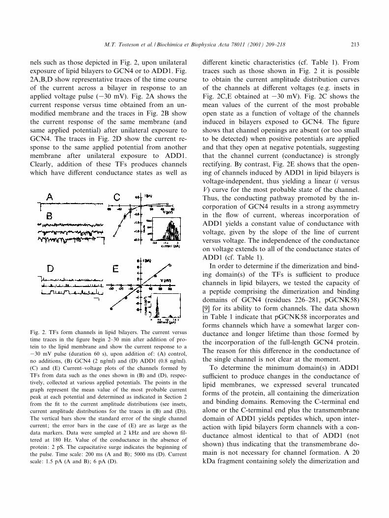
nels such as those depicted in Fig. 2, upon unilateralexposure of lipid bilayers to GCN4 or to ADD1. Fig.2A,B,D show representative traces of the time courseof the current across a bilayer in response to anapplied voltage pulse (330 mV). Fig. 2A shows thecurrent response versus time obtained from an un-modi¢ed membrane and the traces in Fig. 2B showthe current response of the same membrane (andsame applied potential) after unilateral exposure toGCN4. The traces in Fig. 2D show the current re-sponse to the same applied potential from anothermembrane after unilateral exposure to ADD1.Clearly, addition of these TFs produces channelswhich have di¡erent conductance states as well as
di¡erent kinetic characteristics (cf. Table 1). Fromtraces such as those shown in Fig. 2 it is possibleto obtain the current amplitude distribution curvesof the channels at di¡erent voltages (e.g. insets inFig. 2C,E obtained at 330 mV). Fig. 2C shows themean values of the current of the most probableopen state as a function of voltage of the channelsinduced in bilayers exposed to GCN4. The ¢gureshows that channel openings are absent (or too smallto be detected) when positive potentials are appliedand that they open at negative potentials, suggestingthat the channel current (conductance) is stronglyrectifying. By contrast, Fig. 2E shows that the open-ing of channels induced by ADD1 in lipid bilayers isvoltage-independent, thus yielding a linear (i versusV) curve for the most probable state of the channel.Thus, the conducting pathway promoted by the in-corporation of GCN4 results in a strong asymmetryin the £ow of current, whereas incorporation ofADD1 yields a constant value of conductance withvoltage, given by the slope of the line of currentversus voltage. The independence of the conductanceon voltage extends to all of the conductance states ofADD1 (cf. Table 1).
In order to determine if the dimerization and bind-ing domain(s) of the TFs is su¤cient to producechannels in lipid bilayers, we tested the capacity ofa peptide comprising the dimerization and bindingdomains of GCN4 (residues 226^281, pGCNK58)[9] for its ability to form channels. The data shownin Table 1 indicate that pGCNK58 incorporates andforms channels which have a somewhat larger con-ductance and longer lifetime than those formed bythe incorporation of the full-length GCN4 protein.The reason for this di¡erence in the conductance ofthe single channel is not clear at the moment.
To determine the minimum domain(s) in ADD1su¤cient to produce changes in the conductance oflipid membranes, we expressed several truncatedforms of the protein, all containing the dimerizationand binding domains. Removing the C-terminal endalone or the C-terminal end plus the transmembranedomain of ADD1 yields peptides which, upon inter-action with lipid bilayers form channels with a con-ductance almost identical to that of ADD1 (notshown) thus indicating that the transmembrane do-main is not necessary for channel formation. A 20kDa fragment containing solely the dimerization and
Fig. 2. TFs form channels in lipid bilayers. The current versustime traces in the ¢gure begin 2^30 min after addition of pro-tein to the lipid membrane and show the current response to a330 mV pulse (duration 60 s), upon addition of: (A) control,no additions, (B) GCN4 (2 ng/ml) and (D) ADD1 (0.8 ng/ml).(C) and (E) Current^voltage plots of the channels formed byTFs from data such as the ones shown in (B) and (D), respec-tively, collected at various applied potentials. The points in thegraph represent the mean value of the most probable currentpeak at each potential and determined as indicated in Section 2from the ¢t to the current amplitude distributions (see insets,current amplitude distributions for the traces in (B) and (D)).The vertical bars show the standard error of the single channelcurrent; the error bars in the case of (E) are as large as thedata markers. Data were sampled at 2 kHz and are shown ¢l-tered at 180 Hz. Value of the conductance in the absence ofprotein: 2 pS. The capacitative surge indicates the beginning ofthe pulse. Time scale: 200 ms (A and B); 5000 ms (D). Currentscale: 1.5 pA (A and B); 6 pA (D).
BBAMEM 78011 30-1-01
M.T. Tosteson et al. / Biochimica et Biophysica Acta 78011 (2001) 209^218 213
binding domain of ADD1, ADD1(p278^403), incor-porates into lipid membranes, and forms channelswhose major conductance state coincides with oneof the conductance states of ADD1 cf. Table 1).These results then indicate that for ADD1 as well,the binding and dimerization domain(s) is su¤cientto produce channels of the same qualitative electricalproperties as those of the full-length protein. Theresults further indicate that binding of the dimeriza-tion and binding domains of the TFs to lipid mem-branes leads to the formation of channels, whichresemble those formed by the full-length proteins,thus suggesting that these domains might form anintegral part of the ion-permeable pathways.
The fact that the conductance of the ADD1 frag-ment is lower than that of the full-length proteinmight be a consequence of the lack of the N-terminaldomain in this peptide, as suggested by the fact thatone of two other fragments derived from ADD1 con-taining the N-terminal domain through the bindingand dimerization domains induces channels with thesame conductance as that of ADD1-WT (not shown).
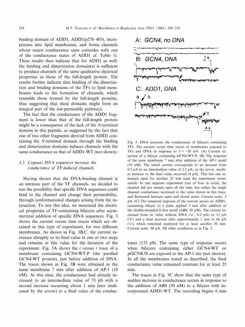
3.2. Cognate DNA sequences increase theconductance of TF-induced channels
Having shown that the DNA-binding element isan intrinsic part of the TF channels, we decided totest the possibility that speci¢c DNA sequences couldbind to the channel and change their permeability,through conformational changes arising from the in-teraction. To test this idea, we measured the electri-cal properties of TF-containing bilayers after asym-metrical addition of speci¢c DNA sequences. Fig. 3shows the current versus time traces which are ob-tained in this type of experiment, for two di¡erentmembranes. As shown in Fig. 3B,C, the current in-creases abruptly to its ¢nal value in one or two stepsand remains at this value for the duration of theexperiment. Fig. 3A shows the i versus t trace of amembrane containing GCN4-WT-P (the puri¢edGCN4-WT protein), just before addition of DNA.The traces shown in Fig. 3B were obtained in thesame membrane 7 min after addition of AP-1 (10nM). At this time, the conductance had already in-creased to an intermediate value of 73 pS with asecond increase occurring about 1 min later (indi-cated by the arrow) to a ¢nal value of the conduc-
tance (133 pS). The same type of response occurswhen bilayers containing either GCN4-WT orpGCNK58 are exposed to the AP-1 site (not shown).In all the membranes tested as described, the ¢nalconductance value remained constant for at least 25min.
The traces in Fig. 3C show that the same type ofsudden increase in conductance occurs in response tothe addition of ABS (50 nM) to a bilayer with in-corporated ADD1-WT. The recording begins 6 min
Fig. 3. DNA increases the conductance of bilayers containingTFs. The current versus time traces of membranes exposed toTFs and DNA in response to V =330 mV. (A) Current re-sponse of a bilayer containing GCN4-WT-P. (B) The responseof the same membrane 7 min after addition of the AP-1 motif(10 nM). The initial current corresponds to an increase from0.5 pA to an intermediate value of 2.2 pA; at the arrow, anoth-er increase to the ¢nal value occurred (4 pA). This last one re-mained open for another 25 min (and the experiment termi-nated). In one separate experiment (out of four in total), thechannel did not remain open all the time, but rather the singlechannel conductance increased to the value shown in this trace,and £uctuated between open and closed states. Current scale: 3pA. (C) The temporal response of the current across an ADD1-containing bilayer to a pulse applied 5 min after addition ofthe double-stranded E-box motif (ABS, 50 nM). The current in-creased from its value without DNA (`a': 6.2 pA) to 12 pA(`b') and a ¢nal increase after approximately 1 min to 66 pA(`c'), which remained unaltered for at least another 30 min.Current scale: 60 pA. All other conditions as in Fig. 2.
BBAMEM 78011 30-1-01
M.T. Tosteson et al. / Biochimica et Biophysica Acta 78011 (2001) 209^218214
after addition of the DNA sequence. The segmentlabeled `a' corresponds to a channel conductance of207 pS, obtained in the absence of DNA. At thearrow, there is a rather abrupt increase to about400 pS (`b') which lasts just over 1 min, followedby the last increase (`c') corresponding to a conduc-tance of about 2000 pS. The same type of responsewas observed when bilayers containing ADD1(p278^403) were exposed to either ABS or SRE (notshown).
Addition of non-cognate DNA sequences to TF-containing membranes either do not modify the ex-isting conductance or produce closure of the chan-nels (not shown). These results then suggest thatwhen TFs incorporate into lipid bilayers promotingthe formation of channels, the DNA-binding regionwithin the channel architecture is accessible to nu-cleotides added to the aqueous phase surroundingthe membrane. It is also interesting to point outthat addition of either SRE-1 (6 nM) or ABS (5nM) to bilayers containing channels formed by thefull-length protein ADD1 leads to an increase in theconductance of the membrane, as shown in Table 2.The channels induced by ADD1(p278^403), whichrespond to the addition of ABS (5 nM), requireabout six times as much SRE-1 to produce the con-ductance increase (cf. Table 2). This suggests that thea¤nity of the site for SRE-1 is lower in the incorpo-rated peptide (p278^403) than in the incorporatedfull-length protein (ADD1). A similar type of changein the binding a¤nity for DNA has been previouslyreported for the segment containing the DNA-bind-ing domain of NFTc [16].
3.3. The dimerization and binding domains areimportant for channel formation andDNA recognition
To explore further the speci¢city and importanceof the dimerization and binding domains both tochannel formation and to DNA binding, we tookadvantage of existent mutants of GCN4, which con-tain alterations in the LZ^basic region [17,18]. Themutant proteins with various insertions of aminoacids between the basic and the LZ regions producechanges in the conductance of bilayers with varyingphenotypes. Thus, (GCN4-2AA) a mutant with atwo amino acid insert between the basic region and
the LZ resulting in loss of function of GCN4, in-duces channels in bilayers. The value of the mostprobable conductance is somewhat larger than themost probably conductance of the channel formedby GCN4-WT (cf. Table 2) and with a longer life-time (not shown). Addition of DNA, however, pro-duces a partial blockage of the channel, since theconductance of the GCN4-2AA-containing bilayerchanges from about 25 pS to a value around 10 pS(cf. Table 2, `GCN4-2AA'). Addition of GCN4-N235S, a mutant with an N235 substitution forS235, which can dimerize but is devoid of DNAbinding, results in channels which lack recognitionof DNA, since the value of the most probable con-ductance state is not changed upon addition of AP-1,as shown in Table 2 `GCN4-N235S'. These resultsthen show that changes in the regions importantfor DNA recognition also have consequences forthe DNA recognition in the in vitro system.
3.4. E¡ects of other TFs
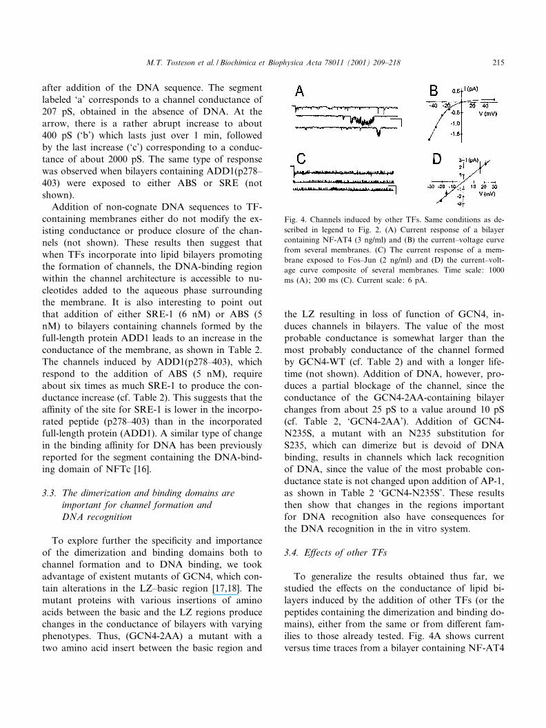
To generalize the results obtained thus far, westudied the e¡ects on the conductance of lipid bi-layers induced by the addition of other TFs (or thepeptides containing the dimerization and binding do-mains), either from the same or from di¡erent fam-ilies to those already tested. Fig. 4A shows currentversus time traces from a bilayer containing NF-AT4
Fig. 4. Channels induced by other TFs. Same conditions as de-scribed in legend to Fig. 2. (A) Current response of a bilayercontaining NF-AT4 (3 ng/ml) and (B) the current^voltage curvefrom several membranes. (C) The current response of a mem-brane exposed to Fos^Jun (2 ng/ml) and (D) the current^volt-age curve composite of several membranes. Time scale: 1000ms (A); 200 ms (C). Current scale: 6 pA.
BBAMEM 78011 30-1-01
M.T. Tosteson et al. / Biochimica et Biophysica Acta 78011 (2001) 209^218 215
and the averaged results obtained at various appliedvoltages in several membranes are shown in the cur-rent^voltage curves in Fig. 4B. Fig. 4C shows thecurrent versus time from another membrane contain-ing Fos^Jun and the (average current) versus voltagecurve for the heterodimer is shown in Fig. 4D. Sim-ilarly to the channels induced by GCN4, the i^Vcurve of the channels formed by NF-AT4 showsstrong recti¢cation, whereas the i^V plot of mem-branes containing Fos^Jun is linear, as is the casefor the i^V) plot of membranes containing ADD1(cf. Fig. 2).
We also attempted to incorporate peptides com-prising the dimerization and binding regions of theE-box proteins Myo-D and E47 which contain thebHLH DNA-binding domain. These proteins, whichdo not homodimerize and do not form dimers withother bHLH proteins [10], do not form channelswhen added to lipid membranes and addition of spe-ci¢c DNA fails to produce a change in the conduc-tance (not shown).
These data reinforce the notion that TFs capableof homodimerizing form channels in lipid mem-branes, since NF-AT4 (which forms homodimers)also forms channels in lipid bilayers (Fig. 4A), asdo GCN4 and ADD-1 (cf. Fig. 2). The heterodi-merizing proteins Fos or Jun failed to form channelswhen present alone (not shown). However, if bothpartners are present, the proteins form channels(Fig. 4C) capable of recognizing speci¢c DNA se-quences (cf. Table 2). Formation of channels in thepresence of Jun alone, however, was detected if theconcentration of the protein was approximately ¢vetimes that required to produce the Fos^Jun hetero-dimers. This suggests that even in the presence ofmembranes, Jun has a lower a¤nity for homodi-merization than for heterodimerization, as has al-ready been described [19]. Thus, taken altogether,the results presented suggest that the channel forma-tion induced by TFs only occurs if the appropriatedimerization partners are present.
A comparison of the traces shown in Fig. 2A (andC) with those shown in Fig. 4A (and C) reveals thatthe channels produced by the various TFs tested aredi¡erent in their amplitude and kinetic characteristics(cf. Table 1). Comparison of the plots in Fig. 2C(and E) with those in Fig. 4B (and D) further showthat the voltage dependence of the conductance in-
duced by the di¡erent proteins vary as well. Thus,one of the TFs containing the LZ motif (GCN4) andthe L-sheet representative (NF-AT4) induce a volt-age-dependent conductance when incorporated intolipid membranes (cf. Figs. 2C and 4B). However, thisdoes not seem to be a characteristic of the LZ motif,since other members of this family, Fos^Jun, interactwith lipid bilayers and produce channels with a linearresponse of the current with voltage, as does thebHLH-LZ protein, ADD1 (cf. Figs. 2E and 4D).
To summarize, the data we have presented con¢rmthe hypothesis that TFs form channels in lipid bi-layers. The particular electrical characteristics ofthese channels were found to be di¡erent for eachof the particular proteins. Apparently, the electricalproperties of the channels are subtle manifestationsof slight di¡erences in the conformations of eachparticular protein (cf. Figs. 2 and 4), as has beenfound to be the case in studies of the structure^func-tion relation of voltage-gated peptides [20,21].
Moreover, the data obtained with the mutants ofGCN4, have underscored the importance of the dis-tance between the basic region (DNA-binding) andthe dimerization region for channel formation. Thus,modi¢cations in the distance between these regionsvia addition of amino acids (spacers) result in alteredchannel characteristics as well as altered DNA bind-ing (cf. Table 2).
The data in this communication further reveal aninteresting property of the channels induced by thedimerization and binding domains of the transcrip-tions factors tested: the recognition of speci¢c DNAsequences. Recent reports have shown that DNA cantranslocate across planar lipid bilayers containinglarge channels formed by porin (a protein from theouter wall of bacteria) or by K-hemolysin (fromStaphylococcus aureus), producing transient closuresof the channels in their transit [5,22]. The results ofour experiments suggest that the channels formed byTFs are speci¢c DNA-recognizing channels ratherthan DNA-translocating ones, based on the factthat addition of speci¢c DNA sequences leads toan increase in the channel conductance (cf. Fig. 3and Table 2), and that this increase in the conduc-tance only happens upon addition of cognate DNAsequences, irrespective of the size of the channel con-ductance (cf. Tables 1 and 2). This contrasts with thestudies on the translocation of DNA through porin
BBAMEM 78011 30-1-01
M.T. Tosteson et al. / Biochimica et Biophysica Acta 78011 (2001) 209^218216
and K-hemolysin channels, since in the small-sizedchannels (10^40 pS) the DNA sequences (no specif-icity required) cannot penetrate into the channel andproduce the characteristic transient changes in cur-rent [5]. In contrast, in our experiments the conduc-tance of the TF-induced channels does not determinethe events which follow the addition of DNA sequen-ces. The increase in the conductance of the proteinchannels occurs only upon addition of cognate DNAsequences. Thus, we would suggest that the increasein the conductance which we observe upon additionof the speci¢c DNA sequences to TF-induced chan-nels is due to the binding of DNA and rearrange-ment of the channel's architecture to accommodatethe bound DNA molecule(s). The fact that the sig-nal(s) which this process produces are speci¢c forcognate DNA-binding domains could be utilized todetermine the existence and nature of as yet undeter-mined binding sequences in proteins.
Acknowledgements
We thank Dr. H. Aktas for the cloning and invitro synthesis of GCN4-WT, GCN4-2AA, GCN4-7AA and GCN4-N235S, as well as for the puri¢ca-tion of the GCN4 protein, Dr. E. Roydon Price forthe in vitro synthesis of NF-AT4 and of Fos, Junand Fos^Jun. The synthetic peptides containing thedimerization and binding domains of Myo-D, E-47and pGCNK58 were generously provided by Dr. T.Ellenberger, to whom we are also indebted for manyhelpful discussions. The plasmids for GCN4 and mu-tants were generously provided by Dr. K. Struhl. Wethank Dr. J.A. Halperin, Dr. M. Chow and Dr. F.McKeon for reading the manuscript and providingus with incisive comments. Supported in part by theDana Foundation (D.C.T., M.T.T.) and by theAnna-Fuller fellowship (J.B.K.).
References
[1] C.O. Paba, R.M. Saber, Transcription factors: structuralfamilies and principles of DNA recognition, Annu. Rev. Bio-chem. 61 (1992) 1053^1095.
[2] E. London, How bacterial protein toxins enter cells ; the role
of partial unfolding in membrane translocation, Biochim.Biophys. Acta 1113 (1992) 25^51.
[3] W.A. Cramer, J.B. Heymann, S.L. Schendel, B.N. Deriy,F.S. Cohen, P.A. Alkanes, C.V. Stau¡acher, Structure^func-tion of the channel-forming colicins, Annu. Rev. Biophys.Biomol. Struct. 24 (1995) 611^641.
[4] A.J. Minn, P. Velez, S.L. Schendel, H. Liang, S.W. Much-more, S.W. Fesik, M. Fill, C.B. Thompson, Bcl-x(L) formsan ion channel in synthetic lipid membranes, Nature 385(1997) 353^357.
[5] I. Szabo, G. Bathori, F. Tombola, M. Brini, A. Coppola, M.Zoratte, DNA translocation across planar bilayers contain-ing Bacillus subtilis ion channels, J. Biol. Chem 40 (1997)25275^25282.
[6] M. Montal, P. Mueller, Formation of bimolecular mem-branes from lipid monolayers and a study of their electricalproperties, Proc. Natl. Acad. Sci. USA 69 (1997) 3561^3566.
[7] M.T. Tosteson, M. Chow, Characterization of the ion chan-nels formed by poliovirus in planar lipid membranes, J. Vi-rol. 71 (1997) 507^511.
[8] F.J. Sigworth, S.M. Sine, Data transformations for im-proved display and ¢tting of single-channel dwell time histo-grams, Biophys. J. 52 (1987) 1047^1054.
[9] T.E. Ellenberger, C.J. Brandt, K.I. Struhl, S.C. Harrison,The GCN4 basic region leucine zipper binds DNA as adimer of uninterrupted K helices: crystal structure of theprotein^DNA complex, Cell 71 (1992) 1223^1237.
[10] T. Ellenberger, D. Fass, M. Arnaud, S.C. Harrison, Crystalstructure of transcription factor E47: E-box recognition by abasic region helix^loop^helix dimer, Genes Dev. 8 (1994)970^980.
[11] J.B. Kim, G.D. Spotts, Y.-D. Halvorsen, H.-M. Shih, T.Ellenberger, H.C. Towle, B.M. Spiegelman, Dual DNAbinding speci¢city of ADD1/SREBP1 controlled by a singleamino acid in the basic helix^loop^helix domain, Mol. Cell.Biol. 15 (1995) 2582^2588.
[12] I.A. Hope, K. Struhl, GCN4, a eukaryotic transcriptionalactivator protein, binds as a dimer to target DNA, EMBOJ. 6 (1987) 2781^2784.
[13] P. Tontonoz, J.B. Kim, R.A. Graves, B.M. Spiegelman,ADD1: a novel helix^loop^helix transcription factor associ-ated with adipocyte determination and di¡erentiation, Mol.Cell. Biol. 13 (1993) 4753^4759.
[14] C. Yokoyama, X. Wang, SREBP-1, a basic-helix^loop^he-lix-leucine zipper protein that controls transcription of thelow density lipoprotein receptor gene, Cell 75 (1993) 187^197.
[15] H. Shimano, J.D. Horton, I. Shimomura, R.E. Hammer,M.S. Brown, J.L. Goldstein, Elevated levels of SREBP-2and cholesterol synthesis in livers of mice homozygous fora targeted disruption of the SREBP-1 gene, J. Clin. Invest.99 (1997) 846^854.
[16] S.A. Wolfe, P. Zhou, V. Do«tsch, L. Chen, A. You, S.N. Ho,G.R. Crabtree, G. Wagner, G.L. Verdine, Unusual Rel-like
BBAMEM 78011 30-1-01
M.T. Tosteson et al. / Biochimica et Biophysica Acta 78011 (2001) 209^218 217
architecture in the DNA-binding domain of the transcriptionfactor NFATc, Nature 385 (1997) 172^176.
[17] W.T. Pu, K. Struhl, The leucine zipper symmetrically posi-tions the adjacent basic regions for speci¢c DNA binding,Proc. Natl. Acad. Sci. USA 88 (1991) 6901^6905.
[18] W.T. Pu, K. Struhl, Highly conserved residues in the bZIPdomain of yeast GCN4 are not essential for DNA binding,Mol. Cell. Biol. 11 (1991) 4918^4926.
[19] L.J. Ransone, P. Wamsley, K.L. Morley, I.M. Verma, Do-main swapping reveals the modular nature of Fos, Jun andCREB proteins, Mol. Cell. Biol. 10 (1990) 4565^4573.
[20] M.T. Tosteson, M.P. Caul¢eld, J.J. Levy, M. Rosenblatt,
D.C. Tosteson, Solid-phase synthesis of melittin: puri¢cationand functional characterization, Biosci. Rep. 8 (1988) 173^183.
[21] M.T. Tosteson, O. Alvarez, W. Hubbell, R.M. Bieganski, C.Attenbach, L.H. Caporales, J.J. Levy, R.F. Nutt, M. Rosen-blatt, D.C. Tosteson, Primary structure of peptides and ionchannels. Role of amino acid side chains in voltage gating ofmelittin channels, Biophys. J. 58 (1990) 1367^1375.
[22] J.J. Kasianowicz, E. Brandin, D. Branton, D.W. Deamer,Characterization of individual polynucleotide molecules us-ing a membrane channel, Proc. Natl. Acad. Sci. USA 93(1996) 1859^1866.
BBAMEM 78011 30-1-01
M.T. Tosteson et al. / Biochimica et Biophysica Acta 78011 (2001) 209^218218