interaction of human mannose‐binding protein with mycobacterium avium
TRANSCRIPT

1159
Interaction of Human Mannose-Binding Protein with Mycobacterium avium
Vsevolod Y. Polotsky,* John T. Belisle, Infectious Diseases Section, Department of Internal Medicine, YaleUniversity School of Medicine, New Haven, Connecticut; Department ofKatarina Mikusova, R. Alan B. Ezekowitz,
Microbiology, Colorado State University, Fort Collins, Colorado;and Keith A. JoinerDivisions of Hematology and Infectious Diseases, Children’s Hospital,
Harvard Medical School, Boston, Massachusetts
The interaction between human mannose-binding protein (MBP) and Mycobacterium avium wasexplored. By ELISA, calcium-dependent and mannan-inhibitable binding of human recombinantMBP (rMBP) to live M. avium was observed. Preincubation of M. avium with rMBP resulted in a2-fold increase in uptake by human neutrophils. Mycobacterial cell wall components were assessedby ELISA for their ability to bind the carbohydrate recognition domain of rMBP. The best ligandwas mannosyl-lipoarabinomannan, followed by lipomannan, phosphatidylinositol mannoside, arabi-nosyl-lipoarabinomannan, and dimycolated trehalose (cord factor). rMBP did not bind to partiallypurified lipid fractions containing glycopeptidolipids. These results are consistent with the knownstructural basis for rMBP ligand recognition. They suggest that MBP may play a role in hostdefense against M. avium by opsonizing both whole organisms and free cell wall components forinternalization.
The mechanisms of host recognition for mycobacterial spe- MBP monomers (32 kDa) form a trimeric structural subunit,which in turn multimerizes into pentamers or hexamers ofcies are still poorly understood. Since infections with Mycobac-
terium tuberculosis and Mycobacterium avium are of increasing Ç600 kDa [15, 16] that resemble C1q in overall structure.Recombinant MBP (rMBP) can activate the complement cas-importance in immunocompromised patients [1–3], studies of
innate resistance, capable of modulating mycobacterial infec- cade via both the alternative [17] and classical pathways, al-though classical pathway activation now is attributed to antion at the very early stages of infection, are of particular
relevance. The studies reported here focus on mycobacterial MBP-associated serine protease capable of cleaving C1 de-tected in some preparations of MBP [18].recognition by one such primary recognition molecule, human
mannose-binding protein (MBP). MBP is a pattern-recognition molecule that recognizes car-bohydrate configurations on a wide variety of pathogens. Man-MBP belongs to a family of collagenous molecules, termed
collectins, all of which are involved in innate pathogen recogni- nan-inhibitable binding of MBP to Salmonella montevideo,Streptococcus pneumoniae, Streptococcus agalactiae, Crypto-tion. These oligomeric proteins contain a carboxy terminal car-
bohydrate recognition domain (CRD). In addition to serum coccus neoformans, human immunodeficiency virus gp120, andinfluenza A hemagglutinin has been described [17, 19–22].MBPs, the collectins include lung surfactant proteins (SP-A
and SP-D), conglutinin, and a newly described bovine protein Although not previously tested, mycobacteria contain a varietyof cell wall lipoglycans and glycolipids that are potential li-termed collectin-43 [4–8]. The first complement component,
C1q, and the macrophage scavenger receptor are two other gands for MBP, some of which are already shown to be ligandsfor the macrophage mannose receptor [23–25]. We thereforecollagen-containing proteins that also appear to play a role in
host defense but do not have lectin-like domains [5, 9–14]. initiated studies on binding of MBP to M. avium and to myco-bacterial cell wall (MCW) components. Since the structuralHuman MBP is composed of a short noncollagenous N-
terminal segment followed by 19 collagen (Gly-X-Y) repeats. basis for carbohydrate recognition by MBP is now known [26–31], we could compare our results directly with predictions ofThe 130 carboxy terminal amino acids constitute the CRD.which MCW components should be recognized by MBP. Thisapproach should be generally applicable to analysis of host-parasite interactions with molecules of known structure.
Received 16 May 1996; revised 5 December 1996.Financial support: NIH (AI-30286 to K.A.J., AI-25147 to J.T.B., and RR-
02172 to R.A.B.E.); Bristol-Myers Squibb Institute (grant-in-aid to R.A.B.E.);MethodsPediatric AIDS Foundation (grant to R.A.B.E.).
Reprints or correspondence: Dr. Keith A. Joiner, Infectious Diseases Section,Bacteria and growth conditions. M. avium serovar 2 strainsDept. of Internal Medicine, Yale University School of Medicine, 808 LCI,
P.O. Box 208022, New Haven, CT 06520-8022. 2151 SmO (glycopeptidolipid [GPL] positive [GPL/], smooth-* Present affiliations: Department of Medicine, Norwalk Hospital, Norwalk, domed, opaque), 2151 RgO (GPL negative [GPL0], lipopeptide
Connecticut (V.Y.P.); Department of Pediatrics, Massachusetts General Hospi-positive, rough), 2151 Rg3 (GPL0, lipopeptide negative, rough)tal, Boston, Massachusetts (A.B.E.).[32], and M. avium serovar 1 strain 101 [33] were grown on 7H11
The Journal of Infectious Diseases 1997;175:1159–68agar for 2–3 weeks, subcultured to 7H9 broth supplemented with� 1997 by The University of Chicago. All rights reserved.
0022–1899/97/7505–0018$01.00 medium (oleic acid, albumin, dextrose, catalase), and incubated at
/ 9d27$$my30 04-04-97 09:43:05 jinfa UC: J Infect
by guest on January 29, 2016http://jid.oxfordjournals.org/
Dow
nloaded from

1160 Polotsky et al. JID 1997;175 (May)
37�C to mid-log phase. M. tuberculosis H37Rv and Erdman were dependent binding was determined by subtraction of [125I]rMBPbinding to bacteria in the presence of 20 mM EDTA from itsgrown in glycerol alanine salt broth [34] for 2 weeks at 37�C
with gentle agitation. The rapidly growing Mycobacterium species, binding in the presence of 50 mM Ca//.Association of M. avium with human neutrophils. The phago-expressing arabinosyl-lipoarabinomannan (AraLAM) [35, 36], was
grown in glycerol alanine salt broth for 1 week at 37�C with gentle cytosis assay was done as described [39] with minor modifications.In brief, mid-log phase M. avium serovar 1 strain 101 was washedagitation. S. montevideo (SH 5770; from P. H. Makela, Central
Public Health Laboratory, Helsinki) and Streptococcus pyogenes and vigorously stirred overnight in HBSS plus 50 mM CaCl2 at4�C to break up clumps of mycobacteria. Then organisms were(T1/195/2; from V. Fischetti, Rockefeller University, New York)
were grown as described previously [17]. Bacteria were washed incubated in the same buffer with or without rMBP (10 mg/mL),or rMBP and mannan (2.5 mg/mL) for 1 h at 37�C. Bacteria werethree times with Hanks’ balanced salt solution (HBSS) containing
0.15 mM CaCl2 and 1 mM MgCl2 , then resuspended to 2 1 109 next washed three times in HBSS plus 0.15 mM CaCl2 , 1 mMMgCl2 , and 0.1% gelatin, and then resuspended at 5 1 107 cells/bacteria/mL (optical density at 600 nm Å 2.00) in HBSS containing
50 mM CaCl2 unless otherwise stated. mL and added to 107 polymorphonuclear leukocytes in 1 mL ofthe same solution. The bacteria and phagocytes were gently rockedReagents. Sepharose CL-6B, dextran T500, and prepacked
PD10 columns were from Pharmacia (Piscataway, NJ), bovine at 37�C for 20 min, and thereafter unattached bacteria were re-moved by four washes and centrifugation at 82 g. Neutrophilsserum albumin (BSA) was from United States Biochemicals
(Cleveland), IodoBeads were from Pierce Chemical (Rockford, were adhered to coverslips for 10 min at 37�C, fixed by heatingat 65�C overnight, and stained with auramine O (Remel, Windsor,IL), Na125I was from Amersham (Arlington Heights, IL), and gela-
tin was from Difco (Detroit). Mannose-Sepharose was prepared CT).MCW components. Mannosyl-lipoarabinomannan (ManLAM)by divinyl sulfone–coupling of D-mannose to Sepharose CL-6B,
using minor modifications of earlier procedures [37]. rMBP and from M. tuberculosis H37Rv, AraLAM, and lipomannan (LM)from a rapidly growing Mycobacterium species, and phosphatidyl-native MBP were produced as described in [38]. A murine mono-
clonal antibody, MAb3, with specificity for bound and free MBPs inositol mannoside (PIM) from M. tuberculosis Erdman were pro-duced as previously described [40, 41]. Cord factor (dimycolated[38], was conjugated to horseradish peroxidase and designated
aMBP3-HRP. All other reagents were from Sigma (St. Louis) trehalose) from M. tuberculosis H37Rv was isolated as described[42]. Total lipid was isolated from M. avium, and mild alkalineunless otherwise stated.
ELISA. Microtitration plates (Immulon 2; Dynatech, Burling- hydrolysis of total lipid was performed as described [43].The total lipid and deacylated lipid were fractionated by columnton, MA) were coated overnight at 4�C with live bacteria resus-
pended at 2 1 108/mL (A600 Å 0.2) in carbonate buffer (15 mM chromatography (Florisil Sep-Pak cartridge; Waters Chromatogra-phy, Milford, MA). The Sep-Pak was equilibrated with 5 mL ofNa2CO3, 30 mM NaHCO3, 3 mM NaN3, pH 9.5) or with MCW
components resuspended in the same buffer. The plates were CHCl3 , and 100 mg of lipid in 0.5 mL of CHCl3 was applied tothe cartridge. The lipid was sequentially eluted with 5 mL eachwashed four times in PBS with 0.05% Tween 80 (Sigma), blocked
with 5% BSA at 37�C for 1 h, and then washed and coated with of the following solvents: 100% CHCl3 followed by 1%, 2%, 5%,10%, 15%, 20%, 30%, 40%, and 50% CH3OH in CHCl3 , and thenrMBP in HBSS plus 50 mM CaCl2 or with rMBP in HBSS plus
20 mM EDTA with or without competitor (mannan). After a 1-h with 100% CH3OH. Lipids from individual fractions were resolvedby thin-layer chromatography (TLC), using aluminum-backedincubation at 37�C, plates were washed four times in PBS with
0.05% Tween 80, blocked with 5% BSA for 1 h at 37�C, and TLC plates (SilicaGel 60; EM Science, Gibbstown, NJ). The sol-vent used was CHCl3/CH3OH/H2O (65:25:4). Lipids were visual-probed with aMBP3-HRP (1:2000) in PBS with 1% BSA for 1 h
at 37�C. The plates were washed in PBS with 0.05% Tween 80 ized by spraying the TLC plates with 10% H2SO4 in CH3OH andheating them at 110�C.and developed with a solution of 3 mM H2O2, 22.4 mM citric acid,
51.4 mM Na2HPO4, and 2.2 mM o-phenyldiamine in the dark. Thereaction was stopped with 4 M sulfuric acid and read on a Mi-croplate Reader 450 (Bio-Rad, Richmond, CA) at 450 nm. Results Resultswere expressed in optical density units. In some experiments with
rMBP binding to live M. avium. We measured rMBP bind-inhibitors, a fixed concentration of rMBP was used, and resultsing to live M. avium serovar 1 strain 101 by ELISA. In thewere expressed as the percent of ELISA inhibition calculated aspresence of calcium, the binding was dose dependent and100 1 (A450 without inhibitor 0 A450 with inhibitor)/A450 without
inhibitor. reached a plateau at Ç1 mg/mL rMBP. Binding was calcium-[125I ]rMBP binding assay. rMBP was radiolabeled with dependent and mannan-inhibitable (figure 1A). The calcium
Na125I, using IodoBeads as previously described [38]. Live M. dependence of rMBP binding to mycobacteria suggested thatavium serovar 1, S. montevideo, or S. pyogenes were resuspended this binding occurred via the CRD. To confirm this assumption,in HBSS plus 50 mM CaCl2 or HBSS plus 20 mM EDTA at 2 1 we repeated the assay in the presence of mannan with a fixed106 bacteria/mL in 1.5-mL microcentrifuge tubes precoated with
concentration (150 ng/mL) of rMBP chosen from the linear1% BSA. Then [125I]rMBP was added at 900 ng/mL, and unlabeled
part of the ELISA curve. In the presence of Ca//, mannanrMBP was added to bring the total rMBP concentration to 5.64inhibited rMBP binding to organisms in a dose-dependent fash-mg/mL. This concentration was shown in preliminary experimentsion, reaching 80% inhibition at 400 mg/mL (figure 1B).to be saturating under these conditions. After a 30-min incubation
We compared calcium-dependent rMBP binding to M. aviumat 37�C, organisms were washed twice in cold HBSS plus 0.15mM CaCl2 and 1 mM MgCl2 , and counts were determined. Ca//- with binding to mannose-rich S. montevideo [17, 38] and to S.
/ 9d27$$my30 04-04-97 09:43:05 jinfa UC: J Infect
by guest on January 29, 2016http://jid.oxfordjournals.org/
Dow
nloaded from

1161JID 1997;175 (May) Mannose-Binding Protein and M. avium
sorbed human serum resulted in a significant increase in thenumber of mycobacteria associated with neutrophils (15 [P õ.05] and 26 [P õ .05] per 100 neutrophils, respectively, vs. 8without opsonization) and the percentage of neutrophils withattached or engulfed bacteria (6.7 [P õ .02] and 9.2 [P õ.01] vs. 2.7, respectively) (figure 3). When mycobacteria werepreincubated with rMBP in the presence of mannan, the numberof cell-associated organisms (3/100) and the percentage of neu-trophils associated with mycobacteria (1.1%) declined belowthose of the control cells. This suggests that mannan may beinhibiting some process in addition to binding of MBP. Thesedata indicate that MBP can opsonize M. avium by a comple-ment-independent mechanism.
Neutrophils bound either single organisms or clumps of my-cobacteria (figure 4). The main effect of rMBP was to increasethe percentage of neutrophils with attached organisms ratherthan the number of organisms per cell. That is, the averagenumber of organisms per infected neutrophil was 2–3 regard-less of whether opsonized or nonopsonized mycobacteria wereused, indicating that the effects of rMBP were not simply aconsequence of bacterial aggregation. The relatively low per-centage of neutrophils associated with M. avium and the lownumber of bacteria per cell, compared with those in other stud-ies [20, 23, 24], may be explained by the low bacteria-to-neutrophil ratio used in our experiments (5:1). Alternatively,neutrophils may be relatively less efficient at attaching to bacte-ria than are monocytes and macrophages. Regardless, the dif-ference between the association of M. avium with neutrophilsin the presence and absence of rMBP was highly significantstatistically and indicates that rMBP directly opsonizes theorganism for cell interaction.
Binding of rMBP to M. avium GPL variants. The majorcomponent on the M. avium surface is GPL. [125I]rMBP binding
Figure 1. Recombinant MBP (rMBP) binding to M. avium serovar1 strain 101 by ELISA in presence or absence of calcium. A, Bindingof increasing concentrations of rMBP to M. avium. B, Inhibition ofrMBP binding to M. avium by mannan in presence of calcium. rMBPÅ 150 ng/mL. OD (optical density) at 450 nm in absence of inhibitionÅ 0.56 { 0.07. Data points are means { SDs of means of 11 (A) or5 (B) separate experiments.
pyogenes group A, which lacks mannose-rich surface ligandsfor MBP. At saturation, the amount of [125I]rMBP binding toM. avium serovar 1 strain 101 was similar to the binding to S.montevideo and slightly higher than to M. avium serovar 2strain SmO (figure 2). Binding to S. pyogenes was 5- to 9-fold
Figure 2. [125I]rMBP calcium-dependent binding to Mycobacte-lower than binding to the other organisms. It was not possiblerium avium serovar 1 strain 101 and serovar 2 strain 2151 SmOto calculate precisely the number of molecules of rMBP bound(smooth-domed, opaque), Salmonella montevideo strain 5770, andper organism, due to heterogeneity in the multimerization stateStreptococcus pyogenes group A strain T1/195/2. Concentrations:
of the rMBP (data not shown). bacteria Å 2 1 106/mL, rMBP Å 5.7 mg/mL. Binding in EDTA wasOpsonization by rMBP of M. avium for association with subtracted from binding in Ca// to give calcium-dependent binding.
Data points are means { SDs of means of 5 separate experiments.human neutrophils. Preincubation with rMBP or 10% ad-
/ 9d27$$my30 04-04-97 09:43:05 jinfa UC: J Infect
by guest on January 29, 2016http://jid.oxfordjournals.org/
Dow
nloaded from
1162 Polotsky et al. JID 1997;175 (May)
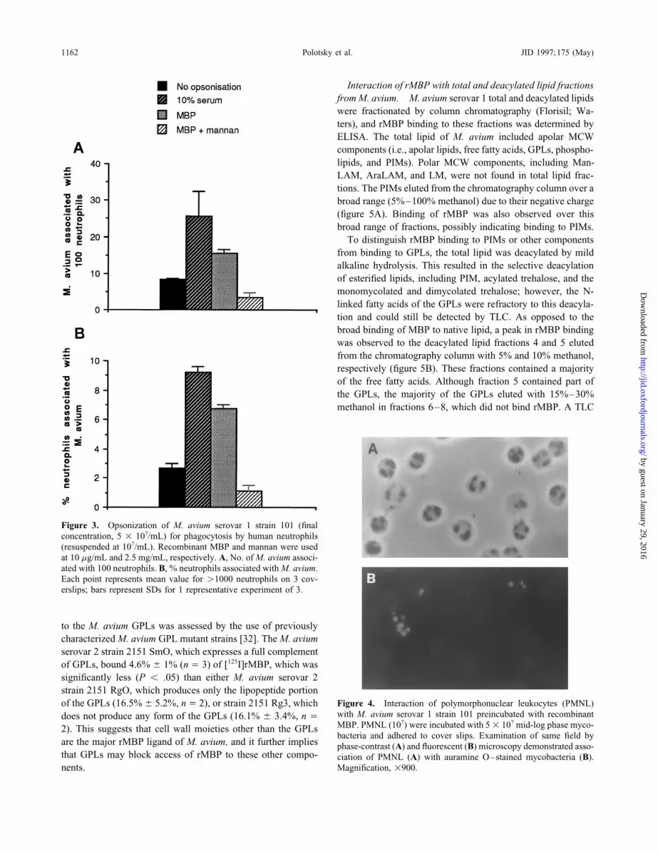
Interaction of rMBP with total and deacylated lipid fractionsfrom M. avium. M. avium serovar 1 total and deacylated lipidswere fractionated by column chromatography (Florisil; Wa-ters), and rMBP binding to these fractions was determined byELISA. The total lipid of M. avium included apolar MCWcomponents (i.e., apolar lipids, free fatty acids, GPLs, phospho-lipids, and PIMs). Polar MCW components, including Man-LAM, AraLAM, and LM, were not found in total lipid frac-tions. The PIMs eluted from the chromatography column over abroad range (5%–100% methanol) due to their negative charge(figure 5A). Binding of rMBP was also observed over thisbroad range of fractions, possibly indicating binding to PIMs.
To distinguish rMBP binding to PIMs or other componentsfrom binding to GPLs, the total lipid was deacylated by mildalkaline hydrolysis. This resulted in the selective deacylationof esterified lipids, including PIM, acylated trehalose, and themonomycolated and dimycolated trehalose; however, the N-linked fatty acids of the GPLs were refractory to this deacyla-tion and could still be detected by TLC. As opposed to thebroad binding of MBP to native lipid, a peak in rMBP bindingwas observed to the deacylated lipid fractions 4 and 5 elutedfrom the chromatography column with 5% and 10% methanol,respectively (figure 5B). These fractions contained a majorityof the free fatty acids. Although fraction 5 contained part ofthe GPLs, the majority of the GPLs eluted with 15%–30%methanol in fractions 6–8, which did not bind rMBP. A TLC
Figure 3. Opsonization of M. avium serovar 1 strain 101 (finalconcentration, 5 1 107/mL) for phagocytosis by human neutrophils(resuspended at 107/mL). Recombinant MBP and mannan were usedat 10 mg/mL and 2.5 mg/mL, respectively. A, No. of M. avium associ-ated with 100 neutrophils. B, % neutrophils associated with M. avium.Each point represents mean value for ú1000 neutrophils on 3 cov-erslips; bars represent SDs for 1 representative experiment of 3.
to the M. avium GPLs was assessed by the use of previouslycharacterized M. avium GPL mutant strains [32]. The M. aviumserovar 2 strain 2151 SmO, which expresses a full complementof GPLs, bound 4.6% { 1% (n Å 3) of [125I]rMBP, which wassignificantly less (P õ .05) than either M. avium serovar 2strain 2151 RgO, which produces only the lipopeptide portion
Figure 4. Interaction of polymorphonuclear leukocytes (PMNL)of the GPLs (16.5% { 5.2%, n Å 2), or strain 2151 Rg3, whichwith M. avium serovar 1 strain 101 preincubated with recombinantdoes not produce any form of the GPLs (16.1% { 3.4%, n ÅMBP. PMNL (107) were incubated with 5 1 107 mid-log phase myco-2). This suggests that cell wall moieties other than the GPLsbacteria and adhered to cover slips. Examination of same field by
are the major rMBP ligand of M. avium, and it further implies phase-contrast (A) and fluorescent (B) microscopy demonstrated asso-that GPLs may block access of rMBP to these other compo- ciation of PMNL (A) with auramine O–stained mycobacteria (B).
Magnification, 1900.nents.
/ 9d27$$my30 04-04-97 09:43:05 jinfa UC: J Infect
by guest on January 29, 2016http://jid.oxfordjournals.org/
Dow
nloaded from

1163JID 1997;175 (May) Mannose-Binding Protein and M. avium
Interaction of rMBP with purified MCW components. Themost likely candidates for the cell surface binding of rMBPare the mannosylated lipoglycans, lipoarabinomannan, and LM,which populate the cellular envelope of all mycobacterial spe-cies [40, 44]. Two forms of LAM have been described, amannose-capped version (ManLAM) that was originally de-fined in M. tuberculosis [41] and a version possessing terminalarabinose residues (AraLAM) that was isolated from a rapidlygrowing Mycobacterium species [35]. It is known that LAMproduced by M. avium is of the mannose-capped form. PurifiedLAM, LM, PIM, and dimycolated trehalose were evaluatedby ELISA for their ability to bind rMBP (figure 6). Initialexperiments (figure 6A) showed that 10 mg/mL of ManLAM,AraLAM, LM, PIM, and dimycolated trehalose gave saturableligand binding of rMBP to the plates. Although differentialbinding of MCW components to the plate is likely, given sub-stantial differences in the structure and hydrophobicity of themolecules, the complete saturability of rMBP binding at MCWbinding concentrations ú10 mg/mL argues strongly that thedifferences observed in figure 6B and C are not due primarilyto binding of the ligand to the plate.
rMBP binding to plate-bound ligand approached saturationat an MBP concentration of 100–200 ng/mL (figure 6B); Man-LAM binding was the greatest and was not saturated at 0.5mg/mL rMBP. In subsequent experiments, we used rMBP at afixed concentration (150 ng/mL) chosen from the linear partof the ELISA curve. Under these conditions, M. tuberculosisManLAM was the best MBP ligand, with LM and PIM exhib-iting two times less MBP binding. AraLAM, as expected,bound significantly less MBP than did ManLAM. In addition,dimycolated trehalose, which does not contain mannose, bindssignificantly less rMBP than does PIM (figure 6C). These re-sults indicated that the terminal mannose caps of ManLAM
Figure 5. Calcium-dependent recombinant MBP (rMBP) binding were responsible for the increased binding capacity of thisto total (A) and deacylated (B) lipids of M. avium serovar 1 fraction- molecule.ated on chromatography column by methanol gradient (�). ELISAplates were coated with lipid fractions solubilized in 1% SDS andresuspended in carbonate buffer at 100 mg/mL. rMBP (625 ng/mL) Discussionin 50 mM Ca// or 20 mM EDTA was tested for binding to lipidfractions (�). Binding in EDTA was subtracted from binding in Ca// MBP may participate in host defense against mycobacteriato give calcium-dependent binding. Horizontal bars show thin-layer through several mechanisms. We have shown here that MBPchromatography fractions containing indicated components from total recognizes a variety of MCW components that are localized(A) or deacylated (B) lipids: PL Å phospholipids, PIM Å phosphati-
in the outermost leaflet of the cell wall [45]. Consistent withdylinositol mannoside, AL Å apolar lipids, FA Å free fatty acids,that finding, MBP opsonizes whole mycobacteria for internal-GPL Å glycopeptidolipids. OD Å optical density.ization. It is also likely that MBP participates, through bothcomplement-dependent [17] and complement-independentpathways, in clearance of isolated MCW components that have
profile of M. avium serovar 4 and 8 lipid fractions and rMBP been shed from the bacterial surface. In so doing, MBP maybinding to these fractions in ELISA (not shown) yielded results modulate the immunologic response to the infection. For exam-similar to those for M. avium serovar 1. These results, combined ple, we have recently shown that rMBP substantially augmentswith the data obtained with the M. avium GPL mutants, suggest binding and internalization of purified fluid-phase ManLAMthat rMBP binding to M. avium lipids was due to the presence by the human monocytic cell line U937 (unpublished data).of PIM but not GPL. These data also indicate that rMBP may Given the myriad of immunologic responses that are eitherbe interacting with free fatty acids or some undefined product triggered or inhibited in monocytes and macrophages by LAM
[46–48], it will now be important to determine the influencein the fractions 4 and 5 eluted with 5%–10% methanol.
/ 9d27$$my30 04-04-97 09:43:05 jinfa UC: J Infect
by guest on January 29, 2016http://jid.oxfordjournals.org/
Dow
nloaded from

1164 Polotsky et al. JID 1997;175 (May)
Figure 6. Binding of recombinant MBP (rMBP) toELISA plates coated with mannose cell wall (MCW)components. A, binding of rMBP (75 ng/mL) to in-creasing concentrations of MCW components in pres-ence of 50 mM Ca//. Since 10 mg/mL MCW compo-nents gave saturable binding of rMBP, thisconcentration was used for experiments in B and C.B, Binding of increasing concentrations of rMBP toMCW components. Values are means { SDs from 3separate experiments done in duplicate. C, Binding ofrMBP (150 ng/mL) expressed as % of rMBP-mannanbinding Å 100 1 optical density (OD) of MCW/ODof mannan at each coating concentration. Values aremeans { SEs from 3 separate experiments done intriplicate. Mannosyl-lipoarabinomannan (ManLAM)vs. lipomannan (LM), P õ .02; ManLAM vs. arabino-syl-LAM (AraLAM), phosphatidylinositol mannoside(PIM), P õ .01; ManLAM vs. cord factor (dimyco-lated trehalose), P õ .005; PIM vs. cord factor, P õ.05, by 2-tailed t test.
of MBP on these parameters. In addition, a common opsonic ciency is a predisposing factor to mycobacterial infections orinfluences the severity of established infections.defect associated with low serum MBP concentrations results
in immunodeficiency associated with repeated bacterial and Co crystals of the rat MBP-A CRD with an oligomannoseligand reveals that mannose binds to the CRD of MBP in directfungal infections in children [49] and adults [50)]. Additional
clinical studies are needed to investigate whether MBP defi- coordination to the calcium ions [26, 27]. The protein-sugar
/ 9d27$$my30 04-04-97 09:43:05 jinfa UC: J Infect
by guest on January 29, 2016http://jid.oxfordjournals.org/
Dow
nloaded from

1165JID 1997;175 (May) Mannose-Binding Protein and M. avium
Figure 7. Localization of residuestentatively available for recombinantMBP binding in mannosyl-lipoara-binomannan (ManLAM), arabino-syl-LAM (AraLAM), lipomannan(LM), and phosphatidylinositolmannoside (PIM) from Mycobacte-rium tuberculosis. Structure is pre-sented according to Chatterjee andcolleagues [35, 40, 41] and Hunterand Brennan [44]. Availability ofresidues for MBP binding is deducedaccording to Weiss and colleagues[26, 27]. M Å mannose, A Å arabi-nose, FA Å fatty acids. Symbols in-dicate mannoses available for MBPbinding in * ManLAM; ** Man-LAM, AraLAM, and LM; *** Man-LAM, AraLAM, LM, and PIM; and† PIM.
interaction is mediated via direct coordination between a cal- indicates that the subunits of the trimers recognize spatiallydistinct regions on the surface of the antigen, which providescium ion and the equatorial 3 and 4 hydroxyl groups of the
terminal mannose. Other hexoses with hydroxyl groups in a for high avidity [30, 31]. The arrangement of rat MBP-A tri-mers is similar to human DMBP, except that in rat MBP-Asimilar orientation to mannose, such as glucose and N-acetyl-
glucosamine, are also ligands for MBP. In contrast, galactose crystals, the CRDs are oriented 53 A apart [51]. Increase ofavidity with multimerization possibly results in preferentialand sialic acid, which are typically the penultimate and ultimate
sugars of self-glycoproteins, lack affinity for MBP because the binding of higher order rMBP multimers to the bacterial sur-face, as observed in our experiments.orientation of OH groups is different [26–28]. The structural
predictions are entirely consistent with the observed ligand Our results complement recent data [23, 24] suggesting thatthe macrophage mannose receptor, with ligand requirementsbinding of MBP and substantially explain how MBP can recog-
nize a wide range of cell walls of microorganisms and yet fail similar to those of MBP [52, 53], mediates phagocytosis ofvirulent M. tuberculosis and M. avium. Polystyrene microspheresto recognize self-glycoproteins.
In addition, Sheriff et al. [30] have now determined that the coated with ManLAM bound 3-fold more to monocyte-derivedmacrophages than did microspheres coated with AraLAM, LM,trimeric structure of truncated MBP (DMBP), comprised of
the CRDs noncovalently joined by the helical neck regions, or buffer [25]. Down-modulation of the macrophage mannosereceptor, removal of the terminal mannosyl units of ManLAMforms a triple a-helical–coiled coil. The orientation of the three
carbohydrate binding sites, positioned Ç45 A from each other, by exomannosidase, or preincubation of mycobacteria with anti-
/ 9d27$$my30 04-04-97 09:43:05 jinfa UC: J Infect
by guest on January 29, 2016http://jid.oxfordjournals.org/
Dow
nloaded from

1166 Polotsky et al. JID 1997;175 (May)
Figure 8. Localization of residues tentatively available for recom-binant MBP binding in glycopeptidolipids (GPL) (A) and cord factor(B). Haptenic oligosaccharide from GPL serovars 1 and 4 containsterminal rhamnose and 4-O-Me-rhamnose, respectively. Analogousstructures from serovars 2 and 8 contain terminal 2,3-di-O-Me-fucoseand 4,6-(1�-carboxyethylidene)-3-O-Me-glucose, respectively. Cordfactor is a-a�-D-trehalose 6,6�-dimycolate. A, X Å nonreducingterminus of GPL. B, R and R� Å mycolic acid residues; * Å groupstentatively available for MBP binding in cord factor. Structures arepresented according to [58–60]. Availability of residues for MBPbinding is deduced according to [26–28].
LAM monoclonal antibodies abolished the capacity of ManLAM The human macrophage mannose receptor is a single-chaintransmembrane protein with eight tandemly repeated CRDs into mediate enhanced adherence of microspheres to macrophages.
Nonetheless, binding of ManLAM or other MCW components the extracellular domain. High affinity binding by the mannosereceptor requires five CRDs in tandem [52]. Binding is mark-to the isolated macrophage mannose-fucose receptor could not
be directly tested. In our experiments, the availability of soluble edly enhanced to short oligosaccharides containing two or threesugar residues [57]. This ‘‘cluster effect’’ with the mannoserMBP permitted a direct analysis of receptor-ligand interaction
with a variety of MCW components. receptor results from multivalent binding between tandemlyrepeated CRDs and adjacent sugars in the oligosaccharide. NoOur findings argue for surface exposure of LAM on M.
avium. This result is consistent with the binding of anti-LAM cluster effect is seen when MBP binds to short oligosaccharides[29], given the geometry of the MBP-ligand interaction de-monoclonal antibodies to live M. avium (unpublished observa-
tions). LAM was originally thought to be associated with the scribed above. Hence, the differences between our results andthose for binding of MCW components to the mannose receptormycobacterial cytoplasmic membrane, with extension of the
polysaccharide moieties through the cell wall [54]. It was also [25] are likely to be explained largely by the difference inspatial orientation of the CRDs when MBP and mannose recep-proposed that GPLs produced a capsular-like structure that
obscured other components of the cell wall, including LAM, tor are compared.All of the mannose-containing MCW components thatfrom the surrounding environment [55, 56]. However, this
model has recently been revised. The GPLs are now hypothe- were tested bound rMBP. LM and PIM, which constitutethe core of LAM (figure 7), bound rMBP equivalently tosized to form a fibrillar structure loosely associated with the
cellular envelope, and LAM is thought to be anchored to the AraLAM. Therefore, addition of arabinan to the mannan-containing core of LM does not appear to block rMBP bind-outer leaflet of the cell wall [56]. Such a model would allow
for a large portion of LAM polysaccharides to be exposed and ing to core mannose residues. The substantial increase inrMBP binding to ManLAM, compared with AraLAM, LM,available for binding to MBP and other ligands or receptors.
Whether GPLs can block access of rMBP to either LAM or or PIM, demonstrates that the terminal mannose residues ofthe arabinan chain are important sites for rMBP binding inother cell wall components, as suggested by our data with GPL
mutants, remains to be determined. ManLAM (figure 7).
/ 9d27$$my30 04-04-97 09:43:05 jinfa UC: J Infect
by guest on January 29, 2016http://jid.oxfordjournals.org/
Dow
nloaded from

1167JID 1997;175 (May) Mannose-Binding Protein and M. avium
6. Holmskov U, Teisner B, Willis AC, Reid KBM, Jensenius JC. PurificationThe polar GPL of M. avium-intracellulare complex is com-and characterization of a bovine serum lectin (CL-43) with structuralposed of a monoglycosylated lipopeptide core (common to allhomology to conglutinin and SP-D and carbohydrate specificity similar
serovars) to which a short serovar-specific haptenic oligosac- to mannan-binding protein. J Biol Chem 1993;268:10120–5.charide is linked (figure 8A) [43, 58, 59]. None of the GPLs 7. Lim BL, Willis AC, Reid KBM, Lu J, Laursen SB, Jensenius JC, Holmskovwe tested would be predicted as MBP ligands because the U. Primary structure of bovine collectin-43 (CL-43). Comparison with
conglutinin and lung surfactant protein. J Biol Chem 1994;269:11820–lipopeptide core and the haptenic oligosaccharides lack hy-4.droxyl groups available for rMBP binding (figure 8A). Our
8. Malhotra R, Laursen SB, Willis AC, Sim RB. Localization of the receptor-data are in accord with this assumption. In contrast, cord factor
binding site in the collectin family of proteins. Biochem J 1993;293:would be expected to be an MBP ligand; however, our results 15–9.showed minimal binding of rMBP to this cell wall component. 9. Acton S, Resnick D, Freeman M, Ekkel Y, Ashkenas J, Krieger M. The
collagenous domains of macrophage scavenger receptors and comple-This inability to interact with MBP may be due to the adjacentment component C1q mediate their similar, but not identical, bindinglocation of glucose residues (figure 8B) or the presence ofspecificities for polyanionic ligands. J Biol Chem 1993;268:3530–7.the bulky hydrophobic mycolic acids. Both of these structural
10. Ashkenas J, Penman M, Vasile E, Acton S, Freeman M, Krieger M.features may diminish the cluster effect of CRDs for multimeric Structures and high and low affinity ligand binding properties of murineligands [29], as would be expected with cord factor. type I and type II macrophage scavenger receptors. J Lipid Res 1993;
34:983–99.All too frequently in analyzing host-parasite interactions, the11. Kodama K, Freeman M, Rohrer L, Zabrecky J, Matsudaira P, Krieger M.structural basis for pathogen recognition or the availability of
Type I macrophage scavenger receptor contains a-helical and collagen-the purified host and microbial components (or both) is limited.like coiled coils. Nature 1990;343:531–5.
In this study, the availability of purified, rMBP, a knowledge12. Reid KBM, Day AJ. Ig-binding domain of C1q. Immunol Today 1990;
of the MBP crystal structure with bound ligands, and the avail- 11:387–8.ability of purified ligands of known structure from M. avium 13. Sellar GC, Blake DJ, Reid KBM. Characterization and organization of
the genes encoding the A-, B- and C-chains of human complementpermitted a more definitive analysis of the interaction.subcomponent C1q. Biochem J 1991;274:481–90.Our studies do not address the in vivo significance of MBP
14. Sim RB, Reid KBM. C1: molecular interactions with activating systems.in mycobacterial infections. It is interesting to speculate thatImmunol Today 1991;12:307–11.
MBP deficiency may, in part, account for susceptibility to my- 15. Ezekowitz RAB, Day LE, Herman Ga. A human mannose-binding proteincobacterial infection, although clearly both clonal and non- is an acute-phase reactant that shares sequence homology with other
vertebrate lectins. J Exp Med 1988;167:1034–46.clonal immune responses collaborate in containing infection16. Super M, Ezekowitz RAB. The role of mannose-binding proteins in hostwith these pathogens. In fact, despite the increasing recognition
defense. Infect Agents Dis 1992;1:194–9.of MBP as a key protein in innate immunity [61], the direct17. Schweinle JE, Ezekowitz RAB, Tenner AJ, Kuhlman M, Joiner KA. Hu-
in vivo role of MBP in host defense in general has yet to beman mannose-binding protein activates the alternative complement
unambiguously demonstrated. Recent data from our laboratory pathway and enhances serum bactericidal activity on a mannose-richindicate that MBP levels decline initially in response to bacter- isolate of Salmonella. J Clin Invest 1989;84:1821–9.
18. Matsushita M, Fujita T. Activation of the classical complement pathwayemia and then increase above baseline once the infection hasby mannose-binding protein in association with a novel C1s-like serinebeen cleared from the bloodstream (unpublished data). Thisprotease. J Exp Med 1994;176:1497–502.work suggests that MBP may be consumed by binding to bacte-
19. Ezekowitz RAB, Kuhlman M, Groopman J, Byrn R. A human serumria and invites the speculation that this phenomenon may also mannose-binding protein inhibits in vitro infection by the human immu-occur in mycobacterial infections. nodeficiency virus. J Exp Med 1988;169:185–6.
20. Levitz SM, Tabuni A, Treseler C. Effect of mannose-binding protein onbinding of Cryptococcus neoformans to human phagocytes. Infect Im-
References mun 1993;61:4891–3.21. Hartshorn K, Sastry K, White MR, et al. Human mannose-binding protein1. Ellner JJ, Hinman AR, Dooley SW, et al. Tuberculosis symposium: emerg-
functions as an opsonin for influenza A viruses. J Clin Invest 1993;91:ing problems and promise. J Infect Dis 1993;168:537–51.1414–20.2. Hopewell PC. Impact of human immunodeficiency virus infection on the
22. Super M, Gillies SD, Foley S, et al. Distinct and overlapping functions ofepidemiology, clinical features, management, and control of tuberculo-allelic forms of human mannose binding protein. Nature Genet 1992;sis. Clin Infect Dis 1992;15:540–7.2:50–5.3. US Public Health Service Task Force on Prophylaxis and Therapy for
23. Roecklein JA, Swartz RP, Yeager H Jr. Nonopsonic uptake of Mycobacte-Mycobacterium avium complex. Recommendations on prophylaxis andrium avium complex by human monocytes and alveolar macrophages.therapy for disseminated Mycobacterium avium complex for adults andJ Lab Clin Med 1992;119:772–81.adolescents infected with human immunodeficiency virus. MMWR
24. Schlesinger LS. Macrophage phagocytosis of virulent but not attenuatedMorb Mortal Wkly Rep 1993;42:14–20.strains of Mycobacterium tuberculosis is mediated by mannose receptors4. Andersen O, Friis P, Nielsen EH, Vilsgaard K, Leslie RGQ, Svenag SE.in addition to complement receptors. J Immunol 1993;150:2920–9.Purification, subunit characterization and ultrastructure of three soluble
25. Schlesinger LS, Hull SR, Kaufman TM. Binding of the terminal mannosylbovine lectins: conglutinin, mannose-binding protein and the pentraxinunits of lipoarabinomannan from a virulent strain of Mycobacteriumserum amyloid P-component. Scand J Immunol 1992;36:131–41.tuberculosis to human macrophages. J Immunol 1994;152:4070–9.5. Holmskov U, Malhotra R, Sim RB, Jensenius JC. Collectins: collagenous
26. Weiss WI, Crichlow GV, Murthy HMK, Hendrickson WA, DrickamerC-type lectins of the innate immune defense system. Immunol Today1994;15:67–74. K. Physical characterization and crystallization of the carbohydrate-
/ 9d27$$my30 04-04-97 09:43:05 jinfa UC: J Infect
by guest on January 29, 2016http://jid.oxfordjournals.org/
Dow
nloaded from

1168 Polotsky et al. JID 1997;175 (May)
recognition domain of a mannose-binding protein from rat. J Biol Chem 44. Hunter SW, Brennan PJ. Evidence for the presence of a phosphatidylinosi-tol anchor on the lipoarabinomannan and lipomannan of Mycobacterium1991;266:20678–86.tuberculosis. J Biol Chem 1990;265:9272–9.27. Weiss WI, Drickamer K, Hendrickson WA. Structure of a C-type mannose-
45. Minnikin DE. Chemical principles in the organization of lipid componentsbinding protein complexed with an oligosaccharide. Nature 1992;360:in the mycobacterial cell envelope. Res Microbiol 1991;142:423–7.127–34.
46. Barnes PF, Chatterjee D, Abrams JS, et al. Cytokine production induced28. Drickamer K. Engineering galactose-binding activity into a C-type man-by Mycobacterium tuberculosis lipoarabinomannan. Relationship tonose-binding protein. Nature 1992;360:183–6.chemical structure. J Immunol 1992;149:541–7.29. Lee RT, Ichikawa Y, Kawasaki T, Drickamer K, Lee YC. Multivalent
47. Chatterjee D, Roberts AD, Lowell K, Brennan PJ, Orme IM. Structuralligand binding by serum mannose-binding protein. Arch Biochem Bio-basis of capacity of lipoarabinomannan to induce secretion of tumorphys 1992;299:129–36.necrosis factor. Infect Immun 1992;60:1249–53.30. Sheriff S, Chang CY, Ezekowitz RAB. Human mannose-binding protein
48. Brown MC, Taffet SM. Lipoarabinomannans derived from different strainscarbohydrate recognition domain trimerizes through a triple a-helicalof Mycobacterium tuberculosis differentially stimulate the activation ofcoiled-coil. Nat Struct Biol 1994;1:789–94.NF-kb and kbF1 in murine macrophages. Infect Immun 1995;63:1960–31. Chang CY, Sastry KN, Gillies SD, Ezekowitz RA, Sheriff S. Crystalliza-8.tion and preliminary X-ray analysis of a trimeric form of human man-
49. Super M, Thiel S, Lu J, Levinsky RJ, Turner MW. Association of lownose binding protein. J Mol Biol 1994;241:125–7.levels of mannan-binding proteins with a common defect of opsonisa-32. Belisle JT, Klaczkiewicz K, Brennan PJ, Jacobs WR Jr, Inamine JM.tion. Lancet 1989;2:1236–9.Rough morphological variants of Mycobacterium avium. Characteriza-
50. Summerfield JA, Ryder S, Sumiya M, et al. Mannose binding proteintion of genomic deletions resulting in the loss of glycopeptidolipid. Jgene mutations associated with unusual and severe infections in adults.Biol Chem 1993;268:10517–23.Lancet 1995;345:886–9.33. Bertram MA, Inderlied CB, Yadegar S, Kolonoski P, Yamada JK, Young
51. Weis WI, Drickamer K. Trimeric structure of C-type mannose-bindingLS. Conformation of the beige mouse model for study of disseminatedprotein. Structure 1994;2:1227–40.infection with Mycobacterium avium complex. J Infect Dis 1986;154:
52. Ezekowitz RAB, Sastry K, Baily P, Warner A. Molecular characterization194–5.of the human macrophage mannose receptor: demonstration of multiple34. Takayama K, Schnoes HK, Armstrong EL, Boyle RW. Site of inhibitorycarbohydrate recognition-like domains and phagocytosis of yeasts inaction of isoniazid in the synthesis of mycolic acids in MycobacteriumCos-1 cells. J Exp Med 1990;172:1785–94.tuberculosis. J Lipid Res 1975;16:308–17.
53. Brennan PJ, Hunter SW, McNeil M, Chaterjee D, Paffe M. Reappraisal35. Chatterjee D, Bozic CM, McNeil M, Brennan PJ. Structural features ofof the chemistry of mycobacterial cell walls, with a vein to understand-the arabinan component of the lipoarabinomannan of Mycobacteriuming the roles of individual entities in disease processes. In: Ayoub EM,
tuberculosis. J Biol Chem 1991;266:9652–60.Cassel GH, Brunche WC, Henry TJ, eds. Microbial determinants of
36. Prinzis S, Chatterjee D, Brennan PJ. Structure and antigenicity of lipoarabi-virulent and host responses. Washington, DC: American Society for
nomannan from Mycobacterium bovis BCG. J Gen Microbiol 1993;Microbiology, 1990:55–75.
139:2649–58.54. Barrow WW, Ullom BP, Brennan PJ. Peptidoglycolipid nature of the
37. Kozutsumi Y, Kawasaki T, Yamashina I. Isolation and characterizationsuperficial cell wall sheath of smooth-colony–forming mycobacteria. J
of a mannose-binding protein from rabbit serum. Biochem Biophys ResBacteriol 1980;144:814–22.
Commun 1980;95:658–64.55. Draper P. The mycoside capsule of Mycobacterium avium 375. J Gen
38. Schweinle JE, Nishiyasu M, Ding TQ, Sastry K, Gillies SD, Ezekowitz Microbiol 1974;83:431–3.RAB. Truncated forms of mannose-binding protein multimerize and 56. Brennan PJ, Nikaido H. The envelope of mycobacteria. Annu Rev Biochembind to mannose-rich Salmonella montevideo but fail to activate comple- 1995;64:29–63.ment in vitro. J Biol Chem 1993;268:364–70. 57. Taylor ME, Drickamer K. Structural requirements for high affinity binding
39. Kuhlman M, Joiner K, Ezekowitz RAB. The human mannose-binding of complex ligands by the macrophage mannose receptor. J Biol Chemprotein functions as an opsonin. J Exp Med 1989;169:1733–45. 1993;268:399–404.
40. Chatterjee D, Hunter SW, McNeil M, Brennan PJ. Lipoarabinomannan. 58. Brennan PJ. Structure of mycobacteria: recent developments in definingMultiglycosylated form of the mycobacterial mannosyl phosphatidylino- cell wall carbohydrates and proteins. Rev Infect Dis 1989;11(suppl 2):sitols. J Biol Chem 1992;267:6228–33. S420–30.
41. Chatterjee D, Lowell K, Rivoire B, McNeil MR, Brennan PJ. Lipoarabino- 59. McNeil MR, Brennan PJ. Structure, function and biogenesis of the cellmannan of Mycobacterium tuberculosis capping with mannosyl residues wall envelope of mycobacteria in relation to bacterial physiology, patho-in some strains. J Biol Chem 1992;267:6234–9. genesis and drug resistance; some thoughts and possibilities arising
42. Datta AK, Takayama K. Isolation and purification of trehalose 6-mono- from recent structural information. Res Microbiol 1991;142:451–63.and 6,6�-di-corynomycolates from Corynebacterium matruchotii. Struc- 60. Thompson C. Protein proves to be a key link in innate immunity. Sciencetural characterization by 1H NMR. Carbohydr Res 1993;245:151–8. 1995;269:301–2.
43. McNeil MR, Chatterjee D, Hunter SW, Brennan PJ. Mycobacterial glyco- 61. Puzo G. The carbohydrate- and lipid-containing cell wall of mycobacteria,lipids: isolation, structures, antigenicity, and synthesis of neoantigens. phenolic glycolipids: structure and immunological properties. Crit Rev
Microbiol 1990;17:305–27.Methods Enzymol 1989;179:215–42.
/ 9d27$$my30 04-04-97 09:43:05 jinfa UC: J Infect
by guest on January 29, 2016http://jid.oxfordjournals.org/
Dow
nloaded from