interaction between mevalonate pathway and retinoic acid-induced differentiation
TRANSCRIPT

© 2001 Hindawi Publishing Corporation
Journal of Biomedicine and Biotechnology • 1:3 (2001) 108–113 • PII. S1110724301000183 • http://jbb.hindawi.com
RESEARCH ARTICLE
Interaction between mevalonate pathwayand retinoic acid-induced differentiation
Naima Gueddari-Pouzols,1 Patrick Duriez,2 Christine Chomienne,3
Aurélie Trussardi,1 and Jean Claude Jardillier1*
1Laboratoire de Biochimie, EA 2063, IFR 53 Biomolécules, UFR de Pharmacie, 51, rue Cognacq-jay, 51096 Reims Cedex, France2Département de Recherche sur les Lipoproteines et l’Athérosclérose et U. 325 INSERM. Institut Pasteur, 59019 Lille, France3Laboratoire de Biologie Cellulaire Hématopoiétique, Hôpital Saint Louis, 1, Avenue Claude Vellefaux 75010 Paris, France
All trans retinoic acid (ATRA) is a potent inducer of differentiation of HL-60 cell line. The pretreatment of the cells by compactin,a competitive inhibitor of 3-hydroxy-3-methylglutaryl (HMG) CoA reductase, during 24 hours, enhances the ATRA-induced celldifferentiation. At 50 nM, the percentage of cell differentiation is 34.9%±2 and 73%±2.96 in the control and compactin-treated cells,respectively. The removal of compactin boosts the level of HMG-CoA reductase and therefore the biosynthesis of sterol and nonsterolisoprenoid compounds. The participation of sterol and nonsterol pathway was then investigated. The supply of an excess of cholesterol(up to 80µg/ml of LDL) leads to a significant decrease of cell differentiation by ATRA from 78%± 0.1 to 54%± 2.8. A concomitantdecrease of cell growth (51%±6.4) was observed. The pretreatment of cells by the geranylgeranyltransferase inhibitor (GGTI-298) hasno effect on the cell differentiation process. By contrast, the farnesyltransferase inhibitors (FTI-II and FTI-277) completely abolishthe ATRA-induced differentiation, thus confirming the involvement of farnesylated proteins in the differentiation mechanism.
INTRODUCTION
The retinoids play an important role in regulating a broadrange of biological processes such as growth differentiationand development in a variety of cell types and tissues [1].
All trans retinoic acid (ATRA) exerts a potent differen-tiating action on human myelogenous leukemia HL-60 cellline and primary bone marrow cultures from patients withacute myelogenous leukemia (AML) [2, 3]. One mechanismfor the differentiating activity of ATRA in cells involves theRA nuclear receptors (RARs and RXR) [4], which have spe-cific high-affinity binding sites for ATRA and some of itsmetabolites [5–7]. Other mechanisms may also be involvedin RA-induced differentiation including retinoic acid acyla-tion [8, 9].
A potent differentiating action of ATRA on leukemic cellline is accompanied by a marked cell growth inhibition, asevidenced by an increase of cells in G0 and a reduction ofcells in S phase [10]. However, until now, no clear evidencehas been given to prove if both processes are independentor closely related. Our work focused on a possible commonpathway based on mevalonate (MVA) metabolism.
Proliferation of cells is known to require at least 2 prod-ucts synthesized from mevalonate: cholesterol and nonsterolisoprenoid derivatives, including farnesylated proteins espe-cially those of ras family [11].
HMG-CoA reductase functions as the rate-limitingenzyme of the MVA pathway. It is highly regulated by anegative feedback mechanism. Indeed, the sterols represstranscription of the HMG-CoA reductase gene through a
short sequence in the 5′ flanking region of the gene desig-nated SRE-1 [12–14]. Nonsterol and sterol compounds con-trol HMG-CoA reductase translation [15] and degradationrate [16, 17].
Mevalonate homeostasis is achieved through (i) sterol-mediated feedback repression of the genes for HMG-CoAsynthetase, HMG-CoA reductase, and the LDL receptorand (ii) posttranscriptional regulation of HMG-CoA reduc-tase [11].
The studies have identified a number of proteins withmevalonate-derived prenyl groups attached post-transla-tionally [18, 19]. Growth-regulating p21ras proteins encodedby ras protooncogenes and oncogenes are covalently attachedto farnesyl residues which anchor them to the cell membrane.
In the search for MVA-derived compounds involved ingrowth control, much attention has been aimed at preny-lated proteins [20]. Among them, Ras proteins have attractedspecial interest [21]. Indeed, p21ras proteins are involvedin cell growth, and oncologically mutated forms of ras arefound in a wide variety of human tumors [18, 22, 23]. In-terestingly, Prendergast et al. [24] reported that inhibitionof ras farnesylation with farnesyltransferase inhibitors leadsto transformed phenotype reversion. However, these authorssuggested that other prenylated proteins than Ras might beimplicated in this mechanism.
Ras proteins are processed through a series of reactionsthat result in either farnesylation or geranylgeranylation at acysteine residue at the fourth amino acid position from thecarboxyl-terminal end [25].
The maturation of Ras proteins, heterodemic G proteins
1:3 (2001) Up-regulation of ATRA-induced cell differentiation by farnesylated proteins 109
(γ subunit), nuclear lamins (A and B, and rhodopsin ki-nase, among others, requires their covalent attachment to C15
(farnesyl) or C20 (geranylgeranyl) isoprenoids derived frommevalonate [11, 26, 27]. Isoprenylation-dependent mem-brane anchorage and subcellular localization of Ras proteinis often required for their maturation and function[26–29].
The farnesylated proteins are necessary for cell growth[18, 19]. On the other hand, induction of differentiation iswell known to parallel a decrease of cell growth [10]. Here,we put in evidence that nonsterol compounds, derived frommevalonate pathway, could be a common key element in-volved in these two processes.
MATERIALS AND METHODS
Cell culture and reagentsHL-60 promyelocytic leukemic cells (American Type Cul-
ture collection, Rockville, MD) were grown in RPMI 1640(GIBCO, France) supplemented with 15% heat inactivatedfetal calf serum (GIBCO, France) and 2 mM L-glutaminein a humidified atmosphere of 95% air, 5% CO2. ATRA,from Hoffman-La Roche, France, was dissolved in dimethyl-sulfoxide (DMSO) at an initial stock concentration of 0.01 Mand stored at −20◦C. At the time of analysis all samples wereallowed to thaw in the dark at room temperature and dilutedat the appropriate concentration in RPMI 1640 medium. Inall cell cultures, the concentration of DMSO never exceeded0.01%. Compactin was obtained from Sigma and Farnesyl-transferase inhibitor II from Calbiochem. Farnesyltransferaseinhibitor 277 (FTI-277) and geranylgeranyltransferase in-hibitor 298 (GGTI-298) were kind gifts from Said M. Sebtiand Michelle A. Blaskovich (University of South Florida, H.Lee Moffitt Cancer Center & Research Institute).
Low-density lipoprotein (LDL) preparation
Human LDL was prepared by ultracentrifugation usinga Beckman TL 100 ultracentrifuge and a Beckman TL 100.2fixed angle rotor [30].
Induction of cell differentiation
HL-60 cells were suspended in growth medium at 10 ×104 cells/ml in the presence or absence of the indicated agent.The cell viability was assessed by trypan blue exclusion test.Differentiation was estimated by NBT reduction as previouslydescribed [31]. A minimum of 300 cells was counted and thepercentage of differentiation (i.e., percentage of NBT positivecells) was calculated.
Western blotting
Cells were lysed with 2× Laemmli sample buffer. Samples(106 cells/100 µl) were boiled, sheared, and clarified by cen-trifugation in a microcentrifuge prior to storage at −20◦C.Samples were separated on a 12.5% SDS polyacrylamide geland electrophoretically transferred to nitrocellulose mem-brane. Membrane was blocked for 2 hours in TBS (50 mMTris, 150 mM NaCl, pH 7.5) containing 0.1% Tween 20(TBST) and 5% powdered milk before antibody addition.Membrane was probed with monoclonal antibody directed
0 5 25 50 10000
15
30
45
60
75
90
ATRA (nM)
200
150
100
50
0FCS LDS+Cp
cell
den
sity
(×
10 4 )
Dif
fere
nti
atio
n (
%)
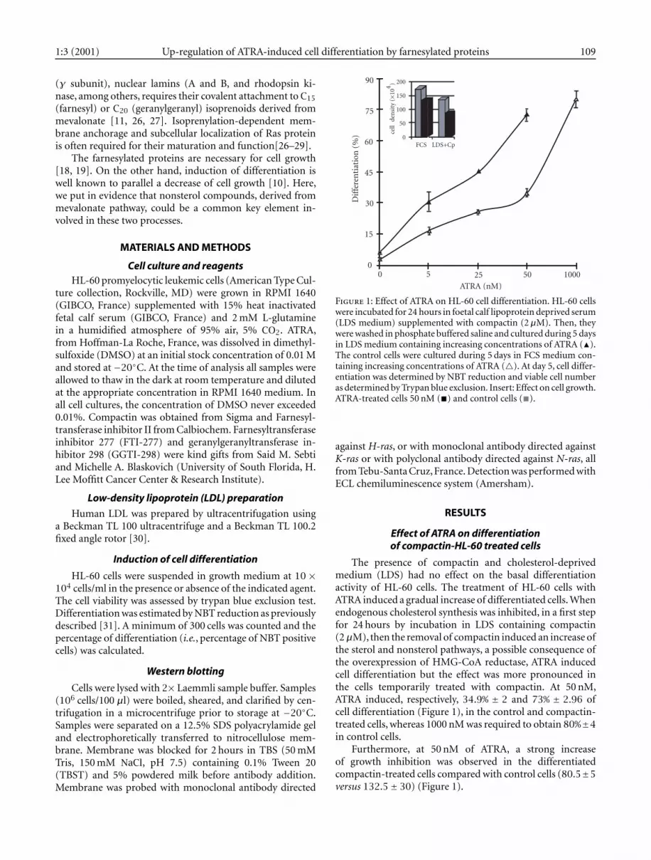
Figure 1: Effect of ATRA on HL-60 cell differentiation. HL-60 cellswere incubated for 24 hours in foetal calf lipoprotein deprived serum(LDS medium) supplemented with compactin (2µM). Then, theywere washed in phosphate buffered saline and cultured during 5 daysin LDS medium containing increasing concentrations of ATRA (�).The control cells were cultured during 5 days in FCS medium con-taining increasing concentrations of ATRA (�). At day 5, cell differ-entiation was determined by NBT reduction and viable cell numberas determined by Trypan blue exclusion. Insert: Effect on cell growth.ATRA-treated cells 50 nM (�) and control cells (�).
against H-ras, or with monoclonal antibody directed againstK-ras or with polyclonal antibody directed against N-ras, allfrom Tebu-Santa Cruz,France. Detection was performed withECL chemiluminescence system (Amersham).
RESULTS
Effect of ATRA on differentiationof compactin-HL-60 treated cells
The presence of compactin and cholesterol-deprivedmedium (LDS) had no effect on the basal differentiationactivity of HL-60 cells. The treatment of HL-60 cells withATRA induced a gradual increase of differentiated cells. Whenendogenous cholesterol synthesis was inhibited, in a first stepfor 24 hours by incubation in LDS containing compactin(2µM), then the removal of compactin induced an increase ofthe sterol and nonsterol pathways, a possible consequence ofthe overexpression of HMG-CoA reductase, ATRA inducedcell differentiation but the effect was more pronounced inthe cells temporarily treated with compactin. At 50 nM,ATRA induced, respectively, 34.9% ± 2 and 73% ± 2.96 ofcell differentiation (Figure 1), in the control and compactin-treated cells, whereas 1000 nM was required to obtain 80%±4in control cells.
Furthermore, at 50 nM of ATRA, a strong increaseof growth inhibition was observed in the differentiatedcompactin-treated cells compared with control cells (80.5±5versus 132.5± 30) (Figure 1).

110 Naima Gueddaria-Pouzols et al. 1:3 (2001)
150
125
100
75
500 20 40 60 80
80
75
70
65
60
55
50
LDH (µg/ml)
Dif
fere
nti
atio
n (
%)
Cel
l den
sity
(%
)
Figure 2: Effect of higher low-density lipoprotein concentrations ongrowth and differentiation of HL-60 cells by ATRA. HL-60 cells wereincubated during 5 days with 1µM ATRA and increasing concentra-tions of LDL. Differentiation as determined by NBT reduction (◦)and viable cell number as determined by Trypan blue exclusion (�).
0 5 10 20 25
0
5
10
15
20
25
30
35
40
45
IFT (µM)
Dif
fere
nti
atio
n (
%)
Figure 3: Effect of farnesyltransferase inhibitors on differentiationof HL-60 cells by ATRA. HL-60 cells were pretreated with increasingconcentrations of FTI-II (•) or FTI-277 (◦). After 24 hours, TheATRA (1µM) was added to culture medium. Cell differentiationwas determined at day 4.
Effect of increasing concentrations of low-densitylipoprotein on differentiation of HL-60
cells by ATRA
In order to test the cholesterol effect on ATRA-inducedcell differentiation, the cells were incubated in the presenceof ATRA (1µM) and increasing concentrations of LDL, upto 80µg/ml. Under such conditions, the differentiation ofATRA-treated cells was decreased from 78% to 54% ± 2.8.Interestingly, a concomitant decrease of cell growth (48%)was evidenced in the presence of high concentrations of LDL(Figure 2).
50
40
30
20
10
0
control GGTI-298 FTI-II FTI-277
Dif
fere
nti
atio
n (
%)
Figure 4: Effect of prenylation protein inhibitor on differentiationof HL-60 cells by ATRA. HL-60 cells were pretreated with 10 µM ofGGTI-298 or FTI-II or FTI-277. After 24 hours, The ATRA (1 µM)was added to culture medium. Cell differentiation was determinedat day 4.
H-ras
N-ras
K-ras
5 µM 10 µM
cont FTI-II FTI-277 GGTI-298
Figure 5: Inhibition of H-ras, N-ras, and K-ras prenylation by FTI-II, FTI-277, and GGTI-298 HL-60 cells were treated with 5 or 10µMof FTI-II or 10µM of FTI-277 or GGTI-298 for 24 hours. Cell lysateswere analyzed by Western blotting with either a H-ras monoclonalantibody or a N-ras polyclonal antibody or a K-ras monoclonal an-tibody. Arrows, unprenylated ras bands.
Evidence for the involvement of prenylated proteinsin HL-60 cell differentiation
The cells were preincubated in the presence of the farsne-syltransferase inhibitor II or farnesyltransferase inhibitorFTI-277 during 24 hours, then treated by ATRA during 3 days(Figure 3). A strong decrease of ATRA-induced differentiationwas observed with increasing concentrations of both inhib-itors. FTI-277 was a more potent inhibitor compared withFTI-II (Figure 3). Indeed, the differentiating activity by ATRA

1:3 (2001) Up-regulation of ATRA-induced cell differentiation by farnesylated proteins 111
HMG-CoA
Mevalonate
Farnesyl-PP
DolicholHaem A
Ubiquinone
Cholesterol
Plasma LDL
Farnesyl protein transferase
Farnesyl proteintransferase inhibitor
Farnesylated proteins(Ras, lamin B, others)
Cell differentiationCell growth
Retinoid response
mRNA
RARE
RXRE
binding
ATRA
9cRAmetabolitesinactive
(CRABP)
ATRA
Acetyl CoA Acetoacetyl CoA
Synthetase
Reductase
+
?
+ +
+–
–
–
–
–
Figure 6: Interaction between mevalonate pathway and retinoic acid-induced differentiation.
was totally abolished at 25 µM of FTI-277 (Figure 3) and40µM of FTI-II (data not shown). These results confirmedthe involvement of the nonsterol mevalonate pathway in celldifferentiation.
When the cells were preincubated in the presence of10µM of GGTI-298 during 24 hours, then treated by ATRAduring 3 days, no significant result was observed com-pared with control cells whereas, at the same concentration(10µM), FTI-II and FTI-277 decrease ATRA-induced differ-entiation of the cells (Figure 4). The prenylation of H-ras,K-ras, and N-ras in the cells is effectively inhibited under theconditions tested (Figure 5). This result suggested that far-
nesylated proteins, such as H-ras, K-ras, and N-ras, could beimplied in the mechanism of cell differentiation by ATRA.
DISCUSSION
At 50 nM, ATRA exhibited 34.9% and 73% of cell differ-entiation in control and compactin-treated cells, respectively.A 24 hours treatment of HL-60 cells by compactin, a potentHMG-CoA reductase inhibitor, increased their sensitivity toATRA. In fact, HMG-CoA reductase is one of the most highlyregulated enzymes. Compactin is well known as inducing anoverexpression of reductase protein. In cultured cells, com-

112 Naima Gueddaria-Pouzols et al. 1:3 (2001)
pactin blocks the synthesis of MVA and triggers adaptive reac-tions that yield a 200-fold increase in reductase protein withina few hours [11]. After removal of compactin, this enzymebecomes active, boosts the mevalonate pathway with con-comitant increase of the sterol and nonsterol products [11].The increase of ATRA differentiating activity in compactin-treated cells could be therefore a consequence of an increasein the sterol or nonsterol compounds or both.
A first attempt was made to check the effect of an excessof cholesterol supply. Increasing concentrations of LDL (upto 80µg/ml) enhanced growth inhibition and decreased celldifferentiation induced by 1 µM of ATRA (Figure 3). In thetwo cases, a limit was reached and this could be a consequenceof the remaining activity of endogenous mevalonate pathway[11]. HL-60 cells have numerous receptors for LDL which cantherefore deliver cholesterol to the cells [32]. The inhibitoryeffects could be attributed to the decrease of the nonsterolproducts.
To confirm the effect of nonsterol products, on celldifferentiation,HL-60 cells were preincubated with the preny-lation inhibitors. In the presence of FTI-II, FTI-277 spe-cific inhibitors for farnesyltransferase, ATRA differentiatingactivity can be totally abolished. By contrast, GGTI-298 hasno significant effect. Comparison of the results confirm apredominant role for farnesylation (inhibited by FT-277)compared with geranygeranylation (inhibited by GGTI-298).A less specific inhibitor FTI-II, acting on the two types ofisoprenoid compounds, has less pronounced effects. West-ern blots electrophoresis confirms that, at the concentrationused, farnesylation and geranygeranylation reactions wereactually inhibited.
Farnesyl proteins include Ras proteins that are involved inthe transduction of mitogenic signals. In addition, our resultsshow that compounds of the farnesyl proteins family includ-ing H-ras, N-ras, and K-ras could be involved in the retinoicacid-induced differentiation of HL-60 cells. Figure 6 showsthe regulation of HMG-CoA reductase activity and the roleof farnesylated proteins on growth and cell differentiation.ATRA is well known to act up on these processes but, in ad-dition, our results show that farnesyl proteins play a key rolein the ATRA-induced differentiation of HL-60 cells. A mech-anism, involving RAR and/or RXR retinoic acid receptors,cannot be excluded but requires further investigations.
Taken together, these results (i) suggested the involvementof mevalonate pathway in ATRA differentiating effect on HL-60 cells, and (ii) support the use of HMG-CoA reductaseinhibitors as potential adjuvant therapeutic agents in differ-entiating chemotherapy.
ACKNOWLEDGEMENTS
We are very grateful to Said M. Sebti and Michelle A.Blaskovich (University of South Florida, H. Lee Moffitt Can-cer Center & Research Institute) for providing us with FTI-277 and GGTI-298. We thank Fabrice Fleury for excellenttechnical assistance.
This work was supported by ANVAR.
REFERENCES
[1] Brockes J. Developmental biology. Reading the retinoidsignals (news; comment). Nature. 1990;345:539–766.
[2] Chomienne C, Ballerini P, Balitrand N, et al. All-trans-retinoic acid in acute promyelocytic leukemias II. Invitro studies: Structure-function relationship. Blood.1990;76:1710–1717.
[3] Razak K, Allen PD, Kelsey SM, Gutteridge CN, NewlandAC. Modulation of CD13 expression during retinoicacid-induced differentiation of HL60 cells. Leuk Res.1994;18:629–636.
[4] Evans RM. The steroid and thyroid hormone receptorsuperfamily. Science. 1988;240:889–895.
[5] Yang N, Schule R, Mangelsdorf DJ, Evans RM. Char-acterization of DNA binding and retinoic acid bindingproperties of retinoic acid receptor. Proc Natl Acad SciUSA. 1991;88:3559–3563.
[6] Levin AA, Sturzenbecker LJ, Kazmer S, et al. 9-cisretinoic acid stereoisomer binds and activates the nu-clear receptor RXR alpha. Nature. 1992;355:359–361.
[7] Heyman RA, Mangelsdorf DJ, Dyck JA, et al. 9-cisretinoic acid is a high affinity ligand for the retinoidX receptor. Cell. 1992;68:397–406.
[8] Breitman TR, Chen ZX, Takahashi N. Potential appli-cations of cytodifferentiation therapy in hematologicmalignancies. Semin Hematol. 1994;31:18–25.
[9] Ruchaud S, Duprez E, Gendron MC, et al. Two dis-tinctly regulated events, priming and triggering, dur-ing retinoid-induced maturation and resistance of NB4promyelocytic leukemia cell line. Proc Natl Acad SciUSA. 1994;91:8428–8432.
[10] Drach J, Lopez-Berestein G, McQueen TT, AndreeffM, Mehta K. Induction of differentiation in myeloidleukemia cell lines and acute promyelocytic leukemiacells by liposomal all-trans-retinoic acid. Cancer Res.1993;53:2100–2104.
[11] Goldstein JL, Brown MS. Regulation of mevalonatepathway. Nature. 1990;343:425–430.
[12] Osborne TF, Gil G, Goldstein JL, Brown MS. Operatorconstitutive mutation of 3-hydroxy-3-methylglutarylcoenzyme A reductase promoter abolishes proteinbinding to sterol regulatory element. J Biol Chem.1988;263:3380–3387.
[13] Osborne TF. Single nucleotide resolution of sterolregulatory region in promoter for 3-hydroxy-3-methylglutaryl coenzyme A reductase. J Biol Chem.1991;266:13947–13951.
[14] Smith JR, Osborne TF, Brown MS, Goldstein JL. Mul-tiple sterol regulatory elements in promoter for ham-ster 3-hydroxy-3-methylglutaryl-coenzyme A synthase.J Biol Chem. 1988;263:18480–18487.
[15] Trzaskos JM, Magolda RM, Favata MF, et al. Modula-tion of 3-hydroxy-3-methylglutaryl-CoA reductase by15 alpha-fluorolanost-7-en-3 beta-ol. A mechanism-based inhibitor of cholesterol biosynthesis. J Biol Chem.1993;268:22591–22599.

1:3 (2001) Up-regulation of ATRA-induced cell differentiation by farnesylated proteins 113
[16] Correll CC, Edwards PA. Mevalonic acid-dependent of3-hydroxy-3-methylglutaryl-coenzyme A reductase invivo and in vitro. J Biol Chem. 1994;269:633–638.
[17] Correll CC, Ng L, Edwards PA. Identification of far-nesol as the non-sterol derivative of mevalonic acidrequired for the accelerated degradation of 3-hydroxy-3-methylglutaryl-coenzyme A reductase. J Biol Chem.1994;269:17390–17393.
[18] Schafer WR, Kim R, Sterne R, Thorner J, Kim SH, Rine J.Genetic and pharmacological suppression of oncogenicmutations in ras genes of yeast and humans. Science.1989;245:379–385.
[19] Hancock JF, Magee AI, Childs JE, Marshall CJ. All rasproteins are polyisoprenylated but only some are palmi-toylated. Cell. 1989;57:1167–1177.
[20] Glomset JA, Gelb MH, Farnsworth CC. Prenyl proteinsin eukaryotic cells: a new type of membrane anchor.Trends Biochem Sci. 1990;15:139–142.
[21] Casey PJ, Solsky PA, Der CJ, Buss JE. p21ras is modi-fied by a farnesyl isoprenoid. Proc Natl Acad Sci USA.1989;86:8323–8327.
[22] Cox AD, Der CJ. The ras/cholesterol connection: impli-cations for ras oncogenicity. Crit Rev Oncog. 1992;3:365–400.
[23] Gibbs JB. Ras C-terminal processing enzymes—newdrug targets?. Cell. 1991;65:1–4.
[24] Prendergast GC, Davide JP, Desolms SJ, et al. Farnesyl-transferase inhibition causes morphological reversionof ras-transformed cells by a complex mechanism thatinvolves regulation of the actin cytoskeleton. Mol CellBiol. 1994;14:4193–4202.
[25] Glomset JA, Farnsworth CC. Role of protein modifi-cation reactions in programming interactions betweenras-related GTPases and cell membranes. Annu Rev CellBiol. 1994;10:181–205.
[26] Casey PJ. Biochemistry of protein prenylation. J LipidRes. 1992;33:1731–1740.
[27] Grunler J, Ericsson J, Dallner G. Branch-point re-actions in the biosynthesis of cholesterol, dolichol,ubiquinone and prenylated proteins. Biochim BiophysActa. 1994;1212:259–277.
[28] Marshall CJ. Protein prenylation: a mediator of protein-protein interactions. Science. 1993;259:1865–1866.
[29] Tilbrook PA, Paterson HF, Marshall CJ. Reversion of ahuman tumour cell line containing oncogenic p21ras isassociated with a defect in the post-translational pro-cessing of the ras protein. Oncogene. 1995;10:805–809.
[30] Brousseau T, Clavey V, Bard JM, Fruchard JC. Sequen-tial ultracentrifugation micromethod for separation ofserum lipoproteins and assays of lipids, apolipoproteins,and lipoprotein particles. Clin Chem. 1993;39:960–964.
[31] Collins SJ, Ruscetti FW, Gallagher RE, Gallo RC. Normalfunctional characteristics of cultured human promyelo-cytic leukemia cells (HL-60) after induction of differen-tiation by dimethylsulfoxide. J Exp Med. 1979;149:969–974.
[32] Gueddari N, Bobichon H, Depierreux C, et al. Enhance-ment of All-Trans-Retinoic Acid Efficiency in granu-locytic differentiation of HL-60 cells by incorporationinto low density lipoproteine. Int J Oncol. 1998;13:1069–1075.
* Corresponding author.E-mail: [email protected]: +33 3 26 05 37 30.