interaction between fish probiotic roseobacters and the natural
TRANSCRIPT
General rights Copyright and moral rights for the publications made accessible in the public portal are retained by the authors and/or other copyright owners and it is a condition of accessing publications that users recognise and abide by the legal requirements associated with these rights.
Users may download and print one copy of any publication from the public portal for the purpose of private study or research.
You may not further distribute the material or use it for any profit-making activity or commercial gain
You may freely distribute the URL identifying the publication in the public portal If you believe that this document breaches copyright please contact us providing details, and we will remove access to the work immediately and investigate your claim.
Downloaded from orbit.dtu.dk on: Jul 17, 2022
Interaction between fish probiotic roseobacters and the natural microbiota inaquaculture settings
Dittmann, Karen Kiesbye
Publication date:2019
Document VersionPublisher's PDF, also known as Version of record
Link back to DTU Orbit
Citation (APA):Dittmann, K. K. (2019). Interaction between fish probiotic roseobacters and the natural microbiota in aquaculturesettings. Technical University of Denmark.
Interaction between fish probiotic roseobacters and the natural microbiota in
aquaculture settings
PhD thesis
by
Karen Kiesbye Dittmann
May 2019
Technical University of Denmark
Department of Biotechnology and Biomedicine
Section for Microbial and Chemical Ecology
Bacterial Ecophysiology and Biotechnology group
i
Preface
This thesis describes the work outcome of my PhD study at the Technical University of
Denmark (DTU). It marks the finale of a project which began on June 1st, 2016 and ended on
May 31st, 2019. The project was funded by a PhD stipend from DTU.
The work was mainly carried out at the Department of Biotechnology and Biomedicine,
DTU, under the supervision of Professor Lone K. Gram and Assistant Professor Mikkel
Bentzon-Tilia. It also included at 4 ½ months stay supervised by Associate Professor Suhelen
Egan at the at the Centre for Marine Bio-Innovation, School of Biological, Earth and
Environmental Sciences, The University of New South Wales in Sydney, Australia.
Karen Kiesbye Dittmann
Kgs. Lyngby, May 2019
ii
This thesis is based on the following papers:
Paper 1:
Dittmann, K.K., Rasmussen, B.B., Castex, M., Gram, L. & Bentzon-Tilia, M. (2017). The aquaculture microbiome at the centre of business creation. Microb. Biotechnol. 10, 1279–1282. doi:10.1111/1751-7915.12877.
Paper 2:
Dittmann, K.K., Sonnenschein, E.C., Egan, S., Gram, L. & Bentzon-Tilia, M. (2019). Impact of Phaeobacter inhibens on marine eukaryote-associated microbial communities. Environ Microbiol Rep., 11(3), 401–413. https://doi.org/10.1111/1758-2229.12698
Paper 3:
Dittmann, K.K., Porsby, C.H., Goncalves, P., Mateiu, R.V., Sonnenschein, E.C., Bentzon-Tilia, M., Egan, S. & Gram, L. (2019). Tropodithietic acid induces oxidative stress response, cell envelope biogenesis and iron uptake in Vibrio vulnificus. Environ Microbiol Rep. Accepted. https://doi.org/10.1111/1758-2229.12771
Paper 4:
Dittmann, K.K., Rasmussen, B.B., Melchiorsen, J., Sonnenschein, E.C., Gram, L. & Bentzon-Tilia, M. (2019). Roseobacter probiotics affect lower-trophic level microbiomes in marine aquaculture. Manuscript in preparation.
Furthermore, I have contributed to the following article during my PhD study (not included in this thesis):
Rasmussen, B.B., Dittmann, K.K., Gram, L. & Bentzon-Tilia, M. (2019). Upscaling probiotic Phaeobacter spp. in Tetraselmis suecica algae cultures. Manuscript in preparation
Conference contributions:
Dittmann, K.K., Sonnenschein, E.C., Gram, L. & Bentzon-Tilia, M. (2016). Impact of tropodithietic acid-producing Phaeobacter inhibens on eukaryote-associated microbial communities. Poster presentation at the Annual Congress of the Danish Microbiological Society, 14th November 2016. Copenhagen, Denmark. Dittmann, K.K., Goncalves, P., Porsby, C.H., Sonnenschein, E.C., Bentzon-Tilia, M., Gram, L. & Egan, S. (2018). The effect of sub-lethal levels of the Roseobacter secondary metabolite, tropodithietic acid, on gene expression of Vibrio vulnificus. Poster presentation at the 17th International Symposium on Microbial Ecology (ISME17), 12th – 17th August 2018. Leipzig, Germany
iii
Summary
Aquaculture is the fastest growing protein producing sector in the world and this growth is
required to feed the growing world population. Microbial diseases are a major bottle-neck in
aquaculture, which must be controlled to avoid great, economic losses. Adult fish can be
vaccinated against the most common bacterial diseases. However, the vaccines cannot be
used on fish larvae because they have underdeveloped immune systems. Antibiotics are
commonly used for acute treatment of infection, however, this increases the risk of antibiotic
resistance dissemination. Therefore, more sustainable, preventive measures are sought and
probiotics has been proposed as one of the solutions. Probiotics are “live organisms which
when administered in adequate amounts confer a health benefit on the host” (FAO and
WHO, 2001). Tropodithietic acid (TDA) producing members of the Roseobacter group, such
as Ruegeria spp. and Phaeobacter spp., have potential as probiotics in aquaculture. They
have repeatedly been isolated from aquaculture environments and they can reduce mortality
of fish larvae challenged with pathogens. However, it is uncertain how the probiotic
treatment affects the commensal microbiome of the larvae.
The purpose of the present PhD project was to determine how probiotic Phaeobacter
inhibens affect the natural microbiota in marine eukaryote systems related to aquaculture.
Given that roseobacters are commonly found in complex communities of marine eukaryotes
in nature and that they are indigenous to the aquaculture environment, the main hypothesis of
this work is that P. inhibens can establish itself in microbiomes associated with aquaculture-
related eukaryotes and protect the host with minor impact on the commensal bacteria.
In this study, 16S rRNA amplicon taxonomics was used to characterize the microbiota of
different trophic levels – Tetraselmis suecica (microalga), Acartia tonsa (copepod), and
Scophthalmus maximus (turbot) larvae – and determine the changes in diversity induced by
treatment with probiotic P. inhibens. Interestingly, the structure of the microbial community
associated with the lower trophic levels were shifted in the presence of P. inhibens, though
not for the fish larval community. The effect was specific and targeted taxa closely related to
the probiotic bacterium. Despite previous studies suspecting the live-feed to be vectors of
infection, these microbiotas had low abundance of Vibrio spp. commonly causing disease in
fish larvae. In contrast, the turbot egg microbiome were dominated by vibrios, however, these
were suppressed after 24 hours incubation and kept stable - most likely due to inherent
roseobacters or the added probiotic.
iv
In nature, members of the Roseobacter group are often found in association with marine
eukaryotes such as algae and molluscs. Secondary metabolite production is believed to be
involved in these interactions, though it is uncertain how they shape the microbiome. In
microalgal blooms, roseobacters increase in abundance, which suggests that they play a role
in the course of the bloom and they likely impact the microbiome. In this study, two model
systems – Emiliania huxleyi (microalga) and Ostrea edulis (European flat oysters) – were
used to study how the secondary metabolite producer P. inhibens affects the diversity and
composition of the associated microbiomes. Roseobacters were indigenous to both
communities and addition of P. inhibens caused substantial changes in the structure of the
low-complexity microbiome of E. huxleyi, though not to the more complex oyster
microbiomes. The impact was specific to vibrios and pseudoalteromonads, which were
decreased in abundance.
The role of TDA in host-bacteria, bacteria-bacteria interactions is unknown. A mode of
action has been proposed for TDA, but it is based on studies of Escherichia coli rather than
marine, non-TDA-producing bacteria which are more likely to encounter TDA in their
surroundings. In this study, a transcriptomics approach was used to study how a sub-lethal
concentration of TDA affected the fish and human pathogen, Vibrio vulnificus. Exposure to
TDA triggered a defense response to reactive oxygen species and iron depletion in V.
vulnificus. Furthermore, there were indications of switch to a biofilm phenotype, which could
explain why inherent resistance and tolerance is rarely observed.
This thesis concludes that TDA-producing P. inhibens causes minor impact on the
microbiomes of various marine eukaryotes. The changes are highly specific to the commensal
microbiome; in part decreasing related taxa, in part decreasing the abundance of putative
pathogens such as vibrios. The molecular mechanism of TDA and role is still uncertain, but
these data indicate that TDA induces a phenotypic switch in the target organism to protect the
cells. Given the ease of introduction, the targeted effect, and the lack of resistance
development, the application of P. inhibens as probiotic in aquaculture is highly promising.
v
Resumé (in Danish)
Akvakultur er den hurtigst voksende, protein-producerende sektor i verden og den vækst er
nødvendig for at brødføde den voksende verdensbefolkning. Mikrobielle sygdomme er en
markant flaskehals i akvakultur og de skal holdes under kontrol for at undgå store,
økonomiske tab. Voksne fisk kan vaccineres mod de mest almindelige bakterielle sygdomme.
Disse vacciner virker dog ikke på fiskelarver, idet de har underudviklede immunforsvar.
Antibiotika bliver almindeligvis brugt mod infektioner i udbrud, men dette øger risikoen for
spredning af antibiotikaresistens. Mere bæredygtige, forebyggende metoder er derfor
efterspurgt og probiotika er en af de foreslåede løsninger. Probiotika er ”levende organismer,
som, når de administreres i passende mængder, giver en sundhedsfordel til værten” (FAO and
WHO, 2001). Tropodithietic acid (TDA) producerende medlemmer af Roseobacter gruppen,
såsom Ruegeria spp. og Phaeobacter spp., har potentiale som probiotika i akvakultur. De er
gentagne gange blevet isoleret fra akvakulturmiljøer og de kan reducere dødeligheden blandt
fiskelarver inficeret med patogener. Det er dog uvist, hvordan den probiotiske behandling
påvirker larvernes kommensale mikrobiomer.
Formålet med dette PhD studium var at klarlægge, hvordan probiotiske Phaeobacter
inhibens påvirker den naturlige mikrobiota i marine eukaryote systemer relateret til
akvakultur. Givet at roseobactere almindeligvis findes i komplekse mikrobielle samfund i og
på marine eukaryoter i naturen, og at de er naturligt forekommende i akvakultursystemer, er
den primære hypotese for dette arbejde, at P. inhibens kan etablere sig i mikrobiomer
tilhørende akvakulturrelaterede eukaryoter og beskytte værten uden betydelig påvirkning af
de kommensale bakterier.
I dette studium blev 16S rRNA amplicon taxonomics brugt til at karakterisere mikrobiotaerne
relateret til de forskellige trofiske niveauer – Tetraselmis suecica (mikroalger), Acartia tonsa
(vandlopper) og Scophthalmus maximus (pighvarlarver) – samt til at klarlægge
diversitetsændringer forårsaget af behandling med probiotiske P. inhibens. Strukturen af
mikrobielle samfund relateret til lavere trofiske niveauer ændrede sig ved tilstedeværelsen af
P. inhibens, men dette skete ikke for fiskelarvemikrobiomet. Effekten var specifik og
målrettet taksonomiske grupper, der er nært beslægtede med den probiotiske bakterie. Til
trods for, at tidligere studier har mistænkt foderorganismer for at være smittebærere, var
mængden af Vibrio spp., der ofte forårsager sygdom i fiskelarver, lav i disse systemer.
Derimod var pighvaræg-mikrobiomet domineret af vibrioer. Disse blev dog undertrykt i løbet
vi
af 24 timers inkubation og holdt på et stabilt niveau – sandsynligvis grundet tilstedeværelsen
af naturlige roseobactere eller den tilsatte probiotiske bakterie.
I naturen er medlemmer af Roseobacter gruppen ofte observeret i association med marine
eukaryoter såsom alger og bløddyr. Man mener, at produktion af sekundære metabolitter er
involveret i disse interaktioner, men det er uvist, hvordan de påvirker mikrobiomet. I
mikroalgeopblomstringer øges tilstedeværelsen af roseobactere, hvilket indikerer, at de spiller
en rolle i opblomstringens forløb, og at de sandsynligvis påvirker mikrobiomet. I dette
studium blev to modelsystemer – Emiliania huxleyi (mikroalgen) og Ostrea edulis
(Europæiske fladøsters) – brugt til at undersøge, hvordan den sekundære metabolit-
producerende P. inhibens påvirker diversiteten og sammensætningen af mikrobiomerne
tilknyttet modelorganismerne. Roseobactere tilhørte de kommensale mikrobiomer.
Tilføjelsen af P. inhibens forårsagede betydelige ændringer i strukturen af det mindre
komplekse E. huxleyi mikrobiom, men ikke i det mere komplekse østers mikrobiom.
Indvirkningen var specifik mod vibrioer og pseudoalteromonader, hvis tilstedeværelse blev
mindsket.
TDAs rolle i interaktioner mellem vært og bakterier, samt mellem bakterier og andre
bakterier er ukendt. En virkningsmekanisme for TDA er blevet foreslået, men den er baseret
på studier af Escherichia coli fremfor marine, ikke-TDA-producerende bakterier, som med
større sandsynlighed vil støde på TDA i deres omgivelser. I dette studium blev
transkriptomundersøgelser anvendt til at undersøge, hvordan en ikke-dræbende koncentration
af TDA påvirker den fiske- og humanpatogene bakterie, Vibrio vulnificus. Eksponering for
TDA udløste et forsvarsrespons imod oxidanter (”reactive oxygen species”) og jernmangel i
V. vulnificus. Derudover var der indikationer på et skift til en biofilm fænotype, hvilket kan
forklare, hvorfor nedarvet resistens og tolerance sjældent er set.
Denne afhandling konkluderer, at TDA-producerende P. inhibens har minimal indvirkning på
mikrobiomer relateret til forskellige marine eukaryoter. Ændringerne er yderst specifikke i
det kommensale mikrobiom; til dels mindskes mængden af nærtbeslægtede taksonomiske
grupper, til dels mindskes mængden af potentielle patogener såsom vibrioer. TDAs
molekylære virkningsmekanisme og dets rolle er endnu uvis, men disse data indikerer at
TDA inducerer et fænotypisk skift for at beskytte cellerne. Letheden af introduktion, den
målrettede effekt og manglen på resistensudvikling er lovende for anvendelsen af P. inhibens
som probiotika i akvakultur.
vii
Table of Contents
Preface ..................................................................................................................................................... i
Summary ............................................................................................................................................... iii
Resumé (in Danish) ............................................................................................................................... v
1. Introduction & outline ...................................................................................................................... 1
2. Microbiomes in aquaculture ............................................................................................................ 5
2.1. Microbiomes associated with reared fish ..................................................................................... 6
2.2. Methods to study microbiomes .................................................................................................... 9
2.2.1. 16S rRNA amplicon taxonomics ........................................................................................ 10
2.3. Conclusions ................................................................................................................................ 11
3. Microbiome management in aquaculture ..................................................................................... 12
3.1. Water conditioning and bioremediation ..................................................................................... 12
3.2. Probiotics in aquaculture ............................................................................................................ 14
3.2.1. Roseobacters as probiotics in aquaculture .......................................................................... 19
3.3. Commercial application of microbiome management in aquaculture ....................................... 24
3.4. Conclusions ................................................................................................................................ 26
4. Roseobacters & TDA ...................................................................................................................... 28
4.1. Colonization of surfaces and interactions with eukaryotes ........................................................ 29
4.2. Tropodithietic acid ..................................................................................................................... 34
4.2.1. Activity spectrum of TDA .................................................................................................. 35
4.2.2. The mode of action for TDA ............................................................................................... 35
4.2.3. Impact of TDA at sub-inhibitory concentrations and potential function ............................ 36
4.2.4. Resistance to TDA .............................................................................................................. 39
4.3. Conclusions ................................................................................................................................ 40
5. Concluding remarks and future perspectives .............................................................................. 42
6. Acknowledgements ......................................................................................................................... 45
7. References ........................................................................................................................................ 46
Paper 1 Paper 2 Paper 3 Paper 4
1
1. Introduction & outline
The world population is growing and is expected to reach 9.8 billion individuals by 2050
(United Nations, Department of Economic and Social Affairs, 2017). This increases the
demand for food production, especially high-quality protein such as fish. Wild fish and
shellfish reservoirs are depleting; in 2015, 93 % of the fish stocks were either maximally,
sustainably fished (59.9 %) or over-exploited (33.1 %) (FAO, 2018). Farmed fish is an
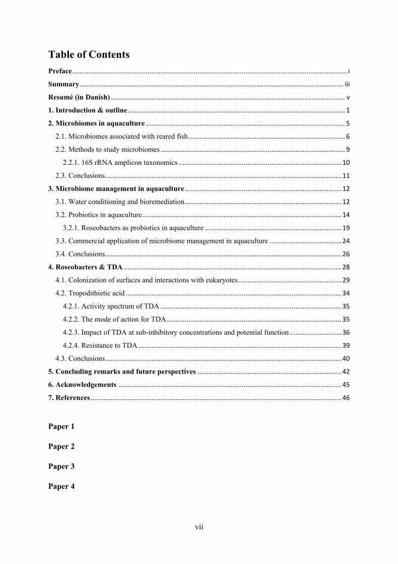
alternative solution to meet the demand. The aquaculture industry is rapidly growing and the
amount of farmed fish produced for human consumption surpassed the wild catches a few
years ago (Figure 1). By 2030, the aquaculture sector is projected to reach 109 million tonnes
of product output (FAO, 2018). The increasing demand in combination with an increased
focus on sustainability and ethics from the general public put pressure on the aquaculture
industry to deliver high quantities of quality outputs through environmentally desirable
production.
Figure 1: Global aquaculture production and capture fisheries for the period 1990 to 2030. The blue graphs reflect the aquaculture (light shade) and capture fisheries (dark shade) for human consumption. The orange graph reflects the total capture fisheries. Modified from FAO (2018).
From hatching of the eggs to full-grown adults, farmed fish are reared in different tanks or
nets with many individuals in a confined space. The intense farming increases the
environmental and social stressors, which, as a consequence, makes the fish more vulnerable
2
to infections and spread of disease. Bacteria are the most common causes (about 55 %) of
disease, though viral, fungal, and parasitic infections are also observed in these systems
(Kibenge et al., 2012). Many bacteria can cause disease in fish, however, members of the
genus Vibrio are common fish pathogens in aquaculture (Thompson et al., 2004; Toranzo et
al., 2005) and also the target pathogen of the work in this thesis.
Antibiotics are deployed in the event of acute infection. In some countries, the use of
antibiotics as prophylactic treatment is still permitted and applied (Cabello, 2006; Cabello et
al., 2013; Miranda et al., 2018). Stricter regulation landscapes do exist, such as in the
European Union (EU), where the EU Veterinary Medicinal Products Directive has banned the
non-therapeutic prophylactic use of antibiotics in 2001 (Directive 2001/82/EC of the
European Parliament and of the Council of 6 November 2001 on the Community code
relating to veterinary medicinal products). However, the use of antibiotics increases the
selective pressure for and the risk of spreading of antibiotic resistance among the commensal
microbiota members (Cabello et al., 2013; Higuera-Llantén et al., 2018; Miranda et al.,
2018). This should be avoided given the antibiotic crisis we are facing (Cooper & Shlaes,
2011); an increasing number of observed multi-drug resistant pathogens combined with a
lack of new antibiotics being developed.
Alternatives are sought to circumvent the use of antibiotics to minimize economical losses
and bacterial antibiotic resistance occurrence. Vaccines have been developed and are working
against the most common pathogens in adult fish (Ringø et al., 2014; Sommerset et al., 2005).
The deployment of vaccines in combination with stricter regulatory oversight of
antimicrobial use and aquaculture management (i.e. hygiene and biosecurity) has decreased
the antimicrobial use in the Norwegian aquaculture by 99 % from 1987 to 2013 despite a
major increase in the production output (300,000 tonnes in 1996; 1.2 mio. tonnes in 2013)
(Norwegian Ministry of Health and Care Services, 2015; The review on antimicrobial
resistance, 2015). However, these vaccines are not working on fish larvae given their
underdeveloped immune systems. Thus, other preventive measures are needed.
Bacteriophage therapy (Rørbo et al., 2018; Silva et al., 2014; Tan et al., 2014), Quorum
Sensing (QS) disruption (Zhao et al., 2015; Zhao et al., 2018), enrichments (Crab et al., 2010;
Crab et al., 2012; Hari et al., 2004; Xu et al., 2013), and probiotics (D’Alvise et al., 2013;
Grotkjær, Bentzon-Tilia, D’Alvise, Dierckens, et al., 2016) are some of the proposed
solutions. Probiotics – the use of beneficial bacteria that when applied have a beneficial effect
on the host (FAO and WHO, 2001) – have been studied for decades and their effects have
3
been tested in many, different kinds of aquaculture-related systems. Most studies have
focused on improving the gut microbiome of the farmed fish by deployment of Firmicutes,
though their origin is not necessarily marine.
Bioactive members of the Gram-negative Roseobacter group have been proposed as
probiotics in marine systems. Particularly, the tropodithietic acid (TDA) producing genus
Phaeobacter has repeatedly shown promising efficiency in warding off pathogenic Vibrio
spp. while imposing minimal effect on the live-feed for the fish larvae and the fish larvae
(D’Alvise et al., 2012, 2013; Hjelm et al., 2004). Resistance to TDA is difficult to induce
(Porsby et al., 2011; Rasmussen et al., 2016), though tolerance has been observed (Dittmann,
Sonnenschein et al., 2019; Harrington et al., 2014). The mechanism of action of TDA on
marine bacteria, as well as the impact of TDA-producers on the inherent microbiota found in
aquacultures, remain to be understood. The microbiota of, for instance, algae used as live-
feed in aquaculture is central to the growth and well-being of the algae. It is therefore of great
importance to understand how the addition of a probiotic organism (over extended periods of
time) affects the commensal microbiota and not just the target pathogen. In this particular
study, the activity of the probiotic bacteria is assumed to be caused, predominantly, by one
molecule, TDA. Understanding the mechanism of action (on other bacteria) of this molecule
is also a way in which potential short- and long-term effects on the commensal microbiota
can be assessed.
The purpose of this PhD project was to determine how probiotic Phaeobacter inhibens affect
the natural microbiota in marine eukaryote systems related to aquaculture. Given that
roseobacters are commonly found in complex communities of marine eukaryotes in nature,
and that they are indigenous to the aquaculture environment, though in low abundance, the
main hypothesis of this work is that P. inhibens can establish itself in microbiomes
associated with aquaculture-related eukaryotes and protect the host with minor impact on the
commensal bacteria. The main part of the work has focused on microbiome characterization
(paper 2 and 4). Another part relates to the influence and mechanism of action of TDA on
pathogenic Vibrio vulnificus by a transcriptomic approach (paper 3).
This thesis consists of an overview section and four papers/manuscripts. The overview
section introduces microbiomes, microbiome management, probiotics, and the probiotic
species investigated in this project. Chapter 2 defines microbiomes in an aquaculture context
including state-of-the-art technologies available to study these complex systems. Based on
4
this knowledge, chapter 3 is focused on management of microbiomes and the exploitation of
beneficial bacteria (probiotics) towards favorable conditions in aquaculture settings. Chapter
4 describes the Roseobacter group, particularly focused on the members producing TDA and
how they interact with other bacteria as well as eukaryotes. The experimental work and
results obtained during this project are summarized in paper 2, 3, and 4, while highlights of
the results are also included in this thesis.
The overall goals of this research is to 1) provide more knowledge on the microbiotas related
to aquacultures, 2) understand how the addition of TDA-producing P. inhibens changes the
bacterial microbiome diversity and determine the target-spectrum of the probiotic effect, and
3) elucidate the mechanism of action of TDA in relation to marine, non-TDA-producers. This
knowledge is essential for the risk assessment of P. inhibens with regards to future
applications in aquaculture.
5
2. Microbiomes in aquaculture
Farming of fish and shellfish in aquacultures creates a unique microbial environment. Every
batch of reared animals comes into a “new” environment - cleaned and disinfected ponds or
tanks – where levels of dissolved organic matter quickly rise. At certain life stages, the fish
and shellfish are moved to new tanks and the cycle is restarted. While this discontinuous
culture cycle is likely stressful to the reared animals, it also affects the microbial community
associated with the fish. The high nutrient levels and repeated disinfection between batches
promote proliferation of fast-growing opportunistic bacteria rather than a stable microbial
community (Skjermo & Vadstein, 1999; Verschuere et al., 2000).
About 10 % to 15 % of fish larvae survive and grow into juveniles in the aquaculture industry
(Vadstein, Attramadal, Bakke, & Olsen, 2018). Several studies have indicated that the major
losses of larvae are due to detrimental interactions and dysbiosis in the microbiota of the fish
larvae (Kanther & Rawls, 2010; Vadstein, Attramadal, Bakke, Forberg, et al., 2018; Vadstein
et al., 2013; Vestrum, Luef, et al., 2018). Antibiotics can be used to avoid these fish larvae
population crashes, though the understanding of why these crashes suddenly occur is still
uncertain. To some extent, this problem originates in the lack of understanding the
microbiome and the interactions occurring at that scale.
A microbiota is “the assemblage of microorganisms present in a defined environment“ while
a microbiome “refers to the entire habitat, including the microorganisms (bacteria, archaea,
lower and higher eurkaryotes, and viruses), their genomes (i.e., genes), and the surrounding
environmental conditions” (Marchesi & Ravel, 2015). While these two concepts are linked,
they are also often used indiscriminately. Microbiomes and microbiotas can be defined at
various levels from an entire animal to parts of the animal such as the gut or skin
microbiome. In aquacultures, multiple microbiomes impact the production including the
rearing water, biofilters, the rearing tanks, microalga, live-feed (rotifers, Artemia, copepods),
and the fish. All of these microbiotas are interlinked and interact through feeding and
exchange of metabolites (Figure 2). This chapter will focus on the microbiomes associated
with fish, live-feed, and feed for live-feed.
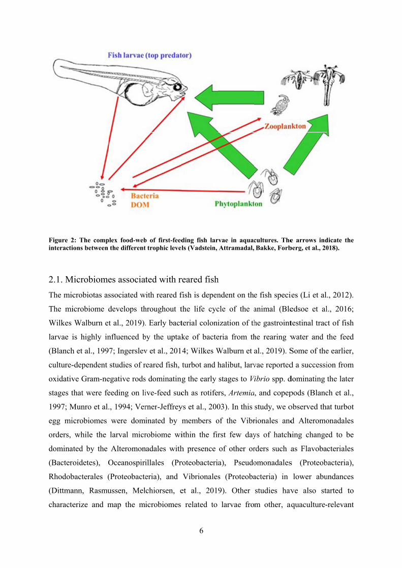
Figure 2interacti
2.1. M
The mic
The mi
Wilkes
larvae i
(Blanch
culture-
oxidativ
stages t
1997; M
egg mi
orders,
domina
(Bacter
Rhodob
(Dittma
characte
2: The compions between
Microbiome
crobiotas as
icrobiome d
Walburn et
is highly in
h et al., 199
-dependent
ve Gram-ne
that were fe
Munro et al.
icrobiomes
while the
ated by the
oidetes), O
bacterales (
ann, Rasmu
erize and m
lex food-webthe different
es associa
ssociated w
develops th
t al., 2019).
nfluenced b
7; Ingerslev
studies of r
egative rods
eeding on li
., 1994; Ver
were dom
larval micr
Alteromon
Oceanospiri
(Proteobacte
ussen, Mel
map the m
b of first-feedt trophic level
ted with r
with reared f
hroughout t
. Early bact
by the uptak
v et al., 201
reared fish,
s dominating
ive-feed suc
rner-Jeffrey
inated by m
robiome wi
nadales with
illales (Pr
eria), and
chiorsen, e
icrobiomes
6
ding fish larls (Vadstein,
reared fish
fish is depen
the life cyc
terial coloni
ke of bacte
4; Wilkes W
turbot and h
g the early
ch as rotife
ys et al., 200
members o
ithin the fir
h presence
roteobacteri
Vibrionales
et al., 201
related to
rvae in aquacAttramadal,
h
ndent on the
cle of the a
ization of th
eria from th
Walburn et
halibut, larv
stages to V
rs, Artemia
03). In this
of the Vibr
rst few day
of other or
a), Pseudo
s (Proteoba
9). Other
larvae fro
cultures. TheBakke, Forb
e fish speci
animal (Ble
he gastroint
he rearing w
al., 2019). S
vae reported
Vibrio spp. d
, and copep
study, we o
rionales and
ys of hatch
rders such a
omonadales
acteria) in
studies hav
m other, a
e arrows indberg, et al., 20
ies (Li et al
edsoe et al
testinal trac
water and t
Some of the
d a successi
dominating
pods (Blanc
observed tha
d Alteromo
hing change
as Flavoba
s (Proteob
lower abu
ve also sta
aquaculture-
dicate the 018).
., 2012).
l., 2016;
ct of fish
the feed
e earlier,
ion from
the later
ch et al.,
at turbot
onadales
ed to be
cteriales
bacteria),
undances
arted to
-relevant
7
species such as rainbow trout (Ingerslev et al., 2014), tilapia (Giatsis 2015), and cod (Bakke
et al., 2015). These studies have revealed a much broader diversity and thereby bridging
some of the knowledge gap.
The impact of feed type and feed nutrient composition also has a major impact on the gut
microbiome composition. A culture-independent study on reared Yellowtail Kingfish (Seriola
lalandi) showed that the microbiota shifted from being dominated by Proteobacteria to being
dominated by Firmicutes, when the larval feed changed from live-feed to formulated pellets
(Wilkes Walburn et al., 2019). Another study on adult Yellowtail Kingfish observed that the
gut microbiota of fish reared in aquaculture was dominated by Firmicutes, while the gut
microbiota of the wild fish was dominated Proteobacteria (Ramírez & Romero, 2017). Along
the same line, artificial feeding decreased the bacterial species diversity of wild Atlantic cod
held in captivity for 6 weeks (Dhanasiri et al., 2011). Ringø et al. (2006) observed that the
digestive tract of adult cod fed with fish meal were dominated by Brochothrix spp. and
Carnobacterium spp. (Gram-positive genera) while the digestive tract of cod fed with soy
bean meal were dominated by Chryseobacterium spp. (Gram-negative), Psychrobacter
glacincola (Gram-negative), and Carnobacterium spp.. Plant-based feed shifted the
microbiome of rainbow trout larvae to Firmicutes, while marine diet (i.e. fish meal and fish
oil) shifted the microbiome to dominance of Proteobacteria (Ingerslev et al., 2014).
Altogether, these studies illustrate not only that the gut microbiomes depend on the feed
(plant-based vs. fish-based), but also on the feed preference of the fish species.
Live-feed have often been suspected as infection vectors in aquaculture larval rearing
(Hansen & Olafsen, 1999; Olafsen, 2001). However, similarly to the fish larval microbiome,
the knowledge on the live-feed microbiotas is scarce. Vibrio spp. are naturally associated
with zooplankton (Colwell et al., 2003; Kaneko & Colwell, 1973; Sochard et al., 1979;
Vezzulli et al., 2015) and culture-dependent studies have observed that Vibrio spp. were
dominating the microbiome (Montanari et al., 1999; Sochard et al., 1979). A culture-
independent study on copepods from the North Atlantic Ocean observed that the microbiome
was dominated by Gammaproteobacteria, particularly Pseudoalteromonas spp., and
Rhodobacteraceae were associated with the transient, food microbiome (Moisander et al.,
2015). Similarly, Gammaproteobacteria of the Alteromonadales and Oceanospirillales orders
dominated the microbiome of Acartia tonsa nauplii in this study, but the abundance of
Vibrionales was less than 2 % of the microbiome (Dittmann, Rasmussen, Melchiorsen, et al.,
2019). Bakke et al. (2015) observed that the copepod microbiome was dominated by
8
Alphaproteobacteria (mainly Rhodobacteraceae) and Flavobacteria, but the Vibrionaceae
were less than 1 % of the microbiome. Hence, the presumed dominance of vibrios observed
in culture-dependent studies does not necessarily reflect the whole bacterial community of
copepods.
Microbiotas of Artemia and rotifers were also assessed in the study by Bakke et al. (2015).
While the rotifer culture was dominated by Actinobacteria and Alphaproteobacteria (mainly
Rhodobacteracea), the Artemia cultures were solely dominated by Alphaproteobacteria
(mainly Rhodobacteracea). Furthermore, Vibrionaceae were only observed in the Artemia
cultures. Høj et al. (2009) reported that the microbiota of newly hatched nauplii was
dominated by Gammaproteobacteria and Planctomycetales. Furthermore, isolates of the
genera Vibrio, Pseudomonas, Micrococcus, Brevundimonas, Sphingomonas, and Rhizobium
could be retrieved from Artemia surface-treated with antibiotics (Høj et al., 2009). Califano et
al. (2017) observed that the rotifers from a gilthead seabream hatchery were dominated by a
single operational taxonomic unit classified as a Loktanella sp., while the Artemia nauplii
were dominated by Flavobacteriaceae, Rhodobacteraceae, and Paracoccus sp.. Hence, the
microbiota of the live-feed is dependent on the cultivation environment rather than the host.
Microalgae are used as feed for the live-feed and fish larvae are in some aquacultures reared
in “green water” with high loads of microalgae. Some of the favored microalgal genera for
larval feeds include Chaeotoceros, Thalassiosira, Tetraselmis, Isochrysis, and
Nannochloropsis (Duerr, 1998). Despite their extensive use, their microbiomes, particularly
in relation to aquaculture settings, are scarcely studied. Biondi et al. (2017) observed that the
microbiome of Tetraselmis suecica was dominated by Proteobacteria – mainly members of
the Roseobacter group and the Rhizobiales order – and Bacteroidetes from the
Flavobacteriales order. A similar community composition was observed in the T. suecica
cultures used in this study, although Planctomycetes (Phycisphaerales) were also prominent
members of the microbiota (Dittmann, Rasmussen, Melchiorsen, et al., 2019). Feeding
Tetraselmis spp. and Chlorella minutissima to Artemia decreased the load of total bacterial
and presumptive Vibrio spp. in the Artemia, most likely due to the presence of bioactive
Gram-positive bacteria in the microbiomes of the algae (Makridis et al., 2006; Olsen et al.,
2000). Furthermore, feeding Atlantic salmon with T. suecica reduced mortalities caused by
Aeromonas salmonicida, Serratia liquefaciens, Vibrio anguillarum, Vibrio salmonicida, and
Yersinia ruckeri type I (Austin et al., 1992). Hence, the microbiotas of the different trophic
layers in an aquaculture are intimately linked and they have high influence on each other.
9
However, it is still uncertain what bacterial species are indicators of a “healthy” microbiome.
This also emphasizes the need for understanding the diversity of the individual microbiotas as
well as their function in relation to the other microbiotas.
2.2. Methods to study microbiomes
The earliest studies of aquaculture bacterial communities were based on classical,
microbiological methods; cultivation and isolation of bacteria, as well as phenotypic and
genotypic characterization (e.g. (Blanch et al., 1997; Munro et al., 1994)). Fingerprinting
techniques, such as polymerase chain reaction denaturing gradient gel electrophoresis (PCR-
DGGE) and terminal restriction fragment length polymorphism (T-RFLP), enabled broader
analysis of the microbial community as a whole and the option to compare the microbiomes
of different niches in a culture-independent way (Fjellheim et al., 2012; Hovda et al., 2007;
Pond et al., 2006). Fjellheim et al. (2012) showed that there was no correlation between
richness and diversity results obtained from T-RFLP on 16S rRNA amplicons and culture-
dependent phenotyping methods. This is most likely due to culturability; 90 % to 99 % of
marine bacteria cannot be cultured at standard laboratory conditions (Glöckner & Joint,
2010), and thus, the culture-dependent studies only reflect the 1 % to 10 % of bacteria that
could grow on agar plates and/or in liquid medium.
The fingerprinting techniques do not provide taxonomic classification to the bacteria without
the use of sequencing. Sequencing of the bands has resulted in a certain level of taxonomy in
some studies (Fjellheim et al., 2012; Hovda et al., 2007). However, this does not provide
information about individual members at genus and species levels. With the rise and
dissemination of Next Generation Sequencing and omics technologies as well as
development of open-source, easy-to-use data handling pipelines, it is now possible to study
diversity, community composition, taxonomy of the community members, and function of the
microbiomes. Combinations of methods such as amplicon sequencing (taxonomy),
metagenomics (taxonomy and genetic potential), metatranscriptomics (gene expression),
proteomics (protein expression), and metabolomics (metabolites) can be used to understand
interactions in complex microbiomes. In this study, 16S rRNA amplicon sequencing was
used to assess diversity and taxonomic distribution of marine eukaryote-associated
microbiotas.
10
2.2.1. 16S rRNA amplicon taxonomics
Currently, one of the most widely used method is 16S rRNA amplicon sequencing
(taxonomics). The method is based on PCR amplification of conserved regions of the 16S
rRNA genes on genomic DNA from the environment. In this study, the 16S rRNA V4 region
was amplified (Dittmann, Rasmussen, Melchiorsen, et al., 2019; Dittmann, Sonnenschein, et
al., 2019), however, other regions and combinations of multiple regions have also been used.
The choice of region determines the taxonomic resolution. Several analysis pipelines - e.g.
DADA2 (Callahan et al., 2016), mothur (Schloss et al., 2009), QIIME (Caporaso et al.,
2010), QIIME 2 (Bolyen et al., 2018), USEARCH (Edgar, 2010), and VSEARCH (Rognes et
al., 2016) – have been developed to process 16S rRNA amplicon sequencing data. The choice
of pipeline is dependent on available computer power, programming language preference,
size of data set, and to some extend also personal preference. In this study, we used mothur
(Dittmann, Sonnenschein, et al., 2019) and QIIME 2 (Dittmann, Rasmussen, Melchiorsen, et
al., 2019). Mothur is relatively easy to approach in the sense that it can run in Windows on a
regular laptop and the pipeline is standardized to take the data from raw reads to Operational
Taxonomic Units (OTUs), as well as calculate measures of alpha- and beta diversity,
including statistics. The data sets in the second taxonomics study were too large and diverse
for our available computer power to handle, which was why we transferred to QIIME 2. This
pipeline is more flexible and plugins from DADA2 (denoising, chimera removal, generation
of Amplicon Sequence Variant, ASV, table) and VSEARCH (classification) can be used,
though it is dependent on running in a UNIX environment. QIIME 2 can be used for
calculating the measures of alpha- and beta-diversity, statistical analysis, and visualizations,
but the R packages Phyloseq and Vegan were used for that purpose in this study (Dittmann,
Rasmussen, Melchiorsen, et al., 2019).
While 16S rRNA taxonomics is becoming relatively affordable, it still has some pitfalls.
Extraction of representative (if not all) genomic DNA, degradation of DNA, amplification
biases in the PCR, and chimeric amplification products are some of the common errors,
which can be introduced prior to sequencing. Furthermore, amplifying a short fraction of a
highly conserved gene, such as the 16S rRNA gene, limits the taxonomic resolution window
and only the bacterial community is assessed. This can be mediated by metagenomics, where
all of the genomic DNA is sequenced. However, this is still an expensive method to apply,
the required computer capacity is beyond the regular benchtop computers, and the DNA
extraction biases are still an issue with this method.
11
2.3. Conclusions
The microbiomes of aquacultures are highly dynamic and the fish microbiota is influenced by
the rearing water, the feed, and environmental factors. To date, there are only few studies on
the aquaculture related microbiomes and more work is needed to determine 1) what a healthy
fish larval microbiome is, 2) what the differences are between larval microbiomes related to
different fish species, and 3) which factors cause dysbiosis leading to crashes in fish
populations.
The technologies for studying microbiome diversity and function are rapidly developing. One
of the key strengths of the Next Generation Sequencing technologies is that a lot of data are
obtained. Combinations of the –omics technologies have the potential to answer the more
complex questions on interactions and functionality of the microbiomes, which could lead to
more rational microbial management and microbiome engineering in aquacultures.
In the following chapter, this thesis will focus on strategies for how aquaculture-related
microbiomes can be managed and engineered to increase the welfare and yield of farmed
fish.
12
3. Microbiome management in aquaculture
Several alternative solutions to microbial management strategies replacing antibiotic
deployment have been proposed for aquacultures; this includes water control, enrichment of
favorable functions, phage-therapy, and probiotics. All of the technologies will alter the
existing microbiome to a presumed “healthier” version or will use beneficial bacteria to
control unwanted pathogens. Some of these principles will briefly be described below with
the main focus being on fish larval probiotics.
3.1. Water conditioning and bioremediation
Improving and stabilizing the water quality is of great importance to ensure balance in
aquaculture systems. Temperature, salinity, pH, and oxygen levels are the strongest
environmental drivers of aquatic microbial communities (Campbell & Kirchman, 2013;
Herlemann et al., 2011; Liu et al., 2015; Lozupone & Knight, 2007; Meron et al., 2011;
Sunagawa et al., 2015; Wright et al., 2012). The chemical properties of the input rearing
water such as temperature, oxygen, salinity and pH are controlled in aquacultures to avoid
environmental stressors from fluctuations (Bentzon-Tilia et al., 2016). Introduction of
pathogenic microorganisms through the inlet water has also been a major concern. Therefore,
the water can be sterilized through UV irradiation or ozonation (Summerfelt, 2003). If the
system is closed, re-circulating the water is an option to keep costs low and avoiding
exchange with the environment (Attramadal et al., 2012). Fish tank water contains high loads
of dissolved organic matter and the system is self-polluting with accumulation of nitrogen
and phosphorus (Schneider et al., 2005). Especially ammonium and nitrite are problematic in
intensive fish rearing (Avnimelech, 1999) because these compounds are toxic to the animals.
Therefore, they should be removed or converted to other, non-toxic compounds prior to outlet
of the water to the environment or re-introduction of the water into the fish tanks. This can be
done by application of recirculating aquaculture systems (RAS). The idea is to condition the
water using microbial communities. First, the water can be filtrated mechanically to remove
accumulating particles of organic matter (Bentzon-Tilia et al., 2016). The water is then
passed through biofilters, which are abiotic structures with biofilms coating the relatively
large surface areas. The biofilms are composed of autotrophic, nitrifying bacteria –
ammonium oxidizing Nitromonas spp. and nitrite oxidizing Nitrospira spp. – which convert
ammonium to nitrate (Foesel et al., 2008). Marine RAS biofilters can also contain members
oxidizing sulfide (Cytryn et al., 2005). However, the community composition of the biofilters
is unique to each RAS and it is highly influenced by factors such as the fish feed,
13
management routines, the fish-associated microbiota, water chemical properties, and
microbial selection pressure in the filter community (Attramadal et al., 2012; Blancheton et
al., 2013; Schreier et al., 2010). Given the variability in these factors, it can be difficult to
establish and maintain a working biofilter which is consistently efficient and safe in terms
invasion of pathogens.
Besides improving water quality, RAS can also be utilized for a slightly different purpose.
Sterilization of the water for the fish larvae is a necessity, but it also diminishes competition
between bacteria, and thus, it gives room for domination of fast-growing, opportunistic
pathogens already in the rearing water. Microbial maturation – re-colonization of the water
by non-opportunistic bacteria – using biofilters could be a solution. Skjermo et al. (1997)
showed reduced proliferation of pathogens after hatching of turbot (Scophthalmus maximus)
eggs and increased survival of Atlantic halibut (Hippoglossus hippoglossus) yolk sac larvae,
when the rearing water was matured after sterile-filtration. Attramadal et al. (2012) observed
a more stable and diverse microbial community composition with a lower fraction of
opportunists in comparison to conventional flow-through systems. This strategy can also
lower the mortality of Atlantic cod larvae (Attramadal et al., 2012, 2014) by selecting for
slow growing, competition-specialized bacteria with affinity for resources (K-selection)
(Attramadal et al., 2014; Vestrum, Attramadal, et al., 2018).
Improving the water quality and directing the aquaculture community composition can also
be done in a relatively simple, low-technology way through bioflocs. The technology is based
on the balance of carbon and nitrogen; if the concentrations are well-balanced in the water,
nitrogenous waste such as ammonium will be converted to bacterial biomass (Schneider et
al., 2005). Adding extra carbon to a system with high loads of ammonium and carbon-
limitation stimulates heterotrophic bacterial growth, which in turn increases the nitrogen-
uptake (Avnimelech, 1999) and improves the water quality. This also creates accumulation of
bacteria in flocs, which the reared animals eat, and thus, improve growth (Crab et al., 2010,
2012; Hari et al., 2004; Xu et al., 2013).
Improving the aquaculture rearing environment by shifting the microbiota to utilize favorable
functions already found in the microbial community is an elegant concept. While it has been
proven that RAS biofilters and bioflocs can be used for manipulating the water microbiome –
both chemically and microbially – the currently established methods are often more
coincidental rather than rationally designed. Seeding the systems with synthetic communities
14
of bacteria with known, beneficial functions, including probiotics, could be one route to
streamline and minimize variability in the production (Bentzon-Tilia et al., 2016; Dittmann,
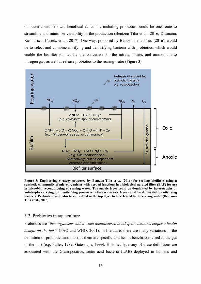
Rasmussen, Castex, et al., 2017). One way, proposed by Bentzon-Tilia et al. (2016), would
be to select and combine nitrifying and denitrifying bacteria with probiotics, which would
enable the biofilter to mediate the conversion of the nitrate, nitrite, and ammonium to
nitrogen gas, as well as release probiotics to the rearing water (Figure 3).
Figure 3: Engineering strategy proposed by Bentzon-Tilia et al. (2016) for seeding biofilters using a synthetic community of microorganisms with needed functions in a biological aerated filter (BAF) for use in microbial reconditioning of rearing water. The anoxic layer could be dominated by heterotrophs or autotrophs carrying out denitrifying processes, whereas the oxic layer could be dominated by nitrifying bacteria. Probiotics could also be embedded in the top layer to be released to the rearing water (Bentzon-Tilia et al., 2016).
3.2. Probiotics in aquaculture
Probiotics are “live organisms which when administered in adequate amounts confer a health
benefit on the host” (FAO and WHO, 2001). In literature, there are many variations in the
definition of probiotics and most of them are specific to a health benefit conferred in the gut
of the host (e.g. Fuller, 1989; Gatesoupe, 1999). Historically, many of these definitions are
associated with the Gram-positive, lactic acid bacteria (LAB) deployed in humans and
15
terrestrial animals. The increasing interest in aquaculture farmed fish and shellfish has also
lead to investigations of probiotics for this industry. However, there are some pronounced
differences between terrestrial and aquatic animals, which need to be considered when
designing probiotics for aquaculture. Farmed fish and shellfish are highly influenced by the
microbiome of the surrounding water (Defoirdt, Sorgeloos, & Bossier, 2011; Verschuere et
al., 2000); they are in constant contact with the water and continuously ingest it. The
aquaculture ecosystem does not only support the life of the eukaryote and the commensal
bacteria, but also of (opportunistic) pathogens, which can reach high densities in this
favorable environment (Moriarty, 1998). Opportunistic pathogens such as Vibrio spp. do not
only invade the host through the gut, but they can also invade fish through the gills and the
skin (Spanggaard et al., 2000; Weber et al., 2010). While the wording of the probiotics
definition is debated, the FAO and WHO definition from 2001 is broad enough to include
probiotics acting on the gut system as well as in/on other organs of the fish including indirect
actions in the water. Hence, this definition will be used in this PhD thesis.
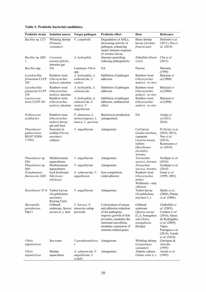
For the past decades, the beneficial effects of probiotics have been extensively studied in
vitro (typically as inhibition of pathogens) and to a lesser extend in vivo (Table 1). However,
the mechanisms behind the effects are still largely uncertain, if even uncovered, and only
partial explanations are provided, given the methodological and ethical limitations
concerning animal trials (Ringø et al., 2014; Tinh et al., 2008; Verschuere et al., 2000). Some
of the suggested mechanisms include (i) competitive exclusion through production of
inhibitory compounds, (ii) competition for nutrients, chemicals, or energy, (iii) adhesion site
competition, (iv) contribution to digestion, (v) contribution to macro- and micronutrients, (vi)
enhancement of immune response, and (vii) reduction of virulence through QS manipulation.
These will be covered below in the descriptions of the probiotic candidates.
16
Table 1: Probiotic bacterial candidates.
Probiotic strain Isolation source Target pathogen Probiotic effect Host Reference
Bacillus sp. LT3 Whiteleg shrimp (Penaeus vannamei)
V. campbellii Degradation of AHLs, decreasing activity of pathogen, enhancing innate immune response of Artemia larvae
Brine shrimp larvae (Artemia franciscana)
Defoirdt et al. (2011), Niu et al. (2014)
Bacillus sp. QSI-1
Carassius auratus gibelio, intestine gut
A. hydrophila Quorum quenching reducing pathogenicity
Zebrafish (Danio rerio)
Chu et al. (2015)
Bacillus spp. NA Luminous Vibrio spp.
NA Prawns Moriarty (1998)
Lactobacillus fermentum CLFP 242
Rainbow trout (Oncorynchus mykiss), intestine
A. hydrophila, A. salmonicida, Y. ruckeri
Inhibition of pathogen adhesion
Rainbow trout (Oncorynchus mykiss) - in vitro
Balcázar et al.(2008)
Lactobacillus plantarum CLFP 238
Rainbow trout (Oncorynchus mykiss), intestine
A. hydrophila, A. salmonicida
Inhibition of pathogen adhesion
Rainbow trout (Oncorynchus mykiss) - in vitro
Balcázar et al.(2008)
Lactococcus lactis CLFP 101
Rainbow trout (Oncorynchus mykiss), intestine
A. hydrophila, A. salmonicida, Y. ruckeri, V. anguillarum
Inhibition of pathogen adhesion, antibacterial effect
Rainbow trout (Oncorynchus mykiss) - in vitro
Balcázar et al.(2008)
Pediococcus acidilactici
Rainbow trout (Oncorynchus mykiss) larvae, gut and feed
P. damnosus, L. monocytogenes, L. innocu, L. garvieae
Bacteriocin production (antagonism)
NA Araújo et al.(2015, 2016)
Phaeobacter gallaeciensis BS107 (DSM 17395)
Seawater in scallop (Pecten maximus) cultures
V. anguillarum Antagonism Cod larvae (Gadus morhua), copepods (Acartia tonsa), rotifers (Brachionus plicatilis), Artemia
D’Alvise et al. (2010, 2012), Neu et al. (2014), Rasmussen et al. (2018)
Phaeobacter sp. S26
Mediterranean aquacultures
V. anguillarum Antagonism Tetraselmis suecica, Artemia
Grotkjær et al. (2016)
Phaeobacter sp. S60
Mediterranean aquacultures
V. anguillarum Antagonism Tetraselmis suecica, Artemia
Grotkjær et al. (2016)
Pseudomonas fluorescens AH2
Iced freshwater fish (Lates niloticus)
A. salmonicida, V. anguillarum
Iron competition (siderophores)
Rainbow trout (Oncorynchus mykiss Walbaum) - only vibriosis
Gram et al. (1999, 2001)
Roseobacter 27-4 Turbot Larvae (Scophthalmus maximus) Rearing Units
V. anguillarum Antagonism Turbot larvae (Scophthalmus maximus L.)
Hjelm et al., (2004), Planas et al. (2006),
Shewanella putrefaciens Pdp11
Gilthead seabream, Sparus aurata (L.), skin
V. harveyi, P. damselae subsp piscicida
Colonization of mucus and adhesion reduction of the pathogens, improve growth of fish juveniles, modulate the intestinal microbiota, modulate expression of immune-related genes
Gilthead seabream (Sparus aurata (L.)), Senegalese sole (Solea senegalensis (Kaup))
Chabrillón et al. (2005), Cordero et al. (2016), Sáenz de Rodrigáñez et al. (2009), Tapia-Paniagua et al. (2014), Varela et al. (2010).
Vibrio alginolyticus
Sea water V.parahaemolyticus
Antagonism Whiteleg shrimp (Litopenaeus vannamei)
Garriques & Arevalo (1995)
Vibrio alginolyticus
Shrimp aquaculture
A. salmonicida, V. anguillarum, V. ordalii
Antagonism Atlantic salmon (Salmo salar L.)
Austin et al. (1995)
17
Some of the most studied probiotic candidates belong to the Firmicutes phylum, namely LAB
and bacilli (Araújo et al., 2016; Balcázar et al., 2008; Carnevali et al., 2004; Gatesoupe, 1991,
1994; Moriarty, 1998; Venkat et al., 2004). These probiotics have been successful in humans
and livestock, and the bacilli as biocontrol in horticulture, though they are not adapted to nor
common in the marine environment. LAB can tolerate acidic pH and bile salts, which enable
them to survive in gut systems (Balcázar et al., 2008; Bentzon-Tilia et al., 2016; Merrifield et
al., 2010). These bacteria can colonize the intestinal mucus, where they are believed to act as
an infection barrier and assist in the processing and uptake of feed, which in turn can promote
growth of the fish (Ringø et al., 2010; Vieco-Saiz et al., 2019). Pediococcus acidilactici was
isolated from the gut of rainbow trout larvae (Oncorhynchus mykiss) as well as their feed
(Araújo et al., 2016; Araújo et al., 2015). The strains were bioactive against common fish
pathogens, in part due to bacteriocin production, and they performed well in safety
assessment as they did not display antibiotic resistance, produce hemolysins, or degrade
gastric mucin (Araújo et al., 2016). Other LAB such as Carnobacterium maltaromaticum,
Lactobacillus curvatus, Lactobacillus sakei, Lactobacillus plantarum, Lactococcus lactis, and
Leuconostoc mesenteroides have also been isolated from the intestines of salmonids
(Balcázar et al., 2007). Some of these strains – L. lactis CLFP 101, L. plantarum CLFP 238,
and Lactobacillus fermentum CLFP 242 – were tested for their antibacterial effect and their
ability to inhibit adhesion of the fish pathogens Aeromonas hydrophila, A. salmonicida, Y.
ruckeri, and V. anguillarum to intestinal mucus from rainbow trout (in vitro) (Balcázar et al.,
2008). Only L. lactis CLFP 101 reduced adhesion of all the tested pathogens in the mucus
assay, and supernatant from the LAB strain inhibited growth of all pathogens, too. L.
fermentum CLFP 242 reduced adhesion of all pathogens except V. anguillarum, but its
supernatant did not show antibacterial activity, indicating that its probiotic potential is most
likely not due to production and secretion of antimicrobial agents. This was also the case for
L. plantarum CLFP 238 and its ability to inhibit adhesion was restricted to the tested
Aeromonas spp.. Hence, their probiotic mode of action is specific at species level, if not
strain level. Thus, all probiotic candidates would have to be tested in vivo to determine their
exact activity spectrum and potential for application.
Bacilli have also been observed to improve survival of reared shrimp and controlling
luminous Vibrio spp. (Moriarty, 1998). Bacillus sp. can also protect the live-feed (Artemia)
and increase survival by decreasing the activity of Vibrio campbellii and enhancing the innate
immune response of the Artemia larvae (Niu et al., 2014). Defoirdt et al. (2011) isolated
18
Bacillus spp. from whiteleg shrimp and European sea bass, which could degrade N-acyl-
homoserine lactones (AHL). Degrading the AHLs can disrupt the QS modulated phenotypes
such as virulence. Quorum quenching Bacillus sp. QSI-1 reduced pathogenicity of A.
hydrophila in zebrafish (Danio rerio) and thereby improved the survival rate (Chu et al.,
2015). Hence, probiotic effect does not have to be due to competition, but it can also be due
to modulation of behavior in the microbiota. It will not necessarily decrease the pathogen
load and an imbalance might still let the opportunists gain dominance.
While LAB strains seem somewhat promising as probiotics in aquaculture, it is important to
assess both strengths and weaknesses. If they are to be used as probiotics in larvicultures,
they may not serve their full purpose in the early fish life stages, because the gastrointestinal
tract is not fully developed and the microbiome inside the larvae is transient – being an
extension of the microbiota in the tank (Bentzon-Tilia et al., 2016) – which is not (yet)
dominated by Firmicutes. Hence, other species, that are adapted to and act in the marine
environment are likely more suitable at this stage. Proteobacteria such as Pseudomonas spp.,
Shewanella spp., Vibrio spp., and members of the Roseobacter group have been proposed as
non-LAB probiotics (Chabrillón, Rico, Arijo, et al., 2005; Chabrillón, Rico, Balebona, &
Morinigo, 2005; Dittmann et al., 2017; Garriques & Arevalo, 1995; Gram et al., 2001; Gram
et al., 1999; Porsby & Gram, 2016; Prado et al., 2009; Tapia-Paniagua et al., 2014),
especially due to their antagonism against pathogens. Gram et al. investigated the probiotic
potential of Pseudomonas fluorescens strain AH2 against V. anguillarum and A. salmonicida
(Gram et al., 2001, 1999). The growth of both pathogens was inhibited in vitro by P.
fluorescens AH2 and the effect was increased during iron-limited growth conditions. This
indicated that part of the probiotic effect could be due to iron competition (siderophores),
though the experimental conditions did not allow for an exact determination of this (Gram et
al., 1999). While the probiotic Pseudomonas could protect rainbow trout against vibriosis
(Gram et al., 1999), furunculosis caused by A. salmonicida in Atlantic salmon (Salmo salar
L.) was unaffected by the probiotic treatment (Gram et al., 2001). Hence, it is not possible to
predict a “good” probiotic in situ based on in vitro experimental results; in vivo trials of
probiotic candidates against different target pathogens in different fish systems are necessary
to determine their spectrum of activity.
Despite their pathogenicity to some fish and shellfish (Ben Kahla-Nakbi et al., 2009; Cao et
al., 2018; Gómez-León et al., 2005), addition of Vibrio alginolyticus to the culture water,
could reduce the occurrence of Vibrio parahaemolyticus and increase the survival of whiteleg
19
shrimp, Litopenaeus vannamei (Garriques & Arevalo, 1995). Similarly, bathing Atlantic
salmon in culture of a V. alginolyticus strain - used as disease control in shrimp aquaculture
in Ecuador - reduced mortality of the fish challenged with A. salmonicida and to a lesser
extent salmon challenged with V. anguillarum and Vibrio ordalii (Austin et al., 1995). Both
studies suggested that the probiotic properties came from antagonism towards the target
pathogens. Shewanella putrefaciens Pdp11 isolated from the skin of healthy gilthead
seabream, Sparus aurata (L.) (Chabrillón, Rico, Balebona, et al., 2005), was able to colonize
the mucus and reduce adhesion of the pathogens Vibrio harveyi and Photobacterium
damselae subsp piscicida, both in gilthead seabream and in Senegalese sole, Solea
senegalensis (Kaup) (Chabrillón, Rico, Arijo, et al., 2005; Chabrillón, Rico, Balebona, et al.,
2005). Further studies have revealed that S. putrefaciens Pdp11 is able to improve growth
when added to the feed of juveniles of both fish species (Sáenz de Rodrigáñez et al., 2009;
Varela et al., 2010). The strain can also modulate the intestinal microbiota and expression of
immune-related genes (Tapia-Paniagua et al., 2014; Varela et al., 2010) during high-stocking
induced stress (Cordero et al., 2016; Tapia-Paniagua et al., 2014; Varela et al., 2010).
Altogether, this indicates that probiotics, exemplified by S. putrefaciens Pdp11, can have
multiple mechanisms, which act together to protect and improve health of aquaculture related
animals.
3.2.1. Roseobacters as probiotics in aquaculture
Members of the Roseobacter group, mainly Phaeobacter spp., have shown great potential as
probiotics in aquaculture. They have been isolated in multiple aquaculture units (Grotkjær,
Bentzon-Tilia, D’Alvise, Dourala, et al., 2016; Porsby et al., 2008; Ruiz-Ponte et al., 1998),
which indicates that they might play a more or less important role in the microbiome in some
farms. Phaeobacter gallaeciensis BS107 can antagonize V. anguillarum in vitro and protect
cod (Gadus morhua) larvae from vibriosis (D’Alvise et al., 2012). The antagonistic effect
was likely due to production of the secondary metabolite TDA, given that a TDA-negative
mutant did not protect the larvae to the same extend (Figure 4). Similarly, Grotkjær et al.
(2016) observed that TDA-producing Phaeobacter sp. S26 and Phaeobacter sp. S60 isolated
from Mediterranean aquacultures could reduce growth of V. anguillarum in non-axenic
microalgae, T. suecica, and Artemia systems (used as live-feed in aquacultures). Altogether,
this would argue that TDA-producing Phaeobacter spp. used as probiotics could not only
protect the larvae from infection but also prevent proliferation and introduction of pathogens
through the live-feed.
20
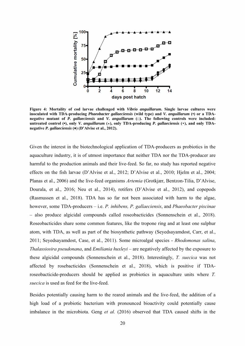
Figure 4: Mortality of cod larvae challenged with Vibrio anguillarum. Single larvae cultures were inoculated with TDA-producing Phaeobacter gallaeciensis (wild type) and V. anguillarum (•) or a TDA-negative mutant of P. gallaeciensis and V. anguillarum (□). The following controls were included: untreated control (▪), only V. anguillarum (▴), only TDA-producing P. gallaeciensis (▾), and only TDA-negative P. gallaeciensis (♦) (D’Alvise et al., 2012).
Given the interest in the biotechnological application of TDA-producers as probiotics in the
aquaculture industry, it is of utmost importance that neither TDA nor the TDA-producer are
harmful to the production animals and their live-feed. So far, no study has reported negative
effects on the fish larvae (D’Alvise et al., 2012; D’Alvise et al., 2010; Hjelm et al., 2004;
Planas et al., 2006) and the live-feed organisms Artemia (Grotkjær, Bentzon-Tilia, D’Alvise,
Dourala, et al., 2016; Neu et al., 2014), rotifers (D’Alvise et al., 2012), and copepods
(Rasmussen et al., 2018). TDA has so far not been associated with harm to the algae,
however, some TDA-producers – i.e. P. inhibens, P. gallaeciensis, and Phaeobacter piscinae
– also produce algicidal compounds called roseobacticides (Sonnenschein et al., 2018).
Roseobacticides share some common features, like the tropone ring and at least one sulphur
atom, with TDA, as well as part of the biosynthetic pathway (Seyedsayamdost, Carr, et al.,
2011; Seyedsayamdost, Case, et al., 2011). Some microalgal species - Rhodomonas salina,
Thalassiosira pseudonana, and Emiliania huxleyi – are negatively affected by the exposure to
these algicidal compounds (Sonnenschein et al., 2018). Interestingly, T. suecica was not
affected by rosebacticides (Sonnenschein et al., 2018), which is positive if TDA-
roseobacticide-producers should be applied as probiotics in aquaculture units where T.
suecica is used as feed for the live-feed.
Besides potentially causing harm to the reared animals and the live-feed, the addition of a
high load of a probiotic bacterium with pronounced bioactivity could potentially cause
imbalance in the microbiota. Geng et al. (2016) observed that TDA caused shifts in the
21
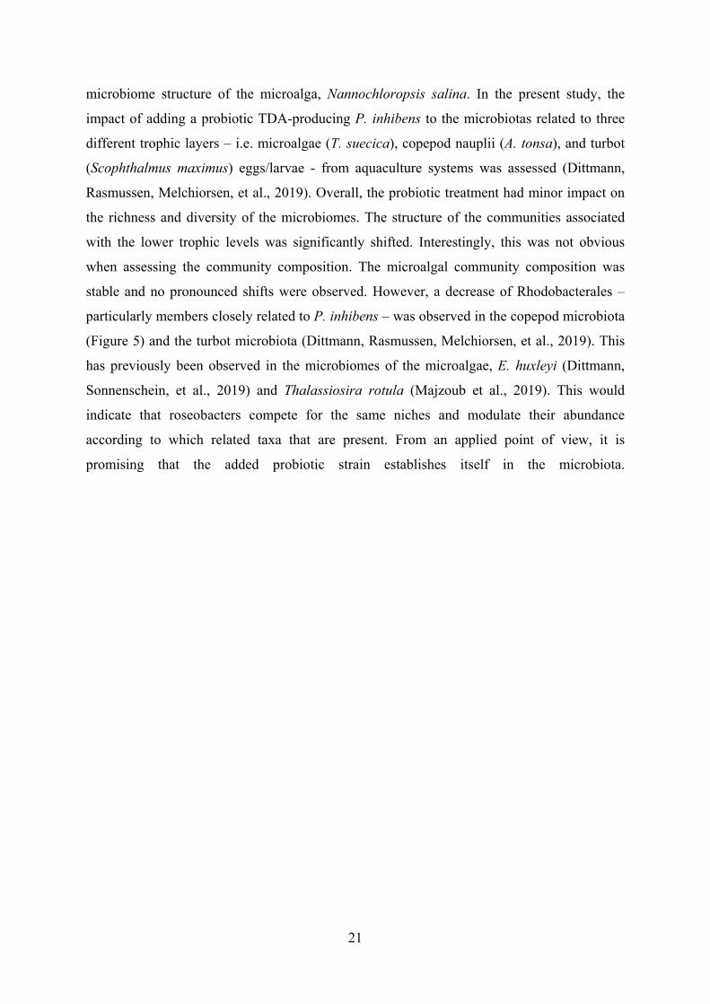
microbiome structure of the microalga, Nannochloropsis salina. In the present study, the
impact of adding a probiotic TDA-producing P. inhibens to the microbiotas related to three
different trophic layers – i.e. microalgae (T. suecica), copepod nauplii (A. tonsa), and turbot
(Scophthalmus maximus) eggs/larvae - from aquaculture systems was assessed (Dittmann,
Rasmussen, Melchiorsen, et al., 2019). Overall, the probiotic treatment had minor impact on
the richness and diversity of the microbiomes. The structure of the communities associated
with the lower trophic levels was significantly shifted. Interestingly, this was not obvious
when assessing the community composition. The microalgal community composition was
stable and no pronounced shifts were observed. However, a decrease of Rhodobacterales –
particularly members closely related to P. inhibens – was observed in the copepod microbiota
(Figure 5) and the turbot microbiota (Dittmann, Rasmussen, Melchiorsen, et al., 2019). This
has previously been observed in the microbiomes of the microalgae, E. huxleyi (Dittmann,
Sonnenschein, et al., 2019) and Thalassiosira rotula (Majzoub et al., 2019). This would
indicate that roseobacters compete for the same niches and modulate their abundance
according to which related taxa that are present. From an applied point of view, it is
promising that the added probiotic strain establishes itself in the microbiota.
22
Figure 5: Relative abundances of the 30 most abundant Amplicon Sequence Variants (ASVs) observed in the Acartia tonsa (AT) microbiome. The populations were either untreated (controls, C) or exposed to probiotic Phaeobacter inhibens DSM 17395 (P). Each population was sampled at day 0, 1, 2, and 4. The relative abundances have been log10(x+1) transformed. Each row represent a unique ASV and the assigned taxonomy is listed next to the plotted relative abundances (Dittmann, Rasmussen, Melchiorsen, et al., 2019).
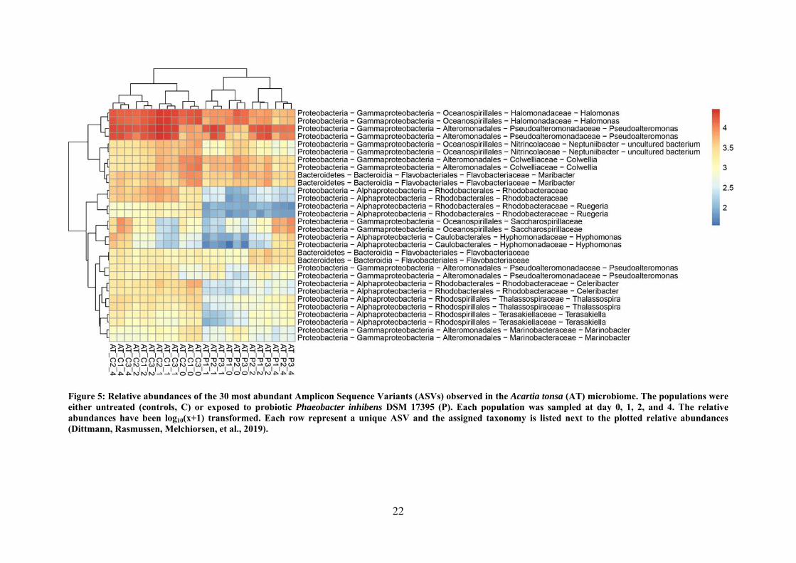
As men
only ob
stable r
TDA-pr
Majzou
(NCV12
regardle
Phaeob
already
amount
sufficie
interact
and the
Figure 6either uncommun
ntioned prev
bserved vib
regardless o
roduction is
ub et al. (2
2a1) with r
ess of the
bacter-like s
present, an
t might be i
ent to unde
tions in arti
mechanism
6: Communityntreated (Connity was samp
viously, ent
brios in the
of treatment
s necessary
2019) used
reduced an
bioactivity.
species wer
nd the added
indifferent
erstand the
ificial micro
m of action f
y compositionntrols) or exppled at 0, 24,
try of Vibrio
e turbot larv
t (Figure 6)
for the prob
both a TD
tagonistic e
In both th
re already
d probiotic
to the vibri
mechanism
obiomes, w
for TDA, w
n of the microposed to prob48, and 96 ho
23
o spp. has b
rval microb
) (Dittmann
biotic effec
DA-producin
effect; the
his study a
present in
bacterium
ios in the s
ms behind
we need to
which will be
obiota of turbbiotic Phaeobaours post-exp
been linked
biota and th
n, Rasmusse
t against vib
ng P. inhib
microbiotas
nd the stud
the microb
replaces the
system. How
the probiot
comprehend
e elaborated
bot eggs and acter inhiben
posure in dup
to live-feed
heir relative
en, Melchio
briosis (D’A
ens 2.10 (W
s developed
dy by Majz
biomes. If T
e inherent g
wever, taxo
tic effect.
d the ecolo
d on in chap
larvae. The ps DSM 17395
plicates. The b
d. In this st
e abundanc
orsen, et al.
Alvise et al
WT) and a
d in the sam
zoub et al.
TDA-produ
genera, then
onomics dat
To underst
ogy of rose
pter 4 of this
populations w5 (Treatmentbars illustrat
tudy, we
ces were
., 2019).
., 2012).
a variant
me way
(2019),
ucers are
n the net
ta is not
tand the
obacters
s thesis.
were t). The te the
24
relative abundances of all the bacterial orders observed on the eggs / larvae. Only orders with relative abundance above 2 % were included and Amplicon Sequence Variants (ASVs) containing the added P. inhibens was removed from the dataset prior to plotting. T0: untreated time zero control (Dittmann, Rasmussen, Melchiorsen, et al., 2019).
The effects of probiotic P. inhibens on aquaculture-related microbiomes is notable, though it
is not possible to predict whether the impact is positive, negative, or even indifferent to the
microbiota balance. Studies over extended periods of time, assessing the impact of addition
route (e.g. microalgal, live-feed, rearing water) and the dose is necessary to optimize the
probiotic effect with minimal harm to the microbiome prior to commercial application.
3.3. Commercial application of microbiome management in aquaculture
Protection of the animals and faster growth is of great interest to commercial aquaculture, in
order to live up to regulations and sustainability-focused governmental stakeholders and
consumers. While the ideas for improving the aquaculture industry in a sustainable way is
getting increasing attention, the implementation and routine application of products targeting
the microbiome in this sector is still in its infancy (Dittmann et al., 2017). An editorial
describing the status quo of commercialized microbiome-focused products for the
aquaculture industry, and looking into the “crystal ball” of the future aquaculture industry
(Dittmann et al., 2017), was included in this thesis. Some products are already on the market
and they fall into two categories: 1) targeting the water and pond environment, and 2)
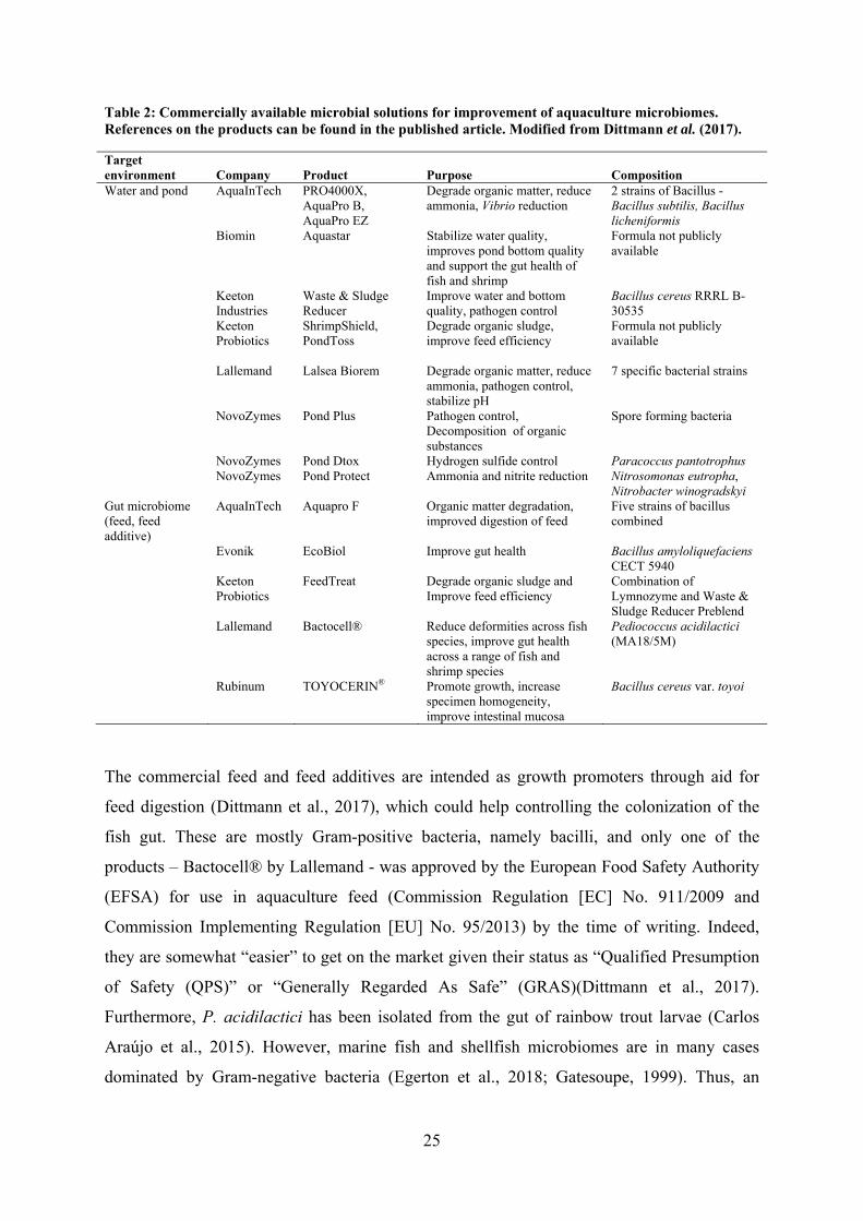
targeting the gut microbiome (feed and feed additives) (Table 2). The products available for
targeting the water and environment, are generally focused on improving the water quality
and decreasing the self-polluting dissolved organic matter as well as toxic compounds
(ammonium, nitrite, and hydrogen sulphide). The discontinuous culture cycles of
aquacultures leaves little to no room for establishment of a mature, healthy microbiome. The
water and environmental products could be applied to seed biofilters and prime the rearing
environment, thus, potentially excluding the opportunity for pathogens to get a “head start”
and establish themselves.
25
Table 2: Commercially available microbial solutions for improvement of aquaculture microbiomes. References on the products can be found in the published article. Modified from Dittmann et al. (2017).
Target environment Company Product Purpose Composition Water and pond AquaInTech PRO4000X,
AquaPro B, AquaPro EZ
Degrade organic matter, reduce ammonia, Vibrio reduction
2 strains of Bacillus - Bacillus subtilis, Bacillus licheniformis
Biomin Aquastar Stabilize water quality, improves pond bottom quality and support the gut health of fish and shrimp
Formula not publicly available
Keeton Industries
Waste & Sludge Reducer
Improve water and bottom quality, pathogen control
Bacillus cereus RRRL B-30535
Keeton Probiotics
ShrimpShield, PondToss
Degrade organic sludge, improve feed efficiency
Formula not publicly available
Lallemand Lalsea Biorem Degrade organic matter, reduce ammonia, pathogen control, stabilize pH
7 specific bacterial strains
NovoZymes Pond Plus Pathogen control, Decomposition of organic substances
Spore forming bacteria
NovoZymes Pond Dtox Hydrogen sulfide control Paracoccus pantotrophus NovoZymes Pond Protect Ammonia and nitrite reduction Nitrosomonas eutropha,
Nitrobacter winogradskyi Gut microbiome (feed, feed additive)
AquaInTech Aquapro F Organic matter degradation, improved digestion of feed
Five strains of bacillus combined
Evonik EcoBiol Improve gut health Bacillus amyloliquefaciens CECT 5940
Keeton Probiotics
FeedTreat Degrade organic sludge and Improve feed efficiency
Combination of Lymnozyme and Waste & Sludge Reducer Preblend
Lallemand Bactocell® Reduce deformities across fish species, improve gut health across a range of fish and shrimp species
Pediococcus acidilactici (MA18/5M)
Rubinum TOYOCERIN® Promote growth, increase specimen homogeneity, improve intestinal mucosa
Bacillus cereus var. toyoi
The commercial feed and feed additives are intended as growth promoters through aid for
feed digestion (Dittmann et al., 2017), which could help controlling the colonization of the
fish gut. These are mostly Gram-positive bacteria, namely bacilli, and only one of the
products – Bactocell® by Lallemand - was approved by the European Food Safety Authority
(EFSA) for use in aquaculture feed (Commission Regulation [EC] No. 911/2009 and
Commission Implementing Regulation [EU] No. 95/2013) by the time of writing. Indeed,
they are somewhat “easier” to get on the market given their status as “Qualified Presumption
of Safety (QPS)” or “Generally Regarded As Safe” (GRAS)(Dittmann et al., 2017).
Furthermore, P. acidilactici has been isolated from the gut of rainbow trout larvae (Carlos
Araújo et al., 2015). However, marine fish and shellfish microbiomes are in many cases
dominated by Gram-negative bacteria (Egerton et al., 2018; Gatesoupe, 1999). Thus, an
26
avenue of new possibilities would be commercialization of probiotics of marine origin
(Dittmann et al., 2017), which requires in-depth knowledge of marine fish microbiomes and
the microbial drivers of a healthy microbiota.
One of the key issues has been – and still is – the lack of knowledge on the microbiome
constituents present in the aquacultures and how that is impacted by manipulations (Dittmann
et al., 2017; Dittmann, Rasmussen, Melchiorsen, et al., 2019). The dissemination and
“normalization” of Next-Generation Sequencing technologies as well as accessible,
understandable bioinformatics tools for data processing has significantly increased the
amount of studies trying to facilitate the understanding of fish and shellfish microbiome
diversity and functionality (Egerton et al., 2018). It is becoming evident that the microbiomes
of fish are versatile – from species to species, one environment to the other, wild vs. captured
– and they develop throughout the life-cycle of the fish, in part due to changing feed (Egerton
et al., 2018; Wilkes Walburn et al., 2019). Having knowledge about the different
microbiomes and how they change due to stressors – e.g. environmental and social stress as
found in aquacultures – would pave the path to rational microbiome engineering. Thus, the
future probiotics could be single-cultures or mixtures of bacteria, which would be more suited
to 1) survive in the environment, 2) exert the desired effect, and 3) cause minimal damage to
the existing microbiota, given a rational development towards aquatic/marine microbiomes.
3.4. Conclusions
Managing microbiomes can be done in several ways – from giving preferential treatment to
certain microbial members through targeted feeding to introducing new microbial members
with desirable traits and functions. However, all actions will impose a change and thereby an
imbalance of the microbiome. Stabilizing in a new microbial balance should ideally benefit
the system to improved health, but prediction of the effect is challenging, and it could also
range from no or minor effect to diminished or harmed health of the system. Therefore, it is
of crucial importance to 1) know the microbiome of the system you are intending to manage,
and 2) assess the potential consequences of imposing changes to the system e.g. by pilot or
full-scale studies using Next Generation Sequencing technologies to monitor the changes.
Probiotics have shown great potential to reduce the load of pathogens or modify their
behavior based on numerous in vitro and in vivo studies. However, the exact “mode of
action” is still uncertain for many candidates and very few studies have assessed their impact
on the microbiomes found in aquaculture. This study is focused on the probiotic candidate, P.
27
inhibens. Despite it being a prominent secondary metabolite producer, minor effects on the
microbiomes exposed to probiotic concentrations are observed and they are highly specific to
other roseobacters and vibrios. This suggests that addition of TDA producers to aquaculture
systems is beneficial, however, the interactions at the molecular level is still uncertain, and it
would need to be tested in real aquaculture systems where fish and live-feed interact with
each other.
In order to understand their behavior in artificial systems, it is important to take their
behavior in nature into consideration. The following chapter is focused on the ecology of the
TDA-producing roseobacters and the molecular mechanisms underlying TDA activity.
28
4. Roseobacters & TDA
The Roseobacter group (previously “Roseobacter clade”) are Gram-negative α-
Proteobacteria. Roseobacters are almost exclusively isolated from hypersaline and marine
environments, either tolerating or requiring salt for living (Buchan et al., 2005). Members of
this group are distributed across the world oceans, from the polar to the temperate oceans
(Selje et al., 2004), accounting for 2 % to 8 % of the surface water bacterioplankton
(Sunagawa et al., 2015; Wietz et al., 2010). In coastal waters, roseobacters are highly
abundant and constitute up to 20 % to 40 % of the microbiome (Buchan et al., 2005; Moran et
al., 2003; Prabagaran et al., 2007). Especially the bacterial communities of phytoplankton
blooms are dominated by roseobacters (González et al., 2000), but these bacteria are also
observed in the microbiotas of other eukaryotes such as macroalgae (Rao et al., 2005) and
molluscs (Prado et al., 2009; Ruiz-Ponte et al., 1998; Wegner et al., 2013).
The highly diverse ecological niches, where members of the Roseobacter group can be found,
point to a metabolic versatility and adaptability. The Roseobacter group members are
“ecological generalists” (Moran et al., 2007; Newton et al., 2010). Both phototrophs – e.g.
Roseobacter litoralis and Roseobacter denitrificans – and heterotrophs – e.g. Phaeobacter
spp. - can be found among the members of the group (Buchan et al., 2005; González &
Moran, 1997; Newton et al., 2010). Roseobacters also play a major role in the oceanic sulfur
cycling (González et al., 2000; Malmstrom et al., 2004) – both inorganic and organic forms
of sulfur. Particularly, some members are able to assimilate and metabolize the algal
osmolyte, dimethylsulfoniopropionate (DMSP) to the climate influencing gas dimethyl
sulfide (DMS) (Miller & Belas, 2004; Moran et al., 2003). Other ways of gaining energy is
through oxidation of inorganic sulfur and carbon monoxide (Buchan et al., 2005; Moran et
al., 2004; Sorokin, 1994, 1995).
From a genomic point of view, they have variable genomes – averaging at 4.4 Mb (Buchan et
al., 2005) – and they often harbor plasmids (Buchan et al., 2005; Moran et al., 2004). These
plasmids often carry metabolically important features such as photosynthetic gene clusters
(Petersen et al., 2012), secondary metabolite biosynthesis genes (Berger et al., 2012;
Brinkhoff et al., 2004), and type IV secretion systems (Petersen et al., 2013), which adds to
the physiological diversity of this prominent bacterial group.
29
4.1. Colonization of surfaces and interactions with eukaryotes
Some members of the Roseobacter group are excellent biofilm formers and they colonize
both abiotic and biotic surfaces. Dang and Lovell (2000) observed that these bacteria were
some of the earliest and most prominent colonizers of polymer surfaces. For example,
Phaeobacter spp. colonize the walls of the fish larval rearing tanks (Hjelm et al., 2004) in
aquaculture units. P. inhibens strain 2.10 was able to colonize the surface of a diatom within
2 days of incubation (Majzoub et al., 2019), further emphasizing their versatile ability to
rapidly colonize these surfaces.
Roseobacters are known for interacting with eukaryotes, both symbiotically and
pathogenically. These interactions are believed to be mediated by bacterial motility and
chemotaxis, QS, and antimicrobial biosynthesis. Some of the best described interactions
involve phytoplankton. Coccolithophorid microalgae and dinoflagellates produce DMSP
(González et al., 2000; Zubkov et al., 2001, 2002), which can be metabolized by roseobacters
(Miller & Belas, 2004), but the compound can also act as a chemoattractant (Seymour et al.,
2010). Hence, the algae attract and feed the bacteria. However, symbiosis is not a one-way
bargain; in return for continuous nutrition, bacteria feed the microalgae with supplements
such as vitamins (Cooper et al., 2015; Croft et al., 2005) and/or protection against predation
(Seyedsayamdost, Case, et al., 2011). Such a relationship has been proposed between P.
inhibens and the coccolithophorid microalga, E. huxleyi (Figure 7) (Seyedsayamdost, Case, et
al., 2011). At symbiotic conditions, the algae produces DMSP and provides the P. inhibens
with a biofilm surface in return for growth-promoting compounds – phenylacetic acid and
indole-3-acetic acid – and protection (Segev et al., 2016; Seyedsayamdost, Case, et al., 2011).
The protective effect comes from the production of the antibiotic TDA and its tautomer
thiotropocin. When the algae age, they release p-coumaric acid, which along with indole-3-
acetic acid triggers a production of algicidal compounds, roseobacticides, by P. inhibens
(Segev et al., 2016; Seyedsayamdost, Carr, et al., 2011; Seyedsayamdost, Case, et al., 2011).
Thus, the mutualistic relationship and the Phaeobacter becomes pathogenic to the individual
algae – potentially, it also escalates the termination of algal blooms in nature.
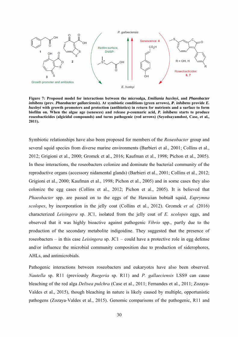
Figure 7inhibenshuxleyi wbiofilm oroseobac2011).
Symbio
several
2012; G
In these
reprodu
Grigion
coloniz
Phaeob
scolope
characte
observe
product
roseoba
and/or
AHLs,
Pathoge
Nautell
bleachin
Valdes
pathoge
7: Proposed s (prev. Phaeowith growth on. When thcticides (algic
otic relation
squid speci
Grigioni et a
e interaction
uctive organ
ni et al., 200
e the egg
bacter spp.
es, by incor
erized Leis
ed that it w
tion of the
acters – in t
influence th
and antimic
enic interac
la sp. R11
ng of the re
et al., 2015
ens (Zozaya
model for inobacter gallaepromoters an
he algae age cidal compou
nships have
ies from div
al., 2000; G
ns, the rose
ns (accessor
00; Kaufma
cases (Co
are passed
rporation in
singera sp.
was highly
secondary
this case Le
he microbia
crobials.
ctions betw
(previousl
ed alga Deli
5), though
a-Valdes et
nteractions beeciensis). At snd protection(senesces) an
unds) and tur
also been p
verse marin
Gromek et a
obacters co
ry nidament
an et al., 19
llins et al.
d on to the
n the jelly
JC1, isola
bioactive
metabolite
eisingera sp
al commun
ween roseob
ly Ruegeria
isea pulchra
bleaching i
al., 2015).
30
etween the msymbiotic con (antibioticsnd release prns pathogen
proposed fo
ne environm
al., 2016; K
olonize and
tal glands) (
98; Pichon
., 2012; Pi
e eggs of
coat (Coll
ated from t
against pat
e indigoidin
p. JC1 – cou
nity compos
bacters and
a sp. R11)
a (Case et a
in nature is
. Genomic
microalga, Emnditions (gre) in return fo-coumaric ac
nic (red arrow
or members
ments (Barb
Kaufman et a
dominate t
(Barbieri et
et al., 2005
ichon et al
the Hawai
lins et al.,
the jelly co
thogenic V
ne. They su
uld have a
sition due t
d eukaryote
) and P. g
al., 2011; F
likely caus
comparison
miliania huxen arrows), Por nutrients acid, P. inhibews) (Seyedsa
of the Rose
ieri et al., 2
al., 1998; P
he bacterial
al., 2001; C
5) and in so
l., 2005). I
iian bobtail
2012). Gro
oat of E. s
Vibrio spp.,
uggested th
protective r
o productio
es have al
gallaeciensis
ernandes et
sed by mul
ns of the pa
xleyi, and PhP. inhibens pand a surfacens starts to
ayamdost, Ca
eobacter gr
2001; Collin
Pichon et al.
l communit
Collins et a
ome cases th
It is believ
l squid, Eu
omek et al.
scolopes eg
partly due
hat the pres
role in egg
on of sidero
so been ob
s LSS9 ca
t al., 2011;
ltiple, oppo
athogenic, R
aeobacter rovide E. e to form produce
ase, et al.,
roup and
ns et al.,
., 2005).
ty of the
l., 2012;
hey also
ved that
uprymna
. (2016)
ggs, and
e to the
sence of
defense
ophores,
bserved.
an cause
Zozaya-
rtunistic
R11 and
31
LSS9 isolates, and non-pathogenic isolates revealed putative virulence factors in all genomes,
though one QS-dependent regulator was unique to the pathogenic isolates (Zozaya-Valdes et
al., 2015). While the exact molecular mechanisms behind the pathogenicity are uncertain, it
has been speculated that members of the indigenous community can switch from a symbiotic
to pathogenic lifestyle, when environmental conditions compromise the host’s chemical
defenses (Case et al., 2011; Fernandes et al., 2011). Another Roseobacter group member,
Roseovarius crassostreae, can cause juvenile oyster disease and massive mortalities among
hatchery-raised American oysters, Crassostrea virginica (Boardman et al., 2008; Boettcher et
al., 2000; Boettcher et al., 2005). However, the mechanisms of pathogenicity remain
uncertain.
Despite numerous studies observing roseobacters in microbiomes of eukaryotes and testing
their in vitro phenotypes – such as production of secondary metabolites and bioactivity, QS-
abilities, and biofilm formation – the knowledge on how they behave and impact microbial
communities in vivo and in situ is scarce. In the present study, two marine eukaryotes – E.
huxleyi and Ostrea edulis (European flat oyster) – model systems were constructed to
investigate the impact of TDA-producing P. inhibens on the associated bacterial community
(Dittmann, Sonnenschein, et al., 2019). Interestingly, the richness was significantly increased
in the complex oyster microbiota (Figure 8), which is an indicator of healthy oysters (King et
al., 2012; Wegner et al., 2013), though this change was not in the microalgal microbiota.
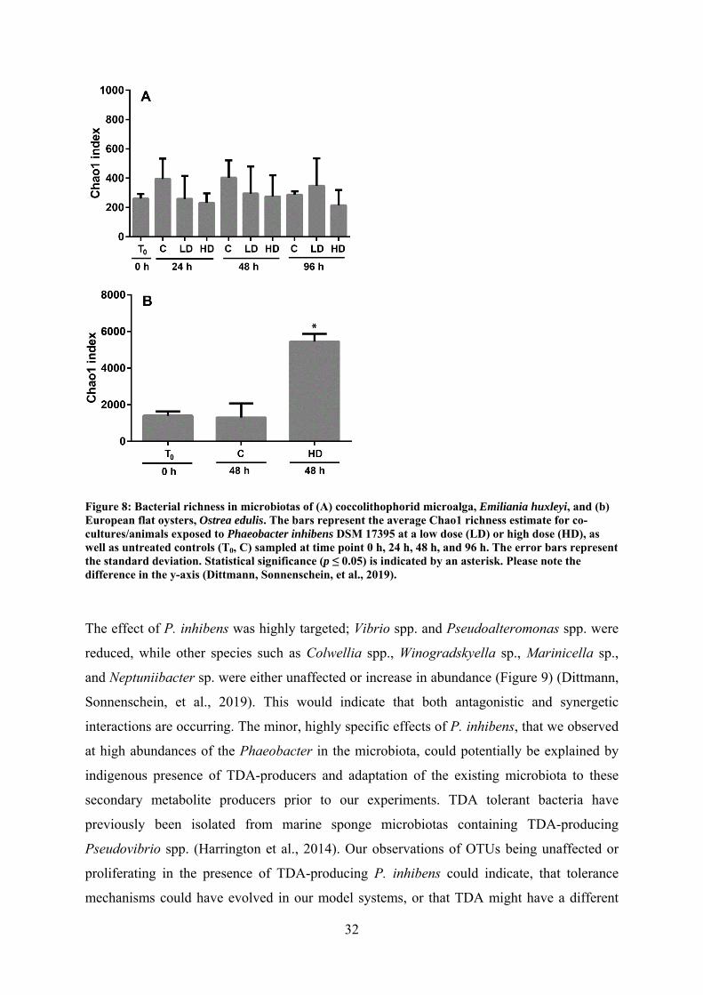
32
Figure 8: Bacterial richness in microbiotas of (A) coccolithophorid microalga, Emiliania huxleyi, and (b) European flat oysters, Ostrea edulis. The bars represent the average Chao1 richness estimate for co-cultures/animals exposed to Phaeobacter inhibens DSM 17395 at a low dose (LD) or high dose (HD), as well as untreated controls (T0, C) sampled at time point 0 h, 24 h, 48 h, and 96 h. The error bars represent the standard deviation. Statistical significance (p ≤ 0.05) is indicated by an asterisk. Please note the difference in the y-axis (Dittmann, Sonnenschein, et al., 2019).
The effect of P. inhibens was highly targeted; Vibrio spp. and Pseudoalteromonas spp. were
reduced, while other species such as Colwellia spp., Winogradskyella sp., Marinicella sp.,
and Neptuniibacter sp. were either unaffected or increase in abundance (Figure 9) (Dittmann,
Sonnenschein, et al., 2019). This would indicate that both antagonistic and synergetic
interactions are occurring. The minor, highly specific effects of P. inhibens, that we observed
at high abundances of the Phaeobacter in the microbiota, could potentially be explained by
indigenous presence of TDA-producers and adaptation of the existing microbiota to these
secondary metabolite producers prior to our experiments. TDA tolerant bacteria have
previously been isolated from marine sponge microbiotas containing TDA-producing
Pseudovibrio spp. (Harrington et al., 2014). Our observations of OTUs being unaffected or
proliferating in the presence of TDA-producing P. inhibens could indicate, that tolerance
mechanisms could have evolved in our model systems, or that TDA might have a different
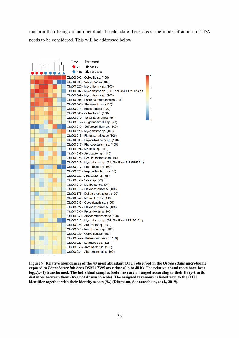
33
function than being an antimicrobial. To elucidate these areas, the mode of action of TDA
needs to be considered. This will be addressed below.
Figure 9: Relative abundances of the 40 most abundant OTUs observed in the Ostrea edulis microbiome exposed to Phaeobacter inhibens DSM 17395 over time (0 h to 48 h). The relative abundances have been log10(x+1) transformed. The individual samples (columns) are arranged according to their Bray-Curtis distances between them (tree not drawn to scale). The assigned taxonomy is listed next to the OTU identifier together with their identity scores (%) (Dittmann, Sonnenschein, et al., 2019).
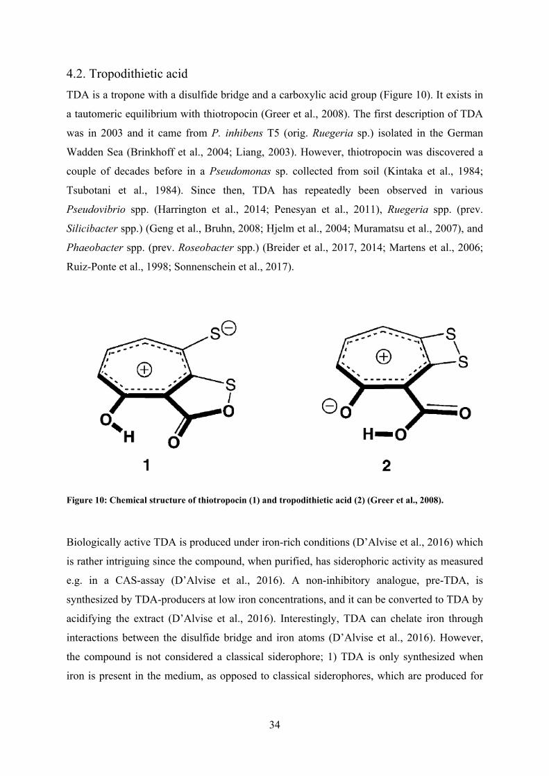
34
4.2. Tropodithietic acid
TDA is a tropone with a disulfide bridge and a carboxylic acid group (Figure 10). It exists in
a tautomeric equilibrium with thiotropocin (Greer et al., 2008). The first description of TDA
was in 2003 and it came from P. inhibens T5 (orig. Ruegeria sp.) isolated in the German
Wadden Sea (Brinkhoff et al., 2004; Liang, 2003). However, thiotropocin was discovered a
couple of decades before in a Pseudomonas sp. collected from soil (Kintaka et al., 1984;
Tsubotani et al., 1984). Since then, TDA has repeatedly been observed in various
Pseudovibrio spp. (Harrington et al., 2014; Penesyan et al., 2011), Ruegeria spp. (prev.
Silicibacter spp.) (Geng et al., Bruhn, 2008; Hjelm et al., 2004; Muramatsu et al., 2007), and
Phaeobacter spp. (prev. Roseobacter spp.) (Breider et al., 2017, 2014; Martens et al., 2006;
Ruiz-Ponte et al., 1998; Sonnenschein et al., 2017).
Figure 10: Chemical structure of thiotropocin (1) and tropodithietic acid (2) (Greer et al., 2008).
Biologically active TDA is produced under iron-rich conditions (D’Alvise et al., 2016) which
is rather intriguing since the compound, when purified, has siderophoric activity as measured
e.g. in a CAS-assay (D’Alvise et al., 2016). A non-inhibitory analogue, pre-TDA, is
synthesized by TDA-producers at low iron concentrations, and it can be converted to TDA by
acidifying the extract (D’Alvise et al., 2016). Interestingly, TDA can chelate iron through
interactions between the disulfide bridge and iron atoms (D’Alvise et al., 2016). However,
the compound is not considered a classical siderophore; 1) TDA is only synthesized when
iron is present in the medium, as opposed to classical siderophores, which are produced for
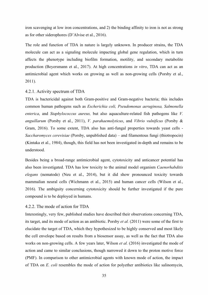
35
iron scavenging at low iron concentrations, and 2) the binding affinity to iron is not as strong
as for other siderophores (D’Alvise et al., 2016).
The role and function of TDA in nature is largely unknown. In producer strains, the TDA
molecule can act as a signaling molecule impacting global gene regulation, which in turn
affects the phenotype including biofilm formation, motility, and secondary metabolite
production (Beyersmann et al., 2017). At high concentrations in vitro, TDA can act as an
antimicrobial agent which works on growing as well as non-growing cells (Porsby et al.,
2011).
4.2.1. Activity spectrum of TDA
TDA is bactericidal against both Gram-positive and Gram-negative bacteria; this includes
common human pathogens such as Escherichia coli, Pseudomonas aeruginosa, Salmonella
enterica, and Staphylococcus aureus, but also aquaculture-related fish pathogens like V.
anguillarum (Porsby et al., 2011), V. parahaemolyticus, and Vibrio vulnificus (Porsby &
Gram, 2016). To some extent, TDA also has anti-fungal properties towards yeast cells -
Saccharomyces cerevisiae (Porsby, unpublished data) – and filamentous fungi (thiotropocin)
(Kintaka et al., 1984), though, this field has not been investigated in-depth and remains to be
understood.
Besides being a broad-range antimicrobial agent, cytotoxicity and anticancer potential has
also been investigated. TDA has low toxicity to the animal model organism Caenorhabditis
elegans (nematode) (Neu et al., 2014), but it did show pronounced toxicity towards
mammalian neural cells (Wichmann et al., 2015) and human cancer cells (Wilson et al.,
2016). The ambiguity concerning cytotoxicity should be further investigated if the pure
compound is to be deployed in humans.
4.2.2. The mode of action for TDA
Interestingly, very few, published studies have described their observations concerning TDA,
its target, and its mode of action as an antibiotic. Porsby et al. (2011) were some of the first to
elucidate the target of TDA, which they hypothesized to be highly conserved and most likely
the cell envelope based on results from a biosensor assay, as well as the fact that TDA also
works on non-growing cells. A few years later, Wilson et al. (2016) investigated the mode of
action and came to similar conclusions, though narrowed it down to the proton motive force
(PMF). In comparison to other antimicrobial agents with known mode of action, the impact
of TDA on E. coli resembles the mode of action for polyether antibiotics like salinomycin,
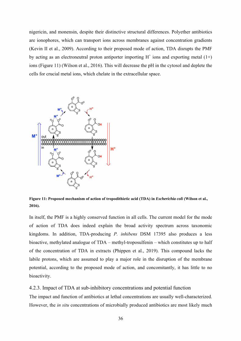
36
nigericin, and monensin, despite their distinctive structural differences. Polyether antibiotics
are ionophores, which can transport ions across membranes against concentration gradients
(Kevin II et al., 2009). According to their proposed mode of action, TDA disrupts the PMF
by acting as an electroneutral proton antiporter importing H+ ions and exporting metal (1+)
ions (Figure 11) (Wilson et al., 2016). This will decrease the pH in the cytosol and deplete the
cells for crucial metal ions, which chelate in the extracellular space.
Figure 11: Proposed mechanism of action of tropodithietic acid (TDA) in Escherichia coli (Wilson et al.,
2016).
In itself, the PMF is a highly conserved function in all cells. The current model for the mode
of action of TDA does indeed explain the broad activity spectrum across taxonomic
kingdoms. In addition, TDA-producing P. inhibens DSM 17395 also produces a less
bioactive, methylated analogue of TDA – methyl-troposulfenin – which constitutes up to half
of the concentration of TDA in extracts (Phippen et al., 2019). This compound lacks the
labile protons, which are assumed to play a major role in the disruption of the membrane
potential, according to the proposed mode of action, and concomitantly, it has little to no
bioactivity.
4.2.3. Impact of TDA at sub-inhibitory concentrations and potential function
The impact and function of antibiotics at lethal concentrations are usually well-characterized.
However, the in situ concentrations of microbially produced antibiotics are most likely much
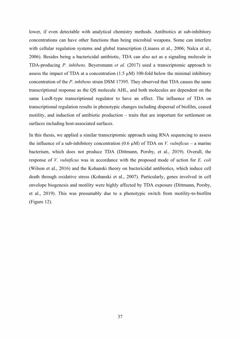
37
lower, if even detectable with analytical chemistry methods. Antibiotics at sub-inhibitory
concentrations can have other functions than being microbial weapons. Some can interfere
with cellular regulation systems and global transcription (Linares et al., 2006; Nalca et al.,
2006). Besides being a bactericidal antibiotic, TDA can also act as a signaling molecule in
TDA-producing P. inhibens. Beyersmann et al. (2017) used a transcriptomic approach to
assess the impact of TDA at a concentration (1.5 µM) 100-fold below the minimal inhibitory
concentration of the P. inhibens strain DSM 17395. They observed that TDA causes the same
transcriptional response as the QS molecule AHL, and both molecules are dependent on the
same LuxR-type transcriptional regulator to have an effect. The influence of TDA on
transcriptional regulation results in phenotypic changes including dispersal of biofilm, ceased
motility, and induction of antibiotic production – traits that are important for settlement on
surfaces including host-associated surfaces.
In this thesis, we applied a similar transcriptomic approach using RNA sequencing to assess
the influence of a sub-inhibitory concentration (0.6 µM) of TDA on V. vulnificus – a marine
bacterium, which does not produce TDA (Dittmann, Porsby, et al., 2019). Overall, the
response of V. vulnificus was in accordance with the proposed mode of action for E. coli
(Wilson et al., 2016) and the Kohanski theory on bactericidal antibiotics, which induce cell
death through oxidative stress (Kohanski et al., 2007). Particularly, genes involved in cell
envelope biogenesis and motility were highly affected by TDA exposure (Dittmann, Porsby,
et al., 2019). This was presumably due to a phenotypic switch from motility-to-biofilm
(Figure 12).
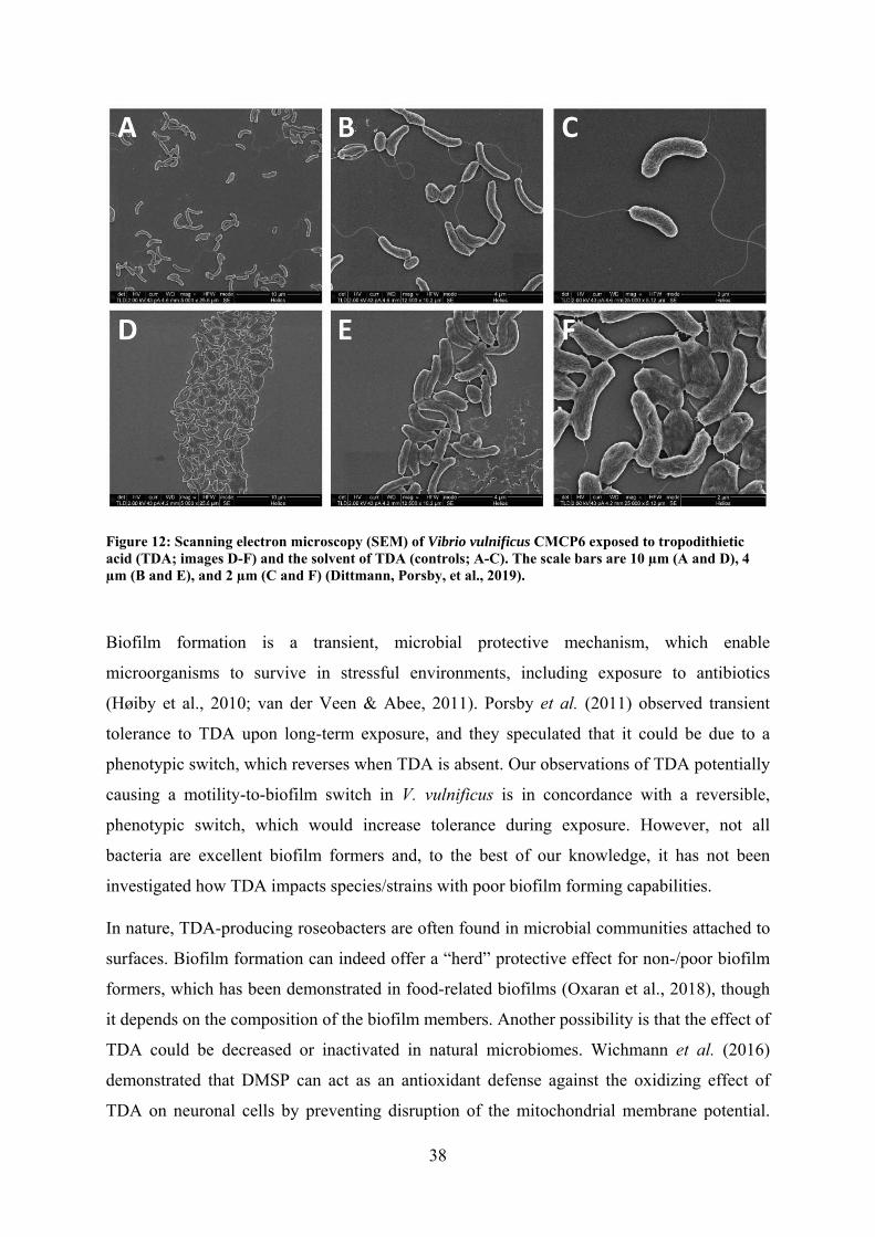
38
Figure 12: Scanning electron microscopy (SEM) of Vibrio vulnificus CMCP6 exposed to tropodithietic acid (TDA; images D-F) and the solvent of TDA (controls; A-C). The scale bars are 10 µm (A and D), 4 µm (B and E), and 2 µm (C and F) (Dittmann, Porsby, et al., 2019).
Biofilm formation is a transient, microbial protective mechanism, which enable
microorganisms to survive in stressful environments, including exposure to antibiotics
(Høiby et al., 2010; van der Veen & Abee, 2011). Porsby et al. (2011) observed transient
tolerance to TDA upon long-term exposure, and they speculated that it could be due to a
phenotypic switch, which reverses when TDA is absent. Our observations of TDA potentially
causing a motility-to-biofilm switch in V. vulnificus is in concordance with a reversible,
phenotypic switch, which would increase tolerance during exposure. However, not all
bacteria are excellent biofilm formers and, to the best of our knowledge, it has not been
investigated how TDA impacts species/strains with poor biofilm forming capabilities.
In nature, TDA-producing roseobacters are often found in microbial communities attached to
surfaces. Biofilm formation can indeed offer a “herd” protective effect for non-/poor biofilm
formers, which has been demonstrated in food-related biofilms (Oxaran et al., 2018), though
it depends on the composition of the biofilm members. Another possibility is that the effect of
TDA could be decreased or inactivated in natural microbiomes. Wichmann et al. (2016)
demonstrated that DMSP can act as an antioxidant defense against the oxidizing effect of
TDA on neuronal cells by preventing disruption of the mitochondrial membrane potential.
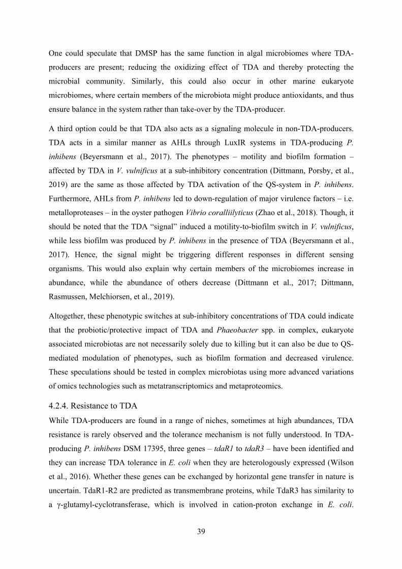
39
One could speculate that DMSP has the same function in algal microbiomes where TDA-
producers are present; reducing the oxidizing effect of TDA and thereby protecting the
microbial community. Similarly, this could also occur in other marine eukaryote
microbiomes, where certain members of the microbiota might produce antioxidants, and thus
ensure balance in the system rather than take-over by the TDA-producer.
A third option could be that TDA also acts as a signaling molecule in non-TDA-producers.
TDA acts in a similar manner as AHLs through LuxIR systems in TDA-producing P.
inhibens (Beyersmann et al., 2017). The phenotypes – motility and biofilm formation –
affected by TDA in V. vulnificus at a sub-inhibitory concentration (Dittmann, Porsby, et al.,
2019) are the same as those affected by TDA activation of the QS-system in P. inhibens.
Furthermore, AHLs from P. inhibens led to down-regulation of major virulence factors – i.e.
metalloproteases – in the oyster pathogen Vibrio coralliilyticus (Zhao et al., 2018). Though, it
should be noted that the TDA “signal” induced a motility-to-biofilm switch in V. vulnificus,
while less biofilm was produced by P. inhibens in the presence of TDA (Beyersmann et al.,
2017). Hence, the signal might be triggering different responses in different sensing
organisms. This would also explain why certain members of the microbiomes increase in
abundance, while the abundance of others decrease (Dittmann et al., 2017; Dittmann,
Rasmussen, Melchiorsen, et al., 2019).
Altogether, these phenotypic switches at sub-inhibitory concentrations of TDA could indicate
that the probiotic/protective impact of TDA and Phaeobacter spp. in complex, eukaryote
associated microbiotas are not necessarily solely due to killing but it can also be due to QS-
mediated modulation of phenotypes, such as biofilm formation and decreased virulence.
These speculations should be tested in complex microbiotas using more advanced variations
of omics technologies such as metatranscriptomics and metaproteomics.
4.2.4. Resistance to TDA
While TDA-producers are found in a range of niches, sometimes at high abundances, TDA
resistance is rarely observed and the tolerance mechanism is not fully understood. In TDA-
producing P. inhibens DSM 17395, three genes – tdaR1 to tdaR3 – have been identified and
they can increase TDA tolerance in E. coli when they are heterologously expressed (Wilson
et al., 2016). Whether these genes can be exchanged by horizontal gene transfer in nature is
uncertain. TdaR1-R2 are predicted as transmembrane proteins, while TdaR3 has similarity to
a γ-glutamyl-cyclotransferase, which is involved in cation-proton exchange in E. coli.
40
Interestingly, TDA can also inhibit growth of the TDA-producing Phaeobacter spp. (Porsby
et al., 2011; Will et al., 2017), most likely due to a high energetic demand (Will et al., 2017).
Among non-TDA-producers, genetic and phenotypic resistance to TDA have not yet been
found nor developed in the laboratory. However, TDA tolerant bacteria have been isolated
from a marine sponge microbiome, where a TDA-producing Pseudovibrio spp. had also been
isolated (Harrington et al., 2014). So far, efflux pumps and porins have been out ruled as
possible innate tolerance mechanisms (Porsby et al., 2011). Porsby et al. (2011) investigated
different in vitro approaches to adapt and provoke mutations for inducing TDA resistance and
tolerance in non-producer strains. Interestingly, several approaches were not successful in
Salmonella typhimurium, E. coli, P. aeruginosa, and S. aureus. Tolerance to 2 x the minimal
inhibitory concentration was transient when the adapted strains were passed through medium
without TDA (Porsby et al., 2011). Rasmussen et al. (2016) also attempted to adapt the fish
pathogen, V. anguillarum, to TDA through adaptive laboratory evolution, but no resistant or
tolerant strains came out of it. Whole-genome sequencing revealed point mutations, though
none of them were consistently due to TDA exposure (Rasmussen et al., 2016). Altogether,
our lacking understanding on the TDA resistance and/or tolerance mechanism(s) come back
to the insufficient knowledge on the exact target(s) and molecular mechanisms of TDA on/in
a cell – particularly at concentrations resembling the levels found in nature.
4.3. Conclusions
Members of the Roseobacter group are found in many different environmental niches,
reflecting their versatility and adaptability. Both symbiotic and pathogenic relationships are
observed between roseobacters and eukaryotes. Some of the interactions with the host have
been demonstrated to be influenced by the production of bioactive agents, such as TDA. The
impact of TDA-producers on host-associated microbiomes is dependent on the complexity
and composition of the microbial community. While the probiotic effect has been coupled to
TDA production, the role of TDA in shaping microbiomes is still debatable.
TDA is a broad-spectrum antibiotic compound, which can serve as a competitive advantage
for the bacterial producer by protection of its host. Resistance towards TDA is rarely
observed and it is difficult to induce, while tolerance can be found, although it is reversible.
Disruption of the PMF by acting as an electroneutral proton antiporter importing H+ ions and
exporting metal (1+) ions is the proposed mode of action in E. coli. TDA can also act as a
signaling molecule regulating different phenotypes, such as biofilm formation, motility, and
41
secondary metabolite production, both in TDA-producers and V. vulnificus. This could
indicate that the compound either has multiple functions or that the function in situ is
modulation of behavior.
42
5. Concluding remarks and future perspectives
The aquaculture industry is growing rapidly to match the increasing demand for high-quality
protein to feed the world population. High intensity farming of animals increases the stress on
the animals, and thereby raises the risk of disease. A more productive and sustainable
aquaculture requires measures for disease control which are minimally dependent on the
antibiotic deployment due to resistance development. Vaccines have decreased the use of
antibiotics, but the fish larvae with underdeveloped immune systems need to be protected by
measures controlling the microbiome. Microbiome management encompasses stabilization of
the rearing water quality, removal of toxic compounds, directing the community towards
slow-growing heterotrophs rather than fast-growing opportunists, and addition of probiotics
to improve the health of the host.
The successful management of aquaculture microbiotas require in-depth knowledge on the
ecology and the microbial interactions occurring in these systems. Our current knowledge-
base on marine microbiomes, including microbiotas related to aquaculture, is increasing
drastically with the advances and availability of sequencing based methods. We still have a
lot to unravel and understand, i.e. what is a “healthy” microbiome? And how can we
manipulate and maintain the local microbiota to withstand take-over from opportunistic
pathogens?
This PhD took the initial steps of this process by assessing the impact of a probiotic candidate
– TDA-producing P. inhibens – on the microbiota associated with different trophic levels
found in aquaculture and in natural aquatic systems. Previous studies had focused on the
antagonistic effect, protection of fish larvae, the bioactive compound, and occurrence in
nature, which indeed is important information to understand in the screening of probiotic
candidates. However, in most cases, they did not account for the system and the bacterial
context that the P. inhibens and TDA is acting in. Minor changes were observed in the
microbiomes exposed to TDA-producing P. inhibens; the changes were highly specific and
consistently targeting other roseobacters and (potentially pathogenic) vibrios. While these
observations add to the positive outlook for probiotic application, there are still uncertainties
regarding the long-term effects of adding the probiotic P. inhibens, the optimal introduction
route, and how they should be monitored in the aquaculture units to ensure maximum
probiotic effect. Further studies using pilot or full-scale trials over extended periods of time
43
in aquaculture settings are needed to answer some of these unexplored areas, which are
essential in the assessment of the application potential.
Besides determining establishment, impact on the commensal microbiome, and long-term
effect, it should be noted that Phaeobacter spp. and closely related roseobacters have been
linked to pathogenicity in algae and molluscs. The virulence factors important for infection
are largely unknown, although it is confirmed that certain Phaeobacter spp. produce algicidal
compounds such as the roseobacticides and that these compounds can kill microalgal species
used in aquaculture. Given that not all TDA-producers produce roseobacticides and that
certain microalgal species are unaffected by certain variants, it is likely possible to find
probiotic strains, which could be used in aquaculture without harming the live-feed.
However, this should be further investigated and used for risk assessments of the final strains.
The other pillar of this PhD was focused on the bioactive compound TDA. The in vitro
bactericidal effect of the pure compound on a range of Gram-positive and Gram-negative
bacteria, as well as the killing effect on yeasts and the cytotoxicity to some mammalian cells,
had indicated that the target and mechanism of action was essential to all living cells. The
proposed mode of action – i.e. disruption of the PMF by acting as an electroneutral proton
antiporter importing H+ ions and exporting metal (1+) ions – had been studied in E. coli.
Studies had indicated that TDA can also act as a signaling molecule in TDA-producers but
interestingly, no studies had looked into mechanism in marine, non-TDA-producing bacteria.
In this study, this knowledge gap was addressed. While the compound did not have a growth
inhibiting effect, it still caused a pronounced metabolic shift in the V. vulnificus cells.
Defense phenotypes such as oxidative stress defense, iron scavenging, and biofilm formation
were affected. While the oxidative stress was in line with the proposed mode of action in E.
coli, the phenotypic motility-to-biofilm switch had not been observed. If the defense against
TDA is related to a transient phenotype, and that production of antioxidants (such as DMSP
in algae) could limit the damage, it could explain why inherent resistance is rarely seen.
Motility and biofilm formation is also QS-regulated, which could indicate that the response
and impact on several genes observed in the transcriptome, might in fact be QS-induced
rather than defensive. If that is the case, then TDA might also act as a behavioral modulator
in the microbiomes. Therefore it would be interesting to determine if TDA affects QS-
regulated phenotypes in Gram-positive bacteria as well. Furthermore, it would be relevant to
assess the impact of TDA on the functionalities in the microbiomes. Pure TDA can change
the community structure of the microbiota, but whether or not it modulates behavior is a
44
different aspect, which is not caught by DNA sequencing. Doing functional studies using
metatranscriptomics, metaproteomics, or metabolomics could reveal if pure TDA, at sub-
inhibitory concentrations, modulates behavior and potentially limits virulence expression for
opportunistic pathogens.
45
6. Acknowledgements
First of all, I would like to thank my supervisors, Lone Gram and Mikkel Bentzon-Tilia, for
their immense support throughout my PhD project. Your enthusiasm, support, feedback, and
care has made this journey educational – both at academic and personal level.
I wish to thank Su Egan (Centre for Marine Bio-Innovation, UNSW, Sydney, Australia) for
welcoming me into her group and the centre - you also made sure that I had a fantastic,
inspiring research stay. Our many discussions and feedback sessions over coffee by the
Library Lawn gave valuable output for two of the papers in this thesis. I also wish to thank
current and past members of the Centre for Marine Bio-Innovation – particularly Professor
Torsten Thomas, Dr. Priscila Goncalves, Elizabeth Copeland, Jennifer Hudson, Dr. Bernd
Wemheuer, and Dr. Emmanuelle Botté – for scientific discussion and support, mentoring,
inspiration, and much more.
I would like to acknowledge the funding bodies: “Oticon Fonden”, “IDAs og Berg-Nielsens
Studie- og Støttefond”, “Augustinus Fonden”, “Fabrikant P.A. Fiskers”, “the scholarship
Christian og Ottilia Brorsons Rejselegat for yngre videnskabsmænd og –kvinder”, and “Otto
Mønsted Fonden” for financial support of my research stay in Australia and partipation at the
ISME17 conference.
Gratitude should also go to my colleagues in the Bacterial Ecophysiology and Biotechnology
group. Our work environment with space for scientific discussion, moral support, and social
interactions has been invaluable for me. Thank you goes to Jette for immense support with
troublesome DNA extractions and preparation of amplicon libraries. Thanks to the three other
leaves - Bastian Barker Rasmussen, Nicole Ciacotich, and Sara Skøtt Paulsen - of a four-
leaved clover having their PhD projects side-by-side. We did it! A special thank you goes to
Eva Sonnenschein – initially my office mate but soon became a great mentor and a dear
friend.
Finally, a special thank you goes to my dear family, my partner Nicolai, and closest friends.
Your loving support, encouragement, and stability has meant so much to me. Thank you for
believing in me and supporting my ambitious dreams.
46
7. References
Araújo, C., Muñoz-Atienza, E., Nahuelquín, Y., Poeta, P., Igrejas, G., Hernández, P. E., …
Cintas, L. M. (2015). Inhibition of fish pathogens by the microbiota from rainbow trout
(Oncorhynchus mykiss, Walbaum) and rearing environment. Anaerobe, 32, 7–14.
https://doi.org/10.1016/j.anaerobe.2014.11.001
Araújo, C., Muñoz-Atienza, E., Poeta, P., Igrejas, G., Hernández, P., Herranz, C., & Cintas,
L. (2016). Characterization of Pediococcus acidilactici strains isolated from rainbow
trout (Oncorhynchus mykiss) feed and larvae: safety, DNA fingerprinting, and
bacteriocinogenicity. Diseases of Aquatic Organisms, 119(2), 129–143.
https://doi.org/10.3354/dao02992
Attramadal, K. J. K., Salvesen, I., Xue, R., Øie, G., Størseth, T. R., Vadstein, O., & Olsen, Y.
(2012). Recirculation as a possible microbial control strategy in the production of
marine larvae. Aquacultural Engineering, 46, 27–39.
https://doi.org/10.1016/j.aquaeng.2011.10.003
Attramadal, K. J. K., Truong, T. M. H., Bakke, I., Skjermo, J., Olsen, Y., & Vadstein, O.
(2014). RAS and microbial maturation as tools for K-selection of microbial communities
improve survival in cod larvae. Aquaculture, 432, 483–490.
https://doi.org/10.1016/J.AQUACULTURE.2014.05.052
Austin, B., Baudet, E., & Stobie, M. (1992). Inhibition of bacterial fish pathogens by
Tetraselmis suecica. Journal of Fish Diseases, 15(1), 55–61.
https://doi.org/10.1111/j.1365-2761.1992.tb00636.x
Austin, B., Stuckey, L. F., Robertson, P. A. W., Effendi, I., & Griffith, D. R. W. (1995). A
probiotic strain of Vibrio alginolyticus effective in reducing diseases caused by
Aeromonas salmonicida, Vibrio anguillarum and Vibrio ordalii. Journal of Fish
Diseases, 18(1), 93–96. https://doi.org/10.1111/j.1365-2761.1995.tb01271.x
Avnimelech, Y. (1999). Carbon/nitrogen ratio as a control element in aquaculture systems.
Aquaculture, 176(3–4), 227–235. https://doi.org/10.1016/S0044-8486(99)00085-X
Bakke, I., Coward, E., Andersen, T., & Vadstein, O. (2015). Selection in the host structures
the microbiota associated with developing cod larvae (Gadus morhua). Environmental
Microbiology, 17(10), 3914–3924. https://doi.org/10.1111/1462-2920.12888
47
Balcázar, J. L., de Blas, I., Ruiz-Zarzuela, I., Vendrell, D., Gironés, O., & Muzquiz, J. L.
(2007). Sequencing of variable regions of the 16S rRNA gene for identification of lactic
acid bacteria isolated from the intestinal microbiota of healthy salmonids. Comparative
Immunology, Microbiology and Infectious Diseases, 30(2), 111–118.
https://doi.org/10.1016/j.cimid.2006.12.001
Balcázar, J. L., Vendrell, D., de Blas, I., Ruiz-Zarzuela, I., Muzquiz, J. L., & Girones, O.
(2008). Characterization of probiotic properties of lactic acid bacteria isolated from
intestinal microbiota of fish. Aquaculture, 278(1–4), 188–191.
https://doi.org/10.1016/j.aquaculture.2008.03.014
Barbieri, E., Paster, B. J., Hughes, D., Zurek, L., Moser, D. P., Teske, A., & Sogin, M. L.
(2001). Phylogenetic characterization of epibiotic bacteria in the accessory nidamental
gland and egg capsules of the squid Loligo pealei (Cephalopoda: Loliginidae).
Environmental Microbiology, 3(3), 151–167. https://doi.org/10.1046/j.1462-
2920.2001.00172.x
Ben Kahla-Nakbi, A., Chaieb, K., & Bakhrouf, A. (2009). Investigation of several virulence
properties among Vibrio alginolyticus strains isolated from diseased cultured fish in
Tunisia. Diseases of Aquatic Organisms, 86(1), 21–28.
https://doi.org/10.3354/dao02091
Bentzon-Tilia, M., Sonnenschein, E. C., & Gram, L. (2016). Monitoring and managing
microbes in aquaculture - Towards a sustainable industry. Microbial Biotechnology,
9(5), 576–584. https://doi.org/10.1111/1751-7915.12392
Berger, M., Brock, N. L., Liesegang, H., Dogs, M., Preuth, I., Simon, M., … Brinkhoff, T.
(2012). Genetic Analysis of the Upper Phenylacetate Catabolic Pathway in the
Production of Tropodithietic Acid by Phaeobacter gallaeciensis. Applied and
Environmental Microbiology, 78(10), 3539–3551. https://doi.org/10.1128/AEM.07657-
11
Beyersmann, P. G., Tomasch, J., Son, K., Stocker, R., Göker, M., Wagner-Döbler, I., …
Brinkhoff, T. (2017). Dual function of tropodithietic acid as antibiotic and signaling
molecule in global gene regulation of the probiotic bacterium Phaeobacter inhibens.
Scientific Reports, 7(1), 730. https://doi.org/10.1038/s41598-017-00784-7
Biondi, N., Cheloni, G., Tatti, E., Decorosi, F., Rodolfi, L., Giovannetti, L., … Tredici, M. R.
48
(2017). The bacterial community associated with Tetraselmis suecica outdoor mass
cultures. Journal of Applied Phycology, 29(1), 67–78. https://doi.org/10.1007/s10811-
016-0966-5
Blanch, A. R., Alsina, M., Simón, M., & Jofre, J. (1997). Determination of bacteria
associated with reared turbot (Scophthalmus maximus) larvae. Journal of Applied
Microbiology, 82(6), 729–734. https://doi.org/10.1046/j.1365-2672.1997.00190.x
Blancheton, J. P., Attramadal, K. J. K., Michaud, L., D’Orbcastel, E. R., & Vadstein, O.
(2013). Insight into bacterial population in aquaculture systems and its implication.
Aquacultural Engineering, 53, 30–39. https://doi.org/10.1016/j.aquaeng.2012.11.009
Bledsoe, J. W., Peterson, B. C., Swanson, K. S., & Small, B. C. (2016). Ontogenetic
Characterization of the Intestinal Microbiota of Channel Catfish through 16S rRNA
Gene Sequencing Reveals Insights on Temporal Shifts and the Influence of
Environmental Microbes. PLoS ONE, 11(11), e0166379.
https://doi.org/10.1371/journal.pone.0166379
Boardman, C. L., Maloy, A. P., & Boettcher, K. J. (2008). Localization of the bacterial agent
of juvenile oyster disease (Roseovarius crassostreae) within affected eastern oysters
(Crassostrea virginica). Journal of Invertebrate Pathology, 97(2), 150–158.
https://doi.org/10.1016/J.JIP.2007.08.007
Boettcher, K. J., Barber, B. J., & Singer, J. T. (2000). Additional Evidence that Juvenile
Oyster Disease Is Caused by a Member of the Roseobacter Group and Colonization of
Nonaffected Animals by Stappia stellulata-Like Strains. Applied and Environmental
Microbiology, 66(9), 3924–3930. https://doi.org/10.1128/AEM.66.9.3924-3930.2000
Boettcher, K. J., Geaghan, K. K., Maloy, A. P., & Barber, B. J. (2005). Roseovarius
crassostreae sp. nov., a member of the Roseobacter clade and the apparent cause of
juvenile oyster disease (JOD) in cultured Eastern oysters. International Journal of
Systematic and Evolutionary Microbiology, 55(4), 1531–1537.
https://doi.org/10.1099/ijs.0.63620-0
Bolyen, E., Rideout, J. R., Dillon, M. R., Bokulich, N. A., Abnet, C., Al-Ghalith, G. A., …
Caporaso, J. G. (2018). QIIME 2: Reproducible, interactive, scalable, and extensible
microbiome data science. PeerJ Preprints.
https://doi.org/10.7287/peerj.preprints.27295v2
49
Breider, S., Freese, H. M., Spröer, C., Simon, M., Overmann, J., & Brinkhoff, T. (2017).
Phaeobacter porticola sp. nov., an antibiotic-producing bacterium isolated from a sea
harbour. International Journal of Systematic and Evolutionary Microbiology, 67(7),
2153–2159. https://doi.org/10.1099/ijsem.0.001879
Breider, S., Scheuner, C., Schumann, P., Fiebig, A., Petersen, J., Pradella, S., … Göker, M.
(2014). Genome-scale data suggest reclassifications in the Leisingera-Phaeobacter
cluster including proposals for Sedimentitalea gen. nov. and Pseudophaeobacter gen.
nov. Frontiers in Microbiology, 5, 1–13. https://doi.org/10.3389/fmicb.2014.00416
Brinkhoff, T., Bach, G., Heidorn, T., Liang, L., Schlingloff, A., & Simon, M. (2004).
Antibiotic Production by a Roseobacter Clade-Affiliated Species from the German
Wadden Sea and Its Antagonistic Effects on Indigenous Isolates. Applied and
Environmental Microbiology, 70(4), 2560–2565.
https://doi.org/10.1128/AEM.70.4.2560-2565.2003
Buchan, A., Gonzalez, J. M., & Moran, M. A. (2005). Overview of the Marine Roseobacter
Lineage. Applied and Environmental Microbiology, 71(10), 5665–5677.
https://doi.org/10.1128/AEM.71.10.5665-5677.2005
Cabello, F. C. (2006). Heavy use of prophylactic antibiotics in aquaculture: a growing
problem for human and animal health and for the environment. Environmental
Microbiology, 8(7), 1137–1144. https://doi.org/10.1111/j.1462-2920.2006.01054.x
Cabello, F. C., Godfrey, H. P., Tomova, A., Ivanova, L., Dölz, H., Millanao, A., &
Buschmann, A. H. (2013). Antimicrobial use in aquaculture re-examined: its relevance
to antimicrobial resistance and to animal and human health. Environmental
Microbiology, 15(7), 1917–1942. https://doi.org/10.1111/1462-2920.12134
Califano, G., Castanho, S., Soares, F., Ribeiro, L., Cox, C. J., Mata, L., & Costa, R. (2017).
Molecular Taxonomic Profiling of Bacterial Communities in a Gilthead Seabream
(Sparus aurata) Hatchery. Frontiers in Microbiology, 8, 1–16.
https://doi.org/10.3389/fmicb.2017.00204
Callahan, B. J., McMurdie, P. J., Rosen, M. J., Han, A. W., Johnson, A. J. A., & Holmes, S.
P. (2016). DADA2: High-resolution sample inference from Illumina amplicon data.
Nature Methods, 13(7), 581–583. https://doi.org/10.1038/nmeth.3869
50
Campbell, B. J., & Kirchman, D. L. (2013). Bacterial diversity, community structure and
potential growth rates along an estuarine salinity gradient. The ISME Journal, 7(1), 210–
220. https://doi.org/10.1038/ismej.2012.93
Cao, J., Zhang, J., Ma, L., Li, L., Zhang, W., & Li, J. (2018). Identification of fish source
Vibrio alginolyticus and evaluation of its bacterial ghosts vaccine immune effects.
MicrobiologyOpen, 7(3), e00576. https://doi.org/10.1002/mbo3.576
Caporaso, J. G., Kuczynski, J., Stombaugh, J., Bittinger, K., Bushman, F. D., Costello, E. K.,
… Knight, R. (2010). QIIME allows analysis of high-throughput community sequencing
data. Nature Methods, 7(5), 335–336. https://doi.org/10.1038/nmeth.f.303
Carnevali, O., Zamponi, M. C., Sulpizio, R., Rollo, A., Nardi, M., Orpianesi, C., … Cresci,
A. (2004). Administration of Probiotic Strain to Improve Sea Bream Wellness during
Development. Aquaculture International, 12(4/5), 377–386.
https://doi.org/10.1023/B:AQUI.0000042141.85977.bb
Case, R. J., Longford, S. R., Campbell, A. H., Low, A., Tujula, N., Steinberg, P. D., &
Kjelleberg, S. (2011). Temperature induced bacterial virulence and bleaching disease in
a chemically defended marine macroalga. Environmental Microbiology, 13(2), 529–537.
https://doi.org/10.1111/j.1462-2920.2010.02356.x
Chabrillón, M., Rico, R. M., Arijo, S., Díaz-Rosales, P., Balebona, M. C., & Morinigo, M. A.
(2005). Interactions of microorganisms isolated from gilthead sea bream, Sparus aurata
L., on Vibrio harveyi, a pathogen of farmed Senegalese sole, Solea senegalensis (Kaup).
Journal of Fish Diseases, 28(9), 531–537. https://doi.org/10.1111/j.1365-
2761.2005.00657.x
Chabrillón, M., Rico, R. M., Balebona, M. C., & Morinigo, M. A. (2005). Adhesion to sole,
Solea senegalensis Kaup, mucus of microorganisms isolated from farmed fish, and their
interaction with Photobacterium damselae subsp. piscicida. Journal of Fish Diseases,
28, 229–237. https://doi.org/10.1111/j.1365-2761.2005.00623.x
Chu, W., Zhou, S., Zhu, W., & Zhuang, X. (2015). Quorum quenching bacteria Bacillus sp.
QSI-1 protect zebrafish (Danio rerio) from Aeromonas hydrophila infection. Scientific
Reports, 4(1), 5446. https://doi.org/10.1038/srep05446
Collins, A. J., LaBarre, B. A., Wong Won, B. S., Shah, M. V., Heng, S., Choudhury, M. H.,
51
… Nyholm, S. V. (2012). Diversity and Partitioning of Bacterial Populations within the
Accessory Nidamental Gland of the Squid Euprymna scolopes. Applied and
Environmental Microbiology, 78(12), 4200–4208. https://doi.org/10.1128/AEM.07437-
11
Colwell, R. R., Huq, A., Islam, M. S., Aziz, K. M. A., Yunus, M., Khan, N. H., … Russek-
Cohen, E. (2003). Reduction of cholera in Bangladeshi villages by simple filtration.
Proceedings of the National Academy of Sciences of the United States of America,
100(3), 1051–1055. https://doi.org/10.1073/pnas.0237386100
Cooper, M. A., & Shlaes, D. (2011). Fix the antibiotics pipeline. Nature, 472(7341), 32–32.
https://doi.org/10.1038/472032a
Cooper, M. B., Smith, A. G., Paszkowski, U., & Scott, D. B. (2015). Exploring mutualistic
interactions between microalgae and bacteria in the omics age. Current Opinion in Plant
Biology, 26, 147–153. https://doi.org/10.1016/j.pbi.2015.07.003
Cordero, H., Morcillo, P., Meseguer, J., Cuesta, A., & Esteban, M. Á. (2016). Effects of
Shewanella putrefaciens on innate immunity and cytokine expression profile upon high
stocking density of gilthead seabream specimens. Fish & Shellfish Immunology, 51, 33–
40. https://doi.org/10.1016/j.fsi.2016.02.008
Crab, R., Chielens, B., Wille, M., Bossier, P., & Verstraete, W. (2010). The effect of different
carbon sources on the nutritional value of bioflocs, a feed for Macrobrachium
rosenbergii postlarvae. Aquaculture Research, 41(4), 559–567.
https://doi.org/10.1111/j.1365-2109.2009.02353.x
Crab, R., Defoirdt, T., Bossier, P., & Verstraete, W. (2012). Biofloc technology in
aquaculture: Beneficial effects and future challenges. Aquaculture, 356–357, 351–356.
https://doi.org/10.1016/j.aquaculture.2012.04.046
Croft, M. T., Lawrence, A. D., Raux-Deery, E., Warren, M. J., & Smith, A. G. (2005). Algae
acquire vitamin B12 through a symbiotic relationship with bacteria. Nature, 438(7064),
90–93. https://doi.org/10.1038/nature04056
Cytryn, E., van Rijn, J., Schramm, A., Gieseke, A., de Beer, D., & Minz, D. (2005).
Identification of bacteria potentially responsible for oxic and anoxic sulfide oxidation in
biofilters of a recirculating mariculture system. Applied and Environmental
52
Microbiology, 71(10), 6134–6141. https://doi.org/10.1128/AEM.71.10.6134-6141.2005
D’Alvise, P. W., Lillebø, S., Prol-Garcia, M. J., Wergeland, H. I., Nielsen, K. F., Bergh, Ø.,
& Gram, L. (2012). Phaeobacter gallaeciensis reduces Vibrio anguillarum in cultures of
microalgae and rotifers, and prevents vibriosis in cod larvae. PloS One, 7(8), e43996.
https://doi.org/10.1371/journal.pone.0043996
D’Alvise, P. W., Lillebø, S., Wergeland, H. I., Gram, L., & Bergh, Ø. (2013). Protection of
cod larvae from vibriosis by Phaeobacter spp.: A comparison of strains and introduction
times. Aquaculture, 384–387, 82–86.
https://doi.org/10.1016/J.AQUACULTURE.2012.12.013
D’Alvise, P. W., Melchiorsen, J., Porsby, C. H., Nielsen, K. F., & Gram, L. (2010).
Inactivation of Vibrio anguillarum by attached and planktonic Roseobacter cells.
Applied and Environmental Microbiology, 76(7), 2366–2370.
https://doi.org/10.1128/AEM.02717-09
D’Alvise, P. W., Phippen, C. B. W., Nielsen, K. F., & Gram, L. (2016). Influence of Iron on
Production of the Antibacterial Compound Tropodithietic Acid and Its Noninhibitory
Analog in Phaeobacter inhibens. Applied and Environmental Microbiology, 82(2), 502–
509. https://doi.org/10.1128/AEM.02992-15
Dang, H., & Lovell, C. R. (2000). Bacterial Primary Colonization and Early Succession on
Surfaces in Marine Waters as Determined by Amplified rRNA Gene Restriction
Analysis and Sequence Analysis of 16S rRNA Genes. Applied and Environmental
Microbiology, 66(2), 467–475. https://doi.org/10.1128/AEM.66.2.467-475.2000
Defoirdt, T., Sorgeloos, P., & Bossier, P. (2011). Alternatives to antibiotics for the control of
bacterial disease in aquaculture. Current Opinion in Microbiology, 14(3), 251–258.
https://doi.org/10.1016/j.mib.2011.03.004
Defoirdt, T., Thanh, L. D., Van Delsen, B., De Schryver, P., Sorgeloos, P., Boon, N., &
Bossier, P. (2011). N-acylhomoserine lactone-degrading Bacillus strains isolated from
aquaculture animals. Aquaculture, 311, 258–260.
https://doi.org/10.1016/j.aquaculture.2010.11.046
Dhanasiri, A. K. S., Brunvold, L., Brinchmann, M. F., Korsnes, K., Bergh, Ø., & Kiron, V.
(2011). Changes in the Intestinal Microbiota of Wild Atlantic cod Gadus morhua L.
53
Upon Captive Rearing. Microbial Ecology, 61, 20–30. https://doi.org/10.1007/s00248-
010-9673-y
Dittmann, K. K., Porsby, C. H., Goncalves, P., Mateiu, R. V., Sonnenschein, E. C., Bentzon�
Tilia, M., … Gram, L. (2019). Tropodithietic acid induces oxidative stress response, cell
envelope biogenesis and iron uptake in Vibrio vulnificus. Environmental Microbiology
Reports, 1758–2229.12771. https://doi.org/10.1111/1758-2229.12771
Dittmann, K. K., Rasmussen, B. B., Castex, M., Gram, L., & Bentzon-Tilia, M. (2017). The
aquaculture microbiome at the centre of business creation. Microbial Biotechnology,
10(6), 1279–1282. https://doi.org/10.1111/1751-7915.12877
Dittmann, K. K., Rasmussen, B. B., Melchiorsen, J., Sonnenschein, E. C., Gram, L., &
Bentzon-Tilia, M. (2019). Roseobacter probiotics affect lower-trophic level
microbiomes in marine aquaculture. Manuscript in Preparation.
Dittmann, K. K., Sonnenschein, E. C., Egan, S., Gram, L., & Bentzon�Tilia, M. (2019).
Impact of Phaeobacter inhibens on marine eukaryote�associated microbial
communities. Environmental Microbiology Reports, 11(3), 401–413.
https://doi.org/10.1111/1758-2229.12698
Duerr, E. O. (1998). Cultured microalgae as aquaculture feeds. Journal of Marine
Biotechnology, 6(2). Retrieved from https://findit.dtu.dk/en/catalog/2272023584
Edgar, R. C. (2010). Search and clustering orders of magnitude faster than BLAST.
Bioinformatics, 26(19), 2460–2461. https://doi.org/10.1093/bioinformatics/btq461
Egerton, S., Culloty, S., Whooley, J., Stanton, C., & Ross, R. P. (2018). The Gut Microbiota
of Marine Fish. Frontiers in Microbiology, 9, 873.
https://doi.org/10.3389/fmicb.2018.00873
FAO. (2018). The State of World Fisheries and Aquaculture 2018 - Meeting the sustainable
development goals. Rome.
FAO and WHO. (2001). Probiotics in food - Health and nutrional properties ad guidelines
for evaluation. Food and Nutrition Paper (Vol. 85). Rome.
Fernandes, N., Case, R. J., Longford, S. R., Seyedsayamdost, M. R., Steinberg, P. D.,
Kjelleberg, S., & Thomas, T. (2011). Genomes and virulence factors of novel bacterial
pathogens causing bleaching disease in the marine red alga Delisea pulchra. PloS One,
54
6(12), e27387. https://doi.org/10.1371/journal.pone.0027387
Fjellheim, A. J., Playfoot, K. J., Skjermo, J., & Vadstein, O. (2012). Inter-individual variation
in the dominant intestinal microbiota of reared Atlantic cod (Gadus morhua L.) larvae.
Aquaculture Research, 43(10), 1499–1508. https://doi.org/10.1111/j.1365-
2109.2011.02952.x
Foesel, B. U., Gieseke, A., Schwermer, C., Stief, P., Koch, L., Cytryn, E., … Schramm, A.
(2008). Nitrosomonas Nm143-like ammonia oxidizers and Nitrospira marina-like nitrite
oxidizers dominate the nitrifier community in a marine aquaculture biofilm. FEMS
Microbiology Ecology, 63(2), 192–204. https://doi.org/10.1111/j.1574-
6941.2007.00418.x
Fuller, R. (1989). Probiotics in man and animals. Journal of Applied Bacteriology, 66(5),
365–378. https://doi.org/10.1111/j.1365-2672.1989.tb05105.x
Garriques, D., & Arevalo, G. (1995). An evaluation of the production and use of a live
bacterial isolate to manipulate the microbial flora in the commercial production of
Penaeus vannamei postlarvae in Ecuador. In Swimming Through Troubled Water.
Proceedings of the Special Session on Shrimp Farming, Aquaculture ’95. (pp. 53–59).
Gatesoupe, F.-J. (1991). The effect of three strains of lactic bacteria on the production rate of
rotifers, Brachionus plicatilis, and their dietary value for larval turbot, Scophthalmus
maximus. Aquaculture, 96(3–4), 335–342. https://doi.org/10.1016/0044-8486(91)90162-
Z
Gatesoupe, F.-J. (1994). Lactic acid bacteria increase the resistance of turbot larvae,
Scophthalmus maximus, against pathogenic vibrio. Aquatic Living Resources, 7(4), 277–
282. https://doi.org/10.1051/alr:1994030
Gatesoupe, F.-J. (1999). The use of probiotics in aquaculture. Aquaculture, 180(1–2), 147–
165. https://doi.org/10.1016/S0044-8486(99)00187-8
Geng, H., Bruhn, J. B., Nielsen, K. F., Gram, L., & Belas, R. (2008). Genetic dissection of
tropodithietic acid biosynthesis by marine roseobacters. Applied and Environmental
Microbiology, 74(5), 1535–1545. https://doi.org/10.1128/AEM.02339-07
Geng, H., Tran-Gyamfi, M. B., Lane, T. W., Sale, K. L., & Yu, E. T. (2016). Changes in the
Structure of the Microbial Community Associated with Nannochloropsis salina
55
following Treatments with Antibiotics and Bioactive Compounds. Frontiers in
Microbiology, 7, 1155. https://doi.org/10.3389/fmicb.2016.01155
Glöckner, F. O., & Joint, I. (2010). Marine microbial genomics in Europe: current status and
perspectives. Microbial Biotechnology, 3(5), 523–530. https://doi.org/10.1111/j.1751-
7915.2010.00169.x
Gómez-León, J., Villamil, L., Lemos, M. L., Novoa, B., & Figueras, A. (2005). Isolation of
Vibrio alginolyticus and Vibrio splendidus from aquacultured carpet shell clam
(Ruditapes decussatus) larvae associated with mass mortalities. Applied and
Environmental Microbiology, 71(1), 98–104. https://doi.org/10.1128/AEM.71.1.98-
104.2005
González, J. M., & Moran, M. A. (1997). Numerical dominance of a group of marine bacteria
in the alpha-subclass of the class Proteobacteria in coastal seawater. Applied and
Environmental Microbiology, 63(11), 4237–4242. Retrieved from
http://www.ncbi.nlm.nih.gov/pubmed/4265956
González, J. M., Simó, R., Massana, R., Covert, J. S., Casamayor, E. O., Pedrós-Alió, C., &
Moran, M. A. (2000). Bacterial community structure associated with a
dimethylsulfoniopropionate-producing North Atlantic algal bloom. Applied and
Environmental Microbiology, 66(10), 4237–4246.
https://doi.org/10.1128/AEM.66.10.4237-4246.2000
Gram, L., Løvold, T., Nielsen, J., Melchiorsen, J., & Spanggaard, B. (2001). In vitro
antagonism of the probiont Pseudomonas fluorescens strain AH2 against Aeromonas
salmonicida does not confer protection of salmon against furunculosis. Aquaculture,
199(1–2), 1–11. https://doi.org/10.1016/S0044-8486(01)00565-8
Gram, L., Melchiorsen, J., Spanggaard, B., Huber, I., & Nielsen, T. F. (1999). Inhibition of
Vibrio anguillarum by Pseudomonas fluorescens AH2, a Possible Probiotic Treatment
of Fish. Applied and Environmental Microbiology, 65(3), 969–973.
Greer, E. M., Aebisher, D., Greer, A., & Bentley, R. (2008). Computational Studies of the
Tropone Natural Products, Thiotropocin, Tropodithietic Acid, and Troposulfenin.
Significance of Thiocarbonyl−Enol Tautomerism. The Journal of Organic Chemistry,
73(1), 280–283. https://doi.org/10.1021/jo7018416
56
Grigioni, S., Boucher-Rodoni, R., Demarta, A., Tonolla, M., & Peduzzi, R. (2000).
Phylogenetic characterisation of bacterial symbionts in the accessory nidamental glands
of the sepioid Sepia officinalis (Cephalopoda: Decapoda). Marine Biology, 136(2), 217–
222. https://doi.org/10.1007/s002270050679
Gromek, S. M., Suria, A. M., Fullmer, M. S., Garcia, J. L., Gogarten, J. P., Nyholm, S. V., &
Balunas, M. J. (2016). Leisingera sp. JC1, a bacterial isolate from hawaiian bobtail
squid eggs, produces indigoidine and differentially inhibits vibrios. Frontiers in
Microbiology, 7(SEP), 1–16. https://doi.org/10.3389/fmicb.2016.01342
Grotkjær, T., Bentzon-Tilia, M., D’Alvise, P. W., Dierckens, K., Bossier, P., & Gram, L.
(2016). Phaeobacter inhibens as probiotic bacteria in non-axenic Artemia and algae
cultures. Aquaculture, 462, 64–69.
Grotkjær, T., Bentzon-Tilia, M., D’Alvise, P. W., Dourala, N., Nielsen, K. F., & Gram, L.
(2016). Isolation of TDA-producing Phaeobacter strains from sea bass larval rearing
units and their probiotic effect against pathogenic Vibrio spp. in Artemia cultures.
Systematic and Applied Microbiology, 39, 180–188.
Hansen, G. H., & Olafsen, J. A. (1999). Bacterial Interactions in Early Life Stages of Marine
Cold Water Fish. Microbial Ecology, 38(1), 1–26.
https://doi.org/10.1007/s002489900158
Hari, B., Madhusoodana Kurup, B., Varghese, J. T., Schrama, J. W., & Verdegem, M. C. .
(2004). Effects of carbohydrate addition on production in extensive shrimp culture
systems. Aquaculture, 241(1–4), 179–194.
https://doi.org/10.1016/j.aquaculture.2004.07.002
Harrington, C., Reen, F., Mooij, M., Stewart, F., Chabot, J.-B., Guerra, A., … O’Gara, F.
(2014). Characterisation of Non-Autoinducing Tropodithietic Acid (TDA) Production
from Marine Sponge Pseudovibrio Species. Marine Drugs, 12, 5960–5978.
https://doi.org/10.3390/md12125960
Herlemann, D. P., Labrenz, M., Jürgens, K., Bertilsson, S., Waniek, J. J., & Andersson, A. F.
(2011). Transitions in bacterial communities along the 2000 km salinity gradient of the
Baltic Sea. The ISME Journal, 5(10), 1571–1579. https://doi.org/10.1038/ismej.2011.41
Higuera-Llantén, S., Vásquez-Ponce, F., Barrientos-Espinoza, B., Mardones, F. O., Marshall,
57
S. H., & Olivares-Pacheco, J. (2018). Extended antibiotic treatment in salmon farms
select multiresistant gut bacteria with a high prevalence of antibiotic resistance genes.
PLoS ONE, 13(9), e0203641. https://doi.org/10.1371/journal.pone.0203641
Hjelm, M., Bergh, O., Riaza, A., Nielsen, J., Melchiorsen, J., Jensen, S., … Gram, L. (2004).
Selection and identification of autochthonous potential probiotic bacteria from turbot
larvae (Scophthalmus maximus) rearing units. Systematic and Applied Microbiology, 27,
360–371. https://doi.org/10.1078/0723-2020-00256
Høiby, N., Bjarnsholt, T., Givskov, M., Molin, S., & Ciofu, O. (2010). Antibiotic resistance
of bacterial biofilms. International Journal of Antimicrobial Agents, 35(4), 322–332.
https://doi.org/10.1016/j.ijantimicag.2009.12.011
Høj, L., Bourne, D. G., & Hall, M. R. (2009). Localization, abundance and community
structure of bacteria associated with Artemia: Effects of nauplii enrichment and
antimicrobial treatment. Aquaculture, 293(3–4), 278–285.
https://doi.org/10.1016/j.aquaculture.2009.04.024
Hovda, M. B., Lunestad, B. T., Fontanillas, R., & Rosnes, J. T. (2007). Molecular
characterisation of the intestinal microbiota of farmed Atlantic salmon (Salmo salar L.).
Aquaculture, 272(1–4), 581–588. https://doi.org/10.1016/j.aquaculture.2007.08.045
Ingerslev, H. C., von Gersdorff Jørgensen, L., Lenz Strube, M., Larsen, N., Dalsgaard, I.,
Boye, M., & Madsen, L. (2014). The development of the gut microbiota in rainbow trout
(Oncorhynchus mykiss) is affected by first feeding and diet type. Aquaculture, 424–425,
24–34. https://doi.org/10.1016/j.aquaculture.2013.12.032
Kaneko, T., & Colwell, R. R. (1973). Ecology of Vibrio parahaemolyticus in Chesapeake
Bay. Journal of Bacteriology, 113(1), 24–32.
Kanther, M., & Rawls, J. F. (2010). Host–microbe interactions in the developing zebrafish.
Current Opinion in Immunology, 22(1), 10–19.
https://doi.org/10.1016/J.COI.2010.01.006
Kaufman, M. R., Ikeda, Y., Patton, C., van Dykhuizen, G., & Epel, D. (1998). Bacterial
Symbionts Colonize the Accessory Nidamental Gland of the Squid Loligo opalescens
via Horizontal Transmission. The Biological Bulletin, 194(1), 36–43.
https://doi.org/10.2307/1542511
58
Kevin II, D. A., Meujo, D. A., & Hamann, M. T. (2009). Polyether ionophores: broad-
spectrum and promising biologically active molecules for the control of drug-resistant
bacteria and parasites. Expert Opinion on Drug Discovery, 4(2), 109–146.
https://doi.org/10.1517/17460440802661443
Kibenge, F. S. B., Godoy, M. G., Fast, M., Workenhe, S., & Kibenge, M. J. T. (2012).
Countermeasures against viral diseases of farmed fish. Antiviral Research, 95(3), 257–
281. https://doi.org/10.1016/J.ANTIVIRAL.2012.06.003
King, G. M., Judd, C., Kuske, C. R., & Smith, C. (2012). Analysis of Stomach and Gut
Microbiomes of the Eastern Oyster (Crassostrea virginica) from Coastal Louisiana,
USA. PLoS ONE, 7(12), e51475. https://doi.org/10.1371/journal.pone.0051475
Kintaka, K., Ono, H., Tsubotani, S., Harada, S., & Okazaki, H. (1984). Thiotropocin, a new
sulfur-containing 7-membered-ring antibiotic produced by a Pseudomonas sp. The
Journal of Antibiotics, 37(11), 1294–1300. https://doi.org/10.7164/antibiotics.37.1294
Kohanski, M. A., Dwyer, D. J., Hayete, B., Lawrence, C. A., & Collins, J. J. (2007). A
Common Mechanism of Cellular Death Induced by Bactericidal Antibiotics. Cell,
130(5), 797–810. https://doi.org/10.1016/J.CELL.2007.06.049
Li, X., Yu, Y., Feng, W., Yan, Q., & Gong, Y. (2012). Host species as a strong determinant
of the intestinal microbiota of fish larvae. The Journal of Microbiology, 50(1), 29–37.
https://doi.org/10.1007/s12275-012-1340-1
Liang, L. (2003). Investigation of Secondary Metabolites of North Sea Bacteria:
Fermentation, Isolation, Structure Elucidation and Bioactivity. University of Göttingen.
Linares, J. F., Gustafsson, I., Baquero, F., & Martinez, J. L. (2006). Antibiotics as
intermicrobial signaling agents instead of weapons. Proceedings of the National
Academy of Sciences, 103(51), 19484–19489. https://doi.org/10.1073/pnas.0608949103
Liu, S., Ren, H., Shen, L., Lou, L., Tian, G., Zheng, P., & Hu, B. (2015). pH levels drive
bacterial community structure in sediments of the Qiantang River as determined by 454
pyrosequencing. Frontiers in Microbiology, 6, 285.
https://doi.org/10.3389/fmicb.2015.00285
Lozupone, C. A., & Knight, R. (2007). Global patterns in bacterial diversity. Proceedings of
the National Academy of Sciences of the United States of America, 104(27), 11436–
59
11440. https://doi.org/10.1073/pnas.0611525104
Majzoub, M. E., Beyersmann, P. G., Simon, M., Thomas, T., Brinkhoff, T., & Egan, S.
(2019). Phaeobacter inhibens controls bacterial community assembly on a marine
diatom. FEMS Microbiology Ecology, 95(6). https://doi.org/10.1093/femsec/fiz060
Makridis, P., Costa, R. A., & Dinis, M. T. (2006). Microbial conditions and antimicrobial
activity in cultures of two microalgae species, Tetraselmis chuii and Chlorella
minutissima, and effect on bacterial load of enriched Artemia metanauplii. Aquaculture,
255(1–4), 76–81. https://doi.org/10.1016/j.aquaculture.2005.12.010
Malmstrom, R. R., Kiene, R. P., & Kirchman, D. L. (2004). Identification and enumeration of
bacteria assimilating dimethylsulfoniopropionate (DMSP) in the North Atlantic and Gulf
of Mexico. Limnology and Oceanography, 49(2), 597–606.
https://doi.org/10.4319/lo.2004.49.2.0597
Marchesi, J. R., & Ravel, J. (2015). The vocabulary of microbiome research: a proposal.
Microbiome, 3, 31. https://doi.org/10.1186/s40168-015-0094-5
Martens, T., Heidorn, T., Pukall, R., Simon, M., Tindall, B. J., & Brinkhoff, T. (2006).
Reclassification of Roseobacter gallaeciensis Ruiz-Ponte et al. 1998 as Phaeobacter
gallaeciensis gen. nov., comb. nov., description of Phaeobacter inhibens sp. nov.,
reclassification of Ruegeria algicola (Lafay et al. 1995) Uchino et al. 1999
as Marinovum algicola gen. nov., comb. nov., and emended descriptions of the
genera Roseobacter, Ruegeria and Leisingera. International Journal of Systematic and
Evolutionary Microbiology, 56(6), 1293–1304. https://doi.org/10.1099/ijs.0.63724-0
Meron, D., Atias, E., Iasur Kruh, L., Elifantz, H., Minz, D., Fine, M., & Banin, E. (2011).
The impact of reduced pH on the microbial community of the coral Acropora
eurystoma. The ISME Journal, 5(1), 51–60. https://doi.org/10.1038/ismej.2010.102
Merrifield, D. L., Dimitroglou, A., Foey, A., Davies, S. J., Baker, R. T. M., Bøgwald, J., …
Ringø, E. (2010). The current status and future focus of probiotic and prebiotic
applications for salmonids. Aquaculture, 302(1–2), 1–18.
https://doi.org/10.1016/j.aquaculture.2010.02.007
Miller, T. R., & Belas, R. (2004). Dimethylsulfoniopropionate Metabolism by Pfiesteria-
Associated Roseobacter spp. Applied and Environmental Microbiology, 70(6), 3383–
60
3391. https://doi.org/10.1128/AEM.70.6.3383-3391.2004
Miranda, C. D., Godoy, F. A., & Lee, M. R. (2018). Current Status of the Use of Antibiotics
and the Antimicrobial Resistance in the Chilean Salmon Farms. Frontiers in
Microbiology, 9, 1284. https://doi.org/10.3389/fmicb.2018.01284
Moisander, P. H., Sexton, A. D., & Daley, M. C. (2015). Stable Associations Masked by
Temporal Variability in the Marine Copepod Microbiome. PLoS ONE, 10(9), e0138967.
https://doi.org/10.1371/journal.pone.0138967
Montanari, M. P., Pruzzo, C., Pane, L., & Colwell, R. R. (1999). Vibrios associated with
plankton in a coastal zone of the Adriatic Sea (Italy). FEMS Microbiology Ecology,
29(3), 241–247. https://doi.org/10.1111/j.1574-6941.1999.tb00615.x
Moran, M. A., Belas, R., Schell, M. A., González, J. M., Sun, F., Sun, S., … Buchan, A.
(2007). Ecological Genomics of Marine Roseobacters. Applied and Environmental
Microbiology, 73(14), 4559. https://doi.org/10.1128/AEM.02580-06
Moran, M. A., Buchan, A., González, J. M., Heidelberg, J. F., Whitman, W. B., Kiene, R. P.,
… Ward, N. (2004). Genome sequence of Silicibacter pomeroyi reveals adaptations to
the marine environment. Nature, 432(7019), 910–913.
https://doi.org/10.1038/nature03170
Moran, M. A., González, J. M., & Kiene, R. P. (2003). Linking a Bacterial Taxon to Sulfur
Cycling in the Sea: Studies of the Marine Roseobacter Group. Geomicrobiology
Journal, 20(4), 375–388. https://doi.org/10.1080/01490450303901
Moriarty, D. J. W. (1998). Control of luminous Vibrio species in penaeid aquaculture ponds.
Aquaculture, 164(1–4), 351–358. https://doi.org/10.1016/S0044-8486(98)00199-9
Munro, P. D., Barbour, A., & Birkbeck, T. H. (1994). Comparison of the gut bacterial flora of
start-feeding larval turbot reared under different conditions. Journal of Applied
Bacteriology, 77(5), 560–566. https://doi.org/10.1111/j.1365-2672.1994.tb04402.x
Muramatsu, Y., Uchino, Y., Kasai, H., Suzuki, K. -i., & Nakagawa, Y. (2007). Ruegeria
mobilis sp. nov., a member of the Alphaproteobacteria isolated in Japan and Palau.
International Journal of Systematic and Evolutionary Microbiology, 57(6), 1304–1309.
https://doi.org/10.1099/ijs.0.64572-0
Nalca, Y., Jänsch, L., Bredenbruch, F., Geffers, R., Buer, J., & Häussler, S. (2006). Quorum-
61
Sensing Antagonistic Activities of Azithromycin in Pseudomonas aeruginosa PAO1: a
Global Approach. Antimicrobial Agents and Chemotherapy, 50(5), 1680–1688.
https://doi.org/10.1128/AAC.50.5.1680-1688.2006
Neu, A. K., Månsson, M., Gram, L., & Prol-García, M. J. (2014). Toxicity of Bioactive and
Probiotic Marine Bacteria and Their Secondary Metabolites in Artemia sp. and
Caenorhabditis elegans as Eukaryotic Model Organisms. Applied and Environmental
Microbiology, 80(1), 146–153. https://doi.org/10.1128/AEM.02717-13
Newton, R. J., Griffin, L. E., Bowles, K. M., Meile, C., Gifford, S., Givens, C. E., … Moran,
M. A. (2010). Genome characteristics of a generalist marine bacterial lineage. The ISME
Journal, 4(6), 784–798. https://doi.org/10.1038/ismej.2009.150
Niu, Y., Defoirdt, T., Baruah, K., Van de Wiele, T., Dong, S., & Bossier, P. (2014). Bacillus
sp. LT3 improves the survival of gnotobiotic brine shrimp (Artemia franciscana) larvae
challenged with Vibrio campbellii by enhancing the innate immune response and by
decreasing the activity of shrimp-associated vibrios. Veterinary Microbiology, 173(3–4),
279–288. https://doi.org/10.1016/j.vetmic.2014.08.007
Norwegian Ministry of Health and Care Services. (2015). National Strategy against
Antibiotic Resistance 2015-2020. Retrieved from
https://www.regjeringen.no/contentassets/5eaf66ac392143b3b2054aed90b85210/antibio
tic-resistance-engelsk-lavopploslig-versjon-for-nett-10-09-15.pdf
Olafsen, J. A. (2001). Interactions between fish larvae and bacteria in marine aquaculture.
Aquaculture, 200(1–2), 223–247. https://doi.org/10.1016/S0044-8486(01)00702-5
Olsen, A. I., Olsen, Y., Attramadal, Y., Christie, K., Birkbeck, T. H., Skjermo, J., &
Vadstein, O. (2000). Effects of short term feeding of microalgae on the bacterial flora
associated with juvenile Artemia franciscana. Aquaculture, 190(1–2), 11–25.
https://doi.org/10.1016/S0044-8486(00)00396-3
Oxaran, V., Dittmann, K. K., Lee, S. H. I., Chaul, L. T., Fernandes de Oliveira, C. A.,
Corassin, C. H., … Gram, L. (2018). Behavior of Foodborne Pathogens Listeria
monocytogenes and Staphylococcus aureus in Mixed-Species Biofilms Exposed to
Biocides. Applied and Environmental Microbiology, 84(24), e02038-18.
https://doi.org/10.1128/AEM.02038-18
62
Penesyan, A., Tebben, J., Lee, M., Thomas, T., Kjelleberg, S., Harder, T., & Egan, S. (2011).
Identification of the Antibacterial Compound Produced by the Marine Epiphytic
Bacterium Pseudovibrio sp. D323 and Related Sponge-Associated Bacteria. Marine
Drugs, 9, 1391–1402. https://doi.org/10.3390/md9081391
Petersen, J., Brinkmann, H., Bunk, B., Michael, V., Päuker, O., & Pradella, S. (2012). Think
pink: photosynthesis, plasmids and the Roseobacter clade. Environmental Microbiology,
14(10), 2661–2672. https://doi.org/10.1111/j.1462-2920.2012.02806.x
Petersen, J., Frank, O., Göker, M., & Pradella, S. (2013). Extrachromosomal, extraordinary
and essential - the plasmids of the Roseobacter clade. Applied Microbiology and
Biotechnology, 97(7), 2805–2815. https://doi.org/10.1007/s00253-013-4746-8
Phippen, C. B. W., Jørgensen, C. M., Bentzon-Tilia, M., Gotfredsen, C. H., Larsen, T. O.,
Gram, L., & Sonnenschein, E. C. (2019). Isolation of Methyl Troposulfenin from
Phaeobacter inhibens. Journal of Natural Products, acs.jnatprod.8b01097.
https://doi.org/10.1021/acs.jnatprod.8b01097
Pichon, D., Gaia, V., Norman, M. D., & Boucher-Rodoni, R. (2005). Phylogenetic diversity
of epibiotic bacteria in the accessory nidamental glands of squids (Cephalopoda:
Loliginidae and Idiosepiidae). Marine Biology, 147(6), 1323–1332.
https://doi.org/10.1007/s00227-005-0014-5
Planas, M., Pérez-Lorenzo, M., Hjelm, M., Gram, L., Uglenes Fiksdal, I., Bergh, Ø., &
Pintado, J. (2006). Probiotic effect in vivo of Roseobacter strain 27-4 against Vibrio
(Listonella) anguillarum infections in turbot (Scophthalmus maximus L.) larvae.
Aquaculture, 255, 323–333. https://doi.org/10.1016/j.aquaculture.2005.11.039
Pond, M. J., Stone, D. M., & Alderman, D. J. (2006). Comparison of conventional and
molecular techniques to investigate the intestinal microflora of rainbow trout
(Oncorhynchus mykiss). Aquaculture, 261(1), 194–203.
https://doi.org/10.1016/j.aquaculture.2006.06.037
Porsby, C. H., & Gram, L. (2016). Phaeobacter inhibens as biocontrol agent against Vibrio
vulnificus in oyster models. Food Microbiology, 57, 63–70.
https://doi.org/10.1016/j.fm.2016.01.005
Porsby, C. H., Nielsen, K. F., & Gram, L. (2008). Phaeobacter and Ruegeria species of the
63
Roseobacter clade colonize separate niches in a Danish Turbot (Scophthalmus
maximus)-rearing farm and antagonize Vibrio anguillarum under different growth
conditions. Applied and Environmental Microbiology, 74(23), 7356–7364.
https://doi.org/10.1128/AEM.01738-08
Porsby, C. H., Webber, M. a, Nielsen, K. F., Piddock, L. J. V, & Gram, L. (2011). Resistance
and tolerance to tropodithietic acid, an antimicrobial in aquaculture, is hard to select.
Antimicrobial Agents and Chemotherapy, 55(4), 1332–1337.
https://doi.org/10.1128/AAC.01222-10
Prabagaran, S. R., Manorama, R., Delille, D., & Shivaji, S. (2007). Predominance of
Roseobacter, Sulfitobacter, Glaciecola and Psychrobacter in seawater collected off
Ushuaia, Argentina, Sub-Antarctica. FEMS Microbiology Ecology, 59, 342–355.
https://doi.org/10.1111/j.1574-6941.2006.00213.x
Prado, S., Montes, J., Romalde, J. L., & Barja, J. L. (2009). Inhibitory activity of
Phaeobacter strains against aquaculture pathogenic bacteria. International
Microbiology, 12(2), 107–114. https://doi.org/10.2436/20.1501.01.87
Ramírez, C., & Romero, J. (2017). The Microbiome of Seriola lalandi of Wild and
Aquaculture Origin Reveals Differences in Composition and Potential Function.
Frontiers in Microbiology, 8, 1844. https://doi.org/10.3389/fmicb.2017.01844
Rao, D., Webb, J. S., & Kjelleberg, S. (2005). Competitive interactions in mixed-species
biofilms containing the marine bacterium Pseudoalteromonas tunicata. Applied and
Environmental Microbiology, 71(4), 1729–1736.
https://doi.org/10.1128/AEM.71.4.1729-1736.2005
Rasmussen, B. B., Erner, K. E., Bentzon-Tilia, M., & Gram, L. (2018). Effect of TDA-
producing Phaeobacter inhibens on the fish pathogen Vibrio anguillarum in non-axenic
algae and copepod systems. Microbial Biotechnology, 0(0), 1–10.
https://doi.org/10.1111/1751-7915.13275
Rasmussen, B. B., Grotkjær, T., D’Alvise, P. W., Yin, G., Zhang, F., Bunk, B., … Gram, L.
(2016). Vibrio anguillarum is genetically and phenotypically unaffected by long-term
continuous exposure to the antibacterial compound tropodithietic acid. Applied and
Environmental Microbiology. https://doi.org/10.1128/AEM.01047-16
64
Ringø, E., Løvmo, L., Kristiansen, M., Bakken, Y., Salinas, I., Myklebust, R., … Mayhew, T.
M. (2010). Lactic acid bacteria vs. pathogens in the gastrointestinal tract of fish: a
review. Aquaculture Research, 41(4), 451–467. https://doi.org/10.1111/j.1365-
2109.2009.02339.x
Ringø, E., Olsen, R. E., Jensen, I., Romero, J., & Lauzon, H. L. (2014). Application of
vaccines and dietary supplements in aquaculture: possibilities and challenges. Reviews
in Fish Biology and Fisheries, 24(4), 1005–1032. https://doi.org/10.1007/s11160-014-
9361-y
Ringø, E., Sperstad, S., Myklebust, R., Refstie, S., & Krogdahl, Å. (2006). Characterisation
of the microbiota associated with intestine of Atlantic cod (Gadus morhua L.).
Aquaculture, 261(3), 829–841. https://doi.org/10.1016/j.aquaculture.2006.06.030
Rognes, T., Flouri, T., Nichols, B., Quince, C., & Mahé, F. (2016). VSEARCH: a versatile
open source tool for metagenomics. PeerJ, 4, e2584. https://doi.org/10.7717/peerj.2584
Rørbo, N., Rønneseth, A., Kalatzis, P. G., Rasmussen, B. B., Engell-Sørensen, K., Kleppen,
H. P., … Middelboe, M. (2018). Exploring the Effect of Phage Therapy in Preventing
Vibrio anguillarum Infections in Cod and Turbot Larvae. Antibiotics, 7(2), 42.
https://doi.org/10.3390/antibiotics7020042
Ruiz-Ponte, C., Cilia, V., Lambert, C., & Nicolas, J. L. (1998). Roseobacter gallaeciensis sp.
nov., a new marine bacterium isolated from rearings and collectors of the scallop Pecten
maximus. International Journal of Systematic Bacteriology, 48(1 998), 537–542.
https://doi.org/10.1099/00207713-48-2-537
Sáenz de Rodrigáñez, M. A., Díaz-Rosales, P., Chabrillón, M., Smidt, H., Arijo, S., León-
Rubio, J. M., … Moyano, F. J. (2009). Effect of dietary administration of probiotics on
growth and intestine functionality of juvenile Senegalese sole (Solea senegalensis, Kaup
1858). Aquaculture Nutrition, 15(2), 177–185. https://doi.org/10.1111/j.1365-
2095.2008.00581.x
Schloss, P. D., Westcott, S. L., Ryabin, T., Hall, J. R., Hartmann, M., Hollister, E. B., …
Weber, C. F. (2009). Introducing mothur: open-source, platform-independent,
community-supported software for describing and comparing microbial communities.
Applied and Environmental Microbiology, 75(23), 7537–7541.
https://doi.org/10.1128/AEM.01541-09
65
Schneider, O., Sereti, V., Eding, E. H., & Verreth, J. A. J. (2005). Analysis of nutrient flows
in integrated intensive aquaculture systems. Aquacultural Engineering, 32(3–4), 379–
401. https://doi.org/10.1016/j.aquaeng.2004.09.001
Schreier, H. J., Mirzoyan, N., & Saito, K. (2010). Microbial diversity of biological filters in
recirculating aquaculture systems. Current Opinion in Biotechnology, 21, 318–325.
https://doi.org/10.1016/j.copbio.2010.03.011
Segev, E., Wyche, T. P., Kim, K. H., Petersen, J., Ellebrandt, C., Vlamakis, H., … Kolter, R.
(2016). Dynamic metabolic exchange governs a marine algal-bacterial interaction. ELife,
5, 1–28. https://doi.org/10.7554/eLife.17473
Selje, N., Simon, M., & Brinkhoff, T. (2004). A newly discovered Roseobacter cluster in
temperate and polar oceans. Nature, 427, 445–448. https://doi.org/10.1038/nature02272
Seyedsayamdost, M. R., Carr, G., Kolter, R., & Clardy, J. (2011). Roseobacticides: Small
Molecule Modulators of an Algal-Bacterial Symbiosis. Journal of the American
Chemical Society, 133(45), 18343–18349. https://doi.org/10.1021/ja207172s
Seyedsayamdost, M. R., Case, R. J., Kolter, R., & Clardy, J. (2011). The Jekyll-and-Hyde
chemistry of Phaeobacter gallaeciensis. Nature Chemistry, 3(4), 331–335.
https://doi.org/10.1038/nchem.1002
Seymour, J. R., Simó, R., Ahmed, T., & Stocker, R. (2010). Chemoattraction to
Dimethylsulfoniopropionate Throughout the Marine Microbial Food Web. Science,
329(5989), 342–345. https://doi.org/10.1126/SCIENCE.1188418
Silva, Y. J., Costa, L., Pereira, C., Cunha, Â., Calado, R., Gomes, N. C. M., & Almeida, A.
(2014). Influence of environmental variables in the efficiency of phage therapy in
aquaculture. Microbial Biotechnology, 7(5), 401–413. https://doi.org/10.1111/1751-
7915.12090
Skjermo, J., Salvesen, I., Øie, G., Olsen, Y., & Vadstein, O. (1997). Microbially matured
water: a technique for selection of a non-opportunistic bacterial flora in water that may
improve performance of marine larvae. Aquaculture International, 5(1), 13–28.
https://doi.org/10.1007/BF02764784
Skjermo, J., & Vadstein, O. (1999). Techniques for microbial control in the intensive rearing
of marine larvae. Aquaculture, 177(1–4), 333–343. https://doi.org/10.1016/S0044-
66
8486(99)00096-4
Sochard, M. R., Wilson, D. F., Austin, B., & Colwell, R. R. (1979). Bacteria associated with
the surface and gut of marine copepods. Applied and Environmental Microbiology,
37(4), 750–759.
Sommerset, I., Krossøy, B., Biering, E., & Frost, P. (2005). Vaccines for fish in aquaculture.
Expert Review of Vaccines, 4(1), 89–101. https://doi.org/10.1586/14760584.4.1.89
Sonnenschein, E. C., Phippen, C. B. W., Bentzon-Tilia, M., Rasmussen, S. A., Nielsen, K. F.,
& Gram, L. (2018). Phylogenetic distribution of roseobacticides in the Roseobacter
group and their effect on microalgae. Environmental Microbiology Reports, 10(3), 383–
393. https://doi.org/10.1111/1758-2229.12649
Sonnenschein, E. C., Phippen, C. B. W., Nielsen, K. F., Mateiu, R. V., Melchiorsen, J., Gram,
L., … Freese, H. M. (2017). Phaeobacter piscinae sp. nov., a species of the Roseobacter
group and potential aquaculture probiont. International Journal of Systematic and
Evolutionary Microbiology, 67(11), 4559–4564. https://doi.org/10.1099/ijsem.0.002331
Sorokin, D. Y. (1994). Influence of thiosulphate on growth of sulphate-forming heterotrophic
bacteria from the Black Sea in continuous culture. Microbiology, 63, 255–259.
Sorokin, D. Y. (1995). Sulfitobacter pontiacus gen. nov., sp. nov — a new heterotrophic
bacterium from the Black Sea, specialized on sulfite oxidation. Microbiology, 64, 295–
305.
Spanggaard, B., Huber, I., Nielsen, J., Nielsen, T., & Gram, L. (2000). Proliferation and
location of Vibrio anguillarum during infection of rainbow trout, Oncorhynchus mykiss
(Walbaum). Journal of Fish Diseases, 23(6), 423–427. https://doi.org/10.1046/j.1365-
2761.2000.00257.x
Summerfelt, S. T. (2003). Ozonation and UV irradiation—an introduction and examples of
current applications. Aquacultural Engineering, 28(1–2), 21–36.
https://doi.org/10.1016/S0144-8609(02)00069-9
Sunagawa, S., Coelho, L. P., Chaffron, S., Kultima, J. R., Labadie, K., Salazar, G., … Bork,
P. (2015). Ocean plankton. Structure and function of the global ocean microbiome.
Science (New York, N.Y.), 348(6237), 1261359. https://doi.org/10.1126/science.1261359
Tan, D., Gram, L., & Middelboe, M. (2014). Vibriophages and their interactions with the fish
67
pathogen Vibrio anguillarum. Applied and Environmental Microbiology, 80(10), 3128–
3140. https://doi.org/10.1128/AEM.03544-13
Tapia-Paniagua, S. T., Vidal, S., Lobo, C., Prieto-Álamo, M. J., Jurado, J., Cordero, H., …
Moriñigo, M. A. (2014). The treatment with the probiotic Shewanella putrefaciens
Pdp11 of specimens of Solea senegalensis exposed to high stocking densities to enhance
their resistance to disease. Fish & Shellfish Immunology, 41(2), 209–221.
https://doi.org/10.1016/j.fsi.2014.08.019
The review on antimicrobial resistance. (2015). Antimicrobials in agriculture and the
environment: Reducing unnecessary use and waste. Retrieved from https://amr-
review.org/sites/default/files/Antimicrobials in agriculture and the environment -
Reducing unnecessary use and waste.pdf
Thompson, F. L., Iida, T., & Swings, J. (2004). Biodiversity of vibrios. Microbiology and
Molecular Biology Reviews : MMBR, 68(3), 403–31, table of contents.
https://doi.org/10.1128/MMBR.68.3.403-431.2004
Tinh, N. T. N., Dierckens, K., Sorgeloos, P., & Bossier, P. (2008). A Review of the
Functionality of Probiotics in the Larviculture Food Chain. Marine Biotechnology,
10(1), 1–12. https://doi.org/10.1007/s10126-007-9054-9
Toranzo, A. E., Magariños, B., & Romalde, J. L. (2005). A review of the main bacterial fish
diseases in mariculture systems. Aquaculture, 246(1–4), 37–61.
https://doi.org/10.1016/J.AQUACULTURE.2005.01.002
Tsubotani, S., Wada, Y., Kamiya, K., Okazaki, H., & Harada, S. (1984). Structure of
thiotropocin, a new sulfur-containing 7-membered antibiotic. Tetrahedron Letters,
25(4), 419–422. https://doi.org/10.1016/S0040-4039(00)99900-3
United Nations, Department of Economic and Social Affairs, P. D. (2017). World Population
Prospects: The 2017 Revision, Key Findings and Advance Tables. Working Paper No.
ESA/P/WP/248.
Vadstein, O., Attramadal, K. J. K., Bakke, I., Forberg, T., Olsen, Y., Verdegem, M., …
Bossier, P. (2018). Managing the Microbial Community of Marine Fish Larvae: A
Holistic Perspective for Larviculture. Frontiers in Microbiology, 9, 1820.
https://doi.org/10.3389/fmicb.2018.01820
68
Vadstein, O., Attramadal, K. J. K., Bakke, I., & Olsen, Y. (2018). K-Selection as Microbial
Community Management Strategy: A Method for Improved Viability of Larvae in
Aquaculture. Frontiers in Microbiology, 9, 2730.
https://doi.org/10.3389/fmicb.2018.02730
Vadstein, O., Bergh, Ø., Gatesoupe, F.-J., Galindo-Villegas, J., Mulero, V., Picchietti, S., …
Bossier, P. (2013). Microbiology and immunology of fish larvae. Reviews in
Aquaculture, 5(Suppl. 1), S1–S25. https://doi.org/10.1111/j.1753-5131.2012.01082.x
van der Veen, S., & Abee, T. (2011). Mixed species biofilms of Listeria monocytogenes and
Lactobacillus plantarum show enhanced resistance to benzalkonium chloride and
peracetic acid. International Journal of Food Microbiology, 144(3), 421–431.
https://doi.org/10.1016/j.ijfoodmicro.2010.10.029
Varela, J. L., Ruiz-Jarabo, I., Vargas-Chacoff, L., Arijo, S., León-Rubio, J. M., García-
Millán, I., … Mancera, J. M. (2010). Dietary administration of probiotic Pdp11
promotes growth and improves stress tolerance to high stocking density in gilthead
seabream Sparus auratus. Aquaculture, 309, 265–271.
https://doi.org/10.1016/j.aquaculture.2010.09.029
Venkat, H. K., Sahu, N. P., & Jain, K. K. (2004). Effect of feeding Lactobacillus-based
probiotics on the gut microflora, growth and survival of postlarvae of Macrobrachium
rosenbergii (de Man). Aquaculture Research, 35, 501–507.
https://doi.org/10.1111/j.1365-2109.2004.01045.x
Verner-Jeffreys, D. W., Shields, R. J., Bricknell, I. R., & Birkbeck, T. H. (2003). Changes in
the gut-associated microflora during the development of Atlantic halibut (Hippoglossus
hippoglossus L.) larvae in three British hatcheries. Aquaculture, 219, 21–42.
https://doi.org/10.1016/S0044-8486(02)00348-4
Verschuere, L., Rombaut, G., Sorgeloos, P., & Verstraete, W. (2000). Probiotic bacteria as
biological control agents in aquaculture. Microbiology and Molecular Biology Reviews :
MMBR, 64(4), 655–671. https://doi.org/10.1128/MMBR.64.4.655-671.2000
Vestrum, R. I., Attramadal, K. J. K., Winge, P., Li, K., Olsen, Y., Bones, A. M., … Bakke, I.
(2018). Rearing Water Treatment Induces Microbial Selection Influencing the
Microbiota and Pathogen Associated Transcripts of Cod (Gadus morhua) Larvae.
Frontiers in Microbiology, 9, 851. https://doi.org/10.3389/fmicb.2018.00851
69
Vestrum, R. I., Luef, B., Forberg, T., Bakke, I., & Vadstein, O. (2018). Investigating Fish
Larvae-Microbe Interactions in the 21st Century: Old Questions Studied with New
Tools. In Emerging Issues in Fish Larvae Research (pp. 1–35). Springer International
Publishing. https://doi.org/10.1007/978-3-319-73244-2_1
Vezzulli, L., Pezzati, E., Stauder, M., Stagnaro, L., Venier, P., & Pruzzo, C. (2015). Aquatic
ecology of the oyster pathogens Vibrio splendidus and Vibrio aestuarianus.
Environmental Microbiology, 17(4), 1065–1080. https://doi.org/10.1111/1462-
2920.12484
Vieco-Saiz, N., Belguesmia, Y., Raspoet, R., Auclair, E., Gancel, F., Kempf, I., & Drider, D.
(2019). Benefits and Inputs From Lactic Acid Bacteria and Their Bacteriocins as
Alternatives to Antibiotic Growth Promoters During Food-Animal Production. Frontiers
in Microbiology, 10. https://doi.org/10.3389/fmicb.2019.00057
Weber, B., Chen, C., & Milton, D. L. (2010). Colonization of fish skin is vital for Vibrio
anguillarum to cause disease. Environmental Microbiology Reports, 2(1), 133–139.
https://doi.org/10.1111/j.1758-2229.2009.00120.x
Wegner, K., Volkenborn, N., Peter, H., & Eiler, A. (2013). Disturbance induced decoupling
between host genetics and composition of the associated microbiome. BMC
Microbiology, 13(1), 252. https://doi.org/10.1186/1471-2180-13-252
Wichmann, H., Brinkhoff, T., Simon, M., Richter-Landsberg, C., Wichmann, H., Brinkhoff,
T., … Richter-Landsberg, C. (2016). Dimethylsulfoniopropionate Promotes Process
Outgrowth in Neural Cells and Exerts Protective Effects against Tropodithietic Acid.
Marine Drugs, 14(5), 89. https://doi.org/10.3390/md14050089
Wichmann, H., Vocke, F., Brinkhoff, T., Simon, M., Richter-Landsberg, C., Wichmann, H.,
… Richter-Landsberg, C. (2015). Cytotoxic Effects of Tropodithietic Acid on
Mammalian Clonal Cell Lines of Neuronal and Glial Origin. Marine Drugs, 13(12),
7113–7123. https://doi.org/10.3390/md13127058
Wietz, M., Gram, L., Jørgensen, B., & Schramm, A. (2010). Latitudinal patterns in the
abundance of major marine bacterioplankton groups. Aquatic Microbial Ecology, 61(2),
179–189. https://doi.org/10.3354/ame01443
Wilkes Walburn, J., Wemheuer, B., Thomas, T., Copeland, E., O’Connor, W., Booth, M., …
70
Egan, S. (2019). Diet and diet-associated bacteria shape early microbiome development
in Yellowtail Kingfish (Seriola lalandi). Microbial Biotechnology, 12(2), 275–288.
https://doi.org/10.1111/1751-7915.13323
Will, S. E., Neumann-Schaal, M., Heydorn, R. L., Bartling, P., Petersen, J., & Schomburg, D.
(2017). The limits to growth - energetic burden of the endogenous antibiotic
tropodithietic acid in Phaeobacter inhibens DSM 17395. PloS One, 12(5), e0177295.
https://doi.org/10.1371/journal.pone.0177295
Wilson, M. Z., Wang, R., Gitai, Z., & Seyedsayamdost, M. R. (2016). Mode of action and
resistance studies unveil new roles for tropodithietic acid as an anticancer agent and the
γ-glutamyl cycle as a proton sink. Proceedings of the National Academy of Sciences,
113(6), 1630–1635. https://doi.org/10.1073/pnas.1518034113
Wright, J. J., Konwar, K. M., & Hallam, S. J. (2012). Microbial ecology of expanding oxygen
minimum zones. Nature Reviews Microbiology, 10(6), 381–394.
https://doi.org/10.1038/nrmicro2778
Xu, W. J., Pan, L. Q., Sun, X. H., & Huang, J. (2013). Effects of bioflocs on water quality,
and survival, growth and digestive enzyme activities of Litopenaeus vannamei (Boone)
in zero-water exchange culture tanks. Aquaculture Research, 44(7), 1093–1102.
https://doi.org/10.1111/j.1365-2109.2012.03115.x
Zhao, J., Chen, M., Quan, C., & Fan, S. (2015). Mechanisms of quorum sensing and
strategies for quorum sensing disruption in aquaculture pathogens. Journal of Fish
Diseases, 38(9), 771–786. https://doi.org/10.1111/jfd.12299
Zhao, W., Yuan, T., Piva, C., Spinard, E. J., Schuttert, C. W., Rowley, D. C., & Nelson, D. R.
(2018). The Probiotic Bacterium Phaeobacter inhibens Downregulates Virulence Factor
Transcription in the Shellfish Pathogen Vibrio coralliilyticus by N -Acyl Homoserine
Lactone Production. Applied and Environmental Microbiology, 85(2), e01545-18.
https://doi.org/10.1128/AEM.01545-18
Zozaya-Valdes, E., Egan, S., & Thomas, T. (2015). A comprehensive analysis of the
microbial communities of healthy and diseased marine macroalgae and the detection of
known and potential bacterial pathogens. Frontiers in Microbiology, 6, 146.
https://doi.org/10.3389/fmicb.2015.00146
71
Zubkov, M. V., Fuchs, B. M., Archer, S. D., Kiene, R. P., Amann, R., & Burkill, P. H.
(2001). Linking the composition of bacterioplankton to rapid turnover of dissolved
dimethylsulphoniopropionate in an algal bloom in the North Sea. Environmental
Microbiology, 3(5), 304–311. https://doi.org/10.1046/j.1462-2920.2001.00196.x
Zubkov, M. V., Fuchs, B. M., Archer, S. D., Kiene, R. P., Amann, R., & Burkill, P. H.
(2002). Rapid turnover of dissolved DMS and DMSP by defined bacterioplankton
communities in the stratified euphotic zone of the North Sea. Deep Sea Research Part
II: Topical Studies in Oceanography, 49(15), 3017–3038.
https://doi.org/10.1016/S0967-0645(02)00069-3
Paper 1
Dittmann, K.K., Rasmussen, B.B., Castex, M., Gram, L. & Bentzon-Tilia, M. (2017).
The aquaculture microbiome at the centre of business creation.
(Editorial)
Microb. Biotechnol. 10, 1279–1282.
doi:10.1111/1751-7915.12877.
doi:10.1111/1751-7915.12877
Editorial: The microbiome as a source of new enterprises and job creation
The aquaculture microbiome at the centre of businesscreation
Karen K. Dittmann,1 Bastian B. Rasmussen,1 MathieuCastex,2 Lone Gram1 and Mikkel Bentzon-Tilia11Department of Biotechnology and Biomedicine,Technical University of Denmark, Kgs. Lyngby,Denmark.2Lallemand SAS, Blagnac Cedex, France.
Twelve per cent of the world’s population is currentlysecuring their livelihood partly, or fully, through the fish-eries and aquaculture sector (FAO Fisheries and Aqua-culture Department, 2016). Most people occupied in thissector rely on wild catches; however, fish stocks arebecoming depleted with 90% of stocks being fully oroverexploited (FAO Fisheries and Aquaculture Depart-ment, 2016). A more productive and sustainable aquacul-ture sector is needed to meet the sustainabledevelopment goals (SDGs) of the UN number 2, 12 and14 and supply a growing world population, which isexpected to reach 1010 individuals in approximately30 years (United Nations, Department of Economic andSocial Affairs, Population Division, 2015), with high-qual-ity protein. The aquaculture sector has, within the pastfew years, surpassed wild catches in the production ofseafood (fish and plants combined; Bentzon-Tilia et al.,2016), and overall employment in the fisheries sector hasdecreased by approximately one million individuals from2010 to 2014, while the aquaculture sector saw anincrease of 0.1 million individuals. In general, a shift hasbeen seen from 1990, where 83% were employed in fish-eries and 17% in aquaculture, to 2014 where 67% wereemployed in fisheries and 33% in aquaculture (FAO Fish-eries and Aquaculture Department, 2016). The sector isprojected to increase its output from 74 million tons in2014 to 102 million tons by 2025, and up to 121 milliontons by 2030 (FAO Fisheries and Aquaculture Depart-ment, 2016). Furthermore, it was recently suggested thatthe global biological production potential for marine aqua-culture is more than 100 times the current global seafoodconsumption, thus suitable habitats do not seem to be alimiting factor in the growth of the sector (Gentry et al.,2017). Consequently, the industry is faced with a need tosignificantly increase productivity while at the same timesecuring both livelihoods and sustainability.
Controlling the microorganisms that are associatedwith aquaculture systems (i.e. the aquaculture micro-biome) has always been essential in high-intensity rear-ing of fish. Disease outbreaks caused by pathogenicbacteria are believed to be one of the most serious chal-lenges faced by the aquaculture industry (Meyer, 1991),and consequently, extensive measures are taken to limitthe introduction and proliferation of such bacteria in theaquaculture systems. Furthermore, microbial activity inthese naturally eutrophied systems may produceunwanted toxic metabolites such as hydrogen sulphide(H2S), which is formed when microorganisms reduce sul-phate (SO4
�) in anaerobic respiration and which inter-feres with mammalian respiration. However, microbesmay also serve as a solution to an array of these verychallenges. In the agriculture industry, microbiome-basedproducts such as seed coatings that increase nutrientuptake in crops, and which antagonize plant pathogenicsoil organisms, are becoming increasingly popular toolsto improve productivity in a sustainable manner, andmicrobiome-based products may reach a market sizecomparable to that of chemical agro-chemicals within afew years (Singh, 2017). The very same technologiesthat have facilitated this development, for exampleadvances in high-throughput sequencing and syntheticbiology, have been proposed to be key in the sustain-able development of the aquaculture industry in the com-ing years as well (Bentzon-Tilia et al., 2016). However,with a few exceptions, such as studies on recirculatingaquaculture systems and fish-associated microbial com-munities (van Kessel et al., 2011; Llewellyn et al., 2014),the aquaculture microbiome has not been characterizedto the same degree as its terrestrial counterpart. In con-trast, most studies concerning the aquaculture micro-biome relies on bacterial isolation and PCR-basedapproaches. Hence, the implementation of microbiome-based products is in its infancy and many practices arestill of a ‘hope for the best’ fertilization-based nature(Moriarty, 1997), where specific functional groups of theaquaculture microbiome are enriched for by adding, forexample carbon-rich substrates. This is the case formost ‘biofloc’ approaches where molasses or an equiva-lent C-rich fertilizer is added as a means to increase the
ª 2017 The Authors. Microbial Biotechnology published by John Wiley & Sons Ltd and Society for Applied Microbiology.This is an open access article under the terms of the Creative Commons Attribution License, which permits use, distribution andreproduction in any medium, provided the original work is properly cited.
bs_bs_banner
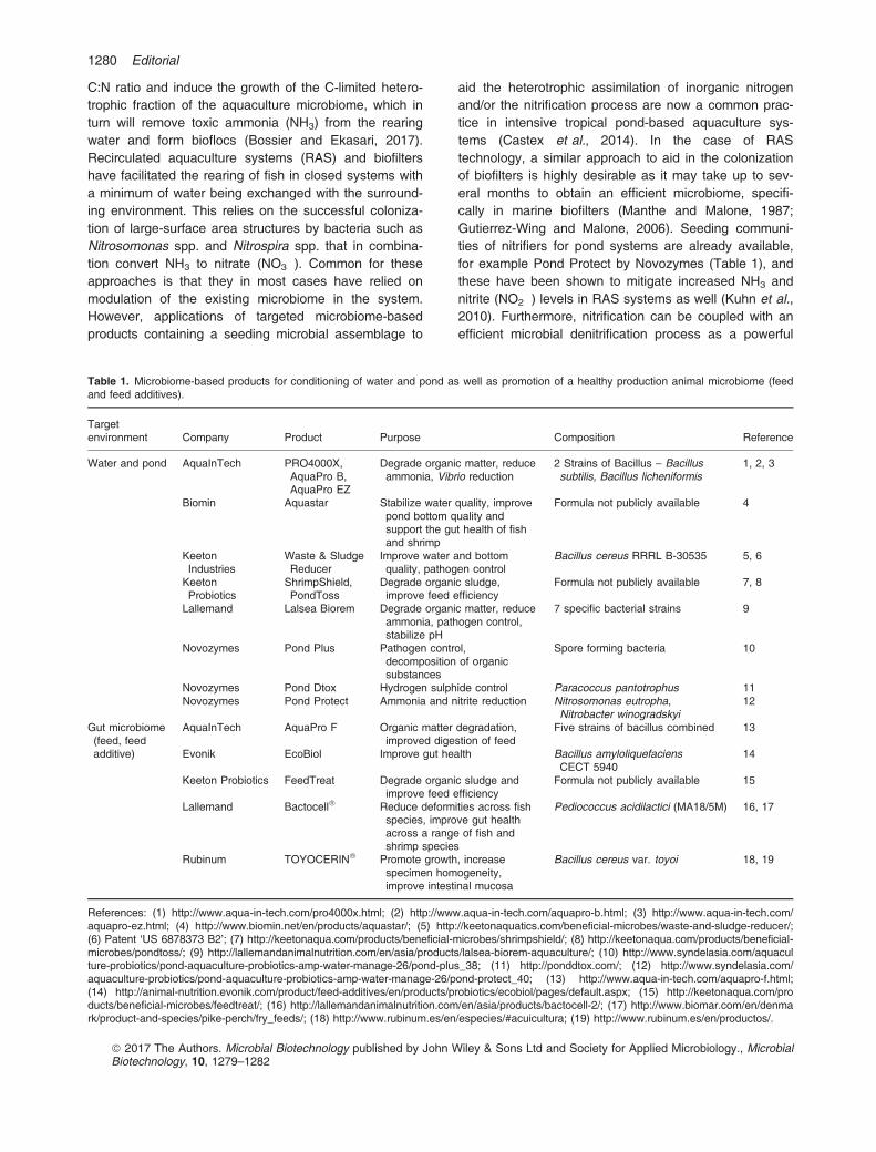
C:N ratio and induce the growth of the C-limited hetero-trophic fraction of the aquaculture microbiome, which inturn will remove toxic ammonia (NH3) from the rearingwater and form bioflocs (Bossier and Ekasari, 2017).Recirculated aquaculture systems (RAS) and biofiltershave facilitated the rearing of fish in closed systems witha minimum of water being exchanged with the surround-ing environment. This relies on the successful coloniza-tion of large-surface area structures by bacteria such asNitrosomonas spp. and Nitrospira spp. that in combina-tion convert NH3 to nitrate (NO3
�). Common for theseapproaches is that they in most cases have relied onmodulation of the existing microbiome in the system.However, applications of targeted microbiome-basedproducts containing a seeding microbial assemblage to
aid the heterotrophic assimilation of inorganic nitrogenand/or the nitrification process are now a common prac-tice in intensive tropical pond-based aquaculture sys-tems (Castex et al., 2014). In the case of RAStechnology, a similar approach to aid in the colonizationof biofilters is highly desirable as it may take up to sev-eral months to obtain an efficient microbiome, specifi-cally in marine biofilters (Manthe and Malone, 1987;Gutierrez-Wing and Malone, 2006). Seeding communi-ties of nitrifiers for pond systems are already available,for example Pond Protect by Novozymes (Table 1), andthese have been shown to mitigate increased NH3 andnitrite (NO2
�) levels in RAS systems as well (Kuhn et al.,2010). Furthermore, nitrification can be coupled with anefficient microbial denitrification process as a powerful
Table 1. Microbiome-based products for conditioning of water and pond as well as promotion of a healthy production animal microbiome (feedand feed additives).
Targetenvironment Company Product Purpose Composition Reference
Water and pond AquaInTech PRO4000X,AquaPro B,AquaPro EZ
Degrade organic matter, reduceammonia, Vibrio reduction
2 Strains of Bacillus – Bacillussubtilis, Bacillus licheniformis
1, 2, 3
Biomin Aquastar Stabilize water quality, improvepond bottom quality andsupport the gut health of fishand shrimp
Formula not publicly available 4
KeetonIndustries
Waste & SludgeReducer
Improve water and bottomquality, pathogen control
Bacillus cereus RRRL B-30535 5, 6
KeetonProbiotics
ShrimpShield,PondToss
Degrade organic sludge,improve feed efficiency
Formula not publicly available 7, 8
Lallemand Lalsea Biorem Degrade organic matter, reduceammonia, pathogen control,stabilize pH
7 specific bacterial strains 9
Novozymes Pond Plus Pathogen control,decomposition of organicsubstances
Spore forming bacteria 10
Novozymes Pond Dtox Hydrogen sulphide control Paracoccus pantotrophus 11Novozymes Pond Protect Ammonia and nitrite reduction Nitrosomonas eutropha,
Nitrobacter winogradskyi12
Gut microbiome(feed, feedadditive)
AquaInTech AquaPro F Organic matter degradation,improved digestion of feed
Five strains of bacillus combined 13
Evonik EcoBiol Improve gut health Bacillus amyloliquefaciensCECT 5940
14
Keeton Probiotics FeedTreat Degrade organic sludge andimprove feed efficiency
Formula not publicly available 15
Lallemand Bactocell� Reduce deformities across fishspecies, improve gut healthacross a range of fish andshrimp species
Pediococcus acidilactici (MA18/5M) 16, 17
Rubinum TOYOCERIN� Promote growth, increasespecimen homogeneity,improve intestinal mucosa
Bacillus cereus var. toyoi 18, 19
References: (1) http://www.aqua-in-tech.com/pro4000x.html; (2) http://www.aqua-in-tech.com/aquapro-b.html; (3) http://www.aqua-in-tech.com/aquapro-ez.html; (4) http://www.biomin.net/en/products/aquastar/; (5) http://keetonaquatics.com/beneficial-microbes/waste-and-sludge-reducer/;(6) Patent ‘US 6878373 B2’; (7) http://keetonaqua.com/products/beneficial-microbes/shrimpshield/; (8) http://keetonaqua.com/products/beneficial-microbes/pondtoss/; (9) http://lallemandanimalnutrition.com/en/asia/products/lalsea-biorem-aquaculture/; (10) http://www.syndelasia.com/aquaculture-probiotics/pond-aquaculture-probiotics-amp-water-manage-26/pond-plus_38; (11) http://ponddtox.com/; (12) http://www.syndelasia.com/aquaculture-probiotics/pond-aquaculture-probiotics-amp-water-manage-26/pond-protect_40; (13) http://www.aqua-in-tech.com/aquapro-f.html;(14) http://animal-nutrition.evonik.com/product/feed-additives/en/products/probiotics/ecobiol/pages/default.aspx; (15) http://keetonaqua.com/products/beneficial-microbes/feedtreat/; (16) http://lallemandanimalnutrition.com/en/asia/products/bactocell-2/; (17) http://www.biomar.com/en/denmark/product-and-species/pike-perch/fry_feeds/; (18) http://www.rubinum.es/en/especies/#acuicultura; (19) http://www.rubinum.es/en/productos/.
ª 2017 The Authors. Microbial Biotechnology published by John Wiley & Sons Ltd and Society for Applied Microbiology., MicrobialBiotechnology, 10, 1279–1282
1280 Editorial
tool in the complete removal of nitrogenous compoundsfrom the system, and the development and application ofa joined nitrification and denitrification approach for recir-culated aquaculture systems, similar to the Aqua Scien-ce� concept from Camanor, likely represents an area ofpotential business development. The commercializationof targeted microbiome-based products containing livingmicroorganisms, such as seeding microbial assemblagesthat improve water quality, has been seen for use inaquaria for decades, for example the BIO-Spira productfrom MarineLand Labs and its predecessors, which likePond Protect and similar microbiome-based products foraquaculture systems contain bacterial assemblages thatremove ammonia and nitrite. Similar microbiome-basedproducts for use in conjunction with biofloc technologyare also available now. One such product is Shrimp-Shield by Keeton Probiotics, which facilitates biofloc for-mation, degradation of sludge as well as microbialremoval of NH3 and NO2
� (Table 1). Hence, such micro-biome-based products aim to improve water quality andin some cases remove potential pathogens through, forexample, competitive exclusion (Table 1).Another category of microbiome-based products that
is being developed for the aquaculture industry targetsthe gut of the animal directly (Table 1), equivalent to themore conventional probiotics for livestock and humanconsumption. Microbial strains evaluated as probioticsfor aquaculture are from many phylogenetic lineages;however, most of them belong to two bacterial phyla, theFirmicutes (e.g. Bacillus spp., Lactobacillus spp., Lacto-coccus spp. and Carnobacterium spp.) and the Pro-teobacteria (e.g. Vibrio spp., Pseudomonas spp. andShewanella spp.), while yeasts are rarely studied (Gate-soupe, 2007). The majority of the commercially availableprobiotic feed and feed additives for aquaculture arebased on pure or mixed cultures of lactic acid bacteriaand Bacilli (Merrifield et al., 2010; Castex et al., 2014).This includes Bactocell� (Lallemand; Table 1), which isbased on a Pediococcus acidilactici strain and is, to thebest of our knowledge, the only probiotic registered inEurope for use in aquaculture feed. These bacteria areusually well studied and well known for their positiveeffect on the human and animal gut microbiome (Cutting,2011). Furthermore, they are Generally Regarded AsSafe (GRAS) or Qualified Presumption of Safety (QPS),which makes it easier to obtain authorization for theiruse in food and feed products. A natural extension ofthis type of microbiome-based products, and a potentialnew avenue to be explored in aquaculture microbiomebusiness creation, is the controlled colonization of thereared fish from larvae to adult by a microbiome that hasthe desired functional traits and can act as an infectionbarrier against pathogens and prevent major economic
losses by crashes in the population (De Schryver andVadstein, 2014).The successful application of probiotic Firmicutes,
originally applied as probiotics for humans or livestock,in aquaculture is fortunate considering the divergentniches in which these probiotics need to establish them-selves and function. An avenue of potential new enter-prises is to develop similar products based on bacteriaof marine origin instead. Marine bacteria including mem-bers of the Roseobacter group and the Vibrio and She-wanella genera have been studied extensively for theirprobiotic potential (Austin et al., 1995; Ringø and Vad-stein, 1998; D�ıaz-Rosales et al., 2009; D’Alvise et al.,2012; Lobo et al., 2014; Grotkjær et al., 2016; Bentzon-Tilia and Gram, 2017). Furthermore, these are oftenfound as part of the indigenous microbiome of marineeukaryotes, and although their application as probioticshas been proposed, they have not yet reached a com-mercialization stage. To succeed with this approach,much more thorough characterizations of aquacultureand marine host microbiomes are needed. Furthermore,in most cases, the putative probiotic candidates reportedin scientific publications do not go on to commercializa-tion and industrial application. Getting a probiont to thecommercial market requires many additional stepsincluding assessments of safety, scale-up efficacy, pro-duction scale-up and pre-market registration. Consis-tency, efficiency and most importantly safety are keypoints in all large-scale productions, and they should beconsidered from the early stages of the discovery phaseto the final application in feed products. Thus, not onlydoes the development of a commercial product rely onsubstantial financial investments, but also on the contri-bution from a multidisciplinary team encompassing closecollaborations between scientists, aquaculture experts,fermentation engineers and regulatory personnel. Thelatter part of the team is important for success in a regu-latory landscape which varies from an absence of regu-lation in certain countries to a rigid regulatory frameworknot always adapted to the effect a probiotic can display.Despite these challenges, the aquaculture industry hasalready embraced the industrial application of micro-biome-based products for the last two decades, and thishas truly created a vast range of new enterprises espe-cially in South East Asia, Central and South Americaand more recently in Europe.Using microbiome-based products also requires devel-
opments of production, packaging and distribution tech-nology. One must consider that the efficiency of suchproducts only in part depends on the choice of themicrobial strains that compose it (selection), but also onthe way the product is produced, conditioned and finallypackaged to withstand a variety of storage conditions.
ª 2017 The Authors. Microbial Biotechnology published by John Wiley & Sons Ltd and Society for Applied Microbiology., MicrobialBiotechnology, 10, 1279–1282
Editorial 1281
In conclusion, the aquaculture industry is one of thefastest growing food producing sectors in the world andthe increased productivity of this sector is essential forthe fulfilment of the sustainable development goals ofthe UN. Microbiome-based products for application inindustrial aquaculture are today a reality, but the fullpotential is far from exploited. Despite decades of experi-ence and an increasing number of microbial biotechno-logical products, there is a large innovation potential;from the discovery of new probionts of marine origin andlarge-scale cultivation strategies to manoeuvering thepolitical, regulatory landscape and disseminating the useof probiotics to ensure future, sustainable technologiesfor high-quality protein production.
References
Austin, B., Stuckey, L. F., Robertson, P. A. W., Effendi, I.,and Griffith, D. R. W. (1995) A probiotic strain of Vibrioalginolyticus effective in reducing diseases caused byAeromonas salmonicida, Vibrio anguillarum and Vibrioordalii. J Fish Dis 18: 93–96.
Bentzon-Tilia, M., and Gram, L. (2017) BiotechnologicalApplications of the Roseobacter Clade. In Biotechnologi-cal Applications of the Roseobacter Clade. Paterson, R.,and Lima, N. (eds). Basel: Springer International Publish-ing, pp. 137–166.
Bentzon-Tilia, M., Sonnenschein, E. C., and Gram, L. (2016)Monitoring and managing microbes in aquaculture – towardsa sustainable industry.Microb Biotechnol 9: 576–584.
Bossier, P., and Ekasari, J. (2017) Biofloc technology appli-cation in aquaculture to support sustainable developmentgoals. Microb Biotechnol 10: 1012–1016.
Castex, M., Durand, H., and Okeke, B. (2014) Issues withindustrial probiotics scale-up. Aquacult Nutr Gut HealthProbiot Prebiot. Merrifield, D. L., and Ringo, E. (Eds).Wiley-Blackwell. 347–359.
Cutting, S. M. (2011) Bacillus probiotics. Food Microbiol 28:214–220.
D’Alvise, P. W., Lillebø, S., Prol-Garcia, M. J., Wergeland,H. I., Nielsen, K. F., Bergh, Ø., and Gram, L. (2012)Phaeobacter gallaeciensis reduces Vibrio anguillarum incultures of microalgae and rotifers, and prevents vibriosisin cod larvae. PLoS ONE 7: e43996.
De Schryver, P., and Vadstein, O. (2014) Ecological theoryas a foundation to control pathogenic invasion in aquacul-ture. ISME J 8: 2360–2368.
D�ıaz-Rosales, P., Arijo, S., Chabrill�on, M., Alarc�on, F. J., Tapia-Paniagua, S. T., Mart�ınez-Manzanares, E., et al. (2009)Effects of two closely related probiotics on respiratory burstactivity of Senegalese sole (Solea senegalensis, Kaup)phagocytes, and protection against Photobacterium damse-lae subsp. piscicida. Aquaculture 293: 16–21.
Food and Agriculture Organization of the United Nations(FAO), Fisheries and Aquaculture Department (2016) TheState of World Fisheries and Aquaculture 2016.
Gatesoupe, F. J. (2007) Live yeasts in the gut: naturaloccurrence, dietary introduction, and their effects on fishhealth and development. Aquaculture 267: 20–30.
Gentry, R. R., Froehlich, H. E., Grimm, D., Kareiva, P.,Parke, M., Rust, M., et al. (2017) Mapping the globalpotential for marine aquaculture. Nat Ecol Evol 1: 1317–1324.
Grotkjær, T., Bentzon-Tilia, M., D’Alvise, P. W., Dourala, N.,Nielsen, K. F., and Gram, L. (2016) Isolation of TDA-pro-ducing Phaeobacter strains from sea bass larval rearingunits and their probiotic effect against pathogenic Vibriospp. in Artemia cultures. Syst Appl Microbiol 39: 180–188.
Gutierrez-Wing, M. T., and Malone, R. F. (2006) Biological filtersin aquaculture: trends and research directions for freshwaterand marine applications. Aquacult Eng 34: 163–171.
van Kessel, M. A., Dutilh, B. E., Neveling, K., Kwint, M. P.,Veltman, J. A., Flik, G., et al. (2011) Pyrosequencing of16S rRNA gene amplicons to study the microbiota in thegastrointestinal tract of carp (Cyprinus carpio L.). AMBExpress 1: 41.
Kuhn, D. D., Drahos, D. D., Marsh, L., and Flick, G. J.(2010) Evaluation of nitrifying bacteria product to improvenitrification efficacy in recirculating aquaculture systems.Aquacult Eng 43: 78–82.
Llewellyn, M. S., Boutin, S., Hoseinifar, S. H., and Derome,N. (2014) Teleost microbiomes: the state of the art in theircharacterization, manipulation and importance in aquacul-ture and fisheries. Front Microbiol 5: 207.
Lobo, C., Moreno-Ventas, X., Tapia-Paniagua, S.,Rodr�ıguez, C., Mori~nigo, M. A., and de La Banda, I. G.(2014) Dietary probiotic supplementation (Shewanellaputrefaciens Pdp11) modulates gut microbiota and pro-motes growth and condition in Senegalese sole larvicul-ture. Fish Physiol Biochem 40: 295–309.
Manthe, D. P., and Malone, R. F. (1987) Chemical additionfor accelerated biological filter acclimation in closed bluecrab shedding systems. Aquacult Eng 6: 227–236.
Merrifield, D. L., Dimitroglou, A., Foey, A., Davies, S. J.,Baker, R. T. M., Bøgwald, J., et al. (2010) The currentstatus and future focus of probiotic and prebiotic applica-tions for salmonids. Aquaculture 302: 1–18.
Meyer, F. P. (1991) Aquaculture disease and health man-agement. J Anim Sci 69: 4201.
Moriarty, D. J. W. (1997) The role of microorganisms inaquaculture ponds. Aquaculture 151: 333–349.
Ringø, E., and Vadstein, O. (1998) Colonization of Vibriopelagius and Aeromonas caviae in early developing turbot(Scophthalmus maximus L.) larvae. J Appl Microbiol 84:227–233.
Singh, B. K. (2017) Creating new business, economicgrowth and regional prosperity through microbiome-basedproducts in the agriculture industry. Microb Biotechnol 10:224–227.
United Nations, Department of Economic and Social Affairs,Population Division (2015) World Population Prospects:The 2015 Revision, Key Findings and Advance Tables.ESA/P/WP.241.
ª 2017 The Authors. Microbial Biotechnology published by John Wiley & Sons Ltd and Society for Applied Microbiology., MicrobialBiotechnology, 10, 1279–1282
1282 Editorial
Paper 2
Dittmann, K.K., Sonnenschein, E.C., Egan, S., Gram, L. & Bentzon-Tilia, M. (2019).
Impact of Phaeobacter inhibens on marine eukaryote-associated microbial communities.
Environ Microbiol Rep., 11(3), 401–413.
https://doi.org/10.1111/1758-2229.12698
Brief Report
Impact of Phaeobacter inhibens on marine eukaryote-associated microbial communities
Karen K. Dittmann,1 Eva C. Sonnenschein,1
Suhelen Egan,2 Lone Gram1 andMikkel Bentzon-Tilia1*1Department of Biotechnology and Biomedicine,Technical University of Denmark, Copenhagen,Denmark.2School of Biological, Earth and EnvironmentalSciences, The University of New South Wales,Randwick, NSW, Australia.
Summary
Bacteria–host interactions are universal in nature andhave significant effects on host functionality. Bacte-rial secondary metabolites are believed to play keyroles in such interactions as well as in interactionswithin the host-associated microbial community.Hence, prominent secondary metabolite-producingbacteria may be strong drivers of microbial commu-nity composition in natural host-associated micro-biomes. This has, however, not been rigorouslytested, and the purpose of this study was to investi-gate how the secondary metabolite producer Phaeo-bacter inhibens affects the diversity and compositionof microbiomes associated with the microalgaEmiliania huxleyi and the European flat oyster, Ostreaedulis. Roseobacters were indigenous to both com-munities exhibiting relative abundances between2.8% and 7.0%. Addition of P. inhibens causedsubstantial changes in the overall structure of thelow-complexity microbiome of E. huxleyi, but did notshape microbial community structure to the samedegree in the more complex oyster microbiomes.Species-specific interactions occurred in both micro-biomes and specifically the abundances of otherputative secondary metabolite-producers such as vib-rios and pseudoalteromonads were reduced. Thus,the impact of a bioactive strain like P. inhibens onhost-associated microbiomes depends on the com-plexity and composition of the existing microbiome.
Introduction
In nature, microorganisms live and interact as part ofcomplex multispecies communities. These interspeciesinteractions may be of a synergistic, amensal or com-mensal nature and can be facilitated by the exchange ofmetabolites in syntrophic cooperation, or by the produc-tion of bioactive secondary metabolites (Cole, 1982). Inparallel to the use of secondary metabolites as defenceagainst microbial infections by plants (Pusztahelyi et al.,2015), bacterial secondary metabolites with antimicrobialproperties are currently believed to facilitate the successof the compound-producer by killing competitors. Not allbacteria are equally proficient in secondary metaboliteproduction, and whereas some groups appear to producefew or no metabolites, others such as filamentous soilbacteria and marine vibrios, roseobacters and Pseudoal-teromonas spp. produce an array of different bioactivecompounds (Brinkhoff et al., 2004; Murphy et al., 2012;Rasmussen et al., 2014; Maansson et al., 2016; Son-nenschein et al., 2017b). Hence, if the role of these com-pounds is to eliminate competing microorganisms,proficient secondary metabolite producers should bestrong drivers of microbial community composition in nat-ural environments.
The Roseobacter group represents one of the mostabundant groups of marine bacteria, constituting on aver-age 3%–5% of microbial communities in the upper mixedlayer (Wietz et al., 2010). On a global scale, the groupexhibits a positive abundance-chlorophyll a correlation(Wietz et al., 2010) and may exhibit relative abundancesof up to 20%–30% during algal blooms (González andMoran, 1997; González et al., 2000; Zubkov et al., 2001;West et al., 2008), suggesting association with microal-gae. The paraphyletic Roseobacter group comprisesmultiple deeply branching clades (Newton et al., 2010;Simon et al., 2017), of which especially clade 1 includesprominent producers of bioactive secondary metabolites,such as the antimicrobial compounds tropodithietic acid(TDA) (Brinkhoff et al., 2004; Bruhn et al., 2005; Son-nenschein et al., 2017a), indigoidine (Cude et al., 2012;Gromek et al., 2016) and likely multiple other small
Received 23 February, 2018; accepted 25 September, 2018. *Forcorrespondence. E-mail [email protected]; Tel. (+45) 45 25 25 18;Fax (+45) 45 88 49 22.
© 2018 Society for Applied Microbiology and John Wiley & Sons Ltd
Environmental Microbiology Reports (2019) 11(3), 401–413 doi:10.1111/1758-2229.12698
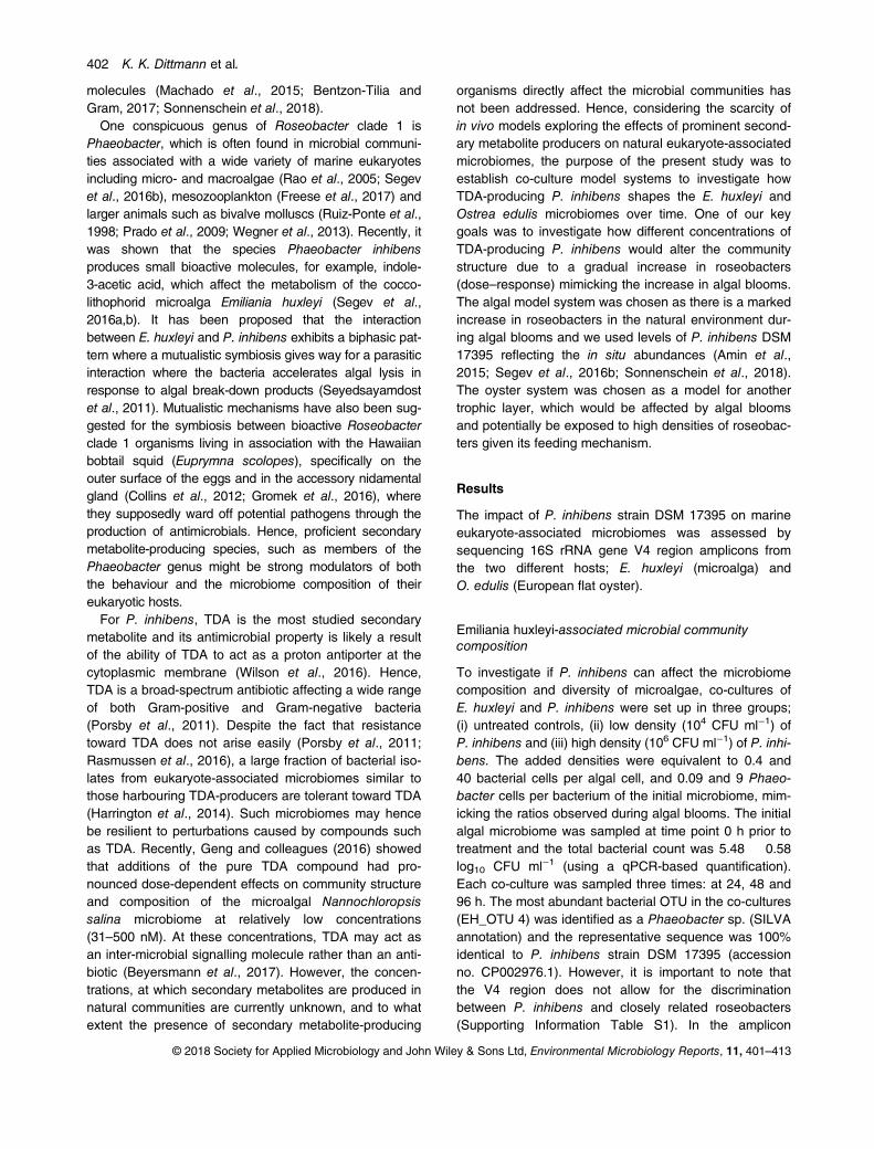
molecules (Machado et al., 2015; Bentzon-Tilia andGram, 2017; Sonnenschein et al., 2018).One conspicuous genus of Roseobacter clade 1 is
Phaeobacter, which is often found in microbial communi-ties associated with a wide variety of marine eukaryotesincluding micro- and macroalgae (Rao et al., 2005; Segevet al., 2016b), mesozooplankton (Freese et al., 2017) andlarger animals such as bivalve molluscs (Ruiz-Ponte et al.,1998; Prado et al., 2009; Wegner et al., 2013). Recently, itwas shown that the species Phaeobacter inhibensproduces small bioactive molecules, for example, indole-3-acetic acid, which affect the metabolism of the cocco-lithophorid microalga Emiliania huxleyi (Segev et al.,2016a,b). It has been proposed that the interactionbetween E. huxleyi and P. inhibens exhibits a biphasic pat-tern where a mutualistic symbiosis gives way for a parasiticinteraction where the bacteria accelerates algal lysis inresponse to algal break-down products (Seyedsayamdostet al., 2011). Mutualistic mechanisms have also been sug-gested for the symbiosis between bioactive Roseobacterclade 1 organisms living in association with the Hawaiianbobtail squid (Euprymna scolopes), specifically on theouter surface of the eggs and in the accessory nidamentalgland (Collins et al., 2012; Gromek et al., 2016), wherethey supposedly ward off potential pathogens through theproduction of antimicrobials. Hence, proficient secondarymetabolite-producing species, such as members of thePhaeobacter genus might be strong modulators of boththe behaviour and the microbiome composition of theireukaryotic hosts.For P. inhibens, TDA is the most studied secondary
metabolite and its antimicrobial property is likely a resultof the ability of TDA to act as a proton antiporter at thecytoplasmic membrane (Wilson et al., 2016). Hence,TDA is a broad-spectrum antibiotic affecting a wide rangeof both Gram-positive and Gram-negative bacteria(Porsby et al., 2011). Despite the fact that resistancetoward TDA does not arise easily (Porsby et al., 2011;Rasmussen et al., 2016), a large fraction of bacterial iso-lates from eukaryote-associated microbiomes similar tothose harbouring TDA-producers are tolerant toward TDA(Harrington et al., 2014). Such microbiomes may hencebe resilient to perturbations caused by compounds suchas TDA. Recently, Geng and colleagues (2016) showedthat additions of the pure TDA compound had pro-nounced dose-dependent effects on community structureand composition of the microalgal Nannochloropsissalina microbiome at relatively low concentrations(31–500 nM). At these concentrations, TDA may act asan inter-microbial signalling molecule rather than an anti-biotic (Beyersmann et al., 2017). However, the concen-trations, at which secondary metabolites are produced innatural communities are currently unknown, and to whatextent the presence of secondary metabolite-producing
organisms directly affect the microbial communities hasnot been addressed. Hence, considering the scarcity ofin vivo models exploring the effects of prominent second-ary metabolite producers on natural eukaryote-associatedmicrobiomes, the purpose of the present study was toestablish co-culture model systems to investigate howTDA-producing P. inhibens shapes the E. huxleyi andOstrea edulis microbiomes over time. One of our keygoals was to investigate how different concentrations ofTDA-producing P. inhibens would alter the communitystructure due to a gradual increase in roseobacters(dose–response) mimicking the increase in algal blooms.The algal model system was chosen as there is a markedincrease in roseobacters in the natural environment dur-ing algal blooms and we used levels of P. inhibens DSM17395 reflecting the in situ abundances (Amin et al.,2015; Segev et al., 2016b; Sonnenschein et al., 2018).The oyster system was chosen as a model for anothertrophic layer, which would be affected by algal bloomsand potentially be exposed to high densities of roseobac-ters given its feeding mechanism.
Results
The impact of P. inhibens strain DSM 17395 on marineeukaryote-associated microbiomes was assessed bysequencing 16S rRNA gene V4 region amplicons fromthe two different hosts; E. huxleyi (microalga) andO. edulis (European flat oyster).
Emiliania huxleyi-associated microbial communitycomposition
To investigate if P. inhibens can affect the microbiomecomposition and diversity of microalgae, co-cultures ofE. huxleyi and P. inhibens were set up in three groups;(i) untreated controls, (ii) low density (104 CFU ml−1) ofP. inhibens and (iii) high density (106 CFU ml−1) of P. inhi-bens. The added densities were equivalent to 0.4 and40 bacterial cells per algal cell, and 0.09 and 9 Phaeo-bacter cells per bacterium of the initial microbiome, mim-icking the ratios observed during algal blooms. The initialalgal microbiome was sampled at time point 0 h prior totreatment and the total bacterial count was 5.48 � 0.58log10 CFU ml−1 (using a qPCR-based quantification).Each co-culture was sampled three times: at 24, 48 and96 h. The most abundant bacterial OTU in the co-cultures(EH_OTU 4) was identified as a Phaeobacter sp. (SILVAannotation) and the representative sequence was 100%identical to P. inhibens strain DSM 17395 (accessionno. CP002976.1). However, it is important to note thatthe V4 region does not allow for the discriminationbetween P. inhibens and closely related roseobacters(Supporting Information Table S1). In the amplicon
© 2018 Society for Applied Microbiology and John Wiley & Sons Ltd, Environmental Microbiology Reports, 11, 401–413
402 K. K. Dittmann et al.
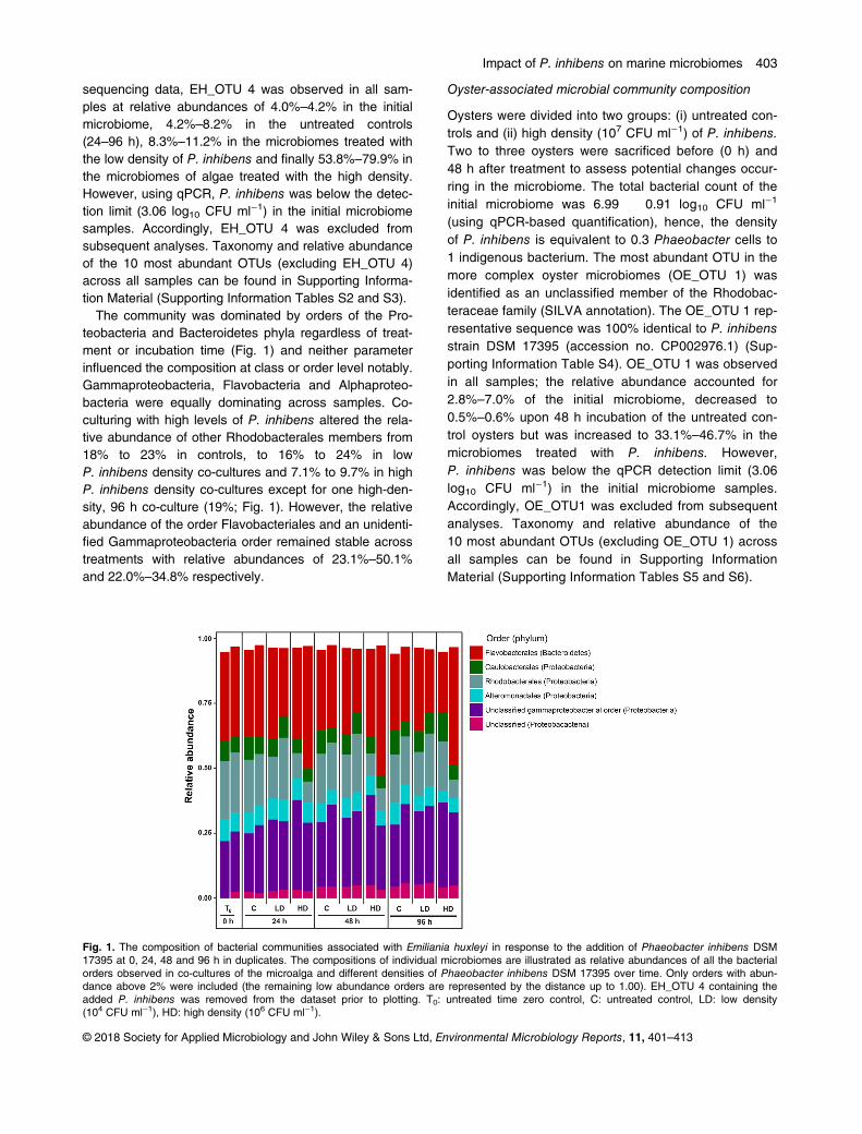
sequencing data, EH_OTU 4 was observed in all sam-ples at relative abundances of 4.0%–4.2% in the initialmicrobiome, 4.2%–8.2% in the untreated controls(24–96 h), 8.3%–11.2% in the microbiomes treated withthe low density of P. inhibens and finally 53.8%–79.9% inthe microbiomes of algae treated with the high density.However, using qPCR, P. inhibens was below the detec-tion limit (3.06 log10 CFU ml−1) in the initial microbiomesamples. Accordingly, EH_OTU 4 was excluded fromsubsequent analyses. Taxonomy and relative abundanceof the 10 most abundant OTUs (excluding EH_OTU 4)across all samples can be found in Supporting Informa-tion Material (Supporting Information Tables S2 and S3).
The community was dominated by orders of the Pro-teobacteria and Bacteroidetes phyla regardless of treat-ment or incubation time (Fig. 1) and neither parameterinfluenced the composition at class or order level notably.Gammaproteobacteria, Flavobacteria and Alphaproteo-bacteria were equally dominating across samples. Co-culturing with high levels of P. inhibens altered the rela-tive abundance of other Rhodobacterales members from18% to 23% in controls, to 16% to 24% in lowP. inhibens density co-cultures and 7.1% to 9.7% in highP. inhibens density co-cultures except for one high-den-sity, 96 h co-culture (19%; Fig. 1). However, the relativeabundance of the order Flavobacteriales and an unidenti-fied Gammaproteobacteria order remained stable acrosstreatments with relative abundances of 23.1%–50.1%and 22.0%–34.8% respectively.
Oyster-associated microbial community composition
Oysters were divided into two groups: (i) untreated con-trols and (ii) high density (107 CFU ml−1) of P. inhibens.Two to three oysters were sacrificed before (0 h) and48 h after treatment to assess potential changes occur-ring in the microbiome. The total bacterial count of theinitial microbiome was 6.99 � 0.91 log10 CFU ml−1
(using qPCR-based quantification), hence, the densityof P. inhibens is equivalent to 0.3 Phaeobacter cells to1 indigenous bacterium. The most abundant OTU in themore complex oyster microbiomes (OE_OTU 1) wasidentified as an unclassified member of the Rhodobac-teraceae family (SILVA annotation). The OE_OTU 1 rep-resentative sequence was 100% identical to P. inhibensstrain DSM 17395 (accession no. CP002976.1) (Sup-porting Information Table S4). OE_OTU 1 was observedin all samples; the relative abundance accounted for2.8%–7.0% of the initial microbiome, decreased to0.5%–0.6% upon 48 h incubation of the untreated con-trol oysters but was increased to 33.1%–46.7% in themicrobiomes treated with P. inhibens. However,P. inhibens was below the qPCR detection limit (3.06log10 CFU ml−1) in the initial microbiome samples.Accordingly, OE_OTU1 was excluded from subsequentanalyses. Taxonomy and relative abundance of the10 most abundant OTUs (excluding OE_OTU 1) acrossall samples can be found in Supporting InformationMaterial (Supporting Information Tables S5 and S6).
Fig. 1. The composition of bacterial communities associated with Emiliania huxleyi in response to the addition of Phaeobacter inhibens DSM17395 at 0, 24, 48 and 96 h in duplicates. The compositions of individual microbiomes are illustrated as relative abundances of all the bacterialorders observed in co-cultures of the microalga and different densities of Phaeobacter inhibens DSM 17395 over time. Only orders with abun-dance above 2% were included (the remaining low abundance orders are represented by the distance up to 1.00). EH_OTU 4 containing theadded P. inhibens was removed from the dataset prior to plotting. T0: untreated time zero control, C: untreated control, LD: low density(104 CFU ml−1), HD: high density (106 CFU ml−1).
© 2018 Society for Applied Microbiology and John Wiley & Sons Ltd, Environmental Microbiology Reports, 11, 401–413
Impact of P. inhibens on marine microbiomes 403
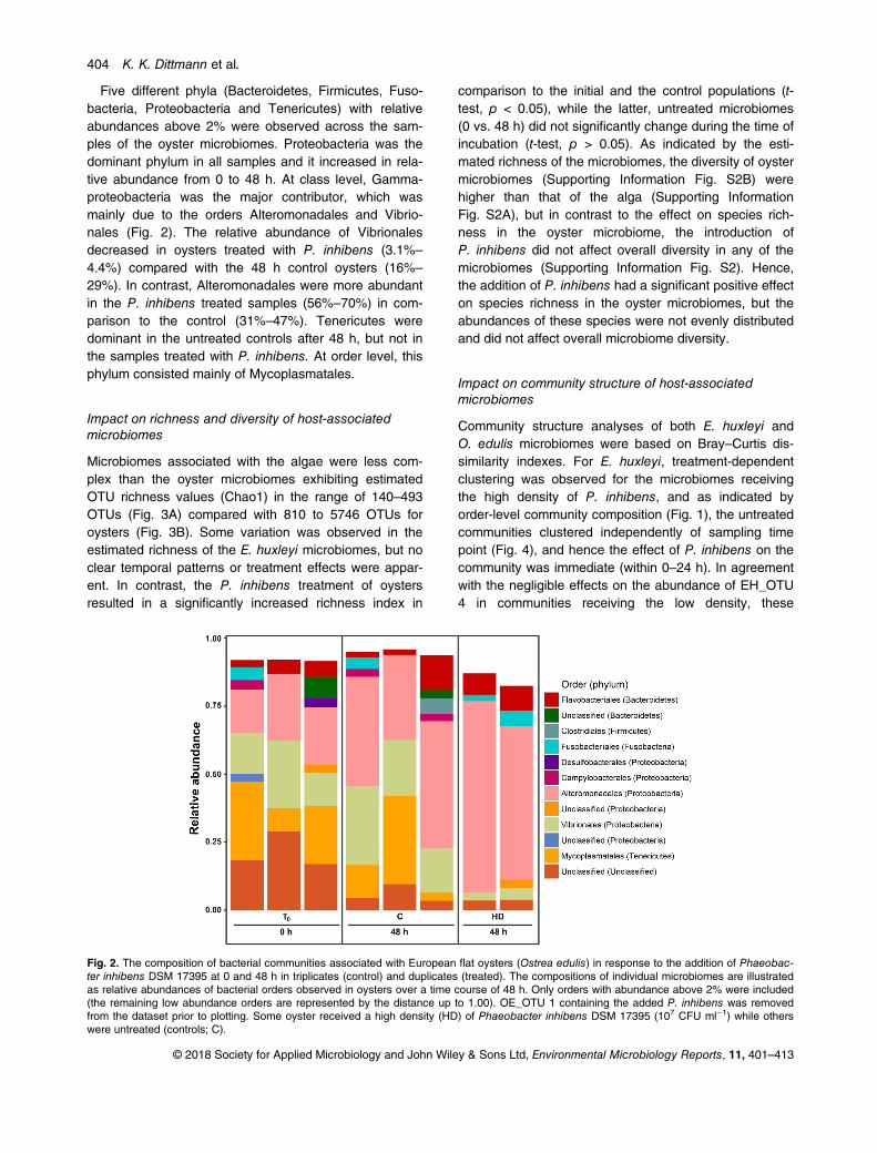
Five different phyla (Bacteroidetes, Firmicutes, Fuso-bacteria, Proteobacteria and Tenericutes) with relativeabundances above 2% were observed across the sam-ples of the oyster microbiomes. Proteobacteria was thedominant phylum in all samples and it increased in rela-tive abundance from 0 to 48 h. At class level, Gamma-proteobacteria was the major contributor, which wasmainly due to the orders Alteromonadales and Vibrio-nales (Fig. 2). The relative abundance of Vibrionalesdecreased in oysters treated with P. inhibens (3.1%–
4.4%) compared with the 48 h control oysters (16%–
29%). In contrast, Alteromonadales were more abundantin the P. inhibens treated samples (56%–70%) in com-parison to the control (31%–47%). Tenericutes weredominant in the untreated controls after 48 h, but not inthe samples treated with P. inhibens. At order level, thisphylum consisted mainly of Mycoplasmatales.
Impact on richness and diversity of host-associatedmicrobiomes
Microbiomes associated with the algae were less com-plex than the oyster microbiomes exhibiting estimatedOTU richness values (Chao1) in the range of 140–493OTUs (Fig. 3A) compared with 810 to 5746 OTUs foroysters (Fig. 3B). Some variation was observed in theestimated richness of the E. huxleyi microbiomes, but noclear temporal patterns or treatment effects were appar-ent. In contrast, the P. inhibens treatment of oystersresulted in a significantly increased richness index in
comparison to the initial and the control populations (t-test, p < 0.05), while the latter, untreated microbiomes(0 vs. 48 h) did not significantly change during the time ofincubation (t-test, p > 0.05). As indicated by the esti-mated richness of the microbiomes, the diversity of oystermicrobiomes (Supporting Information Fig. S2B) werehigher than that of the alga (Supporting InformationFig. S2A), but in contrast to the effect on species rich-ness in the oyster microbiome, the introduction ofP. inhibens did not affect overall diversity in any of themicrobiomes (Supporting Information Fig. S2). Hence,the addition of P. inhibens had a significant positive effecton species richness in the oyster microbiomes, but theabundances of these species were not evenly distributedand did not affect overall microbiome diversity.
Impact on community structure of host-associatedmicrobiomes
Community structure analyses of both E. huxleyi andO. edulis microbiomes were based on Bray–Curtis dis-similarity indexes. For E. huxleyi, treatment-dependentclustering was observed for the microbiomes receivingthe high density of P. inhibens, and as indicated byorder-level community composition (Fig. 1), the untreatedcommunities clustered independently of sampling timepoint (Fig. 4), and hence the effect of P. inhibens on thecommunity was immediate (within 0–24 h). In agreementwith the negligible effects on the abundance of EH_OTU4 in communities receiving the low density, these
Fig. 2. The composition of bacterial communities associated with European flat oysters (Ostrea edulis) in response to the addition of Phaeobac-ter inhibens DSM 17395 at 0 and 48 h in triplicates (control) and duplicates (treated). The compositions of individual microbiomes are illustratedas relative abundances of bacterial orders observed in oysters over a time course of 48 h. Only orders with abundance above 2% were included(the remaining low abundance orders are represented by the distance up to 1.00). OE_OTU 1 containing the added P. inhibens was removedfrom the dataset prior to plotting. Some oyster received a high density (HD) of Phaeobacter inhibens DSM 17395 (107 CFU ml−1) while otherswere untreated (controls; C).
© 2018 Society for Applied Microbiology and John Wiley & Sons Ltd, Environmental Microbiology Reports, 11, 401–413
404 K. K. Dittmann et al.
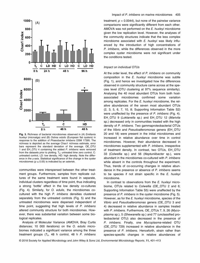
communities were interspersed between the other treat-ment groups. Furthermore, samples from replicate cul-tures of the same treatment were found in separate,individual clusters regardless of time point, thus indicatinga strong ‘bottle’ effect in the low density co-cultures(Fig. 4). Similarly, for O. edulis, the microbiomes co-cultured with the high P. inhibens densities clusteredseparately from the untreated controls (Fig. 5) and theuntreated microbiomes were dispersed independent oftime point, suggesting that high levels of P. inhibensaltered community structure for both microbiomes. How-ever, there was substantial variation between some bio-logical replicates.
Analysis of Molecular Variance (AMOVA; Bray Curtisdistances; 10 000 iterations) on the O. edulis micro-biomes indicated a significant variance among the threetreatment groups (T0, 48 h control, 48 h P. inhibens
treatment; p = 0.0044), but none of the pairwise variancecomparisons were significantly different from each other.AMOVA was not performed on the E. huxleyi microbiomegiven the low replication level. However, the analyses ofthe community structures indicate that the less complexmicrobiome associated with E. huxleyi was likely influ-enced by the introduction of high concentrations ofP. inhibens, while the differences observed in the morecomplex oyster microbiome were not significant underthe conditions tested.
Impact on individual OTUs
At the order level, the effect of P. inhibens on communitycomposition in the E. huxleyi microbiome was subtle(Fig. 1), and hence we investigated how the differencesobserved in community structure came across at the spe-cies level (OTU clustering at 97% sequence similarity).Analysing the 40 most abundant OTUs from both host-associated microbiomes confirmed some variationamong replicates. For the E. huxleyi microbiome, the rel-ative abundances of the seven most abundant OTUs(2, 3, 5, 6, 7, 10, 8; Supporting Information Table S2)were unaffected by the presence of P. inhibens (Fig. 4).EH_OTU 9 (Loktanella sp.) and EH_OTU 12 (Marivitasp.) decreased only in communities treated with the highdensity of P. inhibens. Two gammaproteobacterial OTUsof the Vibrio and Pseudoalteromonas genera (EH_OTU20 and 18) were present in the initial microbiomes andincreased in relative abundance over time in controlmicrobiomes. However, their abundance decreased inmicrobiomes supplemented with P. inhibens, irrespectiveof treatment density. In contrast, two OTUs, EH_OTU33 (Colwellia sp.) and 56 (Neptuniibacter sp.), wereabundant in the microbiomes co-cultured with P. inhibenswhile absent in the controls throughout the experiment.Thus, trends of co-occurring changes in relative abun-dance in the presence or absence of P. inhibens seemsto be species if not strain specific in the E. huxleyimicrobiome.
In contrast to observations from the E. huxleyi micro-biome, OTUs related to Colwellia (OE_OTU 2 and 6;Supporting Information Table S5) were unaffected by thepresence of P. inhibens in the oyster microbiome (Fig. 5).However, as for the E. huxleyi microbiome, species of theVibrio and Pseudoalteromonas genera (OE_OTU 3 and4) decreased in relative abundance in samples treatedwith P. inhibens. Furthermore, OE_OTUs 7, 9, 26 (Myco-plasma sp.), 5 (Shewanella sp.) and 77 (unclassified pro-teobacterial OTU) also decreased in the presence ofP. inhibens. Finally, one Mycoplasma-related OTU(OE_OTU 729) increased in relative abundance in thepresence of P. inhibens. Henceforth, strain rather thanspecies-specific changes in relative abundances of
Fig. 3. Richness of bacterial microbiomes observed in (A) Emilianiahuxleyi (microalga) and (B) Ostrea edulis (European flat oysters) inresponse to the addition of Phaeobacter inhibens DSM 17395. Therichness is depicted as the average Chao1 richness estimate, errorbars represent the standard deviation of the average. OE_OTU1 and EH_OTU 4 containing the added P. inhibens were removedfrom the datasets prior to plotting. T0: untreated time zero control, C:untreated control, LD: low density, HD: high density. Note the differ-ence in the y-axis. Statistical significance of the change in the oystermicrobiomes (p ≤ 0.05) is indicated by an asterisk.
© 2018 Society for Applied Microbiology and John Wiley & Sons Ltd, Environmental Microbiology Reports, 11, 401–413
Impact of P. inhibens on marine microbiomes 405
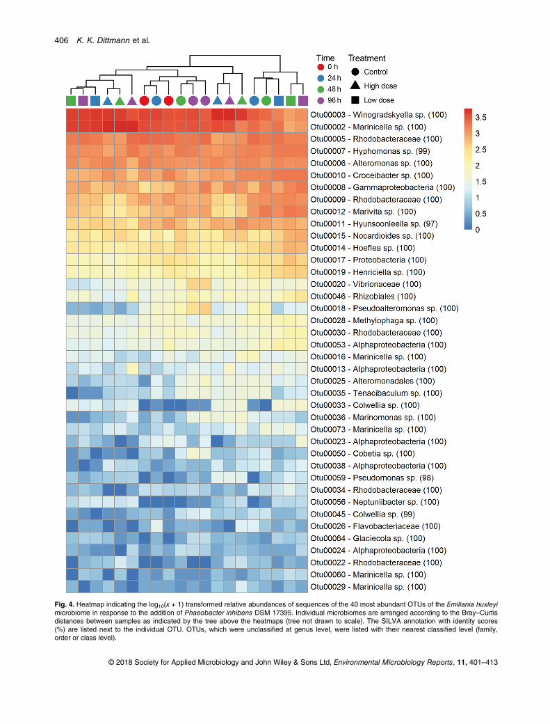
Fig. 4. Heatmap indicating the log10(x + 1) transformed relative abundances of sequences of the 40 most abundant OTUs of the Emiliania huxleyimicrobiome in response to the addition of Phaeobacter inhibens DSM 17395. Individual microbiomes are arranged according to the Bray–Curtisdistances between samples as indicated by the tree above the heatmaps (tree not drawn to scale). The SILVA annotation with identity scores(%) are listed next to the individual OTU. OTUs, which were unclassified at genus level, were listed with their nearest classified level (family,order or class level).
© 2018 Society for Applied Microbiology and John Wiley & Sons Ltd, Environmental Microbiology Reports, 11, 401–413
406 K. K. Dittmann et al.
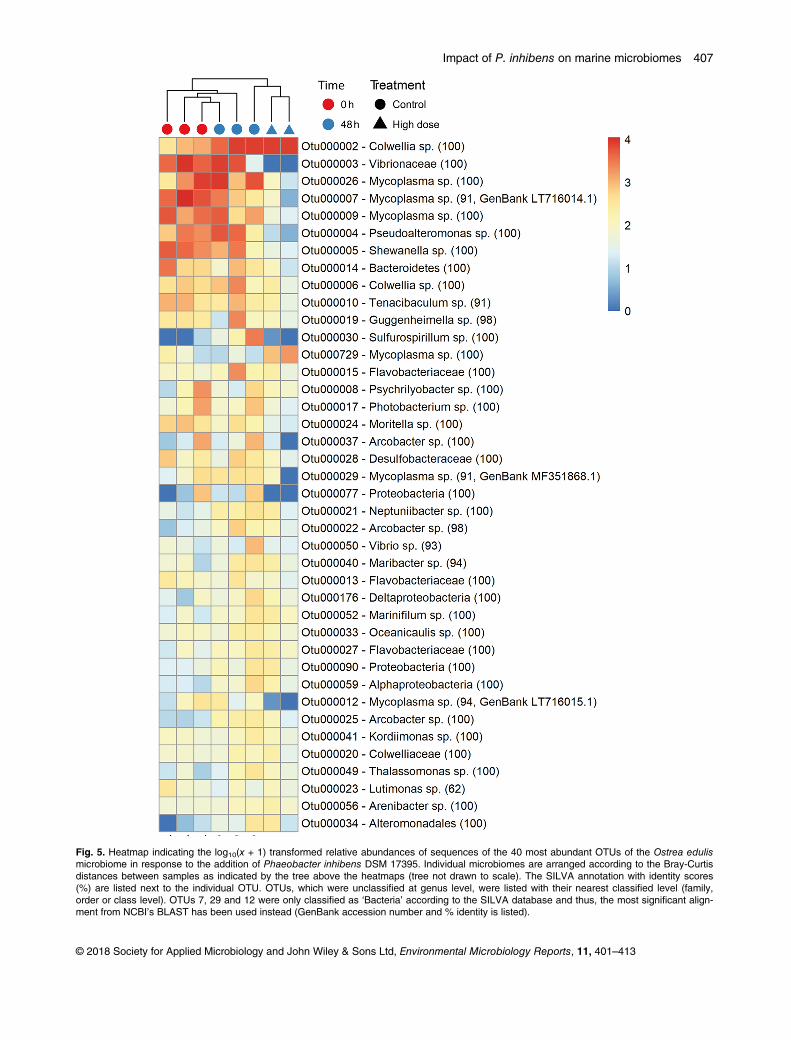
Fig. 5. Heatmap indicating the log10(x + 1) transformed relative abundances of sequences of the 40 most abundant OTUs of the Ostrea edulismicrobiome in response to the addition of Phaeobacter inhibens DSM 17395. Individual microbiomes are arranged according to the Bray-Curtisdistances between samples as indicated by the tree above the heatmaps (tree not drawn to scale). The SILVA annotation with identity scores(%) are listed next to the individual OTU. OTUs, which were unclassified at genus level, were listed with their nearest classified level (family,order or class level). OTUs 7, 29 and 12 were only classified as ‘Bacteria’ according to the SILVA database and thus, the most significant align-ment from NCBI’s BLAST has been used instead (GenBank accession number and % identity is listed).
© 2018 Society for Applied Microbiology and John Wiley & Sons Ltd, Environmental Microbiology Reports, 11, 401–413
Impact of P. inhibens on marine microbiomes 407
Mycoplasma species occur in the presence of P. inhi-bens in the oyster microbiome.To investigate a potentially specific, amensal interac-
tion between P. inhibens and vibrios, given the potentialpathogenicity of certain species, we assessed Vibrio spp.abundances in oyster microbiomes quantitatively usingquantitative real-time PCR (qPCR). As suggested by thecommunity composition analysis (Fig. 2), the introductionof P. inhibens did not remove vibrios in the oyster micro-biomes completely (Supporting Information Fig. S3).However, it resulted in a reduction of Vibrio spp. 16SrRNA genes to 5.61 � 0.23 log10 copies g−1 as com-pared with the initial abundance of 5.91 � 0.40 log10copies g−1, and it further reduced Vibrio abundances sig-nificantly with more than one order of magnitude com-pared with control oysters (6.89 � 0.18; p = 0.0328), thuscorroborating the results of the sequence analyses.
Discussion
Bacterial communities associated with eukaryotes have asignificant impact on the health and function of theirhosts, and investigating how microbiomes of higherorganisms are formed and affected by external and inter-nal factors, has become an area of broad and currentinterest (e.g., Ley et al., 2008; Lebeis et al., 2015). Somemembers of the host-associated microbiomes carry thecapacity to produce bioactive secondary metabolites thatmay act in the competition with other members of themicrobiomes, yet, it is currently unknown to what extentthese bacteria can directly influence and shape the struc-ture of host-associated microbial communities. Ourresults suggest that the TDA-producing P. inhibens hasthe capacity to influence and shape marine eukaryote-associated microbiomes, yet the effects are variable,dependent on the abundance of P. inhibens, and on thecomplexity and species composition of the host micro-biome. Furthermore, the imposed changes occur within ashort temporal scale (≤24 h) and are otherwise indepen-dent of time (within 5 days).The two eukaryote model systems were used to inves-
tigate how a TDA-producing P. inhibens would shapeeukaryote-associated microbiomes; during algal bloomsversus non-bloom conditions for the microalga, and howpotentially high densities of roseobacters would impactthe microbiome of a filter-feeder at another trophic layerduring algal blooms. Several studies have been con-ducted on bacteria associated with E. huxleyi and oysters(Zabeti et al., 2010; Carella et al., 2013; Farto Seguínet al., 2014; Green et al., 2015), but the amount of com-prehensive culture-independent, diversity studies is lim-ited. The oyster microbiomes of Crassostrea spp. havebeen characterized (King et al., 2012; Wegner et al.,2013; Ossai et al., 2017; Vezzulli et al., 2018) due to their
importance in aquaculture, but the present study is to thebest of our knowledge, the first culture-independent studyinvestigating the bacterial population of O. edulis. Roseo-bacters were indigenous to both of the investigatedmicrobiomes in agreement with previous findings of mol-luscan species (Ruiz-Ponte et al., 1998; Grigioni et al.,2000; Barbieri et al., 2001; Martens et al., 2006; Pradoet al., 2009) and algae (González et al., 2000; Greenet al., 2015; Segev et al., 2016b). The occurrence ofroseobacters in both native microbiomes supports ourchoice of P. inhibens DSM 17395 as a model organismfor a secondary metabolite producer in the natural envi-ronment of the eukaryotes in the event of an algal bloom.
The estimated OTU richness of the E. huxleyi micro-biome was four to seven times lower than the richnessof the complex O. edulis microbiome. Increasing theP. inhibens abundance in the low complexity microbiomeof E. huxleyi had little to no effect on the total speciesrichness and diversity, whereas the richness increaseddramatically in oysters. If P. inhibens uses its bioactivecompounds to kill competitors, that is, as antibiotics, adecrease in bacterial richness and diversity could beexpected, but the addition of P. inhibens had the oppo-site effect in the oyster system, indicating that the bioac-tive compounds, such as TDA, are either not produced orserve another function. The expression of TDA-encodinggenes have been shown in algal co-culture systems(D’Alvise et al., 2012). Furthermore, TDA is likely pro-duced in our model systems as P. inhibens DSM 17395was pre-grown at conditions known to induce TDA pro-duction, that is, nutrient/iron rich broth. While nutrient-richmedium, such as marine broth, differs from the naturalenvironment, some heterotrophs thrive at high nutrientlevels (Alonso and Pernthaler, 2006; Pohlner et al.,2017), which are comparable to the dense, nutrient-richsurroundings of algal cells or in oysters. Henceforth, it ismost likely that TDA has another function than being abroad-spectrum defence compound, and that it is proba-bly highly dependent on the investigated conditions.
Species-rich microbiomes are typical of healthy, marineinvertebrates including oysters (King et al., 2012; Wegneret al., 2013), whereas ill and diseased oyster microbiomesare characterized by a decrease in complexity, loss of rarebacterial strains (Wegner et al., 2013), disruption of thecommunity structure (Lokmer and Mathias Wegner, 2015)and increased abundance of few, specialist OTUs(Wegner et al., 2013; Lokmer and Mathias Wegner, 2015).Thus, it could be speculated that an increase inP. inhibens might be beneficial for the host by decreasingthe load of potential opportunistic pathogens and allowingrare taxa to proliferate, which has been suggested as therole of various Roseobacter group members associatedwith other molluscs (Collins et al., 2012; Gromek et al.,2016) and microalgae (Seyedsayamdost et al., 2011).
© 2018 Society for Applied Microbiology and John Wiley & Sons Ltd, Environmental Microbiology Reports, 11, 401–413
408 K. K. Dittmann et al.
P. inhibens influenced the bacterial community struc-ture in a dose dependent manner, independently of tem-poral space. The high density of P. inhibens caused ashift in the community structure of the E. huxleyi micro-biome, but no evident clustering patterns were observedfor the algal microbiome exposed to the low density.While due to the low sample number these results stillrequire further verification, similar patterns were observedin the microbiome structure of N. salina after the additionof pure TDA (Geng et al., 2016).
The oyster microbiome was richer than the algal micro-biome indicating that the microalga represented a nar-rower niche in comparison to the larger, moredifferentiated bivalve. As filter feeders, oysters accumu-late detritus and hence also bacteria. In combination withthe higher degree of tissue differentiation, it is not surpris-ing that the oyster microbiome comprises a broader taxo-nomic assortment of bacteria than that of the microalga.Furthermore, the alga has been kept under continuouslaboratory cultivation since its isolation, which may havereduced the richness and diversity of its associatedmicrobiome. Proteobacteria dominated the bacterial com-munity of both E. huxleyi and O. edulis in agreement withprevious observations in coccolithophorid microalgae andother oyster species (Wegner et al., 2013; Green et al.,2015; García Bernal et al., 2017). Green et al., 2015 useda culture-dependent approach and found that Alphapro-teobacteria were dominating in their coccolithophorid cul-tures. Similarly, it has been reported that Gamma- andAlphaproteobacteria of the orders Alteromonadales andRhodobacterales dominated E. huxleyi blooms in theNorth Atlantic Ocean (Segev et al., 2016b). In ourculture-independent approach, we found both Gamma-and Alphaproteobacteria as well as Flavobacteria(Bacteroidetes) dominated in the E. huxleyi microbiomes.
In O. edulis, Tenericutes and Bacteroidetes wereprominent phyla although less dominating than Proteo-bacteria. Wegner and colleagues (2013) similarly foundProteobacteria as the dominant phyla followed by Flavo-bacteria and Bacteroidetes in pacific oysters (Crassos-trea gigas). The community composition and dominanceof individual taxa depends on local environment, tissue(different organs vs. whole organism) and individual oys-ter genetics and physiology (King et al., 2012; Wegneret al., 2013; García Bernal et al., 2017; Vezzulli et al.,2018). The Vibrionales and Mycoplasmatales ordersdecreased in abundance in the presence of P. inhibenswhile Alteromonadales increased in abundance. In themicrobiome of the microalga N. salina, the abundance ofAlteromonadales also increased in the presence of pureTDA while Rhodobacteraceae decreased (Geng et al.,2016). We did not find that P. inhibens decreased otherRhodobacterales in the oyster microbiome, however, anychange in relative abundance of this order would not be
observed since it was below the 2% cutoff in all micro-biomes. Although, the relative abundance of the Rhodo-bacterales order did decrease in the E. huxleyimicrobiome when exposed to the high density ofP. inhibens while the abundance of Alteromonadaleswas unaffected. Hence, the impact of the TDA-producerP. inhibens on overall community composition is likelydependent on the eukaryotic host and/or the indigenouscommunity composition.
Given the subtle alterations at higher taxonomic levelsin some microbiomes, we assessed changes at the OTU-level (species level) to identify the underlying causes.Indeed, we observed some differences in the OTUsaffected by different densities of P. inhibens, though withnoticeable variability between replicate cultures. Somespecies (Colwellia and Sulfurospirillum) were either unaf-fected or increased by the presence of P. inhibens,whereas others including Vibrio spp. and Pseudoaltero-monas spp. were reduced in both eukaryote micro-biomes. Interestingly, vibrios and pseudalteromonads arealso considered proficient secondary metabolite pro-ducers and hence it is plausible that P. inhibens specifi-cally and efficiently antagonizes other microorganismsoccupying similar niches. The efficient inhibition of vibriosby TDA-producing roseobacters have been repeatedlydemonstrated in studies targeting the fish pathogenic Vib-rio spp. in marine eukaryotes (Porsby et al., 2008; Pradoet al., 2009; D’Alvise et al., 2012; Grotkjær et al., 2016a,b; Porsby and Gram, 2016; Rasmussen et al., 2018). Thehigh degree of target organism specificity is howeversomewhat surprising considering the spectrum of bioac-tivity of TDA (Porsby et al., 2011). Multiple Mycoplasmaspp. decreased in abundance while a single OTU classi-fied to the same genus increased in abundance furthersubstantiating that the effects of P. inhibens are very spe-cific (species level or below). Corroborating the observa-tion that the majority of the species in the microbiomesexhibited minor changes in abundance due to the pres-ence of P. inhibens, is the findings by Harrington and col-leagues (2014) who observed a high degree of TDAtolerance in non-TDA producing bacterial isolates frommarine eukaryote-associated microbiomes. Thus, theimpact of increased abundances of a particular second-ary metabolite producing microorganism such asP. inhibens seems to be highly selective and likelydependent on its specific secondary metabolite profile.
In conclusion, TDA-producing P. inhibens can shapehost-associated microbiomes of marine eukaryotes.These alterations are subtle at the broader taxonomiclevels, but seems to be highly selective and consistent atthe OTU level (97% similarity) across eukaryote host sys-tems. The effects of P. inhibens on the species richnessand microbiome structure are multifaceted; the richnessin the complex microbiome of oysters increased while it
© 2018 Society for Applied Microbiology and John Wiley & Sons Ltd, Environmental Microbiology Reports, 11, 401–413
Impact of P. inhibens on marine microbiomes 409
remained constant in the microalgal model and the micro-biome structure shifted only due to high densities ofP. inhibens. Thus, the impact likely relies on the compo-sition and complexity of the indigenous bacterial commu-nities. This suggests that a highly diverse microbiome ismore stable, though future work across more, variedeukaryote–microbiome associations with larger samplesizes would provide more knowledge to substantiate thishypothesis.
Acknowledgements
The authors wish to thank Professor Torsten Thomas forproviding valuable discussions. We acknowledge the help byCisse Hedegaard Porsby and the Danish Shellfish Center(National Institute of Aquatic Resources, Technical Univer-sity of Denmark, Denmark) with providing oysters and settingup experiments with oysters.
The present study was funded by the Technical Universityof Denmark (PhD grant for KKD), the Villum Kann Rasmus-sen foundation (grant VKR023285), the Danish Council forStrategic Research, Committee for Strategic Research inHealth, Food and Welfare (grant 0603-00515B), the DanishNational Research Foundation (grant DNRF137), the Oticonfoundation (Personal travel grant for KKD), IDAs og Berg-Nielsens Studie- og Støttefond (Personal travel grant forKKD), the Augustinus foundation (Personal travel grant forKKD), the Fabrikant P.A. Fiskers foundation (Personal travelgrant for KKD), and the scholarship Christian og Ottilia Bror-sons Rejselegat for yngre videnskabsmænd og –kvinder(Personal travel grant for KKD).
References
Alonso, C., and Pernthaler, J. (2006) Roseobacter andSAR11 dominate microbial glucose uptake in coastalNorth Sea waters. Environ Microbiol 8: 2022–2030.
Amin, S.A., Hmelo, L.R., van Tol, H.M., Durham, B.P.,Carlson, L.T., Heal, K.R., et al. (2015) Interaction and sig-nalling between a cosmopolitan phytoplankton and associ-ated bacteria. Nature 522: 98–101.
Barbieri, E., Paster, B.J., Hughes, D., Zurek, L., Moser, D.P.,Teske, A., and Sogin, M.L. (2001) Phylogenetic character-ization of epibiotic bacteria in the accessory nidamentalgland and egg capsules of the squid Loligo pealei(Cephalopoda: Loliginidae). Environ Microbiol 3: 151–167.
Bentzon-Tilia, M., and Gram, L. (2017) BiotechnologicalApplications of the Roseobacter Clade. In Biotechnologi-cal applications of the roseobacter clade. Paterson, R.,and Lima, N. (eds). Basel, Switzerland: Springer Interna-tional Publishing, pp. 137–166.
Beyersmann, P.G., Tomasch, J., Son, K., Stocker, R.,Göker, M., Wagner-Döbler, I., et al. (2017) Dual functionof tropodithietic acid as antibiotic and signaling moleculein global gene regulation of the probiotic bacterium Phaeo-bacter inhibens. Sci Rep 7: 730.
Brinkhoff, T., Bach, G., Heidorn, T., Liang, L., Schlingloff, A.,and Simon, M. (2004) Antibiotic production by a Roseo-bacter clade-affiliated species from the German Wadden
Sea and its antagonistic effects on indigenous isolates.Appl Environ Microbiol 70: 2560–2565.
Bruhn, J.B., Nielsen, K.F., Hjelm, M., Hansen, M., Bresciani,J., Schulz, S., and Gram, L. (2005) Ecology, inhibitoryactivity, and morphogenesis of a marine antagonistic bac-terium belonging to the Roseobacter clade. Appl EnvironMicrobiol 71: 7263–7270.
Carella, F., Carrasco, N., Andree, K.B., Lacuesta, B.,Furones, D., and De Vico, G. (2013) Nocardiosis in Medi-terranean bivalves: first detection of Nocardia crassos-treae in a new host Mytilus galloprovincialis and in Ostreaedulis from the Gulf of Naples (Italy). J Invertebr Pathol114: 324–328.
Cole, J.J. (1982) Interactions between bacteria and algae inaquatic ecosystems. Annu Rev Ecol Syst 13: 291–314.
Collins, A.J., LaBarre, B.A., Wong Won, B.S., Shah, M.V.,Heng, S., Choudhury, M.H., et al. (2012) Diversity andpartitioning of bacterial populations within the accessoryNidamental gland of the squid Euprymna scolopes. ApplEnviron Microbiol 78: 4200–4208.
Cude, W.N., Mooney, J., Tavanaei, A.A., Hadden, M.K.,Frank, A.M., Gulvik, C.A., et al. (2012) Production of theantimicrobial secondary metabolite Indigoidine contributesto competitive surface colonization by the marine Roseo-bacter Phaeobacter sp. strain Y4I. Appl Environ Microbiol78: 4771–4780.
D’Alvise, P.W., Lillebø, S., Prol-Garcia, M.J., Wergeland, H.I., Nielsen, K.F., Bergh, �., and Gram, L. (2012) Phaeo-bacter gallaeciensis reduces Vibrio anguillarum in cul-tures of microalgae and rotifers, and prevents vibriosis incod larvae. PLoS One 7: e43996.
Farto Seguín, R.M., Bermúdez, M.B., Rivera, L., andNieto, T.P. (2014) Increased survival of juvenile turbotScophthalmus maximus by using bacteria associated withcultured oysters. J Aquat Anim Health 26: 251–262.
Freese, H.M., Methner, A., and Overmann, J. (2017) Adapta-tion of surface-associated bacteria to the Open Ocean: aGenomically distinct subpopulation of Phaeobacter gallae-ciensis colonizes Pacific Mesozooplankton. Front Micro-biol 8: 1–12.
García Bernal, M., Trabal Fernández, N., Saucedo Lastra, P.E., Medina Marrero, R., and Mazón-Suástegui, J.M.(2017) Streptomyces effect on the bacterial microbiotaassociated to Crassostrea sikamea oyster. J Appl Micro-biol 122: 601–614.
Geng, H., Tran-Gyamfi, M.B., Lane, T.W., Sale, K.L., andYu, E.T. (2016) Changes in the structure of the microbialcommunity associated with Nannochloropsis salina follow-ing treatments with antibiotics and bioactive compounds.Front Microbiol 7: 1155.
González, J.M., and Moran, M.A. (1997) Numerical domi-nance of a group of marine bacteria in the alpha-subclassof the class Proteobacteria in coastal seawater. ApplEnviron Microbiol 63: 4237–4242.
González, J.M., Simó, R., Massana, R., Covert, J.S.,Casamayor, E.O., Pedrós-Alió, C., and Moran, M.A.(2000) Bacterial community structure associated with adimethylsulfoniopropionate-producing North Atlantic algalbloom. Appl Environ Microbiol 66: 4237–4246.
Green, D.H., Echavarri-Bravo, V., Brennan, D., and Hart, M.C. (2015) Bacterial diversity associated with the
© 2018 Society for Applied Microbiology and John Wiley & Sons Ltd, Environmental Microbiology Reports, 11, 401–413
410 K. K. Dittmann et al.
Coccolithophorid algae Emiliania huxleyi and Coccolithuspelagicus f. braarudii. Biomed Res Int 2015: 1–15.
Grigioni, S., Boucher-Rodoni, R., Demarta, A., Tonolla, M.,and Peduzzi, R. (2000) Phylogenetic characterisation ofbacterial symbionts in the accessory nidamental glands ofthe sepioid Sepia officinalis (Cephalopoda: Decapoda).Mar Biol 136: 217–222.
Gromek, S.M., Suria, A.M., Fullmer, M.S., Garcia, J.L.,Gogarten, J.P., Nyholm, S.V., and Balunas, M.J. (2016)Leisingera sp. JC1, a bacterial isolate from hawaiian bob-tail squid eggs, produces indigoidine and differentiallyinhibits vibrios. Front Microbiol 7: 1–16.
Grotkjær, T., Bentzon-Tilia, M., D’Alvise, P.W.,Dierckens, K., Bossier, P., and Gram, L. (2016a) Phaeo-bacter inhibens as probiotic bacteria in non-axenic Arte-mia and algae cultures. Aquaculture 462: 64–69.
Grotkjær, T., Bentzon-Tilia, M., D’Alvise, P.W., Dourala, N.,Nielsen, K.F., and Gram, L. (2016b) Isolation ofTDA-producing Phaeobacter strains from sea bass larvalrearing units and their probiotic effect against pathogenicVibrio spp. in Artemia cultures. Syst Appl Microbiol 39:180–188.
Harrington, C., Reen, F., Mooij, M., Stewart, F.,Chabot, J.-B., Guerra, A., et al. (2014) Characterisation ofnon-autoinducing Tropodithietic acid (TDA) productionfrom marine sponge Pseudovibrio species. Mar Drugs 12:5960–5978.
King, G.M., Judd, C., Kuske, C.R., and Smith, C. (2012)Analysis of stomach and gut microbiomes of the easternoyster (Crassostrea virginica) from coastal Louisiana,USA. PLoS One 7: e51475.
Lebeis, S.L., Paredes, S.H., Lundberg, D.S., Breakfield, N.,Gehring, J., McDonald, M., et al. (2015) Salicylic acidmodulates colonization of the root microbiome by specificbacterial taxa. Science 349: 860–864.
Ley, R.E., Lozupone, C.A., Hamady, M., Knight, R., andGordon, J.I. (2008) Worlds within worlds: evolution of thevertebrate gut microbiota. Nat Rev Microbiol 6: 776–788.
Lokmer, A., and Mathias Wegner, K. (2015) Hemolymphmicrobiome of Pacific oysters in response to temperature,temperature stress and infection. ISME J 9: 670–682.
Maansson, M., Vynne, N.G., Klitgaard, A., Nybo, J.L.,Melchiorsen, J., Nguyen, D.D., et al. (2016) An integratedMetabolomic and genomic mining workflow to uncoverthe biosynthetic potential of bacteria. mSystems 1:e00028-15.
Machado, H., Sonnenschein, E.C., Melchiorsen, J., andGram, L. (2015) Genome mining reveals unlocked bioac-tive potential of marine gram-negative bacteria. BMCGenom 16: 158.
Martens, T., Heidorn, T., Pukall, R., Simon, M., Tindall, B.J.,and Brinkhoff, T. (2006) Reclassification of Roseobactergallaeciensis Ruiz-Ponte et al. 1998 as Phaeobactergallaeciensis gen. Nov., comb. nov., description of Phaeo-bacter inhibens sp. nov., reclassification of Ruegeria algi-cola (Lafay et al. 1995) Uchino et al. 1999 as Marinovumalgicola gen. Nov., comb. nov., and emended descriptionsof the genera Roseobacter, Ruegeria and Leisingera. IntJ Syst Evol Microbiol 56: 1293–1304.
Murphy, B., Jensen, P., and Fenical, W. (2012) The chemis-try of marine bacteria. In Handbook of Marine Natural
Products. Fattorusso, E., Gerwick, W., andTaglialatela-Scafati, O. (eds). New York, NY: Springer,pp. 153–190.
Newton, R.J., Griffin, L.E., Bowles, K.M., Meile, C.,Gifford, S., Givens, C.E., et al. (2010) Genome character-istics of a generalist marine bacterial lineage. ISME J 4:784–798.
Ossai, S., Ramachandran, P., Ottesen, A., Reed, E.,DePaola, A., and Parveen, S. (2017) Microbiomes ofAmerican oysters (Crassostrea virginica) harvested from twosites in the Chesapeake Bay.Genome Announc 5: 1–12.
Pohlner, M., Degenhardt, J., von Hoyningen-Huene, A.J.E.,Wemheuer, B., Erlmann, N., Schnetger, B., et al. (2017)The biogeographical distribution of benthic Roseobactergroup members along a Pacific transect is structured bynutrient availability within the sediments and primary pro-duction in different oceanic provinces. Front Microbiol 8:2550.
Porsby, C.H., and Gram, L. (2016) Phaeobacter inhibens asbiocontrol agent against Vibrio vulnificus in oyster models.Food Microbiol 57: 63–70.
Porsby, C.H., Nielsen, K.F., and Gram, L. (2008) Phaeobac-ter and Ruegeria species of the Roseobacter clade colo-nize separate niches in a Danish turbot (Scophthalmusmaximus)-rearing farm and antagonize Vibrio anguillarumunder different growth conditions. Appl Environ Microbiol74: 7356–7364.
Porsby, C.H., Webber, M.A., Nielsen, K.F., Piddock, L.J.V.,and Gram, L. (2011) Resistance and tolerance to tropo-dithietic acid, an antimicrobial in aquaculture, is hard toselect. Antimicrob Agents Chemother 55: 1332–1337.
Prado, S., Montes, J., Romalde, J.L., and Barja, J.L. (2009)Inhibitory activity of Phaeobacter strains against aquacul-ture pathogenic bacteria. Int Microbiol 12: 107–114.
Pusztahelyi, T., Holb, I.J., and Pócsi, I. (2015) Secondarymetabolites in fungus-plant interactions. Front Plant Sci6: 573.
Rao, D., Webb, J.S., and Kjelleberg, S. (2005) Competitiveinteractions in mixed-species biofilms containing themarine bacterium Pseudoalteromonas tunicata. Appl Envi-ron Microbiol 71: 1729–1736.
Rasmussen, B., Nielsen, K., Machado, H., Melchiorsen, J.,Gram, L., and Sonnenschein, E. (2014) Global and phylo-genetic distribution of quorum sensing signals, acyl Homo-serine lactones, in the family of Vibrionaceae. Mar Drugs12: 5527–5546.
Rasmussen, B.B., Erner, K.E., Bentzon-Tilia, M., andGram, L. (2018) Effect of TDA-producing Phaeobacterinhibens on the fish pathogen Vibrio anguillarum innon-axenic algae and copepod systems. J Microbial Bio-technol 0: 1–10.
Rasmussen, B.B., Grotkjær, T., D’Alvise, P.W., Yin, G.,Zhang, F., Bunk, B., et al. (2016) Vibrio anguillarum isgenetically and phenotypically unaffected by long-termcontinuous exposure to the antibacterial compound tropo-dithietic acid. Appl Environ Microbiol 82: 4802–4810.
Ruiz-Ponte, C., Cilia, V., Lambert, C., and Nicolas, J.L.(1998) Roseobacter gallaeciensis sp. nov., a new marinebacterium isolated from rearings and collectors of the scal-lop Pecten maximus. Int J Syst Bacteriol 48: 537–542.
© 2018 Society for Applied Microbiology and John Wiley & Sons Ltd, Environmental Microbiology Reports, 11, 401–413
Impact of P. inhibens on marine microbiomes 411
Segev, E., Castañeda, I.S., Sikes, E.L., Vlamakis, H., andKolter, R. (2016a) Bacterial influence on alkenones in livemicroalgae. J Phycol 52: 125–130.
Segev, E., Wyche, T.P., Kim, K.H., Petersen, J.,Ellebrandt, C., Vlamakis, H., et al. (2016b) Dynamic meta-bolic exchange governs a marine algal-bacterial interac-tion. Elife 5: 1–28.
Seyedsayamdost, M.R., Case, R.J., Kolter, R., and Clardy,J. (2011) The Jekyll-and-Hyde chemistry of Phaeobactergallaeciensis. Nat Chem 3: 331–335.
Simon, M., Scheuner, C., Meier-Kolthoff, J.P., Brinkhoff, T.,Wagner-Döbler, I., Ulbrich, M., et al. (2017) Phyloge-nomics of Rhodobacteraceae reveals evolutionary adap-tation to marine and non-marine habitats. ISME J 11:1483–1499.
Sonnenschein, E.C., Nielsen, K.F., D’Alvise, P., Porsby, C.H., Melchiorsen, J., Heilmann, J., et al. (2017a) Globaloccurrence and heterogeneity of the Roseobacter-cladespecies Ruegeria mobilis. ISME J 11: 569–583.
Sonnenschein, E.C., Phippen, C.B.W., Bentzon-Tilia, M.,Rasmussen, S.A., Nielsen, K.F., and Gram, L. (2018) Phy-logenetic distribution of roseobacticides in the Roseobac-ter group and their effect on microalgae. Environ MicrobiolRep 10: 383–393.
Sonnenschein, E.C., Stierhof, M., Goralczyk, S., Vabre, F.M., Pellissier, L., Hanssen, K.�., et al. (2017b) Pseudo-chelin a, a siderophore of Pseudoalteromonas piscicidaS2040. Tetrahedron 73: 2633–2637.
Vezzulli, L., Stagnaro, L., Grande, C., Tassistro, G.,Canesi, L., and Pruzzo, C. (2018) Comparative 16S rDNAgene-based microbiota profiles of the Pacific oyster (Cras-sostrea gigas) and the Mediterranean mussel (Mytilusgalloprovincialis) from a shellfish farm (Ligurian Sea,Italy). Microb Ecol 75: 495–504.
Wegner, K., Volkenborn, N., Peter, H., and Eiler, A. (2013)Disturbance induced decoupling between host geneticsand composition of the associated microbiome. BMCMicrobiol 13: 252.
West, N.J., Obernosterer, I., Zemb, O., and Lebaron, P.(2008) Major differences of bacterial diversity and activityinside and outside of a natural iron-fertilized phytoplanktonbloom in the Southern Ocean. Environ Microbiol 10:738–756.
Wietz, M., Gram, L., Jørgensen, B., and Schramm, A. (2010)Latitudinal patterns in the abundance of major marine bac-terioplankton groups. Aquat Microb Ecol 61: 179–189.
Wilson, M.Z., Wang, R., Gitai, Z., and Seyedsayamdost, M.R. (2016) Mode of action and resistance studies unveilnew roles for tropodithietic acid as an anticancer agentand the γ-glutamyl cycle as a proton sink. Proc Natl AcadSci 113: 1630–1635.
Zabeti, N., Bonin, P., Volkman, J.K., Guasco, S., andRontani, J.-F. (2010) Fatty acid composition of bacterialstrains associated with living cells of the haptophyte Emi-liania huxleyi. Org Geochem 41: 627–636.
Zubkov, M.V., Fuchs, B.M., Archer, S.D., Kiene, R.P.,Amann, R., and Burkill, P.H. (2001) Linking the composi-tion of bacterioplankton to rapid turnover of dissolveddimethylsulphoniopropionate in an algal bloom in theNorth Sea. Environ Microbiol 3: 304–311.
Supporting Information
Additional Supporting Information may be found in the onlineversion of this article at the publisher’s web-site:
Table S1: Species determination of OTU classified asPhaeobacter sp. in Emiliania huxleyi microbiome. The iden-tity percentage, E-value and GenBank accession numberlisted for sequences producing significant alignments withthe representative sequence of OTU 4 in NCBI’s Basic LocalAlignment Search Tool (BLAST, nucleotide).Table S2: The ten most abundant OTUs (excluding the OTUof the added Phaeobacter sp.) in the Emiliania huxleyimicrobiome. The SILVA taxonomy with identity scores (%)are listed next to the individual OTU. *: OTU which wasunclassified at genus level and listed with its nearest classi-fied level (family, order or class level). Further genus deter-mination using NCBI’s Basic Local Alignment Search Tool(BLAST, nucleotide) can be found in Table S3.Table S3: Genus determination of OTUs unclassified at thislevel in Emiliania huxleyi microbiome. The identity percent-age, E-value and GenBank accession number listed forsequences producing the ten most significant alignmentswith the representative sequences in NCBI’s Basic LocalAlignment Search Tool (BLAST, nucleotide).Table S4: Species determination of abundant OTU classifiedto the Rhodobacteraceae family in Ostrea edulis microbiome.The identity percentage, E-value and GenBank accessionnumber listed for sequences producing significant alignmentswith the representative sequence of OTU 1 in NCBI’s BasicLocal Alignment Search Tool (BLAST, nucleotide).Table S5: The ten most abundant OTUs (excluding the OTUof the added Phaeobacter sp.) in the Ostrea edulis micro-biome. The SILVA taxonomy with identity scores (%) arelisted next to the individual OTU. *: OTU which was unclassi-fied at genus level and listed with its nearest classified level(family, order or class level). Further genus determinationusing NCBI’s Basic Local Alignment Search Tool (BLAST,nucleotide) can be found in Table S6.Table S6: Genus determination of highly abundant OTUunclassified at this level in Ostrea edulis microbiome. Theidentity percentage, E-value and GenBank accession num-ber listed for sequences producing the ten most significantalignments with the representative sequences in NCBI’sBasic Local Alignment Search Tool (BLAST, nucleotide).Table S7: List of indexed used for PCR amplification andsequencing of V4 16S rRNA amplicons from the bacterialmicrobiomes of Emiliania huxleyi and Ostrea edulis.Table S8: Primer combinations for PCR amplification andsequencing of V4 16S rRNA amplicons from the bacterialmicrobiomes of Emiliania huxleyi and Ostrea edulis.Fig. S1: Rarefaction curves for all sequenced samples fromthe Emiliania huxleyi (A) and Ostrea edulis (B) microbiomes.After quality filtering, 1.3 × 10⁶ and 2.5 × 10⁶ V4 sequenceswere obtained from the E. huxleyi- and O. edulis-associatedmicrobiomes respectively. Sequences were clustered at a97% sequence similarity, which resulted in 1,346 and 6,706unique operational taxonomic units (OTUs) for the E.huxleyi-and O. edulis-associated microbiomes respectively. Inthe community structure analyses, 17,000 (E. huxleyi) and
© 2018 Society for Applied Microbiology and John Wiley & Sons Ltd, Environmental Microbiology Reports, 11, 401–413
412 K. K. Dittmann et al.
41,000 (O. edulis) sequences from each sample wereanalysed.Fig. S2: Diversity of bacterial microbiomes observed in A)Emiliania huxleyi (microalga) and B) Ostrea edulis(European flat oysters) in response to the addition of Phaeo-bacter inhibens DSM 17395. The diversity is expressed asthe average Shannon diversity index value, error bars repre-sent the standard deviation of the average. OE_OTU 1 andEH_OTU 4 containing the added P. inhibens were removedfrom the datasets prior to plotting. T0: untreated time zero
control, C: untreated control, LD: low density, HD: highdensity.Fig. S3: Changes in Vibrio abundances (Vibrio 16S rRNAgene copies/g oyster) in the European flat oyster microbiomeas a function of the addition of Phaeobacter inhibens DSM17395. O: untreated time zero control, C: untreated control,HD: high density. Error bars represent the standard deviationof the mean. Statistically significant differences in Vibrioabundances (P ≤ 0.05) are indicated by Tukey groupings.Appendix S1: Supporting information
© 2018 Society for Applied Microbiology and John Wiley & Sons Ltd, Environmental Microbiology Reports, 11, 401–413
Impact of P. inhibens on marine microbiomes 413
Impact of P. inhibens on marine microbiomes
1
Supplementary File 1 1
Impact of Phaeobacter inhibens on marine eukaryote-associated microbial 2
communities 3
4
Karen K. Dittmann, Eva C. Sonnenschein, Suhelen Egan, Lone Gram, Mikkel Bentzon-Tilia* 5
* Address correspondence to Mikkel Bentzon-Tilia: [email protected] 6
7
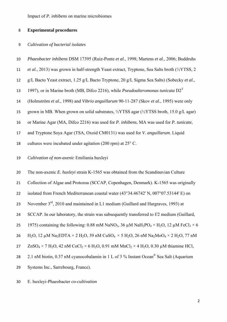
Impact of P. inhibens on marine microbiomes
2
Experimental procedures 8
Cultivation of bacterial isolates 9
Phaeobacter inhibens DSM 17395 (Ruiz-Ponte et al., 1998; Martens et al., 2006; Buddruhs 10
et al., 2013) was grown in half-strength Yeast extract, Tryptone, Sea Salts broth (½YTSS, 2 11
g/L Bacto Yeast extract, 1.25 g/L Bacto Tryptone, 20 g/L Sigma Sea Salts) (Sobecky et al., 12
1997), or in Marine broth (MB, Difco 2216), while Pseudoalteromonas tunicata D2T 13
(Holmström et al., 1998) and Vibrio anguillarum 90-11-287 (Skov et al., 1995) were only 14
grown in MB. When grown on solid substrates, ½YTSS agar (½YTSS broth, 15.0 g/L agar) 15
or Marine Agar (MA, Difco 2216) was used for P. inhibens, MA was used for P. tunicate, 16
and Tryptone Soya Agar (TSA, Oxoid CM0131) was used for V. anguillarum. Liquid 17
cultures were incubated under agitation (200 rpm) at 25° C. 18
Cultivation of non-axenic Emiliania huxleyi 19
The non-axenic E. huxleyi strain K-1565 was obtained from the Scandinavian Culture 20
Collection of Algae and Protozoa (SCCAP, Copenhagen, Denmark). K-1565 was originally 21
isolated from French Mediterranean coastal water (43°34.46742' N, 007°07.53144' E) on 22
November 3rd, 2010 and maintained in L1 medium (Guillard and Hargraves, 1993) at 23
SCCAP. In our laboratory, the strain was subsequently transferred to f/2 medium (Guillard, 24
1975) containing the following: 0.88 mM NaNO3, 36 µM NaH2PO4 × H2O, 12 µM FeCl3 × 6 25
H2O, 12 µM Na2EDTA × 2 H2O, 39 nM CuSO4 × 5 H2O, 26 nM Na2MoO4 × 2 H2O, 77 nM 26
ZnSO4 × 7 H2O, 42 nM CoCl2 × 6 H2O, 0.91 mM MnCl2 × 4 H2O, 0.30 µM thiamine HCl, 27
2.1 nM biotin, 0.37 nM cyanocobalamin in 1 L of 3 % Instant Ocean® Sea Salt (Aquarium 28
Systems Inc., Sarrebourg, France). 29
E. huxleyi-Phaeobacter co-cultivation 30
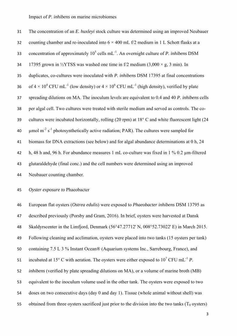
Impact of P. inhibens on marine microbiomes
3
The concentration of an E. huxleyi stock culture was determined using an improved Neubauer 31
counting chamber and re-inoculated into 6 × 400 mL f/2 medium in 1 L Schott flasks at a 32
concentration of approximately 105 cells mL-1. An overnight culture of P. inhibens DSM 33
17395 grown in ½YTSS was washed one time in f/2 medium (3,000 × g, 3 min). In 34
duplicates, co-cultures were inoculated with P. inhibens DSM 17395 at final concentrations 35
of 4 × 10⁴ CFU mL-1 (low density) or 4 × 10⁶ CFU mL-1 (high density), verified by plate 36
spreading dilutions on MA. The inoculum levels are equivalent to 0.4 and 40 P. inhibens cells 37
per algal cell. Two cultures were treated with sterile medium and served as controls. The co-38
cultures were incubated horizontally, rolling (20 rpm) at 18° C and white fluorescent light (24 39
µmol m-2 s-1 photosynthetically active radiation; PAR). The cultures were sampled for 40
biomass for DNA extractions (see below) and for algal abundance determinations at 0 h, 24 41
h, 48 h and, 96 h. For abundance measures 1 mL co-culture was fixed in 1 % 0.2 µm-filtered 42
glutaraldehyde (final conc.) and the cell numbers were determined using an improved 43
Neubauer counting chamber. 44
Oyster exposure to Phaeobacter 45
European flat oysters (Ostrea edulis) were exposed to Phaeobacter inhibens DSM 13795 as 46
described previously (Porsby and Gram, 2016). In brief, oysters were harvested at Dansk 47
Skaldyrscenter in the Limfjord, Denmark (56°47.27712' N, 008°52.73022' E) in March 2015. 48
Following cleaning and acclimation, oysters were placed into two tanks (15 oysters per tank) 49
containing 7.5 L 3 % Instant Ocean® (Aquarium systems Inc., Sarrebourg, France), and 50
incubated at 15° C with aeration. The oysters were either exposed to 107 CFU mL-1 P. 51
inhibens (verified by plate spreading dilutions on MA), or a volume of marine broth (MB) 52
equivalent to the inoculum volume used in the other tank. The oysters were exposed to two 53
doses on two consecutive days (day 0 and day 1). Tissue (whole animal without shell) was 54
obtained from three oysters sacrificed just prior to the division into the two tanks (T0 oysters) 55
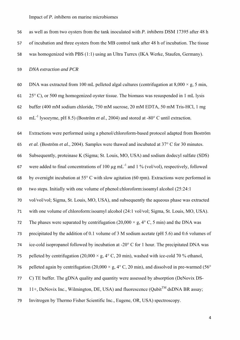
Impact of P. inhibens on marine microbiomes
4
as well as from two oysters from the tank inoculated with P. inhibens DSM 17395 after 48 h 56
of incubation and three oysters from the MB control tank after 48 h of incubation. The tissue 57
was homogenized with PBS (1:1) using an Ultra Turrex (IKA Werke, Staufen, Germany). 58
DNA extraction and PCR 59
DNA was extracted from 100 mL pelleted algal cultures (centrifugation at 8,000 × g, 5 min, 60
25° C), or 500 mg homogenized oyster tissue. The biomass was resuspended in 1 mL lysis 61
buffer (400 mM sodium chloride, 750 mM sucrose, 20 mM EDTA, 50 mM Tris-HCl, 1 mg 62
mL-1 lysozyme, pH 8.5) (Boström et al., 2004) and stored at -80° C until extraction. 63
Extractions were performed using a phenol/chloroform-based protocol adapted from Boström 64
et al. (Boström et al., 2004). Samples were thawed and incubated at 37° C for 30 minutes. 65
Subsequently, proteinase K (Sigma; St. Louis, MO, USA) and sodium dodecyl sulfate (SDS) 66
were added to final concentrations of 100 µg mL-1 and 1 % (vol/vol), respectively, followed 67
by overnight incubation at 55° C with slow agitation (60 rpm). Extractions were performed in 68
two steps. Initially with one volume of phenol:chloroform:isoamyl alcohol (25:24:1 69
vol/vol/vol; Sigma, St. Louis, MO, USA), and subsequently the aqueous phase was extracted 70
with one volume of chloroform:isoamyl alcohol (24:1 vol/vol; Sigma, St. Louis, MO, USA). 71
The phases were separated by centrifugation (20,000 × g, 4° C, 5 min) and the DNA was 72
precipitated by the addition of 0.1 volume of 3 M sodium acetate (pH 5.6) and 0.6 volumes of 73
ice-cold isopropanol followed by incubation at -20° C for 1 hour. The precipitated DNA was 74
pelleted by centrifugation (20,000 × g, 4° C, 20 min), washed with ice-cold 70 % ethanol, 75
pelleted again by centrifugation (20,000 × g, 4° C, 20 min), and dissolved in pre-warmed (56° 76
C) TE buffer. The gDNA quality and quantity were assessed by absorption (DeNovix DS-77
11+, DeNovix Inc., Wilmington, DE, USA) and fluorescence (QubitTM dsDNA BR assay; 78
Invitrogen by Thermo Fisher Scientific Inc., Eugene, OR, USA) spectroscopy. 79
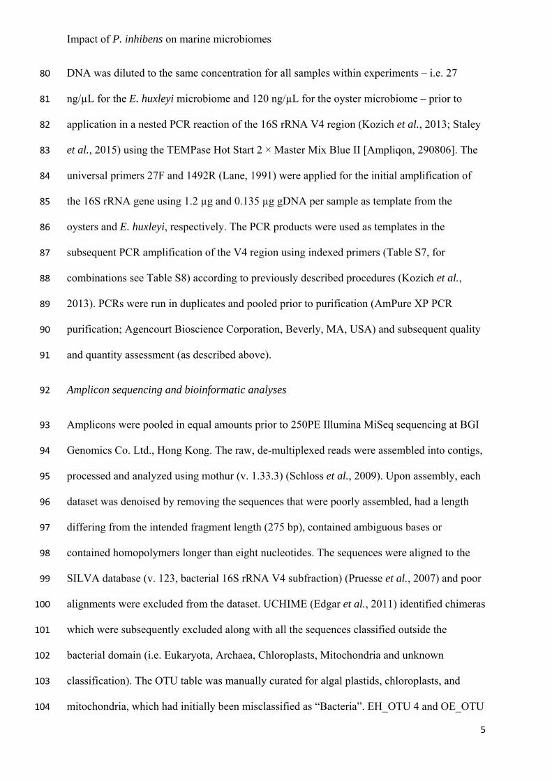
Impact of P. inhibens on marine microbiomes
5
DNA was diluted to the same concentration for all samples within experiments – i.e. 27 80
ng/µL for the E. huxleyi microbiome and 120 ng/µL for the oyster microbiome – prior to 81
application in a nested PCR reaction of the 16S rRNA V4 region (Kozich et al., 2013; Staley 82
et al., 2015) using the TEMPase Hot Start 2 × Master Mix Blue II [Ampliqon, 290806]. The 83
universal primers 27F and 1492R (Lane, 1991) were applied for the initial amplification of 84
the 16S rRNA gene using 1.2 µg and 0.135 µg gDNA per sample as template from the 85
oysters and E. huxleyi, respectively. The PCR products were used as templates in the 86
subsequent PCR amplification of the V4 region using indexed primers (Table S7, for 87
combinations see Table S8) according to previously described procedures (Kozich et al., 88
2013). PCRs were run in duplicates and pooled prior to purification (AmPure XP PCR 89
purification; Agencourt Bioscience Corporation, Beverly, MA, USA) and subsequent quality 90
and quantity assessment (as described above). 91
Amplicon sequencing and bioinformatic analyses 92
Amplicons were pooled in equal amounts prior to 250PE Illumina MiSeq sequencing at BGI 93
Genomics Co. Ltd., Hong Kong. The raw, de-multiplexed reads were assembled into contigs, 94
processed and analyzed using mothur (v. 1.33.3) (Schloss et al., 2009). Upon assembly, each 95
dataset was denoised by removing the sequences that were poorly assembled, had a length 96
differing from the intended fragment length (275 bp), contained ambiguous bases or 97
contained homopolymers longer than eight nucleotides. The sequences were aligned to the 98
SILVA database (v. 123, bacterial 16S rRNA V4 subfraction) (Pruesse et al., 2007) and poor 99
alignments were excluded from the dataset. UCHIME (Edgar et al., 2011) identified chimeras 100
which were subsequently excluded along with all the sequences classified outside the 101
bacterial domain (i.e. Eukaryota, Archaea, Chloroplasts, Mitochondria and unknown 102
classification). The OTU table was manually curated for algal plastids, chloroplasts, and 103
mitochondria, which had initially been misclassified as “Bacteria”. EH_OTU 4 and OE_OTU 104
Impact of P. inhibens on marine microbiomes
6
1 were excluded in the datasets used in subsequent analyses in order to dismiss any effects of 105
the increased Phaeobacter abundance on composition, and alpha- and beta-diversity 106
measures, hence focusing the analyses on the background microbiota. 107
The cleaned sequences were analyzed with an Operational Taxonomic Unit (OTU) approach. 108
First, the sequences were clustered into OTUs and classified with a 97 % nucleotide sequence 109
similarity cut-off (species-level). Measures of alpha (Chao1, Shannon) and beta (Bray-Curtis 110
distances) diversity were calculated based on a subsampling of sequences; 17,000 and 41,000 111
sequences for each sample in the E. huxleyi and the O. edulis datasets, respectively 112
(rarefaction curves are shown in Figure S2). Community composition analyses and 113
visualizations were performed in R (v. 3.4.2) using the phyloseq and ggplot2 packages 114
(Wickham, 2009; McMurdie and Holmes, 2013). The rarefaction curves and alpha diversity 115
measures were visualized in GraphPad Prism 6. Microbial community structures (beta-116
diversity) were visualized as trees using the iTOL web-based tool 117
(https://itol.embl.de/itol.cgi). Abundances of specific OTUs were log10(x+1) transformed 118
and heatmaps were made using the pheatmap package in R. Using the built-in mothur 119
functions, the significance of the differences in microbial community structure in the oyster 120
microbiome (Bray-Curtis distances) were assessed using AMOVA (10,000 iterations, 121
significance level α = 0.05). Statistics were not applied to the algal microbiome due to the 122
low number of replicates (n = 2). 123
Quantification of total bacterial abundance, Phaeobacter inhibens and vibrios in oysters by 124
Quantitative PCR (qPCR) 125
Total bacterial abundance of the initial microbiome was estimated by using a previously 126
described quantitative PCR method with universal primers (Bernbom et al., 2013). In brief, 127
standard curves based on gDNA from dilution series of three marine bacterial species, 128
Impact of P. inhibens on marine microbiomes
7
Phaeobacter inhibens DSM 17395, Pseudoalteromonas tunicata D2T, and Vibrio 129
anguillarum 90-11-287, were used to relate the threshold cycle (CT-value) to CFU/mL. The 130
gDNA was extracted by the same phenol-chloroform-based method as described above and 131
CFU/mL was determined by plate spreading on MA and TSA. SYBR® Green Master Mix 132
(Applied Biosystems, Warrington, UK; 4309155) was used for the qPCR with 0.7µM (final 133
concentration) of each universal primer; 338F (ACT CCT ACG GGA GGC AGC AG) and 134
518R (ATT ACC GCG GCT GCT GG). The standard curve was based on qPCR performed 135
on 1 µL of gDNA template from each dilution of each strain in triplicates. qPCR on the 136
microbiome samples was performed in triplicates on 1.6875 ng of gDNA from the E. huxleyi 137
microbiome samples (T0) and 60 ng of gDNA from the O. edulis microbiome samples (T0 138
oysters). MilliQ water was included as non-template controls. The final reaction volume was 139
15 µL for all reactions. The 2-step PCR amplifications followed by a melting curve were 140
performed with a MX3000P instrument (Stratagene, La Jollla, CA); SYBR was detected as 141
the fluorescent tag, while ROX was the reference dye. The annealing/elongation temperature 142
was 60�. 143
Detection of P. inhibens was performed according to the method described for the total 144
bacterial count though with specific primers: Pi_Fw (GTG TGT TGC GGT CTT TCA CC) 145
and Pi_Rev (AGG ACC ATG TCC CCT CTA CC). Pi_Fw and Pi_Rev were designed based 146
on the P. inhibens DSM 17395 genome (GenBank accession CP002976.1) using the Primer-147
BLAST tool (https://www.ncbi.nlm.nih.gov/tools/primer-blast/). The primers align to the 148
positions 44271-44290 and 44447-44428, resulting in a fragment length of 177 bp. We 149
applied 60� as annealing/elongation temperature. A standard curve for relating CT-values to 150
CFU/mL was based on P. inhibens DSM 17395 as described above. 151
Vibrio spp. were quantified in the O. edulis microbiome using a previously described qPCR 152
approach (Thompson et al., 2004). Briefly, 20 µL qPCR reactions contained 0.5 µM of each 153
Impact of P. inhibens on marine microbiomes
8
of the primers 567F and 680R (Thompson et al., 2004) as well as 10 µL 2 × SYBR® Green 154
Master Mix (Applied Biosystems) and 60 ng of DNA extracted from O. edulis. A standard 155
dilution series containing 10 – 107 16S rRNA genes from V. anguillarum 90-11-286 was 156
included. All standards and samples were run in triplicates alongside three No Template 157
Controls (NTCs) receiving sterile water instead of DNA. Thermal cycling was done in a 158
Stratagene Mx3000P series thermal cycler with the following conditions: one cycle of 95° C 159
for 10 min followed by 40 cycles of 95° C for 15 s and 58° C for 1 min. A dissociation curve 160
was included at the end of the program (95° C for 1 min, 58° C for 30 s, 95° C for 1 min). 161
Accession numbers. 162
The demultiplexed sequencing reads were deposited in the sequencing read archive (SRA) at 163
NCBI under the project number SRP132348. 164
References. 165
Bernbom, N., Ng, Y.Y., Olsen, S.M., and Gram, L. (2013) Pseudoalteromonas spp. Serve as 166
Initial Bacterial Attractants in Mesocosms of Coastal Waters but Have Subsequent 167
Antifouling Capacity in Mesocosms and when Embedded in Paint. Appl. Environ. 168
Microbiol. 79: 6885–6893. 169
Boström, K.H., Simu, K., Hagström, Å., and Riemann, L. (2004) Optimization of DNA 170
extraction for quantitative marine bacterioplankton community analysis. Limnol. 171
Oceanogr. Methods 2: 365–373. 172
Buddruhs, N., Pradella, S., Goker, M., Pauker, O., Pukall, R., Sproer, C., et al. (2013) 173
Molecular and phenotypic analyses reveal the non-identity of the Phaeobacter 174
gallaeciensis type strain deposits CIP 105210T and DSM 17395. Int. J. Syst. Evol. 175
Microbiol. 63: 4340–4349. 176
Impact of P. inhibens on marine microbiomes
9
Edgar, R.C., Haas, B.J., Clemente, J.C., Quince, C., and Knight, R. (2011) UCHIME 177
improves sensitivity and speed of chimera detection. Bioinformatics 27: 2194–2200. 178
Guillard, R.R.L. (1975) Culture of Phytoplankton for Feeding Marine Invertebrates. In, 179
Culture of Marine Invertebrate Animals. Springer US, Boston, MA, pp. 29–60. 180
Guillard, R.R.L. and Hargraves, P.E. (1993) Stichochrysis immobilis is a diatom, not a 181
chrysophyte. 32: 234–236. 182
Holmström, C., James, S., Neilan, B.A., White, D.C., and Kjelleberg, S. (1998) 183
Pseudoalteromonas tunicata sp. nov., a bacterium that produces antifouling agents. Int. 184
J. Syst. Bacteriol. 48 Pt 4: 1205–12. 185
Kozich, J.J., Westcott, S.L., Baxter, N.T., Highlander, S.K., and Schloss, P.D. (2013) 186
Development of a dual-index sequencing strategy and curation pipeline for analyzing 187
amplicon sequence data on the miseq illumina sequencing platform. Appl. Environ. 188
Microbiol. 79: 5112–5120. 189
Lane, D. (1991) 16S/23S rRNA sequencing. In, Stackebrandt,E. and Goodfellow,M. (eds), 190
Nucleic Acid Techniques in Bacterial Systematics. J.Wiley and Sons, Amsterdam, pp. 191
115–175. 192
Martens, T., Heidorn, T., Pukall, R., Simon, M., Tindall, B.J., and Brinkhoff, T. (2006) 193
Reclassification of Roseobacter gallaeciensis Ruiz-Ponte et al. 1998 as Phaeobacter 194
gallaeciensis gen. nov., comb. nov., description of Phaeobacter inhibens sp. nov., 195
reclassification of Ruegeria algicola (Lafay et al. 1995) Uchino et al. 1999 as 196
Marinovum algicola gen. nov., comb. nov., and emended descriptions of the genera 197
Roseobacter, Ruegeria and Leisingera. Int. J. Syst. Evol. Microbiol. 56: 1293–1304. 198
McMurdie, P.J. and Holmes, S. (2013) phyloseq: An R Package for Reproducible Interactive 199
Impact of P. inhibens on marine microbiomes
10
Analysis and Graphics of Microbiome Census Data. PLoS One 8: e61217. 200
Porsby, C.H. and Gram, L. (2016) Phaeobacter inhibens as biocontrol agent against Vibrio 201
vulnificus in oyster models. Food Microbiol. 57: 63–70. 202
Pruesse, E., Quast, C., Knittel, K., Fuchs, B.M., Ludwig, W., Peplies, J., and Glockner, F.O. 203
(2007) SILVA: a comprehensive online resource for quality checked and aligned 204
ribosomal RNA sequence data compatible with ARB. Nucleic Acids Res. 35: 7188–205
7196. 206
Ruiz-Ponte, C., Cilia, V., Lambert, C., and Nicolas, J.L. (1998) Roseobacter gallaeciensis sp. 207
nov., a new marine bacterium isolated from rearings and collectors of the scallop Pecten 208
maximus. Int. J. Syst. Bacteriol. 48: 537–542. 209
Schloss, P.D., Westcott, S.L., Ryabin, T., Hall, J.R., Hartmann, M., Hollister, E.B., et al. 210
(2009) Introducing mothur: open-source, platform-independent, community-supported 211
software for describing and comparing microbial communities. Appl. Environ. 212
Microbiol. 75: 7537–41. 213
Skov, M.N., Pedersen, K., and Larsen, J.L. (1995) Comparison of Pulsed-Field Gel 214
Electrophoresis, Ribotyping, and Plasmid Profiling for Typing of Vibrio anguillarum 215
Serovar O1. Appl. Environ. Microbiol. 61: 1540–1545. 216
Sobecky, P.A., Mincer, T.J., Chang, M.C., and Helinski, D.R. (1997) Plasmids isolated from 217
marine sediment microbial communities contain replication and incompatibility regions 218
unrelated to those of known plasmid groups. Appl. Environ. Microbiol. 63: 888–95. 219
Staley, C., Gould, T.J., Wang, P., Phillips, J., Cotner, J.B., and Sadowsky, M.J. (2015) 220
Evaluation of water sampling methodologies for amplicon-based characterization of 221
bacterial community structure. J. Microbiol. Methods 114: 43–50. 222
Impact of P. inhibens on marine microbiomes
11
Thompson, J.R., Randa, M.A., Marcelino, L.A., Tomita-Mitchell, A., Lim, E., and Polz, M.F. 223
(2004) Diversity and Dynamics of a North Atlantic Coastal Vibrio Community. Appl. 224
Environ. Microbiol. 70: 4103–4110. 225
Wickham, H. (2009) ggplot2: Elegant Graphics for Data Analysis Springer New York, New 226
York, NY. 227
228
229
230
Impact of P. inhibens on microbial communities
Supplementary File 2 1
Impact of Phaeobacter inhibens on marine eukaryote-associated microbial communities 2
3
Karen K. Dittmann, Eva C. Sonnenschein, Suhelen Egan, Lone Gram, Mikkel Bentzon-Tilia* 4
* Address correspondence to Mikkel Bentzon-Tilia: [email protected] 5
6

Impact of P. inhibens on microbial communities
Table S1: Species determination of OTU classified as Phaeobacter sp. in Emiliania huxleyi microbiome. The identity percentage, E-value and GenBank 7
accession number listed for sequences producing significant alignments with the representative sequence of OTU 4 in NCBI’s Basic Local Alignment Search 8
Tool (BLAST, nucleotide). 9
OTU no. BLAST hit scores
Species - strain description Identity E-value GenBank Accession
4 Phaeobacter inhibens strain DOK1-1, complete genome 100% 4.00E-128 CP019307.1
Phaeobacter inhibens strain P59 chromosome, complete genome 100% 4.00E-128 CP010741.1
Phaeobacter inhibens strain P72 chromosome, complete genome 100% 4.00E-128 CP010735.1
Phaeobacter inhibens strain P88 chromosome, complete genome 100% 4.00E-128 CP010725.1
Phaeobacter inhibens strain P66 chromosome, complete genome 100% 4.00E-128 CP010705.1
Phaeobacter inhibens strain P24 chromosome, complete genome 100% 4.00E-128 CP010696.1
Phaeobacter inhibens strain P57 chromosome, complete genome 100% 4.00E-128 CP010668.1
Phaeobacter inhibens strain P74 chromosome, complete genome 100% 4.00E-128 CP010661.1
Phaeobacter inhibens strain P54 chromosome, complete genome 100% 4.00E-128 CP010650.1
Phaeobacter inhibens strain P78 chromosome, complete genome 100% 4.00E-128 CP010629.1
Phaeobacter inhibens strain P51 chromosome, complete genome 100% 4.00E-128 CP010623.1
Phaeobacter inhibens strain P30 isolate M4-3.1A chromosome, complete genome 100% 4.00E-128 CP010617.1
Phaeobacter inhibens strain P83 chromosome, complete genome 100% 4.00E-128 CP010599.1
Phaeobacter inhibens strain P92 chromosome, complete genome 100% 4.00E-128 CP010610.1
Phaeobacter inhibens strain P80 chromosome, complete genome 100% 4.00E-128 CP010756.1
Phaeobacter inhibens strain P70 chromosome, complete genome 100% 4.00E-128 CP010749.1
Phaeobacter inhibens strain P48 isolate M21-2.3 chromosome, complete genome 100% 4.00E-128 CP010745.1
Phaeobacter inhibens strain P10 chromosome, complete genome 100% 4.00E-128 CP010595.1
Phaeobacter inhibens strain P93 16S ribosomal RNA gene, partial sequence 100% 4.00E-128 KY357447.1
Phaeobacter inhibens strain P92 16S ribosomal RNA gene, partial sequence 100% 4.00E-128 KY357446.1
Phaeobacter inhibens strain P88 clone 3 16S ribosomal RNA gene, partial sequence 100% 4.00E-128 KY357444.1
Phaeobacter inhibens strain P88 clone 2 16S ribosomal RNA gene, partial sequence 100% 4.00E-128 KY357443.1
Phaeobacter inhibens strain P88 clone 1 16S ribosomal RNA gene, partial sequence 100% 4.00E-128 KY357442.1
Phaeobacter inhibens strain P87 16S ribosomal RNA gene, partial sequence 100% 4.00E-128 KY357441.1
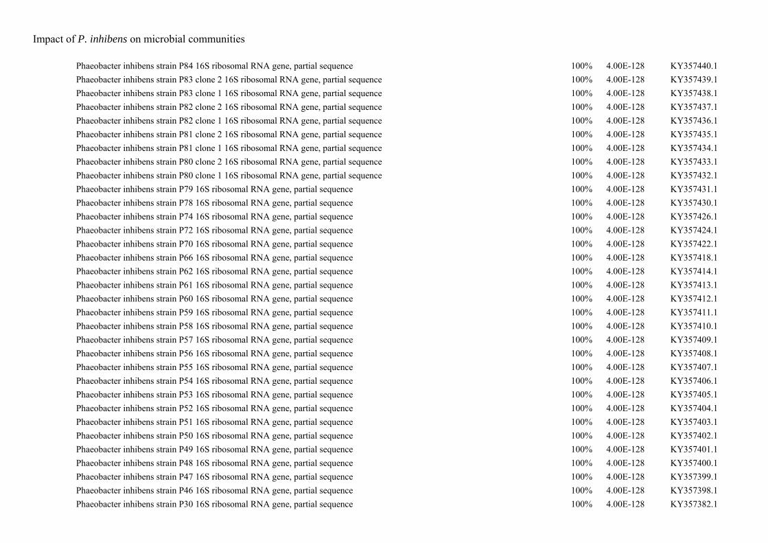
Impact of P. inhibens on microbial communities
Phaeobacter inhibens strain P84 16S ribosomal RNA gene, partial sequence 100% 4.00E-128 KY357440.1
Phaeobacter inhibens strain P83 clone 2 16S ribosomal RNA gene, partial sequence 100% 4.00E-128 KY357439.1
Phaeobacter inhibens strain P83 clone 1 16S ribosomal RNA gene, partial sequence 100% 4.00E-128 KY357438.1
Phaeobacter inhibens strain P82 clone 2 16S ribosomal RNA gene, partial sequence 100% 4.00E-128 KY357437.1
Phaeobacter inhibens strain P82 clone 1 16S ribosomal RNA gene, partial sequence 100% 4.00E-128 KY357436.1
Phaeobacter inhibens strain P81 clone 2 16S ribosomal RNA gene, partial sequence 100% 4.00E-128 KY357435.1
Phaeobacter inhibens strain P81 clone 1 16S ribosomal RNA gene, partial sequence 100% 4.00E-128 KY357434.1
Phaeobacter inhibens strain P80 clone 2 16S ribosomal RNA gene, partial sequence 100% 4.00E-128 KY357433.1
Phaeobacter inhibens strain P80 clone 1 16S ribosomal RNA gene, partial sequence 100% 4.00E-128 KY357432.1
Phaeobacter inhibens strain P79 16S ribosomal RNA gene, partial sequence 100% 4.00E-128 KY357431.1
Phaeobacter inhibens strain P78 16S ribosomal RNA gene, partial sequence 100% 4.00E-128 KY357430.1
Phaeobacter inhibens strain P74 16S ribosomal RNA gene, partial sequence 100% 4.00E-128 KY357426.1
Phaeobacter inhibens strain P72 16S ribosomal RNA gene, partial sequence 100% 4.00E-128 KY357424.1
Phaeobacter inhibens strain P70 16S ribosomal RNA gene, partial sequence 100% 4.00E-128 KY357422.1
Phaeobacter inhibens strain P66 16S ribosomal RNA gene, partial sequence 100% 4.00E-128 KY357418.1
Phaeobacter inhibens strain P62 16S ribosomal RNA gene, partial sequence 100% 4.00E-128 KY357414.1
Phaeobacter inhibens strain P61 16S ribosomal RNA gene, partial sequence 100% 4.00E-128 KY357413.1
Phaeobacter inhibens strain P60 16S ribosomal RNA gene, partial sequence 100% 4.00E-128 KY357412.1
Phaeobacter inhibens strain P59 16S ribosomal RNA gene, partial sequence 100% 4.00E-128 KY357411.1
Phaeobacter inhibens strain P58 16S ribosomal RNA gene, partial sequence 100% 4.00E-128 KY357410.1
Phaeobacter inhibens strain P57 16S ribosomal RNA gene, partial sequence 100% 4.00E-128 KY357409.1
Phaeobacter inhibens strain P56 16S ribosomal RNA gene, partial sequence 100% 4.00E-128 KY357408.1
Phaeobacter inhibens strain P55 16S ribosomal RNA gene, partial sequence 100% 4.00E-128 KY357407.1
Phaeobacter inhibens strain P54 16S ribosomal RNA gene, partial sequence 100% 4.00E-128 KY357406.1
Phaeobacter inhibens strain P53 16S ribosomal RNA gene, partial sequence 100% 4.00E-128 KY357405.1
Phaeobacter inhibens strain P52 16S ribosomal RNA gene, partial sequence 100% 4.00E-128 KY357404.1
Phaeobacter inhibens strain P51 16S ribosomal RNA gene, partial sequence 100% 4.00E-128 KY357403.1
Phaeobacter inhibens strain P50 16S ribosomal RNA gene, partial sequence 100% 4.00E-128 KY357402.1
Phaeobacter inhibens strain P49 16S ribosomal RNA gene, partial sequence 100% 4.00E-128 KY357401.1
Phaeobacter inhibens strain P48 16S ribosomal RNA gene, partial sequence 100% 4.00E-128 KY357400.1
Phaeobacter inhibens strain P47 16S ribosomal RNA gene, partial sequence 100% 4.00E-128 KY357399.1
Phaeobacter inhibens strain P46 16S ribosomal RNA gene, partial sequence 100% 4.00E-128 KY357398.1
Phaeobacter inhibens strain P30 16S ribosomal RNA gene, partial sequence 100% 4.00E-128 KY357382.1
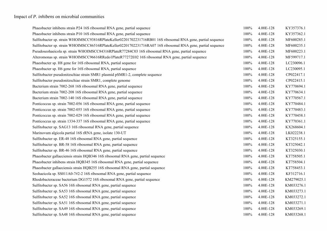
Impact of P. inhibens on microbial communities
Phaeobacter inhibens strain P24 16S ribosomal RNA gene, partial sequence 100% 4.00E-128 KY357376.1
Phaeobacter inhibens strain P10 16S ribosomal RNA gene, partial sequence 100% 4.00E-128 KY357362.1
Sulfitobacter sp. strain WHOIMSCC93816RPlateKeller02201702231716RB01 16S ribosomal RNA gene, partial sequence 100% 4.00E-128 MF600285.1
Sulfitobacter sp. strain WHOIMSCC86516RPlateKeller02201702231716RA07 16S ribosomal RNA gene, partial sequence 100% 4.00E-128 MF600235.1
Pseudoseohaeicola sp. strain WHOIMSCC84316RPlateR77284C03 16S ribosomal RNA gene, partial sequence 100% 4.00E-128 MF600223.1
Alteromonas sp. strain WHOIMSCC96616RRedo1PlateR77272E02 16S ribosomal RNA gene, partial sequence 100% 4.00E-128 MF599717.1
Phaeobacter sp. H8 gene for 16S ribosomal RNA, partial sequence 100% 4.00E-128 LC230096.1
Phaeobacter sp. H6 gene for 16S ribosomal RNA, partial sequence 100% 4.00E-128 LC230095.1
Sulfitobacter pseudonitzschiae strain SMR1 plasmid pSMR1-2, complete sequence 100% 4.00E-128 CP022417.1
Sulfitobacter pseudonitzschiae strain SMR1, complete genome 100% 4.00E-128 CP022415.1
Bacterium strain 7002-268 16S ribosomal RNA gene, partial sequence 100% 4.00E-128 KY770694.1
Bacterium strain 7002-208 16S ribosomal RNA gene, partial sequence 100% 4.00E-128 KY770634.1
Bacterium strain 7002-140 16S ribosomal RNA gene, partial sequence 100% 4.00E-128 KY770567.1
Ponticoccus sp. strain 7002-056 16S ribosomal RNA gene, partial sequence 100% 4.00E-128 KY770484.1
Ponticoccus sp. strain 7002-055 16S ribosomal RNA gene, partial sequence 100% 4.00E-128 KY770483.1
Ponticoccus sp. strain 7002-029 16S ribosomal RNA gene, partial sequence 100% 4.00E-128 KY770458.1
Ponticoccus sp. strain 1334-337 16S ribosomal RNA gene, partial sequence 100% 4.00E-128 KY770361.1
Sulfitobacter sp. SAG13 16S ribosomal RNA gene, partial sequence 100% 4.00E-128 KX268604.1
Marinovum algicola partial 16S rRNA gene, isolate 130-UT 100% 4.00E-128 LK022238.1
Sulfitobacter sp. ER-48 16S ribosomal RNA gene, partial sequence 100% 4.00E-128 KT325155.1
Sulfitobacter sp. BR-58 16S ribosomal RNA gene, partial sequence 100% 4.00E-128 KT325042.1
Sulfitobacter sp. BR-46 16S ribosomal RNA gene, partial sequence 100% 4.00E-128 KT325030.1
Phaeobacter gallaeciensis strain HQB346 16S ribosomal RNA gene, partial sequence 100% 4.00E-128 KT758505.1
Phaeobacter inhibens strain HQB345 16S ribosomal RNA gene, partial sequence 100% 4.00E-128 KT758504.1
Phaeobacter gallaeciensis strain HQB255 16S ribosomal RNA gene, partial sequence 100% 4.00E-128 KT758453.1
Seohaeicola sp. SS011A0-7#2-2 16S ribosomal RNA gene, partial sequence 100% 4.00E-128 KF312716.1
Rhodobacteraceae bacterium DG1572 16S ribosomal RNA gene, partial sequence 100% 4.00E-128 KM279025.1
Sulfitobacter sp. SA56 16S ribosomal RNA gene, partial sequence 100% 4.00E-128 KM033276.1
Sulfitobacter sp. SA53 16S ribosomal RNA gene, partial sequence 100% 4.00E-128 KM033273.1
Sulfitobacter sp. SA52 16S ribosomal RNA gene, partial sequence 100% 4.00E-128 KM033272.1
Sulfitobacter sp. SA51 16S ribosomal RNA gene, partial sequence 100% 4.00E-128 KM033271.1
Sulfitobacter sp. SA49 16S ribosomal RNA gene, partial sequence 100% 4.00E-128 KM033269.1
Sulfitobacter sp. SA48 16S ribosomal RNA gene, partial sequence 100% 4.00E-128 KM033268.1
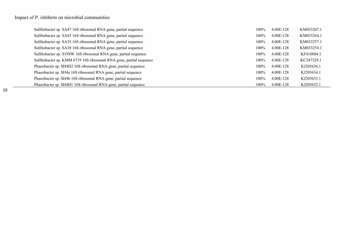
Impact of P. inhibens on microbial communities
Sulfitobacter sp. SA47 16S ribosomal RNA gene, partial sequence 100% 4.00E-128 KM033267.1
Sulfitobacter sp. SA43 16S ribosomal RNA gene, partial sequence 100% 4.00E-128 KM033264.1
Sulfitobacter sp. SA35 16S ribosomal RNA gene, partial sequence 100% 4.00E-128 KM033257.1
Sulfitobacter sp. SA30 16S ribosomal RNA gene, partial sequence 100% 4.00E-128 KM033254.1
Sulfitobacter sp. S19SW 16S ribosomal RNA gene, partial sequence 100% 4.00E-128 KF418804.1
Sulfitobacter sp. KMM 6719 16S ribosomal RNA gene, partial sequence 100% 4.00E-128 KC247329.1
Phaeobacter sp. SH4H2 16S ribosomal RNA gene, partial sequence 100% 4.00E-128 KJ205636.1
Phaeobacter sp. SH4a 16S ribosomal RNA gene, partial sequence 100% 4.00E-128 KJ205634.1
Phaeobacter sp. SH4b 16S ribosomal RNA gene, partial sequence 100% 4.00E-128 KJ205633.1
Phaeobacter sp. SH4H1 16S ribosomal RNA gene, partial sequence 100% 4.00E-128 KJ205632.1 10
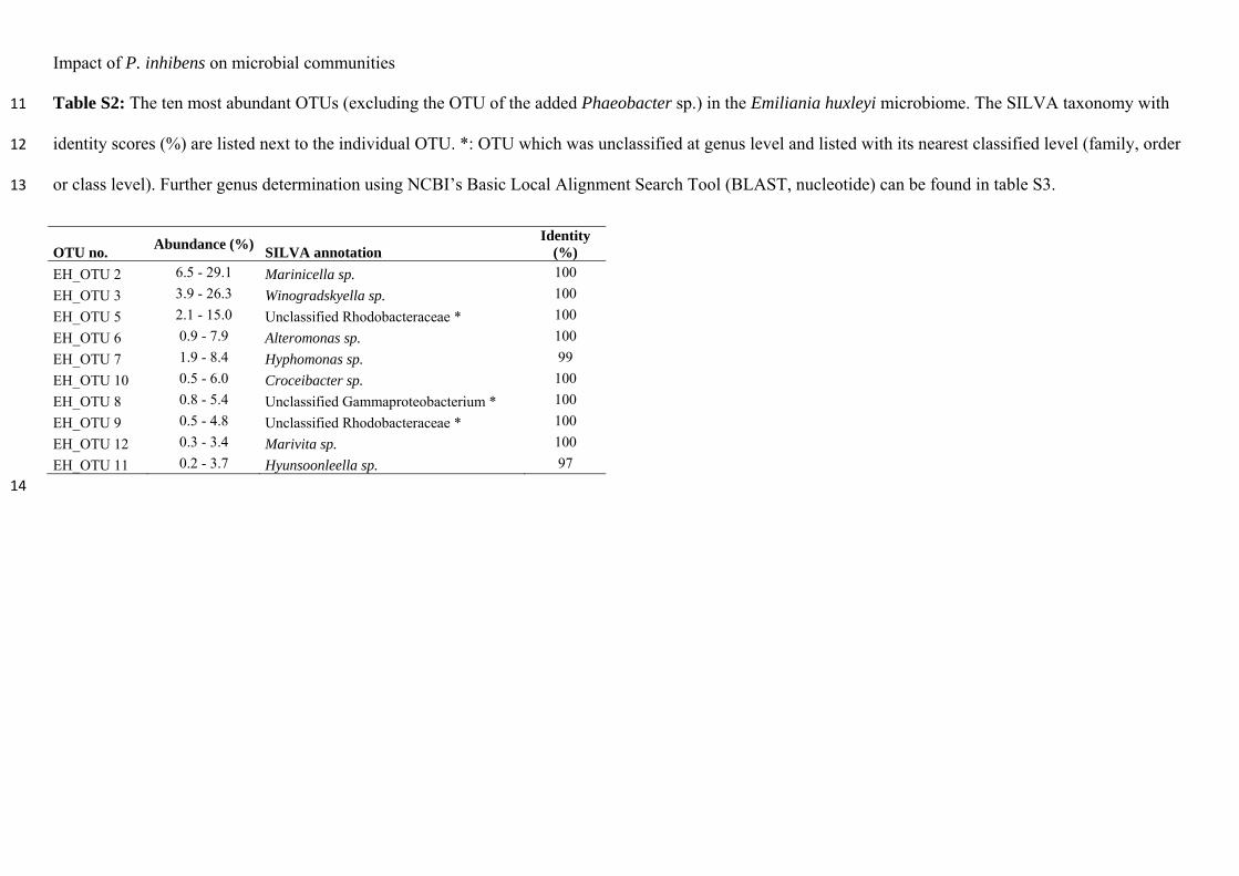
Impact of P. inhibens on microbial communities
Table S2: The ten most abundant OTUs (excluding the OTU of the added Phaeobacter sp.) in the Emiliania huxleyi microbiome. The SILVA taxonomy with 11
identity scores (%) are listed next to the individual OTU. *: OTU which was unclassified at genus level and listed with its nearest classified level (family, order 12
or class level). Further genus determination using NCBI’s Basic Local Alignment Search Tool (BLAST, nucleotide) can be found in table S3. 13
OTU no. Abundance (%)
SILVA annotation Identity
(%)
EH_OTU 2 6.5 - 29.1 Marinicella sp. 100
EH_OTU 3 3.9 - 26.3 Winogradskyella sp. 100
EH_OTU 5 2.1 - 15.0 Unclassified Rhodobacteraceae * 100
EH_OTU 6 0.9 - 7.9 Alteromonas sp. 100
EH_OTU 7 1.9 - 8.4 Hyphomonas sp. 99
EH_OTU 10 0.5 - 6.0 Croceibacter sp. 100
EH_OTU 8 0.8 - 5.4 Unclassified Gammaproteobacterium * 100
EH_OTU 9 0.5 - 4.8 Unclassified Rhodobacteraceae * 100
EH_OTU 12 0.3 - 3.4 Marivita sp. 100
EH_OTU 11 0.2 - 3.7 Hyunsoonleella sp. 97
14
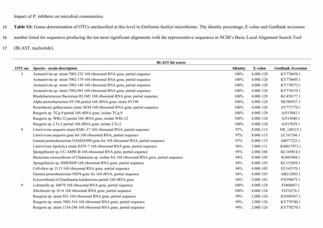
Impact of P. inhibens on microbial communities
Table S3: Genus determination of OTUs unclassified at this level in Emiliania huxleyi microbiome. The identity percentage, E-value and GenBank accession 15
number listed for sequences producing the ten most significant alignments with the representative sequences in NCBI’s Basic Local Alignment Search Tool 16
(BLAST, nucleotide). 17
OTU no.
BLAST hit scores
Species - strain description Identity E-value GenBank Accession
5 Aestuariivita sp. strain 7002-232 16S ribosomal RNA gene, partial sequence 100% 4.00E-128 KY770658.1
Aestuariivita sp. strain 7002-179 16S ribosomal RNA gene, partial sequence 100% 4.00E-128 KY770605.1
Aestuariivita sp. strain 7002-146 16S ribosomal RNA gene, partial sequence 100% 4.00E-128 KY770573.1
Aestuariivita sp. strain 7002-091 16S ribosomal RNA gene, partial sequence 100% 4.00E-128 KY770519.1
Rhodobacteraceae Bacterium R11M1 16S ribosomal RNA gene, partial sequence 100% 4.00E-128 KC439177.1
Alpha proteobacterium SY190 partial 16S rRNA gene, strain SY190 100% 4.00E-128 HE589557.1
Roseobacter gallaeciensis clone SE84 16S ribosomal RNA gene, partial sequence 100% 4.00E-128 AY771774.1
Ruegeria sp. TCg-9 partial 16S rRNA gene, isolate TCg-9 100% 4.00E-128 AJ515042.1
Ruegeria sp. WRs-12 partial 16S rRNA gene, isolate WRs-12 100% 4.00E-128 AJ515040.1
Ruegeria sp. LTs-2 partial 16S rRNA gene, isolate LTs-2 100% 4.00E-128 AJ515039.1
8 Litorivivens aequoris strain KMU-37 16S ribosomal RNA, partial sequence 97% 8.00E-115 NR_149215.1
Litorivivens aequoris gene for 16S ribosomal RNA, partial sequence 97% 8.00E-115 LC167346.1
Gamma proteobacterium NAMAF009 gene for 16S ribosomal RNA, partial sequence 97% 8.00E-115 AB377223.1
Litorivivens lipolytica strain HJTF-7 16S ribosomal RNA gene, partial sequence 96% 2.00E-111 KM017973.1
Spongiibacter sp. CC-AMW-B 16S ribosomal RNA gene, partial sequence 95% 2.00E-106 KC169814.1
Bacterium ectosymbiont of Cladonema sp. isolate SA 16S ribosomal RNA gene, partial sequence 94% 8.00E-105 KJ493944.1
Spongiibacter sp. HME8849 16S ribosomal RNA gene, partial sequence 94% 4.00E-103 KC153058.1
Cellvibrio sp. J113 16S ribosomal RNA gene, partial sequence 94% 4.00E-103 EU143370.1
Gamma proteobacterium NEP4 gene for 16S rRNA, partial sequence 94% 4.00E-103 AB212803.1
Ectosymbiont of Gianthauma karukerense partial 16S rRNA gene 94% 2.00E-101 FN398075.1
9 Loktanella sp. S4079 16S ribosomal RNA gene, partial sequence 100% 4.00E-128 FJ460047.1
Silicibacter sp. S1-6 16S ribosomal RNA gene, partial sequence 100% 4.00E-128 FJ218376.1
Ruegeria sp. strain S51 16S ribosomal RNA gene, partial sequence 99% 2.00E-126 KX989367.1
Ruegeria sp. strain 7002-314 16S ribosomal RNA gene, partial sequence 99% 2.00E-126 KY770740.1
Ruegeria sp. strain 1334-246 16S ribosomal RNA gene, partial sequence 99% 2.00E-126 KY770270.1
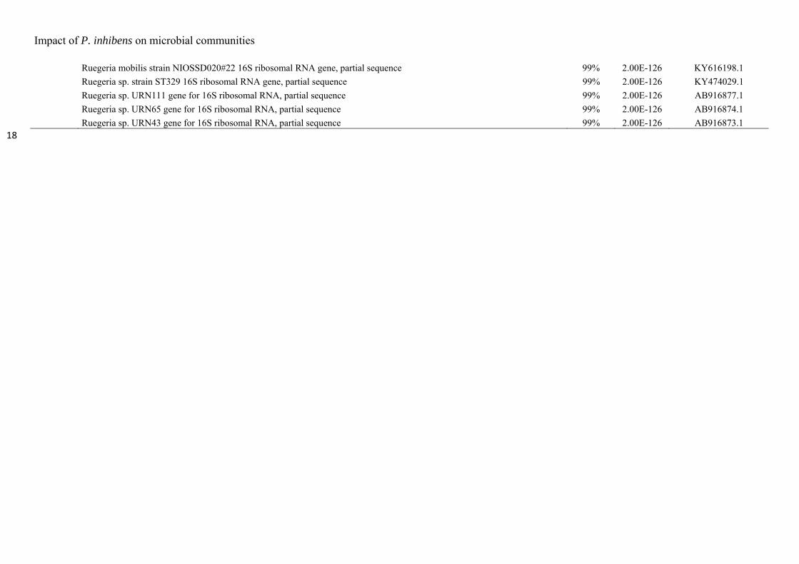
Impact of P. inhibens on microbial communities
Ruegeria mobilis strain NIOSSD020#22 16S ribosomal RNA gene, partial sequence 99% 2.00E-126 KY616198.1
Ruegeria sp. strain ST329 16S ribosomal RNA gene, partial sequence 99% 2.00E-126 KY474029.1
Ruegeria sp. URN111 gene for 16S ribosomal RNA, partial sequence 99% 2.00E-126 AB916877.1
Ruegeria sp. URN65 gene for 16S ribosomal RNA, partial sequence 99% 2.00E-126 AB916874.1
Ruegeria sp. URN43 gene for 16S ribosomal RNA, partial sequence 99% 2.00E-126 AB916873.1 18
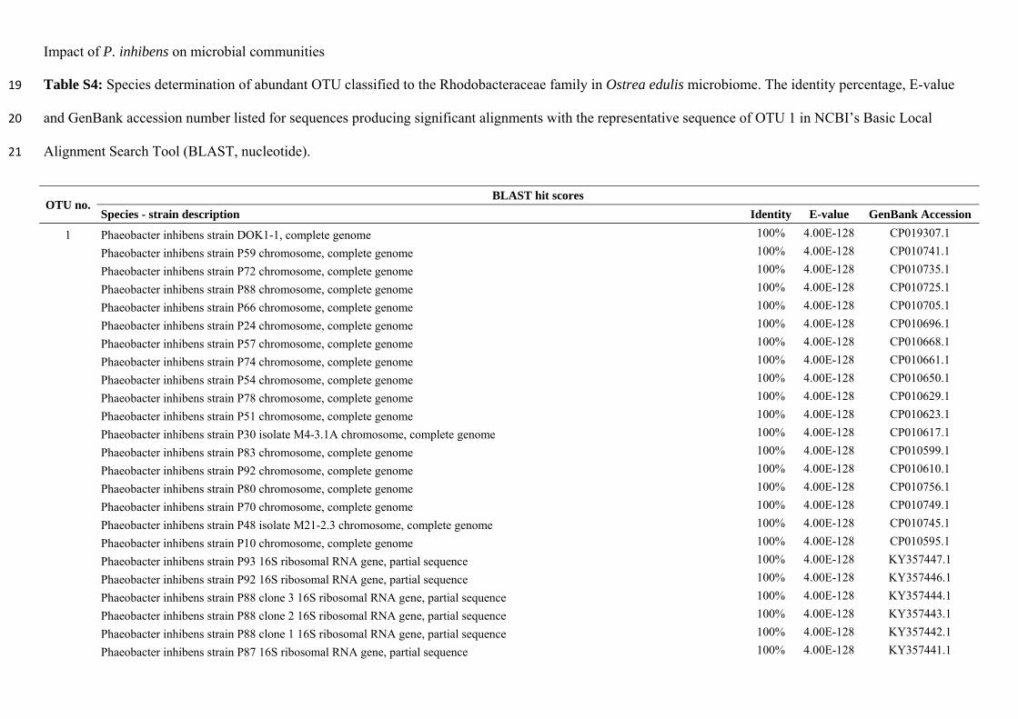
Impact of P. inhibens on microbial communities
Table S4: Species determination of abundant OTU classified to the Rhodobacteraceae family in Ostrea edulis microbiome. The identity percentage, E-value 19
and GenBank accession number listed for sequences producing significant alignments with the representative sequence of OTU 1 in NCBI’s Basic Local 20
Alignment Search Tool (BLAST, nucleotide). 21
OTU no. BLAST hit scores
Species - strain description Identity E-value GenBank Accession
1 Phaeobacter inhibens strain DOK1-1, complete genome 100% 4.00E-128 CP019307.1
Phaeobacter inhibens strain P59 chromosome, complete genome 100% 4.00E-128 CP010741.1
Phaeobacter inhibens strain P72 chromosome, complete genome 100% 4.00E-128 CP010735.1
Phaeobacter inhibens strain P88 chromosome, complete genome 100% 4.00E-128 CP010725.1
Phaeobacter inhibens strain P66 chromosome, complete genome 100% 4.00E-128 CP010705.1
Phaeobacter inhibens strain P24 chromosome, complete genome 100% 4.00E-128 CP010696.1
Phaeobacter inhibens strain P57 chromosome, complete genome 100% 4.00E-128 CP010668.1
Phaeobacter inhibens strain P74 chromosome, complete genome 100% 4.00E-128 CP010661.1
Phaeobacter inhibens strain P54 chromosome, complete genome 100% 4.00E-128 CP010650.1
Phaeobacter inhibens strain P78 chromosome, complete genome 100% 4.00E-128 CP010629.1
Phaeobacter inhibens strain P51 chromosome, complete genome 100% 4.00E-128 CP010623.1
Phaeobacter inhibens strain P30 isolate M4-3.1A chromosome, complete genome 100% 4.00E-128 CP010617.1
Phaeobacter inhibens strain P83 chromosome, complete genome 100% 4.00E-128 CP010599.1
Phaeobacter inhibens strain P92 chromosome, complete genome 100% 4.00E-128 CP010610.1
Phaeobacter inhibens strain P80 chromosome, complete genome 100% 4.00E-128 CP010756.1
Phaeobacter inhibens strain P70 chromosome, complete genome 100% 4.00E-128 CP010749.1
Phaeobacter inhibens strain P48 isolate M21-2.3 chromosome, complete genome 100% 4.00E-128 CP010745.1
Phaeobacter inhibens strain P10 chromosome, complete genome 100% 4.00E-128 CP010595.1
Phaeobacter inhibens strain P93 16S ribosomal RNA gene, partial sequence 100% 4.00E-128 KY357447.1
Phaeobacter inhibens strain P92 16S ribosomal RNA gene, partial sequence 100% 4.00E-128 KY357446.1
Phaeobacter inhibens strain P88 clone 3 16S ribosomal RNA gene, partial sequence 100% 4.00E-128 KY357444.1
Phaeobacter inhibens strain P88 clone 2 16S ribosomal RNA gene, partial sequence 100% 4.00E-128 KY357443.1
Phaeobacter inhibens strain P88 clone 1 16S ribosomal RNA gene, partial sequence 100% 4.00E-128 KY357442.1
Phaeobacter inhibens strain P87 16S ribosomal RNA gene, partial sequence 100% 4.00E-128 KY357441.1

Impact of P. inhibens on microbial communities
Phaeobacter inhibens strain P84 16S ribosomal RNA gene, partial sequence 100% 4.00E-128 KY357440.1
Phaeobacter inhibens strain P83 clone 2 16S ribosomal RNA gene, partial sequence 100% 4.00E-128 KY357439.1
Phaeobacter inhibens strain P83 clone 1 16S ribosomal RNA gene, partial sequence 100% 4.00E-128 KY357438.1
Phaeobacter inhibens strain P82 clone 2 16S ribosomal RNA gene, partial sequence 100% 4.00E-128 KY357437.1
Phaeobacter inhibens strain P82 clone 1 16S ribosomal RNA gene, partial sequence 100% 4.00E-128 KY357436.1
Phaeobacter inhibens strain P81 clone 2 16S ribosomal RNA gene, partial sequence 100% 4.00E-128 KY357435.1
Phaeobacter inhibens strain P81 clone 1 16S ribosomal RNA gene, partial sequence 100% 4.00E-128 KY357434.1
Phaeobacter inhibens strain P80 clone 2 16S ribosomal RNA gene, partial sequence 100% 4.00E-128 KY357433.1
Phaeobacter inhibens strain P80 clone 1 16S ribosomal RNA gene, partial sequence 100% 4.00E-128 KY357432.1
Phaeobacter inhibens strain P79 16S ribosomal RNA gene, partial sequence 100% 4.00E-128 KY357431.1
Phaeobacter inhibens strain P78 16S ribosomal RNA gene, partial sequence 100% 4.00E-128 KY357430.1
Phaeobacter inhibens strain P74 16S ribosomal RNA gene, partial sequence 100% 4.00E-128 KY357426.1
Phaeobacter inhibens strain P72 16S ribosomal RNA gene, partial sequence 100% 4.00E-128 KY357424.1
Phaeobacter inhibens strain P70 16S ribosomal RNA gene, partial sequence 100% 4.00E-128 KY357422.1
Phaeobacter inhibens strain P66 16S ribosomal RNA gene, partial sequence 100% 4.00E-128 KY357418.1
Phaeobacter inhibens strain P62 16S ribosomal RNA gene, partial sequence 100% 4.00E-128 KY357414.1
Phaeobacter inhibens strain P61 16S ribosomal RNA gene, partial sequence 100% 4.00E-128 KY357413.1
Phaeobacter inhibens strain P60 16S ribosomal RNA gene, partial sequence 100% 4.00E-128 KY357412.1
Phaeobacter inhibens strain P59 16S ribosomal RNA gene, partial sequence 100% 4.00E-128 KY357411.1
Phaeobacter inhibens strain P58 16S ribosomal RNA gene, partial sequence 100% 4.00E-128 KY357410.1
Phaeobacter inhibens strain P57 16S ribosomal RNA gene, partial sequence 100% 4.00E-128 KY357409.1
Phaeobacter inhibens strain P56 16S ribosomal RNA gene, partial sequence 100% 4.00E-128 KY357408.1
Phaeobacter inhibens strain P55 16S ribosomal RNA gene, partial sequence 100% 4.00E-128 KY357407.1
Phaeobacter inhibens strain P54 16S ribosomal RNA gene, partial sequence 100% 4.00E-128 KY357406.1
Phaeobacter inhibens strain P53 16S ribosomal RNA gene, partial sequence 100% 4.00E-128 KY357405.1
Phaeobacter inhibens strain P52 16S ribosomal RNA gene, partial sequence 100% 4.00E-128 KY357404.1
Phaeobacter inhibens strain P51 16S ribosomal RNA gene, partial sequence 100% 4.00E-128 KY357403.1
Phaeobacter inhibens strain P50 16S ribosomal RNA gene, partial sequence 100% 4.00E-128 KY357402.1
Phaeobacter inhibens strain P49 16S ribosomal RNA gene, partial sequence 100% 4.00E-128 KY357401.1
Phaeobacter inhibens strain P48 16S ribosomal RNA gene, partial sequence 100% 4.00E-128 KY357400.1
Phaeobacter inhibens strain P47 16S ribosomal RNA gene, partial sequence 100% 4.00E-128 KY357399.1
Phaeobacter inhibens strain P46 16S ribosomal RNA gene, partial sequence 100% 4.00E-128 KY357398.1
Phaeobacter inhibens strain P30 16S ribosomal RNA gene, partial sequence 100% 4.00E-128 KY357382.1
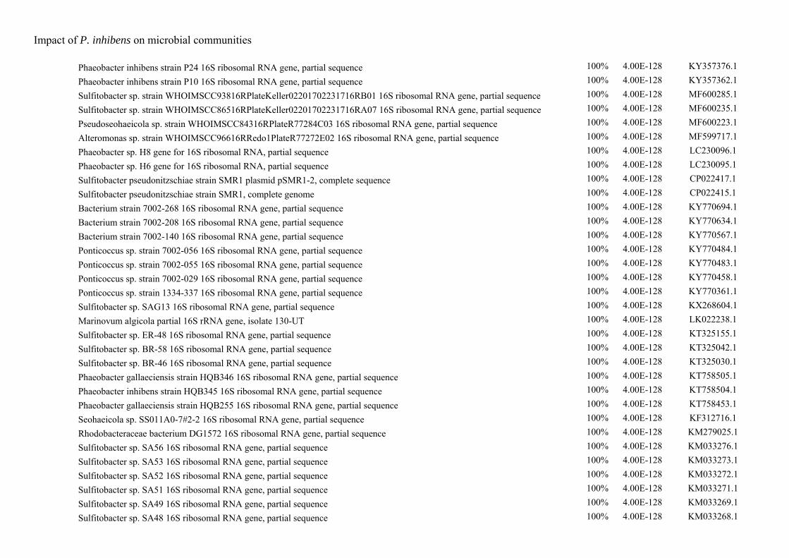
Impact of P. inhibens on microbial communities
Phaeobacter inhibens strain P24 16S ribosomal RNA gene, partial sequence 100% 4.00E-128 KY357376.1
Phaeobacter inhibens strain P10 16S ribosomal RNA gene, partial sequence 100% 4.00E-128 KY357362.1
Sulfitobacter sp. strain WHOIMSCC93816RPlateKeller02201702231716RB01 16S ribosomal RNA gene, partial sequence 100% 4.00E-128 MF600285.1
Sulfitobacter sp. strain WHOIMSCC86516RPlateKeller02201702231716RA07 16S ribosomal RNA gene, partial sequence 100% 4.00E-128 MF600235.1
Pseudoseohaeicola sp. strain WHOIMSCC84316RPlateR77284C03 16S ribosomal RNA gene, partial sequence 100% 4.00E-128 MF600223.1
Alteromonas sp. strain WHOIMSCC96616RRedo1PlateR77272E02 16S ribosomal RNA gene, partial sequence 100% 4.00E-128 MF599717.1
Phaeobacter sp. H8 gene for 16S ribosomal RNA, partial sequence 100% 4.00E-128 LC230096.1
Phaeobacter sp. H6 gene for 16S ribosomal RNA, partial sequence 100% 4.00E-128 LC230095.1
Sulfitobacter pseudonitzschiae strain SMR1 plasmid pSMR1-2, complete sequence 100% 4.00E-128 CP022417.1
Sulfitobacter pseudonitzschiae strain SMR1, complete genome 100% 4.00E-128 CP022415.1
Bacterium strain 7002-268 16S ribosomal RNA gene, partial sequence 100% 4.00E-128 KY770694.1
Bacterium strain 7002-208 16S ribosomal RNA gene, partial sequence 100% 4.00E-128 KY770634.1
Bacterium strain 7002-140 16S ribosomal RNA gene, partial sequence 100% 4.00E-128 KY770567.1
Ponticoccus sp. strain 7002-056 16S ribosomal RNA gene, partial sequence 100% 4.00E-128 KY770484.1
Ponticoccus sp. strain 7002-055 16S ribosomal RNA gene, partial sequence 100% 4.00E-128 KY770483.1
Ponticoccus sp. strain 7002-029 16S ribosomal RNA gene, partial sequence 100% 4.00E-128 KY770458.1
Ponticoccus sp. strain 1334-337 16S ribosomal RNA gene, partial sequence 100% 4.00E-128 KY770361.1
Sulfitobacter sp. SAG13 16S ribosomal RNA gene, partial sequence 100% 4.00E-128 KX268604.1
Marinovum algicola partial 16S rRNA gene, isolate 130-UT 100% 4.00E-128 LK022238.1
Sulfitobacter sp. ER-48 16S ribosomal RNA gene, partial sequence 100% 4.00E-128 KT325155.1
Sulfitobacter sp. BR-58 16S ribosomal RNA gene, partial sequence 100% 4.00E-128 KT325042.1
Sulfitobacter sp. BR-46 16S ribosomal RNA gene, partial sequence 100% 4.00E-128 KT325030.1
Phaeobacter gallaeciensis strain HQB346 16S ribosomal RNA gene, partial sequence 100% 4.00E-128 KT758505.1
Phaeobacter inhibens strain HQB345 16S ribosomal RNA gene, partial sequence 100% 4.00E-128 KT758504.1
Phaeobacter gallaeciensis strain HQB255 16S ribosomal RNA gene, partial sequence 100% 4.00E-128 KT758453.1
Seohaeicola sp. SS011A0-7#2-2 16S ribosomal RNA gene, partial sequence 100% 4.00E-128 KF312716.1
Rhodobacteraceae bacterium DG1572 16S ribosomal RNA gene, partial sequence 100% 4.00E-128 KM279025.1
Sulfitobacter sp. SA56 16S ribosomal RNA gene, partial sequence 100% 4.00E-128 KM033276.1
Sulfitobacter sp. SA53 16S ribosomal RNA gene, partial sequence 100% 4.00E-128 KM033273.1
Sulfitobacter sp. SA52 16S ribosomal RNA gene, partial sequence 100% 4.00E-128 KM033272.1
Sulfitobacter sp. SA51 16S ribosomal RNA gene, partial sequence 100% 4.00E-128 KM033271.1
Sulfitobacter sp. SA49 16S ribosomal RNA gene, partial sequence 100% 4.00E-128 KM033269.1
Sulfitobacter sp. SA48 16S ribosomal RNA gene, partial sequence 100% 4.00E-128 KM033268.1
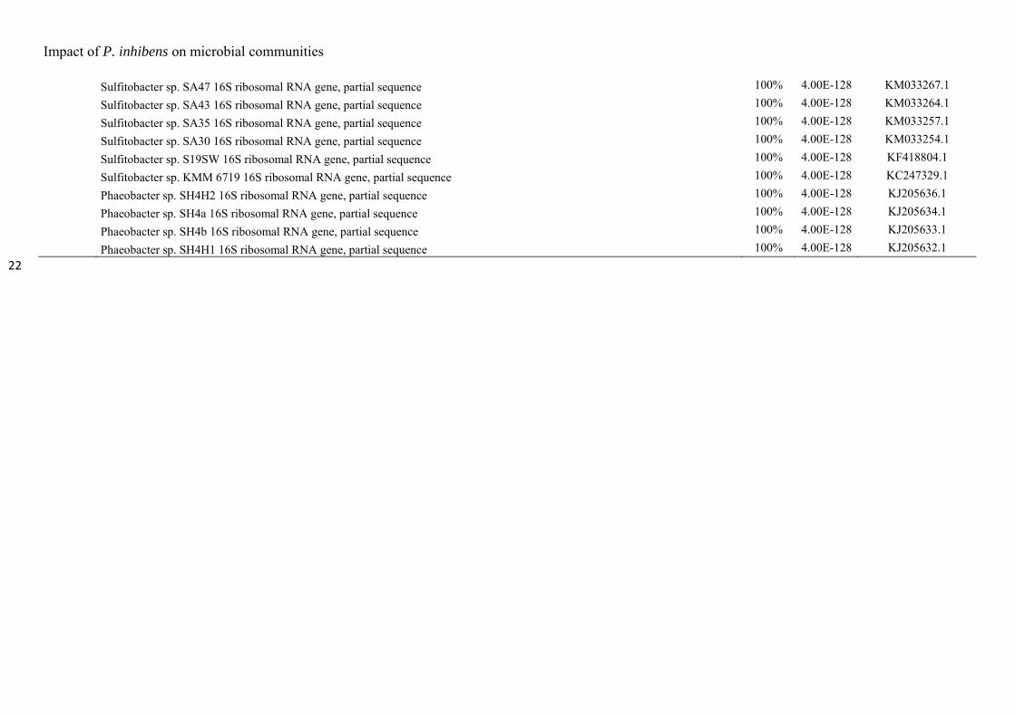
Impact of P. inhibens on microbial communities
Sulfitobacter sp. SA47 16S ribosomal RNA gene, partial sequence 100% 4.00E-128 KM033267.1
Sulfitobacter sp. SA43 16S ribosomal RNA gene, partial sequence 100% 4.00E-128 KM033264.1
Sulfitobacter sp. SA35 16S ribosomal RNA gene, partial sequence 100% 4.00E-128 KM033257.1
Sulfitobacter sp. SA30 16S ribosomal RNA gene, partial sequence 100% 4.00E-128 KM033254.1
Sulfitobacter sp. S19SW 16S ribosomal RNA gene, partial sequence 100% 4.00E-128 KF418804.1
Sulfitobacter sp. KMM 6719 16S ribosomal RNA gene, partial sequence 100% 4.00E-128 KC247329.1
Phaeobacter sp. SH4H2 16S ribosomal RNA gene, partial sequence 100% 4.00E-128 KJ205636.1
Phaeobacter sp. SH4a 16S ribosomal RNA gene, partial sequence 100% 4.00E-128 KJ205634.1
Phaeobacter sp. SH4b 16S ribosomal RNA gene, partial sequence 100% 4.00E-128 KJ205633.1
Phaeobacter sp. SH4H1 16S ribosomal RNA gene, partial sequence 100% 4.00E-128 KJ205632.1
22
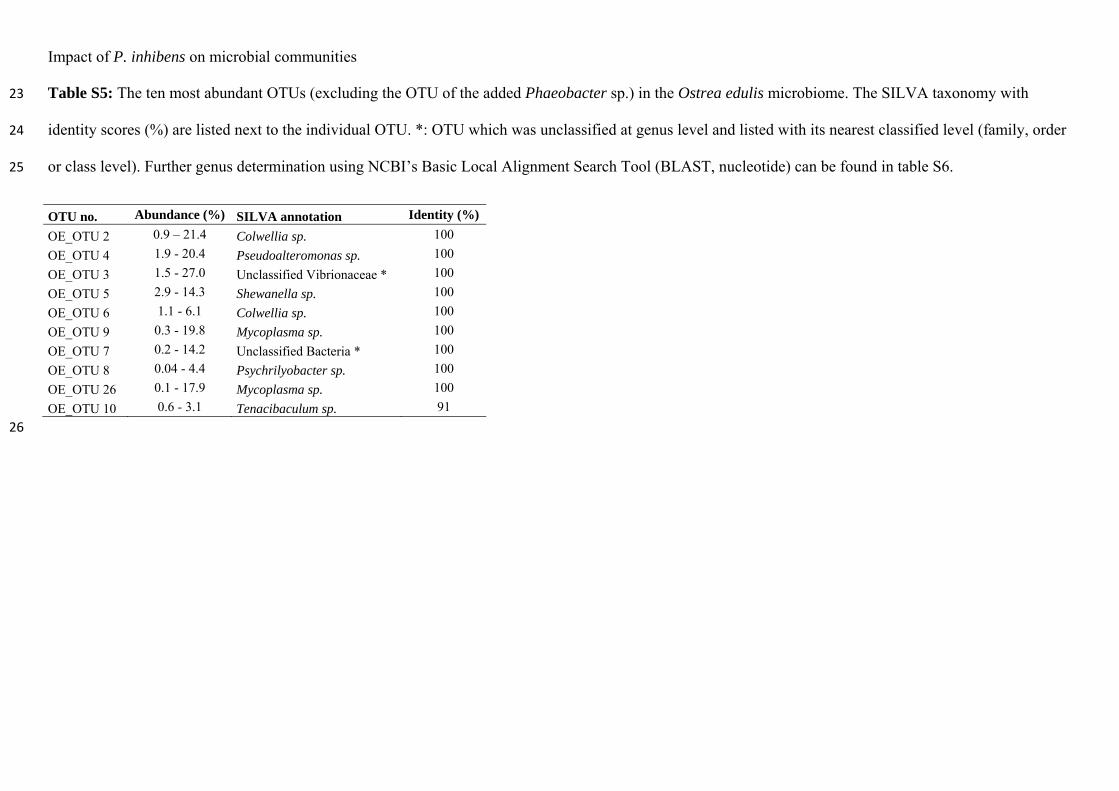
Impact of P. inhibens on microbial communities
Table S5: The ten most abundant OTUs (excluding the OTU of the added Phaeobacter sp.) in the Ostrea edulis microbiome. The SILVA taxonomy with 23
identity scores (%) are listed next to the individual OTU. *: OTU which was unclassified at genus level and listed with its nearest classified level (family, order 24
or class level). Further genus determination using NCBI’s Basic Local Alignment Search Tool (BLAST, nucleotide) can be found in table S6. 25
OTU no. Abundance (%) SILVA annotation Identity (%)
OE_OTU 2 0.9 – 21.4 Colwellia sp. 100
OE_OTU 4 1.9 - 20.4 Pseudoalteromonas sp. 100
OE_OTU 3 1.5 - 27.0 Unclassified Vibrionaceae * 100
OE_OTU 5 2.9 - 14.3 Shewanella sp. 100
OE_OTU 6 1.1 - 6.1 Colwellia sp. 100
OE_OTU 9 0.3 - 19.8 Mycoplasma sp. 100
OE_OTU 7 0.2 - 14.2 Unclassified Bacteria * 100
OE_OTU 8 0.04 - 4.4 Psychrilyobacter sp. 100
OE_OTU 26 0.1 - 17.9 Mycoplasma sp. 100
OE_OTU 10 0.6 - 3.1 Tenacibaculum sp. 91
26
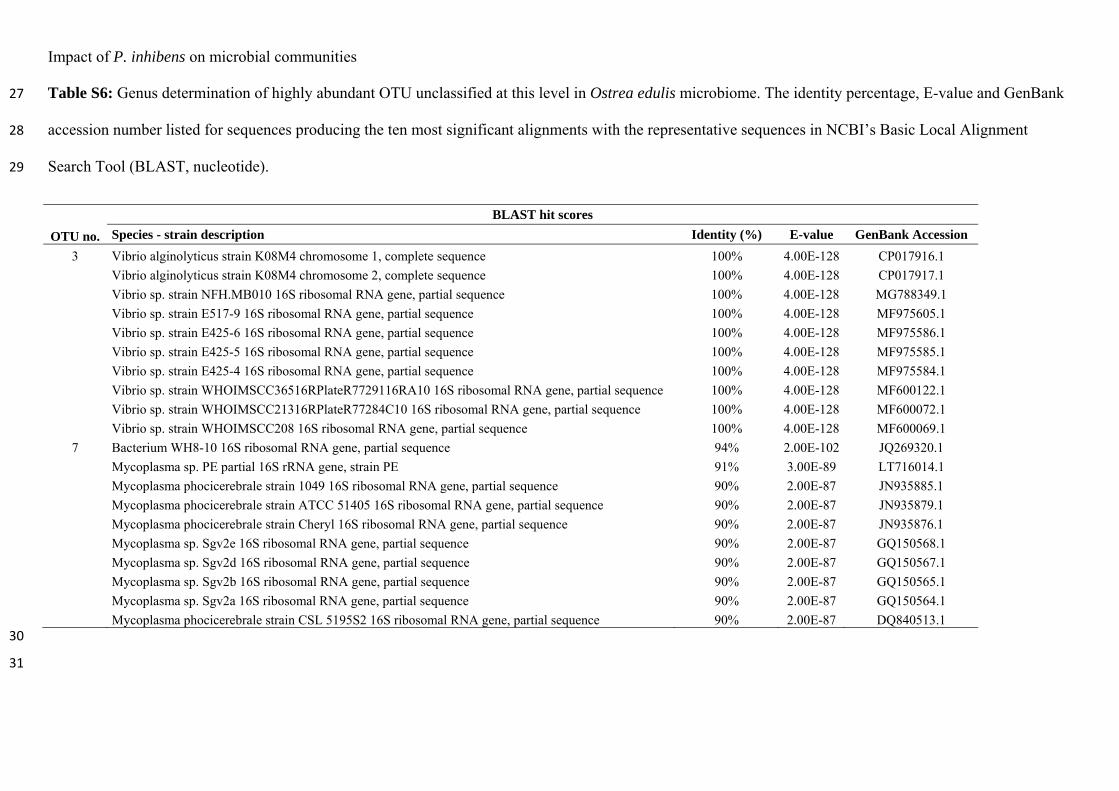
Impact of P. inhibens on microbial communities
Table S6: Genus determination of highly abundant OTU unclassified at this level in Ostrea edulis microbiome. The identity percentage, E-value and GenBank 27
accession number listed for sequences producing the ten most significant alignments with the representative sequences in NCBI’s Basic Local Alignment 28
Search Tool (BLAST, nucleotide). 29
OTU no.
BLAST hit scores
Species - strain description Identity (%) E-value GenBank Accession
3 Vibrio alginolyticus strain K08M4 chromosome 1, complete sequence 100% 4.00E-128 CP017916.1
Vibrio alginolyticus strain K08M4 chromosome 2, complete sequence 100% 4.00E-128 CP017917.1
Vibrio sp. strain NFH.MB010 16S ribosomal RNA gene, partial sequence 100% 4.00E-128 MG788349.1
Vibrio sp. strain E517-9 16S ribosomal RNA gene, partial sequence 100% 4.00E-128 MF975605.1
Vibrio sp. strain E425-6 16S ribosomal RNA gene, partial sequence 100% 4.00E-128 MF975586.1
Vibrio sp. strain E425-5 16S ribosomal RNA gene, partial sequence 100% 4.00E-128 MF975585.1
Vibrio sp. strain E425-4 16S ribosomal RNA gene, partial sequence 100% 4.00E-128 MF975584.1
Vibrio sp. strain WHOIMSCC36516RPlateR7729116RA10 16S ribosomal RNA gene, partial sequence 100% 4.00E-128 MF600122.1
Vibrio sp. strain WHOIMSCC21316RPlateR77284C10 16S ribosomal RNA gene, partial sequence 100% 4.00E-128 MF600072.1
Vibrio sp. strain WHOIMSCC208 16S ribosomal RNA gene, partial sequence 100% 4.00E-128 MF600069.1
7 Bacterium WH8-10 16S ribosomal RNA gene, partial sequence 94% 2.00E-102 JQ269320.1
Mycoplasma sp. PE partial 16S rRNA gene, strain PE 91% 3.00E-89 LT716014.1
Mycoplasma phocicerebrale strain 1049 16S ribosomal RNA gene, partial sequence 90% 2.00E-87 JN935885.1
Mycoplasma phocicerebrale strain ATCC 51405 16S ribosomal RNA gene, partial sequence 90% 2.00E-87 JN935879.1
Mycoplasma phocicerebrale strain Cheryl 16S ribosomal RNA gene, partial sequence 90% 2.00E-87 JN935876.1
Mycoplasma sp. Sgv2e 16S ribosomal RNA gene, partial sequence 90% 2.00E-87 GQ150568.1
Mycoplasma sp. Sgv2d 16S ribosomal RNA gene, partial sequence 90% 2.00E-87 GQ150567.1
Mycoplasma sp. Sgv2b 16S ribosomal RNA gene, partial sequence 90% 2.00E-87 GQ150565.1
Mycoplasma sp. Sgv2a 16S ribosomal RNA gene, partial sequence 90% 2.00E-87 GQ150564.1
Mycoplasma phocicerebrale strain CSL 5195S2 16S ribosomal RNA gene, partial sequence 90% 2.00E-87 DQ840513.1 30
31
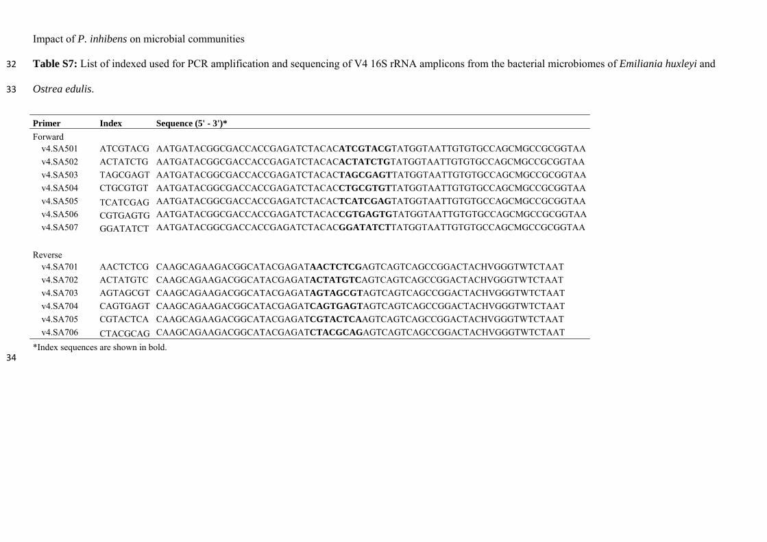
Impact of P. inhibens on microbial communities
Table S7: List of indexed used for PCR amplification and sequencing of V4 16S rRNA amplicons from the bacterial microbiomes of Emiliania huxleyi and 32
Ostrea edulis. 33
Primer Index Sequence (5' - 3')*
Forward v4.SA501 ATCGTACG AATGATACGGCGACCACCGAGATCTACACATCGTACGTATGGTAATTGTGTGCCAGCMGCCGCGGTAA
v4.SA502 ACTATCTG AATGATACGGCGACCACCGAGATCTACACACTATCTGTATGGTAATTGTGTGCCAGCMGCCGCGGTAA
v4.SA503 TAGCGAGT AATGATACGGCGACCACCGAGATCTACACTAGCGAGTTATGGTAATTGTGTGCCAGCMGCCGCGGTAA
v4.SA504 CTGCGTGT AATGATACGGCGACCACCGAGATCTACACCTGCGTGTTATGGTAATTGTGTGCCAGCMGCCGCGGTAA
v4.SA505 TCATCGAG AATGATACGGCGACCACCGAGATCTACACTCATCGAGTATGGTAATTGTGTGCCAGCMGCCGCGGTAA
v4.SA506 CGTGAGTG AATGATACGGCGACCACCGAGATCTACACCGTGAGTGTATGGTAATTGTGTGCCAGCMGCCGCGGTAA
v4.SA507 GGATATCT AATGATACGGCGACCACCGAGATCTACACGGATATCTTATGGTAATTGTGTGCCAGCMGCCGCGGTAA
Reverse v4.SA701 AACTCTCG CAAGCAGAAGACGGCATACGAGATAACTCTCGAGTCAGTCAGCCGGACTACHVGGGTWTCTAAT
v4.SA702 ACTATGTC CAAGCAGAAGACGGCATACGAGATACTATGTCAGTCAGTCAGCCGGACTACHVGGGTWTCTAAT
v4.SA703 AGTAGCGT CAAGCAGAAGACGGCATACGAGATAGTAGCGTAGTCAGTCAGCCGGACTACHVGGGTWTCTAAT
v4.SA704 CAGTGAGT CAAGCAGAAGACGGCATACGAGATCAGTGAGTAGTCAGTCAGCCGGACTACHVGGGTWTCTAAT
v4.SA705 CGTACTCA CAAGCAGAAGACGGCATACGAGATCGTACTCAAGTCAGTCAGCCGGACTACHVGGGTWTCTAAT
v4.SA706 CTACGCAG CAAGCAGAAGACGGCATACGAGATCTACGCAGAGTCAGTCAGCCGGACTACHVGGGTWTCTAAT
*Index sequences are shown in bold. 34
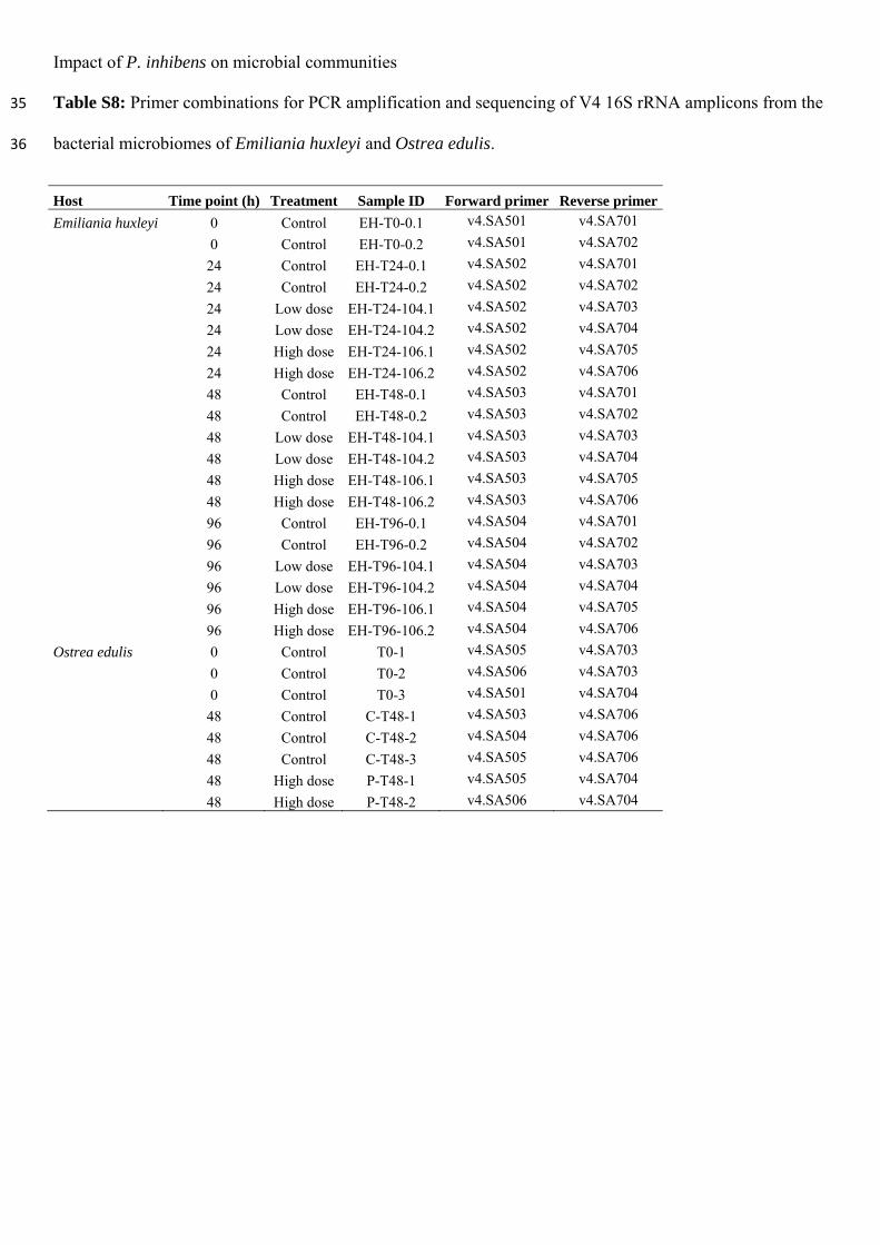
Impact of P. inhibens on microbial communities
Table S8: Primer combinations for PCR amplification and sequencing of V4 16S rRNA amplicons from the 35
bacterial microbiomes of Emiliania huxleyi and Ostrea edulis. 36
Host Time point (h) Treatment Sample ID Forward primer Reverse primer
Emiliania huxleyi 0 Control EH-T0-0.1 v4.SA501 v4.SA701
0 Control EH-T0-0.2 v4.SA501 v4.SA702
24 Control EH-T24-0.1 v4.SA502 v4.SA701
24 Control EH-T24-0.2 v4.SA502 v4.SA702
24 Low dose EH-T24-104.1 v4.SA502 v4.SA703
24 Low dose EH-T24-104.2 v4.SA502 v4.SA704
24 High dose EH-T24-106.1 v4.SA502 v4.SA705
24 High dose EH-T24-106.2 v4.SA502 v4.SA706
48 Control EH-T48-0.1 v4.SA503 v4.SA701
48 Control EH-T48-0.2 v4.SA503 v4.SA702
48 Low dose EH-T48-104.1 v4.SA503 v4.SA703
48 Low dose EH-T48-104.2 v4.SA503 v4.SA704
48 High dose EH-T48-106.1 v4.SA503 v4.SA705
48 High dose EH-T48-106.2 v4.SA503 v4.SA706
96 Control EH-T96-0.1 v4.SA504 v4.SA701
96 Control EH-T96-0.2 v4.SA504 v4.SA702
96 Low dose EH-T96-104.1 v4.SA504 v4.SA703
96 Low dose EH-T96-104.2 v4.SA504 v4.SA704
96 High dose EH-T96-106.1 v4.SA504 v4.SA705
96 High dose EH-T96-106.2 v4.SA504 v4.SA706
Ostrea edulis 0 Control T0-1 v4.SA505 v4.SA703
0 Control T0-2 v4.SA506 v4.SA703
0 Control T0-3 v4.SA501 v4.SA704
48 Control C-T48-1 v4.SA503 v4.SA706
48 Control C-T48-2 v4.SA504 v4.SA706
48 Control C-T48-3 v4.SA505 v4.SA706
48 High dose P-T48-1 v4.SA505 v4.SA704
48 High dose P-T48-2 v4.SA506 v4.SA704
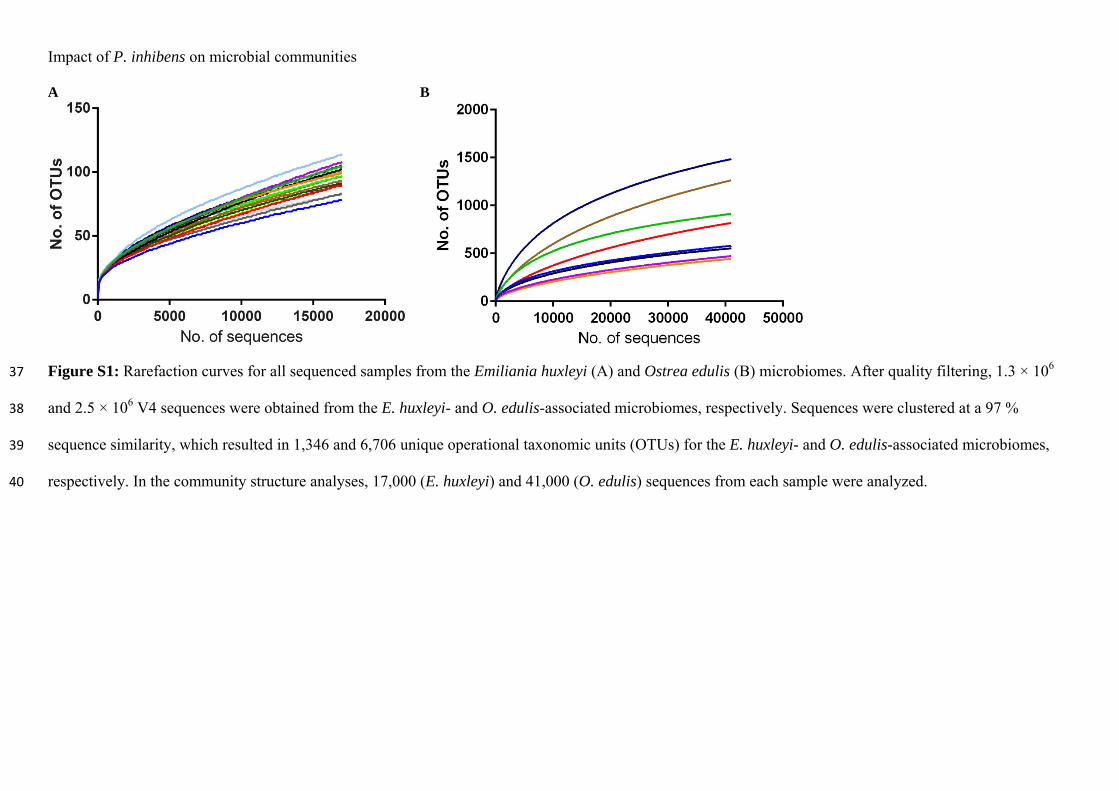
Impact of P. inhibens on microbial communities A B
Figure S1: Rarefaction curves for all sequenced samples from the Emiliania huxleyi (A) and Ostrea edulis (B) microbiomes. After quality filtering, 1.3 × 10⁶ 37
and 2.5 × 10⁶ V4 sequences were obtained from the E. huxleyi- and O. edulis-associated microbiomes, respectively. Sequences were clustered at a 97 % 38
sequence similarity, which resulted in 1,346 and 6,706 unique operational taxonomic units (OTUs) for the E. huxleyi- and O. edulis-associated microbiomes, 39
respectively. In the community structure analyses, 17,000 (E. huxleyi) and 41,000 (O. edulis) sequences from each sample were analyzed. 40
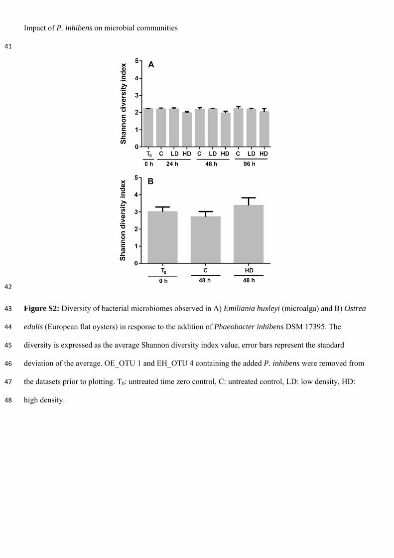
Impact of P. inhibens on microbial communities
41
42
Figure S2: Diversity of bacterial microbiomes observed in A) Emiliania huxleyi (microalga) and B) Ostrea 43
edulis (European flat oysters) in response to the addition of Phaeobacter inhibens DSM 17395. The 44
diversity is expressed as the average Shannon diversity index value, error bars represent the standard 45
deviation of the average. OE_OTU 1 and EH_OTU 4 containing the added P. inhibens were removed from 46
the datasets prior to plotting. T0: untreated time zero control, C: untreated control, LD: low density, HD: 47
high density. 48
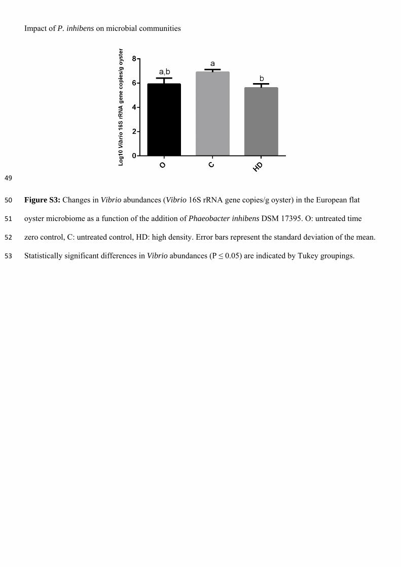
Impact of P. inhibens on microbial communities
49
Figure S3: Changes in Vibrio abundances (Vibrio 16S rRNA gene copies/g oyster) in the European flat 50
oyster microbiome as a function of the addition of Phaeobacter inhibens DSM 17395. O: untreated time 51
zero control, C: untreated control, HD: high density. Error bars represent the standard deviation of the mean. 52
Statistically significant differences in Vibrio abundances (P ≤ 0.05) are indicated by Tukey groupings. 53
Paper 3
Dittmann, K.K., Porsby, C.H., Goncalves, P., Mateiu, R.V., Sonnenschein, E.C., Bentzon-Tilia, M., Egan, S. & Gram, L. (2019).
Tropodithietic acid induces oxidative stress response, cell envelope biogenesis and iron uptake in Vibrio vulnificus.
Environ Microbiol Rep. Accepted.
https://doi.org/10.1111/1758-2229.12771
Tropodithietic acid induces oxidative stress response, cell envelope
biogenesis and iron uptake in Vibrio vulnificus
Karen K. Dittmanna,b, Cisse H. Porsbya*, Priscila Goncalvesb, Ramona Valentina Mateiuc,
Eva C. Sonnenscheina, Mikkel Bentzon-Tiliaa, Suhelen Eganb & Lone Grama#
a Department of Biotechnology and Biomedicine, Technical University of Denmark, DK-
2800 Kgs. Lyngby, Denmark.
b School of Biological, Earth and Environmental Sciences, The University of New South
Wales, Randwick NSW 2052, Australia.
c Center for Electron Nanoscopy, Technical University of Denmark, DK-2800 Kgs. Lyngby,
Denmark.
# Corresponding author: Lone Gram, Department of Biotechnology and Biomedicine,
Technical University of Denmark, Søltofts Plads Bldg. 221, DK-2800 Kgs. Lyngby,
Denmark, Tel.: (+45) 45 25 25 86, Fax: (+45) 45 25 25 01, [email protected],
*Current address: Chr. Hansen A/S, DK-2970 Hørsholm, Denmark.
This article has been accepted for publication and undergone full peer review but has not been through the copyediting, typesetting, pagination and proofreading process which may lead to differences between this version and the Version of Record. Please cite this article as doi: 10.1111/1758-2229.12771
This article is protected by copyright. All rights reserved.
Impact of TDA on gene expression of V. vulnificus
Running title: Impact of TDA on gene expression of V. vulnificus
Keywords: Transcriptomics, marine bacteria, biofilm, roseobacters, secondary metabolites,
tropodithietic acid, TDA, motility
Originality-Significance Statement
The natural role of the antibiotic tropodithietic acid (TDA) and its impact on non-producing
marine bacteria at in situ concentrations is currently unknown. Here we determine how a sub-
lethal concentration of TDA affects the opportunistic fish and human pathogen Vibrio
vulnificus, providing insight into the mechanism of action of TDA and its potential effects on
both pathogenic and commensal bacteria.
Abstract
The Roseobacter group is a widespread marine bacterial group, of which some species
produce the broad-spectrum antibiotic tropodithietic acid (TDA). A mode of action for TDA
has previously been proposed in Escherichia coli, but little is known about its effect on non-
producing marine bacteria at in situ concentrations. The purpose of this study was to
investigate how a sub-lethal level of TDA affects Vibrio vulnificus at different time points (30
min and 60 min) using a transcriptomic approach. Exposure to TDA for as little as 30 min
resulted in the differential expression of genes associated with cell regeneration, including the
up-regulation of those involved in biogenesis of the cell envelope. Defense mechanisms
including oxidative stress defense proteins and iron uptake systems were also up-regulated in
response to TDA, while motility-related genes were down-regulated. Gene expression data
and scanning electron microscopy imaging revealed a switch to a biofilm phenotype in the
Page of 31
This article is protected by copyright. All rights reserved.
Impact of TDA on gene expression of V. vulnificus
presence of TDA. Our study shows that a low concentration of this antibiotic triggers a
defense response to reactive oxygen species and iron depletion in V. vulnificus, which
indicates that the mode of action of TDA is likely more complex in this bacterium than what
is known for E. coli.
Page of 31
This article is protected by copyright. All rights reserved.
Impact of TDA on gene expression of V. vulnificus
Introduction
The Roseobacter group is one of the most widespread marine bacterial groups and is often
associated with eukaryotes such as algae (González et al., 2000; Buchan et al., 2005;
Sonnenschein et al., 2017; Dittmann et al., 2018) and molluscs (Ruiz-Ponte et al., 1998;
Prado et al., 2009; Wegner et al., 2013). Members of this group have also been repeatedly
detected in aquaculture systems (Hjelm, Riaza, et al., 2004; Porsby et al., 2008). Some
species, including Phaeobacter inhibens and Ruegeria mobilis, produce the potent
antibacterial compound tropodithietic acid (TDA) (Bruhn et al., 2005; Porsby et al., 2008),
which is active against a range of Gram-positive and Gram-negative bacteria (Kintaka et al.,
1984; Porsby et al., 2011; Rabe et al., 2014). TDA has low toxicity to the roundworm
Caenorhabditis elegans (Neu et al., 2014), which is often used as a model organism for
testing cytotoxicity (Sese et al., 2009; Sprando et al., 2009) resembling the effects induced in
mammalian model organisms (Sprando et al., 2009). However, TDA has also shown
anticancer activity (Wilson et al., 2016) and the compound can be cytotoxic to mammalian
neuronal cells (Wichmann et al., 2015), which indicates that the effect of TDA on eukaryotic
cells and eukaryotes is dependent on the cell type and target organism. The activity towards
neuronal cells is believed to be driven by disruption of the mitochondrial membrane potential
and activation of oxidative stress response (Wichmann et al., 2015). Altogether, the broad
target range and toxicity towards mammalian cells should be taken into consideration if TDA
and TDA-producers are to be used as treatment in humans as well as in rearing of fish and
shellfish.
Page of 31
This article is protected by copyright. All rights reserved.
Impact of TDA on gene expression of V. vulnificus
TDA-producing bacteria have potential as probiotics in aquaculture systems as
they can prevent fish larvae mortality caused by pathogenic Vibrio spp. (Planas et al., 2006a;
D’Alvise et al., 2013) without causing adverse effects on either the larvae (Hjelm, Bergh, et
al., 2004; Planas et al., 2006b; D’Alvise et al., 2010, 2012) or the live-feed organisms such as
Artemia (Neu et al., 2014; Grotkjær et al., 2016), copepods (Rasmussen et al., 2018), rotifers,
and microalgae (D’Alvise et al., 2012). The prospect of using antibiotic-producing bacteria in
aquaculture raises the concern of resistance development in target organisms. However,
previous studies have shown that human and fish pathogens are genetically and
phenotypically unaffected by long-term exposure to TDA (Porsby et al., 2011; Rasmussen et
al., 2016). Although TDA-tolerant bacteria have been co-isolated with TDA-producing
Pseudovibrio spp. from marine sponges (Harrington et al., 2014), their
tolerance/susceptibility mechanisms and long-term stability remain unclear. Understanding
such processes is crucial for a broader implementation of TDA-producing strains in the
aquaculture industry.
The antagonistic effect of TDA against a wide range of prokaryotes, in
conjunction with the fact that resistance to this compound is rarely observed, suggest that
TDA has multiple targets in the cell, and that at least one of the targets has a vital function,
which is conserved and sensitive to mutations. Wilson et al. (2016) proposed that TDA in
Escherichia coli acts as an electroneutral proton-antiporter creating an acidic cytosol by the
import of H+ ions while exporting metal ions, which would be chelated in the extracellular
space. As a result, the proton motive force (PMF) is disrupted and the cells are killed. The
natural role, mode of action, and effect of TDA on bacteria in marine environments remain to
Page of 31
This article is protected by copyright. All rights reserved.
Impact of TDA on gene expression of V. vulnificus
be understood. We have previously shown that the impact of TDA-producers on marine
microbiomes is dependent on the complexity and composition of the established microbial
community, with stronger influence on specific community members (Dittmann et al., 2018).
Particularly, genera known to include fast-growing opportunistic fish pathogens, such as
Vibrio spp. and Pseudoalteromonas spp., decrease in abundance in the presence of TDA
(Dittmann et al., 2018). Within TDA-producers, TDA can also act as a quorum sensing
molecule regulating motility, biofilm formation, and antibiotic production (Beyersmann et
al., 2017). Henceforth, TDA may have multiple functions and induce different responses
depending on the sensing organism.
Altogether, TDA and TDA-producing bacteria are promising candidates for
controlling pathogenic bacteria in aquaculture. However, it is crucial to understand the impact
of TDA and the potential consequences of bacterial exposure to the compound – particularly
how TDA-susceptible pathogens compensate metabolically to avoid mortality and if TDA
exposure triggers expression of genes related to undesirable phenotypes such as virulence.
Thus, the purpose of this study was to determine how a sub-lethal concentration of TDA
affects the transcriptome of the human and fish pathogenic bacterium Vibrio vulnificus upon
30 min and 60 min exposure to the compound. This species was chosen as a model organism
for vibrios, which are some of the most common causes of bacterial diseases in aquaculture.
V. vulnificus is one of a few species causing major economic losses in rearing of several fish
species (Thompson et al., 2004; Toranzo et al., 2005). Furthermore, vibrios are known to be
particularly susceptible to TDA (Porsby et al., 2011).
Results and Discussion
Page of 31
This article is protected by copyright. All rights reserved.
Impact of TDA on gene expression of V. vulnificus
The effect of TDA on the transcriptional profiles of V. vulnificus CMCP6 was
assessed by mRNA sequencing. A sub-lethal concentration of TDA (0.6 µM; 260 times lower
than the determined MIC value of 15.6 µM) was chosen based on repeated growth
experiments (data not included). This concentration had only a very marginal effect on
growth (Supplementary File 2, Figure S1) and, hence, is likely to produce a metabolic effect.
Cells exposed to TDA for 30 min and 60 min showed distinct gene expression profiles
compared to control groups, i.e. cells exposed to DMSO (TDA solvent) for 30 and 60 min
(Figure 1). Such a difference in gene expression between TDA-treated and control groups
became more pronounced with increasing exposure time (TDA vs. control at 60 min
compared to TDA vs. control at 30 min). Differential gene expression analysis comparing the
transcriptome of the TDA-treated cells to the controls (FDR < 0.05, absolute log2FC > 1)
revealed 164 genes which were differentially expressed (DE) at 30 min of exposure (139 up-
regulated, 25 down-regulated) and 687 DE genes at 60 min of exposure (417 up-regulated,
270 down-regulated). A total of 140 genes were DE at both time points; of these, 122 were
up-regulated and 18 were down-regulated by TDA exposure. A full list of the DE genes with
annotations can be found in Supplementary File 2 (Supplementary Table S1).
TDA exposure induced the expression of genes related to amino acid,
carbohydrate, and lipid metabolism along with genes involved in biogenesis of the cell
envelope, wall, and membrane (Figure 2). The up-regulation of these genes occurred
regardless of the exposure time, being observed at both time points. In contrast, genes related
to energy generation and conversion were down-regulated. Of the 547 DE genes unique for
time point 60 min, 3.8% were involved in cell motility (down-regulated) and 1.3% in defense
Page of 31
This article is protected by copyright. All rights reserved.
Impact of TDA on gene expression of V. vulnificus
mechanisms (up-regulated). Between 41% and 46% of the annotated protein sequences across
the different time points were functionally annotated as “unknown” or had no comparable hit
in the eggNOG database. This may not be surprising as proteins and protein domains of
unknown function encompass a large fraction of the entries in biological data repositories
(Nadzirin and Firdaus-Raih, 2012). The need for accurate functional annotation tools
becomes inevitable with increasing amounts of –omics data being generated, and, while some
open-source candidates exist, e.g. eggNOG (Huerta-Cepas et al., 2016), BlastKOALA
(Kanehisa et al., 2016), and PANNZER2 (Törönen et al., 2018), experimental validation of
these predictions are still required.
It has recently been demonstrated that TDA can act as an electroneutral proton-
antiporter disrupting the PMF by the import of H+ ions and export of 1+ metal ions in E. coli
(Wilson et al., 2016). The transcriptomic assessment of V. vulnificus CMCP6 performed in
the current study indicates that the final parts of the electron transport chain were affected by
TDA exposure, as well as genes related to oxidative stress and iron starvation. Genes
encoding the Cytochrome C oxidase complex IV (ccoN, ccoO, ccoP, ccoQ) were down-
regulated by 2.8 to 3.0-fold at 30 min and by 4.6 to 5.3-fold at 60 min, while the cytochrome
bd oxidoreductase complex encoding genes (cydA, cydB, cydX) were down-regulated by 2.3
to 2.6-fold at 60 min of TDA exposure (Figure 3; Supplementary File 2, Supplementary
Table S1). In general, blocking the electron transport chain, and particularly the terminal
oxidases, results in energy depletion and increased levels of intracellular superoxide radicals
(Poole and Cook, 2000). Our findings show that a predicted oxidative stress defense protein
(WP_011079481.1) as well as a superoxide dismutase (WP_011079237.1) were highly up-
Page of 31
This article is protected by copyright. All rights reserved.
Impact of TDA on gene expression of V. vulnificus
regulated in the presence of TDA at both time points (FC = 13.0 to 22.6 and 2.8 to 3.2,
respectively). Furthermore, a gene annotated as a tellurite resistance TerB family protein was
also up-regulated in response to TDA (WP_011079743.1, FC = 5.3 at 30 min, 14.9 at 60 min)
and proteins from this functional category are also known to alleviate oxidative stress
(Chasteen et al., 2009). Given that TDA is a bactericidal antibiotic, these observations are in
line with the theory by Kohanski et al. (2007); for E. coli, they proposed that bactericidal
antibiotics induce cell death by stimulating the Fenton-mediated production of reactive
oxygen species (ROS) through hyperactivation of the electron transport chain. This mode of
action has been confirmed for other species (Thomas et al., 2013; Van Acker et al., 2013),
and it might explain why components of the ROS stress-response are triggered by TDA.
Alternatively, the mode of action of TDA proposed by Wilson et al. (2016), involving the
export of protons, could explain why the down-regulation of cytochrome oxidases would be
counteracting the change in membrane potential. Hence, even sub-lethal concentrations of
TDA with insignificant effect on bacterial growth (Supplementary File 2, Figure S1) can
induce a metabolic stress response that negatively affects the PMF, though the exact
molecular interactions remain uncertain.
In addition to disruption of the PMF, TDA is potentially able to chelate +1
charged metal ions in the extracellular space of E. coli (Wilson et al., 2016). Exposure of V.
vulnificus CMCP6 to sub-lethal levels of TDA resulted in the up-regulation of several genes
that play a role in iron transport and utilization. Three out of five core genes - a peptide
synthetase (WP_011081748.1), an amino acid adenylation domain-containing protein
(WP_052298478.1), and an isochorismatase (WP_011081755.1) - involved in production of
Page of 31
This article is protected by copyright. All rights reserved.
Impact of TDA on gene expression of V. vulnificus
the siderophore vulnibactin were up-regulated by > 2-fold after 60 min of TDA exposure
(Figure 3; Supplementary File 2, Supplementary Table S1). Additionally, several iron
transporters and iron utilization systems were up-regulated by 2.3 to 4.6-fold (e.g.
WP_043921119.1, WP_011081918.1, WP_011081754.1, WP_011082460.1). TDA is
produced under iron-enriched conditions (D’Alvise et al., 2016), and is therefore not
considered to be a siderophore despite its iron-chelating ability. However, TDA production
by roseobacters in marine broth is accompanied by the formation of a characteristic brown
pigment (Prol García et al., 2014), which is a TDA-iron complex (D’Alvise et al., 2016).
This complex is produced as a result of TDA’s capacity to chelate ferric iron (D’Alvise et al.,
2016). Henceforth, the iron chelating effect of TDA could potentially trigger an iron-
starvation response or induce iron scavenging as a defense mechanism in V. vulnificus.
In E. coli, disruption of the PMF by TDA exposure leads to several phenotypic
changes, including decreased or eliminated motility (Wilson et al., 2016). Our study shows
that several flagella biogenesis-related genes are down-regulated by 2.0 to 2.5-fold in V.
vulnificus due to TDA exposure. Motility assays confirmed decreased motility of V.
vulnificus in the presence of a sub-lethal dose of TDA (Supplementary File 2, Figure S2). Our
transcriptomic data revealed that the expression of genes involved in motility-to-biofilm
phenotype (e.g. the outer membrane protein OmpU - WP_011079605.1 - and pilus assembly
proteins - WP_011080200.1, WP_052298469.1, WP_011081080.1; Figure 3) was increased
in response to TDA exposure (FC = 4.3 to 5.7 and 3.0 to 22.6). SEM analysis showed lack of
flagella as well as pili-mediated cell-cell aggregation and cell-surface attachment following
TDA exposure (Figure 4). Collectively, this indicates that TDA-exposed cells could have
Page of 31
This article is protected by copyright. All rights reserved.
Impact of TDA on gene expression of V. vulnificus
switched to a biofilm phenotype. The biofilm matrix produced by vibrios consists mainly of
extracellular polysaccharides (EPS) (Yildiz and Visick, 2009) and V. vulnificus also produces
capsular polysaccharides when the cell density is high (Hayat et al., 1993; Lee et al., 2013).
Multiple genes encoding capsular polysaccharide biosynthesis proteins (WP_011080173.1
and WP_011080171.1) or polysaccharide export protein (WP_043920971.1) were up-
regulated by fold change above 5.7 at both tested time points, which further supports the shift
of V. vulnificus to a biofilm phenotype in the presence of TDA (Figure 3; Supplementary File
2, Supplementary Table S1). Biofilm formation is one of many phenotypes that enable V.
vulnificus to survive and proliferate in a variety of ecosystems, both during infection and
when naturally occurring in the environment (Jones and Oliver, 2009; Yildiz and Visick,
2009). The phenotype is regulated by quorum sensing (McDougald et al., 2006; Lee et al.,
2013), but its induction by antibiotics is, to the best of our knowledge, not described in this
species. In TDA-producing species, TDA can act as a signaling molecule at low
concentrations, regulating motility, biofilm formation, and antibiotic production (Beyersmann
et al., 2017). Therefore, it is possible that this molecule also induces biofilm formation in
other marine species, and further studies should be performed to confirm this hypothesis.
In conclusion, a sub-lethal concentration of TDA has substantial effects on the transcriptome
of V. vulnificus, particularly altering the expression of genes associated with a range of
defense responses, such as oxidative stress response and biofilm formation. Our data support
the previously reported model whereby TDA acts by disrupting the PMF in V. vulnificus,
though some cellular reactions (e.g. lack of an acid response) do not fully concur with the
Page of 31
This article is protected by copyright. All rights reserved.
Impact of TDA on gene expression of V. vulnificus
effect of TDA observed in E. coli. This suggests that the TDA mode of action is likely more
complex than currently understood.
Funding
This work was supported by the Danish Research Council for Technology and Production
[grant number 0602-01740b]; a DTU PhD Stipend for KKD, Oticon foundation (travel grant
for KKD); IDAs og Berg-Nielsens Studie- og Støttefond (travel grant for KKD); the
Augustinus foundation (travel grant for KKD); the Fabrikant P.A. Fiskers foundation (travel
grant for KKD); Otto Mønsted Foundation (travel grant for KKD) and the scholarship
Christian og Ottilia Brorsons Rejselegat for yngre videnskabsmænd og –kvinder (travel grant
for KKD).
Acknowledgements
The authors wish to thank Professor Torsten Thomas, Dr. Bernd Wemheuer, and Jennifer
Hudson for providing valuable discussions. Bioinformatic analysis reported in this study was
performed using the Linux computational cluster Katana supported by the Faculty of Science,
UNSW Australia.
The authors declare no conflict of interests.
References
Van Acker, H., Sass, A., Bazzini, S., De Roy, K., Udine, C., Messiaen, T., et al. (2013)
Biofilm-Grown Burkholderia cepacia Complex Cells Survive Antibiotic Treatment by
Avoiding Production of Reactive Oxygen Species. PLoS One 8: e58943.
Page of 31
This article is protected by copyright. All rights reserved.
Impact of TDA on gene expression of V. vulnificus
Beyersmann, P.G., Tomasch, J., Son, K., Stocker, R., Göker, M., Wagner-Döbler, I., et al.
(2017) Dual function of tropodithietic acid as antibiotic and signaling molecule in global
gene regulation of the probiotic bacterium Phaeobacter inhibens. Sci Rep 7: 730.
Bruhn, J.B., Nielsen, K.F., Hjelm, M., Hansen, M., Bresciani, J., Schulz, S., and Gram, L.
(2005) Ecology, inhibitory activity, and morphogenesis of a marine antagonistic
bacterium belonging to the Roseobacter clade. Appl Environ Microbiol 71: 7263–7270.
Buchan, A., Gonzalez, J.M., and Moran, M.A. (2005) Overview of the Marine Roseobacter
Lineage. Appl Environ Microbiol 71: 5665–5677.
Chasteen, T.G., Fuentes, D.E., Tantaleán, J.C., and Vásquez, C.C. (2009) Tellurite: history,
oxidative stress, and molecular mechanisms of resistance. FEMS Microbiol Rev 33:
820–832.
D’Alvise, P.W., Lillebø, S., Prol-Garcia, M.J., Wergeland, H.I., Nielsen, K.F., Bergh, Ø., and
Gram, L. (2012) Phaeobacter gallaeciensis reduces Vibrio anguillarum in cultures of
microalgae and rotifers, and prevents vibriosis in cod larvae. PLoS One 7: e43996.
D’Alvise, P.W., Lillebø, S., Wergeland, H.I., Gram, L., and Bergh, Ø. (2013) Protection of
cod larvae from vibriosis by Phaeobacter spp.: A comparison of strains and introduction
times. Aquaculture 384–387: 82–86.
D’Alvise, P.W., Melchiorsen, J., Porsby, C.H., Nielsen, K.F., and Gram, L. (2010)
Inactivation of Vibrio anguillarum by attached and planktonic Roseobacter cells. Appl
Environ Microbiol 76: 2366–70.
Page of 31
This article is protected by copyright. All rights reserved.
Impact of TDA on gene expression of V. vulnificus
D’Alvise, P.W., Phippen, C.B.W., Nielsen, K.F., and Gram, L. (2016) Influence of Iron on
Production of the Antibacterial Compound Tropodithietic Acid and Its Noninhibitory
Analog in Phaeobacter inhibens. Appl Environ Microbiol 82: 502–9.
Dittmann, K.K., Sonnenschein, E.C., Egan, S., Gram, L., and Bentzon-Tilia, M. (2018)
Impact of Phaeobacter inhibens on marine eukaryote-associated microbial communities.
Environ Microbiol Rep 00:.
González, J.M., Simó, R., Massana, R., Covert, J.S., Casamayor, E.O., Pedrós-Alió, C., and
Moran, M.A. (2000) Bacterial community structure associated with a
dimethylsulfoniopropionate-producing North Atlantic algal bloom. Appl Environ
Microbiol 66: 4237–46.
Grotkjær, T., Bentzon-Tilia, M., D’Alvise, P.W., Dourala, N., Nielsen, K.F., and Gram, L.
(2016) Isolation of TDA-producing Phaeobacter strains from sea bass larval rearing
units and their probiotic effect against pathogenic Vibrio spp. in Artemia cultures. Syst
Appl Microbiol 39: 180–188.
Harrington, C., Reen, F., Mooij, M., Stewart, F., Chabot, J.-B., Guerra, A., et al. (2014)
Characterisation of Non-Autoinducing Tropodithietic Acid (TDA) Production from
Marine Sponge Pseudovibrio Species. Mar Drugs 12: 5960–5978.
Hayat, U., Reddy, G.P., Bush, C.A., Johnson, J.A., Wright, A.C., and Morris, J.G. (1993)
Capsular Types of Vibrio vulnificus: An Analysis of Strains from Clinical and
Environmental Sources. J Infect Dis 168: 758–762.
Hjelm, M., Bergh, O., Riaza, A., Nielsen, J., Melchiorsen, J., Jensen, S., et al. (2004)
Page of 31
This article is protected by copyright. All rights reserved.
Impact of TDA on gene expression of V. vulnificus
Selection and identification of autochthonous potential probiotic bacteria from turbot
larvae (Scophthalmus maximus) rearing units. Syst Appl Microbiol 27: 360–371.
Hjelm, M., Riaza, A., Formoso, F., Melchiorsen, J., and Gram, L. (2004) Seasonal incidence
of autochthonous antagonistic Roseobacter spp. and Vibrionaceae strains in a turbot
larva (Scophthalmus maximus) rearing system. Appl Environ Microbiol 70: 7288–7294.
Huerta-Cepas, J., Szklarczyk, D., Forslund, K., Cook, H., Heller, D., Walter, M.C., et al.
(2016) EGGNOG 4.5: A hierarchical orthology framework with improved functional
annotations for eukaryotic, prokaryotic and viral sequences. Nucleic Acids Res 44:
D286–D293.
Jones, M.K. and Oliver, J.D. (2009) Vibrio vulnificus: Disease and Pathogenesis. Infect
Immun 77: 1723–1733.
Kanehisa, M., Sato, Y., and Morishima, K. (2016) BlastKOALA and GhostKOALA: KEGG
Tools for Functional Characterization of Genome and Metagenome Sequences. J Mol
Biol 428: 726–731.
Kintaka, K., Ono, H., Tsubotani, S., Harada, S., and Okazaki, H. (1984) Thiotropocin, a new
sulfur-containing 7-membered-ring antibiotic produced by a Pseudomonas sp. J Antibiot
(Tokyo) 37: 1294–1300.
Kohanski, M.A., Dwyer, D.J., Hayete, B., Lawrence, C.A., and Collins, J.J. (2007) A
Common Mechanism of Cellular Death Induced by Bactericidal Antibiotics. Cell 130:
797–810.
Page of 31
This article is protected by copyright. All rights reserved.
Impact of TDA on gene expression of V. vulnificus
Lee, K.-J., Kim, J.-A., Hwang, W., Park, S.-J., and Lee, K.-H. (2013) Role of capsular
polysaccharide (CPS) in biofilm formation and regulation of CPS production by
quorum-sensing in Vibrio vulnificus. Mol Microbiol 90: 841–857.
McDougald, D., Lin, W.H., Rice, S.A., and Kjelleberg, S. (2006) The role of quorum sensing
and the effect of environmental conditions on biofilm formation by strains of Vibrio
vulnificus. Biofouling 22: 161–172.
Nadzirin, N. and Firdaus-Raih, M. (2012) Proteins of unknown function in the Protein Data
Bank (PDB): an inventory of true uncharacterized proteins and computational tools for
their analysis. Int J Mol Sci 13: 12761–72.
Neu, A.K., Månsson, M., Gram, L., and Prol-García, M.J. (2014) Toxicity of Bioactive and
Probiotic Marine Bacteria and Their Secondary Metabolites in Artemia sp. and
Caenorhabditis elegans as Eukaryotic Model Organisms. Appl Environ Microbiol 80:
146–153.
Planas, M., Pérez-Lorenzo, M., Hjelm, M., Gram, L., Uglenes Fiksdal, I., Bergh, Ø., and
Pintado, J. (2006a) Probiotic effect in vivo of Roseobacter strain 27-4 against Vibrio
(Listonella) anguillarum infections in turbot (Scophthalmus maximus L.) larvae.
Aquaculture 255: 323–333.
Planas, M., Pérez-Lorenzo, M., Hjelm, M., Gram, L., Uglenes Fiksdal, I., Bergh, Ø., and
Pintado, J. (2006b) Probiotic effect in vivo of Roseobacter strain 27-4 against Vibrio
(Listonella) anguillarum infections in turbot (Scophthalmus maximus L.) larvae.
Aquaculture 255: 323–333.
Page of 31
This article is protected by copyright. All rights reserved.
Impact of TDA on gene expression of V. vulnificus
Poole, R.K. and Cook, G.M. (2000) Redundancy of aerobic respiratory chains in bacteria?
Routes, reasons and regulation., pp. 165–224.
Porsby, C., Nielsen, K., and Gram, L. (2008) Phaeobacter and Ruegeria species of the
Roseobacter clade colonize separate niches in a Danish Turbot (Scophthalmus
maximus)-rearing farm and antagonize Vibrio anguillarum under different growth
conditions. Appl Environ Microbiol 74: 7356–7364.
Porsby, C.H., Webber, M. a, Nielsen, K.F., Piddock, L.J. V, and Gram, L. (2011) Resistance
and tolerance to tropodithietic acid, an antimicrobial in aquaculture, is hard to select.
Antimicrob Agents Chemother 55: 1332–7.
Prado, S., Montes, J., Romalde, J.L., and Barja, J.L. (2009) Inhibitory activity of Phaeobacter
strains against aquaculture pathogenic bacteria. Int Microbiol 12: 107–114.
Prol García, M.J., D’Alvise, P.W., Rygaard, A.M., and Gram, L. (2014) Biofilm formation is
not a prerequisite for production of the antibacterial compound tropodithietic acid in
Phaeobacter inhibens DSM17395. J Appl Microbiol 117: 1592–1600.
Rabe, P., Klapschinski, T.A., Brock, N.L., Citron, C.A., D’Alvise, P., Gram, L., and
Dickschat, J.S. (2014) Synthesis and bioactivity of analogues of the marine antibiotic
tropodithietic acid. Beilstein J Org Chem 10: 1796–801.
Rasmussen, B.B., Erner, K.E., Bentzon-Tilia, M., and Gram, L. (2018) Effect of TDA-
producing Phaeobacter inhibens on the fish pathogen Vibrio anguillarum in non-axenic
algae and copepod systems. Microb Biotechnol 11: 1070–1079.
Page of 31
This article is protected by copyright. All rights reserved.
Impact of TDA on gene expression of V. vulnificus
Rasmussen, B.B., Grotkjær, T., D’Alvise, P.W., Yin, G., Zhang, F., Bunk, B., et al. (2016)
Vibrio anguillarum is genetically and phenotypically unaffected by long-term
continuous exposure to the antibacterial compound tropodithietic acid. Appl Environ
Microbiol.
Ruiz-Ponte, C., Cilia, V., Lambert, C., and Nicolas, J.L. (1998) Roseobacter gallaeciensis sp.
nov., a new marine bacterium isolated from rearings and collectors of the scallop Pecten
maximus. Int J Syst Bacteriol 48: 537–542.
Sese, B.T., Grant, A., and Reid, B.J. (2009) Toxicity of Polycyclic Aromatic Hydrocarbons to
the Nematode Caenorhabditis elegans. J Toxicol Environ Heal Part A 72: 1168–1180.
Sonnenschein, E.C., Nielsen, K.F., D’Alvise, P., Porsby, C.H., Melchiorsen, J., Heilmann, J.,
et al. (2017) Global occurrence and heterogeneity of the Roseobacter-clade species
Ruegeria mobilis. ISME J 11: 569–583.
Sprando, R.L., Olejnik, N., Cinar, H.N., and Ferguson, M. (2009) A method to rank order
water soluble compounds according to their toxicity using Caenorhabditis elegans, a
Complex Object Parametric Analyzer and Sorter, and axenic liquid media. Food Chem
Toxicol 47: 722–728.
Thomas, V.C., Kinkead, L.C., Janssen, A., Schaeffer, C.R., Woods, K.M., Lindgren, J.K., et
al. (2013) A Dysfunctional Tricarboxylic Acid Cycle Enhances Fitness of
Staphylococcus epidermidis During β-Lactam Stress. MBio 4: e00437-13.
Thompson, F.L., Iida, T., and Swings, J. (2004) Biodiversity of vibrios. Microbiol Mol Biol
Rev 68: 403–31, table of contents.
Page of 31
This article is protected by copyright. All rights reserved.
Impact of TDA on gene expression of V. vulnificus
Toranzo, A.E., Magariños, B., and Romalde, J.L. (2005) A review of the main bacterial fish
diseases in mariculture systems. Aquaculture 246: 37–61.
Törönen, P., Medlar, A., and Holm, L. (2018) PANNZER2: a rapid functional annotation
web server. Nucleic Acids Res 46: W84–W88.
Wegner, K., Volkenborn, N., Peter, H., and Eiler, A. (2013) Disturbance induced decoupling
between host genetics and composition of the associated microbiome. BMC Microbiol
13: 252.
Wichmann, H., Vocke, F., Brinkhoff, T., Simon, M., Richter-Landsberg, C., Wichmann, H.,
et al. (2015) Cytotoxic Effects of Tropodithietic Acid on Mammalian Clonal Cell Lines
of Neuronal and Glial Origin. Mar Drugs 13: 7113–7123.
Wilson, M.Z., Wang, R., Gitai, Z., and Seyedsayamdost, M.R. (2016) Mode of action and
resistance studies unveil new roles for tropodithietic acid as an anticancer agent and the
γ-glutamyl cycle as a proton sink. Proc Natl Acad Sci 113: 1630–1635.
Yildiz, F.H. and Visick, K.L. (2009) Vibrio biofilms: so much the same yet so different.
Trends Microbiol 17: 109–118.
Page of 31
This article is protected by copyright. All rights reserved.
Impact of TDA on gene expression of V. vulnificus
Figure legends
Figure 1. Gene expression profiles of Vibrio vulnificus CMCP6 upon exposure to 0.6 µM
tropodithietic acid (TDA). The multi-dimensional scaling plot depicts distances as leading
log-fold-changes (logFC); the root-mean-square of the largest absolute log-fold-changes
between each sample pair. Circles indicate samples exposed to TDA, triangles indicate
controls exposed to DMSO, the solvent of TDA. Each triplicate culture was sampled upon 30
min (green) and 60 min (blue) exposure. The plot is based on the top 500 genes.
Figure 2. Functional categories of differentially expressed genes in Vibrio vulnificus CMCP6
upon exposure to 0.6 µM tropodithietic acid (TDA). The bars represent the percentage of
annotated, differentially expressed genes (FDR < 0.05, absolute log2FC > 1) upon 30 min
(green) and 60 min (blue) exposure to TDA. The up-regulated genes are represented by the
lighter shade of color, the down-regulated genes are represented by the darker shade of color.
The functional categories were assigned using the eggNOG 4.5.1 tool (Huerta-Cepas et al.,
2016) on protein sequences of the annotated genes. Genes with multiple functions have been
excluded from the diagram.
Figure 3. Expression levels of a set of target genes in Vibrio vulnificus CMCP6 upon
exposure to 0.6 µM tropodithietic acid (TDA) or solvent (DMSO) acting as control at time
point 30 min and 60 min. The heatmaps visualize Z-scores calculated from the normalised
Page of 31
This article is protected by copyright. All rights reserved.
Impact of TDA on gene expression of V. vulnificus
counts per million (CPM). Each treatment-time point column is based on mRNA sequencing
data from three biological replicates.
Figure 4. Scanning electron microscopy of Vibrio vulnificus CMCP6 upon exposure to 0.03
mM TDA tropodithietic acid (TDA). A-C) Controls exposed to solvent (DMSO); D-F) Cells
exposed to TDA. The scale bars are 10 µm (A and D), 4 µm (B and E), and 2 µm (C and F).
Page of 31
This article is protected by copyright. All rights reserved.
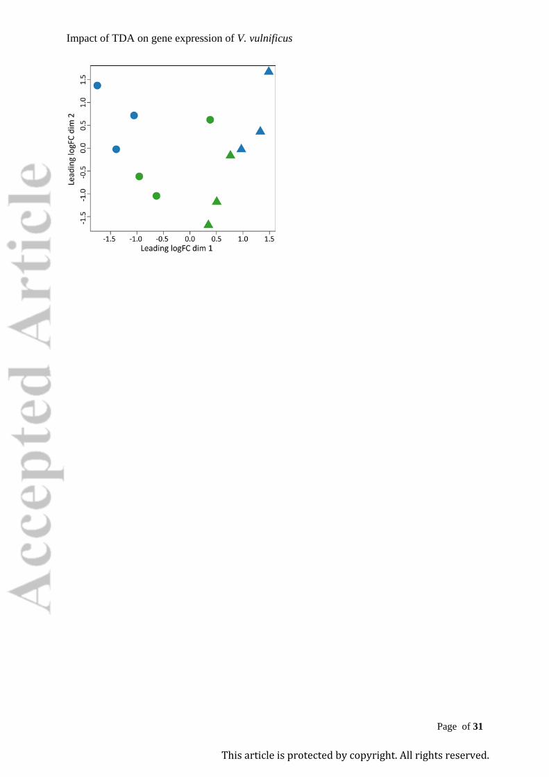
Impact of TDA on gene expression of V. vulnificus
Page of 31
This article is protected by copyright. All rights reserved.
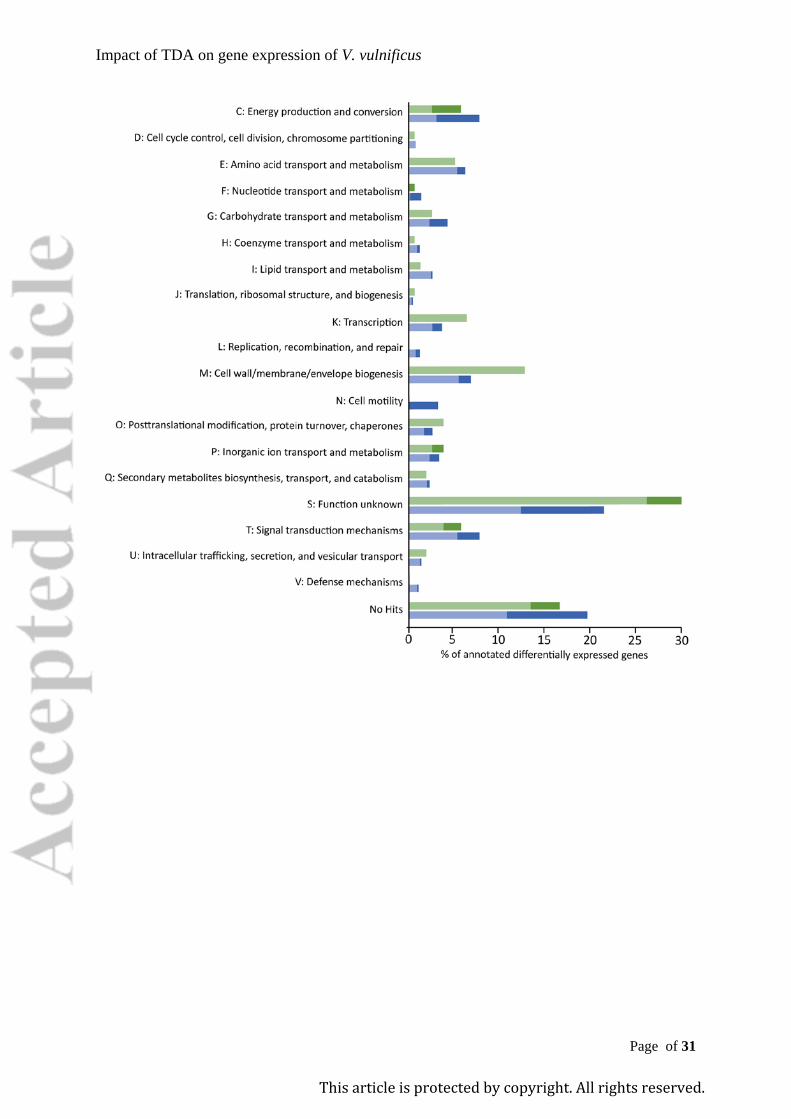
Impact of TDA on gene expression of V. vulnificus
Page of 31
This article is protected by copyright. All rights reserved.
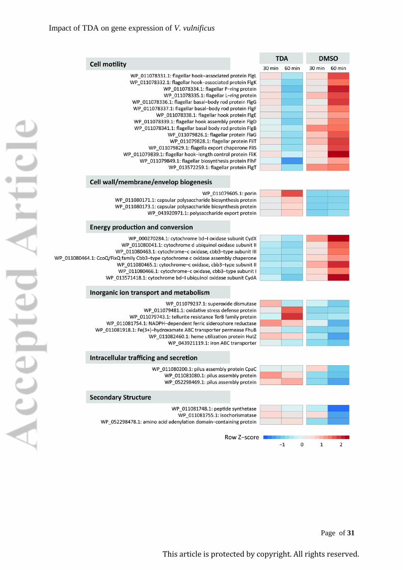
Impact of TDA on gene expression of V. vulnificus
Page of 31
This article is protected by copyright. All rights reserved.
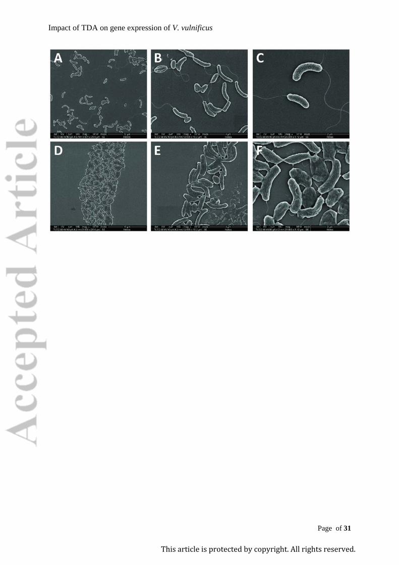
Impact of TDA on gene expression of V. vulnificus
Page of 31
This article is protected by copyright. All rights reserved.

Impact of TDA on gene expression of V. vulnificus
Page 1 of 7
Supplementary File 1 1
Tropodithietic acid induces oxidative stress response, cell envelope 2
biogenesis and iron uptake in Vibrio vulnificus 3
4
Karen K. Dittmann, Cisse H. Porsby, Priscila Goncalves, Ramona Valentina Mateiu, Eva C. 5
Sonnenschein, Mikkel Bentzon-Tilia, Suhelen Egan & Lone Gram# 6
# Address correspondence to: Lone Gram ([email protected]) 7
8
Impact of TDA on gene expression of V. vulnificus
Page 2 of 7
Experimental Procedures 9
Bacterial strain and culture media 10
Vibrio vulnificus CMCP6 (Kim et al., 2003) was used in this study. CMCP6 was stored in 11
freeze storage medium (Gibson and Khoury, 1986) at -80 °C. Unless otherwise stated, the 12
strain was grown at 37 °C overnight on Mueller Hinton II agar (Cation-Adjusted) (211438; 13
BD) (MHA) or in 5 ml Mueller Hinton II broth (Cation-Adjusted) (212322; BD) (MHB) at 14
300 rpm. 15
Dilution of tropodithietic acid (TDA). 16
Stock solutions of TDA (BioViotica, Germany) were dissolved in 100% dimethyl sulfoxide 17
(D8418; Sigma-Aldrich, Missouri, USA) (DMSO) to 8 mM and stored at -20 °C. Working 18
solutions were prepared by diluting TDA stock solutions in demineralized H2O to a final 19
concentration of 1 mM and sonicating the solution 2 x 30 s in an ultrasound bath (Aerosec, 20
France). 21
Transcriptomic analyses following TDA exposure. 22
Overnight cultures of V. vulnificus CMCP6 were inoculated at 1% level in pre-warmed (37 23
°C) MHB and incubated at 37 °C in a shaking water bath (SW22; Julabo, Pennsylvania, 24
USA) at 200 rpm. At OD600 0.5 ± 0.02 (Novaspec III; Amersham Biosciences, Piscataway, 25
USA), cultures were divided into two, and pre-warmed MHB (1:1 v/v) was added. When 26
OD600 reached 0.5 ± 0.02, 0.6 µl of 1 mM TDA was added per ml of culture, giving a final 27
concentration of 0.6 µM (260 x lower than the minimal inhibitory concentration - MIC - 28
value of 15.6 µM). The solvent of TDA was added to control cultures to a final concentration 29
of 0.007%. Samples for transcriptomic analysis were taken after 30 and 60 min of exposure. 30
All controls and treatments were performed in biological triplicates (n = 3 per treatment per 31
Impact of TDA on gene expression of V. vulnificus
Page 3 of 7
time point). Cell densities were determined by serial ten-fold dilution and spread-plating on 32
MHA. 33
RNA extraction. 34
Samples were mixed with RNA protect (76506; Qiagen, Netherlands) and incubated 5 min at 35
room temperature. Samples were centrifuged for 10 min at 10,000 x g and pellets stored at -36
80 °C prior to treatment with proteinase K (P8102S; New England Biolabs Inc., MA, USA) 37
and lysozyme (L6876; Sigma). RNA was purified using RNeasy Mini Kit (74104; Qiagen) 38
including an additional DNase treatment using RNase-free DNase (79254; Qiagen) according 39
to the manufacturer’s instruction. Quality and quantity of total RNA was assessed using 40
NanoDrop (ND-1000; Thermo Fisher Scientific Inc., Delaware, USA) and Bioanalyzer 41
(2100; Agilent, California, USA) combined with RNA 6000 Nano kit (5067-1511; Agilent). 42
Five µg of total RNA (RIN ≥ 8.3) were precipitated with ethanol, dissolved in 10 µl RNase-43
free water and used in the MICROBExpress™ Bacterial mRNA Enrichment Kit (AM1905; 44
Ambion) by which 16S and 23S rRNA were removed. Qubit RNA assay kit (Q32852; 45
Invitrogen) and a Bioanalyzer (RNA 6000 Nano kit) were used for evaluation of quantity and 46
quality of rRNA-depleted RNA. 47
cDNA library construction and sequencing. 48
The TruSeq RNA Sample preparation Kit v2 (set A: RS-122-2001 and set B: RS-122-2001; 49
Illumina, California, USA) was used according to the manufacturer’s protocol for preparing 50
libraries for RNA sequencing. In brief, fragments of cDNA of approx. 180 bp were made 51
from RNA (rRNA-depleted) and specific adaptors were ligated to each of the 12 samples. 52
Quantity and quality of the libraries were verified using Qubit dsDNA BR assay kit (Q32850; 53
Invitrogen) and a Bioanalyzer on DNA-1000 (r 5067-1504; Agilent) or High Sensitivity 54
(5067-4626; Agilent) chips. Each library was diluted to 10 nM and pooled. The average size 55
Impact of TDA on gene expression of V. vulnificus
Page 4 of 7
was 270 bp. Sequencing (100 bp paired ends reads) was performed on pooled libraries by 56
BGI-Hong Kong using the HiSeq2000 platform. 57
RNA sequence analyses. 58
Quality assessment of the reads before and after trimming was performed using FastQC (v. 59
0.11.6, https://www.bioinformatics.babraham.ac.uk/projects/fastqc/). Number of reads, 60
PHRED-score, % GC, nucleotide contribution, quality distribution and enriched 5mers 61
sequences were used for quality assessment of RNA sequencing data. Trimmomatic (v. 0.33, 62
Bolger et al. 2014) was used for quality-based trimming of the reads, which included adapter 63
removal, leading/trailing low quality bases (below quality 3) removal, 4-base sliding window 64
and trim when average dropped below 20, and filtering of sequences with length below 36 65
bp. The trimmed reads were processed using the HISAT2-StringTie pipeline (Pertea et al., 66
2016). In brief, the reads were aligned to the reference genome (downloaded from NCBI; 67
NC_004459 and NC_004460 for V. vulnificus CMCP6), transcripts were assembled and 68
quantified, transcripts from individual sample were merged and transcript abundances were 69
estimated for generation of a count table (ballgown element). The raw counts were extracted 70
by using the prepDE.py script from the StringTie manual. Differential expression analysis 71
was performed on the raw counts using edgeR (Robinson et al., 2010) by fitting a quasi-72
likelihood negative binomial generalized log-linear model to the counts. Genes were 73
considered to be significantly differentially expressed if absolute log2-fold change (log2FC) 74
between conditions > 1 (either up- or down-regulation), at p-value < 0.05, and false discovery 75
rate (FDR) < 0.05. Significantly differentially expressed genes were functionally annotated 76
using EggNOG (Huerta-Cepas et al., 2016) and BlastKOALA (Kanehisa et al., 2016). 77
Functional categories were based on cluster of orthologous groups (COGs). The expression 78
levels of a set of target genes were visualised by heatmaps; row Z-scores were calculated 79
Impact of TDA on gene expression of V. vulnificus
Page 5 of 7
from the normalized counts per million (CPM, computed by edgeR) and plotted using R 80
package ggplot2. 81
Transcriptomic data. 82
Raw sequencing reads can be accessed through the Sequencing Read Archives (SRA) under 83
PRJNA516163. 84
Motility assay. 85
A swimming motility assay was used to investigate if a sub-lethal concentration of TDA 86
affects the motility of V. vulnificus CMCP6 cells. TDA or the solvent (DMSO) were added to 87
MHB supplemented with 0.25 % agar resulting in the final concentrations of 0.6 µM and 88
0.007 % respectively. The assay was conducted in 24-well Nunc NuclonTM Surface plates 89
(142475). CMCP6 was inoculated in the center of the agar and incubated at 30 °C. The 90
diameter of the colonies was measured after 5 h of exposure. The viable cell count was 91
determined by diluting the content of each well in PBS and subsequent plate spreading on 92
MHA. 93
Scanning electron microscopy (SEM). 94
SEM was used to inspect if TDA induces any visible damages to V. vulnificus CMCP6 cells 95
and cell envelope at concentrations above the MIC. A total of 225 µl of overnight culture was 96
treated with 25 µl TDA or DMSO at final concentrations of 30 µM (2 x MIC) and 0.3%, 97
respectively. Samples were incubated at 37 °C, 300 rpm for 60 min. Fixation of the cells was 98
done in two steps. In the first step, 25 µL of 25 % glutaraldehyde (G5882; Sigma-Aldrich) 99
was added to each sample (final concentrations of 2.5 %), samples were incubated for 10 min 100
at room temperature and pelleted at 2,000 x g for 10 min. In the second step, glutaraldehyde 101
diluted to 2 % in phosphate buffered saline (BR0014R; Oxoid) (PBS) was added to each cell 102
pellet and the mixture was incubated overnight at 5 °C. Samples were then centrifuged at 103
Impact of TDA on gene expression of V. vulnificus
Page 6 of 7
2000 x g, 5 min and the cell pellets were dried in ethanol series (50, 60, 70, 80, 90, 99.9%) 104
for 10 minutes each step. Cells were placed onto a square piece of silicon wafer and left to 105
dry overnight in the fume hood. The silicon substrate with the sample was attached onto an 106
aluminum stub with a carbon tape. The uncoated sample was imaged in a FEI Helios dual 107
beam SEM. For imaging, an accelerating voltage of 2 keV and a current of 43 pA was used 108
and the secondary electron was detected with the lens detector. 109
References 110
Bolger, A.M., Lohse, M., and Usadel, B. (2014) Trimmomatic: a flexible trimmer for 111
Illumina sequence data. Bioinformatics 30: 2114–20. 112
Gibson, L.F. and Khoury, J.T. (1986) Storage and survival of bacteria by ultra-freeze. Lett. 113
Appl. Microbiol. 3: 127–129. 114
Huerta-Cepas, J., Szklarczyk, D., Forslund, K., Cook, H., Heller, D., Walter, M.C., et al. 115
(2016) EGGNOG 4.5: A hierarchical orthology framework with improved functional 116
annotations for eukaryotic, prokaryotic and viral sequences. Nucleic Acids Res. 44: 117
D286–D293. 118
Kanehisa, M., Sato, Y., and Morishima, K. (2016) BlastKOALA and GhostKOALA: KEGG 119
Tools for Functional Characterization of Genome and Metagenome Sequences. J. Mol. 120
Biol. 428: 726–731. 121
Kim, Y.R., Lee, S.E., Kim, C.M., Kim, S.Y., Shin, E.K., Shin, D.H., et al. (2003) 122
Characterization and Pathogenic Significance of Vibrio vulnificus Antigens 123
Preferentially Expressed in Septicemic Patients. Infect. Immun. 71: 5461–5471. 124
Robinson, M.D., McCarthy, D.J., and Smyth, G.K. (2010) edgeR: a Bioconductor package 125
for differential expression analysis of digital gene expression data. Bioinformatics 26: 126
Impact of TDA on gene expression of V. vulnificus
Page 7 of 7
139–40. 127
128
Impact of TDA on gene expression of V. vulnificus
Page 1 of 24
Supplementary File 2 1
Tropodithietic acid induces oxidative stress response, cell envelope 2
biogenesis and iron uptake in Vibrio vulnificus 3
4
Karen K. Dittmann, Cisse H. Porsby, Priscila Goncalves, Ramona Valentina Mateiu, Eva C. 5
Sonnenschein, Mikkel Bentzon-Tilia, Suhelen Egan & Lone Gram# 6
# Address correspondence to: Lone Gram ([email protected]) 7
8
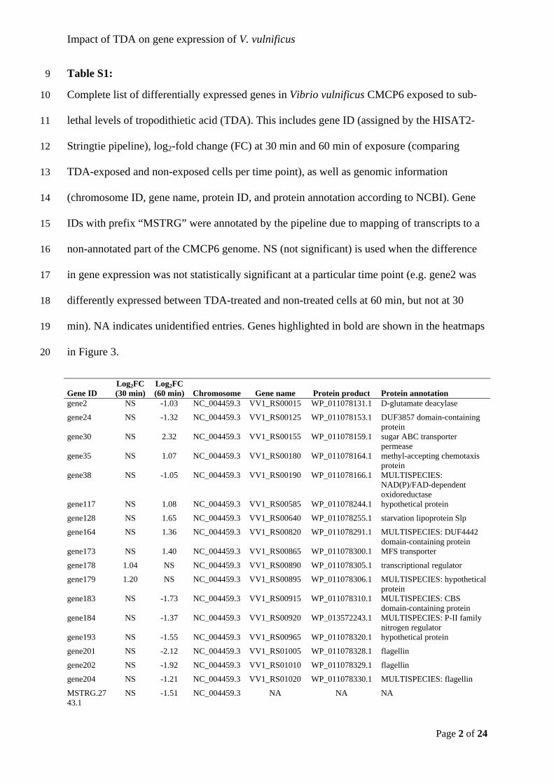
Impact of TDA on gene expression of V. vulnificus
Page 2 of 24
Table S1: 9
Complete list of differentially expressed genes in Vibrio vulnificus CMCP6 exposed to sub-10
lethal levels of tropodithietic acid (TDA). This includes gene ID (assigned by the HISAT2-11
Stringtie pipeline), log2-fold change (FC) at 30 min and 60 min of exposure (comparing 12
TDA-exposed and non-exposed cells per time point), as well as genomic information 13
(chromosome ID, gene name, protein ID, and protein annotation according to NCBI). Gene 14
IDs with prefix “MSTRG” were annotated by the pipeline due to mapping of transcripts to a 15
non-annotated part of the CMCP6 genome. NS (not significant) is used when the difference 16
in gene expression was not statistically significant at a particular time point (e.g. gene2 was 17
differently expressed between TDA-treated and non-treated cells at 60 min, but not at 30 18
min). NA indicates unidentified entries. Genes highlighted in bold are shown in the heatmaps 19
in Figure 3. 20
Gene ID Log2FC (30 min)
Log2FC (60 min) Chromosome Gene name Protein product Protein annotation
gene2 NS -1.03 NC_004459.3 VV1_RS00015 WP_011078131.1 D-glutamate deacylase
gene24 NS -1.32 NC_004459.3 VV1_RS00125 WP_011078153.1 DUF3857 domain-containing protein
gene30 NS 2.32 NC_004459.3 VV1_RS00155 WP_011078159.1 sugar ABC transporter permease
gene35 NS 1.07 NC_004459.3 VV1_RS00180 WP_011078164.1 methyl-accepting chemotaxis protein
gene38 NS -1.05 NC_004459.3 VV1_RS00190 WP_011078166.1 MULTISPECIES: NAD(P)/FAD-dependent oxidoreductase
gene117 NS 1.08 NC_004459.3 VV1_RS00585 WP_011078244.1 hypothetical protein
gene128 NS 1.65 NC_004459.3 VV1_RS00640 WP_011078255.1 starvation lipoprotein Slp
gene164 NS 1.36 NC_004459.3 VV1_RS00820 WP_011078291.1 MULTISPECIES: DUF4442 domain-containing protein
gene173 NS 1.40 NC_004459.3 VV1_RS00865 WP_011078300.1 MFS transporter
gene178 1.04 NS NC_004459.3 VV1_RS00890 WP_011078305.1 transcriptional regulator
gene179 1.20 NS NC_004459.3 VV1_RS00895 WP_011078306.1 MULTISPECIES: hypothetical protein
gene183 NS -1.73 NC_004459.3 VV1_RS00915 WP_011078310.1 MULTISPECIES: CBS domain-containing protein
gene184 NS -1.37 NC_004459.3 VV1_RS00920 WP_013572243.1 MULTISPECIES: P-II family nitrogen regulator
gene193 NS -1.55 NC_004459.3 VV1_RS00965 WP_011078320.1 hypothetical protein
gene201 NS -2.12 NC_004459.3 VV1_RS01005 WP_011078328.1 flagellin
gene202 NS -1.92 NC_004459.3 VV1_RS01010 WP_011078329.1 flagellin
gene204 NS -1.21 NC_004459.3 VV1_RS01020 WP_011078330.1 MULTISPECIES: flagellin
MSTRG.2743.1
NS -1.51 NC_004459.3 NA NA NA
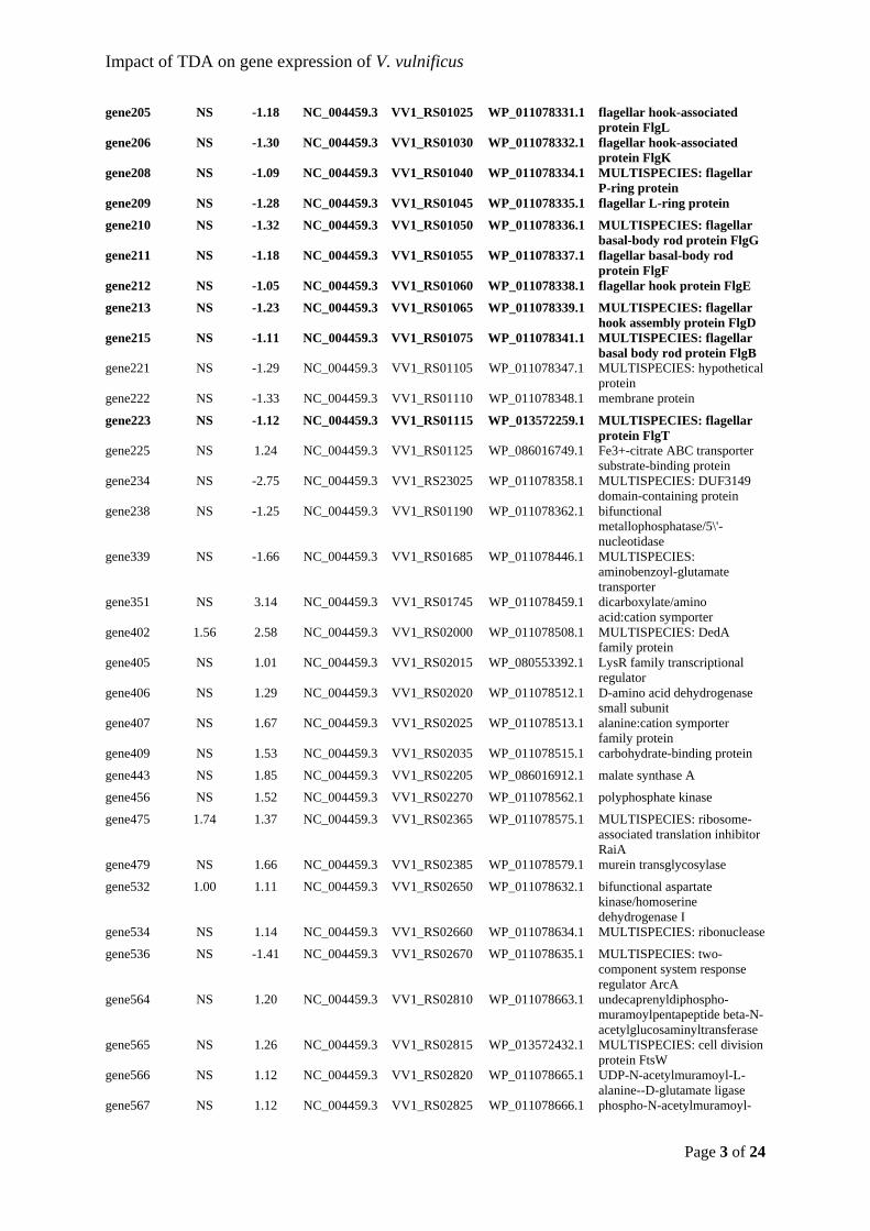
Impact of TDA on gene expression of V. vulnificus
Page 3 of 24
gene205 NS -1.18 NC_004459.3 VV1_RS01025 WP_011078331.1 flagellar hook-associated protein FlgL
gene206 NS -1.30 NC_004459.3 VV1_RS01030 WP_011078332.1 flagellar hook-associated protein FlgK
gene208 NS -1.09 NC_004459.3 VV1_RS01040 WP_011078334.1 MULTISPECIES: flagellar P-ring protein
gene209 NS -1.28 NC_004459.3 VV1_RS01045 WP_011078335.1 flagellar L-ring protein
gene210 NS -1.32 NC_004459.3 VV1_RS01050 WP_011078336.1 MULTISPECIES: flagellar basal-body rod protein FlgG
gene211 NS -1.18 NC_004459.3 VV1_RS01055 WP_011078337.1 flagellar basal-body rod protein FlgF
gene212 NS -1.05 NC_004459.3 VV1_RS01060 WP_011078338.1 flagellar hook protein FlgE
gene213 NS -1.23 NC_004459.3 VV1_RS01065 WP_011078339.1 MULTISPECIES: flagellar hook assembly protein FlgD
gene215 NS -1.11 NC_004459.3 VV1_RS01075 WP_011078341.1 MULTISPECIES: flagellar basal body rod protein FlgB
gene221 NS -1.29 NC_004459.3 VV1_RS01105 WP_011078347.1 MULTISPECIES: hypothetical protein
gene222 NS -1.33 NC_004459.3 VV1_RS01110 WP_011078348.1 membrane protein
gene223 NS -1.12 NC_004459.3 VV1_RS01115 WP_013572259.1 MULTISPECIES: flagellar protein FlgT
gene225 NS 1.24 NC_004459.3 VV1_RS01125 WP_086016749.1 Fe3+-citrate ABC transporter substrate-binding protein
gene234 NS -2.75 NC_004459.3 VV1_RS23025 WP_011078358.1 MULTISPECIES: DUF3149 domain-containing protein
gene238 NS -1.25 NC_004459.3 VV1_RS01190 WP_011078362.1 bifunctional metallophosphatase/5\'-nucleotidase
gene339 NS -1.66 NC_004459.3 VV1_RS01685 WP_011078446.1 MULTISPECIES: aminobenzoyl-glutamate transporter
gene351 NS 3.14 NC_004459.3 VV1_RS01745 WP_011078459.1 dicarboxylate/amino acid:cation symporter
gene402 1.56 2.58 NC_004459.3 VV1_RS02000 WP_011078508.1 MULTISPECIES: DedA family protein
gene405 NS 1.01 NC_004459.3 VV1_RS02015 WP_080553392.1 LysR family transcriptional regulator
gene406 NS 1.29 NC_004459.3 VV1_RS02020 WP_011078512.1 D-amino acid dehydrogenase small subunit
gene407 NS 1.67 NC_004459.3 VV1_RS02025 WP_011078513.1 alanine:cation symporter family protein
gene409 NS 1.53 NC_004459.3 VV1_RS02035 WP_011078515.1 carbohydrate-binding protein
gene443 NS 1.85 NC_004459.3 VV1_RS02205 WP_086016912.1 malate synthase A
gene456 NS 1.52 NC_004459.3 VV1_RS02270 WP_011078562.1 polyphosphate kinase
gene475 1.74 1.37 NC_004459.3 VV1_RS02365 WP_011078575.1 MULTISPECIES: ribosome-associated translation inhibitor RaiA
gene479 NS 1.66 NC_004459.3 VV1_RS02385 WP_011078579.1 murein transglycosylase
gene532 1.00 1.11 NC_004459.3 VV1_RS02650 WP_011078632.1 bifunctional aspartate kinase/homoserine dehydrogenase I
gene534 NS 1.14 NC_004459.3 VV1_RS02660 WP_011078634.1 MULTISPECIES: ribonuclease
gene536 NS -1.41 NC_004459.3 VV1_RS02670 WP_011078635.1 MULTISPECIES: two-component system response regulator ArcA
gene564 NS 1.20 NC_004459.3 VV1_RS02810 WP_011078663.1 undecaprenyldiphospho-muramoylpentapeptide beta-N- acetylglucosaminyltransferase
gene565 NS 1.26 NC_004459.3 VV1_RS02815 WP_013572432.1 MULTISPECIES: cell division protein FtsW
gene566 NS 1.12 NC_004459.3 VV1_RS02820 WP_011078665.1 UDP-N-acetylmuramoyl-L-alanine--D-glutamate ligase
gene567 NS 1.12 NC_004459.3 VV1_RS02825 WP_011078666.1 phospho-N-acetylmuramoyl-
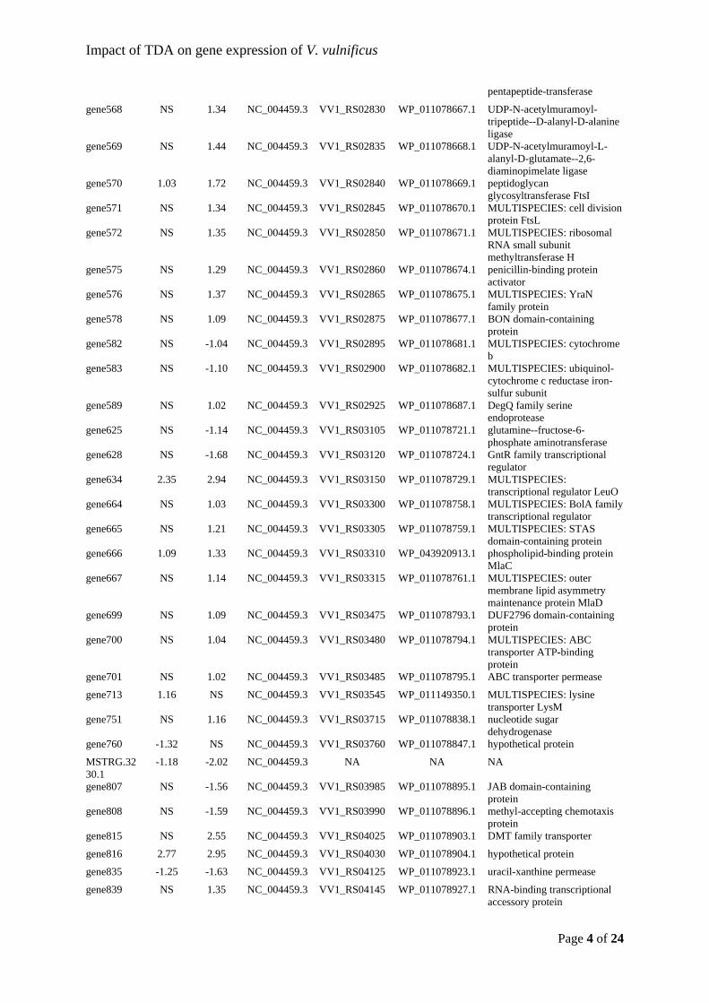
Impact of TDA on gene expression of V. vulnificus
Page 4 of 24
pentapeptide-transferase
gene568 NS 1.34 NC_004459.3 VV1_RS02830 WP_011078667.1 UDP-N-acetylmuramoyl-tripeptide--D-alanyl-D-alanine ligase
gene569 NS 1.44 NC_004459.3 VV1_RS02835 WP_011078668.1 UDP-N-acetylmuramoyl-L-alanyl-D-glutamate--2,6-diaminopimelate ligase
gene570 1.03 1.72 NC_004459.3 VV1_RS02840 WP_011078669.1 peptidoglycan glycosyltransferase FtsI
gene571 NS 1.34 NC_004459.3 VV1_RS02845 WP_011078670.1 MULTISPECIES: cell division protein FtsL
gene572 NS 1.35 NC_004459.3 VV1_RS02850 WP_011078671.1 MULTISPECIES: ribosomal RNA small subunit methyltransferase H
gene575 NS 1.29 NC_004459.3 VV1_RS02860 WP_011078674.1 penicillin-binding protein activator
gene576 NS 1.37 NC_004459.3 VV1_RS02865 WP_011078675.1 MULTISPECIES: YraN family protein
gene578 NS 1.09 NC_004459.3 VV1_RS02875 WP_011078677.1 BON domain-containing protein
gene582 NS -1.04 NC_004459.3 VV1_RS02895 WP_011078681.1 MULTISPECIES: cytochrome b
gene583 NS -1.10 NC_004459.3 VV1_RS02900 WP_011078682.1 MULTISPECIES: ubiquinol-cytochrome c reductase iron-sulfur subunit
gene589 NS 1.02 NC_004459.3 VV1_RS02925 WP_011078687.1 DegQ family serine endoprotease
gene625 NS -1.14 NC_004459.3 VV1_RS03105 WP_011078721.1 glutamine--fructose-6-phosphate aminotransferase
gene628 NS -1.68 NC_004459.3 VV1_RS03120 WP_011078724.1 GntR family transcriptional regulator
gene634 2.35 2.94 NC_004459.3 VV1_RS03150 WP_011078729.1 MULTISPECIES: transcriptional regulator LeuO
gene664 NS 1.03 NC_004459.3 VV1_RS03300 WP_011078758.1 MULTISPECIES: BolA family transcriptional regulator
gene665 NS 1.21 NC_004459.3 VV1_RS03305 WP_011078759.1 MULTISPECIES: STAS domain-containing protein
gene666 1.09 1.33 NC_004459.3 VV1_RS03310 WP_043920913.1 phospholipid-binding protein MlaC
gene667 NS 1.14 NC_004459.3 VV1_RS03315 WP_011078761.1 MULTISPECIES: outer membrane lipid asymmetry maintenance protein MlaD
gene699 NS 1.09 NC_004459.3 VV1_RS03475 WP_011078793.1 DUF2796 domain-containing protein
gene700 NS 1.04 NC_004459.3 VV1_RS03480 WP_011078794.1 MULTISPECIES: ABC transporter ATP-binding protein
gene701 NS 1.02 NC_004459.3 VV1_RS03485 WP_011078795.1 ABC transporter permease
gene713 1.16 NS NC_004459.3 VV1_RS03545 WP_011149350.1 MULTISPECIES: lysine transporter LysM
gene751 NS 1.16 NC_004459.3 VV1_RS03715 WP_011078838.1 nucleotide sugar dehydrogenase
gene760 -1.32 NS NC_004459.3 VV1_RS03760 WP_011078847.1 hypothetical protein
MSTRG.3230.1
-1.18 -2.02 NC_004459.3 NA NA NA
gene807 NS -1.56 NC_004459.3 VV1_RS03985 WP_011078895.1 JAB domain-containing protein
gene808 NS -1.59 NC_004459.3 VV1_RS03990 WP_011078896.1 methyl-accepting chemotaxis protein
gene815 NS 2.55 NC_004459.3 VV1_RS04025 WP_011078903.1 DMT family transporter
gene816 2.77 2.95 NC_004459.3 VV1_RS04030 WP_011078904.1 hypothetical protein
gene835 -1.25 -1.63 NC_004459.3 VV1_RS04125 WP_011078923.1 uracil-xanthine permease
gene839 NS 1.35 NC_004459.3 VV1_RS04145 WP_011078927.1 RNA-binding transcriptional accessory protein
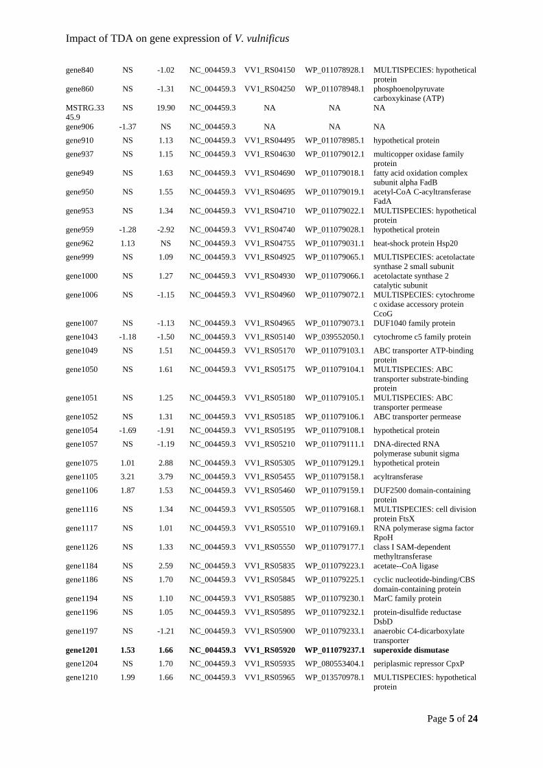
Impact of TDA on gene expression of V. vulnificus
Page 5 of 24
gene840 NS -1.02 NC_004459.3 VV1_RS04150 WP_011078928.1 MULTISPECIES: hypothetical protein
gene860 NS -1.31 NC_004459.3 VV1_RS04250 WP_011078948.1 phosphoenolpyruvate carboxykinase (ATP)
MSTRG.3345.9
NS 19.90 NC_004459.3 NA NA NA
gene906 -1.37 NS NC_004459.3 NA NA NA
gene910 NS 1.13 NC_004459.3 VV1_RS04495 WP_011078985.1 hypothetical protein
gene937 NS 1.15 NC_004459.3 VV1_RS04630 WP_011079012.1 multicopper oxidase family protein
gene949 NS 1.63 NC_004459.3 VV1_RS04690 WP_011079018.1 fatty acid oxidation complex subunit alpha FadB
gene950 NS 1.55 NC_004459.3 VV1_RS04695 WP_011079019.1 acetyl-CoA C-acyltransferase FadA
gene953 NS 1.34 NC_004459.3 VV1_RS04710 WP_011079022.1 MULTISPECIES: hypothetical protein
gene959 -1.28 -2.92 NC_004459.3 VV1_RS04740 WP_011079028.1 hypothetical protein
gene962 1.13 NS NC_004459.3 VV1_RS04755 WP_011079031.1 heat-shock protein Hsp20
gene999 NS 1.09 NC_004459.3 VV1_RS04925 WP_011079065.1 MULTISPECIES: acetolactate synthase 2 small subunit
gene1000 NS 1.27 NC_004459.3 VV1_RS04930 WP_011079066.1 acetolactate synthase 2 catalytic subunit
gene1006 NS -1.15 NC_004459.3 VV1_RS04960 WP_011079072.1 MULTISPECIES: cytochrome c oxidase accessory protein CcoG
gene1007 NS -1.13 NC_004459.3 VV1_RS04965 WP_011079073.1 DUF1040 family protein
gene1043 -1.18 -1.50 NC_004459.3 VV1_RS05140 WP_039552050.1 cytochrome c5 family protein
gene1049 NS 1.51 NC_004459.3 VV1_RS05170 WP_011079103.1 ABC transporter ATP-binding protein
gene1050 NS 1.61 NC_004459.3 VV1_RS05175 WP_011079104.1 MULTISPECIES: ABC transporter substrate-binding protein
gene1051 NS 1.25 NC_004459.3 VV1_RS05180 WP_011079105.1 MULTISPECIES: ABC transporter permease
gene1052 NS 1.31 NC_004459.3 VV1_RS05185 WP_011079106.1 ABC transporter permease
gene1054 -1.69 -1.91 NC_004459.3 VV1_RS05195 WP_011079108.1 hypothetical protein
gene1057 NS -1.19 NC_004459.3 VV1_RS05210 WP_011079111.1 DNA-directed RNA polymerase subunit sigma
gene1075 1.01 2.88 NC_004459.3 VV1_RS05305 WP_011079129.1 hypothetical protein
gene1105 3.21 3.79 NC_004459.3 VV1_RS05455 WP_011079158.1 acyltransferase
gene1106 1.87 1.53 NC_004459.3 VV1_RS05460 WP_011079159.1 DUF2500 domain-containing protein
gene1116 NS 1.34 NC_004459.3 VV1_RS05505 WP_011079168.1 MULTISPECIES: cell division protein FtsX
gene1117 NS 1.01 NC_004459.3 VV1_RS05510 WP_011079169.1 RNA polymerase sigma factor RpoH
gene1126 NS 1.33 NC_004459.3 VV1_RS05550 WP_011079177.1 class I SAM-dependent methyltransferase
gene1184 NS 2.59 NC_004459.3 VV1_RS05835 WP_011079223.1 acetate--CoA ligase
gene1186 NS 1.70 NC_004459.3 VV1_RS05845 WP_011079225.1 cyclic nucleotide-binding/CBS domain-containing protein
gene1194 NS 1.10 NC_004459.3 VV1_RS05885 WP_011079230.1 MarC family protein
gene1196 NS 1.05 NC_004459.3 VV1_RS05895 WP_011079232.1 protein-disulfide reductase DsbD
gene1197 NS -1.21 NC_004459.3 VV1_RS05900 WP_011079233.1 anaerobic C4-dicarboxylate transporter
gene1201 1.53 1.66 NC_004459.3 VV1_RS05920 WP_011079237.1 superoxide dismutase
gene1204 NS 1.70 NC_004459.3 VV1_RS05935 WP_080553404.1 periplasmic repressor CpxP
gene1210 1.99 1.66 NC_004459.3 VV1_RS05965 WP_013570978.1 MULTISPECIES: hypothetical protein
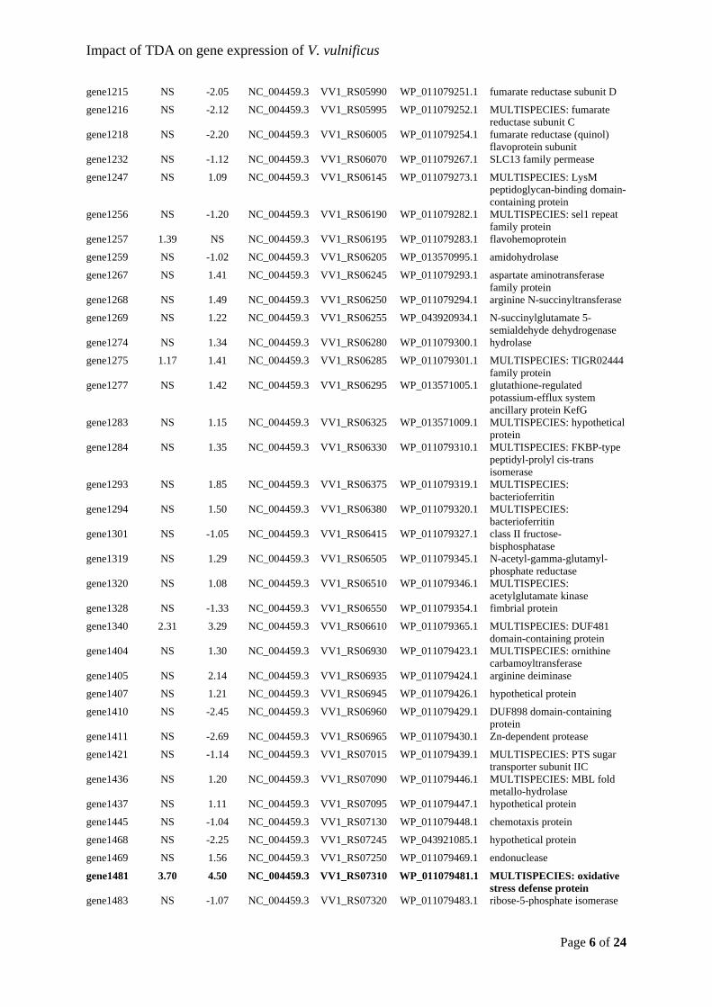
Impact of TDA on gene expression of V. vulnificus
Page 6 of 24
gene1215 NS -2.05 NC_004459.3 VV1_RS05990 WP_011079251.1 fumarate reductase subunit D
gene1216 NS -2.12 NC_004459.3 VV1_RS05995 WP_011079252.1 MULTISPECIES: fumarate reductase subunit C
gene1218 NS -2.20 NC_004459.3 VV1_RS06005 WP_011079254.1 fumarate reductase (quinol) flavoprotein subunit
gene1232 NS -1.12 NC_004459.3 VV1_RS06070 WP_011079267.1 SLC13 family permease
gene1247 NS 1.09 NC_004459.3 VV1_RS06145 WP_011079273.1 MULTISPECIES: LysM peptidoglycan-binding domain-containing protein
gene1256 NS -1.20 NC_004459.3 VV1_RS06190 WP_011079282.1 MULTISPECIES: sel1 repeat family protein
gene1257 1.39 NS NC_004459.3 VV1_RS06195 WP_011079283.1 flavohemoprotein
gene1259 NS -1.02 NC_004459.3 VV1_RS06205 WP_013570995.1 amidohydrolase
gene1267 NS 1.41 NC_004459.3 VV1_RS06245 WP_011079293.1 aspartate aminotransferase family protein
gene1268 NS 1.49 NC_004459.3 VV1_RS06250 WP_011079294.1 arginine N-succinyltransferase
gene1269 NS 1.22 NC_004459.3 VV1_RS06255 WP_043920934.1 N-succinylglutamate 5-semialdehyde dehydrogenase
gene1274 NS 1.34 NC_004459.3 VV1_RS06280 WP_011079300.1 hydrolase
gene1275 1.17 1.41 NC_004459.3 VV1_RS06285 WP_011079301.1 MULTISPECIES: TIGR02444 family protein
gene1277 NS 1.42 NC_004459.3 VV1_RS06295 WP_013571005.1 glutathione-regulated potassium-efflux system ancillary protein KefG
gene1283 NS 1.15 NC_004459.3 VV1_RS06325 WP_013571009.1 MULTISPECIES: hypothetical protein
gene1284 NS 1.35 NC_004459.3 VV1_RS06330 WP_011079310.1 MULTISPECIES: FKBP-type peptidyl-prolyl cis-trans isomerase
gene1293 NS 1.85 NC_004459.3 VV1_RS06375 WP_011079319.1 MULTISPECIES: bacterioferritin
gene1294 NS 1.50 NC_004459.3 VV1_RS06380 WP_011079320.1 MULTISPECIES: bacterioferritin
gene1301 NS -1.05 NC_004459.3 VV1_RS06415 WP_011079327.1 class II fructose-bisphosphatase
gene1319 NS 1.29 NC_004459.3 VV1_RS06505 WP_011079345.1 N-acetyl-gamma-glutamyl-phosphate reductase
gene1320 NS 1.08 NC_004459.3 VV1_RS06510 WP_011079346.1 MULTISPECIES: acetylglutamate kinase
gene1328 NS -1.33 NC_004459.3 VV1_RS06550 WP_011079354.1 fimbrial protein
gene1340 2.31 3.29 NC_004459.3 VV1_RS06610 WP_011079365.1 MULTISPECIES: DUF481 domain-containing protein
gene1404 NS 1.30 NC_004459.3 VV1_RS06930 WP_011079423.1 MULTISPECIES: ornithine carbamoyltransferase
gene1405 NS 2.14 NC_004459.3 VV1_RS06935 WP_011079424.1 arginine deiminase
gene1407 NS 1.21 NC_004459.3 VV1_RS06945 WP_011079426.1 hypothetical protein
gene1410 NS -2.45 NC_004459.3 VV1_RS06960 WP_011079429.1 DUF898 domain-containing protein
gene1411 NS -2.69 NC_004459.3 VV1_RS06965 WP_011079430.1 Zn-dependent protease
gene1421 NS -1.14 NC_004459.3 VV1_RS07015 WP_011079439.1 MULTISPECIES: PTS sugar transporter subunit IIC
gene1436 NS 1.20 NC_004459.3 VV1_RS07090 WP_011079446.1 MULTISPECIES: MBL fold metallo-hydrolase
gene1437 NS 1.11 NC_004459.3 VV1_RS07095 WP_011079447.1 hypothetical protein
gene1445 NS -1.04 NC_004459.3 VV1_RS07130 WP_011079448.1 chemotaxis protein
gene1468 NS -2.25 NC_004459.3 VV1_RS07245 WP_043921085.1 hypothetical protein
gene1469 NS 1.56 NC_004459.3 VV1_RS07250 WP_011079469.1 endonuclease
gene1481 3.70 4.50 NC_004459.3 VV1_RS07310 WP_011079481.1 MULTISPECIES: oxidative stress defense protein
gene1483 NS -1.07 NC_004459.3 VV1_RS07320 WP_011079483.1 ribose-5-phosphate isomerase
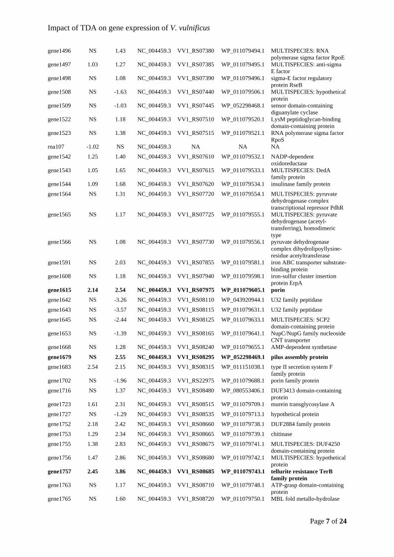
Impact of TDA on gene expression of V. vulnificus
Page 7 of 24
gene1496 NS 1.43 NC_004459.3 VV1_RS07380 WP_011079494.1 MULTISPECIES: RNA polymerase sigma factor RpoE
gene1497 1.03 1.27 NC_004459.3 VV1_RS07385 WP_011079495.1 MULTISPECIES: anti-sigma E factor
gene1498 NS 1.08 NC_004459.3 VV1_RS07390 WP_011079496.1 sigma-E factor regulatory protein RseB
gene1508 NS -1.63 NC_004459.3 VV1_RS07440 WP_011079506.1 MULTISPECIES: hypothetical protein
gene1509 NS -1.03 NC_004459.3 VV1_RS07445 WP_052298468.1 sensor domain-containing diguanylate cyclase
gene1522 NS 1.18 NC_004459.3 VV1_RS07510 WP_011079520.1 LysM peptidoglycan-binding domain-containing protein
gene1523 NS 1.38 NC_004459.3 VV1_RS07515 WP_011079521.1 RNA polymerase sigma factor RpoS
rna107 -1.02 NS NC_004459.3 NA NA NA
gene1542 1.25 1.40 NC_004459.3 VV1_RS07610 WP_011079532.1 NADP-dependent oxidoreductase
gene1543 1.05 1.65 NC_004459.3 VV1_RS07615 WP_011079533.1 MULTISPECIES: DedA family protein
gene1544 1.09 1.68 NC_004459.3 VV1_RS07620 WP_011079534.1 insulinase family protein
gene1564 NS 1.31 NC_004459.3 VV1_RS07720 WP_011079554.1 MULTISPECIES: pyruvate dehydrogenase complex transcriptional repressor PdhR
gene1565 NS 1.17 NC_004459.3 VV1_RS07725 WP_011079555.1 MULTISPECIES: pyruvate dehydrogenase (acetyl-transferring), homodimeric type
gene1566 NS 1.08 NC_004459.3 VV1_RS07730 WP_011079556.1 pyruvate dehydrogenase complex dihydrolipoyllysine-residue acetyltransferase
gene1591 NS 2.03 NC_004459.3 VV1_RS07855 WP_011079581.1 iron ABC transporter substrate-binding protein
gene1608 NS 1.18 NC_004459.3 VV1_RS07940 WP_011079598.1 iron-sulfur cluster insertion protein ErpA
gene1615 2.14 2.54 NC_004459.3 VV1_RS07975 WP_011079605.1 porin
gene1642 NS -3.26 NC_004459.3 VV1_RS08110 WP_043920944.1 U32 family peptidase
gene1643 NS -3.57 NC_004459.3 VV1_RS08115 WP_011079631.1 U32 family peptidase
gene1645 NS -2.44 NC_004459.3 VV1_RS08125 WP_011079633.1 MULTISPECIES: SCP2 domain-containing protein
gene1653 NS -1.39 NC_004459.3 VV1_RS08165 WP_011079641.1 NupC/NupG family nucleoside CNT transporter
gene1668 NS 1.28 NC_004459.3 VV1_RS08240 WP_011079655.1 AMP-dependent synthetase
gene1679 NS 2.55 NC_004459.3 VV1_RS08295 WP_052298469.1 pilus assembly protein
gene1683 2.54 2.15 NC_004459.3 VV1_RS08315 WP_011151038.1 type II secretion system F family protein
gene1702 NS -1.96 NC_004459.3 VV1_RS22975 WP_011079688.1 porin family protein
gene1716 NS 1.37 NC_004459.3 VV1_RS08480 WP_080553406.1 DUF3413 domain-containing protein
gene1723 1.61 2.31 NC_004459.3 VV1_RS08515 WP_011079709.1 murein transglycosylase A
gene1727 NS -1.29 NC_004459.3 VV1_RS08535 WP_011079713.1 hypothetical protein
gene1752 2.18 2.42 NC_004459.3 VV1_RS08660 WP_011079738.1 DUF2884 family protein
gene1753 1.29 2.34 NC_004459.3 VV1_RS08665 WP_011079739.1 chitinase
gene1755 1.38 2.83 NC_004459.3 VV1_RS08675 WP_011079741.1 MULTISPECIES: DUF4250 domain-containing protein
gene1756 1.47 2.86 NC_004459.3 VV1_RS08680 WP_011079742.1 MULTISPECIES: hypothetical protein
gene1757 2.45 3.86 NC_004459.3 VV1_RS08685 WP_011079743.1 tellurite resistance TerB family protein
gene1763 NS 1.17 NC_004459.3 VV1_RS08710 WP_011079748.1 ATP-grasp domain-containing protein
gene1765 NS 1.60 NC_004459.3 VV1_RS08720 WP_011079750.1 MBL fold metallo-hydrolase
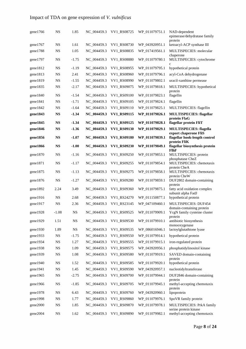
Impact of TDA on gene expression of V. vulnificus
Page 8 of 24
gene1766 NS 1.85 NC_004459.3 VV1_RS08725 WP_011079751.1 NAD-dependent epimerase/dehydratase family protein
gene1767 NS 1.61 NC_004459.3 VV1_RS08730 WP_043920951.1 ketoacyl-ACP synthase III
gene1788 NS 1.05 NC_004459.3 VV1_RS08835 WP_017419561.1 MULTISPECIES: molecular chaperone
gene1797 NS -1.75 NC_004459.3 VV1_RS08880 WP_011079780.1 MULTISPECIES: cytochrome c
gene1812 NS -1.19 NC_004459.3 VV1_RS08955 WP_011079795.1 hypothetical protein
gene1813 NS 2.41 NC_004459.3 VV1_RS08960 WP_011079796.1 acyl-CoA dehydrogenase
gene1819 NS -1.55 NC_004459.3 VV1_RS08990 WP_011079802.1 uracil-xanthine permease
gene1835 NS -2.17 NC_004459.3 VV1_RS09075 WP_011079818.1 MULTISPECIES: hypothetical protein
gene1840 NS -1.54 NC_004459.3 VV1_RS09100 WP_011079823.1 flagellin
gene1841 NS -1.71 NC_004459.3 VV1_RS09105 WP_011079824.1 flagellin
gene1842 NS -1.64 NC_004459.3 VV1_RS09110 WP_011079825.1 MULTISPECIES: flagellin
gene1843 NS -1.34 NC_004459.3 VV1_RS09115 WP_011079826.1 MULTISPECIES: flagellar protein FlaG
gene1845 NS -1.34 NC_004459.3 VV1_RS09125 WP_011079828.1 flagellar protein FliT
gene1846 NS -1.36 NC_004459.3 VV1_RS09130 WP_011079829.1 MULTISPECIES: flagella export chaperone FliS
gene1856 NS -1.07 NC_004459.3 VV1_RS09180 WP_011079839.1 flagellar hook-length control protein FliK
gene1866 NS -1.00 NC_004459.3 VV1_RS09230 WP_011079849.1 flagellar biosynthesis protein FlhF
gene1870 NS -1.16 NC_004459.3 VV1_RS09250 WP_011079853.1 MULTISPECIES: protein phosphatase CheZ
gene1871 NS -1.17 NC_004459.3 VV1_RS09255 WP_011079854.1 MULTISPECIES: chemotaxis protein CheA
gene1875 NS -1.13 NC_004459.3 VV1_RS09275 WP_011079858.1 MULTISPECIES: chemotaxis protein CheW
gene1876 NS -1.27 NC_004459.3 VV1_RS09280 WP_011079859.1 DUF2802 domain-containing protein
gene1892 2.24 3.49 NC_004459.3 VV1_RS09360 WP_011079875.1 fatty acid oxidation complex subunit alpha FadJ
gene1916 NS 2.68 NC_004459.3 VV1_RS24270 WP_011150877.1 hypothetical protein
gene1917 NS 2.36 NC_004459.3 VV1_RS23145 WP_047109460.1 MULTISPECIES: DUF454 domain-containing protein
gene1928 -1.08 NS NC_004459.3 VV1_RS09525 WP_011079909.1 YcgN family cysteine cluster protein
gene1929 1.51 NS NC_004459.3 VV1_RS09530 WP_011079910.1 antibiotic biosynthesis monooxygenase
gene1930 1.89 NS NC_004459.3 VV1_RS09535 WP_086016946.1 lactoylglutathione lyase
gene1933 NS -1.75 NC_004459.3 VV1_RS09550 WP_011079914.1 hypothetical protein
gene1934 NS 1.27 NC_004459.3 VV1_RS09555 WP_011079915.1 iron-regulated protein
gene1938 NS 1.09 NC_004459.3 VV1_RS09575 WP_043920956.1 phosphatidylinositol kinase
gene1939 NS 1.08 NC_004459.3 VV1_RS09580 WP_011079919.1 SAVED domain-containing protein
gene1940 NS 1.52 NC_004459.3 VV1_RS09585 WP_011079920.1 hypothetical protein
gene1941 NS 1.45 NC_004459.3 VV1_RS09590 WP_043920957.1 nucleotidyltransferase
gene1965 NS -2.75 NC_004459.3 VV1_RS09700 WP_011079944.1 DUF2846 domain-containing protein
gene1966 NS -1.85 NC_004459.3 VV1_RS09705 WP_011079945.1 methyl-accepting chemotaxis protein
gene1978 NS 6.43 NC_004459.3 VV1_RS09760 WP_043920960.1 lipoprotein
gene1998 NS 1.77 NC_004459.3 VV1_RS09860 WP_011079976.1 SpoVR family protein
gene2000 NS 1.85 NC_004459.3 VV1_RS09870 WP_011079978.1 MULTISPECIES: PrkA family serine protein kinase
gene2004 NS 1.62 NC_004459.3 VV1_RS09890 WP_011079982.1 methyl-accepting chemotaxis
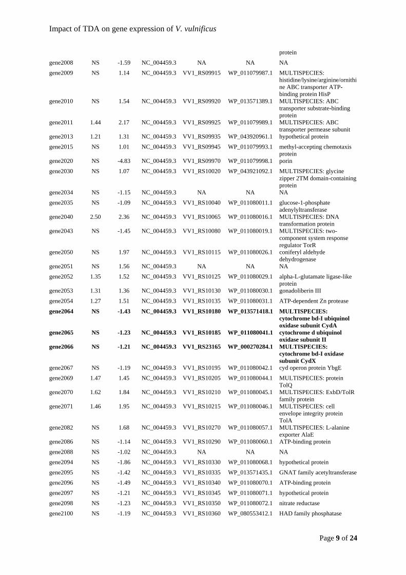
Impact of TDA on gene expression of V. vulnificus
Page 9 of 24
protein
gene2008 NS -1.59 NC_004459.3 NA NA NA
gene2009 NS 1.14 NC_004459.3 VV1_RS09915 WP_011079987.1 MULTISPECIES: histidine/lysine/arginine/ornithine ABC transporter ATP-binding protein HisP
gene2010 NS 1.54 NC_004459.3 VV1_RS09920 WP_013571389.1 MULTISPECIES: ABC transporter substrate-binding protein
gene2011 1.44 2.17 NC_004459.3 VV1_RS09925 WP_011079989.1 MULTISPECIES: ABC transporter permease subunit
gene2013 1.21 1.31 NC_004459.3 VV1_RS09935 WP_043920961.1 hypothetical protein
gene2015 NS 1.01 NC_004459.3 VV1_RS09945 WP_011079993.1 methyl-accepting chemotaxis protein
gene2020 NS -4.83 NC_004459.3 VV1_RS09970 WP_011079998.1 porin
gene2030 NS 1.07 NC_004459.3 VV1_RS10020 WP_043921092.1 MULTISPECIES: glycine zipper 2TM domain-containing protein
gene2034 NS -1.15 NC_004459.3 NA NA NA
gene2035 NS -1.09 NC_004459.3 VV1_RS10040 WP_011080011.1 glucose-1-phosphate adenylyltransferase
gene2040 2.50 2.36 NC_004459.3 VV1_RS10065 WP_011080016.1 MULTISPECIES: DNA transformation protein
gene2043 NS -1.45 NC_004459.3 VV1_RS10080 WP_011080019.1 MULTISPECIES: two-component system response regulator TorR
gene2050 NS 1.97 NC_004459.3 VV1_RS10115 WP_011080026.1 coniferyl aldehyde dehydrogenase
gene2051 NS 1.56 NC_004459.3 NA NA NA
gene2052 1.35 1.52 NC_004459.3 VV1_RS10125 WP_011080029.1 alpha-L-glutamate ligase-like protein
gene2053 1.31 1.36 NC_004459.3 VV1_RS10130 WP_011080030.1 gonadoliberin III
gene2054 1.27 1.51 NC_004459.3 VV1_RS10135 WP_011080031.1 ATP-dependent Zn protease
gene2064 NS -1.43 NC_004459.3 VV1_RS10180 WP_013571418.1 MULTISPECIES: cytochrome bd-I ubiquinol oxidase subunit CydA
gene2065 NS -1.23 NC_004459.3 VV1_RS10185 WP_011080041.1 cytochrome d ubiquinol oxidase subunit II
gene2066 NS -1.21 NC_004459.3 VV1_RS23165 WP_000270284.1 MULTISPECIES: cytochrome bd-I oxidase subunit CydX
gene2067 NS -1.19 NC_004459.3 VV1_RS10195 WP_011080042.1 cyd operon protein YbgE
gene2069 1.47 1.45 NC_004459.3 VV1_RS10205 WP_011080044.1 MULTISPECIES: protein TolQ
gene2070 1.62 1.84 NC_004459.3 VV1_RS10210 WP_011080045.1 MULTISPECIES: ExbD/TolR family protein
gene2071 1.46 1.95 NC_004459.3 VV1_RS10215 WP_011080046.1 MULTISPECIES: cell envelope integrity protein TolA
gene2082 NS 1.68 NC_004459.3 VV1_RS10270 WP_011080057.1 MULTISPECIES: L-alanine exporter AlaE
gene2086 NS -1.14 NC_004459.3 VV1_RS10290 WP_011080060.1 ATP-binding protein
gene2088 NS -1.02 NC_004459.3 NA NA NA
gene2094 NS -1.86 NC_004459.3 VV1_RS10330 WP_011080068.1 hypothetical protein
gene2095 NS -1.42 NC_004459.3 VV1_RS10335 WP_013571435.1 GNAT family acetyltransferase
gene2096 NS -1.49 NC_004459.3 VV1_RS10340 WP_011080070.1 ATP-binding protein
gene2097 NS -1.21 NC_004459.3 VV1_RS10345 WP_011080071.1 hypothetical protein
gene2098 NS -1.23 NC_004459.3 VV1_RS10350 WP_011080072.1 nitrate reductase
gene2100 NS -1.19 NC_004459.3 VV1_RS10360 WP_080553412.1 HAD family phosphatase
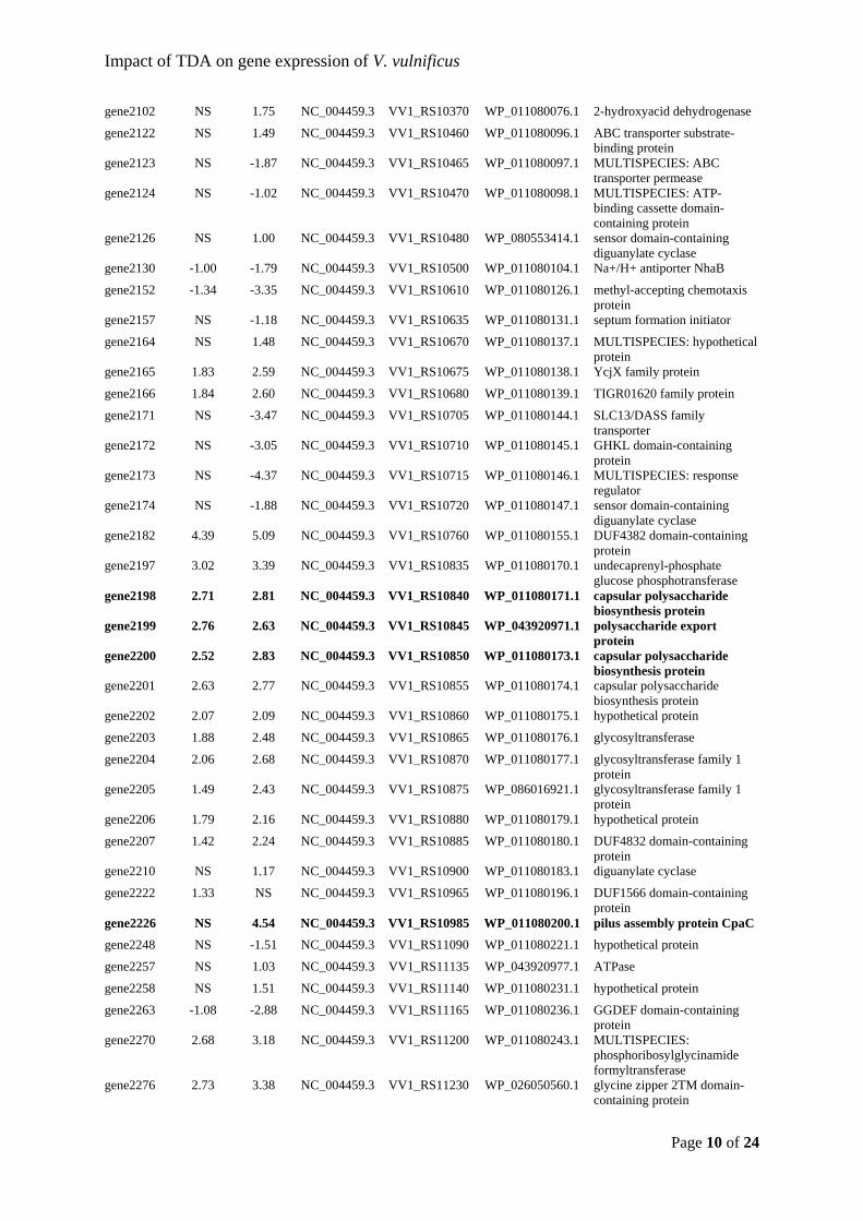
Impact of TDA on gene expression of V. vulnificus
Page 10 of 24
gene2102 NS 1.75 NC_004459.3 VV1_RS10370 WP_011080076.1 2-hydroxyacid dehydrogenase
gene2122 NS 1.49 NC_004459.3 VV1_RS10460 WP_011080096.1 ABC transporter substrate-binding protein
gene2123 NS -1.87 NC_004459.3 VV1_RS10465 WP_011080097.1 MULTISPECIES: ABC transporter permease
gene2124 NS -1.02 NC_004459.3 VV1_RS10470 WP_011080098.1 MULTISPECIES: ATP-binding cassette domain-containing protein
gene2126 NS 1.00 NC_004459.3 VV1_RS10480 WP_080553414.1 sensor domain-containing diguanylate cyclase
gene2130 -1.00 -1.79 NC_004459.3 VV1_RS10500 WP_011080104.1 Na+/H+ antiporter NhaB
gene2152 -1.34 -3.35 NC_004459.3 VV1_RS10610 WP_011080126.1 methyl-accepting chemotaxis protein
gene2157 NS -1.18 NC_004459.3 VV1_RS10635 WP_011080131.1 septum formation initiator
gene2164 NS 1.48 NC_004459.3 VV1_RS10670 WP_011080137.1 MULTISPECIES: hypothetical protein
gene2165 1.83 2.59 NC_004459.3 VV1_RS10675 WP_011080138.1 YcjX family protein
gene2166 1.84 2.60 NC_004459.3 VV1_RS10680 WP_011080139.1 TIGR01620 family protein
gene2171 NS -3.47 NC_004459.3 VV1_RS10705 WP_011080144.1 SLC13/DASS family transporter
gene2172 NS -3.05 NC_004459.3 VV1_RS10710 WP_011080145.1 GHKL domain-containing protein
gene2173 NS -4.37 NC_004459.3 VV1_RS10715 WP_011080146.1 MULTISPECIES: response regulator
gene2174 NS -1.88 NC_004459.3 VV1_RS10720 WP_011080147.1 sensor domain-containing diguanylate cyclase
gene2182 4.39 5.09 NC_004459.3 VV1_RS10760 WP_011080155.1 DUF4382 domain-containing protein
gene2197 3.02 3.39 NC_004459.3 VV1_RS10835 WP_011080170.1 undecaprenyl-phosphate glucose phosphotransferase
gene2198 2.71 2.81 NC_004459.3 VV1_RS10840 WP_011080171.1 capsular polysaccharide biosynthesis protein
gene2199 2.76 2.63 NC_004459.3 VV1_RS10845 WP_043920971.1 polysaccharide export protein
gene2200 2.52 2.83 NC_004459.3 VV1_RS10850 WP_011080173.1 capsular polysaccharide biosynthesis protein
gene2201 2.63 2.77 NC_004459.3 VV1_RS10855 WP_011080174.1 capsular polysaccharide biosynthesis protein
gene2202 2.07 2.09 NC_004459.3 VV1_RS10860 WP_011080175.1 hypothetical protein
gene2203 1.88 2.48 NC_004459.3 VV1_RS10865 WP_011080176.1 glycosyltransferase
gene2204 2.06 2.68 NC_004459.3 VV1_RS10870 WP_011080177.1 glycosyltransferase family 1 protein
gene2205 1.49 2.43 NC_004459.3 VV1_RS10875 WP_086016921.1 glycosyltransferase family 1 protein
gene2206 1.79 2.16 NC_004459.3 VV1_RS10880 WP_011080179.1 hypothetical protein
gene2207 1.42 2.24 NC_004459.3 VV1_RS10885 WP_011080180.1 DUF4832 domain-containing protein
gene2210 NS 1.17 NC_004459.3 VV1_RS10900 WP_011080183.1 diguanylate cyclase
gene2222 1.33 NS NC_004459.3 VV1_RS10965 WP_011080196.1 DUF1566 domain-containing protein
gene2226 NS 4.54 NC_004459.3 VV1_RS10985 WP_011080200.1 pilus assembly protein CpaC
gene2248 NS -1.51 NC_004459.3 VV1_RS11090 WP_011080221.1 hypothetical protein
gene2257 NS 1.03 NC_004459.3 VV1_RS11135 WP_043920977.1 ATPase
gene2258 NS 1.51 NC_004459.3 VV1_RS11140 WP_011080231.1 hypothetical protein
gene2263 -1.08 -2.88 NC_004459.3 VV1_RS11165 WP_011080236.1 GGDEF domain-containing protein
gene2270 2.68 3.18 NC_004459.3 VV1_RS11200 WP_011080243.1 MULTISPECIES: phosphoribosylglycinamide formyltransferase
gene2276 2.73 3.38 NC_004459.3 VV1_RS11230 WP_026050560.1 glycine zipper 2TM domain-containing protein
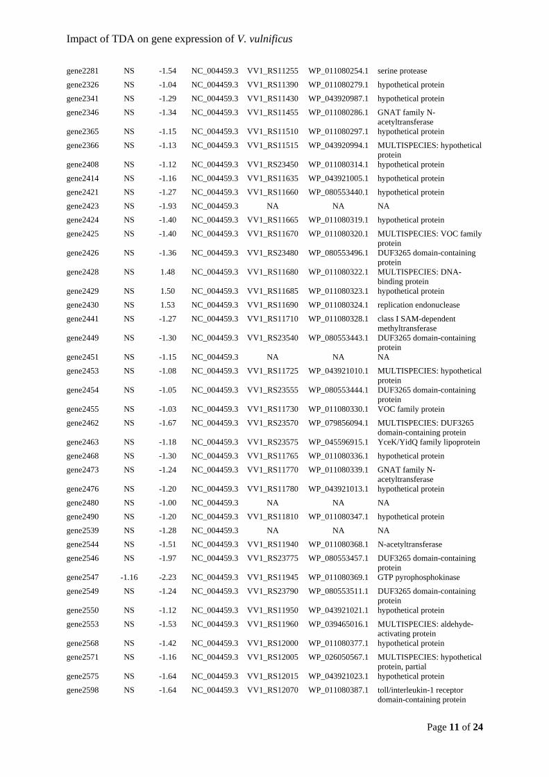
Impact of TDA on gene expression of V. vulnificus
Page 11 of 24
gene2281 NS -1.54 NC_004459.3 VV1_RS11255 WP_011080254.1 serine protease
gene2326 NS -1.04 NC_004459.3 VV1_RS11390 WP_011080279.1 hypothetical protein
gene2341 NS -1.29 NC_004459.3 VV1_RS11430 WP_043920987.1 hypothetical protein
gene2346 NS -1.34 NC_004459.3 VV1_RS11455 WP_011080286.1 GNAT family N-acetyltransferase
gene2365 NS -1.15 NC_004459.3 VV1_RS11510 WP_011080297.1 hypothetical protein
gene2366 NS -1.13 NC_004459.3 VV1_RS11515 WP_043920994.1 MULTISPECIES: hypothetical protein
gene2408 NS -1.12 NC_004459.3 VV1_RS23450 WP_011080314.1 hypothetical protein
gene2414 NS -1.16 NC_004459.3 VV1_RS11635 WP_043921005.1 hypothetical protein
gene2421 NS -1.27 NC_004459.3 VV1_RS11660 WP_080553440.1 hypothetical protein
gene2423 NS -1.93 NC_004459.3 NA NA NA
gene2424 NS -1.40 NC_004459.3 VV1_RS11665 WP_011080319.1 hypothetical protein
gene2425 NS -1.40 NC_004459.3 VV1_RS11670 WP_011080320.1 MULTISPECIES: VOC family protein
gene2426 NS -1.36 NC_004459.3 VV1_RS23480 WP_080553496.1 DUF3265 domain-containing protein
gene2428 NS 1.48 NC_004459.3 VV1_RS11680 WP_011080322.1 MULTISPECIES: DNA-binding protein
gene2429 NS 1.50 NC_004459.3 VV1_RS11685 WP_011080323.1 hypothetical protein
gene2430 NS 1.53 NC_004459.3 VV1_RS11690 WP_011080324.1 replication endonuclease
gene2441 NS -1.27 NC_004459.3 VV1_RS11710 WP_011080328.1 class I SAM-dependent methyltransferase
gene2449 NS -1.30 NC_004459.3 VV1_RS23540 WP_080553443.1 DUF3265 domain-containing protein
gene2451 NS -1.15 NC_004459.3 NA NA NA
gene2453 NS -1.08 NC_004459.3 VV1_RS11725 WP_043921010.1 MULTISPECIES: hypothetical protein
gene2454 NS -1.05 NC_004459.3 VV1_RS23555 WP_080553444.1 DUF3265 domain-containing protein
gene2455 NS -1.03 NC_004459.3 VV1_RS11730 WP_011080330.1 VOC family protein
gene2462 NS -1.67 NC_004459.3 VV1_RS23570 WP_079856094.1 MULTISPECIES: DUF3265 domain-containing protein
gene2463 NS -1.18 NC_004459.3 VV1_RS23575 WP_045596915.1 YceK/YidQ family lipoprotein
gene2468 NS -1.30 NC_004459.3 VV1_RS11765 WP_011080336.1 hypothetical protein
gene2473 NS -1.24 NC_004459.3 VV1_RS11770 WP_011080339.1 GNAT family N-acetyltransferase
gene2476 NS -1.20 NC_004459.3 VV1_RS11780 WP_043921013.1 hypothetical protein
gene2480 NS -1.00 NC_004459.3 NA NA NA
gene2490 NS -1.20 NC_004459.3 VV1_RS11810 WP_011080347.1 hypothetical protein
gene2539 NS -1.28 NC_004459.3 NA NA NA
gene2544 NS -1.51 NC_004459.3 VV1_RS11940 WP_011080368.1 N-acetyltransferase
gene2546 NS -1.97 NC_004459.3 VV1_RS23775 WP_080553457.1 DUF3265 domain-containing protein
gene2547 -1.16 -2.23 NC_004459.3 VV1_RS11945 WP_011080369.1 GTP pyrophosphokinase
gene2549 NS -1.24 NC_004459.3 VV1_RS23790 WP_080553511.1 DUF3265 domain-containing protein
gene2550 NS -1.12 NC_004459.3 VV1_RS11950 WP_043921021.1 hypothetical protein
gene2553 NS -1.53 NC_004459.3 VV1_RS11960 WP_039465016.1 MULTISPECIES: aldehyde-activating protein
gene2568 NS -1.42 NC_004459.3 VV1_RS12000 WP_011080377.1 hypothetical protein
gene2571 NS -1.16 NC_004459.3 VV1_RS12005 WP_026050567.1 MULTISPECIES: hypothetical protein, partial
gene2575 NS -1.64 NC_004459.3 VV1_RS12015 WP_043921023.1 hypothetical protein
gene2598 NS -1.64 NC_004459.3 VV1_RS12070 WP_011080387.1 toll/interleukin-1 receptor domain-containing protein
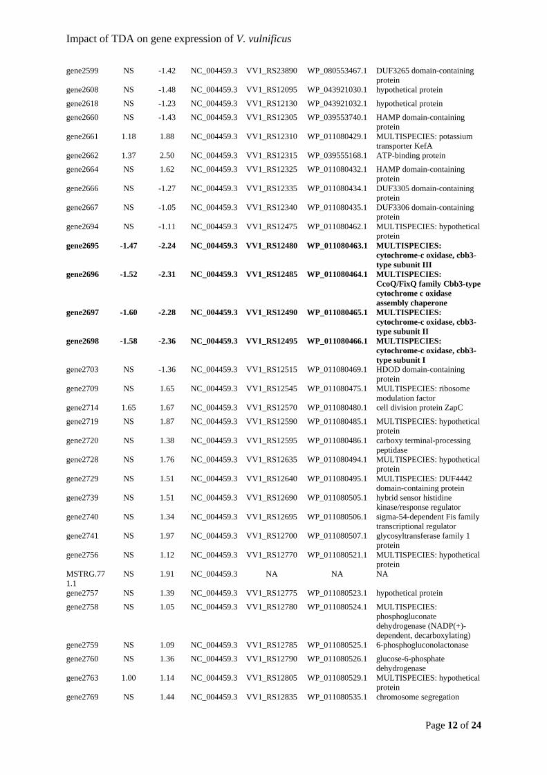
Impact of TDA on gene expression of V. vulnificus
Page 12 of 24
gene2599 NS -1.42 NC_004459.3 VV1_RS23890 WP_080553467.1 DUF3265 domain-containing protein
gene2608 NS -1.48 NC_004459.3 VV1_RS12095 WP_043921030.1 hypothetical protein
gene2618 NS -1.23 NC_004459.3 VV1_RS12130 WP_043921032.1 hypothetical protein
gene2660 NS -1.43 NC_004459.3 VV1_RS12305 WP_039553740.1 HAMP domain-containing protein
gene2661 1.18 1.88 NC_004459.3 VV1_RS12310 WP_011080429.1 MULTISPECIES: potassium transporter KefA
gene2662 1.37 2.50 NC_004459.3 VV1_RS12315 WP_039555168.1 ATP-binding protein
gene2664 NS 1.62 NC_004459.3 VV1_RS12325 WP_011080432.1 HAMP domain-containing protein
gene2666 NS -1.27 NC_004459.3 VV1_RS12335 WP_011080434.1 DUF3305 domain-containing protein
gene2667 NS -1.05 NC_004459.3 VV1_RS12340 WP_011080435.1 DUF3306 domain-containing protein
gene2694 NS -1.11 NC_004459.3 VV1_RS12475 WP_011080462.1 MULTISPECIES: hypothetical protein
gene2695 -1.47 -2.24 NC_004459.3 VV1_RS12480 WP_011080463.1 MULTISPECIES: cytochrome-c oxidase, cbb3-type subunit III
gene2696 -1.52 -2.31 NC_004459.3 VV1_RS12485 WP_011080464.1 MULTISPECIES: CcoQ/FixQ family Cbb3-type cytochrome c oxidase assembly chaperone
gene2697 -1.60 -2.28 NC_004459.3 VV1_RS12490 WP_011080465.1 MULTISPECIES: cytochrome-c oxidase, cbb3-type subunit II
gene2698 -1.58 -2.36 NC_004459.3 VV1_RS12495 WP_011080466.1 MULTISPECIES: cytochrome-c oxidase, cbb3-type subunit I
gene2703 NS -1.36 NC_004459.3 VV1_RS12515 WP_011080469.1 HDOD domain-containing protein
gene2709 NS 1.65 NC_004459.3 VV1_RS12545 WP_011080475.1 MULTISPECIES: ribosome modulation factor
gene2714 1.65 1.67 NC_004459.3 VV1_RS12570 WP_011080480.1 cell division protein ZapC
gene2719 NS 1.87 NC_004459.3 VV1_RS12590 WP_011080485.1 MULTISPECIES: hypothetical protein
gene2720 NS 1.38 NC_004459.3 VV1_RS12595 WP_011080486.1 carboxy terminal-processing peptidase
gene2728 NS 1.76 NC_004459.3 VV1_RS12635 WP_011080494.1 MULTISPECIES: hypothetical protein
gene2729 NS 1.51 NC_004459.3 VV1_RS12640 WP_011080495.1 MULTISPECIES: DUF4442 domain-containing protein
gene2739 NS 1.51 NC_004459.3 VV1_RS12690 WP_011080505.1 hybrid sensor histidine kinase/response regulator
gene2740 NS 1.34 NC_004459.3 VV1_RS12695 WP_011080506.1 sigma-54-dependent Fis family transcriptional regulator
gene2741 NS 1.97 NC_004459.3 VV1_RS12700 WP_011080507.1 glycosyltransferase family 1 protein
gene2756 NS 1.12 NC_004459.3 VV1_RS12770 WP_011080521.1 MULTISPECIES: hypothetical protein
MSTRG.771.1
NS 1.91 NC_004459.3 NA NA NA
gene2757 NS 1.39 NC_004459.3 VV1_RS12775 WP_011080523.1 hypothetical protein
gene2758 NS 1.05 NC_004459.3 VV1_RS12780 WP_011080524.1 MULTISPECIES: phosphogluconate dehydrogenase (NADP(+)-dependent, decarboxylating)
gene2759 NS 1.09 NC_004459.3 VV1_RS12785 WP_011080525.1 6-phosphogluconolactonase
gene2760 NS 1.36 NC_004459.3 VV1_RS12790 WP_011080526.1 glucose-6-phosphate dehydrogenase
gene2763 1.00 1.14 NC_004459.3 VV1_RS12805 WP_011080529.1 MULTISPECIES: hypothetical protein
gene2769 NS 1.44 NC_004459.3 VV1_RS12835 WP_011080535.1 chromosome segregation
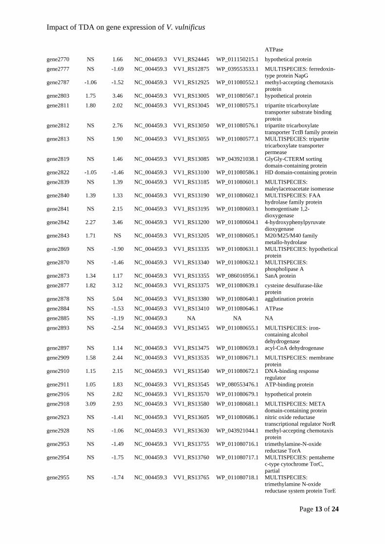
Impact of TDA on gene expression of V. vulnificus
Page 13 of 24
ATPase
gene2770 NS 1.66 NC_004459.3 VV1_RS24445 WP_011150215.1 hypothetical protein
gene2777 NS -1.69 NC_004459.3 VV1_RS12875 WP_039553533.1 MULTISPECIES: ferredoxin-type protein NapG
gene2787 -1.06 -1.52 NC_004459.3 VV1_RS12925 WP_011080552.1 methyl-accepting chemotaxis protein
gene2803 1.75 3.46 NC_004459.3 VV1_RS13005 WP_011080567.1 hypothetical protein
gene2811 1.80 2.02 NC_004459.3 VV1_RS13045 WP_011080575.1 tripartite tricarboxylate transporter substrate binding protein
gene2812 NS 2.76 NC_004459.3 VV1_RS13050 WP_011080576.1 tripartite tricarboxylate transporter TctB family protein
gene2813 NS 1.90 NC_004459.3 VV1_RS13055 WP_011080577.1 MULTISPECIES: tripartite tricarboxylate transporter permease
gene2819 NS 1.46 NC_004459.3 VV1_RS13085 WP_043921038.1 GlyGly-CTERM sorting domain-containing protein
gene2822 -1.05 -1.46 NC_004459.3 VV1_RS13100 WP_011080586.1 HD domain-containing protein
gene2839 NS 1.39 NC_004459.3 VV1_RS13185 WP_011080601.1 MULTISPECIES: maleylacetoacetate isomerase
gene2840 1.39 1.33 NC_004459.3 VV1_RS13190 WP_011080602.1 MULTISPECIES: FAA hydrolase family protein
gene2841 NS 2.15 NC_004459.3 VV1_RS13195 WP_011080603.1 homogentisate 1,2-dioxygenase
gene2842 2.27 3.46 NC_004459.3 VV1_RS13200 WP_011080604.1 4-hydroxyphenylpyruvate dioxygenase
gene2843 1.71 NS NC_004459.3 VV1_RS13205 WP_011080605.1 M20/M25/M40 family metallo-hydrolase
gene2869 NS -1.90 NC_004459.3 VV1_RS13335 WP_011080631.1 MULTISPECIES: hypothetical protein
gene2870 NS -1.46 NC_004459.3 VV1_RS13340 WP_011080632.1 MULTISPECIES: phospholipase A
gene2873 1.34 1.17 NC_004459.3 VV1_RS13355 WP_086016956.1 SanA protein
gene2877 1.82 3.12 NC_004459.3 VV1_RS13375 WP_011080639.1 cysteine desulfurase-like protein
gene2878 NS 5.04 NC_004459.3 VV1_RS13380 WP_011080640.1 agglutination protein
gene2884 NS -1.53 NC_004459.3 VV1_RS13410 WP_011080646.1 ATPase
gene2885 NS -1.19 NC_004459.3 NA NA NA
gene2893 NS -2.54 NC_004459.3 VV1_RS13455 WP_011080655.1 MULTISPECIES: iron-containing alcohol dehydrogenase
gene2897 NS 1.14 NC_004459.3 VV1_RS13475 WP_011080659.1 acyl-CoA dehydrogenase
gene2909 1.58 2.44 NC_004459.3 VV1_RS13535 WP_011080671.1 MULTISPECIES: membrane protein
gene2910 1.15 2.15 NC_004459.3 VV1_RS13540 WP_011080672.1 DNA-binding response regulator
gene2911 1.05 1.83 NC_004459.3 VV1_RS13545 WP_080553476.1 ATP-binding protein
gene2916 NS 2.82 NC_004459.3 VV1_RS13570 WP_011080679.1 hypothetical protein
gene2918 3.09 2.93 NC_004459.3 VV1_RS13580 WP_011080681.1 MULTISPECIES: META domain-containing protein
gene2923 NS -1.41 NC_004459.3 VV1_RS13605 WP_011080686.1 nitric oxide reductase transcriptional regulator NorR
gene2928 NS -1.06 NC_004459.3 VV1_RS13630 WP_043921044.1 methyl-accepting chemotaxis protein
gene2953 NS -1.49 NC_004459.3 VV1_RS13755 WP_011080716.1 trimethylamine-N-oxide reductase TorA
gene2954 NS -1.75 NC_004459.3 VV1_RS13760 WP_011080717.1 MULTISPECIES: pentaheme c-type cytochrome TorC, partial
gene2955 NS -1.74 NC_004459.3 VV1_RS13765 WP_011080718.1 MULTISPECIES: trimethylamine N-oxide reductase system protein TorE
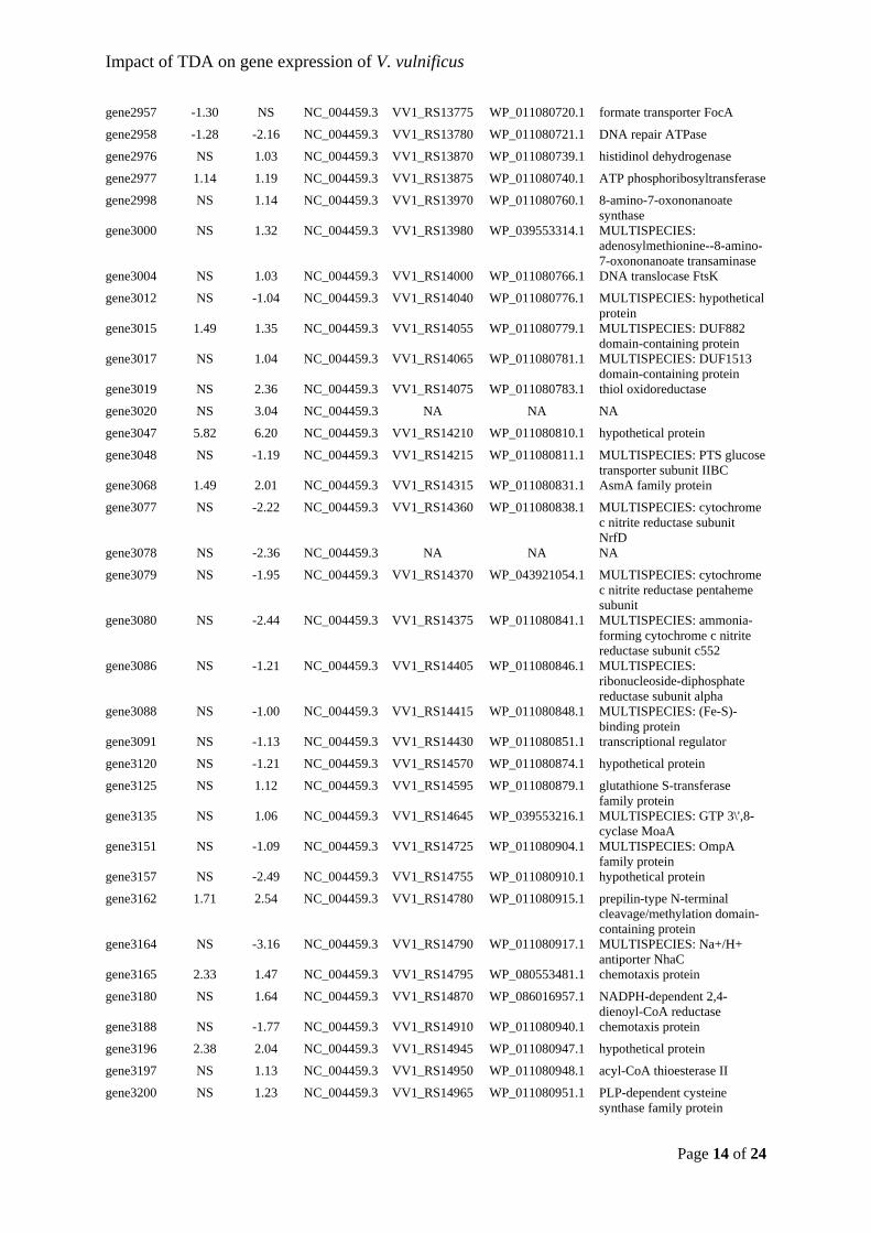
Impact of TDA on gene expression of V. vulnificus
Page 14 of 24
gene2957 -1.30 NS NC_004459.3 VV1_RS13775 WP_011080720.1 formate transporter FocA
gene2958 -1.28 -2.16 NC_004459.3 VV1_RS13780 WP_011080721.1 DNA repair ATPase
gene2976 NS 1.03 NC_004459.3 VV1_RS13870 WP_011080739.1 histidinol dehydrogenase
gene2977 1.14 1.19 NC_004459.3 VV1_RS13875 WP_011080740.1 ATP phosphoribosyltransferase
gene2998 NS 1.14 NC_004459.3 VV1_RS13970 WP_011080760.1 8-amino-7-oxononanoate synthase
gene3000 NS 1.32 NC_004459.3 VV1_RS13980 WP_039553314.1 MULTISPECIES: adenosylmethionine--8-amino-7-oxononanoate transaminase
gene3004 NS 1.03 NC_004459.3 VV1_RS14000 WP_011080766.1 DNA translocase FtsK
gene3012 NS -1.04 NC_004459.3 VV1_RS14040 WP_011080776.1 MULTISPECIES: hypothetical protein
gene3015 1.49 1.35 NC_004459.3 VV1_RS14055 WP_011080779.1 MULTISPECIES: DUF882 domain-containing protein
gene3017 NS 1.04 NC_004459.3 VV1_RS14065 WP_011080781.1 MULTISPECIES: DUF1513 domain-containing protein
gene3019 NS 2.36 NC_004459.3 VV1_RS14075 WP_011080783.1 thiol oxidoreductase
gene3020 NS 3.04 NC_004459.3 NA NA NA
gene3047 5.82 6.20 NC_004459.3 VV1_RS14210 WP_011080810.1 hypothetical protein
gene3048 NS -1.19 NC_004459.3 VV1_RS14215 WP_011080811.1 MULTISPECIES: PTS glucose transporter subunit IIBC
gene3068 1.49 2.01 NC_004459.3 VV1_RS14315 WP_011080831.1 AsmA family protein
gene3077 NS -2.22 NC_004459.3 VV1_RS14360 WP_011080838.1 MULTISPECIES: cytochrome c nitrite reductase subunit NrfD
gene3078 NS -2.36 NC_004459.3 NA NA NA
gene3079 NS -1.95 NC_004459.3 VV1_RS14370 WP_043921054.1 MULTISPECIES: cytochrome c nitrite reductase pentaheme subunit
gene3080 NS -2.44 NC_004459.3 VV1_RS14375 WP_011080841.1 MULTISPECIES: ammonia-forming cytochrome c nitrite reductase subunit c552
gene3086 NS -1.21 NC_004459.3 VV1_RS14405 WP_011080846.1 MULTISPECIES: ribonucleoside-diphosphate reductase subunit alpha
gene3088 NS -1.00 NC_004459.3 VV1_RS14415 WP_011080848.1 MULTISPECIES: (Fe-S)-binding protein
gene3091 NS -1.13 NC_004459.3 VV1_RS14430 WP_011080851.1 transcriptional regulator
gene3120 NS -1.21 NC_004459.3 VV1_RS14570 WP_011080874.1 hypothetical protein
gene3125 NS 1.12 NC_004459.3 VV1_RS14595 WP_011080879.1 glutathione S-transferase family protein
gene3135 NS 1.06 NC_004459.3 VV1_RS14645 WP_039553216.1 MULTISPECIES: GTP 3\',8-cyclase MoaA
gene3151 NS -1.09 NC_004459.3 VV1_RS14725 WP_011080904.1 MULTISPECIES: OmpA family protein
gene3157 NS -2.49 NC_004459.3 VV1_RS14755 WP_011080910.1 hypothetical protein
gene3162 1.71 2.54 NC_004459.3 VV1_RS14780 WP_011080915.1 prepilin-type N-terminal cleavage/methylation domain-containing protein
gene3164 NS -3.16 NC_004459.3 VV1_RS14790 WP_011080917.1 MULTISPECIES: Na+/H+ antiporter NhaC
gene3165 2.33 1.47 NC_004459.3 VV1_RS14795 WP_080553481.1 chemotaxis protein
gene3180 NS 1.64 NC_004459.3 VV1_RS14870 WP_086016957.1 NADPH-dependent 2,4-dienoyl-CoA reductase
gene3188 NS -1.77 NC_004459.3 VV1_RS14910 WP_011080940.1 chemotaxis protein
gene3196 2.38 2.04 NC_004459.3 VV1_RS14945 WP_011080947.1 hypothetical protein
gene3197 NS 1.13 NC_004459.3 VV1_RS14950 WP_011080948.1 acyl-CoA thioesterase II
gene3200 NS 1.23 NC_004459.3 VV1_RS14965 WP_011080951.1 PLP-dependent cysteine synthase family protein
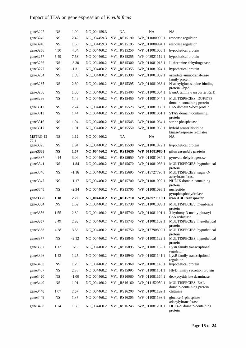
Impact of TDA on gene expression of V. vulnificus
Page 15 of 24
gene3227 NS 1.09 NC_004459.3 NA NA NA
gene3245 NS 2.42 NC_004459.3 VV1_RS15190 WP_011080993.1 response regulator
gene3246 NS 1.65 NC_004459.3 VV1_RS15195 WP_011080994.1 response regulator
gene3256 4.30 4.84 NC_004460.2 VV1_RS15250 WP_011081003.1 hypothetical protein
gene3257 5.49 7.53 NC_004460.2 VV1_RS15255 WP_043921112.1 hypothetical protein
gene3266 NS -3.20 NC_004460.2 VV1_RS15300 WP_011081013.1 L-threonine dehydrogenase
gene3277 NS -1.31 NC_004460.2 VV1_RS15355 WP_011081024.1 hypothetical protein
gene3284 NS 1.09 NC_004460.2 VV1_RS15390 WP_011081032.1 aspartate aminotransferase family protein
gene3285 NS 2.60 NC_004460.2 VV1_RS15395 WP_011081033.1 N-acetylglucosamine-binding protein GbpA
gene3286 NS 1.03 NC_004460.2 VV1_RS15400 WP_011081034.1 EamA family transporter RarD
gene3296 NS 1.49 NC_004460.2 VV1_RS15450 WP_011081044.1 MULTISPECIES: DUF3763 domain-containing protein
gene3312 NS 2.24 NC_004460.2 VV1_RS15525 WP_011081060.1 PAS domain S-box protein
gene3313 NS 1.44 NC_004460.2 VV1_RS15530 WP_011081061.1 STAS domain-containing protein
gene3316 NS 1.04 NC_004460.2 VV1_RS15545 WP_011081064.1 serine phosphatase
gene3317 NS 1.01 NC_004460.2 VV1_RS15550 WP_011081065.1 hybrid sensor histidine kinase/response regulator
MSTRG.1272.1
NS 1.12 NC_004460.2 NA NA NA
gene3325 NS 1.94 NC_004460.2 VV1_RS15590 WP_011081072.1 hypothetical protein
gene3333 NS 1.57 NC_004460.2 VV1_RS15630 WP_011081080.1 pilus assembly protein
gene3337 4.14 3.06 NC_004460.2 VV1_RS15650 WP_011081084.1 pyruvate dehydrogenase
gene3341 NS -1.84 NC_004460.2 VV1_RS15670 WP_011081086.1 MULTISPECIES: hypothetical protein
gene3346 NS -1.16 NC_004460.2 VV1_RS15695 WP_015727796.1 MULTISPECIES: sugar O-acetyltransferase
gene3347 NS -1.17 NC_004460.2 VV1_RS15700 WP_011081092.1 NUDIX domain-containing protein
gene3348 NS -2.34 NC_004460.2 VV1_RS15705 WP_011081093.1 nucleotide pyrophosphohydrolase
gene3350 1.18 2.22 NC_004460.2 VV1_RS15710 WP_043921119.1 iron ABC transporter
gene3354 NS 1.62 NC_004460.2 VV1_RS15730 WP_011081099.1 MULTISPECIES: membrane protein
gene3356 1.55 2.82 NC_004460.2 VV1_RS15740 WP_011081101.1 3-hydroxy-3-methylglutaryl-CoA reductase
gene3357 3.49 2.93 NC_004460.2 VV1_RS15745 WP_011081102.1 MULTISPECIES: hypothetical protein
gene3358 4.28 3.58 NC_004460.2 VV1_RS15750 WP_017790802.1 MULTISPECIES: hypothetical protein
gene3377 NS -2.12 NC_004460.2 VV1_RS15845 WP_011081122.1 MULTISPECIES: hypothetical protein
gene3387 1.12 NS NC_004460.2 VV1_RS15895 WP_011081132.1 LysR family transcriptional regulator
gene3396 1.43 1.25 NC_004460.2 VV1_RS15940 WP_011081141.1 LysR family transcriptional regulator
gene3400 NS 1.29 NC_004460.2 VV1_RS15960 WP_011081145.1 hypothetical protein
gene3407 NS 2.38 NC_004460.2 VV1_RS15995 WP_011081151.1 HlyD family secretion protein
gene3420 NS -1.00 NC_004460.2 VV1_RS16060 WP_011081164.1 deoxycytidylate deaminase
gene3440 NS 1.01 NC_004460.2 VV1_RS16160 WP_011152050.1 MULTISPECIES: EAL domain-containing protein
gene3448 1.07 2.57 NC_004460.2 VV1_RS16200 WP_011081192.1 chitinase
gene3449 NS 1.37 NC_004460.2 VV1_RS16205 WP_011081193.1 glucose-1-phosphate adenylyltransferase
gene3458 1.24 1.30 NC_004460.2 VV1_RS16245 WP_011081201.1 DUF479 domain-containing protein
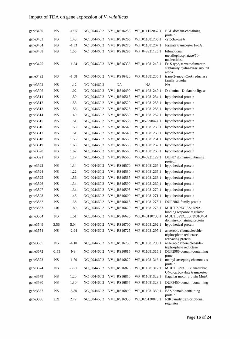
Impact of TDA on gene expression of V. vulnificus
Page 16 of 24
gene3460 NS -1.05 NC_004460.2 VV1_RS16255 WP_011152067.1 EAL domain-containing protein
gene3462 NS 1.43 NC_004460.2 VV1_RS16265 WP_011081205.1 cytochrome b
gene3464 NS -1.53 NC_004460.2 VV1_RS16275 WP_011081207.1 formate transporter FocA
gene3468 NS 1.55 NC_004460.2 VV1_RS16295 WP_043921125.1 bifunctional metallophosphatase/5\'-nucleotidase
gene3475 NS -1.54 NC_004460.2 VV1_RS16335 WP_011081218.1 Fe-S type, tartrate/fumarate subfamily hydro-lyase subunit alpha
gene3492 NS -1.58 NC_004460.2 VV1_RS16420 WP_011081235.1 trans-2-enoyl-CoA reductase family protein
gene3502 NS 1.12 NC_004460.2 NA NA NA
gene3506 NS 1.02 NC_004460.2 VV1_RS16490 WP_011081249.1 D-alanine--D-alanine ligase
gene3511 NS 1.59 NC_004460.2 VV1_RS16515 WP_011081254.1 hypothetical protein
gene3512 NS 1.58 NC_004460.2 VV1_RS16520 WP_011081255.1 hypothetical protein
gene3513 NS 1.58 NC_004460.2 VV1_RS16525 WP_011081256.1 hypothetical protein
gene3514 NS 1.49 NC_004460.2 VV1_RS16530 WP_011081257.1 hypothetical protein
gene3515 NS 1.51 NC_004460.2 VV1_RS16535 WP_052298474.1 hypothetical protein
gene3516 NS 1.58 NC_004460.2 VV1_RS16540 WP_011081259.1 hypothetical protein
gene3517 NS 1.51 NC_004460.2 VV1_RS16545 WP_011081260.1 hypothetical protein
gene3518 NS 1.55 NC_004460.2 VV1_RS16550 WP_011081261.1 hypothetical protein
gene3519 NS 1.63 NC_004460.2 VV1_RS16555 WP_011081262.1 hypothetical protein
gene3520 NS 1.62 NC_004460.2 VV1_RS16560 WP_011081263.1 hypothetical protein
gene3521 NS 1.17 NC_004460.2 VV1_RS16565 WP_043921129.1 DUF87 domain-containing protein
gene3522 NS 1.34 NC_004460.2 VV1_RS16570 WP_011081265.1 hypothetical protein
gene3524 NS 1.22 NC_004460.2 VV1_RS16580 WP_011081267.1 hypothetical protein
gene3525 NS 1.56 NC_004460.2 VV1_RS16585 WP_011081268.1 hypothetical protein
gene3526 NS 1.34 NC_004460.2 VV1_RS16590 WP_011081269.1 hypothetical protein
gene3527 NS 1.34 NC_004460.2 VV1_RS16595 WP_011081270.1 hypothetical protein
gene3528 NS 1.46 NC_004460.2 VV1_RS16600 WP_011081271.1 hypothetical protein
gene3532 NS 1.38 NC_004460.2 VV1_RS16615 WP_011081275.1 DUF2861 family protein
gene3533 1.01 1.89 NC_004460.2 VV1_RS16620 WP_011081276.1 MULTISPECIES: DNA-binding response regulator
gene3534 NS 1.51 NC_004460.2 VV1_RS16625 WP_040110783.1 MULTISPECIES: DUF3404 domain-containing protein
gene3549 3.56 5.04 NC_004460.2 VV1_RS16700 WP_011081292.1 hypothetical protein
gene3554 NS -2.94 NC_004460.2 VV1_RS16725 WP_011081297.1 anaerobic ribonucleoside-triphosphate reductase-activating protein
gene3555 NS -4.10 NC_004460.2 VV1_RS16730 WP_011081298.1 anaerobic ribonucleoside-triphosphate reductase
gene3572 -1.53 NS NC_004460.2 VV1_RS16815 WP_011081315.1 DUF2986 domain-containing protein
gene3573 NS -1.70 NC_004460.2 VV1_RS16820 WP_011081316.1 methyl-accepting chemotaxis protein
gene3574 NS -3.21 NC_004460.2 VV1_RS16825 WP_011081317.1 MULTISPECIES: anaerobic C4-dicarboxylate transporter
gene3579 NS 1.20 NC_004460.2 VV1_RS16850 WP_011081322.1 flagellar motor protein MotA
gene3580 NS 1.30 NC_004460.2 VV1_RS16855 WP_011081323.1 DUF3450 domain-containing protein
gene3587 NS -3.80 NC_004460.2 VV1_RS16890 WP_011081330.1 PAS domain-containing protein
gene3596 1.21 2.72 NC_004460.2 VV1_RS16935 WP_026130873.1 IclR family transcriptional regulator
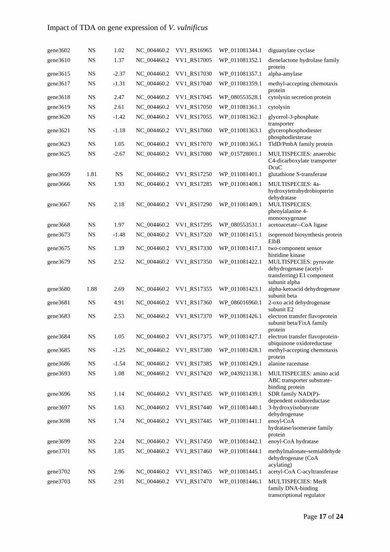
Impact of TDA on gene expression of V. vulnificus
Page 17 of 24
gene3602 NS 1.02 NC_004460.2 VV1_RS16965 WP_011081344.1 diguanylate cyclase
gene3610 NS 1.37 NC_004460.2 VV1_RS17005 WP_011081352.1 dienelactone hydrolase family protein
gene3615 NS -2.37 NC_004460.2 VV1_RS17030 WP_011081357.1 alpha-amylase
gene3617 NS -1.31 NC_004460.2 VV1_RS17040 WP_011081359.1 methyl-accepting chemotaxis protein
gene3618 NS 2.47 NC_004460.2 VV1_RS17045 WP_080553528.1 cytolysin secretion protein
gene3619 NS 2.61 NC_004460.2 VV1_RS17050 WP_011081361.1 cytolysin
gene3620 NS -1.42 NC_004460.2 VV1_RS17055 WP_011081362.1 glycerol-3-phosphate transporter
gene3621 NS -1.18 NC_004460.2 VV1_RS17060 WP_011081363.1 glycerophosphodiester phosphodiesterase
gene3623 NS 1.05 NC_004460.2 VV1_RS17070 WP_011081365.1 TldD/PmbA family protein
gene3625 NS -2.67 NC_004460.2 VV1_RS17080 WP_015728001.1 MULTISPECIES: anaerobic C4-dicarboxylate transporter DcuC
gene3659 1.81 NS NC_004460.2 VV1_RS17250 WP_011081401.1 glutathione S-transferase
gene3666 NS 1.93 NC_004460.2 VV1_RS17285 WP_011081408.1 MULTISPECIES: 4a-hydroxytetrahydrobiopterin dehydratase
gene3667 NS 2.18 NC_004460.2 VV1_RS17290 WP_011081409.1 MULTISPECIES: phenylalanine 4-monooxygenase
gene3668 NS 1.97 NC_004460.2 VV1_RS17295 WP_080553531.1 acetoacetate--CoA ligase
gene3673 NS -1.48 NC_004460.2 VV1_RS17320 WP_011081415.1 isoprenoid biosynthesis protein ElbB
gene3675 NS 1.39 NC_004460.2 VV1_RS17330 WP_011081417.1 two-component sensor histidine kinase
gene3679 NS 2.52 NC_004460.2 VV1_RS17350 WP_011081422.1 MULTISPECIES: pyruvate dehydrogenase (acetyl-transferring) E1 component subunit alpha
gene3680 1.88 2.69 NC_004460.2 VV1_RS17355 WP_011081423.1 alpha-ketoacid dehydrogenase subunit beta
gene3681 NS 4.91 NC_004460.2 VV1_RS17360 WP_086016960.1 2-oxo acid dehydrogenase subunit E2
gene3683 NS 2.53 NC_004460.2 VV1_RS17370 WP_011081426.1 electron transfer flavoprotein subunit beta/FixA family protein
gene3684 NS 1.05 NC_004460.2 VV1_RS17375 WP_011081427.1 electron transfer flavoprotein-ubiquinone oxidoreductase
gene3685 NS -1.25 NC_004460.2 VV1_RS17380 WP_011081428.1 methyl-accepting chemotaxis protein
gene3686 NS -1.54 NC_004460.2 VV1_RS17385 WP_011081429.1 alanine racemase
gene3693 NS 1.08 NC_004460.2 VV1_RS17420 WP_043921138.1 MULTISPECIES: amino acid ABC transporter substrate-binding protein
gene3696 NS 1.14 NC_004460.2 VV1_RS17435 WP_011081439.1 SDR family NAD(P)-dependent oxidoreductase
gene3697 NS 1.63 NC_004460.2 VV1_RS17440 WP_011081440.1 3-hydroxyisobutyrate dehydrogenase
gene3698 NS 1.74 NC_004460.2 VV1_RS17445 WP_011081441.1 enoyl-CoA hydratase/isomerase family protein
gene3699 NS 2.24 NC_004460.2 VV1_RS17450 WP_011081442.1 enoyl-CoA hydratase
gene3701 NS 1.85 NC_004460.2 VV1_RS17460 WP_011081444.1 methylmalonate-semialdehyde dehydrogenase (CoA acylating)
gene3702 NS 2.96 NC_004460.2 VV1_RS17465 WP_011081445.1 acetyl-CoA C-acyltransferase
gene3703 NS 2.91 NC_004460.2 VV1_RS17470 WP_011081446.1 MULTISPECIES: MerR family DNA-binding transcriptional regulator
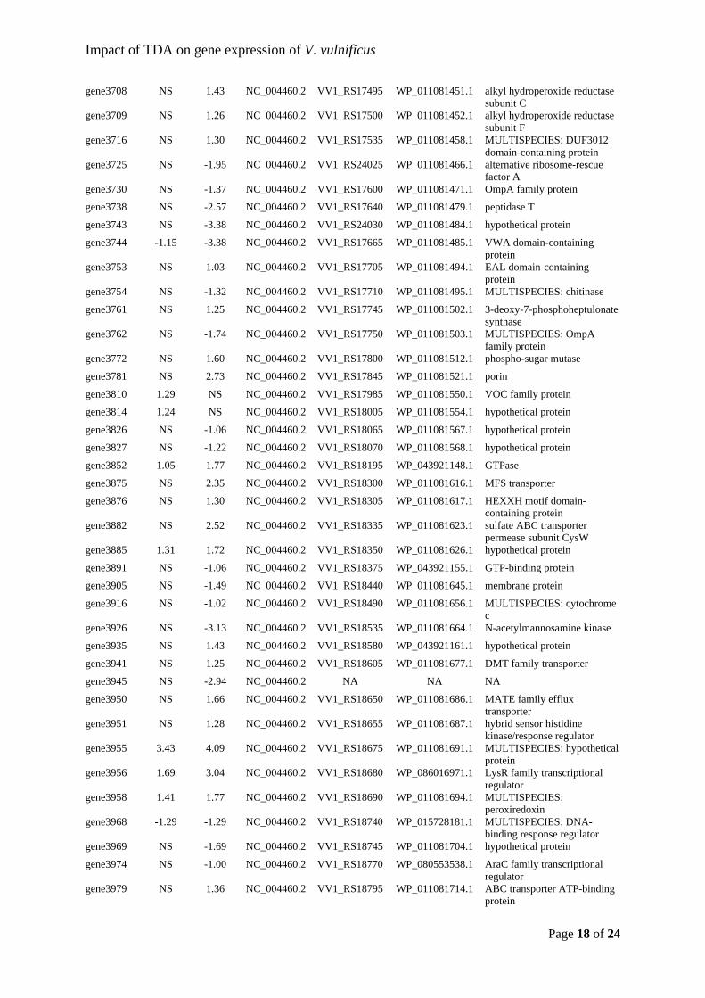
Impact of TDA on gene expression of V. vulnificus
Page 18 of 24
gene3708 NS 1.43 NC_004460.2 VV1_RS17495 WP_011081451.1 alkyl hydroperoxide reductase subunit C
gene3709 NS 1.26 NC_004460.2 VV1_RS17500 WP_011081452.1 alkyl hydroperoxide reductase subunit F
gene3716 NS 1.30 NC_004460.2 VV1_RS17535 WP_011081458.1 MULTISPECIES: DUF3012 domain-containing protein
gene3725 NS -1.95 NC_004460.2 VV1_RS24025 WP_011081466.1 alternative ribosome-rescue factor A
gene3730 NS -1.37 NC_004460.2 VV1_RS17600 WP_011081471.1 OmpA family protein
gene3738 NS -2.57 NC_004460.2 VV1_RS17640 WP_011081479.1 peptidase T
gene3743 NS -3.38 NC_004460.2 VV1_RS24030 WP_011081484.1 hypothetical protein
gene3744 -1.15 -3.38 NC_004460.2 VV1_RS17665 WP_011081485.1 VWA domain-containing protein
gene3753 NS 1.03 NC_004460.2 VV1_RS17705 WP_011081494.1 EAL domain-containing protein
gene3754 NS -1.32 NC_004460.2 VV1_RS17710 WP_011081495.1 MULTISPECIES: chitinase
gene3761 NS 1.25 NC_004460.2 VV1_RS17745 WP_011081502.1 3-deoxy-7-phosphoheptulonate synthase
gene3762 NS -1.74 NC_004460.2 VV1_RS17750 WP_011081503.1 MULTISPECIES: OmpA family protein
gene3772 NS 1.60 NC_004460.2 VV1_RS17800 WP_011081512.1 phospho-sugar mutase
gene3781 NS 2.73 NC_004460.2 VV1_RS17845 WP_011081521.1 porin
gene3810 1.29 NS NC_004460.2 VV1_RS17985 WP_011081550.1 VOC family protein
gene3814 1.24 NS NC_004460.2 VV1_RS18005 WP_011081554.1 hypothetical protein
gene3826 NS -1.06 NC_004460.2 VV1_RS18065 WP_011081567.1 hypothetical protein
gene3827 NS -1.22 NC_004460.2 VV1_RS18070 WP_011081568.1 hypothetical protein
gene3852 1.05 1.77 NC_004460.2 VV1_RS18195 WP_043921148.1 GTPase
gene3875 NS 2.35 NC_004460.2 VV1_RS18300 WP_011081616.1 MFS transporter
gene3876 NS 1.30 NC_004460.2 VV1_RS18305 WP_011081617.1 HEXXH motif domain-containing protein
gene3882 NS 2.52 NC_004460.2 VV1_RS18335 WP_011081623.1 sulfate ABC transporter permease subunit CysW
gene3885 1.31 1.72 NC_004460.2 VV1_RS18350 WP_011081626.1 hypothetical protein
gene3891 NS -1.06 NC_004460.2 VV1_RS18375 WP_043921155.1 GTP-binding protein
gene3905 NS -1.49 NC_004460.2 VV1_RS18440 WP_011081645.1 membrane protein
gene3916 NS -1.02 NC_004460.2 VV1_RS18490 WP_011081656.1 MULTISPECIES: cytochrome c
gene3926 NS -3.13 NC_004460.2 VV1_RS18535 WP_011081664.1 N-acetylmannosamine kinase
gene3935 NS 1.43 NC_004460.2 VV1_RS18580 WP_043921161.1 hypothetical protein
gene3941 NS 1.25 NC_004460.2 VV1_RS18605 WP_011081677.1 DMT family transporter
gene3945 NS -2.94 NC_004460.2 NA NA NA
gene3950 NS 1.66 NC_004460.2 VV1_RS18650 WP_011081686.1 MATE family efflux transporter
gene3951 NS 1.28 NC_004460.2 VV1_RS18655 WP_011081687.1 hybrid sensor histidine kinase/response regulator
gene3955 3.43 4.09 NC_004460.2 VV1_RS18675 WP_011081691.1 MULTISPECIES: hypothetical protein
gene3956 1.69 3.04 NC_004460.2 VV1_RS18680 WP_086016971.1 LysR family transcriptional regulator
gene3958 1.41 1.77 NC_004460.2 VV1_RS18690 WP_011081694.1 MULTISPECIES: peroxiredoxin
gene3968 -1.29 -1.29 NC_004460.2 VV1_RS18740 WP_015728181.1 MULTISPECIES: DNA-binding response regulator
gene3969 NS -1.69 NC_004460.2 VV1_RS18745 WP_011081704.1 hypothetical protein
gene3974 NS -1.00 NC_004460.2 VV1_RS18770 WP_080553538.1 AraC family transcriptional regulator
gene3979 NS 1.36 NC_004460.2 VV1_RS18795 WP_011081714.1 ABC transporter ATP-binding protein
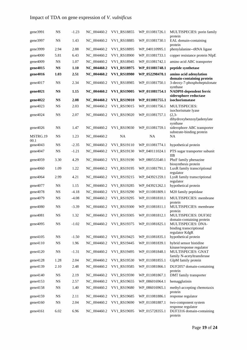
Impact of TDA on gene expression of V. vulnificus
Page 19 of 24
gene3991 NS -1.23 NC_004460.2 VV1_RS18855 WP_011081726.1 MULTISPECIES: porin family protein
gene3997 NS 1.43 NC_004460.2 VV1_RS18885 WP_011081730.1 EAL domain-containing protein
gene3999 2.94 2.88 NC_004460.2 VV1_RS18895 WP_040110995.1 phenylalanine--tRNA ligase
gene4000 5.81 6.43 NC_004460.2 VV1_RS18900 WP_011081733.1 copper resistance protein NlpE
gene4009 NS 1.07 NC_004460.2 VV1_RS18945 WP_011081742.1 amino acid ABC transporter
gene4015 NS 1.10 NC_004460.2 VV1_RS18975 WP_011081748.1 peptide synthetase
gene4016 1.03 2.51 NC_004460.2 VV1_RS18980 WP_052298478.1 amino acid adenylation domain-containing protein
gene4017 NS 2.34 NC_004460.2 VV1_RS18985 WP_011081750.1 3-deoxy-7-phosphoheptulonate synthase
gene4021 NS 1.15 NC_004460.2 VV1_RS19005 WP_011081754.1 NADPH-dependent ferric siderophore reductase
gene4022 NS 2.08 NC_004460.2 VV1_RS19010 WP_011081755.1 isochorismatase
gene4023 NS 2.03 NC_004460.2 VV1_RS19015 WP_011081756.1 MULTISPECIES: isochorismate lyase
gene4024 NS 2.07 NC_004460.2 VV1_RS19020 WP_011081757.1 (2,3-dihydroxybenzoyl)adenylate synthase
gene4026 NS 1.47 NC_004460.2 VV1_RS19030 WP_011081759.1 siderophore ABC transporter substrate-binding protein
MSTRG.1900.1
NS 1.23 NC_004460.2 NA NA NA
gene4043 NS -2.35 NC_004460.2 VV1_RS19110 WP_011081774.1 hypothetical protein
gene4047 NS -1.21 NC_004460.2 VV1_RS19130 WP_040111024.1 PTS sugar transporter subunit IIB
gene4059 3.30 4.29 NC_004460.2 VV1_RS19190 WP_080553540.1 PhzF family phenazine biosynthesis protein
gene4060 1.09 1.22 NC_004460.2 VV1_RS19195 WP_011081791.1 LuxR family transcriptional regulator
gene4064 2.99 4.21 NC_004460.2 VV1_RS19215 WP_043921259.1 LysR family transcriptional regulator
gene4077 NS 1.15 NC_004460.2 VV1_RS19285 WP_043921262.1 hypothetical protein
gene4078 NS -4.18 NC_004460.2 VV1_RS19290 WP_011081809.1 M20 family peptidase
gene4079 NS -4.08 NC_004460.2 VV1_RS19295 WP_011081810.1 MULTISPECIES: membrane protein
gene4080 NS -3.39 NC_004460.2 VV1_RS19300 WP_011081811.1 MULTISPECIES: membrane protein
gene4081 NS 1.32 NC_004460.2 VV1_RS19305 WP_011081812.1 MULTISPECIES: DUF302 domain-containing protein
gene4095 NS -1.02 NC_004460.2 VV1_RS19375 WP_011081825.1 MULTISPECIES: DNA-binding transcriptional regulator KdgR
gene4105 NS -1.50 NC_004460.2 VV1_RS19425 WP_011081835.1 hypothetical protein
gene4110 NS 1.96 NC_004460.2 VV1_RS19445 WP_011081839.1 hybrid sensor histidine kinase/response regulator
gene4120 NS -1.31 NC_004460.2 VV1_RS19495 WP_011081848.1 MULTISPECIES: GNAT family N-acetyltransferase
gene4128 1.28 2.04 NC_004460.2 VV1_RS19530 WP_011081855.1 GlpM family protein
gene4139 2.10 2.48 NC_004460.2 VV1_RS19585 WP_011081866.1 DUF2057 domain-containing protein
gene4140 NS 2.19 NC_004460.2 VV1_RS19590 WP_011081867.1 DMT family transporter
gene4153 NS 2.57 NC_004460.2 VV1_RS19655 WP_086016964.1 hemagglutinin
gene4158 NS 1.40 NC_004460.2 VV1_RS19680 WP_086016965.1 methyl-accepting chemotaxis protein
gene4159 NS 2.11 NC_004460.2 VV1_RS19685 WP_011081886.1 response regulator
gene4160 NS 2.04 NC_004460.2 VV1_RS19690 WP_011081887.1 two-component system response regulator
gene4161 6.02 6.96 NC_004460.2 VV1_RS19695 WP_015728355.1 DUF3316 domain-containing protein
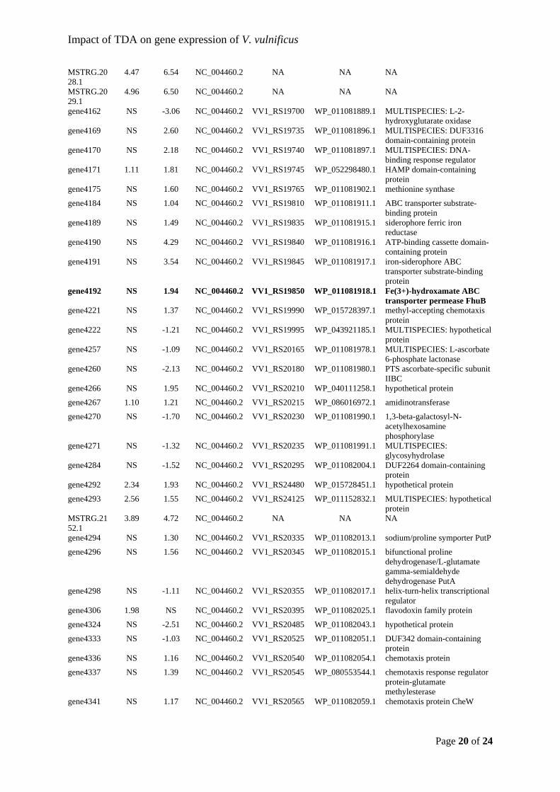
Impact of TDA on gene expression of V. vulnificus
Page 20 of 24
MSTRG.2028.1
4.47 6.54 NC_004460.2 NA NA NA
MSTRG.2029.1
4.96 6.50 NC_004460.2 NA NA NA
gene4162 NS -3.06 NC_004460.2 VV1_RS19700 WP_011081889.1 MULTISPECIES: L-2-hydroxyglutarate oxidase
gene4169 NS 2.60 NC_004460.2 VV1_RS19735 WP_011081896.1 MULTISPECIES: DUF3316 domain-containing protein
gene4170 NS 2.18 NC_004460.2 VV1_RS19740 WP_011081897.1 MULTISPECIES: DNA-binding response regulator
gene4171 1.11 1.81 NC_004460.2 VV1_RS19745 WP_052298480.1 HAMP domain-containing protein
gene4175 NS 1.60 NC_004460.2 VV1_RS19765 WP_011081902.1 methionine synthase
gene4184 NS 1.04 NC_004460.2 VV1_RS19810 WP_011081911.1 ABC transporter substrate-binding protein
gene4189 NS 1.49 NC_004460.2 VV1_RS19835 WP_011081915.1 siderophore ferric iron reductase
gene4190 NS 4.29 NC_004460.2 VV1_RS19840 WP_011081916.1 ATP-binding cassette domain-containing protein
gene4191 NS 3.54 NC_004460.2 VV1_RS19845 WP_011081917.1 iron-siderophore ABC transporter substrate-binding protein
gene4192 NS 1.94 NC_004460.2 VV1_RS19850 WP_011081918.1 Fe(3+)-hydroxamate ABC transporter permease FhuB
gene4221 NS 1.37 NC_004460.2 VV1_RS19990 WP_015728397.1 methyl-accepting chemotaxis protein
gene4222 NS -1.21 NC_004460.2 VV1_RS19995 WP_043921185.1 MULTISPECIES: hypothetical protein
gene4257 NS -1.09 NC_004460.2 VV1_RS20165 WP_011081978.1 MULTISPECIES: L-ascorbate 6-phosphate lactonase
gene4260 NS -2.13 NC_004460.2 VV1_RS20180 WP_011081980.1 PTS ascorbate-specific subunit IIBC
gene4266 NS 1.95 NC_004460.2 VV1_RS20210 WP_040111258.1 hypothetical protein
gene4267 1.10 1.21 NC_004460.2 VV1_RS20215 WP_086016972.1 amidinotransferase
gene4270 NS -1.70 NC_004460.2 VV1_RS20230 WP_011081990.1 1,3-beta-galactosyl-N-acetylhexosamine phosphorylase
gene4271 NS -1.32 NC_004460.2 VV1_RS20235 WP_011081991.1 MULTISPECIES: glycosyhydrolase
gene4284 NS -1.52 NC_004460.2 VV1_RS20295 WP_011082004.1 DUF2264 domain-containing protein
gene4292 2.34 1.93 NC_004460.2 VV1_RS24480 WP_015728451.1 hypothetical protein
gene4293 2.56 1.55 NC_004460.2 VV1_RS24125 WP_011152832.1 MULTISPECIES: hypothetical protein
MSTRG.2152.1
3.89 4.72 NC_004460.2 NA NA NA
gene4294 NS 1.30 NC_004460.2 VV1_RS20335 WP_011082013.1 sodium/proline symporter PutP
gene4296 NS 1.56 NC_004460.2 VV1_RS20345 WP_011082015.1 bifunctional proline dehydrogenase/L-glutamate gamma-semialdehyde dehydrogenase PutA
gene4298 NS -1.11 NC_004460.2 VV1_RS20355 WP_011082017.1 helix-turn-helix transcriptional regulator
gene4306 1.98 NS NC_004460.2 VV1_RS20395 WP_011082025.1 flavodoxin family protein
gene4324 NS -2.51 NC_004460.2 VV1_RS20485 WP_011082043.1 hypothetical protein
gene4333 NS -1.03 NC_004460.2 VV1_RS20525 WP_011082051.1 DUF342 domain-containing protein
gene4336 NS 1.16 NC_004460.2 VV1_RS20540 WP_011082054.1 chemotaxis protein
gene4337 NS 1.39 NC_004460.2 VV1_RS20545 WP_080553544.1 chemotaxis response regulator protein-glutamate methylesterase
gene4341 NS 1.17 NC_004460.2 VV1_RS20565 WP_011082059.1 chemotaxis protein CheW
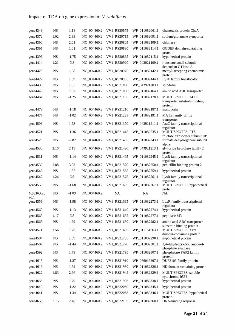
Impact of TDA on gene expression of V. vulnificus
Page 21 of 24
gene4343 NS 1.18 NC_004460.2 VV1_RS20575 WP_011082061.1 chemotaxis protein CheA
gene4372 1.02 2.33 NC_004460.2 VV1_RS20715 WP_011082091.1 sodium/glutamate symporter
gene4390 NS 2.01 NC_004460.2 VV1_RS20805 WP_011082109.1 chitinase
gene4395 NS 1.01 NC_004460.2 VV1_RS20830 WP_011082114.1 GGDEF domain-containing protein
gene4396 NS -1.75 NC_004460.2 VV1_RS20835 WP_011082115.1 hypothetical protein
gene4414 1.21 NS NC_004460.2 VV1_RS20920 WP_043921199.1 ribosome small subunit-dependent GTPase A
gene4425 NS 1.58 NC_004460.2 VV1_RS20975 WP_011082142.1 methyl-accepting chemotaxis protein
gene4427 NS 1.59 NC_004460.2 VV1_RS20985 WP_011082144.1 LysE family translocator
gene4430 NS 1.35 NC_004460.2 VV1_RS21000 WP_043921203.1 spindolin
gene4448 NS 1.82 NC_004460.2 VV1_RS21090 WP_011082164.1 amino acid ABC transporter
gene4464 NS -1.25 NC_004460.2 VV1_RS21165 WP_011082178.1 MULTISPECIES: ABC transporter substrate-binding protein
gene4473 NS -1.18 NC_004460.2 VV1_RS21210 WP_011082187.1 maltoporin
gene4477 NS -1.02 NC_004460.2 VV1_RS21225 WP_011082191.1 MATE family efflux transporter
gene4506 NS 1.73 NC_004460.2 VV1_RS21370 WP_043921211.1 AraC family transcriptional regulator
gene4521 NS -1.36 NC_004460.2 VV1_RS21445 WP_011082235.1 MULTISPECIES: PTS fructose transporter subunit IIB
gene4529 NS -2.82 NC_004460.2 VV1_RS21485 WP_011082243.1 formate dehydrogenase subunit alpha
gene4530 2.10 2.19 NC_004460.2 VV1_RS21490 WP_043921213.1 glycoside hydrolase family 2 protein
gene4531 NS -1.14 NC_004460.2 VV1_RS21495 WP_011082245.1 LysR family transcriptional regulator
gene4536 2.88 3.03 NC_004460.2 VV1_RS21520 WP_011082250.1 penicillin-binding protein 2
gene4545 NS 1.37 NC_004460.2 VV1_RS21565 WP_011082259.1 hypothetical protein
gene4547 1.24 NS NC_004460.2 VV1_RS21575 WP_011082261.1 LysR family transcriptional regulator
gene4553 NS -1.68 NC_004460.2 VV1_RS21605 WP_011082267.1 MULTISPECIES: hypothetical protein
MSTRG.2396.1
NS -1.03 NC_004460.2 NA NA NA
gene4559 NS -1.98 NC_004460.2 VV1_RS21635 WP_011082273.1 LuxR family transcriptional regulator
gene4560 NS -1.13 NC_004460.2 VV1_RS21640 WP_011082274.1 hypothetical protein
gene4563 1.17 NS NC_004460.2 VV1_RS21655 WP_011082277.1 peptidase M3
gene4568 NS 1.69 NC_004460.2 VV1_RS21680 WP_011082282.1 amino acid ABC transporter substrate-binding protein
gene4571 1.56 1.70 NC_004460.2 VV1_RS21695 WP_011151663.1 MULTISPECIES: YccF domain-containing protein
gene4584 NS 1.09 NC_004460.2 VV1_RS21755 WP_011082298.1 hypothetical protein
gene4587 NS -1.44 NC_004460.2 VV1_RS21770 WP_011082301.1 3,4-dihydroxy-2-butanone-4-phosphate synthase
gene4592 NS 1.79 NC_004460.2 VV1_RS21795 WP_011082307.1 phosphatase PAP2 family protein
gene4615 NS -1.27 NC_004460.2 VV1_RS21910 WP_086016897.1 DUF3103 family protein
gene4619 NS 1.59 NC_004460.2 VV1_RS21930 WP_011082326.1 HD domain-containing protein
gene4622 1.83 2.66 NC_004460.2 VV1_RS21945 WP_011082329.1 MULTISPECIES: soluble cytochrome b562
gene4633 NS 1.79 NC_004460.2 VV1_RS21995 WP_011082338.1 hypothetical protein
gene4640 NS -1.22 NC_004460.2 VV1_RS22030 WP_011082345.1 hypothetical protein
gene4641 NS -1.34 NC_004460.2 VV1_RS22035 WP_011082346.1 MULTISPECIES: hypothetical protein
gene4656 2.15 2.48 NC_004460.2 VV1_RS22105 WP_011082360.1 DNA-binding response
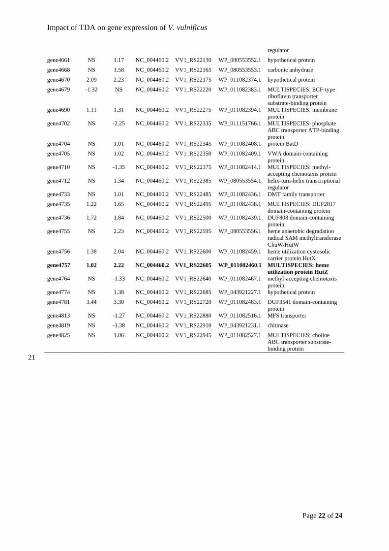
Impact of TDA on gene expression of V. vulnificus
Page 22 of 24
regulator
gene4661 NS 1.17 NC_004460.2 VV1_RS22130 WP_080553552.1 hypothetical protein
gene4668 NS 1.58 NC_004460.2 VV1_RS22165 WP_080553553.1 carbonic anhydrase
gene4670 2.09 2.23 NC_004460.2 VV1_RS22175 WP_011082374.1 hypothetical protein
gene4679 -1.32 NS NC_004460.2 VV1_RS22220 WP_011082383.1 MULTISPECIES: ECF-type riboflavin transporter substrate-binding protein
gene4690 1.11 1.31 NC_004460.2 VV1_RS22275 WP_011082394.1 MULTISPECIES: membrane protein
gene4702 NS -2.25 NC_004460.2 VV1_RS22335 WP_011151766.1 MULTISPECIES: phosphate ABC transporter ATP-binding protein
gene4704 NS 1.01 NC_004460.2 VV1_RS22345 WP_011082408.1 protein BatD
gene4705 NS 1.02 NC_004460.2 VV1_RS22350 WP_011082409.1 VWA domain-containing protein
gene4710 NS -1.35 NC_004460.2 VV1_RS22375 WP_011082414.1 MULTISPECIES: methyl-accepting chemotaxis protein
gene4712 NS 1.34 NC_004460.2 VV1_RS22385 WP_080553554.1 helix-turn-helix transcriptional regulator
gene4733 NS 1.01 NC_004460.2 VV1_RS22485 WP_011082436.1 DMT family transporter
gene4735 1.22 1.65 NC_004460.2 VV1_RS22495 WP_011082438.1 MULTISPECIES: DUF2817 domain-containing protein
gene4736 1.72 1.84 NC_004460.2 VV1_RS22500 WP_011082439.1 DUF808 domain-containing protein
gene4755 NS 2.23 NC_004460.2 VV1_RS22595 WP_080553556.1 heme anaerobic degradation radical SAM methyltransferase ChuW/HutW
gene4756 1.38 2.04 NC_004460.2 VV1_RS22600 WP_011082459.1 heme utilization cystosolic carrier protein HutX
gene4757 1.02 2.22 NC_004460.2 VV1_RS22605 WP_011082460.1 MULTISPECIES: heme utilization protein HutZ
gene4764 NS -1.33 NC_004460.2 VV1_RS22640 WP_011082467.1 methyl-accepting chemotaxis protein
gene4774 NS 1.38 NC_004460.2 VV1_RS22685 WP_043921227.1 hypothetical protein
gene4781 3.44 3.30 NC_004460.2 VV1_RS22720 WP_011082483.1 DUF3541 domain-containing protein
gene4813 NS -1.27 NC_004460.2 VV1_RS22880 WP_011082516.1 MFS transporter
gene4819 NS -1.38 NC_004460.2 VV1_RS22910 WP_043921231.1 chitinase
gene4825 NS 1.06 NC_004460.2 VV1_RS22945 WP_011082527.1 MULTISPECIES: choline ABC transporter substrate-binding protein
21
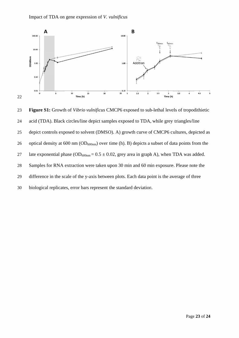
22
23
24
25
26
27
28
29
30
Impact
Figure
acid (TD
depict c
optical
late exp
Sample
differen
biologic
of TDA on
S1: Growth
DA). Black
controls exp
density at 6
ponential ph
es for RNA
nce in the sc
cal replicate
gene expre
h of Vibrio
k circles/line
posed to solv
600 nm (OD
hase (OD600
extraction w
cale of the y
es, error bar
ession of V.
vulnificus C
e depict sam
vent (DMSO
D600nm) over
nm = 0.5 ± 0
were taken u
y-axis betwe
rs represent
vulnificus
CMCP6 exp
mples expos
O). A) grow
time (h). B
0.02, grey ar
upon 30 min
een plots. E
the standar
posed to sub
ed to TDA,
wth curve of
B) depicts a
rea in graph
n and 60 mi
Each data po
rd deviation
b-lethal leve
while grey
f CMCP6 cu
subset of da
h A), when T
in exposure
oint is the av
n.
Page
els of tropod
y triangles/li
ultures, dep
ata points fr
TDA was a
e. Please not
verage of th
23 of 24
dithietic
ine
picted as
rom the
dded.
te the
hree
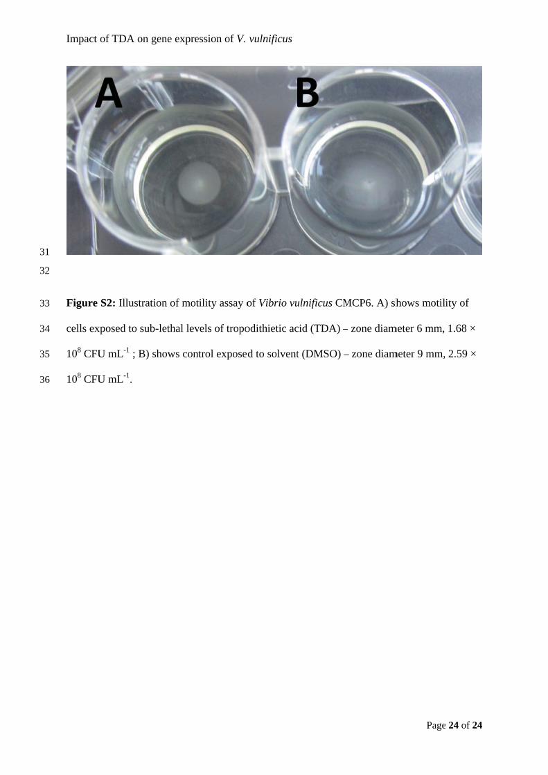
31
32
33
34
35
36
Impact
Figure
cells ex
108 CFU
108 CFU
of TDA on
S2: Illustra
xposed to su
U mL-1 ; B)
U mL-1.
gene expre
ation of mot
ub-lethal lev
) shows con
ession of V.
tility assay o
vels of tropo
ntrol exposed
vulnificus
of Vibrio vu
odithietic ac
d to solvent
ulnificus CM
cid (TDA) –
t (DMSO) –
MCP6. A) sh
– zone diam
– zone diam
Page
hows motili
meter 6 mm,
meter 9 mm,
24 of 24
ity of
1.68 ×
2.59 ×
Paper 4
Dittmann, K.K., Rasmussen, B.B., Melchiorsen, J., Sonnenschein, E.C., Gram, L. & Bentzon-Tilia, M. (2019).
Roseobacter probiotics affect lower-trophic level microbiomes in marine aquaculture
Manuscript in preparation
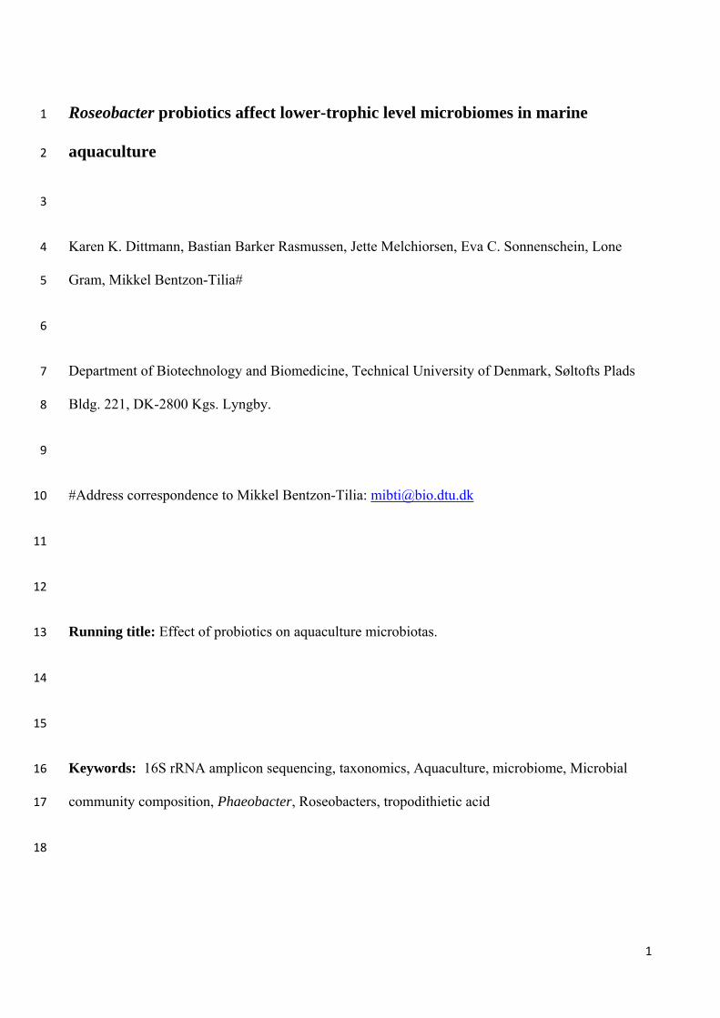
1
Roseobacter probiotics affect lower-trophic level microbiomes in marine 1
aquaculture 2
3
Karen K. Dittmann, Bastian Barker Rasmussen, Jette Melchiorsen, Eva C. Sonnenschein, Lone 4
Gram, Mikkel Bentzon-Tilia# 5
6
Department of Biotechnology and Biomedicine, Technical University of Denmark, Søltofts Plads 7
Bldg. 221, DK-2800 Kgs. Lyngby. 8
9
#Address correspondence to Mikkel Bentzon-Tilia: [email protected] 10
11
12
Running title: Effect of probiotics on aquaculture microbiotas. 13
14
15
Keywords: 16S rRNA amplicon sequencing, taxonomics, Aquaculture, microbiome, Microbial 16
community composition, Phaeobacter, Roseobacters, tropodithietic acid 17
18
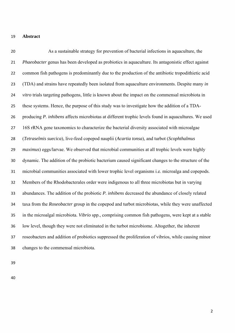
2
Abstract 19
As a sustainable strategy for prevention of bacterial infections in aquaculture, the 20
Phaeobacter genus has been developed as probiotics in aquaculture. Its antagonistic effect against 21
common fish pathogens is predominantly due to the production of the antibiotic tropodithietic acid 22
(TDA) and strains have repeatedly been isolated from aquaculture environments. Despite many in 23
vitro trials targeting pathogens, little is known about the impact on the commensal microbiota in 24
these systems. Hence, the purpose of this study was to investigate how the addition of a TDA-25
producing P. inhibens affects microbiotas at different trophic levels found in aquacultures. We used 26
16S rRNA gene taxonomics to characterize the bacterial diversity associated with microalgae 27
(Tetraselmis suecica), live-feed copepod nauplii (Acartia tonsa), and turbot (Scophthalmus 28
maximus) eggs/larvae. We observed that microbial communities at all trophic levels were highly 29
dynamic. The addition of the probiotic bacterium caused significant changes to the structure of the 30
microbial communities associated with lower trophic level organisms i.e. microalga and copepods. 31
Members of the Rhodobacterales order were indigenous to all three microbiotas but in varying 32
abundances. The addition of the probiotic P. inhibens decreased the abundance of closely related 33
taxa from the Roseobacter group in the copepod and turbot microbiotas, while they were unaffected 34
in the microalgal microbiota. Vibrio spp., comprising common fish pathogens, were kept at a stable 35
low level, though they were not eliminated in the turbot microbiome. Altogether, the inherent 36
roseobacters and addition of probiotics suppressed the proliferation of vibrios, while causing minor 37
changes to the commensal microbiota. 38
39
40
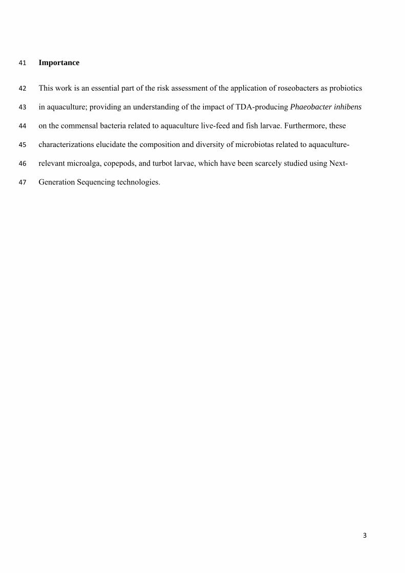
3
Importance 41
This work is an essential part of the risk assessment of the application of roseobacters as probiotics 42
in aquaculture; providing an understanding of the impact of TDA-producing Phaeobacter inhibens 43
on the commensal bacteria related to aquaculture live-feed and fish larvae. Furthermore, these 44
characterizations elucidate the composition and diversity of microbiotas related to aquaculture-45
relevant microalga, copepods, and turbot larvae, which have been scarcely studied using Next-46
Generation Sequencing technologies. 47
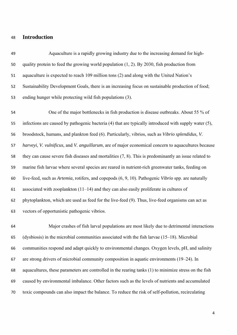
4
Introduction 48
Aquaculture is a rapidly growing industry due to the increasing demand for high-49
quality protein to feed the growing world population (1, 2). By 2030, fish production from 50
aquaculture is expected to reach 109 million tons (2) and along with the United Nation’s 51
Sustainability Development Goals, there is an increasing focus on sustainable production of food; 52
ending hunger while protecting wild fish populations (3). 53
One of the major bottlenecks in fish production is disease outbreaks. About 55 % of 54
infections are caused by pathogenic bacteria (4) that are typically introduced with supply water (5), 55
broodstock, humans, and plankton feed (6). Particularly, vibrios, such as Vibrio splendidus, V. 56
harveyi, V. vulnificus, and V. anguillarum, are of major economical concern to aquacultures because 57
they can cause severe fish diseases and mortalities (7, 8). This is predominantly an issue related to 58
marine fish larvae where several species are reared in nutrient-rich greenwater tanks, feeding on 59
live-feed, such as Artemia, rotifers, and copepods (6, 9, 10). Pathogenic Vibrio spp. are naturally 60
associated with zooplankton (11–14) and they can also easily proliferate in cultures of 61
phytoplankton, which are used as feed for the live-feed (9). Thus, live-feed organisms can act as 62
vectors of opportunistic pathogenic vibrios. 63
Major crashes of fish larval populations are most likely due to detrimental interactions 64
(dysbiosis) in the microbial communities associated with the fish larvae (15–18). Microbial 65
communities respond and adapt quickly to environmental changes. Oxygen levels, pH, and salinity 66
are strong drivers of microbial community composition in aquatic environments (19–24). In 67
aquacultures, these parameters are controlled in the rearing tanks (1) to minimize stress on the fish 68
caused by environmental imbalance. Other factors such as the levels of nutrients and accumulated 69
toxic compounds can also impact the balance. To reduce the risk of self-pollution, recirculating 70
5
aquaculture systems (RAS) often use mechanic filters and biofilters to clean the rearing water for 71
dissolved organic matter and accumulated inorganic nitrogen and phosphorus (1). In the event of an 72
imbalance in the system, e.g. rapid increase in nutrient levels and temperature increase, the result 73
can be proliferation of opportunistic pathogens. The pathogens have traditionally been controlled by 74
disinfection of the rearing tanks (25, 26), sterilization of the rearing water (27), and deployment of 75
antibiotics (28, 29) and in recent years by vaccination of the fish (30, 31), but the latter does not 76
work on fish larvae due to their underdeveloped immune systems (30). Thus, sustainable 77
alternatives to antibiotics are sought, given the severe impact on the commensal microbiome and 78
the increasing problem of spreading antimicrobial resistance genes (29, 30, 32). One proposed 79
alternative is the use of probiotics; live microorganisms that provide a health benefit to the host 80
when administered in adequate amounts (33). The potential application of probiotics in aquaculture 81
as prophylactic and acute treatment of disease outbreaks has been studied for decades; most of them 82
focusing on the gut microbiome of the farmed animal (34–36). Currently, the majority of 83
commercially available probiotics for aquaculture are based on mono- or mixed cultures of 84
Firmicutes (3), which have been successful in humans and livestock, though not adapted to the 85
diverse aquatic environments. Proteobacteria such as Shewanella spp. and tropodithietic acid 86
(TDA)-producing members of the Roseobacter group have been studied extensively for their 87
bioactivity and probiotic potential (3, 37–39) as an alternative solution of marine origin. 88
TDA-producers are often found in microbiomes of marine eukaryotes including 89
micro- and macroalgae (40, 41), zooplankton (42), sponges (43) and molluscs (39, 44, 45). 90
Mutualistic interactions between the microalga Emiliania huxleyi and Phaeobacter inhibens have 91
been proposed due to the production of TDA and algal-growth promoting auxins (40, 46), though 92
this relation can turn parasitic, when P. inhibens responds to algal break-down products and 93
accelerates the lysis process by production of algaecides known as roseobacticides (47). TDA is a 94
6
bactericidal antibiotic against a wide range of both Gram-negative and Gram-positive bacteria (48). 95
In low concentration, it can act as a signaling molecule affecting global gene regulation and 96
behavior including motility, biofilm formation, and secondary metabolite production (49). The 97
antibiotic mode of action is highly complex and still not fully understood, though the compound is 98
most likely disrupting the proton motive force by changing the membrane potential (50, 51). 99
Resistance towards TDA does not develop easily (48, 52), but tolerance of non-TDA producing 100
bacteria has been observed among isolates from deep sea sponges (43). Furthermore, the presence 101
of TDA-producers has varying impact on marine microbiomes depending on the composition and 102
complexity of the existing community (53). While some community members are unaffected, 103
Vibrio spp. and Pseudoalteromonas spp. diminish in the presence of P. inhibens (53). Thus, 104
individual tolerance or cross-protective mechanisms may result in microbiome resilience to 105
perturbations caused by TDA, while potential pathogens are kept down. 106
Phaeobacter spp. have been isolated from multiple aquacultures (44, 54, 55), which 107
indicates that they are already an inherent part of the microbiome in some farms. Several studies 108
have demonstrated their antagonistic effect against fish pathogens in live-feed cultures (9, 55, 56) 109
without noticeable adverse effect on the eukaryote (9, 10). Most importantly, addition of 106 to 107 110
CFU mL-1 can decrease mortality of turbot and cod larvae when challenged with vibrios (9, 57, 58). 111
The selective impact on microbiomes along with the lack of resistance development despite their 112
global occurrence in microbiomes, including aquaculture microbiomes, highlight the applicability 113
of Phaeobacter as probiotic. However, perturbations with probiotic levels of Phaeobacter could 114
potentially cause imbalance and thereby give rise to proliferation of other pathogens than vibrios. 115
Furthermore, little is currently known about aquaculture microbiomes at the different trophic levels 116
and hence, the purpose of this study was to investigate how the addition of a TDA-producing P. 117
inhibens affects microbiotas related to different trophic levels found in aquaculture systems. 118
7
119
Results 120
The impact of probiotic P. inhibens strain DSM 17395 on the microbiotas of 121
aquaculture-relevant, marine microalga, copepods, and fish eggs/larvae was assessed by sequencing 122
16S rRNA gene V4 region amplicons and analyzing their taxonomic composition and diversity over 123
4 days; each co-culture as well as a control cultures without addition of the probiotic was sampled 124
four times: at 0 h (T0), 24 h (T24), 48 h (T48), and 96 h (T96). The co-culture experiment with turbot 125
was initiated with eggs, which all hatched within 48 hours of incubation; up to 2 % of the eggs were 126
hatched at time point 24 h. Amplicon Sequence Variants (ASVs) containing the added P. inhibens 127
were identified and removed from the data sets in the subsequent analysis to assess the potential 128
changes in the inherent background microbiota. 129
Effects of probiotic treatment on microbial community composition 130
The community of the microalga T. suecica was dominated by bacteria from the phyla 131
Proteobacteria, particularly members of the Rhodobacterales order, and Bacteroidetes, particularly 132
members of the Flavobacteriales order (Figure 1). Other observed orders above 2 % relative 133
abundance included Alteromonadales, Burkholderiales, Caulobacterales, Rhizobiales, 134
Sphingomonadales, Phycisphaerales, and Cytophagales. Burkholderiales were only present in the 135
initial microbiomes (T0), while Rhizobiales appeared after 96 hours of incubation. The communities 136
were stable and no obvious changes occurred due to probiotic treatment at the order level. Hence, 137
incubation time was the main driver of the observed changes in community composition at this 138
trophic level. 139
8
The A. tonsa bacterial community composition was dominated by Proteobacteria, 140
particularly members of the orders Alteromonadales and Oceanospirillales (Figure 2). Members of 141
the Rhodobacterales, Rhodospirillales, and Flavobacteriales orders were also present in all samples, 142
though in lower abundance. Desulfobacterales only occurred in the initial microbiota (T0), while 143
Caulobacterales turned up in the microbiota following 96 hours of incubation. The addition of P. 144
inhibens decreased the abundance of Rhodobacterales and Rhodospirillales. Furthermore, 145
Alteromonadales increased initially (T24) in the probiotic group, though their dominance decreased 146
over time. Hence, at this trophic level, both time and the probiotic treatment affected the 147
composition of the bacterial community. 148
The turbot egg and larval microbiotas were dominated by Proteobacteria, particularly 149
Gammaproteobacteria order such as members of the orders Alteromonadales and Vibrionales 150
(Figure 3). Vibrionales were most prominent in the initial egg microbiome (T0; relative abundance 151
46.2 % to 46.9 %), though their relative abundance decreased to 14.3 % to 19.2 % after 24 hour 152
incubation and remained at the same level throughout the experiment. Concurrently, the relative 153
abundance of Alteromonadales and Rhodobacterales increased in abundance after 24 hours of 154
incubation. Both Rhodobacterales and Oceanospirillales increased in abundance while 155
Alteromonadales decreased over time. Pseudomonadales occurred in the microbiota after 48 hour 156
incubation and remained throughout the experiment. The bacterial community associated with 157
turbot eggs/larvae receiving probiotic treatment did not contain members of the Rhodobacterales 158
order (relative abundance above 2 %). Altogether, the biggest shift occurred within the first 24 159
hours of the experiment (establishment phase) and the bacterial community was stable throughout 160
the monitored time. The presence of P. inhibens decreased the abundance of other Rhodobacterales 161
bacteria, but otherwise the community was mainly affected by incubation time. 162
Impact of probiotic treatment on bacterial microbiota richness and diversity 163
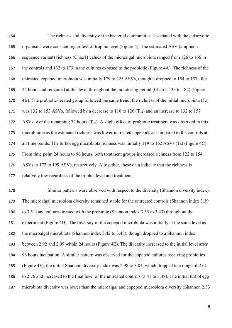
9
The richness and diversity of the bacterial communities associated with the eukaryotic 164
organisms were constant regardless of trophic level (Figure 4). The estimated ASV (amplicon 165
sequence variant) richness (Chao1) values of the microalgal microbiota ranged from 126 to 166 in 166
the controls and 132 to 173 in the cultures exposed to the probiotic (Figure 4A). The richness of the 167
untreated copepod microbiota was initially 179 to 225 ASVs, though it dropped to 154 to 157 after 168
24 hours and remained at this level throughout the monitoring period (Chao1; 133 to 182) (Figure 169
4B). The probiotic-treated group followed the same trend; the richness of the initial microbiota (T0) 170
was 132 to 153 ASVs, followed by a decrease to 110 to 126 (T24) and an increase to 132 to 157 171
ASVs over the remaining 72 hours (T96). A slight effect of probiotic treatment was observed in this 172
microbiome as the estimated richness was lower in treated copepods as compared to the controls at 173
all time points. The turbot egg microbiota richness was initially 119 to 162 ASVs (T0) (Figure 4C). 174
From time point 24 hours to 96 hours, both treatment groups increased richness from 122 to 154 175
ASVs to 172 to 199 ASVs, respectively. Altogether, these data indicate that the richness is 176
relatively low regardless of the trophic level and treatment. 177
Similar patterns were observed with respect to the diversity (Shannon diversity index). 178
The microalgal microbiota diversity remained stable for the untreated controls (Shannon index 3.39 179
to 3.51) and cultures treated with the probiotic (Shannon index 3.35 to 3.43) throughout the 180
experiment (Figure 4D). The diversity of the copepod microbiota was initially at the same level as 181
the microalgal microbiota (Shannon index 3.42 to 3.43), though dropped to a Shannon index 182
between 2.92 and 2.99 within 24 hours (Figure 4E). The diversity increased to the initial level after 183
96 hours incubation. A similar pattern was observed for the copepod cultures receiving probiotics 184
(Figure 4F); the initial Shannon diversity index was 2.98 to 3.04, which dropped to a range of 2.61 185
to 2.76 and increased to the final level of the untreated controls (3.41 to 3.48). The initial turbot egg 186
microbiota diversity was lower than the microalgal and copepod microbiota diversity (Shannon 2.33 187
10
to 2.45 at T0), though increased steadily to a Shannon index between 3.38 and 3.54 (both controls 188
and probiotics treated) upon incubation for 96 hours. Altogether, these observations demonstrate 189
that probiotic P. inhibens has little to no impact on the richness and diversity of the microbiotas 190
associated with microalgae, copepods, and fish larvae. 191
Impact of probiotic treatment on community structure 192
Unconstrained ordinations – i.e. Principal Coordinate Analysis (PCoA) - on Bray-193
Curtis distances were used to assess the impact of P. inhibens on community structure of the 194
microbiotas associated with the three aquaculture-related eukaryotes (Figure 5). The community 195
structure shifted during incubation time for all three microbial communities, regardless of treatment. 196
The microalgal microbial community structures treated with probiotics were significantly different 197
from the untreated controls (PERMANOVA; p = 0.001) (Figure 5A). This was also observed in the 198
copepod associated microbiota (PERMANOVA; p = 0.001) (Figure 5B). However, the turbot larval 199
microbial community structure was not significantly affected by the presence of P. inhibens 200
(PERMANOVA; p = 0.279) (Figure 5C). Hence, incubation was the major driver of the microbial 201
community structure and the impact of the probiotic treatment depends on the trophic layer at hand; 202
the biggest impact occurs at the lower tropic levels, while the fish larval community structure is 203
unaffected. 204
Probiotic impact on specific taxa 205
At the order level, the probiotic P. inhibens DSM 17395 decreased the abundance of 206
Rhodobacterales in two of the assessed microbiotas (Figure 2, Figure 3), while other effects of the 207
treatment were minor. Therefore, differences at ASV level (100 % sequence similarity, no 208
clustering) were investigated to elucidate which of the most abundant members were affected. No 209
major impact on individual taxa was observed in the microbiota associated with T. suecica due to 210
11
treatment (Figure 6). In the A. tonsa microbiota, the most abundant Halomonas sp. were slightly 211
lower in abundance in the probiotic treated samples, but still dominating (Figure 7). Members of the 212
Rhodobacteraceae family, such as Ruegeria sp. and Celeribacter sp., decreased in the presence of 213
P. inhibens, which is in line with the observations in the community composition analysis (Figure 214
2). Members of the Saccharospirillaceae family and Hyphomonas spp. were initially lower in 215
abundance in both treatment groups, but increased over time. In contrast to the microalgal and 216
copepod microbiotas, the samples from fish eggs and larvae clustered according to time rather than 217
treatment (Figure 8). No major changes were observed due to treatment, but changes over time were 218
observed, confirming the PCoA (Figure 5C). Some Colwellia sp. ASVs disappeared with time, 219
while others increased in abundance. Other Alteromonadales bacteria such as Psychrobium sp. and 220
Alteromonas sp. increased over time. Vibrio spp. were highly abundant in the initial microbiota 221
(T0), though decreased as a function of incubation time. Altogether, the occurring changes in the 222
presence of P. inhibens were unique to the eukaryotic host and the largest changes were observed in 223
the copepod microbiota. 224
Discussion 225
Characterizing what a healthy microbiome is, determining the course of disease at the 226
bacterial population level, and assessing the impact of measures for disease control, such as 227
probiotics, is of crucial importance to increase sustainable aquaculture production without 228
increasing the risk of economic losses due to disease. P. inhibens has potential as probiotic in 229
aquaculture given efficient killing of common pathogens (9, 55, 56) and protection of vibrio-230
challenged fish larvae (9, 57, 58) without noticeable adverse effect on the live-feed or the larvae (9, 231
10). Their natural association with eukaryotes (39, 44, 45, 53, 59–61) and aquaculture systems (54, 232
57) along with their efficient killing of opportunistic pathogens and no resistance development are 233
some of the key features highlighting their applicability in the aquaculture industry. However, it is 234
12
currently not known how probiotic concentrations of P. inhibens influence and shape the structure 235
the commensal microbiota of aquaculture-related eukaryotes. The results of this study suggest that 236
the microbial communities of all trophic levels in aquaculture are highly dynamic, in all cases 237
changing over time. The impact of adding P. inhibens to the system is overall minor and highly 238
dependent on the commensal microbiota with greater impact on the bacterial communities of the 239
lower trophic levels. 240
Three eukaryotes – T. suecica, A. tonsa, and turbot – were selected to represent 241
different trophic levels – feed for live-feed, live-feed, and reared fish – found in aquacultures. 242
Several studies have been conducted on microalgal microbiomes and how roseobacters interact with 243
these unicellular eukaryotes (47, 62, 63). Despite T. suecica being widely used and produced in 244
hatcheries, the microbial community associated with this microalga is not well studied. Biondi et al. 245
(64) observed that the microbiota of T. suecica was dominated by Proteobacteria – particularly 246
members of the Roseobacter group, but also Rhizobiales and Bacteroidetes (Flavobacteriales). This 247
is in concordance with the community composition observed in the T. suecica cultures assessed in 248
this study as well as for another aquaculture-related microalgal genus, Nannochloropsis (65). In 249
addition, we also observed a relatively high abundance of Phycisphaerales (Planctomycetes). The 250
microbiome of A. tonsa was also dominated by Proteobacteria, particularly Gammaproteobacteria in 251
this study. This has previously also been observed in copepods from the North Atlantic Ocean (66). 252
Cultivation-based methods have found that Vibrio spp. were dominating (14, 67), however, the 253
order of Vibrionales was below the 2 % relative abundance cutoff in our community composition 254
analysis, indicating that these bacteria were not abundant in the cultured copepods. Moisander et al. 255
(66) also observed that Rhodobactereceae dominated the transient food microbiome and proposed 256
that they might contribute to copepod nutrition. Members of the Rhodobacterales order were also 257
abundant in our copepod system, though Alteromonadales and Oceanospirillales bacteria were the 258
13
most abundant. These differences in the composition of the copepod-associated community most 259
likely due to differences in the composition of the bacterial community in the immediate 260
environment (natural vs. laboratory cultivation). 261
The culturable microbiota of turbot eggs and larvae has been studied for decades, 262
however, it remains poorly understood. Culture-dependent studies have isolated multiple members 263
of the Vibrionales and Aeromonadales orders (68, 69). While we observed that Vibrionales 264
dominated the egg microbial community, the Aeromonadales were not abundant (below the 2 % 265
cutoff) in any of the samples. By contrast, we observed high abundances of Alteromonadales. Poor 266
correlation between culture-dependent and -independent investigations of the microbiotas has also 267
been observed by Fjellheim et al. in cod larval microbiomes (70). Hence, there is a need for studies 268
characterizing the microbiome of aquaculture-related fish species, particularly at the larval stages, 269
where detrimental bacterial-fish interactions can lead to population crashes and economic losses. 270
The addition of probiotic P. inhibens had minor effects on the overall bacterial 271
community composition at the higher taxonomic levels (order level and above). This was previously 272
observed in the microbiome of the microalga E. huxleyi as well (53). Interestingly, closely related 273
taxa from the Rhodobacterales order decreased in abundance in the microbiotas associated with the 274
copepod and fish larvae, though they were unaffected in the microalgal, T. suecica, microbiota, in 275
the presence of P. inhibens. This was also observed in the microbiota of the microalga E. huxleyi 276
exposed to the same P. inhibens strain (53). Several genera of the Rhodobacteraceae family – 277
namely Sulfitobacter, Phaeobacter, Pelagicola, and Loktanella – were reduced or absent in the 278
presence of another strain of P. inhibens (2.10) in the microbiota of the diatom Thalassiosira rotula 279
(71). Among the 30 most abundant taxa in the copepod and turbot microbiotas, unclassified genera 280
of the Rhodobacteraceae family, Ruegeria spp., Celeribacter spp., and Pseudophaeobacter spp. 281
decreased in abundance. Roseobacter spp. closely related to Phaeobacter gallaeciencis (originally 282
14
Roseobacter gallaeciencis (72)) have previously been isolated from copepods (73), though the 283
taxonomic resolution on the V4 region would not be sufficient to tell closely related Roseobacter 284
spp. and Phaeobacter spp. apart. Both P. inhibens DSM 17395 and 2.10 produce the broad-285
spectrum antimicrobial agent, TDA and addition of pure TDA to cultures of Nannochloropsis 286
salina have been shown to decrease the relative abundance of Rhodobacteraceae at relatively low 287
concentrations (31.25 – 500 nM; 74), which could indicate that this molecule is causing the 288
observed decrease. Potentially, production and / or sensing of TDA is involved in the interspecies 289
competition within the Roseobacter group, although TDA production has not been reported in 290
Pseudophaeobacter spp. and Celeribacter spp.. However, if TDA acted as a broad spectrum 291
antibiotic, it would be expected that TDA also affected the remaining commensal microbiome. 292
Vibrio spp. and Pseudoalteromonas spp. diminished in the microbiota of the microalga E. huxleyi in 293
the presence of P. inhibens DSM 17395 (75), though in this study, the orders Vibrionales and 294
Alteromonadales were unaffected by the presence of P. inhibens in comparison to the controls. 295
Furthermore, Majzoub et al. (71) also saw that the microbiome exposed to a P. inhibens 2.10 296
variant (NCV12a1) with reduced antagonistic effect, developed in the same way as the microbiomes 297
exposed to the original, bioactive strain. Another possibility is that the decrease is due to quorum 298
sensing (QS). P. inhibens produces TDA and N-acyl homoserine lactones (AHLs), which both can 299
modulate motility, biofilm formation, and production of secondary metabolites in producer strains 300
(49). Beyersmann et al. (49) proposed that the induction of QS reduced attachment and induced 301
dispersal of the biofilm to associate with a new host. If a high dose of P. inhibens resembles the cue 302
to dispersal from the host, it would mean that the Rhodobacterales were in a planktonic state in the 303
water and therefore not captured as highly abundant in the sequences from eukaryotes. However, 304
this does not explain why the addition of P. inhibens had minor effect on the high abundance of 305
Rhodobacterales in the microalgal, T. suecica, microbiome. Altogether, these results indicate that 306
15
closely related roseobacters compete for the same niches and that the impact is dependent on the 307
eukaryotic host as well as the abundance of the roseobacters present in the commensal microbiota. 308
From an applied point of view, it is promising that a potential probiotic, such as P. inhibens, enters 309
the microbiome and establishes itself rather than being out-competed by the inherent and closely 310
related taxa. Further studies should reveal how interactions determine which species take over, and 311
which ones disappear in the microbiome. 312
All microbiotas had similar richness and diversity indexes. Bakke et al. (76) reported 313
that richness and diversity varied throughout the life-stages of cod larvae. While the turbot larvae in 314
this study were younger, the alpha diversity measure were similar to the observations by Bakke et 315
al. (76). Interestingly, the richness and diversity of the rearing water (i.e., green water prepared with 316
algal, Nannochloropsis oculata, paste) and live-feed (copepod, A. tonsa, and rotifers, Brachionus 317
‘Nevada’) was much higher than observed in the larval microbiome (76) and the live-feed assessed 318
in this study. However, this is most likely due to experimental differences; this study was conducted 319
in laboratory, small-scale cultures, while the study by Bakke et al. (76) was conducted in large-320
scale, aquaculture flow-through systems. The addition of the probiotic treatment in this study had 321
only a slight effect on richness, though a decrease, and it only occurred in the copepod 322
microbiomes. Dittmann et al. (53) observed that treatment with P. inhibens DSM 17395 did not 323
impact the richness and diversity of the microbiota of the microalga E. huxleyi. In contrast, the 324
oyster microbiota increased in richness when P. inhibens had been added to the system, though the 325
diversity was unaffected. Together with the minor impact on diversity and community composition, 326
these observations indicate that a perturbation of the microbiota with high loads of the probiotic 327
treatment does not have major impact on the overall diversity and taxa present, thereby likely 328
causes minor imbalance, which is mediated within the first 24 hours of incubation. 329
16
The microbial communities associated with the three investigated microbiotas were generally very 330
dynamic and changed over time, which is in concordance with previous studies (71, 76, 77). The 331
addition of probiotic P. inhibens had significant impact on the microbiome structure of T. suecica 332
and A. tonsa. In contrast, the microbiota associated with the turbot larvae was more affected by 333
incubation time compared to probiotic treatment. The eggs hatched within the first 48 hours of the 334
experiment and thereby, a sudden increase in nutrients has likely occurred. In contrast, no nutrients 335
were added to the microalgal and copepod systems, and thus, nutrients from the medium and the 336
eukaryotic hosts were slowly consumed and competition likely increased. The minor impact of P. 337
inhibens addition to the turbot egg and larval microbiome would indicate that addition of probiotics 338
at this trophic level would not cause dysbiosis in a healthy larval microbiota and a subsequent 339
population crash. However, it might also mean that the probiotic is less efficient at this level. Vibrio 340
spp. are commonly reported as detrimental pathogens to fish larvae (7, 8), while they are also part 341
of the commensal microbiota (68, 69). In this study, the high abundance of Vibrionales in the turbot 342
microbiota was due to relatively few ASVs belonging to the Vibrio genus and the abundance of 343
these ASVs did not change regardless of treatment (from day one through day four). We added P. 344
inhibens at probiotic concentrations similar to those used for protection of fish larvae against 345
vibriosis in challenge trials (9, 58, 78). In those trials, vibrios were reduced in numbers if not kept at 346
inoculation level (9, 58, 78), depending on the initial concentration of Vibrio spp. (9). Combined, 347
these results would suggest that addition of probiotic P. inhibens, or the presence of inherent, 348
closely related taxa can keep vibrios in the fish microbiota at a stable level, however, this does not 349
necessarily eliminate potential pathogens from the system. The effect is likely dose-dependent, 350
which was observed in a previous study of the E. huxleyi microbiome (53). Altogether, these data 351
emphasize the need for investigating the optimal addition of probiotic P. inhibens – in relation to 352
dose and which trophic level to add the probiotic treatment to – in order to obtain the most efficient 353
17
protection against opportunistic pathogens with minor effects on the commensal microbiota. The 354
addition of the probiotic is likely more efficient at the lower trophic levels such as the microalgal or 355
live-feed level, where P. inhibens establishes itself and changes the structure. However, it is not 356
possible to say whether the changes would be beneficial or detrimental to the microbiome function. 357
Broader –omics studies should elucidate this in the future. 358
In conclusion, addition of probiotic P. inhibens caused significant changes to the 359
structure of the microbial communities associated with the microalgae and copepods, though no 360
effect was seen on the community associated with turbot larvae. Particularly, the abundances of 361
closely related taxa from the Roseobacter group were reduced as a function of probiotic treatment, 362
but only in the copepod and turbot larval microbiotas. Vibrio spp. were highly abundant in the 363
turbot microbiota and these were kept at a stable level, though not eliminated, which indicates that 364
the probiotic effect towards vibrios is likely dose-dependent. Hence, the effect of adding a probiotic 365
bacterium such as P. inhibens to the microbiota of aquaculture-related eukaryotes is dependent on 366
the commensal microbiota composition and the eukaryotic host with greater impact at the lower 367
trophic levels. 368
369
Materials and Methods 370
Bacterial cultivation 371
Phaeobacter inhibens DSM 17395 (44, 72, 79) was routinely grown in half-strength Yeast extract, 372
Tryptone, Sea Salts broth (½YTSS, 2 g/L Bacto Yeast extract, 1.25 g/L Bacto Tryptone, 20 g/L 373
Sigma Sea Salts) (80). Liquid cultures were incubated under agitation (250 rpm) at 25° C or room 374
temperature. When grown on solid substrates, Marine Agar (MA, Difco 2216) or ½YTSS agar 375
(½YTSS with 15 g/L agar) was used. 376
18
Algae-Phaeobacter co-culturing. 377
A non-axenic strain of the green algae Tetraselmis suecica, was obtained from the aquaculture 378
facility Selonda Aquaculture SA, Athens, Greece. It was grown in f/2 medium (81) without 379
Na2SiO3 but with 5 mM NH4Cl in 1 L of 3 % Instant Ocean® Sea Salt (Aquarium Systems Inc., 380
Sarrebourg, France). This modified f/2 will from this point be referred to as f/2. 381
The cell density of T. suecica in the stock culture was determined using an improved Neubauer 382
counting chamber. The cells were re-inoculated in f/2 medium at a final concentration of 383
approximately 5 × 10⁵ algae mL-1 before splitting into six cultures of 600 mL in 1 L Erlen-Meyer 384
flasks. Three overnight cultures of P. inhibens DSM 17395 in ½YTSS were adjusted to Optical 385
Density at 600 nm (OD600nm) = 1.0 and washed one time in f/2 medium (7,000 × rpm, 3 min). In 386
triplicates, co-cultures of T. suecica were inoculated with P. inhibens DSM 17395 at a final 387
concentration of 4.06 × 10⁶ ± 1.05 × 10⁶ CFU mL-1 (equivalent to 8 P. inhibens cells per algal cell), 388
verified by plate spreading dilutions on MA. The remaining three cultures of T. suecica were treated 389
with sterile 2% Instant Ocean and served as controls. The cultures were incubated stagnant, at 18°C 390
with white fluorescent light (24 µmol m-2 s-1 photosynthetically active radiation; PAR). The cultures 391
were sampled at 0 h, 24 h, 48 h and, 96 h for algal abundance determinations and for biomass to be 392
used in DNA extractions. For abundance measures, 1 mL co-culture was fixed in 1 % 0.2 µm-393
filtered glutaraldehyde (final conc.) and the cell numbers were determined using an improved 394
Neubauer counting chamber. For DNA extraction, 100 mL of each culture was pelleted (8000 x g, 5 395
minutes, 25°C) and resuspended in 1 mL lysis buffer (400 mM sodium chloride, 750 mM sucrose, 396
20 mM EDTA, 50 mM Tris-HCl, 1 mg mL-1 lysozyme, pH 8.5) (82) and stored at -80°C until 397
extraction. 398
Copepod-Phaeobacter co-culturing. 399
19
A. tonsa eggs were kindly provided by Prof. B. W. Hansen, Roskilde University and stored at 5°C 400
until use. Three days before the experiment, eggs were inoculated in 3 % Instant Ocean and 401
incubated at 18°C with white fluorescent light (24 µmol m-2 s-1 PAR). The density of A. tonsa 402
nauplii in the culture was determined using Sedgewick rafter counting cell and the culture was 403
adjusted to 2 nauplii per mL using 3 % Instant Ocean. Seven cultures of 30 mL adjusted nauplii 404
culture were set-up in 50-mL Falcon tubes. In triplicates, overnight cultures of P. inhibens DSM 405
17395 in ½YTSS was inoculated into the A. tonsa nauplii culture to a level of 0.5 % (equivalent to 406
5 × 10⁶ CFU mL-1, verified by plate spreading on MA). Three A. tonsa cultures were treated with 407
sterile ½YTSS and served as controls. The last culture was used untreated for quantification of live 408
A. tonsa. All co-cultures were incubated horizontally with shake (60 rpm) at 18°C with white 409
fluorescent light (24 µmol m-2 s-1 PAR) and sampled at day 0, 1, 2, and 4. Before sampling, each 410
tube was mixed by inversion and 5 mL culture (equivalent to 10 A. tonsa nauplii) was taken out for 411
filtration onto a MontaMil Polycarbonate membrane filter (pore size 0.2µm, diameter 47mm). The 412
filters were transferred to cryo tubes, flooded in sucrose lysis buffer, and stored at -80°C until 413
extraction. 414
Turbot egg and larvae - Phaeobacter co-culturing. 415
Non-axenic turbot eggs were received from France Turbot, hatchery L’Epine (Noirmoutier Island, 416
France), with 24 h of transport before conducting the experiment. One-hundred eggs were 417
transferred to four Petri dishes (20 cm diameter, glass) containing sterile-filtered (0.22 µm filter) 418
sea water adjusted to salinity 34 ‰ with Sigma sea salts [S9883, Sigma] and pre-tempered to 15°C. 419
The final volume was 200 mL. An overnight culture of P. inhibens DSM 17395 in ½YTSS was 420
washed one time in 2 % sterile Instant Ocean (7,000 × rpm, 3 min). In duplicates, co-cultures of 421
turbot eggs were inoculated with P. inhibens DSM 17395 at a final concentration of 1 × 10⁷ CFU 422
mL-1 in the sea water (equivalent to 2 × 10⁷ P. inhibens cells per egg), verified by plate spreading 423
20
dilutions on ½YTSS agar. The remaining two cultures of eggs were treated with an equivalent 424
volume of sterile 2% Instant Ocean and served as controls. The experiment was initiated with 0 % 425
of the eggs being hatched. After 24 h incubation, 0 % to 2 % of the eggs were hatched, while all the 426
eggs were hatched after 48 hours of incubation. Biomass samples for DNA extraction were taken at 427
day 0, 1, 2 and, 4 by transferring 15 eggs from each culture to a cryo tube. Transferred sea water 428
was removed, the eggs were resuspended in sucrose lysis buffer and stored at 80°C until extraction. 429
At each sampling time point, the number of eggs that had hatched was noted. 430
DNA extraction and PCR amplification. 431
Extractions were performed using the phenol/chloroform-based protocol described by Dittmann et 432
al. (53).The gDNA was eluted in TE buffer and incubated at 4° C overnight. Quality and quantity 433
were assessed by absorption (DeNovix DS-11+, DeNovix Inc., Wilmington, DE, USA) and 434
fluorescence (QubitTM dsDNA BR assay; Invitrogen by Thermo Fisher Scientific Inc., Eugene, OR, 435
USA) spectroscopy. The DNA was diluted to the same concentration (15 ng/µL) for all samples – 436
except samples with lower DNA yield, which were used undiluted – prior to application in a nested 437
PCR reaction of the 16S rRNA V4 region. The universal primers 27F and 1492R (83) were applied 438
for the initial amplification of the 16S rRNA gene using the TEMPase Hot Start 2 × Master Mix 439
Blue II [Ampliqon, 290806];75 ng gDNA was used as template for each reaction except for samples 440
with lower yield, where the added amount was down to 10 ng. The PCR products were used as 441
templates in the subsequent PCR amplification of the V4 region using the primers 515F-Y 442
(GTGYCAGCMGCCGCGGTAA) (84) and 806R (GGACTACNVGGGTWTCTAAT) (85). The 443
V4 PCRs were run in duplicates using the KAPA HiFi HotStart ReadyMix [Roche, 07958935001] 444
and pooled prior to purification (AmPure XP PCR purification; Agencourt Bioscience Corporation, 445
Beverly, MA, USA) and subsequent quality and quantity assessment (as described above). 446
21
Amplicon sequencing and bioinformatics data analysis. 447
Amplicons were indexed and prepared for 250PE Illumina MiSeq sequencing at the sequencing 448
core at the Novo Nordisk Foundation Center for Biosustainability, Kgs. Lyngby, Denmark. The 449
raw, de-multiplexed reads were checked for quality and trimmed using AfterQC (86) default 450
settings; i.e. trim front and tail based on auto-detected quality, per-base quality trimming using 451
phred-score ≥ 20, minimum sequence length 35 bp, maximum number of N = 5, and filtering of 452
sequences with phred-score below 20. The trimmed reads were processed through the QIIME2 453
pipeline (v. 2019.1) (87) run in a Docker virtual machine (https://www.docker.com/). In brief, the 454
reads were imported along with metadata. The DADA2 (88) plugin for QIIME2 was used for 455
removing PhiX, denoising, merging of paired reads, merging duplicate sequences, removal of 456
chimeric sequences, and construction of the amplicon sequence variant (ASV) table. Taxonomy of 457
the ASVs was assigned by global alignment against the SILVA database (v132 SSU release, V4 458
fraction extracted reference sequences using the primers applied in this study) using the VSEARCH 459
consensus taxonomy classifier (89). The ASV table and taxonomy was extracted from the QIIME2 460
format using the qiime tools “export” and “convert”, followed by import into R (v. 3.5.2) along with 461
the metadata. ASVs classified as chloroplasts were filtered using the dplyr and tidyr packages for R. 462
ASVs containing the added P. inhibens DSM 17395 were classified as Rhodobacteraceae by 463
VSEARCH; these were identified based on their relative abundances in the “probiotic” treated 464
samples compared to the “controls” as well as 100 % similarity of the representative sequence to P. 465
inhibens strain DSM 17395 (accession no. CP002976.1). Two ASVs - relative abundances of 0.02 466
% to 0.1 % in controls, 3.2 % to 7.6 % in samples treated with probiotic - were determined to 467
contain the added P. inhibens bacteria in the T. suecica microbiota. Four (ASVs) - relative 468
abundances of 0 % to 2.3 % in controls, 0.2 % to 32.4 % in samples treated with probiotic - were 469
determined to contain the added P. inhibens bacteria in the A. tonsa microbiota. Five ASVs - 470
22
relative abundances of 0 % to 0.1 % in controls, 0.08 % to 11.9 % in samples treated with probiotic 471
- were determined to contain the added P. inhibens bacteria in the turbot microbiota. To reduce any 472
biasing effects of the increased abundance of the added probiont, these ASVs were excluded in 473
subsequent analyses of composition, and alpha- and beta-diversity measures, thus focusing the 474
analyses on the background microbiota. 475
The community composition of each microbiome was analyzed and visualized using the functions 476
of the phyloseq and qqplot2 packages. These packages were also used to calculate measures of 477
alpha diversity – Chao1 estimated richness and Shannon diversity index – and Beta diversity – Bray 478
Curtis distances – on data rarefied to even sampling depth: 68,163 for the T. suecica data set, 479
62,049 for the copepod data set, and 85,621 for the turbot egg / larval data set. The richness and 480
diversity estimates were calculated based on 100 iterations. Multivariate analysis was conducted 481
using unconstrained ordinations - i.e. Principle Coordinate Analysis, PCoA – on Bray Curtis 482
distances and Permutational Analysis Of Variance (PERMANOVA) using the vegan package was 483
applied to test significance of treatment (control vs. probiotics, 999 permutations). Statistics were 484
not applied to the turbot egg / larval microbiome due to the low number of replicates (n = 2). 485
Accession numbers. 486
The demultiplexed sequencing reads will be deposited in the Sequencing Read Archive (SRA). 487
Acknowledgements 488
The authors wish to thank Nancy Dourala, Selonda Aquaculture, Greece, for providing cultures of 489
T. suecica and Professor Benni W. Hansen, Department of Science and Environment, Roskilde 490
University, Denmark for providing copepod eggs. 491
23
The present study was funded by the Technical University of Denmark (PhD grant for KKD) and 492
The Danish Council for Strategic Research, Programme Commission on Health, Food and Welfare 493
(12-132390; ProAqua). 494
The authors declare no conflict of interests. 495
24
References 496
1. Bentzon-Tilia M, Sonnenschein EC, Gram L. 2016. Monitoring and managing microbes in 497
aquaculture - Towards a sustainable industry. Microb Biotechnol 9:576–584. 498
2. FAO. 2018. The State of World Fisheries and Aquaculture 2018 - Meeting the sustainable 499
development goals. Rome. 500
3. Dittmann KK, Rasmussen BB, Castex M, Gram L, Bentzon-Tilia M. 2017. The aquaculture 501
microbiome at the centre of business creation. Microb Biotechnol 10:1279–1282. 502
4. Kibenge FSB, Godoy MG, Fast M, Workenhe S, Kibenge MJT. 2012. Countermeasures 503
against viral diseases of farmed fish. Antiviral Res 95:257–281. 504
5. Douillet P., Pickering P. 1999. Seawater treatment for larval culture of the fish Sciaenops 505
ocellatus Linnaeus (red drum). Aquaculture 170:113–126. 506
6. Eddy SD, Jones SH. 2002. Microbiology of summer flounder Paralichthys dentatus 507
fingerling production at a marine fish hatchery. Aquaculture 211:9–28. 508
7. Thompson FL, Iida T, Swings J. 2004. Biodiversity of vibrios. Microbiol Mol Biol Rev 509
68:403–31, table of contents. 510
8. Toranzo AE, Magariños B, Romalde JL. 2005. A review of the main bacterial fish diseases in 511
mariculture systems. Aquaculture 246:37–61. 512
9. D’Alvise PW, Lillebø S, Prol-Garcia MJ, Wergeland HI, Nielsen KF, Bergh Ø, Gram L. 513
2012. Phaeobacter gallaeciensis reduces Vibrio anguillarum in cultures of microalgae and 514
rotifers, and prevents vibriosis in cod larvae. PLoS One 7:e43996. 515
10. Rasmussen BB, Erner KE, Bentzon-Tilia M, Gram L. 2018. Effect of TDA-producing 516
25
Phaeobacter inhibens on the fish pathogen Vibrio anguillarum in non-axenic algae and 517
copepod systems. Microb Biotechnol 0:1–10. 518
11. Kaneko T, Colwell RR. 1973. Ecology of Vibrio parahaemolyticus in Chesapeake Bay. J 519
Bacteriol 113:24–32. 520
12. Colwell RR, Huq A, Islam MS, Aziz KMA, Yunus M, Khan NH, Mahmud A, Sack RB, Nair 521
GB, Chakraborty J, Sack DA, Russek-Cohen E. 2003. Reduction of cholera in Bangladeshi 522
villages by simple filtration. Proc Natl Acad Sci U S A 100:1051–5. 523
13. Vezzulli L, Pezzati E, Stauder M, Stagnaro L, Venier P, Pruzzo C. 2015. Aquatic ecology of 524
the oyster pathogens Vibrio splendidus and Vibrio aestuarianus. Environ Microbiol 17:1065–525
1080. 526
14. Sochard MR, Wilson DF, Austin B, Colwell RR. 1979. Bacteria associated with the surface 527
and gut of marine copepods. Appl Environ Microbiol 37:750–9. 528
15. Vadstein O, Bergh Ø, Gatesoupe F-J, Galindo-Villegas J, Mulero V, Picchietti S, Scapigliati 529
G, Makridis P, Olsen Y, Dierckens K, Defoirdt T, Boon N, De Schryver P, Bossier P. 2013. 530
Microbiology and immunology of fish larvae. Rev Aquac 5:S1–S25. 531
16. Kanther M, Rawls JF. 2010. Host–microbe interactions in the developing zebrafish. Curr 532
Opin Immunol 22:10–19. 533
17. Vadstein O, Attramadal KJK, Bakke I, Forberg T, Olsen Y, Verdegem M, Giatsis C, Skjermo 534
J, Aasen IM, Gatesoupe F-J, Dierckens K, Sorgeloos P, Bossier P. 2018. Managing the 535
Microbial Community of Marine Fish Larvae: A Holistic Perspective for Larviculture. Front 536
Microbiol 9:1820. 537
26
18. Vestrum RI, Luef B, Forberg T, Bakke I, Vadstein O. 2018. Investigating Fish Larvae-538
Microbe Interactions in the 21st Century: Old Questions Studied with New Tools, p. 1–35. In 539
Emerging Issues in Fish Larvae Research. Springer International Publishing, Cham. 540
19. Lozupone CA, Knight R. 2007. Global patterns in bacterial diversity. Proc Natl Acad Sci U S 541
A 104:11436–40. 542
20. Herlemann DP, Labrenz M, Jürgens K, Bertilsson S, Waniek JJ, Andersson AF. 2011. 543
Transitions in bacterial communities along the 2000 km salinity gradient of the Baltic Sea. 544
ISME J 5:1571–1579. 545
21. Meron D, Atias E, Iasur Kruh L, Elifantz H, Minz D, Fine M, Banin E. 2011. The impact of 546
reduced pH on the microbial community of the coral Acropora eurystoma. ISME J 5:51–60. 547
22. Wright JJ, Konwar KM, Hallam SJ. 2012. Microbial ecology of expanding oxygen minimum 548
zones. Nat Rev Microbiol 10:381–394. 549
23. Campbell BJ, Kirchman DL. 2013. Bacterial diversity, community structure and potential 550
growth rates along an estuarine salinity gradient. ISME J 7:210–220. 551
24. Liu S, Ren H, Shen L, Lou L, Tian G, Zheng P, Hu B. 2015. pH levels drive bacterial 552
community structure in sediments of the Qiantang River as determined by 454 553
pyrosequencing. Front Microbiol 6:285. 554
25. Skjermo J, Vadstein O. 1999. Techniques for microbial control in the intensive rearing of 555
marine larvae. Aquaculture 177:333–343. 556
26. Verschuere L, Rombaut G, Sorgeloos P, Verstraete W. 2000. Probiotic bacteria as biological 557
control agents in aquaculture. Microbiol Mol Biol Rev 64:655–71. 558
27
27. Summerfelt ST. 2003. Ozonation and UV irradiation—an introduction and examples of 559
current applications. Aquac Eng 28:21–36. 560
28. Cabello FC. 2006. Heavy use of prophylactic antibiotics in aquaculture: a growing problem 561
for human and animal health and for the environment. Environ Microbiol 8:1137–1144. 562
29. Cabello FC, Godfrey HP, Tomova A, Ivanova L, Dölz H, Millanao A, Buschmann AH. 563
2013. Antimicrobial use in aquaculture re-examined: its relevance to antimicrobial resistance 564
and to animal and human health. Environ Microbiol 15:1917–1942. 565
30. Defoirdt T, Sorgeloos P, Bossier P. 2011. Alternatives to antibiotics for the control of 566
bacterial disease in aquaculture. Curr Opin Microbiol 14:251–258. 567
31. Ringø E, Olsen RE, Jensen I, Romero J, Lauzon HL. 2014. Application of vaccines and 568
dietary supplements in aquaculture: possibilities and challenges. Rev Fish Biol Fish 569
24:1005–1032. 570
32. Higuera-Llantén S, Vásquez-Ponce F, Barrientos-Espinoza B, Mardones FO, Marshall SH, 571
Olivares-Pacheco J. 2018. Extended antibiotic treatment in salmon farms select multiresistant 572
gut bacteria with a high prevalence of antibiotic resistance genes. PLoS One 13:e0203641. 573
33. FAO and WHO. 2001. Probiotics in food - Health and nutrional properties ad guidelines for 574
evaluation.Food and Nutrition Paper. Rome. 575
34. Vine NG, Leukes WD, Kaiser H. 2006. Probiotics in marine larviculture. FEMS Microbiol 576
Rev 30:404–427. 577
35. Wang Y-B, Li J-R, Lin J. 2008. Probiotics in aquaculture: Challenges and outlook. 578
Aquaculture 281:1–4. 579
28
36. Balcázar JL, Blas I de, Ruiz-Zarzuela I, Cunningham D, Vendrell D, Múzquiz JL. 2006. The 580
role of probiotics in aquaculture. Vet Microbiol 114:173–186. 581
37. Tapia-Paniagua ST, Vidal S, Lobo C, Prieto-Álamo MJ, Jurado J, Cordero H, Cerezuela R, 582
García de la Banda I, Esteban MA, Balebona MC, Moriñigo MA. 2014. The treatment with 583
the probiotic Shewanella putrefaciens Pdp11 of specimens of Solea senegalensis exposed to 584
high stocking densities to enhance their resistance to disease. Fish Shellfish Immunol 585
41:209–221. 586
38. Porsby CH, Gram L. 2016. Phaeobacter inhibens as biocontrol agent against Vibrio 587
vulnificus in oyster models. Food Microbiol 57:63–70. 588
39. Prado S, Montes J, Romalde JL, Barja JL. 2009. Inhibitory activity of Phaeobacter strains 589
against aquaculture pathogenic bacteria. Int Microbiol 12:107–114. 590
40. Segev E, Wyche TP, Kim KH, Petersen J, Ellebrandt C, Vlamakis H, Barteneva N, Paulson 591
JN, Chai L, Clardy J, Kolter R. 2016. Dynamic metabolic exchange governs a marine algal-592
bacterial interaction. Elife 5. 593
41. Rao D, Webb JS, Kjelleberg S. 2005. Competitive interactions in mixed-species biofilms 594
containing the marine bacterium Pseudoalteromonas tunicata. Appl Environ Microbiol 595
71:1729–1736. 596
42. Freese HM, Methner A, Overmann J. 2017. Adaptation of Surface-Associated Bacteria to the 597
Open Ocean: A Genomically Distinct Subpopulation of Phaeobacter gallaeciensis Colonizes 598
Pacific Mesozooplankton. Front Microbiol 8:1–12. 599
43. Harrington C, Reen F, Mooij M, Stewart F, Chabot J-B, Guerra A, Glöckner F, Nielsen K, 600
Gram L, Dobson A, Adams C, O’Gara F. 2014. Characterisation of Non-Autoinducing 601
29
Tropodithietic Acid (TDA) Production from Marine Sponge Pseudovibrio Species. Mar 602
Drugs 12:5960–5978. 603
44. Ruiz-Ponte C, Cilia V, Lambert C, Nicolas JL. 1998. Roseobacter gallaeciensis sp. nov., a 604
new marine bacterium isolated from rearings and collectors of the scallop Pecten maximus. 605
Int J Syst Bacteriol 48:537–542. 606
45. Wegner K, Volkenborn N, Peter H, Eiler A. 2013. Disturbance induced decoupling between 607
host genetics and composition of the associated microbiome. BMC Microbiol 13:252. 608
46. Segev E, Castañeda IS, Sikes EL, Vlamakis H, Kolter R. 2016. Bacterial influence on 609
alkenones in live microalgae. J Phycol 52:125–130. 610
47. Seyedsayamdost MR, Case RJ, Kolter R, Clardy J. 2011. The Jekyll-and-Hyde chemistry of 611
Phaeobacter gallaeciensis. Nat Chem 3:331–335. 612
48. Porsby CH, Webber M a, Nielsen KF, Piddock LJ V, Gram L. 2011. Resistance and 613
tolerance to tropodithietic acid, an antimicrobial in aquaculture, is hard to select. Antimicrob 614
Agents Chemother 55:1332–7. 615
49. Beyersmann PG, Tomasch J, Son K, Stocker R, Göker M, Wagner-Döbler I, Simon M, 616
Brinkhoff T. 2017. Dual function of tropodithietic acid as antibiotic and signaling molecule 617
in global gene regulation of the probiotic bacterium Phaeobacter inhibens. Sci Rep 7:730. 618
50. Wilson MZ, Wang R, Gitai Z, Seyedsayamdost MR. 2016. Mode of action and resistance 619
studies unveil new roles for tropodithietic acid as an anticancer agent and the γ-glutamyl 620
cycle as a proton sink. Proc Natl Acad Sci 113:1630–1635. 621
51. Dittmann KK, Porsby CH, Goncalves P, Mateiu RV, Sonnenschein EC, Bentzon�Tilia M, 622
30
Egan S, Gram L. 2019. Tropodithietic acid induces oxidative stress response, cell envelope 623
biogenesis and iron uptake in Vibrio vulnificus. Environ Microbiol Rep 1758–2229.12771. 624
52. Rasmussen BB, Grotkjær T, D’Alvise PW, Yin G, Zhang F, Bunk B, Spröer C, Bentzon-625
Tilia M, Gram L. 2016. Vibrio anguillarum is genetically and phenotypically unaffected by 626
long-term continuous exposure to the antibacterial compound tropodithietic acid. Appl 627
Environ Microbiol. 628
53. Dittmann KK, Sonnenschein EC, Egan S, Gram L, Bentzon�Tilia M. 2019. Impact of 629
Phaeobacter inhibens on marine eukaryote�associated microbial communities. Environ 630
Microbiol Rep 11:401–413. 631
54. Porsby CH, Nielsen KF, Gram L. 2008. Phaeobacter and Ruegeria species of the 632
Roseobacter clade colonize separate niches in a Danish Turbot (Scophthalmus maximus)-633
rearing farm and antagonize Vibrio anguillarum under different growth conditions. Appl 634
Environ Microbiol 74:7356–64. 635
55. Grotkjær T, Bentzon-Tilia M, D’Alvise PW, Dourala N, Nielsen KF, Gram L. 2016. 636
Isolation of TDA-producing Phaeobacter strains from sea bass larval rearing units and their 637
probiotic effect against pathogenic Vibrio spp. in Artemia cultures. Syst Appl Microbiol 638
39:180–188. 639
56. Grotkjær T, Bentzon-Tilia M, D’Alvise PW, Dierckens K, Bossier P, Gram L. 2016. 640
Phaeobacter inhibens as probiotic bacteria in non-axenic Artemia and algae cultures. 641
Aquaculture 462:64–69. 642
57. Hjelm M, Bergh O, Riaza A, Nielsen J, Melchiorsen J, Jensen S, Duncan H, Ahrens P, 643
Birkbeck H, Gram L. 2004. Selection and identification of autochthonous potential probiotic 644
31
bacteria from turbot larvae (Scophthalmus maximus) rearing units. Syst Appl Microbiol 645
27:360–371. 646
58. D’Alvise PW, Lillebø S, Wergeland HI, Gram L, Bergh Ø. 2013. Protection of cod larvae 647
from vibriosis by Phaeobacter spp.: A comparison of strains and introduction times. 648
Aquaculture 384–387:82–86. 649
59. Buchan A, Gonzalez JM, Moran MA. 2005. Overview of the Marine Roseobacter Lineage. 650
Appl Environ Microbiol 71:5665–5677. 651
60. González JM, Simó R, Massana R, Covert JS, Casamayor EO, Pedrós-Alió C, Moran MA. 652
2000. Bacterial community structure associated with a dimethylsulfoniopropionate-producing 653
North Atlantic algal bloom. Appl Environ Microbiol 66:4237–46. 654
61. Sonnenschein EC, Nielsen KF, D’Alvise P, Porsby CH, Melchiorsen J, Heilmann J, Kalatzis 655
PG, López-Pérez M, Bunk B, Spröer C, Middelboe M, Gram L. 2017. Global occurrence and 656
heterogeneity of the Roseobacter-clade species Ruegeria mobilis. ISME J 11:569–583. 657
62. Seyedsayamdost MR, Carr G, Kolter R, Clardy J. 2011. Roseobacticides: Small Molecule 658
Modulators of an Algal-Bacterial Symbiosis. J Am Chem Soc 133:18343–18349. 659
63. Sonnenschein EC, Phippen CBW, Bentzon-Tilia M, Rasmussen SA, Nielsen KF, Gram L. 660
2018. Phylogenetic distribution of roseobacticides in the Roseobacter group and their effect 661
on microalgae. Environ Microbiol Rep 10:383–393. 662
64. Biondi N, Cheloni G, Tatti E, Decorosi F, Rodolfi L, Giovannetti L, Viti C, Tredici MR. 663
2017. The bacterial community associated with Tetraselmis suecica outdoor mass cultures. J 664
Appl Phycol 29:67–78. 665
32
65. Nakase G, Eguchi M. 2007. Analysis of bacterial communities in Nannochloropsis sp. 666
cultures used for larval fish production. Fish Sci 73:543–549. 667
66. Moisander PH, Sexton AD, Daley MC. 2015. Stable Associations Masked by Temporal 668
Variability in the Marine Copepod Microbiome. PLoS One 10:e0138967. 669
67. Montanari MP, Pruzzo C, Pane L, Colwell RR. 1999. Vibrios associated with plankton in a 670
coastal zone of the Adriatic Sea (Italy). FEMS Microbiol Ecol 29:241–247. 671
68. Munro PD, Barbour A, Birkbeck TH. 1994. Comparison of the gut bacterial flora of start-672
feeding larval turbot reared under different conditions. J Appl Bacteriol 77:560–566. 673
69. Blanch AR, Alsina M, Simón M, Jofre J. 1997. Determination of bacteria associated with 674
reared turbot (Scophthalmus maximus) larvae. J Appl Microbiol 82:729–734. 675
70. Fjellheim AJ, Playfoot KJ, Skjermo J, Vadstein O. 2012. Inter-individual variation in the 676
dominant intestinal microbiota of reared Atlantic cod (Gadus morhua L.) larvae. Aquac Res 677
43:1499–1508. 678
71. Majzoub ME, Beyersmann PG, Simon M, Thomas T, Brinkhoff T, Egan S. 2019. 679
Phaeobacter inhibens controls bacterial community assembly on a marine diatom. FEMS 680
Microbiol Ecol. 681
72. Martens T, Heidorn T, Pukall R, Simon M, Tindall BJ, Brinkhoff T. 2006. Reclassification of 682
Roseobacter gallaeciensis Ruiz-Ponte et al. 1998 as Phaeobacter gallaeciensis gen. nov., 683
comb. nov., description of Phaeobacter inhibens sp. nov., reclassification of Ruegeria 684
algicola (Lafay et al. 1995) Uchino et al. 1999 as Marinovum algicola gen. nov., comb. nov., 685
and emended descriptions of the genera Roseobacter, Ruegeria and Leisingera. Int J Syst 686
Evol Microbiol 56:1293–1304. 687
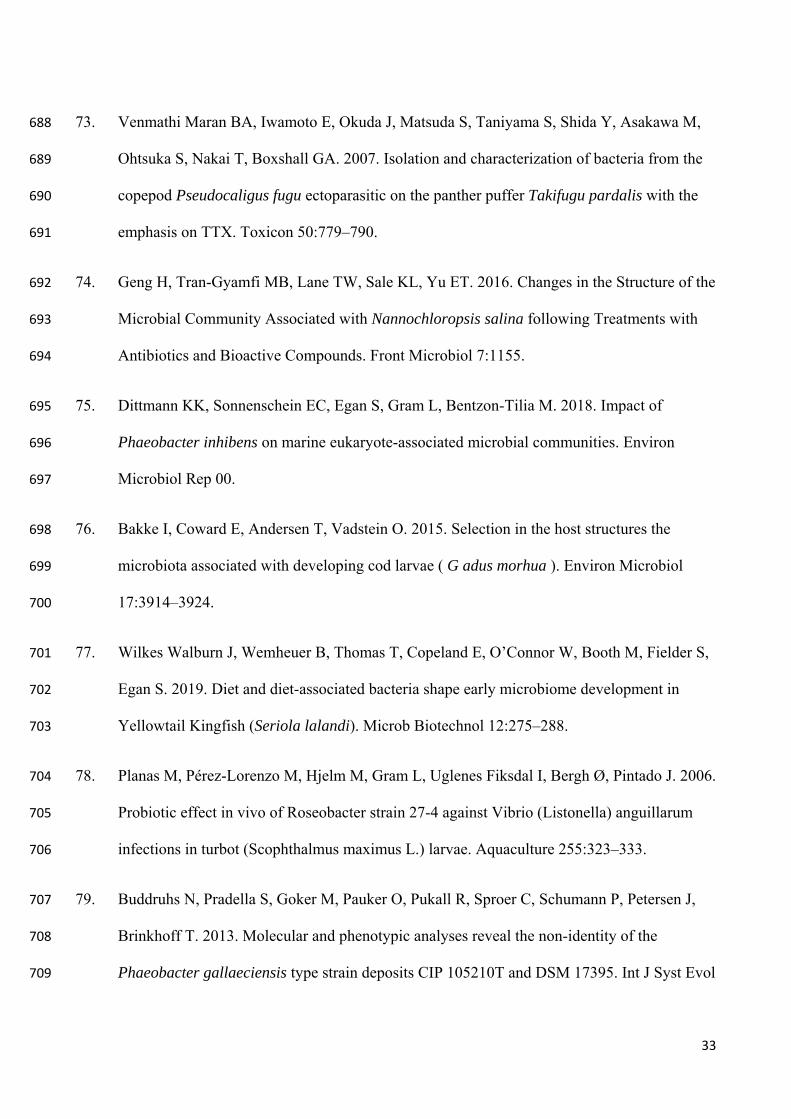
33
73. Venmathi Maran BA, Iwamoto E, Okuda J, Matsuda S, Taniyama S, Shida Y, Asakawa M, 688
Ohtsuka S, Nakai T, Boxshall GA. 2007. Isolation and characterization of bacteria from the 689
copepod Pseudocaligus fugu ectoparasitic on the panther puffer Takifugu pardalis with the 690
emphasis on TTX. Toxicon 50:779–790. 691
74. Geng H, Tran-Gyamfi MB, Lane TW, Sale KL, Yu ET. 2016. Changes in the Structure of the 692
Microbial Community Associated with Nannochloropsis salina following Treatments with 693
Antibiotics and Bioactive Compounds. Front Microbiol 7:1155. 694
75. Dittmann KK, Sonnenschein EC, Egan S, Gram L, Bentzon-Tilia M. 2018. Impact of 695
Phaeobacter inhibens on marine eukaryote-associated microbial communities. Environ 696
Microbiol Rep 00. 697
76. Bakke I, Coward E, Andersen T, Vadstein O. 2015. Selection in the host structures the 698
microbiota associated with developing cod larvae ( G adus morhua ). Environ Microbiol 699
17:3914–3924. 700
77. Wilkes Walburn J, Wemheuer B, Thomas T, Copeland E, O’Connor W, Booth M, Fielder S, 701
Egan S. 2019. Diet and diet-associated bacteria shape early microbiome development in 702
Yellowtail Kingfish (Seriola lalandi). Microb Biotechnol 12:275–288. 703
78. Planas M, Pérez-Lorenzo M, Hjelm M, Gram L, Uglenes Fiksdal I, Bergh Ø, Pintado J. 2006. 704
Probiotic effect in vivo of Roseobacter strain 27-4 against Vibrio (Listonella) anguillarum 705
infections in turbot (Scophthalmus maximus L.) larvae. Aquaculture 255:323–333. 706
79. Buddruhs N, Pradella S, Goker M, Pauker O, Pukall R, Sproer C, Schumann P, Petersen J, 707
Brinkhoff T. 2013. Molecular and phenotypic analyses reveal the non-identity of the 708
Phaeobacter gallaeciensis type strain deposits CIP 105210T and DSM 17395. Int J Syst Evol 709
34
Microbiol 63:4340–4349. 710
80. Sobecky PA, Mincer TJ, Chang MC, Helinski DR. 1997. Plasmids isolated from marine 711
sediment microbial communities contain replication and incompatibility regions unrelated to 712
those of known plasmid groups. Appl Environ Microbiol 63:888–95. 713
81. Guillard RRL. 1975. Culture of Phytoplankton for Feeding Marine Invertebrates, p. 29–60. 714
In Culture of Marine Invertebrate Animals. Springer US, Boston, MA. 715
82. Boström KH, Simu K, Hagström Å, Riemann L. 2004. Optimization of DNA extraction for 716
quantitative marine bacterioplankton community analysis. Limnol Oceanogr Methods 2:365–717
373. 718
83. Lane D. 1991. 16S/23S rRNA sequencing, p. 115–175. In Stackebrandt, E, Goodfellow, M 719
(eds.), Nucleic Acid Techniques in Bacterial Systematics. J.Wiley and Sons, Amsterdam. 720
84. Parada AE, Needham DM, Fuhrman JA. 2016. Every base matters: assessing small subunit 721
rRNA primers for marine microbiomes with mock communities, time series and global field 722
samples. Environ Microbiol 18:1403–1414. 723
85. Apprill A, McNally S, Parsons R, Weber L. 2015. Minor revision to V4 region SSU rRNA 724
806R gene primer greatly increases detection of SAR11 bacterioplankton. Aquat Microb 725
Ecol 75:129–137. 726
86. Chen S, Huang T, Zhou Y, Han Y, Xu M, Gu J. 2017. AfterQC: automatic filtering, 727
trimming, error removing and quality control for fastq data. BMC Bioinformatics 18:80. 728
87. Bolyen E, Rideout JR, Dillon MR, Bokulich NA, Abnet C, Al-Ghalith GA, Alexander H, 729
Alm EJ, Arumugam M, Asnicar F, Bai Y, Bisanz JE, Bittinger K, Brejnrod A, Brislawn CJ, 730
35
Brown CT, Callahan BJ, Caraballo-Rodríguez AM, Chase J, Cope E, Silva R Da, Dorrestein 731
PC, Douglas GM, Durall DM, Duvallet C, Edwardson CF, Ernst M, Estaki M, Fouquier J, 732
Gauglitz JM, Gibson DL, Gonzalez A, Gorlick K, Guo J, Hillmann B, Holmes S, Holste H, 733
Huttenhower C, Huttley G, Janssen S, Jarmusch AK, Jiang L, Kaehler B, Kang K Bin, Keefe 734
CR, Keim P, Kelley ST, Knights D, Koester I, Kosciolek T, Kreps J, Langille MG, Lee J, 735
Ley R, Liu Y-X, Loftfield E, Lozupone C, Maher M, Marotz C, Martin BD, McDonald D, 736
McIver LJ, Melnik A V, Metcalf JL, Morgan SC, Morton J, Naimey AT, Navas-Molina JA, 737
Nothias LF, Orchanian SB, Pearson T, Peoples SL, Petras D, Preuss ML, Pruesse E, 738
Rasmussen LB, Rivers A, Michael S Robeson I, Rosenthal P, Segata N, Shaffer M, Shiffer 739
A, Sinha R, Song SJ, Spear JR, Swafford AD, Thompson LR, Torres PJ, Trinh P, Tripathi A, 740
Turnbaugh PJ, Ul-Hasan S, Hooft JJ van der, Vargas F, Vázquez-Baeza Y, Vogtmann E, 741
Hippel M von, Walters W, Wan Y, Wang M, Warren J, Weber KC, Williamson CH, Willis 742
AD, Xu ZZ, Zaneveld JR, Zhang Y, Zhu Q, Knight R, Caporaso JG. 2018. QIIME 2: 743
Reproducible, interactive, scalable, and extensible microbiome data science. PeerJ Prepr. 744
88. Callahan BJ, McMurdie PJ, Rosen MJ, Han AW, Johnson AJA, Holmes SP. 2016. DADA2: 745
High-resolution sample inference from Illumina amplicon data. Nat Methods 13:581–583. 746
89. Rognes T, Flouri T, Nichols B, Quince C, Mahé F. 2016. VSEARCH: a versatile open source 747
tool for metagenomics. PeerJ 4:e2584. 748
749
750
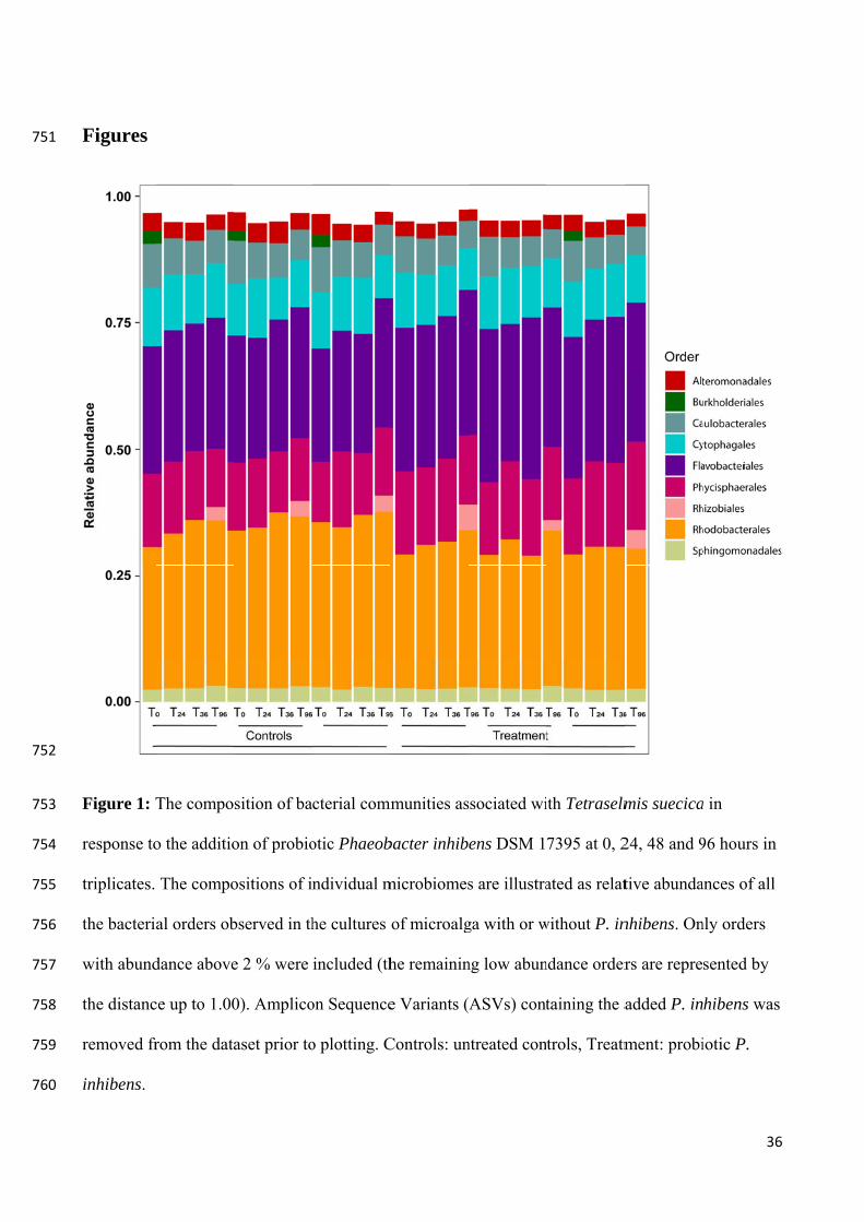
751
752
753
754
755
756
757
758
759
760
Figures
Figure 1: T
response to
triplicates.
the bacteria
with abund
the distanc
removed fr
inhibens.
The compo
o the additio
The compo
al orders ob
dance above
ce up to 1.00
rom the data
sition of ba
on of probio
ositions of i
bserved in th
e 2 % were
0). Amplico
aset prior to
cterial comm
otic Phaeob
ndividual m
he cultures
included (th
on Sequence
o plotting. C
mmunities as
bacter inhib
microbiomes
of microalg
he remainin
e Variants (
Controls: un
ssociated wi
ens DSM 1
s are illustra
ga with or w
ng low abun
ASVs) cont
ntreated con
ith Tetraselm
7395 at 0, 2
ated as relat
without P. in
ndance order
taining the a
trols, Treatm
mis suecica
24, 48 and 9
tive abunda
nhibens. On
rs are repre
added P. in
ment: probi
36
a in
96 hours in
ances of all
nly orders
sented by
hibens was
iotic P.
6
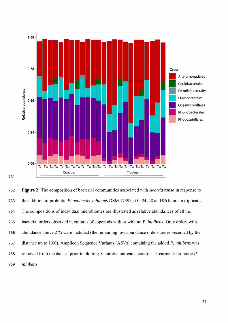
761
762
763
764
765
766
767
768
769
Figure 2: T
the additio
The compo
bacterial or
abundance
distance up
removed fr
inhibens.
The compo
n of probiot
ositions of i
rders observ
e above 2 %
p to 1.00). A
rom the data
sition of ba
tic Phaeoba
individual m
ved in cultu
% were inclu
Amplicon S
aset prior to
cterial comm
acter inhibe
microbiomes
ures of cope
ded (the rem
equence Va
o plotting. C
mmunities as
ens DSM 17
s are illustra
epods with o
maining low
ariants (ASV
Controls: un
ssociated wi
7395 at 0, 24
ated as relat
or without P
w abundance
Vs) contain
ntreated con
ith Acartia t
4, 48 and 96
tive abunda
P. inhibens.
e orders are
ing the adde
trols, Treatm
tonsa in res
6 hours in tr
ances of all t
Only order
e represente
ed P. inhibe
ment: probi
37
ponse to
riplicates.
the
rs with
d by the
ens was
iotic P.
7
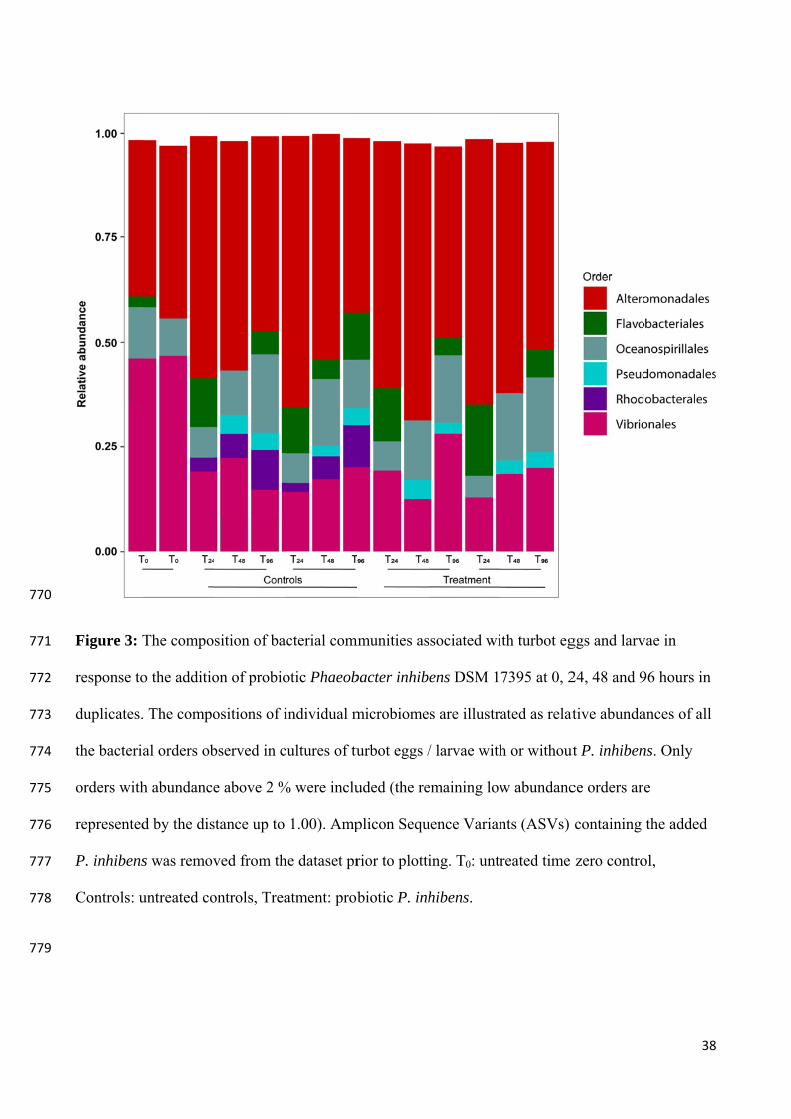
770
771
772
773
774
775
776
777
778
779
Figure 3: T
response to
duplicates.
the bacteria
orders with
represented
P. inhibens
Controls: u
The compo
o the additio
. The compo
al orders ob
h abundance
d by the dis
s was remov
untreated co
sition of ba
on of probio
ositions of i
bserved in c
e above 2 %
stance up to
ved from th
ontrols, Trea
cterial comm
otic Phaeob
individual m
cultures of tu
% were inclu
1.00). Amp
he dataset pr
atment: prob
mmunities as
bacter inhib
microbiome
urbot eggs /
uded (the re
plicon Sequ
rior to plotti
biotic P. inh
ssociated wi
ens DSM 1
s are illustr
/ larvae with
emaining low
uence Varian
ing. T0: untr
hibens.
ith turbot eg
7395 at 0, 2
ated as relat
h or without
w abundanc
nts (ASVs)
reated time
ggs and larv
24, 48 and 9
ative abunda
ut P. inhiben
ce orders ar
containing
zero contro
38
vae in
96 hours in
ances of all
ns. Only
e
the added
ol,
8
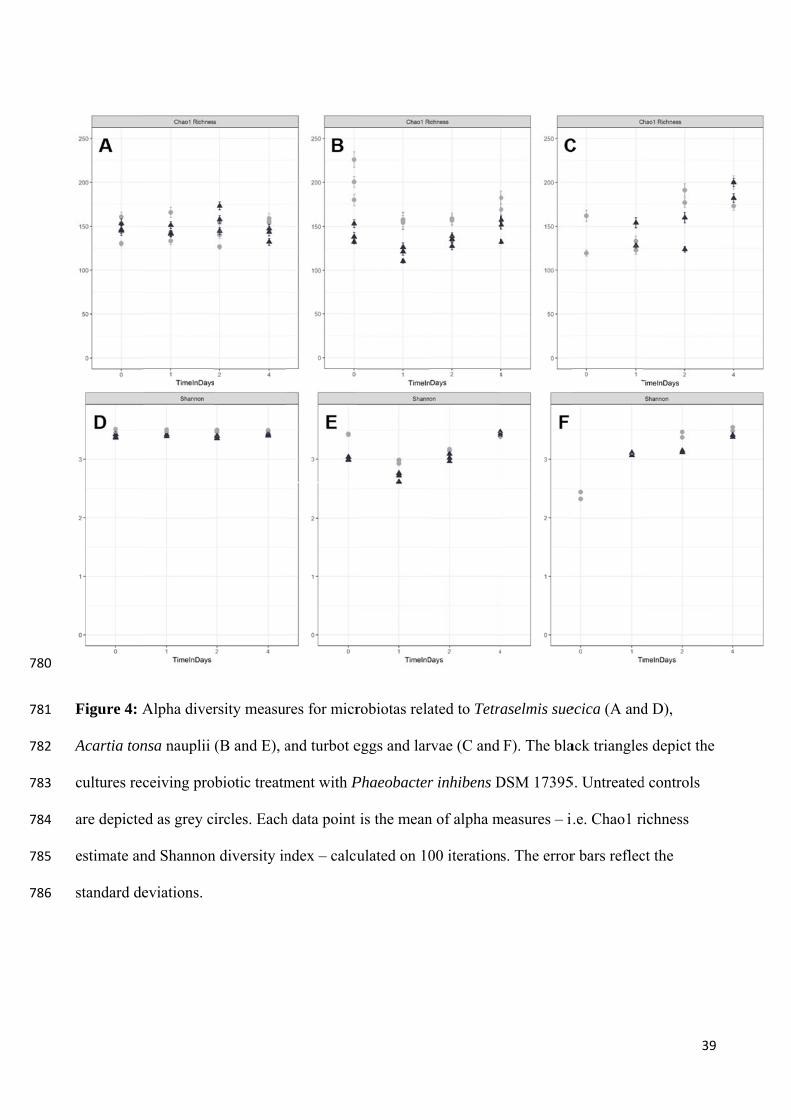
780
781
782
783
784
785
786
Figure 4: A
Acartia ton
cultures rec
are depicte
estimate an
standard de
Alpha diver
nsa nauplii
ceiving pro
ed as grey ci
nd Shannon
eviations.
rsity measur
(B and E), a
biotic treatm
ircles. Each
n diversity in
res for micr
and turbot e
ment with P
h data point
ndex – calcu
robiotas rela
eggs and lar
Phaeobacter
is the mean
ulated on 10
ated to Tetr
rvae (C and
r inhibens D
n of alpha m
00 iteration
aselmis sue
F). The bla
DSM 17395
measures – i
s. The error
ecica (A and
ack triangles
5. Untreated
.e. Chao1 ri
r bars reflec
39
d D),
s depict the
d controls
ichness
ct the
9
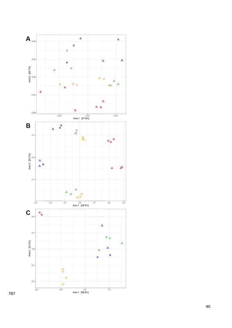
787
400
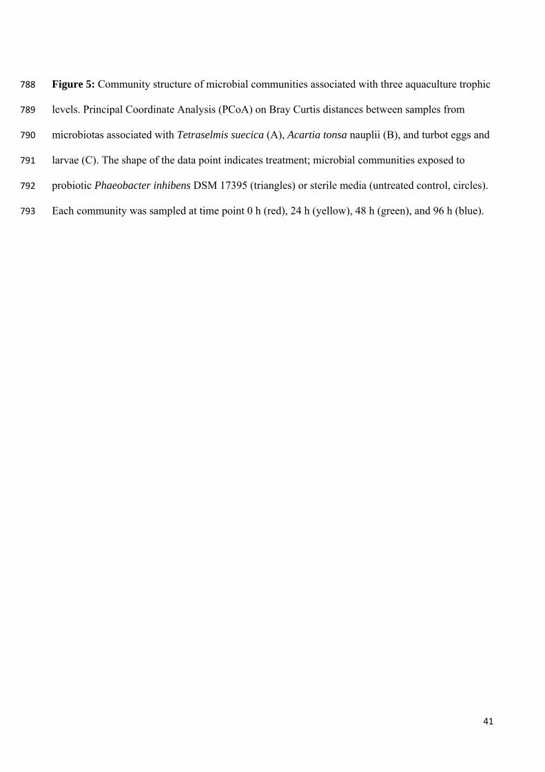
41
Figure 5: Community structure of microbial communities associated with three aquaculture trophic 788
levels. Principal Coordinate Analysis (PCoA) on Bray Curtis distances between samples from 789
microbiotas associated with Tetraselmis suecica (A), Acartia tonsa nauplii (B), and turbot eggs and 790
larvae (C). The shape of the data point indicates treatment; microbial communities exposed to 791
probiotic Phaeobacter inhibens DSM 17395 (triangles) or sterile media (untreated control, circles). 792
Each community was sampled at time point 0 h (red), 24 h (yellow), 48 h (green), and 96 h (blue). 793
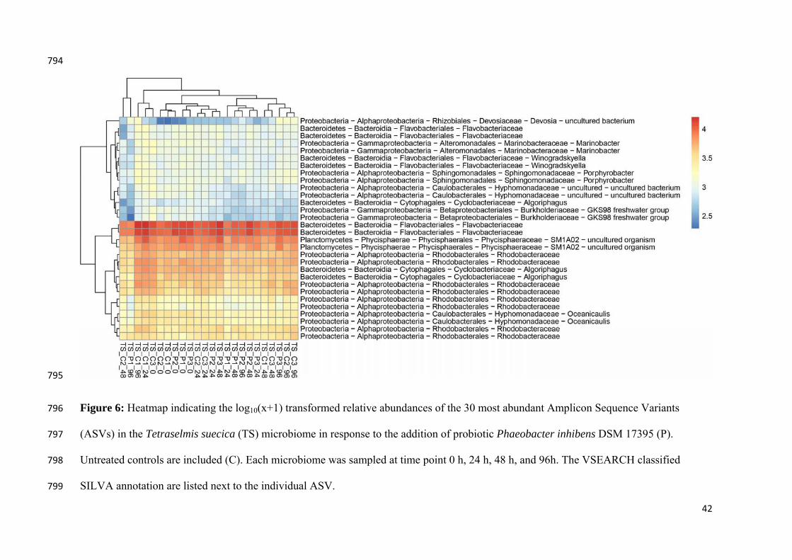
42
794
795
Figure 6: Heatmap indicating the log10(x+1) transformed relative abundances of the 30 most abundant Amplicon Sequence Variants 796
(ASVs) in the Tetraselmis suecica (TS) microbiome in response to the addition of probiotic Phaeobacter inhibens DSM 17395 (P). 797
Untreated controls are included (C). Each microbiome was sampled at time point 0 h, 24 h, 48 h, and 96h. The VSEARCH classified 798
SILVA annotation are listed next to the individual ASV. 799
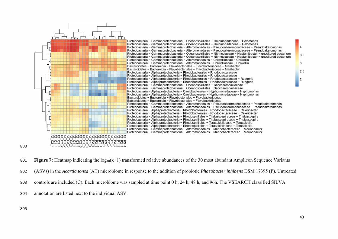
43
800
Figure 7: Heatmap indicating the log10(x+1) transformed relative abundances of the 30 most abundant Amplicon Sequence Variants 801
(ASVs) in the Acartia tonsa (AT) microbiome in response to the addition of probiotic Phaeobacter inhibens DSM 17395 (P). Untreated 802
controls are included (C). Each microbiome was sampled at time point 0 h, 24 h, 48 h, and 96h. The VSEARCH classified SILVA 803
annotation are listed next to the individual ASV. 804
805
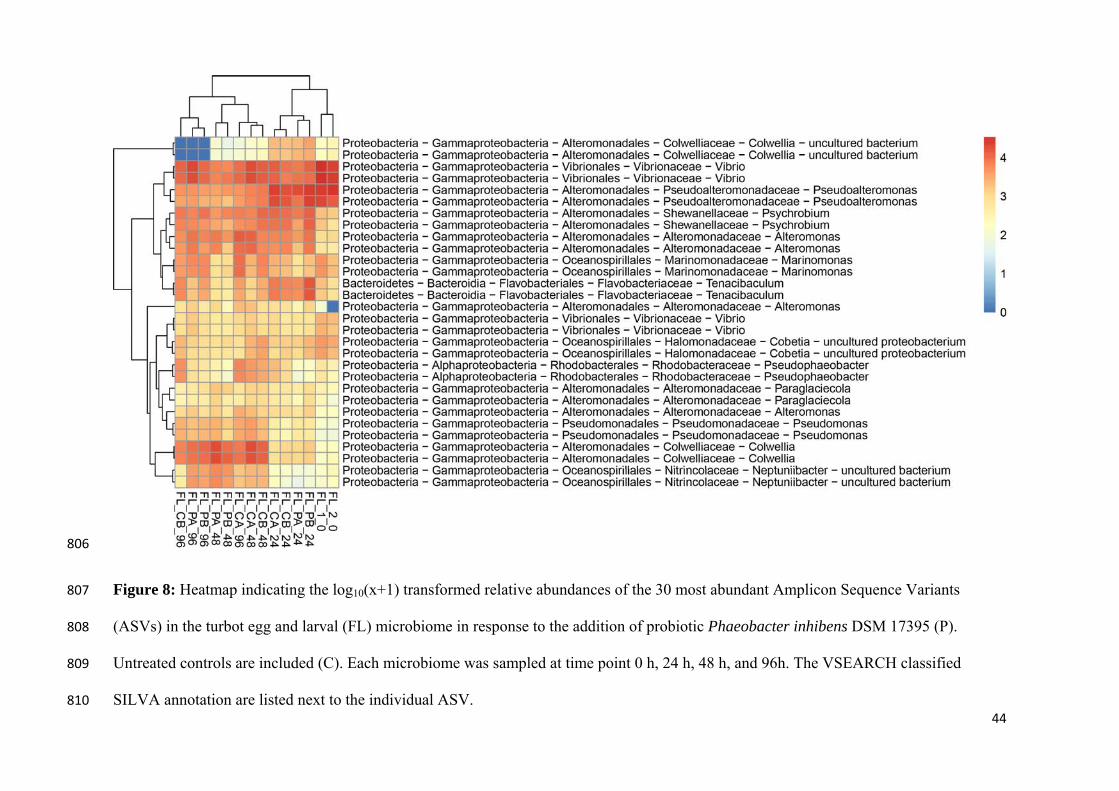
44
806
Figure 8: Heatmap indicating the log10(x+1) transformed relative abundances of the 30 most abundant Amplicon Sequence Variants 807
(ASVs) in the turbot egg and larval (FL) microbiome in response to the addition of probiotic Phaeobacter inhibens DSM 17395 (P). 808
Untreated controls are included (C). Each microbiome was sampled at time point 0 h, 24 h, 48 h, and 96h. The VSEARCH classified 809
SILVA annotation are listed next to the individual ASV. 810