insertion of vaccinia virus c7l host range gene into nyvac-b genome potentiates immune responses...
TRANSCRIPT
Insertion of Vaccinia Virus C7L Host Range Gene intoNYVAC-B Genome Potentiates Immune Responsesagainst HIV-1 AntigensJose Luis Najera1, Carmen Elena Gomez1, Juan Garcıa-Arriaza1, Carlos Oscar Sorzano2, Mariano
Esteban1*
1 Department of Molecular and Cellular Biology, Centro Nacional de Biotecnologıa, CSIC, Ciudad Universitaria Cantoblanco, Madrid, Spain, 2 Biocomputing Unit, Centro
Nacional de Biotecnologıa, CSIC, Ciudad Universitaria Cantoblanco, Madrid, Spain
Abstract
Background: The highly attenuated vaccinia virus strain NYVAC expressing HIV-1 components has been evaluated as avaccine candidate in preclinical and clinical trials with encouraging results. We have previously described that the presenceof C7L in the NYVAC genome prevents the induction of apoptosis and renders the vector capable of replication in humanand murine cell lines while maintaining an attenuated phenotype in mice.
Methodology/Principal Findings: In an effort to improve the immunogenicity of NYVAC, we have developed a novelpoxvirus vector by inserting the VACV host-range C7L gene into the genome of NYVAC-B, a recombinant virus thatexpresses four HIV-1 antigens from clade B (Env, Gag, Pol and Nef) (referred as NYVAC-B-C7L). In the present study, we havecompared the in vitro and in vivo behavior of NYVAC-B and NYVAC-B-C7L. In cultured cells, NYVAC-B-C7L expresses higherlevels of heterologous antigen than NYVAC-B as determined by Western blot and fluorescent-activated cell sorting to scoreGag expressing cells. In a DNA prime/poxvirus boost approach with BALB/c mice, both recombinants elicited robust, broadand multifunctional antigen-specific T-cell responses to the HIV-1 immunogens expressed from the vectors. However, theuse of NYVAC-B-C7L as booster significantly enhanced the magnitude of the T cell responses, and induced a more balancedcellular immune response to the HIV-1 antigens in comparison to that elicited in animals boosted with NYVAC-B.
Conclusions/Significance: These findings demonstrate the possibility to enhance the immunogenicity of the highlyattenuated NYVAC vector by the insertion of the host-range gene C7L and suggest the use of this modified vector as animproved vaccine candidate against HIV/AIDS.
Citation: Najera JL, Gomez CE, Garcıa-Arriaza J, Sorzano CO, Esteban M (2010) Insertion of Vaccinia Virus C7L Host Range Gene into NYVAC-B Genome PotentiatesImmune Responses against HIV-1 Antigens. PLoS ONE 5(6): e11406. doi:10.1371/journal.pone.0011406
Editor: Maciej Lesniak, The University of Chicago, United States of America
Received March 18, 2010; Accepted June 8, 2010; Published June 30, 2010
Copyright: � 2010 Najera et al. This is an open-access article distributed under the terms of the Creative Commons Attribution License, which permitsunrestricted use, distribution, and reproduction in any medium, provided the original author and source are credited.
Funding: This investigation was supported by grants from FIPSE-36551/06, the Ministry of Science and Innovation (SAF2008-02036) and Foundation Botin ofSpain. The funders had no role in study design, data collection and analysis, decision to publish, or preparation of the manuscript.
Competing Interests: The authors have declared that no competing interests exist.
* E-mail: [email protected]
Introduction
AIDS is one of the greatest pandemics of our time, affecting the
health and the social and economic foundations of countries
worldwide. An effective human immunodeficiency virus (HIV)
vaccine offers the best hope for controlling the spread of the virus.
While the immune correlates of protection are not well defined,
both antibodies and T-cell responses contribute to control the
infection with HIV or the related simian immunodeficiency virus
(SIV), as well as disease progression [1,2,3,4,5]. Appropriate
designed envelope immunogens able to induce broad and potent
neutralizing antibodies remained a major goal for vaccine
development and hence, vaccines directed to elicit virus specific
cellular immune responses have been more readily developed, but
their role in protection remains to be established. In this regard the
recent observations of limited protection against HIV-1 infection,
about 31%, in a phase III Thai clinical trial with a combination of
a recombinant canarypoxvirus and the protein gp-120, points in
the direction that both humoral and cellular immune responses
might be needed for protection against HIV/AIDS, although the
specific T cell and neutralizing antibody responses in the trial were
low [6]. These clinical findings highlight that poxvirus vectors
should be considered as one of the future HIV/AIDS vaccine
candidate vectors, but that further vector development is needed.
Indeed, poxvirus vectors have emerged as prominent vehicles
for delivering antigens of HIV-1. Different strains of Vaccinia
Virus (VACV) expressing HIV-1 antigens such as Env, Gag, Pol
and Nef or other components of HIV-1 have been evaluated in
non-human primate [7,8,9,10] and human trials [11,12,13]. While
most of these recombinant viruses do not produce virus progeny in
human cells, which assures safety, they are generally not potent
HIV-1 immunogens by themselves and required priming with
other vectors, such as DNA, to enhance the immune responses to
HIV-1 antigens in animal models [14] and humans [12]. NYVAC
and MVA are promising highly attenuated VACV vectors [15,16],
that in a head-to-head comparison in macaques elicited similar
PLoS ONE | www.plosone.org 1 June 2010 | Volume 5 | Issue 6 | e11406
levels of protection after a challenge with SHIV89.6P,[9]. In a
phase I clinical trial, the combination of recombinant DNA
prime/NYVAC boost regimen (with both vectors expressing Env,
Gag, Pol and Nef of HIV-1 from clade C) revealed that this
vaccination approach was highly immunogenic, eliciting potent,
broad, polyfunctional, and durable T-cells responses in 90% of
vaccinees [13]. Since the protocol of DNA/NYVAC induced a
greater CD4+ T cell response over CD8+ T cells and immuno-
dominance for Env over Gag-Pol-Nef antigens, it suggest that to
obtain a more balanced response to HIV-1 antigens with the
DNA/NYVAC immunization protocol, further improvements of
the NYVAC vector are desirable. One way to achieve this goal
might be through genetic modifications of the NYVAC vector.
NYVAC was derived from Copenhagen strain by the precise
deletion of 18 open reading frames encoding functions involved in the
pathogenicity of the virus as well as in host-range regulatory functions
governing the replication competency of the virus on cells derived
from certain species, including human and mouse [17]. By
reintroduction of the VACV C7L host range gene [18] into NYVAC
vector, the capacity of the virus to replicate in human and rodent cells
was restored, while maintaining an attenuated phenotype in mice
[19]. Replication-competent recombinant VACV-based vaccines
have received increased attention. To date, several replication-
competent recombinants based on VACV viruses have been used as
vaccine vectors for many infectious diseases, demonstrating that they
are able to elicit potent humoral and cell mediated immune
responses, and they are able to confer lasting protection while
maintaining a safe phenotype [20,21,22,23]. In fact, a phase I vaccine
trial in China has recently begun using attenuated, replication
competent Tiantan VACV vector expressing multiple HIV-1 genes
(Wen, J. Phase I study of China’s compound HIV/AIDS vaccine
begins. http://www.asia-lifesciences.com, January 10, 2008).
To improve the immunogenicity of the NYVAC based HIV/
AIDS vaccine candidate, we have developed a novel vector by
inserting the host range gene VACV C7L under the control of the
synthetic early/late virus promoter into NYVAC-B, a recombi-
nant virus which expresses four HIV-1 antigens from clade B (Env,
Gag, Pol and Nef). Previously we showed that insertion of C7L into
NYVAC genome prevented virus-induced apoptosis and the
phosphorylation of the translational initiation factor eIF-2 alpha,
allowing virus multiplication in human cells while the virus
remains attenuated in infected mice [19]. Here, we have
characterized the magnitude, breath, durability and quality of
anti-HIV-1 immunity elicited by the replication competent
NYVAC-B-C7L in comparison to the replication restricted
NYVAC-B. Our findings revealed that NYVAC-B-C7L is superior
to NYVAC-B for induction of specific cellular immune responses
against HIV-1 antigens. Prime/boost with DNA-B/NYVAC-B-
C7L-induced T-cell responses mediated by both CD4+ and CD8+
T cells, and these responses are multifunctional, persistent, durable
and directed to the different vaccine-encoded HIV-1 antigens
Results
Generation and characterization of recombinant NYVAC-B-C7L in infected human cells
In an effort to further improve the immunogenicity of NYVAC
based vaccine candidates against HIV/AIDS, we have generated a
new vector by inserting the VACV C7L host range gene in the
attenuated NYVAC-B recombinant which expresses four HIV-1
antigens from clade B (Env, Gag, Pol and Nef). As shown in
Figure 1A, NYVAC-B-C7L contains the C7L gene inserted into
the HA locus (A56R) of NYVAC-B genome under the transcrip-
tional control of the synthetic early/late (E/L) virus promoter and
preserves, as its parental NYVAC-B, the HIV-1 genes inserted in
the TK locus (J2R). The correct insertion of heterologous genes in
the recombinant virus as well as the purity of the stocks was
confirmed by PCR (data not shown).
The functionality of C7L gene was determined by measuring the
viral growth efficiency of the recombinant viruses in three different
cell lines of human (HeLa), mouse (3T3) and monkey (BSC40)
origin. Monolayers of cells were infected at 0.01 PFU/cell with
Figure 1. Diagram of the genome of NYVAC-B and NYVAC-B-C7L recombinant vectors. A. Diagram of the two vectors with the genesencoding HIV-1 antigens inserted in the TK locus of the viral genome and with C7L at the HA locus.doi:10.1371/journal.pone.0011406.g001
HIV/AIDS Vaccines
PLoS ONE | www.plosone.org 2 June 2010 | Volume 5 | Issue 6 | e11406
NYVAC-B or NYVAC-B-C7L, and at 0 and 48 hours postinfec-
tion the cells were collected with the media and virus titers in cell
homogenates were determined by plaque assay. As shown in
figure 2A, insertion of C7L gene in NYVAC-B genome renders
the vector capable of replication in human and murine cell lines.
The yields of NYVAC-B-C7L in HeLa cells were similar to those
produced by replication-competent VACV-WR strain or in
permissive BSC-40 cells. In mouse cells (3T3), while NYVAC
does not produce progeny virus, NYVAC-B-C7L also replicates
similarly as VACV-WR but the virus yields were 1 to 2 logs lower
than in infected HeLa cells. The presence or absence of C7L gene
has no effect on NYVAC-B replication in BSC-40 cells.
We have previously observed that NYVAC-B expresses
efficiently the HIV-1 proteins, Bx08gp-120 as a protein secreted
Figure 2. Viral growth efficiency and expression of HIV-1 antigens by NYVAC-B and NYVAC-B-C7L vectors. A. HeLa, 3T3 and BSC40cells were infected with NYVAC-B, NYVAC-B-C7L or VACV-WR at 0.01 PFU per cell and at 48 hpi cells were collected with the media. The cells werefrozen and thawed three times, sonicated and virus infectivity was titrated by plaque assay in BSC-40 cells. Data are presented as the mean 6 SD. B.Western blot showing the kinetics of expression of gp-120 and GPN with time of infection. HeLa cells were infected with 5 PFU/cell in the absence orpresence of AraC (A), cell extracts collected at various times, analyzed by SDS-PAGE, and Western blots reacted with specific antibodies to Env andGPN. Actin was used as a loading control. M: uninfected mock cells.doi:10.1371/journal.pone.0011406.g002
HIV/AIDS Vaccines
PLoS ONE | www.plosone.org 3 June 2010 | Volume 5 | Issue 6 | e11406
from cells and III-BGPN as an intracellular fusion polyprotein of
about 150 kDa [24]. To compare the expression levels of the
HIV-1 proteins by NYVAC-B and NYVAC-B-C7L, a time
course was carried out in infected HeLa cells. As shown in the
Western blot of Figure 2B, the kinetics of synthesis of HIV-1
proteins were similar in cells infected with both viral recombi-
nants. However, the Bx08gp-120 and III-BGPN expression levels
at late times post-infection were higher in cells infected with
NYVAC-B-C7L than in cells infected with NYVAC-B. The
protein expression levels were similar when infected cells were
treated with the DNA synthesis inhibitor Ara C, suggesting that
differences in antigen expression between both viral recombi-
nants are mainly a late event. Similar results for enhanced
expression of gp120 and of GPN by NYVAC-B-C7L were
observed in infected monkey BSC40 and mouse 3T3 cells (Fig.
S1, supplementary data).
To analyze in more detail the differences in expression between
NYVAC-B and NYVAC-B-C7L recombinants, we performed a
FACS analysis to quantify the production of HIV-1 and VACV
proteins. HeLa cells were infected with different viral doses of the
two recombinants or with the parental viruses (NYVAC-WT and
NYVAC-C7L). At 18 hours post-infection, cells were harvested,
stained for HIV-1 p24 Gag or for late structural VACV proteins
with the corresponding polyclonal antibodies, and we analyzed the
frequency of Gag and VACV expressing cells as described in
Materials and Methods. The FACS analysis revealed that the
presence of C7L in NYVAC-B genome increased by 2- to 5.3- fold
the percentage of cells expressing HIV-1 p24 Gag (Figure 3A) or
Figure 3. FACS analysis of HIV-1 Gag and VACV antigens expression. HeLa cells were infected with different MOIs of NYVAC-B or NYVAC-B-C7L, and cells were processed for FACS after reactivity with antibodies to HIV-1 p24 for Gag (A) or anti-VACV for virus expression (B), as describedunder Materials and Methods. The doses of virus used to infect HeLa cells are given above FACScans. The extent of expression by NYVAC-B is denotedby the black curve, while NYVAC-B-C7L expression is denoted by the grey curve. Dose-response curves for expression of vaccine insert vs. titer ofinput virus are shown in the right panels.doi:10.1371/journal.pone.0011406.g003
HIV/AIDS Vaccines
PLoS ONE | www.plosone.org 4 June 2010 | Volume 5 | Issue 6 | e11406
VACV antigens (Figure 3B) in comparison to NYVAC-B. The
maximum differences in HIV-1 p24 Gag expression were observed
at 0.1 PFU/cell (4.3 fold) and 0.5 PFU/cell (5.3 fold). We
hypothesize that this is because at low viral doses not all cells
become infected by NYVAC-B, while NYVAC-B-C7L was able to
replicate and spread leading to a higher number of cells expressing
the heterologous antigen. At higher MOIs of 5 and 10 PFU/cell
the increase was only two-fold, as all cells are infected with both
viruses. After NYVAC-B-C7L infection, there was a near
proportional increase as a function of the MOI in HIV-1- and
VACV- expressing cells compared with NYVAC-B infection
(Figure 3A and 3B). The reduced reactivity of the antibodies to
VACV in NYVAC-B infected cells reflects decreased levels of late
viral antigens in these cells compared with cells infected with
NYVAC-B-C7L, an indicator of enhanced virus multiplication as
a result of the presence of C7L gene. We have previously shown a
blockade of certain late viral proteins in HeLa cells infected with
NYVAC [19]. These findings suggest that C7L not only allowed
NYVAC-B-C7L to replicate in HeLa cells but also improved the
production of HIV-1 antigens in infected cells.
Viral distribution of NYVAC-C7L in tissue organs aftersystemic infection
We have previously reported the use of the luciferase marker to
follow VACV replication and antigen expression in mice [25,26].
To determine whether the insertion of the C7L gene into NYVAC
genome affects the virus distribution and heterologous antigen
expression in vivo, BALB/c mice were inoculated i.p. with a dose of
26107 PFU of NYVAC-Luc, NYVAC-C7L-Luc or WR-Luc which
is one of the most effective routes for systemic virus dissemination
[26]. At 24, 48 and 72 h post-inoculation, animals were sacrificed
and the luciferase activity and virus production in different tissues
were assayed in cell homogenates derived from peritoneal cavity,
ovaries, spleen, lymph nodes and liver. While luciferase activity was
clearly observed in all tissues from WR-Luc infected mice, in
comparison NYVAC-Luc expresses greatly reduced levels of
luciferase. Insertion of C7L gene into the NYVAC genome allows
the recombinant virus to express luciferase to levels higher than the
parental virus, mainly in peritoneal cells, ovaries and spleen
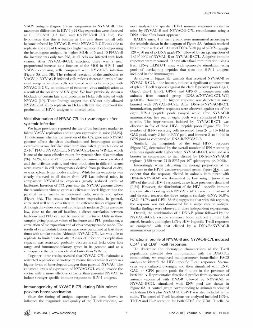
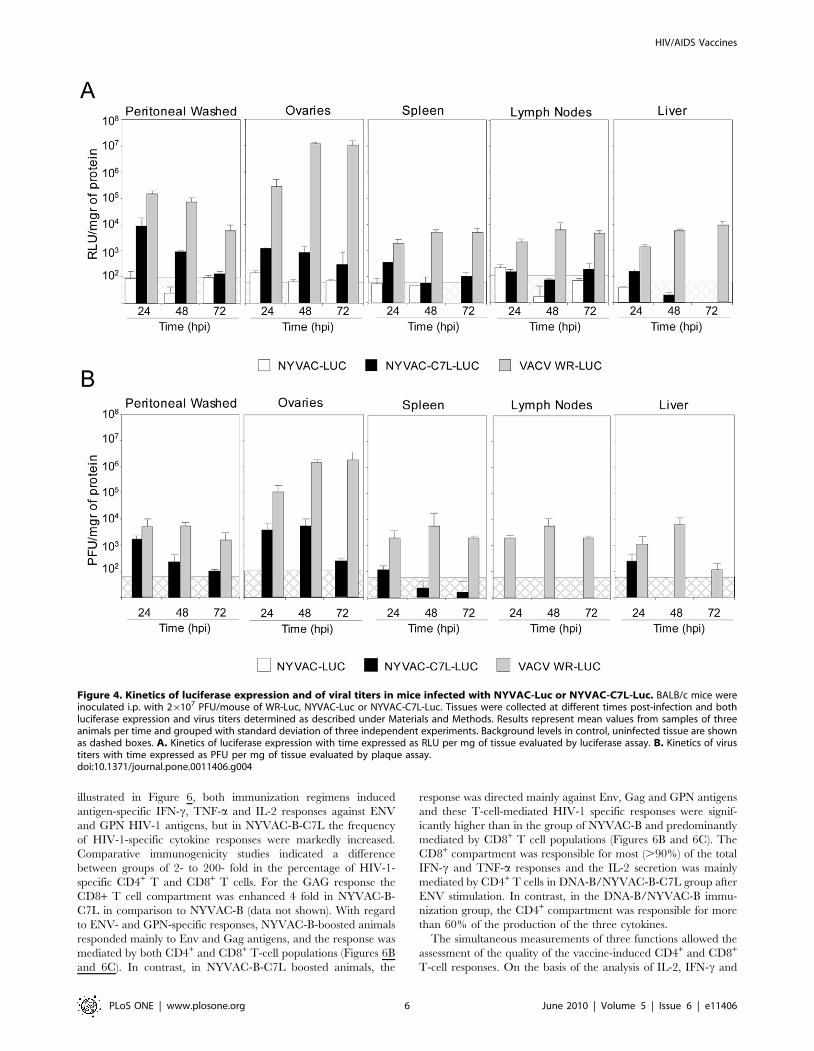
(Figure 4A). The results on luciferase expression, in general,
correlated well with virus titers in the different tissues (Figure 4B).
Although the values observed in the lymph nodes at 24 hpi are quite
low, close to the cut-off baseline, a direct correlation between
luciferase and PFU can not be made in this tissue. Only in those
samples giving positive values of luciferase and PFU production, a
correlation of luc expression and of virus progeny can be made. The
results of viral biodistribution in mice were performed at least three
times with similar results. Although NYVAC-C7L-Luc was able to
replicate to limited extent after 3 days of infection, its replication
capacity was restricted, probably because it still lacks other host
range and immunomodulatory genes in its genome and as a
consequence the virus was clarified faster than WR-Luc.
Together, these results revealed that NYVAC-C7L maintains a
restricted replication phenotype in mouse tissues while it expresses
higher levels of heterologous antigen than parental NYVAC. The
enhanced levels of expression of NYVAC-C7L could provide the
vector with a more effective capacity than parental NYVAC to
induce stronger specific immune response to HIV-1 antigens.
Immunogenicity of NYVAC-B-C7L during DNA prime/poxvirus boost vaccination
Since the timing of antigen exposure has been shown to
influence the magnitude and quality of the T-cell response, we
next analyzed the specific HIV-1 immune responses elicited in
mice by NYVAC-B and NYVAC-B-C7L recombinants using a
DNA prime/Pox boost approach.
BALB/c mice, 4 in each group, were immunized according to
the schedule shown in the diagram of Figure 5A. Animals received
by i.m. route a dose of 100 mg of DNA-B (50 mg of pCMV- Bx08gp-
120 + 50 mg of pcDNA III-BGPN) followed by an i.p. injection of
16107 PFU of NYVAC-B or NYVAC-B-C7L. Adaptive immune
responses were measured 10 days after final immunization using a
fresh IFN-c ELISPOT assay with splenocyte stimulation using
pools of overlapping peptides that span the HIV-1 antigens
included in the immunogens.
As shown in Figure 5B, animals that received NYVAC-B or
NYVAC-B-C7L in the booster, induced a significant enhancement
of splenic T-cell responses against the clade B peptide pools Gag-1,
Gag-2, Env-1, Env-2, GPN-1 and GPN-3 in comparison with
animals from control group (DNA-w/NYVAC-C7L-WT)
(p,0.05). However, the highest response was detected in mice
boosted with NYVAC-B-C7L. After DNA-B/NYVAC-B-C7L
immunization, positive responses were observed against six of the
eight HIV-1 peptide pools assayed while after NYVAC-B
immunization, five out of eight pools were considered HIV-1-
specific. The improvement induced by NYVAC-B-C7L was
observed in five of those HIV-1 peptide pools (Figure 5B). The
number of IFN-c secreting cells increased from 2- to 10- fold in
GAG pool, nearly 2-fold in ENV pool, and between 2- to 4- fold in
GPN pool as compared to DNA-B/NYVAC-B.
Similarly, the magnitude of the total HIV-1 response
(Figure 5C), determined by the overall number of IFN-c secreting
cells, was significantly higher when NYVAC-B-C7L was used as a
booster in comparison to that elicited by DNA-B/NYVAC-B
regimen (4389 versus 2115 SFU per 106 splenocytes, p,0.005).
Interestingly, when calculating the average proportion of the
response to the HIV-1 vaccine-expressed genes (Figure 5D), it was
evident that the response elicited in animals immunized with
DNA-B/NYVAC-B was dominated by Env antigen (more than
50% of the total HIV-1 response), as we have previously described
[9,24]. However, the distribution of the HIV-1 specific immune
response after boosting with NYVAC-B-C7L was more balanced
and directed towards the three antigens similarly (ENV: 36.8%;
GAG: 24.7% and GPN: 38.4%) suggesting that with this regimen,
the response was not dominated by a single vaccine antigen.
Similar findings were observed in three independent experiments.
Overall, the combination of a DNA-B prime followed by the
NYVAC-B-C7L vaccine construct boost induced a more bal-
anced, broader, and higher IFN-c HIV-1-specific T-cell responses
as compared with that elicited by a DNA-B/NYVAC-B
immunization protocol.
Functional profile of NYVAC-B and NYVAC-B-C7L inducedCD4+ and CD8+ T-cell responses
To determine the phenotypic characteristics of the T-cell
populations activated after immunization with the DNA/Pox
combination, we employed multiparameter intracellular FACS
analysis to identify the HIV-1-specific T-cell responses. Spleno-
cytes were cultured overnight and then stimulated with ENV,
GAG or GPN peptide pools for 6 hours in the presence of
brefeldin A. Representative functional profiles from splenocytes of
animals vaccinated with DNA-B followed by NYVAC-B or
NYVAC-B-C7L stimulated with ENV pool are shown in
Figure 6A. A control group corresponding to animals vaccinated
with sham DNA plus NYVAC-C7L-WT was also included in the
study. The panel of T-cell functions we analyzed included IFN-c,
TNF-a and IL-2 secretion for both CD4+ and CD8+ T cells. As
HIV/AIDS Vaccines
PLoS ONE | www.plosone.org 5 June 2010 | Volume 5 | Issue 6 | e11406
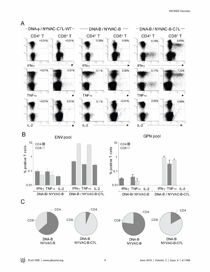
illustrated in Figure 6, both immunization regimens induced
antigen-specific IFN-c, TNF-a and IL-2 responses against ENV
and GPN HIV-1 antigens, but in NYVAC-B-C7L the frequency
of HIV-1-specific cytokine responses were markedly increased.
Comparative immunogenicity studies indicated a difference
between groups of 2- to 200- fold in the percentage of HIV-1-
specific CD4+ T and CD8+ T cells. For the GAG response the
CD8+ T cell compartment was enhanced 4 fold in NYVAC-B-
C7L in comparison to NYVAC-B (data not shown). With regard
to ENV- and GPN-specific responses, NYVAC-B-boosted animals
responded mainly to Env and Gag antigens, and the response was
mediated by both CD4+ and CD8+ T-cell populations (Figures 6B
and 6C). In contrast, in NYVAC-B-C7L boosted animals, the
response was directed mainly against Env, Gag and GPN antigens
and these T-cell-mediated HIV-1 specific responses were signif-
icantly higher than in the group of NYVAC-B and predominantly
mediated by CD8+ T cell populations (Figures 6B and 6C). The
CD8+ compartment was responsible for most (.90%) of the total
IFN-c and TNF-a responses and the IL-2 secretion was mainly
mediated by CD4+ T cells in DNA-B/NYVAC-B-C7L group after
ENV stimulation. In contrast, in the DNA-B/NYVAC-B immu-
nization group, the CD4+ compartment was responsible for more
than 60% of the production of the three cytokines.
The simultaneous measurements of three functions allowed the
assessment of the quality of the vaccine-induced CD4+ and CD8+
T-cell responses. On the basis of the analysis of IL-2, IFN-c and
Figure 4. Kinetics of luciferase expression and of viral titers in mice infected with NYVAC-Luc or NYVAC-C7L-Luc. BALB/c mice wereinoculated i.p. with 26107 PFU/mouse of WR-Luc, NYVAC-Luc or NYVAC-C7L-Luc. Tissues were collected at different times post-infection and bothluciferase expression and virus titers determined as described under Materials and Methods. Results represent mean values from samples of threeanimals per time and grouped with standard deviation of three independent experiments. Background levels in control, uninfected tissue are shownas dashed boxes. A. Kinetics of luciferase expression with time expressed as RLU per mg of tissue evaluated by luciferase assay. B. Kinetics of virustiters with time expressed as PFU per mg of tissue evaluated by plaque assay.doi:10.1371/journal.pone.0011406.g004
HIV/AIDS Vaccines
PLoS ONE | www.plosone.org 6 June 2010 | Volume 5 | Issue 6 | e11406
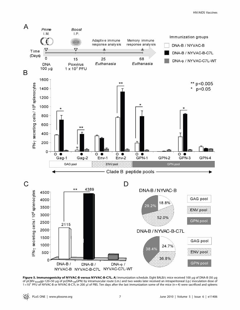
Figure 5. Immunogenicity of NYVAC-B versus NYVAC-B-C7L. A) Immunization schedule. Eight BALB/c mice received 100 mg of DNA-B (50 mgof pCMV-BX08gp-120+50 mg of pcDNA-IIIBGPN) by intramuscular route (i.m.) and two weeks later received an intraperitoneal (i.p.) inoculation dose of16107 PFU of NYVAC-B or NYVAC-B-C7L in 200 ml of PBS. Ten days after the last immunization some of the mice (n = 4) were sacrificed and spleens
HIV/AIDS Vaccines
PLoS ONE | www.plosone.org 7 June 2010 | Volume 5 | Issue 6 | e11406
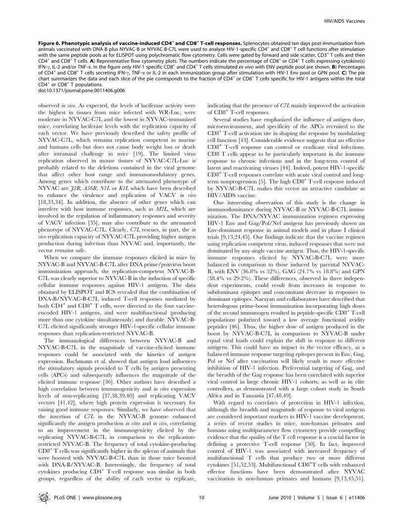
TNF-a secretion, seven distinct HIV-1 specific CD4+ and CD8+
T-cell populations were identified. To further characterize the
immunogenicity triggered in each immunized group, we assessed
polyfunctional T-cell responses. The ENV-specific CD4+ and
CD8+ T cell responses elicited by DNA-B/NYVAC-B or DNA-B/
NYVAC-B-C7L groups were polyfunctional, as more than 70% of
CD8+ T cells and 80% of CD4+ T cells had two or three functions
based on IFN-c, TNF-a and IL-2 secretion. However, some
differences were observed between both groups. GPN-specific
CD8+ and CD4+ T-cell responses were highly polyfunctional in
the DNA-B/NYVAC-B immunization group, while in DNA-B/
NYVAC-B-C7L only CD8+ T cells were multifunctional.
Examination of the relative contributions of individual cytokines
versus polyfunctional responses showed that NYVAC-B-C7L
improves the quality of the response in CD8+ T cell compartment
but not in the NYVAC-B group, with a proportion (1–5%) of the
HIV-1-specific CD8+ T cells expressing simultaneously the three
cytokines,. In contrast, no improvement was found in the CD4+ T-
cell compartment in terms of the quality of the response (Figure 7).
Overall, these results indicated that both immunization
regimens induced polyfunctional ENV and GPN-specific T cell
responses and demonstrated that DNA-B/NYVAC-B-C7L im-
munization improved the magnitude and quality of the anti-HIV-
1 response compared to NYVAC-B.
Polyfunctionality of long-lived HIV-1 specific T-cellresponses induced by NYVAC-B-C7L
The longevity of the immune response generated by a vaccine
may be critical for its success. In order to gain insights on the
durability of the T-cell response generated in mice by the DNA/
Pox immunization protocol outlined in Figure 5A, we analyzed the
T cell profiling at 53 days post-vaccination. Functional character-
ization was performed from the measurements of CD4+ and CD8+
T cells secreting IFN-c and/or IL-2 after Ag-specific stimulation.
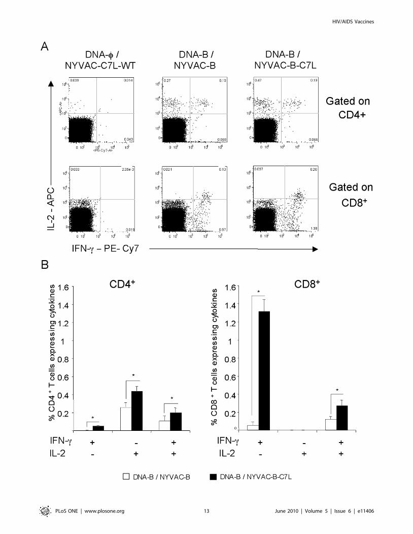
As shown in Figure 8 for CD8+ and CD4+ T cells, the NYVAC-
B-C7L vector showed significantly higher Env-specific immune
responses than NYVAC-B (2.25% versus 1.01%, p.0.05). While
the Gag- and GPN-specific responses were also higher in NYVAC-
B-C7L group, the differences were not statistically significant. For
CD8+ T cells, higher levels of both IFN-c and IL-2 were produced
in mice primed with DNA-B and boosted with NYVAC-B-C7L
than when boosted with NYVAC-B (2 fold). Interestingly, the
production of IFN-c by these cells was considerably higher than the
production of IL-2. In fact, a high frequency of total IL-2
production by CD8+ T cells (more than 90%) resulted also positive
for IFN-c secretion representing 15% of the total Env-specific CD8+
T cell response. However, the Env-specific CD4+ T cells generated
a higher frequency of IL-2 than IFN-c response in both
immunization groups and the percentage of multifunctional Env-
specific CD4+ T cells was about 26% of the total response.
These findings demonstrate that both NYVAC-B and NYVAC-
B-C7L induced long-lived HIV-1 specific T-cell responses but the
magnitude and quantity of these responses were significantly
enhanced by the use of NYVAC-B-C7L in the booster.
Discussion
Different viral vectors of RNA and DNA origin are being
considered as HIV/AIDS vaccine candidates, but to date only the
poxvirus vector ALVAC in combination with a purified gp-120
prime boost has shown some efficacy in a phase III clinical trial
[6]. Since ALVAC exhibits restricted replication in human cells,
which limits the immune potency of the vector, other poxvirus
vectors are sought as vaccine candidates. Among them, the
attenuated strains of vaccinia virus MVA and NYVAC have been
considered as promising HIV/AIDS vaccine candidates due to
their strong safety record in preclinical and clinical trials, high
stability, ability to induce potent and long-lasting immune
responses to foreign antigens, and efficacy in immunized macaques
after challenge with SIV [15]. Although MVA and NYVAC are
safe vectors, their replication in vivo is limited which curtails the
amount of antigen that can be produced in cells [26]. Thus, while
vaccination with replication-restricted VACV recombinants is
highly desirable for safety reasons, there might be an advantage in
the use of replicating vectors eliciting prolonged immune
responses. A close examination of licensed vaccines against
infectious diseases reveals that some of the most potent and
efficacious constructs currently in use are replicating vectors
[27,28]. Replication-competent recombinant VACV-based vac-
cines have received increased attention as potential vaccine
vectors. To date, several replication-competent recombinant
VACV-based vaccines have been used for various infectious
diseases, demonstrating that they are able to elicit potent humoral
and cellular mediated immune responses, and to confer lasting
protection while maintaining a safety phenotype [20,21,22,23]. A
recent smallpox vaccine study has further emphasized the
superiority of replication competent vectors to non-replicating in
long term protection and immunogenicity against lethal cowpox
challenge [29]. The need for replication competence of VACV
vectors has been proposed as potential solution to the lack of
efficacy of some of the highly attenuated vaccines that have been
tested in human trials [20].
In an effort to improve the immune potency of the NYVAC
vector, we have constructed a new vector with replication
competence in human cultured cells. This was done by the
insertion of VACV C7L gene into the genome of NYVAC-B, a
vector that expresses gp-120 and Gag-Pol-Nef antigens of HIV-1
from clade B. The C7L gene was selected for insertion into
NYVAC-B genome because it is a VACV host range gene [18]
necessary for viral replication in human cells [30] and the viral
product inhibits antiviral activities induced by type I interferons
[31,32]. Furthermore, we have previously described that C7L
insertion into the parental NYVAC genome restores the viral
replication cycle in cultured human and murine cells by
preventing eIF2-a phosphorylation and the induction of apoptosis
[19]. Here, we observed that the presence of C7L in the NYVAC-
B genome improved significantly the level of expression of HIV-1
antigens as a result of enhanced translation, thus increasing viral
replication and cell spreading. An increase in antigen expression
observed in culture cells infected with NYVAC-C7L was also
processed for ELISPOT assay and Intracellular Cytokine Staining (ICS) to analyze adaptive immune response. At day 53 the remaining animals in eachgroup (n = 4) were sacrificed and spleen processed for ICS to analyze the long-lived specific response. B) Cellular responses against HIV-1 antigens asmeasured by ELISPOT. Bars show mean values of the number of IFN-c secreting splenocytes from mice immunized groups for each HIV-1 specificpeptide pools that span the four HIV-1 antigens, and error bars represent the standard deviation. Filled symbols represent significant differencesbetween each peptide pool and the negative control. * Represent statistically significant differences between groups. C) Magnitude of the totalresponse for clade B pools. Bars represent the total number of antigen-specific IFN-c secreting cells detected in each group against all the peptidepools spanning the HIV-1 antigens included in the recombinants. D) Mean proportion of the response directed to each of the HIV-1 vaccine antigensin both immunization groups. All the values are shown as net percentage of total HIV-1 specific IFN-c secreting cells.doi:10.1371/journal.pone.0011406.g005
HIV/AIDS Vaccines
PLoS ONE | www.plosone.org 8 June 2010 | Volume 5 | Issue 6 | e11406
HIV/AIDS Vaccines
PLoS ONE | www.plosone.org 9 June 2010 | Volume 5 | Issue 6 | e11406
observed in vivo. As expected, the levels of luciferase activity were
the highest in tissues from mice infected with WR-Luc, were
moderate in NYVAC-C7L and the lowest in NYVAC-immunized
mice, correlating luciferase levels with the replication capacity of
each vector. We have previously described the safety profile of
NYVAC-C7L, which remains replication competent in murine
and humans cells but does not cause body weight loss or death
after intranasal challenge in mice [19]. The limited virus
replication observed in mouse tissues of NYVAC-C7L-Luc is
probably related to the deletions contained in the viral genome
that affect other host range and immunomodulatory genes.
Among genes which contribute to the attenuated phenotype of
NYVAC are J2R, A56R, N1L or K1L which have been described
to enhance the virulence and replication of VACV in vivo
[18,33,34]. In addition, the absence of other genes which can
interfere with host immune responses, such as M2L, which are
involved in the regulation of inflammatory responses and severity
of VACV infection [35], may also contribute to the attenuated
phenotype of NYVAC-C7L. Clearly, C7L rescues, in part, the in
vivo replication capacity of NYVAC-C7L providing higher antigen
production during infection than NYVAC and, importantly, the
vector remains safe.
When we compare the immune responses elicited in mice by
NYVAC-B and NYVAC-B-C7L after DNA prime/poxvirus boost
immunization approach, the replication-competent NYVAC-B-
C7L was clearly superior to NYVAC-B in the induction of specific
cellular immune responses against HIV-1 antigens. The data
obtained by ELISPOT and ICS revealed that the combination of
DNA-B/NYVAC-B-C7L induced T-cell responses mediated by
both CD4+ and CD8+ T cells, were directed to the four vaccine-
encoded HIV-1 antigens, and were multifunctional (producing
more than one cytokine simultaneously) and durable. NYVAC-B-
C7L elicited significantly stronger HIV-1-specific cellular immune
responses than replication-restricted NYVAC-B.
The immunological differences between NYVAC-B and
NYVAC-B-C7L in the magnitude of vaccine-elicited immune
responses could be associated with the kinetics of antigen
expression. Bachmann et al. showed that antigen load influences
the stimulatory signals provided to T cells by antigen presenting
cells (APCs) and subsequently influences the magnitude of the
elicited immune response [36]. Other authors have described a
high correlation between immunogenicity and in vitro expression
levels of non-replicating [37,38,39,40] and replicating VACV
vectors [41,42], where high protein expression is necessary for
raising good immune responses. Similarly, we have observed that
the insertion of C7L in the NYVAC-B genome enhanced
significantly the antigen production in vitro and in vivo, correlating
to an improvement in the immunogenicity elicited by the
replicating NYVAC-B-C7L in comparison to the replication-
restricted NYVAC-B. The frequency of total cytokine-producing
CD8+ T cells was significantly higher in the spleens of animals that
were boosted with NYVAC-B-C7L than in those mice boosted
with DNA-B/NYVAC-B. Interestingly, the frequency of total
cytokines producing CD4+ T-cell response was similar in both
groups, regardless of the ability of each vector to replicate,
indicating that the presence of C7L mainly improved the activation
of CD8+ T-cell responses.
Several studies have emphasized the influence of antigen dose,
microenvironment, and specificity of the APCs recruited to the
CD8+ T-cell activation site in shaping the response by modulating
cell function [43]. Considerable evidence suggests that an effective
CD8+ T-cell response can control or eradicate viral infections.
CD8 T cells appear to be particularly important in the immune
response to chronic infections and in the long-term control of
latent and reactivating viruses [44]. Indeed, potent HIV-1-specific
CD8+ T-cell responses correlate with acute viral control and long-
term nonprogression [5]. The high CD8+ T-cell response induced
by NYVAC-B-C7L makes this vector an attractive candidate as
HIV/AIDS vaccine.
One interesting observation of this study is the change in
immunodominance during NYVAC-B or NYVAC-B-C7L immu-
nization. The DNA/NYVAC immunization regimen expressing
HIV-1 Env and Gag/Pol/Nef antigens has previously shown an
Env-dominant response in animal models and in phase I clinical
trials [9,13,24,45]. Our findings indicate that the vaccine regimen
using replication competent virus, induced responses that were not
dominated by any single vaccine antigen. Thus, the HIV-1-specific
immune responses elicited by NYVAC-B-C7L were more
balanced in comparison to those induced by parental NYVAC-
B, with ENV (36.8% vs 52%), GAG (24.7% vs 18.8%) and GPN
(38.4% vs 29.2%). These differences, observed in three indepen-
dent experiments, could result from increases in response to
subdominant epitopes and concomitant decrease in responses to
dominant epitopes. Narayan and collaborators have described that
heterologous prime-boost immunization incorporating high doses
of the second immunogen resulted in peptide-specific CD8+ T-cell
populations polarized toward a low average functional avidity
peptides [46]. Thus, the higher dose of antigen produced in the
boost by NYVAC-B-C7L in comparison to NYVAC-B under
equal viral loads could explain the shift in response to different
antigens. This could have an impact in the vector efficacy, as a
balanced immune response targeting epitopes present in Env, Gag,
Pol or Nef after vaccination will likely result in more effective
inhibition of HIV-1 infection. Preferential targeting of Gag, and
the breadth of the Gag response has been correlated with superior
viral control in large chronic HIV-1 cohorts, as well as in elite
controllers, as demonstrated with a large cohort study in South
Africa and in Tanzania [47,48,49].
With regard to correlates of protection in HIV-1 infection,
although the breadth and magnitude of response to viral antigens
are considered important markers in HIV-1 vaccine development,
a series of recent studies in mice, non-human primates and
humans using multiparameter flow cytometry provide compelling
evidence that the quality of the T cell response is a crucial factor in
defining a protective T-cell response [50]. In fact, improved
control of HIV-1 was associated with increased frequency of
multifunctional T cells that produce two or more different
cytokines [51,52,53]. Multifunctional CD8+T cells with enhanced
effector functions have been demonstrated after NYVAC
vaccination in non-human primates and humans [9,13,45,51].
Figure 6. Phenotypic analysis of vaccine-induced CD4+ and CD8+ T-cell responses. Splenocytes obtained ten days post-immunization fromanimals vaccinated with DNA-B plus NYVAC-B or NYVAC-B-C7L were used to analyze HIV-1-specific CD4+ and CD8+ T cell functions after stimulationwith the same peptide pools as for ELISPOT using polychromatic flow cytometry. Cells were gated by forward and side scatter, CD3+ T cells and thenCD4+ and CD8+ T cells. A) Representative flow cytometry plots. The numbers indicate the percentage of CD8+ or CD4+ T cells expressing cytokine(s)IFN-c, IL-2 and/or TNF-a. In the figure only HIV-1 specific CD8+ and CD4+ T cells stimulated ex vivo with ENV peptide pool are shown. B) Percentagesof CD4+ and CD8+ T cells secreting IFN-c, TNF-a or IL-2 in each immunization group after stimulation with HIV-1 Env pool or GPN pool. C) The piechart summarizes the data and each slice of the pie corresponds to the fraction of CD4+ or CD8+ T cells specific for HIV-1 antigens within the totalCD4+ or CD8+ T populations.doi:10.1371/journal.pone.0011406.g006
HIV/AIDS Vaccines
PLoS ONE | www.plosone.org 10 June 2010 | Volume 5 | Issue 6 | e11406
HIV/AIDS Vaccines
PLoS ONE | www.plosone.org 11 June 2010 | Volume 5 | Issue 6 | e11406
In our mouse model, both vectors NYVAC-B and NYVAC-B-
C7L induced polyfunctional CD4+ and CD8+ T cells that were
mainly IFN-c+TNF-a+. Of note, Env- and GPN-specific CD4+
and CD8+ T-cell responses were also able to secrete some amounts
of IL-2. In fact, a high proportion (10-16%) of the HIV-1-specific
CD4+ T cells expressing simultaneously the three cytokines were
found in both immunization groups while the percentages of this
population in the CD8+ T-cell compartment were lower (1–5%)
and only observed in animals boosted with NYVAC-B-C7L,
suggesting that good quality of both T-cell responses was induced
by the vectors. The presence of polyfunctional CD4+ T cells after
immunization with both vectors could be favorable for the
induction and maintenance of CD8+ T-cell memory responses
[54]. Indeed, a dysfunctional HIV-1-specific CD4+ response is a
hallmark of chronic HIV-1 infection, and adversely influences the
development and functioning of the CD8+ T-cell response [55,56].
These results might correlate with those previously described in
which the functional profile of T cells is directly related to antigen
concentration and/or antigen persistence [57,58].
An important consideration in HIV-1 vaccine development is
the establishment of memory responses. Previous studies had
shown the induction of long lasting T cell immunity in most of the
human volunteers vaccinated with DNA-C prime/NYVAC-C
boost [13]. Here, we observed that NYVAC-B-C7L improved the
magnitude and quality of the cellular immune responses measured
at 53 days post-boost. High levels of IFN-c and IL-2 were
produced in both CD4+ and CD8+ T-cell compartments after Env
stimulation as compared to NYVAC. Hovav and colleagues
described that the priming and boosting vectors influenced the
magnitude of the secondary CD8+ T cell response, but that the
ultimate differentiation of these cells to memory cells was
exclusively shaped by the second immunogen [59]. In addition,
the duration of antigen expression was found to modulate the rate
of differentiation of memory CD8+ T cells, as this process begins
only after most of the antigen is cleared [36,60]. Furthermore,
other factors also appear to influence the evolution of cellular
immune memory, and early inflammatory signals delivered by the
vector to the immune system have been reported to enhance
antigen-presenting activity and control the rate of memory CD8+
T-cell development [61]. Thus the differences observed in
NYVAC behavior after C7L insertion with regard to cytopathic
effect, apoptosis induction or viral clearance [19] might have an
impact in the type of immune response elicited. Further studies
will be needed to determine the innate immunity induced by
NYVAC-B-C7L as well as to how the infection impacts on
dendritic cells.
Overall, the present data demonstrate that both NYVAC-B and
NYVAC-B-C7L in combination with DNA-B are highly immu-
nogenic in mice, induced vigorous and broad T cell responses,
comprising of both CD4+ and CD8+ T cells, which are
polyfunctional, and more importantly, these immunization
regimens induce long-lasting T cell immunity. Furthermore, we
showed that insertion of C7L in NYVAC-B genome clearly
improves the magnitude and breadth of the cellular immune
response induced against the HIV-1 antigens as compared to
NYVAC-B. In addition, the replication capability of NYVAC-
C7L allows the administration of the vaccine at low and safe doses,
thus reducing manufacture burden and making production of
NYVAC-C7L vaccines cost effective. Therefore, NYVAC recom-
binants expressing C7L gene are attractive live viral vectors for the
development of vaccines and highlight the use of safe attenuated
replication competent VACV vectors as improved platform for
vaccine development against HIV/AIDS and other infectious
diseases.
Materials and Methods
Cells and VirusesCells were maintained in a humidified air 5% CO2 atmosphere
at 37uC. African green monkey kidney cells (BSC40) and human
cells (HeLa) were grown in Dulbecco’s modified Eagle’s medium
(DMEM) supplemented with 10% newborn calf serum (NCS). 3T3
murine cells were grown in Dulbecco’s modified Eagle’s medium
(DMEM) supplemented with 10% calf serum (CS).
Recombinant viruses with the luciferase gene inserted in the
viral thymidine kinase (TK) locus (J2R) were used to test virus
biodistribution in vivo. NYVAC-Luc and WR-Luc recombinants
have been previously described [25,26]. NYVAC-C7L-Luc was
generated according to standard methodology using the same
plasmid transfer vector used for the generation of WR-Luc and
NYVAC-Luc (pSC11-LUC [25]). In addition to C7L gene, this
new recombinant vector NYVAC-C7L-Luc encodes the luciferase
reporter gene under the transcriptional control of the synthetic
early/late (E/L) virus promoter.
Parental NYVAC and recombinant NYVAC-B expressing the
HIV-1 Bx08gp-120 and III-BGPN proteins from clade B, kindly
provided by Aventis-Pasteur, were previously described [24].
NYVAC-B-C7L was generated using the same plasmid transfer
vector used for the generation of NYVAC-C7L (pJR101-C7L
[19]). This plasmid directs the insertion of C7L gene into the HA
locus (A56R) of NYVAC-B genome under the transcriptional
control of the synthetic early/late (E/L) virus promoter. BSC-40
cells were infected with NYVAC-B recombinant at a multiplicity
of 0.01 PFU/cell, and then transfected with 10 mg of pJR101-C7L
DNA using lipofectamine reagent according to manufacturer’s
instructions (Invitrogen). Recombinant NYVAC-B viruses con-
taining the C7L gene were selected by consecutive rounds of
plaque purification in BSC-40 cells stained with X-Gluc (5-bromo-
4-chloro-3-indoxyl-b-D-glucuronidase acid). In addition to C7L
gene, this new recombinant vector NYVAC-B-C7L contains, as
for NYVAC-B, the same cassette of HIV-1 genes in the TK locus
(J2R) under the transcriptional control of the synthetic early/late
virus promoter.
All viruses were grown in BSC-40 cells, similarly purified
through two 45% (w/v) sucrose cushions, and titrated by plaque
assay in BSC-40 cells. Purity of the recombinants was confirmed
by PCR analyses. Throughout the manuscript we will refer to
parental NYVAC as replication-restricted and NYVAC-C7L as
replication-competent, based on their different abilities to produce
progeny virus in cultured human cells, with limited virus
replication cycle for the former and complete replication cycle
for the latter.
Figure 7. Functional profile of vaccine-induced CD4+ and CD8+ T cells. Functional composition of CD4+ and CD8+ T cells responses againstENV (A) and GPN (B) based on the secretion of IFN-c, IL-2 and/or TNF-a. All the possible combinations of the responses are shown on the X axis,whereas the percentages of the functionally distinct cell populations are shown on the Y axis. Bars correspond to the fraction of different functionallydistinct T-cell populations within total CD4+ and CD8+ populations. Responses are grouped and color-coded on the basis of the number functions.The pie chart summarizes the data and each slice of the pie correspond to the fraction of CD4+ or CD8+ T cells with a given number of functionswithin the total CD4+ or CD8+ T populations. The size of the pie chart represents the magnitude of the specific HIV-1 immune response induced.doi:10.1371/journal.pone.0011406.g007
HIV/AIDS Vaccines
PLoS ONE | www.plosone.org 12 June 2010 | Volume 5 | Issue 6 | e11406
HIV/AIDS Vaccines
PLoS ONE | www.plosone.org 13 June 2010 | Volume 5 | Issue 6 | e11406
Analysis of Virus growthMonolayers of human (HeLa), mouse (3T3) and monkey (BSC-
40) cells grown in 12-well tissue culture plates were infected at
0.01 PFU/cell with NYVAC-B, NYVAC-B-C7L and VACV-WR
recombinants, at different times postinfection cells were collected
with the media, sonicated and virus yields determined by plaque
assay in BSC-40 cells as previously described [19].
Expression of Bx08gp-120 and IIIBGPN proteins by NYVAC-B and NYVAC-B-C7L in cultured cells
HeLa, 3T3 and BSC-40 cells grown in 12 well-plates were
infected at 5 PFU/cell with the recombinants NYVAC-B or
NYVAC-B-C7L in presence or absence of adenine arabinoside
(AraC, 40 mg/ml, Sigma), an inhibitor of viral DNA replication.
At 2, 4, 8 and 24 hours post-infection (hpi), cells were collected
and centrifuged at 1500 rpm for 10 min. Cellular pellets were
lysed in cold buffer (50 mM Tris-HCl pH 8, 0.5 M NaCl, 10%
NP-40, 1% SDS) and samples containing equal amounts of protein
were run in 10% SDS-PAGE. The expression of HIV-1 Bx08gp-
120 and IIIBGPN was visualized following western blotting using
rabbit polyclonal anti-gp-120 antibody (Centro Nacional de
Biotecnologia) and rabbit polyclonal anti-Gag p24 serum (ARP
432, NIBSC, Centralized Facility for AIDS reagent, UK),
respectively. Detection of cellular b-actin protein by specific
antibody was used as an internal loading control.
FACS analysis of HIV-1 and VACV viral antigens incultured human cells
Fluorescent activated cell sorting (FACS) analysis was used to
assay the frequency of antigen expressing cells [62]. Monolayers of
HeLa cells grown in 6 well tissue culture plates were infected with
different doses of NYVAC-B or NYVAC-B-C7L (from 0.01 to
10 PFU/cell). Cultures were harvested at 18 hours post-infection
by treating with trypsin. Approximately 106 cells were fixed and
permeabilized using cytofix/cytoperm solution (Pharmigen Inc.)
according to manufacture’s instructions. For detection of viral
vaccinia antigen expression, cells were stained using a polyclonal
anti-vaccinia antibody (VACV-WR) and then incubated with
phycoerythrin conjugated anti-rabbit immunoglobuling (Pharmin-
gen). To test HIV-1 antigen expression we used anti-human
immunodeficiency virus (HIV) Gag antibody conjugated to
phycoerithrin (clone KC57, Beckman Coulter). A total of
100000 events were acquired on a LSR II FACS (Beckton
Dickinson) and analysed using FlowJo software (FlowJo).
DNA vectorsThe two DNA constructs expressing the HIV-1 BX08gp-120
(pCMV-BX08gp-120) and HIV-1 IIIB Gag-Pol-Nef (GPN) fusion
protein (pcDNA-IIIBGPN) have been previously reported [24,63].
Plasmids were purified using Maxi-prep purification kits (Qiagen) and
diluted for injection in endotoxin-free phosphate buffered saline (PBS).
PeptidesThe HIV-1 peptide pools Gag-1, Gag-2, Env-1, Env-2, GPN-1,
GPN-2, GPN-3 and GPN-4 were provided by the EuroVacc
Foundation and were previously described [24]. They spanned the
HIV-1 Env, Gag, Pol and Nef regions from clade B included in the
immunogens as consecutive 15-mers overlapping by 11 amino
acids. For immunological analysis, we grouped the HIV-1 peptides
in three main pools: ENV, GAG and GPN. ENV pool comprises
Env-1 and Env-2; GAG pool comprises Gag-1 and Gag-2, and
GPN pool includes GPN-1, GPN-2, GPN-3 and GPN-4.
Mice immunizationBALB/c mice were purchased from Harlan. To analyze the
biodistribution of virus recombinants in animals, BALB/c were
immunized by intraperitoneal route (i.p.) with a dose of
26107 PFU/mouse of WR-Luc, NYVAC-Luc or NYVAC-C7L-
Luc. At different times post-infection mice were sacrificed and
mouse tissues were processed for luciferase analysis and plaque
assay titration.
To test the immunogeniciy induced by the HIV-1 recombi-
nants, we structured the study around our standard immunization
regimen (DNA/Pox combination) which we have previously
shown that it elicited strong immune responses to HIV-1 antigens
[24,64]. Animals (n = 4 per group) were primed with 100 mg of
DNA-B (50 mg of pCMV-BX08gp-120 + 50 mg of pcDNA-IIIBGPN)
by intramuscular route (i.m.) and two weeks later received an i.p.
inoculation dose of 16107 PFU of NYVAC-B or NYVAC-B-C7L
in 200 ml of PBS. Ten days after the last immunization, mice were
sacrificed and spleens processed for ELISPOT assay and
Intracellular Cytokine Staining (ICS). When long-lived immune
response was assayed, the animals (n = 4 per group) were sacrificed
at day 53 after the boost. Three independent experiments were
performed.
Measurement of luciferase activity in mouse tissuesGene expression of recombinant viruses was monitored by the
luciferase assay to quantify heterologous gene expression in tissues
from mice inoculated i.p with NYVAC-Luc, NYVAC-C7L-Luc or
WR-Luc. Tissues were collected at 24, 48 or 72 h post-infection.
Peritoneal cells were harvested by mouse peritoneal cavity lavage
with 10 ml of sterile PBS, centrifuged at room temperature for
5 min at 1200 rpm and stored at -70uC. At indicated times post-
inoculation, animals were sacrificed, spleen, liver, draining lymph
nodes and ovaries were dissected under sterile conditions and
stored at 270uC. Tissues from individual mice were homogenized
in luciferase extraction buffer (300 ml/spleen and 200 ml/ovary,
lymph node or peritoneal extracts) (Promega Corp., Madison,
Wis.) using an Eppendorf-fitted Dounce homogenizer. Luciferase
activity was measured in the presence of luciferin and ATP
according to the manufacturer’s instructions using a Lumat LB
9501 luminometer (Berthold, Nashua,N.H.), and was expressed as
Reference Luciferase Units (RLU) per milligram of protein.
Protein content in tissue extracts was measured with a bicinch-
oninic protein assay reagent kit (Pierce Co., Rockford, Ill.). Tissues
from individual mice were collected and homogenized in complete
DMEM media to test for the production of infectious virus by
plaque assay in BSC-40 cells. The virus titer was expressed as
Plaque Forming Units (PFU) per milligram of protein.
Figure 8. Long-lived responses to vaccination after ENV stimulation. Splenocytes obtained 53 days post- immunization from animalsvaccinated with DNA-B plus NYVAC-B or NYVAC-B-C7L were used to analyze HIV-1-specific CD4+ and CD8+ T cell functions after stimulation with thesame peptide pools used previously. Cells were gated by forward and side scatter, CD3+ T cells and then CD4+ and CD8+ T cells. In the figure only HIV-1 specific CD4+ and CD8+T cells stimulated ex vivo with ENV peptide pool are shown. A) Representative flow cytometry plots. The numbers indicatethe percentage of CD4+ or CD8+ T cells expressing cytokine(s) IFN-c and/or IL-2. B) Percentages of CD4+ and CD8+ T cells secreting IFN-c and/or IL-2 ineach immunization group after stimulation with HIV-1 ENV pool.doi:10.1371/journal.pone.0011406.g008
HIV/AIDS Vaccines
PLoS ONE | www.plosone.org 14 June 2010 | Volume 5 | Issue 6 | e11406
IFN-c ELISPOT assayThe vaccine- specific cellular immune response in mice was
determined using ELISPOT assay measuring the secretion of IFN-
c by splenocytes after stimulation with different peptides pools.
Briefly, 105–106 splenocytes (depleted of red blood cells) were
plated in triplicate in 96-well nitrocellulose-bottomed plates
previously coated with 6 mg/ml of anti-mouse IFN-c mAb R4-
6A2 (Pharmingen, San Diego, CA). HIV-1 peptide pools from
clade B and negative control (CTRL) pool were resuspended in
RPMI 1640 supplemented with 10% FCS and added to the cells at
a final concentration of 5 mg/ml for each peptide. Plates were
incubated at 37uC, 5% CO2 for 48 h, washed extensively with
PBS containing 0.05% of Tween20 (PBS-T) and incubated 2 h at
room temperature (RT) with a solution of 2 mg/ml of biotinylated
anti-mouse IFN-c mAb XMG1.2 (Pharmingen, San Diego, CA) in
PBS-T. Afterwards, plates were washed with PBS-T and 100 ml of
peroxidase-labeled avidin (Sigma, St Louis, Mo) at 1:800 dilution
in PBS-T was added to each well. After 1 h of incubation at RT,
wells were washed with PBS-T and PBS. The spots were
developed by adding 1 mg/ml of the substrate 3,39-diaminobenzi-
dine tetrahydrochloride (Sigma, St Louis, Mo) in 50 mM Tris-
HCl, pH 7.5 containing 0.015% hydrogen peroxide. The spots
were counted with the aid of a stereomicroscope. The responses
were given as the number of spot-forming cells per million of
splenocytes. In all the cases the background levels were subtracted
to each specific peptide pool.
Intracellular Cytokine Staining (ICS)The phenotypes of responding T cells were analyzed by ICS
and fluorescence-activated cell sorting analysis as described
elsewhere [9]. After an overnight rest, 56106 splenocytes (depleted
of red blood cells) were resuspended in RPMI 1640 supplemented
with 10% FCS and containing 1 ml/ml Golgiplug (BD Biosciences)
to inhibit cytokine secretion. Cells were seeded on M96 plates and
stimulated with overlapping peptides that span the entire HIV-1
BX08gp-120 and IIIBGPN proteins (Gag-pool, Env-pool or GPN-
pool) added to the cells at a final concentration of 5 mg/ml for
each peptide pool. Cells were incubated at 37uC, 5% CO2, and
then analyzed by ICS. After 6 h of stimulation, cells were washed,
stained for the surface markers, fixed, permeabilized using the BD
Cytofix/CytopermTM Kit (Becton Dickinson) and stained intra-
cellularly using the appropriate fluorochromes. To analyze the
adaptive immune responses (day 10 post-boost), the following
fluorochrome-conjugated antibodies were used: CD3-FITC, CD4-
Alexa 700, CD8-PerCP, IL-2-PE, IFN-c-APC and TNF-a-PECy-
7. For analyses at day 53 post-boost, the following antibodies were
used: CD4-Alexa 700, CD8-FITC, IFN-c-PECy-7 and IL-2-
Alexa-647. All antibodies were from BD Biosciences. Cells were
acquired using an LSRII flow cytometer (Becton Dickinson)
equipped with a high throughput system. Sample analysis was
performed using FlowJo version 8.5.3 (Tree Star, Ashland, OR).
The number of lymphocyte-gated events ranged between 105 and
106 dead cells were excluded using violet LIVE/DEAD stain kit
(Becton Dickinson). Lymphocytes were gated on a forward scatter
area versus side scatter area pseudo-color dot plot and dead cells
were removed according to violet LIVE/DEAD staining. To
analyze the adaptive immune responses, CD4+ and CD8+ events
(gate previously on CD3+ cells) were gated versus IFN-c, TNF-aand IL-2, and then combined together using the boolean operator.
For the analyses at day 53 after the boost, CD4+ and CD8+ events
were gated and the secretion of IFN-c and IL-2 was analyzed.
Data were analyzed using a one-tailed z test, to test if the antigen-
specific response was higher than the negative control response
(response with medium alone).
Statistical analysisFor the statistical analysis, we have developed a novel approach
that corrects measurements for the medium response (RPMI) and
at the same time it allows the exact calculation of confidence
intervals and p-values of hypothesis tests. For ELISPOT and ICS
statistical analysis, it was proceeded as follow. Given the total
number of cells, NT , and the number of cells responding to a given
antigen, NAg, an estimate of the proportion of cells responding to
this antigen is given by ppAg~NAg
NT
. The Bayesian a posteriori
distribution of ppAg without any a priori assumption (i.e., assuming
that the true proportion is uniformly distributed between 0 and 1)
is the Beta distribution with parameters NAgz1,NT{NAgz1� �
[65]. Let us call fppAgxð Þ the corresponding probability density
function a posteriori. Analogously, we can derive the distribution of
the proportion of cells responding to RPMI, obtaining the
distribution fppRPMIxð Þ. To test whether the antigen response is
significantly larger than the RPMI response, we computed the
probability density function of the variable ppAgCorrected~
ppAg{ppRPMI as fppAgCorrectedxð Þ~
Ð?
{?fppAg
xð ÞfppRPMIxzxð Þdx. The
cumulative density function of this variable is defined in the usual
way FppAgCorrectedxð Þ~
Ðx
{?fppAgCorrected
xð Þdx. The a percentile of this
variable is defined as xa such that FppAgCorrectedxað Þ~a. We
computed the symmetric 95% confidence interval for the RPMI
corrected proportion as x0:025,x0:975½ �. Finally, we consider ppAg to
be significantly larger than ppRPMI if x0:025w0. In such a case,
x0:025,x0:975½ � gives the 95% symmetric confidence interval for
ppAgCorrected . The average ppAgCorrected is computed as the expected
value of fppAgCorrectedxð Þ (note that this expected value needs not be in
the middle of the confidence interval,x0:025zx0:975
2).
Whenever two corrected proportions need to be summed,
ppAgCorrected1z2~ppAgCorrected1
zppAgCorrected2, we convolved their
probability density functions to obtain the probability den-
sity function of the summed proportion, fppAgCorrected1z2xð Þ~
Ð?
{?fppAgCorrected1
xð ÞfppAgCorrected2x{xð Þdx, in this way confidence inter-
vals for any sum of corrected proportions can be obtained. Antigen
responses were not added unless each component was significantly
larger than the corresponding RPMI.
In the ELISPOT experiment, three replicates were obtained for
each kind of antigen. The average response to that antigen was
computed using only the corrected proportions significantly larger
than the corresponding RPMI. The division implied by the
averaging process needed a reinterpolation of the probability
density function which we carried out using cubic splines as
implemented in MATLAB 2008a.
Supporting Information
Figure S1 Expression of HIV-1 antigens by NYVAC-B and
NYVAC-B-C7L vectors in mouse and monkey cells. Western blot
showing the kinetics of expression of gp-120 and GPN with time of
infection. Murine 3T3 (A) or monkey BSC40 (B) cells were
infected with 5 PFU/cell in the absence or presence of 40 mg/ml
of cytosine arabinoside, AraC (A), cell extracts collected at various
times, analyzed by SDS-PAGE, and Western blots reacted with
specific antibodies to Env and GPN. Actin was used as a loading
control. M: uninfected mock cells.
Found at: doi:10.1371/journal.pone.0011406.s001 (8.71 MB TIF)
Acknowledgments
We are grateful to EuroVacc Foundation for peptide pools of HIV-1
antigens from clade B and to NIBSC (UK) for Gag-antibodies. Special
HIV/AIDS Vaccines
PLoS ONE | www.plosone.org 15 June 2010 | Volume 5 | Issue 6 | e11406
thanks to Victoria Jimenez for excellent technical assistance with the
preparation of cells and viruses, Lidia Mingorance for help with the
Western blots and Beatriz Perdiguero and Alan Goodman for critical
review of the manuscript.
Author Contributions
Conceived and designed the experiments: JLN CEG ME. Performed the
experiments: JLN CEG JGA. Analyzed the data: JLN CEG COSS.
Contributed reagents/materials/analysis tools: ME. Wrote the paper: JLN.
References
1. Jin X, Bauer DE, Tuttleton SE, Lewin S, Gettie A, et al. (1999) Dramatic rise in
plasma viremia after CD8(+) T cell depletion in simian immunodeficiency virus-
infected macaques. J Exp Med 189: 991–998.
2. Loffredo JT, Friedrich TC, Leon EJ, Stephany JJ, Rodrigues DS, et al. (2007)
CD8+ T cells from SIV elite controller macaques recognize Mamu-B*08-bound
epitopes and select for widespread viral variation. PLoS One 2: e1152.
3. Mascola JR, Frankel SS, Broliden K (2000) HIV-1 entry at the mucosal surface:
role of antibodies in protection. Aids 14 Suppl 3: S167–174.
4. Mascola JR, Lewis MG, Stiegler G, Harris D, VanCott TC, et al. (1999)
Protection of Macaques against pathogenic simian/human immunodeficiency
virus 89.6PD by passive transfer of neutralizing antibodies. J Virol 73:
4009–4018.
5. Schmitz JE, Kuroda MJ, Santra S, Sasseville VG, Simon MA, et al. (1999)
Control of viremia in simian immunodeficiency virus infection by CD8+lymphocytes. Science 283: 857–860.
6. Rerks-Ngarm S, Pitisuttithum P, Nitayaphan S, Kaewkungwal J, Chiu J, et al.
(2009) Vaccination with ALVAC and AIDSVAX to prevent HIV-1 infection in
Thailand. N Engl J Med 361: 2209–2220.
7. Amara RR, Villinger F, Staprans SI, Altman JD, Montefiori DC, et al. (2002)
Different patterns of immune responses but similar control of a simian-human
immunodeficiency virus 89.6P mucosal challenge by modified vaccinia virus
Ankara (MVA) and DNA/MVA vaccines. J Virol 76: 7625–7631.
8. Brave A, Boberg A, Gudmundsdotter L, Rollman E, Hallermalm K, et al. (2007)
A new multi-clade DNA prime/recombinant MVA boost vaccine induces broad
and high levels of HIV-1-specific CD8(+) T-cell and humoral responses in mice.
Mol Ther 15: 1724–1733.
9. Mooij P, Balla-Jhagjhoorsingh SS, Koopman G, Beenhakker N, van Haaften P,
et al. (2008) Differential CD4+ versus CD8+ T-cell responses elicited by different
poxvirus-based human immunodeficiency virus type 1 vaccine candidates
provide comparable efficacies in primates. J Virol 82: 2975–2988.
10. Corbett M, Bogers WM, Heeney JL, Gerber S, Genin C, et al. (2008) Aerosol
immunization with NYVAC and MVA vectored vaccines is safe, simple, and
immunogenic. Proc Natl Acad Sci U S A 105: 2046–2051.
11. Ramanathan VD, Kumar M, Mahalingam J, Sathyamoorthy P, Narayanan PR,
et al. (2009) A Phase 1 study to evaluate the safety and immunogenicity of a
recombinant HIV type 1 subtype C-modified vaccinia Ankara virus vaccine
candidate in Indian volunteers. AIDS Res Hum Retroviruses 25: 1107–1116.
12. Sandstrom E, Nilsson C, Hejdeman B, Brave A, Bratt G, et al. (2008) Broad
immunogenicity of a multigene, multiclade HIV-1 DNA vaccine boosted with
heterologous HIV-1 recombinant modified vaccinia virus Ankara. J Infect Dis
198: 1482–1490.
13. Harari A, Bart PA, Stohr W, Tapia G, Garcia M, et al. (2008) An HIV-1 clade C
DNA prime, NYVAC boost vaccine regimen induces reliable, polyfunctional,
and long-lasting T cell responses. J Exp Med 205: 63–77.
14. Walker BD, Burton DR (2008) Toward an AIDS vaccine. Science 320: 760–764.
15. Gomez CE, Najera JL, Krupa M, Esteban M (2008) The poxvirus vectors MVA
and NYVAC as gene delivery systems for vaccination against infectious diseases
and cancer. Curr Gene Ther 8: 97–120.
16. Esteban M (2009) Attenuated poxvirus vectors MVA and NYVAC as promising
vaccine candidates against HIV/AIDS. Hum Vaccin 5: 867–871.
17. Tartaglia J, Perkus ME, Taylor J, Norton EK, Audonnet JC, et al. (1992)
NYVAC: a highly attenuated strain of vaccinia virus. Virology 188: 217–232.
18. Perkus ME, Goebel SJ, Davis SW, Johnson GP, Limbach K, et al. (1990)
Vaccinia virus host range genes. Virology 179: 276–286.
19. Najera JL, Gomez CE, Domingo-Gil E, Gherardi MM, Esteban M (2006)
Cellular and biochemical differences between two attenuated poxvirus vaccine
candidates (MVA and NYVAC) and role of the C7L gene. J Virol 80:
6033–6047.
20. Jacobs BL, Langland JO, Kibler KV, Denzler KL, White SD, et al. (2009)
Vaccinia virus vaccines: past, present and future. Antiviral Res 84: 1–13.
21. Vijaysri S, Jentarra G, Heck MC, Mercer AA, McInnes CJ, et al. (2008)
Vaccinia viruses with mutations in the E3L gene as potential replication-
competent, attenuated vaccines: intra-nasal vaccination. Vaccine 26: 664–676.
22. Huang X, Lu B, Yu W, Fang Q, Liu L, et al. (2009) A novel replication-
competent vaccinia vector MVTT is superior to MVA for inducing high levels of
neutralizing antibody via mucosal vaccination. PLoS One 4: e4180.
23. Dai K, Liu Y, Liu M, Xu J, Huang W, et al. (2008) Pathogenicity and
immunogenicity of recombinant Tiantan Vaccinia Virus with deleted C12L and
A53R genes. Vaccine 26: 5062–5071.
24. Gomez CE, Najera JL, Jimenez EP, Jimenez V, Wagner R, et al. (2007) Head-
to-head comparison on the immunogenicity of two HIV/AIDS vaccine
candidates based on the attenuated poxvirus strains MVA and NYVAC co-
expressing in a single locus the HIV-1BX08 gp120 and HIV-1(IIIB) Gag-Pol-
Nef proteins of clade B. Vaccine 25: 2863–2885.
25. Rodriguez JF, Rodriguez D, Rodriguez JR, McGowan EB, Esteban M (1988)
Expression of the firefly luciferase gene in vaccinia virus: a highly sensitive gene
marker to follow virus dissemination in tissues of infected animals. Proc Natl
Acad Sci U S A 85: 1667–1671.
26. Gomez CE, Najera JL, Domingo-Gil E, Ochoa-Callejero L, Gonzalez-
Aseguinolaza G, et al. (2007) Virus distribution of the attenuated MVA and
NYVAC poxvirus strains in mice. J Gen Virol 88: 2473–2478.
27. Liniger M, Zuniga A, Naim HY (2007) Use of viral vectors for the development
of vaccines. Expert Rev Vaccines 6: 255–266.
28. Koff WC, Parks CL, Berkhout B, Ackland J, Noble S, et al. (2008) Replicating
viral vectors as HIV vaccines Summary Report from IAVI Sponsored Satellite
Symposium, International AIDS Society Conference, July 22, 2007. Biologicals
36: 277–286.
29. Ferrier-Rembert A, Drillien R, Tournier JN, Garin D, Crance JM (2008) Short-
and long-term immunogenicity and protection induced by non-replicating
smallpox vaccine candidates in mice and comparison with the traditional 1st
generation vaccine. Vaccine 26: 1794–1804.
30. Oguiura N, Spehner D, Drillien R (1993) Detection of a protein encoded by the
vaccinia virus C7L open reading frame and study of its effect on virus
multiplication in different cell lines. J Gen Virol 74 (Pt 7): 1409–1413.
31. Meng X, Jiang C, Arsenio J, Dick K, Cao J, et al. (2009) Vaccinia virus K1L and
C7L inhibit antiviral activities induced by type I interferons. J Virol 83:
10627–10636.
32. Backes S, Sperling KM, Zwilling J, Gasteiger G, Ludwig H, et al. Viral host-
range factor C7 or K1 is essential for modified vaccinia virus Ankara late gene
expression in human and murine cells, irrespective of their capacity to inhibit
protein kinase R-mediated phosphorylation of eukaryotic translation initiation
factor 2alpha. J Gen Virol 91: 470–482.
33. Bartlett N, Symons JA, Tscharke DC, Smith GL (2002) The vaccinia virus N1L
protein is an intracellular homodimer that promotes virulence. J Gen Virol 83:
1965–1976.
34. Lee MS, Roos JM, McGuigan LC, Smith KA, Cormier N, et al. (1992)
Molecular attenuation of vaccinia virus: mutant generation and animal
characterization. J Virol 66: 2617–2630.
35. Gedey R, Jin XL, Hinthong O, Shisler JL (2006) Poxviral regulation of the host
NF-kappaB response: the vaccinia virus M2L protein inhibits induction of NF-
kappaB activation via an ERK2 pathway in virus-infected human embryonic
kidney cells. J Virol 80: 8676–8685.
36. Bachmann MF, Wolint P, Schwarz K, Oxenius A (2005) Recall proliferation
potential of memory CD8+ T cells and antiviral protection. J Immunol 175:
4677–4685.
37. Wyatt LS, Earl PL, Vogt J, Eller LA, Chandran D, et al. (2008) Correlation of
immunogenicities and in vitro expression levels of recombinant modified
vaccinia virus Ankara HIV vaccines. Vaccine 26: 486–493.
38. zur Megede J, Chen MC, Doe B, Schaefer M, Greer CE, et al. (2000) Increased
expression and immunogenicity of sequence-modified human immunodeficiency
virus type 1 gag gene. J Virol 74: 2628–2635.
39. Liu J, Wyatt LS, Amara RR, Moss B, Robinson HL (2006) Studies on in vitro
expression and in vivo immunogenicity of a recombinant MVA HIV vaccine.
Vaccine 24: 3332–3339.
40. Zhang X, Cassis-Ghavami F, Eller M, Currier J, Slike BM, et al. (2007) Direct
comparison of antigen production and induction of apoptosis by canarypox
virus- and modified vaccinia virus ankara-human immunodeficiency virus
vaccine vectors. J Virol 81: 7022–7033.
41. Wherry EJ, Puorro KA, Porgador A, Eisenlohr LC (1999) The induction of
virus-specific CTL as a function of increasing epitope expression: responses rise
steadily until excessively high levels of epitope are attained. J Immunol 163:
3735–3745.
42. Wherry EJ, McElhaugh MJ, Eisenlohr LC (2002) Generation of CD8(+) T cell
memory in response to low, high, and excessive levels of epitope. J Immunol 168:
4455–4461.
43. Pulendran B (2004) Immune activation: death, danger and dendritic cells. Curr
Biol 14: R30–32.
44. Wherry EJ, Blattman JN, Murali-Krishna K, van der Most R, Ahmed R
(2003) Viral persistence alters CD8 T-cell immunodominance and tissue
distribution and results in distinct stages of functional impairment. J Virol 77:
4911–4927.
45. Mooij P, Balla-Jhagjhoorsingh SS, Beenhakker N, van Haaften P, Baak I, et al.
(2009) Comparison of human and rhesus macaque T-cell responses elicited by
boosting with NYVAC encoding human immunodeficiency virus type 1 clade C
immunogens. J Virol 83: 5881–5889.
46. Narayan S, Choyce A, Fernando GJ, Leggatt GR (2007) Secondary
immunisation with high-dose heterologous peptide leads to CD8 T cell
populations with reduced functional avidity. Eur J Immunol 37: 406–415.
HIV/AIDS Vaccines
PLoS ONE | www.plosone.org 16 June 2010 | Volume 5 | Issue 6 | e11406
47. Kiepiela P, Ngumbela K, Thobakgale C, Ramduth D, Honeyborne I, et al.
(2007) CD8+ T-cell responses to different HIV proteins have discordant
associations with viral load. Nat Med 13: 46–53.
48. Geldmacher C, Currier JR, Herrmann E, Haule A, Kuta E, et al. (2007) CD8 T-
cell recognition of multiple epitopes within specific Gag regions is associated with
maintenance of a low steady-state viremia in human immunodeficiency virus
type 1-seropositive patients. J Virol 81: 2440–2448.
49. Pereyra F, Addo MM, Kaufmann DE, Liu Y, Miura T, et al. (2008) Genetic and
immunologic heterogeneity among persons who control HIV infection in the
absence of therapy. J Infect Dis 197: 563–571.
50. Seder RA, Darrah PA, Roederer M (2008) T-cell quality in memory and
protection: implications for vaccine design. Nat Rev Immunol 8: 247–258.
51. Precopio ML, Betts MR, Parrino J, Price DA, Gostick E, et al. (2007)
Immunization with vaccinia virus induces polyfunctional and phenotypically
distinctive CD8(+) T cell responses. J Exp Med 204: 1405–1416.
52. Migueles SA, Laborico AC, Shupert WL, Sabbaghian MS, Rabin R, et al.
(2002) HIV-specific CD8+ T cell proliferation is coupled to perforin expression
and is maintained in nonprogressors. Nat Immunol 3: 1061–1068.
53. Critchfield JW, Lemongello D, Walker DH, Garcia JC, Asmuth DM, et al.
(2007) Multifunctional human immunodeficiency virus (HIV) gag-specific CD8+T-cell responses in rectal mucosa and peripheral blood mononuclear cells during
chronic HIV type 1 infection. J Virol 81: 5460–5471.
54. Sun JC, Williams MA, Bevan MJ (2004) CD4+ T cells are required for the
maintenance, not programming, of memory CD8+ T cells after acute infection.
Nat Immunol 5: 927–933.
55. Pantaleo G, Koup RA (2004) Correlates of immune protection in HIV-1
infection: what we know, what we don’t know, what we should know. Nat Med
10: 806–810.
56. Rosenberg ES, Billingsley JM, Caliendo AM, Boswell SL, Sax PE, et al. (1997)
Vigorous HIV-1-specific CD4+ T cell responses associated with control ofviremia. Science 278: 1447–1450.
57. Appay V, Douek DC, Price DA (2008) CD8+ T cell efficacy in vaccination and
disease. Nat Med 14: 623–628.58. Zimmerli SC, Harari A, Cellerai C, Vallelian F, Bart PA, et al. (2005) HIV-1-
specific IFN-gamma/IL-2-secreting CD8 T cells support CD4-independentproliferation of HIV-1-specific CD8 T cells. Proc Natl Acad Sci U S A 102:
7239–7244.
59. Hovav AH, Panas MW, Osuna CE, Cayabyab MJ, Autissier P, et al. (2007) Theimpact of a boosting immunogen on the differentiation of secondary memory
CD8+ T cells. J Virol 81: 12793–12802.60. Kaech SM, Ahmed R (2001) Memory CD8+ T cell differentiation: initial
antigen encounter triggers a developmental program in naive cells. NatImmunol 2: 415–422.
61. Radcliffe JN, Roddick JS, Friedmann PS, Stevenson FK, Thirdborough SM
(2006) Prime-boost with alternating DNA vaccines designed to engage differentantigen presentation pathways generates high frequencies of peptide-specific
CD8+ T cells. J Immunol 177: 6626–6633.62. Liu J, Hellerstein M, McDonnel M, Amara RR, Wyatt LS, et al. (2007) Dose-
response studies for the elicitation of CD8 T cells by a DNA vaccine, used alone
or as the prime for a modified vaccinia Ankara boost. Vaccine 25: 2951–2958.63. Didierlaurent A, Ramirez JC, Gherardi M, Zimmerli SC, Graf M, et al. (2004)
Attenuated poxviruses expressing a synthetic HIV protein stimulate HLA-A2-restricted cytotoxic T-cell responses. Vaccine 22: 3395–3403.
64. Gomez CE, Najera JL, Jimenez V, Bieler K, Wild J, et al. (2007) Generation andimmunogenicity of novel HIV/AIDS vaccine candidates targeting HIV-1 Env/
Gag-Pol-Nef antigens of clade C. Vaccine 25: 1969–1992.
65. Rouder JN, Lu J (2005) An introduction to Bayesian hierarchical models with anapplication in the theory of signal detection. Psychon Bull Rev 12: 573–604.
HIV/AIDS Vaccines
PLoS ONE | www.plosone.org 17 June 2010 | Volume 5 | Issue 6 | e11406