inhibition of gli1 gene activation by patched1
TRANSCRIPT

Biochem. J. (2006) 394, 19–26 (Printed in Great Britain) doi:10.1042/BJ20050941 19
Inhibition of GLI1 gene activation by Patched1Fahimeh RAHNAMA*1, Takashi SHIMOKAWA*, Matthias LAUTH*, Csaba FINTA*, Priit KOGERMAN†, Stephan TEGLUND*,Rune TOFTGARD* and Peter G. ZAPHIROPOULOS**Department of Biosciences at Novum, Karolinska Institute, Huddinge, Sweden, and †Laboratory of Molecular Genetics, National Institute of Chemical Physics and Biophysicsand Department of Gene Technology, Tallinn Technical University, Akadeemia 23, 12618 Tallinn, Estonia
Patched1 (PTCH1) is a human tumour suppressor that acts as anHH (Hedgehog) receptor protein and is important for embry-onic patterning. PTCH1 mediates its effects through SMO(Smoothened) and represses the expression of HH target genessuch as the transcription factor GLI1 (glioma 1) as well as PTCH1.Up-regulation of these genes has been observed in several cancerforms, including basal cell carcinoma, digestive track tumours andsmall cell lung cancer. The fact that PTCH1 down-regulates itsown expression via ‘negative feedback’ is an important featurein HH signalling, as it keeps the balance between HH andPTCH1 activities that are essential for normal development. Inthe present study, we provide evidence that a novel mechanismallowing PTCH1 to maintain this balance may also exist. We showthat gene activation by GLI1, the transcriptional effector of the
pathway, can be down-regulated by PTCH1 without involvementof the canonical cascade of HH signalling events. Specifically,the SMO antagonist cyclopamine has no appreciable effects inblocking this PTCH1-mediated inhibition. Moreover, the negativeGLI1 regulator SUFU (Suppressor of Fused) was also found tobe dispensable. Additionally, deletion mapping of PTCH1 hasrevealed that the domains encompassed by amino acids 180–786and 1058–1210 are of highest significance in inhibiting GLI1gene activation. This contrasts with the importance of the PTCH1C-terminal domain for HH signalling.
Key words: gene activation, GLI1, Patched1 (PTCH1) tumoursuppressor gene, Smoothened, Sonic Hedgehog (SHH), Suppres-sor of Fused.
INTRODUCTION
The HH (Hedgehog) signalling pathway has diverse roles inhuman development and cancer biology [1], and is evolutionarilyconserved from insects to vertebrates. The endpoint of the HHsignal-transduction cascade, representing the nuclear componentof the pathway, is the zinc-finger transcription factor GLI [Ci(Cubitus interruptus) in Drosophila]. Early work in the fly hasshown that Ptch (Patched), a 12 transmembrane protein, inhibitsthe seven transmembrane signalling protein Smo (Smoothened)[1a]. Upon binding of the Hh ligand to its Ptch receptor, Smois derepressed and a large complex containing Cos2 (Costal-2),a protein with kinesin-like motif, the serine/threonine kinase, Fu(Fused), Sufu (Suppressor of Fused) and Ci is released from themicrotubules allowing active Ci to translocate to the nucleus.Mammals have three different types of HH proteins, SHH (SonicHH), IHH (Indian HH) and DHH (Desert HH), two patchedreceptors, PTCH1 and PTCH2, and three Ci-like proteins, GLI1(glioma 1), GLI2 and GLI3 [2]. All GLIs bind to DNA throughfive Zn-finger domains that recognize the consensus sequence 5′-TGGGTGGTC [3]. GLI1 is a transcriptional activator, whereasGLI2 and GLI3 can act as both activators and repressors [4].The GLI proteins can be found in the cytoplasm as well as in thenucleus depending on context [5]. The mechanisms of these trans-locations may involve the regulation of interactions of the GLIswith anchoring protein factors, or the modification of the GLI pro-teins themselves, including the possibility of proteolytic process-ing. According to recent models, GLI1 activity is mainly regulatedby nuclear–cytoplasmic shuttling that is mediated by interactionwith SUFU, a conserved negative regulator of GLI1 [6].
PTCH1 itself is a target gene of HH signalling and acts in anegative feedback loop within the pathway [7,8]. Increased levelsof PTCH1 prevent transcription of both GLI1 and PTCH1, twogenes that are normally induced by SHH [9]. SMO is apparentlyinvolved in this inhibition; however, the mechanistic details arenot clearly known. A recent study has shown that free PTCH1(unbound by HH) acts sub-stoichiometrically to suppress SMOactivity, possibly through changes in distribution or concentrationof a small molecule, and thus is critical in specifying the level ofpathway activity [10]. PTCH1 also functions in the receptor-mediated endocytosis of SHH protein [11], which might limitthe range of signalling by degradation of the secreted ligand.
In the present study, we show that PTCH1 can inhibit GLI1activation of the promoter of PTCH2, an additional target gene.We could also show that this inhibition is directly mediatedthrough GLI binding sites and is not due to apoptotic effects.Moreover, by using mutated GLI1 expression constructs, whichare not inhibited by SUFU, as well as Sufu−/− cells, we coulddemonstrate that the PTCH1 down-regulation of GLI1 activitydoes not involve SUFU and thus must occur through a novel mech-anism. Additionally, no role for the signalling molecule SMOwas observed, as blocking SMO activity did not alter the PTCH1effects. Finally, by performing deletion analysis of PTCH1, thedomains responsible for this inhibition were mapped.
MATERIALS AND METHODS
Expression constructs
To generate the PTCH1 deletion constructs, plasmids containingthe full-length PTCH1 cDNA with exon 1B as the first exon,
Abbreviations used: AP, alkaline phosphatase; Ci, Cubitus interruptus; Cos2, Costal-2; GLI1, glioma 1; HA, haemagglutinin; HEK-293 cells, humanembryonic kidney 293 cells; HH, Hedgehog; PTCH1, Patched1; RT, reverse transcriptase; SHH, Sonic HH; SMO, Smoothened; SUFU, Suppressor ofFused; TK, thymidine kinase; TM, transmembrane domain; Z-VAD-FMK, benzyloxycarbonyl-valylalanyl-DL-aspartylfluoromethane.
1 To whom correspondence should be addressed at present address: Liggins Institute, University of Auckland, 2–6 Park Avenue, Private Bag 92019,Auckland, New Zealand (email [email protected]).
c© 2006 Biochemical Society

20 F. Rahnama and others
Table 1 Sequences of the PCR primers used for generating deletionconstructs
Primer name Primer sequence
PTCH1,Xho�2F 5′-AAAGCAGCGAACCTCGAGPTCH1,Not�2R 5′-GCGCGGCCGCCTAGTCCAGGTGTTGTAGGAGPTCH1,Xho�8,�9,�CF 5′-AGCCTCCACTGCCTCGAPTCH1,Not�CR 5′-GCGCGGCCGCCTACGGCATGGCGAAGCGGACPTCH1,Not�8R 5′-GCGCGGCCGCCTATCTGGTTTCCCGAGGTACPTCH1,Not�9R 5′-GCGCGGCCGCCTACATCACAATGATCCCGGC
flanked by restriction enzyme cleavage sites NheI and NotI, weredigested with NotI and subsequently partially with XhoI. Themixture was incubated at 37 ◦C and 20 µl samples were takenat different time points after the addition of XhoI. The cleavagewas stopped by 0.5 M EDTA. The reactions were loaded on toa 0.8% soft agarose gel and the XhoI–NotI partially digestedfragment containing the vector backbone and the 5′-end of PTCH1was extracted and cleaned, using the Qiagen PCR Preps DNAPurification System.
The 3′-deletion fragments from PTCH1 were amplifiedusing the Advantage HFTM PCR kit (ClonTech Laboratories). ThePCR products were cleaved with XhoI and NotI and ligated tothe XhoI–NotI partially digested fragment from above. The primersequences used for PCR are listed in Table 1.
Other expression constructs used were the human full-lengthHA (haemagglutinin)-tagged GLI1 and Myc-tagged SUFU [6] aswell as the cytochrome CYP3A4 [12].
Immunofluorescence
COS7 or HEK-293 (human embryonic kidney 293) cells trans-fected with Myc- and FLAG-tagged PTCH1 [13] or HA-taggedGLI1 expression constructs were permeabilized and blocked inPBS + 0.5% Triton X-100 + 10% (v/v) donkey or rabbit serumfor 1 h at room temperature (20 ◦C). Subsequently, cells were incu-bated with primary followed by secondary antibodies diluted inblocking solution [10% of appropriate serum in PBS (dependingon secondary antibody)] for 1 h at room temperature. Washeswith PBS were performed twice between the incubations. Thefollowing antibodies were used: HA antibody (Santa CruzBiotechnology) at 1:400 dilution; Anti-FLAG® M2 Antibody(Sigma-Aldrich; Stratagene) at 1:400; Patched (G-19) (SantaCruz Biotechnology) antibody at 1:200; Rhodamine RedTM-X-conjugated donkey anti-rabbit IgG (Jackson ImmunoresearchLaboratories) at 1:400; fluorescein-conjugated donkey anti-mouseIgG (Jackson Immunoresearch Laboratories) at 1:400; andfluorescein-conjugated donkey anti-goat IgG (Jackson Immuno-research Laboratories) at 1:400. For nuclear staining, cells wereincubated with 5 µM DRAQTM (Alexis Biochemical) in PBS for10 min, after incubation with fluorescein antibody.
Transfection assays
Confluent HEK-293, NIH-3T3 or Sufu−/− cells were plated at 1:6dilution on to 24-well plates. On the next day, cells were trans-fected with 0.2 µg of PTCH2-luc reporter [14], the 12GLIRETKO luciferase construct [6], or the 8 × GLIBS-luc reporterconstruct [15], 0.2 µg of GLI1 cDNA, additional expression con-structs as indicated and 50 ng of Renilla luciferase (pRL-SV40)as transfection control. FuGENETM (3 µl; Roche) was used, withequal amounts of DNA per well (1.45, 1.25 or 1.20 µg) usingpcDNA3.1/His B (Invitrogen), CMV5 or pCI vector as nece-ssary. One day after transfection, the medium was changed toDulbecco’s modified Eagle’s medium containing 0.5% fetal
bovine serum with or without 10 µM cyclopamine or 1 µM ofthe apoptotic inhibitor Z-VAD-FMK (benzyloxycarbonyl-valyl-alanyl-DL-aspartylfluoromethane). After incubation for 24 h, cellswere washed with PBS to remove apoptotic and necrotic cells andnormalized luciferase activity was determined with the dual-luciferase reporter assay system (Promega) using the MicroplateLuminometer (Berthold Detection System). The experiments andthe individual measurements were performed at least twice.
Apoptosis assays
HEK-293 cells were seeded in 96-well plates and treated for 24 hwith 5 µg/ml mitomycin C as well as with 1 µM of the caspaseinhibitor Z-VAD-FMK (Alexis Biochemicals). Caspase inductionwas measured luminometrically using the Caspase-Glo 3/7 Assayfrom Promega.
Repression of endogenous Gli1 expression
One day after transfection, total RNA was isolated by the RNeasykit (Qiagen, Hilden, Germany) and reverse transcription wasperformed as described previously [16]. The amount of single-stranded cDNA used for subsequent PCR amplification was equal-ized by monitoring the GAPDH (glyceraldehyde-3-phosphatedehydrogenase) gene expression as a quantitative control. RT (re-verse transcriptase)–PCR products were generated with primersobtained from Cybergene AB (Huddinge, Sweden). Each reactionmixture consisted of 1× buffer B (Takara, Japan), 2 mMMgCl2, 0.2 mM of each dNTP, 1.0 µM of each primer (Gli1:5′-AGTTTCCAGCCTGGACCACG and 5′-GAGGTCCGGATT-ACGGTTT), 1.0 µl of Taq DNA polymerase (5 units/µl, MBIFermentas) and 1 ng of cDNA in a total volume of 25 µl. Thirtycycles with 30 s at 95 ◦C, 30 s at 54 ◦C and 1 min at 72 ◦C wereperformed on a PerkinElmer thermocycler. For nested PCR,0.5 µl of the initial amplification products was used with anested primer set (Gli1: 5′-CACCGCGCCCGACGGAG and 5′-ATCAGAAAGGGGCGAGATGG). The nested products wereanalysed on a 4% NuSieve 3:1 agarose gel (FMC BioProducts,Rockland, ME, U.S.A.). All PCR products were sequence-verified.
Differentiation assays
The differentiation assays were performed 4 days after trans-fection of GLI1, PTCH1, CYP3A4 or SUFU expression con-structs into C3H10T1/2 cells. AP (alkaline phosphatase) stainingwas performed using a commercial detection kit (Chemicon Inter-national) according to the manufacturer’s instructions. Imagesfrom differentiated colonies expressing AP (red cells), versus non-differentiated colonies (colourless cells), were taken from selectedareas (see Figure 10). For the quantitative analysis, cells were fixedand stained for AP activity with BCIP (5-bromo-4-chloroindol-3-yl phosphate) and Nitro Blue Tetrazolium (Sigma) according tothe manufacturer’s instructions. The amount of reaction productswas calculated by taking readings at 595 nm. At least three inde-pendent experiments were compiled for the quantitative analysis.
RESULTS
Inhibition of GLI1 activity by PTCH1
During the analysis of the GLI1 up-regulation of the PTCH2 pro-moter [14], a surprising finding regarding the role of PTCH1 wasmade. GLI1 induction was apparently inhibited by PTCH1 afterco-transfection in HEK-293 cells. This inhibition could not beobserved when equal amounts of GLI1 and PTCH1 cDNAs wereco-transfected. However, it was obvious with amounts of PTCH1at least three times higher than that of GLI1. To verify that this
c© 2006 Biochemical Society

PTCH1 and negative feedback loop 21
Figure 1 Inhibition of GLI1 activation of reporter constructs by PTCH1
(A) HEK-293 cells were transfected with PTCH2 reporter, Renilla luciferase transfection controland expression constructs as indicated. PTCH1 could strongly inhibit the PTCH2 promoterup-regulation by GLI1. The inhibition could be observed only when the amount of the PTCH1construct was at least three times higher than that of GLI1. No inhibition could be observedusing a CYP3A4 expression construct instead of PTCH1. (B) As in (A), except that the reporterconstruct was a synthetic enhancer consisting of 12 copies of the GLI-binding consensus sitelinked to the TK (thymidine kinase) basal promoter and the luciferase reporter gene. The resultswere similar to those obtained with the PTCH2 promoter except that GLI1 activated this constructto a higher extent (100–150-fold). The observed activation could be suppressed by PTCH1 butnot by CYP3A4.
inhibition is PTCH1-specific, we used as a control a CYP3A4expression construct that is unrelated to the SHH signallingpathway [12]. In contrast with PTCH1, CYP3A4 could not inhibitthe GLI1 activity even at the highest amounts used (Figure 1A).
Inhibition of GLI1 activity by PTCH1 is via GLI-binding sites
To examine whether the observed inhibition is directly mediatedthrough a GLI-binding site such as the one present in the PTCH2promoter [14], we performed the same experiment using the12GLI RETKO luciferase construct [6]. This construct contains
Figure 2 Apoptosis is not involved in the PTCH1 inhibition of GLI1 activity
(A) Mitomycin C (‘Mito’) induced apoptosis in HEK-293 cells as measured by a caspase3/7 luminometric assay. This was inhibited in the presence of 1 µM of the caspase inhibitorZ-VAD-FMK. (B) PTCH1 inhibition of GLI1 activity is not blocked by the presence of 1 µM ofthe caspase inhibitor Z-VAD-FMK. HEK-293 cells were transfected as in Figure 1(B) but with orwithout the presence of the caspase inhibitor.
12 GLI-binding sites as a tandem repeat. Similarly to the resultsobtained above, the same inhibition pattern was observed. TheGLI1 activity was inhibited with amounts of PTCH1 at least threetimes higher than GLI1. Again CYP3A4 was used as a controland no inhibition could be observed with any amount of constructused (Figure 1B).
To verify that these observations are not cell- or vector-specific,the NIH-3T3 cell line and PTCH1 constructs with different vectorbackbones were also used. These experiments confirmed the re-sults regardless of cell type or vector construct (results not shown).
Inhibition of GLI1 activity by PTCH1 is apoptosis-independent
PTCH1 has recently been shown to also have the capacity to in-duce an apoptotic response [17]. To examine whether apoptosismay be involved in the observed down-regulation of GLI1 activity,HEK-293 cells were transfected with the same constructs asabove but also in the presence of the caspase inhibitor Z-VAD-FMK. No difference in the PTCH inhibition of GLI1 activity wasobserved with this treatment, implying that apoptosis has no rolein mediating the PTCH effects on GLI1 (Figure 2).
c© 2006 Biochemical Society

22 F. Rahnama and others
Figure 3 Subcellular localization of co-transfected PTCH1 and GLI1
HEK-293 cells were transfected with FLAG-tagged PTCH1 with HA-tagged GLI1 and stained withantibodies to the respective tag. The proteins were visualized by immunofluorescence confocalmicroscopy. Dual-channel imaging indicates co-localized region, with GLI1 shown in red andPTCH1 in green. Nuclear staining with DRAQTM is shown in blue.
Cellular localization of GLI1 and PTCH1
It has been shown that the GLI1 protein activity can be inhibitedby a change in subcellular localization from the nucleus to thecytoplasm [6]. Therefore HEK-293 cells were co-transfected withFLAG-tagged PTCH1 and HA-tagged GLI1 followed by im-munofluorescence analysis, using antibodies against the epitopetags. GLI1 and PTCH1 were both mainly localized in the cyto-plasm; however, GLI1 had a more widespread pattern whilePTCH1, as reported previously, was found to be more concen-trated near the nucleus [10]. Thus a co-localization of these twoproteins was only observed in the regions where PTCH1 wasdetected (Figure 3) and no change of GLI1 localization couldbe observed in the presence or absence of PTCH1, even whenthe nuclear export inhibitor leptomycin B was used (results notshown). Additionally, we were unable to detect strong interactionsbetween PTCH1 and GLI1 using immunoprecipitation assays(results not shown).
Inhibition of GLI1 activity by PTCH1 is SUFU-independent
Mutational studies have revealed that Gly122 and His123 in GLI1are crucial for binding to SUFU. Functional analysis has alsoshown that the HH target gene activation by GLI1 with alaninesubstituting for either Gly122 or His123 is no longer suppressed byco-transfection of SUFU [18]. To examine whether the PTCH1inhibition of the GLI1 activity occurs via SUFU, we used express-ion vectors encoding these mutated GLI1 proteins in the trans-fection assays. It was found that the GLI1 activation of the PTCH2promoter could be inhibited by PTCH1 regardless of the mutationin the SUFU-binding site. Similar results were observed whenthese experiments were performed using the 12GLI RETKOluciferase construct (Figures 4A and 4B).
Inhibition of GLI1 activity by PTCH1 is SMO-independent
Cyclopamine is a plant-derived molecule that acts as a SMOantagonist, blocking signalling activation [19,20]. To examine
Figure 4 Analysis of the inhibition of mutated GLI1 activity by PTCH1
(A) Effects of PTCH1 on the transcriptional activity of wild-type and mutated forms of GLI1in HEK-293 cells. Both the Gly122 to alanine and His123 to alanine mutated GLI1 constructs,which are resistant to SUFU inhibition, can be inhibited by PTCH1. (B) As in (A), except thatthe reporter construct was a synthetic enhancer consisting of 12 copies of the GLI-bindingconsensus site linked to the TK basal promoter and the luciferase reporter gene. The resultswere similar to those obtained with the PTCH2 promoter.
whether the PTCH1 inhibition of GLI1 activity is dependent onSMO, NIH-3T3 cells were treated with cyclopamine prior to theanalysis of the 12GLI RETKO construct. In these cells, cyclo-pamine at the range of concentrations tested, from 2.5 to 40 µM,completely blocks SHH signalling activation, as determinedby GLI1 reporter constructs (results not shown). However, thePTCH1 inhibition of GLI1 activity was found to be independentof cyclopamine treatment (Figure 5). Similar results were alsoobtained with HEK-293 cells and the PTCH2 promoter construct(results not shown). Thus these observations provide strongevidence that SMO signalling blockage has no role in the inhibi-tory effects of PTCH1 on GLI1 activity.
Mapping the PTCH1 domains responsible for GLI1 inhibition
To map the region of PTCH1 required for GLI1 inhibition, PTCH1C-terminal deletion constructs were generated (Figure 6A), and byusing an immunofluorescence assay, their expression was found
c© 2006 Biochemical Society

PTCH1 and negative feedback loop 23
Figure 5 Cyclopamine does not influence the PTCH1 inhibition of GLI1activity
NIH-3T3 cells were transfected with the 12GLI RETKO luciferase construct, Renilla luciferasetransfection control and expression constructs as indicated, and treated with or without the SMOantagonist cyclopamine at 10 µM concentration. Note that this treatment has no appreciableeffects on the PTCH1 inhibition of GLI1 activity.
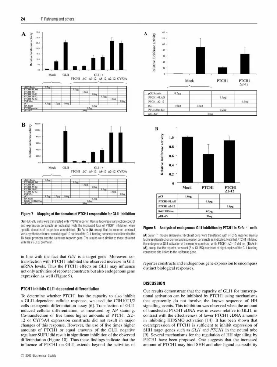
to be comparable (Figure 6B). PTCH1 and the deletion constructswere then co-transfected into HEK-293 cells along with GLI1and using as a reporter the PTCH2 promoter construct. PTCH1�2–12, which lacks TMs (transmembrane domains) 2–12 (aminoacids 180–1448), failed to inhibit GLI1 activity. Similar resultswere also obtained using the 12GLI RETKO luciferase constructinstead of the PTCH2 promoter. The other deletion constructs,PTCH1 �8–12 lacking TM 8–12 (amino acid 786–1448), PTCH1�9–12 lacking TM 9–12 (amino acids 1058–1448) andPTCH1 �C lacking only the last 238 amino acids, were still able toinhibit the GLI1 activation, albeit to different extents (Figures 7Aand 7B). The fact that PTCH1 �C inhibited GLI1 to the sameextent as the full-length PTCH1 provides additional evidence thatapoptosis is not involved in mediating these effects, as the C-terminal region contains the so-called ‘pro-apoptotic domain’ ofPTCH1 [17]. Thus, based on these experiments, we conclude thatthe regions between amino acids 180–768 and 1058–1210 are ofcritical importance for the observed inhibition by PTCH1.
Endogenous Gli1 activity is inhibited by PTCH1
We also used mouse embryonic fibroblasts with the Sufu geneinactivated for analysis of the PTCH1 expression constructs.These Sufu−/− cells are characterized by a high expression ofGli1, in line with the lack of the Gli1 negative regulator, Sufu(J. Svard, K. H. Henricson, M. Persson-Lek, B. Rozell, M.Lauth, J. Ericson, R. Toftgard and S. Teglund, unpublishedwork). As anticipated, PTCH1 could inhibit the endogenous Gli1activation of the PTCH2 promoter construct while PTCH1 �2–12 lacked this capacity (Figure 8A). We could also observe aninhibition by PTCH1 when an 8 × GLI-reporter construct [15]instead of the PTCH2 promoter was used (Figure 8B), althoughthis did not reach statistical significance. The observation ofonly a weak inhibition may be rationalized by the recentlyobtained evidence that these cells also express increased levels ofendogenous PTCH1, raising the possibility that these are maskingthe effects of exogenously added PTCH1. Thus these experimentsprovide additional evidence that PTCH1 can inhibit GLI1 activityindependently of SUFU.
Figure 6 Cellular expression of PTCH1 deletion constructs
(A) Diagram representing the C-terminal deletion constructs of PTCH1 that were used in thepresent study. The end of each construct is indicated by the corresponding amino acid basedon the numbering of GenBank® entry NM 000264. (B) COS7 cells transfected with PTCH1,PTCH1 �9–12, PTCH1 �8–12 or PTCH1 �2–12 constructs. The cells were stained withantibodies recognizing epitopes at the PTCH1 N-terminal domain. The proteins were visualizedby immunofluorescence microscopy. The number of transfected cells and the amount of proteinexpressed in individual cells were comparable with each of the four constructs used. The cellswere also stained with the nuclear marker DRAQTM.
Endogenous Gli1 expression, activated by GLI1,is inhibited by PTCH1
To investigate whether endogenous Gli1 expression is subjected tothe PTCH1 inhibition of GLI1 activity, cyclopamine-treated NIH-3T3 cells were used. Transfection with GLI1 induced endogenousexpression of the Gli1 mRNA, as revealed by RT–PCR analysis,
c© 2006 Biochemical Society
24 F. Rahnama and others
Figure 7 Mapping of the domains of PTCH1 responsible for GLI1 inhibition
(A) HEK-293 cells were transfected with PTCH2 reporter, Renilla luciferase transfection controland expression constructs as indicated. Note the increased loss of PTCH1 inhibition whenspecific domains of the protein were deleted. (B) As in (A), except that the reporter constructwas a synthetic enhancer consisting of 12 copies of the GLI-binding consensus site linked to theTK basal promoter and the luciferase reporter gene. The results were similar to those obtainedwith the PTCH2 promoter.
in line with the fact that Gli1 is a target gene. Moreover, co-transfection with PTCH1 inhibited the observed increase in Gli1mRNA levels. Thus the PTCH1 effects on GLI1 may influencenot only activities of reporter constructs but also endogenous geneexpression as well (Figure 9).
PTCH1 inhibits GLI1-dependent differentiation
To determine whether PTCH1 has the capacity to also inhibita GLI1-dependent cellular response, we used the C3H10T1/2cells osteogenic differentiation assay [6]. Transfection of GLI1induced cellular differentiation, as measured by AP staining.Co-transfection of five times higher amounts of PTCH1 �2–12 or CYP3A4 expression constructs did not result in majorchanges of this response. However, the use of five times higheramounts of PTCH1 or equal amounts of the GLI1 negativeregulator SUFU did result in significant inhibition of the observeddifferentiation (Figure 10). Thus these findings indicate that theinfluence of PTCH1 on GLI1 extends beyond the activities of
Figure 8 Analysis of endogenous Gli1 inhibition by PTCH1 in Sufu−/− cells
(A) Sufu−/− mouse embryonic fibroblast cells were transfected with PTCH2 reporter, Renillaluciferase transfection control and expression constructs as indicated. Note that PTCH1 inhibitedthe endogenous Gli1 activation of the reporter construct, while PTCH1 �2–12 did not. (B) As in(A), except that the reporter construct (8 × GLIBS) consisted of eight copies of the GLI-bindingconsensus site linked to the luciferase gene.
reporter constructs and endogenous gene expression to encompassdistinct biological responses.
DISCUSSION
Our results demonstrate that the capacity of GLI1 for transcrip-tional activation can be inhibited by PTCH1 using mechanismsthat apparently do not involve the known sequence of HHsignalling events. This inhibition was observed when the amountof transfected PTCH1 cDNA was in excess relative to GLI1, incontrast with the effectiveness of lower PTCH1 cDNA amountsin inhibiting HH/SMO activation [14]. It has been shown thatoverexpression of PTCH1 is sufficient to inhibit expression ofSHH target genes such as GLI1 and PTCH1 in the neural tube[9]. Several mechanisms for the regulation of HH signalling byPTCH1 have been proposed. One suggests that the increasedamount of PTCH1 may bind SHH and alter ligand accessibility
c© 2006 Biochemical Society

PTCH1 and negative feedback loop 25
Figure 9 GLI1 activation of endogenous Gli1 gene expression is inhibitedby PTCH1
NIH-3T3 cells were transfected with GLI1 and PTCH1 expression constructs as indicated and theendogenous Gli1 expression was monitored by RT–PCR. Note that GLI1 induced endogenousGli1 expression in cyclopamine-treated cells, while co-transfection of PTCH1 inhibited thiscapacity of GLI1.
for different target cells by, for example, endocytosis [11]. PTCH1might also regulate the level of signal activity by forcing SMOto a subcellular compartment either containing small moleculeantagonists or lacking small molecule agonists [10]. It has alsobeen shown that PTCH1 induces apoptotic cell death unless itsligand SHH is present [17]. However, apoptosis is apparently notinvolved in mediating these novel PTCH1 effects on GLI1, as theuse of the caspase inhibitor Z-VAD-FMK does not alleviatethe observed PTCH1 inhibition. An additional negative regulatorof SHH signalling that has been reported is SUFU [6]. This actseither by retaining the active GLI1 protein in the cytoplasm orby direct binding to GLI1 in the nucleus. To check if SUFU maybe involved in the observed PTCH1 inhibition of GLI1 activity,we tested two mutated GLI1 constructs that have been shown notto be sensitive for SUFU inhibition but still retaining full GLI1activity [18]. To our surprise, both the Gly122 to alanine and theHis123 to alanine constructs could be inhibited by PTCH1. Theseresults as well as the finding that endogenous Gli1 in Sufu−/−
cells is inhibited by PTCH1 indicate that the mechanism of thisinhibition does not include the SHH signalling component SUFU[21]. Additionally, the use of the SMO antagonist cyclopaminedid not alter the inhibitory effects of PTCH1 on GLI1 activity,supporting the notion that the capacity for active signalling isdispensable for this inhibition. Thus at relatively high PTCH1 toGLI1 levels, novel mechanisms that influence gene activation bythe transcription factor GLI1 may be involved. Conceivably, thesemay represent an additional means for PTCH1 to down-regulatesignalling when its levels exceed a certain threshold.
To test whether the PTCH1 effects are due to localizationchanges of GLI1 protein, we used immunofluorescence in HEK-293 cells. These cells were transfected with GLI1 in the presenceor absence of PTCH1. No changes could be observed in thesubcellular staining of GLI1, indicating that major localizationchanges are not involved in the mechanism of this inhibition.Recent studies have also shown that the components of HHsignalling are interacting in patterns that are beyond a simplelinear order, as Ci was found to bind Smo via Cos2 [22]. Thussome unidentified component(s) may be involved in mediatingthe PTCH1 effects on GLI1, as no major direct protein–proteininteractions could be revealed in an immunoprecipitation assay.Moreover, by generating deletion constructs of PTCH1, thedomains responsible for inhibition of GLI1 activation weremapped. Deletion of the regions encompassed by amino acids
Figure 10 Inhibition of GLI1-induced osteogenic differentiation by PTCH1
Osteogenic differentiation of C3H10T1/2 cells, as detected by AP staining. Cells were transfectedwith (a) GLI-1, (b) GLI1 and PTCH1, (c) GLI1 and 5 × PTCH1, and (d) GLI1 and 5 × CYP3A4.Note that only 5 × PTCH1-1B could significantly inhibit the differentiation in these cells.(e) Quantification of differentiation assays, based on three independent experiments. The y-axisrepresents the AP activity in arbitrary units that reflect absorbance values. Note that GLI1strongly induces osteogenic differentiation. This could be inhibited by five times higher amountsof full-length but not �2–12-truncated PTCH1. The GLI1-dependent differentiation was alsoreduced by co-expression of SUFU but not five times higher amounts of CYP3A4.
180–786 and 1058–1210 were found to negatively influence theinhibition of GLI1 by PTCH1. It should be mentioned that the for-mer segment encodes the SSD (sterol-sensing domain) that isimportant for signal transduction [23,24]. However, the secondextracellular loop between TM7 and TM8, which is thought tointeract with HH ligands [25,26], is not critical for the observedinhibition. Additionally, the C-terminal region that has a rolein blocking HH/SMO signalling [27,28] and, moreover, wasrecently shown to also be involved in an apoptotic response [17] isdispensable for this PTCH1-mediated inhibition. Therefore thesefindings highlight the distinct downstream effects that uniquePTCH1 domains may elicit.
c© 2006 Biochemical Society

26 F. Rahnama and others
In summary, the present study reveals an additional function ofPTCH1. This protein acts not only as an HH receptor, but alsoas a factor that has the capacity to limit GLI1 activity not onlythrough the known, canonical signalling cascade but also by novelmechanisms that are SUFU/SMO-independent. Additionally, twodistinct domains in PTCH1 responsible for the observed GLI1inhibition were delineated.
This study was supported by the Swedish Cancer Fund. P. K. acknowledges support fromthe Estonian Science Foundation grant no. 5552.
REFERENCES
1 McMahon, A. P., Ingham, P. W. and Tabin, C. J. (2003) Developmental roles and clinicalsignificance of hedgehog signaling. Curr. Top. Dev. Biol. 53, 1–114
1a Chen, Y. and Struhl, G. (1998) In vivo evidence that Patched and Smoothened constitutedistinct binding and transducing components of a Hedgehog receptor complex.Development 125, 4943–4948
2 Bai, C. B. and Joyner, A. L. (2001) Gli1 can rescue the in vivo function of Gli2.Development 128, 5161–5172
3 Kinzler, K. W., Ruppert, J. M., Bigner, S. H. and Vogelstein, B. (1988) The GLI gene is amember of the Kruppel family of zinc finger proteins. Nature (London) 332, 371–374
4 Sasaki, H., Nishizaki, Y., Hui, C., Nakafuku, M. and Kondoh, H. (1999) Regulation of Gli2and Gli3 activities by an amino-terminal repression domain: implication of Gli2 and Gli3as primary mediators of Shh signaling. Development 126, 3915–3924
5 Ruiz i Altaba, A. (1999) Gli proteins encode context-dependent positive and negativefunctions: implications for development and disease. Development 126, 3205–3216
6 Kogerman, P., Grimm, T., Kogerman, L., Krause, D., Unden, A. B., Sandstedt, B.,Toftgard, R. and Zaphiropoulos, P. G. (1999) Mammalian suppressor-of-fused modulatesnuclear-cytoplasmic shuttling of Gli-1. Nat. Cell Biol. 1, 312–319
7 Ingham, P. W., Taylor, A. M. and Nakano, Y. (1991) Role of the Drosophila patched gene inpositional signalling. Nature (London) 353, 184–187
8 Johnson, R. L., Grenier, J. K. and Scott, M. P. (1995) Patched overexpression alters wingdisc size and pattern: transcriptional and post-transcriptional effects on hedgehog targets.Development 121, 4161–4170
9 Goodrich, L. V., Jung, D., Higgins, K. M. and Scott, M. P. (1999) Overexpression of ptc1inhibits induction of Shh target genes and prevents normal patterning in the neural tube.Dev. Biol. 211, 323–334
10 Taipale, J., Cooper, M. K., Maiti, T. and Beachy, P. A. (2002) Patched acts catalytically tosuppress the activity of Smoothened. Nature (London) 418, 892–897
11 Incardona, J. P., Lee, J. H., Robertson, C. P., Enga, K., Kapur, R. P. and Roelink, H. (2000)Receptor-mediated endocytosis of soluble and membrane-tethered Sonic hedgehog byPatched-1. Proc. Natl. Acad. Sci. U.S.A. 97, 12044–12049
12 Finta, C. and Zaphiropoulos, P. G. (2002) Intergenic mRNA molecules resulting fromtrans-splicing. J. Biol. Chem. 277, 5882–5890
13 Kogerman, P., Krause, D., Rahnama, F., Kogerman, L., Unden, A. B., Zaphiropoulos, P. G.and Toftgard, R. (2002) Alternative first exons of PTCH1 are differentially regulated in vivoand may confer different functions to the PTCH1 protein. Oncogene 21, 6007–6016
14 Rahnama, F., Toftgard, R. and Zaphiropoulos, P. G. (2004) Distinct roles of PTCH2 splicevariants in Hedgehog signalling. Biochem. J. 378, 325–334
15 Sasaki, H., Hui, C., Nakafuku, M. and Kondoh, H. (1997) A binding site for Gli proteins isessential for HNF-3beta floor plate enhancer activity in transgenics and can respond toShh in vitro. Development 124, 1313–1322
16 Shimokawa, T., Rahnama, F. and Zaphiropoulos, P. G. (2004) A novel first exon of thePatched1 gene is upregulated by Hedgehog signaling resulting in a protein with pathwayinhibitory functions. FEBS Lett. 578, 157–162
17 Thibert, C., Teillet, M. A., Lapointe, F., Mazelin, L., Le Douarin, N. M. and Mehlen, P.(2003) Inhibition of neuroepithelial patched-induced apoptosis by sonic hedgehog.Science 301, 843–846
18 Dunaeva, M., Michelson, P., Kogerman, P. and Toftgard, R. (2003) Characterization ofthe physical interaction of Gli proteins with SUFU proteins. J. Biol. Chem. 278,5116–5122
19 Taipale, J., Chen, J. K., Cooper, M. K., Wang, B., Mann, R. K., Milenkovic, L., Scott, M. P.and Beachy, P. A. (2000) Effects of oncogenic mutations in Smoothened and Patched canbe reversed by cyclopamine. Nature (London) 406, 1005–1009
20 Chen, J. K., Taipale, J., Cooper, M. K. and Beachy, P. A. (2002) Inhibition of Hedgehogsignaling by direct binding of cyclopamine to Smoothened. Genes Dev. 16,2743–2748
21 Kalderon, D. (2004) Hedgehog signaling: costal-2 bridges the transduction gap.Curr. Biol. 14, R67–R69
22 Ruel, L., Rodriguez, R., Gallet, A., Lavenant-Staccini, L. and Therond, P. P. (2003) Stabilityand association of Smoothened, Costal2 and fused with Cubitus interruptus are regulatedby Hedgehog. Nat. Cell Biol. 5, 907–913
23 Martin, V., Carrillo, G., Torroja, C. and Guerrero, I. (2001) The sterol-sensing domain ofpatched protein seems to control smoothened activity through patched vesiculartrafficking. Curr. Biol. 11, 601–607
24 Strutt, H., Thomas, C., Nakano, Y., Stark, D., Neave, B., Taylor, A. M. and Ingham, P. W.(2001) Mutations in the sterol-sensing domain of patched suggest a role for vesiculartrafficking in smoothened regulation. Curr. Biol. 11, 608–613
25 Marigo, V., Davey, R. A., Zuo, Y., Cunningham, J. M. and Tabin, C. J. (1996) Biochemicalevidence that patched is the Hedgehog receptor. Nature (London) 384, 176–179
26 Stone, D. M., Hynes, M., Armanini, M., Swanson, T. A., Gu, Q., Johnson, R. L., Scott,M. P., Pennica, D., Goddard, A., Phillips, H. et al. (1996) The tumour-suppressor genepatched encodes a candidate receptor for Sonic hedgehog. Nature (London) 384,129–134
27 Johnson, R. L., Milenkovic, L. and Scott, M. P. (2000) In vivo functions of the patchedprotein: requirement of the C terminus for target gene inactivation but not Hedgehogsequestration. Mol. Cell 6, 467–478
28 Makino, S., Masuya, H., Ishijima, J., Yada, Y. and Shiroishi, T. (2001) A spontaneousmouse mutation, mesenchymal dysplasia (mes), is caused by a deletion of the mostC-terminal cytoplasmic domain of patched (ptc). Dev. Biol. 239, 95–106
Received 14 June 2005/23 August 2005; accepted 17 October 2005Published as BJ Immediate Publication 17 October 2005, doi:10.1042/BJ20050941
c© 2006 Biochemical Society