activation of p53 by sirt1 inhibition enhances elimination of cml leukemia stem cells in combination...
TRANSCRIPT
Cancer Cell
Article
Activation of p53 by SIRT1 Inhibition EnhancesElimination of CML Leukemia Stem Cellsin Combination with ImatinibLing Li,1 Lisheng Wang,1 Liang Li,1 Zhiqiang Wang,2 Yinwei Ho,1 Tinisha McDonald,1 Tessa L. Holyoake,3
WenYong Chen,2,4,* and Ravi Bhatia1,4,*1Division of Hematopoietic Stem Cell and Leukemia Research2Department of Cancer BiologyCity of Hope National Medical Center, Duarte, CA 91010, USA3Section of Experimental Haematology, Institute of Cancer Sciences, University of Glasgow, Glasgow, G12 8QQ Scotland, UK4These authors contributed equally to this work
*Correspondence: [email protected] (W.C.), [email protected] (R.B.)DOI 10.1016/j.ccr.2011.12.020
SUMMARY
BCR-ABL tyrosine kinase inhibitors (TKI) fail to eliminate quiescent leukemia stem cells (LSC) in chronicmyelogenous leukemia (CML). Thus, strategies targeting LSC are required to achieve cure. We show thatthe NAD+-dependent deacetylase SIRT1 is overexpressed in human CML LSC. Pharmacological inhibitionof SIRT1 or SIRT1 knockdown increased apoptosis in LSC of chronic phase and blast crisis CML and reducedtheir growth in vitro and in vivo. SIRT1 effects were enhanced in combination with the BCR-ABL TKI imatinib.SIRT1 inhibition increased p53 acetylation and transcriptional activity in CML progenitors, and the inhibitoryeffects of SIRT1 targeting on CML cells depended on p53 expression and acetylation. Activation of p53 viaSIRT1 inhibition represents a potential approach to target CML LSC.
INTRODUCTION
Chronic myelogenous leukemia (CML) results from malignant
transformation of a hematopoietic stem cell (HSC) by the BCR-
ABL oncogene. CML usually presents in a chronic phase (CP)
but progresses to an accelerated phase (AP) and a terminal blast
crisis (BC) (Sawyers, 1999). The BCR-ABL tyrosine kinase inhib-
itors (TKI) imatinib (IM), nilotinib, and dasatinib are effective in
inducing remissions and prolonging survival of CP CML patients
but is less effective against advanced phase CML (Eiring et al.,
2011). However, even in CP CML, primitive leukemia stem cells
(LSC) are retained in patients achieving remission with TKI
treatment (Chu et al., 2011). Primitive, quiescent CML LSC are
resistant to apoptosis following TKI treatment despite effective
inhibition of BCR-ABL kinase activity (Holtz et al., 2005; Corbin
et al., 2011), the mechanisms for which are not well understood.
Significance
BCR-ABL kinase inhibitors (TKI) are effective in the treatment oremain a potential source of recurrence. The NAD-dependentstress and functions as a tumor suppressor or tumor promoter doverexpressed in CML LSC and that SIRT1 inhibition selectiveland activation of the p53 tumor suppressor. These results areacetylation contributes to CML LSC survival and resistance toselectively target LSC that resist elimination by current treatm
266 Cancer Cell 21, 266–281, February 14, 2012 ª2012 Elsevier Inc.
Disease recurrence is usually seen following cessation of drug
treatment, even in patients with undetectable BCR-ABL expres-
sion by q-PCR (Mahon et al., 2010). These observations suggest
that ‘‘cure’’ may be elusive for most CML patients with TKI alone.
CML patients currently need to take TKI treatment indefinitely,
with risks of toxicity, lack of compliance, drug resistance,
relapse, and associated expense.
Recent studies from our group have shown that pan-histone
deacetylase (HDAC) inhibitors in combination with IM signifi-
cantly increase apoptosis in quiescent CML stem cells (Zhang
et al., 2010). However, toxicity of this approach to normal stem
cells remains a potential concern. Sirtuins are NAD-dependent
histone deacetylases that have been linked to longevity in lower
organisms and to mammalian metabolism (Bordone and Guar-
ente, 2005; Liu et al., 2009a). Sirtuin 1 (SIRT1) is a member of
the sirtuin family that regulates numerous processes, including
f CML but do not eliminate leukemia stem cells (LSC), whichdeacetylase SIRT1 is reported to protect stem cells againstepending on cellular context. Our studies show that SIRT1 isy reduces CML LSC survival and growth through acetylationimportant because they show that SIRT1-mediated p53 de-TKI treatment. SIRT1 inhibition is an attractive approach toents.
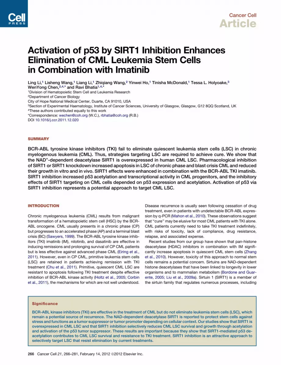
Figure 1. Increased SIRT1 Expression in CML
Patients Compared with Normal Stem/Progenitor
Cells
(A) Expression of SIRT1mRNA in CP CML (n = 5), BC CML
(n = 5), cord blood (CB) (n = 6), and PBSC (n = 6) CD34+
cells analyzed by Q-PCR.
(B) Expression of SIRT1 protein in CP CML (n = 11) and BC
CML (n = 5) compared with CB CD34+ cells (n = 10) and
PBSC CD34+ cells (n = 8) analyzed by intracellular labeling
with anti-SIRT1 antibody. Median fluorescence intensity
(MFI) of SIRT1 was expressed relative to IgG control.
(C) Expression of SIRT1 in CML (n = 11) and CB (n = 10)
CD34+CD38+ committed progenitors (right panel).
Representative results are shown in panel (D): CML (blue),
CB (green), IgG (red).
(E) Expression of SIRT1 in CML (n = 11) and CB (n = 10)
CD34+CD38� stem cells/primitive progenitors. Repre-
sentative results are shown in panel (F): CML (blue), CB
(green), IgG (red). Significance: *p < 0.05, **p < 0.01 for the
indicated comparisons. See also Figure S1 and Table S1.
Cancer Cell
SIRT1 Inhibition Targets CML Stem Cells
aging, DNA repair, cell cycle, metabolism, and cell survival under
stress conditions (Bordone and Guarente, 2005; Liu
et al.,2009a). In contrast to Class I, II, and IV HDACs, SIRT1
activity is not inhibited by pan-HDAC inhibitors (Liu et al.,
2009a). SIRT1 plays an important role inmaintaining self-renewal
and differentiation of murine embryonic stem cells (ESC) and
HSC, especially under conditions of stress (Han et al., 2008; Nar-
ala et al., 2008; Ou et al., 2011). Importantly, SIRT1 may have
a pathogenetic role in solid tumors and leukemias (Brooks and
Gu, 2009; Liu et al., 2009a). SIRT1 can potentially regulate the
acetylation of several transcription factors, including p53 (Luo
et al., 2001), Ku70, and FoxOs (Brooks and Gu, 2009). Despite
the clear inhibitory effect of increased SIRT1 expression on
tumor suppressors like p53 and FoxOs, other studies suggest
that SIRT1 may also have tumor-suppressive functions. In the
Apc+/� mouse model of colon cancer, increased SIRT1 expres-
sion resulted in reduced cell proliferation and tumor formation
(Firestein et al., 2008). Activation of SIRT1 by resveratrol can limit
cell growth and reduce tumor formation in BRCA1-deficient
tumor cells and in Trp53+/�;Sirt1+/� mice (Wang et al., 2008a,
2008b). The precise role of SIRT1 in cancer may depend on
Cancer Cell 21, 266
the specific cell or tumor type and the presence
or absence of p53 (Brooks and Gu, 2009).
Previous studies have shown that SIRT1
expression is increased in CML blast crisis
(BC) cell lines (Chen et al., 2005). Here we inves-
tigated the contribution of SIRT1 to the survival
and growth of CP and BC CML LSC and
progenitor cells and in LSC resistance to TKI
treatment. We also investigated the role of p53
in mediating the effects of SIRT1 inhibition on
CML progenitors.
RESULTS
SIRT1 Is Overexpressed in CML CD34+
CellsSIRT1 mRNA levels were significantly elevated
in CP and BC CML CD34+ cells (Table S1 avail-
able online) compared to CD34+ cells from cord blood (CB) or
normal peripheral blood stem cell collections (PBSC) (Figure 1A).
SIRT1 protein levels in CML and normal CD34+CD38+
committed progenitors and CD34+CD38� primitive progenitors
were measured by intracellular labeling with anti-SIRT1 antibody
and flow cytometry (Figure S1A). The ability of intracellular
labeling to reliably measure SIRT1 expression was confirmed
by western blotting (Figure S1B). SIRT1 protein levels were
significantly elevated in CMLCP and BCCD34+ cells (Figure 1B),
CML CP CD34+CD38+ (Figures 1C and 1D), and CD34+CD38�
cells (Figures 1E and 1F) compared to their normal counterparts.
SIRT1 Inhibition Using shRNA Reduces CML ProgenitorProliferation, Survival, and Colony GrowthTo investigate the functional role of SIRT1 in CML and normal
progenitors, CML and normal CD34+ cells were transduced
with lentivirus vectors coexpressing SIRT1 or control shRNAs
together with RFP. CD34+RFP+ cells were selected using flow
cytometry. Western blotting confirmed effective inhibition of
SIRT1 expression, whereas the expression of the related
SIRT2 was not affected (Figures 2A and 2B).
–281, February 14, 2012 ª2012 Elsevier Inc. 267

Figure 2. SIRT1 Knockdown Using Specific Anti-SIRT1 shRNA Increases Apoptosis and Inhibits Proliferation of CML Progenitors
(A) Western blotting of SIRT1 and b-actin in CB CD34+ and CML CD34+ cells transduced with SIRT1 shRNAs (ShSIRT1-1 and ShSIRT1-2) or with Ctrl shRNA.
(B) Western blotting for SIRT1, SIRT2, and b-actin in ShSIRT1-1, ShSIRT1-2, or CtrlShRNA transduced TF-1 cells. Results are representative of 3 independent
experiments.
Cancer Cell
SIRT1 Inhibition Targets CML Stem Cells
268 Cancer Cell 21, 266–281, February 14, 2012 ª2012 Elsevier Inc.

Cancer Cell
SIRT1 Inhibition Targets CML Stem Cells
CD34+ cells were labeled with carboxyfluorescein diacetate
succinimidyl ester (CFSE) followed by culture for 72 hr in low
growth factor concentrations. SIRT1 knockdown inhibited CML
progenitor proliferation as measured by reduction in CFSE fluo-
rescence. Treatment with IM resulted in further reduction of
proliferation (Figure 2C). SIRT1 knockdown inhibited prolifera-
tion of CB CD34+ cells to a lesser extent than CML CD34+ cells
(Figure 2D). Expression of ShSIRT1-1, which results in near
complete inhibition of SIRT1 expression, resulted in reduced
survival of CML CD34+ cells (Figure 2E). IM treatment signifi-
cantly increased apoptosis of SIRT1 knockdown cells, indicating
that SIRT1 inhibition enhanced sensitivity of CML progenitors to
IM-induced apoptosis (Figure 2E). Enhanced apoptosis of CML
CD34+ cells following SIRT1 knockdown, and further increase
in apoptosis with IM treatment, was confirmed by Wright-
Giemsa staining (Figure S2A), trypan blue staining (Figure S2B),
and activated caspase-3 labeling (Figure S2C). Interestingly,
SIRT1 knockdown did not affect survival of normal progenitors,
with or without IM treatment (Figure 2F). Primitive, quiescent
CML CD34+ cells are especially resistant to IM-induced
apoptosis (Holtz et al., 2005). Importantly, the combination of
SIRT1 inhibition and IM enhanced apoptosis of quiescent CML
progenitors identified on the basis of high CFSE fluorescence
(Figure 2G). In contrast, SIRT1 inhibition did not affect survival
of quiescent normal progenitors (Figure 2H). Expression of
both ShSIRT1-1 and ShSIRT1-2 significantly reduced CML
colony forming cell (CFC) frequency in methylcellulose progen-
itor assays, which was enhanced by IM (Figure 2I). Inhibition of
normal CFC growth was also seen, but was significantly less
than for CML progenitors (Figure 2J).
The increased effects of ShSIRT1-1, compared to ShSIRT1-2,
suggest that partial inhibition of SIRT1 expression is sufficient to
inhibit CML progenitor proliferation, but that near complete
knockdown is required to inhibit survival. To exclude the possi-
bility that these results were related to off-target effects, we
designed a SIRT1 construct resistant to ShSIRT1-1 (SIRT1-R)
(Figure S2D). Lentivirus-mediated expression of wild-type (WT)
or SIRT1-R resulted in enhanced SIRT1 protein levels in TF-1/
BCR-ABL cells transduced with ShSIRT1-1 (Figure S2E) and
abrogated the ability of ShSIRT1-1 to induce apoptosis and
inhibit growth (Figures S2F and S2G). These results indicate
that ShSIRT1-1 shRNA effects are related to SIRT1 knockdown,
rather than off-target effects, and confirm that near complete
SIRT1 suppression is required to induce apoptosis in CML cells.
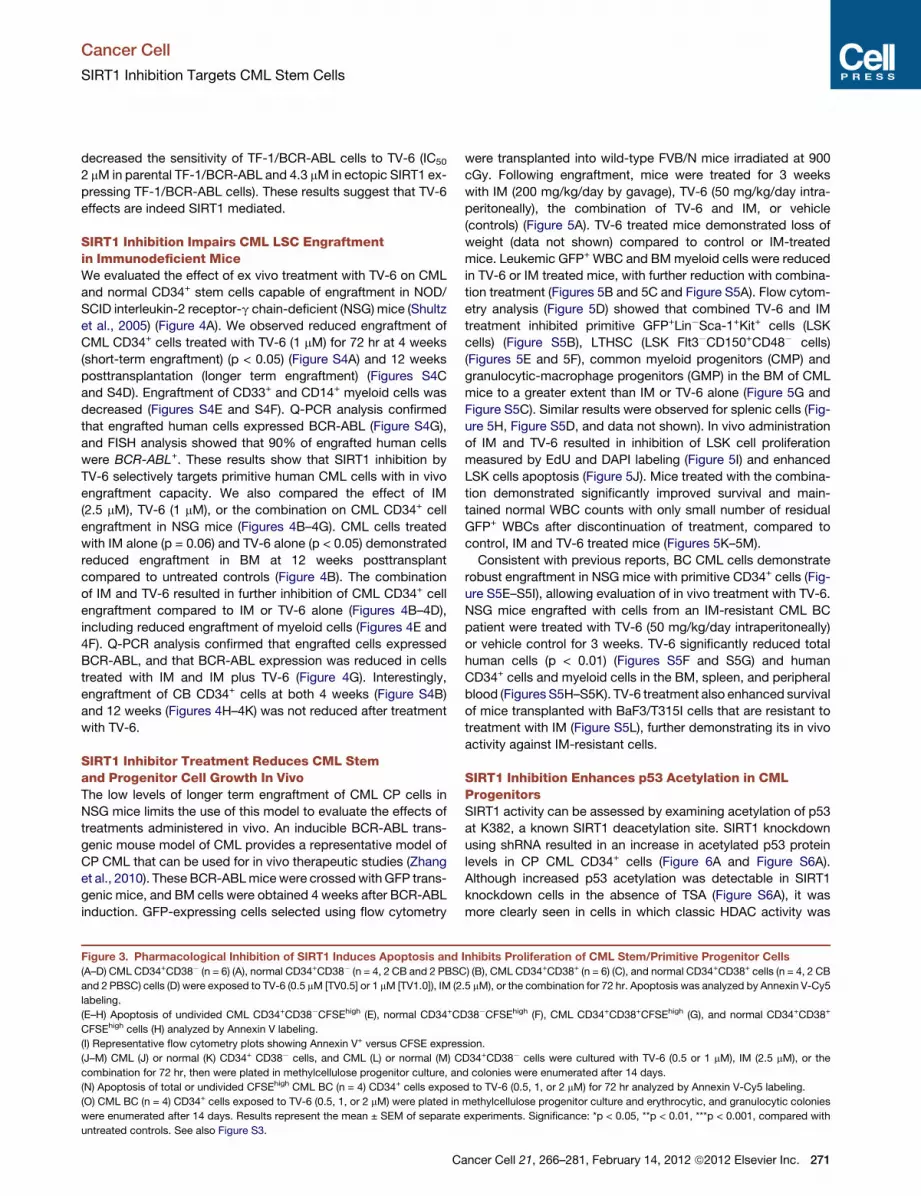
Pharmacological Inhibition of SIRT1 Induces Apoptosisand Inhibits Proliferation of CML Stem/PrimitiveProgenitor CellsWe tested the effects of tenovin-6 (TV-6), a small-molecule inhib-
itor of SIRT1, on CML and normal stem/progenitor cells (Lain
et al., 2008). CFSE-labeled CML and normal CD34+CD38� and
(C–J) CML (n = 5) and normal CB (n = 5) CD34+ cells transduced with Ctrl ShRNA,
72 hr. Division of CML (C) and normal CD34+ (D) cells was analyzed according t
ModFit software. Relative proliferation was calculated normalized to untreated co
Annexin V-Cy5 labeling. Apoptosis of undivided (CFSEhigh) CML (G) and CBCD34+
on CML (I) and normal CD34+ (J) RFP+ cells after culture with or without IM (2.5
14 days. Results represent mean ± SEM of separate experiments. Significance
Figure S2.
C
CD34+CD38+ cells were cultured for 72 hr with TV-6, IM, or the
combination. TV-6 significantly increased apoptosis of CML
CD34+CD38� cells and CD34+CD38+ cells, but not normal cells
(Figures 3A–3D). A small increase in apoptosis of normal
CD34+CD38+ cells was seen with higher doses of TV-6 (Fig-
ure 3D). Normal progenitor apoptosis was significantly less
than for CML progenitors (p < 0.05). The combination of IM
and TV-6 increased apoptosis in CML progenitors compared
to either agent alone, and to a significantly greater extent than
normal progenitors (Figures 3A–3D). Importantly, TV-6 also re-
sulted in increased apoptosis of CFSEhigh undivided CML
CD34+CD38� and CD34+CD38+ cells, but did not increase
apoptosis of undivided normal cells (Figures 3E–3I). A small
increase in apoptosis of undivided normal cells was seen with
higher doses of TV-6 (Figure 3H), but was significantly less
than for CML cells (p < 0.05). Pretreatment with TV-6 for 72 hr
inhibited CFC production from CML CD34+CD38� and CD34+
CD38+ cells but not normal CD34+CD38� andCD34+CD38+ cells
(Figures 3J–3M). The combination of TV-6 with IM enhanced
inhibition of CML CFC growth compared to either agent alone,
but did not enhance inhibition of normal CFC. Similar results
were seen for cells exposed to TV-6 in methylcellulose for the
14-day duration of the CFC assay (Figures S3A and S3B). Expo-
sure to IM for 14 days markedly inhibited CML and normal CFC
growth (p < 0.01) compared to 14 days of exposure to TV-6
(Figures S3A and S3B). TV-6 also increased apoptosis in total
and undivided CD34+ cells from CML BC patients for whom IM
treatment failed, and inhibited growth of CD34+ cells in CFC
assays (Figures 3N and 3O). In addition, TV-6 inhibited BaF3
cells expressing the IM-resistant T315I BCR-ABL mutant
(BaF3/T315I) to a similar extent as BaF3 cells expressing wild-
type BCR-ABL (BaF3/BA) (Figure S3C).
The effects of SIRT1 deletion on BM progenitors from young
mice are apparent only when cells are grown at low O2 tensions
(Ou et al., 2011). We investigated whether selective effects of
SIRT1 inhibition on CML compared to normal cells were main-
tained at lowO2 tensions. TV-6-induced (2 mM) apoptosis of total
(Figures S3D–S3K) or undivided normal CD34+CD38+ cells was
increased in hypoxic compared to normoxic conditions (Figures
S3G and S3K). Inhibition of normal CD34+CD38+ cell prolifera-
tion and CFC formation was also increased in hypoxic conditions
(Figures S3O and S3S). On the other hand, the effects of
TV-6 on survival, proliferation, and colony formation of normal
CD34+CD38� cells (Figures S3E, S3I, S3M, and S3Q) or CML
CD34+CD38� cells (Figures S3D, S3H, S3L, and S3P) andCD34+
CD38+ cells were not significantly different in hypoxic versus nor-
moxic conditions (Figures S3F, S3J, S3N, and S3R).
TV-6 did not result in additional inhibition of proliferation of
SIRT1 knockdown TF-1/BCR-ABL cells (Figure S3T). In addition,
MEF from SIRT1 knockout mice (SIRT1-KO) cells were resistant
to TV-6 (data not shown). Finally, ectopic expression of SIRT1
ShSIRT1-1, or ShSIRT1-2 vectors were cultured with or without IM (2.5 mM) for
o reduction in CFSE intensity, and a proliferation index was determined using
ntrols. Apoptosis of CML (E) and normal (F) CD34+RFP+ cells was analyzed by
(H) cells was analyzed by Annexin V-Cy5 labeling. CFC assayswere performed
mM) for 72 hr. Erythrocytic and granulocytic colonies were enumerated after
: *p < 0.05, **p < 0.01, ***p < 0.001, compared with untreated cells. See also
ancer Cell 21, 266–281, February 14, 2012 ª2012 Elsevier Inc. 269

Cancer Cell
SIRT1 Inhibition Targets CML Stem Cells
270 Cancer Cell 21, 266–281, February 14, 2012 ª2012 Elsevier Inc.
Cancer Cell
SIRT1 Inhibition Targets CML Stem Cells
decreased the sensitivity of TF-1/BCR-ABL cells to TV-6 (IC50
2 mM in parental TF-1/BCR-ABL and 4.3 mM in ectopic SIRT1 ex-
pressing TF-1/BCR-ABL cells). These results suggest that TV-6
effects are indeed SIRT1 mediated.
SIRT1 Inhibition Impairs CML LSC Engraftmentin Immunodeficient MiceWe evaluated the effect of ex vivo treatment with TV-6 on CML
and normal CD34+ stem cells capable of engraftment in NOD/
SCID interleukin-2 receptor-g chain-deficient (NSG) mice (Shultz
et al., 2005) (Figure 4A). We observed reduced engraftment of
CML CD34+ cells treated with TV-6 (1 mM) for 72 hr at 4 weeks
(short-term engraftment) (p < 0.05) (Figure S4A) and 12 weeks
posttransplantation (longer term engraftment) (Figures S4C
and S4D). Engraftment of CD33+ and CD14+ myeloid cells was
decreased (Figures S4E and S4F). Q-PCR analysis confirmed
that engrafted human cells expressed BCR-ABL (Figure S4G),
and FISH analysis showed that 90% of engrafted human cells
were BCR-ABL+. These results show that SIRT1 inhibition by
TV-6 selectively targets primitive human CML cells with in vivo
engraftment capacity. We also compared the effect of IM
(2.5 mM), TV-6 (1 mM), or the combination on CML CD34+ cell
engraftment in NSG mice (Figures 4B–4G). CML cells treated
with IM alone (p = 0.06) and TV-6 alone (p < 0.05) demonstrated
reduced engraftment in BM at 12 weeks posttransplant
compared to untreated controls (Figure 4B). The combination
of IM and TV-6 resulted in further inhibition of CML CD34+ cell
engraftment compared to IM or TV-6 alone (Figures 4B–4D),
including reduced engraftment of myeloid cells (Figures 4E and
4F). Q-PCR analysis confirmed that engrafted cells expressed
BCR-ABL, and that BCR-ABL expression was reduced in cells
treated with IM and IM plus TV-6 (Figure 4G). Interestingly,
engraftment of CB CD34+ cells at both 4 weeks (Figure S4B)
and 12 weeks (Figures 4H–4K) was not reduced after treatment
with TV-6.
SIRT1 Inhibitor Treatment Reduces CML Stemand Progenitor Cell Growth In VivoThe low levels of longer term engraftment of CML CP cells in
NSG mice limits the use of this model to evaluate the effects of
treatments administered in vivo. An inducible BCR-ABL trans-
genic mouse model of CML provides a representative model of
CP CML that can be used for in vivo therapeutic studies (Zhang
et al., 2010). These BCR-ABLmicewere crossedwith GFP trans-
genic mice, and BM cells were obtained 4 weeks after BCR-ABL
induction. GFP-expressing cells selected using flow cytometry
Figure 3. Pharmacological Inhibition of SIRT1 Induces Apoptosis and
(A–D) CML CD34+CD38� (n = 6) (A), normal CD34+CD38� (n = 4, 2 CB and 2 PBSC
and 2 PBSC) cells (D) were exposed to TV-6 (0.5 mM [TV0.5] or 1 mM [TV1.0]), IM (2
labeling.
(E–H) Apoptosis of undivided CML CD34+CD38�CFSEhigh (E), normal CD34+CD
CFSEhigh cells (H) analyzed by Annexin V labeling.
(I) Representative flow cytometry plots showing Annexin V+ versus CFSE expres
(J–M) CML (J) or normal (K) CD34+ CD38� cells, and CML (L) or normal (M) C
combination for 72 hr, then were plated in methylcellulose progenitor culture, an
(N) Apoptosis of total or undivided CFSEhigh CML BC (n = 4) CD34+ cells expose
(O) CML BC (n = 4) CD34+ cells exposed to TV-6 (0.5, 1, or 2 mM) were plated in
were enumerated after 14 days. Results represent the mean ± SEM of separate
untreated controls. See also Figure S3.
C
were transplanted into wild-type FVB/N mice irradiated at 900
cGy. Following engraftment, mice were treated for 3 weeks
with IM (200 mg/kg/day by gavage), TV-6 (50 mg/kg/day intra-
peritoneally), the combination of TV-6 and IM, or vehicle
(controls) (Figure 5A). TV-6 treated mice demonstrated loss of
weight (data not shown) compared to control or IM-treated
mice. Leukemic GFP+ WBC and BMmyeloid cells were reduced
in TV-6 or IM treated mice, with further reduction with combina-
tion treatment (Figures 5B and 5C and Figure S5A). Flow cytom-
etry analysis (Figure 5D) showed that combined TV-6 and IM
treatment inhibited primitive GFP+Lin�Sca-1+Kit+ cells (LSK
cells) (Figure S5B), LTHSC (LSK Flt3�CD150+CD48� cells)
(Figures 5E and 5F), common myeloid progenitors (CMP) and
granulocytic-macrophage progenitors (GMP) in the BM of CML
mice to a greater extent than IM or TV-6 alone (Figure 5G and
Figure S5C). Similar results were observed for splenic cells (Fig-
ure 5H, Figure S5D, and data not shown). In vivo administration
of IM and TV-6 resulted in inhibition of LSK cell proliferation
measured by EdU and DAPI labeling (Figure 5I) and enhanced
LSK cells apoptosis (Figure 5J). Mice treated with the combina-
tion demonstrated significantly improved survival and main-
tained normal WBC counts with only small number of residual
GFP+ WBCs after discontinuation of treatment, compared to
control, IM and TV-6 treated mice (Figures 5K–5M).
Consistent with previous reports, BC CML cells demonstrate
robust engraftment in NSG mice with primitive CD34+ cells (Fig-
ure S5E–S5I), allowing evaluation of in vivo treatment with TV-6.
NSG mice engrafted with cells from an IM-resistant CML BC
patient were treated with TV-6 (50 mg/kg/day intraperitoneally)
or vehicle control for 3 weeks. TV-6 significantly reduced total
human cells (p < 0.01) (Figures S5F and S5G) and human
CD34+ cells and myeloid cells in the BM, spleen, and peripheral
blood (Figures S5H–S5K). TV-6 treatment also enhanced survival
of mice transplanted with BaF3/T315I cells that are resistant to
treatment with IM (Figure S5L), further demonstrating its in vivo
activity against IM-resistant cells.
SIRT1 Inhibition Enhances p53 Acetylation in CMLProgenitorsSIRT1 activity can be assessed by examining acetylation of p53
at K382, a known SIRT1 deacetylation site. SIRT1 knockdown
using shRNA resulted in an increase in acetylated p53 protein
levels in CP CML CD34+ cells (Figure 6A and Figure S6A).
Although increased p53 acetylation was detectable in SIRT1
knockdown cells in the absence of TSA (Figure S6A), it was
more clearly seen in cells in which classic HDAC activity was
Inhibits Proliferation of CML Stem/Primitive Progenitor Cells
) (B), CML CD34+CD38+ (n = 6) (C), and normal CD34+CD38+ cells (n = 4, 2 CB
.5 mM), or the combination for 72 hr. Apoptosis was analyzed by Annexin V-Cy5
38�CFSEhigh (F), CML CD34+CD38+CFSEhigh (G), and normal CD34+CD38+
sion.
D34+CD38� cells were cultured with TV-6 (0.5 or 1 mM), IM (2.5 mM), or the
d colonies were enumerated after 14 days.
d to TV-6 (0.5, 1, or 2 mM) for 72 hr analyzed by Annexin V-Cy5 labeling.
methylcellulose progenitor culture and erythrocytic, and granulocytic colonies
experiments. Significance: *p < 0.05, **p < 0.01, ***p < 0.001, compared with
ancer Cell 21, 266–281, February 14, 2012 ª2012 Elsevier Inc. 271

Figure 4. SIRT1 Inhibition Reduces Longer Term Engraftment of CML Stem Cells in Immunodeficient Mice
(A) CML or normal CD34+ cells were treated with TV-6, IM, or combination in vitro and were injected into sublethally irradiated (300 cGy) NSG mice. After 4 or
12 weeks, human cell engraftment was analyzed by flow cytometric assessment of human CD45+ cells.
(B and C) The number (B) and the percentage (C) of humanCD45+ cells engrafted in the BM12weeks after transplantation of CMLCD34+ cells (13 106 cells/mouse).
Cancer Cell
SIRT1 Inhibition Targets CML Stem Cells
272 Cancer Cell 21, 266–281, February 14, 2012 ª2012 Elsevier Inc.
Cancer Cell
SIRT1 Inhibition Targets CML Stem Cells
inhibited by TSA treatment (Luo et al., 2001; Cheng et al., 2003).
Exposure of CMLCP or BCCD34+ cells to TV-6 also significantly
enhanced acetylated p53 levels in both hypoxic and normoxic
conditions (Figures 6B and 6C and Figures S6B and S6C). TV-
6 treatment also increased total p53 levels, possibly by reducing
degradation (Lain et al., 2008). Cells pretreated with the MDM2
antagonist Nutlin-3 to stabilize p53 levels (Vassilev et al., 2004)
before the addition of TV-6 showed a rapid and marked increase
in p53 acetylationwithout a change in total p53 levels (Figure 6B).
In contrast, TV-6 did not increase acetylated p53 or total p53
levels in normal CD34+ cells (Figure S6D). Acetylated p53 signals
were increased and showed a nuclear distribution in SIRT1
knockdown cells (Figure 6D). A modest increase in acetylation
of the SIRT2 substrate a-tubulin (Lain et al., 2008) was also
seen after 48 hr, suggesting that prolonged TV-6 exposure
may also modestly inhibit SIRT2 activity (Figure S6E). However,
the more rapid and pronounced effect on p53 acetylation indi-
cates more efficient inhibition of SIRT1 compared to SIRT2
activity. Ectopic expression of BCR-ABL in CB CD34+ cells
also resulted in increased expression of SIRT1, which was asso-
ciated with reduced levels of acetylated p53 despite increased
levels of total p53 (Figure S6F).
Treatment with classic HDACs can reduce BCR-ABL expres-
sion in cell lines and BCCML cells (Fiskus et al., 2006). BCR-ABL
protein levels were not reduced in CP or BC CML CD34+ cells
after SIRT1 inhibition using either shRNA or TV-6 (Figures 6B,
6C, and 6E and Figures S6B and S6C). Treatment with IM
reduced tyrosine phosphorylation of the BCR-ABL substrate
CrkL in CML CD34+ cells, confirming inhibition of BCR-ABL
kinase activity (Figure 6A and Figure S6B). In contrast, SIRT1
inhibition did not reduce phosphorylation in CML CD34+ cells
(Figures 6A and 6C and Figures S6B and S6C). Therefore, the
effects of SIRT1 inhibition on CML CD34+ cells cannot be ex-
plained by inhibition of BCR-ABL expression or activity. Treat-
ment with IM resulted in modest reduction in SIRT1 levels and
in total p53 but not acetylated p53 levels (Figure 6A and
Figure S6B).
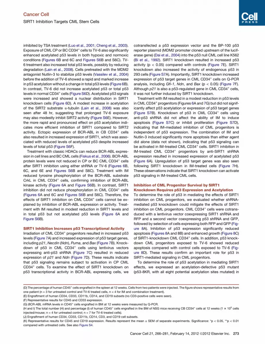
SIRT1 Inhibition Increases p53 Transcriptional ActivityIrradiation of CML CD34+ progenitors resulted in increased p53
levels (Figure 7A) and increased expression of p53 target genes,
including p21, Necdin (Ndn), Puma, and Bax (Figure 7B). Knock-
down of p53 in CML CD34+ cells using lentivirus vectors
expressing anti-p53 shRNA (Figure 7C) resulted in reduced
expression of p21 and Ndn (Figure 7D). These results indicate
that p53 signaling remains subject to activation in CP CML
CD34+ cells. To examine the effect of SIRT1 knockdown on
p53 transcriptional activity in BCR-ABL expressing cells, we
(D) The percentage of human CD45+ cells engrafted in the spleen at 12 weeks. Cel
one patient (n = 3 for untreated control and TV-6 treated cells, n = 4 for IM and c
(E) Engraftment of human CD34, CD33, CD11b, CD14, and CD19 subsets (no C
(F) Representative results for CD45 and CD33 expression.
(G) BCR-ABL mRNA levels in CD45+ cells engrafted in BM at 12 weeks were me
(H and I) The total number (H) and percentage (I) of human CD45+ cells engrafte
injected/mouse; n = 4 for untreated control; n = 7 for TV-6 treated cells).
(J) Engraftment of human CD34, CD33, CD11b, CD14, CD3, and CD19 cell subs
(K) Representative results for CD45 and CD19 expression. Results represent th
compared with untreated cells. See also Figure S4.
C
cotransfected a p53 expression vector and the BP-100 p53
reporter plasmid (MDM2 promoter cloned upstream of the lucif-
erase gene) (Dai et al., 2004) into the p53 null CML cell line K562
(Bi et al., 1992). SIRT1 knockdown resulted in increased p53
activity (p < 0.05) compared with controls (Figure 7E). SIRT1
knockdown also increased the activity of endogenous p53 in
293 cells (Figure S7A). Importantly, SIRT1 knockdown increased
expression of p53 target genes in CML CD34+ cells on Q-PCR
analysis, including Gfi-1, Ndn, and Bax (p < 0.05) (Figure 7F).
Although p21 is also a p53-regulated gene in CML CD34+ cells,
it was not further induced by SIRT1 knockdown.
Treatment with IM resulted in a modest reduction in p53 levels
in CMLCD34+ progenitors (Figures 6A and 7G) but did not signif-
icantly affect p53 acetylation or expression of p53 target genes
(Figure S7B). Knockdown of p53 in CML CD34+ cells using
anti-p53 shRNA did not affect the ability of IM to induce
apoptosis (Figure S7C) or inhibit proliferation (Figure S7D),
indicating that IM-mediated inhibition of CML progenitors is
independent of p53 expression. The combination of IM and
Nutlin-3 induced significantly more apoptosis than either agent
did alone (data not shown), indicating that p53 signaling can
be activated in IM-treated CML CD34+ cells. SIRT1 inhibition in
IM-treated CML CD34+ progenitors by anti-SIRT1 shRNA
expression resulted in increased expression of acetylated p53
(Figure 6A). Upregulation of p53 target genes was also seen
following SIRT1 knockdown in IM-treated cells (Figure 7H).
These observations indicate that SIRT1 knockdown can activate
p53 signaling in IM-treated CML cells.
Inhibition of CML Progenitor Survival by SIRT1Knockdown Requires p53 Expression and AcetylationTo determine the role of p53 in mediating the effects of SIRT1
inhibition on CML progenitors, we evaluated whether shRNA-
mediated p53 knockdown could mitigate the effects of SIRT1
inhibition on CML progenitors. CML CD34+ cells were cotrans-
duced with a lentivirus vector coexpressing SIRT1 shRNA and
RFP and a second vector coexpressing p53 shRNA and GFP,
followed by selection of cells expressing both RFP and GFP (Fig-
ure 8A). Inhibition of p53 expression significantly reduced
apoptosis (Figures 8A and 8B) and enhanced growth (Figure 8C)
of SIRT1-knockdown CML CD34+ cells. In addition, p53 knock-
down CML progenitors exposed to TV-6 showed reduced
apoptosis compared with control cells exposed to TV-6 (Fig-
ure 8D). These results confirm an important role for p53 in
SIRT1-mediated signaling in CML progenitors.
To determine the role of p53 acetylation in mediating SIRT1
effects, we expressed an acetylation-defective p53 mutant
(p53-8KR, with all eight potential acetylation sites mutated) in
ls from two patients were injected. The figure shows representative results from
ombination treatment).
D3-positive cells were seen).
asured by Q-PCR.
d in the BM of NSG mice receiving CB CD34+ cells at 12 weeks (1 3 105 cells
ets.
e mean ± SEM of separate experiments. Significance: *p < 0.05, **p < 0.01
ancer Cell 21, 266–281, February 14, 2012 ª2012 Elsevier Inc. 273

Figure 5. SIRT1 Inhibition Reduces In Vivo Growth of CML Stem Cells
(A) BCR-ABL mice were crossed with GFP transgenic mice and BM cells obtained 4 weeks after induction of BCR-ABL expression. GFP+ cells selected by flow
cytometry were transplanted into wild-type FVB/N mice irradiated at 900 cGy. Following engraftment, mice were treated for 3 weeks with IM (200 mg/kg/day by
gavage), TV-6 (5 0mg/kg/day intraperitoneally), the combination of TV-6 and IM, or vehicle (controls) (n = 6 mice each).
(B) GFP+ WBC counts 3 weeks after start of treatment.
Cancer Cell
SIRT1 Inhibition Targets CML Stem Cells
274 Cancer Cell 21, 266–281, February 14, 2012 ª2012 Elsevier Inc.

D
E
β-actin
BCR-ABL
ShSIRT1
Acetylated p53 p53 DAPI Merge
CtrlShRNA
CA B
Ac-p53(K382)
β-actin
p-CrkL
p53
BCR-ABL
SIRT1
With Nutlin
SIRT1
Ac-p53( K382)
p53
β-actin
BCR-ABL
p-CrkL
p-p53(ser15)
Ac-p53(K382)
p53
SIRT1
β-actin
p-CrkL 210kD145kD
210kD145kD
Figure 6. Increased p53 Acetylation and
Nuclear Localization in SIRT1-Inhibited
CML CD34+ Cells
(A) Western blotting for acetylated p53 (K382),
total p53, p-p53 (ser 15), b-actin, SIRT1, and
p-CrkL in SIRT1 knockdown in CML CD34+ cells
cultured with TSA (0.1 mM) for 2 hr and exposed to
IM (2.5 mM) for 8 hr.
(B) Western blotting for acetylated p53 (K382),
total p53, b-actin, SIRT1, p-CrkL, and BCR-ABL in
CML CD34+ cells cultured with Nutlin-3 (10 mM) for
2 hr and exposed to TV-6 for 6 hr.
(C) Western blotting for acetylated p53 (K382),
total p53, b-actin, SIRT1, p-CrkL, and BCR-ABL in
CML BCCD34+ cells cultured with TV-6 (1 or 2 mM)
for 16 hr.
(D) Immunofluorescence analysis of Ac-p53 and
p53 in CD34+ cells. All scale bars represent a size
of 10 mm. Results are representative of 3 inde-
pendent experiments.
(E) Western blotting for BCR-ABL and b-actin in
SIRT1 knockdown CML CD34+ cells exposed to
IM 2.5 mM for 24 hr. See also Figure S6.
Cancer Cell
SIRT1 Inhibition Targets CML Stem Cells
p53 null K562 cells. Inhibition of SIRT1 using shRNA or TV-6 did
not inhibit growth or induce apoptosis in parental K562 cells (p >
0.05) (Figure S8). However, SIRT1 knockdown in K562 cells that
ectopically expressedWT p53 protein led to increased p53 acet-
ylation (Figure 8E) and significant growth inhibition and
apoptosis (p < 0.05) (Figures 8F and 8G). In contrast, ectopically
(C) GFP+ myeloid cells (Gr-1+Mac-1+) cells in the BM.
(D) Schema for analysis of LTHSC (E) GFP+ LTHSC in BM.
(F) Representative plot for BM LTHSC for vehicle control and combination.
(G) GFP+ GMP in BM.
(H) GFP+ LTHSC in spleen. Apoptosis and cell cycling in BM LSK cells was evaluated after 5 days of treatm
(I) Mice were injected intraperitoneally with EdU and were euthanized 2 hr later. The percentage of stem c
incorporation in BM LSK cells. Representative results from one of two experiments are shown.
(J) Apoptosis was evaluated by Annexin-V and DAPI labeling (n = 3 mice per group). Significance: *p < 0.05
(K) Mice were followed for survival for 85 days after discontinuation of treatment (n = 6 per group). Surv
significantly longer than that of mice receiving IM or TV-6 alone or vehicle-treated cohorts (p < 0.05, Wilcoxo
remained alive until day 85, when the experiment was terminated.
(L andM) The total WBC count (L) andGFP+WBC count (M) in PB ofmice 4weeks after discontinuation of treat
with IM. Results represent the mean ± SEM of separate experiments. See also Figure S5.
Cancer Cell 21, 266–281,
expressed p53-8KR was not acetylated
following SIRT1 knockdown (Figure 8H),
and K562 cells transfected with p53-
8KR did not show significant growth inhi-
bition or apoptosis following SIRT1
knockdown (Figures 8I and 8J). These
results indicate that p53 acetylation is
required for growth inhibition and
apoptosis following SIRT1 inhibition in
BCR-ABL-expressing cells.
DISCUSSION
Our results show that inhibition of SIRT1
deacetylase enhances targeting of LSC
from CML patients by TKI treatment via
activation of p53 signaling, indicating an
important role for SIRT1 in maintaining LSC growth and survival.
Both BCR-ABL kinase-dependent and kinase-independent
mechanisms contribute to increased SIRT1 activity in CML cells,
with the latter potentially including epigenetic silencing of HIC1,
a negative regulator of SIRT1, through methylation (Chen et al.,
2005), or altered miRNA regulation of SIRT1 expression (Strum
ent.
ells in S-phase was determined according to EdU
, **p < 0.01, compared with vehicle control.
ival of mice receiving combination treatment was
n test). Two mice receiving combination treatment
ment. Significance: *p < 0.05, **p < 0.01, compared
February 14, 2012 ª2012 Elsevier Inc. 275

Figure 7. Signaling through p53 Is Intact in CML Progenitors, Is Enhanced by SIRT1 Knockdown, and Is Not Affected by IM(A) Western blotting for p53 in CML CD34+ cells 6 hr after exposure to irradiation (3 Gy). Results are representative of 3 independent experiments.
(B) Q-PCR analysis of p53 target genes in CP CML and normal CD34+ cells 6 hr after irradiation (3 Gy) compared with nonirradiated control cells. b-2M was used
as an internal control.
(C) Western blotting for p53 expression in CML CD34+ cells transduced with an anti-p53 shRNA vector. Results are representative of 3 independent experiments.
(D) Q-PCR analysis of p53 target genes in p53 knockdown and control cells.
(E) SIRT1 knockdown or control K562 cells (n = 3) were cotransfected with plasmids expressing p53, a luciferase reporter for p53 transcription (BP100) and
b-galactosidase. Relative luciferase units are normalized to b-gal expression.
(F) Q-PCR analysis of p53 target genes in shSIRT1 compared with ctrl shRNA expressing CML CD34+ cells (n = 3).
(G) Western blotting for acetylated p53 (K382), total p53, p-CrkL, and b-actin in CML CD34+ cells treated with IM (2.5 mM), Nutlin-3, or the combination for 8 hr.
(H) Q-PCR analysis for p53 target genes in ShSIRT1 compared with control shRNA-transduced CML CD34+ cells treated with IM (2.5 mM) for 24 hr (n = 4).
Significance: *p < 0.05, **p < 0.01, compared with controls. Results shown represent the mean ± SEM of separate experiments. See also Figure S7.
Cancer Cell
SIRT1 Inhibition Targets CML Stem Cells
276 Cancer Cell 21, 266–281, February 14, 2012 ª2012 Elsevier Inc.

Cancer Cell
SIRT1 Inhibition Targets CML Stem Cells
et al., 2009). The selectivity of SIRT1 inhibition towardCML stem/
progenitor cells is maintained in hypoxic conditions, where
SIRT1 plays an important role in supporting normal hematopoi-
esis (Ou et al., 2011).
SIRT1 can deacetylate several lysine residues in the tumor
suppressor p53 (Luo et al., 2001; Brooks andGu, 2009). A variety
of posttranslational modifications that can regulate p53 activity,
including phosphorylation, acetylation, methylation, and sumoy-
lation, have been described (Vousden and Lane, 2007). Acetyla-
tion is reported to play an important role in stabilization, nuclear
localization, and transcriptional activation of p53 (Prives and
Manley, 2001) and can lead to p53 activation independently of
phosphorylation status (Tang et al., 2008). Although p53 muta-
tions may occur on progression to BC CML, they are rare in
CP CML (Prokocimer and Rotter, 1994). Our results indicate
that p53 remains responsive to stress-induced activation in
CML progenitors. SIRT1 inhibition increased p53 acetylation
and expression of several p53 target genes, including Bax,
Necdin, and Gfi-1, in CML CD34+ cells. Bax is an important pro-
apoptotic gene, andNecdin andGfi-1may be important for p53-
regulated quiescence of HSC (Liu et al., 2009b). Additional p53
target genes besides those identified here may also contribute
to the effects of SIRT1 inhibition. Although p21 expression was
reduced in CML progenitors after p53 knockdown, SIRT1 knock-
down did not increase expression of p21 in CML progenitors,
suggesting that other SIRT1-regulated pathways may coun-
teract the effects of p53 acetylation on p21 induction (Cheng
et al., 2003). BC CML cells also demonstrated increased p53
acetylation following SIRT1 inhibition, consistent with recent
reports that p53 can be activated in CML BC cells (Peterson
et al., 2011). Of note, the CML BC samples studied here did
not have p53 mutations (Table S1).
Although previous studies indicated that the p53 inactivation
by SIRT1 promotes cell survival during stress (Luo et al., 2001),
other studies have suggested that small-molecule SIRT1 inhibi-
tors do not affect cell survival (Solomon et al., 2006) and that
developmental defects in a SIRT1 knockout mouse strain are
not rescued by crossing to p53 null mice (Kamel et al., 2006).
The importance of p53 in mediating SIRT1 effects may depend
on the cellular context. SIRT1 deacetylates several other
proteins that regulate cell growth and survival besides p53.
The importance of individual SIRT1 targets may depend on the
cell process and cell type studied. Recent studies within our
group indicate an important role for SIRT1 regulation of Ku70
in DNA repair and mutagenicity of CML cells (unpublished
data). The role of other SIRT1 targets such as the FoxOs and
E2F1 transcription factors in regulating quiescence and survival
of CML stem and progenitor cells requires further evaluation.
There is considerable interest in restoring p53 function in
cancer cells as a means of inhibiting their proliferation, or
inducing senescence or apoptosis. Deacetylation of p53 via
SIRT1 may play an important role in preventing p53 activation
in TKI-treated CML progenitors. BCR-ABL kinase activity could
also modulate p53 in CML cells by upregulation of ARF (Williams
et al., 2006), phosphorylation and inactivation of MDM2 and
MDMX (Zuckerman et al., 2009), or increased translation of
MDM2 (Trotta et al., 2003). Despite the complex regulation of
p53 in CML cells, our studies show that enhanced p53 acetyla-
tion following SIRT1 inhibition is sufficient to increase p53
C
transcriptional activity in CML progenitor cells and that p53 de-
acetylation is an important protective mechanism for CML LSC
following TKI treatment. Therefore, p53 activation is a potential
strategy to enhance targeting of CML LSC, especially in combi-
nation with TKI.
SIRT1 inhibitors are being investigated as potential anticancer
treatments. We observed weight loss in mice during the course
of three-week TV-6 treatment, but it is unclear whether this
was related to SIRT1 inhibition or an off-target effect of this
agent. Although TV-6 itself may not be a candidate for drug
development, our observations support further investigation of
SIRT1 inhibition as an approach for targeting of CML stem/
progenitor cells in combination with TKI treatment. The potential
tumor suppressive effects of SIRT1 need to be kept inmindwhen
considering SIRT1 inhibitors for cancer treatment. Improved
understanding of mechanisms underlying the anticancer versus
tumor-promoting effects of SIRT1 inhibition in specific cell types
will aid the development of more selective, nontoxic approaches
for targeting LSCs in future. The results of the current studies
have broader implication to other leukemias, such as AML,
where SIRT1 overexpression is also observed and p53 muta-
tions are rare (Kojima et al., 2005).
EXPERIMENTAL PROCEDURES
Samples and Materials
CB samples were provided by StemCyte (Arcadia, CA). CP CML samples were
obtained from previously untreated patients at the City of Hope (COH) and the
University of Glasgow. CML BC samples were obtained from patients at COH
(Table S1). CD34+ cell isolation and CD3+ cell depletion were performed using
magnetic beads (StemCell Technologies, Vancouver, BC, Canada). Leuko-
pheresis samples were processed for CD34+ cell selection with CliniMACS
(Miltenyi Biotech, Germany). CD34+CD38� and CD34+CD38+ cells were ob-
tained by flow cytometry sorting. All subjects signed an informed consent
form. Sample acquisition was approved by the Institutional Review Boards
at the City of Hope, in accordance with an assurance filed with and approved
by the Department of Health and Human Services, and the North Glasgow
University Hospital Division of NHS Greater Glasgow and Clyde, and met all
requirements of the Declaration of Helsinki. Details of cell lines, drugs, and
DNA constructs are provided in the Supplemental Experimental Procedures.
Cell Transduction and Transfection
CD34+ cells were transduced with lentivirus vectors expressing SIRT1 shRNA
or p53 shRNA. TF-1/BCR-ABL (TF-1/BA) cells were transduced with PITA-
SIRT1-R, PITA-SIRT1-WT, and vector controls (PITA). Details of the transduc-
tion procedure are provided in the Supplemental Experimental Procedures.
Intracellular Staining for SIRT1
CD34+ cells were labeled with antibodies to CD34-PeCy7, Lin-APC-Cy7
(including CD2, CD7, CD10, CD11B, CD19, CD33, and CD235a), and CD38-
APC (e-Bioscience), followed by fixation and permeabilization (Cytofix/Cyto-
perm Kit, Beckman Coulter, Fullerton, CA). Cells were then labeled with rabbit
anti-human SIRT1 (Epitomics) followed by Alexa 488-conjugated goat anti-
rabbit antibodies (Molecular Probes) and were analyzed by flow cytometry.
Data were analyzed using FlowJo software (version 8.5.2; TreeStar, Ashland,
OR). For immunofluorescence analysis, cells were labeled with anti-p53-
FITC (DO-7, BD) and antiacetylated p53-K382-Alex647 (BD), as described in
the Supplemental Experimental Procedures.
Analysis of Proliferation, Apoptosis, and Colony Growth
CD34+ cells were labeled with CFSE (Molecular Probes, Eugene, OR),
labeled with CD34-PE-Cy7 and CD38-APC, and CD34+CD38� and
CD34+CD38+ cells with uniform CFSE labeling were selected by flow cytom-
etry (MoFlo; Cytomation, Fort Collins, CO). Cells were cultured with low
ancer Cell 21, 266–281, February 14, 2012 ª2012 Elsevier Inc. 277

Cancer Cell
SIRT1 Inhibition Targets CML Stem Cells
278 Cancer Cell 21, 266–281, February 14, 2012 ª2012 Elsevier Inc.

Cancer Cell
SIRT1 Inhibition Targets CML Stem Cells
concentrations of growth factors at 37�C for up to 72 hr in normoxic condi-
tions (21% O2). For specific experiments, cells were cultured hypoxic condi-
tions (5% O2) as specifically indicated in the Results. Cells were analyzed
by flow cytometry for apoptosis by Annexin V labeling and for proliferation
by reduction in CFSE labeling. Committed progenitors or colony forming
cells (CFC) were evaluated in methylcellulose progenitor assays as previ-
ously described. Details are provided in Supplemental Experimental
Procedures.
Engraftment of Human Cells in Immunodeficient Mice
CML CD34+ cells (1 3 106 cells/ mouse) or CB CD34+ cells (1 3 105 cells/
mouse) were cultured for 72 hr with TV-6 (1 mM), IM (2.5 mM), or the
combination or without drug (control) and were transplanted via tail vein
injection into sublethally irradiated (300 cGy) 8-week-old NOD.Cg-
Prkdcscid IL2rgtm1Wjl /SzJ mice (NSG mice, Jackson Laboratory, Bar
Harbor, ME). Mice were euthanized after 4 or 12 weeks, and marrow
contents of femurs, spleen cells, and blood cells were obtained. For
CML BC samples, MNC depleted of CD3+ cells were transplanted via tail
vein injection (5 3 106 cells/mouse). Blood samples were obtained 4 weeks
after transplantation to confirm human CD45+ cells engraftment. Mice were
treated with TV-6 (50 mg/kg intraperitoneally daily for 21 days) (Lain et al.,
2008) or vehicle (control) for 3 weeks and were euthanized, and marrow
and spleen cells were obtained and analyzed as described in Supplemental
Experimental Procedures. Mouse care and experimental procedures were
performed in accordance with established institutional guidance and
approved protocols from the Institutional Animal Care and Use Committee
at COHNMC.
In Vivo Treatment of Transgenic BCR-ABL Mice
These experiments were performed using inducible, transgenic GFP-Scl-tTa-
BCR-ABL mice in the FVB/N background crossed with transgenic GFP-ex-
pressing mice (FVB.Cg-Tg [ACTB-EGFP] B5Nagy/J, Jackson Laboratories)
(Zhang et al., 2010). Mice were treated with IM (200 mg/kg daily by gavage
for 21 days), TV-6 (50 mg/kg body weight intraperitoneally daily for 21 days),
the combination, or vehicle alone (control). After 3 weeks of treatment,
animals were euthanized, and marrow and spleen cells were obtained. The
number of total nucleated cells, GFP-expressing cells, and GFP+ myeloid,
progenitor, and stem cell populations were measured by flow cytometry.
The effect of drug administration on apoptosis and cycling of stem cells
in vivo was evaluated. Another subset of mice was followed after discontin-
uation of treatment, and survival was monitored for 85 days, PB counts were
monitored for 28 days. Details are provided in the Supplemental Experi-
mental Procedures. Mouse care and experimental procedures were per-
formed in accordance with established institutional guidance and approved
protocols from the Institutional Animal Care and Use Committee at
COHNMC.
Figure 8. Effect of SIRT1 Inhibition in CML Progenitors Is Dependent o
(A–C) CML CD34+ cells were cotransduced with PLKO-GFP vectors expressing
control shRNA.
(A) CD34+GFP+RFP+ cells were analyzed for apoptosis by Annexin V labeling. A
(B) Cumulative results for apoptosis (n = 3).
(C) The total number of CD34+GFP+RFP+ cells normalized to ctrl ShRNA expres
(D) p53 knockdown or control CML progenitors were exposed to TV-6 for 48 hr, a
*p < 0.05, **p < 0.01, ***p < 0.001, compared with untreated cells.
(E–G) K562 cells transduced with HIV7-RFP vectors expressing anti-SIRT1 or co
(E) Western blotting for acetylated p53 (K382), total p53, SIRT1, and b-actin.
(F) Apoptosis was assessed after 48 hr by Annexin V labeling.
(G) The fold change in cell numbers was calculated at day 2, day 4, and day 6 re
(H–J) K562 cells expressing a tet transactivator gene (K562-TTA) were generate
RFP+ cells were selected and transfected with acetylation-defective (p53-8KR) an
by cotransfection with a b-gal plasmid.
(H) Western blotting for acetylated p53 (K382), total p53, SIRT1, and b-actin.
(I) Apoptosis was evaluated after 48 hrs by Annexin V labeling. Results are norm
(J) The fold change in cell numbers at day 2, day 4, and day 6 was calculated
Significance: *p < 0.05, **p < 0.01, compared with controls. See also Figure S8.
C
Luciferase Reporter Assays
K562 or 293 cells were transfected with reporter and internal control (b-gal
or Renilla-CMV) plasmids. Luciferase assays were performed after 48 hr in
triplicate using the luciferase reporter assay system (Promega).
Real-Time Q-PCR Analysis
Q-PCR analysis performedwith primers and probes for p21,Bax, Puma,Noxa,
Necdin, Gfi-1, and Sirt1, and BCR-ABL (B3A2) transcripts were measured
using a real-time TaqMan assay as previously described (Chu et al., 2011).
Details are provided in the Supplemental Experimental Procedures.
Western Blotting
Western blotting was performed for p53, acetylated p53, phospho-p53,
SIRT1, CrkL, phopho-CrkL, Bax, ABL, tubulin, and actin. Details are provided
in the Supplemental Experimental Procedures.
Statistics
Data from independent experiments were reported as the mean ± SEM.
Student’s t test analysis was performed to determine statistical significance.
SUPPLEMENTAL INFORMATION
Supplemental Information includes one table, eight figures, Supplemental
Experimental Procedures and can be found with this article online at
doi:10.1016/j.ccr.2011.12.020.
ACKNOWLEDGMENTS
This work was supported by the National Institutes of Health (grants R01
HL77847 and R01 CA95684), a research grant from the Samuel Waxman
Cancer Research Foundation, a Translational Research grant from the
Leukemia and Lymphoma Society (to R.B.), and a V Foundation translational
grant (to W.Y.C. and R.B.). W.Y.C. is supported by National Institutes of
Health grant R01 CA143421. T.L.H. is supported by Cancer Research UK Pro-
gramme grant C11074/A11008. We acknowledge the excellent technical
support of theCOHNMCAnalytical Cytometry, Synthetic Chemistry, andCyto-
genetics cores, and the Animal ResourcesCenter. We thank StemCyte for their
generous gift of CB samples, and Allen Lin for collection of patient samples.
This study was supported by the Glasgow Experimental Cancer Medicine
Centre (ECMC), which is funded by Cancer Research UK and by the Chief
Scientist’s Office (Scotland). We thank Dr. M.S. Dai (Oregon Health and
Sciences University) for the generous gift of the pcDNA3-p53 and mdm2-luc
(BP100) plasmids, and Dr. Wei Gu (Columbia University) for the generous gift
of the PTRE2-hyg-p53 and pTRE2-hyg-p53-8KR plasmids. Ling Li designed
and performed research, analyzed and interpreted data, and wrote the manu-
script. L.W. designed and performed experiments and reviewed the
n p53 Expression and Acetylation
anti-p53 or control shRNA and PHIV7-RFP vectors expressing anti-SIRT1 or
representative plot is shown.
sing cells (n = 3).
nd apoptosis was analyzed by Annexin V labeling (n = 3). Significance values:
ntrol shRNA were transfected with p53 expressing plasmids.
lative to day 0.
d and transduced with HIV7-RFP vectors expressing ShSIRT1 or Ctrl ShRNA.
dwild-type p53 constructs (n = 3). Similar transfection efficiencywas confirmed
alized to K562-RFP cells transfected with empty vector.
relative to day 0. Results represent mean ± SEM for separate experiments.
ancer Cell 21, 266–281, February 14, 2012 ª2012 Elsevier Inc. 279

Cancer Cell
SIRT1 Inhibition Targets CML Stem Cells
manuscript. Liang Li designed and performed experiments and reviewed the
manuscript. Z.W. designed and performed experiments and reviewed
the manuscript. Y.H. performed experiments and reviewed the manuscript.
T.M. performed experiments and reviewed the manuscript. T.H. provided
material, interpreted data, and reviewed the manuscript. W.Y.C. designed
the study, analyzed and interpreted data, and wrote the manuscript. R.B. de-
signed the study, analyzed and interpreted data, and wrote the manuscript.
Received: April 29, 2011
Revised: November 1, 2011
Accepted: December 20, 2011
Published: February 13, 2012
REFERENCES
Bi, S., Hughes, T., Bungey, J., Chase, A., de Fabritiis, P., and Goldman, J.M.
(1992). p53 in chronic myeloid leukemia cell lines. Leukemia 6, 839–842.
Bordone, L., and Guarente, L. (2005). Calorie restriction, SIRT1 and metabo-
lism: understanding longevity. Nat. Rev. Mol. Cell Biol. 6, 298–305.
Brooks, C.L., and Gu, W. (2009). How does SIRT1 affect metabolism, senes-
cence and cancer? Nat. Rev. Cancer 9, 123–128.
Chen, W.Y., Wang, D.H., Yen, R.C., Luo, J., Gu, W., and Baylin, S.B. (2005).
Tumor suppressor HIC1 directly regulates SIRT1 to modulate p53-dependent
DNA-damage responses. Cell 123, 437–448.
Cheng, H.L., Mostoslavsky, R., Saito, S., Manis, J.P., Gu, Y., Patel, P.,
Bronson, R., Appella, E., Alt, F.W., and Chua, K.F. (2003). Developmental
defects and p53 hyperacetylation in Sir2 homolog (SIRT1)-deficient mice.
Proc. Natl. Acad. Sci. USA 100, 10794–10799.
Chu, S., McDonald, T., Lin, A., Chakraborty, S., Huang, Q., Snyder, D.S., and
Bhatia, R. (2011). Persistence of leukemia stem cells in chronic myelogenous
leukemia patients in prolonged remission with imatinib treatment. Blood 118,
5565–5572.
Corbin, A.S., Agarwal, A., Loriaux, M., Cortes, J., Deininger, M.W., and Druker,
B.J. (2011). Human chronic myeloid leukemia stem cells are insensitive to
imatinib despite inhibition of BCR-ABL activity. J. Clin. Invest. 121, 396–409.
Dai, M.S., Zeng, S.X., Jin, Y., Sun, X.X., David, L., and Lu, H. (2004). Ribosomal
protein L23 activates p53 by inhibiting MDM2 function in response to ribo-
somal perturbation but not to translation inhibition. Mol. Cell. Biol. 24, 7654–
7668.
Eiring, A.M., Khorashad, J.S., Morley, K., and Deininger, M.W. (2011).
Advances in the treatment of chronic myeloid leukemia. BMC Med. 9, 99.
Fiskus, W., Pranpat, M., Balasis, M., Bali, P., Estrella, V., Kumaraswamy, S.,
Rao, R., Rocha, K., Herger, B., Lee, F., et al. (2006). Cotreatment with vorino-
stat (suberoylanilide hydroxamic acid) enhances activity of dasatinib (BMS-
354825) against imatinib mesylate-sensitive or imatinib mesylate-resistant
chronic myelogenous leukemia cells. Clin. Cancer Res. 12, 5869–5878.
Firestein, R., Blander, G.,Michan, S., Oberdoerffer, P., Ogino, S., Campbell, J.,
Bhimavarapu, A., Luikenhuis, S., de Cabo, R., Fuchs, C., et al. (2008). The
SIRT1 deacetylase suppresses intestinal tumorigenesis and colon cancer
growth. PLoS ONE 3, e2020.
Han,M.K., Song, E.K., Guo, Y., Ou, X., Mantel, C., and Broxmeyer, H.E. (2008).
SIRT1 regulates apoptosis and Nanog expression in mouse embryonic stem
cells by controlling p53 subcellular localization. Cell Stem Cell 2, 241–251.
Holtz, M.S., Forman, S.J., and Bhatia, R. (2005). Nonproliferating CML CD34+
progenitors are resistant to apoptosis induced by a wide range of proapoptotic
stimuli. Leukemia 19, 1034–1041.
Kamel, C., Abrol, M., Jardine, K., He, X., and McBurney, M.W. (2006). SirT1
fails to affect p53-mediated biological functions. Aging Cell 5, 81–88.
Kojima, K., Konopleva, M., Samudio, I.J., Shikami, M., Cabreira-Hansen, M.,
McQueen, T., Ruvolo, V., Tsao, T., Zeng, Z., Vassilev, L.T., and Andreeff, M.
(2005). MDM2 antagonists induce p53-dependent apoptosis in AML: implica-
tions for leukemia therapy. Blood 106, 3150–3159.
Lain, S., Hollick, J.J., Campbell, J., Staples, O.D., Higgins, M., Aoubala, M.,
McCarthy, A., Appleyard, V., Murray, K.E., Baker, L., et al. (2008). Discovery,
280 Cancer Cell 21, 266–281, February 14, 2012 ª2012 Elsevier Inc.
in vivo activity, and mechanism of action of a small-molecule p53 activator.
Cancer Cell 13, 454–463.
Liu, T., Liu, P.Y., and Marshall, G.M. (2009a). The critical role of the class III
histone deacetylase SIRT1 in cancer. Cancer Res. 69, 1702–1705.
Liu, Y., Elf, S.E., Miyata, Y., Sashida, G., Liu, Y., Huang, G., Di Giandomenico,
S., Lee, J.M., Deblasio, A., Menendez, S., et al. (2009b). p53 regulates hema-
topoietic stem cell quiescence. Cell Stem Cell 4, 37–48.
Luo, J., Nikolaev, A.Y., Imai, S., Chen, D., Su, F., Shiloh, A., Guarente, L., and
Gu, W. (2001). Negative control of p53 by Sir2alpha promotes cell survival
under stress. Cell 107, 137–148.
Mahon, F.X., Rea, D., Guilhot, J., Guilhot, F., Huguet, F., Nicolini, F., Legros, L.,
Charbonnier, A., Guerci, A., Varet, B., et al; Intergroupe Francais des
Leucemies Myeloıdes Chroniques. (2010). Discontinuation of imatinib in
patients with chronic myeloid leukaemia who have maintained complete
molecular remission for at least 2 years: the prospective, multicentre Stop
Imatinib (STIM) trial. Lancet Oncol. 11, 1029–1035.
Narala, S.R., Allsopp, R.C., Wells, T.B., Zhang, G., Prasad, P., Coussens, M.J.,
Rossi, D.J., Weissman, I.L., and Vaziri, H. (2008). SIRT1 acts as a nutrient-
sensitive growth suppressor and its loss is associated with increased AMPK
and telomerase activity. Mol. Biol. Cell 19, 1210–1219.
Ou, X., Chae, H.D., Wang, R.H., Shelley,W.C., Cooper, S., Taylor, T., Kim, Y.J.,
Deng, C.X., Yoder, M.C., and Broxmeyer, H.E. (2011). SIRT1 deficiency
compromises mouse embryonic stem cell hematopoietic differentiation, and
embryonic and adult hematopoiesis in the mouse. Blood 117, 440–450.
Peterson, L.F., Mitrikeska, E., Giannola, D., Lui, Y., Sun, H., Bixby, D., Malek,
S.N., Donato, N.J., Wang, S., and Talpaz, M. (2011). p53 stabilization induces
apoptosis in chronic myeloid leukemia blast crisis cells. Leukemia 25,
761–769.
Prives, C., and Manley, J.L. (2001). Why is p53 acetylated? Cell 107, 815–818.
Prokocimer, M., and Rotter, V. (1994). Structure and function of p53 in normal
cells and their aberrations in cancer cells: projection on the hematologic cell
lineages. Blood 84, 2391–2411.
Sawyers, C.L. (1999). Chronic myeloid leukemia. N. Engl. J. Med. 340, 1330–
1340.
Shultz, L.D., Lyons, B.L., Burzenski, L.M., Gott, B., Chen, X., Chaleff, S., Kotb,
M., Gillies, S.D., King, M., Mangada, J., et al. (2005). Human lymphoid and
myeloid cell development in NOD/LtSz-scid IL2R gamma null mice engrafted
with mobilized human hemopoietic stem cells. J. Immunol. 174, 6477–6489.
Solomon, J.M., Pasupuleti, R., Xu, L., McDonagh, T., Curtis, R., DiStefano,
P.S., and Huber, L.J. (2006). Inhibition of SIRT1 catalytic activity increases
p53 acetylation but does not alter cell survival following DNA damage. Mol.
Cell. Biol. 26, 28–38.
Strum, J.C., Johnson, J.H., Ward, J., Xie, H., Feild, J., Hester, A., Alford, A.,
and Waters, K.M. (2009). MicroRNA 132 regulates nutritional stress-induced
chemokine production through repression of SirT1. Mol. Endocrinol. 23,
1876–1884.
Tang, Y., Zhao, W., Chen, Y., Zhao, Y., and Gu, W. (2008). Acetylation is indis-
pensable for p53 activation. Cell 133, 612–626.
Trotta, R., Vignudelli, T., Candini, O., Intine, R.V., Pecorari, L., Guerzoni, C.,
Santilli, G., Byrom, M.W., Goldoni, S., Ford, L.P., et al. (2003). BCR/ABL acti-
vates mdm2 mRNA translation via the La antigen. Cancer Cell 3, 145–160.
Vassilev, L.T., Vu, B.T., Graves, B., Carvajal, D., Podlaski, F., Filipovic, Z.,
Kong, N., Kammlott, U., Lukacs, C., Klein, C., et al. (2004). In vivo activation
of the p53 pathway by small-molecule antagonists of MDM2. Science 303,
844–848.
Vousden, K.H., and Lane, D.P. (2007). p53 in health and disease. Nat. Rev.
Mol. Cell Biol. 8, 275–283.
Wang, R.H., Sengupta, K., Li, C., Kim, H.S., Cao, L., Xiao, C., Kim, S., Xu, X.,
Zheng, Y., Chilton, B., et al. (2008a). Impaired DNA damage response, genome
instability, and tumorigenesis in SIRT1 mutant mice. Cancer Cell 14, 312–323.
Wang, R.H., Zheng, Y., Kim, H.S., Xu, X., Cao, L., Luhasen, T., Lee, M.H., Xiao,
C., Vassilopoulos, A., Chen,W., et al. (2008b). Interplay among BRCA1, SIRT1,
and Survivin during BRCA1-associated tumorigenesis. Mol. Cell 32, 11–20.

Cancer Cell
SIRT1 Inhibition Targets CML Stem Cells
Williams, R.T., Roussel, M.F., and Sherr, C.J. (2006). Arf gene loss enhances
oncogenicity and limits imatinib response inmousemodels of Bcr-Abl-induced
acute lymphoblastic leukemia. Proc. Natl. Acad. Sci. USA 103, 6688–6693.
Zhang, B., Strauss, A.C., Chu, S., Li, M., Ho, Y., Shiang, K.D., Snyder, D.S.,
Huettner, C.S., Shultz, L., Holyoake, T., and Bhatia, R. (2010). Effective target-
ing of quiescent chronic myelogenous leukemia stem cells by histone deace-
C
tylase inhibitors in combination with imatinib mesylate. Cancer Cell 17,
427–442.
Zuckerman, V., Lenos, K., Popowicz, G.M., Silberman, I., Grossman, T.,
Marine, J.C., Holak, T.A., Jochemsen, A.G., and Haupt, Y. (2009). c-Abl phos-
phorylates Hdmx and regulates its interaction with p53. J. Biol. Chem. 284,
4031–4039.
ancer Cell 21, 266–281, February 14, 2012 ª2012 Elsevier Inc. 281