targeting sirt1, ampk, nrf2, ck2 and soluble ... - mdpi
TRANSCRIPT
Int. J. Mol. Sci. 2022, 23, 4776. https://doi.org/10.3390/ijms23094776 www.mdpi.com/journal/ijms
Review
Targeting Sirt1, AMPK, Nrf2, CK2 and Soluble Guanylate Cyclase with Nutraceuticals: A Practical Strategy for Preserving Bone Mass Mark F. McCarty 1,*, Lidianys Lewis Lujan 2 and Simon Iloki Assanga 3
1 Catalytic Longevity Foundation, San Diego, CA 92109, USA 2 Department of Research and Postgraduate in Food Science, Sonoran University, Hermosillo 83200, Mexico;
[email protected] 3 Department of Biological Chemical Sciences, Sonoran University, Hermosillo 83200, Mexico;
[email protected] * Correspondence: [email protected]
Abstract: There is a vast pre-clinical literature suggesting that certain nutraceuticals have the po-tential to aid the preservation of bone mass in the context of estrogen withdrawal, glucocorticoid treatment, chronic inflammation, or aging. In an effort to bring some logical clarity to these find-ings, the signaling pathways regulating osteoblast, osteocyte, and osteoclast induction, activity, and survival are briefly reviewed in the present study. The focus is placed on the following factors: the mechanisms that induce and activate the RUNX2 transcription factor, a key driver of osteoblast differentiation and function; the promotion of autophagy and prevention of apoptosis in osteo-blasts/osteoclasts; and the induction and activation of NFATc1, which promotes the expression of many proteins required for osteoclast-mediated osteolysis. This analysis suggests that the activa-tion of sirtuin 1 (Sirt1), AMP-activated protein kinase (AMPK), the Nrf2 transcription factor, and soluble guanylate cyclase (sGC) can be expected to aid the maintenance of bone mass, whereas the inhibition of the serine kinase CK2 should also be protective in this regard. Fortuitously, nutraceuticals are available to address each of these targets. Sirt1 activation can be promoted with ferulic acid, N1-methylnicotinamide, melatonin, nicotinamide riboside, glucosamine, and thymo-quinone. Berberine, such as the drug metformin, is a clinically useful activator of AMPK. Many agents, including lipoic acid, melatonin, thymoquinone, astaxanthin, and crucifera-derived sul-foraphane, can promote Nrf2 activity. Pharmacological doses of biotin can directly stimulate sGC. Additionally, certain flavonols, notably quercetin, can inhibit CK2 in high nanomolar concentra-tions that may be clinically relevant. Many, though not all, of these agents have shown favorable effects on bone density and structure in rodent models of bone loss. Complex nutraceutical regi-mens providing a selection of these nutraceuticals in clinically meaningful doses may have an important potential for preserving bone health. Concurrent supplementation with taurine, N-acetylcysteine, vitamins D and K2, and minerals, including magnesium, zinc, and manganese, plus a diet naturally high in potassium, may also be helpful in this regard.
Keywords: osteoporosis; osteoblasts; osteoclasts; RUNX2; NFATc1; Sirt1; AMPK; Nrf2; soluble guanylate cyclase; nutraceuticals
1. Determinates of Bone Loss Post-Menopausally and with Aging and Inflammation The loss of bone mass associated with an increased fracture risk is observed
post-menopausally, with prolonged glucocorticoid therapy, during chronic inflamma-tory disorders, and with advancing age (senile osteoporosis). Post-menopausal bone loss primarily reflects an increase in osteolytic osteoclast activity, reflecting a loss of ERα-mediated estrogen activity that functions to suppress the production of the receptor
Citation: McCarty, M.F.; Lewis
Lujan, L.; Iloki Assanga, S. Targeting
Sirt1, AMPK, Nrf2, CK2 and Soluble
Guanylate Cyclase with
Nutraceuticals: A Practical Strategy
for Preserving Bone Mass. Int. J. Mol.
Sci. 2022, 23, 4776. https://doi.org/
10.3390/ijms23094776
Academic Editor: Jung Eun Kim
Received: 4 April 2022
Accepted: 25 April 2022
Published: 26 April 2022
Publisher’s Note: MDPI stays neu-
tral with regard to jurisdictional
claims in published maps and insti-
tutional affiliations.
Copyright: © 2022 by the authors.
Licensee MDPI, Basel, Switzerland.
This article is an open access article
distributed under the terms and
conditions of the Creative Commons
Attribution (CC BY) license
(https://creativecommons.org/license
s/by/4.0/).
Int. J. Mol. Sci. 2022, 23, 4776 2 of 21
activator of the NF-kB ligand (RANKL) by bone-lining cells [1]. RANKL is an agonist for the receptor activator of NF-kB (RANK) expressed by osteoclasts; the pre-exposure of osteoclast precursors to the macrophage colony-stimulating factor (M-CSF) is required for the expression of RANK [2]. The stimulation of RANK via RANKL is a key mediator of osteoclast maturation and activation [3]. Hence, the loss of estrogen activity up-regulates bone osteolytic activity owing to RANKL overproduction. In contrast, senile osteoporosis reflects a loss of the bone-forming capacity, owing to the decreased differ-entiation of mesenchymal stem cells into osteoblasts, coupled with the decreased survival or senescence of osteoblasts and osteocytes [4]. Although a direct contribution of osteo-cytes to bone formation is unclear, they play a crucial role in regulating the competing functions of osteoblasts and osteoclasts, and mediate the positive impact of mechanical loading on bone density; their excessive loss by apoptosis during estrogen withdrawal, glucocorticoid treatment, or aging is a key factor in the development of osteoporosis [5–7]. Osteocyte senescence is also a factor of bone loss with advancing age [8].
To ward off the loss of bone mass, a logical strategy is to promote the differentiation, function, and survival of osteoblasts and osteocytes, while concurrently suppressing the osteolytic activity of osteoclasts; the latter will be of particular importance in the context of the onset of menopause. With respect to osteoblasts, the RUNX2 transcription factor is the master regulator of osteoblast formation and function, driving the transcription of a number of genes essential for the bone forming process [9]. Hence, up-regulating the signaling pathways driving RUNX2 expression and activation can be expected to pro-mote increased bone formation. The loss of bone-forming capacity associated with senile osteoporosis—and also, in some measure, estrogen deficiency—is characterized by in-creased apoptosis in osteoblasts and osteocytes; measures which suppress this apoptosis should also be useful. Additionally, osteoblast autophagy plays a key role in bone min-eral deposition—autophagic vacuoles in osteoblasts secrete apatite crystals—while helping to ward off apoptosis and senescence in osteoblasts and osteocytes; hence, the up-regulation of autophagy in these cells is another key goal [10–12].
With respect to osteoclasts, the transcription factor nuclear factor of activated T cells c1 (NFATc1) is the primary driver of osteoclast maturation and activity; the down-regulation of NFATc1 expression and activation is therefore a key goal in osteo-porosis prevention [3,13,14].
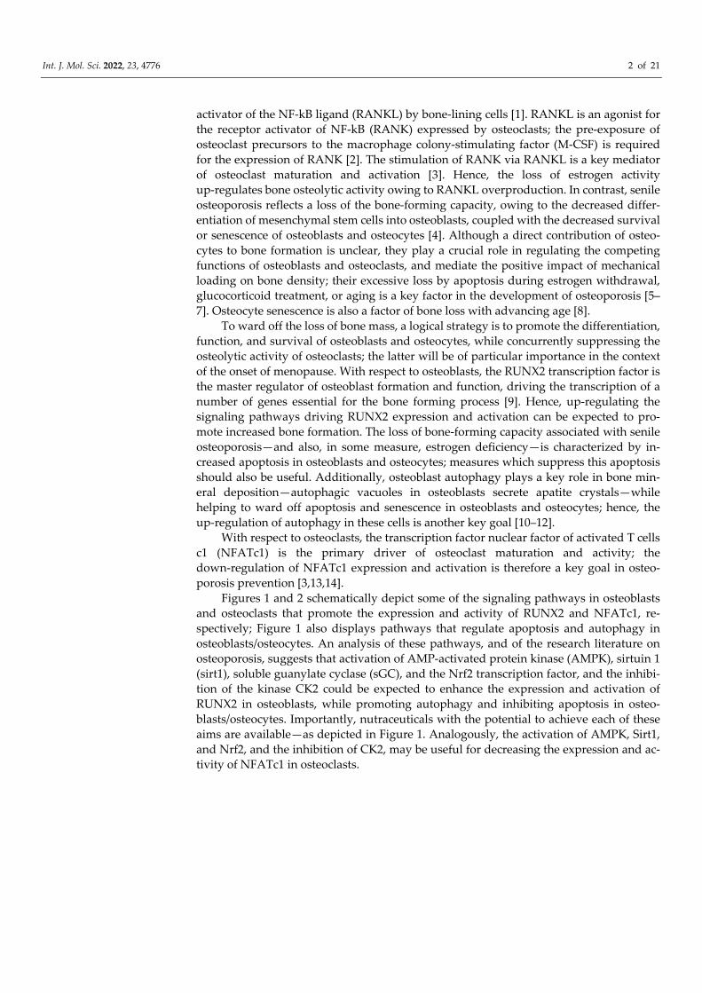
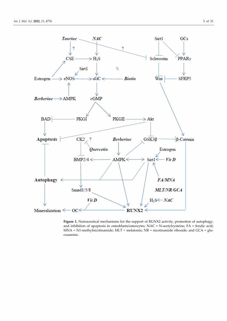
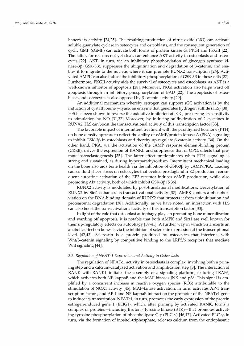
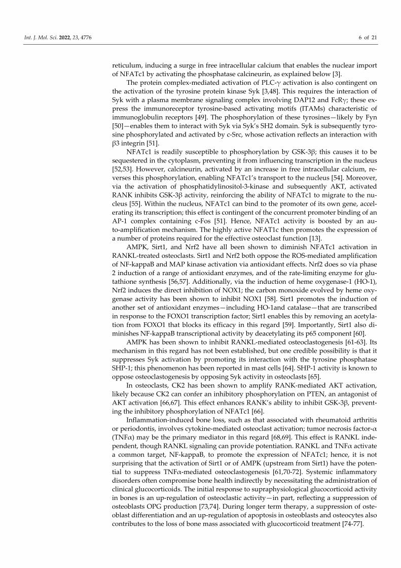
Figures 1 and 2 schematically depict some of the signaling pathways in osteoblasts and osteoclasts that promote the expression and activity of RUNX2 and NFATc1, re-spectively; Figure 1 also displays pathways that regulate apoptosis and autophagy in osteoblasts/osteocytes. An analysis of these pathways, and of the research literature on osteoporosis, suggests that activation of AMP-activated protein kinase (AMPK), sirtuin 1 (sirt1), soluble guanylate cyclase (sGC), and the Nrf2 transcription factor, and the inhibi-tion of the kinase CK2 could be expected to enhance the expression and activation of RUNX2 in osteoblasts, while promoting autophagy and inhibiting apoptosis in osteo-blasts/osteocytes. Importantly, nutraceuticals with the potential to achieve each of these aims are available—as depicted in Figure 1. Analogously, the activation of AMPK, Sirt1, and Nrf2, and the inhibition of CK2, may be useful for decreasing the expression and ac-tivity of NFATc1 in osteoclasts.
Int. J. Mol. Sci. 2022, 23, 4776 3 of 21
Figure 1. Nutraceutical mechanisms for the support of RUNX2 activity, promotion of autophagy, and inhibition of apoptosis in osteoblasts/osteocytes. NAC = N-acetylcysteine; FA = ferulic acid; MNA = N1-methylnicotinamide; MLT = melatonin; NR = nicotinamide riboside; and GCA = glu-cosamine.
Int. J. Mol. Sci. 2022, 23, 4776 4 of 21
Figure 2. Nutraceutical modulation of osteoclast expression and activity of NFATc1, a key driver of osteolysis. LA = lipoic acid; MLT = melatonin; TMQ= thymoquinone; ASX = astaxanthin; SFP = sulforaphane; NAC = N-acetylcysteine; QCT = quercetin; FA = ferulic acid; MNA = N1-methylnicotinamide; NR = nicotinamide riboside; and GCA = glucosamine.
2. A Brief Review of the Molecular Biology Determining Osteoblast and Osteoclast Activity 2.1. Regulation of RUNX2 Activity, Apoptosis, and Autophagy in Osteoblasts and Osteocytes
The major signaling pathways driving the transcription of the RUNX2 gene are triggered by the agonists Wnt—the downstream target of which is β-catenin—and bone morphogenetic proteins 2 and 4 (BMP2/4), which activate Smads 1/5/8 [15]. β-catenin—bound to the LDF1/TCF1 transcription factor—and the activated Smads form complexes on the promoter of the RUNX2 gene, stimulating its transcription [16].
With respect to BMP2/4, AMPK activation promotes the expression of BMP2/4 in osteoblasts [17-19]. Conversely, CK2 diminishes BMP2/4 signaling by binding to the BMP type I receptor [20]. Curiously, certain drugs designed to inhibit CK2’s kinase activity have been shown to alleviate this inhibition [20].
Estrogen activity, on the other hand, promotes RUNX2 transcription by up-regulating β-catenin signaling. This effect is downstream from the estrogen-mediated activation of endothelial nitric oxide synthase, which is observed in osteoblasts and os-teocytes [21,22]. The mechanism of this is likely comparable to that observed in endothe-lial cells—estrogen binding induces the translocation of an N-terminally truncated ERα to the plasma membrane, where it forms a protein complex with eNOS and promotes its phosphorylation and activation via a G-protein-mediated pathway [23]. Importantly, both AMPK and Sirt1 can also boost eNOS activity; AMPK confers an activating phos-phorylation on Ser-1177, whereas SIRT1 performs a deacetylation on eNOS, which en-
Int. J. Mol. Sci. 2022, 23, 4776 5 of 21
hances its activity [24,25]. The resulting production of nitric oxide (NO) can activate soluble guanylate cyclase in osteocytes and osteoblasts, and the consequent generation of cyclic GMP (cGMP) can activate both forms of protein kinase G, PKGI and PKGII [22]. The latter, for reasons not yet clear, can enhance AKT activity in osteoblasts and osteo-cytes [22]. AKT, in turn, via an inhibitory phosphorylation of glycogen synthase ki-nase-3β (GSK-3β), suppresses the ubiquitination and degradation of β-catenin, and ena-bles it to migrate to the nucleus where it can promote RUNX2 transcription [26]. Acti-vated AMPK can also induce the inhibitory phosphorylation of GSK-3β in these cells [27]. Furthermore, PKGII activity aids the survival of osteocytes and osteoblasts, as AKT is a well-known inhibitor of apoptosis [28]. Moreover, PKGI activation also helps ward off apoptosis through an inhibitory phosphorylation of BAD [22]. The apoptosis of osteo-blasts and osteocytes is also opposed by β-catenin activity [29].
An additional mechanism whereby estrogen can support sGC activation is by the induction of cystathionine γ-lyase, an enzyme that generates hydrogen sulfide (H2S) [30]. H2S has been shown to reverse the oxidative inhibition of sGC, preserving its sensitivity to stimulation by NO [31,32] Moreover, by inducing sulfhydration of 2 cysteines in RUNX2, H2S can boost the transactivational activity of this transcription factor [33].
The favorable impact of intermittent treatment with the parathyroid hormone (PTH) on bone density appears to reflect the ability of cAMP/protein kinase A (PKA) signaling to inhibit GSK-3β in osteoblasts and thereby up-regulate β-catenin activity [34]. On the other hand, PKA, via the activation of the cAMP response element-binding protein (CREB), drives the expression of RANKL and suppresses that of OPG, effects that pro-mote osteoclastogenesis [35]. The latter effect predominates when PTH signaling is strong and sustained, as during hyperparathyroidism. Intermittent mechanical loading on the bone also aids bone health via the inhibition of GSK-3β by cAMP/PKA. Loading causes fluid sheer stress on osteocytes that evokes prostaglandin E2 production; conse-quent autocrine activation of the EP2 receptor induces cAMP production, while also promoting Akt activity, both of which inhibit GSK-3β [5,36].
RUNX2 activity is modulated by post-translational modifications. Deacetylation of RUNX2 by Sirt1 enhances its transactivational activity [37]. AMPK confers a phosphor-ylation on the DNA-binding domain of RUNX2 that protects it from ubiquitination and proteasomal degradation [38]. Additionally, as we have noted, an interaction with H2S can also boost the transactivational activity of this transcription factor [33].
In light of the role that osteoblast autophagy plays in promoting bone mineralization and warding off apoptosis, it is notable that both AMPK and Sirt1 are well known for their up-regulatory effects on autophagy [39-41]. A further way in which Sirt1 exerts an anabolic effect on bones is via the inhibition of sclerostin expression at the transcriptional level [42,43]. Sclerostin is a protein produced by osteocytes that interferes with Wnt/β-catenin signaling by competitive binding to the LRP5/6 receptors that mediate Wnt signaling [44].
2.2. Regulation of NFATc1 Expression and Activity in Osteoclasts The regulation of NFATc1 activity in osteoclasts is complex, involving both a prim-
ing step and a calcium-catalyzed activation and amplification step [3]. The interaction of RANK with RANKL initiates the assembly of a signaling platform, featuring TRAF6, which activates both NF-kappaB and the MAP kinases JNK and p38. This signal is am-plified by a concurrent increase in reactive oxygen species (ROS) attributable to the stimulation of NOX1 activity [45]. MAP-kinase activation, in turn, activates AP-1 tran-scription factors, and AP-1 and NF-kappaB interact on the promoter of the NFATc1 gene to induce its transcription. NFATc1, in turn, promotes the early expression of the protein estrogen-induced gene 1 (EEIG1), which, after priming by activated RANK, forms a complex of proteins—including Bruton’s tyrosine kinase (BTK)—that promotes activat-ing tyrosine phosphorylation of phospholipase C-γ (PLC-γ) [46,47]. Activated PLC-γ, in turn, via the formation of inositol-triphosphate, releases calcium from the endoplasmic
Int. J. Mol. Sci. 2022, 23, 4776 6 of 21
reticulum, inducing a surge in free intracellular calcium that enables the nuclear import of NFATc1 by activating the phosphatase calcineurin, as explained below [3].
The protein complex-mediated activation of PLC-γ activation is also contingent on the activation of the tyrosine protein kinase Syk [3,48]. This requires the interaction of Syk with a plasma membrane signaling complex involving DAP12 and FcRγ; these ex-press the immunoreceptor tyrosine-based activating motifs (ITAMs) characteristic of immunoglobulin receptors [49]. The phosphorylation of these tyrosines—likely by Fyn [50]—enables them to interact with Syk via Syk’s SH2 domain. Syk is subsequently tyro-sine phosphorylated and activated by c-Src, whose activation reflects an interaction with β3 integrin [51].
NFATc1 is readily susceptible to phosphorylation by GSK-3β; this causes it to be sequestered in the cytoplasm, preventing it from influencing transcription in the nucleus [52,53]. However, calcineurin, activated by an increase in free intracellular calcium, re-verses this phosphorylation, enabling NFATc1’s transport to the nucleus [54]. Moreover, via the activation of phosphatidylinositol-3-kinase and subsequently AKT, activated RANK inhibits GSK-3β activity, reinforcing the ability of NFATc1 to migrate to the nu-cleus [55]. Within the nucleus, NFATc1 can bind to the promoter of its own gene, accel-erating its transcription; this effect is contingent of the concurrent promoter binding of an AP-1 complex containing c-Fos [51]. Hence, NFATc1 activity is boosted by an au-to-amplification mechanism. The highly active NFAT1c then promotes the expression of a number of proteins required for the effective osteoclast function [13].
AMPK, Sirt1, and Nrf2 have all been shown to diminish NFATc1 activation in RANKL-treated osteoclasts. Sirt1 and Nrf2 both oppose the ROS-mediated amplification of NF-kappaB and MAP kinase activation via antioxidant effects. Nrf2 does so via phase 2 induction of a range of antioxidant enzymes, and of the rate-limiting enzyme for glu-tathione synthesis [56,57]. Additionally, via the induction of heme oxygenase-1 (HO-1), Nrf2 induces the direct inhibition of NOX1; the carbon monoxide evolved by heme oxy-genase activity has been shown to inhibit NOX1 [58]. Sirt1 promotes the induction of another set of antioxidant enzymes—including HO-1and catalase—that are transcribed in response to the FOXO1 transcription factor; Sirt1 enables this by removing an acetyla-tion from FOXO1 that blocks its efficacy in this regard [59]. Importantly, Sirt1 also di-minishes NF-kappaB transcriptional activity by deacetylating its p65 component [60].
AMPK has been shown to inhibit RANKL-mediated osteoclastogenesis [61-63]. Its mechanism in this regard has not been established, but one credible possibility is that it suppresses Syk activation by promoting its interaction with the tyrosine phosphatase SHP-1; this phenomenon has been reported in mast cells [64]. SHP-1 activity is known to oppose osteoclastogenesis by opposing Syk activity in osteoclasts [65].
In osteoclasts, CK2 has been shown to amplify RANK-mediated AKT activation, likely because CK2 can confer an inhibitory phosphorylation on PTEN, an antagonist of AKT activation [66,67]. This effect enhances RANK’s ability to inhibit GSK-3β, prevent-ing the inhibitory phosphorylation of NFATc1 [66].
Inflammation-induced bone loss, such as that associated with rheumatoid arthritis or periodontis, involves cytokine-mediated osteoclast activation; tumor necrosis factor-α (TNFα) may be the primary mediator in this regard [68,69]. This effect is RANKL inde-pendent, though RANKL signaling can provide potentiation. RANKL and TNFα activate a common target, NF-kappaB, to promote the expression of NFATc1; hence, it is not surprising that the activation of Sirt1 or of AMPK (upstream from Sirt1) have the poten-tial to suppress TNFα-mediated osteoclastogenesis [61,70-72]. Systemic inflammatory disorders often compromise bone health indirectly by necessitating the administration of clinical glucocorticoids. The initial response to supraphysiological glucocorticoid activity in bones is an up-regulation of osteoclastic activity—in part, reflecting a suppression of osteoblasts OPG production [73,74]. During longer term therapy, a suppression of oste-oblast differentiation and an up-regulation of apoptosis in osteoblasts and osteocytes also contributes to the loss of bone mass associated with glucocorticoid treatment [74-77].
Int. J. Mol. Sci. 2022, 23, 4776 7 of 21
2.3. Modulation of RANKL and OPG Secretion by Osteoblasts and Osteocytes The extent to which osteoblasts and osteocytes produce RANKL and its functional
antagonist osteoprotegerin (OPG)—which serves as a decoy receptor for RANKL [78]—is also a key determinant of osteoclastogenesis.
While PKA activation in osteoblasts/osteocytes can exert an anabolic effect on bones by boosting the beta-catenin signal, PKA activity can also promote bone catabolism. PKA, via the activation of the cAMP response element-binding protein (CREB), drives the ex-pression of RANKL and suppresses that of OPG, effects that promote osteoclastogenesis [35]. The latter effect predominates when PTH signaling is strong and sustained, as dur-ing hyperparathyroidism.
AMPKα2 acts on osteoblasts to diminish their production of RANKL, while boost-ing their production of the RANKL antagonist OPG; this effect might reflect an opposi-tion to CREB signaling [79-81]. The ability of PTH to drive the expression of RANKL has been attributed to cAMP/PKA/CREB signaling that requires CTRC2 as a coactivator for CREB; notably, AMPK has been reported to antagonize CTRC2 activity by conferring a phosphorylation on it that causes its exclusion from the nucleus [82-84]. Whether CTRC2 participates in the PTH-mediated suppression of OPG should be investigated. Conceiv-ably, AMPK agonists, such as berberine, could make the impact of PTH and mechanical loading on bones more uniformly positive. β-catenin activity, independent of its impact on RUNX2, also increases OPG production by osteoblasts and osteocytes, as the β-catenin/TCF complex binds the promoter of the OPG gene and drives its transcription [85,86].
3. Nutraceutical Measures for Bone Health The preceding discussion enables us to predict that nutraceuticals that activate
AMPK, Sirt1, Nrf2, and sGC, or that inhibit CK2, could favorably influence bone density by promoting RUNX2 activity and autophagy—while also suppressing apoptosis—in osteoblasts and osteocytes. Such agents could also be expected to oppose NFATc1 activ-ity, thereby decreasing osteoclastogenesis and osteolysis.
With respect to AMPK, the prototypical pharmaceutical activator of AMPK, met-formin, has been associated with a lower risk for fracture and higher bone density in di-abetics using this drug, as opposed to not using it [87-91]. Analogously, metformin is protective in rodent models of bone loss [72,92-95]. The nutraceutical berberine, long used in China for treatment of type 2 diabetes and dyslipidemias, is known to activate AMPK in a manner similar to metformin [96-101]. Berberine has been reported to exert bone protective effects in a number of rodent models of bone loss [102-107].
With respect to Sirt1, there is a growing list of nutraceuticals—aside from resvera-trol, whose pharmacokinetics in humans render it of dubious clinical utility [108,109]—which have been reported to increase Sirt1 expression or activity in various contexts. Berberine and other AMPK activators do so, owing to the induction of nico-tinamide phosphoribosyltransferase (NAMPT), which is rate-limiting for the re-synthesis of Sirt1’s obligate substrate NAD+ [110-115]. Moreover, NAMPT induction not only boosts Sirt1 activity by increasing NAD+, but also by decreasing cellular levels of nico-tinamide, a product of Sirt1 activity that acts as an end-protein inhibitor of this enzyme [116]. The nutraceutical nicotinamide riboside (NR) offers an alternative strategy for in-creasing cellular NAD+, as it can function as a substrate for biosynthesis of this com-pound [117]. Melatonin promotes Sirt1 expression at the transcriptional level, likely via the activation of the Bmal1 transcription factor [118-121]. Ferulic acid—likely a key me-diator of the health benefits of dietary anthocyanins and whole grains—also increases Sirt1 expression at the mRNA level [122-127]. N1-methylnicotinamide (MNA), a natural metabolite of nicotinamide with anti-inflammatory activity, prolongs Sirt1 half-life, pos-sibly by opposing a JNK-mediated phosphorylation of Sirt1 that promotes its pro-teasomal degradation [128-130]. Supplemental glucosamine may have the potential for
Int. J. Mol. Sci. 2022, 23, 4776 8 of 21
activating Sirt1, as the O-GlcNAcylation of Sirt1 has been reported to boost Sirt’s deacetylase activity [131,132]. Additionally, the key active component of black cumin seed oil, thymoquinone, boosts Sirt1 activity, likely by promoting the conversion of NADH to NAD+ when reduced by NAD(P)H quinone oxidoreductase (NQO1) [133-138]. The favorable effects of melatonin, ferulic acid, NR, and glucosamine on bone density have been reported in rodent studies, while the effects of thymoquinone and MNA on bones have received minimal, if any, attention [125,139-159].
A number of phytochemicals, as well as the physiologically essential cofactor lipoic acid, have shown utility for boosting Nrf2 activity. Lipoic acid, thymoquinone, and the sulforaphane generated from cruciferous vegetables do so by the covalent interaction with Keap1, the protein that retains Nrf2 in the cytoplasm and promotes its proteasomal degradation [160-164]. Melatonin boosts the expression of Nrf2 via the Bmal1 transcrip-tion factor [165,166]. The xanthophyll carotenoid astaxanthin can also enhance the ex-pression of Nrf2, possibly via the interaction with the aryl hydrocarbon receptor [167-174]. Lipoic acid, astaxanthin, and—as noted—melatonin have shown utility in ro-dent models of bone loss [175-183].
Cinaciguat, a direct activator of the oxidized form of sGC, has been shown to protect bone density in ovariectomized mice [21]. In concentrations that are two orders of mag-nitude higher than the physiological level, the B vitamin biotin can directly activate the native form of sGC, promoting cGMP production [184]. In rodent studies, ample oral bi-otin intakes have been shown to boost cGMP levels [185-187]. Since high-dose biotin is clinically well tolerated, it might be considered as a strategy for aiding bone density [188]. It does not appear to have been studied in that regard in rodents, however. The one known clinical drawback of high-dose biotin is that it can interfere with certain lab assays that employ biotinylated reagents; hence, the discontinuation of biotin for at least several days may be prudent when lab tests are planned [189].
With respect to the inhibition of CK2, a range of flavonones—including quercetin, myricetin, fisetin, kaempferol, luteolin, and apigenin—have been shown to inhibit CK2’s kinase activity in high nanomolar concentrations that might be clinically relevant when high-absorption forms of these flavonols are ingested [190-193]. A number of studies have found that quercetin favorably influences bone density in rodent models of bone loss [194-199]. Derivatized or nanoparticulate preparations of quercetin designed for op-timal absorption are available as nutraceuticals [200-203]. Whether quercetin can prevent the inhibitory interaction between CK2 and the BMP type-I receptor remains to be de-termined.
In addition to the nutraceuticals previously mentioned, taurine has shown positive effects on bone density in rodents [204-209]. In vitro, taurine has been reported to sup-press sclerostin production by osteocytes—which can curiously synthesize their own taurine—and to decrease ROS levels in activated osteoclasts [210,211]; whether these ef-fects are clinically relevant is unclear, as high concentrations of taurine were employed in these cell culture studies. Taurine promotes the induction of cystathionine γ-lyase (CSE) in vascular endothelial cells, and the possibility that it, similar to estrogen, does so in os-teoblasts can be entertained [212-214]. As noted, the H2S that CSE produces has a favor-able impact on osteoblastic activity. N-acetylcysteine (NAC), which generates the cyste-ine that serves as CSE’s substrate, could presumably enhance H2S production in osteo-blasts, and has shown favorable effects on rodent models of bone loss [215-218]. NAC might also aid the maintenance of bone density by boosting osteoclast glutathione syn-thesis, and thereby opposing the up-regulatory effect of ROS on RANKL signaling. In-deed, in a very small pilot trial, the supplementation of recently post-menopausal women with 2 g of NAC daily, as an adjuvant to calcium/vitamin D supplementation, was asso-ciated with a trend toward a greater reduction in the marker of bone resorption serum C-telopeptide than in the placebo group; sadly, this lead has not been followed up [219].
In addition to the nutraceuticals discussed above, there are many other phyto-chemicals with the potential for boosting Sirt1, AMPK, or Nrf2 activities. As examples,
Int. J. Mol. Sci. 2022, 23, 4776 9 of 21
urolithin A, a bacterial metabolite of pomegranate ellagitannins thought to mediate the protective properties of pomegranate juice, has recently been reported to increase Sirt1 expression and NAD+ levels [220-223]. Compounds in bitter melon (Momordica char-antia), a food traditionally used in diabetes management in southeast Asia, have been found to boost AMPK activity by activating its upstream kinase Ca+2/calmodulin-dependent kinase kinase-β [224]. Additionally, a wide range of phyto-chemicals have some potential as Nrf2 activators [225]. The agents highlighted in this essay are distinguished by the fact that they are readily available in nutraceutical form and have, to some degree, been clinically employed.
4. Nutraceutical Control of Systemic Inflammation With respect to the loss of bone mass associated with systemic inflammation, it
stands to reason that nutraceuticals measures, which can quell inflammation, may be of clinical benefit—not only by decreasing the production of pro-inflammatory cytokines that boost osteoclasts activity, but also by decreasing the clinical need for glucocorticoid (GC) therapy. The joint activation of the transcription factors NF-kappaB and AP-1 plays a key role in promoting the macrophage and monocyte expression of TNF-α and other pro-inflammatory hormones at the transcriptional level [226]. Sub-optimal Sirt1 activity often collaborates with oxidative stress in boosting NF-kappaB and AP-1 activity; the oxidant-driven activation of JNK and p38 MAP kinases mediates AP-1 activation [226-228]. Hence, nutraceutical measures that boost Sirt1 activity and control oxidant stress—such as Nrf2 activators and NAC, a glutathione precursor– have the potential for the control of inflammation-driven bone loss. NOX2-dependent NADPH oxidase activity importantly contributes to oxidant production in macrophages, and the phycochemical phycocyanobilin, a chemical relative of bilirubin that functions as a light-harvesting chromophore in cynobacteria and some blue-green algae, has been found to inhibit this activity by mimicking the physiological antioxidant role of intracellular free bilirubin [229,230]. This may explain why spirulina, an exceptionally rich source of phycocyano-bilin, was found to be highly protective in a P. gingivalis-driven rat model of periodontal inflammation and alveolar bone loss [229,231]. Analogously, the oral administration of spirulina or its chief protein phycocyanin (covalently linked to phycocyanobilin) have shown marked efficacy in rodent models of inflammatory arthritis and other inflamma-tory conditions [232-235].
Curiously, there is evidence that nutraceuticals capable of activating Sirt1 can op-pose the ability of GCs to impede osteoblast maturation and induce bone loss in rodents; this has been demonstrated for ferulic acid, melatonin, berberine, and nicotinamide mononucleotide [125,155,236,237]. While these effects might be expected, owing to Sirt1’s ability to work in various ways to promote RUNX2 activity, it is conceivable that it works more proximally to interfere with GC signaling in osteoblasts and their precursors. In this regard, there is recent evidence that the negative impact of high-dose GCs on osteoblast maturation and function may be mediated, in large part, by the transcriptional induction of PPARγ, which, in turn, induces the expression of secreted frizzled-related protein 5 (SFRP5), an antagonist of the Wnt signaling crucial to osteoblast induction [238]. More-over, Sirt1 activity has been shown to oppose PPARγ expression and activity in a pre-osteoblast cell line; it has previously been established that Sirt1 opposes PPARγ-driven transcription in adipocytes [239,240]. (These considerations are evidently pertinent to the adverse impact of thiazolidinedione therapy on bone, as these agents serve as PPARγ agonists [241].) Importantly, the anti-inflammatory effects of GCs do not appear to be mediated by PPARγ [242]. Importantly, the anti-inflammatory effects of GCs do not appear to be mediated by PPARg [Evidently, nutraceuticals that can exert anti-inflammatory effects, while reducing the toxicity of GCs to the bone, might prove to be valuable complements to the therapy of autoimmune disorders.
Int. J. Mol. Sci. 2022, 23, 4776 10 of 21
5. Optimal Intakes of Certain Essential Vitamins and Minerals also Aid the Mainte-nance of Bone Density
Insuring adequate or ample intakes of a number of vitamins and minerals could be expected to complement the benefits of the more novel strategies suggested in this essay. Vitamin D aids bone health not only by helping to prevent secondary hyperparathy-roidism, but also because calcitriol, produced from circulating 25-hydroxyvitamin D by 25-hydroxyvitamin D 1-α-hydroxylase (CYP27B1) in osteoblasts, complements the tran-scriptional activity of RUNX2 when bound to the vitamin D receptor; most notably, it collaborates with RUNX2 in promoting the expression of osteocalcin (OC), a crucial me-diator of hydroxyapatite deposition [243-247]. Hence, bioavailable plasma levels of 25-hydroxyvitamin D tend to positively correlate with bone density [248-251]. Vitamin K2 (the bacterially produced form found in fermented milk and soy milk products, which achieves greater systemic distribution than the more hepatotropic vitamin K1) is thought to promote bone health by inducing γ-carboxylations of the bone matrix proteins OC and periostin, thereby improving their function in the bone [252,253]. Curiously, the benefi-cial impact of supplemental vitamin K2 on fracture risks in post-menopausal women appears to be disproportionate to its modest effect on bone density—possibly reflecting a favorable impact of vitamin K2 on bone flexibility [254,255]. Ample dietary intakes or increased serum levels of magnesium and zinc have been associated with greater bone density or a lower risk of fractures [256-258]. (Perhaps, surprisingly, higher intakes of calcium, while associated with a modestly greater bone density post-menopausally, do not appear to influence fracture risks [259,260]). Rodent studies suggest that increased intakes of manganese or silicon may have a positive impact on bone density [261,262]. Diets habitually high in natural potassium also positively correlate with bone density, likely because the organic counteranions ingested with the potassium are metabolized to bicarbonate, and hence exert an alkalinizing effect that opposes osteolysis [263-265]. Conversely, dietary sulfhydryl amino acids are metabolized to generate sulfate, and hence are acidifying; this suggests that supplemental NAC might have its most favorable net impact on bone health in the context of a diet naturally high in potassium [266]. Comprehensive vitamin–mineral supplementation, particularly in the context of sub-optimally nutritious diets, could be expected to favorably impact bone density and fracture risks [267].
Author Contributions: Conception and original draft: M.F.M.; revisions and additions to manu-script: S.I.L., L.L.L. All authors have read and agreed to the published version of the manuscript
Funding: This research received no external funding.
Institutional Review Board Statement: Not applicable.
Informed Consent Statement: Not applicable.
Data Availability Statement: Not applicable.
Conflicts of Interest: M.F.M. is the co-inventor and co-owner of a US patent covering the nutraceutical uses of phycocyanobilin oligopeptides derived from spirulina. The other authors have no conflicts of interest.
References 1. Streicher, C.; Heyny, A.; Andrukhova, O.; Haigl, B.; Slavic, S.; Schüler, C.; Kollmann, K.; Kantner, I.; Sexl, V.; Kleiter, M.; et al.
Estrogen Regulates Bone Turnover by Targeting RANKL Expression in Bone Lining Cells. Sci. Rep. 2017, 7, 1–14, https://doi.org/10.1038/s41598-017-06614-0.
2. Nakanishi, A.; Hie, M.; Iitsuka, N.; Tsukamoto, I. A crucial role for reactive oxygen species in macrophage colony-stimulating factor-induced RANK expression in osteoclastic differentiation. Int. J. Mol. Med. 2013, 31, 874–880, https://doi.org/10.3892/ijmm.2013.1258.
3. Park, J.H.; Lee, N.K.; Lee, A.S.Y. Current Understanding of RANK Signaling in Osteoclast Differentiation and Maturation. Mol. Cells 2017, 40, 706–713, https://doi.org/10.14348/molcells.2017.0225.
Int. J. Mol. Sci. 2022, 23, 4776 11 of 21
4. Tokuzawa, Y.; Yagi, K.; Yamashita, Y.; Nakachi, Y.; Nikaido, I.; Bono, H.; Ninomiya, Y.; Kanesaki-Yatsuka, Y.; Akita, M.; Motegi, H.; et al. Id4, a New Candidate Gene for Senile Osteoporosis, Acts as a Molecular Switch Promoting Osteoblast Dif-ferentiation. PLoS Genet. 2010, 6, e1001019, https://doi.org/10.1371/journal.pgen.1001019.
5. Bonewald, L.F. The amazing osteocyte. J. Bone Miner. Res. 2011, 26, 229–238, https://doi.org/10.1002/jbmr.320. 6. Ru, J.-Y.; Wang, Y.-F. Osteocyte apoptosis: the roles and key molecular mechanisms in resorption-related bone diseases. Cell
Death Dis. 2020, 11, 1–24, https://doi.org/10.1038/s41419-020-03059-8. 7. Jilka, R.L.; O’Brien, C.A. The Role of Osteocytes in Age-Related Bone Loss. Curr. Osteoporos. Rep. 2016, 14, 16–25,
https://doi.org/10.1007/s11914-016-0297-0. 8. Farr, J.N.; Kaur, J.; Doolittle, M.L.; Khosla, S. Osteocyte Cellular Senescence. Curr. Osteoporos. Rep. 2020, 18, 559–567,
https://doi.org/10.1007/s11914-020-00619-x. 9. Meyer, M.B.; Benkusky, N.A.; Pike, J.W. The RUNX2 cistrome in osteoblasts: Characterization, down-regulation following
differentiation, and relationship to gene expression. J. Biol. Chem. 2014, 289, 16016–16031. 10. Nollet, M.; Santucci-Darmanin, S.; Breuil, V.; et al. Autophagy in osteoblasts is involved in mineralization and bone
homeo-stasis. Autophagy 2014, 10, 1965–1977. 11. Chen, K.; Yang, Y.-H.; Jiang, S.-D.; Jiang, L.-S. Decreased activity of osteocyte autophagy with aging may contribute to the bone
loss in senile population. Histochem. Cell Biol. 2014, 142, 285–295, https://doi.org/10.1007/s00418-014-1194-1. 12. Luo, D.; Ren, H.; Li, T.; Lian, K.; Lin, D. Rapamycin reduces severity of senile osteoporosis by activating osteocyte autophagy.
Osteoporos. Int. 2015, 27, 1093–1101, https://doi.org/10.1007/s00198-015-3325-5. 13. Zhao, Q.; Wang, X.; Liu, Y.; He, A.; Jia, R. NFATc1: Functions in osteoclasts. Int. J. Biochem. Cell Biol. 2010, 42, 576–579,
https://doi.org/10.1016/j.biocel.2009.12.018. 14. Kang, J.Y.; Kang, N.; Yang, Y.M.; Hong, J.H.; Shin, D.M. The Role of Ca(2+)-NFATc1 Signaling and Its Modulation on Os-
te-oclastogenesis. Int. J. Mol. Sci. 2020, 21, 3646. 15. Rutkovskiy, A.; Stensløkken, K.-O.; Vaage, I.J. Osteoblast Differentiation at a Glance. Med Sci. Monit. Basic Res. 2016, 22, 95–
106, https://doi.org/10.12659/msmbr.901142. 16. Rodríguez-Carballo, E.; Ulsamer, A.; Susperregui, A.R.; Manzanares-Céspedes, C.; Sánchez-García, E.; Bartrons, R.; Rosa, J.L.;
Ventura, F. Conserved regulatory motifs in osteogenic gene promoters integrate cooperative effects of canonical Wnt and BMP pathways. J. Bone Miner. Res. 2010, 26, 718–729, https://doi.org/10.1002/jbmr.260.
17. Kanazawa, I.; Yamaguchi, T.; Yano, S.; Yamauchi, M.; Sugimoto, T. Metformin enhances the differentiation and mineralization of osteoblastic MC3T3-E1 cells via AMP kinase activation as well as eNOS and BMP-2 expression. Biochem. Biophys. Res. Commun. 2008, 375, 414–419, https://doi.org/10.1016/j.bbrc.2008.08.034.
18. Kanazawa, I.; Yamaguchi, T.; Yano, S.; Yamauchi, M.; Sugimoto, T. Activation of AMP kinase and inhibition of Rho kinase induce the mineralization of osteoblastic MC3T3-E1 cells through endothelial NOS and BMP-2 expression. Am. J. Physiol. Metab. 2009, 296, E139–E146, https://doi.org/10.1152/ajpendo.90677.2008.
19. Kanazawa, I.; Takeno, A.; Tanaka, K.-I.; Notsu, M.; Sugimoto, T. Osteoblast AMP-Activated Protein Kinase Regulates Postnatal Skeletal Development in Male Mice. Endocrinology 2017, 159, 597–608, https://doi.org/10.1210/en.2017-00357.
20. Moseychuk, O.; Akkiraju, H.; Dutta, J.; et al. Inhibition of CK2 binding to BMPRIa induces C2C12 differentiation into os-teo-blasts and adipocytes. J. Cell Commun. Signal. 2013, 7, 265–278.
21. Joshua, J.; Schwaerzer, G.K.; Kalyanaraman, H.; Cory, E.; Sah, R.L.; Li, M.; Vaida, F.; Boss, G.R.; Pilz, R.B. Soluble Guanylate Cyclase as a Novel Treatment Target for Osteoporosis. Endocrinology 2014, 155, 4720–4730, https://doi.org/10.1210/en.2014-1343.
22. Marathe, N.; Rangaswami, H.; Zhuang, S.; Boss, G.R.; Pilz, R.B. Pro-survival effects of 17β-estradiol on osteocytes are mediated by nitric oxide/cGMP via differential actions of cGMP-dependent protein kinases I and II. J. Biol. Chem. 2012, 287, 978–988.
23. Wyckoff, M.H.; Chambliss, K.L.; Mineo, C.; et al. Plasma membrane estrogen receptors are coupled to endothelial nitric-oxide synthase through Galpha(i). J. Biol. Chem. 2001, 276, 27071–27076.
24. Morrow, V.A.; Foufelle, F.; Connell, J.M.C.; Petrie, J.R.; Gould, G.W.; Salt, I.P. Direct Activation of AMP-activated Protein Ki-nase Stimulates Nitric-oxide Synthesis in Human Aortic Endothelial Cells. J. Biol. Chem. 2003, 278, 31629–31639, https://doi.org/10.1074/jbc.m212831200.
25. Mattagajasingh, I.; Kim, C.S.; Naqvi, A.; et al. SIRT1 promotes endothelium-dependent vascular relaxation by activating en-dothelial nitric oxide synthase. Proc. Natl. Acad. Sci. USA 2007, 104, 14855–14860.
26. Dong, J.; Xu, X.; Zhang, Q.; Yuan, Z.; Tan, B. The PI3K/AKT pathway promotes fracture healing through its crosstalk with Wnt/β-catenin. Exp. Cell Res. 2020, 394, 112137, https://doi.org/10.1016/j.yexcr.2020.112137.
27. Ma, J.; Zhang, Z.-L.; Hu, X.-T.; Wang, X.-T.; Chen, A.-M. Metformin promotes differentiation of human bone marrow derived mesenchymal stem cells into osteoblast via GSK3β inhibition.. Eur. Rev. Med Pharmacol. Sci. 2018, 22, 7962–7968.
28. Marte, B.M.; Downward, J. PKB/Akt: Connecting phosphoinositide 3-kinase to cell survival and beyond. Trends Biochem. Sci. 1997, 22, 355–358.
29. Jähn, K.; Lara-Castillo, N.; Brotto, L.; Mo, C.; Johnson, M.; Brotto, M.; Bonewald, L. Skeletal muscle secreted factors prevent glucocorticoid-induced osteocyte apoptosis through activation of β-catenin. Eur. Cells Mater. 2012, 24, 197–210, https://doi.org/10.22203/ecm.v024a14.
30. Lambertini, E.; Penolazzi, L.; Angelozzi, M.; et al. The expression of cystathionine gamma-lyase is regulated by estrogen re-ceptor alpha in human osteoblasts. Oncotarget 2017, 8, 101686–101696.

Int. J. Mol. Sci. 2022, 23, 4776 12 of 21
31. Zhou, Z.; Martin, E.; Sharina, I.; Esposito, I.; Szabo, C.; Bucci, M.; Cirino, G.; Papapetropoulos, A. Regulation of soluble guan-ylyl cyclase redox state by hydrogen sulfide. Pharmacol. Res. 2016, 111, 556–562, https://doi.org/10.1016/j.phrs.2016.06.029.
32. Szabo, C. Hydrogen sulfide, an enhancer of vascular nitric oxide signaling: mechanisms and implications. Am. J. Physiol. Physiol. 2017, 312, C3–C15, https://doi.org/10.1152/ajpcell.00282.2016.
33. Zheng, Y.; Liao, F.; Lin, X.; et al. Cystathionine γ-Lyase-Hydrogen Sulfide Induces Runt-Related Transcription Factor 2 Sulfhydration, Thereby Increasing Osteoblast Activity to Promote Bone Fracture Healing. Antioxid. Redox Signal. 2017, 27, 742–753.
34. Suzuki, A.; Ozono, K.; Kubota, T.; Kondou, H.; Tachikawa, K.; Michigami, T. PTH/cAMP/PKA signaling facilitates canonical Wnt signaling via inactivation of glycogen synthase kinase-3beta in osteoblastic Saos-2 cells. J. Cell. Biochem. 2008, 104, 304–317.
35. Fu, Q.; Jilka, R.L.; Manolagas, S.C.; O’Brien, C.A. Parathyroid hormone stimulates receptor activator of NFkappa B ligand and inhibits osteoprotegerin expression via protein kinase A activation of cAMP-response element-binding protein. J. Biol. Chem. 2002, 277, 48868–48875.
36. Kitase, Y.; Barragan, L.; Qing, H.; Kondoh, S.; Jiang, J.X.; Johnson, M.L.; Bonewald, L.F. Mechanical induction of PGE2 in os-teocytes blocks glucocorticoid-induced apoptosis through both the β-catenin and PKA pathways. J. Bone Miner. Res. 2010, 25, 2657–2668, https://doi.org/10.1002/jbmr.168.
37. Zainabadi, K.; Liu, C.J.; Guarente, L. SIRT1 is a positive regulator of the master osteoblast transcription factor, RUNX2. PLoS ONE 2017, 12, e0178520, https://doi.org/10.1371/journal.pone.0178520.
38. Chava, S.; Chennakesavulu, S.; Gayatri, B.M.; Reddy, A.B.M. A novel phosphorylation by AMP-activated kinase regulates RUNX2 from ubiquitination in osteogenesis over adipogenesis.. Cell Death Dis. 2018, 9, 754, https://doi.org/10.1038/s41419-018-0791-7.
39. Cetrullo, S.; D'Adamo, S.; Tantini, B.; Borzì, R.M.; Flamigni, F. mTOR, AMPK, and Sirt1: Key Players in Metabolic Stress Management. Crit. Rev. Eukaryot. Gene Expr. 2015, 25, 59–75, https://doi.org/10.1615/critreveukaryotgeneexpr.2015012975.
40. Ge, Y.; Zhou, M.; Chen, C.; Wu, X.; Wang, X. Role of AMPK mediated pathways in autophagy and aging. Biochimie 2021, 195, 100–113, https://doi.org/10.1016/j.biochi.2021.11.008.
41. Salminen, A.; Kaarniranta, K. SIRT1: Regulation of longevity via autophagy. Cell. Signal. 2009, 21, 1356–1360, https://doi.org/10.1016/j.cellsig.2009.02.014.
42. Artsi, H.; Cohen-Kfir, E.; Gurt, I.; et al. The Sirtuin1 activator SRT3025 down-regulates sclerostin and rescues ovariec-to-my-induced bone loss and biomechanical deterioration in female mice. Endocrinology 2014, 155, 3508–3515.
43. Zeng, J.; Xiao, Q.; Li, X.; Chen, J. Advanced oxidation protein products aggravate age‑related bone loss by increasing sclerostin expression in osteocytes via ROS ‑ dependent downregulation of Sirt1. Int. J. Mol. Med. 2021, 47, 1–12, https://doi.org/10.3892/ijmm.2021.4941.
44. Li, X.; Zhang, Y.; Kang, H.; et al. Sclerostin binds to LRP5/6 and antagonizes canonical Wnt signaling. J. Biol. Chem. 2005, 280, 19883–19887.
45. Lee, N.K.; Choi, Y.G.; Baik, J.Y.; Han, S.Y.; Jeong, D.-W.; Bae, Y.S.; Kim, N.; Lee, S.Y. A crucial role for reactive oxygen species in RANKL-induced osteoclast differentiation. Blood 2005, 106, 852–859, https://doi.org/10.1182/blood-2004-09-3662.
46. Choi, H.K.; Kang, H.R.; Jung, E.; Kim, T.E.; Lin, J.J.; Lee, S.Y. Early estrogen-induced gene 1, a novel RANK signaling compo-nent, is essential for osteoclastogenesis. Cell Res. 2013, 23, 524–536, https://doi.org/10.1038/cr.2013.33.
47. Shinohara, M.; Koga, T.; Okamoto, K.; Sakaguchi, S.; Arai, K.; Yasuda, H.; Takai, T.; Kodama, T.; Morio, T.; Geha, R.S.; et al. Tyrosine Kinases Btk and Tec Regulate Osteoclast Differentiation by Linking RANK and ITAM Signals. Cell 2008, 132, 794–806, https://doi.org/10.1016/j.cell.2007.12.037.
48. Kim, J.-Y.; Park, S.-H.; Baek, J.M.; Erkhembaatar, M.; Kim, M.S.; Yoon, K.-H.; Oh, J.; Lee, M.S. Harpagoside Inhibits RANKL-Induced Osteoclastogenesis via Syk-Btk-PLCγ2-Ca2+ Signaling Pathway and Prevents Inflammation-Mediated Bone Loss. J. Nat. Prod. 2015, 78, 2167–2174, https://doi.org/10.1021/acs.jnatprod.5b00233.
49. Mócsai, A.; Humphrey, M.B.; Van Ziffle, J.A.; et al. The immunomodulatory adapter proteins DAP12 and Fc receptor gam-ma-chain (FcRgamma) regulate development of functional osteoclasts through the Syk tyrosine kinase. Proc. Natl. Acad. Sci. USA 2004, 101, 6158–6163.
50. Kim, H.S.; Kim, D.K.; Kim, A.R.; et al. Fyn positively regulates the activation of DAP12 and FcRγ-mediated costimulatory signals by RANKL during osteoclastogenesis. Cell. Signal. 2012, 24, 1306–1314.
51. Asagiri, M.; Takayanagi, H. The molecular understanding of osteoclast differentiation. Bone 2007, 40, 251–264, https://doi.org/10.1016/j.bone.2006.09.023.
52. Sheridan, C.M.; Heist, E.K.; Beals, C.R.; Crabtree, G.R.; Gardner, P. Protein kinase A negatively modulates the nuclear ac-cu-mulation of NF-ATc1 by priming for subsequent phosphorylation by glycogen synthase kinase-3. J. Biol. Chem. 2002, 277, 48664–48676.
53. Jang, H.D.; Shin, J.H.; Park, D.R.; Hong, J.H.; Yoon, K.; Ko, R.; Ko, C.-Y.; Kim, H.-S.; Jeong, D.; Kim, N.; et al. Inactivation of Glycogen Synthase Kinase-3β Is Required for Osteoclast Differentiation. J. Biol. Chem. 2011, 286, 39043–39050, https://doi.org/10.1074/jbc.m111.256768.
54. Hirotani, H.; Tuohy, N.A.; Woo, J.-T.; Stern, P.H.; Clipstone, N.A. The Calcineurin/Nuclear Factor of Activated T Cells Signal-ing Pathway Regulates Osteoclastogenesis in RAW264.7 Cells. J. Biol. Chem. 2004, 279, 13984–13992, https://doi.org/10.1074/jbc.m213067200.
Int. J. Mol. Sci. 2022, 23, 4776 13 of 21
55. Moon, J.B.; Kim, J.H.; Kim, K.; Youn, B.U.; Ko, A.; Lee, S.Y.; Kim, N. Akt Induces Osteoclast Differentiation through Regulating the GSK3β/NFATc1 Signaling Cascade. J. Immunol. 2011, 188, 163–169, https://doi.org/10.4049/jimmunol.1101254.
56. Sakai, E.; Shimada-Sugawara, M.; Yamaguchi, Y.; Sakamoto, H.; Fumimoto, R.; Fukuma, Y.; Nishishita, K.; Okamoto, K.; Tsukuba, T. Fisetin Inhibits Osteoclastogenesis Through Prevention of RANKL-Induced ROS Production by Nrf2-Mediated Up-regulation of Phase II Antioxidant Enzymes. J. Pharmacol. Sci. 2013, 121, 288–298, https://doi.org/10.1254/jphs.12243fp.
57. Florczyk-Soluch, U.; Józefczuk, E.; Stępniewski, J.; et al. Various roles of heme oxygenase-1 in response of bone marrow mac-rophages to RANKL and in the early stage of osteoclastogenesis. Sci. Rep. 2018, 8, 10797.
58. Rodriguez, A.I.; Gangopadhyay, A.; Kelley, E.E.; Pagano, P.J.; Zuckerbraun, B.S.; Bauer, P.M. HO-1 and CO decrease plate-let-derived growth factor-induced vascular smooth muscle cell migration via inhibition of Nox1. Arterioscler. Thromb. Vasc. Biol. 2010, 30, 98–104.
59. Kim, H.-N.; Han, L.; Iyer, S.; de Cabo, R.; Zhao, H.; O'Brien, C.A.; Manolagas, S.C.; Almeida, M. Sirtuin1 Suppresses Osteo-clastogenesis by Deacetylating FoxOs. Mol. Endocrinol. 2015, 29, 1498–1509, https://doi.org/10.1210/me.2015-1133.
60. Yeung, F.; Hoberg, J.E.; Ramsey, C.S.; et al. Modulation of NF-kappaB-dependent transcription and cell survival by the SIRT1 deacetylase. EMBO J. 2004, 23, 2369–2380.
61. Yamaguchi, N.; Kukita, T.; Li, Y.J.; et al. Adiponectin inhibits induction of TNF-alpha/RANKL-stimulated NFATc1 via the AMPK signaling. FEBS Lett. 2008, 582, 451–456.
62. Lee, Y.-S.; Kim, Y.-S.; Lee, S.-Y.; Kim, G.-H.; Kim, B.-J.; Lee, S.-H.; Lee, K.-U.; Kim, G.-S.; Kim, S.-W.; Koh, J.-M. AMP kinase acts as a negative regulator of RANKL in the differentiation of osteoclasts. Bone 2010, 47, 926–937, https://doi.org/10.1016/j.bone.2010.08.001.
63. Kang, N.; Kim, K.W.; Shin, D.M. Humanin suppresses receptor activator of nuclear factor-κB ligand-induced osteoclast dif-ferentiation via AMP-activated protein kinase activation. Korean J. Physiol. Pharmacol. 2019, 23, 411–417.
64. Deng, Y.; Jin, F.; Li, X.; et al. Sauchinone suppresses FcεRI-mediated mast cell signaling and anaphylaxis through regulation of LKB1/AMPK axis and SHP-1-Syk signaling module. Int. Immunopharmacol. 2019, 74, 105702.
65. Kanegasaki, S.; Tsuchiya, T. A possible way to prevent the progression of bone lesions in multiple myeloma via Src-homology-region-2-domain-containing-phosphatase-1 activation. J. Cell. Biochem. 2021, 122, 1313–1325.
66. Son, Y.H.; Moon, S.H.; Kim, J. The protein kinase 2 inhibitor CX-4945 regulates osteoclast and osteoblast differentiation In vitro. Mol. Cells 2013, 36, 417–423, https://doi.org/10.1007/s10059-013-0184-9.
67. Miller, S.J.; Lou, D.Y.; Seldin, D.C.; Lane, W.S.; Neel, B.G. Direct identification of PTEN phosphorylation sites. FEBS Lett. 2002, 528, 145–153, https://doi.org/10.1016/s0014-5793(02)03274-x.
68. Adami, G. Regulation of bone mass in inflammatory diseases. Best Pr. Res. Clin. Endocrinol. Metab. 2021, 36, 101611, https://doi.org/10.1016/j.beem.2021.101611.
69. Yao, Z.; Getting, S.J.; Locke, I.C. Regulation of TNF-Induced Osteoclast Differentiation. Cells 2021, 11, 132, https://doi.org/10.3390/cells11010132.
70. Yan, S.; Miao, L.; Lu, Y.; Wang, L. MicroRNA-506 upregulation contributes to sirtuin 1 inhibition of osteoclastogenesis in bone marrow stromal cells induced by TNF-α treatment. Cell Biochem. Funct. 2019, 37, 598–607, https://doi.org/10.1002/cbf.3436.
71. Yan, S.; Miao, L.; Lu, Y.; Wang, L. Sirtuin 1 inhibits TNF-α-mediated osteoclastogenesis of bone marrow-derived macrophages through both ROS generation and TRPV1 activation. Mol. Cell. Biochem. 2018, 455, 135–145, https://doi.org/10.1007/s11010-018-3477-7.
72. Araújo, A.A.D.; Pereira, A.D.S.B.F.; Medeiros, C.A.C.X.; et al. Effects of metformin on inflammation, oxidative stress, and bone loss in a rat model of periodontitis. PLoS ONE 2017, 12, e0183506.
73. O Vidal, N.; Brandstrom, H.; Jonsson, K.B.; Ohlsson, C. Osteoprotegerin mRNA is expressed in primary human osteoblast-like cells: down-regulation by glucocorticoids. J. Endocrinol. 1998, 159, 191–195, https://doi.org/10.1677/joe.0.1590191.
74. Chotiyarnwong, P.; McCloskey, E.V. Pathogenesis of glucocorticoid-induced osteoporosis and options for treatment. Nat. Rev. Endocrinol. 2020, 16, 437–447, https://doi.org/10.1038/s41574-020-0341-0.
75. Xing, Q.; Feng, J.; Zhang, X. Glucocorticoids suppressed osteoblast differentiation by decreasing Sema3A expression via the PIK3/Akt pathway. Exp. Cell Res. 2021, 403, 112595.
76. Hatano, M.; Kitajima, I.; Nakamura, M.; Isawa, K.; Suwabe, T.; Hoshino, J.; Kinowaki, K.; Ohashi, K.; Sawa, N.; Yamamoto, S.; et al. Long-term use of glucocorticoid exacerbates bone lesions in postmenopausal women with rheumatoid arthritis. Mod. Rheumatol. Case Rep. 2021, 6, 14–18, https://doi.org/10.1093/mrcr/rxab028.
77. Chen, J.H.; Shen, C.; Oh, H.R.; Park, J.H. Glucocorticoids inhibit the maturation of committed osteoblasts via SOX2. J. Mol. Endocrinol. 2022, -1, https://doi.org/10.1530/jme-21-0213.
78. Yasuda, H.; Shima, N.; Nakagawa, N.; et al. Osteoclast differentiation factor is a ligand for oste-opro-tegerin/osteoclastogenesis-inhibitory factor and is identical to TRANCE/RANKL. Proc. Natl. Acad. Sci. USA 1998, 95, 3597–3602.
79. Wang, Y.G.; Han, X.G.; Yang, Y.; et al. Functional differences between AMPK α1 and α2 subunits in osteogenesis, os-teo-blast-associated induction of osteoclastogenesis, and adipogenesis. Sci. Rep. 2016, 6, 32771.
80. Mai, Q.-G.; Zhang, Z.-M.; Xu, S.; Lu, M.; Zhou, R.-P.; Zhao, L.; Jia, C.-H.; Wen, Z.-H.; Jin, D.-D.; Bai, X.-C. Metformin stimulates osteoprotegerin and reduces RANKL expression in osteoblasts and ovariectomized rats. J. Cell. Biochem. 2011, 112, 2902–2909, https://doi.org/10.1002/jcb.23206.
Int. J. Mol. Sci. 2022, 23, 4776 14 of 21
81. Kainuma, S.; Otsuka, T.; Kuroyanagi, G.; Yamamoto, N.; Matsushima-Nishiwaki, R.; Kozawa, O.; Tokuda, H. Possible in-volvement of AMP-activated protein kinase in PGE1-induced synthesis of osteoprotegerin in osteoblasts. Exp. Ther. Med. 2016, 11, 2042–2048, https://doi.org/10.3892/etm.2016.3099.
82. Wein, M.N.; Liang, Y.; Göransson, O.; Sundberg, T.B.; Wang, J.; Williams, E.A.; O’Meara, M.J.; Govea, N.; Beqo, B.; Nishimori, S.; et al. SIKs control osteocyte responses to parathyroid hormone. Nat. Commun. 2016, 7, 13176, https://doi.org/10.1038/ncomms13176.
83. Yoon, Y.-S.; Ryu, N.; Lee, M.-W.; Hong, S.; Koo, S.-H. Adiponectin and thiazolidinedione targets CRTC2 to regulate hepatic gluconeogenesis. Exp. Mol. Med. 2009, 41, 577–583, https://doi.org/10.3858/emm.2009.41.8.063.
84. Koo, S.-H.; Flechner, L.; Qi, L.; Zhang, X.; Screaton, R.A.; Jeffries, S.; Hedrick, S.; Xu, W.; Boussouar, F.; Brindle, P.; et al. The CREB coactivator TORC2 is a key regulator of fasting glucose metabolism. Nature 2005, 437, 1109–1114, https://doi.org/10.1038/nature03967.
85. Glass, D.A., 2nd; Bialek, P.; Ahn, J.D.; Starbuck, M.; Patel, M.S.; Clevers, H.; Taketo, M.M.; Long, F.; McMahon, A.P.; Lang, R.A.; et al. Canonical Wnt Signaling in Differentiated Osteoblasts Controls Osteoclast Differentiation. Dev. Cell 2005, 8, 751–764, https://doi.org/10.1016/j.devcel.2005.02.017.
86. Kramer, I.; Halleux, C.; Keller, H.; et al. Osteocyte Wnt/beta-catenin signaling is required for normal bone homeostasis. Mol. Cell. Biol. 2010, 30, 3071–3085.
87. Salari-Moghaddam, A.; Sadeghi, O.; Keshteli, A.H.; Larijani, B.; Esmaillzadeh, A. Metformin use and risk of fracture: A sys-tematic review and meta-analysis of observational studies. Osteoporos. Int. 2019, 30, 1167–1173.
88. Hidayat, K.; Du, X.; Wu, M.J.; Shi, B.M. The use of metformin, insulin, sulphonylureas, and thiazolidinediones and the risk of fracture: Systematic review and meta-analysis of observational studies. Obes. Rev. 2019, 20, 1494–1503.
89. Wang, L.X.; Wang, G.Y.; Su, N.; Ma, J.; Li, Y.K. Effects of different doses of metformin on bone mineral density and bone me-tabolism in elderly male patients with type 2 diabetes mellitus. World J. Clin. Cases 2020, 8, 4010–4016.
90. Shaik, A.R.; Singh, P.; Shaik, C.; Kohli, S.; Vohora, D.; Ferrari, S.L. Metformin: Is It the Well Wisher of Bone Beyond Glycemic Control in Diabetes Mellitus? Calcif. Tissue Int. 2021, 108, 693–707.
91. Tseng, C.-H. Metformin use is associated with a lower risk of osteoporosis/vertebral fracture in Taiwanese patients with type 2 diabetes mellitus. Eur. J. Endocrinol. 2021, 184, 299–310, https://doi.org/10.1530/eje-20-0507.
92. Gao, Y.; Li, Y.; Xue, J.; Jia, Y.; Hu, J. Effect of the anti-diabetic drug metformin on bone mass in ovariectomized rats. Eur. J. Pharmacol. 2010, 635, 231–236, https://doi.org/10.1016/j.ejphar.2010.02.051.
93. Zhou, Q.; Guan, Z.; Liu, S.; Xuan, Y.; Han, G.; Chen, H.; Jin, X.; Tao, K.; Guan, Z. The effects of metformin and alendronate in attenuating bone loss and improving glucose metabolism in diabetes mellitus mice. Aging 2022, 14, 272–285, https://doi.org/10.18632/aging.203729.
94. Liu, Q.; Xu, X.; Yang, Z.; Liu, Y.; Wu, X.; Huang, Z.; Liu, J.; Huang, Z.; Kong, G.; Ding, J.; et al. Metformin Alleviates the Bone Loss Induced by Ketogenic Diet: An In Vivo Study in Mice. Calcif. Tissue Res. 2018, 104, 59–69, https://doi.org/10.1007/s00223-018-0468-3.
95. Marycz, K.; Tomaszewski, K.A.; Kornicka, K.; Henry, B.M.; Wroński, S.; Tarasiuk, J.; Maredziak, M. Corrigendum to “Met-formin Decreases Reactive Oxygen Species, Enhances Osteogenic Properties of Adipose-Derived Multipotent Mesenchymal Stem Cells In Vitro, and Increases Bone Density In Vivo”. Oxidative Med. Cell. Longev. 2017, 2017, 1–1, https://doi.org/10.1155/2017/5923818.
96. Hawley, S.A.; Ross, F.A.; Chevtzoff, C.; et al. Use of cells expressing gamma subunit variants to identify diverse mechanisms of AMPK activation. Cell Metab. 2010, 11, 554–565.
97. Lee, Y.S.; Kim, W.S.; Kim, K.H.; Yoon, M.J.; Cho, H.J.; Shen, Y.; Ye, J.-M.; Lee, C.H.; Oh, W.K.; Kim, C.T.; et al. Berberine, a Natural Plant Product, Activates AMP-Activated Protein Kinase With Beneficial Metabolic Effects in Diabetic and Insu-lin-Resistant States. Diabetes 2006, 55, 2256–2264, https://doi.org/10.2337/db06-0006.
98. Kim, W.S.; Lee, Y.S.; Cha, S.H.; Jeong, H.W.; Choe, S.S.; Lee, M.-R.; Oh, G.T.; Park, H.-S.; Lee, K.-U.; Lane, M.D.; et al. Berberine improves lipid dysregulation in obesity by controlling central and peripheral AMPK activity. Am. J. Physiol. Endocrinol. Metab. 2009, 296, E812–E819, https://doi.org/10.1152/ajpendo.90710.2008.
99. Turner, N.; Li, J.Y.; Gosby, A.; et al. Berberine and its more biologically available derivative, dihydroberberine, inhibit mi-to-chondrial respiratory complex I: A mechanism for the action of berberine to activate AMP-activated protein kinase and improve insulin action. Diabetes 2008, 57, 1414–1418.
100. Dong, H.; Wang, N.; Zhao, L.; Lu, F. Berberine in the Treatment of Type 2 Diabetes Mellitus: A Systemic Review and Me-ta-Analysis. Evid.-Based Complement. Altern. Med. 2012, 2012, 591654, https://doi.org/10.1155/2012/591654.
101. Liang, Y.; Xu, X.; Yin, M.; Zhang, Y.; Huang, L.; Chen, R.; Ni, J. Effects of berberine on blood glucose in patients with type 2 diabetes mellitus: a systematic literature review and a meta-analysis. Endocr. J. 2019, 66, 51–63, https://doi.org/10.1507/endocrj.ej18-0109.
102. Li, H.; Miyahara, T.; Tezuka, Y.; Le Tran, Q.; Seto, H.; Kadota, S. Effect of Berberine on Bone Mineral Density in SAMP6 as a Senile Osteoporosis Model.. Biol. Pharm. Bull. 2003, 26, 110–111, https://doi.org/10.1248/bpb.26.110.
103. Chen, Q.-C.; Pu, Y.-L.; Bi, J.; Zhang, Y. Protective effects of berberine on senile osteoporosis in mice. J. Bone Miner. Metab. 2021, 39, 748–756, https://doi.org/10.1007/s00774-021-01225-2.
104. Xu, D.; Yang, W.; Zhou, C.; Liu, Y.; Xu, B. Preventive Effects of Berberine on Glucocorticoid-Induced Osteoporosis in Rats. Planta Medica 2010, 76, 1809–1813, https://doi.org/10.1055/s-0030-1250040.
Int. J. Mol. Sci. 2022, 23, 4776 15 of 21
105. Adil, M.; Mansoori, M.N.; Singh, D.; Kandhare, A.; Sharma, M. Pioglitazone-induced bone loss in diabetic rats and its amelio-ration by berberine: A portrait of molecular crosstalk. Biomed. Pharmacother. 2017, 94, 1010–1019, https://doi.org/10.1016/j.biopha.2017.08.001.
106. Xie, H.; Wang, Q.; Zhang, X.; Wang, T.; Hu, W.; Manicum, T.; Chen, H.; Sun, L. Possible therapeutic potential of berberine in the treatment of STZ plus HFD-induced diabetic osteoporosis. Biomed. Pharmacother. 2018, 108, 280–287, https://doi.org/10.1016/j.biopha.2018.08.131.
107. Gu, L.; Ke, Y.; Gan, J.; Li, X. Berberine suppresses bone loss and inflammation in ligature-induced periodontitis through pro-motion of the G protein-coupled estrogen receptor-mediated inactivation of the p38MAPK/NF-κB pathway. Arch. Oral Biol. 2021, 122, 104992.
108. Williams, C.B.; Hughes, M.C.; Edgett, B.; Scribbans, T.D.; Simpson, C.A.; Perry, C.G.R.; Gurd, B.J. An Examination of Resvera-trol's Mechanisms of Action in Human Tissue: Impact of a Single Dose In Vivo and Dose Responses in Skeletal Muscle Ex Vivo. PLoS ONE 2014, 9, e102406, https://doi.org/10.1371/journal.pone.0102406.
109. Chimento, A.; De Amicis, F.; Sirianni, R.; Sinicropi, M.S.; Puoci, F.; Casaburi, I.; Saturnino, C.; Pezzi, V. Progress to Improve Oral Bioavailability and Beneficial Effects of Resveratrol. Int. J. Mol. Sci. 2019, 20, 1381, https://doi.org/10.3390/ijms20061381.
110. Zheng, Y.; Kou, J.; Wang, P.; et al. Berberine-induced TFEB deacetylation by SIRT1 promotes autophagy in peritoneal mac-rophages. Aging 2021, 13, 7096–7119.
111. Wu, Y.Z.; Zhang, L.; Wu, Z.X.; Shan, T.T.; Xiong, C. Berberine Ameliorates Doxorubicin-Induced Cardiotoxicity via a SIRT1/p66Shc-Mediated Pathway. Oxid. Med. Cell. Longev. 2019, 2019, 2150394.
112. Yu, Y.; Zhao, Y.; Teng, F.; et al. Berberine Improves Cognitive Deficiency and Muscular Dysfunction via Activation of the AMPK/SIRT1/PGC-1a Pathway in Skeletal Muscle from Naturally Aging Rats. J. Nutr. Health Aging 2018, 22, 710–717.
113. Fulco, M.; Cen, Y.; Zhao, P.; Hoffman, E.P.; McBurney, M.W.; Sauve, A.A.; Sartorelli, V. Glucose Restriction Inhibits Skeletal Myoblast Differentiation by Activating SIRT1 through AMPK-Mediated Regulation of Nampt. Dev. Cell 2008, 14, 661–673, https://doi.org/10.1016/j.devcel.2008.02.004.
114. Costford, S.R.; Bajpeyi, S.; Pasarica, M.; Albarado, D.C.; Thomas, S.C.; Xie, H.; Church, T.S.; Jubrias, S.A.; Conley, K.E.; Smith, S.R. Skeletal muscle NAMPT is induced by exercise in humans. Am. J. Physiol.-Endocrinol. Metab. 2010, 298, E117–E126, https://doi.org/10.1152/ajpendo.00318.2009.
115. Revollo, J.R.; Grimm, A.A.; Imai, S.-I. The NAD Biosynthesis Pathway Mediated by Nicotinamide Phosphoribosyltransferase Regulates Sir2 Activity in Mammalian Cells. J. Biol. Chem. 2004, 279, 50754–50763, https://doi.org/10.1074/jbc.m408388200.
116. Bitterman, K.J.; Anderson, R.M.; Cohen, H.Y.; Latorre-Esteves, M.; Sinclair, D.A. Inhibition of Silencing and Accelerated Aging by Nicotinamide, a Putative Negative Regulator of Yeast Sir2 and Human SIRT1. J. Biol. Chem. 2002, 277, 45099–45107, https://doi.org/10.1074/jbc.m205670200.
117. Canto, C.; Houtkooper, R.H.; Pirinen, E.; Youn, D.Y.; Oosterveer, M.H.; Cen, Y.; Fernandez-Marcos, P.J.; Yamamoto, H.; An-dreux, P.A.; Cettour-Rose, P.; et al. The NAD+ Precursor Nicotinamide Riboside Enhances Oxidative Metabolism and Protects against High-Fat Diet-Induced Obesity. Cell Metab. 2012, 15, 838–847, https://doi.org/10.1016/j.cmet.2012.04.022.
118. Cristòfol, R.; Porquet, D.; Corpas, R.; Coto-Montes, A.; Serret, J.; Camins, A.; Pallàs, M.; Sanfeliu, C. Neurons from senes-cence-accelerated SAMP8 mice are protected against frailty by the sirtuin 1 promoting agents melatonin and resveratrol. J. Pineal Res. 2012, 52, 271–281, https://doi.org/10.1111/j.1600-079x.2011.00939.x.
119. Yu, L.; Sun, Y.; Cheng, L.; Jin, Z.; Yang, Y.; Zhai, M.; Pei, H.; Wang, X.; Zhang, H.; Meng, Q.; et al. Melatonin receptor-mediated protection against myocardial ischemia/reperfusion injury: role of SIRT1. J. Pineal Res. 2014, 57, 228–238, https://doi.org/10.1111/jpi.12161.
120. Yang, Y.; Jiang, S.; Dong, Y.; Fan, C.; Zhao, L.; Yang, X.; Li, J.; Di, S.; Yue, L.; Liang, G.; et al. Melatonin prevents cell death and mitochondrial dysfunction via a SIRT1-dependent mechanism during ischemic-stroke in mice. J. Pineal Res. 2015, 58, 61–70, https://doi.org/10.1111/jpi.12193.
121. Zhou, B.; Zhang, Y.; Zhang, F.; Xia, Y.; Liu, J.; Huang, R.; Wang, Y.; Hu, Y.; Wu, J.; Dai, C.; et al. CLOCK/BMAL1 regulates circadian change of mouse hepatic insulin sensitivity by SIRT1. Hepatology 2014, 59, 2196–2206, https://doi.org/10.1002/hep.26992.
122. McCarty, M.F.; Assanga, S.B.I. Ferulic acid may target MyD88-mediated pro-inflammatory signaling—Implications for the health protection afforded by whole grains, anthocyanins, and coffee. Med. Hypotheses 2018, 118, 114–120.
123. El-Mesallamy, H.O.; Gawish, R.; Sallam, A.-A.M.; Fahmy, H.A.; Nada, A.S. Ferulic acid protects against radiation-induced testicular damage in male rats: impact on SIRT1 and PARP1. Environ. Sci. Pollut. Res. 2017, 25, 6218–6227, https://doi.org/10.1007/s11356-017-0873-6.
124. Moghadam, F.H.; Mesbah-Ardakani, M.; Nasr-Esfahani, M.-H. Ferulic Acid exerts concentration-dependent anti-apoptotic and neuronal differentiation-inducing effects in PC12 and mouse neural stem cells. Eur. J. Pharmacol. 2018, 841, 104–112, https://doi.org/10.1016/j.ejphar.2018.10.003.
125. Hou, T.; Zhang, L.; Yang, X. Ferulic acid, a natural polyphenol, protects against osteoporosis by activating SIRT1 and NF-κB in neonatal rats with glucocorticoid-induced osteoporosis. Biomed. Pharmacother. 2019, 120, 109205.
126. Xu, T.; Song, Q.; Zhou, L.; Yang, W.; Wu, X.; Qian, Q.; Chai, H.; Han, Q.; Pan, H.; Dou, X.; et al. Ferulic acid alleviates lipotoxi-city-induced hepatocellular death through the SIRT1-regulated autophagy pathway and independently of AMPK and Akt in AML-12 hepatocytes. Nutr. Metab. 2021, 18, 1–13, https://doi.org/10.1186/s12986-021-00540-9.
Int. J. Mol. Sci. 2022, 23, 4776 16 of 21
127. Du, K.; Fang, X.; Li, Z. Ferulic acid suppresses interleukin-1β-induced degeneration of chondrocytes isolated from patients with osteoarthritis through the SIRT1/AMPK/PGC-1α signaling pathway. Immun. Inflamm. Dis. 2021, 9, 710–720.
128. Gebicki, J.; Sysa-Jedrzejowska, A.; Adamus, J.; Woźniacka, A.; Rybak, M.; Zielonka, J. 1-Methylnicotinamide: A potent an-ti-inflammatory agent of vitamin origin. Pol. J. Pharmacol. 2003, 55, 109–112.
129. Hong, S.; Moreno-Navarrete, J.M.; Wei, X.; Kikukawa, Y.; Tzameli, I.; Prasad, D.; Lee, Y.; Asara, J.M.; Fernández-Real, J.M.; Maratos-Flier, E.; et al. Nicotinamide N-methyltransferase regulates hepatic nutrient metabolism through Sirt1 protein stabi-lization. Nat. Med. 2015, 21, 887–894, https://doi.org/10.1038/nm.3882.
130. Campagna, R.; Mateuszuk, .; Wojnar-Lason, K.; Kaczara, P.; Tworzydło, A.; Kij, A.; Bujok, R.; Mlynarski, J.; Wang, Y.; Sartini, D.; et al. Nicotinamide N-methyltransferase in endothelium protects against oxidant stress-induced endothelial injury. Bio-chim. et Biophys. Acta 2021, 1868, 119082, https://doi.org/10.1016/j.bbamcr.2021.119082.
131. Han, C.; Gu, Y.; Shan, H.; et al. O-GlcNAcylation of SIRT1 enhances its deacetylase activity and promotes cytoprotection under stress. Nat. Commun. 2017, 8, 1491.
132. Mccarty, M.F.; O'Keefe, J.H.; DiNicolantonio, J.J. Glucosamine for the Treatment of Osteoarthritis: The Time Has Come for Higher-Dose Trials. J. Diet. Suppl. 2018, 16, 179–192, https://doi.org/10.1080/19390211.2018.1448920.
133. Yang, Y.; Bai, T.; Yao, Y.-L.; Zhang, D.-Q.; Wu, Y.-L.; Lian, L.-H.; Nan, J.-X. Upregulation of SIRT1-AMPK by thymoquinone in hepatic stellate cells ameliorates liver injury. Toxicol. Lett. 2016, 262, 80–91, https://doi.org/10.1016/j.toxlet.2016.09.014.
134. Velagapudi, R.; El-Bakoush, A.; Lepiarz-Raba, I.; Ogunrinade, F.; Olajide, O.A. AMPK and SIRT1 activation contribute to in-hibition of neuroinflammation by thymoquinone in BV2 microglia. Mol. Cell. Biochem. 2017, 435, 149–162, https://doi.org/10.1007/s11010-017-3064-3.
135. Karandrea, S.; Yin, H.; Liang, X.; Slitt, A.L.; Heart, E.A. Thymoquinone ameliorates diabetic phenotype in Diet-Induced Obesity mice via activation of SIRT-1-dependent pathways. PLoS ONE 2017, 12, e0185374, https://doi.org/10.1371/journal.pone.0185374.
136. Lu, Y.; Feng, Y.; Liu, D.; Zhang, Z.; Gao, K.; Zhang, W.; Tang, H. Thymoquinone Attenuates Myocardial Ischemia/Reperfusion Injury Through Activation of SIRT1 Signaling. Cell. Physiol. Biochem. 2018, 47, 1193–1206, https://doi.org/10.1159/000490216.
137. Salam, S.A.; Mostafa, F.; Alnamshan, M.M.; Elshewemi, S.S.; Sorour, J.M. Thymoquinone ameliorates age-related hearing loss in C57BL/6J mice by modulating Sirt1 activity and Bak1 expression. Biomed. Pharmacother. 2021, 143, 112149, https://doi.org/10.1016/j.biopha.2021.112149.
138. Sutton, K.M.; Doucette, C.D.; Hoskin, D.W. NADPH quinone oxidoreductase 1 mediates breast cancer cell resistance to thy-moquinone-induced apoptosis. Biochem. Biophys. Res. Commun. 2012, 426, 421–426.
139. Tao, Z.-S.; Lu, H.-L.; Ma, N.-F.; Zhang, R.-T.; Li, Y.; Yang, M.; Xu, H.-G. Rapamycin could increase the effects of melatonin against age-dependent bone loss. 2019, 53, 671–678, https://doi.org/10.1007/s00391-019-01659-4.
140. Gürler, E.B.; Çilingir-Kaya, .T.; Eyüboglu, I.P.; Ercan, F.; Akkiprik, M.; Reiter, R.J.; Yegen, B.. Melatonin supports alendronate in preserving bone matrix and prevents gastric inflammation in ovariectomized rats. Cell Biochem. Funct. 2019, 37, 102–112, https://doi.org/10.1002/cbf.3379.
141. Chu, Z.-M.; Li, H.-B.; Sun, S.-X.; Jiang, Y.-C.; Wang, B.; Dong, Y.-F. Melatonin promotes osteoblast differentiation of bone marrow mesenchymal stem cells in aged rats.. Eur. Rev. Med Pharmacol. Sci. 2017, 21, 4446–4456.
142. Arabacı, T.; Kermen, E.; Özkanlar, S.; Köse, O.; Kara, A.; Kızıldağ, A.; Duman, .B.; Ibişoğlu, E. Therapeutic Effects of Melatonin on Alveolar Bone Resorption After Experimental Periodontitis in Rats: A Biochemical and Immunohistochemical Study. J. Periodontol. 2015, 86, 874–881, https://doi.org/10.1902/jop.2015.140599.
143. Maria, S.; Witt-Enderby, P.A. Melatonin effects on bone: Potential use for the prevention and treatment for osteopenia, os-te-oporosis, and periodontal disease and for use in bone-grafting procedures. J. Pineal Res. 2014, 56, 115–125.
144. Tresguerres, I.F.; Tamimi, F.; Eimar, H.; Barralet, J.E.; Prieto, S.; Torres, J.; Calvo-Guirado, J.L.; Tresguerres, J.A. Melatonin Dietary Supplement as an Anti-Aging Therapy for Age-Related Bone Loss. Rejuvenation Res. 2014, 17, 341–346, https://doi.org/10.1089/rej.2013.1542.
145. Uslu, S.; Uysal, A.; Oktem, G.; Yurtseven, M.; Tanyalçin, T.; Başdemir, G. Constructive effect of exogenous melatonin against osteoporosis after ovariectomy in rats.. Anal. Quant. Cytol. Histol. 2007, 29.
146. Ladizesky, M.G.; Boggio, V.; Albornoz, L.E.; Castrillón, P.O.; Mautalen, C.; Cardinali, D.P. Melatonin increases oestra-di-ol-induced bone formation in ovariectomized rats. J. Pineal Res. 2003, 34, 143–151.
147. Choi, J.-H.; Jang, A.-R.; Park, M.-J.; Kim, D.-I.; Park, J.-H. Melatonin Inhibits Osteoclastogenesis and Bone Loss in Ovariecto-mized Mice by Regulating PRMT1-Mediated Signaling. Endocrinology 2021, 162, https://doi.org/10.1210/endocr/bqab057.
148. Igarashi-Migitaka, J.; Seki, A.; Ikegame, M.; Honda, M.; Sekiguchi, T.; Mishima, H.; Shimizu, N.; Matsubara, H.; Srivastav, A.K.; Hirayama, J.; et al. Oral administration of melatonin contained in drinking water increased bone strength in naturally aged mice. Acta Histochem. 2020, 122, 151596, https://doi.org/10.1016/j.acthis.2020.151596.
149. Wang, X.; Liang, T.; Zhu, Y.; Qiu, J.; Qiu, X.; Lian, C.; Gao, B.; Peng, Y.; Liang, A.; Zhou, H.; et al. Melatonin prevents bone destruction in mice with retinoic acid–induced osteoporosis. Mol. Med. 2019, 25, 1–14, https://doi.org/10.1186/s10020-019-0107-0.
150. Xu, L.; Zhang, L.; Wang, Z.; et al. Melatonin Suppresses Estrogen Deficiency-Induced Osteoporosis and Promotes Osteo-blas-togenesis by Inactivating the NLRP3 Inflammasome. Calcif. Tissue Int. 2018, 103, 400–410.
151. Ghareghani, M.; Scavo, L.; Arnoult, D.; Zibara, K.; Farhadi, N. Melatonin therapy reduces the risk of osteoporosis and nor-malizes bone formation in multiple sclerosis. Fundam. Clin. Pharmacol. 2018, 32, 181–187.
Int. J. Mol. Sci. 2022, 23, 4776 17 of 21
152. Sharan, K.; Lewis, K.; Furukawa, T.; Yadav, V.K. Regulation of bone mass through pineal-derived melatonin-MT2 receptor pathway. J. Pineal Res. 2017, 63, e12423, https://doi.org/10.1111/jpi.12423.
153. Satomura, K.; Tobiume, S.; Tokuyama, R.; et al. Melatonin at pharmacological doses enhances human osteoblastic differen-tia-tion in vitro and promotes mouse cortical bone formation in vivo. J. Pineal Res. 2007, 42, 231–239.
154. Koyama, H.; Nakade, O.; Takada, Y.; Kaku, T.; Lau, K.H. Melatonin at pharmacologic doses increases bone mass by sup-pressing resorption through down-regulation of the RANKL-mediated osteoclast formation and activation. J. Bone Miner. Res. 2002, 17, 1219–1229.
155. Zhou, W.; Chen, B.; Shang, J.; Li, R. Ferulic acid attenuates osteoporosis induced by glucocorticoid through regulating the GSK-3β/Lrp-5/ERK signalling pathways. Physiol. Int. 2021, 108, 317–341.
156. Sassa, S.; Kikuchi, T.; Shinoda, H.; Suzuki, S.; Kudo, H.; Sakamoto, S. Preventive effect of ferulic acid on bone loss in ova-ri-ectomized rats. In Vivo 2003, 17, 277–280.
157. Kim, H.-N.; Ponte, F.; Warren, A.; Ring, R.; Iyer, S.; Han, L.; Almeida, M. A decrease in NAD+ contributes to the loss of oste-oprogenitors and bone mass with aging. npj Aging Mech. Dis. 2021, 7, 1–11, https://doi.org/10.1038/s41514-021-00058-7.
158. Asai, H.; Nakatani, S.; Kato, T.; Shimizu, T.; Mano, H.; Kobata, K.; Wada, M. Glucosamines Attenuate Bone Loss Due to Men-opause by Regulating Osteoclast Function in Ovariectomized Mice. Biol. Pharm. Bull. 2016, 39, 1035–1041, https://doi.org/10.1248/bpb.b16-00066.
159. Wang, S.X.; Laverty, S.; Dumitriu, M.; Plaas, A.; Grynpas, M.D. The effects of glucosamine hydrochloride on subchondral bone changes in an animal model of osteoarthritis. Arthritis Care Res. 2007, 56, 1537–1548, https://doi.org/10.1002/art.22574.
160. Fratantonio, D.; Speciale, A.; Molonia, M.S.; et al. Alpha-lipoic acid, but not di-hydrolipoic acid, activates Nrf2 response in primary human umbilical-vein endothelial cells and protects against TNF-α induced endothelium dysfunction. Arch. Biochem. Biophys. 2018, 655, 18–25.
161. Kyung, S.; Lim, J.W.; Kim, H. α-Lipoic Acid Inhibits IL-8 Expression by Activating Nrf2 Signaling in Helicobacter py-lo-ri-infected Gastric Epithelial Cells. Nutrients 2019, 11, 2524.
162. Talebi, M.; Talebi, M.; Farkhondeh, T.; Samarghandian, S. Biological and therapeutic activities of thymoquinone: Focus on the Nrf2 signaling pathway. Phytother. Res. 2021, 35, 1739–1753.
163. Dinkova-Kostova, A.T.; Holtzclaw, W.D.; Cole, R.N.; Itoh, K.; Wakabayashi, N.; Katoh, Y.; Yamamoto, M.; Talalay, P. Direct evidence that sulfhydryl groups of Keap1 are the sensors regulating induction of phase 2 enzymes that protect against car-cinogens and oxidants. Proc. Natl. Acad. Sci. USA 2002, 99, 11908–11913, doi:10.1073/pnas.172398899.
164. Hong, F.; Freeman, M.L.; Liebler, D.C. Identification of sensor cysteines in human Keap1 modified by the cancer chemo-pre-ventive agent sulforaphane. Chem. Res. Toxicol. 2005, 18, 1917–1926.
165. Fang, J.; Yan, Y.; Teng, X.; Wen, X.; Li, N.; Peng, S.; Liu, W.; Donadeu, F.X.; Zhao, S.; Hua, J. Melatonin prevents senescence of canine adipose-derived mesenchymal stem cells through activating NRF2 and inhibiting ER stress. Aging 2018, 10, 2954–2972, https://doi.org/10.18632/aging.101602.
166. Early, J.O.; Menon, D.; Wyse, C.A.; Cervantes-Silva, M.P.; Zaslona, Z.; Carroll, R.G.; Palsson-McDermott, E.M.; Angiari, S.; Ryan, D.G.; Corcoran, S.E.; et al. Circadian clock protein BMAL1 regulates IL-1β in macrophages via NRF2. Proc. Natl. Acad. Sci. 2018, 115, E8460–E8468, https://doi.org/10.1073/pnas.1800431115.
167. Xue, Y.; Sun, C.; Hao, Q.; Cheng, J. Astaxanthin ameliorates cardiomyocyte apoptosis after coronary microembolization by inhibiting oxidative stress via Nrf2/HO-1 pathway in rats. Naunyn-Schmiedeberg's Arch. Pharmacol. 2019, 392, 341–348.
168. Li, Y.; Wang, Q.; Chu, C.; Liu, S. Astaxanthin protects retinal ganglion cells from acute glaucoma via the Nrf2/HO-1 pathway. J. Chem. Neuroanat. 2020, 110, 101876.
169. Shatoor, A.S.; Al, H.S.; Almohiy, H.M. Astaxanthin attenuates hepatic steatosis in high-fat diet-fed rats by suppressing mi-croRNA-21 via transactivation of nuclear factor erythroid 2-related factor 2. J. Physiol. Biochem. 2021, 78, 151–168.
170. Wu, Q.; Zhang, X.-S.; Wang, H.-D.; Zhang, X.; Yu, Q.; Li, W.; Zhou, M.-L.; Wang, X.-L. Astaxanthin Activates Nuclear Factor Erythroid-Related Factor 2 and the Antioxidant Responsive Element (Nrf2-ARE) Pathway in the Brain after Subarachnoid Hemorrhage in Rats and Attenuates Early Brain Injury. Mar. Drugs 2014, 12, 6125–6141, https://doi.org/10.3390/md12126125.
171. Montazeri-Najafabady, N.; Dabbaghmanesh, M.H.; Chatrabnous, N.; Arabnezhad, M.R. The Effects of Astaxanthin on Pro-liferation and Differentiation of MG-63 Osteosarcoma Cells via Aryl Hydrocarbon Receptor (AhR) Pathway: A Compari-son with AhR Endogenous Ligand. Nutr. Cancer 2020, 72, 1400–1410.
172. Montazeri-Najafabady, N.; Chatrabnous, N.; Arabnezhad, M.; Azarpira, N. Anti-androgenic effect of astaxanthin in LNCaP cells is mediated through the aryl hydrocarbon-androgen receptors cross talk. J. Food Biochem. 2021, 45, e13702, https://doi.org/10.1111/jfbc.13702.
173. Miao, W.; Hu, L.; Scrivens, P.J.; Batist, G. Transcriptional regulation of NF-E2 p45-related factor (NRF2) expression by the aryl hydrocarbon receptor-xenobiotic response element signaling pathway: Direct cross-talk between phase I and II drug-metabolizing enzymes. J. Biol. Chem. 2005, 280, 20340–20348.
174. Yeager, R.L.; Reisman, S.A.; Aleksunes, L.M.; Klaassen, C.D. Introducing the “TCDD-inducible AhR-Nrf2 gene battery”. Tox-icol. Sci. 2009, 111, 238–246.
175. Kim, H.J.; Chang, E.J.; Kim, H.M.; et al. Antioxidant alpha-lipoic acid inhibits osteoclast differentiation by reducing nuclear factor-kappaB DNA binding and prevents in vivo bone resorption induced by receptor activator of nuclear factor-kappaB ligand and tumor necrosis factor-alpha. Free Radic. Biol. Med. 2006, 40, 1483–1493.
Int. J. Mol. Sci. 2022, 23, 4776 18 of 21
176. Xiao, Y.; Cui, J.; Shi, Y.; Le, G. Lipoic acid increases the expression of genes involved in bone formation in mice fed a high-fat diet. Nutr. Res. 2011, 31, 309–317, https://doi.org/10.1016/j.nutres.2011.03.013.
177. Polat, B.; Halici, Z.; Cadirci, E.; Albayrak, A.; Karakus, E.; Bayir, Y.; Bilen, H.; Sahin, A.; Yuksel, T.N. The effect of alpha-lipoic acid in ovariectomy and inflammation-mediated osteoporosis on the skeletal status of rat bone. Eur. J. Pharmacol. 2013, 718, 469–474, https://doi.org/10.1016/j.ejphar.2013.07.033.
178. Fu, C.; Xu, D.; Wang, C.-Y.; Jin, Y.; Liu, Q.; Meng, Q.; Liu, K.-X.; Sun, H.-J.; Liu, M.-Z. Alpha-Lipoic Acid Promotes Osteoblastic Formation in H2 O2 -Treated MC3T3-E1 Cells and Prevents Bone Loss in Ovariectomized Rats. J. Cell. Physiol. 2015, 230, 2184–2201, https://doi.org/10.1002/jcp.24947.
179. Radzki, R.; Bieńko, M.; Wolski, D.; Lis, A.; Radzka, A. Lipoic acid stimulates bone formation in ovariectomized rats in a dose-dependent manner. Can. J. Physiol. Pharmacol. 2016, 94, 947–954, https://doi.org/10.1139/cjpp-2015-0439.
180. Zhan, J.; Jiang, Y.; Zhu, N.; Fang, W.; Wang, G. Administration of alpha-lipoic acid could maintain bone mass and bone strength in senile female rats with alcohol consumption. 2019, 53, 679–686, https://doi.org/10.1007/s00391-019-01630-3.
181. Radzki, R.P.; Bienko, M.; Wolski, D.; Oniszczuk, T.; Radzka-Pogoda, A.; Polak, P.; Borzecki, A.; Stasiak, M. Lipoic acid (LA) dose-dependently protects bone losses in the mandible of rats during the development of osteopenia by inhibiting oxidative stress and promoting bone formation. Biomed. Pharmacother. 2021, 146, 112467, https://doi.org/10.1016/j.biopha.2021.112467.
182. Hwang, Y.-H.; Kim, K.-J.; Kim, S.-J.; Mun, S.-K.; Hong, S.-G.; Son, Y.-J.; Yee, S.-T. Suppression Effect of Astaxanthin on Osteo-clast Formation In Vitro and Bone Loss In Vivo. Int. J. Mol. Sci. 2018, 19, 912, https://doi.org/10.3390/ijms19030912.
183. Balci, Y.H.; Lektemur, A.A.; Gevrek, F.; Toker, H. Investigation of the effect of astaxanthin on alveolar bone loss in experi-mental periodontitis. J. Periodontal. Res. 2018, 53, 131–138.
184. Vesely, D.L. Biotin Enhances Guanylate Cyclase Activity. Science 1982, 216, 1329–1330, https://doi.org/10.1126/science.6123152. 185. Riverón-Negrete, L.; Fernandez-Mejia, C. Pharmacological Effects of Biotin in Animals. Mini-Reviews Med. Chem. 2017, 17,
529–540, https://doi.org/10.2174/1389557516666160923132611. 186. Vilches-Flores, A.; Tovar, A.R.; Marín-Hernández, .; Rojas-Ochoa, A.; Fernandez-Mejia, C. Biotin increases glucokinase ex-
pression via soluble guanylate cyclase/protein kinase G, adenosine triphosphate production and autocrine action of insulin in pancreatic rat islets. J. Nutr. Biochem. 2010, 21, 606–612, https://doi.org/10.1016/j.jnutbio.2009.03.009.
187. Watanabe-Kamiyama, M.; Kamiyama, S.; Horiuchi, K.; Ohinata, K.; Shirakawa, H.; Furukawa, Y.; Komai, M. Antihypertensive effect of biotin in stroke-prone spontaneously hypertensive rats. Br. J. Nutr. 2008, 99, 756–763, https://doi.org/10.1017/s0007114507841122.
188. Espiritu, A.I.; Remalante-Rayco, P.P.M. High-dose biotin for multiple sclerosis: A systematic review and meta-analyses of randomized controlled trials. Mult. Scler. Relat. Disord. 2021, 55, 103159.
189. Mock, D.M. Biotin: From Nutrition to Therapeutics. J. Nutr. 2017, 147, 1487–1492, https://doi.org/10.3945/jn.116.238956. 190. Lolli, G.; Cozza, G.; Mazzorana, M.; Tibaldi, E.; Cesaro, L.; Donella-Deana, A.; Meggio, F.; Venerando, A.; Franchin, C.; Sarno,
S.; et al. Inhibition of Protein Kinase CK2 by Flavonoids and Tyrphostins. A Structural Insight. Biochemistry 2012, 51, 6097–6107, https://doi.org/10.1021/bi300531c.
191. Russo, M.; Milito, A.; Spagnuolo, C.; Carbone, V.; Rosén, A.; Minasi, P.; Lauria, F.; Russo, G.L. CK2 and PI3K are direct mo-lecular targets of quercetin in chronic lymphocytic leukaemia. Oncotarget 2017, 8, 42571–42587, https://doi.org/10.18632/oncotarget.17246.
192. McCarty, M.F.; Assanga, S.I.; Lujan, L.L. Flavones and flavonols may have clinical potential as CK2 inhibitors in cancer ther-apy. Med Hypotheses 2020, 141, 109723, https://doi.org/10.1016/j.mehy.2020.109723.
193. DiNicolantonio, J.J.; McCarty, M.F. Targeting Casein kinase 2 with quercetin or enzymatically modified isoquercitrin as a strategy for boosting the type 1 interferon response to viruses and promoting cardiovascular health. Med Hypotheses 2020, 142, 109800, https://doi.org/10.1016/j.mehy.2020.109800.
194. Derakhshanian, H.; Djalali, M.; Djazayery, A.; Nourijelyani, K.; Ghadbeigi, S.; Pishva, H.; Saedisomeolia, A.; Bahremand, A.; Dehpour, A.R. Quercetin prevents experimental glucocorticoid-induced osteoporosis: a comparative study with alendronate. Can. J. Physiol. Pharmacol. 2013, 91, 380–385, https://doi.org/10.1139/cjpp-2012-0190.
195. Liang, W.; Luo, Z.; Ge, S.; Li, M.; Du, J.; Yang, M.; Yan, M.; Ye, Z.; Luo, Z. Oral administration of quercetin inhibits bone loss in rat model of diabetic osteopenia. Eur. J. Pharmacol. 2011, 670, 317–324, https://doi.org/10.1016/j.ejphar.2011.08.014.
196. Li, M.; Zhang, C.; Li, X.; Lv, Z.; Chen, Y.; Zhao, J. Isoquercitrin promotes the osteogenic differentiation of osteoblasts and BMSCs via the RUNX2 or BMP pathway. Connect. Tissue Res. 2018, 60, 189–199, https://doi.org/10.1080/03008207.2018.1483358.
197. Wong, S.K.; Chin, K.-Y.; Ima-Nirwana, S. Quercetin as an Agent for Protecting the Bone: A Review of the Current Evidence. Int. J. Mol. Sci. 2020, 21, 6448, https://doi.org/10.3390/ijms21176448.
198. Vakili, S.; Zal, F.; Mostafavi-Pour, Z.; Savardashtaki, A.; Koohpeyma, F. Quercetin and vitamin E alleviate ovariec-to-my-induced osteoporosis by modulating autophagy and apoptosis in rat bone cells. J. Cell. Physiol. 2021, 236, 3495–3509.
199. Huang, Y.-Y.; Wang, Z.-H.; Deng, L.-H.; Wang, H.; Zheng, Q. Oral Administration of Quercetin or Its Derivatives Inhibit Bone Loss in Animal Model of Osteoporosis. Oxidative Med. Cell. Longev. 2020, 2020, 1–21, https://doi.org/10.1155/2020/6080597.
200. Motoyama, K.; Koyama, H.; Moriwaki, M.; Emura, K.; Okuyama, S.; Sato, E.; Inoue, M.; Shioi, A.; Nishizawa, Y. Atheropro-tective and plaque-stabilizing effects of enzymatically modified isoquercitrin in atherogenic apoE-deficient mice. Nutrition 2009, 25, 421–427, https://doi.org/10.1016/j.nut.2008.08.013.
Int. J. Mol. Sci. 2022, 23, 4776 19 of 21
201. Valentová, K.; Vrba, J.; Bancířová, M.; Ulrichová, J.; Křen, V. Isoquercitrin: Pharmacology, toxicology, and metabolism. Food Chem. Toxicol. 2014, 68, 267–282, https://doi.org/10.1016/j.fct.2014.03.018.
202. Omi, N.; Shiba, H.; Nishimura, E.; et al. Effects of enzymatically modified isoquercitrin in supplementary protein powder on athlete body composition: A randomized, placebo-controlled, double-blind trial. J. Int. Soc. Sports Nutr. 2019, 16, 39.
203. Pechanova, O.; Dayar, E.; Cebova, M. Therapeutic Potential of Polyphenols-Loaded Polymeric Nanoparticles in Cardiovascu-lar System. Molecules 2020, 25, 3322, https://doi.org/10.3390/molecules25153322.
204. Koide, M.; Okahashi, N.; Tanaka, R.; Kazuno, K.; Shibasaki, K.-I.; Yamazaki, Y.; Kaneko, K.; Ueda, N.; Ohguchi, M.; Ishihara, Y.; et al. Inhibition of experimental bone resorption and osteoclast formation and survival by 2-aminoethanesulphonic acid. Arch. Oral Biol. 1999, 44, 711–719, https://doi.org/10.1016/s0003-9969(99)00067-9.
205. Choi, M.-J.; DiMarco, N.M. The Effects of Dietary Taurine Supplementation on Bone Mineral Density in Ovariectomized Rats. 2009, 643, 341–349, https://doi.org/10.1007/978-0-387-75681-3_35.
206. Choi, M.-J.; Chang, K.J. Effect of Dietary Taurine and Arginine Supplementation on Bone Mineral Density in Growing Female Rats. 2013, 776, 335–345, https://doi.org/10.1007/978-1-4614-6093-0_31.
207. Choi, M.-J.; Seo, J.-N. Effect of Taurine Feeding on Bone Mineral Density and Bone Markers in Rats. 2013, 776, 51–58, https://doi.org/10.1007/978-1-4614-6093-0_6.
208. Choi, M.-J.; Chang, K.-J.; Lee, J.-W.; Jung, Y.-J. Beneficial Function of Taurine on Bone Metabolism in Alcohol-Fed OVX Rat Model. 2017, 975, 1059–1069, https://doi.org/10.1007/978-94-024-1079-2_84.
209. Choi, M.-J. Taurine May Modulate Bone in Cholesterol Fed Estrogen Deficiency-Induced Rats. 2017, 975, 1093–1102, https://doi.org/10.1007/978-94-024-1079-2_87.
210. Prideaux, M.; Kitase, Y.; Kimble, M.; O’Connell, T.M.; Bonewald, L.F. Taurine, an osteocyte metabolite, protects against oxi-dative stress-induced cell death and decreases inhibitors of the Wnt/β-catenin signaling pathway. Bone 2020, 137, 115374.
211. Jang, H.J.; Kim, S.-J. Taurine exerts anti-osteoclastogenesis activity via inhibiting ROS generation, JNK phosphorylation and COX-2 expression in RAW264.7 cells. J. Recept. Signal Transduct. 2013, 33, 387–391, https://doi.org/10.3109/10799893.2013.839999.
212. Sun, Q.; Wang, B.; Li, Y.; et al. Taurine Supplementation Lowers Blood Pressure and Improves Vascular Function in Pre-hy-pertension: Randomized, Double-Blind, Placebo-Controlled Study. Hypertension 2016, 67, 541–549.
213. Zhao, H.; Qu, J.; Li, Q.; Cui, M.; Wang, J.; Zhang, K.; Liu, X.; Feng, H.; Chen, Y. Taurine supplementation reduces neuroin-flammation and protects against white matter injury after intracerebral hemorrhage in rats. Amino Acids 2017, 50, 439–451, https://doi.org/10.1007/s00726-017-2529-8.
214. Guizoni, D.M.; Freitas, I.N.; Victorio, J.A.; et al. Taurine treatment reverses protein malnutrition-induced endothelial dys-function of the pancreatic vasculature: The role of hydrogen sulfide. Metabolism 2021, 116, 154701.
215. DiNicolantonio, J.J.; Okeefe, J.H.; Mccarty, M.F. Boosting endogenous production of vasoprotective hydrogen sulfide via sup-plementation with taurine and N-acetylcysteine: a novel way to promote cardiovascular health. Open Hear. 2017, 4, e000600, https://doi.org/10.1136/openhrt-2017-000600.
216. Chen, L.; Wang, G.; Wang, Q.; Liu, Q.; Sun, Q.; Chen, L. N-acetylcysteine prevents orchiectomy-induced osteoporosis by in-hibiting oxidative stress and osteocyte senescence. Am. J. Transl. Res. 2019, 11, 4337–4347.
217. Toker, H.; Ozdemir, H.; Eren, K.; Ozer, H.; Şahin, G.; Sahın, G. N-Acetylcysteine, a Thiol Antioxidant, Decreases Alveolar Bone Loss in Experimental Periodontitis in Rats. J. Periodontol. 2009, 80, 672–678, https://doi.org/10.1902/jop.2009.080509.
218. Toker, H.; Ozdemir, H.; Balci, H.; Ozer, H. N-acetylcysteine decreases alveolar bone loss on experimental periodontitis in streptozotocin-induced diabetic rats. J. Periodontal Res. 2012, 47, 793–799, https://doi.org/10.1111/j.1600-0765.2012.01497.x.
219. Sanders, K.M.; Kotowicz, M.A.; Nicholson, G.C. Potential role of the antioxidant N-acetylcysteine in slowing bone resorption in early post-menopausal women: a pilot study. Transl. Res. 2007, 150, 215–215, https://doi.org/10.1016/j.trsl.2007.03.012.
220. Ghosh, N.; Das, A.; Biswas, N.; Gnyawali, S.; Singh, K.; Gorain, M.; Polcyn, C.; Khanna, S.; Roy, S.; Sen, C.K. Urolithin A aug-ments angiogenic pathways in skeletal muscle by bolstering NAD+ and SIRT1. Sci. Rep. 2020, 10, 20184, https://doi.org/10.1038/s41598-020-76564-7.
221. Liu, C.-L.; Zhao, D.; Li, J.-J.; Liu, S.; An, J.-J.; Wang, D.; Hu, F.-A.; Qiu, C.-Y.; Cui, M.-H. Inhibition of glioblastoma progression by Urolithin A in vitro and in vivo by regulating Sirt1-FOXO1 axis via ERK/AKT signaling pathways. Neoplasma 2022, 69, 80–94, https://doi.org/10.4149/neo_2021_210623n834.
222. Shi, P.Z.; Wang, J.W.; Wang, P.C.; et al. Urolithin a alleviates oxidative stress-induced senescence in nucleus pulposus-derived mesenchymal stem cells through SIRT1/PGC-1α pathway. World J. Stem Cells 2021, 13, 1928–1946.
223. Liu, J.; Jiang, J.; Qiu, J.; et al. Urolithin A protects dopaminergic neurons in experimental models of Parkinson’s disease by promoting mitochondrial biogenesis through the SIRT1/PGC-1α signaling pathway. Food Funct. 2022, 13, 375–385.
224. Iseli, T.J.; Turner, N.; Zeng, X.-Y.; Cooney, G.J.; Kraegen, E.W.; Yao, S.; Ye, Y.; James, D.E.; Ye, J.-M. Activation of AMPK by Bitter Melon Triterpenoids Involves CaMKKβ. PLoS ONE 2013, 8, e62309, https://doi.org/10.1371/journal.pone.0062309.
225. Jayasuriya, R.; Dhamodharan, U.; Ali, D.; Ganesan, K.; Xu, B.; Ramkumar, K.M. Targeting Nrf2/Keap1 signaling pathway by bioactive natural agents: Possible therapeutic strategy to combat liver disease. Phytomedicine 2021, 92, 153755.
226. Guha, M.; Bai, W.; Nadler, J.L.; Natarajan, R. Molecular mechanisms of tumor necrosis factor alpha gene expression in mon-ocytic cells via hyperglycemia-induced oxidant stress-dependent and -independent pathways. J. Biol. Chem. 2000, 275, 17728–17739.
Int. J. Mol. Sci. 2022, 23, 4776 20 of 21
227. Tobiume, K.; Matsuzawa, A.; Takahashi, T.; Nishitoh, H.; Morita, K.I.; Takeda, K.; Minowa, O.; Miyazono, K.; Noda, T.; Ichijo, H. ASK1 is required for sustained activations of JNK/p38 MAP kinases and apoptosis. EMBO Rep. 2001, 2, 222–228, https://doi.org/10.1093/embo-reports/kve046.
228. Matsukawa, J.; Matsuzawa, A.; Takeda, K.; Ichijo, H. The ASK1-MAP Kinase Cascades in Mammalian Stress Response. J. Bio-chem. 2004, 136, 261–265, https://doi.org/10.1093/jb/mvh134.
229. McCarty, M.F. Clinical potential of Spirulina as a source of phycocyanobilin. J. Med. Food 2007, 10, 566–570. 230. Zheng, J.; Inoguchi, T.; Sasaki, S.; et al. Phycocyanin and phycocyanobilin from Spirulina platensis protect against diabetic
nephropathy by inhibiting oxidative stress. Am. J. Physiol. Regul. Integr. Comp. Physiol. 2013, 304, R110–R120. 231. Kang, M.S.; Moon, J.-H.; Park, S.C.; Jang, Y.P.; Choung, S.Y. Spirulina maxima reduces inflammation and alveolar bone loss in
Porphyromonas gingivalis-induced periodontitis. Phytomedicine 2020, 81, 153420, https://doi.org/10.1016/j.phymed.2020.153420.
232. Remirez, D.; González, R.; Merino, N.; Rodriguez, S.; Ancheta, O. Inhibitory effects of Spirulina in zymosan-induced arthritis in mice. Mediat. Inflamm. 2002, 11, 75–79, https://doi.org/10.1080/09629350220131917.
233. Rasool, M.; Sabina, E.P.; Lavanya, B. Anti-inflammatory Effect of Spirulina fusiformis on Adjuvant-Induced Arthritis in Mice. Biol. Pharm. Bull. 2006, 29, 2483–2487. https://doi.org/10.1248/bpb.29.2483.
234. Kumar, N.; Singh, S.; Patro, N.; Patro, I. Evaluation of protective efficacy of Spirulina platensis against collagen-induced ar-thritis in rats. Inflammopharmacology 2009, 17, 181–190.
235. Romay, C.H.; Gonzalez, R.; Ledon, N.; Remirez, D.; Rimbau, V. C-phycocyanin: A biliprotein with antioxidant, an-ti-inflammatory and neuroprotective effects. Curr. Protein Pept. Sci. 2003, 4, 207–216.
236. Zhao, R.; Tao, L.; Qiu, S.; Shen, L.; Tian, Y.; Gong, Z.; Tao, Z.B.; Zhu, Y. Melatonin rescues glucocorticoid-induced inhibition of osteoblast differentiation in MC3T3-E1 cells via the PI3K/AKT and BMP/Smad signalling pathways. Life Sci. 2020, 257, 118044, https://doi.org/10.1016/j.lfs.2020.118044.
237. Huang, R.X.; Tao, J. Nicotinamide mononucleotide attenuates glucocorticoid-induced osteogenic inhibition by regulating the SIRT1/PGC-1α signaling pathway. Mol. Med. Rep. 2020, 22, 145–154.
238. He, H.-P.; Gu, S. The PPAR-γ/SFRP5/Wnt/β-catenin signal axis regulates the dexamethasone-induced osteoporosis. Cytokine 2021, 143, 155488, https://doi.org/10.1016/j.cyto.2021.155488.
239. Qu, B.; Ma, Y.; Yan, M.; et al. Sirtuin1 promotes osteogenic differentiation through downregulation of peroxisome prolif-era-tor-activated receptor γ in MC3T3-E1 cells. Biochem. Biophys. Res. Commun. 2016, 478, 439–445.
240. Picard, F.; Kurtev, M.; Chung, N.; et al. Sirt1 promotes fat mobilization in white adipocytes by repressing PPAR-gamma. Na-ture 2004, 429, 771–776.
241. Schwarz, A.B. TZDs and bone: A review of recent clinical evidence. PPAR Res. 2008, 2008, 297893. 242. Smoak, K.A.; Cidlowski, J.A. Mechanisms of glucocorticoid receptor signaling during inflammation. Mech. Ageing Dev. 2004,
125, 697–706, https://doi.org/10.1016/j.mad.2004.06.010. 243. Somjen, D.; Katzburg, S.; Stern, N.; et al. 25 hydroxy-vitamin D(3)-1alpha hydroxylase expression and activity in cultured
human osteoblasts and their modulation by parathyroid hormone, estrogenic compounds and dihydrotestosterone. J. Steroid Biochem. Mol. Biol. 2007, 107, 238–244.
244. Paredes, R.; Arriagada, G.; Cruzat, F.; et al. Bone-specific transcription factor Runx2 interacts with the 1alpha,25-dihydroxyvitamin D3 receptor to up-regulate rat osteocalcin gene expression in osteoblastic cells. Mol. Cell. Biol. 2004, 24, 8847–8861.
245. Shen, Q.; Christakos, S. The Vitamin D Receptor, Runx2, and the Notch Signaling Pathway Cooperate in the Transcriptional Regulation of Osteopontin. J. Biol. Chem. 2005, 280, 40589–40598, https://doi.org/10.1074/jbc.m504166200.
246. Marcellini, S.; Bruna, C.; Henríquez, J.P.; Albistur, M.; E Reyes, A.; Barriga, E.H.; Henríquez, B.; Montecino, M. Evolution of the interaction between Runx2 and VDR, two transcription factors involved in osteoblastogenesis. BMC Evol. Biol. 2010, 10, 78–78, https://doi.org/10.1186/1471-2148-10-78.
247. Stephens, A.S.; Morrison, N.A. Novel Target Genes of RUNX2 Transcription Factor and 1,25-Dihydroxyvitamin D3. J. Cell. Biochem. 2014, 115, 1594–1608, https://doi.org/10.1002/jcb.24823.
248. Johnsen, M.S.; Grimnes, G.; Figenschau, Y.; Torjesen, P.A.; Almås, B.; Jorde, R. Serum free and bio-available 25-hydroxyvitamin D correlate better with bone density than serum total 25-hydroxyvitamin D. Scand. J. Clin. Lab. Investig. 2014, 74, 177–183, https://doi.org/10.3109/00365513.2013.869701.
249. Li, C.; Chen, P.; Duan, X.; Wang, J.; Shu, B.; Li, X.; Ba, Q.; Li, J.; Wang, Y.; Wang, H. Bioavailable 25(OH)D but Not Total 25(OH)D Is an Independent Determinant for Bone Mineral Density in Chinese Postmenopausal Women. EBioMedicine 2016, 15, 184–192, https://doi.org/10.1016/j.ebiom.2016.11.029.
250. El Sabeh, M.; Ghanem, P.; Al-Shaar, L.; Rahme, M.; Baddoura, R.; Halaby, G.; Singh, R.J.; Vanderschueren, D.; Bouillon, R.; Fuleihan, G.E.-H. Total, Bioavailable, and Free 25(OH)D Relationship with Indices of Bone Health in Elderly: A Randomized Controlled Trial. J. Clin. Endocrinol. Metab. 2020, 106, e990–e1001, https://doi.org/10.1210/clinem/dgaa780.
251. Rockwell, M.S.; Kostelnik, S.B.; Mcmillan, R.P.; Lancaster, M.; Larson-Meyer, D.E.; Hulver, M.W. An Association between Bioavailable 25-Hydroxyvitamin D and Bone Mineral Density in a Diverse Cohort of Collegiate Athletes. Med. Sci. Sports Ex-erc. 2021, 54, 371–376, https://doi.org/10.1249/mss.0000000000002807.
252. Vermeer, C. The vitamin K-dependent carboxylation reaction. Mol. Cell. Biochem. 1984, 61, 17–35, https://doi.org/10.1007/bf00239604.
Int. J. Mol. Sci. 2022, 23, 4776 21 of 21
253. Gröber, U.; Reichrath, J.; Holick, M.; Kisters, K. Vitamin K: an old vitamin in a new perspective. Dermato-Endocrinology 2014, 6, e968490, https://doi.org/10.4161/19381972.2014.968490.
254. Rodríguez-Olleros Rodríguez, C.; Díaz Curiel, M. Vitamin K and Bone Health: A Review on the Effects of Vitamin K Deficiency and Supplementation and the Effect of Non-Vitamin K Antagonist Oral Anticoagulants on Different Bone Parameters. J. Os-teoporos. 2019, 2019, 2069176.
255. Schröder, M.; Riksen, E.A.; He, J.; et al. Vitamin K2 Modulates Vitamin D-Induced Mechanical Properties of Human 3D Bone Spheroids In Vitro. JBMR Plus 2020, 4, e10394.
256. Farsinejad-Marj, M.; Saneei, P.; Esmaillzadeh, A. Dietary magnesium intake, bone mineral density and risk of fracture: a sys-tematic review and meta-analysis. Osteoporos. Int. 2015, 27, 1389–1399, https://doi.org/10.1007/s00198-015-3400-y.
257. Ceylan, M.N.; Akdas, S.; Yazihan, N. Is Zinc an Important Trace Element on Bone-Related Diseases and Complications? A Meta-analysis and Systematic Review from Serum Level, Dietary Intake, and Supplementation Aspects. Biol. Trace Element Res. 2020, 199, 535–549, https://doi.org/10.1007/s12011-020-02193-w.
258. Rondanelli, M.; Faliva, M.A.; Tartara, A.; Gasparri, C.; Perna, S.; Infantino, V.; Riva, A.; Petrangolini, G.; Peroni, G. An update on magnesium and bone health. BioMetals 2021, 34, 715–736, https://doi.org/10.1007/s10534-021-00305-0.
259. Shea, B.; A Wells, G.; Cranney, A.; Zytaruk, N.; Griffith, L.; Hamel, C.; Ortiz, Z.; Peterson, J.; Tugwell, P.; Welch, V. Calcium supplementation on bone loss in postmenopausal women. Cochrane Database Syst. Rev. 2006, CD004526, https://doi.org/10.1002/14651858.cd004526.pub3.
260. Bolland, M.J.; Leung, W.; Tai, V.; Bastin, S.; Gamble, G.; Grey, A.; Reid, I. Calcium intake and risk of fracture: systematic re-view. BMJ 2015, 351, h4580, https://doi.org/10.1136/bmj.h4580.
261. Bae, Y.-J.; Kim, M.-H. Manganese Supplementation Improves Mineral Density of the Spine and Femur and Serum Osteocalcin in Rats. Biol. Trace Element Res. 2008, 124, 28–34, https://doi.org/10.1007/s12011-008-8119-6.
262. Bae, Y.-J.; Kim, J.-Y.; Choi, M.-K.; Chung, Y.-S.; Kim, M.-H. Short-term Administration of Water-soluble Silicon Improves Mineral Density of the Femur and Tibia in Ovariectomized Rats. Biol. Trace Element Res. 2008, 124, 157–163, https://doi.org/10.1007/s12011-008-8138-3.
263. De Jonge, E.A.L.; Koromani, F.; Hofman, A.; Uitterlinden, A.G.; Franco, O.H.; Rivadeneira, F.; Jong, J.C.K.-D. Dietary acid load, trabecular bone integrity, and mineral density in an ageing population: the Rotterdam study.. Osteoporos. Int. 2017, 28, 2357–2365, https://doi.org/10.1007/s00198-017-4037-9.
264. Sebastian, A.; Frassetto, L.A. A neglected requirement for optimizing treatment of age-related osteoporosis: Replenishing the skeleton’s base reservoir with net base-producing diets. Med Hypotheses 2016, 91, 103–108, https://doi.org/10.1016/j.mehy.2016.04.027.
265. Burckhardt, P. The role of low acid load in vegetarian diet on bone health: a narrative review. Swiss Med Wkly. 2016, https://doi.org/10.4414/smw.2016.14277.
266. Thorpe, M.; Mojtahedi, M.C.; Chapman-Novakofski, K.; McAuley, E.; Evans, E.M. A Positive Association of Lumbar Spine Bone Mineral Density with Dietary Protein Is Suppressed by a Negative Association with Protein Sulfur. J. Nutr. 2008, 138, 80–85, https://doi.org/10.1093/jn/138.1.80.
267. Strause, L.; Saltman, P.; Smith, K.T.; Bracker, M.; Andon, M.B. Spinal Bone Loss in Postmenopausal Women Supplemented with Calcium and Trace Minerals. J. Nutr. 1994, 124, 1060–1064, https://doi.org/10.1093/jn/124.7.1060.