infrared thermography in zoo and wild animals
TRANSCRIPT
Infrared Thermography in Zoo and Wild Animals
Infrared Thermography in Zoo and Wild Animals
Infrared (IR) thermography is a noninvasive
diagnostic screening tool that does not require
handling or restraint of an animal.
Infrared Thermography in Zoo and Wild Animals
Physiologic or pathologic processes involving
changes in surface temperature may be evaluated
using this technique. This modern method give
visual images with measurements of surface
temperatures over a greater distance.
Infrared Thermography in Zoo and Wild Animals
The first medical application of “thermography”
was by Hippocrates (ca. 460–375 BC), who used
thin layers of mud for his temperature
measurements, similar to modern thermography.
An area of great heat emission caused an area of
the mud to dry first, and thus a “hot spot” was
detected.
Infrared Thermography in Zoo and Wild Animals
It was not until the mid–eighteenth century,
however, that temperature scales were developed
by Fahrenheit, Réaumur, and Celsius, and not
until 1800 that Sir William Herschel discovered
infrared rays distinguishable from visible light.
The first detector was constructed in 1830.
In wildlife
In wildlife biology, IR thermography has been used since the mid-1940s for detecting and monitoring mammal and bird species. To some degree the method could even be used successfully in animal censuses (Counting).
In veterinary medicine this technique has been used on farm and companion animals since the late 1950.
Method
Infrared thermography makes use of the physical
characteristic of bodies or materials to emit
electromagnetic waves, and with the aid of a
special detector, these rays are visible. Therefore,
surface temperatures are measured over a greater
distance.
The advantages of IR thermography
The advantages of IR thermography compared with other imaging techniques (e.g., ultrasonography, radiography, magnetic resonance imaging, endoscopy) are as follows:
1. Is completely noninvasive because no contact with the animal is necessary, and therefore no animal training, immobilization, or sedation is required.
The advantages of IR thermography
2. Offers an ideal, instantaneous (immediate)first
screening method to help the veterinarian in
decision making, monitoring, and determining
whether other measures need to be taken.
The advantages of IR thermography
3. Give real-time visual imaging in gray or False
color coding
4. Provides surface temperature imaging of a
whole animal, or parts of the animal, as well as
easy comparison with herd mates at the same
time.
The advantages of IR thermography
5. Permits examination of motion and direction
(e.g., inflammation, reproductive evaluation).
6. Allows easy monitoring of a condition short
time (e.g., lameness, inflammation, pregnancy).
The advantages of IR thermography
7. Facilitates documentation and preservation of
primary data.
8. Is portable and uses battery packs and thus is
conducive (help) to zoo and wildlife field
conditions.
Technique
Using an IR camera or scanner, the heat emitted
by every material or object may be detected and
made visible through conversion into
temperature-associated shades of gray. The
warmer areas are colored white or light gray, and
the cooler areas are darker gray or black.
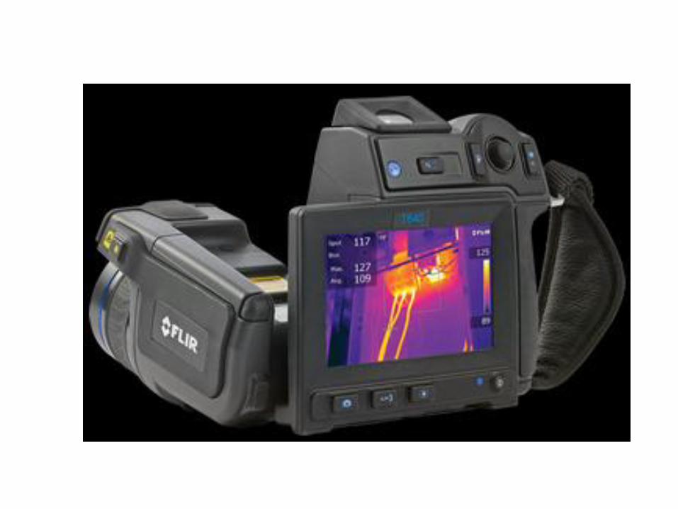
Technique
The system may also use several scales of false
color coding. This means that an image is created
in which each temperature is assigned a specific
color on a reference scale; the best scale for
veterinary diagnostics is the rainbow color scale.
The image created can be interpreted and used
for diagnostic purposes in medical fields.
Technique
An IR system should be certified by the regional
authorities. Only such systems guarantee that the
measured temperatures are accurate and that it is
legal to use the system; specific regulations exist
because of the military use of this technology
this technique should be used throughout
veterinary medicine, especially in zoo and wild
animal medicine, as an aid in primary
diagnostics.
Technique
The images captured by the IR detector may be
saved and stored on a hard disc or other storage
media and viewed and evaluated later on the
computer with specialized software.
Technique
Thermography is best used on animals, or parts
of them, without long hair, such as elephants,
rhinoceroses, hippopotami, giraffes,
zebras/horses, and many larger antelopes. In
longer-haired animals such as carnivores, camels
with winter coats, and mountain animals, the
interpretation of results is more difficult.
The Thermographer
The thermographer must be familiar with:
• Normal skin surface
• Internal anatomy
• Morphology of the animal under investigation.
.
Interpretation
• Regional hair length is an important factor for
interpretation
• location of blood vessels
• Innervation of skin areas under investigation
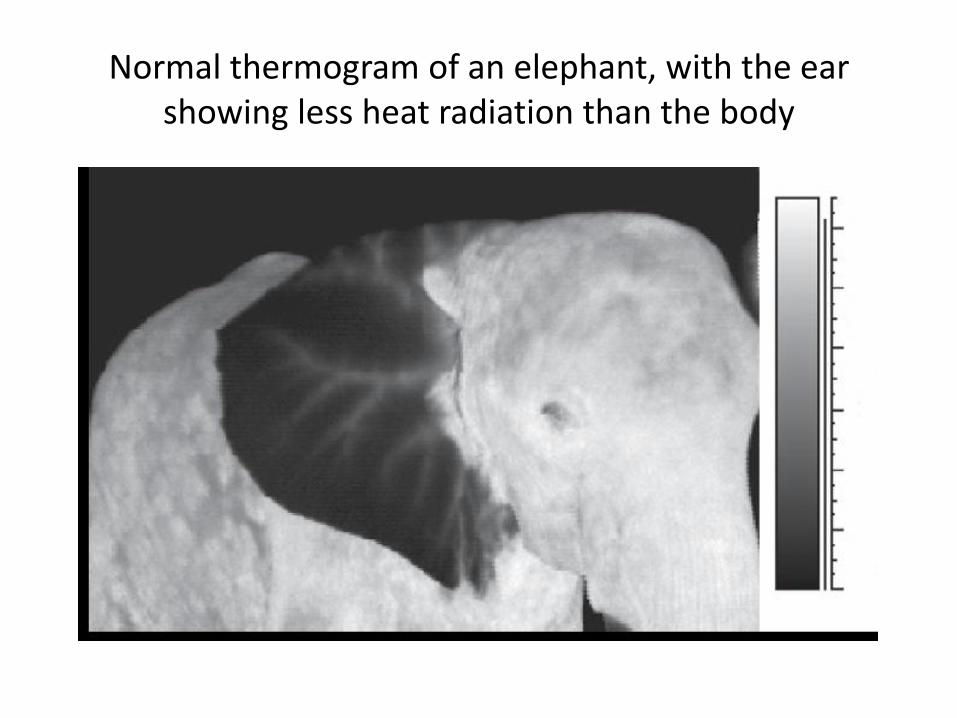
Normal thermogram of an elephant, with the ear showing less heat radiation than the body
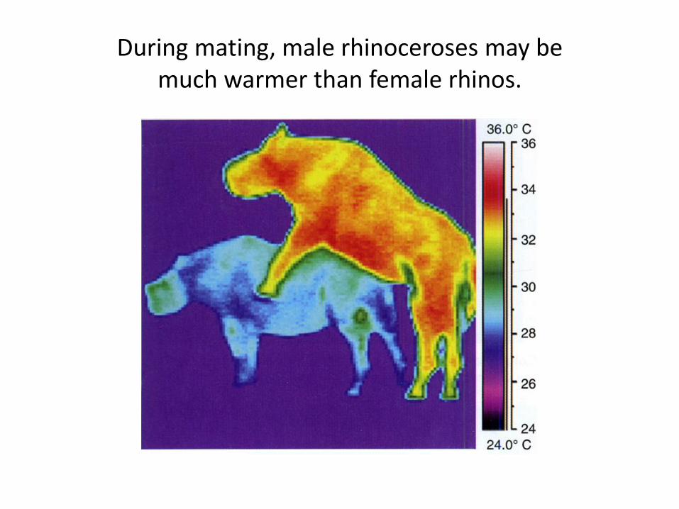
During mating, male rhinoceroses may be much warmer than female rhinos.
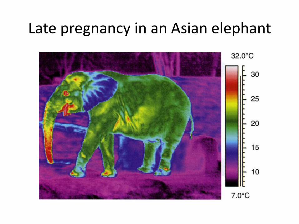
Late pregnancy in an Asian elephant
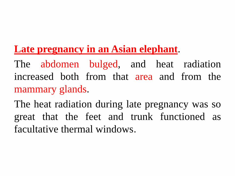
Late pregnancy in an Asian elephant.
The abdomen bulged, and heat radiation
increased both from that area and from the
mammary glands.
The heat radiation during late pregnancy was so
great that the feet and trunk functioned as
facultative thermal windows.
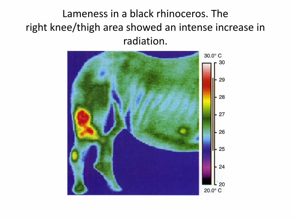
Lameness in a black rhinoceros. The right knee/thigh area showed an intense increase in
radiation.
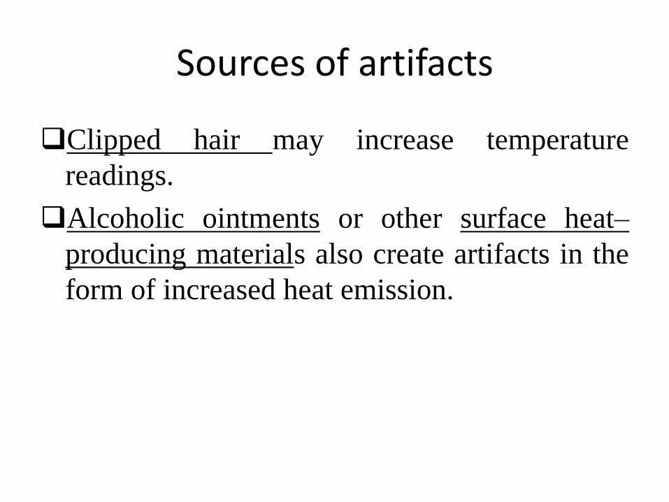
Sources of artifacts
Clipped hair may increase temperature
readings.
Alcoholic ointments or other surface heat–
producing materials also create artifacts in the
form of increased heat emission.
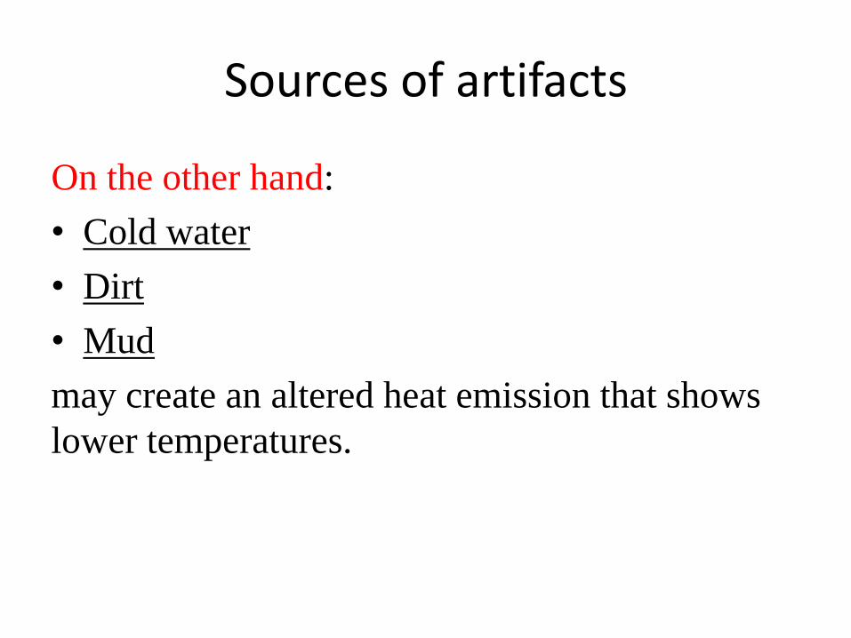
Sources of artifacts
On the other hand:
• Cold water
• Dirt
• Mud
may create an altered heat emission that shows
lower temperatures.
Strong physical activity
Strong physical activity of the animal will create
local heat production at first, but heat emission
from the whole-animal surface may occur later,
depending on the type of animal and the type and
duration of the activity.
High ambient temperature
High ambient temperature poses difficulties
when looking for smaller temperature
differences. Under high ambient temperatures
the difference between the animal core and
surface temperature decreases. This makes the
use of IR thermography more
challenging(difficult) in field investigations than
in zoo settings.
High ambient temperature
A good way to address this problem is using the
technique in a stable or, for wildlife at night, near
a water hole. The sun itself also creates
significant artifacts, and therefore cloudy days
are preferred.
Best place
Again, the best place for an investigation of a
zoo animal is the stable, or the investigations
should take place on a cloudy day, after sunset,
or before sunrise, if absolute temperatures are
required. Otherwise, the investigator should try
to lure the animal into a shady part of the
enclosure.
An experienced thermographer
An experienced thermographer can cope(deal)
with many artifacts or will do a follow-up
investigation a few hours or days later.
Artifacts may also result from sources of heat in
the housing environment of zoo animals, such as
heaters on walls , floor heating, or even heating
from ceilings.
Familiar
Thermoregulation: the Basics for Medical
Thermography
Before veterinarians can make good use of IR
thermography in zoo and wildlife medicine, they
must become familiar with the thermoregulatory
patterns of each species.
specific challenges (difficulties) for
thermography: color patterns; hair length;
thickness of the dermis; location of glands; size
of ears, horns, or antlers; location of potential
thermal windows on the body itself; and the
anatomy of the legs. Thermal windows are areas
of increased heat emission; some are facultative
and some obligatory
Elephants
Because of the lack of hair, elephants (and most
rhino species) display a relatively even surface
temperature under normal conditions, with only
the ears, horns, or tusks showing lesser heat
radiation than the body and legs.
Mammals
Mammals with short hair and thin legs (e.g.,
giraffes, antelopes, zebras) display cooler legs
than bodies under normal thermoregulatory
conditions and in the shade.
Animals body surface
Animals with thick hair may display little
radiation through the body surface, which may
make the use of IR thermography almost
impossible. However, some uses may still be
possible, such as the diagnosis of inflammatory
processes on the legs.
Mammals
The inside of mammalian legs shows a slightly
greater heat radiation than the outside because of
the more superficial location of blood vessels.
When doing

Mammals
In mammals the eyes are always obligatory
thermal windows, as are the mouth, heart region,
and the rectal and vaginal openings, as well as
the penis during urination or erection.
Signs
Experienced trainers and veterinarians are able to
identify potentially lame animals up to 2 weeks
before the animal actually shows clinical signs.
General indicators of altered thermoregulation can be physiologic
or pathologic, as follows:
1. Exposure to strong sun
2. High ambient temperatures with
simultaneous(at the same time) high humidity
3. Physical activity
4. Stress (psychologic)
5. Pregnancy
6. Abrasions
7. Inflammation

Fetus
Depending on the ambient temperature and
relative humidity, this metabolic heat, as well as
the heat of the placenta and the body heat of the
growing fetus, is channeled to the outside of the
mother’s skin by conductance, especially when
the fetus is pressed against the mother’s body
wall.
Pregnancy Diagnosis
During pregnancy the female animal shows
increased metabolism that allows for the growth
of the fetus.
When energy of one form is converted into
another form, some energy is always lost in the
form of heat.
Diagnosing Inflammation
Heat production in inflammatory processes is
one of the cardinal symptoms of inflammation.
IR thermography picks up this heat if the process
is located close to the body surface.
The diagnosis of an inflammatory process in a
leg or ear is a good way to gain experience with
this method.

Thank You