indications for surgery in acute mastoiditis and their complications in children
TRANSCRIPT

International Journal of Pediatric Otorhinolaryngology (2006) 70, 1175—1182
www.elsevier.com/locate/ijporl
Indications for surgery in acute mastoiditis andtheir complications in children
Diego Zanetti, Nader Nassif *
Department of Otolaryngology, University of Brescia, Piazzale Spedali Civili 1, 25100 Brescia, Italy
Received 7 September 2005; received in revised form 2 December 2005; accepted 6 December 2005
KEYWORDSAcute otitis media;Chronic otitis media;Mastoiditis;Complications;Mastoidectomy
Summary
Objective: To review the clinical charts of 45 paediatric patients treated for acuteotomastoiditis at the ORL Department of the University of Brescia (Italy) betweenJanuary 1994 and March 2005 and to discuss the diagnostic workup and the outcome oftreatment.Methods: Twenty-six males and 19 females were admitted with acute mastoiditis andsubperiosteal abscess. Thirteen of them (28.9%) presented an intracranial complica-tion. Only three of them were not operated upon; one received a ventilation tube(VT); all the others underwent a mastoidectomy within 48—72 h. Twenty out of 32uncomplicated mastoiditis were treated conservatively and the remaining 12 under-went myringotomy � VT, associated with a mastoidectomy in 9 cases.Results: Antibiotics alone or with VTs achieved a full recovery in 28 out of 32uncomplicated cases. Mastoidectomy resolved the disease in 13 patients (9 withcomplications). In severe complications, a canal wall down (CWD) (n = 2) or an intactcanal wall (ICW) mastoidectomy (n = 7) were preferred, based on the extent of thelesions and the degree of hearing loss. All children recovered completely at 1 yearfollow-up. In the uncomplicated cases that were operated upon, the mean hospitalstay was 7.8 days (versus 4.3 days for the conservative group). In children withintracranial complications the mean hospital stay was 12.8 days, significantly lessthan the four non-surgical patients, who remained hospitalized for an average of 18days.Conclusion: Acute mastoiditis can fully recover with conservative treatment ormyringotomy + VTs. Immediate surgical treatment is indicated for intracranial com-plications, if the neurological conditions are not critical. A simple mastoidecto-my � tympanoplasty is warranted in: (1) exteriorization, if the child is older than30 months or >15 kg of weight, (2) intracranial complications (combined with aneurosurgical procedure as needed) and (3) cholesteatoma or granulation tissue.# 2005 Elsevier Ireland Ltd. All rights reserved.
* Corresponding author. Tel.: +39 030 3995325; fax: +39 030 395212.E-mail addresses: [email protected] (D. Zanetti), [email protected] (N. Nassif).
0165-5876/$ — see front matter # 2005 Elsevier Ireland Ltd. All rights reserved.doi:10.1016/j.ijporl.2005.12.002

1176 D. Zanetti, N. Nassif
Fig. 1 Right acute mastoiditis in a 6-year-old girl. Highdefinition CT scan of the temporal bone, coronal projec-tion: coalescent mastoiditis with resorption of bony tra-beculae; dense material filling the mastoid, attic andtympanic cavity. Note the erosion of the cortical bone(arrow) and the initial swelling of the soft tissues.
1. Introduction
Prior to the antibiotic era, one quarter to one half ofthe patients with acute otitis media (AOM) andchronic otitis media (COM) presented with mastoi-ditis, subperiosteal abscesses and sigmoid sinusthrombophlebitis [1]. Two to 6% of all patientsdeveloped an intracranial suppurative complica-tion, with a fatal outcome in three quarters of them[2]. Despite the antibiotics reduced the complica-tion rate to 0.02—0.15%, the mortality is still high(�20%), especially in populations with lower socio-economic conditions [3—6].
The incidence of mastoiditis in the paediatric agehas consistently increased over the last two decades[7] even in industrialized countries [8,9]. The samenegative trend has been observed for suppurativeintracranial complications [10]. Abuse of or inade-quacy of antibiotic treatment have been attributeda role in selecting resistant bacterial strains [8,11].
The dilemmas that the otologist is facing whendealing with mastoiditis are:
1. t
he indications for a surgical treatment; 2. t he timing of surgery (immediate versusdelayed);
3. t he choice of the surgical procedure.The objective of this study was to review theclinical charts of 45 paediatric patients admitted tothe ENT Department of the University of Brescia foracute otomastoiditis and discuss the diagnosticworkup and the outcome of treatment.
Fig. 2 A 2-month-old child (patient no. 13 with intra-cranial complication). CT scan of the brain, axial projec-tion: swelling of soft tissues over the mastoid,subperiosteal abscess (arrow), resorption of cortical bone(dotted line). Note the epidural abscess in the posteriorcranial fossa, adjacent to the sigmoid sinus (arrowhead).
2. Materials and methods
Between January 1994 and March 2005, 45 childrenwere admitted to the Otolaryngology Department ofthe University of Brescia for an acute mastoiditis.Their age ranged between 2 months and 15 years(mean 5.2 years). There were 26 males and 19females. Thirteen of them (28.9%) presented anintracranial complication at admission. Exterioriza-tion in the deep neck spaces (Bezold’s abscess) wasobserved in one 6-year-old boy. Retroauricular swel-ling, skin redness, tenderness and pain at palpationof the mastoid region was the typical picture for 30little patients, while 15 others were admitted withfowl smell otorrhoea (n = 13) and/or torticollis(n = 3) or trismus (n = 1). Fever was present in 29patients, headache in 3.
At the time of admission, eight patients had beenalready taking oral antibiotics for 2—10 days (mean6.1 days). The drugs prescribed by the family pedia-
tricians had been cephalosporines (n = 4) macrolides(n = 3) and amoxicillin (n = 1). Swabs for bacterialculture were obtained in 27 children at the time ofsurgery either by exploratory puncture or collectionof purulent material from the ear canal.
A computerized tomography (CT) scan of thebrain and a high definition study of the temporalbone was urgently requested at admission when anintracranial complication was suspected (n = 6)(Figs. 1 and 2). The CTscan was otherwise postponed(within 24—72 h) in other 11 children. An imagingstudy was not deemed necessary for 28 childrenbecause of the benign clinical course of the disease.An MRI helped defining the complications. Sigmoidsinus thrombosis (SST) (n = 7) was assessed by meansof CTscan with i.v. contrast and angio-MR in selected
Acute mastoiditis in children 1177
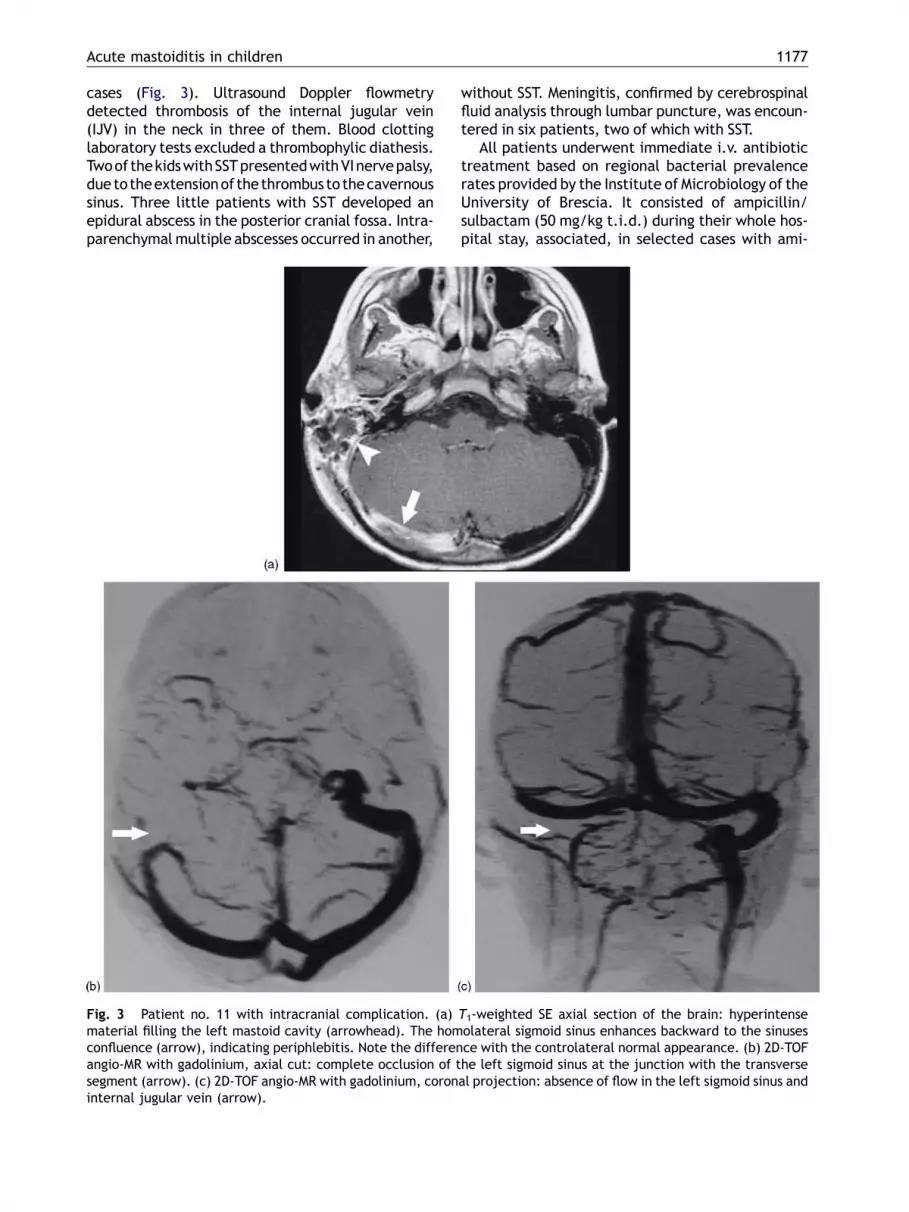
cases (Fig. 3). Ultrasound Doppler flowmetrydetected thrombosis of the internal jugular vein(IJV) in the neck in three of them. Blood clottinglaboratory tests excluded a thrombophylic diathesis.Twoof thekidswithSSTpresentedwithVInervepalsy,due totheextensionof thethrombus to thecavernoussinus. Three little patients with SST developed anepidural abscess in the posterior cranial fossa. Intra-parenchymalmultiple abscesses occurred in another,
Fig. 3 Patient no. 11 with intracranial complication. (a)material filling the left mastoid cavity (arrowhead). The homconfluence (arrow), indicating periphlebitis. Note the differenangio-MR with gadolinium, axial cut: complete occlusion of tsegment (arrow). (c) 2D-TOF angio-MR with gadolinium, coroninternal jugular vein (arrow).
without SST. Meningitis, confirmed by cerebrospinalfluid analysis through lumbar puncture, was encoun-tered in six patients, two of which with SST.
All patients underwent immediate i.v. antibiotictreatment based on regional bacterial prevalencerates provided by the Institute of Microbiology of theUniversity of Brescia. It consisted of ampicillin/sulbactam (50 mg/kg t.i.d.) during their whole hos-pital stay, associated, in selected cases with ami-
T1-weighted SE axial section of the brain: hyperintenseolateral sigmoid sinus enhances backward to the sinusesce with the controlateral normal appearance. (b) 2D-TOFhe left sigmoid sinus at the junction with the transverseal projection: absence of flow in the left sigmoid sinus and

1178 D. Zanetti, N. Nassif
kacin sulphate (7.5 mg/kg b.i.d.) or netilmicin sul-phate (3 mg/kg b.i.d.) for 6 days.
The non-surgical patients were discharged afteran average of 4.45 days (range 1—14 days) and thesurgically treated children after 13.56 days (range9—21 days). All of them continued an oral antibiotic(40 patients: amoxicillin/clavulanic acid 25 mg/kgb.i.d.; 4 patients: a third generation cephalosporinactive against Pseudomonas aeruginosa) for 10—15days. All the non-surgical children were visited inthe office 1 week after discharge and on an indivi-dualized basis thereafter.
When surgery was considered, it was undertakenwithin the first 72 h since admission (mean 2.6 days,range 12 h—12 days). Six patients underwent surgeryon the very same day they entered the hospital andmost of the others in the first 2 days. The timing ofsurgery depended upon the general condition of thepatient and extension and complications associatedwith the mastoiditis. Surgery was delayed only inpatient #2 (intraparenchymal cerebellar abscess)because of critical neurological conditions. Follow-up for the surgical group consisted in office visitsscheduled at 1—2 weeks, 1—3—6—12 months andevery 6 months thereafter. Doppler ultrasound scan-ning of the vessels in the neck was obtained at 3—6—12 months in the three children with IJV thrombosis.
3. Results
Long-term follow-up (range 12—37 months) docu-mented a complete recovery in all 45 children. The
Table 1 Intracranial complications and their management
Patient Sex Age Pathology
1 G.C. M 15 years AOM, meningitis2 B.A. M 14 years AOM, cerebellar a3 C.D. F 3 years AOM, meningitis, S4 G.A. M 3 months AOM, meningitis5 M.M. F 15 months AOM, meningitis6 F.L. M 3 years AOM, meningitis7 A.S. M 14 years Cholesteatoma,
meningitis, SST8 C.G. F 5 years COM granulation t
SST, epidural p.c.f9 M.C. F 8 years AOM, SST
10 B.G. M 6 years AOM, SST11 M.A. M 6 years AOM + Bezold’s ab
SST, epidural p.c.f12 A.A. M 5 years AOM, SST, CST13 L.S. F 2 months AOM, epidural p.c
AOM: acute otitis media; COM: chronic otitis media; CST: cavernousintact canal wall mastoidectomy; MPL: myringoplasty; p.c.f.: postesigmoid sinus; VT: ventilation tube; m: months; yrs: years.
CT scan identified 13 intracranial complications.Table 1 summarizes their clinical features and man-agement.
Twelve out of the 32 patients who had uncompli-cated mastoiditis (37.5%) and 10 out of 13 childrenwith an intracranial complication (77%) underwentsurgery (Table 2). A myringoplasty was associated tothemastoidectomy in three instances. In no case thereconstruction of the ossicular chain, that was foundinterrupted in two children, was attempted in thefirst stage.
All seven children with SST underwent a mastoi-dectomy, either intact canal wall (ICW) (n = 5) orcanal wall down (CWD) (n = 2), based on the extentof the disease. The sinus was surgically managed byskeletonization of the cortical bone in five children,by the evacuation of the thrombus and obliterationof the lumen in one (Table 1, patient #9), and byligation of the lateral sinus and of the IJV in the neckin another one (Table 1, patient #11). Post-operativecourse was uneventful in all cases. Two of the threechildren with IJV thrombosis underwent prolongedanticoagulation (for up to 6 months) with dicumar-oles. The blood flow in the vein was re-establishedat 6months at ultrasound Doppler scanning in all SSTchildren, except in the two with obliterated orligated sinus. The latter recovered completelyand developed an efficient collateral venous drai-nage. In no instance a neurosurgical procedure wasdeemed necessary. In patient #2 the neurosurgeonpreferred to abstain from performing a craniotomy,due to the multifocality of the purulent collectionsin the cerebellum. A delayed ICW mastoidectomy
Surgical treatment Hospital stay
None 15bscess ICW + MPL 10ST, CST CWD + MPL 10
None 11None 21VT 19CWD + MPL 18
issue,. abscess
ICW + VT 10
ICW + VT, SS resectionand obliteration
21
ICW + VT 16scess,. abscess
ICW, SS drainage 12
ICW + VT 9.f. abscess ICW 6
sinus thrombosis; CWD: canal wall down mastoidectomy; ICW:rior cranial fossa; SS: simoid sinus; SST: thrombophlebitis of the

Acute mastoiditis in children 1179
Table 2 Treatment of acute mastoiditis and its complications
Therapy No. ofpatients
Mean duration of hospital stayand antibiotic Tx (days)
Simple mastoiditis32 patients
No surgical treatment 20 4.38 (1—14)V.T. 3 5.6 (7—8)Myringotomy 5 4.2 (2—6)Mastoidectomy 1 7Mastoidectomy + V.T. 3 7 (6—8)
Mastoiditis + intracranialcomplication 13 patients
No surgical treatment 3 17 (15—21)V.T. 1 19Mastoidectomy 5 13 (10—18)Mastoidectomy + V.T. 4 12.8 (8—21)
V.T.: ventilation tubes.
(on the 8th day after admission), obtained a com-plete resolution of the disease. Overall, the meanhospital stay for SST patients was 7.8 days (range 2—21 days).
Among the 32 uncomplicated cases (Table 2), nostatistically significant differences were foundbetween a conservative management with antibio-tics alone (n = 20) and the surgical treatment (n = 5myringotomies, n = 3 myringotomy + VTs, n = 4 cor-tical mastoidectomies). All recovered fully,although the relief of symptoms was somewhatslower in the non-surgical group. Also the meanhospital stays were not different: 4.2 and 5.6 daysfor the myringotomy and myringotomy + VTs group,respectively, versus 7 days in the four children whounderwent amastoidectomy. Ventilation tubes werespontaneously extruded within 6—8 months in allcases except in one; no otorrhoea or perforationspersisted at 1 year follow-up. Only patient #10 hadrecurrent AOM on the same side at 3 months aftersurgery, up to the longest follow-up (37 months). Herecovered without any further surgery, by means ofa 10 days course of cephtriaxone, neither develop-ing sequelae nor suffering of other recurrences.
4. Discussion
Although the majority of middle ear infectionsinvolves the mastoid cellular tract, a subperiostealabscess or an exteriorization into the neck (Bezold’sabscess) nowadays occurs in less than 2% of AOMepisodes [5,12,13]. Acute mastoiditis can developwithout a previous history of recurrent AOM [14] orafter a single episode of AOM, such as in 38.5% ofGhaffar et al.’s young patients [7]. A ‘‘masked’’mastoiditis should be suspected if there is persistentpain or otorrhoea despite 2 weeks of antibiotictreatment [11]. In our series, 37 of the 44 patients(84%) developed mastoidites after the first recog-nized episodes of AOM.
Antibiotic treatment prior to admission does notseem to prevent the onset of mastoiditis [11,15,16].Only a limited number of children of the presentstudy were taking antibiotics before hospital admis-sion (8/45 = 17.8%). In contrast with the literature,where Streptococcus pneumoniae ranks first [17,18],Streptococcus pyogenes was the most commonpathogen isolated from the ears of our patients.
The incidence of complications we observed (13/45 patients: 28.9%) is set at the upper limitsreported in the literature, that ranges between 7and 35% [7,11,15,19,20], and resembles those ofdeveloping countries [21]. Reasonable explanationis the characteristics of tertiary referral centre ofour institution and the significant rate of immigra-tion from developing countries in the last decade.Complications aroused in 11 children after an iso-lated episode of AOM and in two others during thecourse of COM.
The CT yields a sensitivity of 97% and a positivepredictive value of 94% in detecting intracranialcomplications [22,23]. The common ‘‘shadowing’’of the mastoid air cell tract in a CT scan performedduring AOM is not a sign of mastoiditis that, instead,is indicated by resorption of bony trabecula anderosion of the cortical bone (Figs. 1 and 2). Basedon this high rate of complications, we suggest toobtain not only for the patients with clear neurolo-gical signs but in every patient with acute mastoi-dites. If a complication is found, MRI should follow,to help the surgical planning [22].
The commonest complication in the presentstudy was SST (n = 7), often associated with otherco-morbidities (IJV thrombosis in three, cavernoussinus thrombosis in two, epidural abscesses in theposterior fossa in three and meningitis in two). Amastoidectomy, with toilette of the periphlebitis orperisinus abscess proved to be sufficient and suc-cessful in five of them. Evacuation of the thrombusthrough an incision of the sinus, followed by oblit-eration in one child and by IJV ligation in another,

1180 D. Zanetti, N. Nassif
were necessary in order to prevent the dispatch ofseptic emboli to the pulmonary circulation.
Meningitis was the second most common neuro-logical complication (n = 6). Three of the littlepatients with meningitis recovered with pharmaco-logical therapy alone. In one child myringotomy andVTs were beneficial. The last two children under-went a mastoidectomy because of SST (n = 2) andassociated VIth nerve palsy (n = 1), and full recoveryensued.
The role of surgical treatment, and especially ofmastoidectomy, is questioned by some authors [24].The recovery rate with i.v. antibiotics and myrin-gotomy ranges between 60.4 and 87% [20,25,26].Furthermore, some studies point out that no statis-tically significant differences are observed betweenthe cure rates for children treated with myringoto-mies + VTs and those managed more aggressivelywith mastoidectomies [27,28]. Conversely, animmediate drainage of the mastoid abscess couldtheoretically reduce the use of antibiotics and thehospital stay, and would prevent an intracranialspread of the infection. In some centres a corticalmastoidectomy is performed in all instances, includ-ing very young children, starting from 8 months ofage [29]. Other management options include theevacuation of the subperiosteal abscess by incisionand drainage [30,31] and the exploratory puncture,either alone [32] or associated with myringotomy[33]. Based on our experience, we agree with theclinical studies that recommend mastoidectomyonly for complications or failures to improve withantibiotics and myringotomy [25,34,35]. In our ser-ies the hospital stay and the duration of the anti-biotic treatment were not significantly reducedwhen compared with more conservativeapproaches; this reflects the greater severity ofthe disease leading to a mastoidectomy.
In the uncomplicated cases who were operatedupon, the mean hospital stay was 7.8 days versus 4.3days for the conservative group, whereas forpatients with intracranial complications the meanhospital stay was 12.8 days (range 8—21), signifi-cantly less than in the four non-surgical patients,who remained hospitalized for an average of 18 days(range 15—21). Special consideration is to be givento the little patients who develop meningitis: thespectrum of clinical presentation may vary fromslight meningeal signs, for which a prudent ‘‘waitand see’’ policy under antibiotic coverage or myr-ingotomy is warranted, to the occurrence of themore severe drug-resistant otorrhoea and osteiticlesions, which drive the Otosurgeon into action. Inour experience, myringotomy and VTs have alsoaccelerated the recovery of meningitis followingAOM episodes and therefore, it could have possibly
shortened the long hospital stay (mean 17 days) alsoin the three children who were not operated upon.
Reluctance to perform amastoidectomy is mainlyrelated to the anesthesiological risks in very smallchildren, although excessive blood loss is absolutelyrare. The surgical hazards are minimal in a simplecortical mastoidectomy, whose purposes are to clearthe purulent collection in the mastoid and to recre-ate a correct ventilation pathway through the aditusad antrum, without disturbing the middle ear cavityor endangering the labyrinth and the Fallopiancanal. In our experience, mastoidectomy led tocomplete recovery in all nine complicated mastoi-dites in whom it was performed, and it did notoverload themorbidity of the disease in any respect.On the other hand, 20 out of 32 of our children withno intracranial complications recovered without anysurgical treatment. This could lead to the assump-tion that a mastoidectomy is unnecessary in themajority of acute mastoiditis. Actually, this sub-group represented a milder form of the disease,and not even myringotomy was felt necessarybecause of normal eardrums and tympanograms.Most likely, the purulent material was segregatedinto the mastoid by a blocked aditus ad antrum,while the tympanic cavity was free.
The decision to proceed to surgery reflected amore severe clinical course of the disease. Myringo-tomies � VTs were reserved for children with con-sistent retroauricular swelling and bulging of theeardrum, while the mastoidectomy was performedin 4 uncomplicated cases who failed to respond toantibiotics and in 9 out of 13 with complications.
When intracranial complications are identified,otologic surgery is carried out soon after the neu-rosurgical procedure, in order to clear the source ofinfection. It can be performed in the same surgicalsession, or it can be delayed until the neurologicalconditions have stabilized. Epidural abscesses in theposterior cranial fossa can also benefit from theotologic procedure alone, as demonstrated by theexperience in three of our children (#8, #11 and#13).
Overall, all children with intracranial complica-tions who submitted to a mastoidectomy, either ICW(n = 7) or CWD (n = 2) achieved complete control oftheir mastoiditis. In patient #3, the reason for a CWDmastoidectomy was the extension of the osteiticlesions and granulation tissue in the mastoid andattic, associated with SST, perisinus abscess andmeningitis. Myringoplasty completed a CWD mastoi-dectomy in one cholesteatoma, as it is routinelydone in COM surgery. In two other cases, one wouldargue if it was safe to seal a spontaneous drainageroute after AOM. Actually, these two kids had suf-fered of recurrent AOM episodes that always healed

Acute mastoiditis in children 1181
spontaneously, until they developed mastoiditis. Atsurgery coalescent mastoidites was found in one andSST in other one. In both cases, the surgeon wasconfident of having achieved a complete clearanceof the disease and repair of the eardrum was judgedharmless.
By reviewing our clinical experience and in agree-ment with other authors [29,32], our current indi-cations to mastoidectomy for acute mastoiditis arethe following: (1) exteriorized mastoid abscess,age > 30 months (or weight > 15 kg), (2) intracra-nial complications, (3) cholesteatoma and (4) puru-lent otorrhoea and/or granulation tissue, resistantto topical and systemic antibiotics for more than 2weeks.
The cut-off age of 30 months is related to theanestesiological risk of blood loss in toddlers lighterthan 15 kg in weight. A more cautious approach isrecommendable in such cases, i.e. immediate myr-ingotomy and observation of the clinical courseunder proper antibiotic treatment. When a mastoi-dectomy is indicated, it should be performed within48—72 h. This allows a prompt improvement of theclinical picture, often paralleled by a shortening ofthe hospital stay [35].
5. Conclusions
Mastoidectomy is an effective treatment for acutemastoiditis associated with one of the following:subperiosteal abscess or exteriorization, cholestea-toma, intracranial complications and otorrhoea per-sisting for more than 2 weeks despite adequateantibiotic treatment, in children >15 kg of weight.The rational of the procedure is to clear the sup-purative process in the mastoid cell tract, avoidingthe risk of further intracranial spread, and re-estab-lish a ventilation route through the aditus adantrum. In the present series it did not add morbid-ity to the pathology and achieved complete recov-ery in all cases in whom it was performed. Whenindicated, it is preferably performed within 48—72 h. It can otherwise be delayed, in the presenceof intracranial complications, until the neurologicalcompromise is stabilized. A craniotomy is unneces-sary for epidural abscesses, since clearing of themastoid focus is sufficient to control the disease.More cautionary approaches, including antibioticsalone or associated with myringotomy and ventila-tion tubes, are equally effective in the less severeforms of the disease, as well as in meningitis follow-ing AOM. Early myringotomy is advocated in smallerbabies with the typical acute mastoiditis. Followingthese rules, all our children recovered completely at1 year follow-up. Children with intracranial compli-
cations remained in the hospital for a longer periodif they were not operated upon (18 days versus 12.8days), related to greater severity of the disease.
References
[1] C.A. Proctor, Intracranial complications of otitic origin,Laryngoscope 76 (1966) 288—308.
[2] S. Gower, W.F. McGuirt, Intracranial complications of acuteand chronic infectious disease; a problem still with us,Laryngoscope 93 (1983) 1028.
[3] J. Kangsanarak, S. Fooanant, K. Ruckphaopunt, N. Nava-charoen, S. Teotrkakul, Extracranial and intracranial com-plications of suppurative otitis media. Report of 102 cases,J. Laryngol. Otol. 107 (1993) 999—1045.
[4] M.A. Matin, A.H. Khan, F.A. Khan, A.A. Haroon, A profile of100 complicated cases of chronic suppurative otitis media,J. R. Soc. Health 117 (1997) 157—159.
[5] U. Osma, S. Cureoglu, S. Hosoglu, The complications ofchronic otitis media: report of 93 cases, J. Laryngol. Otol.114 (2000) 97—100.
[6] K.M. Shamboul, An unusual prevalence of complications ofchronic suppurative otitis media in young adults, J. Laryn-gol. Otol. 106 (1992) 874—877.
[7] F.A. Ghaffar, M. Wordemann, G.H. McCracken, Acute mas-toiditis in children: a seventeen-year experience in Dallas,Texas, Pediatr. Infect. Dis. J. 20 (2001) 376—380.
[8] D.A. Van Zuijlen, A.G. Schilder, F.A. Van Balen, A.W. Hoes,National differences in incidence of acute mastoiditis: rela-tionship to prescribing patterns of antibiotics for acute otitismedia? Pediatr. Infect. Dis. J. 20 (2001) 1012—1013.
[9] A.I. Ruiz Diaz, F. del Castillo Martin, A. Bilbao Garitagoitia,C. Diaz Roman, M.J. Garcia Miguel, C. Borque Andres, Acutemastoiditis: an increasing entity, An. Esp. Pediatr. 57 (2002)427—431.
[10] C. Go, M.J. Bernstien, A.L. De Jong, M. Sulek, E.M. Fried-man, Intracranial complications of acute mastoiditis, Int. J.Pediatr. Otorhinolaringol. 52 (2000) 143—148.
[11] M. Luntz, A. Brodsky, S. Nusem, J. Kronenberg, G. Keren, L.Migirov, et al., Acute mastoiditis–—the antibiotic era: amulticenter study, Int. J. Pediatr. Otorhinolaryngol. 57(2001) 1—9.
[12] J.H. Spiegel, L.R. Lustig, K.C. Lee, A.H. Murr, R.A. Schindler,Contemporary presentation and management of a spectrumof mastoid abscesses, Laryngoscope 108 (1998) 822—828.
[13] G. Marioni, C. De_Filippis, A. Tregnaghi, R. Marchese_Ra-gona, A. Staffieri, Bezold’s abscess in children: case reportand review of the literature, Int. J. Pediatr. Otorhinolar-yngol. 61 (2001) 173—177.
[14] E. Kvestad, K.J. Kvaerner, I.W. Mair, Acute mastoiditis:predictors for surgery, Int. J. Pediatr. Otorhinolaryngol. 15(2000) 149—155.
[15] T.E. Linder, H.R. Briner, T. Bischoff, Prevention of acutemastoiditis: fact or fiction? Int. J. Pediatr. Otorhinolaryngol.56 (2000) 129—134.
[16] M. Romer, H.R. Briner, T. Linder, Effect of antibiotics on theoccurrence and course of acute mastoiditis, Schweiz. Med.Wochenschr. Suppl. 125 (2000) 20S—22S.
[17] M. Francois, T. Van den Abbeele, P. Viala, P. Narcy, Acuteexternal mastoiditis in children: report of a series of 48cases, Arch. Pediatr. 8 (2001) 1050—1054.
[18] M. Nussinovitch, R. Yoeli, K. Elishkevitz, I. Varsano, Acutemastoiditis in children: epidemiologic, clinical, microbiolo-gic, and therapeutic aspects over past years, Clin. Pediatr.(Phila.) 43 (2004) 261—267.

1182 D. Zanetti, N. Nassif
[19] P. Vera_Cruz, R.R. Farinha, V. Calado, Acute mastoiditis inchildren–—our experience, Int. J. Pediatr. Otorhinolaryngol.50 (1999) 113—117.
[20] J. Spratley, H. Silveira, I. Alvarez, M. Pais_Clemente, Acutemastoiditis in children: review of the current status, Int. J.Pediatr. Otorhinolaryngol. 56 (2000) 33—40.
[21] I. Khan, F. Shahzad, Mastoiditis in children, J. Laryngol. Otol.117 (2003) 177—181.
[22] E. Vazquez, A. Castellote, J. Piqueras, S. Mauleon, S. Creix-ell, F. Pumarola, C. Figueras, J.C. Carreno, J. Lucaya,Imaging of complications of acute mastoiditis in children,Radiographics 23 (2003) 359—372.
[23] L. Migirov, Computed tomographic versus surgical findings incomplicated acute otomastoiditis, Ann. Otol. Rhinol. Lar-yngol. 112 (2003) 675—677.
[24] R. Cohen-Kerem, N. Uri, H. Rennert, N. Peled, E. Green-berg, M. Efrat, Acute mastoiditis in children: is surgicaltreatment necessary? J. Laryngol. Otol. 113 (1999) 1081—1085.
[25] S. De, Z.G. Makura, R.W. Clarke, Paediatric acute mastoi-ditis: the Alder Hey experience, J. Laryngol. Otol. 116 (2002)440—442.
[26] P.W. Bauer, K.R. Brown, D.T. Jones, Mastoid subperiostealabscess management in children, Int. J. Pediatr. Otorhino-laryngol. 15 (2002) 185—188.
[27] E.S. Lee, S.W. Chae, H.H. Lim, S.J. Hwang, H.K. Suh, Clinicalexperiences with acute mastoiditis–—1988 through 1998, EarNose Throat 79 (2000) 884—888, 890—892.
[28] E.H. Harley, T. Sdralis, R.G. Berkowitz, Acute mastoiditis inchildren: a 12-year retrospective study, Otolaryngol. HeadNeck Surg. 116 (1997) 26—30.
[29] G. Michalski, T. Hocke, K. Hoffmann, D. Esser, Therapy ofacute mastoiditis, Laryngorhinootologie 81 (2002) 857—860.
[30] L. Migirov, J. Kronenberg, Bacteriology of mastoid subperios-teal abscess in children, Acta Otolaryngol. 124 (2004) 23—25.
[31] L. Migirov, A. Eyal, J. Kronenberg, Intracranial complicationsfollowing mastoidectomy, Pediatr. Neurosurg. 40 (2004)226—229.
[32] M.F. Taylor, R.G. Berkowitz, Indications for mastoidectomyin acute mastoiditis in children, Ann. Otol. Rhinol. Laryngol.113 (2004) 69—72.
[33] A. Khafif, D. Halperin, I. Hochman, R. Gertler, I. Poria, D.Shindel, G. Marshak, Acute mastoiditis: a 10-year review,Am. J. Otolaryngol. 19 (1998) 170—173.
[34] R.S. Bahadori, R.H. Schwartz, M. Ziai, Acute mastoiditis inchildren: an increase in frequency in Northern Virginia,Pediatr. Infect. Dis. J. 19 (2000) 212—215.
[35] V. Tarantino, R. D’Agostino, G. Taborelli, A. Melagrana, A.Porcu, M. Stura, Acute mastoiditis: a 10 year retrospectivestudy, Int. J. Pediatr. Otorhinolaryngol. 66 (2002) 143—148.