independent component analysis as a preprocessing step for data compression of neonatal eeg
TRANSCRIPT
INDEPENDENT COMPONENT ANALYSIS AS PREPROCESSING TOOL FOR DECOMPOSITION OF SURFACE ELECTRODE-ARRAY
ELECTROMYOGRAM
Gonzalo A. García, Ryouichi Nishitani, Ryuhei Okuno, Kenzo Akazawa
Department of Bioinformatic Engineering, Graduate School of Information Science and Technology, Osaka University, 2-1 Yamada-Oka, Suita City, Osaka, Japan
E-mail: [email protected]
ABSTRACT The purpose of this preliminary work was to evaluate the effectiveness of independent component analysis (ICA) as preprocessing tool for the decomposition of electromyograms (EMG) into their constitutive elements (motor unit action potentials). An experiment was carried out with a healthy subject performing isometric contractions at different force levels. Surface electromyograms were measured with an electrode array. Satisfactory decompositions were obtained for levels up to 10% of maximal voluntary contraction (MVC). Acceptable decompositions could be achieved also at 20 and 30% MVC, but in these cases results were irregular.
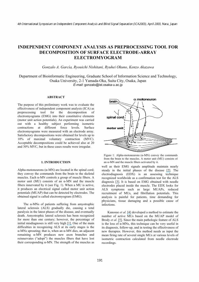
1. INTRODUCTION Alpha-motoneurons (α-MN) are located in the spinal cord; they convey the commands from the brain to the skeletal muscles. Each α-MN controls a group of muscle fibers. A motor unit (MU) consists of an α-MN and the muscle fibers innervated by it (see Fig. 1). When a MU is active, it produces an electrical signal called motor unit action potentials (MUAP) that can be detected by electrodes. The obtained signal is called electromyogram (EMG).
The α-MNs of patients suffering from amyotrophic lateral sclerosis (ALS) gradually die, causing a total paralysis in the latest phases of the disease, and eventually death. Amyotrophic lateral sclerosis has been recognized for more than one century; however, the percentage of initial misdiagnoses is still very high [1]. One of the main difficulties in recognizing ALS at its early stages is the α-MNs sprouting; that is, when an α-MN dies, an adjacent remaining α-MN produces new axon branches and reinnervates (“adopt”) the muscles fibers that have lost their corresponding α-MN. The strength of the muscles as
well as their EMG signals amplitude maintain nearly steady in the initial phases of the disease [2]. The electrodiagnosis (EDX) is an assessing technique recognized worldwide as a confirmation test for the ALS diagnosis [3]. It is based on EMG obtained with needle electrodes placed inside the muscle. The EDX looks for ALS symptoms such as large MUAPs, reduced recruitment of MUs, and fibrillation potentials. This analysis is painful for patients, time demanding for physicians, tissue damaging and a possible cause of infections.
Kanosue et al. [4] developed a method to estimate the number of active MUs based on the MUAP model of Brody et al. [5]. Since the main pathologic feature of ALS is the loss of α-MNs, this technique can be very useful in its diagnosis, follow-up, and in testing the effectiveness of new therapies. However, this method needs as input the mean firing rate of several single MUs at various levels of isometric contraction calculated from needle electrode recordings.
Figure 1: Alpha-motoneurons (α-MN) convey the commands from the brain to the muscles. A motor unit (MU) consists of an α-MN and the muscle fibers activated by it.
motor unit
A non-invasive, painless alternative is to study the surface EMG signals picked up by surface electrodes placed on the patient’s skin. The obtained signals, which are a summation of several MUAPs (interference pattern), have a very low signal to noise ratio. Therefore, some preprocessing of the surface EMG signals is required prior estimation of the motor unit number.
The purpose of the present preliminary study was to
investigate the efficacy of independent component analysis (ICA) as a signal-preprocessing tool to decompose surface EMG into MUAP trains. We used the fast fixed-point algorithm for ICA developed by Hyvärinen and Oja [6, 7] because it is robust and fast (the convergence is cubic). This approach has been successfully applied to other biomedical signals [8].
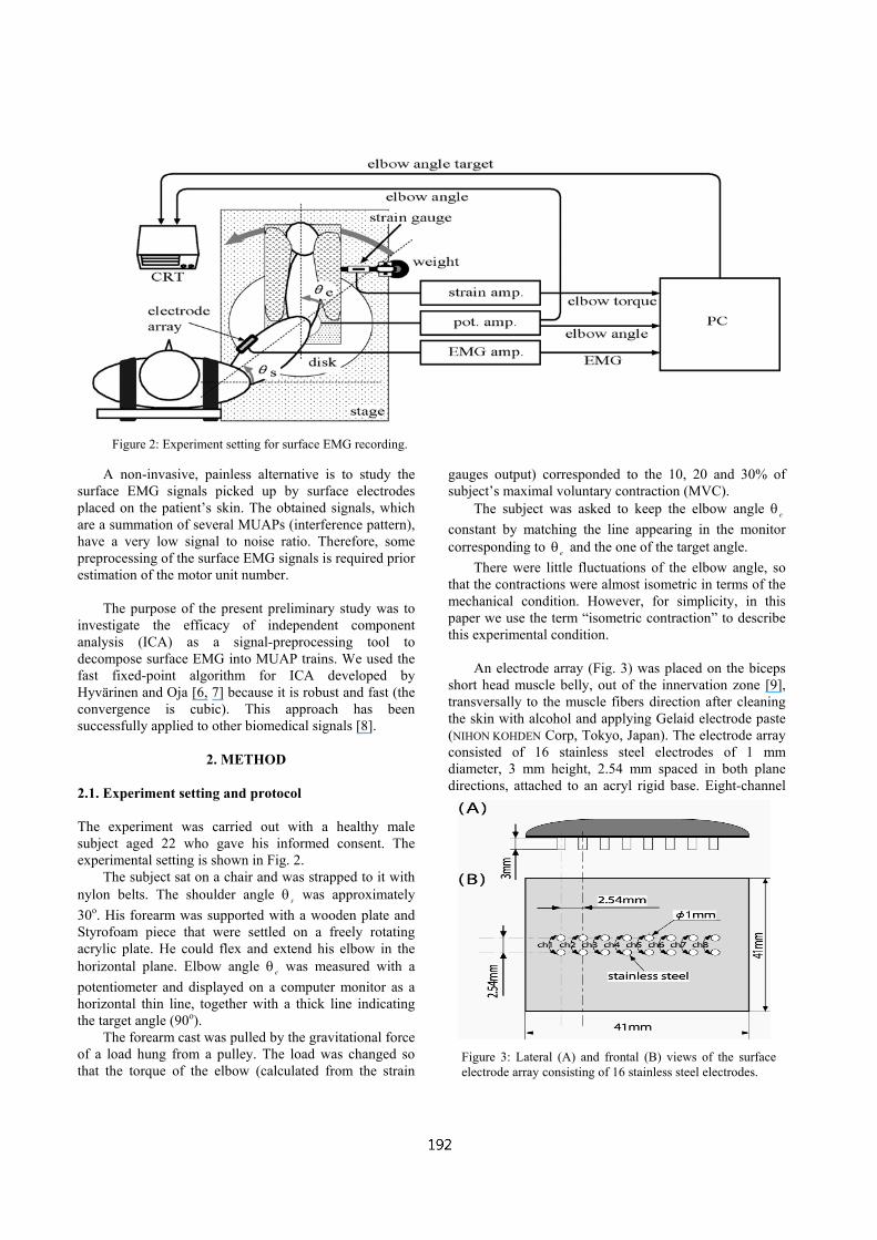
2. METHOD 2.1. Experiment setting and protocol The experiment was carried out with a healthy male subject aged 22 who gave his informed consent. The experimental setting is shown in Fig. 2.
The subject sat on a chair and was strapped to it with nylon belts. The shoulder angle sθ was approximately 30o. His forearm was supported with a wooden plate and Styrofoam piece that were settled on a freely rotating acrylic plate. He could flex and extend his elbow in the horizontal plane. Elbow angle eθ was measured with a potentiometer and displayed on a computer monitor as a horizontal thin line, together with a thick line indicating the target angle (90o).
The forearm cast was pulled by the gravitational force of a load hung from a pulley. The load was changed so that the torque of the elbow (calculated from the strain
gauges output) corresponded to the 10, 20 and 30% of subject’s maximal voluntary contraction (MVC).
The subject was asked to keep the elbow angle eθ constant by matching the line appearing in the monitor corresponding to eθ and the one of the target angle.
There were little fluctuations of the elbow angle, so that the contractions were almost isometric in terms of the mechanical condition. However, for simplicity, in this paper we use the term “isometric contraction” to describe this experimental condition.
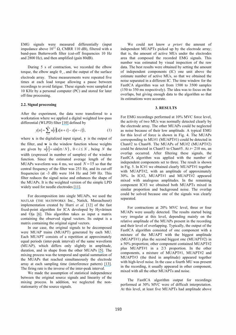
An electrode array (Fig. 3) was placed on the biceps
short head muscle belly, out of the innervation zone [9], transversally to the muscle fibers direction after cleaning the skin with alcohol and applying Gelaid electrode paste (NIHON KOHDEN Corp, Tokyo, Japan). The electrode array consisted of 16 stainless steel electrodes of 1 mm diameter, 3 mm height, 2.54 mm spaced in both plane directions, attached to an acryl rigid base. Eight-channel
Figure 3: Lateral (A) and frontal (B) views of the surface electrode array consisting of 16 stainless steel electrodes.
Figure 2: Experiment setting for surface EMG recording.
EMG signals were measured differentially (input impedance above 1012 Ω, CMRR 110 dB), filtered with a band-pass Butterworth filter (cut-off frequencies 10 Hz and 2800 Hz), and then amplified (gain 80dB).
During 5 s of contraction, we recorded the elbow torque, the elbow angle eθ , and the output of the surface electrode array. These measurements were repeated five times at each load torque allowing a pause between recordings to avoid fatigue. These signals were sampled at 10 KHz by a personal computer (PC) and stored for later off-line processing. 2.2. Signal processing After the experiment, the data were transferred to a workstation where we applied a digital weighted low-pass differential (WLPD) filter [10] defined by
[ ]( )∑=
−−+=N
i
inxinxiwny1
][][][ , (1)
where x is the digitalized input signal, y is the output of the filter, and w is the window function whose weights are given by [ ] ( )Niiw /sin π= , Ni ≤<0 , being N the width (expressed in number of samples) of the window function. Since the estimated average length of the MUAPs waveform was 4 ms, we used 15=N so that the central frequency of the filter was 253 Hz, and its cut-off frequencies (at -3 dB) were 164 Hz and 349 Hz. This filter reduces the signal noise and enhances the shape of the MUAPs. It is the weighted version of the simple LPD widely used for needle electrodes [11].
For decomposition into single MUAPs, we used the MATLAB (THE MATHWORKS Inc., Natick, Massachuset) implementation created by Hurri et al. [12] of the fast fixed-point algorithm for ICA developed by Hyvärinen and Oja [6]. This algorithm takes as input a matrix containing the observed signal vectors. Its output is a matrix containing the original signal vectors.
In our case, the original signals to be decomposed were MUAP trains (MUAPT) generated by each MU. Each MUAPT consists of a repetition at approximately equal periods (inter-peak interval) of the same waveform (MUAP), which differs only slightly in amplitude, duration, and in shape from the other MUAPs [5]. The mixing process was the temporal and spatial summation of the MUAPs that reached simultaneously the electrode array at each sampling time (interference pattern) [13]. The firing rate is the inverse of the inter-peak interval.
We made the assumption of statistical independence between the original source signals and linearity of the mixing process. In addition, we neglected the non-stationarity of the source signals.
We could not know a priori the amount of independent MUAPTs picked up by the electrode array; that is, the amount of active MUs under the electrodes area that composed the recorded EMG signals. This number was estimated by visual inspection of the raw data. The best results were obtained by setting the amount of independent components (IC) one unit above the estimate number of active MUs, so that we obtained the noise separated in a different IC. The time window for the FastICA algorithm was set from 1500 to 3500 samples (150 to 350 ms respectively). The idea was to focus on the overlaps, but giving enough data to the algorithm so that its estimations were accurate.
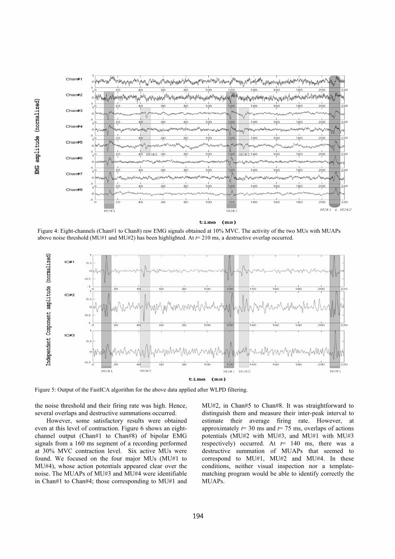
3. RESULTS For EMG recordings performed at 10% MVC force level, the activity of two MUs was normally detected clearly by the electrode array. The other MUAPs could be neglected as noise because of their low amplitude. A typical EMG for this level of force is shown in Fig. 4. The MUAPs corresponding to MU#1 (MUAPT#1) could be detected in Chan#2 to Chan#8. The MUAPs of MU#2 (MUAPT#2) could be detected in Chan#3 to Chan#5. At t= 210 ms, an overlap occurred. After filtering these signals, the FastICA algorithm was applied with the number of independent components set to three. The result is shown in Fig. 5. In IC#1 we obtained clearly MUAPT#1 together with MUAPT#2, with an amplitude of approximately 30%. In IC#2, MUAPT#1 and MUAPT#2 appeared mixed with analogous amplitudes. In the remaining component IC#3 we obtained both MUAPTs mixed in similar proportion and background noise. The overlap could be solved because one of the signals was clearly separated.
For contractions at 20% MVC level, three or four MUAPs were usually detected. The results started being very irregular at this level, depending mainly on the relative amplitude of the MUAPs present in the recording and their level of overlapping. Typically, the output of the FastICA algorithm consisted of one component with a mixture of the MUAPT with the biggest amplitude (MUAPT#1) plus the second biggest one (MUAPT#2) in a 50% proportion; other component contained MUAPT#2 plus MUAPT#1 in a 2/3 proportion. In the other components, a mixture of MUAPT#1, MUAPT#2 and MUAPT#3 (the third in amplitude) appeared together with high-level noise. In the case a fourth MU was present in the recording, it usually appeared in other component, mixed with all the other MUAPTs and noise.
The FastICA algorithm output for recordings
performed at 30% MVC were of difficult interpretation. At this level, at least five MUAPTs had amplitude above
the noise threshold and their firing rate was high. Hence, several overlaps and destructive summations occurred.
However, some satisfactory results were obtained even at this level of contraction. Figure 6 shows an eight-channel output (Chan#1 to Chan#8) of bipolar EMG signals from a 160 ms segment of a recording performed at 30% MVC contraction level. Six active MUs were found. We focused on the four major MUs (MU#1 to MU#4), whose action potentials appeared clear over the noise. The MUAPs of MU#3 and MU#4 were identifiable in Chan#1 to Chan#4; those corresponding to MU#1 and
MU#2, in Chan#5 to Chan#8. It was straightforward to distinguish them and measure their inter-peak interval to estimate their average firing rate. However, at approximately t= 30 ms and t= 75 ms, overlaps of actions potentials (MU#2 with MU#3, and MU#1 with MU#3 respectively) occurred. At t= 140 ms, there was a destructive summation of MUAPs that seemed to correspond to MU#1, MU#2 and MU#4. In these conditions, neither visual inspection nor a template-matching program would be able to identify correctly the MUAPs.
Figure 4: Eight-channels (Chan#1 to Chan8) raw EMG signals obtained at 10% MVC. The activity of the two MUs with MUAPs above noise threshold (MU#1 and MU#2) has been highlighted. At t= 210 ms, a destructive overlap occurred.
Figure 5: Output of the FastICA algorithm for the above data applied after WLPD filtering.
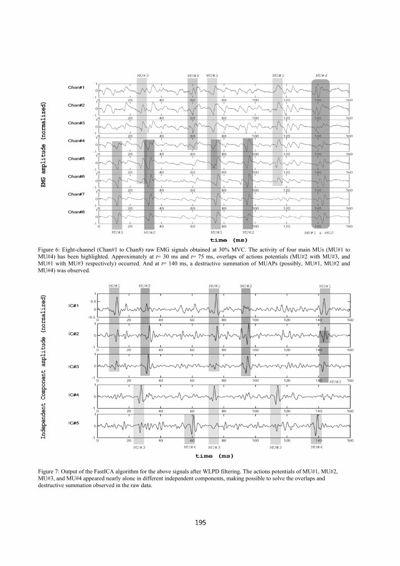
Figure 6: Eight-channel (Chan#1 to Chan8) raw EMG signals obtained at 30% MVC. The activity of four main MUs (MU#1 to MU#4) has been highlighted. Approximately at t= 30 ms and t= 75 ms, overlaps of actions potentials (MU#2 with MU#3, and MU#1 with MU#3 respectively) occurred. And at t= 140 ms, a destructive summation of MUAPs (possibly, MU#1, MU#2 and MU#4) was observed.
Figure 7: Output of the FastICA algorithm for the above signals after WLPD filtering. The actions potentials of MU#1, MU#2, MU#3, and MU#4 appeared nearly alone in different independent components, making possible to solve the overlaps and destructive summation observed in the raw data.
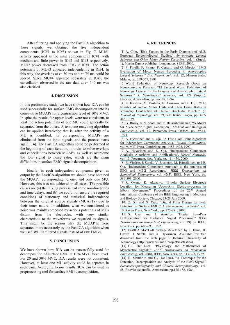
After filtering and applying the FastICA algorithm to these signals, we obtained the five independent components (IC#1 to IC#5) shown in Fig. 7. MU#1 activity appeared as the main component in IC#1, with medium and little power in IC#2 and IC#3 respectively. MU#2 power decreased from IC#3 to IC#1. The action potentials of MU#3 appeared independently in IC#4. In this way, the overlaps at t= 30 ms and t= 75 ms could be solved. Since MU#4 appeared separately in IC#5, the cancellation observed in the raw data at t= 140 ms was also clarified.
4. DISCUSSION In this preliminary study, we have shown how ICA can be used successfully for surface EMG decomposition into its constitutive MUAPs for a contraction level of 10% MVC. In spite the results for upper levels were not consistent, at least the action potentials of one MU could generally be separated from the others. A template-matching algorithm can be applied iteratively; that is, after the activity of a MU is identified, its corresponding MUAPs are eliminated from the input signals, and the process starts again [14]. The FastICA algorithm could be performed at the beginning of each iteration, in order to solve overlaps and cancellations between MUAPs, as well as overcome the low signal to noise ratio, which are the main difficulties in surface EMG signals decomposition.
Ideally, in each independent component given as output by the FastICA algorithm we should have obtained the MUAPT corresponding to one, and only one MU. However, this was not achieved in all cases. The possible causes are (a) the mixing process had some non-linearities and time delays, and (b) we could not ensure the required conditions of stationary and statistical independence between the original source signals (MUAPTs) due to their inner nature. In addition, what we considered as noise was mainly composed by actions potentials of MUs distant from the electrodes, with very similar characteristic to the waveforms we regarded as signals. This might be the reason why the MUAPTs were separated more accurately by the FastICA algorithm when we used WLPD filtered signals instead of raw EMGs.
5. CONCLUSION We have shown how ICA can be successfully used for decomposition of surface EMG at 10% MVC force level. For 20 and 30% MVC, ICA results were not consistent. However, at least one MU activity could be separate in each case. According to our results, ICA can be used as preprocessing tool for surface EMG decomposition.
6. REFERENCES
[1] A. Chio, “Risk Factors in the Early Diagnosis of ALS: European Epidemiological Studies,” Amyotrophic Lateral Sclerosis and Other Motor Neuron Disorders, vol. 1 (Suppl. 1), Martin Dunitz publisher, London, pp. S13-8, 2000. [2] P. Pinelli, F. Pisano, F. Ceriani, and G. Miscio, “EMG Evaluation of Motor Neuron Sprouting in Amyotrophic Lateral Sclerosis,” Ital. Neurol. Sci., vol. 12, Masson Italia, Milano, pp. 359-367, 1991. [3] World Federation of Neurology Research Group on Neuromuscular Diseases, “El Escorial World Federation of Neurology Criteria for the Diagnosis of Amyotrophic Lateral Sclerosis,” J. Neurological Sciences, vol. 124 (Suppl.), Elsevier, Amsterdam, pp. 96-107, 1994. [4] K. Kanosue, M. Yoshida, K. Akazawa, and K. Fujii, “The Number of Active Motor Units and Their Firing Rates in Voluntary Contraction of Human Brachialis Muscle,” Jp. Journal of Physiology, vol. 29, Yas Kuno, Tokyo, pp. 427-443, 1979. [5] G. Brody, R.N. Scott, and R. Balasubramanian, “A Model for Myoelectric Signal Generation,” Medical and Biological Engineering, vol. 12, Pergamon Press, Oxford, pp. 29-41, 1974. [6] A. Hyvärinen and E. Oja, “A Fast Fixed-Point Algorithm for Independent Component Analysis,” Neural Computation, vol. 9, MIT Press, Cambridge, pp. 1483-1492, 1997. [7] A. Hyvärinen and E. Oja, “Independent Component Analysis: Algorithms and Applications”, Neural Networks, vol. 13, Pergamon, New York, pp. 411-430, 2000. [8] R. Vigário, J. Särelä, V. Jousmäki, M. Hämäläinen, and E. Oja, “Independent Component Approach to the Analysis of EEG and MEG Recordings,” IEEE Transactions on Biomedical Engineering, vol. 47(5), IEEE, New York, pp. 589-593, 2000. [9] R. Okuno, K. Akazawa, “Bipolar Surface Electrode Location for Measuring Upper-Arm Electromyograms in Elbow Movements,” Proceedings of the 22nd Annual International Conference of the IEEE Engineering in Medicine and Biology Society, Chicago, 23-28 July 2000. [10] Z. Xu and S. Xiao, “Digital Filter Design for Peak Detection of Surface EMG,” J. Electromyogr. Kinesiol, vol. 10, Raven Press, New York, pp. 275-281, 2000. [11] S. Usui and I. Amidror, “Digital Low-Pass Differentiation for Biological Signal Processing,” IEEE Transactions on Biomedical Engineering, vol. 29(10), IEEE, New York, pp. 686-693, 1982. [12] FastICA MATLAB package developed by J. Hurri, H. Gävert, J. Särelä, and A. Hyvärinen. Available for free download from the web page of Helsinki University of Technology (http://www.cis.hut.fi/project/ica/fastica). [13] C.J. De Luca, “Physiology and Mathematics of Myoelectric Signals,” IEEE Transactions on Biomedical Engineering, vol. 26(6), IEEE, New York, pp. 313-325, 1979. [14] B. Mambrito and C.J. De Luca, “A Technique for the Detection, Decomposition and Analysis of the EMG Signal,” Electroencephalography and Clinical Neurophysiology, vol. 58, Elsevier Scientific, Amsterdam, pp.175-188, 1984.