increase of accuracy in intraoperative navigation through high-resolution flat-panel volume computed...
TRANSCRIPT

1
Title page
Increase of accuracy in intraoperative navigation through high resolution flat-panel Volume-CT: Experimental comparison to multi-slice-CT based navigation
Basic Science Report
Dr. Soenke H Bartling 1 Dr. Martin Leinung 2 Dr. Thomas Rodt 1 Dipl.-ing. Christian Dullin 3 Prof. Dr. Hartmut Becker 1 Prof. Dr. Thomas Lenarz 2 PD Dr. Timo Stöver 2 Dr. Omid Majdani 2 1 Dept. of Neuroradiology, Hannover Medical School, Hannover, Germany 2 Dept. of Otorhinolaryngology, Hannover Medical School, Hannover, Germany 3 Dept. of Diagnostic Radiology, Goettingen University Hospital, Goettingen, Germany Corresponding author and reprint requests: Soenke Bartling Dept. of Neuroradiology, OE 8210 Hannover Medical School Carl-Neuberg-Str. 1 30625 Hannover Germany [email protected] Phone: +49 511 532 6654 Fax: +49 511 532 5876
Short title page
Increase of accuracy in intraoperative navigation through high resolution flat-panel Volume-CT: Experimental comparison to multi-slice-CT based navigation
Basic Science Report
2
Abstract Hypothesis: High-resolution imaging as provided by flat-panel based volume
computed tomography (fpVCT) could increase navigation accuracy and could
therefore improve image-guided procedures or make novel navigated surgery
concepts possible.
Background: Intraoperative navigation is an accepted tool in head & neck surgery.
However its use is limited in the lateral skull base due to its low surgical accuracy.
Surgical accuracy is substantially influenced by the resolution of the underlying
dataset. FpVCT offers a nearly two times higher resolution than multislice computed
tomography (MSCT). Target registration error (TRE) - as a measurement for surgical
navigation accuracy - should decrease when navigation is based on fpVCT datasets.
Methods: An acrylic glass phantom with 37 fiducial points was scanned in a current
MSCT (GE Lightspeed 16 Pro) and in an experimental fpVCT (GE). Both datasets
were imported in an optical navigation system (BrainLab VectorVision2). Five fiducial
points were used for registration and seven for measuring TRE. The distance
between indicated pointer tip and corresponding fiducial point in dataset was
measured as TRE. Registration and TRE measurement was repeated five times for
each CT dataset. Average TREs were calculated and results compared using t-test.
Results: The average TRE using MSCT (0.82 (standard deviation: 0.35) mm) was
significantly higher than using fpVCT (0.46 (standard deviation: 0.22) mm) (p<0,01).
Conclusion: Submillimetric surgical navigation accuracy is possible using high
resolution fpVCT. This could be highly beneficial in skull base surgery navigation.
3
Introduction Image-guided surgery (IGS) systems have found widespread use in ENT surgery.
IGS systems allow real-time, intraoperative tracking of current location within volume
datasets (CT or MRI)(1). They are commonly used for sinus surgery (2-6) and lateral
skull base surgery (7-11).
The demand on surgical accuracy (12) of the intraoperative navigation in different
regions varies strongly. Intraoperative navigation during sinus surgery is highly
supportive with the currently reached surgical accuracy (2, 13-15). However, in the
lateral skull base intraoperative navigation could be much more useful if the surgical
accuracy could be improved beyond the currently achieved (14). This is primarily
because the structures of the lateral skull base are much smaller than the structures
of the maxillo facial and sinus region (8). The currently reached surgical accuracy
does not allow the full utilization of the benefits of intraoperative navigation for
surgery of the lateral skull base (e.g. microsurgery of the middle ear or cochleostomy
for cochlear implantation) (16-19). Therefore it is necessary to improve surgical
accuracy.
The surgical accuracy of intraoperative navigation is a complex function that is
affected by various factors. The resolution of the underlying imaging is one of the
most important (12, 20).
A new kind of CT scanner, a flat-panel based Volume-CT (fpVCT) offers an
increased resolution when compared to state-of-the-art MSCT scanner (21-25).
FpVCT scanning of the human skull base has been recently shown (26). The surgical
accuracy of intraoperative navigation might therefore improve if navigation is based
on fpVCT datasets.

4
Material and Methods
A surgical accuracy comparison of intraoperative navigation based on MSCT and on
fpVCT by average target registration error analysis (TRE) using a phantom (12, 15)
was performed.
TRE is the difference between corresponding positions in the real world (e.g.
phantom or patient) and the underlying dataset (e.g. CT-scan) following registration.
A comprehensive overview of errors associated with intraoperative navigation and
their clinical relevance is given in (15). TRE is the error of interest for the surgeon
(15).
Phantom
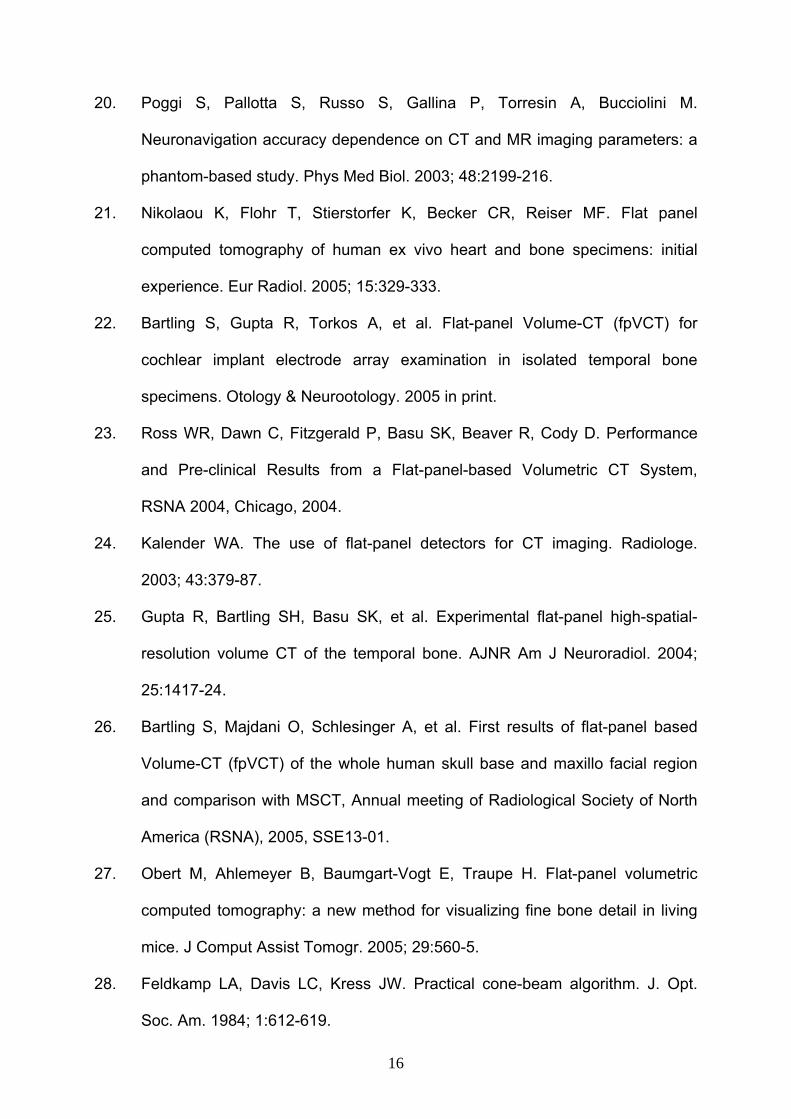
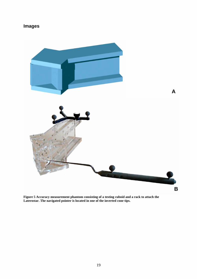
The acrylic glass phantom used for TRE measurement is shown in Figure 1. The
testing area consisted of two quadratic plates that were attached using two oblique
quadratic plates. It measured 10 cm in width, height and length. It was affixed with 37
nearly equally distributed fiducial points in form of precisely defined inverted cones
that were drilled into the acrylic glass. The reference device was fitted on the
phantom (Figure 1).
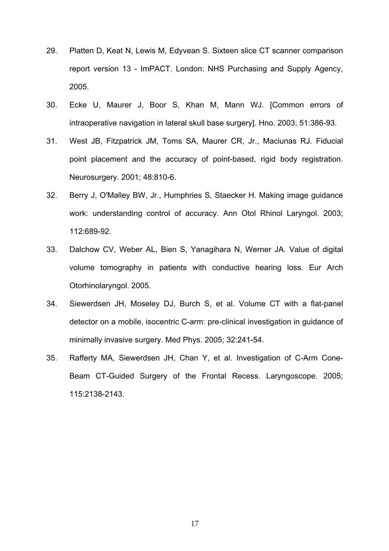
Flat-panel based Volume-CT scanning The phantom was scanned in an experimental flat-panel Volume-CT (Figure 2). The
scanner consisted of a X-ray tube and 2 flat-panel detectors mounted on a standard
CT gantry (22, 23, 27). The scanner can be used in a two-panel- or one-panel-mode.
5
In two-panel mode it provides a scan field of view big enough for a complete human
head (27 cm), while the scan field of view in one-panel-mode was sufficient for the
scanning of the phantom as used in this study (13.5 cm). Both detectors consisted of
1024 by 1024 detector elements on an area of 20.48 cm by 20.48 cm, resulting in a
detector element size of 200 µm by 200 µm. A 360° rotation took 8 seconds and
1000 projections were acquired. Z-coverage was 4.21 cm per rotation. Three scans
in a step and shoot scanning mode were performed to scan the testing area of the
phantom. The scan field of view in the X/Y plane was 13.5 cm. A tube voltage of
140kV together with a current of 20mA was used. The used scanning parameters
were empirically derived in earlier experiments to optimize image quality for high
resolution.
For reconstruction a 512 by 512 matrix was selected to assure compatibility with
standard post-processing software. The resulting voxel size was selected to be (190
µm)3 resulting in a reconstruction field of view of 10 cm in diameter. The
reconstruction was based on the Feldkamp-Davis-Kress cone beam algorithm (28).
The reconstructed volume dataset was exported in standard DICOM3 format. The
measured, isotropic spatial resolution using a 25 µm Tungsten wire object (10%
MTF, high resolution scanning and reconstruction) is approximately 25 line-
pairs(lp)/cm (≈ 200 µm feature size) for this scanner in a similar scanning mode (23).
Multislice-CT scanning The phantom was scanned in a 16-slice-CT (GE Lightspeed 16Pro, GE Healthcare,
Milwaukee, WI) using a high resolution scanning protocol: 120 kV, 80 mA, 0.625 mm
slice thickness and a pitch of 0.5.
6
The reconstruction was performed with a reconstruction field of view of 9.6 cm, a z-
voxel dimension of 0.3 mm, a ”bonepuls” kernel in “standard” modus (180° weighted
interpolation), resulting in 187 µm by 187 µm by 300 µm voxels. The measured
spatial in plane and Z-direction resolution using a 25 µm Tungsten wire object (10%
MTF, high resolution scanning and reconstruction) is approximately 14-15 lp/cm (≈
350 µm feature size) for this scanner (23, 29).
Navigation system An optical navigation system VectorVision2 (BrainLAB, Heimstetten, Germany) was
used. The system is based on paired infrared cameras that detect reflecting markers.
Reflecting markers are placed on the pointing and reference device. By means of
triangulation navigation pointer and reference device can be located in space.
Registration, accuracy measurement and data analysis Registration is the correlation of the coordinates of the phantom in real world and CT
dataset using fiducial points.
The deepest points of the inverted cones were defined as fiducial points (Figure 4).
For registration five evenly distributed fiducial points were selected after importing the
DICOM3 datasets into the navigation system. During the registration process all five
points were touched with the pointer one after another (Figure 1b). The pointer was
slightly swayed around without leaving its position within the inverted cone until the
navigation system signalled for successful detection of the pointer’s tip position.
After registration, TRE analysis was performed using seven fiducial points which
were different from the registration points. Fiducial points were marked within the CT

7
dataset and the pointer was placed in the fiducial point while the shaft of the pointer
was perpendicular to the surface of the phantom. The distance between the indicated
pointer position and the marked fiducial point in the CT dataset was measured by the
system and noted. Screenshots were taken for documentation. The phantom was not
moved during the measurements. The registration and TRE measurement procedure
were repeated five times (passes) for each CT modality, resulting in 35 data points
for each CT modality. The average target registration error (TRE) and standard
deviation (SD) were calculated. Average TREs of both CT modalities were compared.
The significance of the difference between both CT modalities was calculated by
performing two sided t-test on dependent samples.

8
Results
Scans of the phantom, the subsequent import of the datasets into the navigation
system and registrations were successful in all cases. All registration fiducial points
could be identified and all distances (TRE) could be measured.
The average TRE using the MSCT dataset was 0.82 (standard deviation: 0.35) mm
with an error range from 0.3 mm to 1.7 mm. The average TRE using the fpVCT
dataset was 0.46 (standard deviation: 0.22) mm with an error range from 0.1 mm to 1
mm.
As a result, the average TRE using fpVCT was significantly lower than the average
TRE using MSCT (p<0.01). Further, the standard deviation, error range as well as
maximal error were lower in fpVCT based navigation. A box plot diagram of the TRE
results is given in Figure 3.
Representative screenshots are given for MSCT (Figure 4 A) of the measurement of
data point 7 of pass 1 and for fpVCT (Figure 4 B) of the measurement of data point 6
of pass 3.

9
Discussion
Surgical accuracy and target registration error
The terms “accuracy”, “precision” and related expressions are well defined (12, 15).
Often less appropriate error measurements ranging from calculated registration error
(CRE) to fiducial registration error (FRE) have been taken as a measurement for
surgical accuracy (12).
The term “surgical accuracy” has been defined to provide a clinically useful
measurement of intraoperative navigation quality. It is defined as the maximal
deviation of a navigation system from the true position together with the precision (in
terms of reproducibility)(12). Under experimental conditions comparable to this study,
measuring the TRE provides a good approximation of “surgical accuracy” (12, 15).
CT resolution and its influence to surgical accuracy
The resolution of the fpVCT was higher than the resolution of the MSCT system. The
resolution of the fpVCT scanner is with 200 µm isotropic feature size almost half as
big as the resolution of the MSCT system with 350 µm.
However, this increase in resolution does not proportionally translate into an
increased surgical accuracy or decreased TRE, because surgical accuracy of a
navigation system setup depends on a multitude of factors:
First, there are inaccuracies of the navigation system itself, which are complex
functions that are influenced by design factors, such as calibration, resolution of the
infrared camera and so on. Even the position of an object within the detector field of
the infrared camera influences the accuracy and precision of the system (30).

10
Second, there are additional inaccuracies in the registration process. During
registration, corresponding fiducial markers need to be exactly identified to align the
preoperative CT scan with the intraoperative anatomy. Fiducial localization errors
(FLE) are associated both with the identification of fiducial points in the real world
(FLEw) as well as in the CT-dataset (FLECT). Lower FLE on both ends usually results
in a higher surgical accuracy (15).
The FLEw is influenced by the navigation system, the shape of the fiducial marker
and carefulness of the operator (15). The FLECT is strongly influenced by the
resolution of the CT dataset and might have benefited from the higher resolution of
fpVCT.
However, measurement of FLEs and other errors separately is challenging and
requires elaborative technology, while their sole relevance for the surgeon is low.
Therefore rather the resulting TRE was measured in this study as an endpoint
summarizing all potential influences of a higher resolution.
There is another registration method that can further increase the surgical accuracy:
So-called frame based registration results usually in lower FLE than “free” fiducial
registration as used in this study, because the geometry between fiducial points both
in the real world and in the CT dataset is known. Implementation of a frame-based
registration method is currently work in progress.
By decreasing error sources that are associated with the resolution of the underlying
data set, others that are not directly influenced by imaging gain relative weight. Thus
developers and clinicians have to focus on these other error sources to fully utilize
the resolution of future imaging modalities.

11
Fiducial placement
The placement of free registration fiducials with regard to the surgery area of interest
is a significant factor that contributes to TRE (31, 32).
In this study, the registration fiducials have been placed in an even distribution
around the testing area and relatively close to the target fiducial points. This
configuration of fiducial points is considered optimal. However, because of the human
anatomy such an evenly distribution close to the surgery field of interest is difficult to
realize.
Considering lateral skull base surgery, two alternatives can be realized. First, one
could place the registration fiducials somewhat close around one outer ear on the
side where the surgery needs to be performed. Alternatively, one could place the
fiducials on the calvaria of the whole head, including the other side. The first
alternative would only give relatively small TRE values close to the centroid of the
registration fiducials that would be within the outer ear or the very lateral part of this
side of the skull base (31, 32), but would have the advantage that the navigation
datasets do not need to include the whole head. This could be a significant
advantage, because very high resolution datasets with a field of view as big as the
whole human skull would need tremendous computer memory space and calculation
power. Further, flat-panel scanners that become clinically available first could provide
only a small scan field of view big enough for just one temporal bone and closely
placed fiducials (33).
When the problems of big datasets and small scan fields are solved, the second
alternative would certainly be the better registration fiducial point placement.
12
Nevertheless, in both alternatives, the in vivo placement of registration fiducial would
be less optimal as in this experimental study; therefore the TRE values would be
worse. Cadaver head studies should follow.
FpVCT for patient scanning?
FpVCT, as it is used in this study, is a new CT modality that is not yet available for
patient scanning. Despite the fact that several prototypical scanners that are capable
of full head scan - and therefore patient head scanning - exist, this technology is
currently in an experimental state. However, it is likely that in the future flat-panel
based CT systems could find wider use. The flat-panel CT technology could be
promoted further through applications in angiography and maxillo-facial imaging
where flat-panel detectors are replacing image intensifier systems. A C-arm
angiography system based on flat-panel detectors that offers rotational modes (34,
35) could be used to perform high resolution imaging of the temporal bone and the
skull base with a similar image quality to a gantry based CT machine. Their use for
intraoperative guidance of sinus surgery has already been described (35).
Despite widespread assumptions, the increased resolution of fpVCT does not
necessarily means an increased radiation dose, because a higher noise can be
tolerated in imaging of high contrast structures such as the human skull base.
13
Conclusion
In this study the use of high-resolution fpVCT datasets for intraoperative navigation
has been described first.
By contrasting the TRE of navigation that is based on MSCT with higher resolution
fpVCT it was shown that the higher resolution CT dataset yields a lower TRE.
Therefore, it can be concluded that fpVCT might improve surgical accuracy in head
and neck surgery. Discussed limitations of this phantom study apply.
While the dependences of navigation accuracy on the underlying dataset is already
well described, it was not clear that even with standard navigation systems the
average TRE could be lowered to half a millimetre when combined with high
resolution datasets.
With such a surgical accuracy, intraoperative navigation can be improved and new
techniques as well as procedures could become feasible. The results of this study
should stimulate research to develop navigation systems or mechatronic assistance
systems that take full advantage of the higher resolution CT datasets that might
become clinically available soon.
14
References
1. Schlondorff G, Mosges R, Meyer-Ebrecht D, Krybus W, Adams L. [CAS
(computer assisted surgery). A new procedure in head and neck surgery].
Hno. 1989; 37:187-90.
2. Metson R. Image-guided sinus surgery: lessons learned from the first 1000
cases. Otolaryngol Head Neck Surg. 2003; 128:8-13.
3. Freysinger W, Gunkel AR, Thumfart WF. Image-guided endoscopic ENT
surgery. Eur Arch Otorhinolaryngol. 1997; 254:343-6.
4. Gunkel AR, Freysinger W, Thumfart WF. Computer-assisted surgery in the
frontal and maxillary sinus. Laryngoscope. 1997; 107:631-3.
5. Heermann R, Schwab B, Issing PR, Haupt C, Hempel C, Lenarz T. Image-
guided surgery of the anterior skull base. Acta Otolaryngol. 2001; 121:973-8.
6. Majdani O, Leinung M, Lenarz T, Heermann R. [Navigation-supported surgery
in the head and neck region]. Laryngorhinootologie. 2003; 82:632-44.
7. Caversaccio M, Romualdez J, Baechler R, Nolte LP, Kompis M, Hausler R.
Valuable use of computer-aided surgery in congenital bony aural atresia. J
Laryngol Otol. 2003; 117:241-8.
8. Gunkel AR, Vogele M, Martin A, Bale RJ, Thumfart WF, Freysinger W.
Computer-aided surgery in the petrous bone. Laryngoscope. 1999; 109:1793-
9.
9. Hassfeld S, Zoller J, Albert FK, Wirtz CR, Knauth M, Muhling J. Preoperative
planning and intraoperative navigation in skull base surgery. J
Craniomaxillofac Surg. 1998; 26:220-5.
15
10. Heermann R, Issing PR, Husstedt H, Becker H, Lenarz T. [CAS-System
MKM(R): use and results in lateral skull base surgery]. Laryngorhinootologie.
2001; 80:569-75.
11. Heermann R, Mack KF, Issing PR, Haupt C, Becker H, Lenarz T. [Skull base
surgery with an opto-electronic navigation system]. Hno. 2001; 49:1019-25.
12. Strauss G, Hofer M, Korb W, et al. [Accuracy and precision in the evaluation of
computer assisted surgical systems A definition.]. Hno. 2005.
13. Schlaier J, Warnat J, Brawanski A. Registration accuracy and practicability of
laser-directed surface matching. Comput Aided Surg. 2002; 7:284-90.
14. Snyderman C, Zimmer LA, Kassam A. Sources of registration error with image
guidance systems during endoscopic anterior cranial base surgery.
Otolaryngol Head Neck Surg. 2004; 131:145-9.
15. Labadie RF, Davis BM, Fitzpatrick JM. Image-guided surgery: what is the
accuracy? Curr Opin Otolaryngol Head Neck Surg. 2005; 13:27-31.
16. Labadie RF, Chodhury P, Cetinkaya E, et al. Minimally invasive, image-
guided, facial-recess approach to the middle ear: demonstration of the concept
of percutaneous cochlear access in vitro. Otol Neurotol. 2005; 26:557-62.
17. Labadie RF, Shah RJ, Harris SS, et al. In vitro assessment of image-guided
otologic surgery: submillimeter accuracy within the region of the temporal
bone. Otolaryngol Head Neck Surg. 2005; 132:435-42.
18. Schipper J, Klenzner T, Aschendorff A, Arapakis I, Ridder GJ, Laszig R.
[Navigation-controlled cochleostomy. Is an improvement in the quality of
results for cochlear implant surgery possible?]. Hno. 2004; 52:329-35.
19. Schipper J, Aschendorff A, Arapakis I, et al. Navigation as a quality
management tool in cochlear implant surgery. J Laryngol Otol. 2004; 118:764-
70.
16
20. Poggi S, Pallotta S, Russo S, Gallina P, Torresin A, Bucciolini M.
Neuronavigation accuracy dependence on CT and MR imaging parameters: a
phantom-based study. Phys Med Biol. 2003; 48:2199-216.
21. Nikolaou K, Flohr T, Stierstorfer K, Becker CR, Reiser MF. Flat panel
computed tomography of human ex vivo heart and bone specimens: initial
experience. Eur Radiol. 2005; 15:329-333.
22. Bartling S, Gupta R, Torkos A, et al. Flat-panel Volume-CT (fpVCT) for
cochlear implant electrode array examination in isolated temporal bone
specimens. Otology & Neurootology. 2005 in print.
23. Ross WR, Dawn C, Fitzgerald P, Basu SK, Beaver R, Cody D. Performance
and Pre-clinical Results from a Flat-panel-based Volumetric CT System,
RSNA 2004, Chicago, 2004.
24. Kalender WA. The use of flat-panel detectors for CT imaging. Radiologe.
2003; 43:379-87.
25. Gupta R, Bartling SH, Basu SK, et al. Experimental flat-panel high-spatial-
resolution volume CT of the temporal bone. AJNR Am J Neuroradiol. 2004;
25:1417-24.
26. Bartling S, Majdani O, Schlesinger A, et al. First results of flat-panel based
Volume-CT (fpVCT) of the whole human skull base and maxillo facial region
and comparison with MSCT, Annual meeting of Radiological Society of North
America (RSNA), 2005, SSE13-01.
27. Obert M, Ahlemeyer B, Baumgart-Vogt E, Traupe H. Flat-panel volumetric
computed tomography: a new method for visualizing fine bone detail in living
mice. J Comput Assist Tomogr. 2005; 29:560-5.
28. Feldkamp LA, Davis LC, Kress JW. Practical cone-beam algorithm. J. Opt.
Soc. Am. 1984; 1:612-619.
17
29. Platten D, Keat N, Lewis M, Edyvean S. Sixteen slice CT scanner comparison
report version 13 - ImPACT. London: NHS Purchasing and Supply Agency,
2005.
30. Ecke U, Maurer J, Boor S, Khan M, Mann WJ. [Common errors of
intraoperative navigation in lateral skull base surgery]. Hno. 2003; 51:386-93.
31. West JB, Fitzpatrick JM, Toms SA, Maurer CR, Jr., Maciunas RJ. Fiducial
point placement and the accuracy of point-based, rigid body registration.
Neurosurgery. 2001; 48:810-6.
32. Berry J, O'Malley BW, Jr., Humphries S, Staecker H. Making image guidance
work: understanding control of accuracy. Ann Otol Rhinol Laryngol. 2003;
112:689-92.
33. Dalchow CV, Weber AL, Bien S, Yanagihara N, Werner JA. Value of digital
volume tomography in patients with conductive hearing loss. Eur Arch
Otorhinolaryngol. 2005.
34. Siewerdsen JH, Moseley DJ, Burch S, et al. Volume CT with a flat-panel
detector on a mobile, isocentric C-arm: pre-clinical investigation in guidance of
minimally invasive surgery. Med Phys. 2005; 32:241-54.
35. Rafferty MA, Siewerdsen JH, Chan Y, et al. Investigation of C-Arm Cone-
Beam CT-Guided Surgery of the Frontal Recess. Laryngoscope. 2005;
115:2138-2143.
18
Figure legends
Figure 1 Accuracy measurement acrylic glass phantom consisting of a testing cuboid
and a rack to attach the reference device. The navigated pointer is located in one of
the inverted cone tips.
Figure 2 Flat-panel based Volume-CT prototype as used in this study. Despite the
fact that from a technical viewpoint scans of entire human skull are possible, the
machine is not approved for human scanning yet.
Figure 3 Comparison of box plots of TREs (in mm) of navigation that is based on high
resolution fpVCT and of state-of-the-art MSCT. TRE was significantly lower when
navigation was based on high-resolution fpVCT datasets.
Figure 4 Representative screenshots during TRE measuring. While the pointer tip in
the real world is located in the inverted cone tip the indicated distance between the
tip of the green line (as indicated pointer tip) and the red cross (that marks the
inverted cone tip as fiducial point) gives the target registration error. It varies
significantly between MSCT (A) and fpVCT (B).
19
Images
Figure 5 Accuracy measurement phantom consisting of a testing cuboid and a rack to attach the Laterostar. The navigated pointer is located in one of the inverted cone tips.
A
B
20
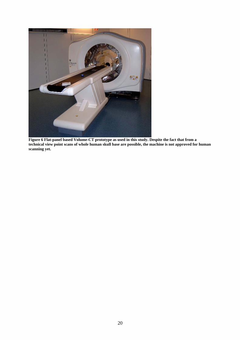
Figure 6 Flat-panel based Volume-CT prototype as used in this study. Despite the fact that from a technical view point scans of whole human skull base are possible, the machine is not approved for human scanning yet.
21
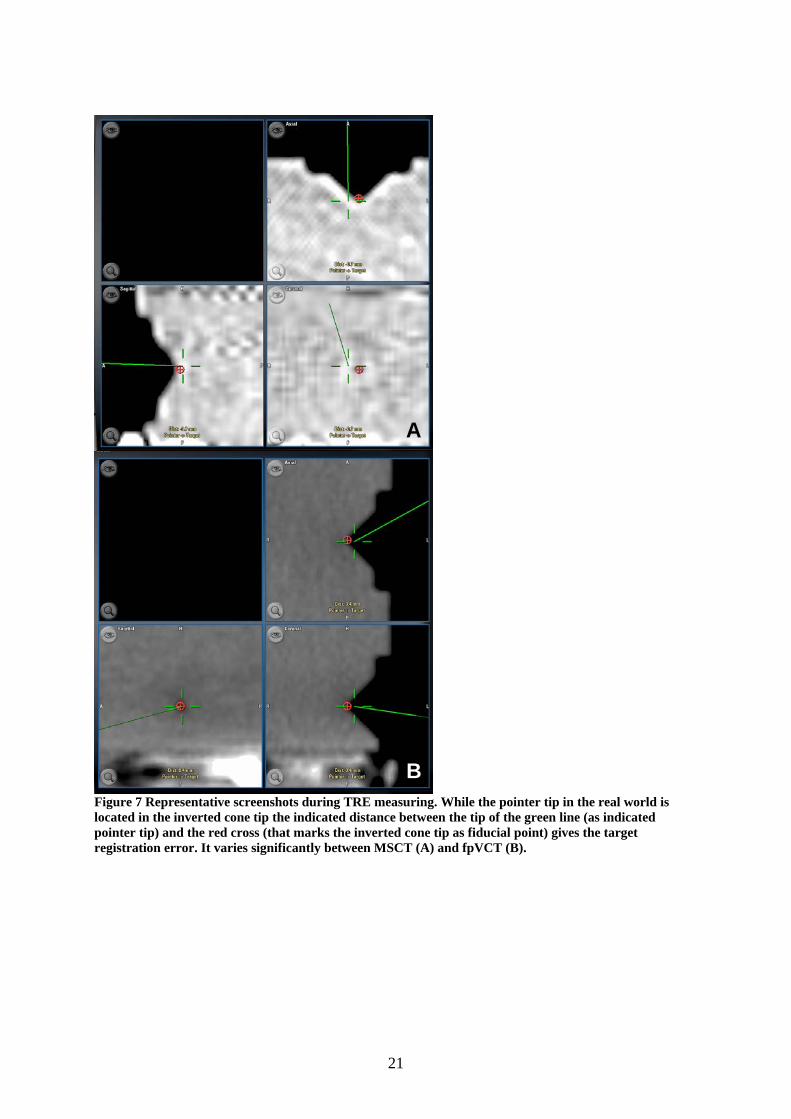
Figure 7 Representative screenshots during TRE measuring. While the pointer tip in the real world is located in the inverted cone tip the indicated distance between the tip of the green line (as indicated pointer tip) and the red cross (that marks the inverted cone tip as fiducial point) gives the target registration error. It varies significantly between MSCT (A) and fpVCT (B).
A
B
22
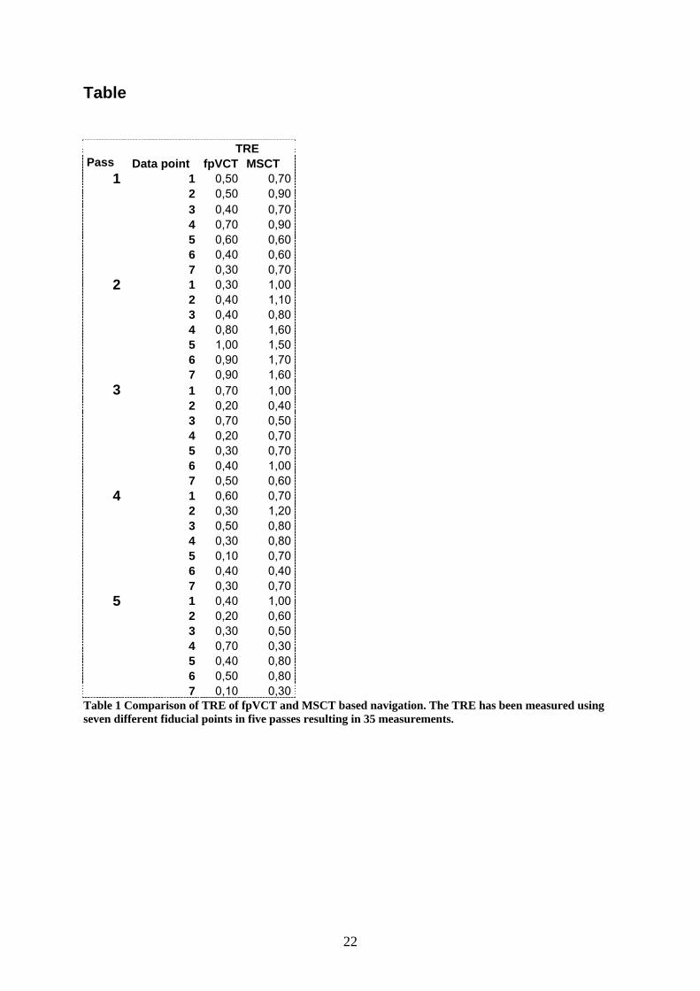
Table TRE Pass Data point fpVCT MSCT
1 0,50 0,702 0,50 0,903 0,40 0,704 0,70 0,905 0,60 0,606 0,40 0,60
1
7 0,30 0,701 0,30 1,002 0,40 1,103 0,40 0,804 0,80 1,605 1,00 1,506 0,90 1,70
2
7 0,90 1,601 0,70 1,002 0,20 0,403 0,70 0,504 0,20 0,705 0,30 0,706 0,40 1,00
3
7 0,50 0,601 0,60 0,702 0,30 1,203 0,50 0,804 0,30 0,805 0,10 0,706 0,40 0,40
4
7 0,30 0,701 0,40 1,002 0,20 0,603 0,30 0,504 0,70 0,305 0,40 0,806 0,50 0,80
5
7 0,10 0,30Table 1 Comparison of TRE of fpVCT and MSCT based navigation. The TRE has been measured using seven different fiducial points in five passes resulting in 35 measurements.