incidental findings on brain magnetic resonance imaging
TRANSCRIPT
Incidental Findings on Brain Magnetic Resonance Imaging ofChildren with Sickle Cell Disease
Lori C. Jordan, M.D., Ph.D.,Department of Neurology Johns Hopkins University School of Medicine 200 N. Wolfe St, Suite2158 Baltimore, MD 21287 [email protected]
Robert C. McKinstry III, M.D., Ph.D.,Department of Radiology Washington University School of Medicine St Louis Children’s HospitalSt Louis, MO 63110 [email protected]
Michael A. Kraut, M.D., Ph.D.,Department of Radiology Johns Hopkins University School of Medicine 600 N. Wolfe St,Baltimore, MD 21287 [email protected]
William S. Ball, M.D.,Department of Radiology Cincinnati Children’s Medical Center 3333 Burnet Avenue Cincinnati,OH 45229 [email protected]
Bruce A. Vendt, B.S., MBA,Electronic Radiology Laboratory, Mallinckrodt Institute of Radiology, Washington UniversitySchool of Medicine 510 S. Kingshighway St. Louis, MO, 63110 [email protected]
James F. Casella, M.D.,Department of Pediatrics Johns Hopkins University School of Medicine Ross Building 1125 720Rutland Avenue Baltimore, MD 21205 [email protected]
Michael R. DeBaun, M.D., M.P.H., andDepartment of Pediatrics Washington University School of Medicine St Louis Children’s HospitalSt Louis, MO 63110 [email protected]
John J. Strouse, M.D., Ph.D.*Department of Pediatrics Johns Hopkins University School of Medicine Ross Building 1125 720Rutland Avenue Baltimore, MD 21205
AbstractObjective—Incidental findings identified by MRI of the brain have been reported in up to18% ofhealthy adults, with clinically significant neuropathology in 0.5-2%. There are two smaller seriesof incidental findings on MRI of the brain in children. We describe the prevalence and range ofincidental intracranial abnormalities identified by MRI of the brain in a large group of childrenscreened for a clinical trial.
Methods—We included 953 children between 5 and 14 years of age screened with MRI of thebrain for the Silent Infarct Transfusion Trial. All have sickle cell anemia or sickle β-nullthalassemia. MRIs were interpreted by 3 neuroradiologists. MRIs reported to have any
*Corresponding author [email protected] Phone: 410-955-6132 Fax: 410-955-8208 .Financial Disclosures: NoneConflicts of Interest: NoneFor the SIT Trial Investigators (see appendix for full list).
NIH Public AccessAuthor ManuscriptPediatrics. Author manuscript; available in PMC 2011 August 10.
Published in final edited form as:Pediatrics. 2010 July ; 126(1): 53–61. doi:10.1542/peds.2009-2800.
NIH
-PA Author Manuscript
NIH
-PA Author Manuscript
NIH
-PA Author Manuscript

abnormality were re-reviewed by 2 study neuroradiologists. Incidental findings were classifiedinto 4 categories: no referral, routine, urgent, and immediate referral recommended. Cerebralinfarcts and vascular lesions were not considered incidental and were excluded.
Results—We identified 63 children (6.6%, 95% CI: 5.1 to 8.4%) with 68 incidental intracranialMRI findings. Findings were classified as urgent in 6 children (0.6%), routine in 25 children(2.6%), and no referral required in 32 children (3.4%). No children required immediate referral.Two children with urgent findings underwent surgery in the next 6 months.
Conclusions—In this large cohort of children, incidental intracranial findings were identified in6.6%, with potentially serious or urgent findings in 0.6%. These data should assist pediatriciansand researchers in planning for and counseling families when unexpected, incidental findings areencountered on MRI of the brain.
KeywordsIncidental Findings; MRI-Magnetic resonance imaging; Brain imaging; Children; Sickle CellDisease
IntroductionIncidental findings identified by MRI of the brain have been reported in up to 18% ofhealthy adults1 with clinically significant neuropathology identified in 0.5-2% of healthyadult volunteers.1, 2 Population-based studies of older adults have found “urgent” incidentalfindings on MRI of the brain, primarily aneurysms and benign or low-grade tumors, in1.7%3 and 3.4%.4 A recently published meta-analysis of incidental findings in 16 primarilyadult research studies found an overall prevalence of 2.7% for all incidental findings onbrain MRI and 4.3% prevalence using high resolution MRI sequences.5
The largest series of incidental MRI of the brain findings in pediatric patients included 225healthy children recruited for multiple neuroimaging studies at a large academic center.6Incidental abnormalities were found in 21%, for a total of 47 children. Within this group,acute or chronic sinusitis represented 57% (27 of 47) of the incidental findings. Excludingextracranial findings such as sinusitis, 20 (8.9%) of these healthy children had incidentalintracranial abnormalities. Just one child (0.4%) had a lesion deemed potentially seriousenough to warrant urgent referral for evaluation. These adult and pediatric studies used astandardized method of classifying the significance of incidental MRI findings. A secondpediatric study reported incidental MRI findings in a series of 60 children, but did not use astandardized method to classify incidental findings.7 The authors reported that 8 (13%)children had incidental findings and only 3 findings (5%) were thought to require “furtherevaluation.” These findings were a 3-mm focus of increased fluid-attenuated inversionrecovery (FLAIR) signal in the parietal white matter, a subcentimeter focus of T2 andFLAIR hyperintensity in the left cerebellar hemisphere, and prominent flow voids in thepineal region suggesting a possible vascular malformation. With detailed follow-up imaging,the first lesion was an artifact, the second remained unchanged over 2 years; in the thirdparticipant, cerebral blood vessels were felt to be normal with no vascular malformationpresent.
In the current study, 953 children with hemoglobin (Hb) SS or sickle β-null thalassemia hadan MRI of the brain to screen them for inclusion in the Silent Infarct Transfusion (SIT)Trial. Children with an infarct seen on screening MRI who chose to continue participating inthe study were examined by a study neurologist blinded to their MRI findings to confirmthat the infarct was clinically silent. The prevalence of incidental MRI findings in sickle celldisease (SCD) is unknown and data regarding incidental findings in all children is limited.
Jordan et al. Page 2
Pediatrics. Author manuscript; available in PMC 2011 August 10.
NIH
-PA Author Manuscript
NIH
-PA Author Manuscript
NIH
-PA Author Manuscript
Information from this study will assist neurologists and hematologists in the interpretation ofincidental MRI findings in children with SCD and may be applicable to children in general.
We sought to describe the prevalence and range of incidental intracranial abnormalities onMRI of the brain in children with SCD and to classify their significance based on apreviously published method.8
Patients and MethodsThe SIT Trial is a multi-center randomized clinical trial; details of the study design andmethods have been reported.9,10 See also: www.clinicaltrials.gov identifier NCT00072761.In brief, the major hypothesis is that prophylactic blood transfusion therapy in children withSCD with silent cerebral infarcts will result in at least an 86% reduction in the rate ofsubsequent overt strokes or new or enlarging cerebral infarcts as defined by MRI of thebrain. The presence of a silent cerebral infarct-like lesion can only be established with aMRI of the brain; therefore 953 consecutive children, ages 5 to 14 years, have been screenedwith MRI (without sedation) to date. Children with a history or a finding on neurologicalexam by a pediatric hematologist concerning for overt stroke were ineligible. Of note,findings related to the primary aims of this study (ischemic stroke or vascular disease) wereexcluded from this analysis as they will be reported separately.
MRI ProtocolDetails of the SIT trial MRI Protocol are available in an on-line supplement and in a paper inpress.9 Briefly, the imaging protocol consists of sagittal T1, axial T2, axial and coronal T2FLAIR images, and axial diffusion-weighted images. The vast majority of images wereobtained on 1.5 Tesla scanners with a few centers switching to 3.0 Tesla scanners in 2008.Forty-six of the 953 MRIs were completed on a 3.0 Tesla scanner; none of these 46 childrenhad an incidental MRI finding. No intravenous gadolinium-based contrast material wasadministered.
The SIT Trial protocol for handling incidental MRI findings is as follows: All MRI studiesare read within 24 hours of being uploaded into the study website to allow detection of anyurgent or emergent medical condition that will require follow up at the local site. This initialreading is performed by one assigned study neuroradiologist. This rapid review has beenincorporated into the study protocol in order to accommodate several study sites that areusing a research MRI facility, without a local clinical interpretation. If an immediate orurgent referral is necessary, the neuroradiologist e-mails and telephones either the studyprincipal investigator, or the chair of the neurology committee. One of these physicians willcall and e-mail the local site investigator with the critical medical information. This chain ofcommunication is to keep the neuroradiologists ‘blinded’ to any other clinical conditionsthat may be discussed during the exchange of pertinent clinical information. Disclosure ofother non-urgent incidental findings is made to the site study coordinator and site principalinvestigator who relay the information to the study participant and their family.
Grading of MRI abnormalitiesWe used methods designed for the Cardiovascular Health Study,8 that were utilized in twopublished studies of incidental MRI findings in adults and children.6,11
Four categories were established:
(1) no referral necessary; findings common in asymptomatic subjects (e.g. cavum septumpellucidum)
Jordan et al. Page 3
Pediatrics. Author manuscript; available in PMC 2011 August 10.
NIH
-PA Author Manuscript
NIH
-PA Author Manuscript
NIH
-PA Author Manuscript
(2) routine referral; findings not requiring immediate medical attention (Chiari Imalformation without tonsillar crowding or spinal cord abnormality)
(3) urgent referral recommended within weeks of the study for abnormality that will requirefurther evaluation (e.g. low-grade glioma, Chiari I malformation with tonsillar crowding orsyrinx)
(4) immediate referral recommended (e.g. acute subdural hematoma, lesion with masseffect)
Three study neuroradiologists interpreted brain MRIs in screened children andindependently recorded results. MRIs reported to have any non-vascular abnormality werere-reviewed by two study neuroradiologists (MAK and RCM) masked to earlierinterpretations, and abnormalities were then classified into one of the four above categories.Disagreements about the proper category were resolved by consensus between theneuroradiologists. Chiari I malformation was defined as cerebellar tonsils more than 5 mmbelow the foramen magnum.12,13
Statistical AnalysesWe calculated the Kappa statistic for inter-rater reliability pre-consensus, for the presence orabsence of a specific incidental MRI finding. Using exact methods, we determinedprevalence and 95% confidence intervals (CI) of all incidental intracranial MRIabnormalities in children with SCD and those requiring urgent or routine referral.
We compared the frequency of incidental intracranial MRI findings in the SIT Trial to thefrequency of incidental intracranial MRI findings in the pediatric study of Kim et al.6 and ina German study of young adults2 via a binomial comparison of proportions.
We conducted analyses using STATA 10.0 (College Station, TX) and considered a p-valueof <0.05 significant for all analyses.
The study was approved by the SIT Trial Data and Safety Monitoring Board and the JohnsHopkins Institutional Review Board.
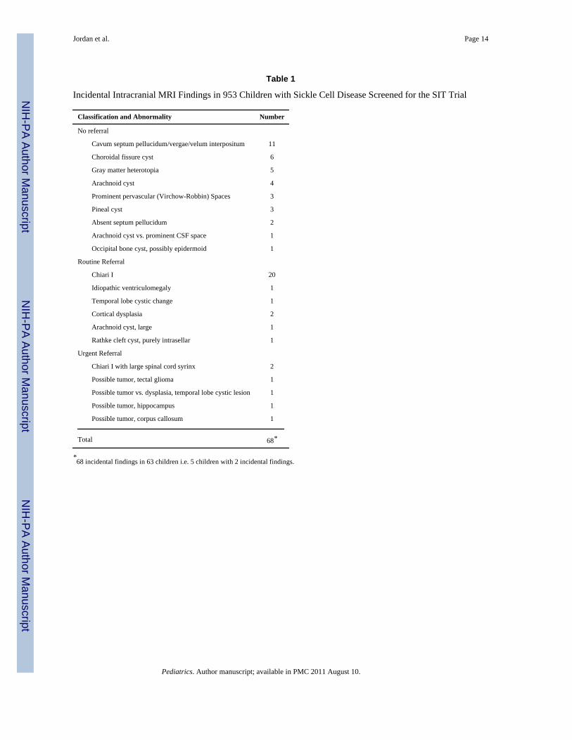
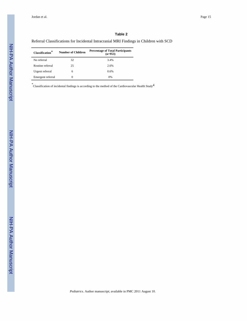
ResultsWe included 953 children screened with MRI of the brain for the SIT trial between February2005 and September 2008. These children were 51.5% male, and 96% of African ancestry,0.5% Caucasian, 0.5% Asian and 3% other race. The median age of the children was 9.1years, with a range of 5.1 to 14.9 years. We identified 6.6% (63 of 953) (95% CI: 5.1 to8.4%) with 68 incidental intracranial MRI findings, re-confirmed by two studyneuroradiologists. (Tables 1 and 2). Five children had two incidental findings each. Chiari Imalformations were the commonest malformation seen, followed by cavum abnormalities.Cortical dysplasia, grey matter heterotopia, and various types of cysts were the other typesof abnormalities seen. Incidental MRI findings were of varying urgency. No child had MRIfindings that were considered to merit immediate attention, i.e. referral to an emergencyroom. Urgent MRI findings were identified in six children (0.6%) (Figure 1). Routinefindings were identified in 25 children (2.6%), and 32 children (3.4%) had MRI findings thatwere considered insignificant, such that no further evaluation or testing was required (Figure2). The Kappa coefficient between neuroradiologists for identifying incidental findings overthe entire study population was 0.918 (95% CI: 0.865 to 0.971) with 99.1% agreement. Inthe 63 children with incidental findings, there was 88.5% agreement for specific findings
Jordan et al. Page 4
Pediatrics. Author manuscript; available in PMC 2011 August 10.
NIH
-PA Author Manuscript
NIH
-PA Author Manuscript
NIH
-PA Author Manuscript
amongst the neuroradiologists. Both values supported excellent agreement between the tworeaders.
The number (chi square 1.1, p=0.29) and type (requiring routine or urgent referral, chisquare 1.5, p=0.22) of incidental findings did not differ by gender.
Incidental MRI Findings and Neurologic SignsAll children who met eligibility criteria for the SIT trial with an MRI of the brain showingan infarct and who agreed to further evaluation to determine eligibility randomized portionof the trial had a standardized neurological examination by study pediatric neurologist. Ofthe 953 screening MRIs performed, 175 children underwent this detailed neurologicalevaluation. Of these 175 children, only 9 also had an incidental MRI finding to allowassessment of whether the incidental MRI finding actually had a clinical correlate onneurological examination. Of these 9 children, two had an abnormal neurologicalexamination. The first child was felt to have mild, non-impairing cognitive-behavioralproblems. The incidental finding on his MRI was a small pineal cyst. The second child had alesion in the splenium of the corpus callosum and had no focal findings on neurologicalexam. The neurologist who examined him was concerned about this patient’s cognitiveperformance, including poor naming and figure copying.
Of the 63 children found to have incidental findings on their screening MRI, urgent MRIfindings were identified in 6 children (0.6%, 95% CI: 0.2 to 1.4%). Two children had ChiariI malformation with spinal cord syrinx (Figure 1a and b). Four children had lesionssuspicious for low grade brain tumor (Figure 1c-f). None of these six children with urgentMRI findings had neurological symptoms that correlated with their lesion. Both childrenwith an incidentally identified Chiari I and cervical spinal cord syrinx were reconfirmed tohave a normal neurological examination prior to neurosurgical decompression of the Chiarimalformation. Two of the four children with lesions suspicious for low grade brain tumorhad follow-up MRI of the brain which did not show any progression of the lesions and haveremained asymptomatic. In two children, follow-up MRI is planned but has not yetoccurred. One of these children is the child mentioned above with a lesion in the splenium ofthe corpus callosum and a normal neurological examination except for concerns regardingcognitive performance including poor figure copying. The second child has a possible tectalglioma, and was reconfirmed to have a normal neurological examination; in particular, eyemovements were normal. Serial neurological assessments and MRI of the brain are plannedfor both children. In this small subset of the study population, no child had findings onneurological exam attributable to the incidental finding.
Specific imaging findingsIncidental cortical dysplasia or gray matter heterotopia were identified in two children andfive children respectively, or 0.7% (95% CI: 0.3 to1.5%) of the study population. Per thestudy intake questionnaire, none of these children had a seizure disorder. Twenty-twochildren (2.3%, 95% CI: 1.4.to 3.4%) were found to have Chiari I malformation.
Comparison to similar studiesUpon evaluating healthy volunteers for research studies in California, Kim et al.6 foundurgent MRI findings in 0.4% (1 of 225) of children compared with 0.6% (6 of 953) ofchildren in this current study, a 0.2% difference in proportions that is not significant (95%CI: −0.8% to 1.1%, p=0.75). Incidental MRI findings requiring routine referral forneurological evaluation were seen in 5.3% (12 of 225) of children in the Kim study and2.6% (25 of 953) of the current study, a 2.7% difference in proportions which wassignificant (95% CI: 0.3 to 5.8%, p=0.03).
Jordan et al. Page 5
Pediatrics. Author manuscript; available in PMC 2011 August 10.
NIH
-PA Author Manuscript
NIH
-PA Author Manuscript
NIH
-PA Author Manuscript

A German study of incidental MRI brain findings in 2,536 young adult male Air Forceapplicants reported that 6.5% (166 of 2536) of young men ( 95% CI: 5.6 to 7.6%) hadabnormal findings;2 however, findings that were considered normal variants were classifiedseparately (cavum vergae, pineal cyst, enlarged Virchow Robin spaces, absent septumpellucidum) When this classification system is applied to our data, incidental findings inchildren screened for the SIT Trial are found in only 4.6%, significantly less than in youngGerman Air Force recruits (risk difference 1.9%, 95% CI: 0.3 to 3.6%, p=0.03).
In this same German study, 1.7% had Chiari I, (95% CI: 1.2 to 2.3%).2 Confidence intervalsoverlap with the current study; therefore the prevalence of Chiari I in these two quitedifferent study populations is not significantly different. The pediatric incidental findingsstudy6 found that only 0.4% of children had tonsillar ectopia, but again this difference wasnot significant when compared to SIT trial data (risk difference 1.9%, 95% CI 0.6 to 3.1%,p=0.07).
DiscussionThis is the largest group of children screened with MRI of the brain to date, with four timesas many children imaged as the next largest study. The prevalence of incidental intracranialMRI findings in these school-age children was 6.6%. Overall, when compared to studies ofchildren and healthy young adults, incidental intracranial findings were slightly lesscommon, though potentially urgent or serious abnormalities were equally common andpresent in 0.6% of children.
We excluded findings such as ischemic or vascular lesions, as these are central to the majoraims of the study and therefore not incidental. The prior pediatric incidental finding study,Kim et al. did not find any ischemic or vascular lesion in their 225 children,6 and we do notconsider them to be normal findings in school age children. In contrast, Weber and Heinzreported 13 (0.5%) young male Air Force applicants (mean age 20.5 years) with vascularabnormalities, including 5 (0.2%) with brain arteriovenous malformations. Importantly,prior studies of incidental finding in children and young adults found no infarct-like lesions.These lesions in children with SCD are pathological findings likely caused by the occlusionof small vessels14 and should not be considered incidental.
A unique aspect of this series is that all children who met eligibility criteria for the SIT trialwith an MRI of the brain showing an infarct and who agreed to enter the randomized portionof the trial after screening underwent both detailed neuroimaging and careful, standardizedneurological examination by a board-certified pediatric neurologist. Of the 953 screeningMRIs performed, 175 children underwent this detailed neurological evaluation as a part ofthe study. Only 9 of the children with these exams also had an incidental MRI finding. Ofthese 9, no children were reported to have neurologic abnormalities corresponding to theirincidental MRI finding. Neurological exams on the 6 children classified as having urgentMRI findings were confirmed as unremarkable or unrelated to the lesion on MRI.
Strengths of this study include the standardized imaging protocol, review of all MRIs of thebrain by three neuroradiologists, and the large study population. Limitations to thegeneralizability of this work include the study population as all have SCD (are not healthychildren), and participants are ethnically restricted as 96% are of African descent. The priorrelatively large pediatric study of incidental MRI findings also suffered from racialhomogeneity, as it included 225 children of primarily European or Asian descent.6Nonetheless, despite the dramatically different populations, both studies suggest aprevalence of approximately 0.5% for incidental findings on MRI of the brain requiringurgent referral for medical evaluation.
Jordan et al. Page 6
Pediatrics. Author manuscript; available in PMC 2011 August 10.
NIH
-PA Author Manuscript
NIH
-PA Author Manuscript
NIH
-PA Author Manuscript
Overall, incidental intracranial findings were slightly less common than reported in otherstudies of children and young adults, even when the same inclusion and exclusion criteriawere applied, as we excluded vascular and ischemic lesions and extracranial abnormalitiessuch as sinusitis. Our study design does not allow us to determine whether the lower numberof incidental MRI findings is related to ethnicity or is perhaps more representative of thetrue prevalence of incidental intracranial MRI findings in children given the larger studypopulation.
Physicians often ask whether a child should be referred for neurological evaluation for anincidental MRI finding. In this study, the MRI classification of incidental findings is basedon a neuroradiologist’s assessment of the potential significance of the finding. Manyfindings, such as gray matter heterotopia and cortical dysplasia may vary in significancebased on the appearance of the individual MRI and the patient history. The risk of seizure ina child with a gray matter heterotopia or cortical dysplasia is unknown.
All of the children in this study underwent an MRI for research purposes. The morecommon clinical situation occurs when a child has an MRI of the brain for a marginalindication, such as uncomplicated headache, and is found to have an MRI abnormality,unrelated to the presenting complaint. Frequently, these children are then subjected tomultiple “follow up” studies to confirm that the incidental MRI finding is indeed benign.Hopefully studies such as this one will aid clinicians as they counsel families. For example,arachnoid cysts are typically benign findings and occurred in at least 0.5% of children in thisstudy and 1.7% in young adults.2
EthicsEthical concerns have been raised over the last several years about the proper disclosure ofincidental abnormalities found in healthy volunteers for research studies.11,15,16 There iscurrently no consensus, despite an NIH workshop to discuss these issues in January, 2005.17
A recent paper, based on this workshop, reviews practical approaches to handling incidentalfindings in brain imaging research in various settings.18
A survey of healthy volunteers who had participated in brain imaging studies found that97% of these volunteers wanted any abnormalities disclosed to them, regardless of clinicalsignificance.11 There is no consensus in the medical or research community that disclosureof findings that are minor or even “normal variant” such as a cavum septum pellucidum is inthe patient’s or research participant’s best interest. In the SIT Trial, incident findinginformation is provided rapidly to the local study coordinator and site principal investigatorfor disclosure to the patient. This system and the fact that every MRI had a prompt clinicalreading by an expert neuroradiologist are strengths of the SIT Trial protocol. Incidentalfindings may lead to additional studies with significant cost to the healthcare system andpotentially to the study participant’s family. The potential risk of identifying an incidentalfinding is included in the consent form for the study.
ConclusionThis series of 953 school-age children is the largest reported pediatric study of incidentalMRI of the brain findings. Incidental intracranial findings were identified in 6.6% withpotentially serious or urgent findings in 0.6%. Incidental findings on MRI of the brain arerelatively common in children and most are benign. The clinical implications of many ofthese findings are unclear, resulting in limited recommendations to parents. Given theexisting data, we recommend routine referral to a pediatric neurologist for findings that arecurrently asymptomatic and are of uncertain significance, (such as a migrational abnormalityseen on MRI that may predispose to epilepsy, in a child who has never had a seizure). In
Jordan et al. Page 7
Pediatrics. Author manuscript; available in PMC 2011 August 10.
NIH
-PA Author Manuscript
NIH
-PA Author Manuscript
NIH
-PA Author Manuscript

addition to its clinical applications, this work may also be useful for investigators using MRIin children who must plan for incidental findings in their studies based on estimatedprevalence.
Supplementary MaterialRefer to Web version on PubMed Central for supplementary material.
AcknowledgmentsThe SIT Trial was funded by NINDS U01-NS-042804. LCJ was supported by NINDS K23NS062110. JJS wassupported by NHLBI K23HL078819, the Doris Duke Charitable Foundation and the American Society ofHematology.
Abbreviations
MRI Magnetic Resonance Imaging
SIT (Silent Infarct Transfusion ) Trial
CI confidence interval
References1. Katzman GL, Dagher AP, Patronas NJ. Incidental findings on brain magnetic resonance imaging
from 1000 asymptomatic volunteers. JAMA. 1999; 282(1):36–39. [PubMed: 10404909]2. Weber F, Knopf H. Incidental findings in magnetic resonance imaging of the brains of healthy
young men. J Neurol Sci. 2006; 240(1-2):81–84. [PubMed: 16256141]3. Yue NC, Longstreth WT Jr, Elster AD, Jungreis CA, O’Leary DH, Poirier VC. Clinically serious
abnormalities found incidentally at MR imaging of the brain: Data from the cardiovascular healthstudy. Radiology. 1997; 202(1):41–46. [PubMed: 8988190]
4. Vernooij MW, Ikram MA, Tanghe HL, et al. Incidental findings on brain MRI in the generalpopulation. N Engl J Med. 2007; 357(18):1821–1828. [PubMed: 17978290]
5. Morris Z, Whiteley WN, Longstreth WT Jr, et al. Incidental findings on brain magnetic resonanceimaging: systematic review and meta-analysis. BMJ. 2009; 339:b3016. [PubMed: 19687093]
6. Kim BS, Illes J, Kaplan RT, Reiss A, Atlas SW. Incidental findings on pediatric MR images of thebrain. AJNR Am J Neuroradiol. 2002; 23(10):1674–1677. [PubMed: 12427622]
7. Kumra S, Ashtari M, Anderson B, Cervellione KL, Kan L. Ethical and practical considerations inthe management of incidental findings in pediatric MRI studies. J Am Acad Child AdolescPsychiatry. 2006; 45(8):1000–1006. [PubMed: 16865043]
8. Bryan RN, Manolio TA, Schertz LD, et al. A method for using MR to evaluate the effects ofcardiovascular disease on the brain: The cardiovascular health study. AJNR Am J Neuroradiol.1994; 15(9):1625–1633. [PubMed: 7847205]
9. Casella JF, King AA, Barton B, White SA, Noetzel MJ, Ichord RN, Terrill C, Hirtz D, McKinstryRC, Strouse JJ, Howard TH, Coates TD, Minnitti CP, Vendt BA, Lehmann H, Debaun MR. Designof the Silent Infarct Transfusion (SIT) Trial. Pediatric Hematology Oncology. in press.
10. Vendt BA, McKinstry RC, Ball WS, et al. Silent Cerebral Infarct Transfusion (SIT) trial imagingcore: application of novel imaging information technology for rapid and central review of MRI ofthe brain. J Digit Imaging. 2008; 22(3):326–343. [PubMed: 18398653]
11. Kirschen MP, Jaworska A, Illes J. Subjects’ expectations in neuroimaging research. J Magn ResonImaging. 2006; 23(2):205–209. [PubMed: 16416438]
12. Aboulezz AO, Sartor K, Geyer CA, Gado MH. Position of cerebellar tonsils in the normalpopulation and in patients with chiari malformation: A quantitative approach with MR imaging. JComput Assist Tomogr. 1985; 9(6):1033–1036. [PubMed: 4056132]
Jordan et al. Page 8
Pediatrics. Author manuscript; available in PMC 2011 August 10.
NIH
-PA Author Manuscript
NIH
-PA Author Manuscript
NIH
-PA Author Manuscript
13. Barkovich AJ, Wippold FJ, Sherman JL, Citrin CM. Significance of cerebellar tonsillar position onMR. AJNR Am J Neuroradiol. 1986; 7(5):795–799. [PubMed: 3096099]
14. Koshy M, Thomas C, Goodwin J. Vascular lesions in the central nervous system in sickle celldisease (neuropathology). J Assoc Acad Minor Phys. 1990; 1(3):71–78. [PubMed: 2136620]
15. Illes J, Kirschen MP, Edwards E, et al. Ethics. incidental findings in brain imaging research.Science. 2006; 311(5762):783–784. [PubMed: 16469905]
16. Check E. Brain-scan ethics come under the spotlight. Nature. 2005; 433(7023):185. [PubMed:15662383]
17. “Detection and disclosure of incidental findings in neuro-imaging research” workshop. Bethesda,MD: January 6-7. 2005 www.ninds.nih.gov/news_and events/proceedings/ifexecsummary.htm.http://www.ninds.nih.gov/news_and_events/proceedings/ifexecsummary.htm
18. Illes J, Kirschen MP, Edwards E, et al. Practical approaches to incidental findings in brain imagingresearch. Neurology. 2008; 70:384–390. [PubMed: 18227420]
Jordan et al. Page 9
Pediatrics. Author manuscript; available in PMC 2011 August 10.
NIH
-PA Author Manuscript
NIH
-PA Author Manuscript
NIH
-PA Author Manuscript

Jordan et al. Page 10
Pediatrics. Author manuscript; available in PMC 2011 August 10.
NIH
-PA Author Manuscript
NIH
-PA Author Manuscript
NIH
-PA Author Manuscript
Jordan et al. Page 11
Pediatrics. Author manuscript; available in PMC 2011 August 10.
NIH
-PA Author Manuscript
NIH
-PA Author Manuscript
NIH
-PA Author Manuscript

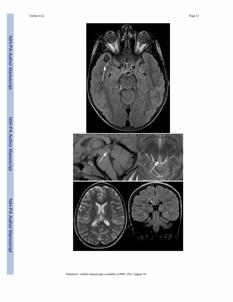
Figure 1(a-f).Urgent Incidental MRI Findings.1a) Sagittal T1 view of an incidental Chiari I malformation (arrow head) with cervical spinalcord syrinx (arrow).1b) Sagittal T1 view of an incidental Chiari I malformation (arrow head) with cervical spinalcord syrinx containing plicae or septations (arrow).1c) Axial FLAIR image with an incidentally detected cystic lesion in the right temporal lobethat could represent a tumor. Arrows point to the cyst and an area of nodularity within thecyst.1d) T1 sagittal and T2 axial views of an incidental tectal lesion, possible glioma (arrows).1e) Axial T2 and coronal FLAIR images of an incidental corpus callosum lesion, potentiallyrepresenting a tumor or an infarct (arrows).1f) Axial FLAIR image of a left hippocampal cystic lesion (arrow), possible tumor versus afocus of dysplasia.
Jordan et al. Page 12
Pediatrics. Author manuscript; available in PMC 2011 August 10.
NIH
-PA Author Manuscript
NIH
-PA Author Manuscript
NIH
-PA Author Manuscript
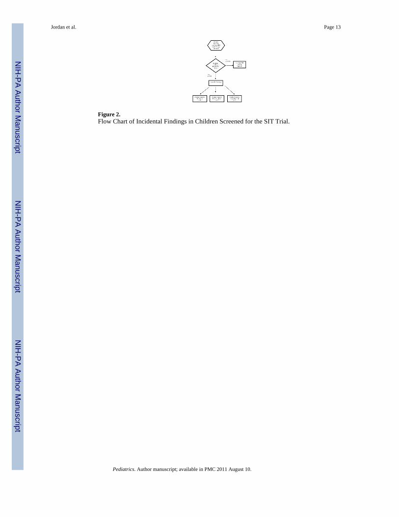
Figure 2.Flow Chart of Incidental Findings in Children Screened for the SIT Trial.
Jordan et al. Page 13
Pediatrics. Author manuscript; available in PMC 2011 August 10.
NIH
-PA Author Manuscript
NIH
-PA Author Manuscript
NIH
-PA Author Manuscript
NIH
-PA Author Manuscript
NIH
-PA Author Manuscript
NIH
-PA Author Manuscript
Jordan et al. Page 14
Table 1
Incidental Intracranial MRI Findings in 953 Children with Sickle Cell Disease Screened for the SIT Trial
Classification and Abnormality Number
No referral
Cavum septum pellucidum/vergae/velum interpositum 11
Choroidal fissure cyst 6
Gray matter heterotopia 5
Arachnoid cyst 4
Prominent pervascular (Virchow-Robbin) Spaces 3
Pineal cyst 3
Absent septum pellucidum 2
Arachnoid cyst vs. prominent CSF space 1
Occipital bone cyst, possibly epidermoid 1
Routine Referral
Chiari I 20
Idiopathic ventriculomegaly 1
Temporal lobe cystic change 1
Cortical dysplasia 2
Arachnoid cyst, large 1
Rathke cleft cyst, purely intrasellar 1
Urgent Referral
Chiari I with large spinal cord syrinx 2
Possible tumor, tectal glioma 1
Possible tumor vs. dysplasia, temporal lobe cystic lesion 1
Possible tumor, hippocampus 1
Possible tumor, corpus callosum 1
Total 68*
*68 incidental findings in 63 children i.e. 5 children with 2 incidental findings.
Pediatrics. Author manuscript; available in PMC 2011 August 10.
NIH
-PA Author Manuscript
NIH
-PA Author Manuscript
NIH
-PA Author Manuscript
Jordan et al. Page 15
Table 2
Referral Classifications for Incidental Intracranial MRI Findings in Children with SCD
Classification* Number of Children Percentage of Total Participants(n=953)
No referral 32 3.4%
Routine referral 25 2.6%
Urgent referral 6 0.6%
Emergent referral 0 0%
*Classification of incidental findings is according to the method of the Cardiovascular Health Study4
Pediatrics. Author manuscript; available in PMC 2011 August 10.