in vivo induction of tight junction proliferation in rat liver
TRANSCRIPT

I N V I V O I N D U C T I O N O F T I G H T J U N C T I O N
P R O L I F E R A T I O N I N R A T L I V E R
ROBERTO MONTESANO, GIULIO GABBIANI, ALAIN PERRELET, and LELIO ORCI. From the Institut d'Histologie et d'Embryologie et D6partement de Pathologie, Universit6 de Gen~:ve, 1211, Gen~:ve 4, Switzerland
The tight junction (zonula occludens) (for review, see 9 and 14) is considered by some authors (15) to be composed of a series of single fibrils shared by the plasma membrane of adjacent cells and to contribute to cell attachment and selective closure of the intercellular space (2, 3, 4). Several experi- ments suggest that tight junctions undergo rapid configurational changes in various physiological or experimental conditions (7, 12, 13, 16). The factors involved in the assembly and modulation of tight junctions as well as in the regulation of their
spatial configuration are so far completely un- known. With freeze-fracture, we report here an- other experimental model in which an extensive development and rearrangement of tight junctions is produced in the liver by the chronic administra- tion of small doses of phalloidin, the toxic principle of the mushroom A manita phalloides.
MATERIALS AND METHODS
Male Wistar rats weighing approx. 140 g, fed ad libitum on Purina chow (Ralston Purina Co., St. Louis, Mo.)
THE JOURNAL OF CELL BIOLOGY . VOLUME 68, 1976 . p a g e s 7 9 3 - 7 9 8 793
and tap water, were given intraperitoneally phailoidin, a bicyclic peptide of A manita phaltoides (17), once daily at a dose of 500 ag/kg body weight in 0.5 ml of NaCI 0.9%. Control animals received the solvent alone. Treated and control animals were sacrified after 0.5, 1, 6, and 12 h and I, 2, 4, 8, and 13 days. The livers were fixed by perfusion through the portal vein with a 1.5% glutaralde- hyde solution in cacodylate buffer, pH 7.2 for 10 min, followed by immersion of the tissue in the same fixative for at least 15 min. Small pieces of the fixed tissue were immersed in 30% glycerol in cacodylate buffer for at least 30 min, rapidly frozen in Freon 22 (E. I. du Pont de Nemours & Co., Inc., Wilmington, Del.), cooled in liquid nitrogen, fractured, and shadowed in a Balzers BAF 301 device (Balzers AG, Balzers, Liechtenstein) according to the technique of Moor and MUhlethaler (il). After thawing of the tissue, the replicas w e r e
cleaned in a sodium hypochl0rite solution for 2 h, soaked overnight in dimethylformamide to dissolve fat, rinsed in distilled water and mounted on copper grids. Repli- cas were examined in a Philips EM 300 electron micro- scope.
R E S U L T S
In fracture faces of plasma membrane of control hepatocytes, tight junctions appear as a network of branching and anastomosing ridges on the A face and complementary furrows on the B face (1, 3, 6, 8), in the vicinity of the bile canaliculus (Fig. !). Most of the tight junctional elements (ridges or furrows) are usually parallel to the lumen of the canaliculus and, under normal conditions, their abluminal extension is very limited since they occupy only a narrow band of membrane on each side of the bile canaliculus. Fracture faces of the hepatocyte plasma membranes from rats treated for 2-13 days with phalloidin show striking changes in the extension and configuration of the pericanalicular tight junctional networks. 1 The junctional elements lose their predominantly par-
1 As judged by qualitative survey of sections for light and electron microscopy, the liver architecture and hepato-
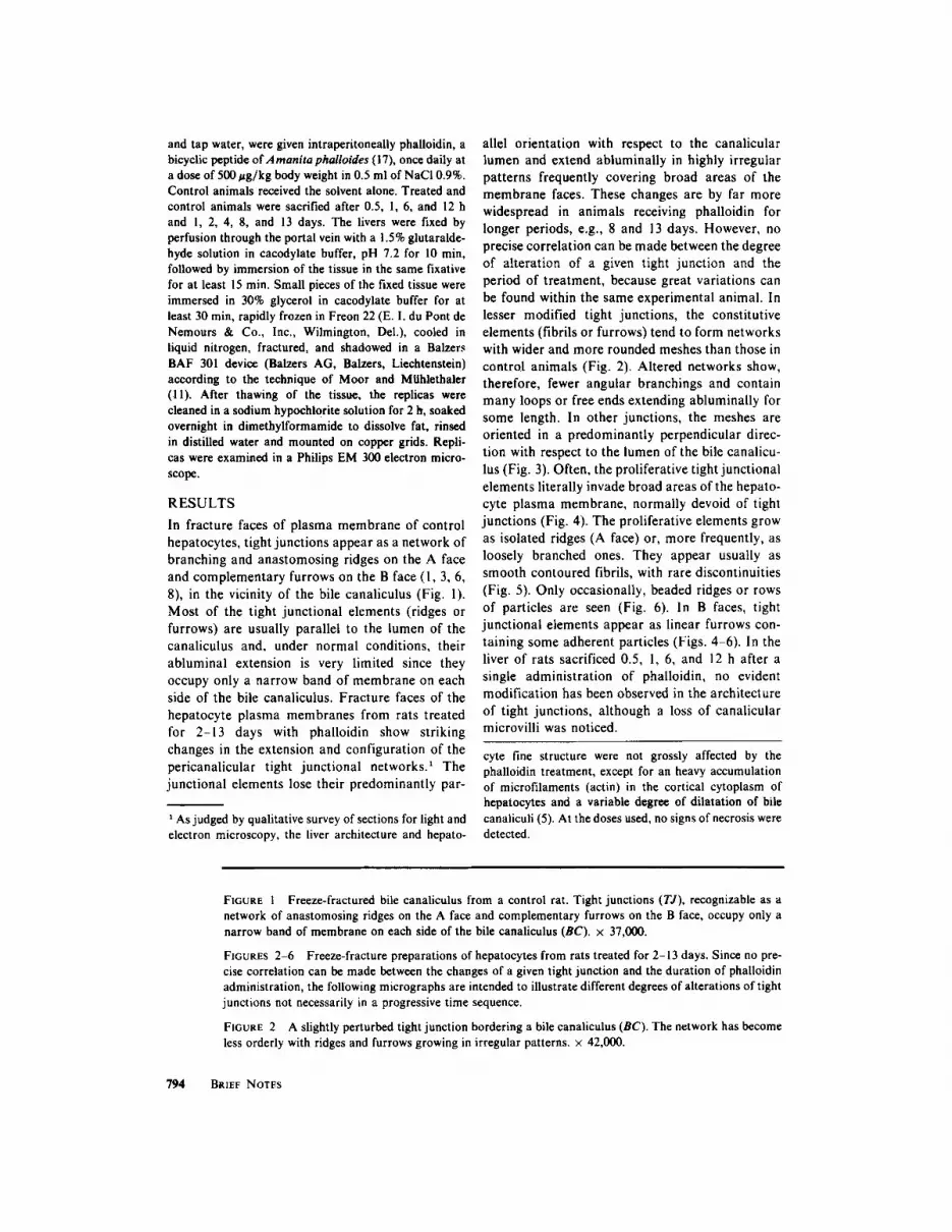
allel orientation with respect to the canalicular lumen and extend abluminaily in highly irregular patterns frequently covering broad areas of the membrane faces. These changes are by far more widespread in animals receiving phalloidin for longer periods, e.g., 8 and 13 days. However, no precise correlation can be made between the degree of alteration of a given tight junction and the period of treatment, because great variations can be found within the same experimental animal. In lesser modified tight junctions, the constitutive elements (fibrils or furrows) tend to form networks with wider and more rounded meshes than those in control animals (Fig. 2). Altered networks show, therefore, fewer angular branchings and contain many loops or free ends extending abluminally for some length. In other junctions, the meshes are oriented in a predominantly perpendicular direc- tion with respect to the lumen of the bile canalicu- lus (Fig. 3). Often, the proliferative tight junctional elements literally invade broad areas of the hepato- cyte plasma membrane, normally devoid of tight junctions (Fig. 4). The proliferative elements grow as isolated ridges (A face) or, more frequently, as loosely branched ones. They appear usually as smooth contoured fibrils, with rare discontinuities (Fig. 5). Only occasionally, beaded ridges or rows of particles are seen (Fig. 6). In B faces, tight junctional elements appear as linear furrows con- taining some adherent particles (Figs. 4-6). In the liver of rats sacrificed 0.5, !, 6, and 12 h after a single administration of phalloidin, no evident modification has been observed in the architecture of tight junctions, although a loss of canalicular microvilli was noticed.
cyte fine structure were not grossly affected by the phalloidin treatment, except for an heavy accumulation of microfilaments (actin) in the cortical cytoplasm of hepatocytes and a variable degree of dilatation of bile canaliculi (5). At the doses used, no signs of necrosis were detected.
FIGURE 1 Freeze-fractured bile canaliculus from a control rat. Tight junctions (TJ), recognizable as a network of anastomosing ridges on the A face and complementary furrows on the B face, occupy only a narrow band of membrane on each side of the bile canaliculus (BC). • 37,000.
FIGURES 2-6 Freeze-fracture preparations of hepatocytes from rats treated for 2-13 days. Since no pre- cise correlation can be made between the changes of a given tight junction and the duration of phalloidin administration, the following micrographs are intended to illustrate different degrees of alterations of tight junctions not necessarily in a progressive time sequence.
FIGURE 2 A slightly perturbed tight junction bordering a bile canaliculus (BC). The network has become less orderly with ridges and furrows growing in irregular patterns. • 42,000.
794 BRIEF NOTES

795

FIGURE 3 The junctional network extends abluminaUy with loose meshes oriented in a predominantly perpendicular direction with respect to the lumen of the bile canaliculus (BC). Many loops and free ends are present. Several diminutive aggregates of particles (arrows), suggesting small gap junctions, are visible in the tight junctional domain. • 31,000
DISCUSSION
The results of the present investigation indicate that the chronic administration of relatively small amounts of phalloidin induces an extensive devel-
opment of tight junctions between rat hepatocytes. By which mechanism(s) phalloidin acts on tight junctions cannot be stated at present. From our images, however, it seems likely that tight junc- tional changes occur through both proliferation
796 BgIEF NOTES
FIGURE 4 The junctional strands (ridges on A face, furrows on B face) spread on the membrane far from the bile canaliculus in highly irregular patterns. Ridges on A face are mostly continuous, but in some places they appear as discontinuous strands. • 31,000. The area outlined by the black square is shown at higher magnification in Fig. 5.
FIGURE 5 A discontinuous tight junctional strand appears to be composed of short segments of smooth- surfaced ridges (arrowheads) alternating with rows of individual particles (arrows). • 61,000.
FIGURE 6 A- to B-fracture face transition showing rows of particles on the A face in register (arrow) with furrows on the B face. x 55,000.
(addition of newly formed junctional elements) and reorganization of preexistent junctional strands.
As far as the fine morphology of the tight junction proliferation is concerned, we observed differences between the present model and another
system in which such junctions are assembled, namely the fetal rat liver (10). In the latter, tight junctions appear to arise by alignment and subse- quent fusion of intramembranous particles into beaded ridges, which in turn become confluent and
BRIEF NOTES 797

transform into smooth ones. Such patterns of intramembranous particles were not found fre- quently in phalloidin-treated hepatocytes. This should, however, not necessarily indicate that the formation of new fibrils in this case occurs by a process other than particle alignment and fusion, as proposed for fetal rat liver. The event may be simply more difficult to visualize. One must con- sider, for example, that we are witnessing the growth of a preformed junction and not its de novo
formation. If the outgrowth of preexisting fibrils proceeds, for example, through the peripheral addition of single or of a few particles which rapidly become confluent with the smooth-con- toured fibrils, the overall process of assembly could not be readily discerned.
Further studies are needed to clarify the possible relat ionships between phal loidin- induced mi- crofilaments (5) and tight junction proliferation as well as to establish whether the described changes are a direct or indirect effect of phalloidin treat- ment.
S U M M A R Y
The chronic administration of phalloidin induces an extensive development of tight junctions be- tween rat hepatocytes. The junctional strands lose their predominantly parallel orientation with re- spect to the canalicular lumen and extend ablumi- nally in irregular patterns which cover large mem- brane areas at considerable distance from the bile canaliculi. These changes indicate both prolifera- tion and reorganization of the junctional elements and provide further evidence that these junctions are not permanent differentiations of the cell membrane.
We are grateful to Professor Theodor Wieland, Max Planck Institut, Heidelberg, Germany for generously providing us with phalloidin. We thank P. Fruleux, M. Bernard, J. Rial, and M. Sidler-Ansermet for technical assistance.
These studies were supported by grants no 3.553.75 and 3.0330.73 from the Foods National Suisse de la Recherche Scientifique.
Received for publication 14 July 1975, and in revised form 22 September 1975.
R E F E R E N C E S
1. CHALCROFT, J. P., and S. BULLIVANT. 1970. An interpretation of liver cell membrane and junction structure based on observation of freeze-fracture
replicas of both sides of the fracture. J. Cell Biol. 47:49-60.
2. CLAUDE, P., and D. A. GOODENOUGH. 1973. Frac- ture faces of zonulae occludentes from "tight" and "leaky" epithelia. J. Cell Biol. 58:390-400.
3. FRIEND, D. S., and N. B. GILULA. 1972. Variations in tight and gap junctions in mammalian tissues. J. Cell Biol. 53:758-776.
4. FROMTER, E., and J. DIAMOND. 1972. Route of passive ion permeation in epithelia. Nat. New Biol. 235:9 - 13.
5. GABBIANI, G., R. MONTESANO, B. TUCHWEBER, M. SALAS, and L. ORO. 1975. Phalloidin-induced hy- perplasia of actin filaments in rat hepatocytes. Lab. Invest. 33:562-569.
6. GOODENOUGH, D. A., and J. P. REVEL. 1970. A fine structural analysis of intercellular junctions in the mouse liver. J. Cell Biol. 45:272 290.
7. HUMBERT, F., A. GRANDCHAMP, C. PRICAM, A. PERRELET, and L. ORCI. 1975. The role of tight junctions in regulating the intercellular sodium back- flux across the proximal tubule of Necturus maculo- sus kidney. Kidney Int. 7:365 (Abstr.).
8. KREUTZIGER, G. O. 1968. Freeze-etching of intercel- lular junctions of mouse liver. Proceedings of the twenty-sixth Electron Microscopy Society of Amer- ica. Claitor's Publishing Division, Baton Rouge, La.
9. MCNUTT, N. S., and R. S. WEINSTEIN. 1973. Membrane ultrastructure at mammalian intercellu- lar junctions. Progr. Bioph. Mol. Biol. 26:42-101.
10. MONTESANO, R., D. S. FRIEND, A. PERRELET, and L. ORCl. 1975. In vivo assembly of tight junctions in fetal rat liver, d. Cell Biol. 67:310-319.
I I. MooR, H., and K. MOHLETHALER. 1963. Fine structure in frozen-etched yeast cells. J. Cell Biol. 17:609-628.
12. ORCI, L., M. AMHERDT, J. C. HENQUIN, A. E. LAMBERT, R. H. UNGER, and A. E. RENOLD. 1973. Pronase effect on pancreatic beta cells secretion and morphology. Science (Wash. D. C.). 180:647-649.
13. PITELKA, D. R., S. T. HAMAMOTO, J. G. DUAFALA, and M. K. NEMANIC. 1973. Cell contacts in the mouse mammary gland. I. Normal gland in postna- tal development and the secretory cycle. J. Cell Biol. 56:797-818.
14. STAEHELIN, L. A. 1974. The structure and function of intercellular junctions. Int. Rev. Cytol. 39:191-283.
15. WADE, J. B., and M. J. KARNOVSKY. 1974. The structure of the zonula occlodens. A single fibril model based on freeze-fracture. J. Cell Biol. 60:168-180.
16. WADE, J. B., and M. J. KARNOVSKV. 1974. Fracture faces of osmotically disrupted zonulae occludentes. J. Cell Biol. 62:344-350.
17. WIELAND, TH., and O. WIELAND. 1972. The toxic peptids of amanita species. Microbial Toxins. 8:249-280.
798 BRIEF NOTES