in vivo comparison of epithelial responses for s-8 versus jp-8 jet fuels below permissible exposure...
TRANSCRIPT
IN VIVO COMPARISON OF EPITHELIAL RESPONSES FOR S-8VERSUS JP-8 JET FUELS BELOW PERMISSIBLE EXPOSURELIMIT
Simon S. Wong1, Jason Vargas1, Alana Thomas1, Cindy Fastje1, Michael McLaughlin1,Ryan Camponovo1, R. Clark Lantz2, Jeffrey Heys3, and Mark L. Witten11 Department of Pediatrics, Southwest Environmental Health Sciences Center, The University ofArizona, Tucson, Arizona, U.S.A.2 Department of Cell Biology & Anatomy, Southwest Environmental Health Sciences Center, TheUniversity of Arizona, Tucson, Arizona, U.S.A.3 Chemical Engineering Department, Arizona State University, Tempe, Arizona, U.S.A.
AbstractThis study was designed to characterize and compare the pulmonary effects in distal lung from alow-level exposure to jet propellant-8 fuel (JP-8) and a new synthetic-8 fuel (S-8). It ishypothesized that both fuels have different airway epithelial deposition and responses.Consequently, male C57BL/6 mice were nose-only exposed to S-8 and JP-8 at averageconcentrations of 53 mg/m3 for 1 hour/day for 7 days. A pulmonary function test performed 24 hrafter the final exposure indicated that there was a significant increase in expiratory lung resistancein the S-8 mice, whereas JP-8 mice had significant increases in both inspiratory and expiratorylung resistance compared to control values. Neither significant S-8 nor JP-8 respiratorypermeability changes were observed compared to controls, suggesting no loss of epithelial barrierintegrity. Morphological examination and morphometric analysis of airway tissue demonstratedthat both fuels showed different patterns of targeted epithelial cells: bronchioles in S-8 and alveoli/terminal bronchioles in JP-8. Collectively, our data suggest that both fuels may have partiallydifferent deposition patterns, which may possibly contribute to specific different adverse effects inlung ventilatory function.
KeywordsJet fuel; Inhalation; Epithelial injury; Morphometry; Respiratory permeability; Lung resistance
INTRODUCTIONJet propellant-8 fuel (JP-8) is currently the primary fuel for the U.S. military and NATO(North Atlantic Treaty Organization) forces where it is used to fuel jet aircraft, tanks,fighting vehicles, ships, helicopters, and portable heating/air conditioning units. It is
Corresponding Authors: Simon S. Wong, M.D., MPH; Mark L. Witten, Ph.D., Department of Pediatrics, University of ArizonaCollege of Medicine, Tucson, AZ, 85724, U.S.A. Phone: 520-626-2610, Fax: 520-626-4993, [email protected]'s Disclaimer: This is a PDF file of an unedited manuscript that has been accepted for publication. As a service to ourcustomers we are providing this early version of the manuscript. The manuscript will undergo copyediting, typesetting, and review ofthe resulting proof before it is published in its final citable form. Please note that during the production process errors may bediscovered which could affect the content, and all legal disclaimers that apply to the journal pertain.
NIH Public AccessAuthor ManuscriptToxicology. Author manuscript; available in PMC 2010 August 24.
Published in final edited form as:Toxicology. 2008 December 5; 254(1-2): 106–111. doi:10.1016/j.tox.2008.09.018.
NIH
-PA Author Manuscript
NIH
-PA Author Manuscript
NIH
-PA Author Manuscript
estimated that over 2 million people worldwide are exposed to 60 billion gallons of JP-8annually. However, the problematic long-term supplies of oil and increasing knowledge ofhealth effects have recently spurred the U.S. military to develop a new synthetic-8 fuel(S-8), an alternative to JP-8. S-8 is derived from synthetic gas through the Fischer-Tropsch(FT) synthetic fuel process (Personal communication with Tim Edwards, Air Force FuelLaboratory, Wright-Patterson AFB, Ohio, U. S. A.). Currently, S-8 has been certified by theU. S. Department of Defense for use in B-52H Stratofortress aircraft when blended withJP-8 and possibly support vehicles or other aircraft in the future. It is reported that S-8 couldreduce engine exhaust by 80% and particulate emission by 90% compared with JP-8 fossilfuel (Inman et al., 2007)
As the current primary fuel, JP-8 has numerous adverse health effects includingdevelopmental (Cooper et al. 1996; Koschier 1999), hepatic (Anand et al. 2007; Dossing etal. 1985), immunological (Harris et al. 1997; Keil et al. 2003; 2004), neurological (Knave etal. 1979; Smith et al. 1997; Struwe et al. 1983), pulmonary (Robledo et al. 1999a; Wang etal. 2001), and dermal (Ullrich, et al. 2000; McDougal et al. 2004; Chao et al. 2006).Because jet fuel vapors and aerosol are mainly an inhalational hazard, the major route of jetfuel exposure to flight and ground crew personnel is via the respiratory tract. Previousstudies on JP-8 pulmotoxicology have found significant physiological (Hays et al. 1995;Robledo et al. 1999b; Wong et al. 2004), cellular (Robledo et al. 1999a), and biochemicalchanges (Espinoza et al. 2006; Witzmann et al. 1999) resulting from jet fuel exposure.Changes in airways were characterized by loss of epithelial barrier integrity and alterationsof ventilatory function in bronchial and bronchiolar airways (Hays et al. 1995; Robledo etal. 1999a; Wang et al. 2001). These injury parameters at the high jet fuel levels may beimportant indicators for lung injury at the low level as well. For example, acute 1-hourinhalation exposures to aerosolized JP-8 have been shown to induce cellular andmorphological indications of pulmonary toxicity that were associated with increasedrespiratory permeability to 99mTc-DTPA (Robledo and Witten, 1998). Most recently,morphological examination and morphometric analysis of distal lung tissue demonstratedthat alveolar type II epithelial cells showed a notable increase in the volume density oflamellar bodies (vacuoles), which is indicative of increased surfactant production at 45 mg/m3 (Herrin et al. 2006). The morphometric analysis techniques appear to provide anincreased sensitivity for detecting the deleterious effects of JP-8 as compared to the evidenceoffered by physiological and biochemical tests.
Little information for jet fuels is available about its toxicological effects at or below thecurrent permissible exposure limit (PEL, 350 mg/m3) and the short-term exposure limit(STEL, 1800 mg/m3). Both limits were based on the risks of vapor-only exposures of more-volatile petroleum distillates. The risks of aerosol plus vapor exposure of less-volatilekerosene-based JP-8 or aliphatic hydrocarbon fuel S-8 were not taken into consideration.Based on our previous dose-effect (45–406 mg/m3) JP-8 study (Herrin et al. 2006), thepresent study was specifically formulated to examine whether toxic effects or lung injuryoccurred after a low level exposure to S-8 compared to an identical exposure to JP-8 at 53mg/m3 for seven consecutive days for one hour/day. The mouse model, established by asimulated flightline exposure protocol, was utilized to examine the effects of both mixturesof jet fuel vapors/aerosols. The results revealed that differences in epithelial responsebetween S-8 and JP-8 may generate respective adverse effects in the distal airways.
MATERIALS AND METHODSA total of 18 specific-pathogen-free male C57BL/6 mice (25–30 g, 6 weeks old, Harlan,Indianapolis, IN, USA) were utilized for this study. Mice were randomly assigned to receiveeither a low dose S-8 exposure of 53 mg/m3, a low dose JP-8 exposure of 53 mg/m3, or
Wong et al. Page 2
Toxicology. Author manuscript; available in PMC 2010 August 24.
NIH
-PA Author Manuscript
NIH
-PA Author Manuscript
NIH
-PA Author Manuscript
controls (ambient air). All mice were housed in the Association for Accreditation andAssessment of Laboratory Animal Care (AAALAC)-approved animal facility at theUniversity of Arizona College of Medicine. Animals were housed two per cage and were fedand watered ad libitum.
S-8 and JP-8 Inhalation Exposure RegimenThis method has been duplicated from the previous jet fuel exposure studies performed inour laboratory (Herrin et al, 2006). Both fuel (Syntroleum, Tulsa, OK, USA) vapor/aerosolmixture were generated using a Lovelace jet nebulizer (Model 01–100, IN-TOX,Albuquerque, New Mexico, USA). A total hydrocarbon (THC) analysis system (VIGIndustries, Anaheim, CA, USA) and a seven-stage cascade impactor (range of 0.25 to 5 μm,IN-TOX, Albuquerque, NM, USA) were used to measure jet fuel vapor/aerosolconcentrations. Previous research in our laboratory has determined that when jet fuel isaerosolized, the animal exposure chamber contains 5–15% aerosol to vapor ratios (Dietzel etal, 2005).
Mice were exposed to either JP-8 or S-8 using a nose-only exposure chamber (IN-TOX,Albuquerque, NM, USA) under constant vacuum flow (25 L/min) through the chamber.Animals were exposed over a period of seven consecutive days for one hour/day at 53 mg/m3 (80 ppm THC, Figure 1) concentration for both JP-8 and S-8. The exposure protocol waschosen based on our recent study that initially indicated cellular alterations in terminalairways at this exposure level (Herrin et al., 2006). Control mice were exposed to ambientair for one hour/day each day for seven consecutive days. These jet fuel exposures simulatedthe intermittent exposures of military personnel during a seven-day work week. Nose-onlyexposures were used to more closely simulate occupational exposures, in addition to beingutilized to minimize oral ingestion of jet fuel during post-exposure grooming.
Pulmonary Function and Respiratory PermeabilityFollowing 24 hours after the last S-8 or JP-8 exposure, the mice were anesthetized with anintramuscular injection mixture of ketamine HCl (80 mg/kg), xylazine (10 mg/kg) andacepromazine maleate (3 mg/kg). Subsequently, a tracheostomy was performed by insertinga teflon IV catheter (20 gauge, Critikon, Tampa Bay, FL, USA) as an endotracheal tube intothe trachea. The mice were placed under pressure-controlled ventilation from a small animalventilator (Kent Scientific, Litchfield, CT, USA). A pneumotachograph (Fleisch, #0000,Instrumentation Associates, New York, NY, USA) measured airflow while connected to adifferential pressure transducer (Validyne, Northridge, CA, USA). A computerizedpulmonary function system (PEDS-LAB, Medical Associated Services, Hatfield, PA, USA)was used to measure pulmonary function and record airflow and pressure signals, whilenormalizing them to each animal’s individual weight. After recording the pulmonaryfunction for each animal, respiratory permeability was measured using the pulmonaryclearance of 99mTc-labeled diethylenetriaminepentaacetic acid (99mTc-DTPA) over a periodof 10 minutes using a gamma counter (Ludlum, Sweetwater, TX, USA). The 99mTc-DTPAlung clearance was expressed as k (% clearance/minute).
Morphological Lung AnalysisAfter the respiratory permeability tests, animals per group were randomly assigned formorphological lung analyses (Robledo et al, 2000; Wong et al, 2004). The mice were killedby exsanguination of the abdominal aorta and the lungs were removed and filled withKarnovsky’s fixative (2% paraformaldehyde, 2% glutaraldehyde, and 0.01% picric acid in0.1 M HEPES buffer solution) at a constant pressure of 20 cm H2O for one hour. The lungswere then immersed in Karnovsky’s fixative for 24 hours at 4 °C. Many sagittal sections (2–3 mm) were taken from all lobes of both the right and left fixed lungs were minced into 1
Wong et al. Page 3
Toxicology. Author manuscript; available in PMC 2010 August 24.
NIH
-PA Author Manuscript
NIH
-PA Author Manuscript
NIH
-PA Author Manuscript
mm3 pieces for electron microscopy, osmicated, and dehydrated through alcohol series andthen embedded in Spurr’s resin. The electron microscopy sections (silver to goldinterference colors, ~80 nm) were prepared by sectioning, and staining with lead citrate anduranyl acetate. A Philips CM-12 transmission electron microscope (Mahwah, NJ, USA) wasused to examine the sections by blinded techniques.
Morphometric ProceduresStandard point counting morphometric techniques adapted to the lung were used for thisstudy (Weibel, 1979). Analysis of electron micrographs was performed as outlinedpreviously (Lantz and Hinton, 1984). Briefly, for point counting, electron micrographs wereenlarged to 1,300× and 3,000×. The test grid, a 19 × 25-square lattice (520 points per field)with a distance between the points of 0.20 μm and 0.12 μm, respectively, was placed overeach micrograph. The number of points falling on structures of interest was used to estimatethe volume density (Vv). In alveolar type II epithelial cells, Vv of the lamellar bodies andmitochondrial vacuoles were determined. In Clara cells located in small airways, Vv ofsecretory granule and mitochondrial vacuoles was determined. A total of 75 bronchiolar/alveolar septa were randomly chosen for controls or each of fuel groups.
Statistical AnalysesAll data are presented as mean ± standard error of the mean (SEM). Comparisons of meansbetween groups were made using one-way ANOVA and t-tests in a log10 scale. Since themeasures are independent variables, mean changes were evaluated when appropriate usingpost-hoc linear contrasts with adjustment for multiple comparisons made using bothBonferroni- and Fisher’s PLSD-corrected significance levels. Statistical analyses wereperformed using SPSS version 14 (Chicago, Illinois), and p values < 0.05 were consideredsignificant (2-tailed).
RESULTSPulmonary Function
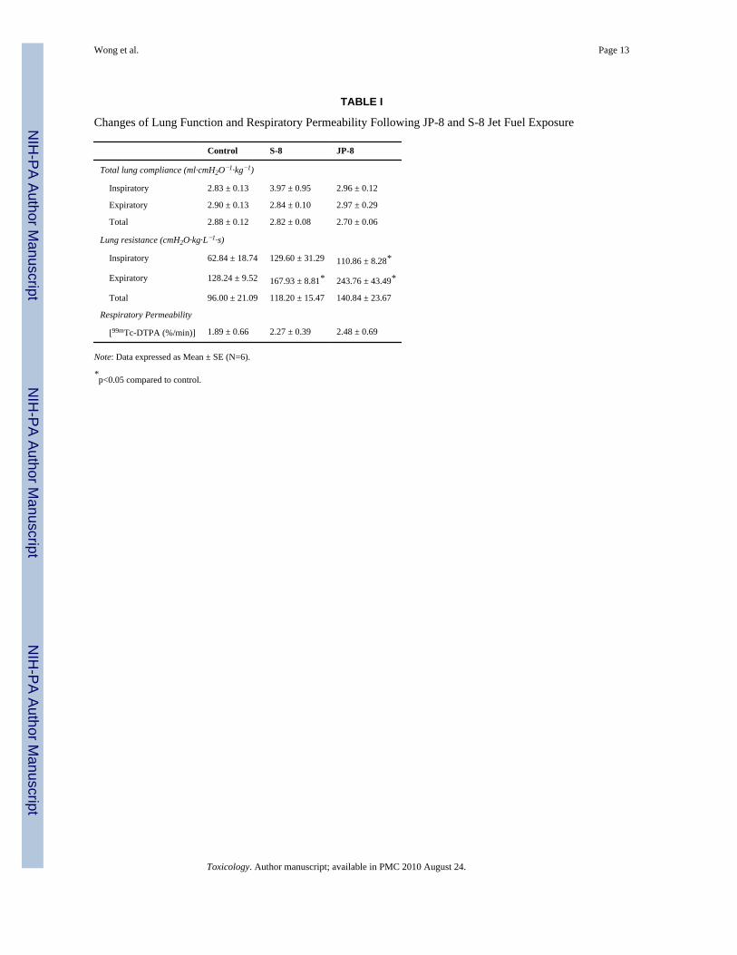
No significant effects or changes in total lung compliance (including inspiratory andexpiratory) were observed either in S-8 or in JP-8 group. There was a significant increase inexpiratory lung resistance in S-8 group compared to control values. Additionally, JP-8 grouphad significant increases in both inspiratory and expiratory lung resistance compared tocontrol values.
Respiratory PermeabilityExposure of mice to S-8 had a 20.1% increase in respiratory permeability compared tocontrols and JP-8 exposure had a 31.2% increase in respiratory permeability compared to thecontrol group (Table 1). However, neither S-8 nor JP-8 induced change were statisticallysignificant when compared to controls.
Histopathological EvaluationUltrastructural examination of distinct lung tissue focused on epithelial cells of alveolar andbronchiolar regions (Figures 2–4). In the air control group, alveolar area appeared normalfor intracellular and extracellular structures (Figure 2B). There were many typical structuresof surfactant-producing lamellar bodies throughout alveolar type II epithelial cells. Themajority of mitochondria exhibited a normal shape with obvious cristae in the cytoplasm.JP-8 group had apparent alterations in the content and size of lamellar bodies within alveolartype II epithelial cells (Figure 2C). Under higher magnification, there was the appearance oflamellar inclusion bodies that appeared to be secondary lysosomes containing intracellular
Wong et al. Page 4
Toxicology. Author manuscript; available in PMC 2010 August 24.
NIH
-PA Author Manuscript
NIH
-PA Author Manuscript
NIH
-PA Author Manuscript
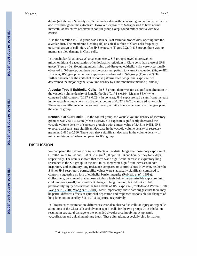
debris (not shown). Severely swollen mitochondria with decreased granulation in the matrixoccurred throughout the cytoplasm. However, exposure to S-8 appeared to have normalintracellular structures observed in control group except round mitochondria with fewcristae.
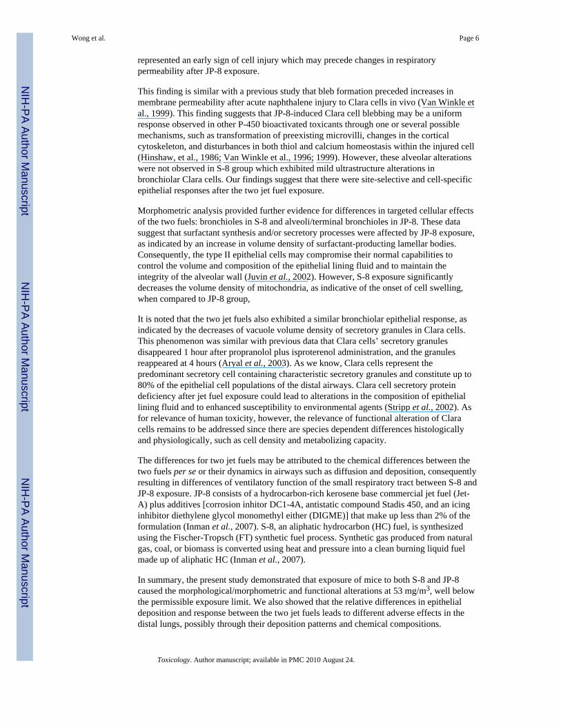
Also the alteration in JP-8 group was Clara cells of terminal bronchioles, opening into thealveolar duct. The membrane blebbing (B) on apical surface of Clara cells frequentlyoccurred, a sign of cell injury after JP-8 exposure (Figure 3C). In S-8 group, there was nomembrane bleb damage in Clara cells.
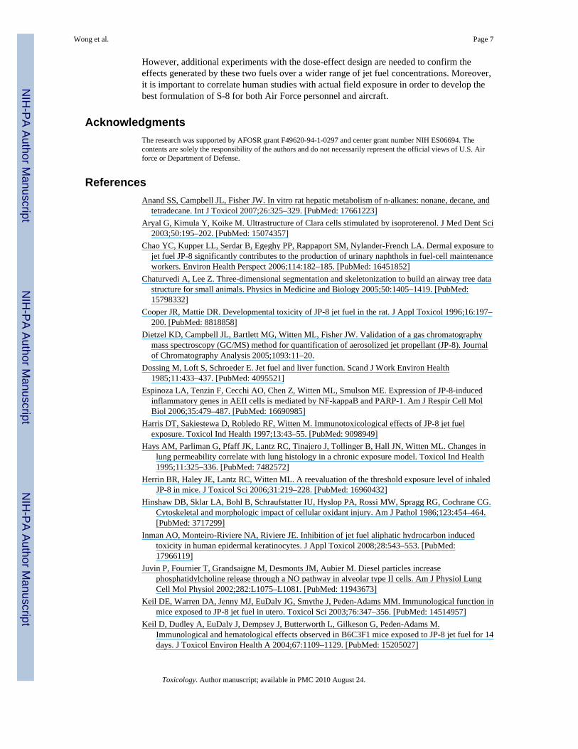
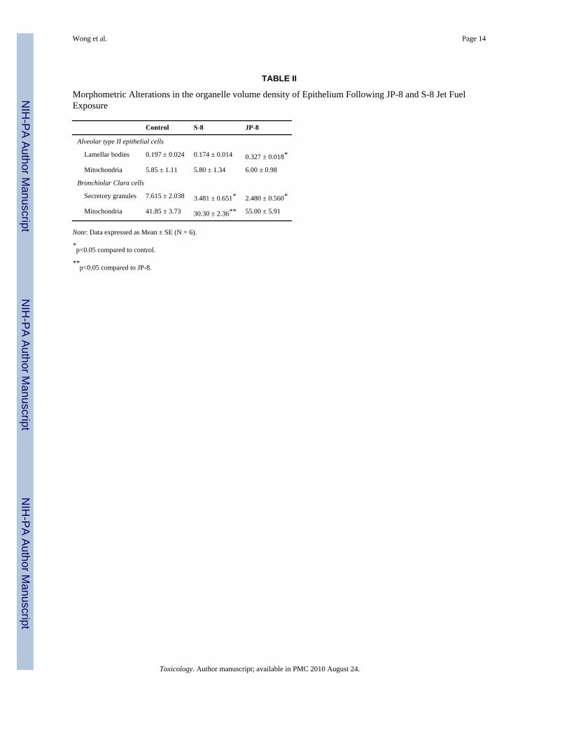
In bronchiolar (small airways) area, conversely, S-8 group showed more swollenmitochondria and vacuolization of endoplasmic reticulum in Clara cells than those of JP-8group (Figure 4B). Sloughing mucus lining and disrupted epithelial cilia were occasionallyobserved in S-8 group, but there was no consistent pattern to warrant evaluation (Figure 4B).However, JP-8 group had no such appearances observed in S-8 group (Figure 4C). Tofurther characterize the epithelial response patterns after two jet fuel exposure, wedetermined the major organelle volume density by a morphometric method (Table II):
Alveolar Type II Epithelial Cells—In S-8 group, there was not a significant alteration inthe vacuole volume density of lamellar bodies (0.174 ± 0.104, Mean ± SEM) whencompared with controls (0.197 ± 0.024). In contrast, JP-8 exposure had a significant increasein the vacuole volume density of lamellar bodies of 0.327 ± 0.018 compared to controls.There was no difference in the volume density of mitochondria between any fuel group andthe control group.
Bronchiolar Clara cells—In the control group, the vacuole volume density of secretorygranules was 7.615 ± 2.038 (Mean ± SEM). S-8 exposure significantly decreased thevacuole volume density of secretory granules with a mean value of 3.481 ± 0.651. JP-8exposure caused a large significant decrease in the vacuole volume density of secretorygranules, 2.480 ± 0.560. There was also a significant decrease in the volume density ofmitochondria in S-8 when compared to JP-8 group.
DISCUSSIONWe compared the cytotoxic or injury effects of the distal lungs after nose-only exposure ofC57BL/6 mice to S-8 and JP-8 at 53 mg/m3 (80 ppm THC) one hour per day for 7 days,respectively. The results showed that there was a significant increase in expiratory lungresistance in the S-8 group. In the JP-8 mice, there were significant increases in bothinspiratory and expiratory lung resistance compared to control values. However, neither theS-8 nor JP-8 respiratory permeability values were statistically significant compared tocontrols, suggesting no loss of epithelial barrier integrity (Robledo et al., 1999a).Collectively, we showed that exposure to both fuels below the permissible exposure limitcould induce a small, but significant change in lung function, but did not exhibitpermeability injury observed at the high levels of JP-8 exposure (Robledo and Witten, 1998;Wang et al., 2001; Wong et al., 2004). More importantly, these data suggest that there maybe partial different effects of epithelial deposition and responses responsible for changes oflung function induced by S-8 or JP-8 exposure, respectively.
In ultrastructure examination, differences were also observed in cellular injury or organellealterations of the Clara cells and alveolar type II cells for the two groups. JP-8 inhalationresulted in structural damage to the extended alveolar area involving cytoplasmicvacuolization and apical membrane blebs. These alterations, especially bleb formation,
Wong et al. Page 5
Toxicology. Author manuscript; available in PMC 2010 August 24.
NIH
-PA Author Manuscript
NIH
-PA Author Manuscript
NIH
-PA Author Manuscript
represented an early sign of cell injury which may precede changes in respiratorypermeability after JP-8 exposure.
This finding is similar with a previous study that bleb formation preceded increases inmembrane permeability after acute naphthalene injury to Clara cells in vivo (Van Winkle etal., 1999). This finding suggests that JP-8-induced Clara cell blebbing may be a uniformresponse observed in other P-450 bioactivated toxicants through one or several possiblemechanisms, such as transformation of preexisting microvilli, changes in the corticalcytoskeleton, and disturbances in both thiol and calcium homeostasis within the injured cell(Hinshaw, et al., 1986; Van Winkle et al., 1996; 1999). However, these alveolar alterationswere not observed in S-8 group which exhibited mild ultrastructure alterations inbronchiolar Clara cells. Our findings suggest that there were site-selective and cell-specificepithelial responses after the two jet fuel exposure.
Morphometric analysis provided further evidence for differences in targeted cellular effectsof the two fuels: bronchioles in S-8 and alveoli/terminal bronchioles in JP-8. These datasuggest that surfactant synthesis and/or secretory processes were affected by JP-8 exposure,as indicated by an increase in volume density of surfactant-producting lamellar bodies.Consequently, the type II epithelial cells may compromise their normal capabilities tocontrol the volume and composition of the epithelial lining fluid and to maintain theintegrity of the alveolar wall (Juvin et al., 2002). However, S-8 exposure significantlydecreases the volume density of mitochondria, as indicative of the onset of cell swelling,when compared to JP-8 group,
It is noted that the two jet fuels also exhibited a similar bronchiolar epithelial response, asindicated by the decreases of vacuole volume density of secretory granules in Clara cells.This phenomenon was similar with previous data that Clara cells’ secretory granulesdisappeared 1 hour after propranolol plus isproterenol administration, and the granulesreappeared at 4 hours (Aryal et al., 2003). As we know, Clara cells represent thepredominant secretory cell containing characteristic secretory granules and constitute up to80% of the epithelial cell populations of the distal airways. Clara cell secretory proteindeficiency after jet fuel exposure could lead to alterations in the composition of epitheliallining fluid and to enhanced susceptibility to environmental agents (Stripp et al., 2002). Asfor relevance of human toxicity, however, the relevance of functional alteration of Claracells remains to be addressed since there are species dependent differences histologicallyand physiologically, such as cell density and metabolizing capacity.
The differences for two jet fuels may be attributed to the chemical differences between thetwo fuels per se or their dynamics in airways such as diffusion and deposition, consequentlyresulting in differences of ventilatory function of the small respiratory tract between S-8 andJP-8 exposure. JP-8 consists of a hydrocarbon-rich kerosene base commercial jet fuel (Jet-A) plus additives [corrosion inhitor DC1-4A, antistatic compound Stadis 450, and an icinginhibitor diethylene glycol monomethyl either (DIGME)] that make up less than 2% of theformulation (Inman et al., 2007). S-8, an aliphatic hydrocarbon (HC) fuel, is synthesizedusing the Fischer-Tropsch (FT) synthetic fuel process. Synthetic gas produced from naturalgas, coal, or biomass is converted using heat and pressure into a clean burning liquid fuelmade up of aliphatic HC (Inman et al., 2007).
In summary, the present study demonstrated that exposure of mice to both S-8 and JP-8caused the morphological/morphometric and functional alterations at 53 mg/m3, well belowthe permissible exposure limit. We also showed that the relative differences in epithelialdeposition and response between the two jet fuels leads to different adverse effects in thedistal lungs, possibly through their deposition patterns and chemical compositions.
Wong et al. Page 6
Toxicology. Author manuscript; available in PMC 2010 August 24.
NIH
-PA Author Manuscript
NIH
-PA Author Manuscript
NIH
-PA Author Manuscript
However, additional experiments with the dose-effect design are needed to confirm theeffects generated by these two fuels over a wider range of jet fuel concentrations. Moreover,it is important to correlate human studies with actual field exposure in order to develop thebest formulation of S-8 for both Air Force personnel and aircraft.
AcknowledgmentsThe research was supported by AFOSR grant F49620-94-1-0297 and center grant number NIH ES06694. Thecontents are solely the responsibility of the authors and do not necessarily represent the official views of U.S. Airforce or Department of Defense.
ReferencesAnand SS, Campbell JL, Fisher JW. In vitro rat hepatic metabolism of n-alkanes: nonane, decane, and
tetradecane. Int J Toxicol 2007;26:325–329. [PubMed: 17661223]Aryal G, Kimula Y, Koike M. Ultrastructure of Clara cells stimulated by isoproterenol. J Med Dent Sci
2003;50:195–202. [PubMed: 15074357]Chao YC, Kupper LL, Serdar B, Egeghy PP, Rappaport SM, Nylander-French LA. Dermal exposure to
jet fuel JP-8 significantly contributes to the production of urinary naphthols in fuel-cell maintenanceworkers. Environ Health Perspect 2006;114:182–185. [PubMed: 16451852]
Chaturvedi A, Lee Z. Three-dimensional segmentation and skeletonization to build an airway tree datastructure for small animals. Physics in Medicine and Biology 2005;50:1405–1419. [PubMed:15798332]
Cooper JR, Mattie DR. Developmental toxicity of JP-8 jet fuel in the rat. J Appl Toxicol 1996;16:197–200. [PubMed: 8818858]
Dietzel KD, Campbell JL, Bartlett MG, Witten ML, Fisher JW. Validation of a gas chromatographymass spectroscopy (GC/MS) method for quantification of aerosolized jet propellant (JP-8). Journalof Chromatography Analysis 2005;1093:11–20.
Dossing M, Loft S, Schroeder E. Jet fuel and liver function. Scand J Work Environ Health1985;11:433–437. [PubMed: 4095521]
Espinoza LA, Tenzin F, Cecchi AO, Chen Z, Witten ML, Smulson ME. Expression of JP-8-inducedinflammatory genes in AEII cells is mediated by NF-kappaB and PARP-1. Am J Respir Cell MolBiol 2006;35:479–487. [PubMed: 16690985]
Harris DT, Sakiestewa D, Robledo RF, Witten M. Immunotoxicological effects of JP-8 jet fuelexposure. Toxicol Ind Health 1997;13:43–55. [PubMed: 9098949]
Hays AM, Parliman G, Pfaff JK, Lantz RC, Tinajero J, Tollinger B, Hall JN, Witten ML. Changes inlung permeability correlate with lung histology in a chronic exposure model. Toxicol Ind Health1995;11:325–336. [PubMed: 7482572]
Herrin BR, Haley JE, Lantz RC, Witten ML. A reevaluation of the threshold exposure level of inhaledJP-8 in mice. J Toxicol Sci 2006;31:219–228. [PubMed: 16960432]
Hinshaw DB, Sklar LA, Bohl B, Schraufstatter IU, Hyslop PA, Rossi MW, Spragg RG, Cochrane CG.Cytoskeletal and morphologic impact of cellular oxidant injury. Am J Pathol 1986;123:454–464.[PubMed: 3717299]
Inman AO, Monteiro-Riviere NA, Riviere JE. Inhibition of jet fuel aliphatic hydrocarbon inducedtoxicity in human epidermal keratinocytes. J Appl Toxicol 2008;28:543–553. [PubMed:17966119]
Juvin P, Fournier T, Grandsaigne M, Desmonts JM, Aubier M. Diesel particles increasephosphatidylcholine release through a NO pathway in alveolar type II cells. Am J Physiol LungCell Mol Physiol 2002;282:L1075–L1081. [PubMed: 11943673]
Keil DE, Warren DA, Jenny MJ, EuDaly JG, Smythe J, Peden-Adams MM. Immunological function inmice exposed to JP-8 jet fuel in utero. Toxicol Sci 2003;76:347–356. [PubMed: 14514957]
Keil D, Dudley A, EuDaly J, Dempsey J, Butterworth L, Gilkeson G, Peden-Adams M.Immunological and hematological effects observed in B6C3F1 mice exposed to JP-8 jet fuel for 14days. J Toxicol Environ Health A 2004;67:1109–1129. [PubMed: 15205027]
Wong et al. Page 7
Toxicology. Author manuscript; available in PMC 2010 August 24.
NIH
-PA Author Manuscript
NIH
-PA Author Manuscript
NIH
-PA Author Manuscript
Knave B, Mindus P, Struwe G. Neurasthenic symptoms in workers occupationally exposed to jet fuel.Acta Psychiatr Scand 1979;60:39–49. [PubMed: 474176]
Koschier FJ. Toxicity of middle distillates from dermal exposure. Drug Chem Toxicol 1999;22:155–164. [PubMed: 10189576]
Lantz RC, Hiton DE. Pulmonary toxicity associated with fly ash from fluidized bed coal combustion.Toxicol Appl Pharm 1984;75:44–51.
McDougal JN, Rogers JV. Local and systemic toxicity of JP-8 from cutaneous exposures. Toxicol Lett2004;149:301–308. [PubMed: 15093277]
Robledo RF, Witten ML. NK1-receptor activation prevents hydrocarbon-induced lung injury in mice.Am J Physiol 1999b;276:L229–L238. [PubMed: 9950884]
Robledo RF, Barber DS, Witten ML. Modulation of bronchial epithelial cell barrier function by invitro jet propulsion fuel 8 exposure. Toxicol Sci 1999a;51:119–125. [PubMed: 10496683]
Robledo RF, Witten ML. Acute pulmonary response to inhaled JP-8 jet fuel in mice. Inhal Toxicol1998;10:531–553.
Robledo RF, Young RS, Lantz RC, Witten ML. Short-term pulmonary response to inhaled JP-8 jetfuel aerosol in mice. Toxicol Pathol 2000;28:656–663. [PubMed: 11026600]
Smith LB, Bhattacharya A, Lemasters G, Succop P, Puhala E, Medvedovic M, Joyce J. Effect ofchronic low-level exposure to jet fuel on postural balance of US Air Force personnel. J OccupEnviron Med 1997;39:623–632. [PubMed: 9253723]
Stripp BR, Reynolds SD, Boe IM, Lund J, Power JH, Coppens JT, Wong V, Reynolds PR, PlopperCG. Clara cell secretory protein deficiency alters clara cell secretory apparatus and the proteincomposition of airway lining fluid. Am J Respir Cell Mol Biol 2002;27:170–178. [PubMed:12151308]
Struwe G, Knave B, Mindus P. Neuropsychiatric symptoms in workers occupationally exposed to jetfuel--a combined epidemiological and casuistic study. Acta Psychiatr Scand Suppl 1983;303:55–67. [PubMed: 6575584]
Ullrich SE, Lyons HJ. Mechanisms involved in the immunotoxicity induced by dermal application ofJP-8 jet fuel. Toxicol Sci 2000;58:290–298. [PubMed: 11099641]
Van Winkle LS, Johnson ZA, Nishio SJ, Brown CD, Plopper CG. Early events in naphthalene-inducedacute Clara cell toxicity: comparison of membrane permeability and ultrastructure. Am J RespirCell Mol Biol 1999;21:44–53. [PubMed: 10385592]
Van Winkle LS, Isaac JM, Plopper CG. Repair of naphthalene-injured microdissected airways in vitro.Am J Respir Cell Mol Biol 1996;15:1–8. [PubMed: 8679213]
Wang S, Young RS, Witten ML. Age-related differences in pulmonary inflammatory responses to JP-8jet fuel aerosol inhalation. Toxicol Ind Health 2001;17:23–29. [PubMed: 12004922]
Weibel, ER. Prectical method for biological morphometry. Vol. 1. Academic Press; New York: 1979.Stereological method.
Witzmann FA, Bauer MD, Fieno AM, Grant RA, Keough TW, Kornguth SE, Lacey MP, Siegel FL,Sun Y, Wright LS, Young RS, Witten ML. Proteomic analysis of simulated occupational jet fuelexposure in the lung. Electrophoresis 1999;20:3659–3669. [PubMed: 10612293]
Wong SS, Hyde J, Sun NN, Lantz RC, Witten ML. Inflammatory responses in mice sequentiallyexposed to JP-8 jet fuel and influenza virus. Toxicology 2004;197:139–147. [PubMed: 15003324]
Wong et al. Page 8
Toxicology. Author manuscript; available in PMC 2010 August 24.
NIH
-PA Author Manuscript
NIH
-PA Author Manuscript
NIH
-PA Author Manuscript
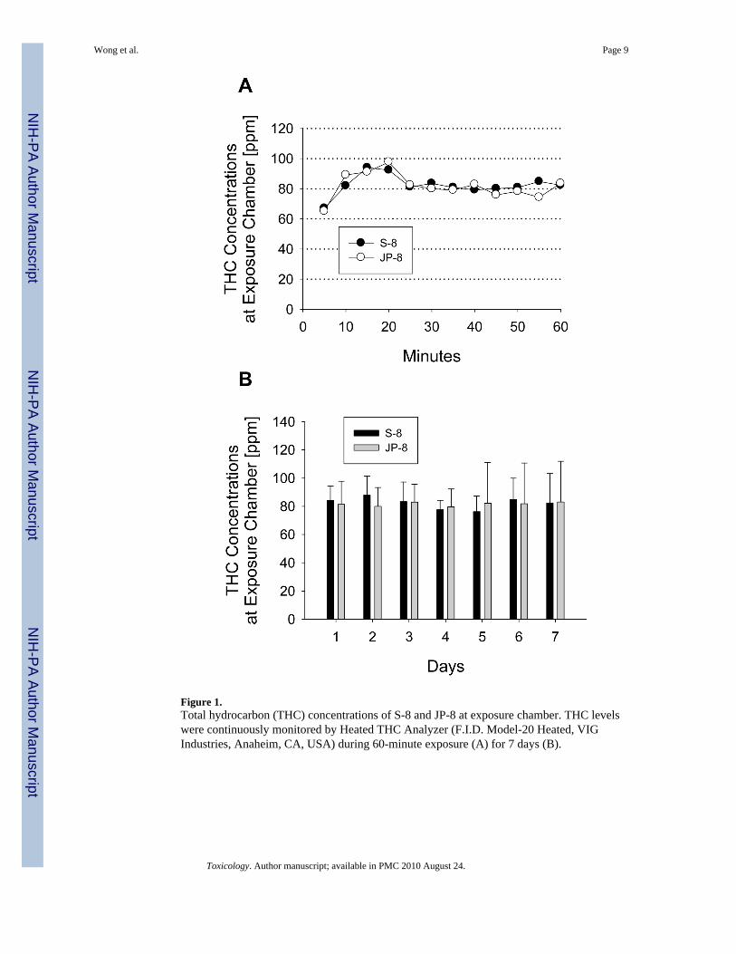
Figure 1.Total hydrocarbon (THC) concentrations of S-8 and JP-8 at exposure chamber. THC levelswere continuously monitored by Heated THC Analyzer (F.I.D. Model-20 Heated, VIGIndustries, Anaheim, CA, USA) during 60-minute exposure (A) for 7 days (B).
Wong et al. Page 9
Toxicology. Author manuscript; available in PMC 2010 August 24.
NIH
-PA Author Manuscript
NIH
-PA Author Manuscript
NIH
-PA Author Manuscript

Figure 2.Representative transmission electron micrographs of alveolar epithelial cells from micefollowing ambient air (A), S-8 (B), and JP-8 (C) exposure. Note the apparent alteration insurfactant-producing lamellar bodies (arrows) of alveolar type II epithelial cells withswollen mitochondria (arrow heads) in the JP-8 group (C). N: Nucleus. Uranyl acetate andlead citrate. Magnification: 8,000×.
Wong et al. Page 10
Toxicology. Author manuscript; available in PMC 2010 August 24.
NIH
-PA Author Manuscript
NIH
-PA Author Manuscript
NIH
-PA Author Manuscript
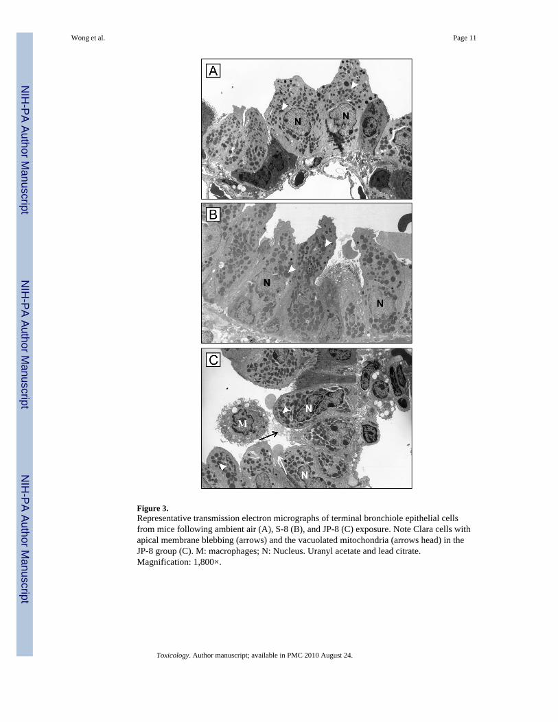
Figure 3.Representative transmission electron micrographs of terminal bronchiole epithelial cellsfrom mice following ambient air (A), S-8 (B), and JP-8 (C) exposure. Note Clara cells withapical membrane blebbing (arrows) and the vacuolated mitochondria (arrows head) in theJP-8 group (C). M: macrophages; N: Nucleus. Uranyl acetate and lead citrate.Magnification: 1,800×.
Wong et al. Page 11
Toxicology. Author manuscript; available in PMC 2010 August 24.
NIH
-PA Author Manuscript
NIH
-PA Author Manuscript
NIH
-PA Author Manuscript
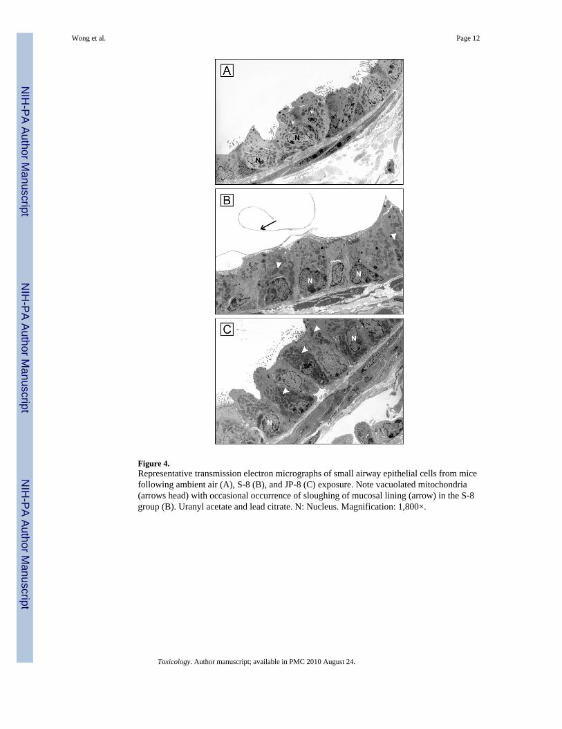
Figure 4.Representative transmission electron micrographs of small airway epithelial cells from micefollowing ambient air (A), S-8 (B), and JP-8 (C) exposure. Note vacuolated mitochondria(arrows head) with occasional occurrence of sloughing of mucosal lining (arrow) in the S-8group (B). Uranyl acetate and lead citrate. N: Nucleus. Magnification: 1,800×.
Wong et al. Page 12
Toxicology. Author manuscript; available in PMC 2010 August 24.
NIH
-PA Author Manuscript
NIH
-PA Author Manuscript
NIH
-PA Author Manuscript
NIH
-PA Author Manuscript
NIH
-PA Author Manuscript
NIH
-PA Author Manuscript
Wong et al. Page 13
TABLE I
Changes of Lung Function and Respiratory Permeability Following JP-8 and S-8 Jet Fuel Exposure
Control S-8 JP-8
Total lung compliance (ml·cmH2O−1·kg−1)
Inspiratory 2.83 ± 0.13 3.97 ± 0.95 2.96 ± 0.12
Expiratory 2.90 ± 0.13 2.84 ± 0.10 2.97 ± 0.29
Total 2.88 ± 0.12 2.82 ± 0.08 2.70 ± 0.06
Lung resistance (cmH2O·kg·L−1·s)
Inspiratory 62.84 ± 18.74 129.60 ± 31.29 110.86 ± 8.28*
Expiratory 128.24 ± 9.52 167.93 ± 8.81* 243.76 ± 43.49*
Total 96.00 ± 21.09 118.20 ± 15.47 140.84 ± 23.67
Respiratory Permeability
[99mTc-DTPA (%/min)] 1.89 ± 0.66 2.27 ± 0.39 2.48 ± 0.69
Note: Data expressed as Mean ± SE (N=6).
*p<0.05 compared to control.
Toxicology. Author manuscript; available in PMC 2010 August 24.
NIH
-PA Author Manuscript
NIH
-PA Author Manuscript
NIH
-PA Author Manuscript
Wong et al. Page 14
TABLE II
Morphometric Alterations in the organelle volume density of Epithelium Following JP-8 and S-8 Jet FuelExposure
Control S-8 JP-8
Alveolar type II epithelial cells
Lamellar bodies 0.197 ± 0.024 0.174 ± 0.014 0.327 ± 0.018*
Mitochondria 5.85 ± 1.11 5.80 ± 1.34 6.00 ± 0.98
Bronchiolar Clara cells
Secretory granules 7.615 ± 2.038 3.481 ± 0.651* 2.480 ± 0.560*
Mitochondria 41.85 ± 3.73 30.30 ± 2.36** 55.00 ± 5.91
Note: Data expressed as Mean ± SE (N = 6).
*p<0.05 compared to control.
**p<0.05 compared to JP-8.
Toxicology. Author manuscript; available in PMC 2010 August 24.