in vivo antioxidative activity of a quantified pueraria lobata root extract
TRANSCRIPT
I
LGa
Bb
Fc
d
B
a
ARR1AA
KPFIMDP
1
boha2pb
em1
0d
Journal of Ethnopharmacology 127 (2010) 112–117
Contents lists available at ScienceDirect
Journal of Ethnopharmacology
journa l homepage: www.e lsev ier .com/ locate / je thpharm
n vivo antioxidative activity of a quantified Pueraria lobata root extract
idiya Bebrevskaa,1, Kenne Fouberta,1, Nina Hermansb, Shyama Chatterjeec, Eric Van Marckc,uido De Meyerd, Arnold Vlietincka, Luc Pietersa,∗, Sandra Apersa
Laboratory of Pharmacognosy and Pharmaceutical Analysis, Department of Pharmaceutical Sciences, Faculty of Pharmaceutical,iomedical and Veterinary Sciences, University of Antwerp, Universiteitsplein 1, 2610 Antwerp, BelgiumLaboratory of Nutrition and Functional Food Science, Department of Pharmaceutical Sciences,aculty of Pharmaceutical, Biomedical and Veterinary Sciences, University of Antwerp, BelgiumLaboratory of Pathology, Faculty of Medicine, University of Antwerp, BelgiumLaboratory of Pharmacology, Department of Pharmaceutical Sciences, Faculty of Pharmaceutical,iomedical and Veterinary Sciences, University of Antwerp, Belgium
r t i c l e i n f o
rticle history:eceived 9 May 2009eceived in revised form9 September 2009ccepted 21 September 2009vailable online 30 September 2009
eywords:ueraria lobataabaceaen vivo antioxidative activity
alondialdehyde (MDA)iabetic rat model
a b s t r a c t
Aim of the study: Oxidative stress has been associated with many pathological disorders such as atheroscle-rosis, diabetes and cancer. Supplementation with exogenous antioxidants, including phenolic compoundsfrom plant sources, may help to restore the pro-oxidative/antioxidative balance. To take into accounteffects of absorption, metabolisation, plasma protein binding, distribution, and elimination, antioxidativeresearch should not be limited to in vitro assays but be extended to in vivo models.Materials and methods: In the present work a quantified 50% EtOH root extract of Pueraria lobata(Willd.) Ohwi (Fabaceae) was selected to determine its in vivo antioxidative activity in a diabetic ratmodel, where diabetes and the accompanying oxidative stress were induced by intraperitoneal admin-istration of streptozotocin. This root extract was found to contain 10.42 ± 0.15% puerarin as the mainconstituent and smaller amounts of the related isoflavonoids 3′-hydroxypuerarin, 3′-methoxypuerarin,6′′-xylosylpuerarin, daidzin, genistin, daidzein and genistein, as determined by a validated HPLC method.
uerarinThis extract was administered orally at a daily dose of 500 mg/kg root extract, corresponding to 50 mg/kgpuerarin, during 3 weeks. In addition the effect on the plasma concentration of some fat-soluble antiox-idants (co-enzyme Q9, �- and �-tocopherol) was evaluated.Results and conclusions: The level of malondialdehyde (MDA) in plasma, used as a marker of oxidativedamage to lipids, was reduced to the same level as in healthy control animals, and as in the positive control
50 me trea
group treated daily withadministration of 10× th
. Introduction
Oxidative stress is caused by a disturbance of the balanceetween the antioxidant defence mechanisms of the humanrganism and the level of reactive oxygen species (ROS), andas been associated with many pathological disorders such as
therosclerosis, diabetes and cancer (Halliwell and Gutteridge,002). Supplementation with exogenous antioxidants, includinghenolic compounds from plant sources, may help to restore thisalance. Because of the increasing interest in Oriental MedicineAbbreviations: BGL, blood glucose level; BHT, butylhydroxytoluene; CoE, co-nzyme; DM, diabetes mellitus; IP, intraperitoneal; LP, lipid peroxidation; MDA,alondialdehyde; ROS, reactive oxygen species; TBA, thiobarbituric acid; TMP,
,1,3,3-tetramethoxypropane.∗ Corresponding author. Tel.: +32 3 26527 15; fax: +32 3 26527 09.
E-mail address: [email protected] (L. Pieters).1 These authors have equally contributed to the work.
378-8741/$ – see front matter © 2009 Elsevier Ireland Ltd. All rights reserved.oi:10.1016/j.jep.2009.09.039
g/kg �-tocopherol acetate. No obvious signs of toxicity were observed bytment dose.
© 2009 Elsevier Ireland Ltd. All rights reserved.
in the Western world, Pueraria lobata (Willd.) Ohwi (Fabaceae)was selected, among other plants, for our investigations. Puer-aria roots as well as flowers are medicinally used. In this workwe have focused on root material. In Japan Pueraria lobata isknown as Kudzu. Roots of Pueraria lobata are included in theChinese Pharmacopoeia (“Gegen”; Radix Puerariae lobatae). It iscommonly used to relieve fever and dysentery, and for the treat-ment of cardiovascular diseases such as hypertension, myocardialinfarction and arrhythmia (Jiang et al., 2005). Nowadays it ismainly known as a treatment for alcohol abuse (antidipsotropicagent) (Keung and Vallee, 1998). The genus Pueraria is a richsource of isoflavonoids, the most abundant isoflavonoid in theroot of Pueraria lobata being puerarin. In previously conducted
work we have developed and validated an HPLC method forthe quantification of the isoflavonoid constituents in this crudedrug (Bebrevska et al., 2008). In addition to the main constituentpuerarin, the minor isoflavonoids included 3′-hydroxypuerarin, 3′-methoxypuerarin, 6′′-xylosylpuerarin, daidzin, genistin, daidzein
nopha
aatpepoaolvnmm
vte(iasAtabottitapoaaeniiOmNohpdsdocRadNwg1a
2
2
(
L. Bebrevska et al. / Journal of Eth
nd genistein. Isoflavonoids exhibit a wide range of biologicalctivities; they have antiinflammatory, antithrombotic, antihyper-ensive, antiarrhythmic, spasmolytic, and cancer chemopreventiveroperties (Jiang et al., 2005). It is well known that the beneficialffects on health of isoflavonoids are due to their antioxidative andhytoestrogenic properties (Cos et al., 2003). Cardiovascular effectsf medicinal plants are exerted in part through their antioxidativend anti-lipidperoxidation properties. The present work will focusnly on the antioxidative effects. Puerariae radix extract and iso-ated pure constituents have extensively been tested in various initro antioxidative test systems (Jiang et al., 2005). However, it isecessary to investigate their in vivo effect as well, since in vitroodels do not account for problems of absorption, distribution,etabolisation and elimination (Hermans et al., 2007a).Therefore, the objective of our study was to evaluate the in
ivo antioxidative potential of a quantified extract prepared fromhe root of Pueraria lobata. This was achieved by evaluating theffect of this extract on a direct marker of systemic oxidative stressmalondialdehyde, MDA), and the status of some fat-soluble antiox-dants in plasma (co-enzyme Q9, �- and �-tocopherol) involved inntioxidative defence mechanisms. MDA is known as an univer-al biomarker of lipid peroxidation. Fat-soluble vitamins (vitamins
and carotenoids, vitamins E) and co-enzyme Q play an impor-ant role in oxidative stress and free radical control (Hermans etl., 2005). The model of streptozotocin induced diabetic rats (dia-etes type I) was used, where diabetes and the accompanyingxidative stress were induced by intraperitoneal (IP) administra-ion of streptozotocin. Oxidative stress plays an important role inhe experimental diabetes induced by streptozotocin. In previousnvestigations we have carried out a comparison of different oxida-ive stress animal models, including a vitamin E deficient rat model,diabetic rat model, and an atherosclerotic rabbit model, where
lasma/serum malondialdehyde was measured as a parameter ofxidative damage. The vitamin E deficient rats were not suitables oxidative stress model, whereas diabetic and atheroscleroticnimals showed increased systemic oxidative damage (Hermanst al., 2007b). Based on this work, and taking into account eco-omic factors, the diabetic rat model was selected for the present
nvestigations. The pathological processes in diabetes mellitus andts complications are closely related with elevated ROS formation.xidative stress in diabetes mellitus (DM) is generated throughultiple non-enzymatic, enzymatic and mitochondrial pathways.on-enzymatic pathways originate from the biochemical oxidationf glucose. Glucose can undergo autoxidation and generate HO•. Inyperglycemic conditions the metabolism of glucose through theolyol pathway is increased, which results in increased O2
•− pro-uction. The enzymatic pathways involve the release of oxidativepecies such as NO• and O2
•− by the enzymes NOS, NAD(P)H oxi-ase and xanthine oxidase. NO• and O2
•− can interact and formther powerful oxidizing agents such as ONOO−. Finally, the mito-hondrial respiratory chain is responsible for O2
•− production.OS cause damage to endothelial cells, oxidise LDL and acceler-te atherosclerosis, oxidise lipids, DNA and proteins leading to theevelopment of DM complications (Johanes et al., 2005; Kohen andyska, 2002; Klaunig and Kamendulis, 2004). In order to evaluatehether the Pueraria extract exhibited any acute toxic effects, a
roup of animals where no oxidative stress was induced received0× the test dose, and was subjected to histopathological analysis;series of biochemical parameters in serum were assessed as well.
. Materials and methods
.1. Materials
Streptozotocin (>98%), butylated hydroxyanisole (>90%), BHT>99%), retinol (>95%), �-tocopherol (>96%), �-tocopherol (>96%),
rmacology 127 (2010) 112–117 113
�-tocopherol acetate (>97%), co-enzymes Q9 (>96%) and Q10(>99%), thiobarbituric acid (>98%), 1,1,3,3-tetramethoxypropane(>99%) (6.01 M), were purchased from Sigma. Pentobarbital(Nembutal®) (20% solution) was bought on medical prescription ina public pharmacy. Solvents of HPLC grade (EtOH, n-hexane, MeOH,n-propanol), Phosphoric acid (>89%), NaOH, ammonium acetate,sodium citrate and citric acid (analytical grade) were obtained fromMerck. Diabetes test kits (FreeStyle teststrips) were purchased fromAbbott. K3E Vacutainer® tubes were obtained from BD.
Reagent solutions: 1% (w/v) BHT in EtOH; 0.67% thiobarbituricacid; 1.22 M phosphoric acid; 1 M NaOH.
2.2. Plant material – Pueraria lobata root 50% ethanol extract
Dried roots from Pueraria lobata were kindly provided bySinecura (Ghent, Belgium). The root plant material was identifiedmacro- and microscopically according to the Chinese Pharma-copoeia, and voucher material is kept at the Department ofPharmaceutical Sciences, University of Antwerp (Bebrevska etal., 2007). The 50% EtOH extract of the root of Pueraria lobatawas prepared by Gehrlicher GmbH, Eurasburg, Germany bymaceration and percolation in a drug to extract ratio (DER) of5:1. The content of isoflavones in the plant material and thedry extract was determined using a validated HPLC method(Bebrevska et al., 2008). By means of this analytical method themajor constituents, i.e. the isoflavone 8-C-glycosides hydroxy-puerarin, puerarin, methoxypuerarin and xylosylpuerarin, the7-O-glycosides daidzin and genistin, and the aglycones daidzeinand genistein were quantified. Briefly, the extraction procedureand the extraction solvent composition were optimized in orderto ensure the exhaustive extraction of the plant material. TheHPLC-conditions were evaluated and optimized for the exactquantification of all individual compounds. The HPLC analysiswas carried out on an Agilent XDB RP C18 (100 mm × 4.6 mm)column gradiently eluted with a binary system consisting ofwater (+0.01% formic acid) and methanol using a linear gra-dient; detection was performed at 262 nm. Daidzin, daidzein,genistin and genistein were used as external standards. Due tothe great difference in content between the C-glycosides on theone hand, and daidzin, genistin and the aglycones on the otherhand, two separate HPLC runs were necessary for a completeanalysis. The final method was fully validated according to theICH guidelines in terms of linearity, precision and accuracy.Linear relationships for the responses of all four standards wereproven. The method was shown to be valid for the individualcompounds: precision: RSD%between days was within the maxi-mum allowed limit of 5% and 7%. The accuracy of the methodwas demonstrated to be acceptable by recovery experiments.These validation results demonstrated the suitability of themethod for the precise and accurate determination of Pueraria rootisoflavones. The root extract used in this study was found to contain10.42 ± 0.15% puerarin as the main constituent and lower amountsof 3′-hydroxypuerarin (2.59 ± 0.05%), 3′-methoxypuerarin(1.97 ± 0.03%), 6′′-xylosylpuerarin (1.63 ± 0.04%), daidzin(2.67 ± 0.07%), genistin (0.42 ± 0.01%), daidzein (0.49 ± 0.01%)and genistein (0.023 ± 0.001%).
2.3. Experimental set-up
Male Wistar Hannover rats were selected as study objects. 45animals were purchased at the age of 3 months and were housed
in an air-conditioned room with 12/12 h-light/dark cycles, in cageswith two animals per cage. They were divided into five groups,which were evaluated simultaneously. Four groups consisted of10 animals and one, the toxicity group, consisted of 5 animals. Atday 7, after an acclimatisation period of 1 week, diabetes melli-
1 nopha
t(�t(iTgs
widafttsamespoe
2
cT5i4bisoamtswpTdgafaumo
2
oaatfiw−
14 L. Bebrevska et al. / Journal of Eth
us (DM) was induced in the animals of the oxidative stress groupnegative control; group 2), the treatment group (group 3), and the-tocopherol acetate supplemented group (group 5) by adminis-
ration of an IP injection of streptozotocin solution in citrate buffer10.01 mg/mL) at a dose of 60 mg/kg. The solution was preparedn antiseptic conditions and was sterilized by membrane filtration.he control animals of the healthy control (group 1) and toxicityroup (group 4) were injected with pure citrate buffer instead oftreptozotocin solution.
After an induction period of 7 weeks, the animals were treatedith the Pueraria root extract or respective control treatment dur-
ng 3 weeks (21 days). Animals were weighed every 7 days. Theevelopment of hyperglycemia and diabetes were evaluated usingdiabetes test kit for blood glucose measurements (blood sample
rom a vein of the rat tail) by random blood glucose concentra-ion measurements, random being defined as without regard toime since the last meal. The blood glucose level (BGL) was mea-ured at equal intervals of 14 days during the induction periodnd intervals of 4 days during the treatment period. Only ani-als in which DM was properly induced (BGL ≥ 10 mM) at the
nd of the induction period of 7 weeks were included in thetudy. The observed symptoms of DM were increased appetite,olydipsy, polyuria, and loss of weight. Also the appearancef diabetic microangiopathy (diabetic retinopathy) was consid-red.
.4. Dosage regime and drug administration
Dosing applied by oral gavage during the treatment period wasalculated based on the puerarin content of the plant material.he therapeutic dose of 500 mg/kg corresponded to approximately0 mg/kg puerarin. A high dose of 5 g/kg corresponding to approx-
mately 500 mg/kg puerarin was applied to the animals of group(toxicity group). �-Tocopherol acetate at a dose of 50 mg/kg
ody weight, similar to the puearin content, was used as pos-tive control (group 5). The dry extract of Pueraria lobata wasuspended in tap water to obtain a suspension with a concentrationf 100.0 mg/mL (about 10.0 mg/mL puerarin) and administered involume calculated with respect to the body weight of every ani-al in the treatment group (group 3). For the evaluation of the
oxicity the extract was suspended in tap water to give a suspen-ion with a concentration of 600.0 mg/mL. �-Tocopherol acetateas applied as an emulsion with a concentration of 10.0 mg/mL,repared with 10% lecithin and by stirring at 40 ◦C during 30 min.he control animals from groups 1 and 2 received tap wateruring the treatment period. All solutions were applied via oralavage. Treatment was performed once per day every day atpproximately the same time interval of 24 h. The animals hadree access to food and water. In view of the fact that Puer-ria contains isoflavonoids, soy-free rat food was purchased andsed during the whole experiment (Harlan/Teklad). All experi-ents were approved by the Ethical Committee of the University
f Antwerp.
.5. Sample collection schedule and handling procedure
Final collection of plasma samples was done at the endf the treatment period. The animals were anesthetized withn IP injection of Nembutal® (60 mg/kg). Blood from the
rteria carotis was collected in 1 mL tubes (Eppendorf) con-aining potassium EDTA (30 �L 7.5% EDTA/mL blood) (takenrom K3E Vacutainer® (BD) containing 7.5% EDTA 0.072 mL) andmmediately centrifuged at 1000 × g for 12.5 min. The plasmaas collected in cryotubes and the samples were stored at70 ◦C.
rmacology 127 (2010) 112–117
2.6. MDA–TBA method for evaluation of lipid peroxidationinhibitory activity
In the present work oxidative damage due to lipid peroxidationwas measured in rat plasma using a HPLC-fluorimetric detectionmethod (Hermans et al., 2007b). Experimental details are given asSupplementary Material.
2.7. Determination of fat-soluble antioxidants
The method published by Hermans et al. (2007b) was used.Experimental details are given as Supplementary Material.
2.8. Toxicological evaluation
In order to evaluate whether the Pueraria extract exhibitedany acute toxic effects the animals from the toxicity groupwere evaluated by following their body weight, by visual inspec-tion, and by histopathological analysis of liver, kidney, pancreasand spleen samples. For the microscopic analysis of the frozensections of pancreas, liver and spleen, hematoxylin–eosine stain-ing was used. For the slides of kidneys, periodic acid–schiff(PAS) staining was used. The sections were evaluated usinga final magnification of 100× (low magnification) and of400× (high magnification). A series of biochemical parame-ters (urea, creatinine, creatinine kinase, alanine-aminotransferase,aspartate-aminotransferase, gamma-glutamyltransferase, alkalinephosphatase) in serum were evaluated using automated routinetests in a clinical laboratory.
2.9. Statistical analysis
All data are presented as mean ± SEM. The data were evalu-ated using SPSS 15.0 and GraphPad Prism 5 software packages,and the difference between the means assessed using an appro-priate test (see below). Homogeneity of variances was tested bythe Levene’s test. If variances were unequal data were mathemati-cally transformed (logarithmic or power transformation). The levelof statistical significance was set at P < 0.05.
3. Results and discussion
Prior to the induction of diabetes mellitus, the body weight andblood glucose level (Fig. 1a) of all animals were measured. No sta-tistically significant differences were observed between the groups(one-way analysis of variance, ANOVA). After streptozotocin injec-tion, oxidative stress was allowed to develop during 7 weeks. At theend of this induction period there was a marked increase in the BGLin the streptozotocin-treated animals, confirming the successfuldevelopment of DM (Fig. 1b).
After the induction period, treatment was started. The bodyweight of the animals at the end of the study, after 3 weeks oftreatment, is shown in Fig. 2, and results of the MDA–TBA deter-mination in Fig. 3. All animals where oxidative stress had beeninduced showed a significant reduction in body weight comparedto the healthy controls, even after treatment with the Puerariaextract (test group) or �-tocopherol (positive control). No signif-icant differences were observed between the body weight of thetest group treated with Pueraria extract or the �-tocopherol sup-plemented group, with the oxidative stress group (Fig. 1b). The level
of the MDA–TBA complex, measured in the plasma of the untreateddiabetic animals (oxidative stress group), was significantly ele-vated as compared to the healthy animals (P < 0.001) (Fig. 3). Thisshowed the suitability of the chosen system for in vivo LP-inhibitoryactivity evaluation. A significant decrease in the oxidative stress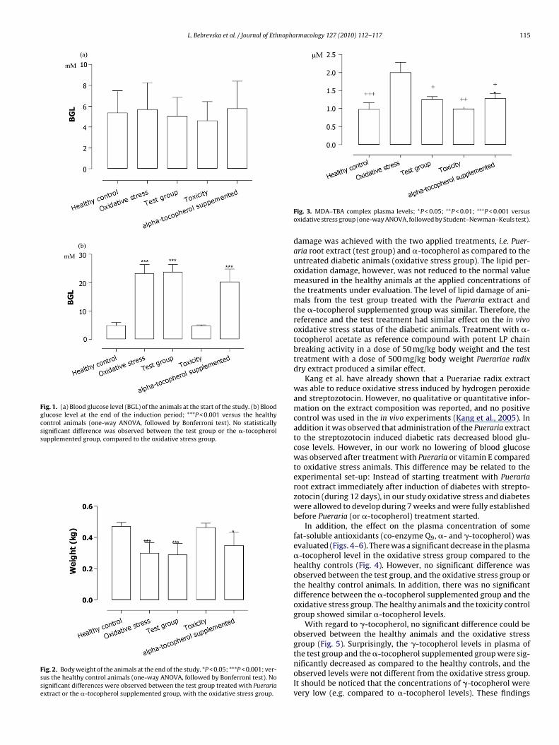
L. Bebrevska et al. / Journal of Ethnopharmacology 127 (2010) 112–117 115
Fig. 1. (a) Blood glucose level (BGL) of the animals at the start of the study. (b) Bloodglucose level at the end of the induction period; ***P < 0.001 versus the healthycontrol animals (one-way ANOVA, followed by Bonferroni test). No statisticallysignificant difference was observed between the test group or the �-tocopherolsupplemented group, compared to the oxidative stress group.
Fig. 2. Body weight of the animals at the end of the study. *P < 0.05; ***P < 0.001; ver-sus the healthy control animals (one-way ANOVA, followed by Bonferroni test). Nosignificant differences were observed between the test group treated with Puerariaextract or the �-tocopherol supplemented group, with the oxidative stress group.
Fig. 3. MDA–TBA complex plasma levels; +P < 0.05; ++P < 0.01; +++P < 0.001 versusoxidative stress group (one-way ANOVA, followed by Student–Newman–Keuls test).
damage was achieved with the two applied treatments, i.e. Puer-aria root extract (test group) and �-tocopherol as compared to theuntreated diabetic animals (oxidative stress group). The lipid per-oxidation damage, however, was not reduced to the normal valuemeasured in the healthy animals at the applied concentrations ofthe treatments under evaluation. The level of lipid damage of ani-mals from the test group treated with the Pueraria extract andthe �-tocopherol supplemented group was similar. Therefore, thereference and the test treatment had similar effect on the in vivooxidative stress status of the diabetic animals. Treatment with �-tocopherol acetate as reference compound with potent LP chainbreaking activity in a dose of 50 mg/kg body weight and the testtreatment with a dose of 500 mg/kg body weight Puerariae radixdry extract produced a similar effect.
Kang et al. have already shown that a Puerariae radix extractwas able to reduce oxidative stress induced by hydrogen peroxideand streptozotocin. However, no qualitative or quantitative infor-mation on the extract composition was reported, and no positivecontrol was used in the in vivo experiments (Kang et al., 2005). Inaddition it was observed that administration of the Pueraria extractto the streptozotocin induced diabetic rats decreased blood glu-cose levels. However, in our work no lowering of blood glucosewas observed after treatment with Pueraria or vitamin E comparedto oxidative stress animals. This difference may be related to theexperimental set-up: Instead of starting treatment with Puerariaroot extract immediately after induction of diabetes with strepto-zotocin (during 12 days), in our study oxidative stress and diabeteswere allowed to develop during 7 weeks and were fully establishedbefore Pueraria (or �-tocopherol) treatment started.
In addition, the effect on the plasma concentration of somefat-soluble antioxidants (co-enzyme Q9, �- and �-tocopherol) wasevaluated (Figs. 4–6). There was a significant decrease in the plasma�-tocopherol level in the oxidative stress group compared to thehealthy controls (Fig. 4). However, no significant difference wasobserved between the test group, and the oxidative stress group orthe healthy control animals. In addition, there was no significantdifference between the �-tocopherol supplemented group and theoxidative stress group. The healthy animals and the toxicity controlgroup showed similar �-tocopherol levels.
With regard to �-tocopherol, no significant difference could beobserved between the healthy animals and the oxidative stressgroup (Fig. 5). Surprisingly, the �-tocopherol levels in plasma of
the test group and the �-tocopherol supplemented group were sig-nificantly decreased as compared to the healthy controls, and theobserved levels were not different from the oxidative stress group.It should be noticed that the concentrations of �-tocopherol werevery low (e.g. compared to �-tocopherol levels). These findings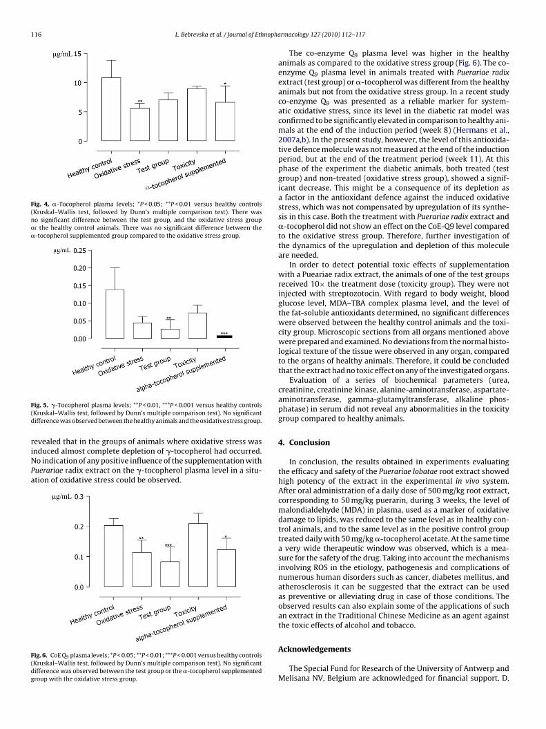
116 L. Bebrevska et al. / Journal of Ethnopha
Fig. 4. �-Tocopherol plasma levels; *P < 0.05; **P < 0.01 versus healthy controls(Kruskal–Wallis test, followed by Dunn’s multiple comparison test). There wasno significant difference between the test group, and the oxidative stress groupor the healthy control animals. There was no significant difference between the�-tocopherol supplemented group compared to the oxidative stress group.
F(d
riNPa
F(dg
ig. 5. �-Tocopherol plasma levels; **P < 0.01, ***P < 0.001 versus healthy controlsKruskal–Wallis test, followed by Dunn’s multiple comparison test). No significantifference was observed between the healthy animals and the oxidative stress group.
evealed that in the groups of animals where oxidative stress wasnduced almost complete depletion of �-tocopherol had occurred.o indication of any positive influence of the supplementation withuerariae radix extract on the �-tocopherol plasma level in a situ-tion of oxidative stress could be observed.
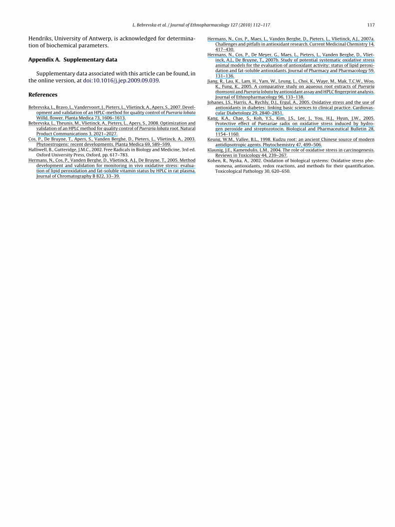
ig. 6. CoE Q9 plasma levels; *P < 0.05; **P < 0.01; ***P < 0.001 versus healthy controlsKruskal–Wallis test, followed by Dunn’s multiple comparison test). No significantifference was observed between the test group or the �-tocopherol supplementedroup with the oxidative stress group.
rmacology 127 (2010) 112–117
The co-enzyme Q9 plasma level was higher in the healthyanimals as compared to the oxidative stress group (Fig. 6). The co-enzyme Q9 plasma level in animals treated with Puerariae radixextract (test group) or �-tocopherol was different from the healthyanimals but not from the oxidative stress group. In a recent studyco-enzyme Q9 was presented as a reliable marker for system-atic oxidative stress, since its level in the diabetic rat model wasconfirmed to be significantly elevated in comparison to healthy ani-mals at the end of the induction period (week 8) (Hermans et al.,2007a,b). In the present study, however, the level of this antioxida-tive defence molecule was not measured at the end of the inductionperiod, but at the end of the treatment period (week 11). At thisphase of the experiment the diabetic animals, both treated (testgroup) and non-treated (oxidative stress group), showed a signif-icant decrease. This might be a consequence of its depletion asa factor in the antioxidant defence against the induced oxidativestress, which was not compensated by upregulation of its synthe-sis in this case. Both the treatment with Puerariae radix extract and�-tocopherol did not show an effect on the CoE-Q9 level comparedto the oxidative stress group. Therefore, further investigation ofthe dynamics of the upregulation and depletion of this moleculeare needed.
In order to detect potential toxic effects of supplementationwith a Pueariae radix extract, the animals of one of the test groupsreceived 10× the treatment dose (toxicity group). They were notinjected with streptozotocin. With regard to body weight, bloodglucose level, MDA–TBA complex plasma level, and the level ofthe fat-soluble antioxidants determined, no significant differenceswere observed between the healthy control animals and the toxi-city group. Microscopic sections from all organs mentioned abovewere prepared and examined. No deviations from the normal histo-logical texture of the tissue were observed in any organ, comparedto the organs of healthy animals. Therefore, it could be concludedthat the extract had no toxic effect on any of the investigated organs.
Evaluation of a series of biochemical parameters (urea,creatinine, creatinine kinase, alanine-aminotransferase, aspartate-aminotransferase, gamma-glutamyltransferase, alkaline phos-phatase) in serum did not reveal any abnormalities in the toxicitygroup compared to healthy animals.
4. Conclusion
In conclusion, the results obtained in experiments evaluatingthe efficacy and safety of the Puerariae lobatae root extract showedhigh potency of the extract in the experimental in vivo system.After oral administration of a daily dose of 500 mg/kg root extract,corresponding to 50 mg/kg puerarin, during 3 weeks, the level ofmalondialdehyde (MDA) in plasma, used as a marker of oxidativedamage to lipids, was reduced to the same level as in healthy con-trol animals, and to the same level as in the positive control grouptreated daily with 50 mg/kg �-tocopherol acetate. At the same timea very wide therapeutic window was observed, which is a mea-sure for the safety of the drug. Taking into account the mechanismsinvolving ROS in the etiology, pathogenesis and complications ofnumerous human disorders such as cancer, diabetes mellitus, andatherosclerosis it can be suggested that the extract can be usedas preventive or alleviating drug in case of those conditions. Theobserved results can also explain some of the applications of suchan extract in the Traditional Chinese Medicine as an agent againstthe toxic effects of alcohol and tobacco.
Acknowledgements
The Special Fund for Research of the University of Antwerp andMelisana NV, Belgium are acknowledged for financial support. D.
nopha
Ht
A
t
R
B
B
C
H
H
antidipsotropic agents. Phytochemistry 47, 499–506.
L. Bebrevska et al. / Journal of Eth
endriks, University of Antwerp, is acknowledged for determina-ion of biochemical parameters.
ppendix A. Supplementary data
Supplementary data associated with this article can be found, inhe online version, at doi:10.1016/j.jep.2009.09.039.
eferences
ebrevska, L., Bravo, L., Vandervoort, J., Pieters, L., Vlietinck, A., Apers, S., 2007. Devel-opment and validation of an HPLC-method for quality control of Pueraria lobataWilld. flower. Planta Medica 73, 1606–1613.
ebrevska, L., Theunis, M., Vlietinck, A., Pieters, L., Apers, S., 2008. Optimization andvalidation of an HPLC method for quality control of Pueraria lobata root. NaturalProduct Communications 3, 2021–2027.
os, P., De Bruyne, T., Apers, S., Vanden Berghe, D., Pieters, L., Vlietinck, A., 2003.Phytoestrogens: recent developments. Planta Medica 69, 589–599.
alliwell, B., Gutteridge, J.M.C., 2002. Free Radicals in Biology and Medicine, 3rd ed.Oxford University Press, Oxford, pp. 617–783.
ermans, N., Cos, P., Vanden Berghe, D., Vlietinck, A.J., De Bruyne, T., 2005. Methoddevelopment and validation for monitoring in vivo oxidative stress: evalua-tion of lipid peroxidation and fat-soluble vitamin status by HPLC in rat plasma.Journal of Chromatography B 822, 33–39.
rmacology 127 (2010) 112–117 117
Hermans, N., Cos, P., Maes, L., Vanden Berghe, D., Pieters, L., Vlietinck, A.J., 2007a.Challenges and pitfalls in antioxidant research. Current Medicinal Chemistry 14,417–430.
Hermans, N., Cos, P., De Meyer, G., Maes, L., Pieters, L., Vanden Berghe, D., Vliet-inck, A.J., De Bruyne, T., 2007b. Study of potential systematic oxidative stressanimal models for the evaluation of antioxidant activity: status of lipid peroxi-dation and fat-soluble antioxidants. Journal of Pharmacy and Pharmacology 59,131–136.
Jiang, R., Lau, K., Lam, H., Yam, W., Leung, L., Choi, K., Waye, M., Mak, T.C.W., Woo,K., Fung, K., 2005. A comparative study on aqueous root extracts of Puerariathomsonii and Pueraria lobata by antioxidant assay and HPLC fingerprint analysis.Journal of Ethnopharmacology 96, 133–138.
Johanes, J.S., Harris, A., Rychly, D.J., Ergul, A., 2005. Oxidative stress and the use ofantioxidants in diabetes: linking basic sciences to clinical practice. Cardiovas-cular Diabetology 29, 2840–2851.
Kang, K.A., Chae, S., Koh, Y.S., Kim, J.S., Lee, J., You, H.J., Hyun, J.W., 2005.Protective effect of Puerariae radix on oxidative stress induced by hydro-gen peroxide and streptozotocin. Biological and Pharmaceutical Bulletin 28,1154–1160.
Keung, W.M., Vallee, B.L., 1998. Kudzu root: an ancient Chinese source of modern
Klaunig, J.E., Kamendulis, L.M., 2004. The role of oxidative stress in carcinogenesis.Reviews in Toxicology 44, 239–267.
Kohen, R., Nyska, A., 2002. Oxidation of biological systems: Oxidative stress phe-nomena, antioxidants, redox reactions, and methods for their quantification.Toxicological Pathology 30, 620–650.