in vitro cadmium-induced alterations in growth and oxidative metabolism of upland cotton (gossypium...
TRANSCRIPT

Research ArticleIn Vitro Cadmium-Induced Alterations inGrowth and Oxidative Metabolism of Upland Cotton(Gossypium hirsutum L.)
M. K. Daud,1,2 Lei Mei,1 Ullah Najeeb,1 Muhammad Azim Khan,3
Farah Deeba,2 Irum Raza,2 Aliya Batool,2 and S. J. Zhu1
1 Department of Agronomy, College of Agriculture and Biotechnology, Zhejiang University, Zijingang Campus,Hangzhou 310058, China
2Department of Biotechnology and Genetic Engineering, Kohat University of Science and Technology, Kohat 26000, Pakistan3Department of Weed Science, The University of Agriculture, Peshawar, Pakistan
Correspondence should be addressed to S. J. Zhu; [email protected]
Received 30 January 2014; Revised 25 May 2014; Accepted 25 May 2014; Published 11 June 2014
Academic Editor: Vahid Niknam
Copyright © 2014 M. K. Daud et al. This is an open access article distributed under the Creative Commons Attribution License,which permits unrestricted use, distribution, and reproduction in any medium, provided the original work is properly cited.
Cadmium (Cd) is a toxic pollutant, which cause both dose- and time-dependent physiological and biochemical alterations in plants.The present in vitro study was undertaken to explore Cd-induced physiological and biochemical changes in cotton callus cultureat 0, 550, 700, 850, and 1000 𝜇M Cd for four different stress periods (7, 14, 21, and 28 days). At 1000𝜇M Cd, mean growth valueswere lower than their respective control.The cell protein contents decreased only after 7-day and 14-day stress treatment. At 550 𝜇MCd, malondialdehyde (MDA) contents decreased after various stress periods except 21-day period. Superoxide dismutase (SOD)activity at 1000 𝜇M Cd improved relative to its respective controls in the first three stress regimes. Almost a decreasing trend inthe hydrogen peroxide (H
2O2) and peroxidase (POD) activities at all Cd levels after different stress periods was noticed. Ascorbate
peroxidase (APX) activity descended over its relevant controls in the first three stress regimes except at 700 𝜇M Cd after 14- and21-day stress duration. Moreover, catalase (CAT) mean values significantly increased as a whole. From this experiment, it can beconcluded that lipid peroxidation as well as reactive oxygen species (ROS) production was relatively higher as has been revealed byhigher MDA contents and greater SOD, CAT activities.
1. Introduction
Cadmium (Cd) is a significant environmental pollutant dueto having high toxicity and large solubility in water [1]. Cdhas close chemical and physical similarities to cations (Fe,Cu, and Zn) and thus can easily enter the food chain andcauses numerous health problems such as cancer. Unlike Fe,Cu, and Zn, it is a nonredox metal with strong phytotoxic[2] nature. Cd induces various functional-based alterationsin plants. They are such as growth retardation, chlorosis andnecrosis of leaves, red-brown coloration of leaf margins orveins, changes in root morphology, root and leaf anatomy,and damages to cell structures as well as disturbance in waterbalance, mineral nutrition, photosynthesis, respiration, andplant development [3].
Cd can cause oxidative damage by stimulating the freeradical production [4–7] in the form of reactive oxygenspecies (ROS). ROS such as hydrogen peroxide (H
2O2),
superoxide radical (O2
−), and hydroxyl radical (OH−) canaltermembranes’ function by changing lipid composition [8–10] as well as affecting the enzymatic activities, for exam-ple, H + ATPase associated with membranes [11]. Againstsuch oxidative damage, plants activate various antioxidativeenzymes system, namely, SOD, POD,APX, andCAT.They arethe most important components in the scavenging system ofROS [12]. SOD is the major O
2
− scavenger, which producesH2O2and O
2as a result of its enzymatic action. H
2O2
is broken down into H2O and O
2by the action of CAT
and several classes of peroxidases [13]. APX presents theascorbate-dependentH
2O2-scavengingmechanism in plants,
Hindawi Publishing Corporatione Scientific World JournalVolume 2014, Article ID 309409, 10 pageshttp://dx.doi.org/10.1155/2014/309409

2 The Scientific World Journal
which reduces H2O2to H2O using ascorbic acid as an
electron acceptor. MDA formation is a general indicator ofperoxidation of lipids [14, 15] in the membranous bodies ofcell.
In vitro culture can rightly provide uniformly controlledenvironmental conditions in order to study various physio-logical and biological processes in plants. It provides betteropportunity to develop new germplasm according to thechanging demands [16–19]. Callus cultures of plant speciessuch as tobacco, sunflower, soybean, coffee, and sugar canehave been extensively used to understand the mechanismof metal resistance [20, 21]. In vitro culturing of plant cells’in the presence of high concentrations of metals provides auseful tool to better comprehend the adaptive mechanismsof plants living in adverse environments. Although efficientantioxidant system in plants is undoubtedly involved tocombat heavy metal stress, the variations in the degreeof responses have demonstrated that multiple mechanismsrather than a single mechanism may be responsible for theadaptation of the tissues to resist metal stress [2].
Cotton has been one of the first plant species used forcallus induction and somatic embryogenesis studies. It hasbeen previously studied for salinity stress both at the callus[22, 23] and at the whole plant level [12]. However, not asingle study has been undertaken in cotton callus cultureregarding the heavy metal toxicities particularly Cd. Keepingin view the global importance of cotton in in vitro studies,toxic nature of the Cd, and lack of information regardingthe cellular responses of cotton callus against Cd stress, thepresent experiment was undertaken. The main objective wasto evaluate functional and oxidative alterations in cottoncallus under Cd stress.
2. Materials and Methods
2.1. Growth of Callus Culture. Matured uniform-sized seedsof upland cotton (cv. YZ1) were decoated. Coatless seeds weresurface sterilized by 70% (v/v) ethyl alcohol for 3 minutes,followed by 0.1% (w/v) HgCl
2for 8 minutes. They were then
germinated on MS (Murashige and Skoog) basal medium[24] supplemented with 1.5% (w/v) glucose and 0.25% (w/v)phytagel at 28 ± 2∘C in the dark for 3 days. The germinatedseedlings were transferred to the culture room (28 ± 2∘C)under a 14 : 10 day : night photoperiod for 7 days. Then, 3-4mm cuttings of hypocotyls of the seedlings were transferredto MSB
5(MS + Gambourg B
5) callus induction medium
by adding 0.5mg/L 2, 4-D, 0.15mg/L KT, 3% (w/v) glucose,and 0.25% (w/v) phytagel. Induced calli were subcultured onfresh MSB
5callus induction medium to get nonembryogenic
callus. After three months of subculturing, well-proliferatednonembryogenic calli were transferred toMSB
5embryogenic
callus induction medium supplemented with 0.5mg/L IBA,0.15mg/L KT, 1 g/L glutamine, 0.5 g/L asparagines, 3% (w/v)glucose, and 0.25% (w/v) phytagel. The parrot green colorembryogenic calli were successfully obtained after subcul-turing for 3-4 times (about 3 months). Moreover, pH 5.8 indifferent media was maintained by adding 0.1 N NaOH orHCl and each subculturing was performed after 3-4 weeks.After 8 months, embryogenic callus with high proliferation
rate was obtained, which was used to study the Cd stressrelated physiological and biochemical changes.
2.2. Supplementation of Cd Stress. In order to study Cd stressin the embryogenic callus culture of upland cotton, fivedifferent levels of Cd in 𝜇M, that is, 0, 550, 700, 850, and1000, were applied. Light parrot green embryogenic calli withhigh proliferation rate were used. Both the stresses weresingularly applied in the MSB
5embryogenic callus induction
and proliferating medium before autoclaving and pH wasadjusted to 5.8. Data were taken for four different stressperiods with one week interval, that is, 7, 14, 21, and 28 days.
2.3. Determination of Relative Fresh Weight and PercentTolerance Index. Relative fresh weight of the embryogeniccalli was calculated at all stress levels for different stressregimes according to the following formula:
RFW =FW𝑓− FW
𝑖
FW𝑖
, (1)
where FW𝑖= initial fresh weight and FW
𝑓= final fresh
weight.Fresh biomass-based tolerance index (TI) of cotton callus
culture was calculated according to the following formula:
TI (%) = Mean biomass in Cd-stressed mediaMean biomass in control media
∗ 100. (2)
2.4. Assays for Oxidative Stress Biomarkers. Malondialdehyde(MDA) contents were determined according to Zhou andLeul [25]. Briefly, 0.5 g of the cotton calli was homogenizedin 10mL of 0.25% TBA dissolved in 10% trichloroacetic acid(TCA). Homogenate was heated at 95∘C for 30min and thenimmediately cooled on ice. Later on, it was centrifuged at5000 rpm for 10min and the absorbance of the supernatantwas measured at 532 nm. Nonspecific absorbance at 600 nmwas subtracted from that at 532 nm. The level of lipidperoxidation was expressed as 𝜇mol g−1 fresh weight by usingan extinction coefficient of 155mMcm−1.
In order to determine soluble proteins and various antiox-idant enzymes, 0.5 gm of cotton calli was homogenized witha prechilled mortar and pestle under chilled conditions inthe extraction buffers specific for each assay.The homogenatewas filtered through four layers of muslin cloth. The filtratewas centrifuged at 10000×g for 20min at 4∘C and thesupernatants were used for various enzymatic assays. Solubleprotein contents were determined according to Bradfordmethod [26] using bovine serum albumin as standard.
Superoxide dismutase (SOD) (EC 1.15.1.1) activity wasdetermined based on the method of Zhou et al. [27] byusing the photochemical NBT method. The samples (0.5 g)of cotton callus culture were homogenized in 5mL extractionbuffer consisting of 50mM phosphate, pH 7.8. The assaymixture in 3mL contained 50mM phosphate buffer, pH 7.8,26mM L-methionine, 750𝜇MNBT, 1 𝜇M EDTA, and 20𝜇Mriboflavin. The photoreduction of NBT (formation of purpleformazan) was measured at 560 nm and an inhibition curvewas made against different volumes of extract. One unit of

The Scientific World Journal 3
Table 1: Relative fresh weight of cotton callus culture (cv. YZ1) grown under various levels of cadmium.
Cd treatment (𝜇M) Stress periods7 days 14 days 21 days 28 days
0 0.33 ± 0.01a(0.00)
1.87 ± 0.45a(0.00)
2.96 ± 0.48a(0.00)
2.59 ± 0.17ab(0.00)
550 0.36 ± 0.02a(−7.23)
1.88 ± 0.49a(−0.39)
3.50 ± 0.27a(−18.11)
2.83 ± 0.63ab(−9.32)
700 0.45 ± 0.05a(−34.18)
1.51 ± 0.15ab(19.21)
3.06 ± 0.27a(−3.39)
2.74 ± 0.27ab(−5.58)
850 0.49 ± 0.09a(−47.54)
1.49 ± 0.15ab(20.37)
2.91 ± 0.46a(1.81)
3.38 ± 0.17a(−30.53)
1000 0.30 ± 0.09a(8.72)
0.69 ± 0.12b(62.93)
2.59 ± 0.21a(12.48)
2.32 ± 0.14b(10.37)
Values are the means ± SE of three replications. Variants possessing the same letters are not statistically significant at 5% probability level. Values in parenthesisare relative inhibition (% relative inhibition = 1 −mean values in treatment/mean values in respective control ∗ 100). −ve values show increase and +ve valuesshow decrease over the related controls.
SOD is defined as being present in the volume of extract thatcauses inhibition of the photoreduction of NBT by 50%.
Hydrogen peroxide (H2O2) content was determined col-
orimetrically as described by Jana and Choudhuri [28]. H2O2
was extracted by homogenizing 0.5 g leaf tissue with 3mLphosphate buffer (50mM, pH 6.5). The homogenate wascentrifuged at 6000 rpm for 25min. To determine H
2O2
level, 3mL of extracted solution was mixed with 1mL of0.1% titanium sulfate in 20% H
2SO4. The mixture was then
centrifuged at 6000 g for 15min. The intensity of the yellowcolor of the supernatant at 410 nm was measured. H
2O2
level was calculated using the extinction coefficient (𝐸 =0.28 𝜇Mcm−1).
Peroxidase (POD) (E.C. 1.11.1.7) activity was measuredas described by Zhou and Leul [29] using guaiacol as thesubstrate in a total volume of 3mL. The reaction mixtureconsisted of 50mM potassium phosphate buffer (pH 6.1),1% guaiacol, 0.4% H
2O2, and enzyme extract. Increase in
the absorbance due to oxidation of guaiacol was measuredat 470 nm. Enzyme activity was calculated in terms ofabsorbance on 470 nm g−1 FW per min at 25 ± 2∘C.
Assay for ascorbate peroxidase (APX) activity (EC1.11.1.11.) was carried out according to Nakano and Asada [30]in a reaction mixture in 3mL containing 100mM phosphate(pH 7.0), 0.1mM EDTA-Na
2, 0.3mM ascorbic acid, 0.06mM
H2O2, and 100 𝜇L enzyme extract. The change in absorption
at 290 nm was recorded 30 s after the addition of H2O2.
Enzyme activity was quantified using the molar extinctioncoefficient for ascorbate (E = 2.8mM−1 cm−1) expressed as𝜇Mg−1 FW.
Catalase (CAT) (EC 1.11.1.6) activity was measuredaccording to Radwan et al.[31]. Briefly, the disappearanceof H2O2was monitored by measuring the decrease in
absorbance at 240 nm (E = 0.036mM−1 cm−1) of a reactionmixture consisting of 25mM potassium phosphate buffer(pH 7.0), 10mMH
2O2, and enzyme extract.The final activity
was expressed as U g−1 FW.
2.5. Statistical Analyses. The data were subjected to one-way analysis of variance (ANOVA) using SAS (Version 9)
software for statistical significance at 𝑃 < 0.05. All theresults were the mean ± SE of three replications. Means wereseparated by least significant difference (LSD) test at 5% levelof significance.
3. Results and Discussion
Cadmium-induced overproduction of reactive oxygenspecies (ROS) may cause oxidative damage in plants. Toabate such damage, plants develop a complex antioxidantenzymes system [32]. In our present in vitro experiment, westudied the physiological and biochemical response reactionsof the cotton callus culture under increasing concentrationsof Cd for different stress periods.
3.1. Growth of Cotton Callus Culture. Growth inhibition interms of biomass reduction is the initial response of plantsto metal toxicity [2]. In order to study the dose-dependenteffect of Cd on cotton callus growth under different stressperiods, the mean values of relative fresh weight growthrate of cotton callus culture were analyzed (Table 1). Atthe end of all four stress periods, mean growth values ofcotton calli at the highest Cd level (i.e., 1000𝜇M) werelower than its respective control, while at other three Cdstress levels their mean growth rates were either relativelyhigher or lower than their relative controls. However, themean values at 1000𝜇M Cd levels in different stress periodswere statistically nonsignificant compared to the control.Moreover, lower Cd stress levels (550 and 700𝜇M) causedprogressive enhancement in the growth responses of thecalli after different stress periods except after 14-day stressduration. Moreover, the highest relative increase (30.53%)over the relevant control was found at 850 𝜇MCd stress after28-day stress regime.
In the present in vitro experiment, relative fresh weightgrowth rate was stimulated by application of low level of Cd,while it was decreased at the highest Cd level (1000 𝜇M).Similar to our findings, Gomes-Junior et al. [33] in coffeesuspension cells, Fornazier et al. [34] in Saccharum offici-narum callus cultures, Sobkowiak andDeckert [35] inG.max,

4 The Scientific World Journal
Table 2: Percent tolerance index of cotton callus culture (cv. YZ1) grown under various levels of cadmium.
Cd treatment (𝜇M) Stress periods7 days 14 days 21 days 28 days
0 100.00 ± 0.00a(0.00)
100.00 ± 0.00a(0.00)
100.00 ± 0.00a(0.00)
100.00 ± 0.00b(0.00)
550 107.97 ± 9.25a(−7.97)
102.64 ± 18.08a(−2.65)
122.19 ± 13.13a(−22.19)
106.93 ± 18.69ab(−6.93)
700 133.41 ± 8.69a(−33.41)
92.17 ± 23.62a(7.83)
106.73 ± 11.22a(−6.73)
105.5 ± 6.26ab(−5.50)
850 150.35 ± 32.02a(−50.35)
87.07 ± 18.87ab(12.93)
102.63 ± 19.94a(−2.63)
131.41 ± 8.62ab(−31.41)
1000 91.79 ± 29.21a(8.21)
38.40 ± 3.97b(61.60)
90.45 ± 9.46a(9.55)
90.00 ± 4.69b(10.00)
Values are the means ± SE of three replications. Variants possessing same letters are not statistically significant at 5% probability level. Other pieces ofinformation are the same as in Table 1.
Table 3: Soluble protein (mgg−1 FW) of cotton callus culture (cv. YZ1) grown under various levels of cadmium.
Cd treatment (𝜇M) Stress periods7 days 14 days 21 days 28 days
0 4.08 ± 0.07ab(0.00)
6.53 ± 0.17a(0.00)
6.92 ± 0.21d(0.00)
4.99 ± 0.19d(0.00)
550 2.19 ± 0.21c(46.44)
4.77 ± 0.19b(27.04)
7.78 ± 0.21c(−12.53)
6.88 ± 0.15b(−37.90)
700 3.82 ± 0.19ab(6.52)
2.89 ± 0.14d(55.87)
7.82 ± 0.16c(−13.01)
5.89 ± 0.14c(−18.18)
850 4.30 ± 0.11a(−5.40)
3.82 ± 0.17c(41.53)
14.87 ± 0.18a(−114.94)
7.21 ± 0.15b(−44.59)
1000 3.60 ± 0.29b(11.66)
4.59 ± 0.19b(29.64)
8.85 ± 0.18b(−28.00)
9.33 ± 0.14a(−87.17)
Values are the means ± SE of three replications. Variants possessing the same letters are not statistically significant at 5% probability level. Other pieces ofinformation are the same as in Table 1.
Hirt et al. [36] in tobacco, and Chakravarty and Srivastava[37] in peanut also found that lower level of Cd stimulatedthe growth of cell cultures. However, Shekhawat et al. [2]showed reduction in the fresh growth with the increasingconcentration of Cd in calli of Brassica.
Nevertheless, enhanced growth rate was observed inthe first 7-day and last 21- and 28-day stress regimes. Theduration dependent (i.e., 7, 21, and 28 days) stimulation inthe growth of cotton callus cultures might be due to severalreasons. This might be due to the fact that callus culturesutilized the available energy resources of MS medium inthe first stress regime. And the overall growth rate wasreduced in the second stress phase (14-day period) due tothe depletion of energy resources. However, the calli becamecapable of activating their own genetic potential to makefood for themselves in the third and fourth phase of theirgrowth period. Other possible reasons could be that Cdand Zinc (Zn) have almost similar structural, geochemical,and environmental properties and can functionally substitutefor Zn in the cell [35]. The stimulatory effect of low Cdconcentration on the growth of cells in culture could beexplained by competition between Zn and Cd for the samecellular binding sites [35].
3.2. Percent Tolerance Index of Cotton Callus Culture. Freshbiomass-based tolerance capability of the cotton callus cul-ture under various Cd stress levels for different durationwas also very interesting (Table 2). As tolerance indices weredetermined using growth responses, so similar trend wasobserved except at 850 𝜇MCd level after 21-day stress period.The highest increasing trend was found after 7-day stressregime having highest relative increase (50.35%) over thecontrol among all the stress regimes. At low Cd concentra-tion, callus culture was more tolerant. However, at higherCd concentration (1000 𝜇M), it was less tolerant. Similarbehavior regarding the tolerance index was also observed byShekhawat et al. [2] with increase in the concentration ofCd.
3.3. Soluble Protein Contents of Cotton Callus Culture. Table 3illustrates the total soluble protein contents in the cottoncallus culture grown for four different stress periods usingvarious Cd stress levels. As compared to their respectivecontrols, the cell protein contents of YZ1 tended to decreaseafter 7-day and 14-day stress treatment except at 850 𝜇MCd level after 7-day stress period. However, these proteincontents showed an inclination over their related controls
The Scientific World Journal 5
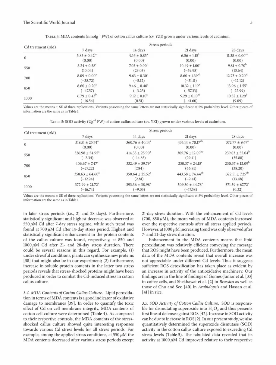
Table 4: MDA contents (nmolg−1 FW) of cotton callus culture (cv. YZ1) grown under various levels of cadmium.
Cd treatment (𝜇M) Stress periods7 days 14 days 21 days 28 days
0 5.83 ± 0.42bc(0.00)
9.16 ± 0.85a(0.00)
6.56 ± 1.15b(0.00)
11.35 ± 0.00ab(0.00)
550 5.24 ± 0.58c(10.04)
7.05 ± 0.00b(23.03)
10.49 ± 1.00a(−59.95)
9.81 ± 0.70b(13.64)
700 8.09 ± 0.00a(−38.72)
9.63 ± 0.30a(−5.12)
8.60 ± 1.39ab(−31.11)
12.73 ± 0.20ab(−12.12)
850 8.60 ± 0.20a(−47.57)
9.46 ± 0.40a(−3.25)
10.32 ± 1.19a(−57.33)
13.96 ± 1.55a(−22.99)
1000 6.79 ± 0.43b(−16.54)
9.12 ± 0.10a(0.51)
9.29 ± 0.10ab(−41.60)
10.32 ± 1.29b(9.09)
Values are the means ± SE of three replications. Variants possessing the same letters are not statistically significant at 5% probability level. Other pieces ofinformation are the same as in Table 1.
Table 5: SOD activity (Ug−1 FW) of cotton callus culture (cv. YZ1) grown under various levels of cadmium.
Cd treatment (𝜇M) Stress periods7 days 14 days 21 days 28 days
0 319.51 ± 25.74a(0.00)
360.76 ± 40.14a(0.00)
433.14 ± 70.17ab(0.00)
372.77 ± 9.67a(0.00)
550 326.98 ± 54.95a(−2.34)
414.35 ± 25.90a(−14.85)
305.76 ± 12.09bc(29.41)
239.03 ± 55.04b(35.88)
700 406.67 ± 7.47a(−27.22)
332.49 ± 39.79a(7.84)
230.37 ± 24.18c(46.81)
230.37 ± 12.09b(38.20)
850 358.63 ± 64.60a(−12.24)
350.64 ± 25.52a(2.81)
443.58 ± 74.44ab(−2.41)
322.51 ± 7.25ab(13.48)
1000 372.99 ± 21.72a(−16.74)
393.36 ± 38.98a(−9.03)
509.30 ± 44.76a(−17.58)
371.59 ± 67.72a(0.32)
Values are the means ± SE of three replications. Variants possessing the same letters are not statistically significant at 5% probability level. Other pieces ofinformation are the same as in Table 1.
in later stress periods (i.e., 21 and 28 days). Furthermore,statistically significant and highest decrease was observed at550𝜇M Cd after 7-day stress regime, while such trend wasfound at 700𝜇M Cd after 14-day stress period. Highest andstatistically significant enhancement in the protein contentsof the callus culture was found, respectively, at 850 and1000 𝜇M Cd after 21- and 28-day stress duration. Therecould be several reasons in this regard. For example, (1)under stressful conditions, plants can synthesize newproteins[38] that might also be in our experiment; (2) furthermore,increase in soluble protein contents in the latter two stressperiods reveals that stress-shocked proteins might have beenproduced in order to combat the Cd-induced stress in cottoncallus culture.
3.4. MDA Contents of Cotton Callus Culture. Lipid peroxida-tion in terms ofMDAcontents is a good indicator of oxidativedamage to membranes [39]. In order to quantify the toxiceffect of Cd on cell membrane integrity, MDA contents ofcotton cell culture were determined (Table 4). As comparedto their respective controls, the MDA contents of the stress-shocked callus culture showed quite interesting responsestowards various Cd stress levels for all stress periods. Forexample, among the applied stress conditions, at 550 𝜇M theMDA contents decreased after various stress periods except
21-day stress duration. With the enhancement of Cd levels(700, 850𝜇M), the mean values of MDA contents increasedover the respective controls after all stress applied periods.However, at 1000 𝜇Mincreasing trendwas only observed after7- and 21-day stress duration.
Enhancement in the MDA contents means that lipidperoxidation was relatively efficient conveying the messagethat ROS might have been produced. Furthermore, the meandata of the MDA contents reveal that overall increase wasnot appreciable under different Cd levels. Thus it suggestssufficient ROS detoxification has taken place as evident byan increase in activity of the antioxidative machinery. Ourfindings are in the line of findings of Gomes-Junior et al. [33]in coffee cells, and Shekhawat et al. [2] in Brassica as well asthose of Cho and Seo [40] in Arabidopsis and Hassan et al.[41] in rice.
3.5. SOD Activity of Cotton Callus Culture. SOD is responsi-ble for dismutating superoxide into H
2O2and thus presents
first line of defense against ROS [42]. Increase in SOD activitycan be due to increase inROS [2]. In our present study,we alsoquantitatively determined the superoxide dismutase (SOD)activity in the cotton callus culture exposed to exceeding Cdstress levels (Table 5). The tabulated data revealed that itsactivity at 1000 𝜇M Cd improved relative to their respective

6 The Scientific World Journal
Table 6: H2O2 (𝜇Mg−1 FW) of cotton callus culture (cv. YZ1) grown under various levels of cadmium.
Cd treatment (𝜇M) Stress periods7 days 14 days 21 days 28 days
0 71.86 ± 10.29a(0.00)
58.94 ± 6.46bc(0.00)
38.15 ± 2.55ab(0.00)
45.98 ± 6.20a(0.00)
550 45.88 ± 5.69b(36.16)
77.97 ± 7.95ab(−32.28)
35.16 ± 2.39b(7.84)
36.71 ± 5.43a(20.18)
700 58.87 ± 3.75ab(18.08)
81.71 ± 7.45a(−38.63)
36.85 ± 5.10ab(3.40)
37.26 ± 5.40a(18.98)
850 75.93 ± 6.89a(−5.66)
36.79 ± 5.72d(37.58)
46.31 ± 3.21a(−21.40)
39.76 ± 3.43a(13.54)
1000 65.34 ± 3.89ab(9.07)
49.97 ± 3.32cd(15.21)
30.43 ± 2.71b(20.23)
40.60 ± 6.84a(11.72)
Values are the means ± SE of three replications. Variants possessing the same letters are not statistically significant at 5% probability level. Other pieces ofinformation are the same as in Table 1.
controls only after 7-, 14-, and 21-day stress periods. Thisinclination trend could also be obtained at all other appliedCd levels except at 700 and 850 𝜇M Cd after 14-day stressperiod and 550 and 700 𝜇M Cd after 21-day stress duration,where the mean values of the SOD were lower than therelated control. However, the mean values of SOD in thecallus culture under various stress levels of Cd after the 28-daystress regime were declined in comparison with the control.Moreover, the SOD activities were almost higher throughoutthe experimental course, albeit nonsignificant to the relativecontrol except at 700 𝜇M Cd of the 21-day stress experiment.Such enhancement in SOD activity under Cd stress has alsobeen previously reported in different plants species [33, 41,43–47].
The overall increase in the SOD activities reveals that Cdcaused increase in ROS production. However, decrease in itsactivity in the last two stress periods particularly after 28-day period reveals that calli became capable of bearing theCd stress. This finding is further supported by a decrease inproduction of H
2O2conveying the message that less ROS
might have been produced in the later course of Cd stressperiods.
3.6. H2O2Activity of Cotton Callus Culture. H
2O2plays a
dual role in plants: at low concentrations, it acts as a signalmolecule in response to various biotic and abiotic stressesand, at high concentrations, it leads to programmed celldeath [42]. Table 6 shows mean values of hydrogen peroxide(H2O2) activity in the upland cotton callus culture grown for
various stress durations of Cd stress. According tomean data,there was almost a decreasing trend in the H
2O2activity in
stress-shocked calli over their relevant controls after differentstress periods with few exceptions. For example, at 850𝜇MCd level the mean values of the activity increased after 7- and21-day stress period, while this increasing trend was found inthe calli at 550 and 700𝜇MCd after 14-day stress duration.
According to data, H2O2was produced, which signals
that Cd stress caused oxidative stress. However, the trendwas decreasing one in all stress periods over their respectivecontrols.This decreasing trendmight be the key point that thecallus culture growth was not affected as a whole. The other
fact is that there is an increase in the activity of CAT and inmost cases in that of APX. And hence the H
2O2production
is decreasing. All these events signal that ROS scavengingenzymes were active in combating the Cd stress. This showsthat the cotton callus culture is resistant to Cd high doses.
3.7. PODActivity of Cotton Callus Culture. Peroxidase (POD)is not only one of the defense proteins, but also an importantantioxidant enzyme involved in the response to environmen-tal stresses [48]. Cd-imposed stress treatments have shownthat POD had transient behavior, which is either increased ordecreased in plants [12, 49]. The peroxidase activity (POD)of the cotton callus culture (YZ1) is shown in Table 7. Itreveals that, in comparison to respective controls, there wasa decreasing behavior of the activity at all Cd levels afterdifferent stress periods with few exceptions. For example,after 14-day stress the mean values of the POD activityprogressively increased over the control while after 21-daystress regime increase in the activity was only observed at1000 𝜇M Cd level. Moreover, among all the stress levels aswell as stress durations, the highest statistically significantincrease (77.45%) over the related control was at 1000𝜇MCd after 14-day stress duration while the lowest statisticallysignificant decrease (24.25%) was also at 1000𝜇MCd after 7-day stress period.
It shows that the decomposition of H2O2at this stage
might have been performed by POD. Our present results arenot in line with those of Cho and Seo [40] and Gomes-Junioret al. [33].
3.8. APX Activity of Cotton Callus Culture. APX is mostessential antioxidant enzymes in scavenging ROS due tohaving higher affinity for H
2O2(even in 𝜇M range) [42]. It
can detoxify H2O2under abiotic stress conditions [50]. The
ascorbate peroxidase (APX) activity also greatly varied inthe experimental cotton callus culture in our present study(Table 8). Its activity descended over their relevant controlsin the first three stress regimes except at 700 𝜇M Cd after14- and 21-day stress duration. However, after 28-day stressregimes, the APX activity first decreased at 550𝜇M Cd andincrementally enhanced at all other Cd stress levels. The data

The Scientific World Journal 7
Table 7: POD activity (OD470 g−1 FW/min) of cotton callus culture (cv. YZ1) grown under various levels of cadmium.
Cd treatment (𝜇M) Stress periods7 days 14 days 21 days 28 days
0 75.96 ± 5.10a(0.00)
52.77 ± 1.98c(0.00)
15.50 ± 0.88a(0.00)
17.63 ± 1.07a(0.00)
550 54.47 ± 1.94b(28.30)
57.80 ± 3.77cd(−9.54)
14.13 ± 0.29a(8.82)
13.37 ± 0.04c(24.20)
700 60.63 ± 1.31b(20.18)
61.72 ± 0.22c(−16.96)
14.27 ± 0.21a(7.96)
15.10 ± 0.04b(14.37)
850 66.44 ± 5.31ab(12.54)
79.73 ± 2.71b(−51.11)
14.53 ± 0.42a(6.24)
16.13 ± 0.12ab(8.51)
1000 57.54 ± 6.97b(24.25)
93.63 ± 3.50a(−77.45)
16.10 ± 1.40a(−3.87)
14.63 ± 0.10bc(17.02)
Values are the means ± SE of three replications. Variants possessing the same letters are not statistically significant at 5% probability level. Other pieces ofinformation are the same as in Table 1.
Table 8: APX activity (𝜇Mg−1 FW) of cotton callus culture (cv. YZ1) grown under various levels of cadmium.
Cd treatment (𝜇M) Stress periods7 days 14 days 21 days 28 days
0 18.16 ± 1.42a(0.00)
12.74 ± 1.13b(0.00)
48.21 ± 1.49a(0.00)
49.49 ± 1.94bc(0.00)
550 9.72 ± 1.09c(46.44)
8.14 ± 0.62c(36.11)
48.00 ± 0.62a(0.44)
47.59 ± 1.75c(3.85)
700 12.64 ± 1.82bc(30.39)
17.06 ± 1.61a(−33.87)
49.50 ± 1.45a(−2.67)
50.36 ± 0.49bc(−1.75)
850 16.76 ± 0.85ab(7.70)
10.07 ±0.50bc(20.98)
47.14 ± 3.34a(2.22)
58.76 ± 2.01a(−18.72)
1000 13.29 ± 2.60abc(26.84)
3.85 ± 0.64d(67.32)
47.80 ± 1.44a(0.85)
53.41 ± 0.92b(−6.51)
Values are the means ± SE of three replications. Variants possessing the same letters are not statistically significant at 5% probability level. Other pieces ofinformation are the same as in Table 1.
further reveals that the observed decrease was statisticallysignificant only at 1000 𝜇MCd after 14-day stress shocks. Andthe significant relative was only found at 850 𝜇MCd after the28-day stress period.
This decreasing behavior was also noticed by Shekhawatet al. [2] during their studies in callus cultures of Brassica.The overall inactivation of APX enzyme might be due tometal-sulfhydryl binding Shekhawat et al. [2]. Our results areconsistent with findings of Sharma et al. [51] in barley andIsrar et al. [52] in Sesbania callus.
3.9. CAT Activity of Cotton Callus Culture. CAT is among theH2O2-scavenging enzymes. The balance between the activity
of H2O2-producing and H
2O2-scavenging enzymes plays
an important role in providing a plant defense mechanismagainst any oxidative damage [38]. The CAT activity alsoshowed obvious results in cotton callus culture after variousCd stressful regimes (Table 9). In comparison to their relatedcontrols, its mean values significantly increased after 7-,21-, and 28-day Cd treatment. However, there was found adecreasing trend over the control with the addition of moreCd in the growing medium after 14-day stress time exceptat 1000 𝜇M Cd, where the CAT activity was 63.55% higherover its respective control.Moreover, after 7-day stress period,
the CAT activity linearly increased, while in case of 21- and28-day stress regimes the increase in the activity over therespective control was severalfold higher with increase in theconcentration of Cd.
In our present study, the CAT activity was activated asa whole. That is why the overall fresh biomass productionshowed upward trend. So it means that POD was activeto decompose H
2O2produced as a result of SOD activity.
Similar upward trends have been noticed by other workers[2, 33, 34, 43, 44, 49]. However, there was observed a decreasein the CAT activity of Brassica callus by Shekhawat et al. [2]and Sandalio et al. [53] in Pisum sativum.
Both APX and CAT showed dissimilar trend in ourpresent study. This might be because both enzymes areworking on the same substrate (H
2O2). Therefore, the detox-
ification of H2O2occurred mainly through CAT and that is
why APX activity was declined due to the lesser availabilityof substrate. Another possible reason for the decreased APXactivity could be induced inactivation of APX enzyme.
4. Conclusion
(i) Cell growth andMDA contents are the two importantindicators which show whether oxidative damage has

8 The Scientific World Journal
Table 9: CAT activity (U/g FW) of cotton callus culture (cv. YZ1) grown under various levels of cadmium.
Cd treatment (𝜇M) Stress periods7 days 14 days 21 days 28 days
0 2.88 ± 0.24d(0.00)
5.71 ± 0.14b(0.00)
4.57 ± 0.24c(0.00)
16.573 ± 22.30a(0.00)
550 4.76 ± 0.21b(−65.47)
3.24 ± 0.42c(43.22)
38.84 ± 5.37b(−749.82)
22.95 ± 5.66c(−38.48)
700 3.62 ± 0.25c(−25.72)
3.62 ± 0.27c(36.57)
79.17 ± 11.42a(−1632.46)
56.99 ± 5.37bc(−243.89)
850 4.95 ± 0.12b(−72.07)
2.28 ± 0.16d(59.99)
71.72 ± 15.87a(−1469.44)
44.77 ± 6.27c(−170.11)
1000 6.66 ± 0.24a(−131.40)
9.33 ± 0.17a(−63.55)
12.20 ± 2.54bc(−167.03)
88.88 ± 9.45b(−436.28)
Values are the means ± SE of three replications. Variants possessing the same letters are not statistically significant at 5% probability level. Other pieces ofinformation are the same as in Table 1.
been caused or not. Here in case of our present studycell growth in terms of relative fresh weight growthrates was not significantly affected. However, the lipidperoxidation was relatively efficient conveying themessage that ROS have been produced.This is furthertestified by the increase in the activity of SOD, CAT,and so forth.
(ii) In our present findings, the overall H2O2activity
was downregulated in all stress periods over theirrespective controls. So due to less production ofH2O2, the overall growth efficiency of the callus under
Cd-exposed conditions was unaffected.(iii) The high MDA contents and SOD show that mem-
brane damage and oxidative stress have been caused.However, low H
2O2concentrations establish that
this is presumably suppressed by the strong antioxi-dant system prevailing in cotton callus culture moreimportantly in the order of CAT > APX > POD.
(iv) The present study set a new avenue to explore themolecular mechanisms in cotton callus culture bothat genetic and proteomic levels under Cd stress.
Abbreviations
APX: Ascorbate peroxidaseCAT: CatalaseMDA: MalondialdehydePOD: PeroxidaseROS: Reactive oxygen speciesSOD: Superoxide dismutaseH2O2: Hydrogen peroxide.
Conflict of Interests
The authors declare that they have no conflict of interests.
Acknowledgment
The study was funded by the National Natural Science Foun-dation of China (31371667).
References
[1] M. K. Daud, Y. Q. Sun, M. Dawood et al., “Cadmium-inducedfunctional and ultrastructural alterations in roots of two trans-genic cotton cultivars,” Journal of Hazardous Materials, vol. 161,no. 1, pp. 463–473, 2009.
[2] G. S. Shekhawat, K. Verma, S. Jana, K. Singh, P. Teotia,and A. Prasad, “In vitro biochemical evaluation of cadmiumtolerancemechanism in callus and seedlings of Brassica juncea,”Protoplasma, vol. 239, no. 1–4, pp. 31–38, 2010.
[3] M. N. V. Prasad, “Cadmium toxicity and tolerance in vascularplants,” Environmental and Experimental Botany, vol. 35, no. 4,pp. 525–545, 1995.
[4] C. Foyer, H. Lopez-Delgado, J. F. Dat, and I. Scott, “Hydrogenperoxide- and glutathione-associated mechanisms of acclima-tory stress tolerance and signalling,” Physiologia Plantarum, vol.100, no. 2, pp. 241–254, 1997.
[5] E. Olmos, J. R. Martınez-Solano, A. Piqueras, and E. Hellın,“Early steps in the oxidative burst induced by cadmium incultured tobacco cells (BY-2 line),” Journal of ExperimentalBotany, vol. 54, no. 381, pp. 291–301, 2003.
[6] H. Zhang, Y. Jiang, Z. He, andM.Ma, “Cadmium accumulationand oxidative burst in garlic (Allium sativum),” Journal of PlantPhysiology, vol. 162, no. 9, pp. 977–984, 2005.
[7] G. S. Shekhawat, A. Prasad, K. Verma et al., “Changes in growth,lipid peroxidation and antioxidant system in seedlings ofBrassica juncea (L.) czern. under cadmium stress,” Biochemicaland Cellular Archives, vol. 8, pp. 145–149, 2008.
[8] O. Ouariti, H. Gouia, and M. H. Ghorbal, “Responses of beanand tomato plants to cadmium: growth, mineral nutrition, andnitrate reduction,” Plant Physiology and Biochemistry, vol. 35,no. 5, pp. 347–354, 1997.
[9] A. S. Molina, C. Nievas, M. V. P. Chaca et al., “Cadmium-induced oxidative damage and antioxidative defense mecha-nisms in Vigna mungo L,” Plant Growth Regulation, vol. 56, no.3, pp. 285–295, 2008.
[10] K. Verma, G. S. Shekhawat, A. Sharma, S. K. Mehta, and V.Sharma, “Cadmium induced oxidative stress and changes insoluble and ionically bound cell wall peroxidase activities inroots of seedling and 3-4 leaf stage plants of Brassica juncea (L.)czern,” Plant Cell Reports, vol. 27, no. 7, pp. 1261–1269, 2008.
[11] A. Fodor, A. Szabo-Nagy, and L. Erdei, “The effects of cadmiumon the fluidity and H+-ATPase activity of plasma membrane

The Scientific World Journal 9
from sunflower and wheat roots,” Journal of Plant Physiology,vol. 147, no. 1, pp. 87–92, 1995.
[12] D. A. Meloni, M. A. Oliva, C. A. Martinez, and J. Cambraia,“Photosynthesis and activity of superoxide dismutase, perox-idase and glutathione reductase in cotton under salt stress,”Environmental and Experimental Botany, vol. 49, no. 1, pp. 69–76, 2003.
[13] V. Niknam, A. A. Meratan, and S. M. Ghaffari, “The effectof salt stress on lipid peroxidation and antioxidative enzymesin callus of two Acanthophyllum species,” In Vitro Cellular &Developmental Biology: Plant, vol. 47, no. 2, pp. 297–308, 2011.
[14] B. V. Somashekaraiah, K. Padmaja, and A. R. K. Prassad,“Phytotoxicity of cadmium ions on germinating seedlings ofmung bean (Phaseolus vulgaris): involvement of lipid peroxidesin chlorophyll degradation,” Physiologia Plantarum, vol. 85, no.1, pp. 85–89, 1992.
[15] A. Chaoui, S. Mazhoudi, M. H. Ghorbal, and E. El Ferjani,“Cadmium and zinc induction of lipid peroxidation and effectson antioxidant enzyme activities in bean (Phaseolus vulgarisL.),” Plant Science, vol. 127, no. 2, pp. 139–147, 1997.
[16] G. S. Shekhawat, A. Batra, and S. Mathur, “A reliable in vitroprotocol for rapidmass propagation ofAzadirachta indica Juss,”Journal of Plant Biology, vol. 29, no. 1, pp. 109–112, 2002.
[17] S. Mathur, A. Batra, and G. S. Shekhawat, “An efficient in vitromethod for mass propagation of Salvadora persica via apicalmeristem,” Journal of Plant Biochemistry and Biotechnology, vol.11, no. 2, pp. 125–127, 2002.
[18] S. Mathur, G. S. Shekhawat, and A. Batra, “Micropropagation ofSalvadora persica Linn. via cotyledonary nodes,” Indian Journalof Biotechnology, vol. 1, no. 2, pp. 197–200, 2002.
[19] S. Mathur, G. S. Shekhawat, and A. Batra, “Somatic embryo-genesis and plantlet regeneration from cotyledon explants ofSalvadora persica L,” Phytomorphology, vol. 58, no. 1-2, pp. 57–63, 2008.
[20] S. M. Gallego, M. P. Benavides, and M. L. Tomaro, “Effectof heavy metal ion excess on sunflower leaves; evidence forinvolvement of oxidative stress,” Plant Science, vol. 121, no. 2, pp.151–159, 1996.
[21] R. Sobkowiak, K. Rymer, R. Rucinska, and J. Deckert,“Cadmium-induced changes in antioxidant enzymes in suspen-sion culture of soybean cells,” Acta Biochimica Polonica, vol. 51,no. 1, pp. 219–222, 2004.
[22] D. R. Gossett, E. P. Millhollon, M. C. Lucas, S. W. Banks, andM. M. Marney, “The effects of NaCl on antioxidant enzymeactivities in callus tissue of salt-tolerant and salt-sensitive cottoncultivars (Gossypium hirsutum L.),” Plant Cell Reports, vol. 13,no. 9, pp. 498–503, 1994.
[23] L. C. Garratt, B. S. Janagoudar, K. C. Lowe, P. Anthony, J. B.Power, and M. R. Davey, “Salinity tolerance and antioxidantstatus in cotton cultures,” Free Radical Biology andMedicine, vol.33, no. 4, pp. 502–511, 2002.
[24] T. Murashige and F. Skoog, “A revised medium for rapidgrowth and bioassays with tobacco tissue cultures,” PhysiolgiaPlantarum, vol. 56, pp. 473–497, 1962.
[25] W. J. Zhou and M. Leul, “Uniconazole-induced alleviation offreezing injury in relation to changes in hormonal balance,enzyme activities and lipid peroxidation in winter rape,” PlantGrowth Regulation, vol. 26, no. 1, pp. 41–47, 1998.
[26] M. M. Bradford, “A rapid and sensitive method for the quanti-tation of microgram quantities of protein utilizing the principleof protein dye binding,”Analytical Biochemistry, vol. 72, no. 1-2,pp. 248–254, 1976.
[27] W. Zhou, D. Zhao, and X. Lin, “Effects of waterlogging onnitrogen accumulation and alleviation of waterlogging damageby application of nitrogen fertilizer and mixtalol in winter rape(Brassica napus L.),” Journal of Plant Growth Regulation, vol. 16,no. 1, pp. 47–53, 1997.
[28] S. Jana and M. A. Choudhuri, “Glycolate metabolism ofthree submersed aquatic angiosperms during ageing,” AquaticBotany, vol. 12, pp. 345–354, 1981.
[29] W. J. Zhou and M. Leul, “Uniconazole-induced tolerance ofrape plants to heat stress in relation to changes in hormonallevels, enzyme activities and lipid peroxidation,” Plant GrowthRegulation, vol. 27, no. 2, pp. 99–104, 1999.
[30] Y. Nakano and K. Asada, “Hydrogen peroxide is scavengedby ascorbate-specific peroxidase in spinach chloroplasts,” Plantand Cell Physiology, vol. 22, no. 5, pp. 867–880, 1981.
[31] D. E. M. Radwan, K. A. Fayez, S. Y. Mahmoud, A. Hamad, andG. Lu, “Salicylic acid alleviates growth inhibition and oxida-tive stress caused by zucchini yellow mosaic virus infectionin Cucurbita pepo leaves,” Physiological and Molecular PlantPathology, vol. 69, no. 4–6, pp. 172–181, 2006.
[32] B. Joseph andD. Jini, “Development of salt stress-tolerant plantsby gene manipulation of antioxidant enzymes,” Asian Journal ofAgricultural Research, vol. 5, no. 1, pp. 17–27, 2011.
[33] R. R.Gomes-Junior, C. A.Moldes, F. S. Delite et al., “Antioxidantmetabolism of coffee cell suspension cultures in response tocadmium,” Chemosphere, vol. 65, no. 8, pp. 1330–1337, 2006.
[34] R. F. Fornazier, R. R. Ferreira, G. J. G. Pereira et al., “Cadmiumstress in sugar cane callus cultures: effect on antioxidantenzymes,” Plant Cell, Tissue and Organ Culture, vol. 71, no. 2,pp. 125–131, 2002.
[35] R. Sobkowiak and J. Deckert, “Cadmium-induced changes ingrowth and cell cycle gene expression in suspension-culturecells of soybean,” Plant Physiology and Biochemistry, vol. 41, no.8, pp. 767–772, 2003.
[36] H. Hirt, G. Casari, and A. Barta, “Cadmium-enhanced geneexpression in suspension-culture cells of tobacco,” Planta, vol.179, no. 3, pp. 414–420, 1989.
[37] B. Chakravarty and S. Srivastava, “Toxicity of some heavymetals in vivo and in vitro in Helianthus annuus,” MutationResearch, vol. 283, no. 4, pp. 287–294, 1992.
[38] S. Torabi and V. Niknam, “Effects of iso-osmotic concentrationsof NaCl and mannitol on some metabolic activity in callusesof two Salicornia species,” In Vitro Cellular & DevelopmentalBiology: Plant, vol. 47, no. 6, pp. 734–742, 2011.
[39] M. Dacosta and B. Huang, “Changes in antioxidant enzymeactivities and lipid peroxidation for bentgrass species inresponse to drought stress,” Journal of the American Society forHorticultural Science, vol. 132, no. 3, pp. 319–326, 2007.
[40] U. Cho and N. Seo, “Oxidative stress in Arabidopsis thalianaexposed to cadmium is due to hydrogen peroxide accumula-tion,” Plant Science, vol. 168, no. 1, pp. 113–120, 2005.
[41] M. J. Hassan, G. P. Zhang, F. B. Wu, K. Wei, and Z. Chen,“Zinc alleviates growth inhibition and oxidative stress causedby cadmium in rice,” Journal of Plant Nutrition and Soil Science,vol. 168, no. 2, pp. 255–261, 2005.
[42] S. S. Gill and N. Tuteja, “Reactive oxygen species and antioxi-dant machinery in abiotic stress tolerance in crop plants,” PlantPhysiology and Biochemistry, vol. 48, no. 12, pp. 909–930, 2010.
[43] A. P. Vitoria, P. J. Lea, and R. A. Azevedo, “Antioxidant enzymesresponses to cadmium in radish tissues,” Phytochemistry, vol. 57,no. 5, pp. 701–710, 2001.

10 The Scientific World Journal
[44] F. B. Wu, G. Zhang, and P. Dominy, “Four barley genotypesrespond differently to cadmium: lipid peroxidation and activ-ities of antioxidant capacity,” Environmental and ExperimentalBotany, vol. 50, no. 1, pp. 67–78, 2003.
[45] T. R. Guo, G. P. Zhang, M. X. Zhou, F. Wu, and J. Chen, “Effectsof aluminum and cadmium toxicity on growth and antioxidantenzyme activities of two barley genotypes with different Alresistance,” Plant and Soil, vol. 258, no. 1-2, pp. 241–248, 2004.
[46] Y. T. Hsu and C. H. Kao, “Cadmium toxicity is reduced by nitricoxide in rice leaves,” Plant Growth Regulation, vol. 42, no. 3, pp.227–238, 2004.
[47] M. P. Benavides, S. M. Gallego, and M. L. Tomaro, “Cadmiumtoxicity in plants,” Brazilian Journal of Plant Physiology, vol. 17,no. 1, pp. 21–34, 2005.
[48] L. Flohe and F. Ursini, “Peroxidase: a term of many meanings,”Antioxidants & Redox Signaling, vol. 10, no. 9, pp. 1485–1490,2008.
[49] K. B. Balestrasse, L. Gardey, S. M. Gallego, and M. L. Tomaro,“Response of antioxidant defence system in soybean nodulesand roots subjected to cadmium stress,” Australian Journal ofPlant Physiology, vol. 28, no. 6, pp. 497–504, 2001.
[50] P. L. Gratao, A. Polle, P. J. Lea, and R. A. Azevedo, “Makingthe life of heavy metal-stressed plants a little easier,” FunctionalPlant Biology, vol. 32, no. 6, pp. 481–494, 2005.
[51] Y. K. Sharma, J. Leon, I. Raskin, and K. R. Davis, “Ozone-induced responses in Arabidopsis thaliana: the role of salicylicacid in the accumulation of defense-related transcripts andinduced resistance,” Proceedings of the National Academy ofSciences of the United States of America, vol. 93, no. 10, pp. 5099–5104, 1996.
[52] M. Israr, S. V. Sahi, and J. Jain, “Cadmium accumulation andantioxidative responses in the Sesbania drummondii callus,”Archives of Environmental Contamination and Toxicology, vol.50, no. 1, pp. 121–127, 2006.
[53] L. M. Sandalio, H. C. Dalurzo, M. Gomez, M. C. Romero-Puertas, and L. A. del Rıo, “Cadmium-induced changes in thegrowth and oxidative metabolism of pea plants,” Journal ofExperimental Botany, vol. 52, no. 364, pp. 2115–2126, 2001.