impaired working-memory after cerebellar infarcts paralleled by changes in bold signal of a...
TRANSCRIPT

A
witiac(dip©
K
1
iwaasdcc
(
0d
Neuropsychologia 45 (2007) 2016–2024
Impaired working-memory after cerebellar infarcts paralleled bychanges in BOLD signal of a cortico-cerebellar circuit
B. Ziemus a,e, O. Baumann c, R. Luerding a, R. Schlosser d,G. Schuierer b, U. Bogdahn a, M.W. Greenlee c,∗
a Department of Neurology, University of Regensburg, Germanyb Department of Neuroradiology BKR, Regensburg, Germany
c Institute for Experimental Psychology, University of Regensburg, Germanyd Department of Psychiatry, University of Jena, Germany
e Clinical Neurophysiology, University of Maastricht, Germany
Received 17 September 2006; received in revised form 2 February 2007; accepted 11 February 2007Available online 23 February 2007
bstract
A considerable body of evidence supports the notion that cerebellar lesions lead to neuropsychological deficits, including impairments inorking-memory, executive tasks and verbal fluency. Studies employing functional magnetic resonance imaging (fMRI) and anatomical tracing
n primates provide evidence for a cortico-cerebellar circuitry as the functional substrate of working-memory. The present fMRI study exploreshe activation pattern during an n-back working-memory task in patients with an isolated cerebellar infarct. To determine each patient’s cognitivempairment, neuropsychological tests of working-memory and attention were carried out. We conducted fMRI in nine patients and nine healthyge-matched controls while they performed a 2-back task in a blocked-design. In both groups we found bilateral activations in a widespreadortico-cerebellar network, consisting of the ventrolateral prefrontal cortex (BA 44, 45), dorsolateral prefrontal cortex (BA 9, 46), parietal cortex
BA 7, 40), pre-supplementary motor area (BA 6) anterior cingulate (BA 32). Relative to healthy controls, patients with isolated cerebellar infarctsemonstrated significantly more pronounced BOLD-activations in the precuneus and the angular gyrus during the 2-back task. The significantncrease in activation in the posterior parietal areas of the cerebellar patients could be attributed to a compensatory recruitment to maintain taskerformance. We conclude that cerebellar lesions affect remote cortical regions that are part of a putative cortico-cerebellar network.2007 Elsevier Ltd. All rights reserved.
tllW2Srm
eywords: Verbal working-memory; Cerebellum; Cerebellar stroke; fMRI
. Introduction
There is mounting evidence that the cerebellum participatesn higher-order cognitive tasks such as executive processing,orking-memory, verbal fluency and planning. Schmahmann
nd Sherman (1998) described for the first time a cognitive-ffective syndrome following cerebellar lesions with executive,patial, linguistic and affective symptoms. They postulate a
isruption in a widespread cortico-cerebellar circuitry as aause of impaired cognitive functions denoted as a fronto-erebellar disconnection syndrome. An anatomical substrate for∗ Corresponding author. Tel.: +49 941 943 3281; fax: +49 941 943 3233.E-mail address: [email protected]
M.W. Greenlee).
B&B
piwi
028-3932/$ – see front matter © 2007 Elsevier Ltd. All rights reserved.oi:10.1016/j.neuropsychologia.2007.02.012
hese functions is a cerebellar feedback loop through the tha-amus to the prefrontal and parietal cortex (inferior parietalobule) as it already has been reported for primates (Clower,
est, Lynch, & Strick, 2001; Middleton & Strick, 1994,000, 2001; Schmahmann, 1991; Schmahman & Pandya, 1995;chmahmann & Sherman, 1998). Several neuropsychologicaleports indicate the presence of working-memory impair-ents due to an isolated cerebellar lesion (Botez-Marquard,ard, Leveille, & Botez, 2001; Gottwald, Wilde, Mihajlovic,
Mehdorn, 2004; Malm et al., 1998; Neau, Arroyo-Anllo,onnaud, Ingrand, & Gil, 2000).
In a recent publication, Ravizza et al. (2006) investigated 15
atients with cerebellar damage and found selective impairmentsn verbal working-memory. Articulatory rehearsal strategiesere unaffected thereby supporting non-motor causes for thempaired verbal working-memory. These clinical observations

cholo
apDaiitansrsulttCtwaitMtaac
ttuOz(Bcafl(
w
csp
2
prp
2
mtfIsria(
a(
(tcfcf
2
aoVrwmN
Ff
B. Ziemus et al. / Neuropsy
nd above-mentioned anatomical findings in primates are com-atible with the results of functional neuroimaging studies:esmond, Gabrieli, Wagner, Ginier, & Glover (1997) and Chen
nd Desmond (2005) identified two cerebellar regions activatedn verbal working-memory (a bilateral superior region and annferior region on the right side) in addition to activations inhe inferior parietal lobule (BA 40), Broca area (BA 6, 44) andnterior cingulum (BA 32). They propose two cortico-cerebellaretworks for verbal working-memory: an articulatory controlystem with involvement of Broca’s area (BA 6, 44) and the supe-ior cerebellum (simplex lobule and crus I) and a phonologicaltorage system connecting parietal areas (inferior parietal lob-le) with the inferior cerebellum. Although the inferior parietalobule is frequently mentioned as the likely locus of verbal short-erm memory storage (Fiez et al., 1996; Smith & Jonides, 1998)his view has been recently challenged by Ravizza, Delgado,hein, Becker, and Fiez (2004), who identified two regions of
he intraparietal sulcus, one ventral and the other more dorsal,ith the dorsal region responding to short-term memory load
nd the ventral region being involved in phonological encod-ng procedures. Neither of these two regions fulfilled, however,he requirements for proper phonological short-term storage.
ajerus et al. (2006) deny this specific role of the parietal cor-ex in working-memory processes and describe its function as
more general superior attentional modulator, shifting focalttention to underlying subordinate networks, according to theognitive process in question.
The VLPFC (BA 10, 47, 44 and 45) has been especially foundo be activated in short-term maintenance (Owen, 2000) and inasks that require selection, comparison and judgement of stim-li held in short-term and long-term memory (Petrides, 1994).n the other hand, the manipulation of information, reorgani-
ation and control of working-memory requires mid-DLPFCBA 9, 46) (Bor, Duncan, Wiseman, & Owen, 2003; D’Esposito,allard, Aguirre, & Zarahn, 1998; Petrides, 1998). Neuropsy-hological studies support the view that the role of DLPFCctivations in working-memory tasks is to increase task per-ormance and facilitate memory, reducing the overall cognitive
oad with the help of structuring and categorizing informationBor, Cumming, Scott, & Owen, 2004; Bor et al., 2003).In our study, we test the hypothesis that impairments inorking-memory can result from damage to this putative
1aFOm
ig. 1. Representative transversal slices of cerebellar infarcts. T1-weighted MRI at tMRI. Arrows denote location of lesions.
gia 45 (2007) 2016–2024 2017
ortico-cerebellar network. Altered BOLD-activation in remoteupratentorial brain regions underlying working-memory wouldoint to a role of the cerebellum in human cognition.
. Methods
The study was approved by the local ethics committee and has therefore beenerformed in accordance with the ethical standards laid down in the 1964 Decla-ation of Helsinki. Written informed consent was obtained from all participantsrior to their inclusion in the study.
.1. Subjects
Nine patients with isolated cerebellar infarctions (five men, four women:ean age 46.2 years, S.D. = 8.1 years; range 38–63 years) were recruited from
he Neurology Department of the University Hospital of Regensburg. All patientsulfilled the inclusion criteria of isolated stroke in cerebellum detected by MRI.nfarct size and location (specified on the basis of affiliation to arterial bloodupply) were determined on the basis of T2-weighted and in cases of territo-ial infarcts as well on T1-weighted images by a neuroradiologist (Fig. 1). Wencluded patients in an acute phase of stroke (2–8 days past infarction), as wells patients in a post-acute phase (2 month past infarction) or in a chronic phaseup to 6 years after cerebellar infarction).
A control group consisted of nine healthy controls matched for agend gender for each patient (mean age 44.2, S.D. = 9.6; range 35–63 years)Table 1).
The average difference in age between matching pairs was 2.4 yearsS.D. = 2.7; range 0–7 years). All study subjects were right handed accordingo the Edinburgh Handedness Inventory. Exclusion criteria were the use of psy-hotropic medication, vascular damage in other brain regions or a history oformer strokes, cognitive impairment due to dementia, history of neurologi-al and/or psychiatric illness, claustrophobia, pregnancy and the presence oferromagnetic surgical pins.
.2. Neuropsychological assessment
All patients were tested with a neuropsychological assessment battery. Verbalnd non-verbal cognition was measured using a short form of the German versionf the Wechsler Intelligence Scale for Adults Revised (WAIS-R; Tewes, 1994).erbal long-term-memory was measured with the Logical Memory delayed free
ecall (LM II; Wechsler, 1987). Non-verbal long-term-memory was assessedith the Rey Complex Figure delayed free recall (Lezak, 1995). Verbal working-emory was measured with the digit span forward and backward (Tewes, 1994).on-verbal working-memory was tested with the Corsi block span (Milner,
971). The results of part B of the Trail Making Test (TMT-B) is reported asn attention measure (Lezak, 1995). The Ruff 2&7 Test (Ruff, Niemann, Allen,arrow, & Wylie, 1992) was employed to measure attention. The Controlledral Word Association Test (COWA; Benton & Hamsher, 1989) provided aeasure of lexical verbal fluency. Semantic verbal fluency was measured byhe level of maximal infarct volume for each patient, acquisition at time of the

2018 B. Ziemus et al. / Neuropsychologia 45 (2007) 2016–2024
Table 1Overview of patient characteristics
Patient Age Sex Infarct age Infarct localization Size (cm) Neuropsychological function z-Score
1 63 M 5 yrs Left PICA 3 × 3 × 3
Verbal working-memory −1Non-verbal working-memory −0.4Attention 2.2Word fluency −2.5
2 39 M 4 yrs Right SCA 1.5 × 2 × 1.5Verbal working-memory 0.2Non-verbal working-memory −1.3Attention −0.3
3 38 F 18 m Right SCA 0.3 × 0.3 × 0.5Verbal working-memory −1.1Non-verbal working-memory 1.2Attention 2.3
4 43 F 6 yrs Left PICA 3 × 2 × 0.5Verbal working-memory −1Non-verbal working-memory −1.1Attention −1.3
5 48 M 3 yrs Right PICA 4 × 2 × 5Verbal working-memory −1.3Non-verbal working-memory 0.7Attention −2
6 54 F 8 d Left PICA 3.5 × 2 × 0.5Verbal working-memory −1.9Non-verbal working-memory −1.1Attention −1.9
7 48 M 2 d Left PICA 0.5 × 0.3 × 0.5, 0.5 × 0.5 × 0.4
Verbal working-memory 0.6Non-verbal working-memory −1.9Attention −1.3Word fluency −1.8
8 42 M 14 m Right PICA, left SCA 2 × 2 × 2, 0.5 × 0.4 × 0.5Verbal working-memory −1.1Non-verbal working-memory 3.3Attention 1.3
9 41 M 2 m Left PICA, left PICA 1 × 1 × 1, 0.5 × 0.4 × 0.5
Verbal working-memory −1.4Non-verbal working-memory −2.3
d , supe
aLn
rhtp
iIi
2
N1PSwil(fitw
ttwtpuiro
2
(ffweomsr
, days; F, female; M, male; m, month; PICA, posterior cerebellar artery; SICA
sking participant to produce animal names in 60 s (Aschenbrenner, Tucha, &ange, 2000). Test values are described as z-scores with respect to age-matchedorms.
The group was not uniform regarding education and general IQ, so neu-opsychological test results of intact and impaired abilities were also veryeterogeneous. Because of these interindividual differences we assessed cogni-ive deficits relative to the stable measure of the full-scale IQ for the individualatient.
Although following cerebellar infarction the full-scale IQ can be diminished,t is one of the most stable of all neuropsychological measures. The full-scaleQ consists of verbal, non-verbal, education-related and non-education-relatedtems.
.3. Working-memory paradigm
All participants performed a 2-back working-memory task (Braver, Cohen,ystrom, Jonides, Smith, & Noll, 1997; Cohen et al., 1994; Gevins and Cutillo,993; Mellers et al., 1995) during fMRI acquisition. With the help of theC-controlled stimulation software Presentation (Version 9.2, Neurobehavioralystems, Inc.) a pseudorandom sequence of uppercase characters of the alphabetas presented on a back-projection screen. During the 2-back condition partic-
pants had to press a button with the right index finger whenever the presented
etter was the same as the one before the last (2-back). In the control condition0-back) they just had to press a button each time the letter “X” appeared. Abre optic response box (Lumitouch, Photon Inc.) was used. Stimulus presen-ation time for one letter was 1300 ms with an interstimulus interval of 700 msith 21.4% targets. Subjects were scanned through four alternating blocks of
Eiate
Attention −1Word fluency −1.3
rior cerebellar artery; yrs, years.
he 2-back and 0-back conditions over a total of eight epochs. Each experimen-al epoch lasted 57 s. At the beginning of each epoch the subject was informedhich task to perform via written instructions (3 s per epoch), resulting in a
otal epoch length of 60 s and an experimental length of 8 min. To score therecision of hits and false positives, we used a discrimination score DS basedpon the ability to distinguish targets from distractor items: {1 − [(false pos-tives + misses)/(targets + distractors)]}× 100. The DS varies from 100% (allesponses are hits or correct rejections) to 0% (all responses are false positivesr misses).
.4. MRI-imaging
The functional studies were performed on a 1.5 T Siemens MagnetomSonata, Siemens, Erlangen, Germany) equipped with a fast gradient systemor echo-planar (EPI) imaging and a eight-channel phase array full-head radio-requency (RF) receive-transmit headcoil (MR-Devices). Functional imagingas performed using a T2*-weighted gradient echo planar imaging (EPI) cov-
ring the whole brain. We acquired volumes with 39 axial slices with a gapf 0.3 mm and could thus image the entire brain. The field of view (FOV)easured 192 mm with a voxel matrix size 64 × 64, resulting in a voxel
ize of 3 mm × 3 mm × 3 mm. The TR was 3500 ms. The time to echo cor-esponded to TE = 50 ms, the flip angle corresponded to 90◦. The first two
PI images were discarded as “dummy” images before the start of the exper-mental paradigm in order to obtain steady-state. After the functional runs,natomical high-resolution sagittal T1-weighted images were acquired withhe MP-Rage sequence (magnetization prepared, rapid acquisition gradientcho).
cholo
2
SCmum
cTicsmaE1
wwar0
fipfmds
atfit(auMitSegcp
3
3
poiesiemip
3
2T(2pastfTt
3
3
npfcpsieo
3
tgtpal16afeap
olF
3
B. Ziemus et al. / Neuropsy
.5. fMRI data analysis
Data were processed and analyzed on a single-subject level usingtatistical Parametric Mapping SPM 2 (SPM2, Wellcome Department ofognitive Neurology, London, UK; http://www.fil.ion./ucl.ac.uk/spm/) imple-ented in MATLAB (The MathWorks Inc.). Echo planar images were
nwarped and realigned to the first acquired volume to correct for headovement.
The images were then transformed into a standard stereotaxic anatomi-al space (Friston, Frith, Liddle, & Frackowiak, 1991; Friston et al., 1995a;alairach & Tournoux, 1998). We used the following procedure to normal-
ze the scans: a T2*-weighted mean image of the unsmoothed images waso-registered with the corresponding anatomical T1 weighted image of theame individual. The individual T1-image was used to derive the transfor-ation parameters for the stereotaxic fit using the MNI-Template (Friston et
l., 1995a), which were then applied to the individual single co-registeredPI images. The normalized images were smoothed with a Gaussian filter of2 mm.
Analysis using the General Linear Model (GLM) (Friston et al., 1995b)as done after applying high-pass filtering (cut-off: 180 s). The periods inhich subjects performed the task were modeled separately for the 2-back
nd the 0-back condition by using a boxcar convolved with the hemodynamicesponse function. The specified contrast on single subject level was “2Back >Back”.
On random-effects group level, we used the “2Back > 0Back” contrast imageor every subject representing the difference between the two conditions on anndividual level. These images were analyzed on the group level with the non-arametric SnPM-Toolbox (Holmes, Blair, Watson, & Ford, 1996), with the testor “2 groups, 1 scan per subject”, the non-parametric equivalent of a t-test. Thisethod uses the assumption that the contrast values of non-activated voxels
istribute evenly around zero. We chose a non-parametric approach, since theample size was quite small.
A 3D variance smoothing using a FWHM of 8 mm was performed. Vari-nce smoothing can enhance the power of the group analysis even abovehe parametric methods of Gaussian random fields if the assumption of suf-cient smoothness of the parametric maps is violated. For small group sizes
his is often the case. Voxels surpassing a statistical threshold of p = 0.05Tmax-contrast analysis, corrected for multiple comparisons) were identifieds activated. MNI coordinates were transformed to Talairach coordinates bysing the WFU-Pickatlas (Wake Forest University Pickatlas, Version 1.02;aldijan, Laurienti, Kraft, & Burdette, 2003). The anatomical locations were
dentified by using the program MSU by S. Pakhomov. This tool relies onhe mni2tal program combined with data of the Talairach demon (Lancaster,ummerln, Rainey, Freitas, & Fox, 1997; Lancaster et al., 2000). Appar-nt cerebellar activations of the cerebellum are not reported for the patientroup, since even with an optimized normalization method the activationsould be artificial due to the lesions and would therefore not be inter-retable.
. Results
.1. Neuropsychological test results
Table 1 shows the results of the neuropsychological testserformed on the patient group. Differences between z-scoresn digit span and full-scale IQ indicate that the patients werempaired in verbal working-memory functions. z-Score differ-nces between non-verbal working-memory (Corsi block span)cores and full-scale IQ served as an indicator of impairment
n non-verbal memory functions after infarction. All patientsxhibited a deficit in either verbal or non-verbal working-emory with a z-score difference greater than −1. Impairmentsn attention were observed in six, in verbal fluency in threeatients (Table 1).
bbpt
gia 45 (2007) 2016–2024 2019
.2. Task performance in fMRI study
There was no significant difference in performance of the-back task during scanning between patients and controls (U-est, p = 0.24). Patients achieved a mean discrimination scoreDS) of 91.9%, whereas the controls had a DS of 89.1% in the-back task. In the 0-back condition, no differences (U-Test,= 0.572) were evident between the patients (mean DS = 99.6%)nd the controls (mean DS = 99.3%). Reaction times showed aignificant delay for the 2-back-task compared to the 0-back-ask. (Wilcoxon-test, p = 0.008). No significant differences wereound for the reaction times between patients and controls (U-est, p = 0.76 for the 0-back-task and p = 0.97 for the 2-back-
ask).
.3. Functional MRI data
.3.1. 2-back-task in controlsFor the contrast 2-back > 0-back in the control group sig-
ificant clusters were found bihemispherically in ventrolateralrefrontal cortex (VLPFC) (BA 44, 45, 47, 10), dorsolateral pre-rontal cortex (DLPFC) (BA 9, 46), pre-supplementary motorortex (BA 6), the cerebellum and the parietal cortex. Thearietal activation comprised clusters in the precuneus and theuperior parietal lobule (BA 7), the supramarginal gyrus andnferior parietal lobule (BA 40). Further there were clear bilat-ral activations in the thalamus and a left hemispheric activationf the cingulate gyrus (Table 2; Fig. 2).
.4. 2-back-task in patients
For the contrast 2-back > 0-back we found an activation pat-ern in the patient group similar to that found in the controlroup, with an extended cluster in the right parietal associa-ion cortex including precuneus, supramarginal gyrus, inferiorarietal lobule and the superior parietal lobule (BA 7, 19, 40)nd in homologue regions in the left hemisphere. The secondargest cluster lies in the DLPFC (BA 9, 46) and VLPFC (BA0, 44, 45, 47) and the pre-supplementary motor cortex (BA) (Table 3). As mentioned in Section 2, cerebellar activationsre not reported, since apparent activations are likely to be arti-acts due to cerebellar lesions. Since our aim is to investigate theffect of a cerebellar lesion on the remote neocortical activationsssociated with working-memory, this potential artifact does notose a problem in our context.
Evidence for a significant activation in the thalamus wasnly present for the right side. Smaller activation clusters wereocated in the middle temporal gyrus on both sides (Table 3;ig. 2).
.5. Differential group analysis of 2-back results
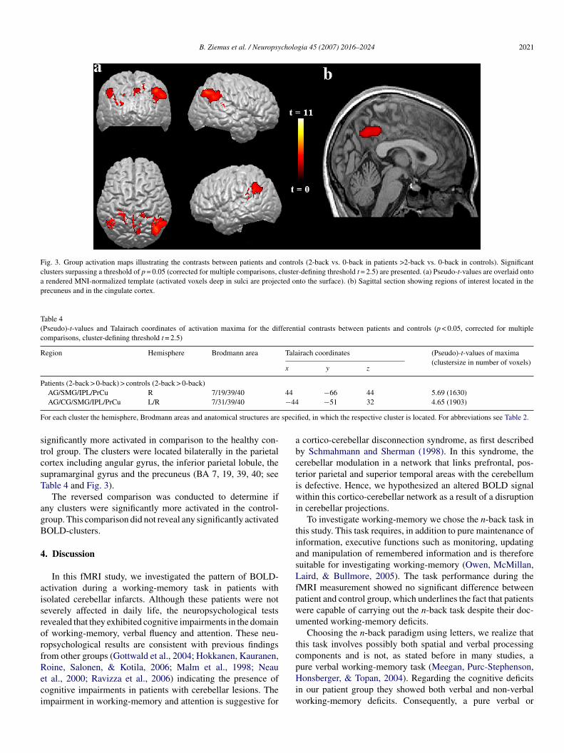
The results of the differential random-effects contrast
etween the patients and controls with respect to the 2-back > 0-ack contrast are shown in Fig. 3 and Table 4 (height threshold:= 0.05, cluster-defining threshold: t = 3). During the 2-backask the patient group exhibited two BOLD clusters that were

2020 B. Ziemus et al. / Neuropsychologia 45 (2007) 2016–2024
Table 2(Pseudo)-t-values and Talairach coordinates of activation maxima for the contrast 2-back > the 0-back in healthy controls (p < 0.05, corrected for multiple comparisons)
Region Hemisphere Brodmann area Talairach coordinates (Pseudo)-t-values of maxima(clustersize in number of voxels)
x y z
2-back > 0-back, controlsCerebellum R 34 −63 −19 10.45 (436)IPL/SPL R 7/40 36 −56 45 9.22 (1135)IPL/SPL/PrCu L 7/40 −28 −58 45 9.40 (1332)IFG/MFG R 9 44 32 26 8.40 (900)MFG/PCG L 6 −30 −5 50 8.36 (270)MFG/PCG R 6 34 −8 52 9.12 (396)IFG/Insula L 13/45/47 −32 23 1 7.66 (216)IFG/MFG L 9 −48 13 27 7.65 (295)Cerebellum L −36 −59 −19 6.92 (102)MFG R 10 42 50 −4 6.83 (72)MFG/SFG L/R 6/8 2 12 49 6.47 (140)MFG L 10 −34 5 9 6.31 (27)MTG R 21 59 −43 −5 6.27 (51)IPL R 40 48 −38 54 6.22 (54)IFG R 13/47 34 21 −1 6.13 (30)
For each cluster the hemisphere, Brodmann areas and anatomical structures are specified, in which the respective cluster is located. Abbreviations: AG, angular gyrus;CG, cingulate gyrus; FFG, fusiform gyrus; SMG, supramarginal gyrus; IFG, inferior frontal gyrus; IPL, inferior parietal lobule; MFG, middle frontal gyrus; MOG,middle occipital gyrus; MTG, middle temporal gyrus; SFG, superior frontal gyrus; SPL, superior parietal lobule; PCG, precentral gyrus; PrCu, precuneus.
Fig. 2. Group activation maps for the contrast 2-back > x-back. Pseudo-t-values are overlaid onto a rendered MNI-normalized template (activated voxels deep in sulciare projected onto the surface). Significant voxels surpassing a threshold of p = 0.05 (corrected for multiple comparisons) are presented. (a) Results for the patientgroup (n = 9). (b) Results for the control group (n = 9).
Table 3(Pseudo)-t-values and Talairach coordinates of activation maxima for the contrast 2-back > the 0-back in patients (p < 0.05, corrected for multiple comparisons)
Region Hemisphere Brodmann area Talairach coordinates (Pseudo)-t-values of maxima(cluster size in number of voxels)
x y z
2-back > 0-back patientsAG/IPL/PrCu/SMG/SPL R 7/19/39/40 30 −62 38 10.21 (3778)AG/IPL/PrCu/SMG/SPL L 7/19/39/40 −40 −56 53 8.78 (2145)
ITG/MTG R 20/21/37 59 −37 −10 9.20 (573)IFG/Insula/MFG/PCG/SFG R 6/8/9/13/45/47 −36 20 5 7.55 (1634)
MFG/PCG/SFG L 6 −28 −9 60 6.48 (144)IFG/Insula L 13/45/47 −36 22 6 6.20 (116)MFG/PCG L 6/8/9 −48 14 40 5.94 (436)SFG L/R 6 2 10 51 5.81 (69)
For each cluster the hemisphere, Brodmann areas and anatomical structures are specified, in which the respective cluster is located. For abbreviations see Table 2.
B. Ziemus et al. / Neuropsychologia 45 (2007) 2016–2024 2021
Fig. 3. Group activation maps illustrating the contrasts between patients and controls (2-back vs. 0-back in patients >2-back vs. 0-back in controls). Significantclusters surpassing a threshold of p = 0.05 (corrected for multiple comparisons, cluster-defining threshold t = 2.5) are presented. (a) Pseudo-t-values are overlaid ontoa rendered MNI-normalized template (activated voxels deep in sulci are projected onto the surface). (b) Sagittal section showing regions of interest located in theprecuneus and in the cingulate cortex.
Table 4(Pseudo)-t-values and Talairach coordinates of activation maxima for the differential contrasts between patients and controls (p < 0.05, corrected for multiplecomparisons, cluster-defining threshold t = 2.5)
Region Hemisphere Brodmann area Talairach coordinates (Pseudo)-t-values of maxima(clustersize in number of voxels)
x y z
Patients (2-back > 0-back) > controls (2-back > 0-back)44−44
F speci
stcsT
agB
4
aisrorfReci
abctiwi
tiasLfpwu
tc
AG/SMG/IPL/PrCu R 7/19/39/40AG/CG/SMG/IPL/PrCu L/R 7/31/39/40
or each cluster the hemisphere, Brodmann areas and anatomical structures are
ignificantly more activated in comparison to the healthy con-rol group. The clusters were located bilaterally in the parietalortex including angular gyrus, the inferior parietal lobule, theupramarginal gyrus and the precuneus (BA 7, 19, 39, 40; seeable 4 and Fig. 3).
The reversed comparison was conducted to determine ifny clusters were significantly more activated in the control-roup. This comparison did not reveal any significantly activatedOLD-clusters.
. Discussion
In this fMRI study, we investigated the pattern of BOLD-ctivation during a working-memory task in patients withsolated cerebellar infarcts. Although these patients were noteverely affected in daily life, the neuropsychological testsevealed that they exhibited cognitive impairments in the domainf working-memory, verbal fluency and attention. These neu-opsychological results are consistent with previous findingsrom other groups (Gottwald et al., 2004; Hokkanen, Kauranen,
oine, Salonen, & Kotila, 2006; Malm et al., 1998; Neaut al., 2000; Ravizza et al., 2006) indicating the presence ofognitive impairments in patients with cerebellar lesions. Thempairment in working-memory and attention is suggestive forpHiw
−66 44 5.69 (1630)−51 32 4.65 (1903)
fied, in which the respective cluster is located. For abbreviations see Table 2.
cortico-cerebellar disconnection syndrome, as first describedy Schmahmann and Sherman (1998). In this syndrome, theerebellar modulation in a network that links prefrontal, pos-erior parietal and superior temporal areas with the cerebellums defective. Hence, we hypothesized an altered BOLD signalithin this cortico-cerebellar network as a result of a disruption
n cerebellar projections.To investigate working-memory we chose the n-back task in
his study. This task requires, in addition to pure maintenance ofnformation, executive functions such as monitoring, updatingnd manipulation of remembered information and is thereforeuitable for investigating working-memory (Owen, McMillan,aird, & Bullmore, 2005). The task performance during the
MRI measurement showed no significant difference betweenatient and control group, which underlines the fact that patientsere capable of carrying out the n-back task despite their doc-mented working-memory deficits.
Choosing the n-back paradigm using letters, we realize thathis task involves possibly both spatial and verbal processingomponents and is not, as stated before in many studies, a
ure verbal working-memory task (Meegan, Purc-Stephenson,onsberger, & Topan, 2004). Regarding the cognitive deficitsn our patient group they showed both verbal and non-verbalorking-memory deficits. Consequently, a pure verbal or

2 cholo
nfdpcd
btttpls6v(9tdwt
2Magm(iata
vvsciaiipptbcn
pemgpIif
tlmTt
ompTrtoowevimrcrat
w8mpwffis(catgsfaawf
5
ntnt
022 B. Ziemus et al. / Neuropsy
on-verbal task was not a prerequisite for our study. It wasurther not our intention in this study to investigate inherentifferences between verbal and non-verbal working- memoryathways. Our concern was to provide evidence for a disruptedortico-cerebellar circuitry in patients with cerebellar lesionsuring a working-memory task.
A meta-analysis of functional imaging studies using the n-ack working-memory paradigm (Owen et al., 2005) revealedhat – in verbal as well as in non-verbal versions – robust activa-ions occur bilaterally in the following brain regions forminghe putative cortico-cerebellar network: the medial posteriorarietal cortex, including precuneus and the inferior parietalobule (BA 7, 40), bilateral premotor cortex (BA 6, 8), dor-al cingulate/medial premotor cortex, including SMA (BA 32,), bilateral rostral prefrontal cortex (BA 10), bilateral mid-entrolateral prefrontal cortex including the frontal operculumBA 45, 47) and bilateral dorsolateral prefrontal cortex (BA, 46). In addition to this pattern of activity, activations inhe medial cerebellum have been reported. The only reportedifference for non-verbal n-back tasks compared to verbalorking-memory tasks was a lack of activation in the left ven-
rolateral prefrontal cortex.In line with the results in the literature (Chen & Desmond,
005; Desmond & Fiez, 1998; Hautzel, Mottaghy, Schmidt,ueller, & Krause, 2003; Krause et al., 2000; Schlosser et
l., 2003; Schlosser, Wagner, & Sauer, 2006) we found in bothroups activations of a cortico-cerebellar network during perfor-ance of the working-memory task, namely in the frontal cortex
DLPFC and VLPFC) and the superior and inferior lobules dur-ng 2-back, compared to 0-back, task performance (Fig. 2). Welso found cerebellar activations in both groups, but the explana-ory power of cerebellar activation is very limited for the patients,s the lesions are likely to cause artefacts.
For the contrast ‘2-back versus 0-back in patients > 2-backersus 0-back in controls’ we found more pronounced acti-ations in the parietal cortex (i.e., angular gyrus (BA 39),upramarginal gyrus (BA 40), precuneus (BA 7), and posterioringulate gyrus (BA 31) (Fig. 3; Table 4). These areas were alsodentified as activated in the separate group analysis for controlsnd patients as reported above (Fig. 2; Tables 2 and 3). As antic-pated the pattern of activation during the 2-back task is similarn patients and controls, whereas the contribution of posteriorarietal regions to this network appears to be augmented in theatients. In light of the relatively high performance exhibited byhe patients in the 2-back task, the results shown in Fig. 3 coulde interpreted as a compensatory up-regulation to offset a lack inerebellar input into this complex cerebello-cortical-subcorticaletwork (Mottaghy et al., 2003).
Regarding the role of the parietal cortex in WM tasks, therecuneus (BA 7) has been shown to be involved in all types ofxecutive function (Wager & Smith, 2003). In verbal working-emory tasks the inferior parietal lobule (namely the angular
yrus and the supramarginal gyrus) is the likely locus of the
honological store (Fiez et al., 1996; Smith & Jonides, 1998).n contrast to this, a recent study by Ravizza et al. (2004) placesn question whether the supramarginal gyrus is a dedicated siteor phonological short-term storage. They could demonstratecmac
gia 45 (2007) 2016–2024
hat the ventral part of the supramarginal gyrus supports phono-ogical encoding whereas the dorsal part responds to short-term
emory load, for visual and as well verbal stimulus material.he precise function of the parietal cortex in working-memory
asks is still a matter of debate.It should be noted that the differences in brain activation
bserved between patients and controls during the working-emory task could reflect, at least in part, altered attentional
rocesses, which are recruited by the working-memory task.he working-memory task places more demands on attentional
esources compared to the control task. In addition, several ofhe patients showed mild impairments in attention. In the lightf our results we favour the view that the PPC takes on the rolef a superordinate attentional modulator for different neural net-orks during a working-memory task as proposed by Majerus
t al. (2006). Depending on the type of information (item, order,erbal, non-verbal) that has to be processed, different underly-ng neuronal networks will be recruited. The intraparietal sulcus
ay play a role in coordinating and synchronizing these neu-onal substrates, which accordingly would up-regulate parietalortex in patients with deficits in the underlying task-specificegions. Ravizza et al. (2004) also reported domain-independentctivations in the dorsal part of intraparietal sulcus in tasks wherehe working-memory demands on attention are high.
According to a meta-analysis of 60 imaging studies onorking-memory conducted by Wager and Smith (2003), BA 6,, 9 in the superior frontal cortex respond most when working-emory must be continuously updated, as required in an n-back
aradigm. We did not find significant enhancement of frontalorking-memory specific areas in the patients. One explanation
or this might be the impaired integrity of the cerebello-thalamo-rontal network leading to downstream activation alterationsn parietal cortex. According to a combined ERP- and fMRItudy by Brass, Ullsperger, Knoesche, von Cramon, Phillips2005) there is strong evidence that in a system of hierarchicalognitive control, activations in the prefrontal cortex precedectivations in the parietal cortex. They interpret this sequen-ial order with the notion that the prefrontal cortex defines taskoals in very abstract terms, while the parietal lobe focuses on thepecific concrete action. Accordingly, this network compensatesor the cerebellar dysfunction by means of up-regulated parietalctivation following commands from the prefrontal cortex. Inddition to these considerations, the parietal cortex is, due to itsidespread connections and its heteromodality, a likely locus
or functional plastic changes.
. Conclusion
We conclude that the cerebellum is involved in higher cog-itive processes such as working-memory. Our findings pointo the cerebellum as part of a widespread cortico-cerebellaretwork involving the frontal lobe, parietal cortex and subcor-ical brain areas. Patients with isolated cerebellar infarcts show
ognitive impairments on neuropsychological tests of working-emory. These deficits can be attributed to a disruption offferent or efferent fibres to cortical connections as part of aortico-cerebellar circuitry. Increased BOLD responses in the
cholo
asc
R
A
B
B
B
B
B
B
C
C
C
D
D
D
F
F
F
F
G
G
H
H
H
H
K
L
L
L
M
M
M
M
M
M
M
M
M
M
N
O
O
P
P
R
R
B. Ziemus et al. / Neuropsy
ngular gyri and the inferior parietal lobule may be an expres-ion of the compensation process evoked after lesion to theerebellum.
eferences
schenbrenner, S., Tucha, O., & Lange, K. W. (2000). Regensburgerwortflussigkeitstest RWT Handanweisung. Hogrefe: Goettingen.
enton, A. L., & Hamsher, K. (1989). Multilingual aphasia examination. IowaCity, IA: AJA Associates.
or, D., Duncan, J., Wiseman, R. J., & Owen, A. M. (2003). Encoding strategiesdissociate prefrontal activity from working-memory demand. Neuron, 37,3612–4367.
or, D., Cumming, N., Scott, C. E. M., & Owen, A. M. (2004). Prefrontal cor-tical involvement in encoding strategies, independent of stimulus modality.European Journal of Neuroscience, 19, 2270–3365.
otez-Marquard, T., Bard, C., Leveille, J., & Botez, M. I. (2001). A severefrontal-parietal lobe syndrome following cerebellar damage. European Jour-nal of Neurology, 8, 347–353.
rass, M., Ullsperger, M., Knoesche, T. R., von Cramon, D. Y., & Phillips, N.A. (2005). Who comes first? The role of the prefrontal and parietal cortex incognitive control. Journal of Cognitive Neuroscience, 17, 1367–1375.
raver, T. S., Cohen, J. D., Nystrom, L. E., Jonides, J., Smith, E. E., & Noll,D. C. (1997). A parametric study of prefrontal cortex involvement in humanworking-memory. NeuroImage, 5, 49–62.
hen, S. H., & Desmond, J. E. (2005). Cerebrocerebellar networks duringarticulatory rehearsal and verbal working-memory tasks. NeuroImage, 24,332–338.
lower, D. M., West, R. A., Lynch, J., & Strick, P. L. (2001). The inferior parietallobule is the target of output from the superior colliculus, hippocampus, andcerebellum. Journal of Neuroscience, 21, 6283–6291.
ohen, J., Forman, S., Braver, T., Casey, B., Servan-Schreiber, D., & Noll, D.(1994). Activation of prefrontal cortex in a non-spatial working- memorytask with functional MRI. Human Brain Mapping, 1, 293–304.
esmond, J. E., & Fiez, J. A. (1998). Neuromaging studies of the cerebellum:Language, learning and memory. Trends Cognitive Science, 2, 355–362.
esmond, J. E., Gabrieli, J. D., Wagner, A. D., Ginier, B. L., & Glover, G. H.(1997). Lobular patterns of cerebellar activation in verbal working-memoryand finger-tapping tasks as revealed by functional MRI. Journal of Neuro-science, 17, 9675–9685.
’Esposito, M., Ballard, D., Aguirre, G. K., & Zarahn, E. (1998). Human pre-frontal cortex is not specific for working-memory: A functional MRI study.NeuroImage, 8, 74–282.
iez, J. A., Raife, E. A., Balota, D. A., Schwarz, J. P., Raichle, M. E., & Petersen,S. E. (1996). A positron emission tomography study of the short-term main-tenance of verbal information. Journal of Neuroscience, 16, 808–822.
riston, K. J., Ashburner, J., Poline, J. P., Frith, C. D., Heather, J. D., & Frack-owiak, R. S. J. (1995). Spatial registration and normalization of images.Human Brain Mapping, 2, 165–189.
riston, K. J., Frith, C. D., Liddle, P. F., & Frackowiak, R. S. (1991). Plastictransformation of PET images. Journal of Computer Assisted Tomography,15, 634–639.
riston, K. J., Holmes, A. P., Worseley, K. J., Poline, J. P., Frith, C. D., & Frack-owiak, R. S. J. (1995). Statistical parametric maps in functional imaging: Ageneral linear approach. Human Brain Mapping, 2, 189–210.
evins, A., & Cutillo, B. (1993). Spatiotemporal dynamics of component pro-cesses in human working-memory. Electroencephalography and ClinicalNeurophysiology, 87, 128–143.
ottwald, B., Wilde, B., Mihajlovic, Z., & Mehdorn, H. M. (2004). Evidence ofdistinct cognitive deficits after focal cerebellar lesions. Journal of Neurology,Neurosurgery and Psychiatry, 75, 1524–1531.
autzel, H., Mottaghy, F. M., Schmidt, D., Mueller, H. W., & Krause, B. J.
(2003). Strategies for data analysis in activation studies on working-memory.Nuklearmedizin, 42, 197–209.okkanen, L. S. K., Kauranen, V., Roine, R. O., Salonen, O., & Kotila, M.(2006). Subtle cognitive deficits after cerebellar infarcts. European Journalof Neurology, 13, 161–170.
R
S
gia 45 (2007) 2016–2024 2023
olmes, A. P. (1994). Statistical issues in functional brain mapping. Doctor ofPhilosophy Thesis, University of Glasgow.
olmes, A. P., Blair, R. C., Watson, J. D. G., & Ford, I. (1996). Nonparametricanalysis of statistic images from functional mapping experiments. Journalof Cerebral Blood Flow and Metabolism, 16, 7–22.
rause, J. B., Taylor, J. G., Schmidt, D., Hautzel, H., Motthagy, F. M., &Muller-Gartner, H. W. (2000). Imaging and neural modelling in episodicand working-memory processes. Neural Networks, 13, 847–859.
ancaster, J. L., Summerln, J. L., Rainey, L., Freitas, C. S., & Fox, P. T. (1997).The Talairach Daemon, a database server for Talairach Atlas Labels. Neu-roImage, 5, 633.
ancaster, J. L., Woldorff, M. G., Parsons, L. M., Liotti, M., Freitas, C. S.,Rainey, L., et al. (2000). Automated Talairach atlas labels for functionalbrain mapping. Human Brain Mapping, 10, 120–131.
ezak, M. D. (1995). Neuropsychological assessment. New York: Oxford Uni-versity Press.
ajerus, S., Poncelet, M., Van der Linden, M., Albouy, G., Salmon, E., Ster-penich, V., et al. (2006). The left intraparietal sulcus and verbal short-termmemory: Focus of attention or serial order? NeuroImage, 32, 880–891.
aldijan, J. A., Laurienti, P. J., Kraft, R. A., & Burdette, J. H. (2003). Anautomated method for neuroanatomic and cytoarchitectonic atlas-basedinterrogation of fMRI data sets. NeuroImage, 19, 1233–1239.
alm, J., Kristensen, B., Karlsson, T., Carlberg, B., Fagerlund, M., & Olsson,T. (1998). Cognitive impairment in young adults with infratentorial infarcts.Neurology, 51, 433–440.
eegan, D. V., Purc-Stephenson, R., Honsberger, M. J. M., & Topan, M. (2004).Task analysis complements neuroimaging: An example from working-memory research. NeuroImage, 21, 1026–1036.
ellers, J. D., Bullmore, E., Brammer, M., Williams, S. C., Andrew, C., Sachs,N., et al. (1995). Neural correlates of working-memory in a visual lettermonitoring task: An fMRI study. NeuroReport, 7, 109–112.
iddleton, F. A., & Strick, P. L. (1994). Anatomical evidence for cerebellarand basal ganglia involvement in higher cognitive function. Science, 266,458–461.
iddleton, F. A., & Strick, P. L. (2000). Basal ganglia and cerebellar loops:Motor and cognitive circuits. Brain Research Reviews, 31, 236–250.
iddleton, F. A., & Strick, P. L. (2001). Cerebellar projections to the prefrontalcortex of the primate. Journal of Neuroscience, 21, 700–712.
ilner, B. (1971). Interhemispheric differences in the localization of psycho-logical processes in man. British Medical Bulletin, 21, 272–277.
ottaghy, F. M., Pascual-Leone, A., Kemna, L. J., Topper, R., Herzog, H.,Muller-Gartner, H. W., et al. (2003). Modulation of a brain–behavior rela-tionship in verbal working-memory by rTMS. Brain Research CognitiveBrain Research, 15, 241–249.
eau, J.-Ph., Arroyo-Anllo, E., Bonnaud, V., Ingrand, P., & Gil, R. (2000).Neuropsychological disturbances in cerebellar infarcts. Acta NeurologicaScandinavica, 102, 363–370.
wen, A. M. (2000). The role of the lateral frontal cortex in mnemonic pro-cessing: The contribution of functional neuroimaging. Experimental BrainResearch, 133, 33–43.
wen, A. M., McMillan, K. M., Laird, A. R., & Bullmore, E. (2005). N-back working-memory paradigm: A meta-analysis of normative functionalneuroimaging studies. Human Brain Mapping, 25, 46–59.
etrides, M. (1994). Frontal lobes and behaviour. Current Opinions in Neurobi-ology, 4, 207–211.
etrides, M. (1998). The role of the mid-dorsolateral prefrontal cortex inworking-memory. Experimental Brain Research, 133, 44–54.
avizza, S. M., Delgado, M. R., Chein, J. M., Becker, J. T., & Fiez, J. A. (2004).Functional dissociations within the inferior parietal cortex in verbal workingmemory. NeuroImage, 22, 562–573.
avizza, S. M., McCormick, C. A., Schlerf, J. E., Justus, T., Ivry, R. B., &Fiez, J. A. (2006). Cerebellar damage produces selective deficits in verbalworking-memory. Brain, 129, 306–320.
uff, R. M., Niemann, H., Allen, C. C., Farrow, C. E., & Wylie, T. (1992). TheRuff 2 and 7 Selective Attention Test: A neuropsychological application.Perceptual and Motor Skills, 75, 1311–1319.
chlosser, R., Gesierich, T., Kaufmann, B., Vucurevic, G., Hunsche, S., Gawehn,J., et al. (2003). Altered effective connectivity during working-memory

2 cholo
S
S
S
S
S
T
TIntelligenztest fur Erwachsene. Bern: Hans Huber.
024 B. Ziemus et al. / Neuropsy
performance in schizophrenia: A study with fMRI and structural equationmodeling. NeuroImage, 19, 751–763.
chlosser, R. G., Wagner, G., & Sauer, H. (2006). Assessing the working-memory network: Studies with functional magnetic resonance imaging andstructural equation modeling. Neuroscience, 139, 91–103.
chmahmann, J. D. (1991). An emerging concept: The cerebellar contributionto higher function. Archives of Neurology, 48, 1178–1187.
chmahmann, J. D., & Pandya, D. N. (1995). Prefrontal cortex projections to thebasilar pons in rhesus monkey: Implications for the cerebellar contributionto higher function. Neuroscience Letters, 199, 175–178.
chmahmann, J. D., & Sherman, J. C. (1998). The cerebellar cognitive affectivesyndrome. Brain, 121, 561–579.
W
W
gia 45 (2007) 2016–2024
mith, E. E., & Jonides, J. (1998). Neuroimaging analysis of human working-memory. Proceedings of the National Academy of Science, United States ofAmerica, 9, 12061–12068.
alairach, J., & Tournoux, P. (1998). Co-planar stereotaxic atlas of the humanbrain. New York: Thieme.
ewes, U. (1994). Handbuch und Testanweisung fur den Hamburg-Wechsler
ager, T. D., & Smith, E. E. (2003). Neuroimaging studies of working-memory.Cognitive, Affective and Behavioral Neuroscience, 3, 255–274.
echsler, D. (1987). Manual for the Wechsler Memory Scale-Revised. SanAntonio, TX: The Psychological Corporation.