impact of alendronate and vegf-antisense combined treatment on highly vegf-expressing a431 cells
TRANSCRIPT

Accepted Manuscript
Title: Impact of Alendronate and VEGF- antisense combinedtreatment on highly VEGF-expressing A431 cells
Authors: Medge Mongerard-Coulanges, EvelyneMigianu-Griffoni, Marc Lecouvey, Beatrice Jolles
PII: S0006-2952(09)00117-8DOI: doi:10.1016/j.bcp.2009.02.016Reference: BCP 10096
To appear in: BCP
Received date: 7-1-2009Revised date: 18-2-2009Accepted date: 19-2-2009
Please cite this article as: Mongerard-Coulanges M, Migianu-Griffoni E, LecouveyM, Jolles B, Impact of Alendronate and VEGF- antisense combined treatmenton highly VEGF-expressing A431 cells, Biochemical Pharmacology (2008),doi:10.1016/j.bcp.2009.02.016
This is a PDF file of an unedited manuscript that has been accepted for publication.As a service to our customers we are providing this early version of the manuscript.The manuscript will undergo copyediting, typesetting, and review of the resulting proofbefore it is published in its final form. Please note that during the production processerrors may be discovered which could affect the content, and all legal disclaimers thatapply to the journal pertain.
peer
-004
9349
5, v
ersi
on 1
- 19
Jun
201
0Author manuscript, published in "Biochemical Pharmacology 77, 10 (2009) 1580"
DOI : 10.1016/j.bcp.2009.02.016

Page 1 of 23
Accep
ted
Man
uscr
ipt
1 2 3 4 5 6 7 8 9 10 11 12 13 14 15 16 17 18 19 20 21 22 23 24 25 26 27 28 29 30 31 32 33 34 35 36 37 38 39 40 41 42 43 44 45 46 47 48 49 50 51 52 53 54 55 56 57 58 59 60 61 62 63 64 65
1Linking powered by eXtyles
Impact of Alendronate and VEGF- antisense combined treatment on highly VEGF-
expressing A431 cells
Medge Mongerard-Coulanges, Evelyne Migianu-Griffoni, Marc Lecouvey, Béatrice Jolles*
Laboratoire de Biophysique Moleculaire, Cellulaire et Tissulaire, CNRS, UMR 7033;
Université Pierre et Marie Curie-Paris 6, Paris, F-75005 France; Université Paris 13, Bobigny,
F-93017 France
*corresponding author: Béatrice Jolles,
Université Pierre et Marie Curie
Bat. C, 5ème étage, case 265
9, quai Saint-Bernard
F. 75005 Paris
France
tel: 33 1 44272299
fax: 33 1 44272215
* Manuscriptpe
er-0
0493
495,
ver
sion
1 -
19 J
un 2
010

Page 2 of 23
Accep
ted
Man
uscr
ipt
1 2 3 4 5 6 7 8 9 10 11 12 13 14 15 16 17 18 19 20 21 22 23 24 25 26 27 28 29 30 31 32 33 34 35 36 37 38 39 40 41 42 43 44 45 46 47 48 49 50 51 52 53 54 55 56 57 58 59 60 61 62 63 64 65
2Linking powered by eXtyles
Abstract
Bisphosphonates, and more specially nitrogen - containing bisphosphonates, which are in
current use for the treatment of bone diseases, demonstrate proapoptotic, antiproliferative,
antiangiogenic and anti-invasive properties on tumor cells. The amino - bisphosphonate
alendronate is considered as a potential anticancer drug. In the case of A431 cells, which
express high levels of VEGF, it had a two-step effect. At 24h, the antitumor properties of
alendronate were counterbalanced by a survival process, which consisted of an enhancement
of VEGF expression (mRNA and protein secretion) and TGFα secretion. It was only at 48h
that alendronate displayed the expected antiproliferative and antiangiogenic properties. The
first step, in which the PI3K pathway was engaged, could be prevented by the use of a VEGF-
antisense oligonucleotide. The combination of such an antisense with small concentrations of
alendronate (~ 2 µM), which is of the order of clinically used concentrations, was shown to
have an antiangiogenic effect as soon as 12h.
Key words:
Alendronate, VEGF expression, TGFα, PI3K pathway, VEGF-antisense, proliferation
peer
-004
9349
5, v
ersi
on 1
- 19
Jun
201
0

Page 3 of 23
Accep
ted
Man
uscr
ipt
1 2 3 4 5 6 7 8 9 10 11 12 13 14 15 16 17 18 19 20 21 22 23 24 25 26 27 28 29 30 31 32 33 34 35 36 37 38 39 40 41 42 43 44 45 46 47 48 49 50 51 52 53 54 55 56 57 58 59 60 61 62 63 64 65
3Linking powered by eXtyles
1. Introduction
Bisphosphonates (BPs) are analogs of endogenous pyrophosphates in which the P-O-P bond
has been replaced by a nonhydrolyzable P-C-P bond. BPs are widely used to inhibit
osteoclastic activity in benign and malignant bone diseases, such as Paget’s disease,
osteoporosis or osteolytic tumor bone metastases [1-3]. The nitrogen containing-BPs (N-BPs)
are more effective and are able to interfere with the metabolism of mevalonate, leading to
inhibition of the prenylation of small GTP-binding proteins (Ras, Rho…) [4, 5]. Those
GTPases are important signaling proteins which regulate a large variety of cellular processes.
It is now clearly established that BPs may exert directly proapoptotic and antiproliferative
effects on tumor cells [3, 6, 7]. Antiangiogenic properties have also been attributed to N-BPs
because of their potency to inhibit the proliferation and the migration of endothelial cells [8,
9]. For instance, the amino - bisphosphonate alendronate (ALN) has been shown to inhibit at
moderate concentrations the endothelial cells proliferation induced by VEGF (Vascular
Endothelial Growth Factor), in vitro as well as in vivo [10]. VEGF is a major angiogenic
factor secreted by tumors [11]; it is produced in highly variable amounts by different tumor
types. High levels of VEGF are of bad prognosis and have been associated with an increased
propensity for metastasis [12]. In the present study, we have investigated the direct action of
ALN on A431 cells (a human squamous cell carcinoma cell line) which produce high amount
of VEGF [13], contrary to MCF7 cells (a human mammary adenocarcinoma cell line) which
were taken as a control cell line. The A431 cells also overexpress epidermal growth factor
receptors (EGFR) and produce its ligand TGFα (Transforming Growth Factor-alpha) [14].
The impact of the important secretion of VEGF by A431 cells on the anticancer properties of
ALN has been studied at the levels of cell proliferation and apoptosis. We also determined
whether ALN modified the A431 VEGF expression. These questions were investigated by
peer
-004
9349
5, v
ersi
on 1
- 19
Jun
201
0

Page 4 of 23
Accep
ted
Man
uscr
ipt
1 2 3 4 5 6 7 8 9 10 11 12 13 14 15 16 17 18 19 20 21 22 23 24 25 26 27 28 29 30 31 32 33 34 35 36 37 38 39 40 41 42 43 44 45 46 47 48 49 50 51 52 53 54 55 56 57 58 59 60 61 62 63 64 65
4Linking powered by eXtyles
studying the ALN effect on cells, combined or not with an anti - VEGF antisense
oligonucleotide (AS) in order to reduce the high endogenous VEGF level in A431 cells.
2. Materials and methods
2.1. Reagents and antisense oligonucleotides
ALN was synthesized in the lab [15]. LY294002 was purchased from Sigma (Saint-Quentin
Fallavier, France). Phosphorothioate oligodeoxynucleotides were synthesized and PAGE
purified by Eurogentec (Seraing, Blgium). The antisense AS (5’-
TGGCTTGAAGATGTACTCGAT) was designed to target the human VEGF mRNA at the
level of the region 257-278 nt relative to start codon [16]. SC was a control scrambled
oligodeoxynucleotide.
2.2. Cell culture and treatments
The A431 and MCF7cell lines were received from ATCC (LGC Standards, Molsheim,
France). Cells were grown in DMEM medium (A431 cells) or RPMI medium (MCF7 cells)
supplemented with 10% decomplemented foetal bovine serum (FBS), 50 U/mL penicillin, 50
μg/mL streptomycin, 2 mM glutamine, in a 5% CO2 atmosphere at 37°C. They were
trypsinized and passed twice a week.
Cells were plated in 6-well plates and, after reaching 30-50% confluence, incubated in
culture medium (10% serum) containing different concentrations of ALN for 24 or 48h or
transfected by AS or SC with OligofectamineTM (Invitrogen, Cergy, France). Transfection
peer
-004
9349
5, v
ersi
on 1
- 19
Jun
201
0
Page 5 of 23
Accep
ted
Man
uscr
ipt
1 2 3 4 5 6 7 8 9 10 11 12 13 14 15 16 17 18 19 20 21 22 23 24 25 26 27 28 29 30 31 32 33 34 35 36 37 38 39 40 41 42 43 44 45 46 47 48 49 50 51 52 53 54 55 56 57 58 59 60 61 62 63 64 65
5Linking powered by eXtyles
was carried out as directed by the manufacturer, i.e. in OPTI-MEM1 (Invitrogen) without
serum or antibiotics. After 4h incubation at 37°C, FBS (10% final concentration), 50 U/ml
penicillin and 50 mg/ml streptomycin were added. The final concentration of AS or SC was
200 nM. For some experiments, ALN was also added at 4h. For LY294002 treatment, cells
were incubated with 50 µM LY294002 for 40 min before addition of ALN [17].
2.3. Cell proliferation assay
Cells (104/well) were plated in 96-well plates and allowed to attach for 24h, and then cultured
under increasing concentrations of ALN (1-150 µM) in full culture medium for 12, 24 or 48h
along, or not, with transfection by AS or SC. Viability was assessed using MTT (3-(4,5-
dimethylthiazol-2-yl)-2,5-diphenyl tetrazolium bromide) at a final concentration of 0.5
mg/ml for 2h at 37°C. Optical density of the dissolved formazan product (in DMSO) was read
at 570 nm.
2.4. Annexin V assay
After 24h treatment with increasing doses of ALN, 106 cells were washed with PBS,
trypsinized and resuspended in 100 µl of binding buffer. A fluorescein isothiocyanate-
conjugated annexin V and propidium iodide were added as directed by the manufacturer
(Santa Cruz Biotechnology, (Tebu, Le Perray en Yvelines, France)). Cells were analyzed with
a flow cytometer (FACSCalibur, BD Biosciences, Le Pont de Claix, France).
2.5. VEGF and TGFα Elisa assays
peer
-004
9349
5, v
ersi
on 1
- 19
Jun
201
0

Page 6 of 23
Accep
ted
Man
uscr
ipt
1 2 3 4 5 6 7 8 9 10 11 12 13 14 15 16 17 18 19 20 21 22 23 24 25 26 27 28 29 30 31 32 33 34 35 36 37 38 39 40 41 42 43 44 45 46 47 48 49 50 51 52 53 54 55 56 57 58 59 60 61 62 63 64 65
6Linking powered by eXtyles
Secretion of VEGF or TGFα into the cell culture supernatant after the different treatments was
determined using Ray Bio Human VEGF ELISA kits (Tebu, Le Perray en Yvelines, France) ,
according to the manufacturer's instructions. In the case of VEGF, supernatant samples were
diluted 50-fold (A431 cells) or 30-fold (MCF7 cells).
peer
-004
9349
5, v
ersi
on 1
- 19
Jun
201
0

Page 7 of 23
Accep
ted
Man
uscr
ipt
1 2 3 4 5 6 7 8 9 10 11 12 13 14 15 16 17 18 19 20 21 22 23 24 25 26 27 28 29 30 31 32 33 34 35 36 37 38 39 40 41 42 43 44 45 46 47 48 49 50 51 52 53 54 55 56 57 58 59 60 61 62 63 64 65
7Linking powered by eXtyles
2.6. RT-PCR
24 and 48h after treatment, total RNA was extracted (RneasyTM kit, Qiagen, Courtaboeuf,
France) and cDNA was synthesized using OmniscriptTM reverse transcription kit (Qiagen).
Reverse transcription was performed using 0.5 µM gene-specific reverse primers of VEGF165
and ß-actin which was chosen as an internal control. Hot Star TaqTM PCR kit (Qiagen) was
used for DNA amplification, which was kept in its exponential phase. Primers used for
VEGF165 amplification (0.5 µM) were: sense, 5'-d(GGAAGTGGTGAAGTTCATGGATG)
and reverse, 5'-d(AGCAAGGCCCACAGGGATTT). The amplification consisted of 21 cycles
for A431 cells, 23 for MCF7 cells (denaturation at 94°C for 30 s, annealing at 59°C for 30 s
and extension at 72°C for 1 min). Because of the difference of 4°C between the melting
temperatures of both primers, a four-cycle touch-down PCR was first carried out with an
annealing temperature decreasing from 63°C to 60°C. Primers for ß-actin amplification (0.3
µM) were: sense, 5'-d(ACCAACTGGGACGACATGGA) and reverse, 5'-
d(CTCCTTAATGTCACGCACGA). Amplification consisted of 20 cycles (95°C, 30 s; 56°C,
30 s; 72°C, 1 min). The PCR products were separated on 1.8% agarose gel stained with
ethidium bromide (6 mg/ml). Gene expression level was quantified by densitometric analysis
using Image J software.
2.7. Statistical analysis of the data
The data were subjected to one-way analysis of variance (ANOVA), followed by Dunnett's
test. P < 0.05 was considered a significant difference.
peer
-004
9349
5, v
ersi
on 1
- 19
Jun
201
0
Page 8 of 23
Accep
ted
Man
uscr
ipt
1 2 3 4 5 6 7 8 9 10 11 12 13 14 15 16 17 18 19 20 21 22 23 24 25 26 27 28 29 30 31 32 33 34 35 36 37 38 39 40 41 42 43 44 45 46 47 48 49 50 51 52 53 54 55 56 57 58 59 60 61 62 63 64 65
8Linking powered by eXtyles
3. Results
3.1. Cell growth inhibition and apoptosis
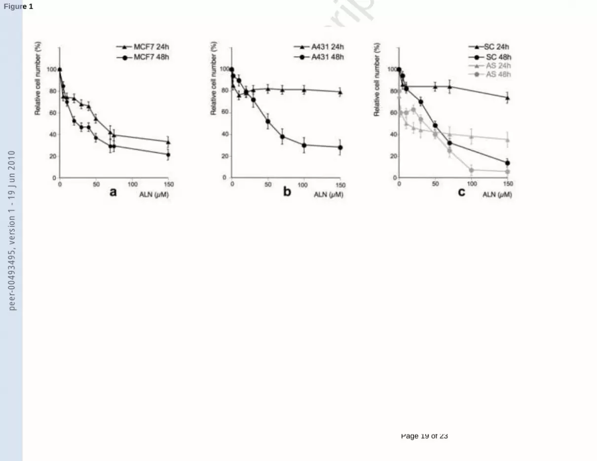
The ALN-induced growth inhibition of A431 or MCF7 has been evaluated by MTT assays
(Figure 1). Whereas a dose-dependent growth decrease was observed as soon as 24h in the
case of MCF7 cells (IC50 ~ 60 µM) (Figure 1a), it is only at 48h that ALN treatment caused a
similar dose-dependent decrease in A431 cell number (Figure 1b), with a ~ 75% decrease at
150 µM and a IC50 around 50 µM. (At 72h, the IC50 was only ~ 10 µM, not shown). During
the first 24h, A431 cell number was only reduced of ~ 20% and this effect was observable as
soon as from about 10 µM ALN without further decrease for higher ALN concentrations.
Because of the high growth dependence of A431 cells on VEGF [18], antiproliferative
properties of ALN were also checked in combination with an anti-VEGF antisense
oligonucleotide (AS), compared to a scrambled control (SC) (Figure 1c). SC had no effect by
itself: results were similar to those obtained with ALN alone (Figure 1b). AS had by itself an
antineoplastic effect on A431 cells (reduction of 20 % in cell number) (Figure 1c). AS
markedly favoured ALN-induced cell growth decrease, which was now efficient as soon as
24h. A reduction in cell number of ~ 40% was obtained with 150 µM ALN. Even if the
growth curve tended to a plateau, ALN had now an antiproliferative effect at least up to ~ 30-
40 µM ALN.
Dot-plots of FITC-annexin V (x-axis)/propidium iodide (y-axis) fluorescence and
corresponding histograms for FITC-annexin V fluorescence are presented on Figure 2. ALN
induced apoptosis as soon as 24h in MCF7 cells and, more surprisingly, in A431 cells. In the
case of MCF7 cells, there was a dose-dependent moderate increase in FITC annexin V
staining up to 50 µM ALN, evidenced by the shift of annexin V fluorescence on the
peer
-004
9349
5, v
ersi
on 1
- 19
Jun
201
0

Page 9 of 23
Accep
ted
Man
uscr
ipt
1 2 3 4 5 6 7 8 9 10 11 12 13 14 15 16 17 18 19 20 21 22 23 24 25 26 27 28 29 30 31 32 33 34 35 36 37 38 39 40 41 42 43 44 45 46 47 48 49 50 51 52 53 54 55 56 57 58 59 60 61 62 63 64 65
9Linking powered by eXtyles
histogram. By contrast, in the case of A431 cells, apoptosis increased with ALN treatment up
to 10 µM for which it became very significant (24% with only ~ 70 % viable cells), then
decreased back with 50 µM ALN. It is worth noticing that, after a 50 µM ALN treatment, the
percentage of viable cells was comparable to that of untreated cells (~ 90%) whereas the
percentage of dead cells had doubled (~10 % instead of 5%). 30 nM EGF has been taken as a
positive control of apoptosis in the case of A431 cells [19].
3.2. VEGF and TGFα expression
The amount of secreted VEGF into the culture medium, determined by Elisa test, was largely
superior in A431 cells compared to MCF7 cells. (Figure 3a). When cells were treated by
increasing amounts of ALN, a consistent increase (~ 30%) in secreted VEGF was evidenced
in A431 cells at 24h, followed by a decrease at 48h (~ 40%). The overall increase in VEGF
secretion along the time (48h compared to 24h) is a usual observation on cells in culture [20].
Correlation between VEGF secretion and VEGF mRNA level in A431 cells has been
established by RT-PCR (Figure 3b). Results concerning the VEGF165 isoform are presented
since VEGF165 is considered as the most mitogenic isoform but similar results have been
obtained for VEGF121 (not shown). After 24h treatment with ALN (0 to 15 µM), the mRNA
expression of VEGF in A431 cells increased of ~ 60%. This was no longer observed at 48h.
Figure 3c presents the amount of TGFα secreted by A431 cells after ALN treatment. In a very
similar way to what was observed for VEGF, increasing doses of ALN induced enhancement
in TGFα secretion at 24h, followed by a decrease at 48h.
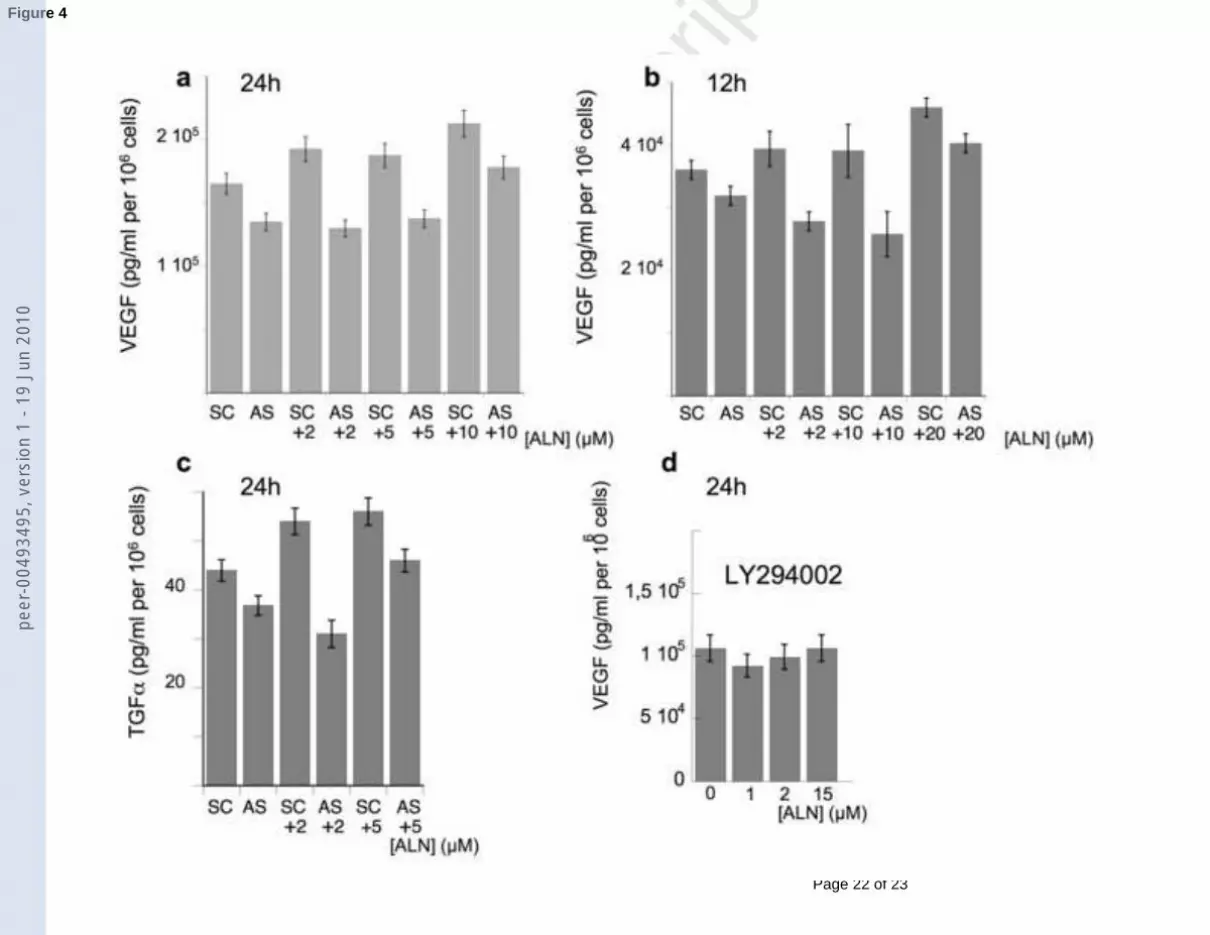
VEGF secretion was lowered after treatment with AS (Figure 4a and 4b), compared to
SC. When combined with AS, ALN treatment of A431 cells did not induce any more increase
in VEGF secretion at 24h, like it was still the case after combination with SC (Figure 4a) or in
peer
-004
9349
5, v
ersi
on 1
- 19
Jun
201
0
Page 10 of 23
Accep
ted
Man
uscr
ipt
1 2 3 4 5 6 7 8 9 10 11 12 13 14 15 16 17 18 19 20 21 22 23 24 25 26 27 28 29 30 31 32 33 34 35 36 37 38 39 40 41 42 43 44 45 46 47 48 49 50 51 52 53 54 55 56 57 58 59 60 61 62 63 64 65
10Linking powered by eXtyles
the case of treatment by ALN alone (Figure 3a). The VEGF level was kept constant for small
ALN concentrations, up to 10 µM ALN for which it increased. After only 12h treatment, the
combination of AS with ALN even induced decrease in VEGF secretion (Figure 4b). It was
only with 20 µM ALN that VEGF secretion increased again. More surprisingly, AS also led
to a decrease in TGFα secretion at 24h, compared to SC (Figure 4c) and, up to 5 µM, opposed
ALN-induced TGFα secretion, which had been observed at 24h (Figure 3c). At 12h, the level
of TGFα was under the sensitivity of the assay. After pretreatment with 50 µM LY294002, an
inhibitor of the phosphatidyl inositol 3-kinase (PI3K) pathway, a global decrease in VEGF
secretion was observed (Figure 4d) and ALN did not induce any more increase in VEGF
secretion by A431 cells at 24h.
4. Discussion
4.1. Cell growth inhibition and apoptosis
The antiproliferative and proapoptotic properties of BPs have generally been described after
48h treatment or more [21]. The slight modifications observed for earlier times were usually
considered as resulting from a metabolic shift in the cell line stressed by the treatment or from
a cytostatic drug effect. In the present work, we specifically focused on the effects observed
after only 24h ALN treatment. Whereas a dose-dependent cell growth decrease was observed
in the case of MCF7 cells, increasing doses of ALN only led to a plateau with 20 % cell
growth inhibition (Figure 1) in the case of A431 cells. It is only at 48h that ALN treatment
caused a dose-dependent decrease in cell number. This delay in cell growth inhibition is not
attributed to a cytostatic effect since there is a clear correlation between (i) the efficient A431
cell apoptosis (24 %) measured at 24h with 10 µM ALN (Figure 2) and (ii) the dose-
peer
-004
9349
5, v
ersi
on 1
- 19
Jun
201
0

Page 11 of 23
Accep
ted
Man
uscr
ipt
1 2 3 4 5 6 7 8 9 10 11 12 13 14 15 16 17 18 19 20 21 22 23 24 25 26 27 28 29 30 31 32 33 34 35 36 37 38 39 40 41 42 43 44 45 46 47 48 49 50 51 52 53 54 55 56 57 58 59 60 61 62 63 64 65
11Linking powered by eXtyles
dependent decrease in cell number, which was observed at 24h (Fig 1b) up to 10 µM ALN.
With 50 µM ALN, whereas the percentage of apoptotic cells increased in the case of MCF7
cells (Figure 2), apoptosis was no longer observable in A431 cells. The percentage of dead
cells has doubled but that of viable cells has returned to its initial value, suggesting new
proliferation. Therefore we assume that, in the first 24h of treatment, there is superimposition
of two different processes in the case of A431 cells: an ALN-induced reduction in cell
viability evidenced by apoptosis and some mechanism stimulating cell proliferation and
leading to new daughter cells. This phenomenon was not observed for MCF7 cells. A
decrease in VEGF expression by using a VEGF-antisense (AS) allowed an improvement of
the antiproliferative effect of ALN at 24h to be obtained: it was now observed for ALN
concentrations up to ~ 30-40 µM (Figure 1c). High VEGF expression in A431 cells may be
therefore involved in the de novo cell proliferation observed after 24h ALN treatment.
4.2. VEGF and TGFα expression
The expected antiproliferative but also antiangiogenic properties of ALN were observed in
A431 cells only after 48h. Direct expression of VEGF by tumor cells has rarely been
considered. In one study, ALN did not alter VEGF expression (protein or mRNA) in human
ovarian Caov-3 cells [10], leading the authors to conclude that antiangiogenic effect of ALN
might not be derived from inhibition of the production of VEGF by the tumor cells. But in
this study we have shown that ALN treatment led to a two-step effect in the case of A431
cells: enhancement of VEGF expression at 24h (mRNA and protein secretion) followed by a
decrease of VEGF expression at 48h (protein secretion) (Figure 3a,b). This two-step effect of
ALN on VEGF has also been observed in the case of TGFα: a dose-dependent increase in
secretion at 24h followed by a decrease at 48h (Figure 3c). TGFα is a ligand of EGFR and has
peer
-004
9349
5, v
ersi
on 1
- 19
Jun
201
0

Page 12 of 23
Accep
ted
Man
uscr
ipt
1 2 3 4 5 6 7 8 9 10 11 12 13 14 15 16 17 18 19 20 21 22 23 24 25 26 27 28 29 30 31 32 33 34 35 36 37 38 39 40 41 42 43 44 45 46 47 48 49 50 51 52 53 54 55 56 57 58 59 60 61 62 63 64 65
12Linking powered by eXtyles
been shown to activate this receptor through an autocrine pathway in A431 cells [14]. Link
between EGFR kinase activation and VEGF up-regulation (VEGF secretion as well as mRNA
expression) has been established, in particular in the case of A431 cells [22]. TGFα is
therefore a potent inducer of VEGF expression in these cells and leads to up-regulation of
VEGF mRNA level in a concentration-dependent fashion [23]. The increase in TGFα
secretion induced by ALN during the first 24h could lead to a survival mechanism of A431
cells, via the EGFR pathway. The PI3K pathway could be involved in this process. Its
inhibition by LY294002 suppressed the increase in VEGF secretion induced by ALN
treatment at 24h (Figure 4d). As a matter of fact, EGFR- mediated activation of the PI3K
pathway has been shown to be directly involved in the survival of tumor cells [24]. The two-
step effect of ALN on proliferation and angiogenesis in A431 cells is therefore explained in
the following way: (i) the first step is characterized by an increase in TGFα secretion. Above
~ 10µM ALN, the amount of secreted TGFα becomes sufficient to counterbalance the
antiproliferative effect of ALN. This transitory increase in TGFα induces up-regulation of
VEGF secretion and consequently masks at 24h the expected antiangiogenic effect of ALN.
(ii) the antiproliferative and antiangiogenic properties of ALN occur only at 48h.
AS lowered VEGF secretion. It allowed an antiangiogenic effect of ALN to be
observed at 12h up to 10 µM ALN and prevented the ALN-dose dependent increase in VEGF
secretion at 24h, at least up to 5 µM ALN. In an unexpected way, AS had a comparable effect
on TGFα secretion, which decreased at least for the smallest concentration. If regulation of
VEGF by TGFα is well established [23], it is the first time, to our knowledge, that influence
of VEGF on TGFα expression has been described: reduction in TGFα secretion after anti-
VEGF AS treatment. The potential role of VEGF on the EGFR pathway should be explored.
Since A431 cells have both receptors to VEGF and TGFα and since (i) TGFα is directly
involved in cell proliferation and (ii) A431 cells express a high VEGF growth dependence
peer
-004
9349
5, v
ersi
on 1
- 19
Jun
201
0

Page 13 of 23
Accep
ted
Man
uscr
ipt
1 2 3 4 5 6 7 8 9 10 11 12 13 14 15 16 17 18 19 20 21 22 23 24 25 26 27 28 29 30 31 32 33 34 35 36 37 38 39 40 41 42 43 44 45 46 47 48 49 50 51 52 53 54 55 56 57 58 59 60 61 62 63 64 65
13Linking powered by eXtyles
[18] through an autocrine pathway [25], it is possible that both effects of AS treatment (direct
on VEGF and indirect on TGFα) contributed to the resulting improvement of the
antiproliferative effect of ALN, now observable at least up to ~ 30-40 µM ALN, instead of
10 µM.
In conclusion, our results agree with the general idea that the combination of N-BPs
with chemotherapeutic or other molecularly targeted anticancer agent should lead to an
enhanced antitumor activity [26]. In the present case, the combination of ALN with AS would
lead to the suppression of the proangiogenic step preceding the expected antiangiogenic
action of ALN in A431 cells. Of particular interest is the fact that the combination of AS with
small concentrations of ALN (~ 2 µM) induced either the reversion (at 12h) or the inhibition
(at 24h) of the proangiogenic step. Indeed, 2µM ALN is of the order of a clinically relevant
ALN concentration [27]. It has been estimated that, after rapid clearance of BPs by uptake in
the skeleton or by the kidney where they are excreted, BP concentration in the circulation
peaks may vary between 0,01 µM and 1 µM, depending on the administration route [27].
When patients were treated by zoledronate, the more efficient of the N-BPs, the peak serum
concentration has been estimated in the range of 1-3 µM and was maintained for only a few
hours [28].
In this study, the early action (proliferative and proangiogenic) of ALN has been described in
cells which express high levels of VEGF. Therefore, the presented results cannot be
generalized. However, hypoxia is a strong inducer of VEGF gene expression, and that more
particularly in cells with low basal abundance of VEGF mRNA [29]. Since low levels of
oxygenation have been demonstrated in malignant solid tumors, It is therefore conceivable
that, in vivo, the two-step effect of ALN could frequently occur.
peer
-004
9349
5, v
ersi
on 1
- 19
Jun
201
0

Page 14 of 23
Accep
ted
Man
uscr
ipt
1 2 3 4 5 6 7 8 9 10 11 12 13 14 15 16 17 18 19 20 21 22 23 24 25 26 27 28 29 30 31 32 33 34 35 36 37 38 39 40 41 42 43 44 45 46 47 48 49 50 51 52 53 54 55 56 57 58 59 60 61 62 63 64 65
14Linking powered by eXtyles
Acknowledgement
This work was supported by the "Ligue contre le cancer (comité de Seine-Saint-Denis)".
REFERENCES
[1] Fleisch H. Development of bisphosphonates. Breast Cancer Res 2002;4:30-4.
[2] Russell RG. Bisphosphonates: mode of action and pharmacology. Pediatrics 2007;119Suppl 2:S150-62 Scopus.
[3] Santini D, Vespasiani Gentilucci U, Vincenzi B, Picardi A, Vasaturo F, La Cesa A, et al. The antineoplastic role of bisphosphonates: from basic research to clinical evidence. Ann Oncol 2003;14:1468-76 Scopus.
[4] van Beek E, Pieterman E, Cohen L, Lowik C, Papapoulos S. Farnesyl pyrophosphate synthase is the molecular target of nitrogen-containing bisphosphonates. Biochem Biophys Res Commun 1999;264:108-11 Scopus.
[5] Zhang FL, Casey PJ. Protein prenylation: molecular mechanisms and functional consequences. Annu Rev Biochem 1996;65:241-69 Scopus.
[6] Senaratne SG, Colston KW. Direct effects of bisphosphonates on breast cancer cells.Breast Cancer Res 2002;4:18-23 Scopus.
[7] Van Poznak CH. The use of bisphosphonates in patients with breast cancer. Cancer Control 2002;9:480-9 Scopus.
[8] Fournier P, Boissier S, Filleur S, Guglielmi J, Cabon F, Colombel M, et al.Bisphosphonates inhibit angiogenesis in vitro and testosterone-stimulated vascular regrowth in the ventral prostate in castrated rats. Cancer Res 2002;62:6538-44 Scopus.
[9] Wood J, Bonjean K, Ruetz S, Bellahcene A, Devy L, Foidart JM, et al. Novel antiangiogenic effects of the bisphosphonate compound zoledronic acid. J Pharmacol Exp Ther 2002;302:1055-61 Scopus.
[10] Hashimoto K, Morishige K, Sawada K, Tahara M, Shimizu S, Ogata S, et al.Alendronate suppresses tumor angiogenesis by inhibiting Rho activation of endothelial cells. Biochem Biophys Res Commun 2007;354:478-84 Scopus.
[11] Graeven U, Rodeck U, Karpinski S, Jost M, Philippou S, Schmiegel W. Modulation of angiogenesis and tumorigenicity of human melanocytic cells by vascular endothelial growth factor and basic fibroblast growth factor. Cancer Res 2001;61:7282-90 Scopus.
[12] Claffey KP, Robinson GS. Regulation of VEGF/VPF expression in tumor cells: consequences for tumor growth and metastasis. Cancer metastasis Rev 1996;15:165-76Scopus.
peer
-004
9349
5, v
ersi
on 1
- 19
Jun
201
0

Page 15 of 23
Accep
ted
Man
uscr
ipt
1 2 3 4 5 6 7 8 9 10 11 12 13 14 15 16 17 18 19 20 21 22 23 24 25 26 27 28 29 30 31 32 33 34 35 36 37 38 39 40 41 42 43 44 45 46 47 48 49 50 51 52 53 54 55 56 57 58 59 60 61 62 63 64 65
15Linking powered by eXtyles
[13] Hamma-Kourbali Y, Starzec A, Vassy R, Martin A, Kraemer M, Perret G, et al.Carboxymethyl benzylamide dextran inhibits angiogenesis and growth of VEGF-overexpressing human epidermoid carcinoma xenograft in nude mice. Brit J Cancer2003;89:215-21 Scopus.
[14] Van de Vijver MJ, Kumar R, Mendelsohn J. Ligand-induced activation of A431 cell epidermal growth factor receptors occurs primarily by an autocrine pathway that acts upon receptors on the surface rather than intracellularly. Journal Biol Chem 1991;266:7503-8Scopus.
[15] Lecouvey M, Leroux Y. Synthesis of 1-hydroxy-1,1-bisphosphonates. Heteroatom Chemistry 2000;11:556-61.
[16] Masood R, Cai J, Zheng T, Smith DL, Naidu Y, Gill PS. Vascular endothelial growth factor/vascular permeability factor is an autocrine growth factor for AIDS-Kaposi sarcoma. Proc Nat Acad Sci USA 1997;94:979-84 Scopus.
[17] Matsuda M, Paterson HF, Rodriguez R, Fensome AC, Ellis MV, Swann K, et al. Real time fluorescence imaging of PLC gamma translocation and its interaction with the epidermal growth factor receptor. J Cell Biol 2001;153:599-612.
[18] Millauer B, Longhi MP, Plate KH, Shawver LK, Risau W, Ullrich A, et al. Dominant-negative inhibition of Flk-1 suppresses the growth of many tumor types in vivo. Cancer Res 1996;56:1615-20 Scopus.
[19] Reddy K. Epidermal growth factor induced apoptosis Apoptosis 1996;1:33-9 Scopus.
[20] Li M, Ye C, Feng C, Riedel F, Liu X, Zeng Q, et al. Enhanced antiangiogenic therapy of squamous cell carcinoma by combined endostatin and epidermal growth factor receptor - antisense therapy. Clin Cancer Res 2002;8:3570-8 Scopus.
[21] Farese JP, Ashton J, Milner R, Ambrose LL, Van Gilder J. The effect of the bisphosphonate alendronate on viability of canine osteosarcoma cells in vitro. In Vitro Cell Dev Biol Anim 2004;40:113-7 Scopus.
[22] Petit AM, Rak J, Hung MC, Rockwell P, Goldstein N, Fendly B, et al. Neutralizing antibodies against epidermal growth factor and ErbB-2/neu receptor tyrosine kinases down-regulate vascular endothelial growth factor production by tumor cells in vitro and in vivo: angiogenic implications for signal transduction therapy of solid tumors. Am J Path1997;151:1523-30 Scopus.
[23] Gille J, Swerlick RA, Caughman SW. Transforming growth factor-alpha-induced transcriptional activation of the vascular permeability factor (VPF/VEGF) gene requires AP-2-dependent DNA binding and transactivation. EMBO J 1997;16:750-9.
[24] Diaz A, Lage A. Therapies based on inhibitors of the epidermal growth factor receptor : reaching for the future. Biotecnol Aplic 2007;24:10-8.
[25] Li S, Kapiotis S, Bischof C, Yang Q, Angelberger P, Valent, P., et al. Characterization of VEGF receptors expressed on human endothelial cells and human tumor cells. Biomed Pharmacother 1996;50:394.
[26] Stresing V, Daubine F, Benzaid I, Monkkonen H, Clezardin P. Bisphosphonates in cancer therapy. Cancer Lett 2007;257:16-35 Scopus.
[27] Heino T, Chagin A, Takigawa M, Sävendahl L. Effects of alendronate and pamidronate on cultured rat metatarsal bones: failure to prevent dexamethasone-induced growth retardation. Bone 2008;42:702-9 Scopus.
peer
-004
9349
5, v
ersi
on 1
- 19
Jun
201
0

Page 16 of 23
Accep
ted
Man
uscr
ipt
1 2 3 4 5 6 7 8 9 10 11 12 13 14 15 16 17 18 19 20 21 22 23 24 25 26 27 28 29 30 31 32 33 34 35 36 37 38 39 40 41 42 43 44 45 46 47 48 49 50 51 52 53 54 55 56 57 58 59 60 61 62 63 64 65
16Linking powered by eXtyles
[28] Yuasa T, Kimura S, Ashihara E, Habuchi T, Maekawa T. Zoledronic acid –a multiplicity of anti – cancer action. Curr Med Chem 2007;14:2126-35.
[29] White FC, Carroll SM, Kamps MP. VEGF mRNA is reversibly stabilized by hypoxia and persistently stabilized in VEGF-overexpressing human tumor cell lines. Growth Factors 1995;12;289-301 Scopus.
peer
-004
9349
5, v
ersi
on 1
- 19
Jun
201
0

Page 17 of 23
Accep
ted
Man
uscr
ipt
1 2 3 4 5 6 7 8 9 10 11 12 13 14 15 16 17 18 19 20 21 22 23 24 25 26 27 28 29 30 31 32 33 34 35 36 37 38 39 40 41 42 43 44 45 46 47 48 49 50 51 52 53 54 55 56 57 58 59 60 61 62 63 64 65
17Linking powered by eXtyles
Figure legends
Figure 1: MTT cell proliferation tests after 24h or 48h treatment with increasing amounts of
ALN. Results are the mean of three separate experiments. (a) MCF7 cells; (b) A431 cells; (c)
A431 cells with ALN combined to 0.2 µM VEGF - antisense (AS) or control scrambled (SC)
oligonucleotides.
Figure 2: Flow cytometry analysis of apoptosis in A431 or MCF7 cells after 24h treatment
with 0, 2, 10 and 50 µM ALN. Above: dot plots of FITC-annexin V / propidium iodide.
Percentage of viable cells (lower left part), early apoptotic cells (lower right), late apoptotic
cells (upper right) and dead cells (upper left) are indicated in each graph. 30 nM EGF was a
positive control of apoptosis for A431 cells. Below: corresponding histograms for FITC-
annexin V fluorescence. The experiment was done in triplicate and is representative of three
independent experiments.
Figure 3: (a): Elisa detection of secreted VEGF at 24h and 48h after treatment with increasing
concentrations of ALN (A431 and MCF7 cells). The histograms (mean ± S.E.M.) are
representative of the average of three different cell treatments, each of them followed by Elisa
test. Values indicated by an * (p < 0.05, ANOVA) are significantly different from control
value, i.e. 0 µM ALN at 24 or 48h. (b) Modulation of the expression of VEGF mRNA by
ALN in A431 cells (24h and 48h). ß-actin was used as an internal control. The histogram is
representative of the average of four different cell treatments, each of them followed by PCR
in duplicate. *: p<0.05 compared to 0 µM ALN. (c) the same as (a) for TGFα.
Figure 4: (a) Elisa detection of secreted VEGF after 24h treatment with AS or SC, combined
with increasing concentrations of ALN (A431 cells). The histogram (mean ± S.E.M.) is
peer
-004
9349
5, v
ersi
on 1
- 19
Jun
201
0

Page 18 of 23
Accep
ted
Man
uscr
ipt
1 2 3 4 5 6 7 8 9 10 11 12 13 14 15 16 17 18 19 20 21 22 23 24 25 26 27 28 29 30 31 32 33 34 35 36 37 38 39 40 41 42 43 44 45 46 47 48 49 50 51 52 53 54 55 56 57 58 59 60 61 62 63 64 65
18Linking powered by eXtyles
representative of the average of three different cell treatments, each of them followed by Elisa
test. (b) the same at 12h. (c) the same for TGFα at 24h. (d) Elisa detection of VEGF secretion
after pretreatment with 50 µM LY294002 and 24h treatment with increasing concentrations of
ALN (A431 cells).
peer
-004
9349
5, v
ersi
on 1
- 19
Jun
201
0
Page 19 of 23
Accep
ted
Man
uscr
ipt
Figure 1pe
er-0
0493
495,
ver
sion
1 -
19 J
un 2
010

Page 20 of 23
Accep
ted
Man
uscr
ipt
Figure 2pe
er-0
0493
495,
ver
sion
1 -
19 J
un 2
010

Page 21 of 23
Accep
ted
Man
uscr
ipt
Figure 3pe
er-0
0493
495,
ver
sion
1 -
19 J
un 2
010
Page 22 of 23
Accep
ted
Man
uscr
ipt
Figure 4pe
er-0
0493
495,
ver
sion
1 -
19 J
un 2
010

Page 23 of 23
Accep
ted
Man
uscr
ipt
Graphical Abstractpe
er-0
0493
495,
ver
sion
1 -
19 J
un 2
010