impact of air pollution in airway diseases - mdpi
TRANSCRIPT
�����������������
Citation: Albano, G.D.; Montalbano,
A.M.; Gagliardo, R.; Anzalone, G.;
Profita, M. Impact of Air Pollution in
Airway Diseases: Role of the
Epithelial Cells (Cell Models and
Biomarkers). Int. J. Mol. Sci. 2022, 23,
2799. https://doi.org/10.3390/
ijms23052799
Academic Editor: Maria
Elena Crespo-Lopez
Received: 25 January 2022
Accepted: 26 February 2022
Published: 3 March 2022
Publisher’s Note: MDPI stays neutral
with regard to jurisdictional claims in
published maps and institutional affil-
iations.
Copyright: © 2022 by the authors.
Licensee MDPI, Basel, Switzerland.
This article is an open access article
distributed under the terms and
conditions of the Creative Commons
Attribution (CC BY) license (https://
creativecommons.org/licenses/by/
4.0/).
International Journal of
Molecular Sciences
Review
Impact of Air Pollution in Airway Diseases: Role of theEpithelial Cells (Cell Models and Biomarkers)Giusy Daniela Albano 1,2,†, Angela Marina Montalbano 1,2,†, Rosalia Gagliardo 1,2, Giulia Anzalone 2
and Mirella Profita 1,2,*
1 Institute of Translational Pharmacology, National Research Council of Italy (CNR), 00133 Rome, Italy;[email protected] (G.D.A.); [email protected] (A.M.M.);[email protected] (R.G.)
2 Institute for Biomedical Research and Innovation (IRIB), National Research Council of Italy (CNR),90100 Palermo, Italy; [email protected]
* Correspondence: [email protected] or [email protected]† These authors contributed equally to this work.
Abstract: Biomedical research is multidisciplinary and often uses integrated approaches performingdifferent experimental models with complementary functions. This approach is important to under-stand the pathogenetic mechanisms concerning the effects of environmental pollution on humanhealth. The biological activity of the substances is investigated at least to three levels using molecular,cellular, and human tissue models. Each of these is able to give specific answers to experimental prob-lems. A scientific approach, using biological methods (wet lab), cell cultures (cell lines or primary),isolated organs (three-dimensional cell cultures of primary epithelial cells), and animal organisms,including the human body, aimed to understand the effects of air pollution on the onset of diseases ofthe respiratory system. Biological methods are divided into three complementary models: in vitro,ex vivo, and in vivo. In vitro experiments do not require the use of whole organisms (in vivo study),while ex vivo experiments use isolated organs or parts of organs. The concept of complementarityand the informatic support are useful tools to organize, analyze, and interpret experimental data,with the aim of discussing scientific notions with objectivity and rationality in biology and medicine.In this scenario, the integrated and complementary use of different experimental models is importantto obtain useful and global information that allows us to identify the effect of inhaled pollutantson the incidence of respiratory diseases in the exposed population. In this review, we focused ourattention on the impact of air pollution in airway diseases with a rapid and descriptive analysis onthe role of epithelium and on the experimental cell models useful to study the effect of toxicants onepithelial cells.
Keywords: environmental pollution; airway diseases; epithelial cells; system biology; exposome
1. Environmental Pollution and Health of Respiratory System
There are two main types of air pollution: pollution of the external environments(outdoor) and pollution of the domestic environments (indoor) [1]. Today, air pollutionis a current problem, since it can cause damage to the environment in which we live andto human health [2]. Air is a mixture of fundamental components (78% nitrogen and 21%oxygen) and secondary components (1% carbon dioxide and other gases). Environmentalpollution is due to the presence of elements in the air deriving from human activities(industrial sources, domestic heating, vehicular traffic) and from natural phenomena (e.g.,volcanic eruption). These last natural sources can produce elements potentially harmful forthe human respiratory system.
People are exposed to contaminants through the respiratory tract and skin; theyfirst reach the bloodstream and, subsequently, the organs, causing more or less seriousdamage to health [3–5]. Thus, the effects of atmospheric pollution affect the respiratory
Int. J. Mol. Sci. 2022, 23, 2799. https://doi.org/10.3390/ijms23052799 https://www.mdpi.com/journal/ijms
Int. J. Mol. Sci. 2022, 23, 2799 2 of 26
tract with acute symptoms and the circulatory system with cardiovascular events, leadingto hospitalizations and mortality. In addition to the acute effects, long-term effects canalso be had, including an alteration of lung function in adults, children, and adolescents.Specifically, in children and adolescents, chronic exposure to air pollution is associated witha reduction in forced vital capacity (FVC), which correlates with age and can be interpretedas a reduction in the lung growth and respiratory function of the lower airways [6,7].Children, together with elderly persons, are the most sensitive subjects to environmentalpollution; to these are added subjects with chronic respiratory diseases such as asthma andchronic obstructive pulmonary disease (COPD).
The substances that modify the composition of atmospheric air, causing pathologicalalterations for the respiratory system, are numerous and of different nature. In the recentdecades, the number of subjects with chronic respiratory diseases increased exponentially inrelation to air pollution [8–11]. Already in the 1960s, when the main source of air pollution(outdoor) in cities was the combustion of coal, a close association between urban pollutionand symptoms of bronchitis was observed, which was generally accompanied by decreasedrespiratory functions [12]. An analysis from the Global Burden of Diseases Study stated thatair pollution is the second most common cause of death and disability for people withCOPD [13].
Air pollutants are largely produced by vehicle exhausts and are represented by carbonmonoxide (CO), nitrogen oxides (NOx), sulfur dioxide (SO2), polycyclic aromatic hydro-carbons (PAH), particulate matter (PM) and suspended powders, cadmium [8], ozone,and volatile organic compounds (VOCs), which include benzene, toluene, and xylene,among others. Note that environmental air pollutants also include heavy metals suchas lead, aluminum, and mercury [9]. It is worth particular attention that carbon monox-ide (CO), once inhaled, binds to hemoglobin, a protein of red blood cells responsible fortransporting oxygen, forming carboxyhemoglobin (COHb). This bond is much more sta-ble (about 200–300 times) than hemoglobin and oxygen; in this way, the CO prevents thenormal transport of oxygen to the tissues, thus creating toxicological effects of differentmagnitudes [14]. In fact, recent studies have reported that short-term exposure to environ-mental CO determines the increased risk of developing respiratory diseases (bronchiectasis,pneumonia, and asthma) [15]. As for nitrogen oxides (NOx), they are compounds of bothnatural origin (volcanic eruptions and soil emissions) and anthropogenic, and their abilityto form nitric acid in the mucosa of the airways and on the skin makes them toxic to humansand animals.
Short-term exposure to nitrogen dioxide (NO2), especially in subjects with pre-existinglung disease, causes an increase in the exacerbation and an increase in bronchial reactivity.In addition, some meta-analysis studies have reported an association between NO2 con-centration and mortality from lung cancer [16] as well as respiratory and cardiovasculardiseases [17]. Sulfur dioxide (SO2) also causes harmful effects on human health. Exposureto this substance for a short period result in a significant increase in hospital admissionsfor respiratory diseases; 10 out of 25 are associated with an increase in symptoms and areduction in lung function, while long-term exposure causes premature death and impairsthe function of the airways, causing heart problems [18,19].
Polycyclic Aromatic Hydrocarbons (PAHs) is a class of numerous organic compoundsstructurally characterized by the presence of two or more aromatic rings condensed together.PAHs include benzopyrene, acenaphthylene, anthracene, and fluoranthene, among others,and according to the Environmental Protection Agency, benzo-[a]-pyrene (B [a] P) is themost potent. PAHs are different for environmental sources and chemical characteristics.However, PAHs are formed during the incomplete combustion of organic products suchas coal, oil, gases, or waste. They are present in the air as mixtures of many dozens ofmolecules, which are often present in different and variable proportions. The presenceof PAH mixtures in the air makes it difficult to understand the specific mechanisms bywhich PAHs affect human health. These compounds are recognized as toxic, mutagenic,and carcinogenic and are an important risk factor for lung cancer [20]. At the same time,

Int. J. Mol. Sci. 2022, 23, 2799 3 of 26
the International Cancer Research Agency (IARC) identifies lead, cadmium, nickel, andarsenic as the most representative heavy metals for environmental risk and, both for theirmassive use and for their toxicity, are classified as carcinogenic to humans. Among these,nickel can have negative effects on the respiratory system, while lead is absorbed by thepulmonary epithelium and enters the bloodstream and is deposited in various organs [21].Therefore, the current legislation (Legislative Decree 155/2010) has defined a limit value forlead, arsenic, cadmium, and nickel in the air. On the other hand, fine powders (e.g., PM10and PM2.5) are polluting particles present in the air, of organic or inorganic nature, capableof adsorbing on their surface various substances with toxic properties, such as sulfates,nitrates, metals, and volatile compounds.
The toxicity of these substances to the respiratory system increases as their size de-creases. However, air contaminants are also present in the confined (indoor) environmentswhere we live and work on a basis daily (formaldehyde, radon, volatile organic compounds,polycyclic hydrocarbons, etc.) [8], and numerous studies have documented a link betweenincreased mortality cardiovascular or respiratory and exposure to fine particulate matter(PM10 and PM2.5) [22–25]. Furthermore, it has been shown that elevated concentrations ofatmospheric PM are associated with an increase in hospital admissions for heart or respira-tory diseases in subjects at risk (e.g., patients with asthma), and that chronic exposures tothese substances are a risk factor for cancer [26,27]. Respirable “particulate matter” (PM)has been identified by the US Environmental Protection Agenc as a “criteria air pollutant”together with carbon monoxide, ground level ozone, nitrogen dioxide, sulfur dioxide, andlead [28–30].
In this regard, the European project ESCAPE (European study of cohorts for airpollution effects), which included nine European countries including Italy, highlightedthe relationship between lung cancer, the degree of pollution in the areas of residence,and exposure to fine dust (PM10 and PM2.5). However, increases in lung cancer caseswere also recorded in groups exposed to pollution levels below the maximum limitsestablished under current European legislation (equal to 40 µg/m3 of PM10 and 25 µg/m3
of PM2.5) [31]. Therefore, the World Health Organization (WHO) defined that it is notpossible to establish a threshold value below which PM2.5 is not harmful to people; therefore,the environmental concentration of PM10 and PM2.5 in the air should be kept as low aspossible [32]. PM10, PM2.5, and nitrogen oxides (NOx) are among the main atmosphericpollutants considered persistent carcinogens. They are monitored at European levels;about 90% of city dwellers are exposed to concentrations of pollutants higher than thevalues considered harmful to health [33,34]. Furthermore, a recent study showed a closerelationship between increased atmospheric concentrations of nitrogen dioxide and PM2.5,death rate, and hospital admissions for COPD [35], while lung cancer cases have beenlinked to the presence of PM in the deepest part of the lungs [36]. The composition ofPM mixtures from underground railways is very different of PM mixtures from urbanareas. PM mixtures from underground railways are rich in metals associated with wheel,track, and brake wear and electrical arc and component wear [37]. When particulatematter enters the respiratory tract, an interaction is created between lung epithelium andthe immune system: this activates the local inflammatory response associated with thedisease [38]. Epidemiological and experimental studies suggest that among particulate airpollutants, diesel exhaust particles seriously affect the increase in morbidity and mortalityfrom respiratory diseases. In fact, in urban areas, fine particulate matter produced by dieselengines (diesel exhaust particle cells) is a major source of PM2.5 as it is readily inhaleddeep into the lungs and remains there for a long time, resulting in cellular responses thatgenerate intense inflammatory reactions in the airways [37].
Respiratory diseases are multifactorial and therefore have various risk factors, includ-ing active and passive smoking, air pollution [39], and cellular oxidative stress related toenvironmental contamination. This last is involved in the deregulation of cellular senes-cence and in severe airway disease with public health implications affecting the respiratorysystem [40]. In the sites characterized by the presence of large industrial settlements, re-
Int. J. Mol. Sci. 2022, 23, 2799 4 of 26
fineries of petrochemical nature, or chemical plants, there are the atmospheric pollutants.The air pollution can affect the respiratory system, causing malignant tumors or chronicinflammatory diseases of the lung in both adult and asthmatic children with a consequentincrease in hospitalizations for asthma at a young age [40].
Among the most dangerous compounds for human health, Persistent Organic Pol-lutants (POPs) should be mentioned, which are halogenated compounds persistent inthe environment. This family of pollutants includes polychlorinated or polychlorinatedbiphenyls (PCBs), organochlorine pesticides, and polybrominated diphenyl ethers (PBDEs),which are all highly toxic. As for PBDEs, these are known as emerging contaminants gener-ally referred to as “flame retardants”. According to the European Food Safety Authority(EFSA 2011), the main source of PBDE exposure is food of animal origin with high fatcontent (fish, meat, and dairy products) [41]. They are ubiquitous and lipophilic, and theytend to accumulate in adipose tissue and interfere with the immune system [9]. Therefore,humans are exposed to PBDEs through diet as well as through the accidental ingestionof dust, skin contact, and inhalation. Atmospheric levels of PBDEs depend on depositionprocesses, weather conditions, long-range atmospheric transport, and the proximity ofPBDE sources to the sampling site (urban/industrial or background locations). A recentstudy monitored atmospheric PBDE levels (PBDE 28, 47, 85, 99, 100, 153, 154, 183, and 209)in Central Europe, and the results indicate a global increase in low-bromine PBDEs in atmo-sphere [42]. This effect is due to the photolysis process, which favors the de-bromination ofPBDEs with a higher bromine content.
Emerging contaminants such as the flame retardants (polybromo-diphenil, PBDEsethers) PBDE-47, -99, and -209 are widespread in indoor and outdoor environmentalcontamination. These substances are present in fabrics, electrical materials, dust; e.g.,they induce pulmonary toxicity by promoting an inflammatory response in lung ep-ithelial cells [43–45]. The same PBDE profile has been described in southern Europe byBesis et al. [46], while Pozo et al. identified, of 26 PBDEs routinely analyzed, the presence ofthree (PBDE-47, -99 and -100) in the coastal areas of Sicily [47]. These substances are highlycarcinogenic, and they certainly interfere with the endocrine system (endocrine disrup-tors). They have a negative effect on the reproductive system and, in mouse study models,alter the immune system [48], reducing the reactivity of macrophages compromising theassociated immune response to cellular signals regulated by the TLR4/NF-kB pathway. Afurther experimental study showed that congeners -47, -99, and -209 are transferred fromthe mother to the fetus via the placenta, representing a risk factor for the developmentand growth of the fetus [49], often leading to the development of child allergic/asthmaticphenotype [9]. Due to their presence in the environment and their proven toxicity, commer-cial blends of Penta and Octa-PBDE have been banned in the European Union (EU) andsome US states since 2004 [50] and in Canada since 2006. In 2009, they were designatedas new persistent organic pollutants (POPs), and the Stockholm Convention banned theirproduction and established the decrease in the use of commercial blends with more thanseven bromine atoms (UNEP, 2015). Furthermore, in 2008, the EU banned the use of Deca-PBDE in electronic and electrical applications at concentrations higher than 0.1% of thetotal concentration of PBDEs [51]. Finally, despite these measures, there are still few studiesto date that seek to identify scientific evidence showing a potential relationship betweenthe effects of inhalation of these substances and chronic inflammatory lung diseases.
2. Role of Bronchial Epithelial Cells in Respiratory Diseases
The pulmonary epithelium separates the air introduced in the lungs from the underly-ing aqueous interstitial compartment. It acts as a barrier for defense against a wide range ofinhalation stimuli ranging from pathogens present in the air to toxins and particulates [52].It has different defense mechanisms such as mucociliary clearance, ion secretions, theproduction of anti-inflammatory substances, i.e., antibacterial and antioxidant moleculesin the mucus, providing an immune defense system organ [53]. The epithelium of theairways plays a key role in orchestrating cellular mechanisms involved in the regulation of
Int. J. Mol. Sci. 2022, 23, 2799 5 of 26
the innate and adaptive immune response. It takes part in the inflammatory response andremodeling phenomena of lung tissue during respiratory diseases [54]. Inhaled harmfulsubstances (exogenous noxae) and environmental contaminants reach the bronchi and set-tle, exerting their cytotoxic activity in the lung epithelium [55]. In this way, the pulmonaryepithelium, by regulating the balance of lung fluid and modulating the metabolism andclearance of inhalation of external agents, promotes the secretion of numerous mediatorsresponsible for the recruitment and activation of cells inflammatory response to injury orinfection in the underlying tissues [55].
The epithelium is a barrier between the environment and the organism. It is selectiveand allows only the passage of soluble molecules and ions from the paracellular spaces. Inthis manner, the epithelium prevents the migration of pathogens or pollutants from the lu-men to the interstitium. This action is controlled mainly by the intercellular junctions: tightjunctions, adherent junctions, and desmosomes [56]. The integrity of the airway epitheliumis essential to ensure the functions of epithelial tissue and to regulate the lung inflammation.The consequent interruption of tension of intercellular junctions compromises the integrityof airway functions. These alterations underlie pathological conditions associated withpulmonary and cardiovascular diseases [57].
The pseudo-stratification of the airway epithelium is constituted to cells with differentmorphology and dimensions, leaning against the basement membrane. The human airwayepithelial surface consists of bronchial compartment with ciliated cells, goblet cells (mucusproduction), and Clara cells, while the alveolar compartment is lined with pneumocytes:alveolar epithelial cells of type I (AT I) and alveolar epithelial cells of type II (AT II). AT Icells are terminally differentiated thin squamous cells that cover 90–95% of the alveolarspace, while AT II cells are cuboidal cells that make up 15% of total lung cells [57].
Alveolar macrophages (AMs) are another important cell type in the lung compart-ment. This sub-population of tissue-resident cells is a constant cell pool essential for lunghomeostasis [58,59]. AMs protect the alveolar space from foreign material engulfing andeliminating it. In this process, AMs play a central role in the regulatory mechanisms ofthe innate and adaptive immunity of the lung [60]. In fact, the mechanism of immunologi-cal response to the inhalation of environmental contaminants is associated to cell-to-cellinteraction and cell communication between epithelial cells of the lung and AMs. In thiscontext, extracellular vesicles exchange (exosomes, microvesicles, and apoptotic bodies)containing proteins, nucleic acids, and lipids plays a relevant role in the immunologicalcommunication [61]. In vitro/ex vivo studies obtained using cell models including co-cultures represent a useful tool to describe the immunological interaction between lungepithelial cells and monocytes/macrophages. Furthermore, they help to define the effects(endpoint) of toxicity from environmental contamination [62].
The environmental factors can alter the normal function of epithelial cells by promot-ing lung diseases such as chronic obstructive pulmonary disease (COPD), asthma, and lungcancer [63]. Following a response to inhaled noxae, the bronchial epithelial cells are a sourceof interleukin hyperproduction (IL-1α, IL-1β, IL-6, IL-8, and IL-18) and mediators suchas tumor necrosis factor-α (TNF -α) [64]. Interleukins and mediators are involved in cellcommunication and in the activation of an inflammatory response in the lung [65–68]. Pro-inflammatory agents such as cationic peptides, cell protease receptors, and prostaglandinsas well as non-proteolytic allergens, bacterial exotoxins, Damage-Associated Molecular Pat-tern (DAMP), and histamine are exogenous factors triggering the release of cytokines IL-6,IL-8, granulocyte-macrophage colony-stimulating factor (GM-CSF), and monocyte chemoat-tractant protein 1 (MCP-1) in human airway epithelia [38,68–78]. They alter mechanismsof cellular transport of calcium ion Ca+ in bronchial epithelial cells [39,79]. IL-25, IL-33,and thymic stromal lymphopoietin (TSLP) are mediators released by bronchial epithelialcells following viral, fungal, and bacterial infections [80–88]. After a continuous inhalationof stimuli, epithelial cells produce reactive oxygen species (ROS) as a consequence of animbalance between cellular oxidants and antioxidants. ROS have a fundamental role in themechanism of inflammatory processes and tissue damage of the lung. The increased release
Int. J. Mol. Sci. 2022, 23, 2799 6 of 26
of mediators in the airways activates hyperplastic bronchial epithelial cells to a strongproduction of mucus that obstructs the lung lumen, damages cellular repair mechanisms,and promotes squamous metaplasia (multilayered epithelium). In this way, the increasein the deposition of extracellular matrix underlying the epithelium is favored, generatingfibrosis and thickening of the wall of the airways. This phenomenon is named “remodelingof the airways”. It occurs in some pathological conditions of the respiratory system [89].
A balanced inflammatory response is a necessary requirement to successfully pro-tect the lungs. An excessive inflammatory response favors damage to lung tissue withconsequent alteration of functional parameters and of the physiological mechanisms ofrespiration at the basis of diseases of the respiratory system. Therefore, the evaluationof the response to environmental exogenous noxae becomes important for establishinghow the epithelium intervenes in orchestrating and directing the immunological response,taking an active part in the functions of the individual’s immune system [53,54,90].
Thus, the respiratory epithelium constitutes an active part of the immune system. Itresponds to environmental stimuli and secretes mediators, causes damage to the mucosa,leads to a reduction in mucociliary clearance, promotes the oxidation of lipids, causes alter-ation of the membrane permeability, destroys the cytoskeletal components, and causes theloss of integrity of the epithelial layer. These cellular events dictated by the epithelium pro-mote the recruitment of inflammatory cells (mast cells, macrophages, and dendritic cells).Once activated, the inflammatory cells reach the lung tissue, enhance local inflammation,and influence the increased production of mediators both by epithelial cells and by the en-dothelial cells of the airways. The result is an altered expression of cell adhesion moleculesthat favor the unregulated recruitment of infiltrating cells such as eosinophils, neutrophils,and lymphocytes [53,54,90]. A bronchial epithelium that increases biomarker release is anactive epithelium and can be considered a sensor of environmental contamination.
The lung is constantly exposed to inhaled pathogens (bacteria, virus) and particulatematter (exhaust gas, diesel, wood smoke). The epithelium of the airways from the tracheato the alveoli plays a fundamental role in maintaining the normal function of the lungtissues [54]. Therefore, the identification of the cellular and molecular processes thatregulate the development of epithelial tissue, its differentiation pathway, the recognition ofpathogens, antimicrobial response and responses in general to inhaled noxae (pathogens,environmental contaminants), favors the understanding of the physiological dysfunctionsthat participate in the pathogenesis of diseases, compromising the health of the respiratorysystem [91–93].
3. Effects of Air Contaminants on Epithelial Cells of the Lung
The main pollutants monitored in the air are sulfur dioxide (SO2), nitrogen oxides(NO and NO2), ozone (O3), carbon monoxide (CO), benzene (C6H6), particulate matterPM10 (particles with aerodynamic diameter < 10 µm), particulate material PM2.5 (particleswith aerodynamic diameter < 2.5 µm), benzo (a) pyrene (B (a) P), arsenic (As), cadmium(Cd), and nickel (Ni) [94]. Many epidemiological studies describe the relationship betweenthese contaminants and diseases of the respiratory system [95,96].
Air pollution is mainly related to urban centers, industrial activities, and road traffic.Numerous epidemiological and in vitro studies linked air pollution to various harmful ef-fects on human health. Atmospheric contaminants are a heterogeneous mixture of particlessuspended in a liquid and gaseous phase. They are involved in the interruption of the redoxhomeostasis known as cellular oxidative stress in relation to the mechanisms of inflamma-tion and cell death that involve phenomena of cell necrosis, apoptosis, or autophagy. Theactivation or repression of the apoptotic process, as an adaptive response to xenobiotics,can lead to acute or chronic toxicity; therefore, the oxidative stress induced by environ-mental pollutants plays a central role on cellular impacts ranging from cytoprotecting, tocytotoxicity, to apoptosis [97].
This paragraph will describe the effects of contaminants present in the air on cellulartoxicity in the respiratory epithelium. PMs should be considered as a toxicologically het-

Int. J. Mol. Sci. 2022, 23, 2799 7 of 26
erogeneous class of chemicals rather than a single homogeneous entity. Much evidenceshows a broad spectrum of adverse effects of PM on the airways and described differ-ent mechanisms by which these effects are exerted on lung epithelial cells. The airwayepithelium is the main place of deposition of PM, and it plays a critical role in initiatingthe immunological response to these substances. Coarse PMs are deposited mainly inthe upper airways, trapped by cilia and mucus, while the thinner PM penetrate moreeasily reaching the bronchioles and terminal alveoli [37]. The latter enter the circulationthrough the gas–blood barrier, persisting for several months after inhalation [97]. Negativerespiratory outcomes associated with PM exposure are exacerbations of asthma and COPD,idiopathic pulmonary fibrosis, and lung cancer [98–101].
Oxidative stress occurs when there is an excess of potentially harmful oxidants, in-cluding free radicals and reactive oxygen species (ROS), which overcomes the antioxidantdefenses of the cells. Consequently, there is oxidation of cellular components, such asnucleic acids, proteins, and lipids, which favor tissue lesions and the infiltration of inflam-matory cells [102]. PM generate oxidative stress and induce antioxidant and inflammatoryresponse, triggering alterations of epithelial cells activities compatible with the main causesof respiratory diseases. The capacity to generate ROS, and therefore to generate oxidativestress in bronchial epithelial cells, increases as the PM size decreases. PM can exert oxidativestress through several mechanisms [103]. Soluble components of PM, particularly transitionmetals, can generate ROS due to their ability to act as electron donors [104]. Transitionmetals can exist in multiple oxidation states and then donate electrons to molecular oxygento generate ROS, forming superoxide, hydrogen peroxide, and hydroxyl radicals, as well aspotentially damaging reactive nitrogen and sulfur species [104,105]. The exposure of epithe-lial cells to PM increases ROS production. The oxidase nicotinamide adenine dinucleotidephosphate (NADPH) and duox oxidase 1 (DUOX1) are key mediators of inflammatoryeffects of PM. The exposure to PM2.5 increases oxidative cell stress through the increasedexpression of duox oxidase 1 (DUOX1) in epithelial cells from human bronchi and DUOX1and NADPH from both epithelial cells from the bronchial and alveolar levels [106–108].Moreover, PMs induce mitochondrial toxicity with the consequent overproduction of mi-tochondrial ROS, deregulation of the electron transport, loss of mitochondrial membranepotential, and impaired oxidative phosphorylation in bronchial epithelial cells [109–111].
Therefore, the action of PM on the respiratory system is mainly associated with tissuedamage, alterations in mucociliary clearance, production of cytokines, and activation ofmitophagy mechanisms in epithelial cells. Their action is exerted through intracellularsignals of the MAPK kinase and NF-κB pathway, and it promote oxidative stress mecha-nisms, cytokine synthesis (IL-1β, IL-6, and IL-8), as well as metallopeptidases-9 (MMP-9)and cyclooxygenase-2 (COX-2). All these markers are involved in the pathogenesis ofrespiratory diseases [112]. Exposure to PM2.5 also reduces the expression of miR-331 viathe ROS/PI3K/Akt pathway resulting in sustained and prolonged activation through anincrease in the expression of the IKK-β kinase, NF-κB pathway, which is involved in the reg-ulation of pro-inflammatory cytokine transcription in human airway epithelial cells [113].Furthermore, PMs through the involvement of receptors Toll-like receptors (TLRs), mainlyTLR4 and TLR2, induce the translocation of NF-κB into the nucleus and influence theproduction of cytokines IL-6 and IL-8 [114].
The underground PMs increase the number of oxidized biomolecules and thereforegenerate greater damage to the DNA of epithelial cell lines A549 [115,116]. The studyof the effect of underground PM has shown their greater ability to generate ROS withrespect to PM from other sources (urban, road wear, diesel, and wood burning) [37].Short-term exposure (1 h) to the particulate matter of diesel exhaust of healthy volunteersdetermines acute inflammatory responses in the lower respiratory tract, which is observedwith biological evaluations obtained in broncho-alveolar lavage and in biopsies of themucosa of these subjects [117].
A cellular defense mechanism against oxidative stress is the phenomenon of autophagy,which is a homeostatic aeration that reduces the cytoplasmic volume by degrading or-
Int. J. Mol. Sci. 2022, 23, 2799 8 of 26
ganelles and proteins damaged in the cell. Thus, a lysosome-dependent degradationprocess occurs, and new organelles and proteins are synthesized in substitution [118–120].Previous studies demonstrated that the exposure to PM induces the formation of reactiveoxygen species (ROS) and increases levels of autophagy and cell death [120,121]. In ad-dition, recent in vivo and in vitro models have shown that oxidative damage caused byexposure to diesel can activate the antioxidant response and maintain cellular homeostasisdue to the activity of nuclear transcription factor 2-like 2 (Nrf2) in bronchial epithelialcells [122]. The functional enrichment of differentially expressed genes indicates that ex-posure to diesel induces the activation of genes involved in TNFα production, via NF-kB,and promotes inflammatory response and hypoxia in bronchial epithelial cells. Exposureto diesel particulate matter also induces the secretion of inflammation biomarkers (CCXL2,EPGN, GREM1, IL1A, IL1B, IL6, IL24, EREG, VEGF) and transcription factors (NFE2L2,MAFF, HES1, FOSL1, TGIF1) involved in pulmonary cardiovascular diseases, involving ep-ithelial tissue. In addition, four genes (STAT3, HIF1a, NFKB1, KRAS) have been identifiedas major regulators of the transcriptional response of bronchial epithelial cells exposed todiesel exhaust [123].
In vitro and ex vivo experimental studies identified carcinogenic effects of PAHs,which are often related to their ability to bind DNA. PAHs promote DNA cross-linkingmechanisms, causing a series of cellular effects that trigger carcinogenesis in numerouscell types including lung epithelial cells. These substances cause cell toxicity, producingreactive oxygen species (ROS), and regulate the processes of cell death (apoptosis). Inaddition, cell toxicity and the weakening of the immune system by industrial pollutantsPAH favors the uncontrolled cell proliferation and the progression toward the mechanismsof lung cancer [124].
After cigarette smoke inhalation, the lung can absorb large amounts of cadmium (Cd)or occupational exposure [125]. Cd causes the transformation of human bronchial epithelialcells and plays an important role in lung carcinogenesis [126,127]. Among the variousmechanisms of Cd-induced malignant transformation, together with the upregulation ofSATB2 transcription signal signals and the downregulation of methyltransferase MGMT,there are increased levels of oxidative stress phenomena in bronchial epithelial cells. Cdinduces oxidative stress by depleting glutathione and protein-bound sulfhydryl groups,leading to an increase in the production of reactive oxygen species (ROS). It can also actas an epi-mutagen through the hypermethylation of gene promoters or by altering post-transcriptional modifications of histones [128]. Prolonged exposure to Cd promotes themalignant progression of lung cancer by activating the cell signaling pathway involvingNotch1, along with HIF-1 and IGF-1R/Akt/ERK/S6K1 in epithelial cell lines [129].
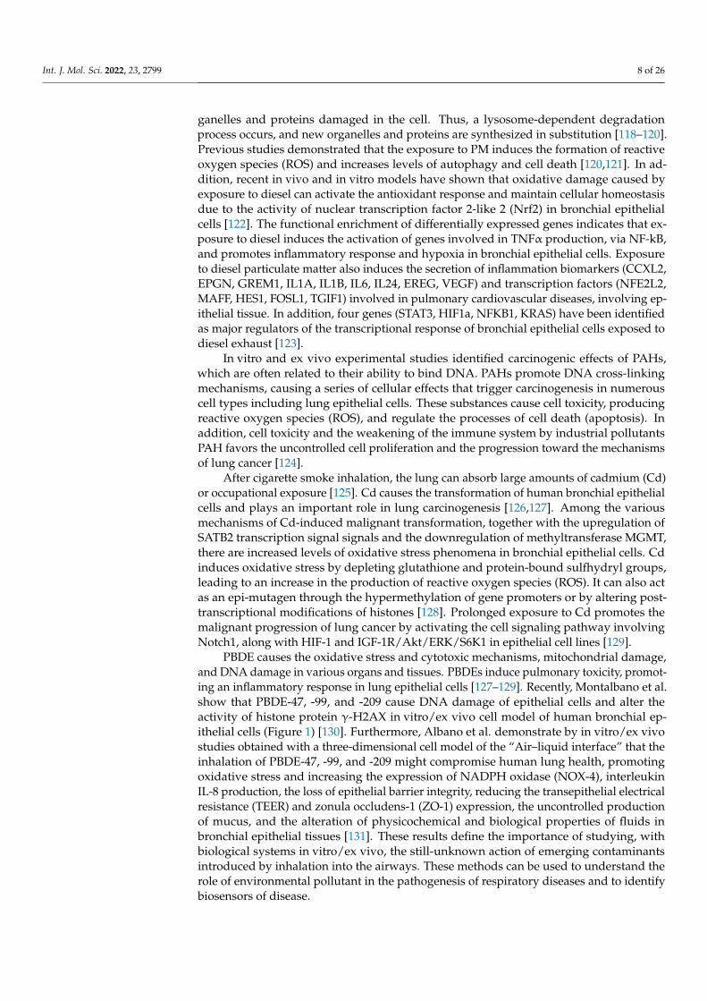
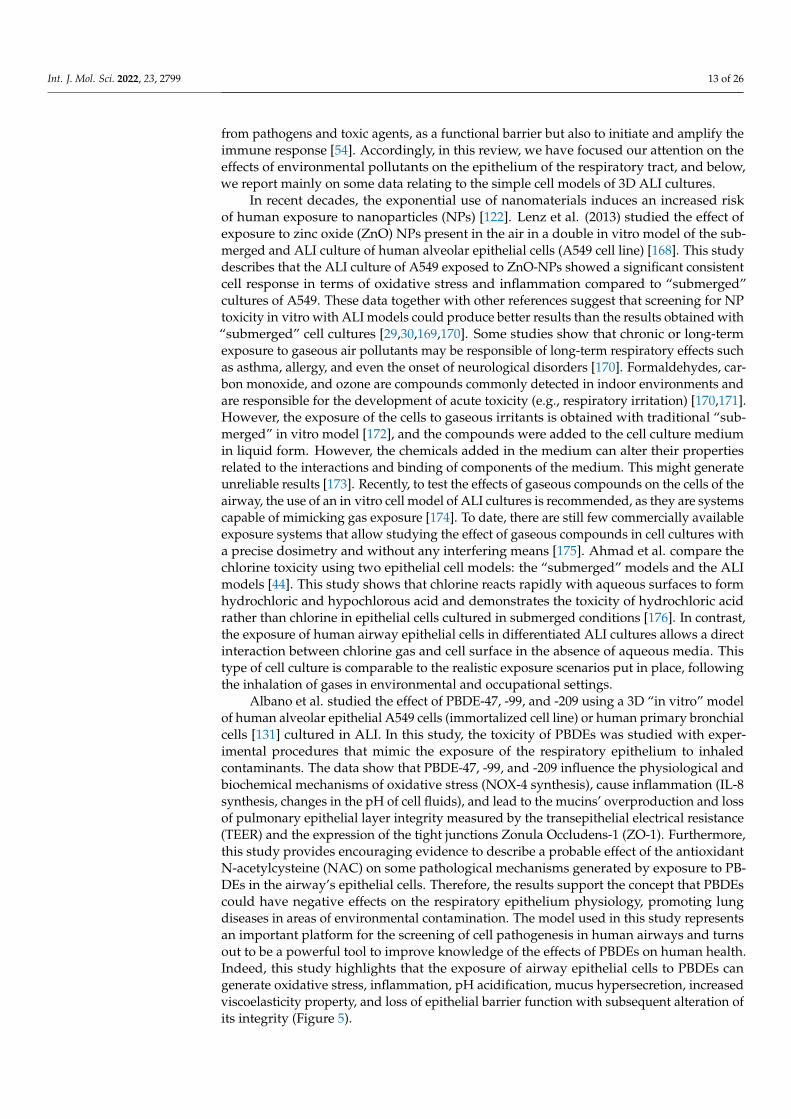
PBDE causes the oxidative stress and cytotoxic mechanisms, mitochondrial damage,and DNA damage in various organs and tissues. PBDEs induce pulmonary toxicity, promot-ing an inflammatory response in lung epithelial cells [127–129]. Recently, Montalbano et al.show that PBDE-47, -99, and -209 cause DNA damage of epithelial cells and alter theactivity of histone protein γ-H2AX in vitro/ex vivo cell model of human bronchial ep-ithelial cells (Figure 1) [130]. Furthermore, Albano et al. demonstrate by in vitro/ex vivostudies obtained with a three-dimensional cell model of the “Air–liquid interface” that theinhalation of PBDE-47, -99, and -209 might compromise human lung health, promotingoxidative stress and increasing the expression of NADPH oxidase (NOX-4), interleukinIL-8 production, the loss of epithelial barrier integrity, reducing the transepithelial electricalresistance (TEER) and zonula occludens-1 (ZO-1) expression, the uncontrolled productionof mucus, and the alteration of physicochemical and biological properties of fluids inbronchial epithelial tissues [131]. These results define the importance of studying, withbiological systems in vitro/ex vivo, the still-unknown action of emerging contaminantsintroduced by inhalation into the airways. These methods can be used to understand therole of environmental pollutant in the pathogenesis of respiratory diseases and to identifybiosensors of disease.
Int. J. Mol. Sci. 2022, 23, 2799 9 of 26
Figure 1. Effects of PBDE-47, -99, and -209 flame retardants in bronchial epithelial cells. PBDE-47, -99,and -209 cause DNA damage of epithelial cells and alter the activity of histone protein γ-H2AX inin vitro/ex vivo cell models of human bronchial epithelial cells.
The toxicology of mixtures has recently generated greater interest than the evaluationof the effect of single substances. The toxicity of pollutant mixtures could represent a moreaccurate way to understand in real time the effects of environmental contamination onthe respiratory system and to determine harmful effects relevant to the health of people.Therefore, the use of suitable cell models could create broad prospects for identifying thetoxicology of mixtures.
4. Cell Systems to Study the Effects of Environmental Contaminants inRespiratory Diseases
One of the “gold standards” of environmental scientific research is to obtain data tounderstand the effects of exposure to inhaled toxic substances on human health. In vivostudies were used only to collect data related to an indirect effect of pollutants. Thesestudied do not establish a direct relationship between ethical and safety precautions, highcosts, very long periods, and environment with pathogenetic alterations regarding humanhealth. Furthermore, data obtained from observational studies of subjects in the areasof environmental contamination are to lower the resolution of pathological effects at thecellular and molecular levels [132].
For many years, scientific research has used animal models as the main tools to evalu-ate the effects of inhaled substances on human health. However, results obtained in mousemodels are not always able to predict diseases, and their general use for research purposeshas raised growing public and animal welfare concerns. The scientific research pushes to-ward the use of alternative and innovative in vitro/ex vivo experimental models [133–136].The history of experimental models began in 1885 with the zoologist Wilhelm Roux. Hewas the pioneer of experimental embryology studying the embryonic chicken cells insaline for several days. However, only in the mid-1950s, Harry Eagle gave a significantboost to this area of research by studying and identifying the nutrients needed by cells inculture [137,138]. To date, the cell cultures and in vitro/ex vivo models are essential for
Int. J. Mol. Sci. 2022, 23, 2799 10 of 26
the identification and the study of the effects of inhaled noxae, which are represented byatmospheric pollutants, on biological systems [139]. In recent decades, tissue engineeringapproaches have made enormous progress, and several in vitro models have been estab-lished to study the effect of inhalation toxicity and disease. The aims of these studies are toimprove the understanding of pathophysiological processes and to provide new and moreindependent experimental systems for pharmacological and toxicological studies.
In vitro lung models are currently available for all important segments of the res-piratory system starting from the nasal cavity and trachea to the proximal and distalairways [140]. Traditional two-dimensional (2D) monolayer culture models (also called“submerged cultures”) are used to identify molecules involved in the signaling of alteredcellular and molecular mechanisms that arise in the case of pulmonary toxicity. However,these models lack the key features of the human airway microenvironment that are essentialfor accurately studying the toxic effects of inhaled exogenous noxae.
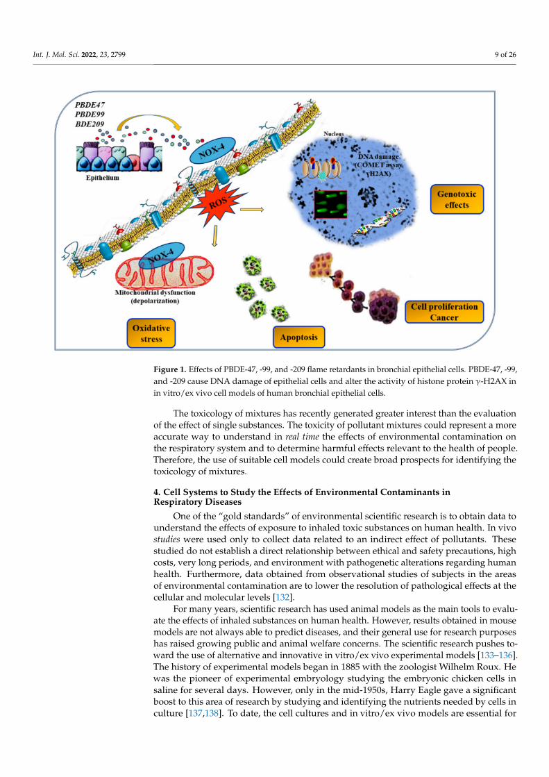
Recently, the traditional method of two-dimensional single-layer cell culture wasreplaced with more innovative methods, since it does not faithfully reflect what is observedwithin tissues in vivo [141,142]. The main reason for the inadequacy of these cell culturesystems is the lack of the architectural support and heterogeneity typical of the cells of thelung tissue. This awareness has led to an increase in the development of more complex three-dimensional (3D) models in which cells can grow in multiple directions in order to betterreflect the cellular interaction present in the natural environment in vivo. Furthermore,multiple cell types may be present in these models [44,143–146] and an extracellular matrix,thus allowing to mimic a model similar to one in vivo. The “air–liquid interface” (ALI)culture is an example of these innovative models of cell culture (Figure 2).
Figure 2. Three-dimensional (3D) ALI cultures of epithelial cells (primary and cell line) to study theeffects of environmental contaminants in airway disease. Steps to obtain primary epithelial cells fromhuman tissue: (a) Collection of bronchial biopsies or surgical specimens; (b) epithelial cell processing:tissue was dissociated, resuspended in bronchial epithelial growth medium; (c) cell expansion andculture of epithelial cells; (d) the cells are seeded onto the microporous membrane pre-coated withcollagen in submerged conditions until confluence and culture in ALI; (e) reaching the confluence,the cells begin to lift at the air–liquid interface starting the differentiation fed by the culture mediumin the basolateral side. Hence, the epithelial cells differentiate in the pseudostratified phenotype andbuild a tissue such as the epithelium of the lung.
Int. J. Mol. Sci. 2022, 23, 2799 11 of 26
This biological system of epithelial cells has a well-differentiated epithelium similar tohuman airways. Thus, epithelial cell cultured in ALI represents a valuable tool for scientificresearch to study the toxic effect of inhaled chemicals on the human health affectingrespiratory diseases, providing the opportunity to evaluate and identify important cellularand molecular mechanisms [147].
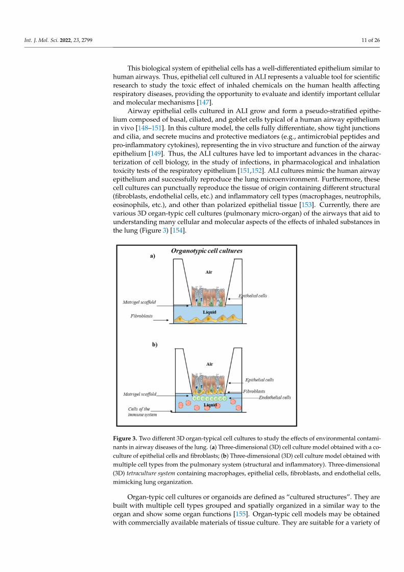
Airway epithelial cells cultured in ALI grow and form a pseudo-stratified epithe-lium composed of basal, ciliated, and goblet cells typical of a human airway epitheliumin vivo [148–151]. In this culture model, the cells fully differentiate, show tight junctionsand cilia, and secrete mucins and protective mediators (e.g., antimicrobial peptides andpro-inflammatory cytokines), representing the in vivo structure and function of the airwayepithelium [149]. Thus, the ALI cultures have led to important advances in the charac-terization of cell biology, in the study of infections, in pharmacological and inhalationtoxicity tests of the respiratory epithelium [151,152]. ALI cultures mimic the human airwayepithelium and successfully reproduce the lung microenvironment. Furthermore, thesecell cultures can punctually reproduce the tissue of origin containing different structural(fibroblasts, endothelial cells, etc.) and inflammatory cell types (macrophages, neutrophils,eosinophils, etc.), and other than polarized epithelial tissue [153]. Currently, there arevarious 3D organ-typic cell cultures (pulmonary micro-organ) of the airways that aid tounderstanding many cellular and molecular aspects of the effects of inhaled substances inthe lung (Figure 3) [154].
Figure 3. Two different 3D organ-typical cell cultures to study the effects of environmental contami-nants in airway diseases of the lung. (a) Three-dimensional (3D) cell culture model obtained with a co-culture of epithelial cells and fibroblasts; (b) Three-dimensional (3D) cell culture model obtained withmultiple cell types from the pulmonary system (structural and inflammatory). Three-dimensional(3D) tetraculture system containing macrophages, epithelial cells, fibroblasts, and endothelial cells,mimicking lung organization.
Organ-typic cell cultures or organoids are defined as “cultured structures”. They arebuilt with multiple cell types grouped and spatially organized in a similar way to theorgan and show some organ functions [155]. Organ-typic cell models may be obtainedwith commercially available materials of tissue culture. They are suitable for a variety of
Int. J. Mol. Sci. 2022, 23, 2799 12 of 26
experimental projects and for modeling complex lung toxic responses, including inflam-mation, oxidative stress, myofibroblast formation, transepithelial migration, and invasion.Organ-typical patterns also mimic epithelial barrier properties and changes in cytotoxicand pro-inflammatory effects upon exposure to environmental toxicants such as thoseobserved in vivo [156–158].
The term “organoid” defines the 3D culture referring to stem cell-derived native-liketissue structures of a given organ created by the induction of genetically encoded self-assembly programming [159]. Similar to processes that regulate organogenesis duringembryonic development, cells within organoids undergo self-organization guided by cell-specific adhesion properties and spatially restricted progenitor differentiation. Organoidculture derived from stem cells or organ-specific progenitor cells that differentiate andself-organize through cell sorting and lineage commitment similar to the in vivo process. Itshould be noted that this recent definition is not yet strictly used in the literature [160,161].
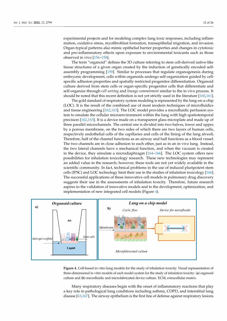
The gold standard of respiratory system modeling is represented by the lung-on-a-chip(LOC). It is the result of the combined use of most modern techniques of microfluidicsand tissue engineering [162,163]. The LOC model provides a microfluidic perfusion sys-tem to emulate the cellular microenvironment within the lung with high spatiotemporalprecision [162,163]. It is a device made on a transparent glass microplate and made up ofthree parallel microchannels. The central one is divided into two halves, lower and upper,by a porous membrane, on the two sides of which there are two layers of human cells,respectively endothelial cells of the capillaries and cells of the lining of the lung alveoli.Therefore, half of the channel functions as an airway and half functions as a blood vessel.The two channels are in close adhesion to each other, just as in an in vivo lung. Instead,the two lateral channels have a mechanical function, and when the vacuum is createdin the device, they simulate a microdiaphragm [164–166]. The LOC system offers newpossibilities for inhalation toxicology research. These new technologies may representan added value in the research; however, these tools are not yet widely available in thescientific community. In fact, technical problems in the use of induced pluripotent stemcells (IPSC) and LOC technology limit their use in the studies of inhalation toxicology [166].The successful applications of these innovative cell models in pulmonary drug discoverysuggests their use in the assessments of inhalation toxicity. Therefore, future researchaspires to the validation of innovative models and to the development, optimization, andimplementation of new integrated cell models (Figure 4).
Figure 4. Cell-based in vitro lung models for the study of inhalation toxicity. Visual representation ofthree-dimensional in vitro models of each model system for the study of inhalation toxicity: (a) organoidculture and (b) microfluidic and microfabricated device culture. ECM, extracellular matrix.
Many respiratory diseases begin with the onset of inflammatory reactions that playa key role in pathological lung conditions including asthma, COPD, and interstitial lungdisease [63,167]. The airway epithelium is the first line of defense against respiratory lesions
Int. J. Mol. Sci. 2022, 23, 2799 13 of 26
from pathogens and toxic agents, as a functional barrier but also to initiate and amplify theimmune response [54]. Accordingly, in this review, we have focused our attention on theeffects of environmental pollutants on the epithelium of the respiratory tract, and below,we report mainly on some data relating to the simple cell models of 3D ALI cultures.
In recent decades, the exponential use of nanomaterials induces an increased riskof human exposure to nanoparticles (NPs) [122]. Lenz et al. (2013) studied the effect ofexposure to zinc oxide (ZnO) NPs present in the air in a double in vitro model of the sub-merged and ALI culture of human alveolar epithelial cells (A549 cell line) [168]. This studydescribes that the ALI culture of A549 exposed to ZnO-NPs showed a significant consistentcell response in terms of oxidative stress and inflammation compared to “submerged”cultures of A549. These data together with other references suggest that screening for NPtoxicity in vitro with ALI models could produce better results than the results obtained with“submerged” cell cultures [29,30,169,170]. Some studies show that chronic or long-termexposure to gaseous air pollutants may be responsible of long-term respiratory effects suchas asthma, allergy, and even the onset of neurological disorders [170]. Formaldehydes, car-bon monoxide, and ozone are compounds commonly detected in indoor environments andare responsible for the development of acute toxicity (e.g., respiratory irritation) [170,171].However, the exposure of the cells to gaseous irritants is obtained with traditional “sub-merged” in vitro model [172], and the compounds were added to the cell culture mediumin liquid form. However, the chemicals added in the medium can alter their propertiesrelated to the interactions and binding of components of the medium. This might generateunreliable results [173]. Recently, to test the effects of gaseous compounds on the cells of theairway, the use of an in vitro cell model of ALI cultures is recommended, as they are systemscapable of mimicking gas exposure [174]. To date, there are still few commercially availableexposure systems that allow studying the effect of gaseous compounds in cell cultures witha precise dosimetry and without any interfering means [175]. Ahmad et al. compare thechlorine toxicity using two epithelial cell models: the “submerged” models and the ALImodels [44]. This study shows that chlorine reacts rapidly with aqueous surfaces to formhydrochloric and hypochlorous acid and demonstrates the toxicity of hydrochloric acidrather than chlorine in epithelial cells cultured in submerged conditions [176]. In contrast,the exposure of human airway epithelial cells in differentiated ALI cultures allows a directinteraction between chlorine gas and cell surface in the absence of aqueous media. Thistype of cell culture is comparable to the realistic exposure scenarios put in place, followingthe inhalation of gases in environmental and occupational settings.
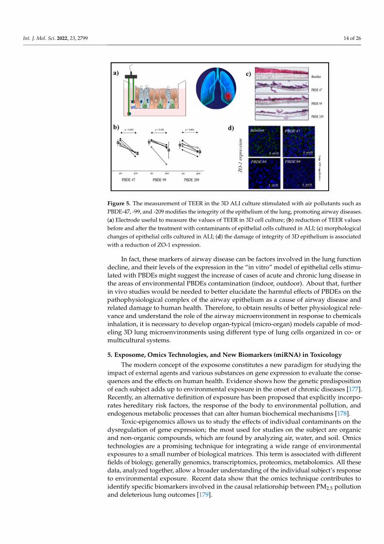
Albano et al. studied the effect of PBDE-47, -99, and -209 using a 3D “in vitro” modelof human alveolar epithelial A549 cells (immortalized cell line) or human primary bronchialcells [131] cultured in ALI. In this study, the toxicity of PBDEs was studied with exper-imental procedures that mimic the exposure of the respiratory epithelium to inhaledcontaminants. The data show that PBDE-47, -99, and -209 influence the physiological andbiochemical mechanisms of oxidative stress (NOX-4 synthesis), cause inflammation (IL-8synthesis, changes in the pH of cell fluids), and lead to the mucins’ overproduction and lossof pulmonary epithelial layer integrity measured by the transepithelial electrical resistance(TEER) and the expression of the tight junctions Zonula Occludens-1 (ZO-1). Furthermore,this study provides encouraging evidence to describe a probable effect of the antioxidantN-acetylcysteine (NAC) on some pathological mechanisms generated by exposure to PB-DEs in the airway’s epithelial cells. Therefore, the results support the concept that PBDEscould have negative effects on the respiratory epithelium physiology, promoting lungdiseases in areas of environmental contamination. The model used in this study representsan important platform for the screening of cell pathogenesis in human airways and turnsout to be a powerful tool to improve knowledge of the effects of PBDEs on human health.Indeed, this study highlights that the exposure of airway epithelial cells to PBDEs cangenerate oxidative stress, inflammation, pH acidification, mucus hypersecretion, increasedviscoelasticity property, and loss of epithelial barrier function with subsequent alteration ofits integrity (Figure 5).
Int. J. Mol. Sci. 2022, 23, 2799 14 of 26
Figure 5. The measurement of TEER in the 3D ALI culture stimulated with air pollutants such asPBDE-47, -99, and -209 modifies the integrity of the epithelium of the lung, promoting airway diseases.(a) Electrode useful to measure the values of TEER in 3D cell culture; (b) reduction of TEER valuesbefore and after the treatment with contaminants of epithelial cells cultured in ALI; (c) morphologicalchanges of epithelial cells cultured in ALI; (d) the damage of integrity of 3D epithelium is associatedwith a reduction of ZO-1 expression.
In fact, these markers of airway disease can be factors involved in the lung functiondecline, and their levels of the expression in the “in vitro” model of epithelial cells stimu-lated with PBDEs might suggest the increase of cases of acute and chronic lung disease inthe areas of environmental PBDEs contamination (indoor, outdoor). About that, furtherin vivo studies would be needed to better elucidate the harmful effects of PBDEs on thepathophysiological complex of the airway epithelium as a cause of airway disease andrelated damage to human health. Therefore, to obtain results of better physiological rele-vance and understand the role of the airway microenvironment in response to chemicalsinhalation, it is necessary to develop organ-typical (micro-organ) models capable of mod-eling 3D lung microenvironments using different type of lung cells organized in co- ormulticultural systems.
5. Exposome, Omics Technologies, and New Biomarkers (miRNA) in Toxicology
The modern concept of the exposome constitutes a new paradigm for studying theimpact of external agents and various substances on gene expression to evaluate the conse-quences and the effects on human health. Evidence shows how the genetic predispositionof each subject adds up to environmental exposure in the onset of chronic diseases [177].Recently, an alternative definition of exposure has been proposed that explicitly incorpo-rates hereditary risk factors, the response of the body to environmental pollution, andendogenous metabolic processes that can alter human biochemical mechanisms [178].
Toxic-epigenomics allows us to study the effects of individual contaminants on thedysregulation of gene expression; the most used for studies on the subject are organicand non-organic compounds, which are found by analyzing air, water, and soil. Omicstechnologies are a promising technique for integrating a wide range of environmentalexposures to a small number of biological matrices. This term is associated with differentfields of biology, generally genomics, transcriptomics, proteomics, metabolomics. All thesedata, analyzed together, allow a broader understanding of the individual subject’s responseto environmental exposure. Recent data show that the omics technique contributes toidentify specific biomarkers involved in the causal relationship between PM2.5 pollutionand deleterious lung outcomes [179].
Int. J. Mol. Sci. 2022, 23, 2799 15 of 26
However, this type of approach is still particularly difficult, especially due to its highcost. Methylome has been of great interest for some time. In fact, epigenetic modificationsremain of great interest in the scientific community; in fact, the study of a cohort of subjectsexposed to different types of pollutants and chemicals demonstrates how there is a variationin DNA methylation performed by gene sequencing [180].
The presence of altered levels of molecules at the systemic level can indicate the progressof specific diseases, which is currently of great interest in the scientific community. In recentyears, RNA molecules have been the subject of great interest, and these studies led to theidentification of microRNAs. MicroRNAs are sequences of approximately 22/25 nucleotides;they are transcribed from non-coding regions of the genome, undergo post-transcriptionalchanges that lead them to “ripening”, and at that point, they are ready to perform their func-tion [181]. Their mechanism of action involves the binding of a complementary portion of amature mRNA to regulate its function: blocking its translation or leading to degradation [177].Some microRNAs regulate dozens of targets RNAs ensuring the maintenance of physiologicalprocesses in all tissue districts: cell proliferation, differentiation, balancing of the oxidativestress, metabolism, and apoptosis [182]. Alterations in the expression of these microRNAslead the cell to lose its homeostasis [183]. Computational biology gives us a great help in theprediction of the target of the microRNA. Through specific databases, the gene sequencesare analyzed to identify predictive targets of the microRNAs examined (the homologousnucleotide sequences of the microRNAs that are complementary and that can potentially beregulated by the microRNA under consideration) [184].
External factors, and in particular environmental pollutants, play a key role in theperturbability of cell functions such as the alteration in the expression of regulatory mi-croRNAs and the consequent gene expression [185]. In the context of human health, recentstudies have associated the increase or decrease of some microRNAs in relation to certaindiseases or to simple exposure to cigarette smoke [186]; generally, they are clusters madeup of some tens of microRNAs that see their expression jointly altered; epigenetic alter-ations and changes in regulatory pathways also lead to connections between inflammatorymechanisms and cancer [185,187].
Polychlorinated biphenyls (PCBs) have been widely used over the years, and theirproduction has recently been banned due to their environmental impact; several studieshave shown how their bioaccumulation in fatty tissues causes a deregulation in gene andmicroRNA expression [188]. Arsenic in its trivalent form (As[3]+) is also associated withcancer risk, and environmental exposure to arsenic, especially long-term, is associated withgene instability and the risk of diseases associated with peripheral vascular lesions [181].High concentrations of mercury (Hg) in the blood are related to the risk of hypertension,and in general of cardiovascular toxicity, endothelial toxicity, hypercholesterolemia, andneurotoxicity, especially when exposure to this heavy metal occurs during prenatal develop-ment [189]. HUVEC cell lines were used to demonstrate the effects of Hg on the expressionof miR-92a and miR-486; the parallel analysis of the plasma of workers exposed to Hgconfirms the increase in the expression of the same microRNAs. This demonstrates howthrough integrated approaches of in vitro/in vivo studies, it is possible to demonstratean aberrant alteration of molecular biomarkers that interfere in the activation pathwaysof inflammatory mechanisms involving NF-κB and the expression of COX-2 altering thephysiological apoptotic processes [190].
The perfect biomarkers should be readily available in the body fluids, such as blood,liquid biopsies, or others. Furthermore, it should also be tissue or cell-specific type andundergo a variation in the expression to be also useful for the control and response to thetherapy used [191]. So, to identify specific biomarkers of diseases, it is important that aninitial approach includes in vitro studies. This approach provides us tools to study andanalyze the biosensors of one cell type at a time. It is highly repeatable, reproducible, andgives the opportunity to connect the levels of biosensor detection of expression directly tothe stimulus used in the experimental procedures [192]. Finally, an integrated approachbetween the three experimental models enclosing in vitro, ex vivo, and in vivo studies

Int. J. Mol. Sci. 2022, 23, 2799 16 of 26
allows us to study quickly the direct effects of the molecules of interest involved in theenvironmental contamination due to toxicants such as PBDEs. However, the validation ofbiomarkers as biosensors passes from the analysis of biological samples and possibly fromthe effect of a potential drug that restores or suppresses its expression and activity.
The bronchial epithelium constitutes the first barrier to the inhaled pollutants presentin the environment; the increase in oxidative stress as well as the release of pro-inflammatorycytokines and exosomes containing microRNA is now widely demonstrated both by in vitroand in vivo studies [193]. Multi-organ studies, ex vivo, give us indications on molecularbiomarkers related to inflammatory processes of the airways such as the Let-7, mir21,mir122, and mir25 microRNA families [181].
PBDE induce a variation in the expression of biomarkers related to inflammatoryprocesses of the lung in an in vitro/ex vivo study performed on human lung epitheliumcells (epithelial cell line and cells isolated from lung biopsies). We studied the effects ofthree different PBDEs (47, 99, and 209) in the mechanisms of oxidative stress, epithelialintegrity, and release of inflammatory cytokines in the cell culture of bronchial epithelialcells [130,131]. Furthermore, the analysis of the transcripts allowed us to isolate the microR-NAs secreted both by immortalized epithelial cells (A549) and primary human bronchialepithelial cells. We highlighted a decrease in the expression of let-7a together with anincrease of both mir21 and mir25 microRNA [193]. These results support the conceptthat biomarkers detected in the in vitro model of cell culture might be a useful tool in theprediction of lung diseases and their progression in subjects exposed in risk areas.
Placenta is fundamental in the regulation of the intrauterine environment, and it isalready known that heavy metals such as Cd and lead together with other environmen-tal pollutants can modify the expression of various microRNAs [190]. To support ourin vitro/ex vivo approach with a 3D cell culture of epithelial cells [193,194], where we high-lighted the let-7a, mirR21, and mirR25 microRNAs production as molecular biomarkersof PBDE contamination, in future studies, we will analyze a cohort of pregnant womenexposed to PBDEs present in the environment as pollutants.
The integrated approach between in vitro ex vivo and in vivo studies proves to bethe key to identifying the patterns of molecular biomarkers (miRNA) that can help in theearly diagnosis of organ pathology and facilitate the identification of the response to drugtreatment, to use a personalized therapy tailored to the patient exposed in the areas ofenvironmental contamination (Figure 6).
Figure 6. Experimental approach to identify molecular biosensors of airway disease. “In vitro/exvivo” studies obtained with experimental approach performed with 3D cell cultures of bronchialepithelial cells (cell line and primary cells); Integrated approach obtained including “in vitro”, exvivo” 3D cell cultures of bronchial epithelial cells and “in vivo” studies.
Int. J. Mol. Sci. 2022, 23, 2799 17 of 26
6. Conclusions
Environmental contamination plays a fundamental role in human health, generatingserious diseases such as inflammatory and neoplastic ones. In this review, we describedsome effects of air environmental contamination on human health, regarding lung diseases.We realized a descriptive approach with the aim to transfer the following in a simple way:(1) fundamental aspects of the activation of epithelial cells in respiratory diseases in case ofexposure to contamination environmental; and (2) useful tools for an adequate in vitro/exvivo experimental design to study the effect of air pollutants in the lung. About this lastpoint, we particularly refer to our experiences regarding the use of 3D ALI cultures ofepithelial cells.
In this scenario, novel 3D in vitro models offer the advantage of enhanced physio-logical relevance through the incorporation of architectural support (i.e., ECM proteinsor scaffolding), cell–cell interactions, and in some instances, biomimetic devices that canrecapitulate physiologically breathing motions. However, despite their contributions, cellmodels have not been able to accurately represent the heterogeneity of the human pop-ulation and account for interindividual variability in response to inhaled toxicants andsusceptibility to the adverse health effects.
In this review, we focalized our attention to the concerns about the effects of air envi-ronmental contamination on human health regarding diseases of the lung with particularattention to the role of epithelial cells. Our descriptive approach has been discreet with theaim to transfer in a simple way the fundamental aspects of the activation of epithelial cellsin respiratory diseases in case of exposure to environmental contamination. Here, we referto our experiences about the use of 3D ALI cultures of epithelial cells to study the effect ofsome toxicants on epithelial cells.
Furthermore, here, we introduced the concept of the exposome, since it constitutes anew paradigm for studying the impact of the environment on human health. Finally, thecontribution of Omics Sciences defined new scientific perspectives aimed at the discoveryof the cellular and molecular mechanisms underlying the immunological response of theairway epithelium in conditions of environmental air contamination. The goal of theresearchers might be to enrich the concept of exposome using innovative biological systemsthat mimic organ situations in real life.
In conclusion, with the short descriptions enclosed in this review, we underline andsuggest the importance of planning new technologic and conceptual perspectives useful tofurther clarify the effect of environmental factors on the health of the lung. The specificobjectives to be achieved are to identify new cellular and molecular pathways associatedwith the concept of the exposome, with the help of complex organotypic and organoidcultures with applications of microfluidics and omics sciences.
Funding: This research received no external.
Acknowledgments: Giusy Daniela Albano, Angela Marina Montalbano, and Rosalia Gagliardo eMirella Profita (authors of the manuscript) thank the National Research Council for their support.
Conflicts of Interest: The authors of the paper declare that they have no competing interests forthis study.
Int. J. Mol. Sci. 2022, 23, 2799 18 of 26
Abbreviations
FVC forced volume capacityCOPD chronic obstructive pulmonary diseaseCO carbon monoxideNOx nitrogen oxidesSO2 sulfur dioxideIPA polycyclic aromatic hydrocarbonsPM particulate matterVOC volatile organic compoundsCOHb carboxyhemoglobinNO2 nitrogen dioxideB[a]P benzo-[a]-pyreneIARC International Agency for Research on CancerESCAPE European study of cohorts for air pollution effectsSIN sites of national interestCISAS International Center for Advanced Studies in Environment,
Ecosystem and Human HealthOMS World Health OrganizationPOP persistent organic pollutantsPCB polychlorinated biphenylsPBDE polybrominated diphenyl ethersEFSA European Food Safety AuthorityTLR4/NF-kB Toll-like receptor 4/nuclear factor
kappa-light-chain-enhancer of activated B cellsUE European UnionAM alveolar macrophagesTNF-α tumor necrosis factorIL- interleukin-DAMP damage-associated molecular patternGM-CSF granulocyte-macrophage colony-stimulating factorMCP-1 monocyte chemoattractant protein 1TSLP timic stromal lymphopoietinO3 ozoneC6H6 benzeneAs arsenicCd cadmiumNi nickelROS reactive oxygen speciesDUOX1 duox ossidasi 1NADPH nicotinamide adenine dinucleotide phosphateNrf2 erythroid 2-like 2ALI air–liquid interfaceLOC lung-on-a-chipIPSC induced pluripotent stem cellsNP nanoparticlesTEER transepithelial electrical resistanceNAC N-acetyl cysteineHg mercuryCOX-2 cyclooxygenase-2RNA ribonucleic acidmRNA messenger RNAZonula Occludens-1 ZO-1
Int. J. Mol. Sci. 2022, 23, 2799 19 of 26
References1. Manisalidis, I.; Stavropoulou, E.; Stavropoulos, A.; Bezirtzoglou, E. Environmental and health im-pacts of air pollution: A review.
Front. Public Health. 2020, 20, 8–14. [CrossRef]2. Ashfaq, A.; Sharma, P. Environmental effects of air pollution and application of engineered methods to combat the problem. J.
Indust. Pollut. Control. 2012, 29, 25–28.3. Guan, W.J.; Zheng, X.Y.; Chung, K.F.; Zhong, N.S. Impact of air pollution on the burden of chronic res-piratory diseases in China:
Time for urgent action. Lancet 2016, 388, 1939–1951. [CrossRef]4. Szyszkowicz, M.; Kousha, T.; Valacchi, G. Ambient air pollution and emergency department visits for skin conditions. Glob.
Dermatol. 2016, 3, 323–329. [CrossRef]5. Kampa, M.; Castanas, E. Human health effects of air pollution. Environ. Pollut. 2008, 151, 362–367. [CrossRef]6. Peters, J.M.; Avol, E.; Gauderman, W.J.; Linn, W.S.; Navidi, W.; London, S.J.; Margolis, H.; Rappaport, E.; Vora, H.;
Gong, H., Jr.; et al. A study of twelve Southern California communities with differing levels types of air pollution: II. Effects onpulmonary function. Am. J. Respir. Crit. Care Med. 1999, 159, 768–775. [CrossRef]
7. Doiron, D.; de Hoogh, K.; Probst-Hensch, N.; Fortier, I.; Cai, Y.; De Matteis, S.; Hansell, A.L. Air pollution, lung function andCOPD: Results from the population-based UK Biobank study. Eur. Respir. J. 2019, 54, 1802140. [CrossRef]
8. Gauderman, W.J.; Avol, E.; Gilliland, F.; Vora, H.; Thomas, D.; Berhane, K.; McConnell, R.; Kuenzli, N.; Lurmann, F.;Rappaport, E.; et al. The effect of air pollution on lung development from 10 to 18 years of age. N. Engl. J. Med. 2004, 351,1057–1067, Erratum in 2005, 352, 1276. [CrossRef] [PubMed]
9. Inquinamento e Salute. Dal Traffico al Fumo, Dalla Chimica all’attività Lavorativa: Come l’ambiente Influenza il Rischio di Ammalarsidi Tumore. Fondazioneveronesi.it. 2018. Available online: https://www.fondazioneveronesi.it/magazine/tools-della-salute/download/i-manuali/inquinamento-e-salute (accessed on 24 January 2022).
10. Proietti, M. Inquinamento e Malattie. Edizioni Minerva Medica 2018. Available online: https://www.minervamedica.it/it/volumi/specialitadiche/igiene/scheda.php?cod=L10091 (accessed on 24 January 2022).
11. Berend, N. Contribution of air pollution to COPD and small airway dysfunction. Respirology. 2016, 21, 237–244. [CrossRef]12. Holland, W.W.; Reid, D.D. The urban factor in chronic bronchitis. Lancet 1965, 1, 445–448. [CrossRef]13. Schwartz, J. Lung function and chronic exposure to air pollution: A cross-sectional analysis of NHANES II. Environ. Res. 1989, 50,
309–321. [CrossRef]14. Robertson, D.S. Health effects of increase in concentration of carbon dioxide in the atmosphere. Curr. Sci. 2006, 90, 1607–1609.15. Zhao, Y.; Hu, J.; Tan, Z.; Liu, T.; Zeng, W.; Li, X.; Huang, C.; Wang, S.; Huang, Z.; Ma, W. Ambient carbon monoxide and increased
risk of daily hospital out-patient visits for respiratory diseases in Dongguan, China. Sci. Total Environ. 2019, 668, 254–260.[CrossRef] [PubMed]
16. Hamra, G.B.; Laden, F.; Cohen, A.J.; Raaschou-Nielsen, O.; Brauer, M.; Loomis, D. Lung cancer and expo-sure to nitrogen dioxideand traffic: A systematic review and meta-analysis. Environ. Health Perspect. 2015, 123, 1107–1112. [CrossRef]
17. Faustini, A.; Rapp, R.; Forastiere, F. Nitrogen dioxide and mortality: Review andmeta-analysis of long-termstudies. Eur. Respir. J.2014, 44, 744–753. [CrossRef] [PubMed]
18. Nriagu, J.O. Air pollution from solid fuels. In Encyclopedia of Environmental Health; Elsevier: Amsterdam, The Netherlands, 2011;pp. 46–52.
19. Abdel-Shafy, H.I.; Mansour, M.S.M. A review on polycyclic aromatic hydrocarbons: Source, envi-ronmental impact, effect onhuman health and remediation. Egypt J. Pet. 2016, 25, 107–123. [CrossRef]
20. Arpalombardia. 2018. Available online: https://www.arpalombardia.it/Pages/Aria/Inquinanti/Metalli.aspx?firstlevel=Inquinanti (accessed on 24 January 2022).
21. Brunekreef, B.; Beelen, R.; Hoek, G.; Schouten, L.; Bausch-Goldbohm, S.; Fischer, P.; Armstrong, B.; Hughes, E.; Jerrett, M.;van den Brandt, P. Effects of long-term exposure to traffic-related air pol-lution on respiratory and cardiovascular mortality in theNetherlands: The NLCS-AIR study. Res. Rep. 2009, 139, 5–71.
22. Colais, P.; Faustini, A.; Stafoggia, M.; Berti, G.; Bisanti, L.; Cadum, E.; Cernigliaro, A.; Mallone, S.; Pacelli, B.; Serinelli, M.; et al.EPIAIR Collaborative Group. Particulate air pollution and hospital admissions for cardiac diseases in potentially sensitivesubgroups. Epidemiology 2012, 23, 473–481. [CrossRef] [PubMed]
23. Beelen, R.; Hoek, G.; van den Brandt, P.A.; Goldbohm, R.A.; Fischer, P.; Schouten, L.J.; Armstrong, B.; Brunekreef, B. Long-termexposure to traffic-related air pollution and lung cancer risk. Epidemiology 2008, 19, 702–710. [CrossRef] [PubMed]
24. Vineis, P.; Hoek, G.; Krzyzanowski, M.; Vigna-Taglianti, F.; Veglia, F.; Airoldi, L.; Autrup, H.; Dunning, A.; Garte, S.;Hainaut, P.; et al. Air pollution and risk of lung cancer in a prospective study in Europe. Int. J. Cancer 2006, 119, 169–174.[CrossRef]
25. Palli, D.; Saieva, C.; Munnia, A.; Peluso, M.; Grechi, D.; Zanna, I.; Caini, S.; Decarli, A.; Sera, F.; Masala, G. DNA adducts andPM10 exposure in traffic-exposed workers and urban residents from the EPIC-Florence city study. Sci. Total Environ. 2008, 403,105–112. [CrossRef]
26. Heinrich, J.; Thiering, E.; Rzehak, P.; Krämer, U.; Hochadel, M.; Rauchfuss, K.M.; Gehring, U.; Wichmann, H.E. Long-termexposure to NO2 and PM10 and all-cause and cause-specific mortality in a prospective cohort of women. Occup. Environ. Med.2013, 70, 179–186. [CrossRef] [PubMed]
Int. J. Mol. Sci. 2022, 23, 2799 20 of 26
27. Raaschou-Nielsen, O.; Andersen, Z.J.; Beelen, R.; Samoli, E.; Stafoggia, M.; Weinmayr, G.; Hoffmann, B.; Fischer, P.; Nieuwenhui-jsen, M.J.; Brunekreef, B.; et al. Air pollution and lung cancer incidence in 17 european cohorts: Prospective analyses from theEuropean Study of Cohorts for Air Pollution Effects (ESCAPE). Lancet Oncol. 2013, 14, 813–822. [CrossRef]
28. Schmid, O.; Stoeger, T. Surface area is the biologically most effective dose metric for acute nanoparticle toxicity in the lung. J.Aerosol. Sci. 2016, 99, 133–143. [CrossRef]
29. Teeguarden, J.G.; Hinderliter, P.M.; Orr, G.; Thrall, B.D.; Pounds, J.G. Particokinetics in vitro: Dosimetry considerations for in vitronanoparticle toxicity assessments. Toxicol. Sci. 2007, 95, 300–312, Erratum in Toxicol. Sci. 2007, 97, 614. [CrossRef] [PubMed]
30. Wilkinson, K.E.; Palmberg, L.; Witasp, E.; Kupczyk, M.; Feliu, N.; Gerde, P.; Seisenbaeva, G.A.; Fadeel, B.; Dahlen, S.E.;Kessler, V.G. Solution engineered palladium nanoparticles: Model for health effect studies of automotive particulate pollution.ACS Nano 2011, 5, 5312–5324. [CrossRef] [PubMed]
31. International Agency for Research on Cancer (IARC). Air Pollution and Cancer; Straif, K., Cohen, A., Samet, J., Eds.; IARC ScientificPublication: Lyon, France, 2013; ISBN1 13978-92-832-2166-1. ISBN2 13978-92-832-2161-6.
32. Piscitelli, P.; Valenzano, B.; Rizzo, E.; Maggiotto, G.; Rivezzi, M.; Esposito Corcione, F.; Miani, A. Air pollution and estimatedhealth costs related to road transportations of goods in Italy: A first healthcare burden assessment. Int. J. Environ. Res. PublicHealth 2019, 16, 2876. [CrossRef] [PubMed]
33. Air Pollution—European Environment Agency. 2021. Available online: https://www.eea.europa.eu/themes/air/intro (accessedon 24 January 2022).
34. GBD 2015 Chronic Respiratory Disease Collaborators. Global, regional, and national deaths, prevalence, disability-adjusted lifeyears, and years lived with disability for chronic obstructive pulmonary disease and asthma, 1990–2015: A systematic analysisfor the Global Burden of Disease Study 2015. Lancet Respir. Med. 2017, 5, 691–706, Erratum in Lancet Respir. Med. 2017, 5, e30.[CrossRef]
35. International Agency for Research on Cancer (IARC). Outdoor Air Pollution 2016—Monographs on the Evaluation of Carcinogenic Risksto Humans; International Agency for Research on Cancer (IARC): Lyon, France, 2016; Volume 109, ISBN1 13-978-92-832-0147-2.ISBN2 13-978-92-832-0175-5.
36. Martin, P.J.; Héliot, A.; Tremolet, G.; Landkocz, Y.; Dewaele, D.; Cazier, F.; Ledoux, F.; Courcot, D. Cellular response andextracellular vesicles characteri-zation of human macrophages exposed to fine atmospheric particulate matter. Environ. Pollut.2019, 254 Pt A, 112933. [CrossRef]
37. Guarnieri, M.; Balmes, J.R. Outdoor air pollution and asthma. Lancet 2014, 383, 1581–1592. [CrossRef]38. Galasso, R.; Gruppo di lavoro Sentieri. SENTIERI/Quinto Rapporto—Studio Epidemiologico Na-zionale dei Territori e degli Inse-
diamenti Esposti a Rischio da Inquinamento. Valutazione della evidenza epidemiologica. Epidemiol. Prev. 2019, 43 (Suppl. 2–3),1–208. [CrossRef]
39. Ko, F.W.S.; Hui, D.S.C. Effects of air pollution on lung health. Clin. Pulm. Med. 2010, 17, 300–304. [CrossRef]40. Pirastu, R.; Ancona, A.; Iavarone, I.; Mitis, F.; Zona, A.; Comba, P. SENTIERI/Quinto Rapporto—Studio Epidemiologico Nazionale
dei Territori e degli Insediamenti Esposti a Rischio da Inqui-namento. Valutazione della evidenza epidemiologica. Epidemiol. Prev.2010, 34 (Suppl. 3), 1–96. [PubMed]
41. Degrendele, C.; Wilson, J.; Kukucka, P.; Klánová, J.; Lemmel, G. Are atmospheric PBDE levels declin-ing in central Europe? Exam-ination of the seasonal and semi-long-term variations, gas—particle partitioning and implications for long-range atmospherictransport. Atmos. Chem. Phys. 2018, 18, 12877–12890. [CrossRef]
42. Besis, A.; Lammel, G.; Kukucka, P.; Samara, C.; Sofuoglu, A.; Dumanoglu, Y.; Eleftheriadis, K.; Kouvarakis, G.; Sofuoglu, S.C.;Vassilatou, V.; et al. Polybrominated diphenyl ethers (PBDEs) in background air around the Aegean: Implications for phasepartitioning and size distribution. Environ. Sci. Pollut. Res. Int. 2017, 24, 28102–28120. [CrossRef] [PubMed]
43. Yuan, Y.; Meeker, J.D.; Ferguson, K.K. Serum polybrominated diphenyl ether (PBDE) concentrations in relation to biomarkers ofoxidative stress and inflammation: The National Health and Nutrition Examination Survey 2003–2004. Sci. Total Environ. 2017,575, 400–405. [CrossRef]
44. Kim, J.S.; Klösener, J.; Flor, S.; Peters, T.M.; Ludewig, G.; Thorne, P.S.; Robertson, L.W.; Luthe, G. Toxicity assessment ofair-delivered particle-bound polybro-minated diphenyl ethers. Toxicology 2014, 317, 31–39. [CrossRef]
45. Montalbano, A.M.; Albano, G.D.; Anzalone, G.; Moscato, M.; Gagliardo, R.; Di Sano, C.; Bonanno, A.; Ruggieri, S.; Cibella, F.;Profita, M. Cytotoxic and genotoxic effects of the flame retardants (PBDE-47, PBDE-99 and PBDE-209) in human bronchialepithelial cells. Chemosphere 2020, 245, 125600. [CrossRef]
46. Pozo, K.; Palmeri, M.; Palmeri, V.; Estellano, V.H.; Mulder, M.D.; Efstathiou, C.I.; Sará, G.L.; Romeo, T.; Lammel, G.; Focardi,S. Assessing persistent organic pollutants (POPs) in the Sicily island atmosphere, mediterranean, using PUF disk passive airsamplers. Environ. Sci. Pollut. Res. Int. 2016, 23, 20796–20804. [CrossRef] [PubMed]
47. Santoro, A.; Ferrante, M.C.; Di Guida, F.; Pirozzi, C.; Lama, A.; Simeoli, R.; Clausi, M.T.; Monnolo, A.; Mollica, M.P.;Mattace Raso, G.; et al. Polychlorinated Biphenyls (PCB 101, 153, and 180) Impair Murine Macrophage Responsiveness toLipopolysaccharide: Involvement of NF-κB Pathway. Toxicol Sci. 2015, 147, 255–269. [CrossRef]
48. Frederiksen, M.; Vorkamp, K.; Mathiesen, L.; Mose, T.; Knudsen, L.E. Placental transfer of the polybrominated di-phenyl ethersBDE-47, BDE-99 and BDE-209 in a human placenta perfusion system: An exper-imental study. Environ. Health 2010, 9, 32.[CrossRef]
Int. J. Mol. Sci. 2022, 23, 2799 21 of 26
49. La Guardia, M.J.; Hale, R.C.; Harvey, E. Detailed polybrominated diphenyl ether (PBDE) congener composition of the widelyused penta-, octa-, and deca-PBDE technical flame-retardant mix-tures. Environ. Sci. Technol. 2006, 40, 6247–6254. [CrossRef]
50. ECD. Official Journal of the European Union Commission Decision 2005/717/EC—Exemption of DecaBDE from the Prohibitionon Use, C 116 (9 May 2008). 2009. Available online: https://eur-lex.europa.eu/legal-content/EN/TXT/PDF/?uri=OJ:C:2008:116:FULL&from=PL (accessed on 24 January 2022).
51. Crystal, R.G.; Randell, S.H.; Engelhardt, J.F.; Voynow, J.; Sunday, M.E. Airway epithelial cells: Current concepts and challenges.Proc. Am Thorac. Soc. 2008, 5, 772–777. [CrossRef] [PubMed]
52. Chang, M.; Shih, L.; Wu, R. Pulmonary epithelium: Cell types and functions. In The Pulmonary Epithelium in Health and Disease,3rd ed.; Proud, D., Ed.; John Wiley and Sons Ltd.: Hoboken, NJ, USA, 2008.
53. Tam, A.; Wadsworth, S.; Dorscheid, D.; Man, S.F.; Sin, D.D. The airway epithelium: More than just a structural barrier. Ther. Adv.Respir. Dis. 2011, 5, 255–273. [CrossRef] [PubMed]
54. Schleimer, R.P.; Kato, A.; Kern, R.; Kuperman, D.; Avila, P.C. Epithelium: At the interface of innate and adaptive immuneresponses. J. Allergy Clin. Immunol. 2007, 120, 1279–1284. [CrossRef] [PubMed]
55. Knight, D.A.; Holgate, S.T. The airway epithelium: Structural and functional properties in health and disease. Respirology 2003, 8,432–446. [CrossRef] [PubMed]
56. Strengert, M.; Knaus, U.G. Analysis of epithelial barrier integrity in polarized lung epithelial cells. Methods Mol. Biol. 2011, 763,195–206. [CrossRef]
57. Murray, P.J.; Wynn, T.A. Protective and pathogenic functions of macrophage subsets. Nat. Rev. Immunol. 2011, 11, 723–737.[CrossRef]
58. Pollard, J.W. Trophic macrophages in development and disease. Nat. Rev. Immunol. 2009, 9, 259–270. [CrossRef]59. Miyata, R.; Van Eeden, S.F. The innate and adaptive immune response induced by alveolar macrophages exposed to ambient
particulate matter. Toxicol. Appl. Pharmacol. 2011, 257, 209–226. [CrossRef]60. Lanyu, Z.; Feilong, H. Emerging role of extracellular vesicles in lung injury and inflammation. Biomed. Pharmacother. 2019, 113,
108748. [CrossRef]61. Luyts, K.; Napierska, D.; Dinsdale, D.; Klein, S.G.; Serchi, T.; Hoet, P.H. A coculture model of the lung–blood barrier: The role of
activated phagocytic cells. Toxicol. In Vitro 2015, 29, 234–241. [CrossRef] [PubMed]62. Hiemstra, P.S.; McCray, P.B., Jr.; Bals, R. The innate immune function of airway epithelial cells in inflammatory lung disease. Eur.
Respir. J. 2015, 45, 1150–1162. [CrossRef]63. Cromwell, O.; Hamid, Q.; Corrigan, C.J.; Barkans, J.; Meng, Q.; Collins, P.D.; Kay, A.B. Expression and generation of interleukin-8,
IL-6 and granulocyte-macrophage colony-stimulating factor by bronchial epithelial cells and enhance-ment by IL-1 beta andtumour necrosis factor-alpha. Immunology 1992, 77, 330–337. [PubMed]
64. Chow, A.W.; Liang, J.F.; Wong, J.S.; Fu, Y.; Tang, N.L.; Ko, W.H. Polarized secretion of interleukin (IL)-6 and IL-8 by human airwayepithelia 16HBE14o- cells in response to cationic polypeptide chal-lenge. PLoS ONE 2010, 5, e12091. [CrossRef] [PubMed]
65. Li, B.; Dong, C.; Wang, G.; Zheng, H.; Wang, X.; Bai, C. Pulmonary epithelial CCR3 promotes LPS-induced lung inflammation bymediating release of IL-8. J. Cell Physiol. 2011, 226, 2398–2405. [CrossRef]
66. Piper, S.C.; Ferguson, J.; Kay, L.; Parker, L.C.; Sabroe, I.; Sleeman, M.A.; Briend, E.; Finch, D.K. The role of Interleukin-1 andInterleukin-18 in pro-inflammatory and anti-viral responses to rhinovirus in primary bronchial epithelial cells. PLoS ONE 2013, 8,e63365. [CrossRef]
67. Standiford, T.J.; Kunkel, S.L.; Basha, M.A.; Chensue, S.W.; Lynch, J.P., 3rd; Toews, G.B.; Westwick, J.; Strieter, R.M. Interleukin-8gene expression by a pulmonary epithelial cell line. A model for cytokine networks in the lung. J. Clin. Investig. 1990, 86,1945–1953. [CrossRef]
68. Wissel, H.; Schulz, C.; Koehne, P.; Richter, E.; Maass, M.; Rüdiger, M. Chlamydophila pneumoniae in-duces expression of toll-likereceptor 4 and release of TNF-alpha and MIP-2 via an NF-kappaB pathway in rat type II pneumocytes. Respir. Res. 2005, 6, 51.[CrossRef]
69. Asokananthan, N.; Graham, P.T.; Fink, J.; Knight, D.A.; Bakker, A.J.; McWilliam, A.S.; Thompson, P.J.; Stewart, G.A. Activation ofprotease-activated receptor (PAR)-1, PAR-2, and PAR-4 stimulates IL-6, IL-8, and prostaglandin E2 release from human respiratoryepithelial cells. J. Immunol. 2002, 168, 3577–3585. [CrossRef]
70. Day, S.B.; Ledford, J.R.; Zhou, P.; Lewkowich, I.P.; Pagina, K. German cockroach proteases and prote-ase-activated receptor-2regulate chemokine production and dendritic cell recruitment. J. Innate Immun. 2012, 4, 100–110. [CrossRef]
71. Jairaman, A.; Maguire, C.H.; Schleimer, R.P.; Prakriya, M. Allergens stimulate store-operated calci-um entry and cytokineproduction in airway epithelial cells. Sci. Rep. 2016, 6, 32311. [CrossRef]
72. Jairaman, A.; Yamashita, M.; Schleimer, R.P.; Prakriya, M. Store-operated Ca2+ release-activated Ca2+ channels regulate PAR2-activated Ca2+ signaling and cytokine production in airway epi-thelial cells. J. Immunol. 2015, 195, 2122–2133. [CrossRef][PubMed]
73. King, C.; Brennan, S.; Thompson, P.J.; Stewart, G.A. Dust Mite Proteolytic Allergens Induce Cytokine Re-lease from CulturedAirway Epithelium. J. Immunol. 1998, 161, 3645–3651. [PubMed]
74. Sun, Y.; Wu, F.; Sun, F.; Huang, P. Adenosine promotes IL-6 release in airway epithelia. J. Immunol. 2008, 180, 4173–4181.[CrossRef]
Int. J. Mol. Sci. 2022, 23, 2799 22 of 26
75. Osterlund, C.; Grönlund, H.; Polovic, N.; Sundström, S.; Gafvelin, G.; Bucht, A. The Non-Proteolytic House Dust Mite AllergenDer p 2 Induce NF-kappaB and MAPK Dependent Activation of Bronchial Epithelial Cells. Clin. Exp. Allergy 2009, 39, 1199–1208.[CrossRef] [PubMed]
76. Usmani, S.M.; von Einem, J.; Frick, M.; Miklavc, P.; Mayenburg, M.; Husmann, M.; Dietl, P.; Wittekindt, O.H. Molecular basis ofearly epithelial response to strep-tococcal exotoxin: Role of STIM1 and Orai1 proteins. Cell Microbiol. 2012, 14, 299–315. [CrossRef][PubMed]
77. Chiba, T.; Kanda, A.; Ueki, S.; Ito, W.; Kamada, Y.; Oyamada, H.; Saito, N.; Kayaba, H.; Chihara, J. Prostaglandin D2 inducesIL-8 and GM-CSF by bronchial epithe-lial cells in a CRTH2-independent pathway. Int. Arch Allergy Immunol. 2006, 141, 300–307.[CrossRef] [PubMed]
78. Takizawa, H.; Ohtoshi, T.; Kikutani, T.; Okazaki, H.; Akiyama, N.; Sato, M.; Shoji, S.; Ito, K. Histamine activates bronchialepithelial cells to re-lease inflammatory cytokines in vitro. Int Arch. Allergy Immunol. 1995, 108, 260–267. [CrossRef] [PubMed]
79. Gao, W.; Li, L.; Wang, Y.; Zhang, S.; Adcock, I.M.; Barnes, P.J.; Huang, M.; Yao, X. Bronchial epithelial cells: The key effector cellsin the pathogenesis of chronic obstructive pulmonary disease? Respirology 2015, 20, 722–729. [CrossRef]
80. Farias, R.; Rousseau, S. The TAK1 → IKKβ → TPL2 → MKK1/MKK2 signaling cascade regulates IL-33 expression in cysticfibrosis airway epithelial cells following infection by Pseudomonas Aeruginosa. Front. Cell Dev. Biol. 2016, 3, 87. [CrossRef]
81. Heyen, L.; Müller, U.; Siegemund, S.; Schulze, B.; Protschka, M.; Alber, G.; Piehler, D. Lung epithelium is the major source ofIL-33 and is regulated by IL-33-dependent and IL-33-independent mechanisms in pulmonary cryptococ-cosis. Pathog. Dis. 2016,74, ftw086. [CrossRef] [PubMed]
82. Ying, S.; Wang, W.; Meng, Q.; Fang, C.; Lv, Z.; An, Y.Q.; Wang, Y.H.; Liu, Y.J.; Caballero, R.; Lee, T.H.; et al. Allergen-inducedexpression of IL-25 and IL-25 receptor in atopic asthmatic airways and late-phase cutaneous responses. J. Allergy Clin. Immunol.2011, 128, 116–124. [CrossRef]
83. Hristova, M.; Habibovic, A.; Veith, C.; Janssen-Heininger, Y.M.; Dixon, A.E.; Geiszt, M.; van der Vliet, A. Airway epithelial dualoxidase 1 mediates allergen-induced IL-33 secretion and activation of type 2 immune responses. J. Allergy Clin. Immunol. 2016,137, 1545–1556.e11. [CrossRef] [PubMed]
84. Khosravi, A.R.; Erle, D.J. Chitin-induced airway epithelial cell innate immune responses are inhib-ited by Carvacrol/thymol.PLoS ONE 2016, 11, e0159459. [CrossRef] [PubMed]
85. Nabe, T.; Wakamori, H.; Yano, C.; Nishiguchi, A.; Yuasa, R.; Kido, H.; Tomiyama, Y.; Tomoda, A.; Kida, H.; Takiguchi, A.; et al.Production of interleukin (IL)-33 in the lungs during multi-ple antigen challenge-induced airway inflammation in mice, and itsmodulation by a glucocor-ticoid. Eur. J. Pharmacol. 2015, 757, 34–41. [CrossRef]
86. Oh, K.; Seo, M.W.; Lee, G.Y.; Byoun, O.J.; Kang, H.R.; Cho, S.H.; Lee, D.S. Airway epithelial cells initiate the allergen responsethrough transglutaminase 2 by inducing IL-33 expression and a subsequent Th2 response. Respir. Res. 2013, 14, 35. [CrossRef]
87. Préfontaine, D.; Nadigel, J.; Chouiali, F.; Audusseau, S.; Semlali, A.; Chakir, J.; Martin, J.G.; Hamid, Q. Increased IL-33 expressionby epithelial cells in bronchial asthma. J. Allergy Clin. Immunol. 2010, 125, 752–754. [CrossRef]
88. Hellermann, G.R.; Nagy, S.B.; Kong, X.; Lockey, R.F.; Mohapatra, S.S. Mechanism of cigarette smoke condensate-induced acuteinflammatory response in human bronchial epithelial cells. Respir. Res. 2002, 3, 22. [CrossRef]
89. Beale, J.; Jayaraman, A.; Jackson, D.J.; Macintyre, J.D.; Edwards, M.R.; Walton, R.P.; Zhu, J.; Ching, Y.M.; Shamji, B.;Edwards, M.; et al. Rhinovirus-induced IL-25 in asthma exacerbation drives type 2 immunity and allergic pulmonaryinflammation. Sci. Transl. Med. 2014, 6, 256ra134. [CrossRef]
90. Takizawa, H.; Ohtoshi, T.; Kawasaki, S.; Abe, S.; Sugawara, I.; Nakahara, K.; Matsushima, K.; Kudoh, S. Diesel exhaust particlesactivate human bronchial epithelial cells to express inflammatory mediators in the airways: A review. Respirology 2000, 5, 197–203.[CrossRef] [PubMed]
91. Puchelle, E.; Zahm, J.M.; Tournier, J.M.; Coraux, C. Airway epithelial repair, regeneration, and re-modeling after injury in chronicobstructive pulmonary disease. Proc. Am. Thorac. Soc. 2006, 3, 726–733. [CrossRef]
92. Aghasafari, P.; George, U.; Pidaparti, R. A review of inflammatory mechanism in airway diseases. Inflamm. Res. 2019, 68, 59–74.[CrossRef] [PubMed]
93. Frank, J.K.; Julia, C.F. Air pollution and public health: Emerging hazards and im-proved understanding of risk. Environ. Geochem.Health 2015, 37, 631–649. [CrossRef]
94. Murrison, L.B.; Brandt, E.B.; Myers, J.B.; Hershey, G.K.K. Environmental exposures and mechanisms in allergy and asthmadevelopment. J. Clin. Investig. 2019, 129, 1504–1515. [CrossRef]
95. Naclerio, R.; Ansotegui, I.J.; Bousquet, J.; Canonica, G.W.; d’Amato, G.; Rosario, N.; Pawankar, R.; Peden, D.; Bergmann, K.C.;Bielory, L.; et al. International expert consensus on the management of allergic rhinitis (AR) aggravated by air pollutants: Impactof air pollution on patients with AR: Current knowledge and future strategies. World Allergy Organ. J. 2020, 13, 100106. [CrossRef]
96. Andreau, K.; Leroux, M.; Bouharrur, A. Health and cellular impacts of air pollutants: From cyto-protection to cytotoxicity. Biochem.Res. Int. 2012, 2012, 493894. [CrossRef]
97. Cooper, D.M.; Loxham, M. Particulate matter and the airway epithelium: The special case of the underground? Eur. Respir. Rev.2019, 28, 190066. [CrossRef] [PubMed]
98. Liu, S.; Zhou, Y.; Liu, S.; Chen, X.; Zou, W.; Zhao, D.; Li, X.; Pu, J.; Huang, L.; Chen, J.; et al. Association between exposure toambient particulate matter and chronic obstructive pulmonary disease: Results from a cross-sectional study in China. Thorax2017, 72, 788–795. [CrossRef] [PubMed]
Int. J. Mol. Sci. 2022, 23, 2799 23 of 26
99. Winterbottom, C.J.; Shah, R.J.; Patterson, K.C.; Kreider, M.E.; Panettieri, R.A., Jr.; Rivera-Lebron, B.; Miller, W.T.; Litzky, L.A.;Penning, T.M.; Heinlen, K.; et al. Exposure to ambient particulate matter is asso-ciated with accelerated functional decline inidiopathic pulmonary fibrosis. Chest 2018, 153, 1221–1228. [CrossRef]
100. Hamra, G.B.; Guha, N.; Cohen, A.; Laden, F.; Raaschou-Nielsen, O.; Samet, J.M.; Vineis, P.; Forastiere, F.; Saldiva, P.;Yorifuji, T.; et al. Outdoor particulate matter exposure and lung cancer: A systematic review and meta-analysis. Environ. HealthPerspect. 2014, 122, 906–991. [CrossRef]
101. Kelly, F.J. Oxidative stress: Its role in air pollution and adverse health effects. Occup. Environ. Med. 2003, 60, 612–616. [CrossRef]102. Øvrevik, J.; Refsnes, M.; Låg, M.; Holme, J.A.; Schwarze, P.E. Activation of proinflammatory responses in cells of the airway
mucosa by particulate matter: Oxidant- and non-oxidant-mediated triggering mechanisms. Biomolecules 2015, 5, 1399–1440.[CrossRef] [PubMed]
103. Van Klaveren, R.J.; Nemery, B. Role of reactive oxygen species in occupational and environmen-tal obstructive pulmonarydiseases. Curr. Opin. Pulm. Med. 1999, 5, 118–123. [CrossRef]
104. Kelly, F.J.; Fussell, J.C. Size, source and chemical composition as determinants of toxicity attribut-able to ambient particulatematter. Atmos. Environ. 2012, 60, 504–526. [CrossRef]
105. Wong, L.N.; Aung, H.H.; Lamé, M.W.; Wegesser, T.C.; Wilson, D.W. Fine particulate matter from urban ambient and wildfiresources from California’s San Joaquin Valley initiate differential inflammatory, oxidative stress, and xenobiotic responses inhuman bronchial epithelial cells. Toxicol. In Vitro 2011, 25, 1895–1905. [CrossRef]
106. Becher, R.; Bucht, A.; Øvrevik, J.; Hongslo, J.K.; Dahlman, H.J.; Samuelsen, J.T.; Schwarze, P.E. Involvement of NADPH oxidaseand iNOS in rodent pulmo-nary cytokine responses to urban air and mineral particles. Inhal. Toxicol. 2007, 19, 645–655. [CrossRef][PubMed]
107. Øvrevik, J.; Refsnes, M.; Totlandsdal, A.I.; Holme, J.A.; Schwarze, P.E.; Låg, M. TACE/TGF-α/EGFR regulates CXCL8 in bronchialepithelial cells exposed to particulate matter components. Eur. Respir. J. 2011, 38, 1189–1199. [CrossRef] [PubMed]
108. Leclercq, B.; Kluza, J.; Antherieu, S.; Sotty, J.; Alleman, L.Y.; Perdrix, E.; Loyens, A.; Coddeville, P.; Guidice, J.M.; Marchetti, P.; et al.Air pollution-derived PM2.5 impairs mitochondrial func-tion in healthy and chronic obstructive pulmonary diseased humanbronchial epithelial cells. Environ. Pollut. 2018, 243, 1434–1449. [CrossRef]
109. Lavrich, K.S.; Corteselli, E.M.; Wages, P.A.; Bromberg, P.A.; Simmons, S.O.; Gibbs-Flournoy, E.A.; Samet, J.M. Investigatingmitochondrial dysfunction in human lung cells exposed to redox-active PM components. Toxicol. Appl. Pharmacol. 2018, 342,99–107. [CrossRef]
110. Wang, Y.; Zhang, M.; Li, Z.; Yue, J.; Xu, M.; Zhang, Y.; Yung, K.K.; Li, R. Fine particulate matter induces mitochondrial dysfunctionand oxidative stress in human SH-SY5Y cells. Chemosphere 2019, 218, 577–588. [CrossRef]
111. Wang, J.; Huang, J.; Wang, L.; Chen, C.; Yang, D.; Jin, M.; Bai, C.; Song, Y. Urban particulate matter triggers lung inflammation viathe ROS-MAPK-NF-κB signaling pathway. J. Thorac. Dis. 2017, 9, 4398–4412. [CrossRef]
112. Song, L.; Li, D.; Li, X.; Ma, L.; Bai, X.; Wen, Z.; Zhang, X.; Chen, D.; Peng, L. Exposure to PM2.5 induces aberrant activation ofNF-κB in human air-way epithelial cells by downregulating miR-331 expression. Environ. Toxicol. Pharmacol. 2017, 50, 192–199.[CrossRef] [PubMed]
113. Berman, R.; Downey, G.P.; Dakhama, A.; Day, B.J.; Chu, H.W. Afghanistan particulate matter enhances pro-inflammatoryresponses in IL-13-exposed human airway epithelium via TLR2 signaling. Toxicol. Sci. 2018, 166, 345–353. [CrossRef] [PubMed]
114. Karlsson, H.L.; Holgersson, A.; Möller, L. Mechanisms related to the genotoxicity of particles in the subway and from othersources. Chem. Res. Toxicol. 2008, 21, 726–731. [CrossRef]
115. Karlsson, H.L.; Ljungman, A.G.; Lindbom, J.; Möller, L. Comparison of genotoxic and inflammatory ef-fects of particles generatedby wood combustion, a road simulator and collected from street and subway. Toxicol. Lett. 2006, 165, 203–211. [CrossRef][PubMed]
116. Salvi, S.; Blomberg, A.; Rudell, B.; Kelly, F.; Sandstrom, T.; Holgate, S.T.; Frew, A. Acute inflammatory responses in the airwaysand periph-eral blood after short-term exposure to diesel exhaust in healthy human volunteers. Am. J. Respir. Crit. Care Med.1999, 159, 702–709. [CrossRef] [PubMed]
117. Johansen, T.; Lamark, T. Selective autophagy mediated by autophagic adapter proteins. Autophagy 2011, 7, 279–296. [CrossRef]118. Lippai, M.; Szatmári, Z. Autophagy-from molecular mechanisms to clinical relevance. Cell Biol. Toxicol. 2017, 33, 145–168.
[CrossRef]119. Bai, R.; Guan, L.; Zhang, W.; Xu, J.; Rui, W.; Zhang, F.; Ding, W. Comparative study of the effects of PM1-induced oxidative stress
on autophagy and surfactant protein B and C expressions in lung alveolar type II epithe-lial MLE-12 cells. Biochim. Biophys. Acta2016, 1860, 2782–2792. [CrossRef]
120. Long, F.; Jiang, H.; Yi, H.; Su, L.; Sun, J. Particulate matter 2.5 induced bronchial epithelial cell injury via activation of 5′-adenosinemonophosphate-activated protein kinase-mediated au-tophagy. J. Cell Biochem. 2019, 120, 3294–3305. [CrossRef] [PubMed]
121. Frias, D.P.; Gomes, R.L.; Yoshizaki, K.; Carvalho-Oliveira, R.; Matsuda, M.; Junqueira, M.D.; Teodoro, W.R.; Vasconcellos, P.D.;Pereira, D.C.; Conceição, P.R.; et al. Nrf2 positively regulates autophagy antioxidant re-sponse in human bronchial epithelial cellsexposed to diesel exhaust particles. Sci. Rep. 2020, 10, 3704. [CrossRef]
122. Grilli, A.; Bengalli, R.; Longhin, E.; Capasso, L.; Proverbio, M.C.; Forcato, M.; Bicciato, S.; Gualtieri, M.; Battaglia, C.; Camatini, M.Transcriptional profiling of human bronchial epithelial cell BEAS-2B exposed to diesel and biomass ultrafine particles. BMCGenomics 2018, 19, 302. [CrossRef] [PubMed]
Int. J. Mol. Sci. 2022, 23, 2799 24 of 26
123. Das, D.N.; Panda, P.K.; Naik, P.P.; Mukhopadhyay, S.; Sinha, N.; Bhutia, S.K. Phytotherapeutic approach: A new hope forpolycyclic aromatic hydrocarbons induced cellular disorders, autophagic and apoptotic cell death. Toxicol. Mech. Methods 2017,27, 1–17. [CrossRef] [PubMed]
124. IARC Working Group. ARC Working Group. A review of human carcinogens: Arsenic, metals, fibres, and dusts. In IARCMonographs on the Evaluation of Carcinogenic Risks to Humans; IARC: Lyon, France, 2012; pp. 121–141. Available online: https://www.ncbi.nlm.nih.gov/books/NBK304375/ (accessed on 24 January 2022).
125. Jing, Y.; Liu, L.Z.; Jiang, Y.; Zhu, Y.; Guo, N.L.; Barnett, J.; Rojanasakul, Y.; Agani, F.; Jiang, B.H. Cadmium increases HIF-1 andVEGF expression through ROS, ERK, and AKT signaling pathways and induces malignant transformation of human bronchialepithelial cells. Toxicol. Sci. 2012, 125, 10–19. [CrossRef]
126. Son, Y.O.; Pratheeshkumar, P.; Roy, R.V.; Hitron, J.A.; Wang, L.; Zhang, Z.; Shi, X. Nrf2/p62 signaling in apoptosis resistance andits role in cadmium-induced carcinogenesis. J. Biol. Chem. 2014, 289, 28660–28675. [CrossRef]
127. Cartularo, L.; Kluz, T.; Cohen, L.; Shen, S.S.; Costa, M. Molecular mechanisms of malignant transfor-mation by low dose cadmiumin normal human bronchial epithelial cells. PLoS ONE 2016, 11, e0155002. [CrossRef]
128. Fujiki, K.; Inamura, H.; Miyayama, T.; Matsuoka, M. Involvement of Notch1 signaling in malignant progression of A549 cellssubjected to prolonged cadmium exposure. J. Biol. Chem. 2017, 292, 7942–7953. [CrossRef]
129. Park, H.-R.; Kamau, P.W.; Loch-Caruso, R. Involvement of reactive oxygen species in brominat-ed diphenyl ether-47-inducedinflammatory cytokine release from human extravillous tropho-blasts in vitro. Toxicol. Appl. Pharmacol. 2014, 274, 283–292.[CrossRef]
130. Albano, G.D.; Moscato, M.; Montalbano, A.M.; Anzalone, G.; Gagliardo, R.; Bonanno, A.; Giacomazza, D.; Barone, R.; Drago, G.;Cibella, F.; et al. Can PBDEs affect the pathophysiologic com-plex of epithelium in lung diseases? Chemosphere 2020, 241, 125087.[CrossRef]
131. Rim, K. In vitro Models for Chemical Toxicity: Review of their applications and prospects. Toxicol. Environ. Health Sci. 2019, 11,94–103. [CrossRef]
132. Schechtman, L.M. Implementation of the 3Rs (refinement, reduction, and replacement): Vali-dation and regulatory acceptanceconsiderations for alternative toxicological test methods. ILAR J. 2002, 43, S85–S94. [CrossRef]
133. Belliveau, M.E. The drive for a safer chemicals policy in the United States. New Solut. 2011, 21, 359–386. [CrossRef]134. Impinen, A.; Nygaard, U.C.; Carlsen, K.L.; Mowinckel, P.; Carlsen, K.H.; Haug, L.S.; Granum, B. Prenatal exposure to perfluoralkyl
sub-stances (PFASs) associated with respiratory tract infections but not allergy- and asthma-related health outcomes in childhood.Environ. Res. 2018, 160, 518–523. [CrossRef] [PubMed]
135. Tannenbaum, J.; Bennett, B.T. Russell and Burch’s 3Rs then and now: The need for clarity in definition and purpose. J. Am. Assoc.Lab. Anim Sci. 2015, 54, 120–132. [PubMed]
136. Eagle, H. Nutrition needs of mammalian cells in tissue culture. Science 1955, 122, 501–514. [CrossRef] [PubMed]137. Eagle, H. The specific amino acid requirements of a human carcinoma cell (Stain HeLa) in tis-sue culture. J. Exp. Med. 1955, 102,
3748. [CrossRef]138. Ritter, D.; Knebel, J.; Niehof, M.; Loinaz, I.; Marradi, M.; Gracia, R.; Te Welscher, Y.; van Nostrum, C.F.; Falciani, C.; Pini, A.; et al.
In vitro inhalation cytotoxicity testing of therapeutic nanosystems for pulmonary infection. Toxicol. In Vitro 2020, 63, 104714.[CrossRef]
139. BeruBe, K.; Aufderheide, M.; Breheny, D.; Clothier, R.; Combes, R.; Duffin, R.; Forbes, B.; Gaca, M.; Gray, A.; Hall, I.; et al. In vitromodels of inhalation toxicity and disease. The report of a FRAME workshop. Altern. Lab. Anim. 2009, 37, 89–141.
140. Hiemstra, P.S.; Grootaers, G.; van der Does, A.M.; Krul, C.A.M.; Kooter, I.M. Human lung epithelial cell cultures for analysis ofinhaled toxicants: Lessons learned and future directions. Toxicol. In Vitro 2018, 47, 137–146. [CrossRef]
141. Lewinski, N.A.; Liu, N.J.; Asimakopoulou, A.; Papaioannou, E.; Konstandopoulos, A.; Riediker, M. Air-liquid interface cellexposures to nanoparticle aerosols. Methods Mol. Biol. 2017, 1570, 301–313. [CrossRef] [PubMed]
142. Polk, W.W.; Sharma, M.; Sayes, C.M.; Hotchkiss, J.A.; Clippinger, A.J. Aerosol generation and charac-terization of multi-walledcarbon nanotubes exposed to cells cultured at the airliquid interface. Part. Fibre Toxicol. 2016, 13, 20. [CrossRef]
143. Kim, J.S.; Peters, T.M.; O’Shaughnessy, P.T.; Adamcakova-Dodd, A.; Thorne, P.S. Validation of an in vitro exposure system fortoxicity assessment of air-delivered nanomaterials. Toxicol. In Vitro 2013, 27, 164–173. [CrossRef] [PubMed]
144. Ji, J.; Hedelin, A.; Malmlöf, M.; Kessler, V.; Seisenbaeva, G.; Gerde, P.; Palmberg, L. Development of combining of human bronchialmucosa models with XposeALI(R) for exposure of air pollution nanoparticles. PLoS ONE 2017, 12, e0170428. [CrossRef]
145. Dvorak, A.; Tilley, A.E.; Shaykhiev, R.; Wang, R.; Crystal, R.G. Do airway epithelium air-liquid cultures represent the in vivoairway epithelium transcriptome? Am. J. Respir. Cell Mol. Biol. 2011, 44, 465–473. [CrossRef] [PubMed]
146. Rock, J.R.; Onaitis, M.W.; Rawlins, E.L.; Lu, Y.; Clark, C.P.; Xue, Y.; Randell, S.H.; Hogan, B.L. Basal cells as stem cells of the mousetrachea and human airway epithelium. Proc. Natl. Acad. Sci. USA 2009, 106, 12771–12775. [CrossRef]
147. Tadokoro, T.; Wang, Y.; Barak, L.S.; Bai, Y.; Randell, S.H.; Hogan, B.L. IL-6/STAT3 promotes regenera-tion of airway ciliated cellsfrom basal stem cells. Proc. Natl. Acad. Sci. USA 2014, 111, E3641–E3649. [CrossRef]
148. Polosukhin, V.V.; Cates, J.M.; Lawson, W.E.; Milstone, A.P.; Matafonov, A.G.; Massion, P.P.; Lee, J.W.; Randell, S.H.; Blackwell, T.S.Hypoxiainducible factor-1 signalling promotes goblet cell hyperplasia in airway epithelium. J. Pathol. 2011, 224, 203–211.[CrossRef] [PubMed]
Int. J. Mol. Sci. 2022, 23, 2799 25 of 26
149. Gao, X.; Bali, A.S.; Randell, S.H.; Hogan, B.L. GRHL2 coordinates regeneration of a polarized mucocil-iary epithelium from basalstem cells. J. Cell Biol. 2015, 211, 669–682. [CrossRef]
150. Upadhyay, S.; Palmberg, L. Air-Liquid Interface: Relevant in vitro models for investigating air pollutant-induced pulmonarytoxicity. Toxicol. Sci. 2018, 164, 21–30. [CrossRef]
151. King, P.T. Inflammation in chronic obstructive pulmonary disease and its role in cardiovascular disease and lung cancer. Clin.Transl. Med. 2015, 4, 68. [CrossRef]
152. Faber, S.C.; McCullough, S.D. Through the looking glass: In vitro models for inhalation toxicology and interindividual variabilityin the airway. Appl. In Vitro Toxicol. 2018, 4, 115–128. [CrossRef]
153. Gordon, S.; Daneshian, M.; Bouwstra, J.; Caloni, F.; Constant, S.; Davies, D.E.; Dandekar, G.; Guzman, C.A.; Fabian, E.;Haltner, E.; et al. Non-animal models of epithelial barriers (skin, in-testine and lung) in research, industrial applications andregulatory toxicology. Altex 2015, 32, 327–378. [CrossRef] [PubMed]
154. Lancaster, M.A.; Knoblich, J.A. Organogenesis in a dish: Modeling development and disease using organoid technologies. Scence2014, 345, 1247125. [CrossRef] [PubMed]
155. Fatehullah, A.; Tan, S.H.; Barker, N. Organoids as an in vitro model of human development and disease. Nat. Cell Biol. 2016, 18,246–254. [CrossRef] [PubMed]
156. Pohl, C.; Hofmann, H.; Moisch, M.; Papritz, M.; Hermanns, M.I.; Dei-Anang, J.; Mayer, E.; Kehe, K.; Kirkpatrick, C.J. Acutecytotoxicity and apoptotic effects after l-Pam ex-posure in different cocultures of the proximal and distal respiratory system. J.Biotechnol. 2010, 148, 31–37. [CrossRef] [PubMed]
157. Papritz, M.; Pohl, C.; Wübbeke, C.; Moisch, M.; Hofmann, H.; Hermanns, M.I.; Thiermann, H.; Kirkpatrick, C.J.; Kehe, K.Side-specific effects by cadmium exposure: Apical and basolateral treatment in a coculture model of the blood-air barrier. Toxicol.Appl. Pharmacol. 2010, 245, 361–369. [CrossRef]
158. Emmler, J.; Hermanns, M.I.; Steinritz, D.; Kreppel, H.; Kirkpatrick, C.J.; Bloch, W.; Szinicz, L.; Kehe, K. Assessment of alterationsin barrier functionality and induction of proinflammatory and cytotoxic effects after sulfur mustard exposure of an in vitrococulture model of the human alveolo-capillary barrier. Inhal. Toxicol. 2007, 19, 657–665. [CrossRef]
159. Miller, A.J.; Hill, D.R.; Nagy, M.S.; Aoki, Y.; Dye, B.R.; Chin, A.M.; Huang, S.; Zhu, F.; White, E.S.; Lama, V.; et al. In vitro inductionand in vivo engraftment of lung bud tip progenitor cells derived from human pluripotent stem cells. Stem Cell Rep. 2018, 10,101–119. [CrossRef]
160. Huh, D.; Matthews, B.D.; Mammoto, A.; Montoya-Zavala, M.; Hsin, H.Y.; Ingber, D.E. Reconstituting organ-level lung functionson a chip. Science 2010, 328, 1662–1668. [CrossRef]
161. Clevers, H. Modeling development and disease with organoids. Cell 2016, 165, 1586–1597. [CrossRef]162. Paget, V.; Dekali, S.; Kortulewski, T.; Grall, R.; Gamez, C.; Blazy, K.; Aguerre-Chariol, O.; Chevillard, S.; Braun, A.; Rat, P.; et al.
Specific uptake and genotoxicity induced by polystyrene nanobeads with distinct surface chemistry on human lung epithelialcells and macrophages. PLoS ONE 2015, 10, e0123297. [CrossRef] [PubMed]
163. Nalayanda, D.D.; Wang, Q.; Fulton, W.B.; Wang, T.H.; Abdullah, F. Engineering an artificial alveolar-capillary membrane: A novelcontinuously perfused model within microchannels. J. Pediatr. Surg. 2010, 45, 45–51. [CrossRef]
164. Stucki, A.O.; Stucki, J.D.; Hall, S.R.; Felder, M.; Mermoud, Y.; Schmid, R.A.; Geiser, T.; Guenat, O.T. A lung-on-a chip array withan integrated bio-inspired respiration mechanism. Lab. Chip 2015, 15, 1302–1310. [CrossRef]
165. Huh, D.D. A human breathing lung-on-a-chip. Ann. Am. Thorac. Soc. 2015, 12 (Suppl. 1), S42–S44. [CrossRef]166. Fishler, R.; Sznitman, J. A microfluidic model of biomimetically breathing pulmonary acinar airways. J. Vis. Exp. 2016, 9, e53588.
[CrossRef]167. Kotas, M.E.; Medzhitov, R. Homeostasis, inflammation, and disease susceptibility. Cell 2015, 160, 816–827. [CrossRef] [PubMed]168. McCullough, S.D.; On, D.M.; Bowers, E.C. Using chromatin immunoprecipitation in toxicology: A Step-by-step guide to increasing
efficiency, reducing variability, and expanding applications. Curr. Protoc. Toxicol. 2017, 72, 3.14.1–3.14.28. [CrossRef]169. Lenz, A.G.; Karg, E.; Brendel, E.; Hinze-Heyn, H.; Maier, K.L.; Eickelberg, O.; Stoeger, T.; Schmid, O. Inflammatory and oxidative
stress responses of an alveolar epithelial cell line to airborne zinc oxide nanoparticles at the air-liquid interface: A comparisonwith conventional, submerged cell-culture conditions. Biomed. Res. Int. 2013, 2013, 652632. [CrossRef] [PubMed]
170. Salthammer, T.; Bahadir, M. Occurrence, dynamics and reactions of organic pol-lutants in the indoor environment. Clean 2009, 37,417–435.
171. Petry, T.; Vitale, D.; Joachim, F.J.; Smith, B.; Cruse, L.; Mascarenhas, R.; Schneider, S.; Singal, M. Human health risk evaluation ofselected VOC, SVOC and particulate emissions from scented candles. Regul. Toxicol. Pharmacol. 2014, 69, 55–70. [CrossRef]
172. Tsoutsoulopoulos, A.; Möhle, N.; Aufderheide, M.; Schmidt, A.; Thiermann, H.; Steinritz, D. Optimization of the CULTEX(®)radial flow system for in vitro investigation of lung damaging agents. Toxicol. Lett. 2016, 244, 28–34. [CrossRef]
173. Gminski, R.; Tang, T.; Mersch-Sundermann, V. Cytotoxicity and genotoxicity in human lung epithelial A549 cells caused byairborne volatile organic compounds emitted from pine wood and oriented strand boards. Toxicol. Lett. 2010, 196, 33–41.[CrossRef]
174. Dwivedi, A.M.; Upadhyay, S.; Johanson, G.; Ernstgård, L.; Palmberg, L. Inflammatory effects of acrolein, crotonaldehyde andhexanal vapors on human primary bronchial epithelial cells cultured at air-liquid interface. Toxicol. In Vitro 2018, 46, 219–228.[CrossRef] [PubMed]
Int. J. Mol. Sci. 2022, 23, 2799 26 of 26
175. Gostner, J.M.; Zeisler, J.; Alam, M.T.; Gruber, P.; Fuchs, D.; Becker, K.; Neubert, K.; Kleinhappl, M.; Martini, S.; Überall, F. Cellularreactions to long-term volatile organic compound (VOC) exposures. Sci. Rep. 2016, 6, 37842. [CrossRef] [PubMed]
176. Siroux, V.; Agier, L.; Slama, R. The exposome concept: A challenge and a potential driver for environmental health research. Eur.Respir. Rev. 2016, 25, 124–129. [CrossRef] [PubMed]
177. Braakhuis, H.M.; Kloet, S.K.; Kezic, S.; Kuper, F.; Park, M.V.; Bellmann, S.; van der Zande, M.; Le Gac, S.; Krystek, P.;Peters, R.J.; et al. Progress and future of in vitro models to study translo-cation of nanoparticles. Arch. Toxicol. 2015, 89, 1469–1495.[CrossRef]
178. Miller, G.W.; Jones, D.P. The nature of nurture: Refining the definition of the exposome. Toxicol. Sci. 2014, 137, 1–2. [CrossRef]179. Maitre, L.; De Bont, J.; Casas, M.; Robinson, O.; Aasvang, G.M.; Agier, L.; Andrušaityte, S.; Ballester, F.; Basagaña, X.;
Borràs, E.; et al. Human Early Life Exposome (HELIX) study: A Eu-ropean population-based exposome cohort. BMJ Open2018, 8, e021311. [CrossRef]
180. Guo, C.; Lv, S.; Liu, Y.; Li, Y. Biomarkers for the adverse effects on respiratory system health associated with atmosphericparticulate matter exposure. J. Hazard. Mater. 2022, 421, 126760. [CrossRef]
181. Vrijens, K.; Bollati, V.; Nawrot, T.S. MicroRNAs as potential sig-natures of environmental exposure or effect: A systematic review.Environ. Health Perspect. 2015, 123, 399–411. [CrossRef]
182. Tseng, C.H. A review on environmental factors regulating arsenic methylation in hu-mans. Toxicol. Appl. Pharmacol. 2009, 235,338–350. [CrossRef]
183. Poston, R.G.; Saha, R.N. epigenetic effects of polybrominated diphenyl ethers on human health. Int. J. Environ. Res. Public Health2019, 16, 2703. [CrossRef]
184. Tutar, Y. miRNA and cancer; computational and experimental approaches. Curr. Pharm. Biotechnol. 2014, 15, 429. [CrossRef]185. Liang, C.; Yu, S.; Luo, J. Adaptive multi-view multi-label learning for identifying dis-ease-associated candidate miRNAs. PLoS
Comput. Biol. 2019, 15, e1006931. [CrossRef] [PubMed]186. O’Brien, J.; Hayder, H.; Zayed, Y.; Peng, C. Front. Overview of microRNA biogenesis, me-chanisms of actions, and circulation.
Endocrinol (Lausanne) 2018, 9, 402. [CrossRef]187. Condrat, C.E.; Thompson, D.C.; Barbu, M.G.; Bugnar, O.L.; Boboc, A.; Cretoiu, D.; Suciu, N.; Cretoiu, S.M.; Voinea, S.C. miRNAs
as biomarkers in disease: Latest findings regarding their role in diagnosis and prognosis. Cells 2020, 9, E276. [CrossRef] [PubMed]188. Izzotti, A.; Calin, G.A.; Arrigo, P.; Steele, V.E.; Croce, C.M.; De Flora, S. Downregulation of microRNA expression in the lungs of
rats exposed to cigarette smoke. FASEB J. 2009, 23, 806–812. [CrossRef] [PubMed]189. Khan, F.; Momtaz, S.; Abdollahi, M. The relationship between mercury exposure and epi-genetic alterations regarding human
health, risk assessment and diagnostic strategies. J. Trace Elem. Med. Biol. 2019, 52, 37–47. [CrossRef] [PubMed]190. Ding, E.; Guo, J.; Bai, Y.; Zhang, H.; Liu, X.; Cai, W.; Zhong, L.; Zhu, B. MiR-92a and miR-486 are potential diagnostic biomarkers
for mercury poisoning and jointly sustain NF-κB activity in mercury toxicity. Sci. Rep. 2017, 7, 15980. [CrossRef]191. Iliopoulos, D.; Jaeger, S.A.; Hirsch, H.A.; Bulyk, M.L.; Struhl, K. STAT3 activation of miR-21 and miR-181b-1 via PTEN and CYLD
are part of the epigenetic switch linking inflammation to cancer. Mol. Cell 2010, 39, 493–506. [CrossRef] [PubMed]192. Li, Q.; Kappil, M.A.; Li, A.; Dassanayake, P.S.; Darrah, T.H.; Friedman, A.E.; Friedman, M.; Lambertini, L.; Landrigan, P.;
Stodgell, C.J.; et al. Exploring the associations between microRNA expression profiles and environmental pollutants in humanplacenta from the National Children’s Study (NCS). Epigenetics 2015, 10, 793–802. [CrossRef] [PubMed]
193. Xu, Z.; Wang, N.; Xu, Y.; Hua, L.; Zhou, D.; Zheng, M.; Deng, X. Effects of chronic PM2.5 exposure on pulmonary epithelia:Transcriptome analysis of mRNA-exosomal miRNA interactions. Toxicol. Lett. 2019, 316, 49–59. [CrossRef]
194. Anzalone, G.; Moscato, M.; Montalbano, A.M.; Albano, G.D.; Gagliardo, R.; Marchese, R.; Fucarino, A.; Nigro, C.L.; Drago, G.;Profita, M. PBDEs affect inflammatory and oncosuppressive mechanisms via the EZH2 methyltransferase in airway epithelialcells. Life Sci. 2021, 282, 119827. [CrossRef] [PubMed]