sputum analysis in diagnosis and management of obstructive airway diseases
TRANSCRIPT

Therapeutics and Clinical Risk Management 2005:1(3) 169 –179© 2005 Dove Medical Press Limited. All rights reserved
169
R E V I E W
Abstract: Induced sputum analysis has recently emerged as a potential new clinical tool in
the diagnosis and management of obstructive airway diseases such as asthma, chronic
obstructive pulmonary disease, and other disorders including bronchiectasis. Its safety has
been demonstrated in numerous studies, and its efficacy is superior to previous techniques for
determining airway inflammation. It is a noninvasive and highly reproducible approach in
generating a measurable index of inflammatory cells in the airways of the lungs. Recent studies
have shown that exacerbations, particularly in patients with moderate to severe asthma, can
be reduced by routine analysis of induced sputum samples. We now have the ability to clinically
apply sputum measurements to manage asthmatics. Inflammatory markers and cell types in
induced sputum can also be investigated using newer technologies with more sensitive
qualitative and quantitative features than basic cellular analysis. This review outlines the
procedure for sputum induction, characterizes inflammatory cell types in the sputum, and
addresses recent advances in the field of sputum analysis.
Keywords: asthma, chronic obstructive pulmonary disease, cystic fibrosis, spirometry,
inflammation
What are obstructive airway diseases?Obstructive airway diseases are defined as respiratory disorders with narrowing of
the bronchi and bronchioles occurring as a major part of their pathophysiology. This
leads to increased resistance in the airways with resultant dyspnea, wheeze, and
cough. Among the most prevalent of these conditions are asthma, chronic obstructive
pulmonary disease (COPD), and bronchiectasis. These are characterized and
diagnosed by specific physiological abnormalities which are determined by
spirometric evaluation. Airway obstruction is defined by a reduced forced expiratory
volume in 1 second (FEV1) to forced vital capacity (FVC) ratio on spirometry.
Current definitions also identify that they are associated with airway inflammation,
but the characteristics of the inflammation are not specific to one disease or another.
In the past, asthma was considered to be eosinophilic, while COPD was thought to
be primarily neutrophilic. However, one-third of asthmatics have primarily non-
eosinophilic lower airway inflammation, whereas one-third of COPD patients have
eosinophilic inflammation. The inflammatory phenotypes in these airway diseases
may be associated with differential prognoses and treatment successes with currently
available pharmacologic agents (Hargreave and Leigh 1999; Wenzel 2004). Thus,
many different types of inflammation result from many different causes in obstructive
airway diseases, and the structural changes which result determine the physiological
abnormalities. In addition, asthma and COPD are heterogeneous in their presentation
and require a careful evaluation of patients on an individual basis in order to arrive at
correct diagnoses and management plans.
Paige LacyJennifer L LeeDilini Vethanayagam
Pulmonary Research Group,Department of Medicine, Universityof Alberta, Edmonton, AB, Canada
Correspondence: Paige Lacy550A HMRC, Department of Medicine,University of Alberta, Edmonton, ABT6G 2S2, CanadaTel +1 780 492 6085Fax +1 780 492 5329Email [email protected]
Sputum analysis in diagnosis and managementof obstructive airway diseases

Therapeutics and Clinical Risk Management 2005:1(3)170
Lacy et al
There are also two other airway diseases that lack the
obstructive characteristics of asthma and COPD. These are
eosinophilic bronchitis, which is associated with prominent
eosinophilic inflammation that often resolves without
accompanying airway damage or remodeling, and chronic
(nonobstructive) bronchitis that is associated with
neutrophilic inflammation and has a similar pathophysiology
to its obstructive counterpart, COPD. These airway diseases
resolve on their own without damaging inflammatory
sequelae or airway structural changes. This implies that acute
inflammatory infiltration on its own is not a predictor of
permanent damage to the airways and that different
mechanisms exist to induce structural changes. In particular,
chronic inflammation, in association with structural changes
due to myofibroblast recruitment (Gizycki et al 1997) and
smooth muscle hyperplasia (Hirst et al 2004), may be a more
important process that leads to damage and alterations in
the airway physiology.
Asthma is a chronic inflammatory disorder of the airways
associated with widespread but variable airflow limitation
that is at least partly reversible, either spontaneously or with
treatment by inhaled bronchodilators (Global Initiative for
Asthma 2004). Many of the physiological symptoms of
asthma are caused by an underlying inflammatory
component that contributes to bronchoconstriction, which
leads to enhanced airway responsiveness to a variety of
stimuli (Global Initiative for Asthma 2004). The most
significant clinical risk factor for the development of asthma
is atopy. Atopic asthma is defined as a heritable tendency to
generate allergen-specific immunoglobulin E (IgE), which
sensitizes the individual to the specific aeroallergen upon
exposure. In atopic asthma, airway eosinophils and their
activation products have been shown to correlate with
disease severity and airway hyperresponsiveness (Wardlaw
et al 1997). However, a significant proportion of severe
asthmatic patients have a neutrophilic inflammation in their
airways, which may contribute to asphyxic episodes of
asthma (Fabbri et al 1998).
Not all asthma patients are atopic. Previously, those
lacking demonstrable atopy were thought to possess
sensitization to as yet unrecognized agents. Evidence now
suggests that factors unrelated to IgE sensitization are
responsible for the development and propagation of
nonatopic asthma, although high total serum IgE, in the
absence of identifiable allergen-specific IgE, can be seen in
patients with nonatopic asthma. For instance, nonatopic
asthma tends to have a stronger association with exac-
erbations related to stress (Klinnert 2003), and stress-related
airway inflammation may be dependent on mast cell
activation (Forsythe et al 2004). Another cause of nonatopic
asthma is airway obstruction following occupational
exposure to toxic agents, most commonly isocyanate
exposure (Tarlo and Liss 2002). Interestingly, the inflam-
matory characteristics in atopic and nonatopic asthma seem
to be similar in some cases.
COPD is defined by the Global Initiative for Obstructive
Lung Disease (GOLD) as a “disease state characterized by
airflow limitation that is not fully reversible” (Pauwels et al
2001). In COPD, the physiological changes in the airways
are due to loss of elasticity and recoil, which in most
circumstances (> 90% in Western countries) is due to
cigarette smoke exposure (Barnes 2000, 2004). In addition
to inflammatory infiltration, the lungs of COPD patients
have an imbalance in their lung protease/antiprotease
environment which is exacerbated by oxidative stress.
Proteolytic enzymes, such as matrix metalloproteases,
elastase, and oxidants are produced in excess in the airways
of COPD patients, and antiproteases (including α1-
antitrypsin and tissue inhibitors of metalloprotease [TIMPs])
are reduced or inactive (Barnes 2000). Antiproteases are
sensitive to inactivation by oxidants, which further decreases
the ability of the lungs to reduce the proteolytic burden in
COPD. Inflammation contributes to the protease/
antiprotease imbalance, resulting in reduced elastic recoil
with consequent airflow limitation and architectural damage
to the airways.
A clear differentiation of asthma and COPD is sometimes
difficult to determine owing to their overlapping
physiological and inflammatory profiles. The heterogeneity
of these two airway diseases is evident upon closer
examination of the different patient populations. COPD is
thought to differ from asthma because of its progressive
nature of non-reversible airway obstruction (O’Donnell DE
et al 2004), while asthma possesses a degree of reversibility
in conjunction with airway hyperresponsiveness in most
cases. Some asthma patients are prone to airway remodeling,
with resultant chronic, irreversible airflow limitation (Lange
et al 1998; Ulrik and Backer 1999). The prevalence of or
susceptibility of patients to chronic airflow limitation in
asthma is not known. This may be due to a combination of
rising prevalence of asthma (ISAAC Study 1998) associated
with the fact that longstanding asthmatics are often smokers,
which leads to worsened control of their disease (Lange et
al 1998). Approximately one-third of all asthma patients
that presented to emergency departments in one multicenter
trial were smokers (Silverman et al 2003).

Therapeutics and Clinical Risk Management 2005:1(3) 171
Sputum analysis in airway diseases
Another obstructive airway disorder, bronchiectasis, may
be due to a range of underlying causes. Its main feature is
chronic mucus secretion and infections, which are
exacerbated by distortions in the airway structure. Among
the most common early colonizers in the airways of patients
with bronchiectasis are Gram-positive organisms such as
Staphylococcus aureus. As the disease progresses, the
airways become colonized by predominantly Gram-negative
organisms such as Pseudomonas aeruginosa and the more
aggressive Burkholderia cepacia complex (Meghdas et al
2004). Other colonizers include atypical mycobacteria and
fungal infections such as Aspergillus fumigatus. One cause
of bronchiectasis is cystic fibrosis (CF), which is the most
common serious genetic disease in Caucasian populations
(Rosenstein and Zeitlin 1998). It is characterized by
excessive mucus production and increased mucus viscosity,
which predisposes the patient to chronic bacteria-induced
respiratory tract infections and contributes to airway
constriction.
The diagnosis and management of obstructive airway
diseases remains a major challenge for physicians and
specialists alike. New and innovative techniques are being
developed in an effort to enhance our ability to recognize
and treat these diseases. In the following sections we address
the applicability of the recently improved sputum induction
technique to facilitate treatment of these diseases.
How are obstructive airwaydiseases diagnosed?Most patients with obstructive airway disease never see a
specialist and are primarily diagnosed and managed by their
primary care physician. Patients who present with a wheeze
or cough in the absence of predating viral infection,
particularly in response to allergen exposure, are usually
diagnosed with asthma. These patients are given
prescriptions for β2-agonists and/or inhaled corticosteroids.
These drugs are highly effective at controlling asthmatic
exacerbations provided they are correctly prescribed and
appropriately administered (LindenSmith et al 2004).
Inhaled steroids were first made available in the early 1970s
and had a significant impact on the treatment of asthma.
Newer formulations of these drugs have reduced systemic
bioavailability, which has improved their safety for use even
in children (Haahtela et al 1991; Laitinen et al 1992).
Patients having difficulty in breathing that may be
accompanied by a chronic, productive cough (chronic
bronchitis) are diagnosed with COPD by their primary care
physicians, particularly if they are smokers. There is no
effective treatment for COPD, apart from strong and repeated
recommendations to quit smoking and to enhance dietary
and lifestyle practices. Treatment options for COPD are
extremely limited because of the restricted range of available
pharmacological therapies, apart from inhaled cortico-
steroids, long- or short-acting β-agonists, leukotriene
receptor antagonists, and theophylline, which are effective
in only a subset of patients. At this time, treatment options
are being increased to include phosphodiesterase-4 inhibitors,
which are not yet available for prescription use. However,
COPD patients given these medications appear to show no
overall improvement in their lung function (Barnes 2000).
Treatment of COPD patients with inhaled steroids may
reduce systemic inflammation and lower cardiovascular
disease risk, but its effects on reversing airway obstruction
are not significant (Sin et al 2004).
Airway obstruction is measured by spirometry, defined
as a reduced FEV1 to FVC ratio. Disease severity is assessed
in relation to percent predicted FEV1, which is based on
healthy population reference values largely determined by
age, sex, height, and ethnicity. Although airway obstruction
in asthma has typically been defined as having a post-
bronchodilator FEV1 below the lower limit of normal values,
this value is variable and does not always follow a consistent
trend. Occasionally, individuals with mild asthma may have
normal FEV1 on spirometry and demonstrate variability
between days or upon bronchodilator administration before
measurement of airway hyperresponsiveness. The most
common bronchoconstricting agents used in clinical
measurements are methacholine and exercise testing.
Bronchoconstrictors may act directly on the airway smooth
muscle (methacholine, histamine) or indirectly by triggering
the release of endogenous mediators in the lungs, such as
exercise, cold air, hypertonic saline inhalation, or adenosine
monophosphate. Recent studies have shown that a significant
proportion of physician-diagnosed asthmatics never receive
a physiological evaluation, such as simple spirometry
(Asthma in America 1998; Asthma in Canada 2004) and
may have been unnecessarily prescribed inhaled medications
(LindenSmith et al 2004). In addition, as many as 6 out of
10 asthma patients do not achieve acceptable control over
their asthma in Canada (Asthma in Canada 2004).
COPD classically encompasses a spectrum of disease,
primarily emphysema (at autopsy) to chronic bronchitis
symptoms (daily sputum for 3 months in at least 2
consecutive years). Similar criteria for airway obstruction
are used for COPD, with airway obstruction again

Therapeutics and Clinical Risk Management 2005:1(3)172
Lacy et al
determined by FEV1. As more sensitive computer
tomography imaging is now available, we can detect earlier,
or more distal disease to determine clinically unrecognizable
emphysematous changes in smokers. These patients may
or may not have demonstrable airflow obstruction upon
spirometric evaluation and administration of a full
pulmonary function test (PFT). Moreover, physiological
measurements of lung function are prone to error or false-
positive results if the procedure is not properly carried out
by healthcare professionals (Crapo et al 2003). For these
reasons, it is occasionally difficult to differentiate the
diagnosis based on physiological measurements alone.
Since obstructive airway diseases have an inflammatory
component, the measurement of lower airway inflammation
should be part of their management.
How do we measure lower airwayinflammation?Until recently, we have had to rely on bronchoscopic
evaluation or exhaled breath condensate to collect specimens
from the lower airways to evaluate inflammation or infection
in the lung. Fiberoptic bronchoscopic evaluation techniques
are well established and have been shown to generate
reproducible findings on inflammatory and immune profiles
in numerous studies (Anon 1985). However, they are
invasive and require the application of local or even general
anesthesia for sample collection. In some patients, such as
those with severe asthma, it is not possible to collect samples
by bronchoscopy, as this may lead to exacerbations.
Noninvasive measures of lower airway inflammation are
the preferred approach, and although induced sputum
acquisition has been available since the 1950s, only in the
past two decades has this procedure been standardized to
reduce variability and adverse effects in the patients.
The use of sputum in research has improved our
understanding of airway diseases in many ways because it
is noninvasive (in the case of spontaneous sputum) or
relatively noninvasive (with induced sputum), and cell
counts in sputum have the qualities of excellent and highly
reproducible measurements that are accurate and sensitive
and identify the presence, type, and severity of airway
inflammation. These measurements can be obtained
repeatedly and in exacerbations, as well as in all severities
of disease. Induced sputum, in particular, has been shown
to be a highly effective method for determining the
inflammatory processes in the airways (Gibson et al 1989;
Pizzichini et al 1996; Pizzichini et al 1997; Jayaram et al
2000). Increasingly, sputum induction has been used in
clinical and research settings to study airway inflammation
in both asthma and COPD (Fahy et al 1993; Keatings and
Barnes 1997; Wielders and Dekhuijzen 1997; Rutgers et al
2001). Sputum induction can also be used for the assessment
of infectious processes (for example, in tuberculosis and
Pneumocystis carinii infection, particularly in AIDS
patients) and avoids the use of invasive bronchoscopies
in these cases (McWilliams et al 2002; Turner et al 2003).
The protocol for the induction of sputum consists of
administration of nebulized hypertonic saline at increasing
doses, which is inhaled by the patient (Figure 1) (Paggiaro
et al 2002). The introduction of nebulized saline allowed
the development of a standardized protocol for effective
collection, whereas previously the analysis of induced
sputum was considered to be highly variable and unsuitable
for determining the underlying inflammatory events. In
general, the quality of the specimen in induced sputum is
much better than spontaneously produced sputum, with
improved cell viability and greater reproducibility of cell
counts.
Following administration of hypertonic saline, the patient
is encouraged to try to raise sputum through voluntary
coughing, which benefits the process of expectoration. In
cases where coughing is not spontaneously elicited, the
patient is asked to cough deeply. Sputum is collected in a
sterile vessel and separated for processing. The sputum
consists of two major components: one is thicker mucous
material that is distinct from saliva, which consists of mucus,
cellular materials, and whole cells, and the second is serous
fluid, which does not contain cells and is contaminated with
saliva. Sputum processing involves the selection of sputum
away from serous fluid, analysis of cell viability, and
measurement of total and differential cell counts. The
sputum can also be examined in detail by microscopy and
other techniques for the presence of inflammatory mediators
and cells. Typically, the yield of sputum is higher in patients
with asthma and COPD than it is in normal individuals,
although in a few patients (~ 9%), it is difficult to generate
sufficient sputum for analysis (Fahy et al 2001).
How safe is sputum induction?Induced sputum has been standardized to ensure it is a safe
tool and has been shown to be a reliable and valid approach
for measuring inflammatory indices in asthma (Pizzichini
et al 1996; Wong and Fahy 1997; de la Fuente et al 1998;
Vlachos-Mayer et al 2000). Induced sputum is safe in
children and those who have moderate to severe disease
(Fahy et al 2001; Covar et al 2004). It has also been shown
Therapeutics and Clinical Risk Management 2005:1(3) 173
Sputum analysis in airway diseases
to be a reliable technique in COPD and other airway
disorders (Rytila et al 2000; Kelly et al 2002; Crapo et al
2003; Turner et al 2003; Paran et al 2004).
The inhalation of hypertonic saline, or isotonic saline in
some patients, is a bronchoconstrictive stimulus. The
multicenter study carried out by Fahy and colleagues (2001)
showed that FEV1 decreased ≥ 20% from the post-
bronchodilator baseline in 14% of all subjects (n = 79) upon
exposure to nebulized saline. Bronchoconstriction induced
by saline inhalation is minimized or prevented by the
administration of bronchodilators prior to the sputum
induction protocol. Bronchoconstriction that occurs during
saline administration can also be easily reversed by the
administration of bronchodilators, and few reported
Perform spirometry
Administer 2-agonist (salbutamol 200 g), wait 10 min
Repeat spirometry
Do not perform induction
FEV1 50%or < 1.0 L
If FEV1/FVC < 70% andreversibility >12%or FEV1 < 70%
Begin standard sputum induction:3% hypertonic saline
If FEV1 > 70%, FEV1/FVC > 70%or FEV1 > 70%, FEV1/FVC < 70%,reversibility < 12%
Begin sputum induction for higher risk patients:0.9% isotonic saline
Ultrasonic nebulizer, 7 min
Repeat spirometry
FEV1 falls > 20% or discomfort occurs FEV1 falls
10%–20%
Standard induction:Increase saline stepwiseby 1% up to maximum of 5%High risk induction:Maintain saline at 0.9%
FEV1 falls < 10%
Sputum collection and processing
2X
Continue at sameconcentration of saline
Stop inductionAdminister β2-agonist
2X
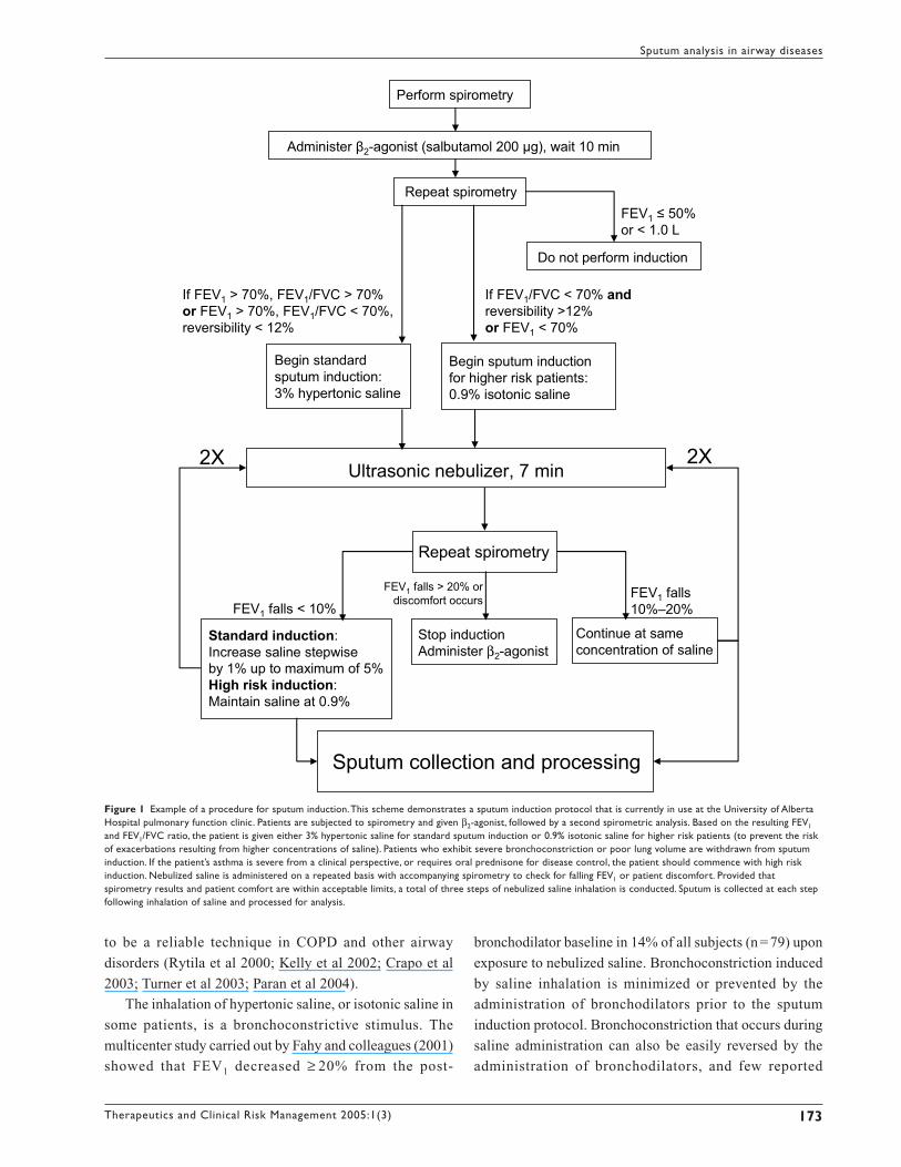
Figure 1 Example of a procedure for sputum induction. This scheme demonstrates a sputum induction protocol that is currently in use at the University of AlbertaHospital pulmonary function clinic. Patients are subjected to spirometry and given β2-agonist, followed by a second spirometric analysis. Based on the resulting FEV1
and FEV1/FVC ratio, the patient is given either 3% hypertonic saline for standard sputum induction or 0.9% isotonic saline for higher risk patients (to prevent the riskof exacerbations resulting from higher concentrations of saline). Patients who exhibit severe bronchoconstriction or poor lung volume are withdrawn from sputuminduction. If the patient’s asthma is severe from a clinical perspective, or requires oral prednisone for disease control, the patient should commence with high riskinduction. Nebulized saline is administered on a repeated basis with accompanying spirometry to check for falling FEV1 or patient discomfort. Provided thatspirometry results and patient comfort are within acceptable limits, a total of three steps of nebulized saline inhalation is conducted. Sputum is collected at each stepfollowing inhalation of saline and processed for analysis.

Therapeutics and Clinical Risk Management 2005:1(3)174
Lacy et al
complications result from this (2 out of 79 patients;
bronchospasm to saline inhalation and sensitivity to
methacholine were cited as reasons for complications in
these patients) (Fahy et al 2001). It was advised that sputum
induction not be carried out in patients with low lung
volumes or poor FEV1 values. The occurrence of broncho-
constriction can be prevented and patient tolerance improved
by using a relatively low output ultrasonic nebulizer without
reducing the success of induction (Popov et al 1995; Kelly
et al 2002). In addition, inhalation of a β2-agonist, such as
albuterol (salbutamol), prior to administration of nebulized
saline promotes bronchodilation and prevents broncho-
constriction (Popov et al 1995; Wong and Fahy 1997; de la
Fuente et al 1998; Rytila et al 2000; Vlachos-Mayer et al
2000; Kelly et al 2002; Pizzichini 2002). Although there
are no reports of fatalities associated with sputum induction,
it is important to emphasize that this procedure should be
done by a PFT nurse or technician trained in safe sputum
induction with an attendant physician. Processing of sputum
samples should be done by a technician trained in
hematology to count and identify leukocytes and airway
cells in sputum, as this is technically more demanding than
assessing other bodily samples such as blood and urine.
In summary, the process of collection of induced sputum
has developed significantly over the past decade to make it
more tolerable to patients, even in those with severe
bronchoconstriction. β2-Agonist administration following
the procedure is routinely done to avoid further broncho-
constriction. Sputum induction can be carried out at any
major hospital with a PFT laboratory and specially trained
staff. The training for sputum induction requires just 1 week
at a suitable PFT laboratory with a background in sputum
induction.
What can sputum analysis showus?Sputum can be analyzed for cellular markers to indicate the
degree and type of inflammation in the airways of the patient
at the time of collection. These measurements are critical in
diagnosis and prediction of patient responses to specific
treatments. Cell counts frequently correlate with respiratory
physiology data and provide a useful confirmation of the
disease state in the patient. In uncontrolled asthma
(particularly in exacerbations), the most commonly observed
inflammatory changes are elevated eosinophil numbers and
increased products of eosinophil activation, including
eosinophil cationic protein (ECP), major basic protein, and
eosinophil-derived neurotoxin (Pizzichini et al 1996).
Increasing levels of severity of asthma have been shown to
correlate with sputum eosinophil levels as well as sputum
ECP (Figure 2) (Louis et al 2000; Duncan et al 2003).
Apoptotic eosinophils can also be detected and quantified
in induced sputum as a putative measure of resolution of
inflammation (Woolley et al 1996; Foresi et al 2000).
Moreover, decreasing numbers of apoptotic eosinophils in
sputum, in association with increased numbers of viable
eosinophils, have been shown to correlate with the severity
of asthma (Adachi et al 1995; Jang et al 2000; Duncan et al
2003).
Sputum analysis can also be used to determine the
inflammatory response to inhaled glucocorticosteroids.
Indeed, a single large dose (2400 µg) of inhaled budesonide
was shown to result in a reduction in sputum eosinophil
numbers 6 hours after administration (Gibson et al 2001).
A recent study by Green and colleagues (2002)
demonstrated that the measurement of sputum eosinophilia
can be used to manage symptoms in patients with moderate
to severe asthma, which supports the use of induced sputum
analysis in patient treatment. In fact, routine measurement
of sputum eosinophil numbers in addition to management
Figure 2 Eosinophil counts and concentrations of eosinophil cationic protein(ECP) in induced sputum of asthmatic and healthy nonatopic control subjects.Reprinted from Louis and colleagues (2000), with permission obtained from theauthors and the American Thoracic Society.
Controlsubjects
Intermittentasthma
Mild –moderateasthma
Severeasthma
Controlgroup
Intermittentasthma
Mild –moderateasthma
Severeasthma
ECP
(ng/
mL)
Eosi
noph
ils (×
103 /g
)

Therapeutics and Clinical Risk Management 2005:1(3) 175
Sputum analysis in airway diseases
according to the British Thoracic Society (BTS) guidelines
was a superior approach in the prevention of asthma
exacerbations in one group (37 patients) compared with
another group of patients who were solely managed
according to the BTS guidelines (37 patients). Patients who
had their sputum eosinophil numbers monitored had their
steroid medication adjusted in line with changing eosinophil
counts and were found to suffer fewer severe exacerbations
that required hospitalization (Green et al 2002). These
findings have been supported in a similar study in Canada
(FE Hargreave, personal communication). These reports
suggest that sputum induction and analysis may be an
important procedure to assist in the management of asthma.
In COPD, the most common sputum change is
neutrophilia and increased products of neutrophil activation,
including proteases, myeloperoxidase, and elastase (Chung
2001; Williams and Jose 2001; Kim and Nadel 2004;
O’Donnell RA et al 2004). In cigarette smokers with COPD,
the degree of neutrophilia is loosely related to the degree of
chronic airway obstruction (Stanescu et al 1996). This
suggests that sputum neutrophils or their products may be
used as early markers of the manifestation of COPD.
In as many as a third of all patients with COPD,
eosinophils are also elevated in the sputum (Pizzichini et al
1998; Hargreave and Leigh 1999; Brightling et al 2000).
Interestingly, COPD patients with eosinophilic sputum are
more responsive to treatment with steroids (prednisone or
prednisolone) (Pizzichini et al 1998). Patients with
eosinophilic COPD demonstrated improved quality-of-life
scores and FEV1 values following inhaled corticosteroid
treatment. This indicates that sputum analysis may be
important in defining a subset of COPD patients having an
eosinophilic component that may benefit from treatment
with inhaled corticosteroids. Glucocorticosteroids are
effective at reducing eosinophilic inflammation to negligible
levels; however, neutrophils are not typically thought to be
responsive to steroid treatment. In fact, the survival and
activity of neutrophils is enhanced by glucocorticoids, at
least in vitro (Cox 1995; Strickland et al 2001). This suggests
that it may be detrimental to administer glucocorticoids to
patients who have a predominantly neutrophilic inflam-
mation. However, one study suggested a beneficial effect
by a 2-month treatment of COPD with high doses of inhaled
beclomethasone (1500 µg/day) on reducing sputum
neutrophil numbers (Confalonieri et al 1998). This
observation is in contradiction to the perceived applicability
of steroid treatment, and sputum analysis may be useful in
determining the sensitivity of neutrophilic inflammation in
the airways to high doses of steroids in further studies. Taken
together, drugs used to treat asthma, COPD, and CF may
benefit individual patients in a differential manner depending
on the primary inflammatory cell type observed in the
airways, particularly in the case of eosinophilic versus
neutrophilic inflammation.
More detailed analyses of inflammatory cell function
can also be carried out in research studies on sputum
samples. A recent study has shown that eosinophils in
asthmatic sputum show an activation profile that resembles
purified peripheral blood eosinophils stimulated in vitro with
chemical agonists (Lacy et al 2003). They were shown to
redistribute an intracellular signaling molecule, called Rac2
GTPase, to their cell membranes, indicating activation of
respiratory burst which leads to the release of damaging
reactive oxygen species. This finding suggests that sputum
eosinophils in asthmatics are fully activated and release
cytotoxic mediators that can induce tissue injury in the
airways. It also indicates that it is not the presence of
eosinophils alone that correlates with their activation in the
airways, as sputum eosinophils from normal subjects did
not exhibit respiratory burst characteristics.
Sputum analysis gives us the opportunity to gauge the
degree of inflammatory cell activation in airway diseases,
which is an important indicator for the treatment and
management of these disorders. It is proposed as a clinical
measurement for asthma and COPD patients to determine
the inflammatory processes and assist in diagnosis and
management of disease. Sputum induction should be
introduced with skilled technical support and an attending
physician to prevent bronchoconstrictive episodes. In some
cases, patients may not be able to generate sufficient sputum
for analysis, and these may be directed towards alternative
noninvasive techniques such as collection of exhaled breath
condensates, which can be analyzed for the presence of
inflammatory markers, although this approach has yet to be
confirmed as a useful diagnostic approach (Effros et al
2005).
Potential new modalities ofsputum analysis with clinicalrelevanceSeveral new approaches have appeared in the analysis of
sputum samples that may provide important new avenues
for the diagnosis and management of patients with asthma
and COPD. Detailed sputum analysis is possible through
the application of metabolomic techniques. Metabolomics

Therapeutics and Clinical Risk Management 2005:1(3)176
Lacy et al
is defined as the measurement of the complete metabolic
response of an organism to an environmental stimulus or
genetic modification, just as genomics describes the genetic
expression of a cell or organism. One technique that can be
applied in metabolomic analysis is nuclear magnetic
resonance (NMR) spectroscopy. Using NMR, it is possible
not only to detect, but also to quantify small amounts of
metabolites in samples obtained from patients. In our case,
we have measured the presence of oxidatively modified
residues in sputum samples, such as modified tyrosine
Eosinophil peroxidase+ Superoxide
Myeloperoxidase+ Superoxide
Hypobromous acid(HOBr)
Hypochlorous acid(HOCl)
Tyrosine
Eosinophil markers3’-Bromotyrosine3,5’-Dibromotyrosine
Neutrophil markers3’-Chlorotyrosine3,5’-Dichlorotyrosine
+ Br¯ + Cl¯
A
B
3,5’-Dibromotyrosine3,5’-Chlorotyrosine 3,5’-Bromotyrosine
Tyrosine
HO CH2 CH COOH
NH2
Eosinophil Neutrophil
Figure 3 Detection of modified tyrosine residues from activated eosinophils and neutrophils in induced sputum as determined by nuclear magnetic resonanceanalysis. (a) Spectrum from an induced sputum sample obtained from a cystic fibrosis patient, compared with (b) a spectrum from control sputum. Peakscorresponding to tyrosine and some modified tyrosine residues are indicated. Spectral traces are from Saude et al (2004).
(a)
(b)

Therapeutics and Clinical Risk Management 2005:1(3) 177
Sputum analysis in airway diseases
residues, which result from activation of airway eosinophils
and neutrophils (Figure 3). The production of modified
tyrosine residues occurs only during respiratory burst and
degranulation of inflammatory cells and can be used to
differentiate between eosinophils and neutrophils to indicate
which type is likely to be dominant in the airways.
Specific modified tyrosine residues can be directly
detected by NMR in sputum samples, which are complex
biological mixtures that usually require extensive extraction
protocols to determine the levels of these substances.
Following a simple processing step, the sputum sample can
be loaded into a high resolution NMR spectrometer for
spectral analysis. In a recent study, it was possible to resolve
3-chlorotyrosine, 3-bromotyrosine, and 3,5-dibromo-
tyrosine in sputum samples from CF patients following a
spectral scan taking less than 10 minutes (Saude et al 2004).
This confirms earlier studies that eosinophilic inflammation
and activation occurs in CF patient airways (Halmerbauer
et al 2000), although the contribution of eosinophils to the
inflammatory processes in this disease is not clear. The levels
of chlorinated tyrosine residues (3-chlorotyrosine) closely
correlated with neutrophil percentages in sputum samples
from CF patients (7 patients, r2 = 0.869) (Figure 4).
Taken together, there are many techniques that may be
used for metabolomic analysis, and NMR is one of the most
powerful approaches. The advantage of NMR analysis of
sputum samples is that it will quantify activation products
of inflammatory cells and extend measurements of total and
differential cell counts, which is the usual approach in basic
cellular analysis of sputum. Furthermore, as mentioned
above, the presence of inflammatory cells alone in airway
tissues is not always an indication that an active
inflammatory process is occurring. Tissue pathology
analysis traditionally assumes that an inflammatory cell
infiltrate is an indication of inflammation (by measuring
increased cell numbers in the tissue section or in
bronchoalveolar lavage/sputum) without providing evidence
to show that the inflammatory cells are concurrently
activated. NMR spectroscopy analysis of sputum samples
allows us to provide strong evidence of inflammatory cell
activation. Moreover, it may be possible to discriminate
between eosinophil and neutrophil activation in the airways,
which will provide a rationale for modifications in drug
treatment on an individualized basis.
SummarySputum induction is a novel, noninvasive, and highly
reproducible technique for the analysis of cellular and
inflammatory indices in obstructive airway diseases. This
can be done quite easily if appropriate trained sputum
laboratory staff are available to perform the inductions in a
safe way. Sputum analysis is useful for continuous
monitoring of airway inflammatory events in order to modify
drug treatment or assist in management of patients with
airway diseases in a highly individualized manner. In
addition, modern techniques such as NMR spectroscopy
may provide additional opportunities to manage these
diseases, although more work is required before the clinical
usefulness of NMR analysis on sputum samples is realized.
The main drawbacks with sputum analysis are the relatively
low yield of sputum in a small proportion of patients and
the potential for bronchoconstrictive episodes induced by
saline inhalation. Although there are no ways of overcoming
poor sputum yield, bronchoconstriction can easily be
reduced by the administration of bronchodilators.
ReferencesAdachi T, Motojima S, Hirata A, et al. 1995. Eosinophil viability-enhancing
activity in sputum from patients with bronchial asthma. Contributionsof interleukin-5 and granulocyte/macrophage colony-stimulatingfactor. Am J Respir Crit Care Med, 151:618–23.
Anon. 1985. Summary and recommendations of a workshop on theinvestigative use of fiberoptic bronchoscopy and bronchoalveolarlavage in asthmatics. Am Rev Respir Dis, 132:180–2.
Asthma in America. 1998. Accessed 9 Mar 2005. URL: http://www.asthmainamerica.com.
Asthma in Canada. 2004. Accessed 9 Mar 2005. URL: http://www.asthmaincanada.com.
A
B
Figure 4 Increased 3-chlorotyrosine production in induced sputum of cysticfibrosis (CF) patients determined by nuclear magnetic resonance (NMR) analysis.Induced sputum samples from seven CF patients were analyzed by NMR todetermine their levels of modified tyrosine residues. (a) The production of3-chlorotyrosine, specific for neutrophil activation, was significantly elevated inCF sputum samples compared with control sputum samples. (b) Sputum levelsof 3-chlorotyrosine correlated significantly with the percentage of sputumneutrophils from CF patients. Figures from Saude and et al (2004).
3-C
hlor
otyr
osin
e (m
mol
/L)
Control Cystic Fibrosis
3-C
hlor
otyr
osin
e (m
mol
/L)
Neutrophil (% cell pop)
(a)
(b)

Therapeutics and Clinical Risk Management 2005:1(3)178
Lacy et al
Barnes PJ. 2000. Chronic obstructive pulmonary disease. N Engl J Med,343:269–80.
Barnes PJ. 2004. Small airways in COPD. N Engl J Med, 350:2635–7.Brightling CE, Monteiro W, Ward R, et al. 2000. Sputum eosinophilia and
short-term response to prednisolone in chronic obstructive pulmonarydisease: a randomised controlled trial. Lancet, 356:1480–5.
Chung KF. 2001. Cytokines in chronic obstructive pulmonary disease. EurRespir J Suppl, 34:50s–59s.
Confalonieri M, Mainardi E, Della PR, et al. 1998. Inhaled corticosteroidsreduce neutrophilic bronchial inflammation in patients with chronicobstructive pulmonary disease. Thorax, 53:583–5.
Covar RA, Spahn JD, Martin RJ, et al. 2004. Safety and application ofinduced sputum analysis in childhood asthma. J Allergy Clin Immunol,114:575–82.
Cox G. 1995. Glucocorticoid treatment inhibits apoptosis in humanneutrophils. Separation of survival and activation outcomes.J Immunol, 154:4719–25.
Crapo RO, Jensen RL, Hargreave FE. 2003. Airway inflammation inCOPD: physiological outcome measures and induced sputum. EurRespir J Suppl, 41:19s–28s.
de la Fuente PT, Romagnoli M, Godard P, et al. 1998. Safety of inducingsputum in patients with asthma of varying severity. Am J Respir CritCare Med, 157:1127–30.
Duncan CJ, Lawrie A, Blaylock MG, et al. 2003. Reduced eosinophilapoptosis in induced sputum correlates with asthma severity. EurRespir J, 22:484–90.
Effros RM, Su J, Casaburi R, et al. 2005. Utility of exhaled breathcondensates in chronic obstructive pulmonary disease: a critical review.Curr Opin Pulm Med, 11:135–9.
Fabbri L, Boschetto P, Caramori G, et al. 1998. Neutrophils and asthma.In Busse WW, Holgate ST (eds). Inflammatory mechanisms in asthma.New York: Marcel Dekker. p 287–322.
Fahy JV, Boushey HA, Lazarus SC, et al. 2001. Safety and reproducibilityof sputum induction in asthmatic subjects in a multicenter study. AmJ Respir Crit Care Med, 163:1470–5.
Fahy JV, Liu J, Wong H, et al. 1993. Cellular and biochemical analysis ofinduced sputum from asthmatic and from healthy subjects. Am RevRespir Dis, 147:1126–31.
Foresi A, Teodoro C, Leone C, et al. 2000. Eosinophil apoptosis in inducedsputum from patients with seasonal allergic rhinitis and withasymptomatic and symptomatic asthma. Ann Allergy Asthma Immunol,84:411–16.
Forsythe P, Ebeling C, Gordon JR, et al. 2004. Opposing effects of short-and long-term stress on airway inflammation. Am J Respir Crit CareMed, 169:220–6.
Gibson PG, Dolovich J, Denburg J, et al. 1989. Chronic cough: eosinophilicbronchitis without asthma. Lancet, 1:1346–8.
Gibson PG, Saltos N, Fakes K. 2001. Acute anti-inflammatory effects ofinhaled budesonide in asthma: a randomized controlled trial. Am JRespir Crit Care Med, 163:32–6.
Gizycki MJ, Adelroth E, Rogers AV, et al. 1997. Myofibroblast involvementin the allergen-induced late response in mild atopic asthma. Am JRespir Cell Mol Biol, 16:664–73.
Global Initiative for Asthma. 2004. Accessed 9 Mar 2005. URL: http://www.ginasthma.com.
Green RH, Brightling CE, McKenna S, et al. 2002. Asthma exacerbationsand sputum eosinophil counts: a randomised controlled trial. Lancet,360:1715–21.
Haahtela T, Jarvinen M, Kava T, et al. 1991. Comparison of a β2-agonist,terbutaline, with an inhaled corticosteroid, budesonide, in newlydetected asthma. N Engl J Med, 325:388–92.
Halmerbauer G, Arri S, Schierl M, et al. 2000. The relationship ofeosinophil granule proteins to ions in the sputum of patients with cysticfibrosis. Clin Exp Allergy, 30:1771–6.
Hargreave FE, Leigh R. 1999. Induced sputum, eosinophilic bronchitis,and chronic obstructive pulmonary disease. Am J Respir Crit CareMed, 160:S53–7.
Hirst SJ, Martin JG, Bonacci JV, et al. 2004. Proliferative aspects of airwaysmooth muscle. J Allergy Clin Immunol, 114:S2–17.
ISAAC Study. 1998. Worldwide variations in the prevalence of asthmasymptoms: the International Study of Asthma and Allergies inChildhood (ISAAC). Eur Respir J, 12:315–35.
Jang AS, Choi IS, Lee S, et al. 2000. Bcl-2 expression in sputum eosinophilsin patients with acute asthma. Thorax, 55:370–4.
Jayaram L, Parameswaran K, Sears MR, et al. 2000. Induced sputum cellcounts: their usefulness in clinical practice. Eur Respir J, 16:150–8.
Keatings VM, Barnes PJ. 1997. Granulocyte activation markers in inducedsputum: comparison between chronic obstructive pulmonary disease,asthma, and normal subjects. Am J Respir Crit Care Med, 155:449–53.
Kelly MG, Brown V, Martin SL, et al. 2002. Comparison of sputuminduction using high-output and low-output ultrasonic nebulizers innormal subjects and patients with COPD. Chest, 122:955–9.
Kim S, Nadel JA. 2004. Role of neutrophils in mucus hypersecretion inCOPD and implications for therapy. Treat Respir Med, 3:147–59.
Klinnert MD. 2003. Evaluating the effects of stress on asthma: a paradoxicalchallenge. Eur Respir J, 22:574–5.
Lacy P, Abdel-Latif D, Steward M, et al. 2003. Divergence of mechanismsregulating respiratory burst in blood and sputum eosinophils andneutrophils from atopic subjects. J Immunol, 170:2670–9.
Laitinen LA, Laitinen A, Haahtela T. 1992. A comparative study of theeffects of an inhaled corticosteroid, budesonide, and a β2-agonist,terbutaline, on airway inflammation in newly diagnosed asthma: arandomized, double-blind, parallel-group controlled trial. J AllergyClin Immunol, 90:32–42.
Lange P, Parner J, Vestbo J, et al. 1998. A 15-year follow-up study ofventilatory function in adults with asthma. N Engl J Med, 339:1194–200.
LindenSmith J, Morrison D, Deveau C, et al. 2004. Overdiagnosis of asthmain the community. Can Respir J, 11:111–16.
Louis R, Lau LC, Bron AO, et al. 2000. The relationship between airwaysinflammation and asthma severity. Am J Respir Crit Care Med, 161:9–16.
McWilliams T, Wells AU, Harrison AC, et al. 2002. Induced sputum andbronchoscopy in the diagnosis of pulmonary tuberculosis. Thorax,57:1010–14.
Meghdas I, Loiez C, Baida N, et al. 2004. Epidemiology of infectionsassociated to “Burkholderia cepacia complex” in the course of cysticfibrosis. Arch Pediatr, 11:360–6.
O’Donnell DE, Aaron S, Bourbeau J, et al. 2004. State of the ArtCompendium: Canadian Thoracic Society recommendations for themanagement of chronic obstructive pulmonary disease. Can Respir J,11 Suppl B:7B–59B.
O’Donnell RA, Peebles C, Ward JA, et al. 2004. Relationship betweenperipheral airway dysfunction, airway obstruction, and neutrophilicinflammation in COPD. Thorax, 59:837–42.
Paggiaro PL, Chanez P, Holz O, et al. 2002. Sputum induction. Eur RespirJ Suppl, 37:3s–8s.
Paran D, Fireman E, Elkayam O. 2004. Pulmonary disease in systemiclupus erythematosus and the antiphospholpid syndrome. AutoimmunRev, 3:70–5.
Pauwels RA, Buist AS, Calverley PM, et al. 2001. Global strategy for thediagnosis, management, and prevention of chronic obstructivepulmonary disease. NHLBI/WHO Global Initiative for ChronicObstructive Lung Disease (GOLD) Workshop summary. Am J RespirCrit Care Med, 163:1256–76.
Pizzichini E, Pizzichini MM, Efthimiadis A, et al. 1996. Indices of airwayinflammation in induced sputum: reproducibility and validity of celland fluid-phase measurements. Am J Respir Crit Care Med, 154:308–17.
Therapeutics and Clinical Risk Management 2005:1(3) 179
Sputum analysis in airway diseases
Pizzichini E, Pizzichini MM, Gibson P, et al. 1998. Sputum eosinophiliapredicts benefit from prednisone in smokers with chronic obstructivebronchitis. Am J Respir Crit Care Med, 158:1511–17.
Pizzichini MM. 2002. Is sputum eosinophilia a good or poor predictor ofbenefit from inhaled corticosteroid therapy in asthma? Eur Respir J,20:1359–61.
Pizzichini MM, Pizzichini E, Clelland L, et al. 1997. Sputum in severeexacerbations of asthma: kinetics of inflammatory indices afterprednisone treatment. Am J Respir Crit Care Med, 155:1501–8.
Popov TA, Pizzichini MM, Pizzichini E, et al. 1995. Some technical factorsinfluencing the induction of sputum for cell analysis. Eur Respir J,8:559–65.
Rosenstein BJ, Zeitlin PL. 1998. Cystic fibrosis. Lancet, 351:277–82.Rutgers SR, Timens W, Kauffman HF, et al. 2001. Markers of active airway
inflammation and remodelling in chronic obstructive pulmonarydisease. Clin Exp Allergy, 31:193–205.
Rytila PH, Lindqvist AE, Laitinen LA. 2000. Safety of sputum inductionin chronic obstructive pulmonary disease. Eur Respir J, 15:1116–19.
Saude EJ, Lacy P, Musat-Marcu S, et al. 2004. NMR analysis of neutrophilactivation in sputum samples from patients with cystic fibrosis. MagnReson Med, 52:807–14.
Silverman RA, Boudreaux ED, Woodruff PG, et al. 2003. Cigarettesmoking among asthmatic adults presenting to 64 emergencydepartments. Chest, 123:1472–9.
Sin DD, Lacy P, York E, et al. 2004. Effects of fluticasone on systemicmarkers of inflammation in chronic obstructive pulmonary disease.Am J Respir Crit Care Med, 170:760–5.
Stanescu D, Sanna A, Veriter C, et al. 1996. Airways obstruction, chronicexpectoration, and rapid decline of FEV
1 in smokers are associated
with increased levels of sputum neutrophils. Thorax, 51:267–71.
Strickland I, Kisich K, Hauk PJ, et al. 2001. High constitutiveglucocorticoid receptor β in human neutrophils enables them to reducetheir spontaneous rate of cell death in response to corticosteroids. J ExpMed, 193:585–94.
Tarlo SM, Liss GM. 2002. Diisocyanate-induced asthma: diagnosis,prognosis, and effects of medical surveillance measures. Appl OccupEnviron Hyg, 17:902–8.
Turner D, Schwarz Y, Yust I. 2003. Induced sputum for diagnosingPneumocystis carinii pneumonia in HIV patients: new data, new issues.Eur Respir J, 21:204–8.
Ulrik CS, Backer V. 1999. Nonreversible airflow obstruction in life-longnonsmokers with moderate to severe asthma. Eur Respir J, 14:892–6.
Vlachos-Mayer H, Leigh R, Sharon RF, et al. 2000. Success and safety ofsputum induction in the clinical setting. Eur Respir J, 16:997–1000.
Wardlaw AJ, Moqbel R, Kay AB. 1997. Eosinophils and the allergicinflammatory response. In Kay AB (ed). Allergy and allergic diseases.Oxford: Blackwell Scientific. p 171–97.
Wenzel SE. 2004. Phenotypes in asthma: useful guides for therapy, distinctbiological processes, or both? Am J Respir Crit Care Med, 170:579–80.
Wielders PL, Dekhuijzen PN. 1997. Disease monitoring in chronicobstructive pulmonary disease: is there a role for biomarkers? EurRespir J, 10:2443–5.
Williams TJ, Jose PJ. 2001. Neutrophils in chronic obstructive pulmonarydisease. Novartis Found Symp, 234:136–41.
Wong HH, Fahy JV. 1997. Safety of one method of sputum induction inasthmatic subjects. Am J Respir Crit Care Med, 156:299–303.
Woolley KL, Gibson PG, Carty K, et al. 1996. Eosinophil apoptosis andthe resolution of airway inflammation in asthma. Am J Respir CritCare Med, 154:237–43.
