immunoregulatory functions of mtor inhibition
TRANSCRIPT
Rapamycin was isolated in the early 1970s from a soil sample obtained on Easter Island (Rapa Nui) and was identified as a potent antifungal metabolite1. This macrolide, which is produced by Streptomyces hygro-scopicus, was found to inhibit cell proliferation and to have potent immunosuppressive activity. It is currently used for the prevention of kidney transplant rejection2. Rapamycin and its derivatives are also undergoing clinical testing for prophylaxis of graft rejection3 and graft-versus-host disease (GVHD)4, chemotherapy of some cancers5 and the prevention of restenosis following angioplasty6.
Our understanding of the mechanisms that underlie the unique immunosuppressive profile of rapamycin continues to evolve. In line with this, the central and pervasive role of the serine/threonine protein kinase mammalian target of rapamycin (mTOR) in innate and adaptive immune responses is becoming apparent. Blockade of mTOR by rapamycin impairs dendritic cell (DC) maturation and function and inhibits T cell proliferation, a mechanism that underpins its immunosuppressive effect. There is now strong evidence that mTOR is crucial for the regulation of antigen responsiveness in CD4+ T cells. This effect seems to be mediated by an influence of mTOR inhibition on naturally occurring regulatory T (TReg) cells, which have a key role in immunological tolerance. Exciting information has emerged regarding the role of the phosphoinositide 3kinase (PI3K)–AKT–mTOR pathway in regulating DC and T cell function, particularly in relation to the expression of forkhead box P3 (FOXP3) and the differentiation of TReg cells. In this Review, we discuss
the remarkable recent progress in the elucidation of the mechanisms by which mTOR inhibition affects intracellular signalling pathways in immune cells, particularly in DCs and T cells, and how this influences immune responses.
The mTOR signalling pathwayDiscovery of mTOR. Genetic screening of Saccharomyces cerevisiae identified two TOR genes (TOR1 and TOR2) and gave an early indication of the importance of TOR in the control of cell proliferation7. The function of TOR proteins was found to be inhibited by rapamycin in a complex with the immunophilin FK506binding protein 1A, 12 kDa (FKBP12)8 (BOX 1). A single mTOR gene was then identified in rat, mouse and human cells, which had a high degree of homology to yeast TOR genes9–11. Subsequent work has identified mTOR as an atypical serine/threonine protein kinase with a central role in mammalian development12,13. It is a crucial regulator of cell growth and proliferation, and of physiological events such as transcription, mRNA turnover and translation, ribosomal biogenesis, vesicular trafficking, autophagy, cytoskeletal organization and cell size14.
mTOR exists in at least two complexes, mTOR complex 1 (mTORC1) and mTORC2 (BOX 1), that have distinct relationships both to upstream and downstream effectors and to each other (FIG. 1). Indeed, mTOR in mTORC1 is highly sensitive to inhibition by rapamycin, whereas mTOR in mTORC2 is resistant to rapamycin; the reason for this is unknown. Notably, rapamycininsensitive functions of mTORC1 in mouse embryonic fibroblasts have been identified recently using
*Starzl Transplantation Institute and Department of Surgery and ‡Department of Immunology, University of Pittsburgh School of Medicine, Pittsburgh, Pennsylvania 15261, USA.Correspondence to A.W.T. e-mail: [email protected]:10.1038/nri2546
Graft-versus-host disease(GVHD). A disease that results from immunological attack by donor allogeneic T cells transferred with the allograft (such as bone marrow, liver or gut) of target recipient organs or tissues (such as skin or gut). GVHD occurs in graft recipients who cannot eliminate the host-reactive donor T cells owing to immunosuppression, immunological immaturity or tolerance of the recipient.
ImmunophilinA peptidyl-prolyl isomerase that is targeted by certain immunosuppressive drugs (such as cyclosporin A, FK506 and rapamycin). The drug–immunophilin complex is responsible for the immunosuppressive action of the drug.
Immunoregulatory functions of mTOR inhibitionAngus W. Thomson*‡, Heth R. Turnquist* and Giorgio Raimondi*
Abstract | The potent immunosuppressive action of rapamycin is commonly ascribed to inhibition of growth factor-induced T cell proliferation. However, it is now evident that the serine/threonine protein kinase mammalian target of rapamycin (mTOR) has an important role in the modulation of both innate and adaptive immune responses. mTOR regulates diverse functions of professional antigen-presenting cells, such as dendritic cells (DCs), and has important roles in the activation of effector T cells and the function and proliferation of regulatory T cells. In this Review, we discuss our current understanding of the mTOR pathway and the consequences of mTOR inhibition, both in DCs and T cells, including new data on the regulation of forkhead box P3 expression.
R E V I E W S
324 | mAy 2009 | VOlumE 9 www.nature.com/reviews/immunol
© 2009 Macmillan Publishers Limited. All rights reserved
AutophagyA tightly regulated catabolic process that involves degradation of the cell’s own intracellular components through the lysosomal machinery and is a normal part of cell growth, development and homeostasis.
Small interfering RNAA class of 20–25-nucleotide long double-stranded RNA molecules that are involved in the RNA interference pathway, which interferes with the expression of a specific gene. Also known as silencing RNA.
a new inhibitor (known as Torin 1) of both mTORC1 and mTORC2 (ReF. 15). Also, prolonged exposure to rapamycin can impede the assembly of mTORC2 in some cells5,16,17.
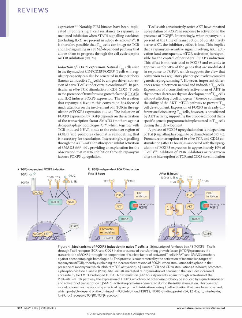
Signalling through mTORC1. In immune cells, mTORC1 is a regulator of cell growth and other processes downstream of PI3K–AKT, WNT–GSK3 (glycogen synthase kinase 3) and AmPactivated protein kinase (AmPK) signalling (FIG. 1). Details of mTORC1 signalling can be found in recent comprehensive reviews14,18,19. Tuberous sclerosis complex 1 (TSC1) and TSC2 together form a functional complex that acts as the upstream inhibitor of mTORC1. Growth factors, cytokines, costimulatory molecules and antigen receptors activate PI3K, which subsequently activates AKT. Fully activated AKT inhibits TSC2 by phosphorylating it20, thereby negating the inhibitory effect of TSC2 on mTORC1. Activation of the RAS–mAPK (mitogenactivated protein kinase) pathway also leads to inhibition of the TSC1–TSC2 complex14. Alternatively, cellular stress and DNA damage can inhibit mTORC1 by promoting the regulatory capacity of TSC1–TSC2 (ReFs 14,19). The inhibitory activity of the TSC1–TSC2 complex is mediated by inhibiting RHEB (RAS homologue enriched in brain), a RASlike GTPase and a positive regulator of mTORC1 (ReFs 18,19).
Removal of mTORC1 inhibition allows mTORC1 to phosporylate S6 kinase 1 (S6K1; also known as RPS6KB1) and the eukaryotic initiation factor EIF4EBP1 (eukaryotic translation initiation factor (EIF4E)binding protein 1). Phosphorylated S6K1 promotes mRNA translation and cell growth by enhancing the biosynthesis of the translational apparatus in the cell14,19. The phosphorylation of EIF4EBP1 prevents it from inhibiting EIF4E, which also stimulates translation14. WNT proteins bind to the Frizzled family of receptors, which are involved in the regulation of effector T cell development, TReg cell activation and DC maturation21. The WNT pathway influences the mTORC1 pathway by inhibiting GSK3, which in the absence of WNT signalling is an additional negative regulator of mTORC1 (ReF. 22).
Signalling through mTORC2. Knowledge of the more recently defined mTORC2 is limited compared with mTORC1 because of the lack of an mTORC2specific inhibitor. Studies targeting RICTOR (rapamycininsensitive companion of mTOR; a defining component of mTORC2) (BOX 1) through small interfering RNA23,24 and studies of Rictorknockout mice13 have shown the importance of mTORC2 in mammalian development and several cellular processes. mTORC2mediated phosphorylation of AKT is stimulated by insulin and can be blocked by PI3K inhibitors25. However, knockdown of Rictor does not decrease S6K1 activation24, indicating that mTORC2 does not activate mTORC1 (ReF. 24).
Targeting Rictor has also established that mTORC2 regulates the actin cytoskeleton through the small GTPase RAS homologue (RHO) and protein kinase C14. The TSC1–TSC2 complex has been shown to regulate cell adhesion and migration26, but it is not clear whether it signals to and regulates mTORC2 directly. Regulation of cell movement and adhesion is an important feature of effective immune responses, so it is possible that mTORC2 might also be shown to modulate immune reactivity when specific inhibitors become available.
S6K1-mediated regulation of mTOR signalling. mTOR is a crucial coordinator of signalling pathways, so it is not surprising that feedback inhibition is an important component of the pathway. Activated S6K1, the main effector of uninhibited mTORC1, negatively regulates input from the PI3K–AKT signalling pathway to mTORC1 (ReFs 19,27–29) by phosphorylating and initiating the degradation of insulin receptor substrate 1 (IRS1), which is the molecular intermediate between insulin receptor and PI3K19,27–31. It has not been determined whether S6K1 can also negatively regulate input from other receptor systems that activate mTOR through PI3K. Activated S6K1 can also positively regulate mTOR activation. GSK3, which is constitutively activated in the absence of growth factors, negatively regulates the mTOR pathway by stimulating the TSC1–TSC2 complex. under certain conditions, activation of S6K1 can negatively regulate GSK3 (ReFs 32,33), thus facilitating cell proliferation.
RAPTOR
Nature Reviews | Immunology
HEAT HEAT FAT FRB Kinase
FATC
RICTOR
MA
PKA
P1
HEAT HEAT FAT FRB Kinase
FATC
mTO
Rm
TOR
mTO
RC1
mTO
RC2
LST8
LST8
Rapamycin
FKBP12
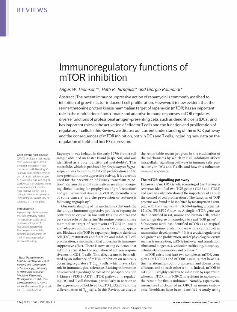
Box 1 | The two distinct mTOR-containing complexes
Mammalian target of rapamycin (mTOR) is a large (~289 kDa) atypical kinase, which, like other members of the phosphoinositide kinase-related kinase (PIKK) family, contains a carboxy-terminal serine/threonine protein kinase domain14,18,19. Also consistent with other PIKKs, mTOR contains a FRAP–ATM–TTRAP (FAT) domain and a C-terminal FAT (FATC) domain that might have a role in its structure and stability134 (see the figure). Mammalian LST8 (also known as GβL) associates with the kinase domain of mTOR and is thought to facilitate mTOR signalling, but its precise role has yet to be defined135,136. In addition, mTOR seems to be involved in various protein–protein interactions that determine its physiological role. It has been established that mTOR exists in at least two distinct complexes, mTOR complex 1 (mTORC1) and mTORC2. mTOR and LST8, together with the regulatory associated protein of mTOR (RAPTOR) form mTORC1. RAPTOR is essential for mTORC1 activity and is proposed to interact with mTOR through shared HEAT domains19. Rapamycin binds to the immunophilin FK506-binding protein 1A, 12 kDA (FKBP12) to form a drug–receptor complex that specifically and effectively blocks the activity of mTORC1. The rapamycin–FKBP12 complex binds next to the kinase region of mTOR in the FKBP12–rapamycin-binding (FRB) domain136 and disrupts the in vitro and in vivo activity of the complex, potentially by disrupting the interaction between RAPTOR and mTOR14. mTORC2 also contains LST8, but instead of RAPTOR, it associates with rapamycin-insensitive companion of mTOR (RICTOR) and possibly MAPKAP1 (mitogen-activated protein kinase-associated protein 1; also known as SIN1)19,137. Unlike mTORC1, mTORC2 is resistant to direct inhibition by rapamycin. It is unknown what prevents the interaction between the rapamycin–FKBP12 complex and the FRB domain on mTORC2 (ReF. 14).
R E V I E W S
NATuRE REVIEWS | Immunology VOlumE 9 | mAy 2009 | 325
© 2009 Macmillan Publishers Limited. All rights reserved

Nature Reviews | Immunology
TSC1–TSC2
Low cell energy levels(increasedAMP)
AMPK
Hypoxia, cellularstress, DNAdamage PI3K
PDK1
AKT
mTORC2
Growth factors, cytokines,co-stimulation and antigen receptors
RAS–MAPK
mTORC1
Rapamycin–FKBP12
RHEB
?
EIF4EBP1 S6K1
Autophagy
WNT proteins
Actin organization
PTEN
mRNA translation
Cell proliferation
RHO PKC
GSK3
Overall, the mTOR kinase, functioning in mTORC1 and mTORC2, acts as a coordinator of signalling pathways that shape the response of cells to various stimuli. Immune cells, using receptors that signal through mTOR directly or indirectly, modulate host responses based on their ‘perception’ of environmental danger. We focus here on the role of mTOR in DCs and T cells, but it is worth noting that the inhibition of mTOR by rapamycin has effects on other immune cells (BOX 2) that are outside the scope of this Review.
mTOR in antigen-presenting cellsDCs and macrophages are phagocytic cells that reside in almost all tissues. Both cell types degrade pathogens and function as antigenpresenting cells. macrophages have a scavenging role and activate and recruit other immune cells, whereas DCs are uniquely wellequipped to present antigen to T cells and initiate adaptive immune responses.
DC differentiation and maturation. The suppressive effects of mTOR inhibition on DC differentiation and maturation are welldocumented in vitro. In addition, rapamycin inhibits the differentiation and mobilization of mouse DCs in vivo in response to the administration of Fms-like tyrosine kinase 3 ligand (FlT3l)34. An early study suggested that rapamycin did not affect the phenotypic differentiation or CD40linduced maturation of human monocytederived DCs in vitro, but instead promoted apoptosis of these DCs and of CD34+ precursor cellderived DCs35. By contrast, studies of mouse bone marrowderived DCs34 showed that mTOR inhibition suppressed interleukin4 (Il4)dependent maturation through the posttranscriptional downregulation of the Il4 receptor (Il4R) complex. In this case, rapamycin inhibited the expression of costimulatory molecules by DCs both in vitro and in vivo and suppressed their Il4induced production of Il12 and tumour necrosis factor (TNF) as well as their T cell stimulatory function34. Further work has shown that mTOR inhibition during DC differentiation inhibits the upregulation of CD86 expression that is induced by Tolllike receptor (TlR) ligands (such as lipopolysaccharide (lPS) or CpG DNA) or by a CD40specific monoclonal antibody36. mTOR inhibition also decreases the production of nitric oxide by lPSstimulated macrophages37, an effect that is due in part to impaired secretion of interferonβ (IFNβ), which is an autocrine cofactor for nitric oxide production. Furthermore, the inactivation of mTOR decreases the secretion of Il2 by DCs following their stimulation through the Ctype lectin receptor dectin 1 (also known as ClEC7A)38, indicating that cytokine production through the activation of Ctype lectin receptors involves mTOR.
Immature DCs undergo differentiation in response to hypoxia and they overexpress hypoxiainducible factor 1α (HIF1α) and its downstream target genes, including vascular endothelial growth factor and glucose transporter 1 (ReF. 39). mTOR inhibition attenuates these responses, and rapamycin can suppress hypoxiainduced inflammation by inhibiting the HIF1α pathway39. All of these observations relate to the effects of soluble rapamycin; however, there are recent reports that the delivery of rapamycin through biodegradable nanoparticles or microparticles enhances its inhibitory effects on the maturation and T cell stimulatory function of mouse and human DCs40–42.
Antigen uptake and presentation. DCs use several pathways for antigen uptake: phagocytosis, constitutive macropinocytosis and mannose receptormediated endocytosis. Rapamycin impairs macropinocytosis and
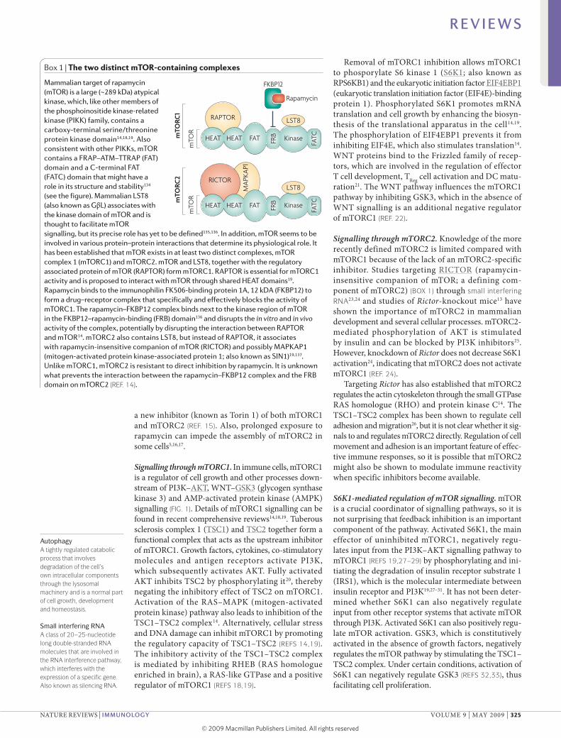
Figure 1 | mToRC1 and mToRC2 signalling pathways. Mammalian target of rapamycin complex 1 (mTORC1) is the direct target of the rapamycin–FK506-binding protein 1A, 12 kDa (FKBP12) complex and regulates cell growth and size by controlling mRNA translation, ribosome biogenesis and autophagy. Diverse signals, arising from growth factors (such as insulin and FMs-like tyrosine kinase 3 ligand (FLT3L)), various cytokines, ligated co-stimulatory molecules and antigen receptors, WNT proteins, cellular energy levels, as well as hypoxia, cellular stress and DNA damage determine mTORC1 activity. These signals all mediate their effects through the tuberous sclerosis complex 1 (TsC1)–TsC2 complex, which is the main negative regulator of mTORC1. Activation of RAs–MAPK (mitogen-activated protein kinase) and phosphoinositide 3-kinase (PI3K)–AKT signalling results in inhibitory phosphorylation of TsC2 and removes repression of RHeB (RAs homologue enriched in brain), which is the mTORC1 stimulator. Activation of PI3K–AKT signalling is negatively regulated by phosphatase and tensin homologue (PTeN). Activated mTORC1 promotes mRNA translation by stimulating s6 kinase 1 (s6K1; also known as RPs6KB1) and inhibiting eIF4eBP1 (eukaryotic translation initiation factor-binding protein 1). Activated s6K1 can also feed back to negatively regulate input from PI3K–AKT by facilitating the degradation of signalling intermediates between surface receptors (such as the insulin receptor) and PI3K. Low energy and nutrient levels (signalling through AMP-activated protein kinase; AMPK), as well as hypoxic conditions, increase the TsC1–TsC2-mediated inhibition of mTORC1 that is downstream of input from glycogen synthase kinase 3 (GsK3). mTORC2 is not inhibited directly by rapamycin, but long-term rapamycin administration disrupts its assembly in some cells. mTORC2, activated by PI3K, directly phosphorylates AKT. mTORC2 also regulates actin cytoskeletal dynamics through the small GTPase RAs homologue (RHO) and protein kinase C (PKC). PDK1, phosphoinositide-dependent kinase 1.
R E V I E W S
326 | mAy 2009 | VOlumE 9 www.nature.com/reviews/immunol
© 2009 Macmillan Publishers Limited. All rights reserved
FmS-like tyrosine kinase 3 ligand(FlT3l). An endogenous cytokine that stimulates stem cell and progenitor cell proliferation through binding to the FlT3 receptor (a type III receptor tyrosine kinase member of the platelet-derived growth factor family). FlT3l administration markedly increases dendritic cell numbers in lymphoid and non-lymphoid tissues.
endocytosis of antigens by cultured mouse immature bone marrowderived DCs43. Rapamycin also inhibits endocytosis in vivo in mouse splenic DCs34. Human monocytederived DCs that have differentiated in the presence of rapamycin have decreased expression of antigen uptake receptors (such as CD32, CD46, CD91 and CD205) and decreased receptormediated and fluidphase endocytosis and phagocytosis of bacteria and apoptotic cells44 (FIG. 2a). Although the mechanisms by which rapamycin inhibits endocytosis are unclear, the inhibition of mTOR by shortterm exposure to rapamycin abolishes the translation of RHO-associated coiledcoilcontaining protein kinase 1 (ROCK1) in mouse macrophages, which results in the inhibition of phagocytosis and chemotaxis45.
unlike calcineurin inhibitors (such as cyclosporin A and FK506), rapamycin does not inhibit the presentation of mHCrestricted antigens (such as ovalbumin) or of minor histocompatibility antigens to T cells by
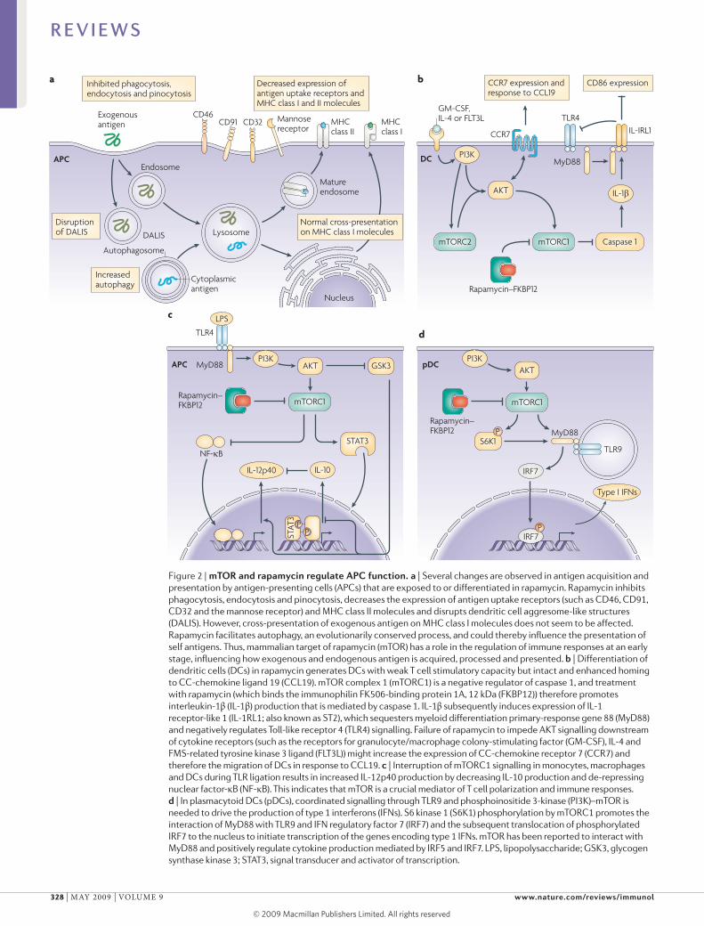
mouse bone marrowderived DCs46–48. This might be because calcineurin inhibitors have inhibitory effects on the early stages of DCinduced activation of naive T cells (such as cytokine production), whereas rapamycin inhibits later stages. There is, however, recent evidence that mTOR inhibition can suppress the transient aggregation of ubiquitylated cytoplasmic proteins during lPSinduced DC maturation49,50. These aggresomes, which in DCs are known as DC aggresomelike structures (DAlIS) (FIG. 2a), might function as depots of antigen that can be cross presented by mHC class I molecules and might allow DCs to coordinate their maturation and antigen presenting functions while they migrate from the periphery to secondary lymphoid tissues.
Crosspresentation of antigen is essential for the generation of cytotoxic T cell responses against certain tumour cells and viruses. The fact that calcineurin inhibitors, but not rapamycin, inhibit the cross presentation of an exogenous antigen by mouse DCs47 might explain why in mice bearing an allograft and a tumour, cyclosporin A inhibits allograft rejection but promotes tumour growth (through inhibiting the crosspresentation of tumourspecific antigens), whereas rapamycin suppresses both processes51.
Autophagy is a constitutive process in DCs52 in which autophagosomes sequester intracellular contents (such as damaged organelles and macromolecules) and target them for lysosomal degradation. This process is thought to be involved in the mHC class II antigen presentation pathway that leads to self tolerance and in innate and adaptive immune responses53,54. TOR1 and TOR2 are involved in the repression of autophagy in Saccharomyces cerevisiae55. Rapamycin induces autophagy in yeast56 and also in mouse macrophages and mouse DCs, thereby enhancing their antigenpresenting ability57. Further studies are required to determine the extent to which autophagy influences the peptide–mHC class II repertoire and how this might be affected by mTOR inhibition. Overall, rapamycin can interfere with antigen uptake by DCs and can modulate events that are associated with antigen presentation, although the mechanistic basis of these effects requires clarification.
DC survival. DC survival is important for the induction of immune responses, for example to viruses and tumours35,58. Blocking of granulocyte/macrophage colonystimulating factor (GmCSF) signalling by rapamycin induced the apoptosis of both human monocytederived DCs and DCs derived from CD34+ precursor cells, but not of monocytes or macrophages. By contrast, the frequency of apoptotic or dead cells was consistently lower than 10% in rapamycinconditioned, bone marrowderived mouse DC cultures and in vivogenerated DCs from rapamycintreated mice34. As pointed out by these authors, pro and antiapoptotic effects of rapamycin have been reported for different cell types, possibly reflecting the differential sensitivity of mTORC2 to disruption in these cells. Interestingly, no increase in cell death was observed following mTOR inhibition by rapamycin in virusinfected, FlT3lderived DCs38.
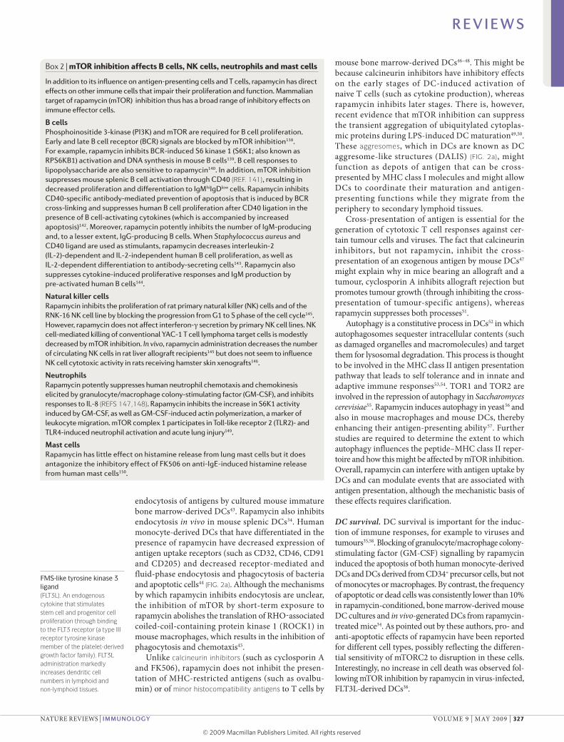
Box 2 | mTOR inhibition affects B cells, NK cells, neutrophils and mast cells
In addition to its influence on antigen-presenting cells and T cells, rapamycin has direct effects on other immune cells that impair their proliferation and function. Mammalian target of rapamycin (mTOR) inhibition thus has a broad range of inhibitory effects on immune effector cells.
B cellsPhosphoinositide 3-kinase (PI3K) and mTOR are required for B cell proliferation. Early and late B cell receptor (BCR) signals are blocked by mTOR inhibition138. For example, rapamycin inhibits BCR-induced S6 kinase 1 (S6K1; also known as RPS6KB1) activation and DNA synthesis in mouse B cells139. B cell responses to lipopolysaccharide are also sensitive to rapamycin140. In addition, mTOR inhibition suppresses mouse splenic B cell activation through CD40 (ReF. 141), resulting in decreased proliferation and differentiation to IgMhiIgDlow cells. Rapamycin inhibits CD40-specific antibody-mediated prevention of apoptosis that is induced by BCR cross-linking and suppresses human B cell proliferation after CD40 ligation in the presence of B cell-activating cytokines (which is accompanied by increased apoptosis)142. Moreover, rapamycin potently inhibits the number of IgM-producing and, to a lesser extent, IgG-producing B cells. When Staphylococcus aureus and CD40 ligand are used as stimulants, rapamycin decreases interleukin-2 (IL-2)-dependent and IL-2-independent human B cell proliferation, as well as IL-2-dependent differentiation to antibody-secreting cells143. Rapamycin also suppresses cytokine-induced proliferative responses and IgM production by pre-activated human B cells144.
natural killer cellsRapamycin inhibits the proliferation of rat primary natural killer (NK) cells and of the RNK-16 NK cell line by blocking the progression from G1 to S phase of the cell cycle145. However, rapamycin does not affect interferon-γ secretion by primary NK cell lines. NK cell-mediated killing of conventional YAC-1 T cell lymphoma target cells is modestly decreased by mTOR inhibition. In vivo, rapamycin administration decreases the number of circulating NK cells in rat liver allograft recipients145 but does not seem to influence NK cell cytotoxic activity in rats receiving hamster skin xenografts146.
neutrophilsRapamycin potently suppresses human neutrophil chemotaxis and chemokinesis elicited by granulocyte/macrophage colony-stimulating factor (GM-CSF), and inhibits responses to IL-8 (ReFs 147,148). Rapamycin inhibits the increase in S6K1 activity induced by GM-CSF, as well as GM-CSF-induced actin polymerization, a marker of leukocyte migration. mTOR complex 1 participates in Toll-like receptor 2 (TLR2)- and TLR4-induced neutrophil activation and acute lung injury149.
mast cellsRapamycin has little effect on histamine release from lung mast cells but it does antagonize the inhibitory effect of FK506 on anti-IgE-induced histamine release from human mast cells150.
R E V I E W S
NATuRE REVIEWS | Immunology VOlumE 9 | mAy 2009 | 327
© 2009 Macmillan Publishers Limited. All rights reserved
IL-1βAKT
mTORC2 Caspase 1mTORC1
Rapamycin–FKBP12
Increasedautophagy
Autophagosome
Disruptionof DALIS
Inhibited phagocytosis, endocytosis and pinocytosis
Decreased expression ofantigen uptake receptors and MHC class I and II molecules
Exogenousantigen CD91 Mannose
receptorCD32 MHC
class IMHC class II
a b
c
EndosomeAPC
APC pDC
DC
Matureendosome
Lysosome
AKT GSK3
DALIS
Nucleus
Normal cross-presentation on MHC class I molecules
CCR7 expression andresponse to CCL19
CD86 expression
GM-CSF,IL-4 or FLT3L
CCR7
TLR4
MyD88
IL-IRL1
PI3K
IL-10
Type I IFNs
IL-12p40
NF-κB
PI3KMyD88
mTORC1Rapamycin–FKBP12
TLR4
LPS
STAT3
PP
Nature Reviews | Immunology
AKT
mTORC1
Rapamycin–FKBP12
d
PI3K
MyD88
TLR9
IRF7
S6K1P
IRF7P
STAT
3Cytoplasmicantigen
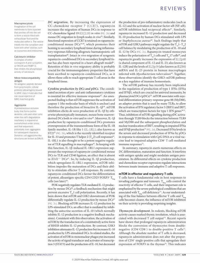
CD46
Figure 2 | mToR and rapamycin regulate APC function. a | several changes are observed in antigen acquisition and presentation by antigen-presenting cells (APCs) that are exposed to or differentiated in rapamycin. Rapamycin inhibits phagocytosis, endocytosis and pinocytosis, decreases the expression of antigen uptake receptors (such as CD46, CD91, CD32 and the mannose receptor) and MHC class II molecules and disrupts dendritic cell aggresome-like structures (DALIs). However, cross-presentation of exogenous antigen on MHC class I molecules does not seem to be affected. Rapamycin facilitates autophagy, an evolutionarily conserved process, and could thereby influence the presentation of self antigens. Thus, mammalian target of rapamycin (mTOR) has a role in the regulation of immune responses at an early stage, influencing how exogenous and endogenous antigen is acquired, processed and presented. b | Differentiation of dendritic cells (DCs) in rapamycin generates DCs with weak T cell stimulatory capacity but intact and enhanced homing to CC-chemokine ligand 19 (CCL19). mTOR complex 1 (mTORC1) is a negative regulator of caspase 1, and treatment with rapamycin (which binds the immunophilin FK506-binding protein 1A, 12 kDa (FKBP12)) therefore promotes interleukin-1β (IL-1β) production that is mediated by caspase 1. IL-1β subsequently induces expression of IL-1 receptor-like 1 (IL-1RL1; also known as sT2), which sequesters myeloid differentiation primary-response gene 88 (MyD88) and negatively regulates Toll-like receptor 4 (TLR4) signalling. Failure of rapamycin to impede AKT signalling downstream of cytokine receptors (such as the receptors for granulocyte/macrophage colony-stimulating factor (GM-CsF), IL-4 and FMs-related tyrosine kinase 3 ligand (FLT3L)) might increase the expression of CC-chemokine receptor 7 (CCR7) and therefore the migration of DCs in response to CCL19. c | Interruption of mTORC1 signalling in monocytes, macrophages and DCs during TLR ligation results in increased IL-12p40 production by decreasing IL-10 production and de-repressing nuclear factor-κB (NF-κB). This indicates that mTOR is a crucial mediator of T cell polarization and immune responses. d | In plasmacytoid DCs (pDCs), coordinated signalling through TLR9 and phosphoinositide 3-kinase (PI3K)–mTOR is needed to drive the production of type 1 interferons (IFNs). s6 kinase 1 (s6K1) phosphorylation by mTORC1 promotes the interaction of MyD88 with TLR9 and IFN regulatory factor 7 (IRF7) and the subsequent translocation of phosphorylated IRF7 to the nucleus to initiate transcription of the genes encoding type 1 IFNs. mTOR has been reported to interact with MyD88 and positively regulate cytokine production mediated by IRF5 and IRF7. LPs, lipopolysaccharide; GsK3, glycogen synthase kinase 3; sTAT3, signal transducer and activator of transcription.
R E V I E W S
328 | mAy 2009 | VOlumE 9 www.nature.com/reviews/immunol
© 2009 Macmillan Publishers Limited. All rights reserved
macropinocytosisInvagination of the cell membrane to form a pocket that pinches off into the cell to form a vesicle filled with extracellular fluid and the molecules within it. The vesicle travels into the cytoplasm and fuses with other vesicles, such as lysosomes and endosomes.
Calcineurin inhibitorA complex of either cyclosporin A and cyclophilin, or FK506 and FKBP12, that inhibits the phosphatase activity of calcineurin.
minor histocompatibility antigenAn antigenic peptide derived from polymorphic cellular proteins (alloantigens) bound to mHC class I molecules that can cause rejection of mHC-matched grafts.
AggresomeA proteinaceous inclusion body that forms (within dendritic cells and other cells) when the cell’s degradation machinery is impaired or overwhelmed under stress and that sequesters potentially toxic aggregates for subsequent clearance. Dendritic cell aggresome-like structures are known as DAlIs.
DC migration. By increasing the expression of CCchemokine receptor 7 (CCR7), rapamycin increases the migration of human DCs in response to CCchemokine ligand 19 (CCl19) in vitro (FIG. 2b) and mouse DC migration to lymph nodes in vivo59. Similarly, functional expression of CCR7 is retained on rapamycin conditioned mouse DCs36,60, which promotes their homing to secondary lymphoid tissue during inflammatory responses following allogeneic haematopoietic cell transplantation60. Intact in vivo migration of syngeneic rapamycinconditioned DCs to secondary lymphoid tissue has also been reported in a heart allograft model61. Retention of this in vivo migratory ability is probably crucial for the immunoregulatory properties that have been ascribed to rapamycinconditioned DCs, as it allows these cells to reach appropriate T cell areas in the lymphoid tissue.
Cytokine production by DCs and pDCs. The coordinated secretion of pro and antiinflammatory cytokines is essential for effective immune responses62. An unexpected finding is that mTOR suppresses the activation of caspase 1 (the molecular basis of which is unclear) and therefore the production of bioactive Il1β38. mTOR inhibition elicits de novo production of Il1β by otherwise phenotypically immature, mouse bone marrowderived DCs both in vitro and in vivo63. moreover, Il1β production by rapamycinconditioned DCs promotes overexpression of the transmembrane form of the Il1R family member, Il1Rlike 1 (Il1Rl1; also known as ST2)63 (FIG. 2b), which is the recently identified receptor for Il33 and promotes T helper 2 (TH2) cell responses64. Il1Rl1 is also thought to be a potent negative regulator of TlR signalling in macrophages65. In keeping with this function, Il1βinduced Il1Rl1 expression suppresses the responses of rapamycinconditioned mouse DCs to TlR and CD40 ligation, an effect that is absent in Il1rl1–/– DCs63. So, by inducing Il1β production, which upregulates Il1Rl1 expression, mTOR inhibition impedes the maturation of DCs and their ability to stimulate effector T cell responses. By contrast, rapamycinconditioned DCs favour the differentiation of potent, alloantigenspecific CD4+CD25+FOXP3+ TReg cells (see later)36.
PI3K negatively regulates TlRmediated Il12 production by mouse DCs66, a feedback mechanism that might prevent excessive TH1 cell polarization. Recently, it has been shown that mTOR and GSK3 downstream of PI3K differentially regulate Il12 production by mouse DCs67
(FIG. 2c). Blocking mTOR increases Il12 production by lPSstimulated DCs, an effect that is mediated by inhibiting the autocrine secretion of Il10 (which normally inhibits Il12 production in a negative feedback mechanism). Consistent with this observation, the activation of mTOR by the transduction of a constitutively active form of RHEB inhibits Il12 production. By contrast, GSK3 inhibition attenuates Il12 production but increases Il10 production by lPSstimulated DCs. In related studies, the activation of mTOR in mononuclear phagocytes increased the activity of signal transducer and activator of transcription 3 (STAT3) and the production of Il10, but decreased
the production of proinflammatory molecules (such as Il12) and the activation of nuclear factorκB (NFκB); mTOR inhibition had reciprocal effects67. Similarly, rapamycin increased Il12 production and decreased Il10 production by human DCs stimulated with lPS or Staphylococcus aureus68. Such findings imply that mTOR and GSK3 pathways might regulate the TH1–TH2 cell balance by modulating the production of Il10 and Il12 by DCs (FIG. 2c). Rapamycintreated monocytes induce the polarization of TH1 cells and TH17 cells68, and rapamycin greatly increases the expression of Il12p40 (a shared component of Il12 and Il23; also known as Il12B) and the levels of IL23p19 (also known as IL23A) mRNA and Il23 protein in human macrophages infected with Mycobacterium tuberculosis69. Together, these observations identify the GSK3–mTOR pathway as a key regulator of immune homeostasis.
The mTOR pathway has recently been implicated in the regulation of production of type 1 IFNs (IFNα and IFNβ), which are crucial for antiviral immunity, by plasma cytoid DCs (pDCs)70. mTOR associates with myeloid differentiation primaryresponse gene 88 (myD88), an adaptor protein that is used by many TlRs, to allow the activation of IFN regulatory factor 5 (IRF5) and IRF7, which are transcription factors for type 1 IFN genes38,71. Thus, inhibition of mTOR signalling during pDC activation through TlR9 blocks the interaction between TlR9 and myD88 and the subsequent phosphorylation and nuclear translocation of IRF7, resulting in impaired IFNα and IFNβ production72 (FIG. 2d). Decreased IFNα levels in the serum and decreased production of IFNα by pDCs in response to stimulation with CpG DNA or a viral vaccine lead to impaired adaptive CD8+ T cellmediated immune responses72.
In summary, rapamycin exerts numerous effects on DC differentiation, maturation and function. It interferes with antigen uptake and might modulate antigen presentation. Its differential effects on cytokine production and chemokine receptor expression regulate interactions between innate immune and adaptive T cell responses.
mTOR in effector and regulatory T cellsT cells have a fundamental role in host responses to invading pathogens and tumours. TReg cells control the reactivity of effector T cells, and their important role is emphasized by the severe pathological conditions that are associated with TReg cell deficiency73. As our understanding of the fine balance between effector T cells and TReg cells becomes clearer, the influence of mTOR inhibition on their activity is providing surprising insights.
Thymocyte development. In rodents, blocking mTOR activity causes marked thymic involution, which is associated with decreased T cell output74. Recent reports have shown that prolonged rapamycin administration blocks the conversion of thymocytes from double negative (CD4–CD8–) to doublepositive T cells75. Although the absolute number of T cells is decreased, rapamycin administration does not alter the proportion of CD4+ singlepositive cells that upregulate their expression of FOXP3 in the thymus76. This indicates
R E V I E W S
NATuRE REVIEWS | Immunology VOlumE 9 | mAy 2009 | 329
© 2009 Macmillan Publishers Limited. All rights reserved
Nature Reviews | Immunology
AKTmTORC2
mTORC1
Otheractivation pathwayssuch asNFAT,MAPKsand AP1
G1 to S phaseprogression
Cell cycleprogression
Downregulation of CD62L, CCR7and SP1
a Conventional T cells b TReg cells
PI3K
NF-κB
Rapamycin–FKBP12
TCR CD28 TCR IL-2R
IL-2
PI3K
PIM2
KLF2
AKTmTOR
mTOR
Expression ofcell cycle progressiongenes, IL-2 and IL-2R
Checkpoint 1 Checkpoint 2
Survivin Aurora B?
PPST
AT5
TCR
CD28 IL-2R
IL-2
PI3K JAK
AKT
PPFOXP3
PTEN
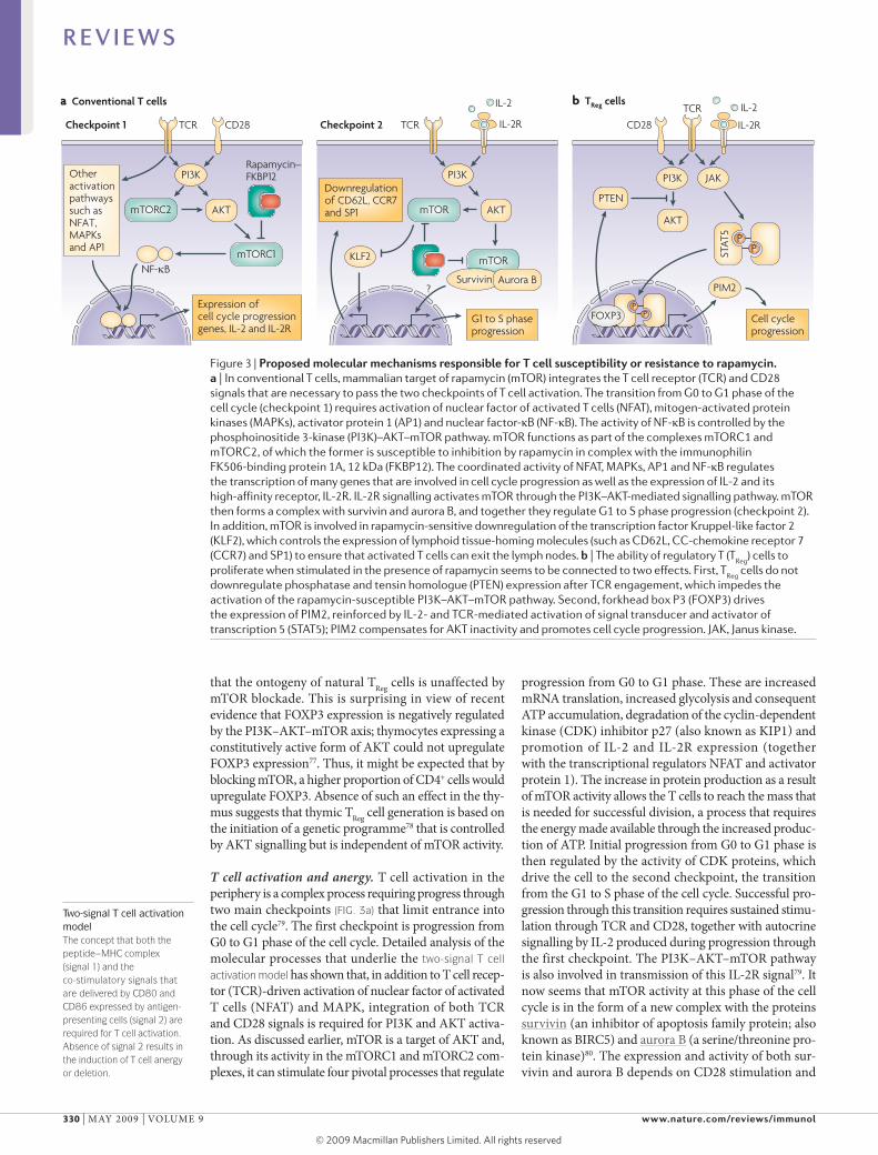
Figure 3 | Proposed molecular mechanisms responsible for T cell susceptibility or resistance to rapamycin. a | In conventional T cells, mammalian target of rapamycin (mTOR) integrates the T cell receptor (TCR) and CD28 signals that are necessary to pass the two checkpoints of T cell activation. The transition from G0 to G1 phase of the cell cycle (checkpoint 1) requires activation of nuclear factor of activated T cells (NFAT), mitogen-activated protein kinases (MAPKs), activator protein 1 (AP1) and nuclear factor-κB (NF-κB). The activity of NF-κB is controlled by the phosphoinositide 3-kinase (PI3K)–AKT–mTOR pathway. mTOR functions as part of the complexes mTORC1 and mTORC2, of which the former is susceptible to inhibition by rapamycin in complex with the immunophilin FK506-binding protein 1A, 12 kDa (FKBP12). The coordinated activity of NFAT, MAPKs, AP1 and NF-κB regulates the transcription of many genes that are involved in cell cycle progression as well as the expression of IL-2 and its high-affinity receptor, IL-2R. IL-2R signalling activates mTOR through the PI3K–AKT-mediated signalling pathway. mTOR then forms a complex with survivin and aurora B, and together they regulate G1 to s phase progression (checkpoint 2). In addition, mTOR is involved in rapamycin-sensitive downregulation of the transcription factor Kruppel-like factor 2 (KLF2), which controls the expression of lymphoid tissue-homing molecules (such as CD62L, CC-chemokine receptor 7 (CCR7) and sP1) to ensure that activated T cells can exit the lymph nodes. b | The ability of regulatory T (T
Reg) cells to
proliferate when stimulated in the presence of rapamycin seems to be connected to two effects. First, TReg
cells do not downregulate phosphatase and tensin homologue (PTeN) expression after TCR engagement, which impedes the activation of the rapamycin-susceptible PI3K–AKT–mTOR pathway. second, forkhead box P3 (FOXP3) drives the expression of PIM2, reinforced by IL-2- and TCR-mediated activation of signal transducer and activator of transcription 5 (sTAT5); PIM2 compensates for AKT inactivity and promotes cell cycle progression. JAK, Janus kinase.
Two-signal T cell activation modelThe concept that both the peptide–mHC complex (signal 1) and the co-stimulatory signals that are delivered by CD80 and CD86 expressed by antigen- presenting cells (signal 2) are required for T cell activation. Absence of signal 2 results in the induction of T cell anergy or deletion.
that the ontogeny of natural TReg cells is unaffected by mTOR blockade. This is surprising in view of recent evidence that FOXP3 expression is negatively regulated by the PI3K–AKT–mTOR axis; thymocytes expressing a constitutively active form of AKT could not upregulate FOXP3 expression77. Thus, it might be expected that by blocking mTOR, a higher proportion of CD4+ cells would upregulate FOXP3. Absence of such an effect in the thymus suggests that thymic TReg cell generation is based on the initiation of a genetic programme78 that is controlled by AKT signalling but is independent of mTOR activity.
T cell activation and anergy. T cell activation in the periphery is a complex process requiring progress through two main checkpoints (FIG. 3a) that limit entrance into the cell cycle79. The first checkpoint is progression from G0 to G1 phase of the cell cycle. Detailed analysis of the molecular processes that underlie the two-signal T cell activation model has shown that, in addition to T cell receptor (TCR)driven activation of nuclear factor of activated T cells (NFAT) and mAPK, integration of both TCR and CD28 signals is required for PI3K and AKT activation. As discussed earlier, mTOR is a target of AKT and, through its activity in the mTORC1 and mTORC2 complexes, it can stimulate four pivotal processes that regulate
progression from G0 to G1 phase. These are increased mRNA translation, increased glycolysis and consequent ATP accumulation, degradation of the cyclindependent kinase (CDK) inhibitor p27 (also known as KIP1) and promotion of Il2 and Il2R expression (together with the transcriptional regulators NFAT and activator protein 1). The increase in protein production as a result of mTOR activity allows the T cells to reach the mass that is needed for successful division, a process that requires the energy made available through the increased production of ATP. Initial progression from G0 to G1 phase is then regulated by the activity of CDK proteins, which drive the cell to the second checkpoint, the transition from the G1 to S phase of the cell cycle. Successful progression through this transition requires sustained stimulation through TCR and CD28, together with autocrine signalling by Il2 produced during progression through the first checkpoint. The PI3K–AKT–mTOR pathway is also involved in transmission of this Il2R signal79. It now seems that mTOR activity at this phase of the cell cycle is in the form of a new complex with the proteins survivin (an inhibitor of apoptosis family protein; also known as BIRC5) and aurora B (a serine/threonine protein kinase)80. The expression and activity of both survivin and aurora B depends on CD28 stimulation and
R E V I E W S
330 | mAy 2009 | VOlumE 9 www.nature.com/reviews/immunol
© 2009 Macmillan Publishers Limited. All rights reserved
T cell anergyA state of T cell unresponsiveness to stimulation with antigen. It can be induced by stimulation with a large amount of specific antigen in the absence of co-stimulatory molecule engagement.
Il2mediated signalling. The target specificity of the mTOR–survivin–aurora B complex is similar to that of mTORC1, with an additional aurora Bregulated capacity to control retinoblastoma phosphorylation, cyclin A expression and CDK1 and CDK2 activity, all of which are involved in G1 to S phase progression79. Interestingly, similarly to mTORC1, this new mTOR complex is susceptible to inhibition by rapamycin.
An additional immunosuppressive effect of mTOR blockade during T cell activation has been recently identified. Il2–Il2R signalling in differentiating effector T cells results in downregulation of the transcription factor Kruppellike factor 2 (KlF2), which controls the expression of the homing molecules SP1, CD62l, CCR7 and other chemokine receptors. This decrease in KlF2 levels depends on PI3K activation and involves mTOR activation81. The presence of rapamycin during Il2mediated signalling prevents the downregulation of KlF2 and, consequently, the modulation of expression of SP1, CD62l and CCR7 that is necessary for the egress of effector T cells from lymph nodes. So, in addition to blocking cell cycle progression during T cell activation, rapamycinmediated mTOR inhibition could sequester activated T cells in lymphoid tissues and prevent them from reaching the target tissue.
mTOR activation is a key process in preventing T cell anergy. Treatment of antigenstimulated T cells with rapamycin slows cell cycle progression to the G1 phase and prevents the downregulation of genes that are involved in the development of anergy82. In addition, it is now clear that Il2–Il2R signalling has a direct role in preventing anergy83. An excess of Il2 can reverse anergy through an mTORdependent, rapamycin sensitive signalling pathway84,85. These observations support the view that proanergy factors are induced early after TCR stimulation and are subsequently degraded or suppressed in response to Il2R signalling. This indicates that conventional mTORC1 activity is necessary for T cell activation but is insufficient to reverse the programme of anergy induction. A separate, Il2specific signalling event is required during the G1 phase to preserve antigen responsiveness and might involve the mTOR–survivin–aurora B complex. The activity of this complex could underlie the recent observations regarding the involvement of the PI3K–AKT–mTOR axis in the induction of FOXP3 expression in the periphery (see below).
Rapamycin is most effective at preventing T cell division under conditions of low Il2 availability79. In the presence of optimal autocrine Il2 secretion or of exogenous Il2, rapamycin delays but cannot prevent cell division86. This alternative regulation of late cell cycle progression by Il2 seems to depend on the recently discovered STAT5dependent expression and activity of the serine/threonine protein kinase PIm2 (ReF. 87). PIm2 can maintain nutrient uptake and ATP synthesis at high levels and, similarly to AKT, regulates cell survival during blastogenesis. So, mTORinduced signalling might regulate the rate of initial cell cycle entry and the integration of sequential signals that are dictated by TCR and CD28 engagement and Il2 production, but it becomes dispensable at later stages, when large quantities of Il2 are available86.
TReg cell homeostasis, function and proliferation. Rapamycin does not affect the function and homeostasis of TReg cells to the same extent as conventional T cells, as recently reviewed88. In vitro exposure of mouse or human TReg cells to rapamycin does not impair their ability to suppress effector T cell proliferation89, an effect that is lost when the calcineurin inhibitor cyclosporin A is used90,91. In addition, prolonged in vivo administration of rapamycin results in a pronounced increase in the number of TReg cells compared with CD4+ T cells in all lymphoid organs, although the absolute numbers of all T cells are decreased (similarly to the thymus)76,92. A similar finding has been made in kidney transplant recipients92,93; patients treated with rapamycin have a markedly increased frequency of CD4+CD25+FOXP3+ TReg cells compared with total CD4+ T cell numbers. This effect is reversed in patients treated with calcineurin inhibitors. In a TCRtransgenic mouse model, rapamycin was used in vitro in combination with Il2 to selectively expand cells with regulatory activity from a starting population of mixed CD4+ T cells94. This observation was then confirmed for human TReg cells89,95,96, which could be expanded selectively in vitro from both healthy donors and patients with autoimmune disease.
The suppressive activity and proliferative capacity of TReg cells depend on TCR engagement and Il2 availability. Differential susceptibility to rapamycin inhibition suggested that the TCR signalling pathway in TReg cells differs from that of conventional T cells. The observation that T cell stimulation in the presence of rapamycin favoured upregulation of FOXP3 expression and acquisition of a regulatory phenotype97 offered the alternative explanation that the observed increase in TReg cell frequency after prolonged rapamycin administration could be due to the peripheral conversion of T cells into TReg cells (see next section). However, our current understanding is that both explanations might be correct. mouse TReg cells cannot activate phospholipase Cγ and consequently generate the downstream signals that result in NFAT, NFκB and RAS–extracellular signalregulated kinase–AP1 activation98. In parallel, TReg cells do not phosphorylate (and therefore activate) AKT in response to stimulation, and restoration of AKT activity impairs their suppressor function99. This has been further clarified by a report that TReg cells express high levels of the negative PI3K regulator phosphatase and tensin homologue (PTEN)100,101, the expression of which is maintained in TReg cells after TCR stimulation (which is in contrast to conventional T cells). Targeted depletion of PTEN does not affect the suppressive capacity of TReg cells, but it does increase their sensitivity to rapamycin. These data indicate that TReg cells do not rely on the conventional PI3K–AKT–mTOR activation pathway and explain their lower susceptibility to rapamycinmediated inhibition.
The above observations do not clarify the mechanism of TReg cellspecific activation in the presence of rapamycin. It is known that the Il2R–STAT5 pathway is essential for TReg cell homeostasis and activity102,103. Furthermore, it has been reported recently that FOXP3 expression is associated with the induction of PIm2
R E V I E W S
NATuRE REVIEWS | Immunology VOlumE 9 | mAy 2009 | 331
© 2009 Macmillan Publishers Limited. All rights reserved
Nature Reviews | Immunology
PI3Kinhibitors
a TGFβ-dependent FOXP3 induction b TGFβ-independent FOXP3 induction
TCRCD28CD28TCR
IL-2 or IL-15TCRCD28 IL-2
PI3K
AKT
mTOR
Rapamycin–FKBP12
PI3KinhibitorsPI3K
AKT
mTOR
Rapamycin–FKBP12
mTOR
First 18 hours After 18 hours
NFAT
NFAT
PPST
AT5
PP FOXP3
FOXP3
TGFβ
TGFβR
SMAD3
SMAD3FOXP3
Chromatinremodelling
IL-2R IL-2R
expression104. Notably, PIm kinases have been implicated in conferring T cell resistance to rapamycinmediated inhibition when STAT5signalling cytokines (including Il2) are present in adequate amounts87. It is therefore possible that TReg cells can integrate TCR and Il2 signalling in a PIm2dependent pathway that allows them to progress through the cell cycle despite mTOR inhibition (FIG. 3b).
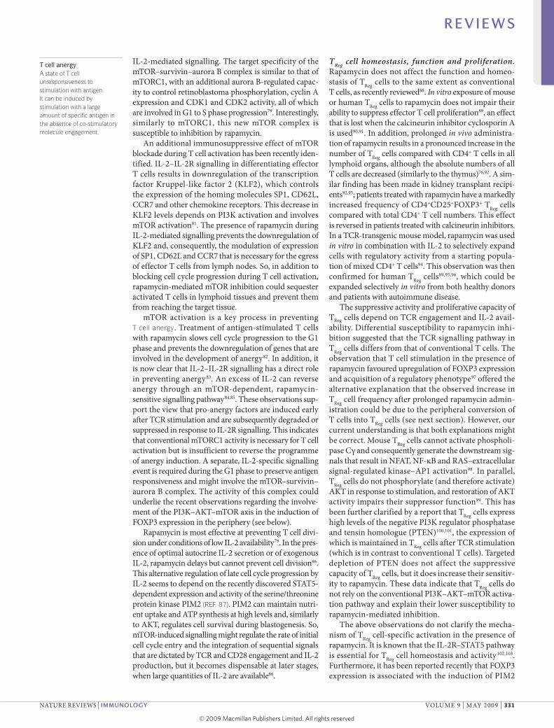
Induction of FOXP3 expression. Natural TReg cells arise in the thymus, but CD4+CD25+FOXP3+ T cells with regulatory capacity can also be generated in the periphery (known as inducible TReg cells) by antigendriven conversion of naive T cells under certain conditions105. In particular, in vitro TCR stimulation of CD4+CD25– T cells in the presence of transforming growth factorβ (TGFβ) and Il2 induces FOXP3 expression. The observation that rapamycin favours this conversion has focused much attention on the involvement of mTOR in the regulation of FOXP3 expression (FIG. 4a). The induction of FOXP3 expression by TGFβ depends on the activation of the transcription factor SmAD3 (mothers against decapentaplegic homologue 3)106, which, together with TCRinduced NFAT, binds to the enhancer region of FOXP3 and promotes chromatin remodelling that is necessary for translation. Interestingly, signalling through the AKT–mTOR pathway can inhibit activation of SmAD3 (ReF. 107), providing an explanation for the observation that mTOR inhibition through rapamycin favours FOXP3 upregulation.
T cells with constitutively active AKT have impaired upregulation of FOXP3 in response to activation in the presence of TGFβ77. Interestingly, when rapamycin is present at the time of transduction of constitutively active AKT, the inhibitory effect is lost. This implies that a rapamycinsensitive signal involving AKT activation (and consequently, mTOR activation) is responsible for the control of peripheral FOXP3 induction. This effect is not restricted to FOXP3 and extends to approximately 50% of the genes that are modulated in response to TGFβ77, which supports the view that conversion to a regulatory phenotype involves complex genetic reprogramming78. However, important differences remain between natural and inducible TReg cells. Expression of a constitutively active form of AKT in thymocytes decreases thymic development of TReg cells without affecting T cell ontogeny77, thereby confirming the ability of the AKT–mTOR pathway to prevent TReg cell development. Expression of FOXP3 in already differentiated circulating TReg cells, however, is not affected by AKT activity, supporting the proposed model that a specific genetic programme is implemented in TReg cells during their development.
A process of FOXP3 upregulation that is independent of TGFβ signalling has begun to be characterized (FIG. 4b). Premature interruption of in vitro TCR and CD28 costimulation (after 18 hours) is associated with the upregulation of FOXP3 expression in approximately 10% of T cells108. Addition of PI3K inhibitors or rapamycin after the interruption of TCR and CD28 costimulation
Figure 4 | mechanisms of FoXP3 induction in naive T cells. a | stimulation of forkhead box P3 (FOXP3)– T cells through T cell receptor (TCR) and CD28 in the presence of transforming growth factor-β (TGFβ) promotes the transcription of FOXP3 through the cooperation of nuclear factor of activated T cells (NFAT) and sMAD3 (mothers against decapentaplegic homologue 3). This process is counteracted by the activation of mammalian target of rapamycin (mTOR), thereby explaining the increased expression of FOXP3 when stimulation takes place in the presence of rapamycin (which inhibits mTOR activation). b | Limited TCR and CD28 stimulation (<18 hours) promotes a phosphoinositide 3-kinase (PI3K)–AKT–mTOR-mediated re-organization of chromatin that includes increased accessibility to FOXP3. Prolonged TCR–CD28 stimulation (>18 hours) prevents, again through activation of the PI3K–AKT–mTOR pathway, the expression of FOXP3, which would otherwise probably be induced by signal transducer and activator of transcription 5 (sTAT5)-activating cytokines generated during the initial stimulation. This two-step model rationalizes the opposing effects of rapamycin administration during T cell activation that have been observed, which probably depend on the timing of mTOR inhibition. FKBP12, FK506-binding protein 1A, 12 kDa; IL, interleukin; IL-2R, IL-2 receptor; TGFβR, TGFβ receptor.
R E V I E W S
332 | mAy 2009 | VOlumE 9 www.nature.com/reviews/immunol
© 2009 Macmillan Publishers Limited. All rights reserved

T regulatory type 1 (TR1) cellA subset of CD4+ regulatory T cells that secrete high levels of interleukin-10 (Il-10) and that downregulate T helper 1 (TH1) and (TH2) cell responses in vitro and in vivo through the secretion of soluble Il-10 and transforming growth factor-β.
increased this fraction to up to 75% of T cells. The effect did not involve TGFβ signalling, as neutralizing antibodies specific for TGFβ and inhibition of SmAD kinase activity did not affect the induction of FOXP3 expression that is promoted by the inhibition of PI3K and mTOR. Interestingly, this effect was closely associated with the timing of interruption of TCR and CD28 costimulation and the addition of PI3K and mTOR inhibitors. Early addition of the inhibitors blocked the necessary T cell activation, whereas prolongation of TCR and CD28 costimulation beyond 18 hours markedly decreased FOXP3 upregulation. In addition, following the initial TCR and CD28 costimulation, histone modifications that facilitate accessibility of the FOXP3 locus to transcription factors were observed and were subsequently lost with continuous TCR and CD28 signalling.
These findings suggest that rapamycin administration establishes conditions for the initiation of a twostep process of FOXP3 upregulation, which is similar to that described for thymic TReg cell development109,110. In this model, TCR and CD28 signalling causes chromatin remodelling that predisposes towards the expression of FOXP3. This first step probably involves PI3K–AKT–mTOR signalling, as the addition of inhibitors at the same time as TCR and CD28 stimulation prevents the necessary T cell activation. After prolonged TCR and CD28 stimulation, the accessibility of FOXP3 is then restricted in a process that involves a second round (or continuation) of PI3K–AKT–mTOR signalling. It is reasonable to imply that, by blocking the second round of signalling, the preactivated cell can then respond differently to stimulation with endogenous cytokines (such as Il2 and Il15). This promotes the activation of the STAT5 pathway, which is involved in both intrathymic TReg cell development and the modulation of FOXP3 expression in peripheral T cells102,103,111. This model of FOXP3 induction might explain the recent observation that a combination of histone deacetylase inhibition and rapamycin increases the number of FOXP3+ TReg cells in vivo112.
On the whole, these observations delineate differences between conventional T cells and TReg cells that shed light on previously unappreciated effects of mTOR inhibition. The observation that rapamycin causes a generalized increase in the frequency of TReg cells is now interpreted as the sum of two effects — the ability of TReg cells to proliferate in the presence of rapamycin and the promotion of FOXP3 expression in peripheral T cells that are then converted into modulators of immune reactivity.
Therapeutic immunosuppressionThe remarkable inhibitory action of rapamycin on DCs and effector T cells, but not TReg cells, singles out mTOR inhibition as a promising therapeutic strategy for the prevention of transplant rejection and the treatment of autoimmune disease.
Transplant tolerance. In renal transplantation, rapamycin is a powerful antirejection agent when used judiciously with other immunosuppressive agents. It has unique antiatherogenic and antineoplastic properties
that distinguish it from other antirejection drugs and it can promote tolerance and decrease the incidence of chronic allograft nephropathy113. Rapamycin was used initially with cyclosporin A, but it is also effective when combined with other immunosuppressive agents. However, the effects of calcineurin inhibitors (such as cyclosporin A) and rapamycin in transplantation seem to be markedly different. In rodents, rapamycin, but not cyclosporin A, allows activationinduced cell death of the large number of alloreactive effector T cells and favours the induction of tolerance114,115. In addition, rapamycin enhances the tolerancepromoting effect of costimulation blockade, whereas cyclosporin A prevents it116,117. In transplant recipients, calcineurin inhibitors, but not rapamycin, decrease the proportion of CD4+CD25+FOXP3+ TReg cells, which is a potentially negative effect of these agents93. Recently, the use of FOXP3 reporter mice has shown that rapamycin promotes the de novo (TGFβdependent) conversion of alloantigenspecific CD4+ T cells to TReg cells under tolerizing conditions, whereas cyclosporin A abrogates this process118. Converted TReg cells were more resistant to apoptosis than conventional T cells, and adoptive transfer of converted TReg cells potently suppressed the rejection of donor, but not third party, skin grafts. The ability of rapamycin plus Il10 to induce T regulatory type 1 (TR1) cells that mediate stable, alloantigenspecific tolerance in pancreatic islet cell transplantation has also been reported119. Collectively, these data provide further evidence that the differential effects of rapamycin on effector T cells and TReg cells (both naturally occurring and inducible) favour its ability to promote tolerance and support its use in tolerancepromoting protocols.
Recent reports suggest that mTOR inhibition, together with targeting of other key molecules that are involved in immune regulation, can promote transplant tolerance in mice. CXCchemokine receptor 3 (CXCR3) and its ligands (CXCl9, CXCl10 and CXCl11) are important for effector T cell recruitment. When combined with a subtherapeutic regimen of rapamycin, a CXCR3specific monoclonal antibody induces indefinite (>100 day) survival of heart or islet allografts120. A second finding concerns the inhibition of histone deacetylase 9 and the consequent increase in FOXP3 expression, as well as the production and function of TReg cells112. When histone deacetylase 9 inhibition is combined with a short course of lowdose rapamycin, permanent donorspecific TReg celldependent heart and pancreatic islet allograft survival is achieved112. TReg cells also seem to be important in mediating the tolerogenic effect of a low dose of rapamycin in combination with monoclonal antibodies specific for T cell immunoglobulin domain and mucin domain protein 1 (TIm1), which is associated with a TH1 to TH2type cytokine switch in experimental heart transplantation121.
Tolerogenic cell therapy in transplantation. The capacity of rapamycin to deplete effector T cells but spare the growth and function of TReg cells can be exploited in the design of new and safe protocols for cell therapy of allograft rejection and other T cellmediated conditions.
R E V I E W S
NATuRE REVIEWS | Immunology VOlumE 9 | mAy 2009 | 333
© 2009 Macmillan Publishers Limited. All rights reserved

IPEXA rare human disease that is linked to dysfunction of the transcriptional factor forkhead box P3 (FOXP3) and that is characterized by the development of autoimmunity.
Rapamycintreated antigenpresenting cells are poor stimulators of allogeneic CD4+ effector T cell proliferation, but enrich for potent CD4+CD25+FOXP3+ TReg cells36. When rapamycinconditioned mouse DCs are pulsed with donor alloantigen and are then adoptively transferred to organ allograft recipients in combination with a short postoperative course of lowdose rapamycin, they induce indefinite (>100 day) graft survival36. Also, rapamycinconditioned recipient DCs inhibit GVHD after allogeneic haematopoietic cell transplantation, which is consistent with their tolerogenic phenotype60. CCR5, CCR7 and CD62l expression on rapamycin conditioned DCs is not affected by mTOR inhibition, which allows them to traffic to secondary lymphoid tissues, where their immunoregulatory function is required59–61. Such observations might, in part, explain the beneficial effects of rapamycin on the incidence of GVHD after haematopoietic cell transplantation that were recently reported in clinical trials122,123. As an example of an alternative strategy, alloantigenspecific TReg cells induce indefinite graft survival and T cell anergy when expanded in vitro in response to immature allogeneic DCs and then infused into rapamycinconditioned heart allograft recipients in the absence of T cell depletion124.
Autoimmune disease. There are numerous reports that mTOR inhibition can suppress experimental autoimmune diseases, in particular type 1 diabetes, nephritis associated with systemic lupus erythematosus (SlE) and adjuvant arthritis125. Rapamycin combined with Il10 blocks the incidence of type 1 diabetes and induces longterm tolerance without chronic immunosuppression in nonobese diabetic (NOD) mice126. In this model, rapamycin mediates the accumulation of suppressive FOXP3+ TReg cells in the pancreas and prevents diabetes. By contrast, when rapamycin is combined with a CD3specific antibody, it has a detrimental effect on disease outcome in NOD mice127. As discussed by the authors of this study, rapamycin might interfere with the ability of the CD3specific antibody to restore Il2 production (which is defective in NOD mice), and thus might prevent this cytokine from maintaining tolerance in these animals. In patients with type 1 diabetes, rapamycin promotes the ex vivo proliferation of functional FOXP3+ TReg cells89. moreover, naturally occurring TReg cells from patients with type 1 diabetes treated solely with rapamycin have a restored ability to suppress the proliferation
of CD4+CD25– effector T cells compared with TReg cells that have not been treated128. In NZB × NZW F1 female mice (a mouse model of SlE), administration of rapamycin (from 12–37 weeks of age) decreases the production of autoantibodies, glomerular deposits of immunoglobulins and the development of proteinuria, and prolongs survival129. Also in this model, rapamycin attenuates the severity of established nephritis through reduced lymphoproliferation, decreased CCl5 expression and decreased infiltration of lymphocytes into the kidneys130. Rapamycin is also effective for the treatment of autoimmune hepatitis following human liver transplantation131 and has recently been used to treat a case of refractory Crohn’s disease132. Interestingly, rapamycin might be a clinically effective and safe therapeutic option in patients with IPeX (immunodysregulation, polyendocrinopathy and enteropathy, Xlinked syndrome) and IPEXlike disease133, who do not have naturally occurring FOXP3+ TReg cells and therefore suffer from severe autoimmune disease.
ConclusionsIn summary, although the importance of mTORC1 in the regulation of innate and adaptive immune responses is now wellrecognized, the role of mTORC2 has yet to be clarified. New evidence that mTOR regulates cytokine production by antigenpresenting cells in response to inflammatory stimuli suggests a pivotal role for this molecule in determining the nature of T cell responses. The mechanisms by which rapamycin suppresses immune responses have been expanded from inhibition of T cell proliferation to blockade of DC maturation and support of TReg cells, including their de novo induction. Ongoing and future areas of enquiry, which are likely to further elucidate the role of mTOR in the regulation of immune responses and tolerance, include the investigation of the role of the newly identified mTOR–survivin–aurora B complex in the activation of T cells (and other immune cells, including DCs) and the clarification of the role of PIm1 and PIm2 in determining TReg cell resistance to mTOR inhibition. In addition, it will be important to elucidate the TGFβdependent and TGFβindependent mechanisms of FOXP3 upregulation and to clarify the physiological role of each mechanism, which will be relevant to therapeutic applications as they are both affected by mTOR modulation. Insight is also needed into the role of mTOR in memory T cells.
1. Vezina, C., Kudelski, A. & Sehgal, S. N. Rapamycin (AY-22, 989), a new antifungal antibiotic. I. Taxonomy of the producing streptomycete and isolation of the active principle. J. Antibiot. (Tokyo) 28, 721–726 (1975).
2. Saunders, R. N., metcalfe, m. S. & Nicholson, m. L. Rapamycin in transplantation: a review of the evidence. Kidney Int. 59, 3–16 (2001).
3. Eisen, H. J. et al. Everolimus for the prevention of allograft rejection and vasculopathy in cardiac-transplant recipients. N. Engl. J. Med. 349, 847–858 (2003).
4. Armand, P. et al. Improved survival in lymphoma patients receiving sirolimus for graft-versus-host disease prophylaxis after allogeneic hematopoietic stem-cell transplantation with reduced-intensity conditioning. J. Clin. Oncol. 26, 5767–5774 (2008).
5. Sabatini, D. m. mTOR and cancer: insights into a complex relationship. Nature Rev. Cancer 6, 729–734 (2006).
6. Roiron, C., Sanchez, P., Bouzamondo, A., Lechat, P. & montalescot, G. Drug eluting stents: an updated meta-analysis of randomised controlled trials. Heart 92, 641–649 (2006).
7. Heitman, J., movva, N. R. & Hall, m. N. Targets for cell cycle arrest by the immunosuppressant rapamycin in yeast. Science 253, 905–909 (1991).
8. Sehgal, S. N. Sirolimus: its discovery, biological properties, and mechanism of action. Transplant Proc. 35, 7S–14S (2003).
9. Brown, E. J. et al. A mammalian protein targeted by G1-arresting rapamycin-receptor complex. Nature 369, 756–758 (1994).
10. Chiu, m. I., Katz, H. & Berlin, V. RAPT1, a mammalian homolog of yeast Tor, interacts with the FKBP12/rapamycin complex. Proc. Natl Acad. Sci. USA 91, 12574–12578 (1994).
11. Sabatini, D. m., Erdjument-Bromage, H., Lui, m., Tempst, P. & Snyder, S. H. RAFT1: a mammalian protein that binds to FKBP12 in a rapamycin-dependent fashion and is homologous to yeast TORs. Cell 78, 35–43 (1994).
12. Gangloff, Y. G. et al. Disruption of the mouse mTOR gene leads to early postimplantation lethality and prohibits embryonic stem cell development. Mol. Cell. Biol. 24, 9508–9516 (2004).
13. Guertin, D. A. et al. Ablation in mice of the mTORC components raptor, rictor, or mLST8 reveals that mTORC2 is required for signaling to Akt–FOXO and PKCα, but not S6K1. Dev. Cell 11, 859–871 (2006).
R E V I E W S
334 | mAy 2009 | VOlumE 9 www.nature.com/reviews/immunol
© 2009 Macmillan Publishers Limited. All rights reserved
14. Wullschleger, S., Loewith, R. & Hall, m. N. TOR signaling in growth and metabolism. Cell 124, 471–484 (2006).
15. Thoreen, C. C. et al. An ATP-competitive mTOR inhibitor reveals rapamycin-insensitive functions of mTORC1. J. Biol. Chem. 284, 8023–8032 (2009).
16. Sarbassov, D. D. et al. Prolonged rapamycin treatment inhibits mTORC2 assembly and Akt/PKB. Mol. Cell 22, 159–168 (2006).
17. Zeng, Z. et al. Rapamycin derivatives reduce mTORC2 signaling and inhibit AKT activation in AmL. Blood 109, 3509–3512 (2007).
18. Sarbassov, D. D., Ali, S. m. & Sabatini, D. m. Growing roles for the mTOR pathway. Curr. Opin. Cell Biol. 17, 596–603 (2005).
19. Yang, Q. & Guan, K. L. Expanding mTOR signaling. Cell Res. 17, 666–681 (2007).
20. Potter, C. J., Pedraza, L. G. & Xu, T. Akt regulates growth by directly phosphorylating Tsc2. Nature Cell Biol. 4, 658–665 (2002).
21. Staal, F. J., Luis, T. C. & Tiemessen, m. m. WNT signalling in the immune system: WNT is spreading its wings. Nature Rev. Immunol. 8, 581–593 (2008).
22. Inoki, K. et al. TSC2 integrates Wnt and energy signals via a coordinated phosphorylation by AmPK and GSK3 to regulate cell growth. Cell 126, 955–968 (2006).
23. Sarbassov, D. D. et al. Rictor, a novel binding partner of mTOR, defines a rapamycin-insensitive and raptor-independent pathway that regulates the cytoskeleton. Curr. Biol. 14, 1296–1302 (2004).
24. Jacinto, E. et al. mammalian TOR complex 2 controls the actin cytoskeleton and is rapamycin insensitive. Nature Cell Biol. 6, 1122–1128 (2004).References 23 and 24 establish that mTOR exists in two functionally distinct complexes: rapamycin-sensitive mTORC1 (which contains RapTOR) and rapamycin-resistant mTORC2 (which contains RiCTOR).
25. Hresko, R. C. & mueckler, m. mTOR.RICTOR is the Ser473 kinase for Akt/protein kinase B in 3T3-L1 adipocytes. J. Biol. Chem. 280, 40406–40416 (2005).
26. Astrinidis, A. et al. Tuberin, the tuberous sclerosis complex 2 tumor suppressor gene product, regulates Rho activation, cell adhesion and migration. Oncogene 21, 8470–8476 (2002).
27. O’Reilly, K. E. et al. mTOR inhibition induces upstream receptor tyrosine kinase signaling and activates Akt. Cancer Res. 66, 1500–1508 (2006).
28. Tremblay, F., Gagnon, A., Veilleux, A., Sorisky, A. & marette, A. Activation of the mammalian target of rapamycin pathway acutely inhibits insulin signaling to Akt and glucose transport in 3T3-L1 and human adipocytes. Endocrinology 146, 1328–1337 (2005).
29. Sun, S. Y. et al. Activation of Akt and eIF4E survival pathways by rapamycin-mediated mammalian target of rapamycin inhibition. Cancer Res. 65, 7052–7058 (2005).
30. Greene, m. W., Sakaue, H., Wang, L., Alessi, D. R. & Roth, R. A. modulation of insulin-stimulated degradation of human insulin receptor substrate-1 by serine 312 phosphorylation. J. Biol. Chem. 278, 8199–8211 (2003).
31. Harrington, L. S. et al. The TSC1–2 tumor suppressor controls insulin–PI3K signaling via regulation of IRS proteins. J. Cell. Biol. 166, 213–223 (2004).
32. Armstrong, J. L., Bonavaud, S. m., Toole, B. J. & Yeaman, S. J. Regulation of glycogen synthesis by amino acids in cultured human muscle cells. J. Biol. Chem. 276, 952–956 (2001).
33. Zhang, Q., Chen, Y., Fairchild, R. L., Heeger, P. S. & Valujskikh, A. Lymphoid sequestration of alloreactive memory CD4 T cells promotes cardiac allograft survival. J. Immunol. 176, 770–777 (2006).
34. Hackstein, H. et al. Rapamycin inhibits IL-4-induced dendritic cell maturation in vitro and dendritic cell mobilization and function in vivo. Blood 101, 4457–4463 (2003).This was the first demonstration that rapamycin administration impairs steady-state DC generation and inhibits their maturation in vivo.
35. Woltman, A. m. et al. Rapamycin induces apoptosis in monocyte- and CD34-derived dendritic cells but not in monocytes and macrophages. Blood 98, 174–180 (2001).
36. Turnquist, H. et al. Rapamycin-conditioned dendritic cells are poor stimulators of allogeneic CD4+ T cells,
but enrich for antigen-specific Foxp3+ T regulatory cells and promote organ transplant tolerance. J. Immunol. 178, 7018–7031 (2007).
37. Weinstein, S. L. et al. Phosphatidylinositol 3-kinase and mTOR mediate lipopolysaccharide-stimulated nitric oxide production in macrophages via interferon-β. J. Leukoc. Biol. 67, 405–414 (2000).
38. Schmitz, F. et al. mammalian target of rapamycin (mTOR) orchestrates the defense program of innate immune cells. Eur. J. Immunol. 38, 2981–2992 (2008).
39. Rama, I. et al. Hypoxia stimulus: an adaptive immune response during dendritic cell maturation. Kidney Int. 73, 816–825 (2008).
40. Haddadi, A. et al. Delivery of rapamycin by PLGA nanoparticles enhances its suppressive activity on dendritic cells. J. Biomed. Mater. Res. A 84, 885–898 (2008).
41. Das, S., Haddadi, A., Veniamin, S. & Samuel, J. Delivery of rapamycin-loaded nanoparticle down regulates ICAm-1 expression and maintains an immunosuppressive profile in human CD34+
progenitor-derived dendritic cells. J. Biomed. Mater. Res. A 85, 983–992 (2008).
42. Jhunjhunwala, S., Raimondi, G., Thomson, A. W. & Little, S. R. Delivery of rapamycin to dendritic cells using degradable microparticles. J. Control. Release 133, 191–197 (2009).
43. Hackstein, H., Taner, T., Logar, A. J. & Thomson, A. W. Rapamycin inhibits macropinocytosis and mannose receptor-mediated endocytosis by bone marrow-derived dendritic cells. Blood 100, 1084–1087 (2002).
44. monti, P. et al. Rapamycin impairs antigen uptake of human dendritic cells. Transplantation 75, 137–145 (2003).
45. Fox, R. et al. PSGL-1 and mTOR regulate translation of ROCK-1 and physiological functions of macrophages. EMBO J. 26, 505–515 (2007).
46. matsue, H. et al. Contrasting impacts of immunosuppressive agents (rapamycin, FK506, cyclosporin A, and dexamethasone) on bidirectional dendritic cell–T cell interaction during antigen presentation. J. Immunol. 169, 3555–3564 (2002).
47. Lee, Y. R. et al. Cyclosporin A and tacrolimus, but not rapamycin, inhibit mHC-restricted antigen presentation pathways in dendritic cells. Blood 105, 3951–3955 (2005).
48. Imai, A. et al. Inhibition of endogenous mHC class II- restricted antigen presentation by tacrolimus (FK506) via FKBP51. Eur. J. Immunol. 37, 1730–1738 (2007).
49. Fassbender, m., Herter, S., Holtappels, R. & Schild, H. Correlation of dendritic cell maturation and the formation of aggregates of poly-ubiquitinated proteins in the cytosol. Med. Microbiol. Immunol. 197, 185–189 (2008).
50. Lelouard, H. et al. Transient aggregation of ubiquitinated proteins during dendritic cell maturation. Nature 417, 177–182 (2002).
51. Koehl, G. E. et al. Rapamycin protects allografts from rejection while simultaneously attacking tumors in immunosuppressed mice. Transplantation 77, 1319–1326 (2004).
52. Schmid, D., Pypaert, m. & munz, C. Antigen-loading compartments for major histocompatibility complex class II molecules continuously receive input from autophagosomes. Immunity 26, 79–92 (2007).
53. Vyas, J. m., Van der Veen, A. G. & Ploegh, H. L. The known unknowns of antigen processing and presentation. Nature Rev. Immunol. 8, 607–618 (2008).
54. Gannage, m. & munz, C. macroautophagy in immunity and tolerance. Traffic 24 Jan 2009 (doi:10.1111/j.1600–08542009.00883.x).
55. Kamada, Y. et al. Tor-mediated induction of autophagy via an Apg1 protein kinase complex. J. Cell. Biol. 150, 1507–1513 (2000).
56. Noda, T. & Ohsumi, Y. Tor, a phosphatidylinositol kinase homologue, controls autophagy in yeast. J. Biol. Chem. 273, 3963–3966 (1998).
57. Jagannath, C. et al. Autophagy enhances the efficacy of BCG vaccine by increasing peptide presentation in mouse dendritic cells. Nature Med. 15, 267–276 (2009).
58. Woltman, A. m. et al. Rapamycin specifically interferes with Gm-CSF signaling in human dendritic cells, leading to apoptosis via increased p27KIP1 expression. Blood 101, 1439–1445 (2003).
59. Sordi, V. et al. Differential effects of immunosuppressive drugs on chemokine receptor CCR7 in human monocyte-derived dendritic cells:
selective upregulation by rapamycin. Transplantation 82, 826–834 (2006).
60. Reichardt, W. et al. Impact of mammalian target of rapamycin inhibition on lymphoid homing and tolerogenic function of nanoparticle-labeled dendritic cells following allogeneic hematopoietic cell transplantation. J. Immunol. 181, 4770–4779 (2008).
61. Taner, T., Hackstein, H., Wang, Z., morelli, A. E. & Thomson, A. W. Rapamycin-treated, alloantigen-pulsed host dendritic cells induce Ag-specific T cell regulation and prolong graft survival. Am. J. Transplant. 5, 228–236 (2005).This was the first demonstration that recipient-derived myeloid DCs generated in rapamycin and pulsed with donor alloantigen can promote indefinite organ allograft survival in the absence of any other therapy.
62. Trinchieri, G. Interleukin-12 and the regulation of innate resistance and adaptive immunity. Nature Rev. Immunol. 3, 133–146 (2003).
63. Turnquist, H. R. et al. IL-1β-driven ST2L expression promotes maturation resistance in rapamycin-conditioned dendritic cells. J. Immunol. 181, 62–72 (2008).
64. Schmitz, J. et al. IL-33, an interleukin-1-like cytokine that signals via the IL-1 receptor-related protein ST2 and induces T helper type 2-associated cytokines. Immunity 23, 479–490 (2005).
65. Brint, E. K. et al. ST2 is an inhibitor of interleukin 1 receptor and Toll-like receptor 4 signaling and maintains endotoxin tolerance. Nature Immunol. 5, 373–379 (2004).
66. Fukao, T. et al. PI3K-mediated negative feedback regulation of IL-12 production in DCs. Nature Immunol. 3, 875–881 (2002).
67. Ohtani, m. et al. mammalian target of rapamycin and glycogen synthase kinase 3 differentially regulate lipopolysaccharide-induced interleukin-12 production in dendritic cells. Blood 112, 635–643 (2008).
68. Weichhart, T. et al. The TSC–mTOR signaling pathway regulates the innate inflammatory response. Immunity 29, 565–577 (2008).References 67 and 68 show that mTOR acts downstream of TLR4 and is a crucial regulator of pro- and anti-inflammatory cytokine production.
69. Yang, C. S. et al. Intracellular network of phosphatidylinositol 3-kinase, mammalian target of the rapamycin/70 kDa ribosomal S6 kinase 1, and mitogen-activated protein kinases pathways for regulating mycobacteria-induced IL-23 expression in human macrophages. Cell. Microbiol. 8, 1158–1171 (2006).
70. Colina, R. et al. Translational control of the innate immune response through IRF-7. Nature 452, 323–328 (2008).
71. Honda, K. et al. IRF-7 is the master regulator of type-I interferon-dependent immune responses. Nature 434, 772–777 (2005).
72. Cao, W. et al. Toll-like receptor-mediated induction of type I interferon in plasmacytoid dendritic cells requires the rapamycin-sensitive PI(3)K–mTOR–p70S76K pathway. Nature Immunol. 9, 1157–1164 (2008).This study provides in vitro and in vivo evidence that pDCs require the activation of mTOR to initiate iFNα production downstream of TLR9 ligation by CpG DNa or in response to viral challenge.
73. Sakaguchi, S., Yamaguchi, T., Nomura, T. & Ono, m. Regulatory T cells and immune tolerance. Cell 133, 775–787 (2008).
74. Luo, H., Duguid, W., Chen, H., maheu, m. & Wu, J. The effect of rapamycin on T cell development in mice. Eur. J. Immunol. 24, 692–701 (1994).
75. Damoiseaux, J. G., Defresne, m. P., Reutelingsperger, C. P. & Van Breda Vriesman, P. J. Cyclosporin-A differentially affects apoptosis during in vivo rat thymocyte maturation. Scand. J. Immunol. 56, 353–360 (2002).
76. Coenen, J. J. et al. Rapamycin, not cyclosporine, permits thymic generation and peripheral preservation of CD4+ CD25+ FoxP3+ T cells. Bone Marrow Transplant. 39, 537–545 (2007).
77. Haxhinasto, S., mathis, D. & Benoist, C. The AKT–mTOR axis regulates de novo differentiation of CD4+Foxp3+ cells. J. Exp. Med. 205, 565–574 (2008).The authors show that the aKT–mTOR signalling pathway dominantly regulates TGFβ-dependent FOXp3 induction in peripheral CD4+ T cells.
R E V I E W S
NATuRE REVIEWS | Immunology VOlumE 9 | mAy 2009 | 335
© 2009 Macmillan Publishers Limited. All rights reserved
78. Zheng, Y. & Rudensky, A. Y. Foxp3 in control of the regulatory T cell lineage. Nature Immunol. 8, 457–462 (2007).
79. mondino, A. & mueller, D. L. mTOR at the crossroads of T cell proliferation and tolerance. Semin. Immunol. 19, 162–172 (2007).
80. Song, J., Salek-Ardakani, S., So, T. & Croft, m. The kinases aurora B and mTOR regulate the G1–S cell cycle progression of T lymphocytes. Nature Immunol. 8, 64–73 (2007).This study shows the involvement of mTOR in a new complex containing aurora B, the activity of which is influenced by iL-2-mediated signalling, which is necessary for the G1 to S phase transition of the cell cycle.
81. Sinclair, L. V. et al. Phosphatidylinositol-3-OH kinase and nutrient-sensing mTOR pathways control T lymphocyte trafficking. Nature Immunol. 9, 513–521 (2008).
82. Gilbert, K. m. & Weigle, W. O. Th1 cell anergy and blockade in G1a phase of the cell cycle. J. Immunol. 151, 1245–1254 (1993).
83. Boussiotis, V. A. et al. Prevention of T cell anergy by signaling through the gamma c chain of the IL-2 receptor. Science 266, 1039–1042 (1994).
84. Beverly, B., Kang, S. m., Lenardo, m. J. & Schwartz, R. H. Reversal of in vitro T cell clonal anergy by IL-2 stimulation. Int. Immunol. 4, 661–671 (1992).
85. Allen, A. et al. The novel cyclophilin binding compound, sanglifehrin A, disassociates G1 cell cycle arrest from tolerance induction. J. Immunol. 172, 4797–4803 (2004).
86. Colombetti, S., Basso, V., mueller, D. L. & mondino, A. Prolonged TCR/CD28 engagement drives IL-2-independent T cell clonal expansion through signaling mediated by the mammalian target of rapamycin. J. Immunol. 176, 2730–2738 (2006).
87. Fox, C. J., Hammerman, P. S. & Thompson, C. B. The Pim kinases control rapamycin-resistant T cell survival and activation. J. Exp. Med. 201, 259–266 (2005).
88. Roncarolo, m. G. & Battaglia, m. Regulatory T-cell immunotherapy for tolerance to self antigens and alloantigens in humans. Nature Rev. Immunol. 7, 585–598 (2007).
89. Battaglia, m. et al. Rapamycin promotes expansion of functional CD4+CD25+FOXP3+ regulatory T cells of both healthy subjects and type 1 diabetic patients. J. Immunol. 177, 8338–8347 (2006).This is the first report of the decreased susceptibility of TReg cells compared with conventional T cells to the anti-proliferative effect of rapamycin.
90. Zeiser, R. et al. Inhibition of CD4+CD25+ regulatory T-cell function by calcineurin-dependent interleukin-2 production. Blood 108, 390–399 (2006).
91. Coenen, J. J., Koenen, H. J., van Rijssen, E., Hilbrands, L. B. & Joosten, I. Rapamycin, and not cyclosporin A, preserves the highly suppressive CD27+ subset of human CD4+CD25+ regulatory T cells. Blood 107, 1018–1023 (2006).
92. Noris, m. et al. Regulatory T cells and T cell depletion: role of immunosuppressive drugs. J. Am. Soc. Nephrol. 18, 1007–1018 (2007).
93. Segundo, D. S. et al. Calcineurin inhibitors, but not rapamycin, reduce percentages of CD4+CD25+FOXP3+ regulatory T cells in renal transplant recipients. Transplantation 82, 550–557 (2006).
94. Battaglia, m., Stabilini, A. & Roncarolo, m. G. Rapamycin selectively expands CD4+CD25+FoxP3+ regulatory T cells. Blood 105, 4743–4748 (2005).
95. Strauss, L. et al. Selective survival of naturally occurring human CD4+CD25+Foxp3+ regulatory T cells cultured with rapamycin. J. Immunol. 178, 320–329 (2007).
96. Keever-Taylor, C. A. et al. Rapamycin enriches for CD4+ CD25+ CD27+ Foxp3+ regulatory T cells in ex vivo-expanded CD25-enriched products from healthy donors and patients with multiple sclerosis. Cytotherapy 9, 144–157 (2007).
97. Valmori, D. et al. Rapamycin-mediated enrichment of T cells with regulatory activity in stimulated CD4+ T cell cultures is not due to the selective expansion of naturally occurring regulatory T cells but to the induction of regulatory functions in conventional CD4+ T cells. J. Immunol. 177, 944–949 (2006).
98. Hickman, S. P., Yang, J., Thomas, R. m., Wells, A. D. & Turka, L. A. Defective activation of protein kinase C and Ras–ERK pathways limits IL-2 production and
proliferation by CD4+CD25+ regulatory T cells. J. Immunol. 177, 2186–2194 (2006).
99. Crellin, N. K., Garcia, R. V. & Levings, m. K. Altered activation of AKT is required for the suppressive function of human CD4+CD25+ T regulatory cells. Blood 109, 2014–2022 (2007).
100. Zeiser, R. et al. Differential impact of mammalian target of rapamycin inhibition on CD4+CD25+Foxp3+ regulatory T cells compared with conventional CD4+ T cells. Blood 111, 453–462 (2008).
101. Walsh, P. T. et al. PTEN inhibits IL-2 receptor-mediated expansion of CD4+ CD25+ Tregs. J. Clin. Invest. 116, 2521–2531 (2006).
102. Burchill, m. A., Yang, J., Vang, K. B. & Farrar, m. A. Interleukin-2 receptor signaling in regulatory T cell development and homeostasis. Immunol. Lett. 114, 1–8 (2007).
103. Zeiser, R. & Negrin, R. S. Interleukin-2 receptor downstream events in regulatory T cells: implications for the choice of immunosuppressive drug therapy. Cell Cycle 7, 458–462 (2008).
104. Basu, S., Golovina, T., mikheeva, T., June, C. H. & Riley, J. L. Cutting Edge: Foxp3-mediated induction of Pim 2 allows human T regulatory cells to preferentially expand in rapamycin. J. Immunol. 180, 5794–5798 (2008).
105. Kretschmer, K., Apostolou, I., Jaeckel, E., Khazaie, K. & von Boehmer, H. making regulatory T cells with defined antigen specificity: role in autoimmunity and cancer. Immunol. Rev. 212, 163–169 (2006).
106. Tone, Y. et al. Smad3 and NFAT cooperate to induce Foxp3 expression through its enhancer. Nature Immunol. 9, 194–202 (2008).
107. Song, K., Wang, H., Krebs, T. L. & Danielpour, D. Novel roles of Akt and mTOR in suppressing TGF-β/ALK5-mediated Smad3 activation. EMBO J. 25, 58–69 (2006).
108. Sauer, S. et al. T cell receptor signaling controls Foxp3 expression via PI3K, Akt, and mTOR. Proc. Natl Acad. Sci. USA 105, 7797–7802 (2008).References 106 and 108 provide complementary insights into the molecular mechanisms by which TCR signalling regulates the expression of FOXp3 in peripheral T cells.
109. Lio, C. W. & Hsieh, C. S. A two-step process for thymic regulatory T cell development. Immunity 28, 100–111 (2008).
110. Burchill, m. A. et al. Linked T cell receptor and cytokine signaling govern the development of the regulatory T cell repertoire. Immunity 28, 112–121 (2008).
111. Passerini, L. et al. STAT5-signaling cytokines regulate the expression of FOXP3 in CD4+CD25+ regulatory T cells and CD4+CD25– effector T cells. Int. Immunol. 20, 421–431 (2008).
112. Tao, R. et al. Deacetylase inhibition promotes the generation and function of regulatory T cells. Nature Med. 13, 1299–1307 (2007).This report shows that combined pharmacological interference with mTOR signalling and with the epigenetic control of FOXp3 expression (histone acetylation) promotes the upregulation of FOXp3 expression in peripheral T cells.
113. Augustine, J. J., Bodziak, K. A. & Hricik, D. E. Use of sirolimus in solid organ transplantation. Drugs 67, 369–391 (2007).
114. Li, X. C. et al. IL-15 and IL-2: a matter of life and death for T cells in vivo. Nature Med. 7, 114–118 (2001).
115. Li, Y. et al. Blocking both signal 1 and signal 2 of T-cell activation prevents apoptosis of alloreactive T cells and induction of peripheral allograft tolerance. Nature Med. 5, 1298–1302 (1999).
116. Li, Y., Zheng, X. X., Li, X. C., Zand, m. S. & Strom, T. B. Combined costimulation blockade plus rapamycin but not cyclosporine produces permanent engraftment. Transplantation 66, 1387–1388 (1998).
117. Blaha, P. et al. The influence of immunosuppressive drugs on tolerance induction through bone marrow transplantation with costimulation blockade. Blood 101, 2886–2893 (2003).References 115 and 117 show that mTOR inhibition, unlike calcineurin inhibition, does not block the induction of experimental transplant tolerance.
118. Gao, W. et al. Contrasting effects of cyclosporine and rapamycin in de novo generation of alloantigen-specific regulatory T cells. Am. J. Transplant. 7, 1722–1732 (2007).
119. Battaglia, m. et al. Rapamycin and interleukin-10 treatment induces T regulatory type 1 cells that mediate antigen-specific transplantation tolerance. Diabetes 55, 40–49 (2006).
120. Uppaluri, R. et al. Prolongation of cardiac and islet allograft survival by a blocking hamster anti-mouse CXCR3 monoclonal antibody. Transplantation 86, 137–147 (2008).
121. Ueno, T. et al. The emerging role of T cell Ig mucin 1 in alloimmune responses in an experimental mouse transplant model. J. Clin. Invest. 118, 742–751 (2008).
122. Antin, J. H. et al. Sirolimus, tacrolimus, and low-dose methotrexate for graft-versus-host disease prophylaxis in mismatched related donor or unrelated donor transplantation. Blood 102, 1601–1605 (2003).
123. Cutler, C. et al. Extended follow-up of methotrexate-free immunosuppression using sirolimus and tacrolimus in related and unrelated donor peripheral blood stem cell transplantation. Blood 109, 3108–3114 (2007).
124. Raimondi, G. et al. Induction of heart transplant tolerance by post-transplant administration of allo-Ag specific Treg and rapamycin. Am. Transplant Congress 8, 249 (2008).
125. Carlson, R. P., Hartman, D. A., Ochalski, S. J., Zimmerman, J. L. & Glaser, K. B. Sirolimus (rapamycin, Rapamune) and combination therapy with cyclosporin A in the rat developing adjuvant arthritis model: correlation with blood levels and the effects of different oral formulations. Inflamm. Res. 47, 339–344 (1998).
126. Battaglia, m. et al. Induction of tolerance in type 1 diabetes via both CD4+CD25+ T regulatory cells and T regulatory type 1 cells. Diabetes 55, 1571–1580 (2006).
127. Valle, A. et al. Rapamycin prevents and breaks the anti-CD3-induced tolerance in NOD mice. Diabetes 16 Jan 2009 (doi: 10.2337/db08–1432).
128. monti, P. et al. Rapamycin monotherapy in patients with type 1 diabetes modifies CD4+CD25+FOXP3+
regulatory T-cells. Diabetes 57, 2341–2347 (2008).This paper shows that rapamycin monotherapy in patients with autoimmune diabetes improves the function of naturally occurring TReg cells.
129. Ramos-Barron, A. et al. Prevention of murine lupus disease in (NZBxNZW)F1 mice by sirolimus treatment. Lupus 16, 775–781 (2007).
130. Lui, S. L. et al. Rapamycin attenuates the severity of established nephritis in lupus-prone NZB/W F1 mice. Nephrol. Dial. Transplant. 23, 2768–2776 (2008).
131. montano Loza, A. J. & Czaja, A. J. Current therapy for autoimmune hepatitis. Nature Clin. Pract. Gastroenterol. Hepatol. 4, 202–214 (2007).
132. massey, D. C., Bredin, F. & Parkes, m. Use of sirolimus (rapamycin) to treat refractory Crohn’s disease. Gut 57, 1294–1296 (2008).
133. Yong, P. L., Russo, P. & Sullivan, K. E. Use of sirolimus in IPEX and IPEX-like children. J. Clin. Immunol. 28, 581–587 (2008).
134. Dames, S. A., mulet, J. m., Rathgeb-Szabo, K., Hall, m. N. & Grzesiek, S. The solution structure of the FATC domain of the protein kinase target of rapamycin suggests a role for redox-dependent structural and cellular stability. J. Biol. Chem. 280, 20558–20564 (2005).
135. Kim, D. H. et al. GβL, a positive regulator of the rapamycin-sensitive pathway required for the nutrient-sensitive interaction between raptor and mTOR. Mol. Cell 11, 895–904 (2003).
136. Chen, E. J. & Kaiser, C. A. LST8 negatively regulates amino acid biosynthesis as a component of the TOR pathway. J. Cell. Biol. 161, 333–347 (2003).
137. Frias, m. A. et al. mSin1 is necessary for Akt/PKB phosphorylation, and its isoforms define three distinct mTORC2s. Curr. Biol. 16, 1865–1870 (2006).
138. Donahue, A. C. & Fruman, D. A. Proliferation and survival of activated B cells requires sustained antigen receptor engagement and phosphoinositide 3-kinase activation. J. Immunol. 170, 5851–5860 (2003).
139. Li, H. L., Davis, W. & Pure, E. Suboptimal cross-linking of antigen receptor induces Syk-dependent activation of p70S6 kinase through protein kinase C and phosphoinositol 3-kinase. J. Biol. Chem. 274, 9812–9820 (1999).
140. Donahue, A. C. & Fruman, D. A. Distinct signaling mechanisms activate the target of rapamycin in response to different B-cell stimuli. Eur. J. Immunol. 37, 2923–2936 (2007).
R E V I E W S
336 | mAy 2009 | VOlumE 9 www.nature.com/reviews/immunol
© 2009 Macmillan Publishers Limited. All rights reserved
141. Sakata, A., Kuwahara, K., Ohmura, T., Inui, S. & Sakaguchi, N. Involvement of a rapamycin-sensitive pathway in CD40-mediated activation of murine B cells in vitro. Immunol. Lett. 68, 301–309 (1999).
142. Heidt, S. et al. Effects of immunosuppressive drugs on purified human B cells: evidence supporting the use of mmF and rapamycin. Transplantation 86, 1292–1300 (2008).
143. Aagaard-Tillery, K. m. & Jelinek, D. F. Inhibition of human B lymphocyte cell cycle progression and differentiation by rapamycin. Cell. Immunol. 156, 493–507 (1994).
144. Hornung, N., Raskova, J., Raska, K. Jr & Degiannis, D. Responsiveness of preactivated B cells to IL-2 and IL-6. Effect of cyclosporine and rapamycin. Transplantation 56, 985–990 (1993).
145. Wai, L. E., Fujiki, m., Takeda, S., martinez, O. m. & Krams, S. m. Rapamycin, but not cyclosporine or FK506, alters natural killer cell function. Transplantation 85, 145–149 (2008).
146. Gourlay, W. A., Chambers, W. H., monaco, A. P. & maki, T. Importance of natural killer cells in the rejection of hamster skin xenografts. Transplantation 65, 727–734 (1998).
147. Gomez-Cambronero, J. Rapamycin inhibits Gm-CSF-induced neutrophil migration. FEBS Lett. 550, 94–100 (2003).
148. Lehman, J. A., Calvo, V. & Gomez-Cambronero, J. mechanism of ribosomal p70S6 kinase activation by granulocyte macrophage colony-stimulating factor in neutrophils: cooperation of a mEK-related, Thr421/Ser424 kinase and a rapamycin-sensitive, m-TOR-related Thr389 kinase. J. Biol. Chem. 278, 28130–28138 (2003).
149. Lorne, E. et al. Participation of mTOR complex 1 in TLR2 and TLR4 induced neutrophil activation and acute lung injury. Am. J. Respir. Cell. Mol. Biol. 8 Jan 2009 (doi:10.1165/rcmb.2008–0290OC).
150. de Paulis, A. et al. Characterization of the anti-inflammatory effect of FK-506 on human mast cells. J. Immunol. 147, 4278–4285 (1991).
acknowledgementsThe authors’ work is supported by National Institutes of Health (NIH) grants R01 AI060994, R01 AI067541 and U01 AI051698 and by the Roche Organ Transplantation Research Foundation 874,279,717 (to A.W.T.), by an American Heart Association Beginning Grant-in-Aid (to G.R.) and by NIH post-doctoral fellowship F32 AI072940 (to H.R.T.)
DATABASESEntrez Gene: http://www.ncbi.nlm.nih.gov/entrez/query.fcgi?db=geneAKT | aurora B | CCL19 | CCR7 | eIF4eBP1 | FOXP3 | GsK3 | IL-1RL1 | IL-12p40 | IL‑23p19 | RICTOR | s6K1 | survivin | TGFβ | TOR1 | TOR2 | TsC1 | TsC2
FURTHER INFORMATIONAngus W. Thomson’s homepage: http://www.gradbiomed.pitt.edu/immunology/faculty.asp?ID=36
All lInks ARe ACTIve In The onlIne PdF
R E V I E W S
NATuRE REVIEWS | Immunology VOlumE 9 | mAy 2009 | 337
© 2009 Macmillan Publishers Limited. All rights reserved