immunologic pressure within class ire stricted cognate hiv epitopes during haart
TRANSCRIPT

JOURNAL OF VIROLOGY, Mar. 2005, p. 3653–3663 Vol. 79, No. 60022-538X/05/$08.00�0 doi:10.1128/JVI.79.6.3653–3663.2005
Immunologic Pressure within Class I-Restricted Cognate HumanImmunodeficiency Virus Epitopes during Highly Active
Antiretroviral TherapyJoseph P. Casazza,1 Michael R. Betts,1 Brenna J. Hill,2 Jason M. Brenchley,2 David A. Price,2
Daniel C. Douek,2 and Richard A. Koup1*Immunology Laboratory1 and Human Immunology Section,2 Vaccine Research Center, National Institute of Allergy
and Infectious Diseases, National Institutes of Health, Bethesda, Maryland
Received 24 June 2004/Accepted 22 October 2004
Cytotoxic T lymphocytes (CTL) and highly active antiretroviral therapy (HAART) are known to exert strongevolutionary pressures on the virus population during human immunodeficiency virus (HIV) infection. How-ever, it is not known whether CTL responses continue to substantially affect viral evolution during treatment.To study the effect of immunologic pressure on viral sequences during HAART, we identified 10 targetedHIV-specific CD8�-T-cell epitopes in five treatment-naı̈ve patients, sequenced each epitope in plasma-derivedviruses, and then identified evidence of immunologic pressure at these epitopes by comparing the frequency ofviral variants in plasma to the frequency of the CD8�-T-cell response for each variant identified. For one ofthe five patients, evidence of viral evolution was found during therapy. The sequence of the CTL-targetedepitope changed from an apparent escape variant prior to the initiation of therapy, to the sequence that is bestrecognized by the CTL response after the initiation of therapy, and then finally to a new escape variant duringcontinued therapy. These data show that CTL-mediated pressure can continue to affect viral evolution after theinitiation of HAART, even when treatment drives the viral load below detectable levels, and suggest thatantiretroviral therapy may preferentially inhibit those virus variants that escape the CTL response.
In the two decades since AIDS was first described (14, 30),it has become clear that human immunodeficiency virus(HIV)-specific CD8� T lymphocytes play an important role inthe control of HIV replication. A temporal correlation existsbetween the emergence of virus-specific CD8� T cells and theinitial control of viremia during primary infection (5, 26, 37). Arapid expansion of clonal subsets containing HIV-specific cy-totoxic T lymphocytes (CTL) also occurs during this time (38).More recently, structured treatment interruption studies withpatients who were treated soon after infection showed that thecontrol of viremia is associated with an increase in the strengthand breadth of virus-specific CTL responses (44, 54). Viralescape from immunologic containment after mutations affect-ing the amino acid sequence of an immunodominant epitope(15, 23, 29) and viral escape in response to infusions of largenumbers of monospecific cytotoxic T lymphocytes (25) alsosuggest that HIV-specific CD8� T cells contribute to viralcontrol during chronic infection. In simian immunodeficiencyvirus (SIV)-infected rhesus macaques, the depletion of CD8�
T cells results in higher viral loads and a more rapid diseaseprogression (21, 48). Other rhesus macaque studies have dem-onstrated predictable patterns of T-cell immune escapethrough amino acid changes within cognate CD8� T-cellepitopes (12) and a correlation between vaccine-elicited CD8�
T-cell gamma interferon (IFN-�) production and the control ofviremia after simian-human immunodeficiency virus challenge(4).
Highly active antiretroviral therapy (HAART) has revolu-tionized the treatment of AIDS. By lowering the viral load asmuch as 5 log, HAART has increased longevity and allowedthe restoration of immune competency in HIV-infected sub-jects (10, 11, 32, 52). Despite the potency of these drugs, viralreplication continues at low levels during antiretroviral therapy(13, 18, 19, 42, 45). The decrease in viral load caused byantiretroviral therapy also results in a rapid decrease in, butnot elimination of, HIV-specific CD8� T cells (2, 9, 34-36).
Viral quasispecies diversity and evolution are hallmarks ofHIV infection. Most evolution occurs in response to antiviralpressures exerted by the immune response or by specific anti-retroviral agents. Both the level of viral replication and thedegree of antiviral pressure affect the rate of viral quasispeciesevolution. In general, it is believed that HAART exerts moreantiviral pressure upon HIV than that exerted by the T-cellimmune response. In fact, antiretroviral therapy exerts suchpotent antiviral pressure that antiretroviral resistance muta-tions evolve even when viral replication is extremely low. It isalso known that during periods of high viral replication, whenCD8� T cells are likely the dominant antiviral pressure uponthe quasispecies, the CD8� T-cell pressure is sufficient to driveescape within cognate epitopes (6, 41). What remains unclearis whether CD8� T-cell pressure becomes insignificant com-pared to the antiviral pressure of HAART during treatment.Does this new pressure upon the quasispecies alter the balanceof viruses that do and do not escape CD8� T-cell recognition,and is there sufficient residual pressure exerted by CD8� Tcells to drive the further evolution of escape variants withincognate epitopes even when there is minimal viral replication?
To assess whether HIV-specific T cells continue to exertevolutionary pressure on the virus during HAART, we as-
* Corresponding author. Mailing address: Building 40, Room 3502,40 Convent Dr., National Institutes of Health, Bethesda, MD 20892.Phone: (301) 594-8585. Fax: (301) 480-2779. E-mail: [email protected].
3653

sessed viral sequence evolution within cognate epitopes andepitope-specific CD8� IFN-� production both before and dur-ing the first 20 weeks of HAART for five previously treatment-naı̈ve subjects.
MATERIALS AND METHODS
Subjects. Five HIV-infected, antiretroviral treatment-naı̈ve subjects with ini-tial CD4� T-cell counts of �200 cells/�l were recruited for this study. Allsubjects achieved viral loads of �400 copies/ml upon initiation of HAART andmaintained this level throughout the course of the study. HLA typing wasperformed on all five subjects by a PCR sequence-specific primer methodology(8). Subjects signed informed consent forms approved by the Institutional Re-view Board of the University of Texas Southwestern Medical Center at Dallas.
Antibodies. Unconjugated mouse anti-human CD28, unconjugated mouse an-ti-human CD49d, fluorescein isothiocyanate-conjugated mouse anti-humanIFN-�, phycoerythrin-conjugated mouse anti-human CD3, peridinin chlorophyllprotein-conjugated mouse anti-human CD8, and allophycocyanin-conjugatedmouse anti-human CD69 monoclonal antibodies were obtained from BectonDickinson Immunocytometry Systems (BDIS) (San Jose, Calif.).
Cell preparation. Peripheral blood mononuclear cells (PBMC) were preparedby standard Ficoll-Hypaque density gradient centrifugation (Pharmacia, Upp-sala, Sweden). Both fresh and frozen PBMC were used for intracellular IFN-�staining. PBMC were frozen in heat-inactivated fetal calf serum containing 10%dimethyl sulfoxide in a Forma CryoMed cell freezer (Marietta, Ohio). The cellswere stored at �140°C.
Peptides. Peptides were synthesized by Bio-Synthesis Inc. (Lewisville, Tex.)and were �70% pure by high-performance liquid chromatography. The finalconcentration of individual peptides in CD8� T-cell intracellular cytokine stain-ing (ICS) assays was 2 �g/ml unless otherwise noted. Screening and peptidepreparation were performed as described previously (9).
Cell stimulation. Cell stimulation was performed as described previously (24).One million PBMC in 1 ml of R-10 medium (RPMI 1640 supplemented with10% heat-inactivated fetal calf serum, 100 U of penicillin G/ml, 100 �g ofstreptomycin sulfate/ml, and 1.7 mM sodium glutamate) were incubated with 1�g each of costimulatory anti-CD28 and -CD49d monoclonal antibodies and 2�g of each peptide. Cells incubated with costimulatory antibodies only wereincluded in every experiment to control for the spontaneous production ofcytokines and the activation of cells prior to the addition of peptides. Cultureswere incubated at 37°C in a 5% CO2 incubator for 1 h, followed by an additional5-h incubation in the presence of brefeldin A (10 �g/ml; Sigma, St. Louis, Mo.).PBMC that were used to compare the response to autologous peptides, deter-mined by viral sequencing prior to the initiation of therapy, to the response tooptimized peptides were prepared within 16 weeks of the initiation of HAART.
Immunofluorescence staining. Peptide-stimulated and control cultures werewashed twice in cold Dulbecco’s phosphate-buffered saline containing 1% bovineserum albumin and 0.1% sodium azide (fluorescence-activated cell sorting[FACS] buffer) and then stained with directly conjugated anti-human CD3 andanti-human CD8 antibodies for 30 min on ice. After a further wash, the cellswere resuspended in 750 �l of a solution containing 50 �l of enzyme-gradeTween (Sigma) in 100 ml of 2� FACS-Lyse solution (BDIS) for 10 min in thedark at room temperature. Permeabilized cells were immediately washed twicewith cold FACS buffer and spun down at 600 � g for 8 min. The cell pellet wasresuspended in a minimal volume and stained with directly conjugated anti-IFN-� and anti-CD69 antibodies for 30 min at 4°C. After a final wash, the cellswere resuspended in Dulbecco’s phosphate-buffered saline containing 1% para-
formaldehyde (Electron Microscopy Systems, Fort Washington, Pa.) and storedat 4°C until use.
Flow cytometric analysis. Six-parameter flow cytometric analysis was per-formed with a FACSCalibur flow cytometer (BDIS). Between 50,000 and 130,000events were acquired, gated on small lymphocytes, and assessed for CD3, CD8,CD69, and IFN-� expression. The results were analyzed with either PAINT-A-GATE Plus (BDIS) or FlowJo (Tree Star Software, Ashland, Oreg.) software.
Autologous virus sequencing. HIV was concentrated from 8 to 11 ml of plasmaby centrifugation at 28,000 � g for 1 h. The supernatant was removed, and theviral pellet was extracted by use of a QIAmp viral RNA extraction kit (QIAGEN,Valencia, Calif.). First-strand cDNA synthesis was performed with the outerreverse primers shown in Table 1 by use of a SuperScript first-strand synthesissystem (Invitrogen, Carlsbad, Calif.). A two-step nested PCR amplification of thefirst-strand cDNA was performed by the use of Platinum Taq polymerase Hi-Fidelity kits (Invitrogen). The cycling parameters were as follows: activation at94°C for 2 min followed by 30 cycles of 15 s of denaturation at 94°C, 30 s ofannealing at 54°C, and 30 s of extension at 68°C for both primary and secondaryamplification. Amplified DNAs were purified by gel electrophoresis, extracted byuse of a QIAquick gel extraction kit (QIAGEN), and ligated into the p-GEMT-Easy vector system (Promega, Madison, Wis.). Plasmids were transformed intocompetent DH5� Escherichia coli cells (Invitrogen). Individual clones werepicked by �-complementation and then amplified by the use of Platinum Taqpolymerase Hi-Fidelity kits (Invitrogen) and M13 primers. Colony PCRs used a2-min activation step at 94°C followed by 30 cycles of 30 s of denaturation at94°C, 30 s of annealing at 57°C, and 3 min of extension at 72°C. Excess primerswere degraded by the incubation of 4 �l of reaction product with 2 U each ofexonuclease I and shrimp alkaline phosphatase at 37°C for 1 h. Heating to 72°Cfor 15 min terminated these reactions. The reaction products were sequenced onan automated DNA sequencer (Applied Biosystems, Foster City, Calif.) by useof an M13 forward or reverse primer.
At least 11 individual plasma-derived clones were sequenced at each region ofinterest for each time point. The nucleotide sequence of each clone was com-pared to those of all other clones isolated from the peripheral blood for that dayand subject. Each difference from the consensus sequence for that clone wasmanually checked to ensure correct identification of the base sequence. Any basesequence that was not identified at least twice within a specific epitope wasconsidered to be the result of polymerase infidelity and was disallowed, aspreviously suggested (50). All PCR amplifications were performed with proce-dural safeguards, including the division of all reagents into aliquots and thephysical separation of sample processing and post-PCR handling steps (27).Control amplifications were performed by using first-strand DNA reaction mix-tures to which no viral RNA was added. These reactions were carried throughthe secondary PCR amplification step to ensure that there was no carryovercontamination.
Primers. Primers for gag, pol, nef, and env sequence amplification and first-strand cDNA synthesis were designed based on the consensus B clade sequence(51) and were synthesized by Sigma Genosys (Woodlands, Tex.) (Table 1). Thenucleotide-encoded regions sequenced with each primer set were as follows: p17,Gag 4-149; p24, Gag 232-348; Pol, Pol 240-345; Env, Env 773-824 and Nef 1-60;and Nef, Nef 36-161.
HLA-B51 stabilization studies. HLA-B51 stabilization experiments were per-formed by a modification of the method of Hansen and Myers (17), using T2 (174� CE.T2) cells (46) obtained from the American Type Culture Collection (Ma-nassas, Va.). T2 cells were grown to log phase in R-10 and then washed threetimes with RPMI 1640 supplemented with 100 U of penicillin G/ml, 100 �g ofstreptomycin sulfate/ml, and 1.7 mM sodium glutamate. Each incubation con-
TABLE 1. Primers used for amplification of viral sequences
Primers Forward sequence Reverse sequence
P17 primary AGAGCGTCAGTATTAAGCGGG CCCATGCATTTAAAGTTCTAGGTGP17 secondary GGGAAAATTGGATAAATGGG CTAGGTGATATGGCCTGATGTACP24 primary AGATTGCATCCAGTGCATGC CCACATTTCCAACAGCCCTTP24 secondary GGGAAGTGACATAGCAGGAACTACT ACATGCTGTCATCATTTCTTCTAGTGTAPol primary TTTGTACAGAAATGGAAAAGGAAGG AGGCTGTACTGTCCATTTATCAGGATPol secondary CTCAAGACTTCTGGGAAGTTCAATT CAGATCCTACATACAAATCATCCATGTATNef primary GGTGGGAGCAGCATCTCG TGCAGCTCTCGGGCCANef secondary CCTGGGAAAACATGGAGCAA GTTGTTCTCTCCTTCATTGGCCEnv primary GAGGATTGTGGAACTTCTGGGAC CCCTGGTGTGTAGTTCTGCCAEnv secondary GGAGTCAGGAACTAAAGAATAGTGCTG TGCTTCTAGCCAGGCACAATC
3654 CASAZZA ET AL. J. VIROL.

tained 500,000 cells in 1 ml of RPMI 1640 containing 10 �g of 2-microglobu-lin/ml and between 0 and 100 �g of peptide/ml. After 9 h of incubation at 37°Cin 5% CO2, the cells were washed and spun down at 300 � g for 8 min in coldFACS buffer. The supernatant was removed, and the pellet was resuspended ina minimal volume and then stained for 30 min on ice with phycoerythrin-conjugated anti-HLA-A,B,C (clone W6/32) obtained from Serotec (Raleigh,N.C.). The cells were once again washed with FACS buffer and then were fixedwith 1% paraformaldehyde and stored at 4°C until analysis. Analyses wereperformed with a FACSCalibur flow cytometer (BDIS).
RESULTS
Screening and initial sequencing. Responses to 1 to 3 of the95 screening epitopes were found for each of the five subjectsstudied (Table 2), as previously reported (9). Sequencing ofplasma viruses showed that there was a predominance of asequence that did not match that of the screening peptide inthree of the five subjects. In subjects B and F, a predominantsequence of YPLTFGWCF was found rather than the se-quence YPLTFGWCY(nef 135-143) used for screening (the vari-ant residue is indicated in bold italics). In subject F, the viralsequence IRLRPGGRK was found at the HLA-B27 epitope,for which the peptide IRLRPGGKK(p17 19-27) was used forscreening, and two viral sequences, DIYKRWII andEVYKRWII, were found at the HLA-B8 epitope, for which thepeptide EIYKRWII(p24 128-135) was used for screening. In sub-ject R, two viral sequences, RLRPGGKKQ and RLRPGGKKR, were found at the A3 epitope, for which the peptideRLRPGGKKK(p17 20-28) was used for screening, and a singleviral sequence, TAFTIPST, was found at the HLA-B51epitope, for which the peptide TAFTIPSI(pol 295-302) was usedfor screening.
In patient F, the response to the peptide IRLRPGGKK(p17 19-27), a B27 optimized epitope, was discordant with thepatient’s HLA type. This patient did not have a detectableresponse to the overlapping HLA-A3-restricted optimizedepitopes RLRPGGKKK(p17 20-28), KIRLRPGGK(p17 18-26),and RLRPGGKKKY(p17 20-29). Patient R (HLA type A31) re-sponded to the HLA-A3-restricted epitope RLRPGGKKK(p17 20-28) but had no detectable response to the other
overlapping optimized A3 epitopes in this region, i.e., KIRLRPGGK(p17 18-26) and RLRPGGKKKY(p17 20-29).
Responses to autologous virus sequences. To determine ifautologous virus sequences could be recognized by CD8� Tcells from each subject, we ran parallel incubations containingeither a peptide representing the subject virus’s autologoussequences or a peptide representing the optimized consensusepitope. These assays were performed with frozen PBMC fromthe closest date possible to the initiation of therapy. The fre-quency of CD8� T cells producing IFN-� in response to thescreening epitope and the autologous sequence were thencompared.
For subject B, the CD8� T-cell response to the screeningepitope YPLTFGWCY(nef 135-143) was 0.58% and the responseto the autologous viral sequence, YPLTFGWCF, was 0.95%.For subject F, the frequency of response to YPLTFGWCY,1.84%, was not significantly different from the 1.94% responseobserved for the autologous virus sequence YPLTFGWCF.For both subjects, only the YPLTFGWCF epitope sequencewas found in peripheral blood. For both subjects, assays of theresponses to YPLTFGWCY and YPLTFGWCF were donewith cells taken from the patients immediately prior to theinitiation of therapy. These results argue against viral escape atthese epitopes.
We next examined cognate CD8� T-cell epitopes at whichmultiple autologous viral sequences were found. In all cases,the least frequent viral sequence at the time of initiation oftreatment stimulated the largest CD8� T-cell population. Con-versely, the most frequent viral epitope was the most poorlyrecognized in terms of the total number of responding CD8�
T cells. Specifically, in subject F, the dominant viral sequenceat epitope IRLRPGGKK(p17 19-27) was IRLRPGGRK, whichwas found in 12 of 15 clones. The sequence coding for IRLR-PGGKK was found in only 3 of 15 clones. The CD8� T-cellresponse (0.75%) to the less frequent clone, IRLRPGGKK,was more than twice the response to the more frequent se-quence, IRLRPGGRK (0.33%), when measured 12 weeks af-ter the initiation of therapy. Both of the sequences found at the
TABLE 2. HLA types, optimized epitopes used to screen for CD8� T-cell responses, and frequencies of epitope sequences found prior to theinitiation of therapy based on viral RNA sequencing
Subject HLA type Optimized epitope sequence Epitope restrictionInitial CD8�-
T-cellresponse (%)
Plasma viral sequencea
(no. of clones with sequence/total no. of clones)
A A2, A2, B44, B70 SLYNTVATL(p17 77–85) A2 0.62 ––––––––– (20/20)B A3, A31, B18, B39 YPLTFGWCY(nef 135–143) B7, B18, B35, B49, B53 0.58 ––––––––F (11/11)F A1, A3, B8, B35 IRLRPGGKK(p17 19–27) B27b 0.65 ––––––––– (3/15)
–––––––R– (12/15)EIYKRWII(p24 128–135) B8 2.35 D––––––– (12/17)
–V–––––– (5/17)YPLTFGWCY(nef 135–143) B7, B18, B35, B49, B53 1.84 ––––––––F (15/15)
G A2, A3, B7, B44 RLRPGGKKKY(p17 20–29) A3, A30, B42, Bw62 0.62a –––––––––– (18/18)QVPLRPMTYK(nef 73–82) A3 0.37a –––––––––– (18/18)
R A2, A31, B51, B58w4 SLYNTVATL(p17 77–85) A2 0.15 ––––––––– (20/20)RLRPGGKKK(p17 20–28) A3, A30, B42, Bw62 0.31 ––––––––– (6/20)
––––––––Q (11/20)––––––––R (3/20)
TAFTIPSI(pol 295–302) B51 1.59 ––––––––T (24/24)
a Initial response determined at week 14 of therapy.b Mismatch with patient’s HLA type.c Dashes indicate identity with the optimized epitope sequence.
VOL. 79, 2005 IMMUNOLOGIC PRESSURE ON HIV EPITOPES DURING HAART 3655

HLA-B8 epitope EIYKRWII(p24 128-135), DIYKRWII andEVYKRWII, were different from the peptide used to screenfor a response to this epitope. Of the autologous viral se-quences found at this epitope, EVYKRWII was found in 5 of17 clones and DIYKRWII was found in 12 of 17 clones. Thefrequency of IFN-� production by CD8� T cells incubated withthe less frequent sequence, EVYKRWII, was 3.69%, which wastwice that of CD8� T cells incubated with the more frequentviral sequence, DIYKRWII (1.86%), when measured 16 weeksafter the initiation of therapy.
Sequencing of the epitope RLRPGGKKK(p17 20-28) inplasma viruses from subject R identified sequences corre-sponding to the screening peptide and two other sequences,one in which the anchor position lysine was replaced with anarginine (which has the same charge) (RLRPGGKKR) andanother in which the anchor position was replaced by anamide-containing amino acid glutamine (RLRPGGKKQ).Similar to the results for patient F, the production of IFN-� inresponse to the most frequently found variant, RLRPGGKKQ,was less than that observed for the less common variant, RLRPGGKKR, and the A3 optimized epitope RLRPGGKKKwhen measured 14 weeks after the initiation of therapy. Of the20 autologous viral sequences from subject R, 30% containedthe sequence RLRPGGKKK, 15% contained the sequenceRLRPGGKKR, and 55% contained the sequence RLRPGGKKQ. The responses to RLRPGGKKK, RLRPGGKKR, andRLRPGGKKQ were 0.09, 0.11, and 0.02%, respectively. Forsubject R, all 24 clones sequenced for the HLA-B51 epitopeTAFTIPSI(Pol 295-302) showed the previously described HLA-B51 escape variant TAFTIPST (49). Again, the frequency ofresponse to the less frequent viral sequence, TAFTIPSI(0.45%), was higher than that to the more frequent viral se-quence, TAFTIPST (0.31%), when measured 14 weeks afterthe initiation of therapy.
Longitudinal virus sequencing at sites of CD8� T-cell pres-sure. We sequenced plasma viruses from three patients (sub-jects B, F, and R) at six targeted epitopes multiple times afterthe initiation of therapy. At four of these epitopes, there wasevidence of viral evolution, as shown by multiple viral se-quences within the targeted epitopes, a larger response to theleast common sequence, and a lesser response to the mostfrequently observed sequence. There was no evidence of viralevolution at the other two epitopes. It is known that antiret-roviral therapy causes dramatic changes in both the viral loadand HIV-specific T cells (2, 9, 34–36). Due to the very lowplasma virus loads after the initiation of therapy, not all re-gions of the plasma virus were successfully amplified and se-quenced at each time point. Viral sequences were monitoredfor 12 to 44 weeks after the initiation of therapy (Tables 3, 4,and 5).
For subjects B and F, there was no evidence of immunepressure on the Nef135-143 epitope before the initiation of ther-apy and no changes in the autologous plasma virus sequencewithin this epitope during the first 28 and 44 weeks of therapy(Table 3). Similarly, for epitopes p1719-27 and p24259-267 insubject F, no substantial changes in the viral quasispecieswithin these targeted epitopes were found during the first 20weeks of treatment (Table 4). For patient F, it was possible toevaluate the response to variant peptide epitopes in CD8� Tcells that were present either concurrently or within 1 month of
the viral quasispecies sequencing (Fig. 1). Once again, the mostfrequently seen viral sequences at the optimized epitopes IRLRPGGKK(p17 19-27) and EIYKRWII(p24 128-135) were thosewhich stimulated the smallest specific CD8�-T-cell response.This was not unexpected since no dramatic changes in se-quence frequency were seen after the initiation of therapy.
A different pattern was observed for subject R. The majorityof the autologous plasma virus sequences found for epitopep1720-28 prior to the initiation of therapy encoded the peptideRLRPGGKKQ, which was different from the screening pep-tide RLRPGGKKK (Table 5). At week 14 of antiretroviraltherapy, there was a major change in the viral quasispeciessuch that the plasma virus sequences present were either RLRPGGKKK (14 of 16 clones) or RLRPGGKKR (2 of 16clones), both of which were epitopes that were well recognizedby HIV-specific CD8� T cells. By week 19, the viral quasispe-cies had reverted back to the sequence RLRPGGKKQ, whichwas not well recognized.
Similarly, prior to the initiation of therapy, only the se-quence TAFTIPST was seen at the Pol295-302 epitope (Table5). After 14 weeks of treatment, this viral sequence was com-pletely replaced within the plasma virus compartment by vi-ruses that contained the sequence TAFTIPSI at this site. Thissequence was not seen in this subject prior to antiretroviraltherapy. By week 19 of therapy, the quasispecies was againreplaced, this time by viruses that all had the sequence TAFTIPSM at the Pol295-302 epitope. The nucleotide sequencesresponsible for the changes in amino acid sequences fromTAFTIPST to TAFTIPSI to TAFTIPSM suggest a stepwiseprogression. Prior to HAART, the original codon for the C-terminal threonine in this epitope was ACA (24 of 24 clones).At week 14, a single C-to-T change in this codon (ACA toATA) was responsible for a change to isoleucine at the Cterminus of the epitope (14 of 14 clones). At week 19, a furthersingle nucleotide change in this codon (ATA to ATG) wasresponsible for a change to methionine at the C terminus (18
TABLE 3. Frequencies of optimized epitope YPLTFGWCY(nef 135–143)and of variant epitope before and after the initiation of therapy in
patients B and F based on plasma viral RNA sequencing
Patient and time(wks) of therapy
Viralloada
No. of clonesencoding
YPLTFGWCY/F/no. of clones
No. of clonesencoding
YPLTFGWC/F/no. of clones
B0 245,000 0/10 10/108 ND 0/14 14/14
20 563 0/18 18/1828 � 400 0/17 17/1736 � 400 0/19 19/1944 � 400 0/14 14/14
F0 23,620 0/15 15/15
12 ND 0/16 16/1616 �40020 ND 0/16 16/1624 �40028 ND 0/15 15/1532 �400
a ND, not determined.
3656 CASAZZA ET AL. J. VIROL.

of 18 clones). In PBMC collected 19 weeks after the initiationof therapy, when TAFTIPSM was the only viral sequencefound, the frequency of a response to TAFTIPSM, 0.63%, wasonly slightly less than that to TAFTIPSI (0.73%). The responseto TAFTIPST was 0.55%. No evidence of a significant changein the relative response to TAFTIPSI versus that to TAFTIPSTat weeks 12 and 19 or a change in the relative response toTAFTIPSI versus that to TAFTIPSM at weeks 19 and 25 couldbe seen (Fig. 2).
Evidence for CD8� T-cell escape at Pol295-302. In a popula-tion-based study, Moore et al. (33) reported that substitutionof the consensus sequence isoleucine at position 135 of theB51-restricted epitope TAFTIPSI occurred in 39 of 40 HLAB51 individuals compared to 127 of 431 non-HLA-B51 indi-viduals. This HLA-dependent substitution of isoleucine in thisepitope suggests that substitution of a dissimilar amino acid atthe B51 anchor point may affect the ability of variant peptidesto bind to HLA-B51. Although fewer CD8� T cells producedIFN-� when stimulated with TAFTIPST or TAFTIPSM thanwhen stimulated with TAFTIPSI, the differences observedwere small. Despite the modest differences in IFN-� produc-tion, CD8� T cells showed marked differences in the down-regulation of CD3 when incubated with TAFTIPST or TAFTIPSM compared to that observed with TAFTIPSI (Fig. 3a).The mean geometric CD3 fluorescence intensity in the non-stimulated CD8�-T-cell population was 160 to 165 in theseassays. In comparison, the mean CD3 fluorescence intensitieswere 24.8 in CD8� T cells responding to TAFTIPSI, 57.9 inCD8� T cells responding to TAFTIPSM, and 87.8 in CD8� Tcells responding to TAFTIPST. A similar, although less
marked pattern, was seen with CD8 staining (Fig. 3b). Thesedata suggest that fewer T-cell receptors are triggered by theformation of the TCR/pMHCI complex when TAFTIPSM andTAFTIPST are incubated with PBMC than when TAFTIPSI isused at a concentration of 2 �g/ml (53).
To define differential recognition in more detail, we mea-sured ex vivo IFN-� production in response to both TAFTIPSIand TAFTIPST over a range of peptide concentrations. Asshown in Fig. 4a, a 50% maximal response to TAFTIPSTrequired a sixfold higher concentration of peptide (90 nM)than that required for TAFTIPSI (15 nM), clearly demonstrat-ing the fact that TAFTIPST was less well recognized by theHIV-specific CD8� T cells of subject R. A similar titrationcurve showed decreased responses to TAFTIPSM at 23 nM,230 nM, and 2.3 �M compared to the responses to TAFTIPSI(Fig. 4b). The limited number of cells did not allow an estima-tion of a half-saturation point for TAFTIPSM.
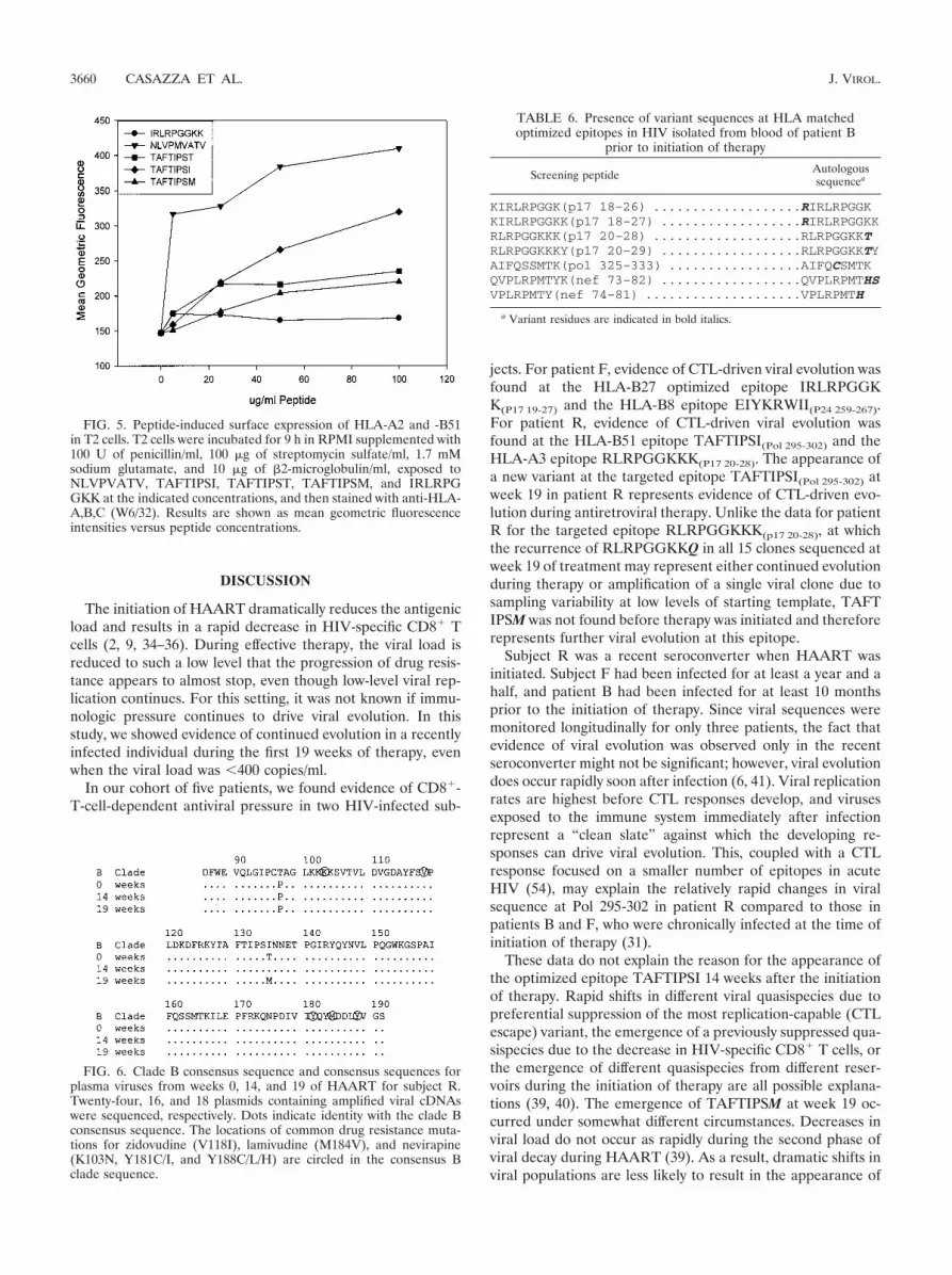
HLA class I stabilization studies were performed with 174 �CEM.T2 (T2) cells to investigate the binding of TAFTIPSI,TAFTIPST, and TAFTIPSM to HLA-B51 (Fig. 5). The cyto-megalovirus pp65 HLA-A2 immunodominant peptide NLVPMVATV (acting as a positive control), the HLA-B51 epitopeTAFTIPSI, the variants TAFTIPST and TAFTIPSM, and theHLA-B27 epitope IRLRPGGKK (acting as a negative control)were tested for the ability to stabilize HLA-A2 and -B51 mol-ecules. Incubations with TAFTIPSI and NLVPMVATVshowed the most HLA class I stabilization, as measured by thesurface staining of HLA with an anti-HLA-A,B,C monoclonalantibody (W6/32). The HLA stabilization level with TAFTIPST and TAFTIPSM was lower than that observed with TAF
TABLE 4. Frequencies of optimized epitope and variant epitope sequences in subject F before and after initiation of therapy for epitopesIRLRPGGKK(p17 19–27) and EIYKRWII(p24 128–135) based on viral RNA sequencing
Time (wks)in therapy Viral loada
Sequence (no. of clones/total)
IRLRPGGKK EIYKRWII
0 19,627 IRLRPGGKK (3/15), –––––––R– (12/15) EIYKRWII (0/17), D––––––– (12/17), –V–––––– (5/17)4 ND IRLRPGGKK (6/19), –––––––R– (13/19) EIYKRWII (0/22), D––––––– (22/22), –V–––––– (0/22)
12 �400 IRLRPGGKK (6/14), –––––––R– (8/14) EIYKRWII (0/14), D––––––– (14/14), –V–––––– (0/14)16 ND IRLRPGGKK (8/17), –––––––R– (9/17)20 ND IRLRPGGKK (0/17), –––––––R– (17/17)24 �400
a ND, not determined.
TABLE 5. Frequencies of optimized epitope and variant epitope sequences in subject R before and after initiation of therapy for epitopesRLRPGGKKK(p17 20–28) and TAFTIPSI(pol 295–302) based on viral RNA sequencing
Time (wks)in therapy Viral load
Sequence (no. of clones/total)
RLRPGGKKK TAFTIPSI
0 98,597 RLRPGGKKK (6/20) TAFTIPSI (0/24)––––––––Q (11/20) –––––––T (24/24)––––––––R (3/20) –––––––M (0/24)
9.5 47814 NDa RLRPGGKKK (14/16) TAFTIPSI (14/14)
––––––––Q (0/16) –––––––T (0/14)––––––––R (2/16) –––––––M (0/14)
19 �400 RLRPGGKKK (0/15) TAFTIPSI (0/18)––––––––Q (15/15) –––––––T (0/18)––––––––R (0/15) –––––––M (18/18)
a ND, not determined.
VOL. 79, 2005 IMMUNOLOGIC PRESSURE ON HIV EPITOPES DURING HAART 3657

TIPSI and NLVPMVATV but higher than that observed withthe HLA-B27 epitope IRLRPGGKK.
These data explain the similarities in the production ofIFN-� in response to stimulation by 2-�g/ml TAFTIPSI, TAFTIPST, and TAFTIPSM despite the substitution of a polar-CHOHCH3 group in the case of TAFTIPST and a
-C2H4SCH3 group in the case of TAFTIPSM for the-CH(CH3)C2H5 group of TAFTIPSI. At saturating concentra-tions of peptide, the effects of the differences in binding avidityfor TAFTIPSI, TAFTIPST, and TAFTIPSM were minimized.This masked the effects of substitutions of threonine and me-thionine for isoleucine at the carboxy-terminal anchor residue.
Lack of nucleoside reverse transcriptase resistance muta-tions. The primers used to sequence the pol gene allowed thesequencing of amino acids 241 to 347 (HXB2 sequence), whichcorrespond to amino acids 86 to 191 of reverse transcriptase(Fig. 6). In this 105-amino-acid sequence, the only point wherethe sequence changed over the 19 weeks of therapy in subjectR was at the terminal anchor position for the epitope TAFTIPSI. Subject R’s treatment regimen included combivir(zidovudine and lamivudine) and nevirapine. No evidence ofdrug resistance mutations for either lamivudine or nevirapinewas found during this period. While not all potential sitescorresponding to zidovudine resistance were sequenced, therewas no evidence of a resistance mutation to zidovudine foundat the V118 position. In addition, prior to the initiation oftherapy, eight plasma virus clones were sequenced, and therewas no evidence of zidovudine resistance mutations (M41L,E44D, D67N, or K70R) or multinucleoside reverse transcrip-tase complex mutations (A62V or the 69 insertion complex)(data not shown).
Lack of response to autologous peptides at other HLAmatched epitopes. To assess whether screening with peptideepitopes that differed from the autologous virus sequences ledto a failure to detect specific responses, we sequenced plasma
FIG. 1. Epitope-specific CD8� T-cell responses and autologous virus sequences from subject F. CD8� T-cell responses to screening peptidesand peptide variants are shown to the left in each graph. Epitope frequencies, determined by nucleic acid sequencing of viral clones, are shownto the right in each graph. Screening peptides and variant amino acid sequences are shown on the abscissa. None of the sequenced clones containedthe screening peptide EIYKRWII. Sequencing data and response data for the epitope IRLRPGGKK and its variant are from week 12 of therapy.Sequencing data are from 12 weeks and response data are from week 16 of therapy for EIYKRWII and its variants. An asterisk indicates that nonucleotide sequence corresponding to the screening epitope EIYKRWII was found in any viral sequence determined either prior to or after theinitiation of therapy.
FIG. 2. Plot of viral load and CD8� IFN-� production in responseto TAFTIPSI, TAFTIPST, and TAFTIPSM in patient R. CD8�-T-cellresponses to individual peptides and viral loads are plotted against thetime after the initiation of treatment. Responses to either TAFTIPSI,TAFTIPST, or TAFTIPSM were measured at the same time. Thearrows and text indicate the numbers of viral clones sequenced and thesequence found at Pol 295–302 at the indicated times. The peptideconcentration in all assays was 2 �g/ml.
3658 CASAZZA ET AL. J. VIROL.

viral RNAs from subject B that had been collected prior to theinitiation of therapy at the five regions for which we had PCRprimers (Table 1). These regions corresponded to amino acidsin p17 (4-149), p24 (232-348), Pol (240-345), Env (773-824),and Nef (1-161), or approximately 10% of the total HIV ge-nome. Based on the HLA type of subject B, eight potentialepitopes were identified for this subject. Seven variant se-
quences were found at those sites (Table 6). Each of thesepeptides was synthesized and used to assay CD8� T-cell re-sponses. As with the peptides used for the initial screen, noresponse was found to any of the variant peptides for thissubject (data not shown). Therefore, it is unlikely that the useof nonautologous virus sequences in this subject affected ourability to detect epitope-specific CD8� T cells.
FIG. 3. Contour plots showing peptide-specific CD3 and CD8 down-regulation in CD8� T cells from patient R in response to TAFTIPSI andother autologous viral epitopes 19 weeks after the initiation of HAART. PBMC were gated sequentially on small lymphocytes (a) and on smalllymphocytes and CD3 expression (b). Plots show CD3 or CD8 surface staining versus intracellular IFN-� staining. Cells were incubated in theabsence or presence of added peptide, as indicated. The peptide concentration was 2 �g/ml throughout. The geometric mean fluorescence for CD3or CD8 expression in responding CD8� T cells is given in the lower right corner of each contour plot.
FIG. 4. Plot of data showing frequency of CD8�-T-cell IFN-� production in response to various concentrations of TAFTIPSI, TAFTIPST, andTAFTIPSM. Plot a shows a sigmoidal fit of CD8� IFN-� production in response to various concentrations of TAFTIPSI (E) and TAFTIPST (F).The half-saturation point for TAFTIPSI was 15 nM and that for TAFTIPST was 90 nM. Responses shown are from PBMC prepared from blooddrawn during week 14 of treatment. Plot b shows CD8� IFN-� production in response to various concentrations of TAFTIPSI (E) and TAFTIPSM(F). Responses shown are from blood drawn during week 25 of treatment.
VOL. 79, 2005 IMMUNOLOGIC PRESSURE ON HIV EPITOPES DURING HAART 3659
DISCUSSION
The initiation of HAART dramatically reduces the antigenicload and results in a rapid decrease in HIV-specific CD8� Tcells (2, 9, 34–36). During effective therapy, the viral load isreduced to such a low level that the progression of drug resis-tance appears to almost stop, even though low-level viral rep-lication continues. For this setting, it was not known if immu-nologic pressure continues to drive viral evolution. In thisstudy, we showed evidence of continued evolution in a recentlyinfected individual during the first 19 weeks of therapy, evenwhen the viral load was �400 copies/ml.
In our cohort of five patients, we found evidence of CD8�-T-cell-dependent antiviral pressure in two HIV-infected sub-
jects. For patient F, evidence of CTL-driven viral evolution wasfound at the HLA-B27 optimized epitope IRLRPGGKK(P17 19-27) and the HLA-B8 epitope EIYKRWII(P24 259-267).For patient R, evidence of CTL-driven viral evolution wasfound at the HLA-B51 epitope TAFTIPSI(Pol 295-302) and theHLA-A3 epitope RLRPGGKKK(P17 20-28). The appearance ofa new variant at the targeted epitope TAFTIPSI(Pol 295-302) atweek 19 in patient R represents evidence of CTL-driven evo-lution during antiretroviral therapy. Unlike the data for patientR for the targeted epitope RLRPGGKKK(p17 20-28), at whichthe recurrence of RLRPGGKKQ in all 15 clones sequenced atweek 19 of treatment may represent either continued evolutionduring therapy or amplification of a single viral clone due tosampling variability at low levels of starting template, TAFTIPSM was not found before therapy was initiated and thereforerepresents further viral evolution at this epitope.
Subject R was a recent seroconverter when HAART wasinitiated. Subject F had been infected for at least a year and ahalf, and patient B had been infected for at least 10 monthsprior to the initiation of therapy. Since viral sequences weremonitored longitudinally for only three patients, the fact thatevidence of viral evolution was observed only in the recentseroconverter might not be significant; however, viral evolutiondoes occur rapidly soon after infection (6, 41). Viral replicationrates are highest before CTL responses develop, and virusesexposed to the immune system immediately after infectionrepresent a “clean slate” against which the developing re-sponses can drive viral evolution. This, coupled with a CTLresponse focused on a smaller number of epitopes in acuteHIV (54), may explain the relatively rapid changes in viralsequence at Pol 295-302 in patient R compared to those inpatients B and F, who were chronically infected at the time ofinitiation of therapy (31).
These data do not explain the reason for the appearance ofthe optimized epitope TAFTIPSI 14 weeks after the initiationof therapy. Rapid shifts in different viral quasispecies due topreferential suppression of the most replication-capable (CTLescape) variant, the emergence of a previously suppressed qua-sispecies due to the decrease in HIV-specific CD8� T cells, orthe emergence of different quasispecies from different reser-voirs during the initiation of therapy are all possible explana-tions (39, 40). The emergence of TAFTIPSM at week 19 oc-curred under somewhat different circumstances. Decreases inviral load do not occur as rapidly during the second phase ofviral decay during HAART (39). As a result, dramatic shifts inviral populations are less likely to result in the appearance of
FIG. 5. Peptide-induced surface expression of HLA-A2 and -B51in T2 cells. T2 cells were incubated for 9 h in RPMI supplemented with100 U of penicillin/ml, 100 �g of streptomycin sulfate/ml, 1.7 mMsodium glutamate, and 10 �g of 2-microglobulin/ml, exposed toNLVPVATV, TAFTIPSI, TAFTIPST, TAFTIPSM, and IRLRPGGKK at the indicated concentrations, and then stained with anti-HLA-A,B,C (W6/32). Results are shown as mean geometric fluorescenceintensities versus peptide concentrations.
FIG. 6. Clade B consensus sequence and consensus sequences forplasma viruses from weeks 0, 14, and 19 of HAART for subject R.Twenty-four, 16, and 18 plasmids containing amplified viral cDNAswere sequenced, respectively. Dots indicate identity with the clade Bconsensus sequence. The locations of common drug resistance muta-tions for zidovudine (V118I), lamivudine (M184V), and nevirapine(K103N, Y181C/I, and Y188C/L/H) are circled in the consensus Bclade sequence.
TABLE 6. Presence of variant sequences at HLA matchedoptimized epitopes in HIV isolated from blood of patient B
prior to initiation of therapy
Screening peptide Autologoussequencea
KIRLRPGGK(p17 18–26) ...................RIRLRPGGKKIRLRPGGKK(p17 18–27) ..................RIRLRPGGKKRLRPGGKKK(p17 20–28) ...................RLRPGGKKTRLRPGGKKKY(p17 20–29) ..................RLRPGGKKTYAIFQSSMTK(pol 325–333) .................AIFQCSMTKQVPLRPMTYK(nef 73–82) ..................QVPLRPMTHSVPLRPMTY(nef 74–81) ....................VPLRPMTH
a Variant residues are indicated in bold italics.
3660 CASAZZA ET AL. J. VIROL.

minor quasispecies. It is also unlikely that the presence ofTAFTIPSM, a rarer sequence than TAFTIPST, would be dueto a preexisting sequence in a recently infected individual,especially as this sequence was not detected prior to the initi-ation of HAART. This, coupled with the stepwise change innucleotide sequence observed in the transition from TAFTIPST to TAFTIPSI to TAFTIPSM, suggests that the emer-gence of TAFTIPSM represents a new variant.
Any mutation in the reverse transcriptase gene carries thepossibility of effecting antiretroviral drug resistance. No evi-dence of a clinically significant drug resistance mutation wasseen in patient R either by the criteria of the Drug ResistanceMutations Group of the International AIDS Society—USA(22) or from clinical data. Patient R has maintained an unde-tectable viral load for �4 years on the same treatment regi-men. Nonetheless, Brown et al. (7) have suggested that bothTAFTIPST and TAFTIPSM may decrease the sensitivity tononnucleoside reverse transcriptase inhibitors (NNRTIs).These authors screened 110 patients who had moderate reduc-tions in sensitivity to NNRTIs. Twenty-seven of these individ-uals had an I135T mutation. Only one patient had an I135Mmutation. In this patient, the I135M mutation occurred incombination with an E138A mutation, a site where mutationsare known to affect the sensitivity to NNRTIs (47). Resistancetest vectors using the consensus reverse transcriptase sequencewith either an I-to-T or I-to-M mutation at amino acid 135 ofthe reverse transcriptase gave a 2- or 2.6-fold change in resis-tance, respectively. A review of the Stanford HIV Drug Data-base (43) showed 10 patients with an I135M mutation and nomajor nevirapine resistance mutations based on sequencingresults. Resistance test vectors for these 10 patients showed amedian change in sensitivity of 1.3. These data suggest that anI135M mutation has a minimal or undetectable effect on drugsensitivity in patients with nonconsensus RT sequences.
Two recent papers have assessed the HIV-specific CD8�
T-cell response based on autologous viral sequences ratherthan consensus sequences. Lee et al. (28) suggested that theresponse to autologous sequences was lower than that ob-served with sequences closely related to the consensus B cladesequence. Altfeld et al. (3) suggested the opposite, i.e., thatresponses to autologous sequences occurred with a higher fre-quency than those to consensus sequences. With individualepitopes, we found both situations. However, our data alsodemonstrated the difficulty with using saturating peptide con-centrations in ex vivo assays to determine the physiologic sig-nificance of a change in sequence at any epitope. Despite therelatively small difference in the responses to TAFTIPSI, TAFTIPSM, and TAFTIPST at 2 �g of peptide/ml, three lines ofevidence suggest that TAFTIPST and TAFTIPSM are escapevariants. First, the down-regulation of CD3 expression waslarger in response to the optimized epitope TAFTIPSI than itwas for either of the variant peptides (TAFTIPSM and TAFTIPST). Second, titration experiments showed decreased re-sponses to TAFTIPST and TAFTIPSM compared to those toTAFTIPSI. Third, HLA stabilization experiments showed aweaker binding of TAFTIPST and TAFTIPSM to HLA-B51than that observed with the optimized epitope TAFTIPSI.These data, in combination with the viral sequencing datapresented, indicate that TAFTIPST and TAFTIPSM representescape variants in subject R. Such a conclusion would be hard
to make based on an isolated test. Our data show that usingpeptides at saturating concentrations increases the ability ofassays to identify responses to variants that differ from theconsensus sequence. Therefore, the best use of screening as-says with overlapping peptides based on consensus sequencesmay be to identify epitopes to which CD8� T cells are directedin vivo rather than to quantify these responses accurately. Ourfailure to find any additional responses when eight peptidesusing autologous sequences were substituted for consensusoptimized epitope sequences suggests that the consensus-based approach does identify most epitopes to which HIV-specific CD8� T cells are directed (Table 6).
A recent paper by Allen et al. (1) may explain the absence ofa response to the frequently recognized A3 epitopes KIRLRPGGKK(p17 19-27) and RLRPGGKKK(p17 20-28) in patient F.Allen et al. (1) showed that a K28Q mutation results in aneightfold increase in the 50% inhibitory concentration forbinding of this variant to the HLA-A3 molecule and also in-terferes with proteasome processing of the overlapping A3epitope KIRLRPGGKK(p17 19-27). At each of the five timepoints observed and in 82 viral sequences, a K28Q mutationwas found in the viruses sequenced from patient F (data notshown), suggesting that the K28Q mutation may be responsiblefor the absence of these common A3 responses.
Evidence of viral evolution at targeted CTL epitopes duringeffective antiretroviral treatment suggests that therapeutic vac-cination of recently infected individuals may be more success-ful than structured treatment interruptions. The developmentof escape variants occurs rapidly in the first months after in-fection (6, 41). It is therefore important to limit viral evolutionas quickly as possible in those who were recently infected.Structured treatment interruption does appear to enhance theimmunologic control of viral replication under some circum-stances (44), but as shown here, conditions exist long after theinitiation of treatment that allow the outgrowth of quasispeciescontaining escape variants. Therapeutic vaccination mightlimit this possibility by decreasing the opportunity for furtherviral evolution, although even with prolonged HAART, thismay not be absolute (16, 20, 55).
ACKNOWLEDGMENTS
This research was supported in part by NIH grant ROI-AI47603.D.A.P. is a Medical Research Council (UK) Clinician Scientist.
REFERENCES
1. Allen, T. M., M. Altfeld, X. G. Yu, K. M. O’Sullivan, M. Lichterfeld, S. LeGall, M. John, B. R. Mothe, P. K. Lee, E. T. Kalife, D. E. Cohen, K. A.Freedberg, D. A. Strick, M. N. Johnston, A. Sette, E. S. Rosenberg, S. A.Mallal, P. J. Goulder, C. Brander, and B. D. Walker. 2004. Selection, trans-mission, and reversion of an antigen-processing cytotoxic T-lymphocyte es-cape mutation in human immunodeficiency virus type 1 infection. J. Virol.78:7069–7078.
2. Alter, G., G. Hatzakis, C. M. Tsoukas, K. Pelley, D. Rouleau, R. LeBlanc,J. G. Baril, H. Dion, E. Lefebvre, R. Thomas, P. Cote, N. Lapointe, J. P.Routy, R. P. Sekaly, B. Conway, and N. F. Bernard. 2003. Longitudinalassessment of changes in HIV-specific effector activity in HIV-infected pa-tients starting highly active antiretroviral therapy in primary infection. J. Im-munol. 171:477–488.
3. Altfeld, M., M. M. Addo, R. Shankarappa, P. K. Lee, T. M. Allen, X. G. Yu,A. Rathod, J. Harlow, K. O’Sullivan, M. N. Johnston, P. J. Goulder, J. I.Mullins, E. S. Rosenberg, C. Brander, B. Korber, and B. D. Walker. 2003.Enhanced detection of human immunodeficiency virus type 1-specific T-cellresponses to highly variable regions by using peptides based on autologousvirus sequences. J. Virol. 77:7330–7340.
4. Barouch, D. H., S. Santra, J. E. Schmitz, M. J. Kuroda, T. M. Fu, W.
VOL. 79, 2005 IMMUNOLOGIC PRESSURE ON HIV EPITOPES DURING HAART 3661

Wagner, M. Bilska, A. Craiu, X. X. Zheng, G. R. Krivulka, K. Beaudry, M. A.Lifton, C. E. Nickerson, W. L. Trigona, K. Punt, D. C. Freed, L. Guan, S.Dubey, D. Casimiro, A. Simon, M. E. Davies, M. Chastain, T. B. Strom, R. S.Gelman, D. C. Montefiori, M. G. Lewis, E. A. Emini, J. W. Shiver, and N. L.Letvin. 2000. Control of viremia and prevention of clinical AIDS in rhesusmonkeys by cytokine-augmented DNA vaccination. Science 290:486–492.
5. Borrow, P., H. Lewicki, B. H. Hahn, G. M. Shaw, and M. B. Oldstone. 1994.Virus-specific CD8� cytotoxic T-lymphocyte activity associated with controlof viremia in primary human immunodeficiency virus type 1 infection. J. Vi-rol. 68:6103–6110.
6. Borrow, P., H. Lewicki, X. Wei, M. S. Horwitz, N. Peffer, H. Meyers, J. A.Nelson, J. E. Gairin, B. H. Hahn, M. B. Oldstone, and G. M. Shaw. 1997.Antiviral pressure exerted by HIV-1-specific cytotoxic T lymphocytes (CTLs)during primary infection demonstrated by rapid selection of CTL escapevirus. Nat. Med. 3:205–211.
7. Brown, A. J., H. M. Precious, J. M. Whitcomb, J. K. Wong, M. Quigg, W.Huang, E. S. Daar, R. T. D’Aquila, P. H. Keiser, E. Connick, N. S. Hellmann,C. J. Petropoulos, D. D. Richman, and S. J. Little. 2000. Reduced suscep-tibility of human immunodeficiency virus type 1 (HIV-1) from patients withprimary HIV infection to nonnucleoside reverse transcriptase inhibitors isassociated with variation at novel amino acid sites. J. Virol. 74:10269–10273.
8. Bunce, M., G. C. Fanning, and K. I. Welsh. 1995. Comprehensive, serolog-ically equivalent DNA typing for HLA-B by PCR using sequence-specificprimers (PCR-SSP). Tissue Antigens 45:81–90.
9. Casazza, J. P., M. R. Betts, L. J. Picker, and R. A. Koup. 2001. Decay kineticsof human immunodeficiency virus-specific CD8� T cells in peripheral bloodafter initiation of highly active antiretroviral therapy. J. Virol. 75:6508–6516.
10. DHHS Panel on Clinical Practices for Treatment of HIV Infection. 2001.Guidelines for use of antiretroviral agents in HIV-infected adults and ado-lescents. U.S. Department of Health and Human Services, Washington, D.C.
11. Douek, D. C., R. D. McFarland, P. H. Keiser, E. A. Gage, J. M. Massey, B. F.Haynes, M. A. Polis, A. T. Haase, M. B. Feinberg, J. L. Sullivan, B. D.Jamieson, J. A. Zack, L. J. Picker, and R. A. Koup. 1998. Changes in thymicfunction with age and during the treatment of HIV infection. Nature 396:690–695.
12. Evans, D. T., D. H. O’Connor, P. Jing, J. L. Dzuris, J. Sidney, J. da Silva,T. M. Allen, H. Horton, J. E. Venham, R. A. Rudersdorf, T. Vogel, C. D.Pauza, R. E. Bontrop, R. DeMars, A. Sette, A. L. Hughes, and D. I. Watkins.1999. Virus-specific cytotoxic T-lymphocyte responses select for amino-acidvariation in simian immunodeficiency virus Env and Nef. Nat. Med. 5:1270–1276.
13. Furtado, M. R., D. S. Callaway, J. P. Phair, K. J. Kunstman, J. L. Stanton,C. A. Macken, A. S. Perelson, and S. M. Wolinsky. 1999. Persistence ofHIV-1 transcription in peripheral-blood mononuclear cells in patients re-ceiving potent antiretroviral therapy. N. Engl. J. Med. 340:1614–1622.
14. Gottlieb, M. S., R. Schroff, H. M. Schanker, J. D. Weisman, P. T. Fan, R. A.Wolf, and A. Saxon. 1981. Pneumocystis carinii pneumonia and mucosalcandidiasis in previously healthy homosexual men: evidence of a new ac-quired cellular immunodeficiency. N. Engl. J. Med. 305:1425–1431.
15. Goulder, P. J., R. E. Phillips, R. A. Colbert, S. McAdam, G. Ogg, M. A.Nowak, P. Giangrande, G. Luzzi, B. Morgan, A. Edwards, A. J. McMichael,and S. Rowland-Jones. 1997. Late escape from an immunodominant cyto-toxic T-lymphocyte response associated with progression to AIDS. Nat. Med.3:212–217.
16. Gunthard, H. F., S. D. Frost, A. J. Leigh-Brown, C. C. Ignacio, K. Kee, A. S.Perelson, C. A. Spina, D. V. Havlir, M. Hezareh, D. J. Looney, D. D. Rich-man, and J. K. Wong. 1999. Evolution of envelope sequences of humanimmunodeficiency virus type 1 in cellular reservoirs in the setting of potentantiviral therapy. J. Virol. 73:9404–9412.
17. Hansen, T., and N. Myers. 2003. Peptide induction of surface expression ofclass I MHC, p. 18.11.1–18.11.8. In J. E. Colgan, A. M. Kruisbeer, D. H.Marguiles, and W. Strober (ed.), Current protocols in immunology, vol. 4.John Wiley & Sons, New York, N.Y.
18. Havlir, D. V., R. Bassett, D. Levitan, P. Gilbert, P. Tebas, A. C. Collier, M. S.Hirsch, C. Ignacio, J. Condra, H. F. Gunthard, D. D. Richman, and J. K.Wong. 2001. Prevalence and predictive value of intermittent viremia withcombination HIV therapy. JAMA 286:171–179.
19. Hermankova, M., S. C. Ray, C. Ruff, M. Powell-Davis, R. Ingersoll, R. T.D’Aquila, T. C. Quinn, J. D. Siliciano, R. F. Siliciano, and D. Persaud. 2001.HIV-1 drug resistance profiles in children and adults with viral load of �50copies/ml receiving combination therapy. JAMA 286:196–207.
20. Izopet, J., M. Cazabat, C. Pasquier, K. Sandres-Saune, E. Bonnet, B. Mar-chou, P. Massip, and J. Puel. 2002. Evolution of total and integrated HIV-1DNA and change in DNA sequences in patients with sustained plasma virussuppression. Virology 302:393–404.
21. Jin, X., D. E. Bauer, S. E. Tuttleton, S. Lewin, A. Gettie, J. Blanchard, C. E.Irwin, J. T. Safrit, J. Mittler, L. Weinberger, L. G. Kostrikis, L. Zhang, A. S.Perelson, and D. D. Ho. 1999. Dramatic rise in plasma viremia after CD8(�)T cell depletion in simian immunodeficiency virus-infected macaques. J. Exp.Med. 189:991–998.
22. Johnson, V. A., F. Brun-Vezinet, B. Clotet, B. Conway, R. T. D’Aquila, L. M.Demeter, D. R. Kuritzkes, D. Pillay, J. M. Schapiro, A. Telenti, and D. D.
Richman. 2003. Drug resistance mutations in HIV-1. Top. HIV Med. 11:215–221.
23. Kelleher, A. D., C. Long, E. C. Holmes, R. L. Allen, J. Wilson, C. Conlon, C.Workman, S. Shaunak, K. Olson, P. Goulder, C. Brander, G. Ogg, J. S.Sullivan, W. Dyer, I. I. Jones, A. J. McMichael, S. Rowland-Jones, and R. E.Phillips. 2001. Clustered mutations in HIV-1 gag are consistently requiredfor escape from HLA-B27-restricted cytotoxic T lymphocyte responses. J.Exp. Med. 193:375–386.
24. Kern, F., I. P. Surel, C. Brock, B. Freistedt, H. Radtke, A. Scheffold, R.Blasczyk, P. Reinke, J. Schneider-Mergener, A. Radbruch, P. Walden, andH. D. Volk. 1998. T-cell epitope mapping by flow cytometry. Nat. Med.4:975–978.
25. Koenig, S., A. J. Conley, Y. A. Brewah, G. M. Jones, S. Leath, L. J. Boots, V.Davey, G. Pantaleo, J. F. Demarest, C. Carter, et al. 1995. Transfer ofHIV-1-specific cytotoxic T lymphocytes to an AIDS patient leads to selectionfor mutant HIV variants and subsequent disease progression. Nat. Med.1:330–336.
26. Koup, R. A., J. T. Safrit, Y. Cao, C. A. Andrews, G. McLeod, W. Borkowsky,C. Farthing, and D. D. Ho. 1994. Temporal association of cellular immuneresponses with the initial control of viremia in primary human immunode-ficiency virus type 1 syndrome. J. Virol. 68:4650–4655.
27. Learn, G. H., Jr., B. T. Korber, B. Foley, B. H. Hahn, S. M. Wolinsky, andJ. I. Mullins. 1996. Maintaining the integrity of human immunodeficiencyvirus sequence databases. J. Virol. 70:5720–5730.
28. Lee, S. K., Z. Xu, J. Lieberman, and P. Shankar. 2002. The functional CD8T cell response to HIV becomes type-specific in progressive disease. J. Clin.Investig. 110:1339–1347.
29. Liu, S. L., J. E. Mittler, D. C. Nickle, T. M. Mulvania, D. Shriner, A. G.Rodrigo, B. Kosloff, X. He, L. Corey, and J. I. Mullins. 2002. Selection forhuman immunodeficiency virus type 1 recombinants in a patient with rapidprogression to AIDS. J. Virol. 76:10674–10684.
30. Masur, H., M. A. Michelis, J. B. Greene, I. Onorato, R. A. Stouwe, R. S.Holzman, G. Wormser, L. Brettman, M. Lange, H. W. Murray, and S.Cunningham-Rundles. 1981. An outbreak of community-acquired Pneumo-cystis carinii pneumonia: initial manifestation of cellular immune dysfunc-tion. N. Engl. J. Med. 305:1431–1438.
31. McMichael, A. J., and R. E. Phillips. 1997. Escape of human immunodefi-ciency virus from immune control. Annu. Rev. Immunol. 15:271–296.
32. McNaghten, A. D., D. L. Hanson, J. L. Jones, M. S. Dworkin, and J. W.Ward. 1999. Effects of antiretroviral therapy and opportunistic illness pri-mary chemoprophylaxis on survival after AIDS diagnosis. Adult/AdolescentSpectrum of Disease Group. AIDS 13:1687–1695.
33. Moore, C. B., M. John, I. R. James, F. T. Christiansen, C. S. Witt, and S. A.Mallal. 2002. Evidence of HIV-1 adaptation to HLA-restricted immuneresponses at a population level. Science 296:1439–1443.
34. Ogg, G. S., X. Jin, S. Bonhoeffer, P. Moss, M. A. Nowak, S. Monard, J. P.Segal, Y. Cao, S. L. Rowland-Jones, A. Hurley, M. Markowitz, D. D. Ho, A. J.McMichael, and D. F. Nixon. 1999. Decay kinetics of human immunodefi-ciency virus-specific effector cytotoxic T lymphocytes after combination an-tiretroviral therapy. J. Virol. 73:797–800.
35. Ortiz, G. M., D. F. Nixon, A. Trkola, J. Binley, X. Jin, S. Bonhoeffer, P. J.Kuebler, S. M. Donahoe, M. A. Demoitie, W. M. Kakimoto, T. Ketas, B. Clas,J. J. Heymann, L. Zhang, Y. Cao, A. Hurley, J. P. Moore, D. D. Ho, and M.Markowitz. 1999. HIV-1-specific immune responses in subjects who tempo-rarily contain virus replication after discontinuation of highly active antiret-roviral therapy. J. Clin. Investig. 104:R13–R18.
36. Oxenius, A., A. R. McLean, M. Fischer, D. A. Price, S. J. Dawson, R. Hafner,C. Schneider, H. Joller, B. Hirschel, R. E. Phillips, R. Weber, and H. F.Gunthard. 2002. Human immunodeficiency virus-specific CD8� T-cell re-sponses do not predict viral growth and clearance rates during structuredintermittent antiretroviral therapy. J. Virol. 76:10169–10176.
37. Pantaleo, G., J. F. Demarest, T. Schacker, M. Vaccarezza, O. J. Cohen, M.Daucher, C. Graziosi, S. S. Schnittman, T. C. Quinn, G. M. Shaw, L. Perrin,G. Tambussi, A. Lazzarin, R. P. Sekaly, H. Soudeyns, L. Corey, and A. S.Fauci. 1997. The qualitative nature of the primary immune response to HIVinfection is a prognosticator of disease progression independent of the initiallevel of plasma viremia. Proc. Natl. Acad. Sci. USA 94:254–258.
38. Pantaleo, G., J. F. Demarest, H. Soudeyns, C. Graziosi, F. Denis, J. W.Adelsberger, P. Borrow, M. S. Saag, G. M. Shaw, R. P. Sekaly, et al. 1994.Major expansion of CD8� T cells with a predominant V beta usage duringthe primary immune response to HIV. Nature 370:463–467.
39. Perelson, A. S., P. Essunger, Y. Cao, M. Vesanen, A. Hurley, K. Saksela, M.Markowitz, and D. D. Ho. 1997. Decay characteristics of HIV-1-infectedcompartments during combination therapy. Nature 387:188–191.
40. Perelson, A. S., A. U. Neumann, M. Markowitz, J. M. Leonard, and D. D. Ho.1996. HIV-1 dynamics in vivo: virion clearance rate, infected cell life-span,and viral generation time. Science 271:1582–1586.
41. Price, D. A., P. J. Goulder, P. Klenerman, A. K. Sewell, P. J. Easterbrook, M.Troop, C. R. Bangham, and R. E. Phillips. 1997. Positive selection of HIV-1cytotoxic T lymphocyte escape variants during primary infection. Proc. Natl.Acad. Sci. USA 94:1890–1895.
42. Ramratnam, B., J. E. Mittler, L. Zhang, D. Boden, A. Hurley, F. Fang, C. A.
3662 CASAZZA ET AL. J. VIROL.

Macken, A. S. Perelson, M. Markowitz, and D. D. Ho. 2000. The decay of thelatent reservoir of replication-competent HIV-1 is inversely correlated withthe extent of residual viral replication during prolonged anti-retroviral ther-apy. Nat. Med. 6:82–85.
43. Rhee, S. Y., M. J. Gonzales, R. Kantor, B. J. Betts, J. Ravela, and R. W.Shafer. 2003. Human immunodeficiency virus reverse transcriptase and pro-tease sequence database. Nucleic Acids Res. 31:298–303.
44. Rosenberg, E. S., M. Altfeld, S. H. Poon, M. N. Phillips, B. M. Wilkes, R. L.Eldridge, G. K. Robbins, R. T. D’Aquila, P. J. R. Goulder, and B. D. Walker.2000. Immune control of HIV-1 after early treatment of acute infection.Nature 407:523–526.
45. Ruff, C. T., S. C. Ray, P. Kwon, R. Zinn, A. Pendleton, N. Hutton, R.Ashworth, S. Gange, T. C. Quinn, R. F. Siliciano, and D. Persaud. 2002.Persistence of wild-type virus and lack of temporal structure in the latentreservoir for human immunodeficiency virus type 1 in pediatric patients withextensive antiretroviral exposure. J. Virol. 76:9481–9492.
46. Salter, R. D., D. N. Howell, and P. Cresswell. 1985. Genes regulating HLAclass I antigen expression in T-B lymphoblast hybrids. Immunogenetics 21:235–246.
47. Schinazi, R. F., B. A. Larder, and J. W. Mellors. 1996. Mutations in retroviralgenes associated with drug resistance. Int. Antiviral News 4:95–107.
48. Schmitz, J. E., M. J. Kuroda, S. Santra, V. G. Sasseville, M. A. Simon, M. A.Lifton, P. Racz, K. Tenner-Racz, M. Dalesandro, B. J. Scallon, J. Ghrayeb,M. A. Forman, D. C. Montefiori, E. P. Rieber, N. L. Letvin, and K. A.Reimann. 1999. Control of viremia in simian immunodeficiency virus infec-tion by CD8� lymphocytes. Science 283:857–860.
49. Sipsas, N. V., S. A. Kalams, A. Trocha, S. He, W. A. Blattner, B. D. Walker,and R. P. Johnson. 1997. Identification of type-specific cytotoxic T lympho-cyte responses to homologous viral proteins in laboratory workers acciden-tally infected with HIV-1. J. Clin. Investig. 99:752–762.
50. Smith, D. B., J. McAllister, C. Casino, and P. Simmonds. 1997. Virus“quasispecies”: making a mountain out of a molehill? J. Gen. Virol. 78:1511–1519.
51. Theoretical Biology and Biophysics Group. 1999. Human retroviruses andAIDS: a compilation and analysis of nucleic acid and amino acid sequences.Theoretical Biology and Biophysics Group T-10, Los Alamos, N.Mex.
52. U.S. Public Health Service. 2001. 2001 USPHS/IDSA guidelines for theprevention of opportunistic infections in persons infected with human im-munodeficiency virus. U.S. Public Health Service, Washington, D.C.
53. Valitutti, S., S. Muller, M. Dessing, and A. Lanzavecchia. 1996. Differentresponses are elicited in cytotoxic T lymphocytes by different levels of T cellreceptor occupancy. J. Exp. Med. 183:1917–1921.
54. Yu, X. G., M. M. Addo, E. S. Rosenberg, W. R. Rodriguez, P. K. Lee, C. A.Fitzpatrick, M. N. Johnston, D. Strick, P. J. Goulder, B. D. Walker, and M.Altfeld. 2002. Consistent patterns in the development and immunodomi-nance of human immunodeficiency virus type 1 (HIV-1)-specific CD8� T-cell responses following acute HIV-1 infection. J. Virol. 76:8690–8701.
55. Zhang, L., B. Ramratnam, K. Tenner-Racz, Y. He, M. Vesanen, S. Lewin, A.Talal, P. Racz, A. S. Perelson, B. T. Korber, M. Markowitz, and D. D. Ho.1999. Quantifying residual HIV-1 replication in patients receiving combina-tion antiretroviral therapy. N. Engl. J. Med. 340:1605–1613.
VOL. 79, 2005 IMMUNOLOGIC PRESSURE ON HIV EPITOPES DURING HAART 3663