immortalized human brain endothelial cells and flow-based vascular modeling: a marriage of...
TRANSCRIPT
Immortalized human brain endothelial cells andflow-based vascular modeling: a marriage ofconvenience for rational neurovascular studies
Luca Cucullo1,2, Pierre-Olivier Couraud3,4,5,6, Babette Weksler4,7, Ignacio-Andres Romero8,Mohammed Hossain1,2, Edward Rapp9 and Damir Janigro1,2,10
1Division of Cerebrovascular Research, Cleveland Clinic Lerner College of Medicine, Cleveland, Ohio, USA;2Department of Neurosurgery, Cleveland Clinic Lerner College of Medicine, Cleveland, Ohio, USA;3Department of Cell Biology, Institut Cochin, Paris, France; 4Inserm, U567, Paris, France; 5CNRS, UMR 8104,Paris, France; 6Universite Paris 5, Faculte de Medecine Rene Descartes, UM 3, Paris, France; 7Department ofMedicine, Weill Medical College, New York, New York, USA; 8Department of Biological Sciences, The OpenUniversity, Milton Keynes, UK; 9Flocel Inc., Cleveland, Ohio, USA; 10Department of Molecular Medicine,Cleveland Clinic Lerner College of Medicine, Cleveland, Ohio, USA
In evaluating drugs that enter or are excluded from the brain, novel pharmaceutical strategies areneeded. For this reason, we have developed a humanized Dynamic In vitro Blood–Brain Barriermodel (hDIV-BBB) based on a novel human brain vascular endothelial cell line (HCMEC/D3), whichclosely mimics the BBB in vivo. In this system, HCMEC/D3 was grown in the lumen of hollowmicroporous fibers and exposed to a physiological pulsatile flow. Comparison with well-establishedhumanized DIV-BBB models (based on human brain and non-brain vascular endothelial cellsco-cultured with abluminal astrocytes) demonstrated that HCMEC/D3 cells cultured under flowconditions maintain in vitro physiological permeability barrier properties of the BBB in situ even inthe absence of abluminal astrocytes. Measurements of glucose metabolism demonstrated thatHCMEC/D3 cells retain an aerobic metabolic pathway. Permeability to sucrose and two relevantcentral nervous system drugs showed that the HCMEC/D3 cells grown under dynamic conditionsclosely mimic the physiological permeability properties of the BBB in situ (slope = 0.93). Osmoticdisruption of the BBB was also successfully achieved. Peak BBB opening in the DIV-BBB lastedfrom 20 to 30 mins and was completely reversible. Furthermore, the sequence of flow cessation/reperfusion in the presence of leukocytes led to BBB failure as demonstrated by a biphasic decreasein transendothelial electrical resistance. Additionally, BBB failure was paralleled by the intraluminalrelease of proinflammatory factors (interleukin-6 and interleukin-1b) and matrix metalloproteinase-9(MMP-9). Pretreatment with ibuprofen (0.125 mmol/L) prevented BBB failure by decreasing theinflammatory response after flow cessation/reperfusion.Journal of Cerebral Blood Flow & Metabolism advance online publication, 4 July 2007; doi:10.1038/sj.jcbfm.9600525
Keywords: drug delivery; drug discovery; drug resistance; pharmacodynamic; shear stress
Introduction
The concept of a ‘neurovascular unit’ has reacheddeserved prominence in basic and clinical neuro-science. Understanding the neurobiology of humandisease requires simultaneous studies of variouscell types (e.g., neurons, endothelium, glia, andwhite blood cells) as well as fluid phase factors(adhesion molecules, cytokines, proinflammatoryfactors, and intravascular shearing forces). Theblood–brain barrier (BBB) exemplifies the impor-tance of this recent approach to neuroscience. Lossof BBB structural integrity and function plays a
Received 18 December 2006; revised 17 May 2007; accepted 22May 2007
Correspondence: Dr D Janigro, Department of CerebrovascularResearch, Cleveland Clinic Foundation NB-20 LRI 9500 EuclidAve, Cleveland, Ohio 44195, USA. E-mail: [email protected]
This work was supported by Alternative Research Development
Foundation (ARDF) and Philip Morris USA and Philip Morris
International external research awards to Dr Luca Cucullo and
was also supported by NIH-2RO1 HL51614, NIH-RO1 NS43284
NIH-RO1 NS38195 and Philip Morris USA and Philip Morris
International external research awards to Damir Janigro.
Journal of Cerebral Blood Flow & Metabolism (2007), 1–17& 2007 ISCBFM All rights reserved 0271-678X/07 $30.00
www.jcbfm.com
pivotal role in the pathogenesis of many diseasesof the central nervous system (CNS). The BBB isaltered in many clinical settings including braintrauma (Unterberg et al, 2004), focal brain ischemia(Latour et al, 2004), meningitis (van der et al, 2004),brain tumor (Lee et al, 2006), stroke (Cipollaet al, 2004), inflammation (Stamatovic et al, 2006),Alzheimer’s disease (Kalaria, 1992), and multiplesclerosis (Minagar and Alexander, 2003). In is-chemic brain injury, matrix metalloproteinases(MMPs) contribute to the disruption of the BBBleading to vasogenic edema, and to the influx ofleukocytes into the CNS (Gasche et al, 2006). Incontrast, it has been recently demonstrated that HIVpenetration into the brain does not involve thedisruption of the BBB. Instead, immune cells,viruses, and viral proteins probably activate brainendothelial cells and enable their own passageacross the BBB by way of highly regulated processessuch as diapedesis and adsorptive endocytosis(Banks et al, 2006). Therefore, understanding howthe BBB might be affected by various pathogenic ordrug factors holds significant promises for thedevelopment of novel pharmacological therapies.
Although the failure of the BBB plays a primaryrole in the pathogenesis of many CNS diseases,normal BBB structural and functional integrity(which maintains the homeostasis of the CNSenvironment (Abbott, 2002)) may severely hinderthe delivery of drugs into the brain. The presence ofactive carrier-mediated transport of substrates fromthe brain to the blood is, in fact, one of the majorfeatures of the BBB. By preventing the passage ofdrugs that are actively extruded by carriers such asP-glycoprotein and multidrug resistance-associatedproteins (a phenomenon known as multidrug resis-tance), the BBB represents a critical obstacle forthe treatment of neurologic diseases (Kubota et al,2006). Developing solutions to augment the minimalingress through the BBB of these potential CNSdrugs is essential to improve neurotherapeutics(Pardridge, 2005).
Given these premises, the development of an invitro model of the human BBB that recapitulates thephysiologic features of the BBB in situ (includingthe presence of intraluminal flow such as thedynamic in vitro BBB model or DIV-BBB) representsan important improvement over the classical staticculture system (Santaguida et al, 2006). This modelhas been further enhanced by using primary brainendothelial and glial cells from rats (Krizanac-Bengez et al, 2003) as well as cells from humanbrain tissue resected from patients undergoing brainsurgery for aneurysm or intractable epilepsy. Thesetechnical advances allowed to establish the firsthuman disease-based DIV-BBB capable of mimick-ing the pathophysiologic properties of its in vivocounterpart (Cucullo et al, 2007).
The major obstacle toward the development ofhumanized models has been the difficulty ofisolating primary brain endothelial and glial cells,
which not only are difficult to obtain in quantity, butalso have to be freshly isolated each time before use.Given these technical difficulties, we decided toturn to an immortalized human brain microvesselendothelial cell line (HCMEC/D3) (Weksler et al,2005), which we show in this report provides anexcellent alternative to primary cells especiallywhen cultured under dynamic (flow-based)conditions. This cell line expresses specific brainendothelial properties, cell surface adhesionmolecules, and junctional markers, and does notrequire the presence of abluminal glial cells to forma physiological as well as anatomical BBB (Weksleret al, 2005). Because of their stable properties andindefinite growth potential, HCMEC/D3 cells, un-like primary brain microvascular endothelial cells(HBMECs), provide a long-lasting source of humanbrain endothelial cells.
In this report, we have measured numerouscritical parameters to determine if combining theDIV-BBB technology with HCMEC/D3 cells pro-vided a system that could be reliably used to assessthe permeability of CNS drugs and to act as aparadigm to study the functional biology of the BBB.For example, we used HCMEC/D3 cells grown as adifferentiated monolayer in the DIV-BBB to evaluatethe efficacy of currently used clinical protocols suchas the osmotic opening of the BBB (which is used toenhance the passage of chemotherapic agents forthe treatment of brain tumors) (Rapoport, 2000). Inaddition, we performed experiments to assess ifHCMEC/D3 mimics the in vivo physiological re-sponses to an ischemic insult (i.e., the BBB failureafter inflammation). This was assessed by imposingflow cessation/reperfusion in the presence of humanwhite blood cells (WBCs).
Materials and methods
Cell Culture
Human brain microvascular endothelial cells (cat. no.6100), human umbilical vein endothelial cells (HUVECs,cat. no. 8000), and human astrocytes (HAs, cat. no. 1800)were purchased from ScienCell Research Laboratories(San Diego, CA, USA). AVM-ECs were isolated fromarteriovenous malformations surgically removed frompatients. Human brain microvascular endothelial cell,HUVEC, and AVM-EC were initially expanded in 75 cm2
flasks precoated with fibronectin (3mg/cm2) with theappropriate growth medium consisting of 500 mL of basalmedium, 25 mL of fetal bovine serum (ScienCell ResearchLaboratories, cat. no. 0025), 5 mL of endothelial cellgrowth supplement (cat. no.; 1052), 100 U/mL penicillinG sodium, and 100 mg/mL streptomycin sulfate.
In addition, we used a novel immortalized human brainmicrovessel endothelial cell line HCMEC/D3, whichretains the morphological characteristics of primary brainendothelial cells and expresses specific brain endothelialmarkers and cell surface adhesion molecules (Weksleret al, 2005). HCMEC/D3 was grown in 75 cm2 flasks
BBB models for CNS drug permeability studiesL Cucullo et al
2
Journal of Cerebral Blood Flow & Metabolism (2007), 1–17

precoated with 5mg/cm2 of rat tail collagen type I (BDBiosciences, cat. no. 354236) in EGM-2 medium (Cambrex,cat. no. CC-4176). HAs were grown in poly-D-lysineprecoated flasks (3mg/cm2 in Dulbecco’s modified essen-tial medium (DMEM-F12) supplemented with 2 mmolglutamine, 5% fetal bovine serum, 100 U of penicillin Gsodium per mL, and 100mg of streptomycin sulfate permL). All cells were maintained at 371C in humidified airwith 5% CO2. Cellular growth was monitored every day byinspection using phase-contrast microscopy.
Dynamic In vitro Blood–Brain Barrier Setup
The DIV-BBB modules used for the experiments describedherein were purchased from Spectrum (cat. no. 400-025,Spectrum Laboratories Inc., Rancho Dominguez, CA,USA). Each module consists of a bundle of 50 hollowpolypropylene fibers embedded in a clear plastic chamberthat provides an accessible space around the fibers (Figure1A). Cells are seeded inside the hollow fibers (see below).The porous walls of the hollow fibers allow gas andnutrient exchange between the luminal and the abluminal(extracellular space, ECS) compartments, but do notpermit the passage of cells between the two compart-ments. Both luminal and abluminal areas are accessibleby ports that connect to a circuit containing a mediumreservoir and a pulsatile pump apparatus (CompleteCellMaxs Quad. cat. no. CMQUAD-C, Spectrum Labora-
tories Inc.). Gaseous exchange (O2 and CO2) occursthrough the gas-permeable silicone tubing that connectsthe DIV-BBB cartridge and the medium reservoir (Figure1A). Four electrodes are positioned in the cartridge, onepair in the luminal and one pair in the abluminalcompartments that connect to a computerized monitoringsystem, which allows for real-time measurements oftransendothelial electrical resistance (TEER). The pulsa-tile pump circulates the medium through the lumen of theartificial capillaries. Nutrients are exchanged betweenlumen and ECS by diffusion through the 0.5mm transca-pillary pores. The pump can be adjusted to produce flowof medium ranging from 1 to 50 ml/min (corresponding toshear stress levels of approximately 1 to 200 dyn/cm2). Theentire apparatus is placed inside a water-jacketed incu-bator at 371C with 5% CO for optimal culture conditions.
The lumens of the hollow fibers were precoated withfibronectin (3 mg/cm2) for HBMEC, HUVEC, and AVM-ECor with 5mg/cm2 of rat tail collagen type I for HCMEC/D3to enhance cell attachment and proliferation. The ablum-inal side of the fibers was precoated with poly-D-lysine(3mg/cm2) to improve HA adhesion.
Human endothelial cells were seeded intraluminallyinside the fibers (E4� 106/cartridge) and allowed toexpand for 14 days in the presence of astrocytes(E6� 106/cartridge) placed in the abluminal compartmentfor HBMEC-, HUVEC-, and AVM-EC-based models orwithout astrocytes for HCMEC/D3 models. To facilitatecell adhesion, we used an initial flow rate of 1 mL/min for
Figure 1 Diagrammatic representation of the DIV-BBB and Transwell systems. (A) A bundle of porous polypropylene hollow fibers issuspended in the DIV-BBB chamber. Note that the hollow fibers are in continuity with a medium source through a flow pathconsisting of gas-permeable silicon tubing. Two three-way stopcocks positioned on either side of the module regulate the access tothe luminal compartment. Detailed dimensional and functional characteristics are shown in the table. (B) The Transwell systemconsists of endothelial cells grown on microporous membranes, whereas astrocytes are seeded on the underside of the samemembrane and release soluble factors, which preserve some of the BBB properties. This system allows for study of bidirectionaltransport across the BBB. See adjacent table for more details.
BBB models for CNS drug permeability studiesL Cucullo et al
3
Journal of Cerebral Blood Flow & Metabolism (2007), 1–17

the initial 48 h after cell inoculation. The flow rate wasthen increased to a steady-state level of 4 mL/min (i.e.,4 dyn/cm2). Medium samples from the luminal andabluminal compartments were taken every 2 days andprocessed to assess for glucose consumption and lactateproduction.
Transwell Systems
Cells were co-cultured using sets of 12-well Transwell-Clear Polyester Membrane plates (Costar cat. no. 3460),which feature a vertical side-by-side diffusion systemthrough a thin, microscopically transparent polyestermembrane of 12 mm diameter and 0.4mm pore sizemounted on a double chamber (Figure 1B). The samecoating materials were utilized as in the DIV-BBB toenhance cell attachment. Human endothelial cells(HCMEC/D3, HBMEC, HUVEC, and AVM-EC) were seededon the top side of the membrane and allowed to establishthemselves for 3 days. Human astrocytes were then seededon the underside of the filter (co-cultures only). Thenumber of cells seeded ranged from 1� 105 for humanendothelial cells to 1.5� 105 for human astrocytes.Transendothelial electrical resistance was monitoredapproximately every 2 days beginning at ‘day 1’ of co-culture. The equipment for measuring TEER consisted of atissue resistance measurement chamber (Endohm cham-ber, WPI), which provides a reproducible electricalresistance measurement of endothelial tissue culture cups.In conjunction with TEER monitoring, the integrity of theendothelial monolayer was directly assessed by inspec-tion using phase-contrast microscopy (data not shown).
Cell Metabolism: Lactate Production and GlucoseConsumption
In conjunction with TEER monitoring, depletion of themain carbohydrate component of the growth medium(glucose) and accumulation of metabolically producedlactic acid were used as indicators of cell growth and theassessment of a viable in vitro BBB (Cucullo et al, 2007;Santaguida et al, 2006). Both the luminal and abluminalcompartments (in both DIV-BBB and Transwell) weresampled at 2-day intervals. The calculations for glucoseconsumption (mg/day) and lactate production rates (mg/day)herein reported were based on medium replacement,volume of non-replaced medium and previous values.Glucose consumption rate was calculated based on theconcentration of glucose in fresh and non-replacedmedium in the system, according to the followingequation:
ðVn�GnÞ þ ðVo�GpÞ � ðVt�GcÞTc � Tp
ð1Þ
where V represents added volumes of medium (mL), G isthe glucose concentration (mg/mL), T is the time ofsampling (in fractions of days), ‘c’ and ‘p’ indicate thecurrent, and the previous samples, respectively), ‘n’represents the fresh (new) medium added after previoussampling, ‘o’ corresponds to the old, unreplaced medium,
and ‘t’ represents the total volume of medium. Lactateproduction rate (mg/day) was calculated similarly:
ðVt�LcÞ � ðVn�LnÞ þ ðVu�LpÞTc � Tp
ð2Þ
(L) refers to the concentration of lactic acid in mg/mL and‘u’ corresponds to the unreplaced volume of media. Adual-channel immobilized oxidase enzyme biochemistryapparatus (YSI 2700 SELECT, YSI Inc., Yellow Springs,OH, USA) was used to measure lactate and glucose in theculture medium. Data obtained with the describedequation were then converted to mmol/L per day.
Dynamic In vitro Blood–Brain BarrierTransendothelial Electrical ResistanceMeasurement System
The TEER measurement provides a quick and easyevaluation of the integrity of the BBB (Cucullo et al,2002, 2007; Santaguida et al, 2006). We used a newlydeveloped TEER measurement device (Flocel Inc., Cleve-land, OH, USA) (Parkinson et al, 2003), which utilizeselectronic multiplexing to measure multiple cartridges inquick succession and to assess the integrity and viabilityof tissue culture monolayers and bilayers rapidly andreliably (Figure 1C). The device uses a Universal SerialBus interface to a PC computer. To sample TEER, theexcitation voltage (0.06 V) is applied across the excitationelectrodes inserted in each cartridge. The microcontrollercomputes the resistivity and capacitance per cm2 of thebarrier from physical parameters. The values of capaci-tance are calculated by comparison of the voltage andcurrent waveforms. The delay from peak to peak of the twowaveforms is proportional to the capacitance value, whichis expressed as archtension. Transendothelial electricalresistance was measured continuously from the initialsetup throughout the course of each experiment.
Drug Permeability: Uptake of [14C]Phenytoin,[14C]Diazepam, and [3H]Sucrose
Concentrated Boluses (0.5 mL each) of the radioactivetracers [14C]phenytoin (PerkinElmer, Boston, MA, USA;cat. no. NEC-246), [14C]diazepam (Amersham, Piscataway,NJ, USA; cat. no. CFA-591), and [3H]sucrose (Amersham,cat. no. TRA-332) were injected upstream (before thecapillary cartridge) into the lumen and the diffusion ofeach substance from luminal into the ECS was monitoredover time while maintaining a 1 mL/min intraluminalperfusion rate. A total of 1 mCi per compound was used.The reduction of flow rate from 4 to 1 mL/min (equivalentto a reduction of shear stress from 4 to 1 dyn/cm2) for ashort period of time (1 h) does not affect BBB integrity.This has been previously demonstrated by our group(Desai et al, 2002) and by others (Mashour and Boock,1999).
[14C]Phenytoin, [14C]diazepam, and [3H]sucrose werechosen because they are representative of three differentclasses of compounds. Sucrose is a well-known paracel-lular marker and its permeability across the BBB has been
BBB models for CNS drug permeability studiesL Cucullo et al
4
Journal of Cerebral Blood Flow & Metabolism (2007), 1–17
acknowledged as a good indicator of BBB integrity.Diazepam is a highly lipophilic antidepressant thattransverses the BBB by a mechanism of passive diffusion.Phenytoin is an antiepileptic drug, which is also asubstrate for multidrug transport systems such as MDR1.
Samples of culture medium (100 mL) were taken fromthe ECS and lumen (Parkinson et al, 2003) at time 0(immediately after the injection of isotope) and at 1, 3, 5,10, 15, 30, and 60 mins after the injection. Note alsothat luminal samples were collected downstream of thecartridge (at the point where the medium leaves thecapillary to enter the silicon tubing). The mediumremoved from the ECS was replaced with equal volumesof fresh medium. Samples were then introduced into vialswith 4 mL of Ready Protein Beckman scintillation cocktail(Packard Ultima Gold, ECN, Costa Mesa, CA, USA).Radioactivity was counted with an LS 6500 scintillationcounter (Beckman Coulter Inc., Fullerton, CA, USA).
Permeability for a given compound was calculated asdescribed elsewhere (Cucullo et al, 2007; Davson andSegal, 1996). In brief, the permeability to a specificcompound was obtained by using a mathematic formuladerived from a differential equation based on Fick’s Lawreported below:
dMECS
dt¼ PAðClumen � CECSÞ ð3Þ
The equation has been modified based on the character-istics of the DIV-BBB (Stanness et al, 1997). In thisequation, dMECS represents the amount of solutes (moles)entering the ECS over time, P is the permeabilitycoefficient, A the surface area of capillaries, Clumen theconcentration of solute at the luminal space, and CECS theconcentration of solute in the ECS.
From Fick’s law, dividing by the ECS volume (VECS), weobtain the differential equation for the solute concentra-tion in the ECS:
dCECS
dt¼ PA
VECS�ðClumen � CECSÞ ð4Þ
where the transfer coefficient K can be expressed asfollows:
PA
VECS¼ K thus
dCECS
dt¼ KðClumen � CECSÞ ð5Þ
By integration of this differential equation betweenthe limits of time 0 and t, we obtainR t
0 dCECS ¼ KR t
0 ðClumen � CECSÞ where the ratio of the lineover the area is
K ¼R t
0 dCECSR t0 ðClumen � CECSÞ
ð6Þ
Now, by solving the defined integral equation, we obtain
K ¼ CECSðtÞ � CECSð0Þ½AUClumen�t0 � ½AUCECS�t0
ð7Þ
where AUClumen and AUCECS represent the area under thecurve defined by the measured concentrations of thespecific compound in the lumen and in the ECS atdifferent time points.
Because K is linked to the permeability as previouslydescribed, PA
VECS¼ K , it is possible to express P as a
function of the surface area of capillaries (A) and the ECSvolume (VECS) as follows: P ¼ K�VECS
A :At this point, thepermeability ‘P’ can be calculated because K is acoefficient obtained experimentally and the other para-meters are derived from the characteristics of the modeland have known values:
VECS = 1.4 cm3; A (capillary surface area) = 128.5 cm2
To obtain the P value in cm/sec, we have to divide by 60thus obtaining the final equation specific for the DIV-BBB:
P½cm=sec�
¼ ðCECSðtÞ � CECSð0ÞÞð½AUClumen�t0 � ½AUCECS�t0Þ
" #� 1:4
128:5� 1
60
ð8Þ
Permeability of the specific compound in ‘single pass’experiments was calculated by integrating the area underthe ECS and lumen data points (AUC) according to Eq. (8),where CECS (t) and CECS (0) are the extraluminal spaceconcentrations of compound x, at time 0 and time(t) = 10 mins. This time interval was chosen to minimizethe contribution of drug efflux. Note also that the drivingforce for drug transfer is not, in these equations,concentration-dependent. In other words, the integral ofluminal and ECS values vary only as a function of time.In addition, the diffusion of the drug is independent fromthe units of measurement used, therefore the dimensionsof the drug are canceled out in the equation and the samepermeability values (expressed in cm/sec) will be ob-tained when using molar concentration (mol) or countsper minute (c.p.m.) units (see Eqs 7) and (8)).
BBB Opening by Hyperosmolar Mannitol
Infusion of 2 mL of growth medium containing mannitol(1.6 mol/L) was used to open the BBB in the DIV-BBBapparatus (Rapoport, 2000). The mannitol solution wasprepared under sterile conditions and injected intralum-inally at a perfusion rate of 2 mL/min (total perfusion timewas 30 secs). TEER was monitored during the course of theexperiment to assess for BBB failure ( = opening) andrecovery.
Isolation of White Blood Cells
Blood was collected from healthy donors (in preservative-free heparin or EDTA tubes) and processed within 2 h.Viable mononuclear cells were isolated using an equalvolume of Histopaque (HISTOPAQUEs-1077, cat. no.1077-1, Sigma Aldrich, St Louis, MO, USA). The blood–Histopaque mixture was centrifuged at 400� g for 30 minsat room temperature with slow deceleration. After cen-trifugation, the upper layer containing mononuclear cellswas aspirated with a Pasteur pipette, transferred into aclean conical centrifuge tube, diluted with 10 mL isotonicphosphate-buffered saline and centrifuged at 250� g for10 mins. WBC in the pellet were resuspended in 5 mL ofculture medium and counted. White blood cells wereinjected downstream into the intraluminal space of theDIV-BBB (E50� 106 cells/module).
BBB models for CNS drug permeability studiesL Cucullo et al
5
Journal of Cerebral Blood Flow & Metabolism (2007), 1–17
Flow Cessation and Effects of Reperfusion onBlood–Brain Barrier Function
Four DIV-BBB modules were perfused with WBC (purifiedmononuclear cell suspensions isolated from healthyvolunteers, 1 million/mL, that is total of 35� 106 WBC/DIV-BBB) in the luminal compartment. A shear stress of4 dyn/cm2 was applied to all the co-cultures. After 24 h,WBCs were injected intraluminally; the pulsatile flow wasdiscontinued for 1 h. The DIV-BBBs were then reperfusedunder normoxic conditions (pO2 E185 mm Hg) with anormal shear stress of 4 dyn/cm2. In parallel, four otherDIV-BBB modules were pre-incubated with ibuprofen for1 h before flow cessation/reperfusion. Ibuprofen wasinjected into the luminal compartment downstream fromthe cartridge (5 mL of ibuprofen at 1 mmol/L concentra-tion) and was diluted in a total of 35 mL of circulatingmedium thus reaching the final concentration of approxi-mately 0.125 mmol/L. This concentration is comparablewith peak drug blood levels achieved with oral ibuprofenat doses of approximately 400 mg, where peak concentra-tions in blood are generally high (20 mg/mL to 0.1 mmol/L).Two other sets of DIV-BBB (four cartridges per set) wereused as controls and/or to establish baseline effectsibuprofen on the BBB, respectively. TEER was measuredduring all the phases of the experiment to monitor theBBB integrity.
Interleukin-6, Interleukin-1b, and MatrixMetalloproteinase-9 Measurement
Samples (100 mL each) were taken from both the luminaland abluminal compartments at 0, 1, 2, 4, 8, 12, and 24 hafter flow cessation/reperfusion. Supernatants of samplesobtained from DIV-BBB were stored at �201C untilanalysis by enzyme-linked immunosorbent assay (ELISA).Interleukin-6, interleukin-1b (IL-1b), and MMP-9 levels incell culture supernatants were measured by specificELISA kits according to manufacturer’s protocol (MMP-9ELISA kit, cat. no. QIA56, EMD Biosciences, San Diego,CA, USA; IL-6 and IL-1b, cat. no. I8428-04 and I7663-14,US Biological Swampscott, MA, USA). Medium sampleswere removed from both the abluminal fluid and theintraluminal perfusate, centrifuged at 5000� g for 5 mins,pellets discarded, and supernatants kept frozen untilperforming ELISA. The biotinylated antibody reagentwas added to 96-well ELISA plates after the addition ofthe specific standards or experimental samples. Afterwashing, the streptavidin–HRP complex was added for30 mins at room temperature, followed by tetra-methyl-benzidine (TMB) and stop solution (1 mol/L sulfuric acid).Cytokine levels were measured by an ELISA plate readerat 450 to 550 nm.
Statistical Analysis
For parametric variables (e.g., TEER levels, glucoseconsumption, lactate production cytokines levels), differ-ences between populations were analyzed by ANOVA.P-values < 0.05 were considered statistically significant.
Bonferroni analysis was used to account for comparisonsof multiple parameters among groups. We used fourcartridges/group. On the basis of previous experiments,this number of cartridges provided sufficient power toshow statistical significance for positive findings.
Results
Transendothelial Electrical Resistance Measurements
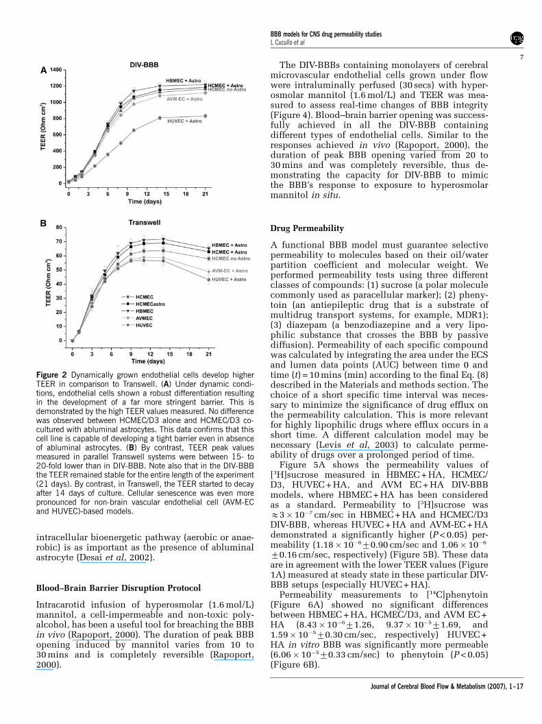
The dynamics of BBB formation and the integrity ofthe barrier itself were compared in DIV-BBB andTranswell plates by using TEER measurements. Wehave previously shown that, under flow conditions,endothelial cells grown in a capillary-like supportdevelop a morphology that closely resembles thenormal endothelial phenotype in situ and permit thedevelopment of higher TEER in comparison with astatic system (Santaguida et al, 2006).
As shown in Figure 2A for three types ofendothelial cell systems, the TEER in the DIV-BBBreaches steady state in approximately 2 weeks,whereas the corresponding co-cultures establishedin Transwell required 1 week (Figure 2B). Tran-sendothelial electrical resistance at the steady statewas E1200 Ohm cm2 in all the DIV-BBB with theexception of the HUVEC/HA co-culture-basedmodel (approximately 800 Ohm cm2). No differencewas observed in TEER between HCMEC/D3 andHCMEC/D3 + HA, thus confirming that this brainvascular endothelium cell line is capable of deve-loping a tight barrier even in the absence ofabluminal astrocytes.
By contrast (Figure 2B), Transwell models werecharacterized by a 15- to 20-fold lower TEER(ranging from 60 to 80 Ohm cm2) in comparisonwith TEER in DIV-BBB. In addition, after 14 days ofco-culture, the Transwell systems developed signs ofcellular senescence as reflected by the decreasingTEER values. This was more evident for AVM-EC/HA and HUVEC/HA co-cultures.
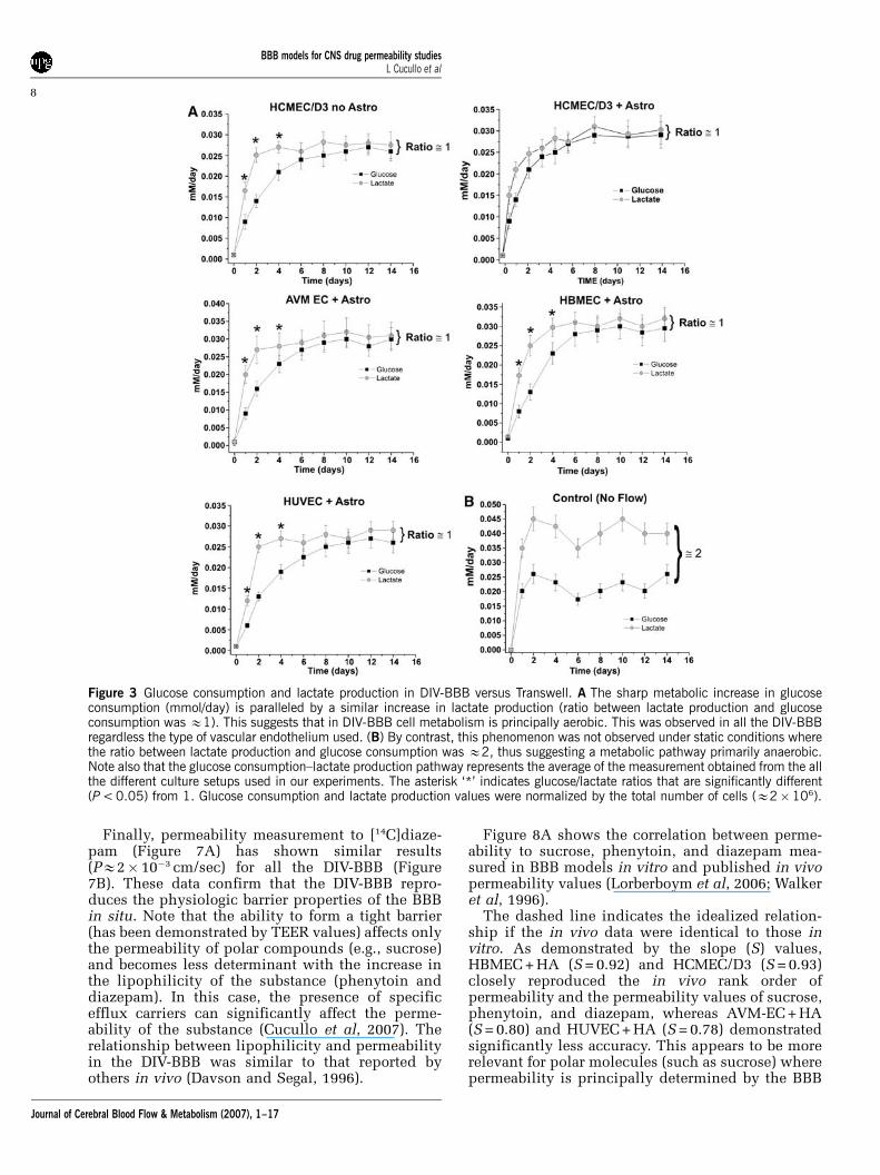
Glucose Metabolism
Measurements of glucose consumption and lactateproduction in the DIV-BBB models (Figure 3)compared to Transwell showed that cells grownunder dynamic conditions develop over time apredominantly aerobic metabolism. This same pat-tern of glucose metabolism was observed in allhumanized DIV-BBB models irrespective of differentEC types. The HCMEC/D3 models showed similaraerobic metabolic pathways both in the presenceand in the absence of abluminal astrocytes. How-ever, showing a marked difference in metabolicbehavior from the monoculture condition, whenHCMEC/D3 cells were cultured with astrocytes, themetabolic shift from anaerobic to aerobic metabo-lism occurred immediately. This suggests that theexposure to intraluminal flow in determining the
BBB models for CNS drug permeability studiesL Cucullo et al
6
Journal of Cerebral Blood Flow & Metabolism (2007), 1–17
intracellular bioenergetic pathway (aerobic or anae-robic) is as important as the presence of abluminalastrocyte (Desai et al, 2002).
Blood–Brain Barrier Disruption Protocol
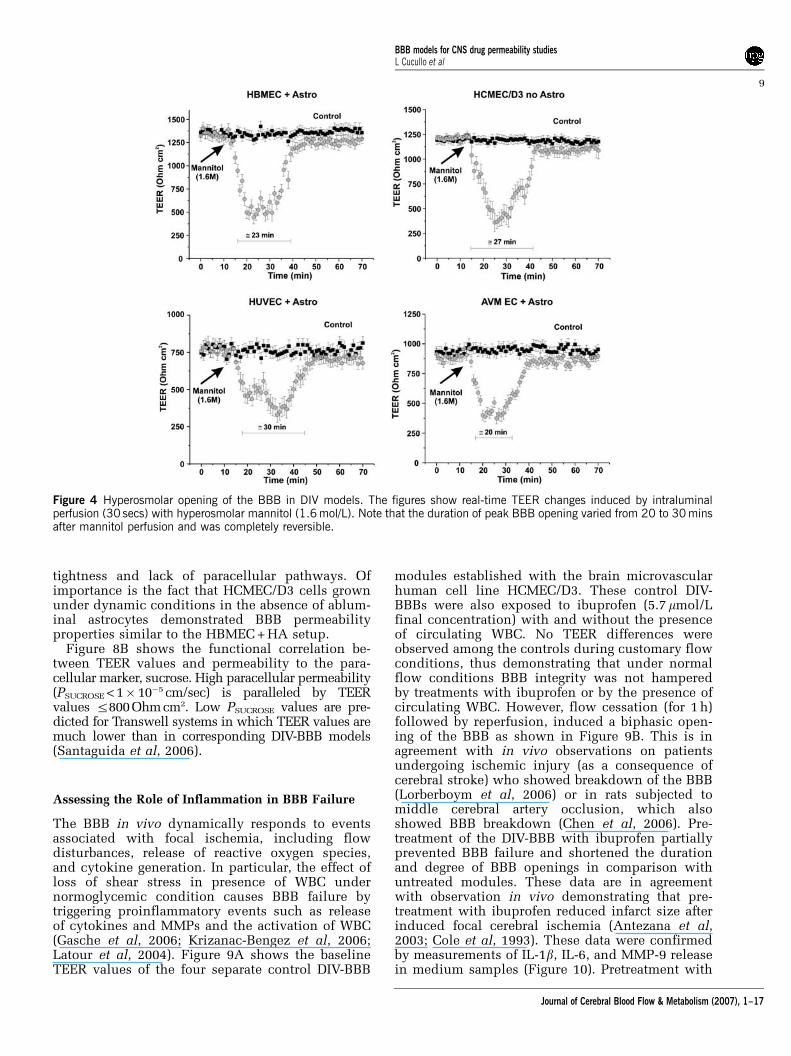
Intracarotid infusion of hyperosmolar (1.6 mol/L)mannitol, a cell-impermeable and non-toxic poly-alcohol, has been a useful tool for breaching the BBBin vivo (Rapoport, 2000). The duration of peak BBBopening induced by mannitol varies from 10 to30 mins and is completely reversible (Rapoport,2000).
The DIV-BBBs containing monolayers of cerebralmicrovascular endothelial cells grown under flowwere intraluminally perfused (30 secs) with hyper-osmolar mannitol (1.6 mol/L) and TEER was mea-sured to assess real-time changes of BBB integrity(Figure 4). Blood–brain barrier opening was success-fully achieved in all the DIV-BBB containingdifferent types of endothelial cells. Similar to theresponses achieved in vivo (Rapoport, 2000), theduration of peak BBB opening varied from 20 to30 mins and was completely reversible, thus de-monstrating the capacity for DIV-BBB to mimicthe BBB’s response to exposure to hyperosmolarmannitol in situ.
Drug Permeability
A functional BBB model must guarantee selectivepermeability to molecules based on their oil/waterpartition coefficient and molecular weight. Weperformed permeability tests using three differentclasses of compounds: (1) sucrose (a polar moleculecommonly used as paracellular marker); (2) pheny-toin (an antiepileptic drug that is a substrate ofmultidrug transport systems, for example, MDR1);(3) diazepam (a benzodiazepine and a very lipo-philic substance that crosses the BBB by passivediffusion). Permeability of each specific compoundwas calculated by integrating the area under the ECSand lumen data points (AUC) between time 0 andtime (t) = 10 mins (min) according to the final Eq. (8)described in the Materials and methods section. Thechoice of a short specific time interval was neces-sary to minimize the significance of drug efflux onthe permeability calculation. This is more relevantfor highly lipophilic drugs where efflux occurs in ashort time. A different calculation model may benecessary (Levis et al, 2003) to calculate perme-ability of drugs over a prolonged period of time.
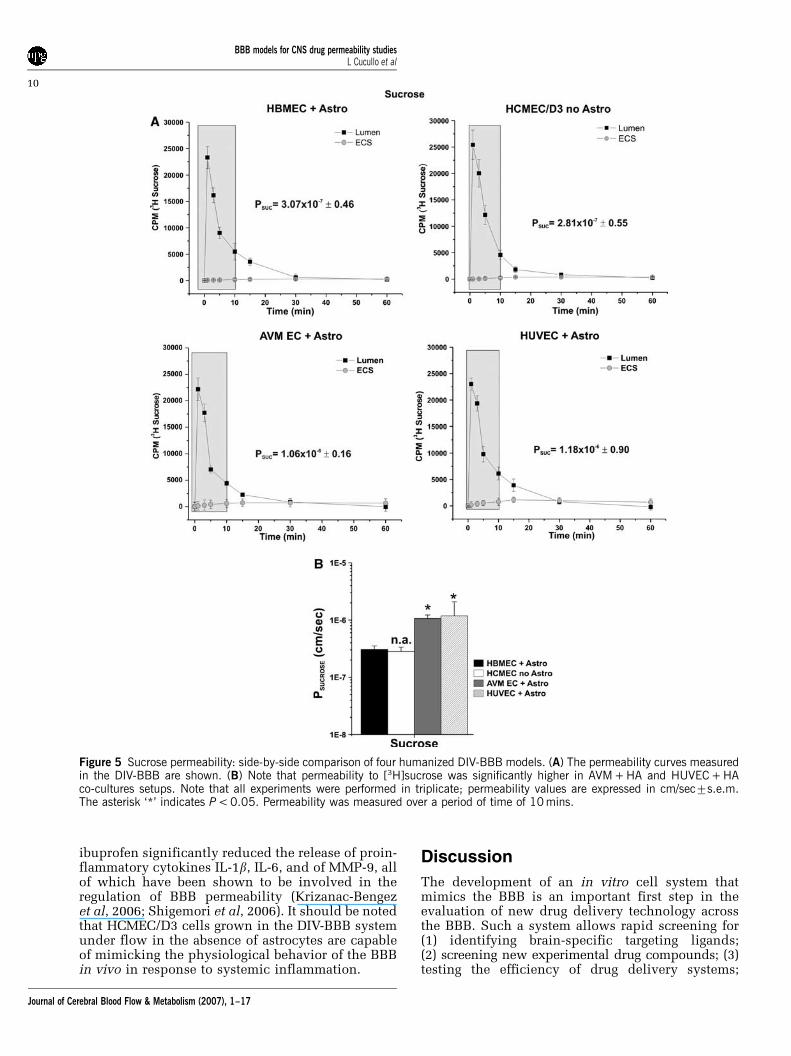
Figure 5A shows the permeability values of[3H]sucrose measured in HBMEC + HA, HCMEC/D3, HUVEC + HA, and AVM EC + HA DIV-BBBmodels, where HBMEC + HA has been consideredas a standard. Permeability to [3H]sucrose wasE3� 10�7 cm/sec in HBMEC + HA and HCMEC/D3DIV-BBB, whereas HUVEC + HA and AVM-EC + HAdemonstrated a significantly higher (P < 0.05) per-meability (1.18� 10�670.90 cm/sec and 1.06� 10�6
70.16 cm/sec, respectively) (Figure 5B). These dataare in agreement with the lower TEER values (Figure1A) measured at steady state in these particular DIV-BBB setups (especially HUVEC + HA).
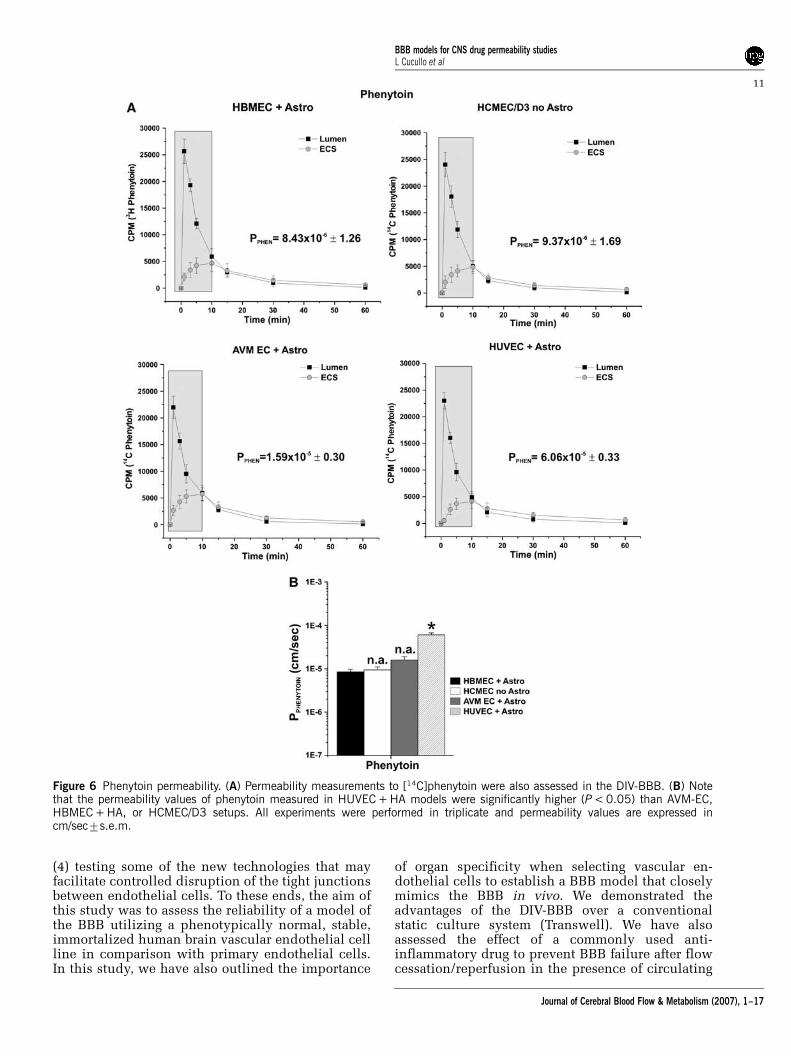
Permeability measurements to [14C]phenytoin(Figure 6A) showed no significant differencesbetween HBMEC + HA, HCMEC/D3, and AVM EC +HA (8.43� 10�671.26, 9.37� 10�571.69, and1.59� 10�570.30 cm/sec, respectively) HUVEC +HA in vitro BBB was significantly more permeable(6.06� 10�570.33 cm/sec) to phenytoin (P < 0.05)(Figure 6B).
Figure 2 Dynamically grown endothelial cells develop higherTEER in comparison to Transwell. (A) Under dynamic condi-tions, endothelial cells shown a robust differentiation resultingin the development of a far more stringent barrier. This isdemonstrated by the high TEER values measured. No differencewas observed between HCMEC/D3 alone and HCMEC/D3 co-cultured with abluminal astrocytes. This data confirms that thiscell line is capable of developing a tight barrier even in absenceof abluminal astrocytes. (B) By contrast, TEER peak valuesmeasured in parallel Transwell systems were between 15- to20-fold lower than in DIV-BBB. Note also that in the DIV-BBBthe TEER remained stable for the entire length of the experiment(21 days). By contrast, in Transwell, the TEER started to decayafter 14 days of culture. Cellular senescence was even morepronounced for non-brain vascular endothelial cell (AVM-ECand HUVEC)-based models.
BBB models for CNS drug permeability studiesL Cucullo et al
7
Journal of Cerebral Blood Flow & Metabolism (2007), 1–17
Finally, permeability measurement to [14C]diaze-pam (Figure 7A) has shown similar results(PE2� 10�3 cm/sec) for all the DIV-BBB (Figure7B). These data confirm that the DIV-BBB repro-duces the physiologic barrier properties of the BBBin situ. Note that the ability to form a tight barrier(has been demonstrated by TEER values) affects onlythe permeability of polar compounds (e.g., sucrose)and becomes less determinant with the increase inthe lipophilicity of the substance (phenytoin anddiazepam). In this case, the presence of specificefflux carriers can significantly affect the perme-ability of the substance (Cucullo et al, 2007). Therelationship between lipophilicity and permeabilityin the DIV-BBB was similar to that reported byothers in vivo (Davson and Segal, 1996).
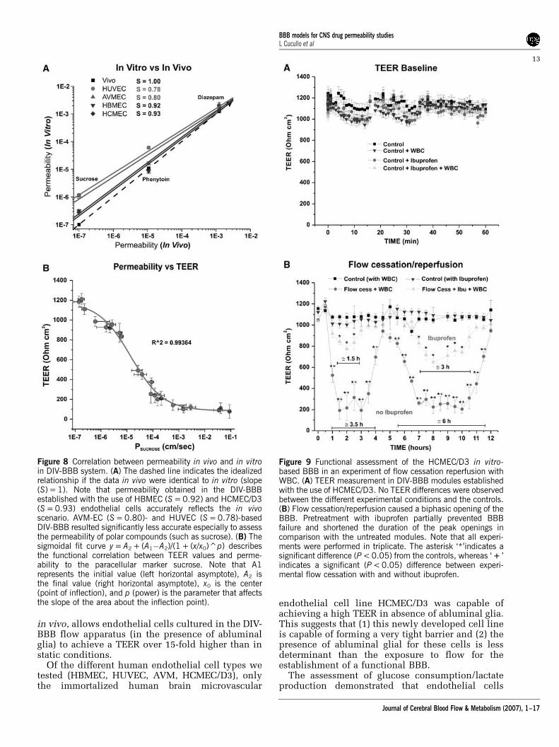
Figure 8A shows the correlation between perme-ability to sucrose, phenytoin, and diazepam mea-sured in BBB models in vitro and published in vivopermeability values (Lorberboym et al, 2006; Walkeret al, 1996).
The dashed line indicates the idealized relation-ship if the in vivo data were identical to those invitro. As demonstrated by the slope (S) values,HBMEC + HA (S = 0.92) and HCMEC/D3 (S = 0.93)closely reproduced the in vivo rank order ofpermeability and the permeability values of sucrose,phenytoin, and diazepam, whereas AVM-EC + HA(S = 0.80) and HUVEC + HA (S = 0.78) demonstratedsignificantly less accuracy. This appears to be morerelevant for polar molecules (such as sucrose) wherepermeability is principally determined by the BBB
Figure 3 Glucose consumption and lactate production in DIV-BBB versus Transwell. A The sharp metabolic increase in glucoseconsumption (mmol/day) is paralleled by a similar increase in lactate production (ratio between lactate production and glucoseconsumption was E1). This suggests that in DIV-BBB cell metabolism is principally aerobic. This was observed in all the DIV-BBBregardless the type of vascular endothelium used. (B) By contrast, this phenomenon was not observed under static conditions wherethe ratio between lactate production and glucose consumption was E2, thus suggesting a metabolic pathway primarily anaerobic.Note also that the glucose consumption–lactate production pathway represents the average of the measurement obtained from the allthe different culture setups used in our experiments. The asterisk ‘*’ indicates glucose/lactate ratios that are significantly different(P < 0.05) from 1. Glucose consumption and lactate production values were normalized by the total number of cells (E2�106).
BBB models for CNS drug permeability studiesL Cucullo et al
8
Journal of Cerebral Blood Flow & Metabolism (2007), 1–17
tightness and lack of paracellular pathways. Ofimportance is the fact that HCMEC/D3 cells grownunder dynamic conditions in the absence of ablum-inal astrocytes demonstrated BBB permeabilityproperties similar to the HBMEC + HA setup.
Figure 8B shows the functional correlation be-tween TEER values and permeability to the para-cellular marker, sucrose. High paracellular permeability(PSUCROSE < 1� 10�5 cm/sec) is paralleled by TEERvalues r800Ohmcm2. Low PSUCROSE values are pre-dicted for Transwell systems in which TEER values aremuch lower than in corresponding DIV-BBB models(Santaguida et al, 2006).
Assessing the Role of Inflammation in BBB Failure
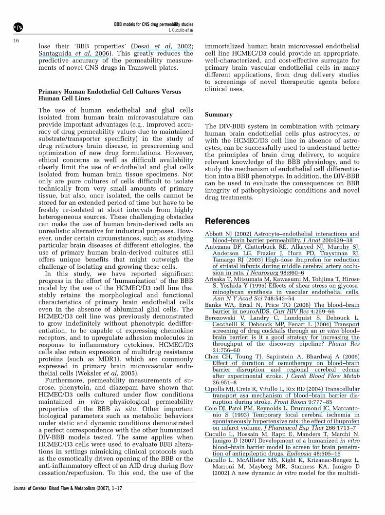
The BBB in vivo dynamically responds to eventsassociated with focal ischemia, including flowdisturbances, release of reactive oxygen species,and cytokine generation. In particular, the effect ofloss of shear stress in presence of WBC undernormoglycemic condition causes BBB failure bytriggering proinflammatory events such as releaseof cytokines and MMPs and the activation of WBC(Gasche et al, 2006; Krizanac-Bengez et al, 2006;Latour et al, 2004). Figure 9A shows the baselineTEER values of the four separate control DIV-BBB
modules established with the brain microvascularhuman cell line HCMEC/D3. These control DIV-BBBs were also exposed to ibuprofen (5.7 mmol/Lfinal concentration) with and without the presenceof circulating WBC. No TEER differences wereobserved among the controls during customary flowconditions, thus demonstrating that under normalflow conditions BBB integrity was not hamperedby treatments with ibuprofen or by the presence ofcirculating WBC. However, flow cessation (for 1 h)followed by reperfusion, induced a biphasic open-ing of the BBB as shown in Figure 9B. This is inagreement with in vivo observations on patientsundergoing ischemic injury (as a consequence ofcerebral stroke) who showed breakdown of the BBB(Lorberboym et al, 2006) or in rats subjected tomiddle cerebral artery occlusion, which alsoshowed BBB breakdown (Chen et al, 2006). Pre-treatment of the DIV-BBB with ibuprofen partiallyprevented BBB failure and shortened the durationand degree of BBB openings in comparison withuntreated modules. These data are in agreementwith observation in vivo demonstrating that pre-treatment with ibuprofen reduced infarct size afterinduced focal cerebral ischemia (Antezana et al,2003; Cole et al, 1993). These data were confirmedby measurements of IL-1b, IL-6, and MMP-9 releasein medium samples (Figure 10). Pretreatment with
Figure 4 Hyperosmolar opening of the BBB in DIV models. The figures show real-time TEER changes induced by intraluminalperfusion (30 secs) with hyperosmolar mannitol (1.6 mol/L). Note that the duration of peak BBB opening varied from 20 to 30 minsafter mannitol perfusion and was completely reversible.
BBB models for CNS drug permeability studiesL Cucullo et al
9
Journal of Cerebral Blood Flow & Metabolism (2007), 1–17
ibuprofen significantly reduced the release of proin-flammatory cytokines IL-1b, IL-6, and of MMP-9, allof which have been shown to be involved in theregulation of BBB permeability (Krizanac-Bengezet al, 2006; Shigemori et al, 2006). It should be notedthat HCMEC/D3 cells grown in the DIV-BBB systemunder flow in the absence of astrocytes are capableof mimicking the physiological behavior of the BBBin vivo in response to systemic inflammation.
Discussion
The development of an in vitro cell system thatmimics the BBB is an important first step in theevaluation of new drug delivery technology acrossthe BBB. Such a system allows rapid screening for(1) identifying brain-specific targeting ligands;(2) screening new experimental drug compounds; (3)testing the efficiency of drug delivery systems;
Figure 5 Sucrose permeability: side-by-side comparison of four humanized DIV-BBB models. (A) The permeability curves measuredin the DIV-BBB are shown. (B) Note that permeability to [3H]sucrose was significantly higher in AVM + HA and HUVEC + HAco-cultures setups. Note that all experiments were performed in triplicate; permeability values are expressed in cm/sec7s.e.m.The asterisk ‘*’ indicates P < 0.05. Permeability was measured over a period of time of 10 mins.
BBB models for CNS drug permeability studiesL Cucullo et al
10
Journal of Cerebral Blood Flow & Metabolism (2007), 1–17
(4) testing some of the new technologies that mayfacilitate controlled disruption of the tight junctionsbetween endothelial cells. To these ends, the aim ofthis study was to assess the reliability of a model ofthe BBB utilizing a phenotypically normal, stable,immortalized human brain vascular endothelial cellline in comparison with primary endothelial cells.In this study, we have also outlined the importance
of organ specificity when selecting vascular en-dothelial cells to establish a BBB model that closelymimics the BBB in vivo. We demonstrated theadvantages of the DIV-BBB over a conventionalstatic culture system (Transwell). We have alsoassessed the effect of a commonly used anti-inflammatory drug to prevent BBB failure after flowcessation/reperfusion in the presence of circulating
Figure 6 Phenytoin permeability. (A) Permeability measurements to [14C]phenytoin were also assessed in the DIV-BBB. (B) Notethat the permeability values of phenytoin measured in HUVEC + HA models were significantly higher (P < 0.05) than AVM-EC,HBMEC + HA, or HCMEC/D3 setups. All experiments were performed in triplicate and permeability values are expressed incm/sec7s.e.m.
BBB models for CNS drug permeability studiesL Cucullo et al
11
Journal of Cerebral Blood Flow & Metabolism (2007), 1–17

WBC, thus establishing a model to study the effect ofinflammation at the BBB in vitro.
Role of Shear Stress in BBB Development and CellMetabolism
Endothelial cells in vivo are long-lived cells con-tinuously exposed to shear stress, a tangential force
generated by the flow of blood across their apicalsurfaces. Shear stress affects endothelial cell differ-entiation, tight junction formation, the expression ofjunction-related proteins (Arisaka et al, 1995; Yoshidaet al, 1995), and induces mitotic arrest (Desai et al,2002; Lin et al, 2000). An index of the endothelial‘tightness’ of the monolayer is the TEER measuredacross the BBB (Santaguida et al, 2006). Our dataclearly show that the exposure to flow, as found
Figure 7 Permeability of diazepam. (A) By comparing the permeability values to [14C]diazepam, we find (B) no significantdifferences between HBMEC, HCMEC/D3, AVM-EC, or HUVEC setups (E2�10�3). Permeability values are expressed in cm/sec7s.e.m. The asterisk *P < 0.05.
BBB models for CNS drug permeability studiesL Cucullo et al
12
Journal of Cerebral Blood Flow & Metabolism (2007), 1–17
in vivo, allows endothelial cells cultured in the DIV-BBB flow apparatus (in the presence of abluminalglia) to achieve a TEER over 15-fold higher than instatic conditions.
Of the different human endothelial cell types wetested (HBMEC, HUVEC, AVM, HCMEC/D3), onlythe immortalized human brain microvascular
endothelial cell line HCMEC/D3 was capable ofachieving a high TEER in absence of abluminal glia.This suggests that (1) this newly developed cell lineis capable of forming a very tight barrier and (2) thepresence of abluminal glial for these cells is lessdeterminant than the exposure to flow for theestablishment of a functional BBB.
The assessment of glucose consumption/lactateproduction demonstrated that endothelial cells
Figure 8 Correlation between permeability in vivo and in vitroin DIV-BBB system. (A) The dashed line indicates the idealizedrelationship if the data in vivo were identical to in vitro (slope(S) = 1). Note that permeability obtained in the DIV-BBBestablished with the use of HBMEC (S = 0.92) and HCMEC/D3(S = 0.93) endothelial cells accurately reflects the in vivoscenario. AVM-EC (S = 0.80)- and HUVEC (S = 0.78)-basedDIV-BBB resulted significantly less accurate especially to assessthe permeability of polar compounds (such as sucrose). (B) Thesigmoidal fit curve y = A2 + (A1�A2)/(1 + (x/x0)^p) describesthe functional correlation between TEER values and perme-ability to the paracellular marker sucrose. Note that A1represents the initial value (left horizontal asymptote), A2 isthe final value (right horizontal asymptote), x0 is the center(point of inflection), and p (power) is the parameter that affectsthe slope of the area about the inflection point).
Figure 9 Functional assessment of the HCMEC/D3 in vitro-based BBB in an experiment of flow cessation reperfusion withWBC. (A) TEER measurement in DIV-BBB modules establishedwith the use of HCMEC/D3. No TEER differences were observedbetween the different experimental conditions and the controls.(B) Flow cessation/reperfusion caused a biphasic opening of theBBB. Pretreatment with ibuprofen partially prevented BBBfailure and shortened the duration of the peak openings incomparison with the untreated modules. Note that all experi-ments were performed in triplicate. The asterisk ‘*’indicates asignificant difference (P < 0.05) from the controls, whereas ‘ + ’indicates a significant (P < 0.05) difference between experi-mental flow cessation with and without ibuprofen.
BBB models for CNS drug permeability studiesL Cucullo et al
13
Journal of Cerebral Blood Flow & Metabolism (2007), 1–17

grown in static conditions have an anaerobicmetabolic pathway. By contrast, the same cellsgrown under dynamic conditions show a highpropensity toward an aerobic type of metabolism(Santaguida et al, 2006). This observation is in
agreement with previous data demonstrating thatexposure to shear stress induces metabolic changes(Desai et al, 2002) such as upregulation of thedehydrogenases and glyceraldehyde-3-phosphatedehydrogenase, and is accompanied by simulta-neous decrease in enzymes of the NADH-depletingpathway, such as lactate dehydrogenase, which playkey roles in the Krebs cycle. In addition to main-taining shear stress, the growth medium in the DIV-BBB is circulated in silicon tubing that allows forcontinuous exchange of oxygen and CO2 with theexternal environment. Moreover, the gas-permeabletubing connecting the cartridge to the mediumreservoir increases the exchange surface. In Trans-well plates, these advantages for maintaining glu-cose and oxygen homeostasis are lost owing to lackof flow, and to limitation of the exchange surface bythe size of the well.
Taking these two last points into account, our dataalso suggest that endothelial cells cultivated in theDIV-BBB are capable of adjusting their metabolicbehavior to adapt to oxygen availability. Therefore,monitoring cellular metabolism is an importantparameter in testing the adaptation of the endo-thelial cells chosen to establish a BBB in vitro. Asshown by our data, the HCMEC/D3 cell linerecapitulates the metabolic behavior of primaryendothelial–glial co-cultures even in the absenceof abluminal astrocytes.
Dynamic In vitro Blood Brain Barrier Mimics the BBBIn vivo: Testing Clinical Protocols In vitro
Osmotic opening of the BBB by hyperosmoticmannitol solution is a common clinical procedureused to facilitate chemotherapic drug penetrationinto the brain parenchyma for the treatment ofmalignant brain tumors. The opening of the BBB ismediated by cerebrovascular dilatation, dehydrationof endothelial cells, and contraction of their cyto-skeleton, which leads to the widening of the inter-endothelial tight junctions. This mechanism isdriven solely by the effect of the osmotic gradientof water from the inside to the outside of the cellbody (Rapoport, 2000) and is fully reversible. Ourexperiments have shown that all of our DIV-BBBmodels (whether based on primary endothelial cellcultures or on the HCMEC/D3 endothelial cell line)closely mimic the in vivo BBB response to hyper-
Figure 10 Measurement of proinflammatory cytokines IL-1b,IL-6, and MCP-9. Levels of IL-1b, IL-6, and MMP-9 weremeasured in medium samples in all experimental conditions.Note that pretreatment with ibuprofen significantly reduced therelease of IL-1b, IL-6, and MMP-9. These data are in agreementwith the levels of BBB failure concomitantly assessed by TEERmeasurements. The asterisk ‘*’ indicates a significant difference(P < 0.05) from the controls, whereas ‘ + ’ indicates asignificant (P < 0.05) difference between experimental flowcessation with and without ibuprofen.
BBB models for CNS drug permeability studiesL Cucullo et al
14
Journal of Cerebral Blood Flow & Metabolism (2007), 1–17

osmolar mannitol (Figure 4). From this perspective,the use of the HCMEC/D3 cell line in conjunctionwith the DIV-BBB system offers significant advan-tages in comparison with primary cell cultures.
Pathophysiology of CNS Diseases: Assessing the Roleof Inflammation in BBB Failure
Increasing evidence indicates that inflammation isinvolved in the pathogenesis of many neurologicaland neurodegenerative diseases and that the BBBdynamically responds to these inflammatory events.This is important because BBB failure can causesubstances circulating in the blood that are normallyexcluded from the CNS (such as potassium, gluta-mate, and plasma proteins) to enter the brain, thusleading to a secondary process of CNS injury. Forexample, BBB failure after vasogenic brain trauma orfocal brain ischemia may trigger the occurrence ofstatus epilepticus (Karhunen et al, 2005; Vezzaniand Granata, 2005). Therefore, it seems reasonableto propose that the use of anti-inflammatory drugs(AIDs) may diminish the cumulative effects ofinflammation in the brain. However, few AIDclinical trials performed so far were minimal andequivocal in their outcomes. This likely occurred forseveral reasons, such as timing of AID administra-tion, non-selective inhibition of cyclooxygenases(COX-1 and -2), inappropriate use of AIDs for agiven disease or disease progression/severity orsuboptimal dose in target sites. Given these con-siderations, the advantages that could come from theuse of a reliable in vitro BBB model for the testing ofAIDs are clearly evident.
For this instance, the DIV-BBB was successfullyused to mimic an ischemic-like event in vitro in thepresence of circulating WBC. The model wassuccessfully used to assess the effect of ibuprofen,a well-known AID, to prevent BBB failure afternormoxic flow cessation/reperfusion and activationof WBCs and vascular endothelium. Our resultsshow that the ischemic-like insult simulated by flowcessation/reperfusion caused a biphasic opening ofthe BBB (Figure 7). However, in the DIV-BBBmodules pretreated with 1mmol/L ibuprofen beforeflow cessation/reperfusion in the presence of WBC,the peak opening of the barrier is significantly lowerand the intervals between the periods of openingis significantly shorter. These data strongly suggestthat the inflammatory response that follows anischemic-like event in vivo is the primary cause ofBBB failure. This effect is relevant because BBBfailure is related to the occurrence of secondarypost-ischemic brain injuries.
Flow cessation/reperfusion triggered an inflam-matory response that led to BBB failure as demon-strated by the biphasic decrease in TEER (Figure 9)observed during the course of the experiment.Additionally, BBB failure was paralleled by theintraluminal release (Figure 10) of proinflammatory
factors (IL-6 and IL-1b) and MMP-9. Pretreatmentwith ibuprofen (0.125 mmol/L) prevented BBBfailure by decreasing the inflammatory responseafter flow cessation/reperfusion.
Taken together, these results show that the DIV-BBB can find useful application in the evaluation ofclinical procedures. A variety of clinical protocolsand novel drugs for the treatment of CNS diseasescould be reliably tested in vitro before moving toreal-life applications.
Drug Delivery into the Central Nervous System:Dynamic In vitro Blood–Brain Barrier as a Tool toAssess Blood–Brain Barrier Permeability of NovelDrugs
Drug delivery to the CNS is subject to the perme-ability limitations imposed by the BBB. Severalsystems in vitro have been described to reproducethe physical and the biochemical behavior of anintact BBB, but most of these lack the key features ofthe barrier in vivo.
Permeability measurements of sucrose, pheny-toin, and diazepam assessed in the DIV-BBB haveshown that (1) the exposure to intraluminal flowplays an essential role in promoting BBB tightness;(2) the humanized DIV-BBB can successfully distin-guish between permeability rankings of differentclasses of substances that penetrate the BBB bydifferent mechanisms, thus providing reproducibleresults that can be reliably matched to the BBB insitu; (3) different from vascular endothelial primarycultures (Abbott, 2002; Hamm et al, 2004), HCMEC/D3 cells grown under dynamic conditions differ-entiate into a very stringent BBB phenotype evenwithout the presence of abluminal astrocytes. It isalso important to note that despite the exposure to aquasi-physiological pulsatile flow and the presenceof abluminal astrocytes, non-brain vascular endo-thelial cells (HUVECs) were the less capable toestablish a BBB in vitro as stringent as the onesbased on brain microvascular endothelial cells. Thislatter finding outlines the importance of organspecificity when selecting vascular endothelial cellsto establish an in vitro BBB model.
What is Left to Transwell?
We have seen that static culture and co-culturemodels cannot reproduce the complete BBB in vivodespite having been used as relatively low cost toolsfor studying drug transport across the BBB. Theattractive features of the Transwell model are itssimplicity (easy to establish cultures) and the fixedvolumes in each compartment that are useful forMichaelis–Menten kinetics of transport (Berezowskiet al, 2004). However, in the absence of shear stress,which plays a central role in the cerebrovascularsystem by promoting the differentiation and main-tenance of the BBB phenotype, the endothelial cells
BBB models for CNS drug permeability studiesL Cucullo et al
15
Journal of Cerebral Blood Flow & Metabolism (2007), 1–17
lose their ‘BBB properties’ (Desai et al, 2002;Santaguida et al, 2006). This greatly reduces thepredictive accuracy of the permeability measure-ments of novel CNS drugs in Transwell plates.
Primary Human Endothelial Cell Cultures VersusHuman Cell Lines
The use of human endothelial and glial cellsisolated from human brain microvasculature canprovide important advantages (e.g., improved accu-racy of drug permeability values due to maintainedsubstrate/transporter specificity) in the study ofdrug refractory brain disease, in prescreening andoptimization of new drug formulations. However,ethical concerns as well as difficult availabilityclearly limit the use of endothelial and glial cellsisolated from human brain tissue specimens. Notonly are pure cultures of cells difficult to isolatetechnically from very small amounts of primarytissue, but also, once isolated, the cells cannot bestored for an extended period of time but have to befreshly re-isolated at short intervals from highlyheterogeneous sources. These challenging obstaclescan make the use of human brain-derived cells anunrealistic alternative for industrial purposes. How-ever, under certain circumstances, such as studyingparticular brain diseases of different etiologies, theuse of primary human brain-derived cultures stilloffers unique benefits that might outweigh thechallenge of isolating and growing these cells.
In this study, we have reported significantprogress in the effort of ‘humanization’ of the BBBmodel by the use of the HCMEC/D3 cell line thatstably retains the morphological and functionalcharacteristics of primary brain endothelial cellseven in the absence of abluminal glial cells. TheHCMEC/D3 cell line was previously demonstratedto grow indefinitely without phenotypic dediffer-entiation, to be capable of expressing chemokinereceptors, and to upregulate adhesion molecules inresponse to inflammatory cytokines. HCMEC/D3cells also retain expression of multidrug resistanceproteins (such as MDR1), which are commonlyexpressed in primary brain microvascular endo-thelial cells (Weksler et al, 2005).
Furthermore, permeability measurements of su-crose, phenytoin, and diazepam have shown thatHCMEC/D3 cells cultured under flow conditionsmaintained in vitro physiological permeabilityproperties of the BBB in situ. Other importantbiological parameters such as metabolic behaviorsunder static and dynamic conditions demonstrateda perfect correspondence with the other humanizedDIV-BBB models tested. The same applies whenHCMEC/D3 cells were used to evaluate BBB altera-tions in settings mimicking clinical protocols suchas the osmotically driven opening of the BBB or theanti-inflammatory effect of an AID drug during flowcessation/reperfusion. To this end, the use of the
immortalized human brain microvessel endothelialcell line HCMEC/D3 could provide an appropriate,well-characterized, and cost-effective surrogate forprimary brain vascular endothelial cells in manydifferent applications, from drug delivery studiesto screenings of novel therapeutic agents beforeclinical uses.
Summary
The DIV-BBB system in combination with primaryhuman brain endothelial cells plus astrocytes, orwith the HCMEC/D3 cell line in absence of astro-cytes, can be successfully used to understand betterthe principles of brain drug delivery, to acquirerelevant knowledge of the BBB physiology, and tostudy the mechanism of endothelial cell differentia-tion into a BBB phenotype. In addition, the DIV-BBBcan be used to evaluate the consequences on BBBintegrity of pathophysiologic conditions and noveldrug treatments.
References
Abbott NJ (2002) Astrocyte–endothelial interactions andblood–brain barrier permeability. J Anat 200:629–38
Antezana DF, Clatterbuck RE, Alkayed NJ, Murphy SJ,Anderson LG, Frazier J, Hurn PD, Traystman RJ,Tamargo RJ (2003) High-dose ibuprofen for reductionof striatal infarcts during middle cerebral artery occlu-sion in rats. J Neurosurg 98:860–6
Arisaka T, Mitsumata M, Kawasumi M, Tohjima T, HiroseS, Yoshida Y (1995) Effects of shear stress on glycosa-minoglycan synthesis in vascular endothelial cells.Ann N Y Acad Sci 748:543–54
Banks WA, Ercal N, Price TO (2006) The blood–brainbarrier in neuroAIDS. Curr HIV Res 4:259–66
Berezowski V, Landry C, Lundquist S, Dehouck L,Cecchelli R, Dehouck MP, Fenart L (2004) Transportscreening of drug cocktails through an in vitro blood–brain barrier: is it a good strategy for increasing thethroughput of the discovery pipeline? Pharm Res21:756–60
Chen CH, Toung TJ, Sapirstein A, Bhardwaj A (2006)Effect of duration of osmotherapy on blood–brainbarrier disruption and regional cerebral edemaafter experimental stroke. J Cereb Blood Flow Metab26:951–8
Cipolla MJ, Crete R, Vitullo L, Rix RD (2004) Transcellulartransport asa mechanism of blood–brain barrier dis-ruption during stroke. Front Biosci 9:777–85
Cole DJ, Patel PM, Reynolds L, Drummond JC, Marcanto-nio S (1993) Temporary focal cerebral ischemia inspontaneously hypertensive rats: the effect of ibuprofenon infarct volume. J Pharmacol Exp Ther 266:1713–7
Cucullo L, Hossain M, Rapp E, Manders T, Marchi N,Janigro D (2007) Development of a humanized in vitroblood–brain barrier model to screen for brain penetra-tion of antiepileptic drugs. Epilepsia 48:505–16
Cucullo L, McAllister MS, Kight K, Krizanac-Bengez L,Marroni M, Mayberg MR, Stanness KA, Janigro D(2002) A new dynamic in vitro model for the multidi-
BBB models for CNS drug permeability studiesL Cucullo et al
16
Journal of Cerebral Blood Flow & Metabolism (2007), 1–17
mensional study of astrocyte-endothelial cell interac-tions at the blood–brain barrier. Brain Res 951:243–54
Davson H, Segal MB (1996) Blood–brain barrier. In:Davson H, Segal MB (eds), Physiology of the CSF andBlood–Brain Barriers. Boca Raton, FL: CRC, 49–91
Desai SY, Marroni M, Cucullo L, Krizanac-Bengez L,Mayberg MR, Hossain MT, Grant GG, Janigro D (2002)Mechanisms of endothelial survival under shear stress.Endothelium 9:89–102
Gasche Y, Soccal PM, Kanemitsu M, Copin JC (2006)Matrix metalloproteinases and diseases of the centralnervous system with a special emphasis on ischemicbrain. Front Biosci 11:1289–301
Hamm S, Dehouck B, Kraus J, Wolburg-Buchholz K,Wolburg H, Risau W, Cecchelli R, Engelhardt B,Dehouck MP (2004) Astrocyte mediated modulationof blood–brain barrier permeability does not correlatewith a loss of tight junction proteins from the cellularcontacts. Cell Tissue Res 315:157–66
Kalaria RN (1992) The blood–brain barrier and cerebralmicrocirculation in Alzheimer disease. CerebrovascBrain Metab Rev 4:226–60
Karhunen H, Jolkkonen J, Sivenius J, Pitkanen A (2005)Epileptogenesis after experimental focal cerebral ische-mia. Neurochem Res 30:1529–42
Krizanac-Bengez L, Kapural M, Parkinson F, Cucullo L,Hossain M, Mayberg MR, Janigro D (2003) Effects oftransient loss of shear stress on blood–brain barrierendothelium: role of nitric oxide and IL-6. Brain Res977:239–46
Krizanac-Bengez L, Mayberg MR, Cunningham E, HossainM, Ponnampalam S, Parkinson FE, Janigro D (2006)Loss of shear stress induces leukocyte-mediated cyto-kine release and blood–brain barrier failure in dynamicin vitro blood–brain barrier model. J Cell Physiol206:68–77
Kubota H, Ishihara H, Langmann T, Schmitz G, Stieger B,Wieser HG, Yonekawa Y, Frei K (2006) Distribution andfunctional activity of P-glycoprotein and multidrugresistance-associated proteins in human brain micro-vascular endothelial cells in hippocampal sclerosis.Epilepsy Res 68:213–28
Latour LL, Kang DW, Ezzeddine MA, Chalela JA, Warach S(2004) Early blood–brain barrier disruption in humanfocal brain ischemia. Ann Neurol 56:468–77
Lee SW, Kim WJ, Park JA, Choi YK, Kwon YW, Kim KW(2006) Blood–brain barrier interfaces and brain tumors.Arch Pharm Res 29:265–75
Levis KA, Lane ME, Corrigan OI (2003) Effect of buffermedia composition on the solubility and effectivepermeability coefficient of ibuprofen. Int J Pharm253:49–59
Lin K, Hsu PP, Chen BP, Yuan S, Usami S, Shyy JY, Li YS,Chien S (2000) Molecular mechanism of endothelialgrowth arrest by laminar shear stress. Proc Natl AcadSci USA 97:9385–9
Lorberboym M, Blankenberg FG, Sadeh M, Lampl Y (2006)In vivo imaging of apoptosis in patients with acute
stroke: correlation with blood–brain barrier permeabi-lity. Brain Res 1103:13–9
Mashour GA, Boock RJ (1999) Effects of shear stress onnitric oxide levels of human cerebral endothelial cellscultured in an artificial capillary system. Brain Res842:233–8
Minagar A, Alexander JS (2003) Blood–brain barrierdisruption in multiple sclerosis. Mult Scler 9:540–9
Pardridge WM (2005) The blood–brain barrier: bottleneckin brain drug development. NeuroRx 2:3–14
Parkinson FE, Friesen J, Krizanac-Bengez L, Janigro D(2003) Use of a three-dimensional in vitro model of therat blood–brain barrier to assay nucleoside efflux frombrain. Brain Res 980:233–41
Rapoport SI (2000) Osmotic opening of the blood–brainbarrier: principles, mechanism, and therapeutic appli-cations. Cell Mol Neurobiol 20:217–30
Santaguida S, Janigro D, Hossain M, Oby E, Rapp E,Cucullo L (2006) Side by side comparisonbetween dynamic versus static models of blood–brainbarrier in vitro: A permeability study. Brain Res 1109:1–13
Shigemori Y, Katayama Y, Mori T, Maeda T, Kawamata T(2006) Matrix metalloproteinase-9 is associated withblood–brain barrier opening and brain edema formationafter cortical contusion in rats. Acta Neurochir Suppl96:130–3
Stamatovic SM, Dimitrijevic OB, Keep RF, Andjelkovic AV(2006) Inflammation and brain edema: new insightsinto the role of chemokines and their receptors. ActaNeurochir Suppl 96:444–50
Stanness KA, Westrum LE, Fornaciari E, Mascagni P,Nelson JA, Stenglein SG, Myers T, Janigro D (1997)Morphological and functional characterization ofan in vitro blood–brain barrier model. Brain Res771:329–42
Unterberg AW, Stover J, Kress B, Kiening KL (2004) Edemaand brain trauma. Neuroscience 129:1021–9
van der FM, Hoppenreijs S, van Rensburg AJ, Ruyken M,Kolk AH, Springer P, Hoepelman AI, Geelen SP,Kimpen JL, Schoeman JF (2004) Vascular endothelialgrowth factor and blood–brain barrier disruptionin tuberculous meningitis. Pediatr Infect Dis J 23:608–613
Vezzani A, Granata T (2005) Brain inflammation inepilepsy: experimental and clinical evidence. Epilepsia46:1724–43
Walker MC, Alavijeh MS, Shorvon SD, Patsalos PN (1996)Microdialysis study of the neuropharmacokinetics ofphenytoin in rat hippocampus and frontal cortex.Epilepsia 37:421–7
Weksler BB, Subileau EA, Couraud PO (2005) Blood–brainbarrier-specific properties of a human adult brainendothelial cell line. FASEB J 19:1872–4
Yoshida Y, Okano M, Wang S, Kobayashi M, Kawasumi M,Hagiwara H, Mitsumata M (1995) Hemodynamic-force-induced difference of interendothelial junctional com-plexes. Ann N Y Acad Sci 748:104–20
BBB models for CNS drug permeability studiesL Cucullo et al
17
Journal of Cerebral Blood Flow & Metabolism (2007), 1–17