identification of tertiary butyl alcohol (tba)-utilizing organisms in biogac reactors using 13c-dna...
TRANSCRIPT
ORIGINAL PAPER
Identification of tertiary butyl alcohol (TBA)-utilizingorganisms in BioGAC reactors using 13C-DNA stable isotopeprobing
Denise Aslett • Joseph Haas • Michael Hyman
Received: 4 October 2010 / Accepted: 10 January 2011 / Published online: 1 February 2011
� Springer Science+Business Media B.V. 2011
Abstract Biodegradation of the gasoline oxygen-
ates methyl tertiary-butyl ether (MTBE) and ethyl
tertiary-butyl ether (ETBE) can cause tertiary butyl
alcohol (TBA) to accumulate in gasoline-impacted
environments. One remediation option for TBA-
contaminated groundwater involves oxygenated gran-
ulated activated carbon (GAC) reactors that have
been self-inoculated by indigenous TBA-degrading
microorganisms in ground water extracted from
contaminated aquifers. Identification of these organ-
isms is important for understanding the range of
TBA-metabolizing organisms in nature and for
determining whether self-inoculation of similar reac-
tors is likely to occur at other sites. In this study13C-DNA-stable isotope probing (SIP) was used to
identify TBA-utilizing organisms in samples of self-
inoculated BioGAC reactors operated at sites in New
York and California. Based on 16S rRNA nucleotide
sequences, all TBA-utilizing organisms identified
were members of the Burkholderiales order of the
b-proteobacteria. Organisms similar to Cupriavidus
and Methylibium were observed in both reactor
samples while organisms similar to Polaromonas
and Rhodoferax were unique to the reactor sample
from New York. Organisms similar to Hydrogenoph-
aga and Paucibacter strains were only detected in the
reactor sample from California. We also analyzed our
samples for the presence of several genes previously
implicated in TBA oxidation by pure cultures of
bacteria. Genes Mpe_B0532, B0541, B0555, and
B0561 were all detected in 13C-metagenomic DNA
from both reactors and deduced amino acid sequences
suggested these genes all encode highly conserved
enzymes. One gene (Mpe_B0555) encodes a putative
phthalate dioxygenase-like enzyme that may be
particularly appropriate for determining the potential
for TBA oxidation in contaminated environmental
samples.
Keywords Tertiary butyl alcohol � Stable isotope
probing � Polaromonas � Methylibium
Introduction
Tertiary-butyl alcohol (TBA) is widely used as an
industrial solvent (Clark 2002). However, environ-
mental contamination by this compound occurs
primarily as a result of biodegradation of gasoline
oxygenates including methyl tertiary-butyl ether
(MTBE) and ethyl tertiary-butyl ether (ETBE).
Anaerobic biodegradation of MTBE can generate
D. Aslett � M. Hyman (&)
Department of Microbiology, 4545 Thomas Hall, North
Carolina State University, Raleigh, NC 27695, USA
e-mail: [email protected]
J. Haas
Office of the New York State Attorney General,
Environmental Protection Bureau, New York,
NY 10271, USA
123
Biodegradation (2011) 22:961–972
DOI 10.1007/s10532-011-9455-3

TBA and, as anaerobic conditions are frequently
encountered at gasoline spill sites, TBA concentra-
tions can often exceed those of MTBE (Schmidt et al.
2004). Accumulation of TBA in ground water sources
of drinking water is of concern as this compound
is potentially more toxic than its parent, MTBE
(Cirvello et al. 1995).
Although TBA is often only slowly biodegraded
under anaerobic conditions (Wilson et al. 2005), rapid
aerobic TBA biodegradation has been demonstrated
in microcosm studies using stream bed sediments
(Bradley et al. 1999), surface water sediments
(Bradley et al. 2002) and various cultures (Deeb
et al. 2003; Fortin et al. 2001; Francois et al. 2002;
Hanson et al. 1999; Hatzinger et al. 2001; Kharoune
et al. 2001; Mo et al. 1997; Piveteau et al. 2001;
Salanitro et al. 1994; Steffan et al. 2000). Several
pure cultures of aerobic TBA-utilizing bacteria have
also been isolated and characterized. These strains
include, among others, Methylibium petroleiphilum
PM1 (Hanson et al. 1999), Aquincola tertiaricarbonis
L108 (Muller et al. 2008), and Hydrogenophaga flava
ENV 735 (Steffan et al. 2000). Aerobic TBA
biodegradation also occurs through a cometabolic
process in which some alkane-oxidizing bacteria
fortuitously convert TBA to products such as
2-methyl-1,2-propanediol and 2-hydroxyisobutyric
acid (Steffan et al. 1997).
Removal of TBA from contaminated ground water
is problematic as this compound is fully miscible
with water. Commonly used water purification
approaches involving sorption of trace organics by
granulated activated carbon (GAC) are also ineffi-
cient due to the limited capacity of these matrixes for
TBA. Treatment of TBA contamination by conven-
tional GAC is therefore expensive due to the need for
frequent carbon replacement. However, many micro-
organisms can colonize GAC beds and these mate-
rials can then be used as effective biological water
treatment systems if the environmental and physio-
logical requirements of the attached microorganisms
can be met and maintained.
Beyers et al. described a GAC-containing packed
bed reactor for the aerobic treatment of water
contaminated with alkyl ethers and TBA (Beyers
et al. 2001). This reactor received oxygen and
nutrients to support and sustain microbial activity.
Operational versions of these systems have since
become known as BioGAC reactors. To decrease
reactor start up times, some BioGAC reactors have
been bioaugmented with commercially available
MTBE- and TBA-degrading cultures (Sun et al.
2003). In other instances the reactors have been
allowed to self-inoculate using indigenous organisms
present in ground water extracted from MTBE- and
TBA-contaminated aquifers. The commercial cul-
tures used to bioaugment BioGAC reactors have not
been extensively characterized but, in the case of the
BioRemedy product, the cultures were derived from
mixed culture BC-1 originally described by Salanitro
et al. (1994). Rhodococcus aetherivorans is a signif-
icant component of this culture (Goodfellow et al.
2004). The only previous microbiological study of
organisms in a self-inoculated BioGAC reactor
described two enrichment cultures (KR1 and YZ1)
that both contained Hydrogenophaga strains (Rein-
auer et al. 2008).
Identification of organisms in self-inoculated Bio-
GAC reactors is important as it can provide insights
into the range of naturally occurring organisms that
can degrade MTBE and TBA. Identification of these
organisms can also potentially provide approaches to
predict whether treatment of contaminated ground
water sources could rely on self-inoculated BioGAC
systems or whether bioaugmentation would be
required to overcome a lack of native organisms
with appropriate metabolic capabilities. Last but not
least, identification of organisms in self-inoculated
reactors can potentially also provide supporting
evidence for the possibility of ongoing MTBE and
TBA biodegradation within the aquifers from which
contaminated ground water is extracted.
In this study, we have used 13C-DNA-SIP to
characterize TBA-utilizing organisms in self-inocu-
lated BioGAC reactors used to treat TBA-contami-
nated ground water at sites in New York and
California. The BioGAC sample obtained from
California was obtained from the same reactors
previously studied by Reinauer et al. (2008). In
addition to identifying the TBA-metabolizing organ-
isms, we were also interested to investigate whether
these organisms possessed genes that have been
implicated in TBA-oxidizing activity in pure cultures
of TBA-metabolizing strains. The results of this study
demonstrate 13C-DNA SIP enabled the identification
of several novel TBA-utilizing organisms from self-
inoculated BioGAC reactors. Several genes that have
been associated with TBA biodegradation in pure
962 Biodegradation (2011) 22:961–972
123

cultures of TBA-oxidizing organisms were also
detected in the 13C-labeled metagenomic DNA
obtained in our study. These findings have potential
implications for decisions regarding bioaugmentation
and self-inoculation of reactors intended to treat
TBA-impacted groundwater.
Materials and methods
Materials
Carbon samples were obtained from self-inoculated
BioGAC reactors at sites in Hampton Bays, Long
Island (NY) and an undisclosed site in Southern
California (CA). The samples were stored refriger-
ated (5�C) in sealed Mason jars until used for
experiments. Methylibium petroleiphilum PM1 was
obtained from the Belgian Coordinated Collections of
Microorganisms (BCCM strain LMG 22953) (Gent,
Belgium). 13C4-TBA (99 at.%13C) (98% purity) was
obtained from Isotec (Sigma-Aldrich, Isotec, Miamis-
burg, OH). CsCl was obtained from J.T. Baker
Ultrapure Bioreagents. Denaturing gradient gels were
made with OmniPur� Acrylamide/Bis (37.5:1) and
OmniPur� formamide obtained from EMD Chemi-
cals Inc. (Gibbstown, NJ), and molecular biology
grade urea obtained from Eastman Kodak Company
(Rochester, NY). PCR primers were obtained from
Eurofins MWG Operon (Huntsville, AL). Com-
pressed gases (H2, N2, air) used for gas chromatog-
raphy were obtained from local industrial vendors.
Microcosms
Microcosms were assembled with BioGAC (5 g wet
weight) added to sterile glass serum vials (160 ml)
using flame-sterilized spatulas. Mineral salts medium
(25 ml) (Yeager et al. 1999) was added and the vials
were sealed with butyl rubber stoppers and aluminum
crimp seals (Wheaton, Millville, NJ). Sterile control
microcosms were constructed in the same manner
except the vials were autoclaved (25 min at 121�C)
three times on three separate days. When required,
TBA (12C or 13C) (130 lmol) was added to micro-
cosms as a neat compound using heat-treated (350�C
for 30 s) glass microsyringes. When required, TBA
was replenished in the microcosms by opening the
microcosms in a sterile laminar flow hood. After
allowing re-aeration of the gas phase for 5 min, the
vials were resealed and TBA added as described
previously. All microcosms were incubated in the
dark at 25�C on a rotary shaker (150 rpm).
Cell cultures
Cultures of M. petroleiphilum PM1 and Escherichia
coli BW545 were grown in glass vials (160 ml) sealed
with butyl rubber stoppers and aluminum crimp seals
and incubated on a rotary shaker (150 rpm) at either
30�C (PM1) or 37�C (E. coli). Strain PM1 was grown
in mineral salts (25 ml) (Yeager et al. 1999) with TBA
(120 lmol) as the sole carbon source. E. coli was
grown in M9 minimal media (Sambrook and Russell
2001) (20 ml) containing D-12C-glucose. Cells were
harvested by centrifugation (10,0009g for 10 min)
and DNA was isolated from the pellet using the
UltracleanTM Microbial DNA Isolation Kit (MoBio
Laboratories, Inc., Carlsbad, CA).
Analytical methods
Consumption of TBA in all microcosms was moni-
tored by gas chromatography (GC). Aqueous phase
samples (2 ll) were taken directly from each micro-
cosm using a heat-treated (350�C for 30 s) microsy-
ringe and injected into a gas chromatograph
(Shimadzu model GC-8A) (Kyoto, Japan) fitted with
a flame ionization detector. The chromatograph was
fitted with stainless steel column (0.3 by 183 cm) filled
with Porapak Q (60/80 mesh) (Waters Associates,
Framingham, Mass.). The column was used at a
temperature of 160�C while the injection port and
detector temperatures were 200 and 220�C respec-
tively. Nitrogen was used as carrier gas and the
chromatograph was interfaced with a Hewlett-Packard
(Palo-Alto, CA) HP3395 integrator for data collection.
The aqueous concentration of TBA was determined
from a 10 point calibration plot (r2 C 0.997) devel-
oped by adding known amounts of TBA to sterile
water in a sealed glass serum vial (160 ml).
DNA isolation
The contents of each microcosm were stored at
-20�C in sterile 50 ml conical tubes until processing.
Samples were thawed at 50�C and then centrifuged
(10,0009g for 10 min). DNA was isolated from each
Biodegradation (2011) 22:961–972 963
123

BioGAC pellet with the PowerSoilTM DNA isolation
kit (MoBio Laboratories, Inc., Carlsbad, CA) follow-
ing the manufacturer’s instructions.
CsCl density gradient ultracentrifugation
and DNA extraction
12C-DNA was separated from 13C-DNA by CsCl
density gradient ultracentrifugation. CsCl gradients
were assembled in polyallomer quick-seal ultracen-
trifugation tubes (11.5 ml) (Sorvall, Kendro Labora-
tory Products, Newtown, CT). Each tube contained
TE buffer pH 8, the sample DNA solution (*30 lg
total DNA), 200 ll of ethidium bromide (1%), and
CsCl (1 g ml-1). The tubes were centrifuged in a
Beckman L8-55 ultracentrifuge using a Sorvall
T-1270 rotor (140,0009g for 69 h at 20�C). Sepa-
rated DNA fractions were visualized with UV light
and carefully removed from each tube using a sterile
20-gauge needle and a 1 ml syringe (Neufeld et al.
2007). DNA was isolated from each fraction by
n-butanol extraction (5 times) and ethanol precipita-
tion. The DNA pellet was resuspended in 0.59 TE
buffer pH 8 and visualized on a 1% agarose gel
stained with ethidium bromide (1%). 12C-DNA
(100 ng) from E. coli was also added to each gradient
as a control. 12C- and 13C-DNA fractions were
analyzed for the presence of E. coli DNA as previously
described (Sabat et al. 2000, Singleton et al. 2005) in
PCR reactions using both 25-cycle and 40-cycle DNA
amplifications. In 25-cycle reactions, E. coli was not
detected in any fractions, but in 40-cycle PCR
reactions, E. coli could be strongly detected in all12C fractions and in some 13C fractions. To avoid
amplification of traces of 12C-DNA in 13C-DNA
fractions, subsequent molecular analyses were con-
ducted with 25-cycle reactions.
Molecular analyses
Purified 12C- and 13C-DNA samples from CsCl
gradients were used as templates in PCR reactions.
For denaturing gradient gel electrophoresis (DGGE),
PCR was performed using purified DNA (3 ng to
5 ng), primers 341GC forward (Muyzer et al. 1993)
and 907 reverse (Lane et al. 1985) (0.5 lM each),
molecular grade PCR water, and an Illustra PuReTaq
Ready-To-GoTM PCR Bead (GE Healthcare Life
Sciences) in a final volume of 25 ll. The hot start
PCR consisted of a touchdown protocol as follows:
denature at 94�C for 5 min; 10 cycles of denature at
94�C for 45 s, anneal at 65�C (decreasing by 1�C/
cycle after the initial cycle to 55�C) for 45 s, and
extend at 72�C for 90 s; 25 cycles of denature at
94�C, anneal at 55�C, and extend at 72�C for 90 s;
and a final extension at 72�C for 7 min. Separation of
PCR products was performed by DGGE using a
DCode Universal Mutation Detection System (Bio-
Rad Laboratories, Inc., Hercules, CA). DGGE gels
were made according to manufacturer’s instructions
for 6% polyacrylamide and a 30–55% denaturing
gradient where 100% denaturant consisted of 7 M urea
and 40% deionized formamide. Gels were electropho-
resed for 14 h at 70 V and 60�C in TAE running
buffer. Gels were post-stained for 25 min on a rotary
shaker (30 rpm) in TAE running buffer containing
ethidium bromide (1%). Gels were destained for
20 min under static conditions in TAE running buffer.
DGGE gel bands were visualized with a FOTO/
PrepTM UV transilluminator (Fotodyne Inc., Hartland,
WI) and excised using sterile blades and forceps.
Excised bands were transferred to sterile 1.5 ml tubes
and washed for 15 min with 300 ll of 0.59 TE buffer
pH 8. Wash buffer was removed and replaced with
50 ll fresh 0.59 TE buffer pH 8. Tubes were stored
overnight (4�C) after which the gel slices were
discarded. Eluted DNA was stored at -20�C. PCR
was performed as previously described using extracted
DNA (2 ll) as template. PCR products were purified
using the QIAquick nucleotide purification kit (Qia-
gen, Germantown, MD), and sent to Eurofins MWG
Operon (Huntsville, AL) for sequencing. All partial
16S rRNA sequences were deposited in GenBank
(accession numbers HQ291074 through HQ291082).
Phylogenetic analyses
PCR sequences of partial 16S rRNA genes were
analyzed and a phylogenetic tree was constructed
with the Tree Builder application from the Ribosomal
Database Project (RDP) Release 10 (Cole et al. 2007;
Wang et al. 2007).
Gene level analysis
Primers were designed using Primer3 (Rozen and
Skaletsky 2000) and GenBank sequences for
M. petroleiphilum PM1 plasmid RPME01 (CP000556.1)
964 Biodegradation (2011) 22:961–972
123

genes Mpe_B0532 (481668..482915), Mpe_B0541
(490848..492536), Mpe_B0555 (502407..503819),
and Mpe_B0561 (509373..510266). Primer
sequences, including the GC clamp sequence that
was added to the 5’ end of all forward primers, are
shown in Table 1. As previously described, purified13C-DNA from BioGAC microcosms and total DNA
from PM1 cultures were used as templates in PCR
reactions. PCR products were separated on a DGGE
gel (6% polyacrylamide with a 40% to 70% denaturing
gradient where 100% denaturant consisted of 7 M urea
and 40% deionized formamide), electrophoresed for
11.5 h in TAE running buffer. Bands were extracted
from the DGGE gel and sequenced as previously
described. Sample sequences were aligned with the
corresponding GenBank gene sequence from M.
petroleiphilum PM1 using Clustal version 2.0.8
(Larkin et al. 2007) and translated to amino acid
sequences based on the M. petroleiphilum PM1
reading frame.
Results
Biodegradation of TBA in microcosms
When TBA (130 lmol) was added to sterilized
microcosms containing BioGAC material there was
an immediate and dramatic ([80%) decrease in the
aqueous concentration of TBA (Fig. 1). However,
after this initial decrease, the concentration of TBA
remained close to constant throughout the remainder
of the incubation (18 days). When TBA was added to
sterile microcosms that contained mineral salts
medium but no BioGAC material, this immediate
decrease in TBA concentration was not observed.
However, the aqueous concentration of TBA also
remained close to constant throughout the remainder
of the incubation. Based on these observations, the
immediate decrease in TBA concentration that
occurred in the presence of BioGAC was attributed
to sorption of TBA by the GAC materials. In active
microcosms, the same initial drop in TBA concen-
tration was also observed. However, unlike the sterile
control microcosms, all of the initial TBA was
subsequently consumed over the following 12 days
in the microcosm containing NY BioGAC and within
9 days in the microcosm containing CA BioGAC. In
the case of NY BioGAC, a separate microcosm
showed that subsequent cycles of re-aeration and
addition of 13C4-TBA resulted in more rapid rates of
TBA consumption. In each case newly added TBA
was consumed within 24–48 h.
Table 1 Primer sequences for PCR amplification and DGGE
analysis of M. petroleiphilum genes in 13C-metagenomic DNA
Gene PCR primer sequences forward (top)
and reverse (bottom)
Mpe_B0532 50-GACAACCTGCGCATCATCTA-30
50-GTGCCTCTTCCTCGTAGTCG-30
Mpe_B0541 50-ACGCGAAGATCATGAAGGAG-30
50-GTTGTCGTACGGGTGGATCT-30
Mpe_B0555 50-CGAGCGACACATGTACACAC-30
50-ACGAAAACCAGATCGACCAC-30
Mpe_B0561 50-CAGCAGGTTGATGTCGTTGT-30
50-TTCGATGACCTTCTGGAACC-30
GC clamp
sequence
50-CGC CCG CCG CGC GCG GCG GGC
GGG GCG GGG GCA CGG GGG GCC-30Fig. 1 Biodegradation of TBA in BioGAC microcosms. The
Figure shows the time course for TBA consumption
(130 lmol) in sterile (closed symbols) and active (opensymbols) microcosms constructed using BioGAC samples
obtained from either New York (NY) or California (CA).
The microcosms were constructed as described in the
‘‘Methods’’ section. The symbols correspond to the following
treatments; (closed circle) mineral salts alone ? 13C4 TBA,
(closed triangle) mineral salts ? autoclaved NY Bio-
GAC ? 13C4 TBA, (open triangle) mineral salts ? active
NY BioGAC ? a single 13C4-TBA addition, (open square)
mineral salts ? active NY BioGAC ? five sequential addi-
tions of 13C4-TBA and (open circle) mineral salts ? active CA
BioGAC ? a single 13C4-TBA addition
Biodegradation (2011) 22:961–972 965
123

Sequence and phylogenetic analyses
After the single-pulse 13C4-TBA incubations
described in Fig. 1 were completed, total DNA was
extracted and 12C- and 13C-DNA were separated
using CsCl density gradient centrifugation. The 16S
rRNA genes present in the recovered 13C-DNA were
PCR amplified and analyzed by DGGE (Fig. 2).
Individual DGGE gel bands were excised, PCR-
amplified, and sequenced as described in the Methods
section. Searches in the Ribosomal Database Project
Release 10 showed that multiple DGGE-resolved 16S
rRNA sequences derived from 13C-DNA fractions
were related to b-proteobacteria (Table 2). Compar-
ison of 13C sequences from the NY and CA
microcosms showed both similarities and differences
in the microbial diversity of the TBA-oxidizing
populations. Sequences related to organisms belong-
ing to the genera Cupriavidus and Methylibium were
obtained from both the NY and CA BioGAC samples.
Differences in the two populations were highlighted
by sequences related to Polaromonas aquatica,
Polaromonas jejuensis, and Rhodoferax ferriredu-
cens (NY only) and Hydrogenophaga taeniospiralis
and Paucibacter toxinivorans (CA only). The
Hydrogenophaga sequence for CA band 7 (Fig. 2
and Table 2) was closely related to the nearest
BLAST sequence match (Genbank accession no.
Fig. 2 DGGE analysis of partial 16S rRNA genes PCR-
amplified from 13C-DNA fractions The Figure shows a DGGE
analysis of partial 16S rRNA genes amplified from 13C-DNA
obtained from the microcosms of BioGAC samples from New
York (NY) and California (CA) exposed to a single addition of13C4-TBA (See Fig. 1). Numbered bands were extracted,
sequenced, and compared to type strain 16S rRNA sequences
available in the Ribosomal Database Project. Type strain
isolates with closest matching partial 16S rRNA sequences are
listed in Table 2
Table 2 List of GenBank
accession numbers and
related Type Strain
organisms for DNA
sequences recovered
following DGGE analysis
of partial 16S rRNA genes
amplified from13C-metagenomic DNA
DGGE Band GenBank accession
number
Related Type Strain isolates in RDP 10
(% sequence similarity)
NY Band 1 HQ291075 Cupriavidus necator (98.8%)
Cupriavidus basilensis (98.8%)
NY Band 2 HQ291076 Polaromonas aquatica (100%)
Polaromonas jejuensis (100%)
NY Band 3 HQ291077 Rhodoferax ferrireducens (99.1%)
NY Band 4 HQ291078 Methylibium fulvum (99.7%),
Roseateles aquatilis (99.7%)
Mitsuaria chitosanitabida (99.7%)
NY band 5 HQ291079 Methylibium petroleiphilum PM1 (99.7%)
Methylibium aquaticum (99.7%)
CA Band 6 HQ291074 Cupriavidus oxalaticus (99.7%)
CA Band 7 HQ291080 Hydrogenophaga taeniospiralis (98.5%)
CA Band 8 HQ291081 Paucibacter toxinivorans (99.0%)
CA Band 9 HQ291082 Methylibium aquaticum (99.4%)
966 Biodegradation (2011) 22:961–972
123
AM403226) for a microbial isolate (Reinauer et al.
2008) derived from the same CA bioreactor used for
the SIP experiments described in the present study. A
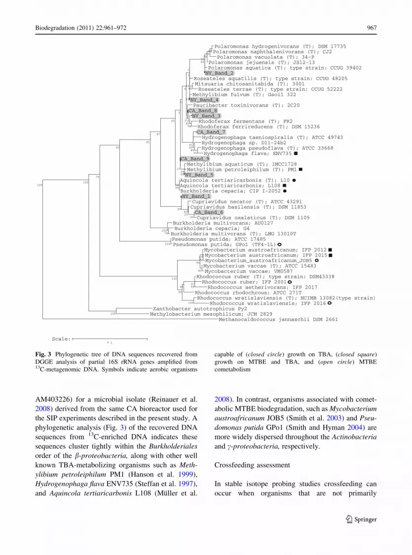
phylogenetic analysis (Fig. 3) of the recovered DNA
sequences from 13C-enriched DNA indicates these
sequences cluster tightly within the Burkholderiales
order of the b-proteobacteria, along with other well
known TBA-metabolizing organisms such as Meth-
ylibium petroleiphilum PM1 (Hanson et al. 1999),
Hydrogenophaga flava ENV735 (Steffan et al. 1997),
and Aquincola tertiaricarbonis L108 (Muller et al.
2008). In contrast, organisms associated with comet-
abolic MTBE biodegradation, such as Mycobacterium
austroafricanum JOB5 (Smith et al. 2003) and Pseu-
domonas putida GPo1 (Smith and Hyman 2004) are
more widely dispersed throughout the Actinobacteria
and c-proteobacteria, respectively.
Crossfeeding assessment
In stable isotope probing studies crossfeeding can
occur when organisms that are not primarily
Fig. 3 Phylogenetic tree of DNA sequences recovered from
DGGE analysis of partial 16S rRNA genes amplified from13C-metagenomic DNA. Symbols indicate aerobic organisms
capable of (closed circle) growth on TBA, (closed square)
growth on MTBE and TBA, and (open circle) MTBE
cometabolism
Biodegradation (2011) 22:961–972 967
123

responsible for metabolism of the isotopically-labeled
test substrate assimilate and grow on labeled metab-
olites excreted by primary degraders. To address the
possibility of this crossfeeding effect in the present
study, we compared DGGE profiles of the partial 16S
rRNA sequences derived from purified 13C-DNA
extracted from NY BioGAC samples receiving either
one or multiple additions of 13C4-TBA (See exper-
iment described in Fig.1). Five distinct DNA bands
were detected for the microcosm that received only a
single 13C4-TBA addition while only three bands
were detected for the microcosm that received five
sequential 13C4-TBA additions (Fig. 4). While higher
amounts of TBA might have been expected to
increase the likelihood of crossfeeding, our results
suggest that multiple additions of 13C4-TBA
decreased the diversity of organisms detected. One
possible explanation of this effect is that repeated
additions of TBA effectively made these microcosms
an enrichment culture and selected for the faster
growing strains of TBA-utilizing organisms.
Gene level analysis
Genomic DNA from pure cultures of M. petroleiph-
ilum PM1 and 13C-DNA obtained from both NY and
CA BioGAC that received a single 13C4-TBA addi-
tion were further analyzed for the presence of specific
genes that have been associated with TBA biodegra-
dation in M. petroleiphilum PM1 (Hristova et al.
2007). PCR reactions targeting four PM1 genes
(Mpe_B0532, Mpe_B0541, Mpe_B0555, and
Mpe_B0561) were conducted on all three DNA
samples, as described in the Methods section. The
products of these amplification reactions were further
analyzed by DGGE (Fig. 5). All visible bands were
extracted from the DGGE gel, purified, sequenced,
and aligned, as described in the Methods section. In
all cases, the DGGE analysis resulted in a single clear
band for each PM1 gene and all DGGE-recovered
PM1 sequences were identical to the partial PM1
gene sequences available in GenBank and as
described in Methods and Materials. The differences
in band migration for amplification products gener-
ated from 13C-DNA from our microcosm studies and
strain PM1 suggested that the DNA sequences of
these products were different than the sequences for
the products obtained from strain PM1. However, in
most cases, the deduced amino acid sequences
encoded by these differently migrating DNA frag-
ments were highly similar (Table 3). For example, for
gene Mpe_B0555 there were 14 nucleotide differ-
ences between sequences amplified from 13C-DNA
obtained from our microcosms as compared to the
PM1 sequence. However, only two of these DNA
sequence changes encode amino acid substitutions.
In contrast, for gene Mpe_B0541, 52 nucleotide
differences were detected in DNA sequences from13C-DNA obtained from our microcosms compared
to the corresponding gene sequence for strain PM1.
In this instance 13 of the 52 differences encode for
amino acid substitutions.
Fig. 4 Effect of TBA
additions on microbial
diversity in NY BioGAC
samples. The Figure shows
a DGGE analysis of partial
16S rRNA genes PCR-
amplified from 13C-DNA
fractions obtained from
microcosms constructed
with BioGAC materials
from New York (NY)
treated with either one or
five sequential additions of13C-TBA (See Fig. 1)
Fig. 5 Analysis of gene diversity in 13C-metagenomic DNA.
The Figure shows a DGGE analysis of partial genes PCR-
amplified from either M. petroleiphilum PM1 or 13C-metage-
nomic DNA obtained from microcosms constructed with
BioGAC obtained from New York (NY) or California (CA)
exposed to a single addition of 13C4-TBA
968 Biodegradation (2011) 22:961–972
123

Discussion
The results of this study demonstrate that 13C-DNA
SIP using 13C4-TBA enabled us to identify several
novel TBA-utilizing organisms from self-inoculated
BioGAC reactors. The 16S rRNA gene sequences
recovered from NY BioGAC microcosms incubated
with a single pulse of 13C-TBA indicated that at
least five different sequences representing five
potentially different microorganisms were recovered.
The DNA sequences of partial 16S rRNA genes
showed these organisms all belonged to class
Betaproteobacteria, order Burkholderiales and
included representatives from the genera Polaro-
monas, Cupriavidus, Rhodoferax, and Methylibium.
Polaromonas strains have previously been shown to
be important members of surface-attached microbial
communities in GAC-based water purification sys-
tems (Magic-Knezev et al. 2009). Cupriavidus
strains are well known for their metabolic versatility
(Lykidis et al. 2010; Perez-Pantoja et al. 2008).
However, as far as we are aware, no members of
three of the four genera identified from our studies
of NY BioGAC have previously been shown to
grow on either TBA or MTBE.
For the CA BioGAC, all four of the 16S rRNA
sequences obtained from the DGGE analysis were
also related to organisms belonging to the Betapro-
teobacteria. However, the organisms represented by
these sequences were different from those obtained
from the NY BioGAC and included representatives of
the genera Methylibium, Hydrogenophaga, Paucib-
acter, and Cupriavidus. Among these genera, only
two out of four are related to organisms that have
previously been shown to metabolize TBA. The best-
characterized aerobic MTBE- and TBA-utilizing
organism is M. petroleiphilum PM1. This organism
was originally isolated from a peat moss biofilter in
California and similar strains have been found at
several sites in that state (Kane et al. 2001). The
Hydrogenophaga sequence we obtained was closely
related (98.5% sequence similarity) to the closest
BLAST sequence match (GenBank accession no.
AM403226) previously reported for a microbial
isolate obtained from the only other previous study
of aerobic TBA-metabolizing bacteria in BioGAC
systems (Reinauer et al. 2008). This Hydrogenoph-
aga strain was the dominant member of a mixed
TBA-degrading culture, derived from samples of theTa
ble
3H
eter
og
enei
tyan
aly
sis
for
gen
esfo
un
din
DG
GE
-sep
arat
edD
NA
seq
uen
ces
fro
mN
Yan
dC
AB
ioG
AC
sam
ple
s
Gen
eID
PM
1g
ene
len
gth
(bp
)
Sam
ple
seq
uen
ce
ov
erla
po
fP
M1
seq
uen
ce(b
p)
%g
ene
cov
erag
e
Nu
mb
ero
fn
ucl
eoti
de
dif
fere
nce
sin
ov
erla
pp
ing
reg
ion
(%si
mil
arit
yto
PM
1g
ene
seq
uen
ce)
Nu
mb
ero
fn
ucl
eoti
de
dif
fere
nce
sco
din
g
for
amin
oac
idsu
bst
itu
tio
ns
Am
ino
acid
sub
stit
uti
on
s
Mp
e_B
05
32
12
48
41
73
31
0(9
7.6
)3
Val
?L
eu
Pro
?S
er
Iso?
Met
Mp
e_B
05
41
16
89
46
22
65
2(8
8.7
)1
3
Mp
e_B
05
55
14
13
49
33
51
4(9
7.1
)2
Try
?S
er
Val
?L
eu
Mp
e_B
05
61
89
44
23
47
19
(95
.5)
4P
ro?
Ala
Ly
s?
Glu
Iso?
Val
Val
?Is
o
Biodegradation (2011) 22:961–972 969
123

same BioGAC reactor used in our present SIP-based
study. This result illustrates that SIP was successful
in identifying at least one TBA-oxidizer belonging to
the same genus as an isolate recovered from the same
BioGAC. However, our results also illustrate that SIP
can identify a wider diversity of metabolically active
microorganisms than those identified using enrich-
ment culture techniques. Even so, SIP may also under
represent the diversity of active metabolizers in a
community, as evidenced by the decrease in the
number of different 16S rRNA sequences recovered
when the number of 13C-TBA pulses was increased
from one to five (Fig. 4).
A concern with all SIP-based approaches is that
labeled substrates are often used at concentrations
that may not be environmentally relevant (Madsen
2006) and that this can cause shifts in the microbial
population so that detected organisms are no longer
representative of the targeted natural community. The
BioGAC samples used in our analyses came from
NY- and CA-based reactors treating groundwater
with TBA levels as high as 10 and 350 ppm,
respectively. As we have also shown in this study,
the sorptive properties of the GAC can cause
substantial decreases in aqueous phase TBA concen-
trations and presumably correspondingly large
increases in the concentration of organics experi-
enced by surface-attached bacteria. We also focused
on results obtained when BioGAC samples were
exposed to a single addition of 13C4-TBA and also
demonstrated that for NY BioGAC, multiple TBA
exposures decreased rather than increased the micro-
bial diversity detected in our samples. We therefore
conclude that the aqueous TBA concentration in our
microcosms (*300 ppm) is comparable, albeit at the
high end, to concentrations of TBA these reactor
samples may have experienced during field operation.
Additionally, we can conclude that the microorgan-
isms identified by the recovered 16S rRNA sequences
are representative of the primary TBA-metabolizers
in the BioGAC microcosms rather than organisms
that have assimilated 13C label through crossfeeding
processes. However, it should be recognized that
operational BioGAC reactors often contain several
thousand kilograms of original GAC and the spatial
and temporal distribution of surface-attached TBA-
utilizing organisms within these reactors is unknown.
As this present study made use of small samples
(\100 g wet weight) of GAC material taken from a
single location within a reactor we do not expect that
our SIP analysis has fully captured the diversity of
TBA-utilizing organisms within each reactor. As
this initial study did not examine the reproducibility
of our SIP analysis between subsamples of Bio-
GAC we also do not know whether small-scale
heterogeneities exist within these materials. We are
currently completing a more detailed SIP analysis
of the colonization and distribution of TBA-utiliz-
ing organisms within an operational BioGAC
reactor. This study directly assesses these issues
of local and reactor-scale heterogeneity and relates
microbial distribution to reactor performance
through a concurrent analysis of reactor pore water.
Our functional gene analysis targeted four genes
that have been associated with TBA biodegradation
by pure cultures of M. petroleiphilum PM1. Genes
Mpe_B0532, Mpe_B0541, Mpe_B0555, Mpe_B0561
were selected for our study based on the results of a
comparative transcriptome analysis which indicated
they were differentially expressed (3- to 12-fold
upregulated) when PM1 was grown on MTBE versus
ethanol (Hristova et al. 2007). Similar genes were all
detected in our NY and CA BioGAC 13C-DNA
samples, suggesting they may be frequently detected
in TBA metabolizing organisms. Additionally, the
deduced amino acid sequences for genes
Mpe_B0555, Mpe_B0532, and Mpe_B0561 detected
in our BioGAC samples were highly similar to the
corresponding sequences for PM1, further suggesting
that the predicted proteins are well conserved. Our
findings are particularly relevant for Mpe_B0555 that
codes for a phthalate dioxygenase-like enzyme, a
protein recently detected during a proteomic analysis
of Aquincola tertiaricarbonis L108 after growth on
TBA (Schafer et al. 2007). Detection of these genes,
specifically Mpe_B0555, may indicate a site’s
potential for TBA-degrading activity and therefore
could inform decisions regarding whether self-inoc-
ulation of BioGAC reactors intended to treat TBA-
impacted ground water is likely to be an effective
strategy.
Acknowledgments We thank Xiaomin Yang (Atlantic
Richfield Company) for providing BioGAC samples from
California. This research was supported by funding to MRH
from the National Science Foundation (Grant CBET-0348392)
and the American Petroleum Institute. DA was supported by a
DoEd Graduate Assistantship in Areas of National Need
fellowship.
970 Biodegradation (2011) 22:961–972
123

References
Beyers DL, Meyer CL, Sun PT, Salanitro JP (2001) Method
and apparatus for biodegradation of alkyl ethers and ter-
tiary butyl alcohol. US Patent # 6458276
Bradley PM, Landmeyer JE, Chapelle FH (1999) Aerobic
mineralization of MTBE and tert-butyl alcohol by stream-
bed sediment microorganisms. Environ Sci Technol
33:1877–1879
Bradley PM, Landmeyer JE, Chapelle FH (2002) TBA bio-
degradation in surface-water sediments under aerobic and
anaerobic conditions. Environ Sci Technol 36:4087–4090
Cirvello JD, Radovsky A, Heath JE, Farnell DR, Lindamood C
(1995) Toxicity and carcinogenicity of t-butyl alcohol in
rats and mice following chronic exposure in drinking
water. Toxicol Ind Health 11:151–165
Clark JJJ (2002) tert-butyl alcohol: chemical properties, pro-
duction and use, fate and transport, toxicology, and
detection in groundwater and regulatory standards. In:
Diaz AF, Drogos DL (eds) Oxygenates in gasoline:
environmental aspects, vol. chapter 7. American Chemical
Society, Washington, pp 92–106
Cole JR, Chai B, Farris RJ, Wang Q, Kulam-Syed-Mohideen
AS, McGarrell DM, Bandela AM, Cardenas E, Garrity
GM, Tiedje JM (2007) The ribosomal database project
(RDP-II): introducing myRDP space and quality con-
trolled public data. Nucleic Acids Res 35
Deeb RA, Chu K-H, Shih T, Linder S, Suffet I, Kavanaugh
MC, Alvarez-Cohen L (2003) MTBE and other oxygen-
ates: environmental sources, analysis, occurrence, and
treatment. Environ Eng Sci 20:433–447
Fortin NY, Morales M, Nakagawa Y, Focht DD, Deshusses
MA (2001) Methyl tert-butyl ether (MTBE) degradation
by a microbial consortium. Environ Microbiol 3:407–416
Francois A, Mathis H, Godefroy D, Piveteau P, Fayolle F,
Monot F (2002) Biodegradation of methyl tert-butyl ether
and other fuel oxygenates by a new strain, Mycobacteriumaustroafricanum IFP 2012. Appl Environ Microbiol
68:2754–2762
Goodfellow M, Jones AL, Maldonado LA, Salanitro J (2004)
Rhodococcus aetherivorans sp. nov. A new species that
contains methyl t-butyl ether-degrading Actinomycetes.
Syst Appl Microbiol 27:61–65
Hanson JR, Ackerman CE, Scow KM (1999) Biodegradation
of methyl tert-butyl ether by a bacterial pure culture. Appl
Environ Microbiol 65:4788–4792
Hatzinger PB, McClay K, Vainberg S, Tugusheva M, Condee
CW, Steffan RJ (2001) Biodegradation of methyl tert-butyl ether by a pure bacterial culture. Appl Environ
Microbiol 67:5601–5607
Hristova KR, Schmidt R, Chakicherla AY, Legler TC, Wu J,
Chain PS, Scow KM, Kane SR (2007) Comparative
transcriptome analysis of Methylibium petroleiphilumPM1 exposed to the fuel oxygenates methyl tert-butyl
ether and ethanol. Appl Environ Microbiol 73:7347–7357
Kane SR, Beller HR, Legler TC, Koester CJ, Pinkart HC,
Halden RU, Happel AM (2001) Aerobic biodegradation of
methyl tert-butyl ether by aquifer bacteria from leaking
underground storage tank sites. Appl Environ Microbiol
67:5824–5829
Kharoune M, Kharoune L, Lebeault JM, Pauss A (2001) Iso-
lation and characterization of two aerobic bacterial strains
that completely degrade ethyl tert-butyl ether (ETBE).
Appl Microbiol Biotechnol 55:348–353
Lane DJ, Pace B, Olsen GJ, Stahl DA, Sogin ML, Pace NR
(1985) Rapid determination of 16S ribosomal RNA
sequences for phylogenetic analyses. Proc Nat Acad Sci
USA 82:6955–6959
Larkin MA, Blackshields G, Brown NP, McGettigan CR,
McGettigan PA, McWilliam H, Valentin F, Wallace IM,
Wilm A, Lopez R, Thompson JD, Gibson TJ, Higgins DG
(2007) ClustalW and ClustalX version 2. Bioinformatics
23:2947–2948
Lykidis A, Perez-Pantoja D, Ledger T, Mavromatis K, Anderson
IJ, Ivanova NN, Hooper DS, Lapidus A, Lucas S, Gonzalez
B, Kyrpides NC (2010) The complete multipartite genome
sequence of Cupriavidus necator JMP134, a versatile
pollutant degrader. PLoS ONE 5:e9729
Madsen EL (2006) The use of stable isotope probing tech-
niques in bioreactor and field studies on bioremediation.
Curr Opin Biotechnol 17:92–97
Magic-Knezev A, Wullings B, Kooij DVD (2009) Polaro-monas and Hydrogenophaga species are the predominant
bacteria cultured from granular activated carbon filters in
water treatment. J Appl Microbiol 107:1457–1467
Mo K, Lora CO, Wanken AE, Javanmardian M, Yang X,
Kulpa CF (1997) Biodegradation of methyl t-butyl ether
by pure bacterial cultures. Appl Microbiol Biotechnol
47:69–72
Muller RH, Rohwerder T, Harms H (2008) Degradation of fuel
oxygenates and their main intermediates by Aquincolatertiaricarbonis L108. Microbiology 154:1414–1421
Muyzer G, Waal ECD, Uitterlinden AG (1993) Profiling of
complex microbial populations by denaturing gradient gel
electrophoresis analysis of polymerase chain reaction-
amplified genes coding for 16S rRNA. Appl Environ
Microbiol 59:695–700
Neufeld JD, Vohra J, Dumont MG, Lueders T, Manefield M,
Friedrich MW, Murrell JC (2007) DNA stable-isotope
probing. Nat Protoc 2:860–866
Perez-Pantoja D, Iglesia RDI, Pieper DH, Gonzalez B (2008)
Metabolic reconstruction of aromatic compounds degra-
dation from the genome of the amazing pollutant-
degrading bacterium Cupriavidus necator JMP134. FEMS
Microbiol Rev 32:736–794
Piveteau P, Fayolle F, Vandecasteele JP, Monot F (2001)
Biodegradation of tert-butyl alcohol and related xenobi-
otics by a methylotrophic bacterial isolate. Appl Micro-
biol Biotechnol 55:369–373
Reinauer K, Zhang Y, Yang X, Finneran K (2008) Aerobic
biodegradation of tert-butyl alcohol (TBA) by psychro-
and thermo-tolerant cultures derived from granular acti-
vated carbon (GAC). Biodegradation 19:259–268
Rozen S, Skaletsky HJ (2000) Primer3 on the WWW for
general users and for biologist programmers. Humana
Press, Totowa, NJ
Sabat G, Rose P, Hickey WJ, Harkin JM (2000) Selective and
sensitive method for PCR amplification of Escherichiacoli 16S rRNA genes in soil. Appl Environ Microbiol
66:844–849
Biodegradation (2011) 22:961–972 971
123
Salanitro JP, Diaz LA, Williams MP, Wisniewski HL (1994)
Isolation of a bacterial culture that degrades methyl
t-butyl ether. Appl Environ Microbiol 60:2593–2596
Sambrook J, Russell DW (2001) Molecular cloning: a labora-
tory manual, 3rd edn. Cold Spring Harbor Press, Cold
Spring Harbor, New York
Schafer F, Breuer U, Benndorf D, Bergen MV, Harms H,
Muller RH (2007) Growth of Aquincola tertiaricarbonisL108 on tert-butyl alcohol leads to the induction of a
phthalate dioxygenase-related protein and its associated
oxidoreductase subunit. Eng Life Sci 7:512–519
Schmidt TC, Zwank L, Elsner M, Berg M, Meckenstock RU,
Haderlein SB (2004) Compound-specific stable isotope
analysis of organic contaminants in natural environments:
a critical review of the state of the art, prospects, and
future challenges. Anal Bioanal Chem 378:283–300
Singleton DR, Powell SN, Sangaiah R, Gold A, Ball LM,
Aitken MD (2005) Stable-isotope probing of bacteria
capable of degrading salicylate, naphthalene, or phenan-
threne in a bioreactor treating contaminated soil. Appl
Environ Microbiol 71:1202–1209
Smith CA, Hyman MR (2004) Oxidation of methyl tert-butyl
ether by alkane hydroxylase in dicyclopropylketone-
induced and n-octane-grown Pseudomonas putida GPo1.
Appl Environ Microbiol 70:4544–4550
Smith CA, O’Reilly KT, Hyman MR (2003) Characterization
of the initial reactions during the cometabolic oxidation of
methyl tert-butyl ether by propane-grown Mycobacteriumvaccae JOB5. Appl Environ Microbiol 69:796–804
Steffan RJ, McClay K, Vainberg S, Condee CW, Zhang D
(1997) Biodegradation of the gasoline oxygenates methyl
tert-butyl ether, ethyl tert-butyl ether, and tert-amyl
methyl ether by propane-oxidizing bacteria. Appl Environ
Microbiol 63:4216–4222
Steffan RJ, Vainberg S, Condee CW, McClay K, Hatzinger P
(2000) Biotreatment of MTBE with a new bacterial iso-
late. In: Wickramanayake GB, Gavaskar AR, Alleman
BC, Magar VS (eds) Bioremediation and phytoremedia-
tion of chlorinated and recalcitrant compounds. Battelle
Press, Columbus, OH
Sun PT, Walsh D, Meyer C, Pickle D (2003) The treatment of
MTBE-contaminated groundwater in bioaugmented
granular activated carbon beds—a case history. Proc
Water Environ Fed 14:450–463
Wang Q, Garrity GM, Tiedje JM, Cole JR (2007) Naive
Bayesian classifier for rapid assignment of rRNA
sequences into the new bacterial taxonomy. Appl Environ
Microbiol 73:5261–5267
Wilson JT, Adair C, Kaiser PM, Kolhatkar R (2005) Anaerobic
biodegradation of MTBE at a gasoline spill site. Ground
Water Monit Remed 25:103–115
Yeager CM, Bottomley PJ, Arp DJ, Hyman MR (1999) Inacti-
vation of toluene 2-monooxygenase in Burkholderia cepa-cia G4 by alkynes. Appl Environ Microbiol 65:632–639
972 Biodegradation (2011) 22:961–972
123