identification of a program of contractile protein gene expression initiated upon skeletal muscle...
TRANSCRIPT

DEVELOPMENTAL DYNAMICS 1 9 6 2 W 6 (1993)
Identification of a Program of Contractile Protein Gene Expression Initiated Upon Skeletal Muscle Differentiation COLIN J. SUTHERLAND, KARYN A. ESSER, VICKI L. ELSOM, MONICA L. GORDON, AND EDNA C. HARDEMAN Muscle Development Unit (C.J.S., K.A.E., V.L.E., M.L.G., E.C.H.) and Cell Biology Unit (KA.E.1, Children’s Medical Research Institute, Locked Bag 23, Wentworthville, N.S.W. 2145, Australia
ABSTRACT The functional diversity of skel- etal muscle is largely determined by the combi- nations of contractile protein isoforms that are expressed in different fibers. Just how the devel- opmental expression of this large array of genes is regulated to give functional phenotypes is thus of great interest. In the present study, we perform a comprehensive analysis of contractile protein iso- form mRNA profiles in skeletal muscle systems representing each generation of fiber formed: pri- mary, secondary, and regenerating fibers. We find that in each system examined there is a common pattern of isoform gene expression during early differentiation for 5 of the 6 gene families we have investigated: myosin light chain (MLCI1, MLC2, tropomyosin, troponin (Tn)C, and TnI. We suggest that the common isoform patterns observed to- gether represent a genetic program of skeletal muscle differentiation that is independent of the mature fiber phenotype and is found in all newly formed myotubes. Within each of these contractile protein gene families the program is independent of the isoforms of myosin heavy chain (MHC) ex- pressed. The maintenance of such a program may reflect a specific requirement of the initial differ- entiation process. 0 1993 Wiley-Liss, Inc.
Key words: Contractile protein genes, Skeletal muscle, Regeneration, Differentia- tion, Rodent, Human, Genetic pro- gram
INTRODUCTION In the rodent, there are three recognized stages of
skeletal muscle cell differentiation. Primary and sec- ondary myoblasts give rise to two sets of fibers which are formed at discrete developmental stages and which together constitute the basic complement of fibers found in the adult (Rubinstein and Kelly, 1981; Harris et al., 1989; Condon et al., 1990). The third stage of differentiation is mediated by satellite cells, quiescent myogenic cells present in juvenile and adult skeletal muscles. When activated, satellite cells proliferate to produce the myoblasts needed for further growth and for the regeneration of injured muscle (Campion, 1984; Schultz et al., 1986). At each of these stages, myoblasts irreversibly exit from the cell cycle prior to differenti-
0 1993 WILEY-LISS. INC.
ation, undergo fusion to form myotubes, and initiate expression of genes encoding the contractile apparatus. The major sarcomeric components of the contractile ap- paratus, myosin, actin, tropomyosin, and troponin, are encoded by multigene families which include fast, slow, cardiac, and developmental isoforms. As a conse- quence, the contractile protein composition of a skele- tal muscle fiber can theoretically be any one of a large number of possible isoform combinations.
It is well established that the contractile protein iso- form profile of developing skeletal muscle cells differs from that of adult muscle. Whalen et al. (1981) describe the transition from expression of development-specific MHC isoforms in embryonic rat muscle to expression of adult isoforms in postnatal muscles. MHC isoform transitions have also been described during myogene- sis in mouse (Weydert et al., 1983, 1987) and chicken (Bandman et al., 1982). Developing avian muscles ex- hibit isoform transitions in the MLCl gene family (Barton et al., 1988), the actin gene family (Hayward and Schwartz, 1986), and the tropomyosin (Tm) and troponin T (TnT) gene families (Matsuda et al., 1981, 1984). Similarly, in mammals, embryonic isoform pro- files differ from those of adult skeletal muscles for MLCl (Periasamy et al., 1984; Barton et al., 1988, 1989; Lyons et al., 1990), actin (Buckingham, 1985), and TnT (Briggs et al., 1990; Saggin et al., 1990). How- ever, the majority of these studies present qualitative data only. In a recent in vivo study of contractile pro- tein isoform gene expression in rat and human limb development, we have shown that embryonic and adult isoform profiles differ for virtually all contractile pro- tein gene families (Sutherland et al., 1991).
An isoform phenotype unlike that of adult muscle is exhibited not only by embryonic muscle in vivo, but also by newly differentiated muscle cells in vitro. Hu- man myogenic cells when differentiated in vitro form myotubes that express an isoform phenotype differing from that of adult human muscle for 8 contractile pro- tein gene families (Gunning et al., 1987; Wade et al.,
Received October 23, 1992. Address reprint requestsicorrespondence to Edna C. Hardeman,
Muscle Development Unit, Children’s Medical Research Institute, Locked Bag 23, Wentworthville, N.S.W. 2145, Australia.

26 SUTHERLAND ET AL.
1990). It would appear, then, that newly differentiated skeletal muscle cells, both in vivo and in vitro, exhibit a contractile protein isoform phenotype that is not like the isoform phenotype of the adult muscle fiber. A cru- cial question thus arises: does the contractile protein isoform phenotype seen during development represent a random pattern of gene expression, or is there an underlying genetic program with implications for our understanding of the regulation of myogenesis?
In this study, we attempt to resolve this question by undertaking a comprehensive quantitative comparison of contractile protein isoform gene expression in a va- riety of differentiating human and rodent skeletal muscle cell systems. These systems include fetal myo- blasts and satellite cells in vitro, and both developing and regenerating muscles in vivo. With this strategy, we sample the widest variety of newly differentiated skeletal muscle cells available to us. This includes myogenic samples representative of mixed muscle com- position (i.e., whole limbs), derivatives of cloned muscle cells, and individual muscle types. We find that for five of the six multigene families investigated a striking qualitative pattern emerges that is repeated in each of the differentiating muscle cell systems. These findings suggest that a program of muscle gene expression ex- ists that is characteristic of differentiating skeletal muscle cells and independent of future fiber fate. One possible explanation is that this program ensures ex- pression of genes which encode contractile protein iso- forms that are advantageous for the initial assembly of sarcomeres.
RESULTS Regenerating Soleus and EDL Muscles Express the Same Heterogenous Pattern of Contractile Protein Isoform Genes Upon Differentiation In Vivo (Fig. 1, Table 1)
The aim of this study is to identify specific patterns of contractile protein gene expression that may be as- sociated with new myotube formation. The regenera- tion of muscle in vivo is a useful model for this purpose, providing a myogenic system in which the cell biology has been well characterized, differentiation occurs rel- atively synchronously, and muscles of different fast or slow phenotypes can be studied. The primary question is as follows: does a consistent pattern of contractile protein isoform gene expression occur upon initial dif- ferentiation in both fast and slow regenerating muscles or, alternatively, do the isoform expression patterns reflect previous fiber history? Muscle regeneration was induced in the soleus (slow) and extensor digitorum longus (EDL) muscles in the Wistar rat. The method involves the use of surgical techniques in combination with the myotoxic agent marcaine (Experimental Pro- cedures). This protocol ensures that the gene expres- sion patterns determined are that of newly formed my- otubes and not of original myofibers, as evidenced by the lack of adult MHC expression in both regenerating muscle types (data not shown).
Fig. 1. MLCZ,, MLCl,, and Tnl, mRNA expression in regenerating soleus and EDL. Northern analysis was performed as described in Ex- perimental Procedures. Total RNA (2-5 pg) from the source indicated was run in each lane. Sol, adult soleus; EDL, adult extensor digitorum longus; d4S, regenerating denervated soleus 4 days after surgery; d4E, regenerating denervated EDL 4 days after surgery.
Northern analysis was performed on total RNA from day 4 regenerating soleus and EDL muscles. Day 4 was chosen as the point when new myotubes are beginning to form within the regenerating muscle. Isoform mRNA expression was measured for 6 contractile pro- tein gene families and representive autoradiographs of 3 gene families are presented in Figure 1. An impor- tant point is illustrated by the isoform expression pat- tern from the MLC1, MLC2, and TnI gene families. Specifically, for the MLCl gene family, as previously shown by others (Carraro et al., 1983), the atrial iso- form is expressed in the intial stages of regeneration by both fast and slow muscles. For the MLC2 gene family, the fast isoform gene is expressed not only in the re- generating fast muscle (EDL), but also in the regen- erating slow muscle (soleus). In contrast, the slow isoform from the TnI gene family is expressed signifi- cantly and to the same magnitude in both regenerating soleus and EDL muscles. These results indicate that the newly formed myotubes display a heterogeneous pattern of isoform expression in that there are contri- butions from fast, slow, and cardiac isoform genes. An- other critical finding is that for MLC2,1,,~,,, MLCl,,, MLC2,trial(a,, and the TnIcardiacfc) genes no signal was detected (level of sensitivity -0.5% of adult level, data not shown). This is significant because although the pattern of isoform gene expression is characterized by a very heterogeneous combination of striated muscle iso- forms, this pattern does not appear to be the result of a program of general expression from all the striated muscle isoform genes.
The level of expression of 18 contractile protein genes in both regenerating muscles is presented in Ta- ble 1. Quantitative densitometry was performed on the autoradiographs and normalized to 18 S ribosomal

GENE EXPRESSION DURING MUSCLE DIFFERENTIATION 27
TABLE 1. Contractile Protein Isoform mRNA Expression
During Skeletal Muscle Regeneration in Fast and Slow
Rat Muscles In Vivoa
% of mRNA total for each Soleus EDL gene family (slow) (fast) MLCl
1/3 Fast 6 12 Slow, 46 43 Slow, - - Atrial 48 45
MLC2 Fast 100 100 slow - -
- - Atrial
a-Fast 5 9 a-Slow 9 3 P 86 88
Fast 45 43 Slow 55 58
Tm
TnC
TnI Fast 5 11 Slow 95 89 Cardiac - -
Fast 2 3 Slow 69 73
TnT
Cardiac 29 24
T h e relative values for each isoform listed within a gene family are esti- mations (see Experimental F’roce- dures).
RNA levels as described in Experimental Procedures. It is apparent that a virtually identical pattern of iso- form expression occurs in both the soleus and EDL muscles at 4 days of regeneration. Each isoform con- tributes the same relative output from its respective gene family and, additionally, those isoforms that are undetectable in the regenerating EDL are also unde- tectable in the soleus regenerates. These data clearly indicate that upon initial myotube formation during regeneration, both EDL and soleus exhibit a very con- sistent pattern of contractile protein isoform gene ex- pression, irrespective of the phenotypic history of the regenerates. This pattern includes expression of fast, slow, and cardiac isoforms, and so does not reflect fast or slow adult fiber fate.
Satellite Cell Differentiation In Vitro (Fig. 2, Table 2, columns 1 through 4)
The myoblasts associated with myofiber regenera- tion, satellite cells, can also be studied in vitro. The C2 myogenic cell line is an established clone derived from adult mouse satellite cells which were stimulated to divide and grow by muscle injury (Yaffe and Saxel, 1977). This model provides a means to examine the
potential of satellite cells differentiating in vitro, al- lowing comparison with the patterns of gene expres- sion we have seen during satellite cell-mediated regen- eration in vivo.
C2 cultures were induced to differentiate and total RNA extracted after 4 days at which time the cultures consisted largely of contracting myotubes. Northern analysis was performed and autoradiographs showing the expression pattern of the two skeletal isoforms of MLC2 are presented in Figure 2. This family repre- sents one of the most consistent and extreme examples of restricted preferential expression as will become ap- parent. C2 myotubes clearly display a strong bias to- wards expression of the fast isoform for this gene fam- ily as was seen with regenerating rat soleus and EDL (Fig. 1; Table 1). Quantitative densitometric analysis was used to estimate the relative contribution of each isoform to the total gene family mRNA pool (Table 2, column 1). The C2 myotubes display a pattern of con- tractile protein mRNA expression that is similar to the pattern found in the regenerating fast and slow mus- cles for 5 gene families: MLC2, Tm, TnC, TnI, and TnT (Table 1). For the MLCl gene family, the only major difference between the two systems is that the atrial isoform is strongly expressed in fast and slow regener- ates in vivo, but only moderately expressed in the C2 cultures.
The similarity of the differentiation pattern of sat- ellite cells to that of regenerating muscle in vivo is reinforced by the finding of a similar pattern in differ- entiating human satellite cells in vitro. We have pre- viously examined contractile protein isoform expres- sion in human myogenic cells which were clonally derived directly from primary cell cultures (Wade et al., 1990). In the present study, we measured the ex- pression of the atrial isoforms MLC1, and MLC2,. The calculation of isoform mRNA levels for all contractile protein isoform genes is presented in Table 2 (column 2). When compared with the C2 cells and with the re- generating rat muscles, the human satellite cells show similar isoform mRNA expression patterns in 4 gene families: MLC2, Tm, TnC, and TnI. The pattern of TnT isoform expression in the human satellite cells differs from the rodent systems examined in that there is sig- nificant expression of the fast isoform.
Previously, we examined the expression of contrac- tile protein isoform mRNAs during early myogenesis by bulk analysis of whole rat and human hindlimbs (Sutherland et al., 1991). At developmental stages when only primary myotubes are present in rat (E16) and human (week 11) hindlimbs, comparison of the ap- proximate relative contribution of each isoform to total output form each of the contractile protein gene fami- lies reveals a number of apparent similarities (Table 2, columns 3 and 4). The expression patterns from the MLC1, MLC2, Tm, and TnI gene families are similar in rat and human primary myotubes and, with the excep- tion of MLC1, expression, qualitatively similar to the patterns already discerned in regenerating muscle in

28 SUTHERLAND ET AL.
Fig. 2. MLC2 isoform mRNA expression in mouse C2 cultures (C2) and in myotubes derived from El9 rat fetal myoblasts (F Mb). Northern analysis was performed as described in Experimental Procedures. Total RNA (2-5 pg) from the source indicated was run in each lane. d, days
vivo and in C2 and human satellite cell cultures in vitro (Table 1; Table 2, columns 1 and 2).
The similarity of contractile protein isoform mRNA levels between primary myotubes and new myotubes derived from satellite cells in vivo and in vitro suggests that the expression patterns exhibited by recently formed myotubes is not random. We therefore sought to evaluate whether this pattern of mRNA accumulation is also evident when secondary myoblasts undergo ini- tial differentiation.
Contractile Protein Gene Expression During Secondary Myoblast Differentiation (Fig. 2, Table 2, Column 5)
Based on MHC expression, primary and secondary myotubes in vivo and in vitro are classically described as displaying distinct phenotypes; the former has a mixed fasttslow composition and the latter a fast iso- form composition (Rubinstein and Kelly, 1981; Vi- varelli et al., 1988; Harris et al., 1989). We reasoned that this restriction in fast and slow isoform expression patterns may apply to the other contractile protein gene families, and so sought to compare isoform mRNA expression in secondary myotubes with our other my- otube data. During development, secondary myotubes are formed in close apposition to the initial primary myotubes (Duxson et al., 1989). Therefore, it is not pos- sible to quantitatively measure gene expression specif- ically in secondary myotubes as they form in vivo in the hindlimb. However, the fetal myoblasts that give rise to secondary myotubes can be isolated and will differentiate in culture. Such cultures are noninner- vated, and so may be more representative of the intrin- sic isoform repertoire of secondary myoblasts than are innervated secondary myotubes in situ. Thus, on the
after addition of fusion medium; Gstr, adult mouse gastrocnemius; Sol, adult mouse (C2) or rat (F Mb) soleus; EDL, adult rat extensor digitorurn longus.
basis of the known pattern of MHC expression, one might predict that the myotubes in these cultures would express a predominantly “fast” contractile pro- tein isoform phenotype.
We cultured cells from the hindlimb of embryonic day (E) 19.5 rat fetuses and induced myoblast fusion. At this stage of gestation the vast majority of myo- blasts will be contributing to secondary fiber forma- tion. Two days after induction of myotube formation, the differentiated cultures were harvested for total RNA extraction. The contractile protein isoform ex- pression pattern of these cultures was determined by northern analysis. Representative autoradiographs showing hybridization to fast and slow MLC2 isoforms are presented in Figure 2. As seen for the other myo- genic systems (Tables 1, 21, these fetal myogenic cul- tures show a strong preference for expression of the fast isoform mRNA in this particular gene family, the slow isoform mRNA being undetectable. However, not all gene families favored expression of the fast isoform. The approximate relative contribution of each isoform from each gene family was calculated by quantitative densitometry as described in Experimental Procedures and is presented in Table 2 (column 5).
For 4 gene families, the pattern of expression of con- tractile protein mRNA levels in these secondary myo- tubes in vitro is qualitatively similar to that observed in the other myogenic systems. In particular, the mRNAs for MLCl,,, MLC2, P-Tm, and TnI, are the predominant isoforms in their respective gene families (Tables 1, 2). We have observed that with time, the representation of the fast isoform mRNAs increases in these cultures (data not shown) suggesting that the isoform expression profile is not stable. Therefore, by examining these cultures on day 2, we may have

GENE EXPRESSION DURING MUSCLE DIFFERENTIATION 29
TABLE 2. Relative Expression of Contractile Protein Isoforms During Skeletal Muscle Differentiation In Vivo and In Vitro"
Satellite cells Primary MT Secondary MT 1 2 3 4 5
% of mRNA Mouse C2 Human satellite Rat embryonic Human fetal El9 fetal rat muscle total for each cultures cell cultures hindlimb hindlimb cultures gene family day 4 day 15 El6 week 11 day 2 MLC 1
113 Fast 21 38 7 14 33 Slow, 59 59 86 85 53 Slow,(vent) - - - - - Atrial 20 2 7 1 14
Fast 100 90 100 99 100 Slow - 10 - Atrial - - -
a-Fast 14 19 44 36 20 a-slow 14 14 - 14 - P 71 67 56 50 80
MLC 2
- 1 - -
Tm
TnC Fast 34 30 20 54 36 Slow 66 70 80 46 64
5 38 Fast 13 Slow 88 100 100 95 62 Cardiac - - -
Fast 12 42 - 79 59 Slow 66 12 69 9 34 Cardiac 22 46 31 12 7
TnI - -
- - TnT
"Comparison of relative contractile protein isoform mRNA levels in 5 myogenic systems involving fusion of myoblasts from different developmental stages. The relative values for each isoform listed within a gene family are estimations (see Exper- imental Procedures). - signifies undetectable. Some of these data were calculated from previous work (Wade et al., 1990; Sutherland et al., 1991).
missed the absolute window of time during which the initial isoform expression profile exists. This may ac- count for the noteably higher contribution of the TnI fast isoform in this system. The expression of the TnC family in rat secondary myotubes resembles that de- termined for rat primary myotubes in that there is a preference for the slow isoform over the fast isoform. As in all systems investigated so far, there is significant expression from both TnC isoforms. Thus for the gene families examined, myotubes derived from fetal myo- blasts, which in vivo would contribute to secondary my- otubes, exhibit a contractile protein isoform phenotype similar both to that of primary myotubes and to that of myotubes derived from adult satellite cells.
Graphic Comparison of Isoform mRNA Expression Patterns (Fig. 3)
In order to clearly illustrate the similarities among the myogenic systems we have investigated, Figure 3 depicts graphically the isoform expression pattern of each system for the 6 gene families. The mean relative mRNA level for each isoform in all 7 systems is graphed as a percentage of the total output of that gene family. The error bars depict standard error of the mean and provide a useful indicator of the variability
for each isoform among the different myogenic sys- tems. As can be seen from Figure 3, the standard errors are relatively small for 3 gene families (MLCZ, Tm, TnI), indicating tight control of the relative contribu- tion of specific isoforms. The isoform expression of the TnC family is for the most part also consistent, in that both isoforms are significantly expressed in all systems and slow isoform levels are greater than or equal to fast isoform levels. The MLCl gene family displays some variability among the differentiation systems in rela- tive isoform expression (see Discussion). However, all 7 systems consistently express MLC1, mRNA, MLCl,, is more abundant than MLC1/3f, and MLCl,, is undetect- able. In contrast, TnT, is expressed in each system, but otherwise the TnT gene family does not exhibit isoform preferences that consistently recur in the different myogenic systems.
In summary, there are consistent similarities be- tween the isoform profiles of 5 contractile protein gene familes in each of the 7 myogenic systems that have been examined. We suggest that this repeated pattern may reflect a differentiation-specific program of gene expression which occurs in newly formed myotubes de- rived from all three developmental classes of myoblast, irrespective of fiber-type fate. However, isoform mRNA

30 SUTHERLAND ET AL
1/31 i s a lsb l a
MLC1
T
TnCf TnCs
Troponin C
2f 2s 2a
MLC2
loo 1 -r
Tnlf Tnls Tnlc
T r o p o n i n I
8o 1 T
60
50
40
30
20
1 0
0
a-f a-s p
T r o p o m y o s i n
T
TnTf TnTs TnTc
T r o p o n i n T
Fig. 3. Graphical comparison of relative isoform expression for 6 gene families in 7 myogenic systems. The graphs represent the mean of the 7 values given in Tables 1 and 2 for each isoform. Vertical bars represent standard error of the mean.
output from the sixth gene family, TnT, does not ap- pear to be regulated according to a pattern in the myo- genic systems we have investigated, apart from the consistent expression of TnT, mRNA in all newly formed myotubes.
The Early Myogenic Differentiation Program Is Independent of Myosin Heavy Chain Isoform Expression (Fig. 4, Table 3)
The MHC isoform repertoire of a developing myotube is considered to have an important role in determining the phenotype of the myofiber, and so many previous studies of muscle differentiation have investigated the patterns of MHC isoform expression during myogene- sis in vitro and in vivo (Whalen et al., 1981; Stockdale and Miller, 1987; Vivarelli et al., 1988; Harris et al., 1989; Condon et al., 1990). We therefore sought to de- fine MHC isoform expression in each of the myogenic systems included in this study. Using isoform-specific DNA probes derived from amplification of rat MHC 3'
untranslated regions (Sutherland et al., 1991), we iden- tified by northern analysis the MHC isoforms ex- pressed by El9 fetal rat myogenic cultures and regen- erating rat soleus and EDL muscles. Figure 4a shows autoradiographs of the El9 hindlimb cultures probed with MHC isoform probes. MHCemb is highly expressed in these cultures, whereas, MHC,,,, mRNA is only just detectable at day 2, but increases in abundance with time in culture. MHCSI,, is not detectable at any stage in these cultures. The initial MHC isoform phenotype of both regenerating rat soleus and EDL is very similar to this pattern (Fig. 4b). MHC,,, is expressed in both regenerating muscles, and very low levels of MHC,,,, are also discernable. MHCS1,, mRNA is undetectable in both muscles. These results reinforce our finding that the initial patterns of isoform gene expression in early regenerating fast and slow muscles are virtually indistinguishable and therefore independent of fiber- type.
The MHC expression data are summarized in Table 3

GENE EXPRESSION DURING MUSCLE DIFFERENTIATION 31
Fig. 4. MHC isoform mRNA expression in (a) myogenic cultures de- rived from E l 9 fetal rat hindlimbs and (b) regenerating rat EDL and soleus. Abbreviations as for Figure 1 ; Nb: newborn rat hindlimb. Northern analysis was performed as described in Experimental Procedures. Total RNA (2-5 pg) from the timepoints shown, adult EDL, adult soleus, and newborn hindlimb was run in the lanes indicated.
and are compared with previously determined MHC expression data for the other systems investigated. Clearly, the MHC isoform repertoire differs between the various types of myogenic cells examined. We can- not calculate the relative contribution of each MHC isoform as we have for the other gene families because the developmental isoforms, MHCemb and MHC,,,, , are not expressed in any adult muscle that could be used as a quantitative reference. In all of the systems pre- sented in Table 3, MHC,,, has been detected either as protein or mRNA except in human satellite cells in vitro. In this case, an isoform-specific human probe for MHC,,, was not available; however, i t is likely that this isoform is expressed in these cells. MHCSI,, is ex- pressed in most systems, but was undetectable by northern analysis in El9 rat fetal hindlimb cultures, and in regenerating soleus and EDL (Fig. 4a,b). Thus, the myogenic systems investigated do not have the
same MHC isoform profiles, in contrast to the common program of isoform mRNA expression seen for 5 of the contractile protein gene families. Therefore, this com- mon program of early differentiation occurs indepen- dently of any specific pattern of MHC expression.
DISCUSSION We have identified a common genetic program of
contractile protein isoform gene expression that occurs during mammalian skeletal muscle differentiation in all 3 developmentally distinct populations of myogenic cells: primary myoblasts, secondary myoblasts, and satellite cells. This program is defined by a common pattern of relative isoform mRNA expression in each of 5 contractile protein gene families (Fig. 3). The pro- gram is independent of the species of origin, the devel- opmental age of the myoblasts, and the developmental fate of the cell. Therefore, it is intrinsic to the myoblast rather than being induced by environmental influ- ences at the site of differentiation.
The Differentiation Program In this study we have analysed a diverse set of myo-
genic systems. These include whole developing limbs of mixed muscle composition, cell clones, primary myo- genic cultures and single adult muscles of defined fiber type. Our study differs from previous work in that we have comprehensively measured gene expression from 6 contractile protein gene families in such a way that quantitative comparison among a variety of myogenic systems is possible. In addition, we describe expression from a seventh gene family, MHC, in qualitative terms.
That this pattern of gene expression is observed in clonal cultures and in homogenates comprised of mus- cles with different developmental fates indicates that we have detected a generally applicable gene program. The simplest explanation for our observations is that upon fusion, all myotubes express an essentially simi- lar pattern of MLC1, MLC2, Tm, TnC, and TnI gene expression. Indeed, if this were not true, then we would not have observed the program in homogenates of de- veloping muscle. Homogenates of El6 embryonic rat hindlimb and week 11 human fetal hindlimb contain primary myotubes which have formed within a variety of muscle beds, and which will be of different develop- mental ages. If isoform expression were microhetero- geneous, then such a program would be obscured using our means of analysis. Instead, our observations sug- gest that during primary myogenesis (1) the program is expressed in all myotubes and (2) the program must be maintained over a significant period of time in each myotube.
Nevertheless, not all of the 6 gene families examined appear to be tightly programmed during differentia- tion. It is clear that the MLC2 and TnI gene families have a particularly strong bias in all systems toward the fast and slow isoform, respectively. Similarly, the
32 SUTHERLAND ET AL.
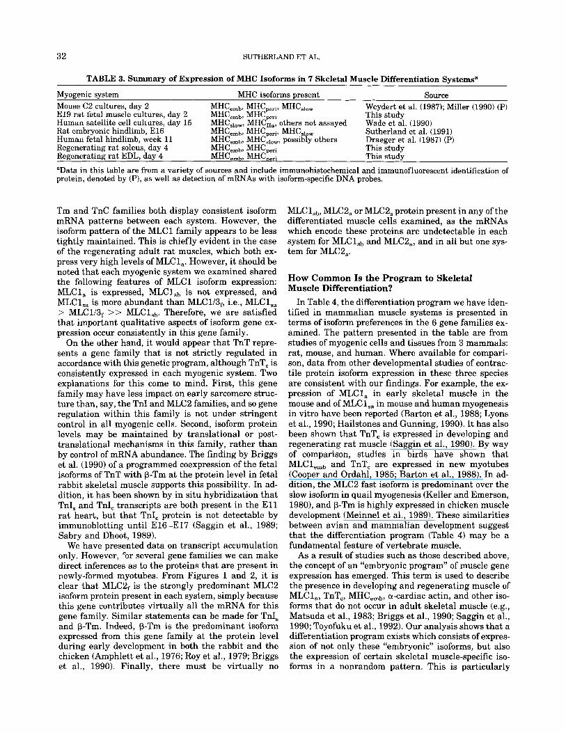
TABLE 3. Summary of Expression of MHC Isoforms in 7 Skeletal Muscle Differentiation Systemsa
Myogenic system MHC isoforms present Source Mouse C2 cultures, day 2 El9 rat fetal muscle cultures, day 2 Human satellite cell cultures, day 15 Rat embryonic hindlimb, El6 Human fetal hindlimb, week 11 Regenerating rat soleus, day 4 MHCemb, MHCperi This study Regenerating rat EDL, day 4 MHCemt,, MHCDeri This study
“Data in this table are from a variety of sources and include immunohistochemical and immunofluorescent identification of protein, denoted by (P), as well as detection of mRNAs with isoform-specific DNA probes.
MHCemb, MHCperi, MHCshw MHC,,,, MHC MHC,,,,, MHC$;: others not assayed MHCem,, MHCperi, MHCs,,w MHCemb, MHC,,,,, possibly others
Weydert et al. (1987); Miller (1990) (P) This study Wade et al. (1990) Sutherland et al. (1991) Draeger et al. (1987) (P)
Tm and TnC families both display consistent isoform mRNA patterns between each system. However, the isoform pattern of the MLCl family appears to be less tightly maintained. This is chiefly evident in the case of the regenerating adult rat muscles, which both ex- press very high levels of MLC1,. However, it should be noted that each myogenic system we examined shared the following features of MLCl isoform expression: MLC1, is expressed, MLCl,, is not expressed, and MLCl,, is more abundant than MLC1/3f, i.e., MLCl,, > MLC1/3f >> MLClSb. Therefore, we are satisfied that important qualitative aspects of isoform gene ex- pression occur consistently in this gene family.
On the other hand, i t would appear that TnT repre- sents a gene family that is not strictly regulated in accordance with this genetic program, although TnT, is consistently expressed in each myogenic system. Two explanations for this come to mind. First, this gene family may have less impact on early sarcomere struc- ture than, say, the TnI and MLCB families, and so gene regulation within this family is not under stringent control in all myogenic cells. Second, isoform protein levels may be maintained by translational or post- translational mechanisms in this family, rather than by control of mRNA abundance. The finding by Briggs et al. (1990) of a programmed coexpression of the fetal isoforms of TnT with p-Tm at the protein level in fetal rabbit skeletal muscle supports this possibility. In ad- dition, it has been shown by in situ hybridization that TnI, and TnI, transcripts are both present in the E l l rat heart, but that TnI, protein is not detectable by immunoblotting until E16-El7 (Saggin et al., 1989; Sabry and Dhoot, 1989).
We have presented data on transcript accumulation only. However, “or several gene families we can make direct inferences as to the proteins that are present in newly-formed myotubes. From Figures 1 and 2, it is clear that MLCBf is the strongly predominant MLCB isoform protein present in each system, simply because this gene contributes virtually all the mRNA for this gene family. Similar statements can be made for TnI, and p-Tm. Indeed, p-Tm is the predominant isoform expressed from this gene family at the protein level during early development in both the rabbit and the chicken (Amphlett et al., 1976; Roy et al., 1979; Briggs et al., 1990). Finally, there must be virtually no
MLCl,,, MLC2, or MLC2, protein present in any of the differentiated muscle cells examined, as the mRNAs which encode these proteins are undetectable in each system for MLCl,, and MLCB,, and in all but one sys- tem for MLC2,.
How Common Is the Program to Skeletal Muscle Differentiation?
In Table 4, the differentiation program we have iden- tified in mammalian muscle systems is presented in terms of isoform preferences in the 6 gene families ex- amined. The pattern presented in the table are from studies of myogenic cells and tissues from 3 mammals: rat, mouse, and human. Where available for compari- son, data from other developmental studies of contrac- tile protein isoform expression in these three species are consistent with our findings. For example, the ex- pression of MLC1, in early skeletal muscle in the mouse and of MLCl,, in mouse and human myogenesis in vitro have been reported (Barton et al., 1988; Lyons et al., 1990; Hailstones and Gunning, 1990). It has also been shown that TnT, is expressed in developing and regenerating rat muscle (Saggin et al., 1990). By way of comparison, studies in birds have shown that MLClemb and TnT, are expressed in new myotubes (Cooper and Ordahl, 1985; Barton et al., 1988). In ad- dition, the MLCB fast isoform is predominant over the slow isoform in quail myogenesis (Keller and Emerson, 1980), and p-Tm is highly expressed in chicken muscle development (Meinnel et al., 1989). These similarities between avian and mammalian development suggest that the differentiation program (Table 4) may be a fundamental feature of vertebrate muscle.
As a result of studies such as those described above, the concept of an “embryonic program” of muscle gene expression has emerged. This term is used to describe the presence in developing and regenerating muscle of MLCl,, TnT,, MHC,,,, a-cardiac actin, and other iso- forms that do not occur in adult skeletal muscle (e.g., Matsuda et al., 1983; Briggs et al., 1990; Saggin et al., 1990; Toyofuku et al., 1992). Our analysis shows that a differentiation program exists which consists of expres- sion of not only these “embryonic” isoforms, but also the expression of certain skeletal muscle-specific iso- forms in a nonrandom pattern. This is particularly

GENE EXPRESSION DURING MUSCLE DIFFERENTIATION 33
striking in the MLC2 and TnI gene families (Table 4). Interestingly, in the MLCl and TnT gene families the “embryonic” (or inappropriate) isoform mRNA is not necessarily the most abundant in most systems (Tables 1, 2).
Significance of the Differentiation Program The existence of a common differentiation program
of contractile protein gene expression that is not de- pendent on fiber fate raises an important question: what significance is there in the consistent expression of certain isoforms and consistent repression of others during skeletal muscle differentiation? One explana- tion is that there are differences in the relative ease with which the various isoforms can be activated im- mediately following terminal differentiation. Those isoforms that are readily expressed upon differentia- tion ultimately may be functionally inappropriate for a particular fiber. Thus, the “early” isoform will be re- placed by the appropriate isoform as the muscle cell matures. In this case, early expression could conceiv- ably be a reflection of chromosomal location and struc- ture of the gene, or of response to early-expressed myo- genic transcription factors, but is not due to the intrinsic properties of the encoded proteins.
An alternative, but not necessarily incompatible ex- planation is that as a group the early-expressed iso- forms do have functional significance at the protein level. One can envisage that this group of isoforms fa- cilitate the rapid de nouo assembly of sarcomeres with fidelity. These isoforms are expressed during differen- tiation when assembly has priority over sarcomere function, but will be replaced by a more appropriate functional set of isoforms as sarcomere function be- comes paramount in the more mature myotube.
There is mounting evidence in the literature that binding affinities differ between isoforms and that this in turn affects assembly capabilities. Chaussepied and Kasprzak (1989) have shown that the presence of dif- ferent MLC isoforms in a myosin molecule confers dif- ferent properties of association with actin. A study of intracompartmental sorting of MLC isoforms in rat cardiomyocytes has suggested that differences in affin- ity between MHC and MLC isoforms affect the sorting process (Soldati and Perriard, 1991). Work on the body wall musculature of C . elegans indicates that the A and B MHC isoforms of the nematode are differentially in- corporated into sarcomeres (Miller et al., 1983; Epstein et al., 1986; Epstein and Fischman, 1991). A study of the incorporation of actin monomers into myofibrils in vitro has shown that a-actin is more efficiently incor- porated into sarcomeres than is (3-actin (Peng and Fisch- man, 1991). Thus, binding and assembly properties of- ten differ between particular contractile protein isoforms. Therefore, it is conceivable that the early dif- ferentiation program provides a mixture of isoforms with properties that favor the efficient formation of new sarcomeres. This model thus suggests that the ac-
quisition of structure precedes the acquisition of func- tion during muscle development.
Helix-loopHelix Regulatory Proteins and the Differentiation Program
Studies of the expression of the basic helix-loophe- lix (bHLH) muscle-regulatory factors indicate that myoD, myogenin, myf5, and MRF4 are regulated inde- pendently during mouse and rat muscle development (Monterras et al., 1989, 1991; Sassoon et al., 1989; Bober et al., 1991; Hinterberger et al., 1991). These studies together with studies of myogenic cells in vitro, which express different combinations of the bHLH fam- ily, have raised the possibility that each member of the family may differentially activate a distinct subset of muscle-specific genes, or may be expressed in distinct myoblast or myocyte populations (Miller, 1990, 1991; Hinterberger et al., 1991). However, in this paper we have identified a differentiation-specific program of gene expression independent of future (or past) pheno- type that is found in myotubes derived from primary, secondary, and adult myoblasts. These different myo- blasts express different combinations of bHLH proteins (summarized in Miller, 1991), yet express a similar contractile protein isoform phenotype upon differenti- ation. This indicates that during early differentiation the six contractile protein gene families examined are ignoring, perhaps temporarily, any phenotypic regula- tory information carried by the bHLH transcription factors. Alternatively, it is possible that bHLH pro- teins can impart qualitative ‘‘on--off)’ information to the contractile protein genes, but do not impart quan- titative information capable of regulation of mRNA abundance.
CONCLUSIONS In recent years, the complexity of contractile protein
isoform expression patterns during the differentiation of skeletal muscle has become clearly evident. The lack of a comprehensive analysis of contractile protein iso- form expression during early myogenesis has obscured the existence of any overt pattern. We have now iden- tified a common program of contractile protein isoform gene expression that occurs in myotubes derived from primary myoblasts, secondary myoblasts, and satellite cells. This program is independent of the intrinsic MHC isoform repertoire and does not fit with proposed roles for the bHLH proteins in determining fiber spe- cialization. Conceivably, this common program of iso- form expression which is initiated upon differentiation facilitates the de nouo assembly of sarcomeres. For 5 of the 6 gene families examined, all skeletal myotubes exhibit a similar isoform mRNA profile when they first form. Therefore, each cell must subsequently modulate gene expression in all of the contractile protein gene families in order to become a specialized, mature my- ofiber. Determining how this transition occurs is now crucial to our understanding of skeletal muscle matu- ration.

34 SUTHERLAND ET AL.
EXPERIMENTAL PROCEDURES Tissue Isolation and Cell Culture
Mouse C2 cells (Yaffe and Saxel, 1977) were grown in Dulbecco’s modified Eagle’s medium (DMEM, Gibco, NY) supplemented with 20% fetal bovine serum (FBS; CSL, Melbourne, Victoria) and 0.5% chick embryo ex- tract (Flow Laboratories, North Ryde, NSW). Myoblast fusion was induced by replacing growth medium with fusion medium (DMEM plus 5% horse serum).
Primary cultures of limb tissue were established from 1 litter of E l 9 rat fetuses. Hindlimbs were re- moved and skin, bone, and paw dissected away. Tissue was crudely dissociated in F10 nutrient medium (Gibco) supplemented with 0.1% BSA and penicillin/ streptomycin. A single cell suspension was produced by repeated short incubations at 37°C in trypsin/EDTA. The cells were grown on a laminin substrate in growth medium (F10 with 20% FBS, 0.5% chick embryo ex- tract, pedstrep). After 48 hr in culture, growth me- dium was replaced with fusion medium (as above) to promote myotube formation.
Isolation of Regenerating Rat Muscles Muscle regeneration was induced in female Wistar
rats using the protocol of Carlson (Carlson et al., 1981; Carlson, 1976). Under sodium pentobarbital anaesthe- sia, either the soleus or extensor digitorum longus (EDL) muscle was isolated in the hindlimb. The ten- dons, nerves and vasculature to the muscle were sev- ered and the muscle injected with 0.75% Marcaine (bupivacaine hydrochloride, a kind gift of Astra Phar- maceuticals Pty Ltd, North Ryde, NSW). The muscle was then sutured back in the original site and the skin sutured closed. The treated muscles were removed 4 days after surgery for total RNA extraction and north- ern analysis. Four days was chosen for tissue collection because at this time satellite cell proliferation has been initiated and new myotubes are just forming (Carlson, 1986; Roberts et al., 1989).
RNA Isolation and Northern Analysis Total RNA was isolated from cultured cells and from
isolated muscle tissue by standard methods (Strohman et al., 1977; Chomczynski and Sacchi, 1987). Gel elec- trophoresis, blotting, hybridization to specific probes, and densitometric analysis were all performed as de- scribed previously (Wade et al., 1990; Sutherland et al., 1991). The relative expression of the different isoform mRNAs within a gene family was calculated as follows. The transcript level in a sample was compared to its expression level in an appropriate adult muscle. The expression level in the adult muscle represents a the- oretical maximum and enables estimation of relative contribution for each isoform in a gene family (Suther- land et al., 1991). The adult muscles used represented a predominately fast, slow, ventricular, or atrial phe- notype and were EDL (rat) or gastrocnemius (mouse), soleus, ventricle, or atrium, respectively. Loading dis-
crepancies were corrected by reprobing each panel for 18 S ribosomal RNA in probe excess and adjusting ac- cordingly. Each measurement was made at least twice and multiple autoradiographic exposures were made within the linear range of the film sensitivity as deter- mined from a serial dilution of RNA probed for a-skel- eta1 actin (Gunning et al., 1987). The percentage val- ues listed in Tables 1 and 2 were derived as follows. The isoform mRNA value within a sample was first expressed as a fraction of the expression in the adult tissue and the values for all isoforms from a gene fam- ily were added. The value for a given isoform was then calculated as a percentage of this total. This method obviates the necessity of correcting for the differing amounts of differentiated myogenic material in each sample. The relative values for each isoform listed within a gene family do not represent the true ratios of mRNA levels, but rather are estimations of these ra- tios.
Detailed descriptions of all isoform-specific probes used in this study have been given elsewhere (Wade et al., 1990; Sutherland et al., 1991; Hailstones et al., 19921, except for atriaUembryonic MLCl (MLC1,). As a DNA probe for detection of rodent MLC1, species, a 340-base pair PstIiPvuII restriction fragment was isolated from a mouse MLC1, cDNA clone kindly provided by Paul Barton (Barton et al., 1988). For de- tection of MLC1, in human cells, the oligonucleotides 5’-GCCTGGCCCTTGGCTTTAGC-3‘ and 5’-GAATC- CCAGAGCCCAGCCTG-3’ were used as primers to amplify from atrial cDNA a 155-base pair sequence encoding the human MLC1, 3’ UTR as published (Kurabayashi et al., 1988). This probe was kindly pro- vided by T. Yeoh and D. Hailstones.
ACKNOWLEDGMENTS We would like to thank Peter Gunning for many
helpful discussions and for comments on the manu- script. We also thank Peter Rowe for critical reading of the manuscript. C.J.S. was the recipient of National Health and Medical Research Council (NH&MRC) Bio- medical Scholarship. This work was generously sup- ported by the Children’s Medical Research Institute and in part by a grant from the NH&MRC to E.C.H.
REFERENCES Amphlett, G.W., Syska, H., and Perry, S.V. (1976) The polymorphic
forms of tropomyosin and troponin I in developing rabbit skeletal muscle. FEBS Lett. 63:22-26.
Bandman, E., Matsuda, R., and Strohman, R.C. (1982) Developmental appearance of myosin heavy and light chain isoforms in vivo and in vitro in chicken skeletal muscle. Dev. Biol. 93508-518.
Barton, P., Robert, B., Cohen, A,, Garner, I., Sassoon, D., Weydert, A,, and Buckingham, M. (1988) Structure and sequence of the myosin alkali light chain gene expressed in adult cardiac atria and fetal striated muscle. J. Biol. Chem. 263:12669-12676.
Barton, P.J.R., Harris, A.J., and Buckingham, M.E. (1989) Myosin light chain gene expression in developing and denervated fetal muscle in the mouse. Development 107:819-824.
Bober, E., Lyons, G.E., Braun, T., Cossu, G., Buckingham, M., and Arnold, H.-H. (1991) The muscle regulatory gene, myf-6, has a bi-

GENE EXPRESSION DURING MUSCLE DIFFERENTIATION 35
phasic pattern of expression during early mouse development. J . Cell Biol. 113:1255-1265.
Briggs, M.M., McGinnis, H.D., and Schachat, F. (1990) Transitions from fetal to fast troponin T isoforms are coordinated with changes in tropomyosin and a-actinin isoforms in developing rabbit skeletal muscle. Dev. Biol. 140:253-260.
Buckingham, M.E. (1985) Actin and myosin multigene families: Their expression during the formation of skeletal muscle. Essays Bio- chem. 20:77-109.
Campion, D.R. (1984) The muscle satellite cell: A review. Int. Rev. Cytol. 87:225-251.
Carlson, B.M. (1976) A quantitative study of muscle fiber survival and regeneration in normal, predenervated, and marcaine-treated free muscle grafts in the rat. Exp. Neurol. 52:421-432.
Carlson, B.M. (1986) Regeneration of entire skeletal muscles. Fed. Proc. 45:1456-1460.
Carlson, B.M., Hnik, P., TuCek, S., Vejsada, R., Bader, D.M., and Faulkner, J.A. (1981) Comparison between grafts with intact nerves and standard free grafts of the rat extensor digitorum longus muscle. Physiol. Bohemoslov. 30:505-514.
Carraro, U., Dalla Libera, L., and Catani, C. (1983) Myosin light and heavy chains in muscle regenerating in absence of the nerve: Tran- sient appearance of the embryonic light chain. Exp. Neurol. 79: 106-117.
Chaussepied, P., and Kasprzak, A.A. (1989) Isolation and character- isation of the G-actin-myosin head complex. Nature (London) 342: 950-953.
Chomczynski, P., and Sacchi, N., (1987) Single-step method of RNA isolation by acid guanidinium thiocyanate-phenol-chloroform ex- traction. Anal. Biochem. 162:156-159.
Condon, K., Silberstein, L., Blau, H.M., and Thompson, W.J. (1990) Development of muscle fiber types in the prenatal rat hindlimb. Dev. Biol. 138:275-295.
Cooper, T.A. and C.P. Ordahl. (1985) A single cardiac troponin T gene generates embryonic and adult isoforms via developmentally regu- lated alternate splicing. J. Biol. Chem. 260:11140-11148.
Duxson, M.J., Usson, Y., and Harris, A.J. (1989) The origin of second- ary myotubes in mammalian skeletal muscles: Ultrastructural studies. Development 107:743-750.
Epstein, H.F., and Fischman, D.A. (1991) Molecular analysis of pro- tein assembly in muscle development. Science 251:1029-1044.
Epstein, H.F., Ortiz, I., and Mackinnon, L.A.T. (1986) The alteration of myosin isoform compartmentation in specific mutants of Caenor- habditis elegans. J . Cell Biol. 103:985-993.
Gunning, P., Hardeman, E., Wade, R., Ponte, P., Bains, W., Blau, H.M., and. Kedes, L. (1987) Differential patterns of transcript ac- cumulation during human myogenesis. Mol. Cell. Biol. 7:4100- 4114.
Hailstones, D.L., and Gunning, P. (1990) Characterisation of human myosin light chains l,, and 3 4 Implications for isoform evolution and function. Mol. Cell. Biol. 10:1095-1104.
Hailstones, D.L., Barton, P., Chan-Thomas, P., Sasse, S., Sutherland, C. Hardeman, E. and Gunning, P. (1992) Differential regulation of the atrial isoforms of the myosin light chains during striated mus- cle development. J . Biol. Chem. 267:23295-23300.
Harris, A.J., Fitzsimons, R.B. and McEwan, J.C. (1989) Neural con- trol of the sequence of expression of myosin heavy chain isoforms in fetal mammalian muscles. Development 107:751-769.
Hayward, L.J., and Schwartz, R.J. (1986) Sequential expression of chicken actin genes during myogenesis. J . Cell Biol. 102:1485- 1493.
Hinterberger, T.J., Sassoon, D.A., Rhodes, S.J., and Konieczny, S.F. (1991) Expression of the muscle regulatory factor MRF4 during somite and skeletal myofiber development. Dev. Biol. 147:144-156.
Keller, L.R., and Emerson, C.P. (1980) Synthesis of adult myosin light chains by embryonic muscle cultures. Proc. Natl. Acad. Sci. U.S.A. 77:1020-1024.
Kurabayashi, M., Komuro, I., Tsuchimochi, H., Takaku, F., and Yazaki, Y. (1988) Molecular cloning and characterization of human atrial and ventricular myosin alkali light chain cDNA clones. J. Biol. Chem. 263:13930-13936.
Lyons, G.E., Ontell, M., Cox, R., Sassoon, D., and Buckingham, M.
(1990) The expression of myosin genes in developing skeletal mus- cle in the mouse embryo. J . Cell. Biol. 111:1465-1476.
Matsuda, R., Obinata, T., and Shimada, Y. (1981) Types of troponin components during development of chicken skeletal muscle. Dev. Biol. 82:ll-19.
Matsuda, R., Spector, D., and Strohman, R.C. (1983) Regenerating adult chicken skeletal muscle and satellite cell cultures express embryonic patterns of myosin and tropomyosin isoforms. Dev. Biol. 11 7:488-492.
Matsuda, R., Spector, D., and Strohman, R.C. (1984) Denervated skel- etal muscle displays discoordinate regulation for the synthesis of several myofibrillar proteins. Proc. Natl. Acad. Sci. U.S.A. 81: 1122-1 125.
Meinnel, T., Libri, D., Mouly, D., Gros, D., Fiszman, M., and Lemon- nier, M. (1989) Tissue-specific transcriptional control of a-and P-tropomyosins in chicken muscle development. Dev. Biol. 131: 430-438.
Miller, D.M., Ortiz, I., Berliner, G.C., and Epstein, H.F. (1983) Dif- ferential localization of two myosins within nematode thick fila- ments. Cell 34:477-490.
Miller, J.B. (1990) Myogenic programs of mouse muscle cell lines: Expression of myosin heavy chain isoforms, MyoD1, and myogenin. J. Cell Biol. 111:1149-1159.
Miller, J.B. (1991) Myoblasts, myosins, myoDs, and the diversifica- tion of muscle fibers. Neuromusc. Disorders 1:7-17.
Monterras, D., Pinset, C., Chelly, J., Kahn, A., and Gros, F. (1989) Expression of MyoDl coincides with terminal differentiation in de- termined but inducible muscle cells. EMBO J. 8:2203-2207.
Monterras, D., Chelly, J . , Bober, E., Arnold, H., Ott, M.-O., Gros, F. and Pinset, C. (1991) Developmental patterns in the expression of Myf 5, MyoD, myogenin, and MRF4 during myogenesis. New Biol- ogist 3592-600.
Peng, I., and Fischman, D.A. (1991) Post-translational incorporation of actin into myofibrils in vitro: Evidence for isoform specificity. Cell Motil. Cytoskeleton 20:158-168.
Periasamy, M., Wieczorek, D., and Nadal-Ginard, B. (1984) Charac- terisation of a developmentally regulated perinatal myosin heavy chain gene expressed in skeletal muscle. J . Biol. Chem. 21: 13573- 13578.
Roberts, P., McGeachie, J.K., Grounds, M.R., and Smith, E.R. (1989) Initiation and duration of myogenic precursor cell replication in transplants of intact skeletal muscles: An autoradiographic study in mice. Anat. Rec. 224:l-6.
Roy, R.K., Sreter, F.A., and Sarkar, S. (1979) Changes in tropomyosin subunits and myosin light chains during development of chicken and rabbit striated muscles. Dev. Biol. 69:15-30.
Rubinstein, N.A., and Kelly, A.M. (1981) Development of muscle fiber specialisation in the rat hindlimb. J. Cell Biol. 90:128-144.
Sabry, M., and Dhoot G. (1989) Identification and pattern of expres- sion of a developmental isoform of troponin I in chicken and rat cardiac muscle. J . Mus. Res. Cell Motil. 10:85-91.
Saggin, L., Gorza, L., Ausoni, S., and Schiafino, S. (1989) Troponin I switching in the developing rat heart. J. Biol. Chem. 27:16299- 16302.
Saggin, L., Gorza, L., Ausoni, S., and Schiaffino, S. (1990) Cardiac troponin T in developing, regenerating and denervated rat skeletal muscle. Development 110547-554.
Sassoon, D., Lyons, G., Wright, W.E., Lin, V., Lassar, A,, Weintraub, H. and Buckingham, M. (1989) Expression of two myogenic regu- latory factors myogenin and MyoDl during mouse embryogenesis. Nature (London) 341:303-307.
Schultz, E., Jaryszak, D.L., Gibson, M.C., and Albright, D.J. (1986) Absence of exogenous satellite cell contribution to regeneration of frozen skeletal muscle. J . Mus. Res. Cell Motil. 7:361-367.
Soldati, T., and Perriard, J.-C. (1991) Intracompartmental sorting of essential myosin light chains: Molecular dissection and in vivo monitoring by epitope tagging. Cell 66:277-289.
Stockdale, F.E., and Miller, J.B. (1987) The cellular basis of myosin heavy chain expression during development of avian skeletal mus- cles. Dev. Biol. 123:l-9.
Strohman, R.C., Moos, P.S., Micou-Eastwood, J., Spector, D., Przbyla,

36 SUTHERLAND ET AL.
A. and Paterson, B. (1977) Messenger RNA for myosin polypeptides: Isolation from single myogenic cell cultures. Cell 10:265-273.
Sutherland, C.J., Elsom, V.E., Gordon, M.L., Dunwoodie, S.L., and Hardeman, E.C. (1991) Coordination of skeletal muscle gene ex- pression occurs late in mammalian development. Dev. Biol. 146: 167-178.
Toyofuku, T., Hoffman, J.R., Zak, R., and Carlson, B.M. (1992) Ex- pression of a-cardiac and a-skeletal actin mRNAs in relation to innervation in regenerating and non-regenerating rat skeletal muscles. Dev. Dynam. 193:332-339.
Vivarelli, E., Brown, W., Whalen, R.G., and Cossu, G. (1988) The expression of slow myosin during mammalian somitogenesis and limb bud differentiation. J . Cell Biol. 107:2191-2197.
Wade, R., Sutherland, C.J., Gahlmann, R., Kedes, L., Hardeman, E., and Gunning, P. (1990) Regulation of contractile protein gene fam- ily mRNA pool sizes during myogenesis. Dev. Biol. 142:270-282.
Weydert, R.G., Daubas, P., Caravatti, M., Minty, A,, Bugaisky, G., Cohen, A., Robert, B., and Buckingham, M. (1983) Sequential ac- cumulation of mRNAs encoding different myosin heavy chain iso- forms during skeletal muscle development in vivo detected with a recombinant plasmid as coding for an adult fast myosin heavy chain from mouse skeletal muscle. J . Biol. Chem. 258:13867-13874.
Weydert, R.G., Harris, A.J., Pinset, C., and Buckingham, M. (1987) Developmental pattern of mouse skeletal myosin heavy chain gene transcripts in vivo and in vitro. Cell 49121-129.
Whalen, R., Sell, S., Butler-Browne, G., Schwartz, K., Bouveret, P., and Pinset-Harstrom, I. (1981) Three myosin heavy chain isozymes appear sequentially in rat muscle development. Nature (London) 292:805-809.
Yaffe, D., and Saxel, 0. (1977) Serial passaging and differentiation of myogenic cells isolated from dystrophic mouse muscle. Nature (London) 270:725-727.