identification and functional characterization of polymorphisms in human cyclooxygenase-1 (ptgs1)
TRANSCRIPT
Identification and Functional Characterization of N-Terminally Acetylated Proteins in DrosophilamelanogasterSandra Goetze1,2,3.*, Ermir Qeli1., Christian Mosimann3., An Staes4,5., Bertran Gerrits6, Bernd
Roschitzki6, Sonali Mohanty1,2, Eva M. Niederer1, Endre Laczko6, Evy Timmerman4,5, Vinzenz Lange2,
Ernst Hafen2, Ruedi Aebersold2,7,8, Joel Vandekerckhove4,5, Konrad Basler1,3, Christian H. Ahrens1, Kris
Gevaert4,5, Erich Brunner1*
1 Center for Model Organism Proteomes, University of Zurich, Switzerland, 2 Institute of Molecular Systems Biology, ETH Zurich, Switzerland, 3 Institute for Molecular
Biology, University of Zurich, Switzerland, 4 Department of Medical Protein Research, Flanders Institute for Biotechnology, Ghent, Belgium, 5 Department of Biochemistry,
Ghent University, Ghent, Belgium, 6 Functional Genomics Center, ETH and University of Zurich, Switzerland, 7 Faculty of Science, University of Zurich, Switzerland,
8 Institute for Systems Biology, Seattle, Washington, United States of America
Abstract
Protein modifications play a major role for most biological processes in living organisms. Amino-terminal acetylation ofproteins is a common modification found throughout the tree of life: the N-terminus of a nascent polypeptide chainbecomes co-translationally acetylated, often after the removal of the initiating methionine residue. While the enzymes andprotein complexes involved in these processes have been extensively studied, only little is known about the biologicalfunction of such N-terminal modification events. To identify common principles of N-terminal acetylation, we analyzed theamino-terminal peptides from proteins extracted from Drosophila Kc167 cells. We detected more than 1,200 mature proteinN-termini and could show that N-terminal acetylation occurs in insects with a similar frequency as in humans. As the soletrue determinant for N-terminal acetylation we could extract the (X)PX rule that indicates the prevention of acetylationunder all circumstances. We could show that this rule can be used to genetically engineer a protein to study the biologicalrelevance of the presence or absence of an acetyl group, thereby generating a generic assay to probe the functionalimportance of N-terminal acetylation. We applied the assay by expressing mutated proteins as transgenes in cell lines and inflies. Here, we present a straightforward strategy to systematically study the functional relevance of N-terminal acetylationsin cells and whole organisms. Since the (X)PX rule seems to be of general validity in lower as well as higher eukaryotes, wepropose that it can be used to study the function of N-terminal acetylation in all species.
Citation: Goetze S, Qeli E, Mosimann C, Staes A, Gerrits B, et al. (2009) Identification and Functional Characterization of N-Terminally Acetylated Proteins inDrosophila melanogaster. PLoS Biol 7(11): e1000236. doi:10.1371/journal.pbio.1000236
Academic Editor: Michael J. MacCoss, University of Washington, United States of America
Received May 21, 2009; Accepted September 23, 2009; Published November 3, 2009
Copyright: � 2009 Goetze et al. This is an open-access article distributed under the terms of the Creative Commons Attribution License, which permitsunrestricted use, distribution, and reproduction in any medium, provided the original author and source are credited.
Funding: This work was funded by the University of Zurich. SG, EQ, SM, EMN, KB, CHA, and EB are members of the Center for Model Organism Proteomes (C-MOP), which is funded by the University of Zurich (www.mop.unizh.ch). This work was supported by the Research Priority Project on Functional Genomics andSystems Biology of the University of Zurich, a grant by Hoffman La-Roche (Mkl/stm 192-2007) and a grant by the SNSF (Swiss National Science Foundation) (Marie-Heim Voegtlin, PMPDP3_122836/1) to SG as well as the SystemsX.ch initiative. The Department of Medical Protein Research acknowledges support by researchgrants from the Fund for Scientific Research-Flanders (Belgium) (project numbers G.0156.05, G.0077.06 and G.0042.07), the Concerted Research Actions (projectBOF07/GOA/012) from the Ghent University, the Inter University Attraction Poles (IUAP06), and the European Union Interaction Proteome (6th FrameworkProgram). The funders had no role in study design, data collection and analysis, decision to publish, or preparation of the manuscript.
Competing Interests: The authors have declared that no competing interests exist.
Abbreviations: BDGP, Berkeley Drosophila Genome Project; CE, collision energy; Cks85A, Cyclin-dependent kinase subunit 85A (CG9790); COFRADIC, CombinedFractional Diagonal Chromatography; GO, Gene Onthology; HA, hemagglutinin; Hyx, Hyrax protein (CG11990); i-Met, initiator methionine; MAP, Methionineaminopeptidase; NAT, N-terminal Acetyl Transferase; Pfam, Protein Families Database of Alignments and Hidden Markov Models; SAX, strong anion exchange;SCX, strong cation exchange; SRM, Selective-Reaction-Monitoring; TCEP, Tris(2-carboxyethyl)phosphine hydrochloride; wt, wild-type.
* E-mail: [email protected] (SG); [email protected] (EB)
. These authors contributed equally to this work.
Introduction
To attain full functionality and/or to reach their final cellular
localization, many proteins undergo obligatory modification or
processing. During this maturation process, proteins are concur-
rently properly folded, proteolytically processed, and enzymati-
cally modified. Some of these processes occur co-translationally,
i.e. during protein synthesis, while others take place after protein
synthesis has been completed. Acetylation of protein N-terminal a-
amino groups takes place during protein synthesis [1]. This very
common and irreversible modification of proteins often combines
two consecutive events [2,3]. In the first step, the N-terminal
methionine (also referred to as initiator methionine [iMet]) is
removed from the nascent polypeptide chain by methionine
aminopeptidases. This event is not obligatory in protein
biosynthesis and has been shown to take place only if the second
amino acid is small and uncharged [4,5]. Larger amino acids at
this position prevent removal of iMet by steric hindrance [6]. In
PLoS Biology | www.plosbiology.org 1 November 2009 | Volume 7 | Issue 11 | e1000236
the second step, the acetylation of the amino-terminus is catalyzed
by N-terminal acetyl transferases (NATs), a class of enzymes
conserved in pro- and eukaryotes [7–11]. In eukaryotes both
processes usually take place co-translationally on the nascent
polypeptide chain and appear to be completed when 25–50
residues extrude from the ribosome, as revealed by in vitro studies
[12,13]. This indicates that the N-terminal region of a protein
defines its acetylation status. Although previous work could show
sequence specificities of the different NAT complexes, for some
proteins acetylation does not take place even if the appropriate
amino acid sequences are present, suggesting that additional yet
unknown amino acid sequence patterns or other determinants like
the secondary structure of the protein’s N-terminus may play a
role [10].
An estimated 60%–90% of the cytosolic proteins are acetylated at
their N-terminus [3,14], however the biological relevance of N-
terminal acetylation has been determined only for a few proteins. This
was in most cases achieved either through the analysis of mutants of
NAT complex components [7], in vitro modification [5], or through
mutants for single proteins [15]. Small GTPases such as Arl3p or Arl8
for instance require amino-terminal acetylation for their recruitment to
Golgi membranes and lysosomes [15,16]. In other cases, the acetylated
N-terminus promotes protein-protein interactions as has been shown to
be important for the binding of F-actin and tropomyosin and the
maintenance of the resulting higher order structure [17,18]. These
examples clearly demonstrate that N-terminal acetylation promotes a
variety of biological functions that cannot be predicted from the
primary amino acid sequence. Therefore, there is a need for a method
to generate and express—in cells and organisms—proteins that differ
in N-terminal acetylation to investigate functional consequences of the
presence or absence of an N-terminal acetyl group.
N-terminal acetylation has been identified in various organisms
[10,19]. A detailed analysis of NAT substrate specificity, sequence
requirements, and conservation of substrate specificity for acetylation
were only recently documented for yeast and human [11]. Datasets
for invertebrates are not available and it has been suggested that
acetylations in invertebrates appear to be rare [10]. Here we present
an extensive compilation of mature protein N-termini of Drosophila
melanogaster that was obtained by shotgun proteomics as well as the
enrichment of N-terminal peptides by COFRADIC [20]. We show
that amino-terminal acetylation is a common event in Drosophila and
that the sequence requirements (amino acids) that promote iMet
cleavage and N-terminal acetylation are similar to those in other
eukaryotes. Moreover, our dataset enabled us to detect the use of 124
previously unknown alternative translation initiation sites and/or
splice variants. A Pfam analysis [21] revealed that a protein’s
acetylation state in some cases strongly correlates with the presence of
certain functional protein domains.
Finally, in contrast to earlier studies that were limited to the
identification of amino acid determinants that promoted or
inhibited N- terminal acetylation, in this study we could identify
a definite determinant, i.e. a proline at position one or two of a
nascent protein that prevents N-terminal acetylation under all
circumstances. We refer to this finding as (X)PX rule. We have
applied this rule to genetically modify a protein such that the
biological relevance of N-terminal acetylation could be studied in
cell lines and in flies. Since the (X)PX motif seems to be conserved
among organisms we propose that by applying the (X)PX rule in
similar ways in other species, the function of N-terminal
acetylation can now be generically studied.
Results
Characterization of N-terminal Most Peptides inDrosophila melanogaster
To enrich for N-terminal peptides, proteins from a membrane,
cytoplasmic, and nuclear fraction of Drosophila Kc167 cells,
respectively, were subjected to combined fractional diagonal
chromatography (COFRADIC) [11,20,22]. In COFRADIC, free
primary amino groups of proteins (i.e. a-N-termini and e-amines
from lysine residues) need to be chemically acetylated on the protein
level. To further distinguish naturally acetylated and non-acetylated
protein N-termini, protein amines were blocked by trideutero-
acetylation, which leaves a mass tag of 3 Dalton on each free
primary amino group [11,23]. The fractions enriched for N-
terminal peptides were then analyzed by mass spectrometry. We
identified 835 N-terminal peptides (peptides starting at the residue 1
or 2 of the predicted sequence; Figure 1A, Table S1) among a total
4,203 distinct peptides (19.5%) identified from 8,402 fragment ion
spectra. This corresponds to roughly 8.7% of the protein N-termini
detectable by mass spectrometry (see Figure S1 for calculations).
The actual coverage reached has to be considered much higher
since only a subset of all annotated proteins will be expressed in
exponentially growing Kc cells. Furthermore, a dataset consisting of
382 N-terminal peptides (Figure 1A) identified by a classical shotgun
proteomics approach on Kc cells, that is not using COFRADIC,
was additionally considered in subsequent analyses (retrieved from
[24]). A comparison of the two datasets revealed that COFRADIC
enriched for N-terminal peptides by a factor of roughly 10. In total
the two datasets identified 1,102 protein N-termini.
Besides the confirmation of these 1,102 distinct annotated protein N-
termini, we expected to find alternative start sites in these two datasets,
i.e. peptides with an amino-terminus that starts at position 3 or later of
the predicted polypeptide chain and by convention are considered to
be internal peptides. However, some of these supposedly internal
peptides start with a Met and are semi-tryptic. Others start with a small
and uncharged residue, are preceded by a Met in the predicted protein
sequence that is missing in the identified peptide, hence indicating an
iMet removal as found for a classical protein N-terminus. Our dataset
contains 124 distinct peptides that fulfill above criteria (Figure 1B,
Table S2) and that we consider to represent alternative translation
initiation sites or un-annotated splice variants. To further verify this, we
Author Summary
Widely hailed as the workhorses of the cell, proteinsparticipate in virtually every process within a livingorganism. How well they perform these diverse tasksdepends on successful passage through the intricatecourse of protein production, from transcription of theprotein-encoded DNA template to processing and foldingof the nascent amino acid chain. Some of the processingsteps—including enzymatic cleavage or the attachment ofchemical modifications—take place during protein syn-thesis, while others occur afterward. One modification thattakes place during protein synthesis is the attachment ofan acetyl group at the tip (N-terminus) of proteins.Although N-terminal acetylation is found throughout thetree of life and the machinery and mechanisms responsiblefor this modification are quite well characterized, little isknown about how it affects protein function. We analyzedthe acetylation state of proteins in the fruit fly Drosophilamelanogaster and show that this modification occurs at alower frequency in flies than in man but at a much higherfrequency than in yeast. Based on our dataset wedeveloped a generic method that can analyze thebiological relevance of N-terminal protein acetylation inany organism.
Assessing the Function of N-Terminal Acetylation
PLoS Biology | www.plosbiology.org 2 November 2009 | Volume 7 | Issue 11 | e1000236

analyzed the sequence context of the AUG that served as putative
alternative start codon with respect to its Cavener sequence (C_A/
G_A_A/C_AUG; Kozak sequence for insects, the initial Kozak
sequence being CC_A/G_C_C AUG_G) [25,26]. In addition, we
analyzed whether the AUG used is the first AUG of that particular
exon. A frequency analysis of the residues in the AUG context revealed
that the presence of the Cavener sequence could be confirmed for the
entire N-terminal dataset (C_A_A_A_AUG; Figure 2A) as well as the
putative alternative start sites (C_A_A_C_AUG; Figure 2B). Notably,
we detected a change in sequence preference at position 21 (from A to
C), which fully complies with the Cavener consensus sequence [25,26].
It is important to note that these consensus patterns are derived from
aggregate frequencies of nucleotides 59 to the AUG used for translation
initiation. If however the Cavener sequence is analyzed for the
proposed alternative initiation sites of a single gene model, the Cavener
sequences of the used AUG may deviate from the consensus sequence.
Cavener and Ray have already recognized this phenomenon and
described the consensus as a ‘‘strictly statistical term’’ whereas the
optimal context for each individual AUG is defined as a ‘‘functional
term’’ [26]. Nevertheless, in about 61% in of the cases the AUG is
flanked by an adequate or a strong Cavener sequence, indicating that
they represent true alternative start sites (Text S1).
Although essential for genome annotation, computer-based
prediction of protein N-termini and alternative translation
initiation sites remains a difficult task [27]. In that respect, our
dataset not only allowed us to confirm many of the predicted
translation sites in the fly but also to identify novel alternative
translation initiation sites. In combination, we identified 1,226
amino termini for Drosophila Kc (Figure 1C, Table S1), which have
been used for all subsequent analyses.
We next analyzed these 1,226 N-termini with respect to N-
terminal acetylation (Figure 1C, Table S1). We observed that in the
majority of cases (63%) the iMet is removed and that aminopeptidase
cleavage follows the same rules as determined for other organisms
[28]. About 71% of the N-terminal peptides are acetylated. Of these
61% have the iMet removed, whereas for free N-termini almost 68%
showed iMet removal. A comparison of the present data with the
respective data from yeast and man [11] shows that N-terminal
acetylation occurs in insects with a similar frequency as in humans.
Moreover, the acetylation frequency with respect to certain residues
seems to have shifted during evolution (Table 1). For instance,
whereas most N-acetylated protein-termini in yeast begin with Ser
and rarely with Ala, Drosophila has a high percentage of acetylated
proteins that start with Ser or Ala, whereas Ala is the most commonly
acetylated N-terminus in man. Finally, the acetylation state of a
protein’s N-terminus appears in most cases fixed in Drosophila cells as
in human HeLa cells but is rare or often incomplete in yeast. Only 57
proteins were identified with both, either a free or an acetylated N-
terminus (5% of total in Drosophila, 8% in human, and 45% in yeast
[11]; Table S3). Thereof, 48 N-termini exhibited the same iMet
cleavage and thus had identical amino acid sequences (Figure 1C),
whereas nine showed alternative iMet cleavage.
Correlation of the N-terminal Acetylation Status with GOCategories and Pfam Domains
To assess whether particular protein functions or functional domains
are preferentially associated with the N-terminal acetylation state, a
Gene Ontology analysis on a reduced set of GO categories (referred to
as GO Slim) on all three levels, namely Cellular Component, Molecular
Function, and Biological Process, was performed [29]. The results of this
analysis are shown in Table S4 as well as in Figure S2A–S2C. Despite
the fact that some categories show a statistically significant (p,0.05)
over- or underrepresentation of either acetylated or free N-termini, the
overall spread of the distributions of acetylated versus non-acetylated
Figure 1. Graphical representation of the datasets using Venndiagrams. (A) Comparison of dataset generated by COFRADIC andshotgun analysis. A total of 1,102 distinct N-terminal peptides wereidentified with an overlap of 115 sequences. The COFRADIC approachyielded 835 N-terminal peptides among a total 4,203 distinct peptidesidentified from 8,402 spectra. In contrast, a classical shotgun approachon Kc cells not using COFRADIC enrichment yielded 382 N-terminalpeptides among 19,915 distinct peptides (34,175 spectra) retrievedfrom Loevenich et al. [24]. (B) Identification of 124 distinct putative,alternative translation initiation sites identified in the COFRADIC andshotgun dataset, respectively. (C) Comparison of N-terminal peptidesaccording to their acetylation status. From 1,226 distinct N-termini 861were found to be acetylated, 317 non-acetylated with an overlap of 48identical N-termini showing partial acetylation.doi:10.1371/journal.pbio.1000236.g001
Assessing the Function of N-Terminal Acetylation
PLoS Biology | www.plosbiology.org 3 November 2009 | Volume 7 | Issue 11 | e1000236

gene models does not allow one to make a clear correlation of protein
function with a certain GO category or a group of GO categories.
Specifically, none of the detected associations with GO categories was
strong enough to predict the acetylation state of a protein.
To determine whether proteins that share a specific functional
domain also share a common N-terminus (i.e., an acetylated or
free amino terminus), a Pfam analysis was performed (see
Materials and Methods for details). Pfam is a specialized database
that stores protein family classifications and protein domain data
and allows one to find relationships between functional domains
and any other protein property of interest or classify a so far
unknown protein into a protein family [21]. Because N-terminal
acetylation is a co-translational process completed after the first
part of a protein has been synthesized [12,13], Pfam domains that
start within the first 60 amino acids of a protein were considered.
In contrast to the GO analysis presented above, some Pfam
domains show a strong association with the acetylation status of
certain protein N-termini (Tables 2 and S5A). For example, for the
Figure 2. Analysis of translational start sites. (A) A Frequency analysis [54] of all predicted N-termini present in the Drosophila databaseBDGP_Release_3.2 revealed the presence of the Cavener consensus sequence C_A/G_A_A/C_ATG [25,26]. The histogram shows the relativefrequencies of nucleotides 59 to the predicted (conventional) initiator codon generating acetylated as well as non-acetylated N-termini starting atposition 1 or 2 of the predicted protein sequence. The nucleotide 39 of the initiator sequence has been proposed to preferentially be a G at +4 [55] forstrong initiation but has been shown not to be relevant for Drosophila [25,26]. Interestingly the G at +4 is nevertheless predominant in all cases. (B)Relative frequencies of nucleotides 59 to the alternative translation initiation sites. The Cavener sequence is conserved showing a shift in sequencepreference at position 21 (from A to C), which fully complies with the Cavener consensus sequence (C_A_A_C_ATG) [25,26].doi:10.1371/journal.pbio.1000236.g002
Assessing the Function of N-Terminal Acetylation
PLoS Biology | www.plosbiology.org 4 November 2009 | Volume 7 | Issue 11 | e1000236

Ta
ble
1.
Fre
qu
en
cie
so
fac
ety
late
dan
dn
on
-ace
tyla
ted
Dro
sop
hila
Kc
16
7,
yeas
t,an
dH
eLa
cell
pro
tein
sw
ith
vari
ou
sN
-te
rmin
alse
qu
en
ces
(po
siti
on
1o
r2
).
Fly
Ye
ast
Hu
man
Ace
Fre
eP
art
ial
To
tal
% Ace
% Fre
e% P
art
ial
% Ace
To
tal
Ace
Fre
eP
art
ial
To
tal
% Ace
% Fre
e% P
art
ial
% Ace
To
tal
Ace
Fre
eP
art
ial
To
tal
% Ace
% Fre
e% P
art
ial
% Ace
To
tal
Na
tAsu
bst
rate
s
Ala
-2
01
21
92
31
87
.09
.13
.99
0.9
02
31
74
00
.05
7.5
42
.54
2.5
25
21
12
62
89
87
.23
.89
.09
6.2
Cys
-6
40
10
60
.04
0.0
0.0
60
.00
10
10
.01
00
.00
.00
.01
00
11
00
.00
.00
.01
00
.0
Gly
-1
53
51
51
29
.46
8.6
2.0
31
.40
13
01
30
.01
00
.00
.00
.06
16
52
72
2.2
59
.31
9.0
40
.7
Ser-
24
81
91
12
78
89
.26
.84
.09
3.2
03
90
93
0.0
3.2
96
.89
6.8
10
91
11
11
98
.20
.91
.09
9.1
Th
r-4
42
05
69
63
.82
9.0
7.2
71
.00
14
82
20
.06
3.6
36
.43
6.4
21
23
26
80
.87
.71
2.0
92
.3
Val
-9
47
56
11
4.8
77
.08
.22
3.0
02
20
22
0.0
10
0.0
0.0
0.0
12
55
31
3.2
80
.61
6.0
19
.4
Na
tBsu
bst
rate
s
Me
t-A
sn-
38
41
43
88
.49
.32
.39
0.7
70
07
10
0.0
0.0
0.0
10
0.0
12
02
14
85
.70
.01
4.0
10
0.0
Me
t-A
sp-
&M
et-
Glu
-2
06
94
21
99
4.1
4.1
1.8
95
.91
80
01
81
00
.00
.00
.01
00
.01
24
03
12
79
7.6
0.0
2.0
10
0.0
Na
tCsu
bst
rate
s
Me
t-Ile
-9
61
16
56
.33
7.5
6.3
62
.50
20
20
.01
00
.00
.00
.02
11
45
0.0
25
.02
5.0
75
.0
Me
t-Le
u-
17
13
23
25
3.1
40
.66
.35
9.4
16
18
12
.57
5.0
12
.52
5.0
43
31
04
0.0
30
.03
0.0
70
.0
Me
t-P
he
13
61
20
65
.03
0.0
5.0
70
.01
10
25
0.0
50
.00
.05
0.0
72
11
07
0.0
20
.01
0.0
80
.0
Oth
er
Asp
-&
Glu
-0
00
0N
.D.
N.D
.N
.D.
N.D
.0
00
0N
.D.
N.D
.N
.D.
N.D
.2
00
21
00
.00
.00
.01
00
.0
Me
t-G
ln-
23
31
27
85
.21
1.1
3.7
88
.92
11
45
0.0
25
.02
5.0
75
.07
12
10
70
.01
0.0
20
.09
0.0
Me
t-Ly
s-3
25
33
19
.78
0.6
9.7
19
.40
10
01
00
.01
00
.00
.00
.02
11
61
91
0.5
57
.93
2.0
42
.1
Me
t-M
et-
51
17
71
.41
4.3
14
.38
5.7
00
00
N.D
.N
.D.
N.D
.N
.D.
70
07
10
0.0
0.0
0.0
10
0.0
Me
t-X
*-2
43
33
60
40
.05
5.0
5.0
45
.02
51
82
5.0
62
.51
2.5
37
.58
23
13
61
.51
5.4
23
.18
4.6
Pro
-0
71
07
10
.01
00
.00
.00
.00
11
01
10
.01
00
.00
.00
.00
40
04
00
.01
00
.00
.00
.0
Ile-
00
00
N.D
.N
.D.
N.D
.N
.D.
01
01
0.0
10
0.0
0.0
0.0
01
01
0.0
10
0.0
0.0
0.0
To
tal
86
13
17
48
12
26
70
.22
5.9
3.9
74
.13
11
13
11
82
62
11
.84
3.1
45
.05
6.9
56
51
16
61
74
27
6.1
15
.68
.28
4.4
Tab
le1
visu
aliz
es
the
Dro
sop
hila
(fly
)d
atas
et
acco
rdin
gto
kno
wn
NA
Tsu
bst
rate
sin
hu
man
san
dye
ast
(tab
lead
apte
dfr
om
[11
]).
Ace
,ac
ety
late
dN
-te
rmin
i;fr
ee
,n
on
ace
tyla
ted
N-t
erm
ini;
X*
=A
,G,H
,P,R
,S,T
,V,Y
.d
oi:1
0.1
37
1/j
ou
rnal
.pb
io.1
00
02
36
.t0
01
Assessing the Function of N-Terminal Acetylation
PLoS Biology | www.plosbiology.org 5 November 2009 | Volume 7 | Issue 11 | e1000236
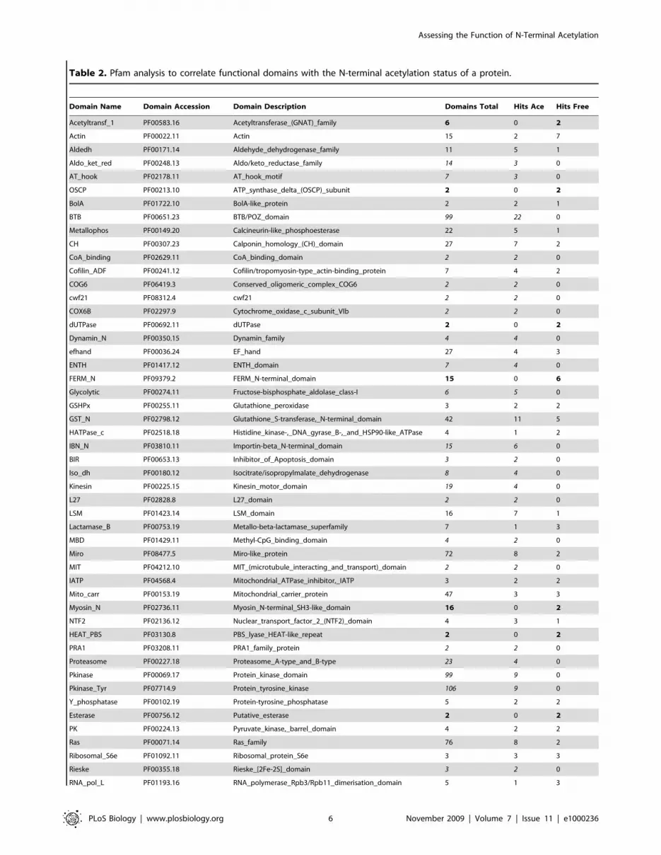
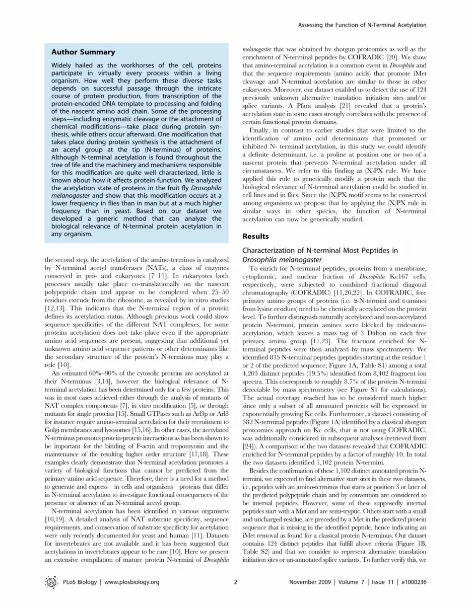
Table 2. Pfam analysis to correlate functional domains with the N-terminal acetylation status of a protein.
Domain Name Domain Accession Domain Description Domains Total Hits Ace Hits Free
Acetyltransf_1 PF00583.16 Acetyltransferase_(GNAT)_family 6 0 2
Actin PF00022.11 Actin 15 2 7
Aldedh PF00171.14 Aldehyde_dehydrogenase_family 11 5 1
Aldo_ket_red PF00248.13 Aldo/keto_reductase_family 14 3 0
AT_hook PF02178.11 AT_hook_motif 7 3 0
OSCP PF00213.10 ATP_synthase_delta_(OSCP)_subunit 2 0 2
BolA PF01722.10 BolA-like_protein 2 2 1
BTB PF00651.23 BTB/POZ_domain 99 22 0
Metallophos PF00149.20 Calcineurin-like_phosphoesterase 22 5 1
CH PF00307.23 Calponin_homology_(CH)_domain 27 7 2
CoA_binding PF02629.11 CoA_binding_domain 2 2 0
Cofilin_ADF PF00241.12 Cofilin/tropomyosin-type_actin-binding_protein 7 4 2
COG6 PF06419.3 Conserved_oligomeric_complex_COG6 2 2 0
cwf21 PF08312.4 cwf21 2 2 0
COX6B PF02297.9 Cytochrome_oxidase_c_subunit_VIb 2 2 0
dUTPase PF00692.11 dUTPase 2 0 2
Dynamin_N PF00350.15 Dynamin_family 4 4 0
efhand PF00036.24 EF_hand 27 4 3
ENTH PF01417.12 ENTH_domain 7 4 0
FERM_N PF09379.2 FERM_N-terminal_domain 15 0 6
Glycolytic PF00274.11 Fructose-bisphosphate_aldolase_class-I 6 5 0
GSHPx PF00255.11 Glutathione_peroxidase 3 2 2
GST_N PF02798.12 Glutathione_S-transferase,_N-terminal_domain 42 11 5
HATPase_c PF02518.18 Histidine_kinase-,_DNA_gyrase_B-,_and_HSP90-like_ATPase 4 1 2
IBN_N PF03810.11 Importin-beta_N-terminal_domain 15 6 0
BIR PF00653.13 Inhibitor_of_Apoptosis_domain 3 2 0
Iso_dh PF00180.12 Isocitrate/isopropylmalate_dehydrogenase 8 4 0
Kinesin PF00225.15 Kinesin_motor_domain 19 4 0
L27 PF02828.8 L27_domain 2 2 0
LSM PF01423.14 LSM_domain 16 7 1
Lactamase_B PF00753.19 Metallo-beta-lactamase_superfamily 7 1 3
MBD PF01429.11 Methyl-CpG_binding_domain 4 2 0
Miro PF08477.5 Miro-like_protein 72 8 2
MIT PF04212.10 MIT_(microtubule_interacting_and_transport)_domain 2 2 0
IATP PF04568.4 Mitochondrial_ATPase_inhibitor,_IATP 3 2 2
Mito_carr PF00153.19 Mitochondrial_carrier_protein 47 3 3
Myosin_N PF02736.11 Myosin_N-terminal_SH3-like_domain 16 0 2
NTF2 PF02136.12 Nuclear_transport_factor_2_(NTF2)_domain 4 3 1
HEAT_PBS PF03130.8 PBS_lyase_HEAT-like_repeat 2 0 2
PRA1 PF03208.11 PRA1_family_protein 2 2 0
Proteasome PF00227.18 Proteasome_A-type_and_B-type 23 4 0
Pkinase PF00069.17 Protein_kinase_domain 99 9 0
Pkinase_Tyr PF07714.9 Protein_tyrosine_kinase 106 9 0
Y_phosphatase PF00102.19 Protein-tyrosine_phosphatase 5 2 2
Esterase PF00756.12 Putative_esterase 2 0 2
PK PF00224.13 Pyruvate_kinase,_barrel_domain 4 2 2
Ras PF00071.14 Ras_family 76 8 2
Ribosomal_S6e PF01092.11 Ribosomal_protein_S6e 3 3 3
Rieske PF00355.18 Rieske_[2Fe-2S]_domain 3 2 0
RNA_pol_L PF01193.16 RNA_polymerase_Rpb3/Rpb11_dimerisation_domain 5 1 3
Assessing the Function of N-Terminal Acetylation
PLoS Biology | www.plosbiology.org 6 November 2009 | Volume 7 | Issue 11 | e1000236

Importin-beta_N-terminal domain (IBN_N, PF03810.11), the N-
termini of six out of 15 proteins predicted to contain such a
domain have been identified (p,0.048). In all six cases the N-
terminus was found to be acetylated. The lack of an N-terminal
acetylation appears to correlate with a few selected domains
comprising for instance the Tubulin/FtsZ_family,_GTPase_do-
main (PF00091.17, six out of 14 found: p,0.00025) or the
Ubiquitin_family (PF00240.15, seven out of 20 found).
The functional relevance of any of the correlations we identified
in this Pfam analysis is currently unknown. Assuming that the
function of a particular domain requires an exclusively acetylated
or free N-terminus, two possibilities arise: (i) the acetylation state of
the N-terminus is determined by the first amino acid residues of
the primary amino acid sequence and/or (ii) the domain contains
a so far unknown motif that prevents or promotes acetylation. An
example for the first is the association of the IBN_N domain with
an acetylated N-terminus: all proteins containing such a domain
have amino acids at their N-terminus that promote acetylation.
The IBN_N domain itself however starts at position 24 or later,
which implies that the conservation of the acetylation state is most
likely linked with function. We cannot rule out that unknown
motifs within the IBN_N domain exist that support N-terminal
acetylation.
For the Tubulin/FtsZ_family,_GTPase_domain and the Ubi-
quitin_family domain that are linked with a non-acetylated N-
terminus, the latter assumption is true: Proteins carrying either
domain do not necessarily have amino acid residues that prevent
N-terminal acetylation. Other, unknown determinants, likely to
reside within the domains themselves, may exist that eventually
prevent an acetylation of these protein N-termini.
Our initial Pfam analysis was restricted to domains starting
within the first 60 residues of the N-terminal protein sequence. To
see whether the correlation of any Pfam domain with the N-
terminal acetylation remains, we extended the Pfam analysis to all
domains irrespective of their location in the protein (Table S5B).
For most of these domains, this association with a free or an
exclusively acetylated N-terminus remains.
Proline Prevents N-terminal Acetylation under allCircumstances: The (X)PX Rule
In the previous section we described the correlation between the
N-terminal acetylation state and certain Pfam domains present in
a protein. We next asked whether a similar correlation could be
detected on the level of the primary amino acid sequence, i.e.
within the first two amino acids of a protein’s N-terminus. As
already shown in Table 1, Ala, Ser, and Thr residues at the
mature N-terminus as well as Met-Asp and Met-Glu are acetylated
with a high frequency in flies and man (Table 1). On the other
hand, proteins with a Val, Gly, or Pro residue at the mature N-
terminus or a lysine or arginine as the penultimate residue tend to
have a free N-terminus (Tables 1 and S1). This has already been
shown for yeast and man [10,11]. However, it has never been
claimed that the presence of one of these amino acid residues (or
any other) at the amino terminus is sufficient to unequivocally
define a protein’s acetylation state. The comparison of our dataset
with the ones available for yeast and man (Table 1) clearly shows
that a proline residue at the first or second position of the mature
protein N-terminus always and in all species analyzed so far
prevents amino-terminal modification by the acetylation machin-
ery and thus seems to represent a generic inhibitory signal. Hence,
we have formulated a simple rule: a protein with the sequence X1-
Pro-X3 or Pro-X2 at its very amino-terminus remains unacetylated
under all circumstances (X1 being Met or any small amino acid
that allows iMet removal by Met aminopeptidases, X2 and X3
being any amino acid; Figure 3A–3D). This means that proteins,
which undergo the partial removal of the initiator Met (iMet) in
proteins with the sequence Met-Pro-X3, as has been reported by
Boissel and others [5], remain not acetylated (Figure 3B, 3C).
Domain Name Domain Accession Domain Description Domains Total Hits Ace Hits Free
RRM_1 PF00076.14 RNA_recognition_motif._(a.k.a._RRM,_RBD,_or_RNP_domain) 70 10 3
SelR PF01641.10 SelR_domain 5 5 0
Septin PF00735.10 Septin 5 3 0
Serpin PF00079.12 Serpin_(serine_protease_inhibitor) 30 9 0
adh_short PF00106.17 short_chain_dehydrogenase 58 5 0
Cpn60_TCP1 PF00118.16 TCP-1/cpn60_chaperonin_family 14 6 4
TPR_1 PF00515.20 Tetratricopeptide_repeat 11 3 2
Thymosin PF01290.12 Thymosin_beta-4_family 4 4 0
Tmemb_18A PF09771.1 Transmembrane_protein_188 3 2 0
Tubulin PF00091.17 Tubulin/FtsZ_family,_GTPase_domain 14 0 6
TPD52 PF04201.7 Tumour_protein_D52_family 4 3 0
ubiquitin PF00240.15 Ubiquitin_family 20 0 7
Cg6151-P PF10233.1 Uncharacterized_conserved_protein_CG6151-P 3 3 0
Vps35 PF03635.9 Vacuolar_protein_sorting-associated_protein_35 2 2 2
V-ATPase_H PF03224.6 V-ATPase_subunit_H 3 3 0
VHS PF00790.11 VHS_domain 4 1 2
WD40 PF00400.24 WD_domain,_G-beta_repeat 42 11 5
Table 2 shows the correlation of Pfam domains starting within the first 60 amino acids of a protein with its N-terminal acetylation status. Pfam domains that were solelyassociated with an acetylated N-terminus are indicated in italics. Pfam domains that were found to be exclusively associated with a free N-terminus are shown in bold.The p values for these correlations are summarized in Table S5A. Ace, acetylated N-termini; free, non-acetylated N-termini.doi:10.1371/journal.pbio.1000236.t002
Table 2. Cont.
Assessing the Function of N-Terminal Acetylation
PLoS Biology | www.plosbiology.org 7 November 2009 | Volume 7 | Issue 11 | e1000236

Similarly, from a protein having the sequence Met-Sur-Pro (Sur
being a small and uncharged amino acid residue), the iMet will be
removed and the processed amino-terminus will remain unac-
etylated (Figure 2D). We would like to emphasize that in this
context a Pro at position 2 of the mature amino-terminus overrules
N-terminal acetylation even in the presence of promoting amino
acids such as Ser or Ala at position 1 (Tables S1). To the above
described inhibitory potential of a Pro, we refer to as (X)PX rule.
To unequivocally confirm the (X)PX rule we quantified Pro
residue containing protein N-termini by Selective-Reaction-
Monitoring (SRM) in total lysates from Kc-cells (for details see
Materials and Methods, Figure S3) [30]. SRM enables one to
specifically target and quantify peptides of interest in complex
mixtures and has shown to be more sensitive and selective than
classical tandem-mass spectrometry experiments [31–33]. In
contrast to conventional mass spectrometry approaches, SRM
not only allows for the detection of peptides and peptide
modifications but also for their absence. SRM measurements on
a set of 17 N-termini following the (X)PX rule were carried out. In
our measurements we included the acetylated and non-acetylated
form of each peptide and also generated transitions for the iMet
either to be cleaved or not (Tables S6 and S7). These targeted
SRM measurements revealed that all N-termini are non-
acetylated. The acetylated isoforms remained undetectable. Taken
together, these findings confirm that a Pro at position 1 or 2
efficiently prevents the acetylation of a protein N-terminus.
Design of Cell Culture Assays to Challenge the (X)PX RuleIn order to test the (X)PX rule in an in vitro situation we
decided to either introduce or replace an inhibitory Pro into
selected proteins with a conserved amino terminus and to measure
the consequences of these alterations on N-terminal acetylation,
similarly to the experiments reported by Boissel and colleagues [5].
First, in our dataset we identified the amino terminal most peptide
(Ace-ADPLSLLR) of Hyrax/Parafibromin (Hyx, CG11990) as
being acetylated after iMet cleavage. The tumor suppressor Hyx is
a component of the Polymerase-Associated Factor 1 (PAF1)
complex and has recently been found to be required for nuclear
Figure 3. Schematic drawing of the (X)PX rule. (A) During translation of a protein with the sequence Met-Sur at its N-terminus (Sur being a smalland uncharged amino acid residue, in this case Sur is equal to an Ala residue in green), the iMet will be removed by a methionine aminopeptidase(MAP, brown bubble) and the processed amino-terminus will be acetylated at the alpha amine of the Ala residue by a NAT (green oval). (B and C) Aprotein with the sequence Met-Pro at its N-terminus (referred to as Pro-X2, panel B) will undergo iMet cleavage by the methionine aminopeptidase(MAP, brown bubble) and the processed amino-terminus will remain unacetylated. If iMet cleavage is not taking place, proteins with the sequenceMet-Pro-X3 at their mature amino-terminus (panel C) will also remain unacetylated. The Pro residue (red) thus prevents acetylation even if iMetcleavage occurs only partially. (D) Similarly, from a protein having the sequence X1-Pro-X3 (X1 being Ala or Ser, in this case Sur is equal to an Alaresidue in green, Pro in red), the iMet will be removed and the processed amino-terminus will remain unacetylated (panel D). Although partialremoval of the iMet is rarely observed under these circumstances, the N-terminus with the amino acid sequence M-A-P usually remains unacetylatedas also observed by SRM measurements.doi:10.1371/journal.pbio.1000236.g003
Assessing the Function of N-Terminal Acetylation
PLoS Biology | www.plosbiology.org 8 November 2009 | Volume 7 | Issue 11 | e1000236
transduction of the Wnt/Wg signal in Drosophila [34]. As a second
test protein, we chose to investigate the Cyclin-dependent kinase
subunit 85A (Cks85A, CG9790) of Drosophila [35]. Cks85A has an
important role in mitotic progression. The protein follows the
(X)PX rule with a proline at position 2 of the primary sequence
and the iMet cleaved upon translation (PADQIQYSEK, Table
S1). In our datasets, we always found the protein to be non-
acetylated.
In order to challenge the (X)PX rule, i.e. to either create or
abolish an N-terminal acetylation, the cDNAs of hyrax and Cks85A
were modified as follows: (i) in the Drosophila hyx cDNA the codon for
the secondary Ala was replaced by a Pro (Figure 4A). Both the wild-
type (wt) as well as the mutated cDNA were C-terminally HA-
tagged, which allowed for the isolation of the respective proteins via
immunoprecipitation; (ii) similarly, we replaced the codon for the
secondary Pro in the Drosophila cks85A cDNA by either a Ser or an
Ala. For both amino acids we found a strong promoting effect on N-
terminal acetylation (Table 1). All constructs are driven by a
ubiquitous tubulin-1a promoter. In the following we will refer to the
different constructs as Hyx-A2P-HA, Hyx-Wt-HA, Cks-P2A-HA,
Cks-P2S-HA, and Cks-Wt-HA, respectively. To test the (X)PX rule
in vitro, Drosophila Schneider S2 cells were transiently transfected
with one of the above cDNAs. The tagged proteins were isolated via
immunoprecipitation trypsinized and the N-terminal peptides were
subjected to mass spectrometry analysis via SRM (Figure 4B).
As expected, the N-terminus of Hyx-Wt-HA was found to be
acetylated whereas the amino terminus of Hyx-A2P-HA was
unmodified. Neither acetylated Hyx-A2P-HA nor unacetylated
Hyx-Wt-HA was detectable. In addition, we observed a complete
iMet removal from the Hyx-Wt-HA N-terminus and we could
detect the unacetylated peptide MPDPLSLLR via SRM, indicat-
ing an incomplete iMet removal from the Hyx-A2P-HA isoform
(Table S7). Quantitative analysis revealed that the iMet cleavage
was omitted for approximately 9% of the proteins (Figure 4B),
confirming the results of Boissel and others that have observed
iMet retention in 20% of the cases [5]. This observation also
demonstrates the inhibitory potential of a Pro residue at the
penultimate position of the mature protein N-terminus ((X)PX-
rule).
For both kinase mutants, Cks-P2A-HA and Cks-P2S-HA, we
found the N-terminus to be acetylated in immunoprecipitation
experiments (Table S7). For Cks-P2S-HA (but not Cks-P2A-HA)
we also detected the acetylated peptide MSADQIQYSEK
indicating an incomplete iMet removal (9.4%). When over-
expressing the wt form of the kinase (Cks-Wt-HA) via a tubulin
promoter (four attempts in two different cell lines), the cells
stopped growing, and the protein could not be successfully
measured after immunoprecipitation, neither by LC-MS/MS nor
by SRM. This might be due to interference of this form of the
protein with a proper cell cycle progression. Wt Cks85A is known
to be essential for progression in mitosis [35]. Since the mutated
Cks proteins could be well detected, this suggests that an
unacetylated N-terminus of Cks seems to be relevant for its
proper function.
Figure 4. Workflow to test the (X)PX rule. (A) The cDNAs of hyrax (hyx) and cks85A were modified as follows: in the Drosophila hyx cDNA thecodon for the secondary Ala was replaced by a Pro to prevent acetylation of the amino-terminus. Similarly, we replaced the codon for the secondaryPro in the Drosophila cks85A cDNA by either a Ser or an Ala to promote acetylation. In addition, all constructs were C-terminally HA-tagged andsubjected to the control of the tubulin-1a promoter. The different constructs subsequently express Hyx-A2P-HA, Hyx-Wt-HA, Cks-P2A-HA, Cks-P2S-HA, and Cks-Wt-HA, respectively. (B) To test the (X)PX rule in vitro transient transfections of S2 cells were performed. The tagged proteins wereisolated via immunoprecipitation and subjected to mass spectrometry analysis via SRM. As expected, the N-terminus of Hyx-Wt-HA was found to beacetylated in combination with a complete iMet removal. In contrast, the amino terminus of Hyx-A2P-HA was always unmodified but showed partialiMet cleavage (9.1%). For both kinase mutants Cks-P2A-HA as well as Cks-P2S-HA the N-Terminus is always acetylated. For Cks-P2S-HA we alsodetected the acetylated peptide MSADQIQYSEK caused by an incomplete iMet removal (9.4%). Cks-Wt-HA could not be detected due to toxicityeffects of the expressed transgene but has initially been isolated via COFRADIC with a free N-terminus.doi:10.1371/journal.pbio.1000236.g004
Assessing the Function of N-Terminal Acetylation
PLoS Biology | www.plosbiology.org 9 November 2009 | Volume 7 | Issue 11 | e1000236
In conclusion, we could show that the (X)PX rule robustly
predicts the acetylation state of proteins in vitro. Furthermore, our
experiments show that a single amino acid change is sufficient to
explore the function of an N-terminal acetylation of any protein of
interest.
Functional In Vivo Assay for Hyx N-Terminal AcetylationTo test the functional relevance of amino-terminal protein
acetylation of Hyx in vivo, transgenic flies were generated using
either tub.a2p-HA or tub.wt-HA, respectively. The hyx transgenes
were integrated at the identical, pre-defined chromosomal locus
(51D) on the second chromosome, making use of the site-specific
phiC31-mediated integration system [36]. Transgene integration
at the identical genomic locus guarantees the same protein
expression levels. In our experimental context it is important to
note that the Ala to Pro exchange in Hyx-A2P-HA alters the
translation initiation context at position +4 and thus expression
levels might differ between the two transgenes. In Drosophila,
however, the +4 position has been shown not to be relevant for
translation initiation efficiency [25,26].
Flies harboring one copy of either the tub.wt-HA or the
tub.a2p-HA construct rescued the lethality of hyx transheterozy-
gous mutants (as had been shown for untagged hyx before [34]). To
determine the expression levels of the transgenes, tub.wt-HA/
CyO; hyx2/TM6B or the tub.a2p-HA/CyO; hyx2/TM6B flies
(carrying one copy of the transgene and only one endogenous hyx
wt gene) were collected. Total fly lysates were subjected to Western
blot analysis without any prefractionation of the samples and
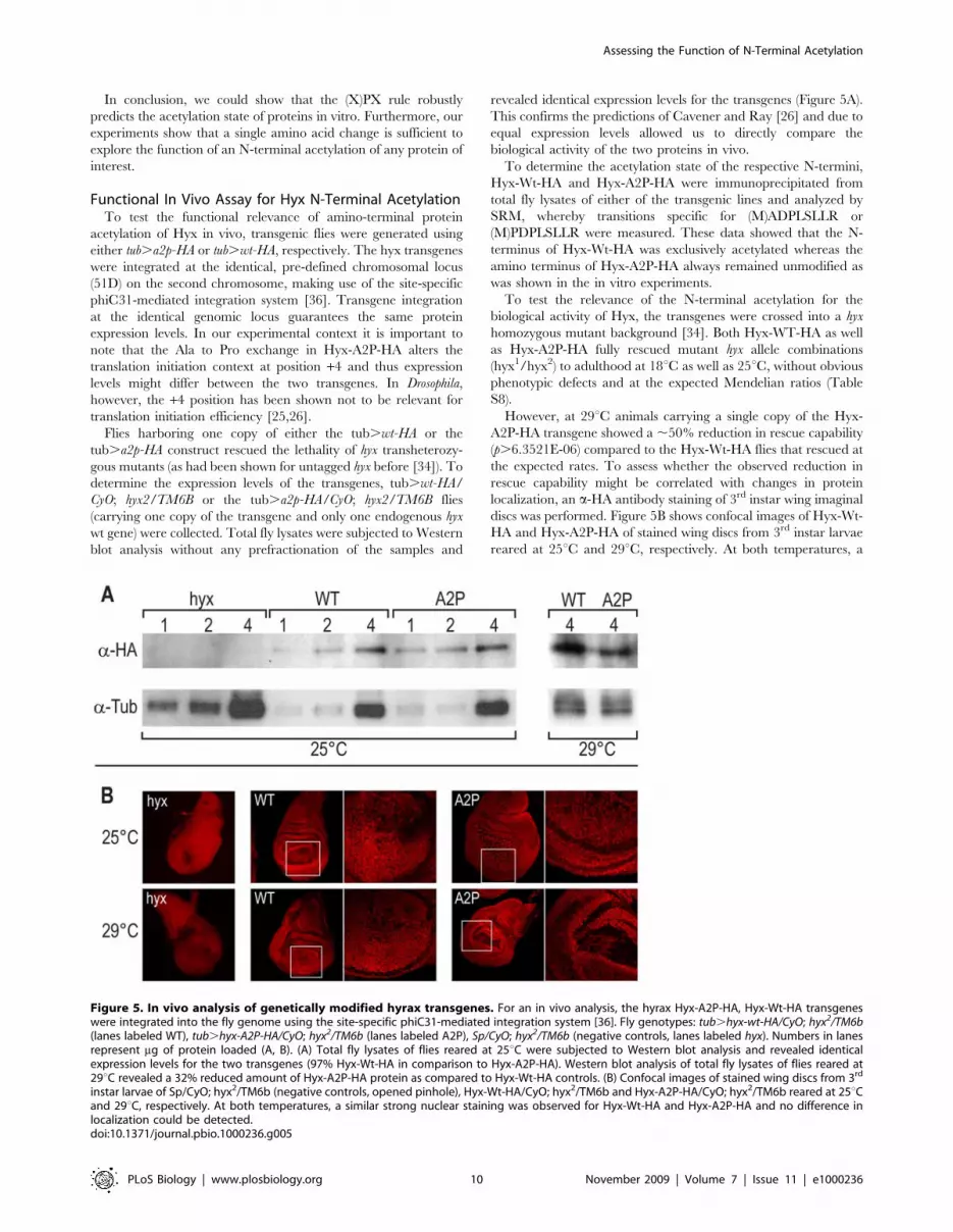
revealed identical expression levels for the transgenes (Figure 5A).
This confirms the predictions of Cavener and Ray [26] and due to
equal expression levels allowed us to directly compare the
biological activity of the two proteins in vivo.
To determine the acetylation state of the respective N-termini,
Hyx-Wt-HA and Hyx-A2P-HA were immunoprecipitated from
total fly lysates of either of the transgenic lines and analyzed by
SRM, whereby transitions specific for (M)ADPLSLLR or
(M)PDPLSLLR were measured. These data showed that the N-
terminus of Hyx-Wt-HA was exclusively acetylated whereas the
amino terminus of Hyx-A2P-HA always remained unmodified as
was shown in the in vitro experiments.
To test the relevance of the N-terminal acetylation for the
biological activity of Hyx, the transgenes were crossed into a hyx
homozygous mutant background [34]. Both Hyx-WT-HA as well
as Hyx-A2P-HA fully rescued mutant hyx allele combinations
(hyx1/hyx2) to adulthood at 18uC as well as 25uC, without obvious
phenotypic defects and at the expected Mendelian ratios (Table
S8).
However, at 29uC animals carrying a single copy of the Hyx-
A2P-HA transgene showed a ,50% reduction in rescue capability
(p.6.3521E-06) compared to the Hyx-Wt-HA flies that rescued at
the expected rates. To assess whether the observed reduction in
rescue capability might be correlated with changes in protein
localization, an a-HA antibody staining of 3rd instar wing imaginal
discs was performed. Figure 5B shows confocal images of Hyx-Wt-
HA and Hyx-A2P-HA of stained wing discs from 3rd instar larvae
reared at 25uC and 29uC, respectively. At both temperatures, a
Figure 5. In vivo analysis of genetically modified hyrax transgenes. For an in vivo analysis, the hyrax Hyx-A2P-HA, Hyx-Wt-HA transgeneswere integrated into the fly genome using the site-specific phiC31-mediated integration system [36]. Fly genotypes: tub.hyx-wt-HA/CyO; hyx2/TM6b(lanes labeled WT), tub.hyx-A2P-HA/CyO; hyx2/TM6b (lanes labeled A2P), Sp/CyO; hyx2/TM6b (negative controls, lanes labeled hyx). Numbers in lanesrepresent mg of protein loaded (A, B). (A) Total fly lysates of flies reared at 25uC were subjected to Western blot analysis and revealed identicalexpression levels for the two transgenes (97% Hyx-Wt-HA in comparison to Hyx-A2P-HA). Western blot analysis of total fly lysates of flies reared at29uC revealed a 32% reduced amount of Hyx-A2P-HA protein as compared to Hyx-Wt-HA controls. (B) Confocal images of stained wing discs from 3rd
instar larvae of Sp/CyO; hyx2/TM6b (negative controls, opened pinhole), Hyx-Wt-HA/CyO; hyx2/TM6b and Hyx-A2P-HA/CyO; hyx2/TM6b reared at 25uCand 29uC, respectively. At both temperatures, a similar strong nuclear staining was observed for Hyx-Wt-HA and Hyx-A2P-HA and no difference inlocalization could be detected.doi:10.1371/journal.pbio.1000236.g005
Assessing the Function of N-Terminal Acetylation
PLoS Biology | www.plosbiology.org 10 November 2009 | Volume 7 | Issue 11 | e1000236

similarly strong nuclear staining was observed for Hyx-Wt-HA and
Hyx-A2P-HA and no difference in localization could be detected.
To test whether the protein levels have changed at 29uC and
might contribute to the difference in rescue capability a Western
blot analysis of flies reared at 29uC was performed (same setup as
described above). Indeed the Western blot revealed a 32%
reduction of Hyx-A2P-HA protein as compared to the Hyx-Wt-
HA control (Figure 5A). Therefore, it might be possible that at
least in part the reduced protein levels of Hyx-A2P-HA at 29uCare the cause for the lower numbers of rescued flies. Mechanis-
tically, the reduced protein levels may be explained in two ways: (i)
one possibility is that protein expression is reduced due to the
exchange of the codon for Ala to the one for Pro. This mutation
leads to an exchange from G to A at position +4. Although
Cavener and colleagues state that the +4 position is not relevant
for translation initiation in Drosophila [25], we cannot rule out
effects on the translation machinery under these conditions (i.e.,
29uC). (ii) As a second possibility, protein half life might be
affected. It has previously been reported that N-terminal
acetylation may contribute to protein stability [3]. If this was the
case for Hyx-A2P-HA, we assume that the reduced protein
amount in A2P flies reared at 29uC is not due to a change of the
N-terminal residue from an Ala to a Pro but rather directly related
to the changed modification state of the protein’s N-terminus. This
hypothesis is supported by the so-called N-end rule, which relates
the in vivo half life of a protein to the identity of its N-terminal
residue. According to this rule both a Pro and an Ala (as well as
Ser, Thr, Gly, Val, and Met) as N-terminal residues confer protein
stability in all species [37].
In summary, our experiments show that the (X)PX rule allows
one to generate transgenes in order to study the biological
relevance of the presence or the absence of an N-terminal acetyl
group in vivo.
Discussion
Although the cellular machinery that is required to acetylate a
nascent protein N-terminus is also conserved in invertebrates, it
had been suggested that acetylation was a rare event in this animal
subphylum since only a few modified N-termini had been reported
[10]. In this study we present a comprehensive compilation of
mature protein N-termini from Drosophila melanogaster Kc 167 cells
and show that (i) N-terminal acetylation in fact is also a common
protein modification in insects and (ii) follows the same rules (in
terms of sequence requirements and iMet cleavage) and (iii) occurs
with a lower frequency than in man but at with a much higher
frequency than in yeast. Moreover, we observed that the
acetylation at the alpha amines of certain residues seem to have
shifted in frequency during evolution such that the acetylation of
an N-terminus in insects is in some cases in strong agreement with
the patterns found in man, while for others it resembles more the
distributions and frequencies found in yeast. These differences in
acetylation frequency, the partial acetylation of proteins, as well as
the lack of comprehensive datasets from different animal kingdoms
may explain why prediction of the acetylation state from a primary
protein sequence is still not yet clear-cut [11].
In fact, for some N-termini we still lack the understanding of
what determines their acetylation state (especially for Gly, Val, or
Thr as the first or second residue in the mature protein). Also some
proteins carrying an acetylation promoting Ser and Ala at position
2 remain unacetylated. In Drosophila, 30 of a total of 289 identified
N-termini that expose a Ser residue at their mature N-terminus
are not acetylated (11 of which are only partially acetylated, i.e.
appear acetylated and non-acetylated after iMet cleavage) (Table
S9). In two cases, an inhibitory Pro residue fulfilling the (X)PX rule
prevents the acetylation in spite of a Ser in the first position. One
protein CG2679-PB, for which we have defined a putative
alternative start site (putative alternative iMet at position 36 in the
predicted sequence; Text S1, Table S2), has a signal sequence
(SignalP 3.0, http://www.cbs.dtu.dk/services/SignalP/; [38])
predicted to be cleaved between residue 25 and 26. For the
remainder (as well as for the Val, Gly, and Thr) the only plausible
explanation is that there must be other features within the first 50
amino acids that prevent N-terminal acetylation during protein
synthesis. Polevoda and Sherman suggested that the secondary
structure of a protein’s N-terminus may play a role [10]. We have
analyzed the predicted secondary structure of all identified
proteins (residues 1–50, unpublished data) but could not identify
any correlation of structure and acetylation state. Consequently
the expected determinants (such as complex sequence patterns
within the first amino acids) remain to be identified through
sophisticated computational analysis for which this dataset may act
as an ideal starting point.
If no additional determinants (such as patterns or structural
motifs) exist steric hindrance could be responsible for the
preservation of a free N-terminus despite the presence of
promoting amino acid sequences. During protein synthesis,
cofactors like chaperones could bind to the nascent polypeptide
chain and prevent proper function of the acetylation machinery:
for instance it is known that tubulins undergo a sequence of folding
steps catalyzed by chaperones, which are commonly assumed to
take place after translation is completed [39,40]. Our data show
that tubulins always appear to have a free N-terminus despite the
occasional presence of amino acids that promote its acetylation. In
this context it may therefore well be that chaperones could already
associate with the transcription machinery, thereby preventing the
acetylation of tubulin by steric hindrance.
We have observed that in Drosophila the acetylation is fixed in
most cases. This is similar to the case in human and in contrast to
yeast where partial acetylation is common [11]. Possible
explanations for this discrepancy between the organisms could
be that yeast cells (but not fly or human cells) grow too fast, making
acetyl-CoA poorly available as substrate, or the degradation of the
unacetylated proteins cannot cope with division rates and thus the
proteins with a free N-terminus accumulate in the cells.
Nevertheless, in certain cases a partial acetylation of proteins
may be of relevance for yeast as well as for higher eukaryotes. To
asses this relevance such proteins could be genetically engineered
according to the (X)PX rule, shifting the equilibrium of the mutant
proteins towards a fixed acetylated or free N-terminus.
N-terminal acetylation has been shown to be absolutely
necessary for the proper localization, activity, or stability of
various proteins. The current view is that the presence rather than
the absence of an acetylation is important for protein function.
Our Pfam analysis challenges this view as it also shows a clear
correlation for a non-acetylated N-terminus and certain protein
domains. Additional evidence for the perspective that the absence
of an acetylation is also important for proper protein function has
been demonstrated in the case of human hemoglobin. The
exchange of Val to Ala at the second position in the human
hemoglobin beta chain reverts the usually free N-terminus into an
acetylated one. Patients carrying this so-called ‘‘Raleigh’’ mutation
suffer from thalassaemia due to a reduced affinity of the mutant
hemoglobin to oxygen [41], underlining the importance of the free
amino terminus for proper function. Finally, our own transgene
analysis on Cks85A strongly indicates that the preservation of the
protein’s non-acetylated N-terminus is relevant for its proper
function. In this context it is important to note that no pattern
Assessing the Function of N-Terminal Acetylation
PLoS Biology | www.plosbiology.org 11 November 2009 | Volume 7 | Issue 11 | e1000236
exists that guarantees an N-terminal acetylation, although Ser and
Ala as second residues usually ascertain a modification. For the
prevention of acetylation the opposite is true: based on the data we
formulated the (X)PX rule that states that a Pro residue at the
primary or secondary position of the mature protein N-terminus
prevents acetylation and assures a free amino terminus under all
circumstances. Since the majority of protein N-termini are
acetylated in eukaryotic organisms investigated so far, and due
to the presence of an absolute inhibitory rather than a promoting
signal, we conclude that the acetylation of protein N-termini is the
default state and, where necessary, is prevented by specific motifs.
Along this line it has been suggested that some N-termini are
acetylated just because of the presence of a promoting pattern but
that acetylation has no functional relevance [3]. One can easily
imagine such a case: a Ser at position 2 in the primary sequence
has been evolutionary conserved because its phosphorylation is
relevant for its biological activity. Upon protein synthesis this Ser
by default also promotes iMet cleavage and the subsequent
acetylation of the N-terminus. The conservation of the Ser,
however, occurred due to the need for Ser phosphorylation and
not N-terminal acetylation. In such a scenario one could apply the
(X)PX rule to discriminate whether acetylation and phosphoryla-
tion or phosphorylation alone would contribute to protein
function: the introduction of an inhibitory Pro after the Ser
residue would allow iMet cleavage as well as the phosphorylation
of the Ser but would prevent the acetylation of the N-terminus.
Likewise, the Ser to Ala (or comparable) amino-acid exchange
would preserve the acetylation (at least in insects and human) but
not the phosphorylation of the protein, allowing one to assess the
contribution of the N-terminal acetylation to protein function. A
relevant example would be the rat RNA polymerase subunit 6
(RPB6), which is phosphorylated by Casein KinaseII (CkII) at a
Ser at residue 2. It has, however, not been reported whether the
acetylation state of this protein is relevant for its function [42].
Unlike a phosphorylation, which is ‘‘dynamic’’ and may be
rapidly added or removed from a protein upon stimulation of a
cell, N-terminal acetylation has been shown to be a co-
translational, irreversible, and thus a ‘‘static’’ modification [3].
However, we and others have evidence that acetylation of N-
termini does not only occur co-translationally [14]: we have
identified 511 internal, acetylated peptides that reside within the
annotated protein sequence (Table S10). These N-termini do not
comply with the suggested N-terminal acetylation rules and thus
are suggested to occur post-translationally. They require a to-date
unknown acetylase activity as well as internal proteolytic cleavage
at a specific site of the protein. It is tempting to speculate that these
post-translational acetylation events may also occur in a dynamic
fashion, allowing fast responses to various stimuli.
It remains to be noted that acetylation of N-terminal Pro
residues has been reported in yeast [43–45]. The experimental
evidence for an N-terminally acetylated Pro residue is solely based
on shifts of protein spots observed in comparative 2D gel
experiments of NAT-mutant versus wt strains. A direct measure-
ment of acetylation has not been performed in this context [45]. In
contrast, in two large-scale studies N-terminal Pro acetylation has
only been observed if an internal Pro residue is exposed at the N-
terminus after post-translational processing of the protein (see
Tables S1 and S10). From this we conclude that the (X)-P-X rule,
which is formulated for the first three residues of the primary
amino acid sequence, is robust and that possible exceptions to this
rule will be extremely rare and most likely attached post-
translationally by a yet undescribed protein activity.
Although amino-terminal acetylation has been studied for more
than 30 years, some aspects, foremost the relevance of its presence
or absence for individual protein function, remain unclear for most
of the cases. With this work we have defined tools that will help to
better understand the elusive mechanisms as well as to explore the
function of protein N-terminal acetylation in all organisms in a
protein-specific manner. Complementary studies using for in-
stance mutations in the relevant NAT enzymes will help to
eventually draw firm conclusions about the function of the N-
terminal acetylation.
Materials and Methods
Construction of VectorsTo generate expression vectors for the hyrax and Cks85A
transgenes, the respective DNAs were amplified by PCR. The
forward primers contained a 59 Kozak consensus (CGCCACC)
and the appropriate base exchanges to generate the desired wt or
mutant forms.
(1) hyx_WT_fw: AAAGGTACCGCCACCATGGCAGATCC-
GCTCAGCCTGC;
(2) hyx_A2P_fw: AAAGGTACCGCCACCATGCCAGATCC-
GCTCAGCCTGC;
(3) hyx_rev: AAAGGCGCGCCCCATATCGTAAATCGGGC-
TTGTACTTGG;
(4) Cks85A_WT_fw: AAAGGTACCGCCACCATGCCGGCC-
GATCAAATTCAATAC;
(5) Cks85A_P2A_fw: AAAGGTACCGCCACCATGGCGGC-
CGATCAAATTCAATAC;
(6) Cks85A_P2S_fw: AAAGGTACCGCCACCATGTCGGCC-
GATCAAATTCAATAC;
(7) Cks85A_rev: AAAAAGCGCGCACCGCAGAGATTCGC-
GATGGC.
PCR fragments were inserted into the pOP-118 vector to create
cDNAs driven by a ubiquitous tubulin-1a promoter and tagged
with a 3xHA. For in vivo experiments the expression cassettes
were cloned into an attB integration backbone [36].
Cell Culture and Immunoprecipitation ExperimentsDrosophila melanogaster Kc 167 and S2 cells were cultivated in
Schneider’s Drosophila medium (Gibco/Invitrogen) supplemented
with 10% heat-inactivated fetal-calf serum, penicillin (100 U/ml),
and streptomycin (100 mg/ml) at 25uC. Cells were split 1:4 (v/v)
every 3–4 d when they reached confluency.
For immunoprecipitations 75 cm2 flasks of S2 cells were
transfected with 2 mg of the respective tub-[cDNA]-HA plasmid
via Effectene Transfection Reagent (Qiagen). Cells were collected
48 h after transfection and lysed with lysis buffer (20 mM sodium
phosphate-buffer, 200 mM NaCl, 0.5% NP40, supplemented just
before use with 1 mM DTT, and CompleteTM protease inhibitor
cocktail (Roche)). Cell lysates were incubated for 3 h with
monoclonal anti-HA agarose beads (clone HA-7, Sigma). Finally,
beads were washed three times with lysis buffer, twice with lysis
buffer without detergent, and bound proteins were eluted with
0.2 M glycine (pH 2.5).
Fly StocksFor in vivo assays, the following tester lines were used:
transgenic vectors were injected into Drosophila (yw) that contained
the second-chromosomal attP landing site at map position 51D
[36]. Potential founders were individually outcrossed against the
balancer stock yw, hs-flp; Sp/CyO; TM6b/MRKS. Their white+progeny was then individually backcrossed to the balancer
Assessing the Function of N-Terminal Acetylation
PLoS Biology | www.plosbiology.org 12 November 2009 | Volume 7 | Issue 11 | e1000236

background to establish clonal stocks. The resulting founder stocks
were verified for the correct transgene by PCR. Successfully
established transgenic insertions were then crossed into hyx mutant
backgrounds, as described previously [34].
ImmunohistochemistryWing discs were isolated, fixed, and incubated as previously
described [46]. Antibodies used for the staining were mouse HA11
(1:1000, BAbCO) and a goat secondary antibody coupled to
Alexa594 (1:500, Molecular Probes).
Western Blot AnalysisFor total protein isolation adult flies were grinded in liquid
nitrogen and the homogenate was resuspended in a 4 times
volume of lysis buffer (20 mM TrisHCl pH 7.5, 150 mM NaCl,
0.2% NP40, 10% glycerol supplemented just before use with
1 mM DTT, and CompleteTM protease inhibitor cocktail
(Roche)). Protein samples were run on a 10% SDS-PAGE at
140 V for 90 min and subsequently transferred to a Hybond-P
PVDF Membrane (Amersham). After the transfer, the membrane
was rinsed in PBST, blocked with 5% nonfat dried milk in PBST
and then incubated with either monoclonal mouse anti-alpha
tubulin DM1A (1:2000, Sigma) or mouse anti-HA.11 (1:1000,
BAbCO). As secondary antibody, peroxidase-conjugated goat
anti-mouse IgG (1:10000, Jackson) was applied. Signals were
detected via ECL Plus (Amersham). Intensities of bands were
quantified using the histogram option (average gray value) of
ImageJ (NIH).
Sample Preparation for Mass SpectrometryTotal protein lysates or proteins isolated via immunoprecipita-
tion were reduced with 5 mM Tris(2-carboxyethyl)phosphine
hydrochloride (TCEP) and treated with 10 mM iodoacetamide
to modify cysteine residues. Tryptic digestion was carried out
overnight using 5 mg trypsin per sample. Samples were purified by
reverse phase C-18 chromatography (Sep-PacK, Waters). For
mass spectrometry analysis samples were resuspended in buffer A
(5% acetonitrile, 0.2% formic acid).
SRMSRM was performed on a triple quadrupole mass spectrometer
(TSQ Quantum Ultra EMR, Thermo Fisher Scientific) operated
with Xcalibur 2.0.7 (Thermo Fisher Scientific). The instrument was
coupled to an Eksigent nano-LC system. Samples were automat-
ically injected into a 10-ml sample loop and loaded onto an
analytical column (9 cm length675 mm (internal diameter) packed
in-house with Magic C18 AQ beads 5 mm, 100 A (Microm)).
Peptide mixtures were delivered to the analytical column at a flow
rate of 500 nl/min of buffer A (5% acetonitrile, 0.2% formic acid)
for 18 min and then eluted using a gradient of acetonitrile (10%–
35%; 0.36%/min) in 0.2% formic acid at a flow rate of 250 nl/min.
SRM measurements were carried out with a Q1 resolution of
0.4 and a Q3 resolution of 0.7 m/z half-maximum peak width.
Scan speed was set to 0.020 ms per scan event. SRM transitions
specific for proteotypic peptides were generated using the SRM
Workflow (software from Thermo Fisher Scientific). For each
precursor transitions were calculated with precursor charges 2+ or
2+ and 3+. At least four y-ions with m/z. precursor were
monitored. Collision energies (CE) were calculated according to
the following formulas: CE = 0.0346m/z+3.314 (2+) and
CE = 0.0446m/z+3.314 (3+). SRM traces were evaluated via the
SRM Workflow. For each peptide the co-elution of all transitions
was confirmed. Peptides with non-coeluting elution profiles or bad
resolution or signal to noise ratios were not considered for further
analysis. iMet cleavage was quantified by comparing the ion
current profile of identical y-product ions.
SRM-triggered MS/MS experiments were performed to
validate the identity of the peptides for the relevant SRM traces.
MS/MS spectra were acquired with a Q3 resolution at 0.7 m/z
half-maximum peak width. The scan range was automatically
determined by the instrument using the assigned charge state of
the precursor ion (2+). MS/MS spectra were assigned to peptide
sequences using the Mascot software version 2.2 (Matrix Science).
Processing of Shotgun DataThe data for Kc167 cells from the previously released proteome
catalog for Drosophila melanogaster (retrieved from [24]) were
searched using the Mascot search algorithm for finding N-terminal
peptides. The search criteria were set as follows: Variable
modifications were set to Acetyl (N-term), Deamidation (NQ),
Carbamidomethyl (C, or where required ICAT-C, ICAT-
C:13C(9)), Oxidation (M), Mass values monoisotopic, Peptide
mass tolerance 3 Da, Fragment mass tolerance 60.8 Da,
Maximum missed cleavages 2, Instrument type Esi Trap. All
peptide assignments where the Mascot ion score is greater than the
homology score and where the Mascot expect score is smaller than
0.05 were considered as good hits for further analysis.
Post-Processing of Searched DataAll peptides called as present by Mascot in the SAX, SCX, and
the published datasets [47] were remapped against the Berkeley
Drosophila melanogaster protein database (BDGP) release 3.2 for the
purpose of extracting all the possible splice variants and alternative
proteins belonging to a specific peptide. Furthermore, the proteins
obtained in this way were then classified according to their
information content into five different classes [48]. This informa-
tion content considers the relationship between the protein
sequences and gene models and minimizes protein inference
errors due to ambiguous protein sequences.
The N-termini were extracted from the overall list of peptides.
Two types of N-termini are distinguished: (i) N-termini that start in
position 1 or 2 (after methionine cleavage) in at least one protein of
all the possible alternatives for the respective peptide and (ii)
potential alternative methionine starting sites, i.e. semi-tryptic
peptides that start with methionine, or where the precedent amino
acid is a methionine. This list was manually cured, excluding fully
tryptic peptides that likely represent internal peptides (the peptides
identified in the COFRADIC approach may be preceded by a Lys
residue since they are modified and thus no longer cleaved by
trypsin). Furthermore, possible redundancies with respect to N-
terminus were reduced, i.e. cases where one N-terminus is a prefix
of another N-terminus with the exception of partially acetylated N-
termini. The entire list of N-termini is presented in Table S1.
Extraction of Kozak/Cavener ContextPeptide sequences were mapped onto the genomic sequence in
order to extract the DNA context information of the iMet (or of
the starting ATG triplet). For this purpose, for each gene model of
BDGP3.2 the protein coding sequence, the respective transcript,
as well as the exon containing the potential alternative iMet were
extracted and the Cavener and Kozak context was analyzed. A
sequence (24 to 21) for which up to 1 nucleotide was identical to
the Cavener or Kozak consensus sequence was labeled as weak, 2–
3 perfect nucleotide matches were labeled as adequate, and
identical sequences were labeled as strong (see Text S1).
Weblogos were created using the library weblogo (http://code.
google.com/p/weblogo/) in Python in order to represent the
Assessing the Function of N-Terminal Acetylation
PLoS Biology | www.plosbiology.org 13 November 2009 | Volume 7 | Issue 11 | e1000236

relative frequency and information content of different nucleotides
at varying positions around the Kozak/Cavener sequence and bar
plots were created using R (www.r-project.org).
Pfam AnalysisPfam (www.sanger.ac.uk/Software/Pfam/) was used to identify
functional domains in the experimentally identified protein
sequences. For this purpose, the set of 16,743 distinct protein
sequences of BDGP3.2 were searched against the Pfam database
of Hidden Markov Models (release July 23, 2008, 10,340 protein
family models) using the HMMER software package (hmmer2.3.2,
http://hmmer.janelia.org/).
The Pfam results were compared then with the results of
acetylated seen protein sequences for any over- or underrepre-
sentation and Fisher’s exact test with multiple testing correction
was performed for selected Pfam categories.
Gene Ontology AnalysisTo assess any possible association between the acetylation status
of a group of proteins and different GO terms, all possible gene
models were extracted from Table 1 and were submitted to a GO
Slim analysis (http://go.princeton.edu/cgi-bin/GOTermMapper).
This tool uses the map2slim.pl script (Chris Mungall, BDGP) to bin
the submitted gene lists to a static set of broader, high-level parent
generic GO-slim terms.
The GO analysis was performed for ‘‘Biological Process,’’
‘‘Cellular Component,’’ and ‘‘Molecular Function,’’ and Fisher’s
exact test, with multiple testing correction, was performed for each
GO term to provide a p value for the respective over- or
underrepresentation.
SAX Peptide Fractionation Prior to N-Terminal COFRADICAnalysis
8 mg of the Kc167 cell cytosolic fraction was first dissolved in
1 ml of 6 M guanidinium hydrochloride in 50 mM phosphate
buffer (pH 7.5), after which the guanidinium hydrochloride
concentration was lowered to 4 M by adding 0.5 ml of 50 mM
phosphate buffer (pH 7.5). Proteins were reduced and alkylated by
adding TCEP and iodoacetamide to final concentrations of
50 mM and 100 mM, respectively (reaction was allowed for
60 min at 30uC). Then, excess reagents were removed by desalting
the protein mixture over a 10 ml Zeba Spin Desalting Column
(Pierce, Thermo Fisher Scientific, Erembodegem, Belgium).
Proteins were collected in 1 ml of 2 M guanidinium hydrochloride
in 50 mM phosphate buffer (pH 8). Free primary amines (protein
a-N-termini and e-amines of lysines) were acetylated using an N-
hydroxysuccinimide ester of trideutero-acetate (prepared accord-
ing to [49]). Here, 5.8 mg (about 36 mmol) of this NHS-ester was
dissolved in 20 ml DMSO and then added to the protein sample.
Acetylation was allowed for 90 min at 30uC, after which O-
acetylation of Ser, Thr, and Tyr side-chains was reversed by
adding 10 ml of 50% of hydroxylamine to the protein solution and
incubation for 20 min at room temperature.
This modified protein mixture was desalted as indicated above, but
now eluted in 50 mM of NH4HCO3 (pH 8, freshly prepared). The
protein mixture was boiled for 5 min, placed on ice for another 5 min,
and then overnight digested by adding 40 mg of sequencing-grade
modified trypsin (Promega Corporation, Madison, WI, USA). Small
particulate matter was removed by centrifugation for 10 min at
10,000 g. Half of the digest (500 ml) was loaded on a strong anion
exchange (SAX) column (SAX-Zirchrom 2.1 mm I.D.6150 mm
length; 3 mm particles, ZirChrom Separations, Anoka, MN, USA)
after which a binary solvent gradient was applied to fractionate
peptides. Solvent A consisted of 10 mM Tris-HCl (pH 8) in 25%
acetonitrile and solvent B was 1 M NaCl in 10 mM Tris-HCl (pH 8)
and 25% acetonitrile. Under a constant flow of 70 ml/min, the
following gradient was applied: 0% solvent B (0–50 min), 10% solvent
B (55 min), 50% solvent B (100 min), washing with 50% solvent B until
105 min, 100% solvent B (107 min), followed by a 5 min wash with
solvent B, 100% solvent A (114 min), and re-equilibration with solvent
A (170 min). SAX-separated peptides were collected in 2 min wide
fractions between 10 and 130 min and were finally pooled into six
fractions in such a way that each fraction contained similar amounts of
total peptide material (judged from the UV absorbance trace).
SCX-Based Enrichment of N-Terminal PeptidesIt was previously demonstrated that strong cation exchange (SCX)
chromatography at acidic pH can be used to enrich N-terminal
peptides from whole proteome tryptic digests [14,22]. These peptides
are in fact less well retained by the SCX column as other peptides and
are thereby segregated from the bulk of non-N-terminal peptides.
One disadvantage, however, is that N-terminal peptides containing
histidine will still be retained by the SCX column and are thus largely
absent from the final lists of reported identifications [22].
Here, SCX was used to enrich N-terminal peptides from digests
of the cytoplasmic, nuclear, and membrane fraction of Kc 167 cells
prior to COFRADIC. Full experimental details can be found in
[22], and in brief, protein preparations were first reduced and S-
alkylated with iodoacetamide, then all free primary amines were
blocked by trideutero-acetylation, and finally proteins were digested
into peptides using trypsin. This peptide mixture was then applied
on a SCX column at pH 3 and blocked N-terminal peptides were
found enriched in the non-binding fraction. These peptides were
then further enriched by N-terminal COFRADIC (see below).
Isolation of N-Terminal Peptides by COFRADIC, LC-MS/MS Analysis, and MASCOT Analysis
All six SAX fractions and the SCX-enriched proteome digests of
the Kc167 cell cytosolic, microsomal, and nuclear fractions were
then further analyzed by the N-terminal COFRADIC technology
[20] as described in [22]. Such isolated peptides were then further
analyzed by LC-MS/MS analysis on either a Bruker Esquire HCT
ion trap (SAX peptides, ion trap operated as described in [50]) or an
LTQ linear iontrap operated as described in [47] or an Agilent
XCT-Ultra IT mass spectrometer equipped with Agilent’s Chip
Cube (SCX peptides, ion trap operated as described in [51]).
Following analysis, raw MS/MS-spectra were converted to mgf
files, which were used to identify the corresponding peptides using
the MASCOT database search tool [52], in-house installed. The
following parameters were set. Carbamidomethylation of Cys and
trideutero-acetylation of Lys were set as fixed modifications.
Acetylation or trideutero-acetylation of peptide N-termini, carba-
mylation of Lys (only for SAX-separated peptides), deamidation of
Asn and Gln, oxidation of Met (sulfoxide), N-terminal pyrocarba-
midomethyl Cys, and N-terminal pyroglutamic acid were all set as
variable modifications. The mass tolerances for both the precursor
ion and fragment ions were set to 60.5 Da. The enzyme was set at
endoproteinase Arg-C and cleavage between Arg-Pro and one
missed cleavage were allowed. MASCOT’s instrument setting was
ESI-TRAP. The BDGP 3.2 database was searched as well truncated
versions of this database that anticipate for protein processing events
and were previously found to increase the number of identifications.
These truncated versions were made using the DBToolkit algorithm
[53]. The raw DAT result files of Mascot were then queried using
in-house developed software tools. Only MS/MS-spectra receiving
a score exceeding Mascot’s identity threshold score at the 95%
confidence level were kept. Such identified peptides were then
Assessing the Function of N-Terminal Acetylation
PLoS Biology | www.plosbiology.org 14 November 2009 | Volume 7 | Issue 11 | e1000236
automatically stored in a MySQL relational database (see http://
genesis.ugent.be/ms_lims/) in which links were made to their MS/
MS-spectra and precursor proteins.
Supporting Information
Figure S1 Calculation of theoretically visible N-termini.(A–B) In theory, 9,319 (56%) of the very N-terminal tryptic
peptides of the 16,743 distinct Drosophila proteins are within the
detectable dynamic range of current mass spectrometers in a
regular trypsin digest (A). For calculation the following constraints
have been applied: the peptides have to be fully tryptic without
missed cleavages, are at least six amino acids long (green lines), and
their precursor m/z value is within the range 450–1,500
(considering the charge states 1+ to 3+). Interestingly, the N-
terminal tryptic peptides of close to 7,000 distinct Drosophila
proteins are six or less amino acids in length (blue lines and below
450 m/z and thus not detectable). Using COFRADIC (B), where
lysines are blocked and no longer available for trypsin cleavage,
the number of detectable N-terminal tryptic peptides rises to
10,373 (62%). However, since some of these N-terminal peptides
might be shared between different proteins, the number of
proteins distinguishable based solely on their N-terminal peptide
drops to 8,347 for a regular digest (Figure S1A, 50% of all
annotated proteins), and to 9,389 for the COFRADIC treatment
(Figure S1B, 56.1%). Finally, if we also consider proteins with a
predicted signal peptide (SignalP 3.0 [38], the number of
detectable proteins is further reduced, and in theory, we could
identify 7430 (A) or 8212 proteins (B), respectively).
Found at: doi:10.1371/journal.pbio.1000236.s001 (0.23 MB TIF)
Figure S2 Analysis to correlate GO categories with theN-terminal acetylation status. (A–C) To assess whether
particular protein functions or functional domains are associated
with the N-terminal acetylation state, a Gene Ontology analysis on
a reduced set of GO categories (referred to as GO Slim) on all
three levels, namely Cellular Component (A), Molecular Function (B),
and Biological Process (C) was performed. The results of this analysis
(representative examples with at least two members seen in a
category) are also shown in Table S4. For more details please see
Text S1. * statistically significant different frequency observed
(p,0.05).
Found at: doi:10.1371/journal.pbio.1000236.s002 (1.09 MB TIF)
Figure S3 The principle of SRM in a triple quadrupolemass spectrometer. SRM is performed in a specific type of
mass spectrometer, which consists of a linear series of three mass
analyzers, so-called quadrupoles. The first quadrupole (Q1)
functions as a mass filter, capable of selectively transmitting only
the parent ion (peptide) of interest from a complex peptide
mixture. The second quadrupole (Q2) serves as a fragmentation
unit, in which the selected peptide is fragmented into smaller
product ions by collisionally activated dissociation. Finally, a single
fragment (product ion) that is characteristic for a peptide is passed
through to the third mass analyzer Q3 and can be quantified.
Found at: doi:10.1371/journal.pbio.1000236.s003 (0.08 MB TIF)
Table S1 Compilation of all N-terminal peptides iden-tified in this study.Found at: doi:10.1371/journal.pbio.1000236.s004 (0.28 MB XLS)
Table S2 Alternative start sites.Found at: doi:10.1371/journal.pbio.1000236.s005 (0.04 MB XLS)
Table S3 Ambiguously acetylated N-termini.Found at: doi:10.1371/journal.pbio.1000236.s006 (0.03 MB XLS)
Table S4 (A–C) Statistics of the GO Slim analysis.Found at: doi:10.1371/journal.pbio.1000236.s007 (0.03 MB XLS)
Table S5 (A–B) PFAM analysis for associating functionaldomains with the N-terminal acetylation status of aprotein.Found at: doi:10.1371/journal.pbio.1000236.s008 (0.05 MB XLS)
Table S6 Confirmation of the XPX and PX rule bytargeted SRM.Found at: doi:10.1371/journal.pbio.1000236.s009 (0.03 MB XLS)
Table S7 Targeted SRM measurements of hyrax andCks85A (pulldown experiments).Found at: doi:10.1371/journal.pbio.1000236.s010 (0.02 MB XLS)
Table S8 Rescue experiments with Hyx-Wt-HA andHyx-A2P-HA.Found at: doi:10.1371/journal.pbio.1000236.s011 (0.02 MB XLS)
Table S9 Non-acetylated N-termini with a serine resi-due at the mature N-terminus.Found at: doi:10.1371/journal.pbio.1000236.s012 (0.02 MB XLS)
Table S10 Acetylated internal peptides. Their N-terminido not comply with the suggested N-terminal acetylationrules.Found at: doi:10.1371/journal.pbio.1000236.s013 (0.11 MB XLS)
Text S1 Kozak/Cavener analysis of putative alternativestart sites, and GO Slim analysis.Found at: doi:10.1371/journal.pbio.1000236.s014 (0.50 MB
DOC)
Acknowledgments
We thank Sarah Steiner, Johannes Bischof, Claus Schertel, Esther Jud, and
Eliane Escher for technical help, and Christian Panse, Simon Barkow-
Oesterreicher, and Jonas Grossmann for help with computational
infrastructure.
Author Contributions
The author(s) have made the following declarations about their
contributions: Conceived and designed the experiments: SG EH RA JV
KB KG EB. Performed the experiments: SG CM AS BG BR SM EMN
ET VL EB. Analyzed the data: SG EQ CM AS BG BR CHA KG EB.
Contributed reagents/materials/analysis tools: EQ EL CHA KG. Wrote
the paper: SG EQ RA KB CHA KG EB.
References
1. Gautschi M, Just S, Mun A, Ross S, Rucknagel P, et al. (2003) The yeastN(alpha)-acetyltransferase NatA is quantitatively anchored to the ribosome and
interacts with nascent polypeptides. Mol Cell Biol 23: 7403–7414.
2. Bradshaw RA, Brickey WW, Walker KW (1998) N-terminal processing: the
methionine aminopeptidase and N alpha-acetyl transferase families. TrendsBiochem Sci 23: 263–267.
3. Polevoda B, Sherman F (2002) The diversity of acetylated proteins. Genome Biol
3: reviews0006.
4. Sherman F, Stewart JW, Tsunasawa S (1985) Methionine or not methionine at
the beginning of a protein. Bioessays 3: 27–31.
5. Boissel JP, Kasper TJ, Bunn HF (1988) Cotranslational amino-terminalprocessing of cytosolic proteins. Cell-free expression of site-directed mutants of
human hemoglobin. J Biol Chem 263: 8443–8449.
6. Lowther WT, Matthews BW (2000) Structure and function of the methionine
aminopeptidases. Biochim Biophys Acta 1477: 157–167.
7. Polevoda B, Norbeck J, Takakura H, Blomberg A, Sherman F (1999)Identification and specificities of N-terminal acetyltransferases from Saccharo-
myces cerevisiae. Embo J 18: 6155–6168.
8. Polevoda B, Sherman F (2003) Composition and function of the eukaryotic N-
terminal acetyltransferase subunits. Biochem Biophys Res Commun 308: 1–11.
Assessing the Function of N-Terminal Acetylation
PLoS Biology | www.plosbiology.org 15 November 2009 | Volume 7 | Issue 11 | e1000236

9. Polevoda B, Sherman F (2000) Nalpha -terminal acetylation of eukaryotic
proteins. J Biol Chem 275: 36479–36482.10. Polevoda B, Sherman F (2003) N-terminal acetyltransferases and sequence
requirements for N-terminal acetylation of eukaryotic proteins. J Mol Biol 325:
595–622.11. Arnesen T, Van Damme P, Polevoda B, Helsens K, Evjenth R, et al. (2009)
Proteomics analyses reveal the evolutionary conservation and divergence of N-terminal acetyltransferases from yeast and humans. Proceedings of the National
Academy of Sciences 106: 8157–8162.
12. Driessen HP, de Jong WW, Tesser GI, Bloemendal H (1985) The mechanism ofN-terminal acetylation of proteins. CRC Crit Rev Biochem 18: 281–325.
13. Persson B, Flinta C, von Heijne G, Jornvall H (1985) Structures of N-terminallyacetylated proteins. Eur J Biochem 152: 523–527.
14. Dormeyer W, Mohammed S, Breukelen B, Krijgsveld J, Heck AJ (2007)Targeted analysis of protein termini. J Proteome Res 6: 4634–4645.
15. Hofmann I, Munro S (2006) An N-terminally acetylated Arf-like GTPase is
localised to lysosomes and affects their motility. J Cell Sci 119: 1494–1503.16. Behnia R, Panic B, Whyte JR, Munro S (2004) Targeting of the Arf-like GTPase
Arl3p to the Golgi requires N-terminal acetylation and the membrane proteinSys1p. Nat Cell Biol 6: 405–413.
17. Hitchcock-DeGregori SE, Heald RW (1987) Altered actin and troponin binding
of amino-terminal variants of chicken striated muscle alpha-tropomyosinexpressed in Escherichia coli. J Biol Chem 262: 9730–9735.
18. Inoue A, Ojima T, Nishita K (2004) N-terminal modification and its effect onthe biochemical characteristics of Akazara scallop tropomyosins expressed in
Escherichia coli. J Biochem 136: 107–114.19. Baerenfaller K, Grossmann J, Grobei M, Hull R, Hirsch-Hoffmann MH, et al.
(2008) A high-density, organ-specific proteome map for Arabidopsis thaliana.
Science 320: 938–941.20. Gevaert K, Goethals M, Martens L, Van Damme J, Staes A, et al. (2003)
Exploring proteomes and analyzing protein processing by mass spectrometricidentification of sorted N-terminal peptides. Nat Biotechnol 21: 566–569.
21. Finn RD, Tate J, Mistry J, Coggill PC, Sammut SJ, et al. (2008) The Pfam
protein families database. Nucleic Acids Res 36: D281–D288.22. Staes A, Van Damme P, Helsens K, Demol H, Vandekerckhove J, et al. (2008)
Improved recovery of proteome-informative, protein N-terminal peptides bycombined fractional diagonal chromatography (COFRADIC). Proteomics 8:
1362–1370.23. Van Damme P, Maurer-Stroh S, Plasman K, Van Durme J, Colaert N, et al.
(2009) Analysis of protein processing by N-terminal proteomics reveals novel
species-specific substrate determinants of granzyme B orthologs. Mol CellProteomics 8: 258–272.
24. Loevenich SN, Brunner E, King NL, Deutsch EW, Stein SE, et al. (2009) TheDrosophila melanogaster PeptideAtlas facilitates the use of peptide data for
improved fly proteomics and genome annotation. BMC Bioinformatics 10: 59.
25. Cavener DR (1987) Comparison of the consensus sequence flanking transla-tional start sites in Drosophila and vertebrates. Nucleic Acids Res 15:
1353–1361.26. Cavener DR, Ray SC (1991) Eukaryotic start and stop translation sites. Nucleic
Acids Res 19: 3185–3192.27. Pedersen AG, Nielsen H (1997) Neural network prediction of translation
initiation sites in eukaryotes: perspectives for EST and genome analysis. Proc Int
Conf Intell Syst Mol Biol 5: 226–233.28. Frottin F, Martinez A, Peynot P, Mitra S, Holz RC, et al. (2006) The proteomics
of N-terminal methionine cleavage. Mol Cell Proteomics 5: 2336–2349.29. Ashburner M, Ball CA, Blake JA, Botstein D, Butler H, et al. (2000) Gene
ontology: tool for the unification of biology. The Gene Ontology Consortium.
Nat Genet 25: 25–29.30. Anderson L, Hunter CL (2006) Quantitative mass spectrometric multiple
reaction monitoring assays for major plasma proteins. Mol Cell Proteomics 5:573–588.
31. Ahrens CH, Brunner E, Hafen E, Aebersold R, Basler K (2007) A Proteome
Catalog of Drosophila melanogaster: An essential resource for targetedquantitative proteomics. Fly 1: e1–5.
32. Stahl-Zeng J, Lange V, Ossola R, Eckhardt K, Krek W, et al. (2007) High
sensitivity detection of plasma proteins by multiple reaction monitoring of N-glycosites. Mol Cell Proteomics 6: 1809–1817.
33. Lange V, Malmstrom JA, Didion J, King NL, Johansson BP, et al. (2008)
Targeted quantitative analysis of Streptococcus pyogenes virulence factors bymultiple reaction monitoring. Mol Cell Proteomics.
34. Mosimann C, Hausmann G, Basler K (2006) Parafibromin/Hyrax activatesWnt/Wg target gene transcription by direct association with beta-catenin/
Armadillo. Cell 125: 327–341.
35. Pearson NJ, Cullen CF, Dzhindzhev NS, Ohkura H (2005) A pre-anaphase role
for a Cks/Suc1 in acentrosomal spindle formation of Drosophila female meiosis.
EMBO Rep 6: 1058–1063.
36. Bischof J, Maeda RK, Hediger M, Karch F, Basler K (2007) An optimized
transgenesis system for Drosophila using germ-line-specific phiC31 integrases.Proc Natl Acad Sci U S A 104: 3312–3317.
37. Varshavsky A (2008) The N-end rule at atomic resolution. Nat Struct Mol Biol15: 1238–1240.
38. Emanuelsson O, Brunak S, von Heijne G, Nielsen H (2007) Locating proteins in
the cell using TargetP, SignalP and related tools. Nat Protoc 2: 953–971.
39. Leroux GK, Hartl FU (2000) Protein folding: Versatility of the cytosolic
chaperonin TRiC/CCT. Curr Biol 10: R260–R264.
40. Nogales E (2000) Structural insights into microtubule function. Annu Rev
Biochem 69: 277–302.
41. Moo-Penn WF, Bechtel KC, Schmidt RM, Johnson MH, Jue DL, et al. (1977)Hemoglobin Raleigh (beta1 valine replaced by acetylalanine). Structural and
functional characterization. Biochemistry 16: 4872–4879.
42. Kayukawa K, Makino Y, Yogosawa S, Tamura T (1999) A serine residue in the
N-terminal acidic region of rat RPB6, one of the common subunits of RNApolymerases, is exclusively phosphorylated by casein kinase II in vitro. Gene 234:
139–147.
43. Garrels JI, McLaughlin CS, Warner JR, Futcher B, Latter GI, et al. (1997)Proteome studies of Saccharomyces cerevisiae: identification and characteriza-
tion of abundant proteins. Electrophoresis 18: 1347–1360.
44. Perrot M, Guieysse-Peugeot AL, Massoni A, Espagne C, Claverol S, et al. (2007)
Yeast proteome map (update 2006). Proteomics 7: 1117–1120.
45. Perrot M, Massoni A, Boucherie H (2008) Sequence requirements for Nalpha-
terminal acetylation of yeast proteins by NatA. Yeast 25: 513–527.
46. Basler K, Struhl G (1994) Compartment boundaries and the control ofDrosophila limb pattern by hedgehog protein. Nature 368: 208–214.
47. Brunner E, Ahrens CH, Mohanty S, Baetschmann H, Loevenich S, et al. (2007)A high-quality catalog of the Drosophila melanogaster proteome. Nat Biotechnol
25: 576–583.
48. Grobei MA, Qeli E, Brunner E, Rehrauer H, Zhang R, et al. (2009)
Deterministic protein inference for shotgun proteomics data provides new
insights into Arabidopsis pollen development and function. Genome Res 19:1786–1800.
49. Ji J, Chakraborty A, Geng M, Zhang X, Amini A, et al. (2000) Strategy forqualitative and quantitative analysis in proteomics based on signature peptides.
J Chromatogr B Biomed Sci Appl 745: 197–210.
50. Lavens D, Montoye T, Piessevaux J, Zabeau L, Vandekerckhove J, et al. (2006)A complex interaction pattern of CIS and SOCS2 with the leptin receptor. J Cell
Sci 119: 2214–2224.
51. Staes A, Timmerman E, Van Damme J, Helsens K, Vandekerckhove J, et al.
(2007) Assessing a novel microfluidic interface for shotgun proteome analyses.J Sep Sci 30: 1468–1476.
52. Perkins DN, Pappin DJ, Creasy DM, Cottrell JS (1999) Probability-based
protein identification by searching sequence databases using mass spectrometrydata. Electrophoresis 20: 3551–3567.
53. Martens L, Vandekerckhove J, Gevaert K (2005) DBToolkit: processing proteindatabases for peptide-centric proteomics. Bioinformatics 21: 3584–3585.
54. Crooks GE, Hon G, Chandonia JM, Brenner SE (2004) WebLogo: a sequencelogo generator. Genome Res 14: 1188–1190.
55. Kozak M (1986) Bifunctional messenger RNAs in eukaryotes. Cell 47: 481–483.
Assessing the Function of N-Terminal Acetylation
PLoS Biology | www.plosbiology.org 16 November 2009 | Volume 7 | Issue 11 | e1000236