hydrogenosomes: convergent adaptations of mitochondria to anaerobic environments
TRANSCRIPT
0944-2006/01/104/03–04-290 $ 15.00/0
communities. Until today freshwater, and oceanic sedi-ments, continental aquifers, and even porous rocks hosta bewildering anaerobic microbiota (Ghiorse, 1997;Whitman et al., 1998). These environments representthe largest ecosystems world-wide, and they still play acrucial role in the global nutrient cycles. Due to the frac-tal structure of these anaerobic niches (Mandelbrot,1982), microorganisms predominate in these anaerobiccommunities (Fenchel and Finlay, 1995). Also, the gas-tro-intestinal tracts of the various animals – regardlesswhether in termites, cockroaches, cattle or man – pro-vide unfathomed niches for extraordinary complex andnumerous anaerobic communities (Hungate, 1966; Sav-
REVIEW
Hydrogenosomes: convergent adaptations of mitochondria to anaerobic environments**
Johannes H.P. Hackstein1,*, Anna Akhmanova1, Frank Voncken1, Angela van Hoek1, Theo van Alen1,Brigitte Boxma1, Seung Yeo Moon-van der Staay1, Georg van der Staay1, Jack Leunissen2,Martijn Huynen2, Jörg Rosenberg3 and Marten Veenhuis4
1 Dept. Evolutionary Microbiology, Fac. Science, University of Nijmegen, The Netherlands2 Centre for Molecular and Biomolecular Informatics, Fac. Science, University of Nijmegen, The Netherlands3 Dept. Animal Physiology, Ruhr-University Bochum, Germany4 Dept. Eukaryotic Microbiology, University of Groningen, Haren, The Netherlands
Summary
Hydrogenosomes are membrane-bound organelles that compartmentalise the final steps of energy metabolism in a number of anaerobiceukaryotes. They produce hydrogen and ATP. Here we will review the data, which are relevant for the questions: how did the hy-drogenosomes originate, and what was their ancestor? Notably, there is strong evidence that hydrogenosomes evolved several times asadaptations to anaerobic environments. Most likely, hydrogenosomes and mitochondria share a common ancestor, but an unequivocalproof for this hypothesis is difficult because hydrogenosomes lack an organelle genome – with one remarkable exception (Nyctotherusovalis). In particular, the diversity of extant hydrogenosomes hampers a straightforward analysis of their origins. Nevertheless, it is con-ceivable to postulate that the common ancestor of mitochondria and hydrogenosomes was a facultative anaerobic organelle that partici-pated in the early radiation of unicellular eukaryotes. Consequently, it is reasonable to assume that both, hydrogenosomes and mito-chondria are evolutionary adaptations to anaerobic or aerobic environments, respectively.
Key words: hydrogenosomes, mitochondria, evolution, eukaryotes, anaerobes
Zoology 104 (2001): 290–302© by Urban & Fischer Verlaghttp://www.urbanfischer.de/journals/zoology
Introduction
Life on earth evolved under anaerobic conditions untiloxygenic photosynthesis provided the basis for the evo-lution of aerobic organisms (Schopf and Klein, 1992).Our everyday’s experience suggests that this transitionfrom a reducing to an oxidising atmosphere was a revo-lution that triggered the evolution of a biosphere totallydominated by aerobic organisms. This view, however,does not withstand a deeper analysis. A wealth ofanoxic and microaerobic environments has persistedfrom the dawn of evolution, still providing niches forthe most divergent and complex anaerobic microbial
*Corresponding author: Prof. Dr. Johannes H.P. Hackstein, Dept. Evolutionary Microbiology, Fac. Science, University of Nijmegen,Toernooiveld 1, NL-6525 ED Nijmegen, The Netherlands; phone: ++31-24-365 2935, fax: +31-24-355 3450; e-mail: [email protected]**Presented at the 94th Annual Meeting of the Deutsche Zoologische Gesellschaft in Osnabrück, June 4–8, 2001
291
Hydrogenosomes
Zoology 104 (2001) 3–4
mitochondrial electron transport chain of (aerobic andfacultative anaerobic) eukaryotes allows the completeoxidation of glucose in the Embden-Meyerhoff path-way and the TCA cycle with a net-gain of approxi-mately 30 to 32 mol ATP per 1 mol glucose (Nelsonand Cox, 2000).
Organisms without mitochondria:“archaezoa”?
Trivially, the “normal” mitochondrial electron transportchain cannot be used for the generation of ATP underanaerobic conditions, it requires oxygen as terminalelectron acceptor. Consequently, mitochondria can ful-fil their energy conservation function only in a ratherlimited way (if at all) under anaerobic conditions.Therefore, it is not surprising that a number of presentday anaerobic eukaryotes lack such “useless” mito-chondria (type I anaerobic eukaryotes). The absence ofmitochondria in these organisms, however, can be in-terpreted in different ways, either as a primitive or a derived character. Until recently, anaerobic eukaryoteswithout mitochondria had been interpreted as “primi-tive” organisms; consequently, they were called “ar-chaezoa” (Cavalier-Smith, 1993). These organismswere supposed to be relics of ancestral, primitive eu-karyotes that evolved in the dawn of evolution beforethe advent of atmospheric oxygen, and of mitochondria(Cavalier-Smith, 1993; Margulis, 1993; Fenchel andFinlay, 1995). Currently, the assumption that the extant amitochondri-ate eukaryotes are “primitive” is no longer favoured.Rather, amitochondriate eukaryotes such as Giardia,Entamoeba, and the various microsporidia are regardedas highly derived eukaryotes that evolved by a differen-tial loss of their aerobic metabolism in adaptation to aparasitic, anaerobic life-style (Fig. 1; Roger, 1999; Bald-auf et al., 2000; van de Peer et al., 2000). Persuasive ev-idence has been presented that these anaerobic, amito-chondrial eukaryotes possess mitochondrial-type chap-eronines and a number of enzymes that are likely to berelics of an ancestral mitochondrion (see for exampleRoger et al., 1998; Roger, 1999). Since lateral genetransfer between an aerobic mitochondriate and ananaerobic amitochondriate eukaryote is very unlikely,the presence of “mitochondrial” proteins in amitochon-driate anaerobes is most easily explained as the result ofa loss of mitochondria. These proteins are, of course,encoded by nuclear genes, as the vast majority of allmitochondrial proteins (see below). Notably, in Enta-moeba, a vestigial organelle, called “mitosome” or“crypton” – has been identified that might represent arelic mitochondrion (Mai et al., 1999; Tovar et al.,1999). Thus, it is plausible, that, at least in certain
age, 1977; Miller and Wolin, 1986; Cruden andMarkovetz, 1987; Hobson, 1988; Hackstein and Stumm,1994; Hackstein and van Alen, 1996; Brune andFriedrich, 2000; Cazemier et al., 1997; van Hoek et al.,1998; Brauman et al., 2001). Thus, in contrast to our ev-eryday’s experience, the biosphere still provides awealth of anoxic niches that are populated by myriads ofanaerobic microorganisms. A number of the eukaryoticmicroorganisms evolved highly specialised adaptationsof their terminal energy metabolism: “anaerobic” mito-chondria and hydrogenosomes.
Adaptations to anaerobic environments
Organisms that can persist in anaerobic communitiesare highly adapted to life without oxygen. Anaerobicprokaryotes, for example, evolved a wide variety of al-ternative, anaerobic respiration processes: instead ofoxygen, certain prokaryotes can use electron acceptorssuch as nitrate, sulphate, carbonate, iron, or even pro-tons (Castresana and Moreira, 1999; Lengeler et al.,1999). Alternatively, and sometimes additionally, manyprokaryotes invented a broad spectrum of fermentationpathways in order to maintain a proper oxidation-reduction balance under anaerobic conditions (Lengeleret al., 1999). Eukaryotes, on the other hand, rely nearlyexclusively on glycolysis for their survival under anaer-obic conditions. Only a rather limited number of eu-karyotes evolved alternative, anaerobic respiratorypathways (Tielens, 1994; Embley and Martin, 1998;Grieshaber and Völkel, 1998; Tielens and van Helle-mond, 1998). It might be concluded that evolutionclearly favoured those eukaryotes that succeeded tomaintain their redox-balance under anaerobic condi-tions by fermentation. We will discuss whether this isdue to Dollo’s law (see below). In fact, most anaerobiceukaryotes degrade glucose (in the cytoplasm) toethanol, lactate or other partially reduced compoundsthat could yield substantial amounts of ATP if oxidisedin the (mitochondrial) TCA cycle. In addition, an or-ganellar electron transport chain that is capable of usingeither oxygen or an alternative electron acceptor couldfurther improve the energy yield by oxidising the re-duction equivalents. The glycolytic Embden-Meyerhoffpathway yields only two mol ATP per mol glucosewhen it is metabolised to pyruvate; organic electron ac-ceptors and reduced cofactors, such as NADH, that aregenerated during glycolysis are “wasted” under anaero-bic conditions. In other words, under anaerobic condi-tions, the reduction equivalents formed during thecatabolic degradation of glucose are transferred to en-dogenous acceptors that give rise to the various fermen-tation products with no (or only a rather limited) addi-tional yield of energy. In the presence of oxygen, the
292
J. H. P. Hackstein et al.
Zoology 104 (2001) 3–4
amitochondriate anaerobic protists, the adaptation ofeukaryotic microorganisms to anoxic niches implicatedthe loss of mitochondria (type I anaerobes; Fig. 1; Mar-tin and Müller, 1998). Moreover, since none of the cur-rently known amitochondriate eukaryotes is completelydevoid of “mitochondrial” genes, it might be ques-tioned as to whether amitochondriate “archaezoa” everexisted. This phenomenon might also explain why mostof the extant type I anaerobic eukaryotes rely on (cyto-plasmic) glycolysis: the mitochondrial pathways havebeen lost once, and they will never be acquired a sec-ond time as predicted by Dollo’s law (Dollo, 1893).Thus Dollo’s law precludes the evolution of alternative,anaerobic electron transport chains, if the ancestral mitochondria have been lost once.
Anaerobic mitochondria
Of course, an adaptation of eukaryotes to anaerobic en-vironments does not necessarily imply a loss of mito-chondria. For example, the yeast Saccharomyces cere-visiae is able to grow under strictly anaerobic condi-tions, without loss of its mitochondria. However, culti-vation of S. cerevisiae under anaerobic conditions doesnot allow the generation of ATP by its mitochondria(Bakker et al., 2001). Since the mitochondria of S. cere-visiae did not retain a mitochondrial complex I, ATPsynthesis under anaerobic conditions solely relies onthe glycolytic fermentation of glucose to ethanol. In thepresence of oxygen, the fermentation pathway is inhib-ited and the yeast’s metabolism is switched to aerobicrespiration with its much higher energy yield (“Pasteur-effect”). Also, certain multicellular organisms such asparasitic helminths, freshwater snails, mussels, lug-worms, and certain marine invertebrates, which be-come regularly exposed to more or less extended peri-ods of anaerobiosis, evolved adaptations to cope withanoxic challenges. These animals evolved substantialmodifications of their mitochondrial metabolism inadaptation to anoxic or microaerobic environments(Tielens, 1994; Grieshaber and Völkel, 1998; Tielensand van Hellemond, 1998). Some of these organismsevolved a peculiar variant of anaerobic respiration,“malate dismutation”, by which endogenous fumarateis reduced to succinate by the enzyme fumarate reduc-tase. In these organisms, fumarate serves as an electronsink. This process requires adaptations of the mitochon-drial electron transport chain, i.e., rhodoquinone in-stead of ubiquinone as electron-carrier (Tielens and vanHellemond, 1998). However, the fumarate “respira-tion” allows functioning of mitochondrial complex I,i.e., the generation of a proton gradient, also underanoxic conditions. Since the generation of a proton-mo-tive force (PMF) by mitochondrial complex I can be
used for the generation of additional ATP, one mightconclude that this adaptation is one of the major reasonsfor the maintenance of the mitochondrial compartmentin multicellular anaerobic eukaryotes.
Type II anaerobes: organisms that host“hydrogenosomes”
In certain anaerobic protists and some anaerobicchytridiomycete fungi the adaptation to anoxic nicheswas accompanied by the evolution of “hydrogeno-somes” (“type II anaerobes”, Müller, 1993, 1998; Mar-tin and Müller, 1998; Fig. 1) These hydrogenosomesare membrane-bound organelles that measure approxi-mately 1–2 micrometer. They compartmentalise the ter-minal reactions of the anaerobic cellular energymetabolism and produce hydrogen and ATP. Character-istically, hydrogenosomes import pyruvate that is ox-idatively decarboxylated to acetyl-CoA by the action ofa pyruvate:ferredoxin oxidoreductase (PFO). An ac-etate:succinylCoA transferase (ASCT) and a succinatethiokinase (STK) mediate the formation of acetate andATP, similar to the situation in the “primitive” mito-chondria of certain trypanosomes (Fig. 2; Müller, 1993,1998; van Hellmond et al., 1998; Hackstein et al.,1999). The reduction equivalents that are formed in thedecarboxylation of pyruvate are used by a hydrogenaseto reduce protons under the formation of molecular hy-drogen. Hydrogenosomes do not co-exist with mitochondria,and, notably, they have neither been detected in multi-cellular organisms nor in facultative anaerobes that faceextended periods of aerobiosis during their life cycles(Roger, 1999). They are found exclusively in anaerobicor microaerophilic unicellular eukaryotes. Since hy-drogenosomes compartmentalise terminal reactions ofthe eukaryotic cellular energy metabolism, they can beregarded as a kind of “anaerobic mitochondria” (Emb-ley et al., 1997; Hackstein et al., 1999; Rotte et al.,2000). In their hypothesis for the origin of the eukary-otic cell, Martin and Müller (1998) suggested that hy-drogenosomes and mitochondria are just alternative is-sues of the same symbiont that evolved from the primor-dial syntrophic association of prokaryotes that eventu-ally gave rise to the eukaryotic cell. They postulated thatmitochondria and hydrogenosomes evolved by differen-tial loss of the aerobic and anaerobic pathways, respec-tively, in aerobic and anaerobic eukaryotes. Althoughthis hypothesis is very persuasive, its validation is com-plicated by the fact that hydrogenosomes, in contrast tomitochondria, did not retain a genome that could proveits mitochondrial descent. With one remarkable excep-tion that will be discussed below (i.e., Nyctotherusovalis), the ancestral symbiont lost its genome com-

293
Hydrogenosomes
Zoology 104 (2001) 3–4
Fig. 1. Cartoon displaying the phylogenetic relationships be-tween aerobic and anaerobic protists (based on a variety ofmolecular data) together with a tentative evolutionary tree of mi-tochondria and hydrogenosomes. There is evidence that mito-chondria have been lost in organisms such as Microsporidia,Giardia, and Entamoeba. The latter organism has retained a vesti-gial organelle, named “crypton” or “mitosome” of unknown func-tion (Mai et al., 1999; Tovar et al., 1999). Anaerobic organismssuch as Trichomonas, Psalteriomonas and the anaerobic ciliatesand chytrids evolved hydrogenosomes (Müller, 1993), whereasthe aerobic protists and the multicellular animals adapted to aero-bic environments retaining aerobic descendants of the ancestral,facultative anaerobic mitochondrion (Martin and Müller, 1998).
hydrogenosomes have been found in various, phyloge-netically rather unrelated eukaryotes such as, for ex-ample, the amoebo-flagellate Psalteriomonaslanterna¸ the ciliates Trimyema compressum, Pla-
pletely during its evolution from symbiont to hy-drogenosome (Fig. 3; Palmer, 1997). Notably, also mi-tochondria lost most of their genes: only a minimal frac-tion of the symbiont’s genome has been retained. Thevast majority of the mitochondrial genes have beentransferred to the nucleus, with the consequence thatmost of the mitochondrial proteins are synthesised inthe cytoplasm and eventually imported into the mito-chondria. In the case of the evolution of most hy-drogenosomes, the symbiont’s genome has been lostcompletely with the consequence that all hydrogenoso-mal proteins are now encoded by nuclear genes, synthe-sised in the cytoplasm and imported into the hydrogeno-some. Since the monophyletic origin of mitochondriacould only be validated by an analysis of their completeresidual genomes (Gray et al., 1999), the proof for a mi-tochondrial origin of hydrogenosomes will be muchmore complicated (Anderson and Kurland, 1999).
Hydrogenosomes are not the same andevolved several times
Hydrogenosomes have been discovered nearly 30years ago in the parasitic parabasalid flagellates Tri-chomonas vaginalis and Tritrichomonas foetus (for re-view see Müller, 1993). Their metabolism and struc-ture has been studied intensively (Fig. 2, 4; Müller,1993, 1998; Benchimol et al., 1996a, b). Subsequently,
Fig. 3. Cartoon of crucial events in the evolution of hydrogeno-somes (Palmer, 1997; Martin and Müller, 1998). The evolution ofmitochondria and hydrogenosomes from a free-living, facultativeanaerobic prokaryote was accompanied by a massive loss of re-dundant and superfluous genes; other genes were transferred tothe nucleus (Herrmann, 1997; Martin and Herrmann, 1998). Inthe evolution of hydrogenosomes the organellar genome has beenlost completely (with one exception, see text). This implicatesthat all hydrogenosomal proteins became nuclear encoded, andare now synthesised in the cytoplasm, targeted to the organelleand imported by the hydrogenosomal import machinery.
Fig. 2. Metabolic scheme of a generalised anaerobic protist witha hydrogenosome (“type II anaerobe“; Müller, 1993, 1998). Pyru-vate is formed in the cytoplasm (C) by glycolysis, imported intothe hydrogenosome (H) and metabolised to acetate and CO2
under formation of H2. ATP is formed by substrate level phospho-rylation by the enzymes acetate succinyl-CoA transferase (ASCT,2) and succinate thiokinase (STK, 3). ATP is exported by anADP-ATP carrier (AAC, 5). The electrons resulting from the ox-idative decarboxylation of pyruvate are transferred to a ferredoxinby pyruvate:ferredoxin oxidoreductase (PFO, 1) and to protonsby a Fe-hydrogenase (HYD, 4). N: nucleus

294
J. H. P. Hackstein et al.
Zoology 104 (2001) 3–4
Trichomonas vaginalis/Tritrichomonas foetus
As already mentioned, hydrogenosomes have been firstdiscovered in Trichomonas vaginalis and its relativeTritrichomonas foetus (Müller, 1993). The phyloge-netic position of the host is still subject to discussions(Roger, 1999; Philippe and Germot, 2000), but it seemslikely that it is related to the primitive, giant poly-mastigote flagellates from the hindgut of termites(Ohkuma et al., 2000). Notably, T. vaginalis has no aer-obic relatives that could host “normal” mitochondria.Electron microscopy revealed that the hydrogenosomesof the trichomonads are more or less spherical, about 1 µm in size, and surrounded by a double membrane(Fig. 4). The matrix of these organelles does not contain
giopyla nasuta, Dasytricha ruminantium, Nyctotherusovalis, and the chytridiomycete fungi Neocallimastixspec. and Piromyces spec. (Fig. 1; Vogels et al., 1980;Yarlett et al., 1981, 1983; van Bruggen et al., 1983,Zwart et al., 1988; Broers et al., 1990; Gijzen et al.,1991; Marvin-Sikkema et al., 1992, 1993b; Hacksteinet al., 1999). Consequently, the questions arosewhether (i) all hydrogenosomes are the same and (ii)all of them evolved from mitochondria (Müller, 1993;Coombs and Hackstein, 1995; Embley et al., 1997).Since the striking metabolic differences between thevarious hydrogenosomes have been subject to a recentreview (Hackstein et al., 1999), we will focus here onevolutionary issues and the ultrastructure of the vari-ous hydrogenosomes.
Fig. 4. Trichomonas: the “golden standard” for studies on hydrogenosomes (Müller, 1993, 1998). (A) Trichomonas vaginalis, light mi-croscopical picture of eosin-stained cells; natural size approximately 10 by 45 µm (courtesy of H. Aspöck, Vienna; c.f. Aspöck, 1994).(B) Electron micrograph of Tritrichomonas foetus, seven hydrogenosomes (H) can be identified in the cytoplasm (N: nucleus; G: Golgiapparatus; A: axostyl). (C) A higher magnification reveals that a double membrane surrounds the hydrogenosomes. (M: marginalplate). (B) and (C) were kindly provided by M. Benchimol, Rio de Janeiro (c.f. Benchimol et al., 1996a, b). Bar in (B) and (C) 1 mi-crometer.
B
AC

295
Hydrogenosomes
Zoology 104 (2001) 3–4
particles that can be interpreted as ribosomes. There isalso no evidence for the presence of mitochondria-typecristae or tubuli, and all attempts to identify a hy-drogenosomal genome by biochemical or cytochemicalmeans had negative results (Clemens and Johnson,2000). Consequently, all of the approximately 200 pro-teins identified in the hydrogenosomes of T. vaginalis(Heinze, 2001) should be encoded by nuclear genes,synthesised in the cytoplasm and imported into the hy-drogenosome posttranslationally (Fig. 3). Phylogeneticanalysis of these genes revealed the presence of “mito-chondrial-type” chaperonines, and the presence of amember of the mitochondrial transporter family withunknown function (Dyall and Johnson, 2000; Dyall etal., 2000). However, evidence in favour of a mitochon-drial ancestry of all of the hydrogenosomal genes is stilllacking. For example, one of the hydrogenosomal keyenzymes, pyruvate:ferredoxin oxidoreductase (PFO) isdefinitively not of α-proteobacterial origin, just as theFe-hydrogenases that have been identified in the hy-drogenosomes of T. vaginalis (Payne et al., 1993;Horner et al., 1999, 2000). Thus, there is ample evi-
dence that the hydrogenosomes of T. vaginalis share acommon ancestry with mitochondria; however, an un-equivocal, straightforward proof for this relationship isstill lacking.
Psalteriomonas lanterna
The anaerobic amoebo-flagellate Psalteriomonaslanterna is a primitive representative of the Vahlkam-phiidae (Percolozoa), a taxon that consists predomi-nantly of aerobic, mitochondriate species (Broers et al.,1990; Fig. 5, 6). In P. lanterna, however, a unique typeof hydrogenosomes could be identified with the aid of ahistochemical hydrogenase assay (Zwart et al., 1988;Broers et al., 1990). Disc-like to sausage-shaped or-ganelles are stacked to form a globular, giant hy-drogenosome that is clearly visible even at low magni-fication (Fig. 5; Broers et al., 1990). As in Trichomonasspp., the individual organelles are surrounded by a dou-ble membrane, possess no internal membranous differ-entiations, exhibit no structures that might be inter-preted as ribosomes or organellar nucleoids, and the
Fig. 5. Psalteriomonas lanterna (Broers et al., 1990). This amitochondriate amoeboflagellate possesses 4 nuclei (N) and 16 flagellawhen it thrives in the flagellate stage (A, B), and 1–2 nuclei when it exists in the amoeba stage (C). Its hydrogenosomes form a volumi-nous complex that consists of many individual, stacked hydrogenosomes H in (A), (B), (C), and (D). In the periphery of the flagellatecell, individual hydrogenosomes are found to be surrounded by 1–2 cisterns of rough endoplasmatic reticulum (ER in E). Bars for (A),(B), (C): 10 µm; for (D) and (E): 1 µm. The help of G. Kreimer, Cologne, with the CLS microscope (B, C) is gratefully acknowledged.
A
B C
D E
296
J. H. P. Hackstein et al.
Zoology 104 (2001) 3–4
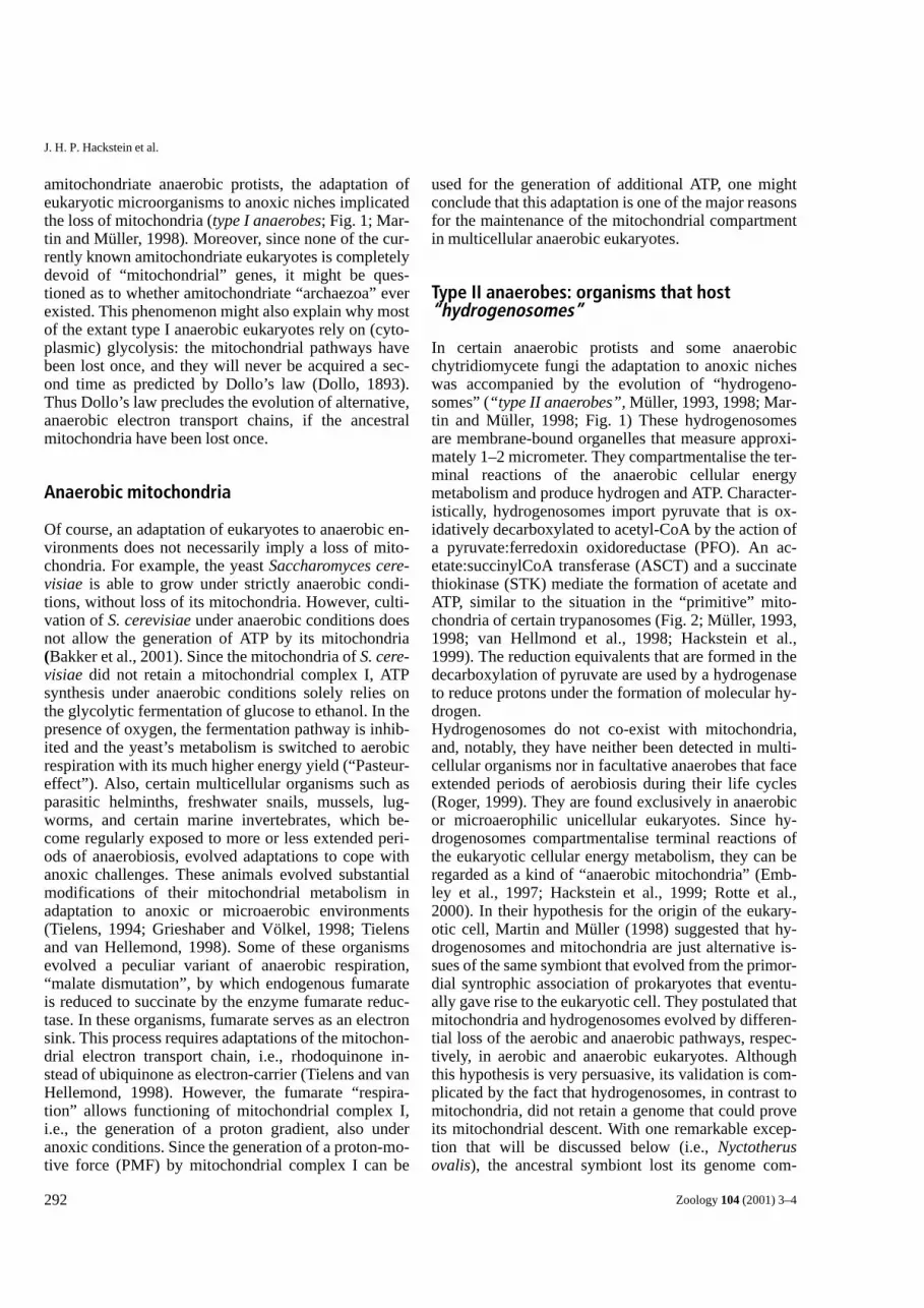
staining with DAPI or ethidium bromide provided noevidence for the presence of DNA. In the periphery ofthe cells, individual, dividing hydrogenosomes can befound, often in close association with the endoplas-matic reticulum (ER). Characteristically, the peripheral,individual hydrogenosomes are surrounded by 1–2 cis-terns of rough ER (Fig. 5).Little is known about the biochemistry, physiology andphylogeny of these hydrogenosomes. A putative ferre-doxin has been identified. It resembles the ferredoxin ofT. vaginalis (Brul et al., 1994). However, phylogeneticanalysis of the 18S rRNA genes does not support aclose relationship between P. lanterna and T. vaginalis(Fig. 6) and seems to exclude a recent common originof their hydrogenosomes. Thus, the data that favour a
mitochondrial ancestry of the hydrogenosomes ofP. lanterna are rather circumstantial.
Neocallimastix sp. L2/Piromyces sp. E2
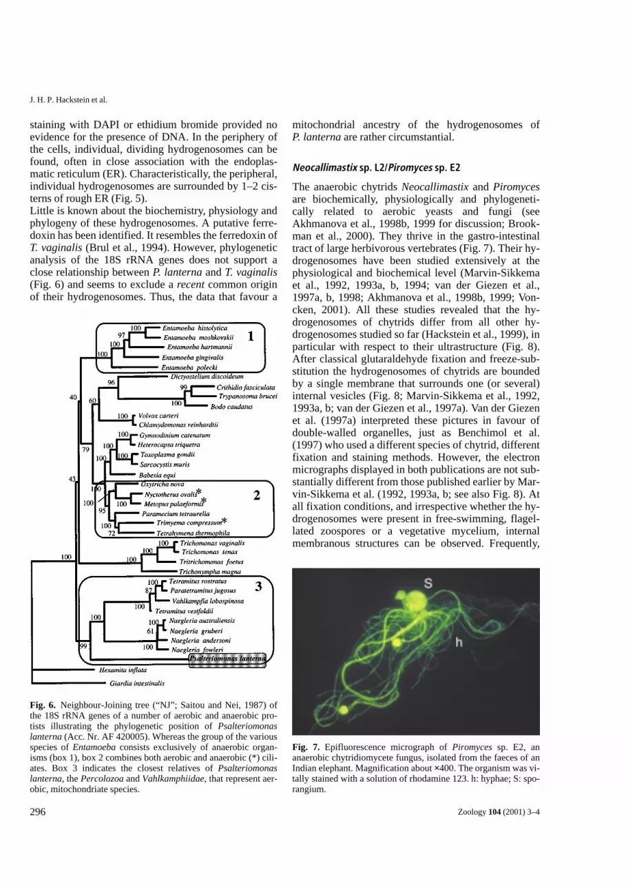
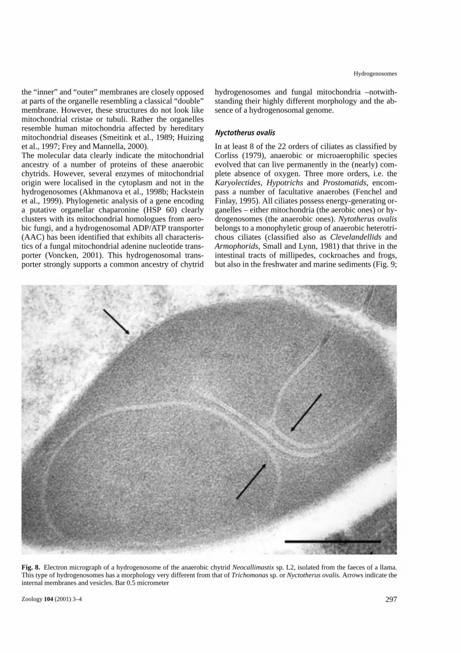
The anaerobic chytrids Neocallimastix and Piromycesare biochemically, physiologically and phylogeneti-cally related to aerobic yeasts and fungi (seeAkhmanova et al., 1998b, 1999 for discussion; Brook-man et al., 2000). They thrive in the gastro-intestinaltract of large herbivorous vertebrates (Fig. 7). Their hy-drogenosomes have been studied extensively at thephysiological and biochemical level (Marvin-Sikkemaet al., 1992, 1993a, b, 1994; van der Giezen et al.,1997a, b, 1998; Akhmanova et al., 1998b, 1999; Von-cken, 2001). All these studies revealed that the hy-drogenosomes of chytrids differ from all other hy-drogenosomes studied so far (Hackstein et al., 1999), inparticular with respect to their ultrastructure (Fig. 8).After classical glutaraldehyde fixation and freeze-sub-stitution the hydrogenosomes of chytrids are boundedby a single membrane that surrounds one (or several)internal vesicles (Fig. 8; Marvin-Sikkema et al., 1992,1993a, b; van der Giezen et al., 1997a). Van der Giezenet al. (1997a) interpreted these pictures in favour ofdouble-walled organelles, just as Benchimol et al.(1997) who used a different species of chytrid, differentfixation and staining methods. However, the electronmicrographs displayed in both publications are not sub-stantially different from those published earlier by Mar-vin-Sikkema et al. (1992, 1993a, b; see also Fig. 8). Atall fixation conditions, and irrespective whether the hy-drogenosomes were present in free-swimming, flagel-lated zoospores or a vegetative mycelium, internalmembranous structures can be observed. Frequently,
Fig. 6. Neighbour-Joining tree (“NJ”; Saitou and Nei, 1987) ofthe 18S rRNA genes of a number of aerobic and anaerobic pro-tists illustrating the phylogenetic position of Psalteriomonaslanterna (Acc. Nr. AF 420005). Whereas the group of the variousspecies of Entamoeba consists exclusively of anaerobic organ-isms (box 1), box 2 combines both aerobic and anaerobic (*) cili-ates. Box 3 indicates the closest relatives of Psalteriomonaslanterna, the Percolozoa and Vahlkamphiidae, that represent aer-obic, mitochondriate species.
Fig. 7. Epifluorescence micrograph of Piromyces sp. E2, ananaerobic chytridiomycete fungus, isolated from the faeces of anIndian elephant. Magnification about ×400. The organism was vi-tally stained with a solution of rhodamine 123. h: hyphae; S: spo-rangium.
297
Hydrogenosomes
Zoology 104 (2001) 3–4
hydrogenosomes and fungal mitochondria –notwith-standing their highly different morphology and the ab-sence of a hydrogenosomal genome.
Nyctotherus ovalis
In at least 8 of the 22 orders of ciliates as classified byCorliss (1979), anaerobic or microaerophilic speciesevolved that can live permanently in the (nearly) com-plete absence of oxygen. Three more orders, i.e. theKaryolectides, Hypotrichs and Prostomatids, encom-pass a number of facultative anaerobes (Fenchel andFinlay, 1995). All ciliates possess energy-generating or-ganelles – either mitochondria (the aerobic ones) or hy-drogenosomes (the anaerobic ones). Nytotherus ovalisbelongs to a monophyletic group of anaerobic heterotri-chous ciliates (classified also as Clevelandellids andArmophorids, Small and Lynn, 1981) that thrive in theintestinal tracts of millipedes, cockroaches and frogs,but also in the freshwater and marine sediments (Fig. 9;
Fig. 8. Electron micrograph of a hydrogenosome of the anaerobic chytrid Neocallimastix sp. L2, isolated from the faeces of a llama.This type of hydrogenosomes has a morphology very different from that of Trichomonas sp. or Nyctotherus ovalis. Arrows indicate theinternal membranes and vesicles. Bar 0.5 micrometer
the “inner” and “outer” membranes are closely opposedat parts of the organelle resembling a classical “double”membrane. However, these structures do not look likemitochondrial cristae or tubuli. Rather the organellesresemble human mitochondria affected by hereditarymitochondrial diseases (Smeitink et al., 1989; Huizinget al., 1997; Frey and Mannella, 2000). The molecular data clearly indicate the mitochondrialancestry of a number of proteins of these anaerobicchytrids. However, several enzymes of mitochondrialorigin were localised in the cytoplasm and not in thehydrogenosomes (Akhmanova et al., 1998b; Hacksteinet al., 1999). Phylogenetic analysis of a gene encodinga putative organellar chaparonine (HSP 60) clearlyclusters with its mitochondrial homologues from aero-bic fungi, and a hydrogenosomal ADP/ATP transporter(AAC) has been identified that exhibits all characteris-tics of a fungal mitochondrial adenine nucleotide trans-porter (Voncken, 2001). This hydrogenosomal trans-porter strongly supports a common ancestry of chytrid

298
J. H. P. Hackstein et al.
Zoology 104 (2001) 3–4
Fig. 9. Phylogenetic relationships among anaerobic heterotric-hous (2, 3) and rumen ciliates (1). STAR decomposition analysisof the 18S rRNA genes (MOLPHI; Adachi and Hasegawa, 1996).The taxa analysed here consist exclusively of anaerobic ciliatesthat (most likely) possess hydrogenosomes. They belong to threedifferent groups, which are consistently identified, regardless ofthe phylogenetic methods (Neighbour Joining; Saitou and Nei,1987; PUZZLE; Strimmer and von Haeseler, 1996) that are usedto calculate the phylogenetic trees. All other branching in the treehas a low statistical support and is sensitive to the sampling ofspecies. The * indicates intestinal ciliates from frogs, millipedesand cockroaches. All other species in 2 and 3 are free-living (c.f.van Hoek et al., 1998, 2000b), all species displayed in box 1 areliving in the rumen of ruminants.
Fig. 10. EM picture of Nyctotherusovalis, KMnO4 fixation. Ma: macronu-cleus; Mi: micronucleus; H: hydrogeno-somes; M: methanogenic endosymbionts(dark dots); CV: contractive vacuole; mf:mouth field; C: cilia; Bar 10 µm. (c.f.Akhmanova et al., 1998a).
299
Hydrogenosomes
Zoology 104 (2001) 3–4
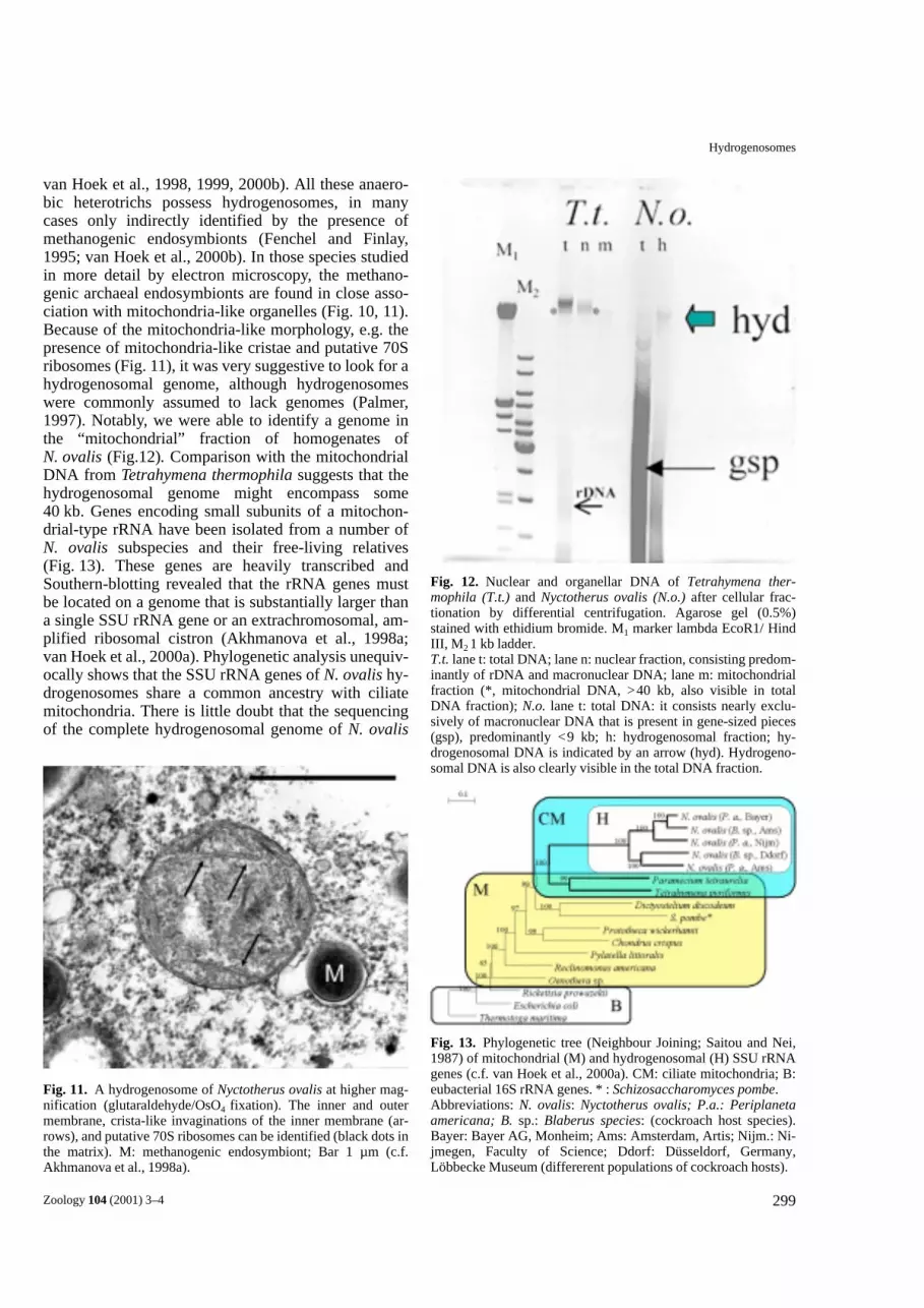
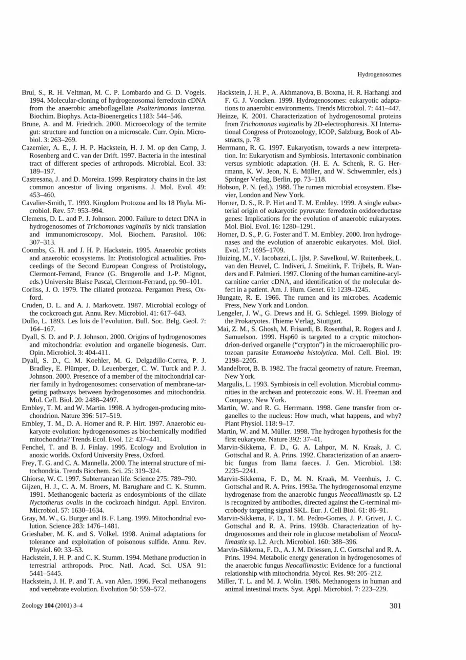
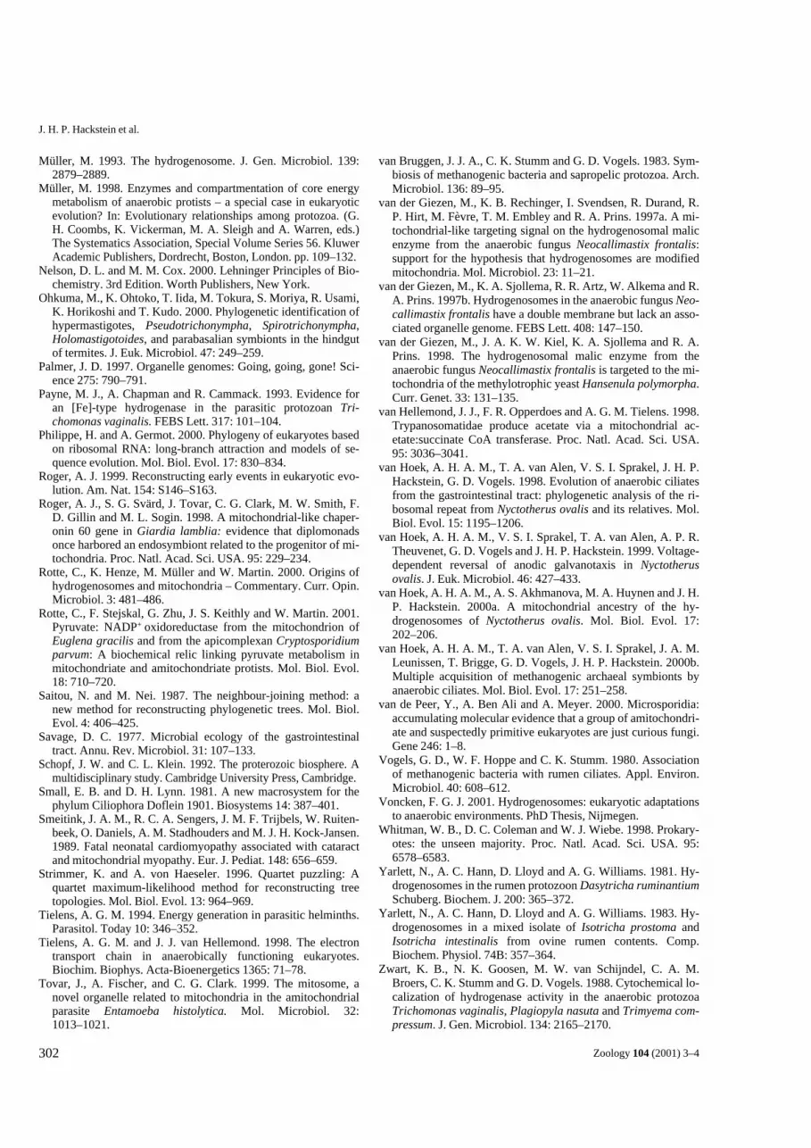
van Hoek et al., 1998, 1999, 2000b). All these anaero-bic heterotrichs possess hydrogenosomes, in manycases only indirectly identified by the presence ofmethanogenic endosymbionts (Fenchel and Finlay,1995; van Hoek et al., 2000b). In those species studiedin more detail by electron microscopy, the methano-genic archaeal endosymbionts are found in close asso-ciation with mitochondria-like organelles (Fig. 10, 11).Because of the mitochondria-like morphology, e.g. thepresence of mitochondria-like cristae and putative 70Sribosomes (Fig. 11), it was very suggestive to look for ahydrogenosomal genome, although hydrogenosomeswere commonly assumed to lack genomes (Palmer,1997). Notably, we were able to identify a genome inthe “mitochondrial” fraction of homogenates ofN. ovalis (Fig.12). Comparison with the mitochondrialDNA from Tetrahymena thermophila suggests that thehydrogenosomal genome might encompass some40 kb. Genes encoding small subunits of a mitochon-drial-type rRNA have been isolated from a number ofN. ovalis subspecies and their free-living relatives(Fig. 13). These genes are heavily transcribed andSouthern-blotting revealed that the rRNA genes mustbe located on a genome that is substantially larger thana single SSU rRNA gene or an extrachromosomal, am-plified ribosomal cistron (Akhmanova et al., 1998a;van Hoek et al., 2000a). Phylogenetic analysis unequiv-ocally shows that the SSU rRNA genes of N. ovalis hy-drogenosomes share a common ancestry with ciliatemitochondria. There is little doubt that the sequencingof the complete hydrogenosomal genome of N. ovalis
Fig. 12. Nuclear and organellar DNA of Tetrahymena ther-mophila (T.t.) and Nyctotherus ovalis (N.o.) after cellular frac-tionation by differential centrifugation. Agarose gel (0.5%)stained with ethidium bromide. M1 marker lambda EcoR1/ HindIII, M2 1 kb ladder. T.t. lane t: total DNA; lane n: nuclear fraction, consisting predom-inantly of rDNA and macronuclear DNA; lane m: mitochondrialfraction (*, mitochondrial DNA, >40 kb, also visible in totalDNA fraction); N.o. lane t: total DNA: it consists nearly exclu-sively of macronuclear DNA that is present in gene-sized pieces(gsp), predominantly <9 kb; h: hydrogenosomal fraction; hy-drogenosomal DNA is indicated by an arrow (hyd). Hydrogeno-somal DNA is also clearly visible in the total DNA fraction.
Fig. 13. Phylogenetic tree (Neighbour Joining; Saitou and Nei,1987) of mitochondrial (M) and hydrogenosomal (H) SSU rRNAgenes (c.f. van Hoek et al., 2000a). CM: ciliate mitochondria; B:eubacterial 16S rRNA genes. * : Schizosaccharomyces pombe. Abbreviations: N. ovalis: Nyctotherus ovalis; P.a.: Periplanetaamericana; B. sp.: Blaberus species: (cockroach host species).Bayer: Bayer AG, Monheim; Ams: Amsterdam, Artis; Nijm.: Ni-jmegen, Faculty of Science; Ddorf: Düsseldorf, Germany,Löbbecke Museum (differerent populations of cockroach hosts).
Fig. 11. A hydrogenosome of Nyctotherus ovalis at higher mag-nification (glutaraldehyde/OsO4 fixation). The inner and outermembrane, crista-like invaginations of the inner membrane (ar-rows), and putative 70S ribosomes can be identified (black dots inthe matrix). M: methanogenic endosymbiont; Bar 1 µm (c.f.Akhmanova et al., 1998a).

300
J. H. P. Hackstein et al.
Zoology 104 (2001) 3–4
will confirm this conclusion. Thus, both morphologyand molecular biology suggest strongly that the hy-drogenosomes of anaerobic heterotrichous ciliates arehighly specialised mitochondria that produce hydrogen.
Conclusions
Here we have reviewed the available data for the evolu-tion of hydrogenosomes. These data strongly suggestthat the hydrogenosomes of the various anaerobic pro-tists evolved repeatedly from “mitochondria” of theirclosest aerobic or facultative anaerobic ancestors, orfrom protomitochondria, respectively. This conclusionis supported by both ultrastructural and molecular data.The presence of a hydrogenosomal genome in theanaerobic ciliate N. ovalis provides the most straight-forward evidence that the hydrogenosomes of anaero-bic ciliates share a recent common ancestor with themitochondria of their aerobic or facultative anaerobicancestors. It remains to be shown whether the hy-drogenosomes in the various ciliates evolved repeat-edly and why certain hydrogenosomes retained agenome and others not. Without any doubt, ciliate hy-drogenosomes differ substantially from hydrogeno-somes of anaerobic chytrids. The phylogenetic analysisof the hydrogenosomal AACs of anaerobic chytrids hasshown that these hydrogenosomes evolved from themitochondria of aerobic yeast and fungi (Voncken,2001). Proteomics might be a suitable approach to an-swer the open questions. The origin of hydrogenosomesof Trichomonas and Psalteriomonas is less clear, due tothe lack of data. Notwithstanding, a “mitochondrial”/protomitochondrial origin also of these hydrogeno-somes is likely. There are a number of arguments infavour of the hypothesis that ancestral mitochondriawere facultative anaerobic organelles, possessing bothaerobic and anaerobic metabolic pathways (Martin andMüller, 1998; Rotte et al., 2001). It is obvious that theancestral mitochondria must have retained their faculta-tive anaerobic nature over extended evolutionary timessince they differentiated as “fungal” or “ciliate” mito-chondria before adapting to either aerobic or anaerobicniches. Future studies will have to provide the neces-sary information for an understanding of these adapta-tions to aerobic and anaerobic environments.
Acknowledgements
We are indebted to M. Benchimol, Rio de Janeiro, andH. Aspöck, Wien, for the pictures of Trichomonas.Also, the help of G. Kreimer, Cologne, with the CLSmicroscopy of Psalteriomonas lanterna is greatfullyacknowledged.
AGM Tielens, Utrecht, and W. Peters, Düsseldorf gaveinvaluable comments on the manuscript. The Dept. ofPhotography and Illustrations of the Faculty of Sci-ences, University of Nijmegen, guaranteed the profes-sional completion of the figures. Frank Voncken andAngela van Hoek were supported by the Dutch ScienceFundation NWO.
References
Adachi, K. and M. Hasegawa. 1996. MOLPHY version 2.3: Pro-grams for molecular phylogenetics based on maximum likeli-hood. Comput. Sci. Monogr. 28: 1–150.
Andersson, S. G. E. and C. G. Kurland. 1999. Origin of mito-chondria and hydrogenosomes. Curr. Opin. Microbiol. 2:535–541.
Akhmanova, A., F. Voncken, T. van Alen, A. van Hoek, B.Boxma, G. Vogels, M. Veenhuis and J. H. P. Hackstein. 1998a.A hydrogenosome with a genome. Nature 396: 527–528.
Akhmanova, A., F. G. J. Voncken, H. Harhangi, K. M. Hosea, G.D. Vogels and J. H. P. Hackstein. 1998b. Cytosolic enzymeswith a mitochondrial ancestry from the anaerobic chytridPiromyces sp. E2. Mol. Microbiol. 30: 1017–1027.
Akhmanova, A., F. G. J. Voncken, K. M. Hosea, H. Harhangi, J.T. Keltjens, H. J. M. op den Camp, G. D. Vogels and J. H. P.Hackstein. 1999. A hydrogenosome with pyruvate formate-lyase: anaerobic chytrid fungi use an alternative route for pyru-vate catabolism. Mol. Microbiol. 32: 1103–1114.
Aspöck, H. 1994. Protozoen als Erreger von Krankheiten desMenschen: Übersicht und aktuelle Probleme in Mitteleuropa.Kataloge des OÖ. Landesmuseums N.F. 71: 219–266.
Bakker, B. M., K. M. Overkamp, A. J. A. van Maris, P. Kotter,M. A. H. Luttik, J. P. van Dijken and J. T. Pronk. 2001. Stoi-chiometry and compartmentation of NADH metabolism inSaccharomyces cerevisiae. FEMS Microbiol. Rev. 25: 15–37.
Baldauf, S. L., A. Roger, I. Wenk-Siefert and W. F. Doolittle.2000. A kingdom-level phylogeny of eukaryotes based oncombined protein data. Science 290: 972–977.
Benchimol, M., J. C. Aquino Almeida, and W. de Souza. 1996a.Further studies on the organisation of the hydrogenosome inTritrichomonas foetus. Tissue Cell 28: 287–299.
Benchimol, M., P. J. Johnson and W. de Souza. 1996b. Morpho-genesis of the hydrogenosome: an ultrastructural study. Biol.Cell 87: 197–205.
Benchimol, M., R. Durand and J. C. A. Almeida. 1997. A doublemembrane surrounds the hydrogenosome of the anaerobic fun-gus Neocallimastix frontalis. FEMS Microbiol. Lett. 154:277–282.
Brauman, A., J. Dore, P. Eggleton, D. Bignell, J. A. Breznak andM. D. Kane. 2001. Molecular phylogenetic profiling ofprokaryotic communities in guts of termites with differentfeeding habits. FEMS Microb. Ecol. 35: 27–36.
Broers, C. A. M., C. K. Stumm, G. D. Vogels and G. Brugerolle.1990. Psalteriomonas lanterna gen. nov., sp. nov., a free-liv-ing ameboflagellate isolated from fresh-water anaerobic sedi-ments. Eur. J. Protistol. 25: 369–380.
Brookman, J. L., G. Mennim, A. P. J. Trinci, M. K. Theodorouand D. S. Tuckwell. 2000. Identification and characterizationof anaerobic gut fungi using molecular methodologies basedon ribosomal ITS1 and 18S rRNA. Microbiology-UK 146:393–403.
301
Hydrogenosomes
Zoology 104 (2001) 3–4
Brul, S., R. H. Veltman, M. C. P. Lombardo and G. D. Vogels.1994. Molecular-cloning of hydrogenosomal ferredoxin cDNAfrom the anaerobic ameboflagellate Psalterimonas lanterna.Biochim. Biophys. Acta-Bioenergetics 1183: 544–546.
Brune, A. and M. Friedrich. 2000. Microecology of the termitegut: structure and function on a microscale. Curr. Opin. Micro-biol. 3: 263–269.
Cazemier, A. E., J. H. P. Hackstein, H. J. M. op den Camp, J.Rosenberg and C. van der Drift. 1997. Bacteria in the intestinaltract of different species of arthropods. Microbial. Ecol. 33:189–197.
Castresana, J. and D. Moreira. 1999. Respiratory chains in the lastcommon ancestor of living organisms. J. Mol. Evol. 49:453–460.
Cavalier-Smith, T. 1993. Kingdom Protozoa and Its 18 Phyla. Mi-crobiol. Rev. 57: 953–994.
Clemens, D. L. and P. J. Johnson. 2000. Failure to detect DNA inhydrogenosomes of Trichomonas vaginalis by nick translationand immunomicroscopy. Mol. Biochem. Parasitol. 106:307–313.
Coombs, G. H. and J. H. P. Hackstein. 1995. Anaerobic protistsand anaerobic ecosystems. In: Protistological actualities. Pro-ceedings of the Second European Congress of Protistology,Clermont-Ferrand, France (G. Brugerolle and J.-P. Mignot,eds.) Universite Blaise Pascal, Clermont-Ferrand, pp. 90–101.
Corliss, J. O. 1979. The ciliated protozoa. Pergamon Press, Ox-ford.
Cruden, D. L. and A. J. Markovetz. 1987. Microbial ecology ofthe cockcroach gut. Annu. Rev. Microbiol. 41: 617–643.
Dollo, L. 1893. Les lois de l’evolution. Bull. Soc. Belg. Geol. 7:164–167.
Dyall, S. D. and P. J. Johnson. 2000. Origins of hydrogenosomesand mitochondria: evolution and organelle biogenesis. Curr.Opin. Microbiol. 3: 404-411.
Dyall, S. D., C. M. Koehler, M. G. Delgadillo-Correa, P. J.Bradley, E. Plümper, D. Leuenberger, C. W. Turck and P. J.Johnson. 2000. Presence of a member of the mitochondrial car-rier family in hydrogenosomes: conservation of membrane-tar-geting pathways between hydrogenosomes and mitochondria.Mol. Cell. Biol. 20: 2488–2497.
Embley, T. M. and W. Martin. 1998. A hydrogen-producing mito-chondrion. Nature 396: 517–519.
Embley, T. M., D. A. Horner and R. P. Hirt. 1997. Anaerobic eu-karyote evolution: hydrogenosomes as biochemically modifiedmitochondria? Trends Ecol. Evol. 12: 437–441.
Fenchel, T. and B. J. Finlay. 1995. Ecology and Evolution inanoxic worlds. Oxford University Press, Oxford.
Frey, T. G. and C. A. Mannella. 2000. The internal structure of mi-tochondria. Trends Biochem. Sci. 25: 319–324.
Ghiorse, W. C. 1997. Subterranean life. Science 275: 789–790.Gijzen, H. J., C. A. M. Broers, M. Barughare and C. K. Stumm.
1991. Methanogenic bacteria as endosymbionts of the ciliateNyctotherus ovalis in the cockroach hindgut. Appl. Environ.Microbiol. 57: 1630–1634.
Gray, M. W., G. Burger and B. F. Lang. 1999. Mitochondrial evo-lution. Science 283: 1476–1481.
Grieshaber, M. K. and S. Völkel. 1998. Animal adaptations fortolerance and exploitation of poisonous sulfide. Annu. Rev.Physiol. 60: 33–53.
Hackstein, J. H. P. and C. K. Stumm. 1994. Methane production interrestrial arthropods. Proc. Natl. Acad. Sci. USA 91:5441–5445.
Hackstein, J. H. P. and T. A. van Alen. 1996. Fecal methanogensand vertebrate evolution. Evolution 50: 559–572.
Hackstein, J. H. P., A. Akhmanova, B. Boxma, H. R. Harhangi andF. G. J. Voncken. 1999. Hydrogenosomes: eukaryotic adapta-tions to anaerobic environments. Trends Microbiol. 7: 441–447.
Heinze, K. 2001. Characterization of hydrogenosomal proteinsfrom Trichomonas vaginalis by 2D-electrophoresis. XI Interna-tional Congress of Protozoology, ICOP, Salzburg, Book of Ab-stracts, p. 78
Herrmann, R. G. 1997. Eukaryotism, towards a new interpreta-tion. In: Eukaryotism and Symbiosis. Intertaxonic combinationversus symbiotic adaptation. (H. E. A. Schenk, R. G. Her-rmann, K. W. Jeon, N. E. Müller, and W. Schwemmler, eds.)Springer Verlag, Berlin, pp. 73–118.
Hobson, P. N. (ed.). 1988. The rumen microbial ecosystem. Else-vier, London and New York.
Horner, D. S., R. P. Hirt and T. M. Embley. 1999. A single eubac-terial origin of eukaryotic pyruvate: ferredoxin oxidoreductasegenes: Implications for the evolution of anaerobic eukaryotes.Mol. Biol. Evol. 16: 1280–1291.
Horner, D. S., P. G. Foster and T. M. Embley. 2000. Iron hydroge-nases and the evolution of anaerobic eukaryotes. Mol. Biol.Evol. 17: 1695–1709.
Huizing, M., V. Iacobazzi, L. Ijlst, P. Savelkoul, W. Ruitenbeek, L.van den Heuvel, C. Indiveri, J. Smeitink, F. Trijbels, R. Wan-ders and F. Palmieri. 1997. Cloning of the human carnitine-acyl-carnitine carrier cDNA, and identification of the molecular de-fect in a patient. Am. J. Hum. Genet. 61: 1239–1245.
Hungate, R. E. 1966. The rumen and its microbes. AcademicPress, New York and London.
Lengeler, J. W., G. Drews and H. G. Schlegel. 1999. Biology ofthe Prokaryotes. Thieme Verlag, Stuttgart.
Mai, Z. M., S. Ghosh, M. Frisardi, B. Rosenthal, R. Rogers and J.Samuelson. 1999. Hsp60 is targeted to a cryptic mitochon-drion-derived organelle (“crypton”) in the microaerophilic pro-tozoan parasite Entamoeba histolytica. Mol. Cell. Biol. 19:2198–2205.
Mandelbrot, B. B. 1982. The fractal geometry of nature. Freeman,New York.
Margulis, L. 1993. Symbiosis in cell evolution. Microbial commu-nities in the archean and proterozoic eons. W. H. Freeman andCompany, New York.
Martin, W. and R. G. Herrmann. 1998. Gene transfer from or-ganelles to the nucleus: How much, what happens, and why?Plant Physiol. 118: 9–17.
Martin, W. and M. Müller. 1998. The hydrogen hypothesis for thefirst eukaryote. Nature 392: 37–41.
Marvin-Sikkema, F. D., G. A. Lahpor, M. N. Kraak, J. C.Gottschal and R. A. Prins. 1992. Characterization of an anaero-bic fungus from llama faeces. J. Gen. Microbiol. 138:2235–2241.
Marvin-Sikkema, F. D., M. N. Kraak, M. Veenhuis, J. C.Gottschal and R. A. Prins. 1993a. The hydrogenosomal enzymehydrogenase from the anaerobic fungus Neocallimastix sp. L2is recognized by antibodies, directed against the C-terminal mi-crobody targeting signal SKL. Eur. J. Cell Biol. 61: 86–91.
Marvin-Sikkema, F. D., T. M. Pedro-Gomes, J. P. Grivet, J. C.Gottschal and R. A. Prins. 1993b. Characterization of hy-drogenosomes and their role in glucose metabolism of Neocal-limastix sp. L2. Arch. Microbiol. 160: 388–396.
Marvin-Sikkema, F. D., A. J. M. Driessen, J. C. Gottschal and R. A.Prins. 1994. Metabolic energy generation in hydrogenosomes ofthe anaerobic fungus Neocallimastix: Evidence for a functionalrelationship with mitochondria. Mycol. Res. 98: 205–212.
Miller, T. L. and M. J. Wolin. 1986. Methanogens in human andanimal intestinal tracts. Syst. Appl. Microbiol. 7: 223–229.
302
J. H. P. Hackstein et al.
Zoology 104 (2001) 3–4
Müller, M. 1993. The hydrogenosome. J. Gen. Microbiol. 139:2879–2889.
Müller, M. 1998. Enzymes and compartmentation of core energymetabolism of anaerobic protists – a special case in eukaryoticevolution? In: Evolutionary relationships among protozoa. (G.H. Coombs, K. Vickerman, M. A. Sleigh and A. Warren, eds.)The Systematics Association, Special Volume Series 56. KluwerAcademic Publishers, Dordrecht, Boston, London. pp. 109–132.
Nelson, D. L. and M. M. Cox. 2000. Lehninger Principles of Bio-chemistry. 3rd Edition. Worth Publishers, New York.
Ohkuma, M., K. Ohtoko, T. Iida, M. Tokura, S. Moriya, R. Usami,K. Horikoshi and T. Kudo. 2000. Phylogenetic identification ofhypermastigotes, Pseudotrichonympha, Spirotrichonympha,Holomastigotoides, and parabasalian symbionts in the hindgutof termites. J. Euk. Microbiol. 47: 249–259.
Palmer, J. D. 1997. Organelle genomes: Going, going, gone! Sci-ence 275: 790–791.
Payne, M. J., A. Chapman and R. Cammack. 1993. Evidence foran [Fe]-type hydrogenase in the parasitic protozoan Tri-chomonas vaginalis. FEBS Lett. 317: 101–104.
Philippe, H. and A. Germot. 2000. Phylogeny of eukaryotes basedon ribosomal RNA: long-branch attraction and models of se-quence evolution. Mol. Biol. Evol. 17: 830–834.
Roger, A. J. 1999. Reconstructing early events in eukaryotic evo-lution. Am. Nat. 154: S146–S163.
Roger, A. J., S. G. Svärd, J. Tovar, C. G. Clark, M. W. Smith, F.D. Gillin and M. L. Sogin. 1998. A mitochondrial-like chaper-onin 60 gene in Giardia lamblia: evidence that diplomonadsonce harbored an endosymbiont related to the progenitor of mi-tochondria. Proc. Natl. Acad. Sci. USA. 95: 229–234.
Rotte, C., K. Henze, M. Müller and W. Martin. 2000. Origins ofhydrogenosomes and mitochondria – Commentary. Curr. Opin.Microbiol. 3: 481–486.
Rotte, C., F. Stejskal, G. Zhu, J. S. Keithly and W. Martin. 2001.Pyruvate: NADP+ oxidoreductase from the mitochondrion ofEuglena gracilis and from the apicomplexan Cryptosporidiumparvum: A biochemical relic linking pyruvate metabolism inmitochondriate and amitochondriate protists. Mol. Biol. Evol.18: 710–720.
Saitou, N. and M. Nei. 1987. The neighbour-joining method: anew method for reconstructing phylogenetic trees. Mol. Biol.Evol. 4: 406–425.
Savage, D. C. 1977. Microbial ecology of the gastrointestinaltract. Annu. Rev. Microbiol. 31: 107–133.
Schopf, J. W. and C. L. Klein. 1992. The proterozoic biosphere. Amultidisciplinary study. Cambridge University Press, Cambridge.
Small, E. B. and D. H. Lynn. 1981. A new macrosystem for thephylum Ciliophora Doflein 1901. Biosystems 14: 387–401.
Smeitink, J. A. M., R. C. A. Sengers, J. M. F. Trijbels, W. Ruiten-beek, O. Daniels, A. M. Stadhouders and M. J. H. Kock-Jansen.1989. Fatal neonatal cardiomyopathy associated with cataractand mitochondrial myopathy. Eur. J. Pediat. 148: 656–659.
Strimmer, K. and A. von Haeseler. 1996. Quartet puzzling: Aquartet maximum-likelihood method for reconstructing treetopologies. Mol. Biol. Evol. 13: 964–969.
Tielens, A. G. M. 1994. Energy generation in parasitic helminths.Parasitol. Today 10: 346–352.
Tielens, A. G. M. and J. J. van Hellemond. 1998. The electrontransport chain in anaerobically functioning eukaryotes.Biochim. Biophys. Acta-Bioenergetics 1365: 71–78.
Tovar, J., A. Fischer, and C. G. Clark. 1999. The mitosome, anovel organelle related to mitochondria in the amitochondrialparasite Entamoeba histolytica. Mol. Microbiol. 32:1013–1021.
van Bruggen, J. J. A., C. K. Stumm and G. D. Vogels. 1983. Sym-biosis of methanogenic bacteria and sapropelic protozoa. Arch.Microbiol. 136: 89–95.
van der Giezen, M., K. B. Rechinger, I. Svendsen, R. Durand, R.P. Hirt, M. Fèvre, T. M. Embley and R. A. Prins. 1997a. A mi-tochondrial-like targeting signal on the hydrogenosomal malicenzyme from the anaerobic fungus Neocallimastix frontalis:support for the hypothesis that hydrogenosomes are modifiedmitochondria. Mol. Microbiol. 23: 11–21.
van der Giezen, M., K. A. Sjollema, R. R. Artz, W. Alkema and R.A. Prins. 1997b. Hydrogenosomes in the anaerobic fungus Neo-callimastix frontalis have a double membrane but lack an asso-ciated organelle genome. FEBS Lett. 408: 147–150.
van der Giezen, M., J. A. K. W. Kiel, K. A. Sjollema and R. A.Prins. 1998. The hydrogenosomal malic enzyme from theanaerobic fungus Neocallimastix frontalis is targeted to the mi-tochondria of the methylotrophic yeast Hansenula polymorpha.Curr. Genet. 33: 131–135.
van Hellemond, J. J., F. R. Opperdoes and A. G. M. Tielens. 1998.Trypanosomatidae produce acetate via a mitochondrial ac-etate:succinate CoA transferase. Proc. Natl. Acad. Sci. USA.95: 3036–3041.
van Hoek, A. H. A. M., T. A. van Alen, V. S. I. Sprakel, J. H. P.Hackstein, G. D. Vogels. 1998. Evolution of anaerobic ciliatesfrom the gastrointestinal tract: phylogenetic analysis of the ri-bosomal repeat from Nyctotherus ovalis and its relatives. Mol.Biol. Evol. 15: 1195–1206.
van Hoek, A. H. A. M., V. S. I. Sprakel, T. A. van Alen, A. P. R.Theuvenet, G. D. Vogels and J. H. P. Hackstein. 1999. Voltage-dependent reversal of anodic galvanotaxis in Nyctotherusovalis. J. Euk. Microbiol. 46: 427–433.
van Hoek, A. H. A. M., A. S. Akhmanova, M. A. Huynen and J. H.P. Hackstein. 2000a. A mitochondrial ancestry of the hy-drogenosomes of Nyctotherus ovalis. Mol. Biol. Evol. 17:202–206.
van Hoek, A. H. A. M., T. A. van Alen, V. S. I. Sprakel, J. A. M.Leunissen, T. Brigge, G. D. Vogels, J. H. P. Hackstein. 2000b.Multiple acquisition of methanogenic archaeal symbionts byanaerobic ciliates. Mol. Biol. Evol. 17: 251–258.
van de Peer, Y., A. Ben Ali and A. Meyer. 2000. Microsporidia:accumulating molecular evidence that a group of amitochondri-ate and suspectedly primitive eukaryotes are just curious fungi.Gene 246: 1–8.
Vogels, G. D., W. F. Hoppe and C. K. Stumm. 1980. Associationof methanogenic bacteria with rumen ciliates. Appl. Environ.Microbiol. 40: 608–612.
Voncken, F. G. J. 2001. Hydrogenosomes: eukaryotic adaptationsto anaerobic environments. PhD Thesis, Nijmegen.
Whitman, W. B., D. C. Coleman and W. J. Wiebe. 1998. Prokary-otes: the unseen majority. Proc. Natl. Acad. Sci. USA. 95:6578–6583.
Yarlett, N., A. C. Hann, D. Lloyd and A. G. Williams. 1981. Hy-drogenosomes in the rumen protozoon Dasytricha ruminantiumSchuberg. Biochem. J. 200: 365–372.
Yarlett, N., A. C. Hann, D. Lloyd and A. G. Williams. 1983. Hy-drogenosomes in a mixed isolate of Isotricha prostoma andIsotricha intestinalis from ovine rumen contents. Comp.Biochem. Physiol. 74B: 357–364.
Zwart, K. B., N. K. Goosen, M. W. van Schijndel, C. A. M.Broers, C. K. Stumm and G. D. Vogels. 1988. Cytochemical lo-calization of hydrogenase activity in the anaerobic protozoaTrichomonas vaginalis, Plagiopyla nasuta and Trimyema com-pressum. J. Gen. Microbiol. 134: 2165–2170.