human h-ficolin inhibits replication of seasonal and pandemic influenza a viruses
TRANSCRIPT
of November 13, 2012.This information is current as
Seasonal and Pandemic Influenza A VirusesHuman H-Ficolin Inhibits Replication of
Jensenius, Steffen Thiel and Kevan L. HartshornJeffery K. Taubenberger, Kazue Takahashi, Jens C.Shweta Tripathi, Julvet Mbianda, Micheal Ieong, Li Qi, Anamika Verma, Mitchell White, Vinod Vathipadiekal,
http://www.jimmunol.org/content/189/5/2478doi: 10.4049/jimmunol.1103786July 2012;
2012; 189:2478-2487; Prepublished online 30J Immunol
Referenceshttp://www.jimmunol.org/content/189/5/2478.full#ref-list-1
, 16 of which you can access for free at: cites 39 articlesThis article
Subscriptionshttp://jimmunol.org/subscriptions
is online at: The Journal of ImmunologyInformation about subscribing to
Permissionshttp://www.aai.org/ji/copyright.htmlSubmit copyright permission requests at:
Email Alertshttp://jimmunol.org/cgi/alerts/etocReceive free email-alerts when new articles cite this article. Sign up at:
Print ISSN: 0022-1767 Online ISSN: 1550-6606. All rights reserved.9650 Rockville Pike, Bethesda, MD 20814-3994.The American Association of Immunologists, Inc.,
is published twice each month byThe Journal of Immunology
at Boston U
niv Lib on N
ovember 13, 2012
http://jimm
unol.org/D
ownloaded from
The Journal of Immunology
Human H-Ficolin Inhibits Replication of Seasonal andPandemic Influenza A Viruses
Anamika Verma,* Mitchell White,* Vinod Vathipadiekal,† Shweta Tripathi,*
Julvet Mbianda,* Micheal Ieong,* Li Qi,‡ Jeffery K. Taubenberger,‡ Kazue Takahashi,x
Jens C. Jensenius,{ Steffen Thiel,{ and Kevan L. Hartshorn*
The collectins have been shown to have a role in host defense against influenza Avirus (IAV) and other significant viral pathogens
(e.g., HIV). The ficolins are a related group of innate immune proteins that are present at relatively high concentrations in
serum, but also in respiratory secretions; however, there has been little study of the role of ficolins in viral infection. In this
study, we demonstrate that purified recombinant human H-ficolin and H-ficolin in human serum and bronchoalveolar lavage
fluid bind to IAV and inhibit viral infectivity and hemagglutination activity in vitro. Removal of ficolins from human serum or
bronchoalveolar lavage fluid reduces their antiviral activity. Inhibition of IAV did not involve the calcium-dependent lectin
activity of H-ficolin. We demonstrate that H-ficolin is sialylated and that removal of sialic acid abrogates IAV inhibition, while
addition of the neuraminidase inhibitor oseltamivir potentiates neutralization, hemagglutinin inhibition, and viral aggregation
caused by H-ficolin. Pandemic and mouse-adapted strains of IAVare generally not inhibited by the collectins surfactant protein
D or mannose binding lectin because of a paucity of glycan attachments on the hemagglutinin of these strains. In contrast,
H-ficolin inhibited both the mouse-adapted PR-8 H1N1 strain and a pandemic H1N1 strain from 2009. H-ficolin also fixed com-
plement to a surface coated with IAV. These findings suggest that H-ficolin contributes to host defense against IAV. The Journal
of Immunology, 2012, 189: 2478–2487.
The collectins surfactant protein D (SP-D), surfactantprotein A (SP-A), and mannose-binding lectin (MBL)have been shown to contribute to innate defense against
influenza A virus (IAV) infection. The ficolins resemble MBL intheir overall structure, calcium-dependent binding to pathogens,and ability to fix complement in an Ab-independent manner (1).In humans, there are three different ficolin forms (H-, L-, and M-ficolin) and one form of MBL (2). H-ficolin has a shorter collagendomain than the other two ficolins. H-ficolin exists in blood ata mean level of ∼20 mg/ml (reported ranges of 8–80 mg/ml)(3, 4), which greatly exceeds that of the other ficolins (L-ficolin,3.4 mg/ml; M-ficolin, 1.4 mg/ml) or MBL (1.1 mg/ml) (5). H-ficolin is also produced by alveolar type II cells and ciliatedbronchial epithelial cells in the lung and has been demonstrated tobe present in bronchoalveolar lavage fluid (BALF) (6), althoughthe concentration in BALF has not been determined. Subjects havebeen described who are homozygous for a truncated version of
H-ficolin and essential absence of this protein in serum (7, 8). Themajor clinical manifestation of the adult patient was recurrentrespiratory infections, whereas the other two patients were neo-nates with necrotizing enterocolitis. Therefore, it is likely that Hficolin has a role in innate immunity.The interactions of ficolins with bacteria have been well studied,
but there are limited data regarding ficolin interactions with viruses.
L-Ficolin has been shown to bind to envelope proteins of hepatitis
C virus (HCV) and to fix complement on HCV-infected hep-
atocytes (9). Recently L-ficolin was also shown to inhibit influenza
A virus (IAV) in vitro and in mice (10). Porcine ficolin has been
shown to neutralize porcine reproductive and respiratory syn-
drome virus (11). In both of these cases, the antiviral effect was
related to recognition of N-linked glycans on the viral envelope
proteins by the ficolin. Recent studies have also shown that chi-
meric proteins containing the N-terminal domains of ficolins and
the carbohydrate recognition domain of MBL strongly inhibit
Ebola virus and IAV (12, 13); however, in this case the reaction is
mediated by the binding of the carbohydrate recognition domain
of MBL to virus associated carbohydrates. The recognition do-
main of the ficolins differs from that of the collectins, which are
C-type lectins; it has some homology to domains of fibrinogen,
and is thus named a fibrinogen-like domain. Ficolins recognize
acetylated compounds (both N-acetylated sugars and other acet-
ylated molecules), whereas MBL and other collectins preferen-
tially bind to terminal carbohydrate groups with horizontal OH
groups at the 3 and 4 positions (e.g., mannose-rich glycans on
pathogens) (14).In this study, we focused mainly on H-ficolin because of its
probable role in respiratory infections. We demonstrate that H-
ficolin neutralizes various strains of IAV through a distinct
mechanism that does not involve their calcium-dependent lectin
activity. This feature allows the ficolins to inhibit viral strains not
inhibited well by collectins.
*Department of Medicine, Boston University School of Medicine, Boston, MA02118; †Massachusetts General Hospital Cancer Center, Harvard Medical School,Boston, MA 02114; ‡National Institute of Allergy and Infectious Diseases, Bethesda,MD 20892; xProgram of Developmental Immunology, Department of Pediatrics,Massachusetts General Hospital, Boston, MA 02114; and {Department of Biomed-icine, Aarhus University, Aarhus 8000, Denmark
Received for publication December 27, 2011. Accepted for publication June 28,2012.
This work was supported by National Institutes of Health Grants AI-83222 andHL069031 (to K.L.H.), National Institutes of Health intramural funds (to J.K.T.),and the Novo Nordisk Foundation (to S. Thiel).
Address correspondence and reprint requests to Dr. Kevan L. Hartshorn, BostonUniversity School of Medicine, Room 414, 650 Albany Street, Boston, MA 02118.E-mail address: [email protected]
Abbreviations used in this article: BALF, bronchoalveolar lavage fluid; HA, hemag-glutinin; HCV, hepatitis C virus; IAV, influenza A virus; MAA, Maackia amurensisagglutinin; MBL, mannose-binding lectin; MDCK, Madin–Darby canine kidney;MGB, minor groove binder; PTX3, pentraxin-3; SNA, Sambucus Nigra agglutinin;SP-A, surfactant protein A; SP-D, surfactant protein D.
www.jimmunol.org/cgi/doi/10.4049/jimmunol.1103786
at Boston U
niv Lib on N
ovember 13, 2012
http://jimm
unol.org/D
ownloaded from

Materials and MethodsVirus preparations
Philippines 82/H3N2 (Phil82) strain was provided by Dr. E. Margot Anders(University of Melbourne, Melbourne, Australia). The PR-8 (1934 H1N1)strain was provided by Jon Abramson (Wake Forest University, Winston-Salem, NC). These IAV strains were grown in the chorioallantoic fluidof 10-day-old chicken eggs and purified on a discontinuous sucrose gradientas described previously (15). The virus was dialyzed against PBS to removesucrose, sectioned into aliquots, and stored at 280˚C until needed. Afterthawing, the viral stocks contained ∼53 108 infectious focus forming unitsper milliliter. The California 2009 H1N1 strain was derived by reversegenetics and grown in Madin–Darby canine kidney (MDCK) cells.
Protein preparations and other reagents
Recombinant H-ficolin was produced and purified as described previously(4). In addition, H-ficolin was purified from human serum in complex withthe MBL-associated serine protein (MASP2) as described (4). L-Ficolin waspurified as described (14). M-ficolin was produced and purified as described(16). Pentraxin-3 was purchased from Abcam (ab85335). Recombinanthuman MBL was a gift from Dr. Kazue Takahashi (Massachusetts GeneralHospital, Boston, MA), and human alveolar proteinosis derived SP-A wasa gift from Dr. Frank McCormack (University of Cincinnati School ofMedicine, Cincinnati, OH). Oseltamivir was provided by Roche.
Measurement of H-ficolin levels in human serum and BALF
Human serum and BALF were obtained from healthy volunteer donorsunder approval from the Institutional Review Board of the Boston Uni-versity School of Medicine. The degree of dilution of BALF from the twodonors was similar because the urea concentrations of these samples were430 and 490 mg/ml prior to concentration. The level of H-ficolin in theserum or BALF was obtained using a commercial ELISA kit designed forthis purpose (Hycult Biotechnology). Each sample was measured at vari-ous dilutions, and the average of results from these dilutions was calcu-lated. A standard curve was used to give H-ficolin levels. To deplete ficolinfrom serum or BALF, the fluids were incubated with N-acetyl-D-galactosamine-agarose (Sigma, catalog no. A2787) overnight at 4˚C asdescribed (17). Ficolin bound to N-acetyl-D-galactosamine-agarose wasremoved by centrifugation at 35003 g for 10 min. Effective removal of H-ficolin was confirmed by ELISA.
Binding of H-ficolin to IAV
Binding of H-ficolin was assessed by solid-phase ELISA. Plates were coatedwith 10 mg/ml IAV in coating buffer (15 mM Na2CO3, 35 mM NaHCO3,pH 9.6) overnight at 4˚C with PBS containing 2.5% (w/v) BSA (fraction V,fatty acid free, and low endotoxin, A8806; Sigma-Aldrich) as backgroundcontrol. After washing three times with PBS with 2 mM calcium andmagnesium (PBS++), the plates were blocked with PBS++ containing 2.5%BSA for 3 h. These coated plates were then incubated with ficolin and thenwashed with PBS++ with 0.02% Tween 20, followed by addition of an Abagainst H-ficolin (Santa Cruz, catalog no. SC55202) diluted in the samebuffer. Incubation of IAV with ficolins was performed in PBS++. Boundanti-ficolin Ab was detected with HRP-labeled goat anti-rabbit Abs fol-lowed by incubation with tetramethylbenzidine as a substrate (Bio-Rad).The reaction was stopped using 1 N sulfuric acid. The OD was measuredon an ELISA plate reader at 450 nm wavelength. Each individual datapoint was performed in duplicate. Background nonspecific binding wasassessed by coating plates with fatty acid free BSA but no IAV. Binding toBSA was subtracted from the ficolin binding values.
Assessment of ficolins by Western blot or glycan blot
Human serum (30 ml of a 1:100 dilution per well) or BALF (30 ml ofconcentrated fluid per well) was subjected to SDS-PAGE followed bytransfer to nitrocellulose and treatment with anti–H-ficolin Ab. For glycanblotting, 10 ml recombinant H-ficolin or MBL was added directly to ni-trocellulose, and labeled Sambucus nigra agglutinin (SNA) or Maackiaamurensis agglutinin (MAA) lectins (Roche Applied Science) were added.Binding was detected by use of anti-digoxigenin–alkaline phosphatase perthe manufacturer’s recommendation. SNA and MAA lectins detect a(2,3)-and a(2,6)-linked sialic acids, respectively.
Hemagglutination inhibition assay
Hemagglutinin (HA) inhibition was measured by serially diluting ficolins orother host defense protein preparations in round-bottom, 96-well plates(Serocluster U-Vinyl plates; Costar, Cambridge, MA) using PBS++ asa diluent (18). After adding 25 ml IAV, giving a final concentration of 40
HA units per ml or 4 HA units/well, the IAV–protein mixture was incubatedfor 15 min at room temperature, followed by addition of 50 ml of a type Ohuman erythrocyte suspension. The minimum concentration of protein re-quired to fully inhibit the hemagglutinating activity of the viral suspensionwas determined by noting the highest dilution of protein that still inhibitedhemagglutination. Inhibition of HA activity in a given well is demonstratedby the absence of the formation of an erythrocyte pellet. If no inhibition ofHA activity was observed at the highest protein concentration used, then thevalue is expressed as greater than the maximal protein concentration.
Measurement of viral aggregation
Viral aggregation was measured by assessing light absorbance by stirredsuspensions of IAV. This was done using a Perkin Elmer Lambda 35ultraviolet/Vis spectrophotometer at 350 nm. In addition, viral aggregationwas assessed using electron microscopy as described (19).
Fluorescent focus assay of IAV infectivity
MDCK cell monolayers were prepared in 96-well plates and grown toconfluency. These layers were then infected with diluted IAV preparationsfor 45 min at 37˚C in PBS and tested for presence of IAV infected cellsafter 7 h using an mAb directed against the influenza A viral nucleoprotein(provided by Dr. Nancy Cox, Centers for Disease Control and Prevention,Atlanta, GA) as described previously (20). IAV was preincubated for30 min at 37˚C with ficolins or control buffer, followed by addition ofthese viral samples to the MDCK cells.
Measurement of viral RNA
RNA for the viral M protein was measured using real-time PCR. A549 cellswere infected with Phil 82 virus strain incubated for 30 min at 37˚C with orwithout various doses of ficolin. RNA extraction was done at 45 min and24 h after infection using Magmax viral RNA isolation kit (AppliedBiosystems, Carlsbad, CA) per the manufacturer’s instructions. Both lysedcells and cell supernatant were used for extraction. Viral RNA was alsoextracted from different concentrations of virus with known infectiousunits per milliliter (as defined by the fluorescent focus assay; see above),which was used as standard series. RNA was reverse transcribed usingTaqMan reverse transcription reagents (Applied Biosystems). The reactionmix and the cycle conditions were established per manufacturer’sinstructions. For real-time PCR, primers specific for IAV M protein (for-ward 59-AGA CCA ATC CTG TCA CCT CTGA-39 and reverse 59-CTGCAG TCC TCG CTC ACT-39) were used. The primers and TaqMan-labeled probes with nonfluorescent minor groove binder (MGB) moietieswere designed manually using the Primer Express software version 3.0(Applied Biosystems) and were also synthesized by Applied Biosystems.The assay sequences were examined for specificity by nucleotide BLAST.The experiment was performed in a 7500 real-time PCR system (AppliedBiosystems) using volume of 20 ml containing 2 ml template cDNA,0.9 mM primer 0.25 mM of 6-FAM dye-labeled TaqMan MGB probe (6-FAM-ATT TGT GTT CAC GCT CAC CGT G-MGB), and 13 TaqManUniversal PCR master mix (Applied Biosystems). Thermal cycling pro-ceeded at 50˚C for 2 min, 95˚C for 10 min followed by 40 cycles of 95˚Cfor 15 s, 60˚C for 1 min, and 72˚C for 30 s.
Confocal microscopy
MDCK cells were preincubated with the PR-8 strain of IAV, H-ficolin (10mg/ml), or H-ficolin and oseltamivir (10 mg/ml) for 45 min, followed bywashing and fixation using 1% paraformaldehyde. Wheat germ agglutininOregon Green 488 (4 mg/ml) and DAPI 350 were used to stain the cellmembrane and nucleus respectively. The virus was Alexa Flour 594 la-beled. Confocal pictures were taken with a Zeiss LSM510 (LSEB) at3100resolution.
Complement fixation assay
Fixation of complement component C4 on to ELISA plates coated with IAVor acetylated BSAwas tested as described (4) using the H-ficolin/MASP-2complex purified from human serum. Briefly, 96-well plates were coatedovernight in sodium bicarbonate buffer with either 5 mg/ml acetylatedBSA (Sigma, catalog no. B2518) or 10 mg/ml Phil82 IAV at 4˚C. The nextmorning, plates were washed three times with wash buffer (10 mM Tris,pH 7.4, 120 mM NaCl, 10 mM CaCl2, 0.05% Tween 20) and blocked withblocking buffer (0.1% BSA in 10 mM Tris, pH7.4, 120 mM NaCl, 10 mMCaCl2) for 2 h and shaking at 37˚C. Plates were then washed three times.H-ficolin–MASP was combined with C4 complement in BBS buffer (4 mMbarbital buffer, pH 7.5, 145 mM NaCl2, 2 mM CaCl2, 1 mM MgCl2) andadded to the plate for 1.5 h. Plates were then washed and labeled with rabbitanti-human C4c (1 h at 37˚C) followed by peroxidase-labeled streptavidin
The Journal of Immunology 2479
at Boston U
niv Lib on N
ovember 13, 2012
http://jimm
unol.org/D
ownloaded from

(Kirkegaard and Perry Laboratories, catalog no. 14-30-00). Plates werewashed three times between each Ab, and HRP levels were detecting usingOne-Step ELISA (Fisher Scientific). Plates were read at 450 nm witha wavelength correction of 540 nm on a POLARstar OPTIMA plate reader(BMG Labtech).
Statistics
Statistical comparisons were made using Student paired, two-tailed t test orANOVA with post hoc test (Tukey). ANOVA was used for multiple com-parisons to a single control.
ResultsHuman H-ficolin binds to IAV
We first tested the ability of recombinant human H-ficolin to bind tostrains of IAV. Fig. 1A shows that recombinant H-ficolin bound tothe Phil82, PR-8, and Cal09 strains in a dose-dependent manner.Unexpectedly, binding of H-ficolin to IAV was increased in bufferlacking calcium and magnesium (Fig. 1B). Fig. 1C demonstratesthat H-ficolin present in human serum also bound to IAV (PR-8and Phil82 strains used in this case). Human BALF has also beenreported to contain H-ficolin (6). We measured the level of H-ficolin in BALF using ELISA. The mean level of H-ficolin ob-tained in BALF samples from two healthy volunteers was 102 6 2ng/ml. This BALF was concentrated 5-fold prior to assay. Pre-sumably the concentration of H-ficolin in alveolar lining fluidsubstantially exceeds the levels present in BALF. As shown in Fig.2A, H-ficolin in BALF also bound to IAV as measured ELISA. Asan additional means of testing binding of H-ficolin in serum andBALF to IAV, we preincubated IAV (PR-8 strain) with humanserum or BALF, followed by centrifugation of the fluids for 15min at 13,000 3 g. We have previously shown that collectinsinduce viral aggregation such that centrifugation of collectin-IAVmixtures in this manner results in pelleting of the virus (21). Thisprocedure resulted in precipitation of H-ficolin out of serum (Fig.1D) or BALF (Fig. 2B). This again confirms that IAV binds to H-ficolin present in human serum and BALF.
Human ficolins inhibit infectivity of IAV strains, includingCal09 H1N1
The ability of the ficolins to inhibit infectivity of IAV strains wastested using a fluorescent focus assay detecting expression of viralnucleoprotein in MDCK cells. As shown in Fig. 3A and 3B, H-, M-,and L-ficolins inhibited the Phil82 and PR-8 strains of IAV. H-ficolin also had relatively strong inhibitory activity for the Cal09pandemic H1N1 strain. The Cal09 strain is not inhibited effec-tively by SP-D or pentraxin (22, 23). We compared the activity ofH-ficolin to that of SP-A or MBL in an additional set of experi-ments. As shown in Fig. 3D, H-ficolin caused significantly greaterinhibition of Cal09 H1N1 than either MBL or SP-A.To confirm that native H-ficolin present in serum and BAL fluid
contributes to inhibition of IAV, we tested neutralizing activity ofthese fluids before and after removal of H-ficolin through the use ofN-acetyl-D-galactosamine overnight at 4˚C as described (17).Ficolin bound to N-acetyl-D-galactosamine-agarose was removedby centrifugation at 3500 3 g for 10 min. H-ficolin levels in theserum used were 70 and 0.46 mg/ml before and after incubationwith the N-acetyl-D-galactosamine-agarose. As shown in Fig. 4,removal of ficolin from either serum or BALF significantly reducedantiviral activity. We used the PR-8 virus for these experiments toexclude any possible effect of SP-D or MBL, which do not inhibitthis viral strain.As a first step in determining the mechanism of action of H-
ficolin, we compared the effects of adding H-ficolin to the cellsbefore or postinfection of the cells with the PR-8 virus. Fig. 5Ashows the effect of adding H-ficolin or MBL to cells for 45 minfollowed by washing off excess protein and infecting the cellswith IAV. H-ficolin had similar viral inhibitory activity using thismethod as when the virus and H-ficolin were preincubated in Fig.3. Of interest, MBL appeared to be somewhat more effective atinhibiting the Cal09 strain using this method of incubation thanwhen virus was preincubated with MBL (compare Figs. 5A and3D). We next infected the cells with the virus for 45 min, followed
FIGURE 1. Binding of recombinant and hu-
man serum H-ficolin to IAV. (A) ELISA plates
were coated with three IAV strains (California
2009 H1N1, PR-8 1934 H1N1, and Philippines
1982 H3N2) and then incubated with increasing
concentrations of recombinant H-ficolin. Back-
ground binding to BSA-coated plates was sub-
tracted from the values shown. There was
significant binding (*p , 0.05) of H-ficolin to
all viruses at all the doses shown. (B) Binding of
H-ficolin to the PR-8 strain of IAV in the pres-
ence or absence of calcium and magnesium.
Binding was significantly greater (**p , 0.05)
in the absence of calcium and magnesium. (C)
The results of incubation of various dilutions of
human serum with similar ELISA plates, fol-
lowed by Ab detection of bound H-ficolin. Again,
there was significant binding of H-ficolin at all
dilutions shown, although binding to PR-8 was
significantly greater than binding to Phil82.
Results are mean 6 SEM of four experiments.
(D) A Western blot of H-ficolin in serum. In this
experiment the serum was incubated with the PR-
8 strain of IAV or in control buffer, followed by
centrifugation to pellet the virus. Supernatant and
pellet samples (resuspended in the same amount
of buffer) were analyzed by Western blot. This
experiment is representative of three similar
experiments.
2480 H-FICOLIN INHIBITS INFLUENZA AVIRUSES
at Boston U
niv Lib on N
ovember 13, 2012
http://jimm
unol.org/D
ownloaded from

by washing off the remaining virus and then adding H-ficolin orMBL (Fig. 5B). Using this method, there was still some inhibition
of infectivity, but the effect was reduced compared with eitherpreincubating the cells or the virus with ficolin. This findingsuggests that H-ficolin mainly acts through binding to virus andpossibly cells prior to viral internalization.We also used quantitative PCR to assess whether H-ficolin
inhibits viral attachment or uptake to A549 cells. To do this, wemeasured the amount of viral copies in homogenates of the cellsafter a 45-min infection period and extensive washing of cells toremove free virus. As shown in Fig. 6A, H-ficolin did not reducethe amount of cell associated virus measured after 45 min. How-ever, H-ficolin inhibited synthesis of RNA of the viral M protein inthe cells or in the cell free supernatant after 24 h of infectionas shown in Fig. 6B (for supernatant) or 6C (for cell associatedvirus).
H-ficolin inhibits hemagglutination activity of IAV in anon–calcium-dependent manner
We tested the ability of H-ficolin to inhibit HA activity caused bytwo strains of virus, Phil82 H3N2 and PR-8. As shown in Table I,H-ficolin inhibited Phil82, although the activity was considerablyreduced as compared with that of MBL. However, when the assaywas performed in buffer lacking calcium or in buffer containingEDTA, the activity of H-ficolin was strongly enhanced, whereasthat of MBL was abrogated. H-ficolin had considerably strongerHA inhibitory activity against the PR-8 strain of IAV than againstPhil82, and this activity was again further increased in bufferlacking calcium and magnesium or containing EDTA. As shownin Fig. 1B, binding of H-ficolin to IAV was also increased in theabsence of calcium and magnesium, perhaps accounting for theincreased HA inhibition. As reported previously, MBL did notinhibit the PR-8 strain under any condition (24). This finding isconsistent with the known mechanism of inhibition by MBL,which involves calcium-dependent binding to glycans on the viral
FIGURE 2. Binding of H-ficolin in human BAL fluid to IAV. Binding
was demonstrated using two methods. (A) BALF was incubated with IAV
coated plates and binding of H-ficolin in BALF was detected by ELISA.
Binding was significant (p, 0.05 for all tested dilutions of BALF). Results
are mean6 SEM of four experiments. (B) Virus (PR-8 strain) was incubated
with BALF for 45 min followed by centrifugation to pellet virus particles.
The presence of H-ficolin in the supernatant and pellet was demonstrated by
Western blot. Results are representative of three experiments.
FIGURE 3. Neutralization of IAV strains by ficolins.
Viral neutralization was tested using a fluorescent fo-
cus assay and MDCK cells as described. The number
of fluorescent (infected) cells was counted after 7 h of
infection. Diluted viral strains were incubated with
control buffer (PBS with 2 mM calcium and magne-
sium) or different concentrations of ficolins, MBL, or
SP-A, followed by addition of these samples to cells.
The multiplicity of infection for these experiments was
1. M-, L-, and H-ficolin were all tested for their ability
to inhibit the Phil82 (A) or PR-8 (B) strains of IAV. (C)
H-ficolin was tested for its activity against Cal09. All
ficolins caused significant inhibition of all IAV strains
at the concentrations tested (p , 0.05 versus control).
(D) An additional set of experiments in which the
activity of H-ficolin was compared with that of human
SP-A (native protein from BAL) or recombinant hu-
man MBL. H-ficolin caused significantly greater in-
hibition of Cal09 than either SP-A or MBL at the
concentrations tested. Results are mean6 SEM of four
experiments. *Significant difference by ANOVA.
The Journal of Immunology 2481
at Boston U
niv Lib on N
ovember 13, 2012
http://jimm
unol.org/D
ownloaded from

HA. The PR-8 strain lacks glycan attachments on the head region ofthe HA, whereas the Phil82 strain has multiple such glycans (25).
Role of ficolin-associated sialic acid-rich glycan in IAVinhibition
Several innate inhibitors of IAV (e.g., SP-A, pentraxin, gp340)work by presenting a decoy sialic acid-rich ligand towhich the viralHA binds (26, 27). This process, called g-inhibition, is not calciumdependent. In our previous studies with other g-inhibitors of IAV(e.g., mucins, SP-A or gp-340), addition of the neuraminidaseinhibitor, oseltamivir, led to potentiation of antiviral activity (28,29). When H-ficolin was combined with oseltamivir, a significantincrease in HA-inhibitory activity occurred (Table I). This sug-gests a role for binding of the viral HA to sialylated glycans on H-ficolin in HA inhibition.The ficolins have N-linked glycan attachments in their
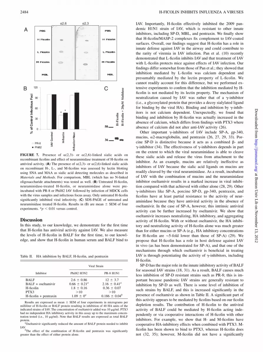
fibrinogen-like domain (30). To determine whether these are sia-lylated, we performed glycan blotting of the ficolins. As shown inFig. 7A, all three ficolins showed the presence of a(2,3)- anda(2,6)-linked glycans, whereas MBL (which does not have any N-linked glycan attachment site) did not. To confirm the role forficolin-associated sialic acids in viral inhibition, we treated H-ficolin with neuraminidase and then repurified the protein. This
treatment completely abrogated the HA inhibitory activity of H-ficolin (Fig. 7B). Neuraminidase treatment caused the expectedsmall decrease in the apparent m.w. of H-ficolin, but did not resultin degradation of the protein (Fig. 7C).BALF causes inhibition of HA activity of IAV (Table II). BALF
was much less effective at inhibiting HA activity of the PR-8strain than the Phil82 strain. This reflects the importance of SP-D in inhibiting seasonal strains of IAV as reported previously (22,25). Note, however, that there is some inhibition of the fully SP-D–resistant PR-8 strain that was strongly potentiated by oselta-mivir, suggesting that this inhibition is mediated to a significantextent by g-inhibitors. The viral neutralizing activity of BALF forPhil82 and PR-8 strains of IAV was also increased in the presenceof oseltamivir (Fig. 8A, 8B). There are multiple g-inhibitors inBALF, some of which have been shown to inhibit IAV, includingpentraxin-3 (PTX3) and gp340 (26, 31). Of interest, recent studiesdemonstrated specific binding of PTX3 to M- and L-ficolins and
FIGURE 4. Effect of ficolin depletion on viral neutralizing activity of
human serum or BALF. Normal donor human serum or BALF was incu-
bated with N-acetyl-D-galactosamine-agarose as described to remove
ficolins and the serum or BALF before or after depletion were compared
for viral neutralizing activity using the infectious focus assay and PR-8.
The serum or BALF was preincubated with the virus for 45 min followed
by titration on MDCK cells. (A) Results obtained with serum from one
donor at the indicated dilutions. (B) Results obtained with BALF from two
separate donors (labeled 1 and 2). The BALF was used either undiluted or
at 1:1 dilution in PBS as indicated. The results are mean 6 SEM of three
experiments with each dilution of serum or BALF. *Depleted BALF or
serum had significantly less neutralizing activity than the untreated samples.
FIGURE 5. Effect of adding H-ficolin to MDCK cells before or after
virus infection on neutralizing activity. The neutralization assay was per-
formed as in Fig. 3, except that in (A) the MDCK cells were preincubated
with the ficolin prior to infection with IAV, and in (B) ficolin was added to
MDCK cells after viral infection. (In Fig. 3, virus was preincubated with
ficolins prior to infection of MDCK cells). Preincubation of MDCK cells
with H-ficolin caused comparable inhibition of viral infection with the
three indicated viral strains as in Fig. 3. Results with human MBL are
shown for comparison. H-ficolin caused significantly greater inhibition of
the PR-8 strain than MBL, whereas MBL caused significantly greater in-
hibition of the Phil82 strain than H-ficolin at 4 mg/ml Results in (B) show
that the addition of H-ficolin or MBL after viral infection for 45 min
causes much less inhibition than found in (A) or in Fig. 3. Note, however,
that H-ficolin still caused significant inhibition compared with control
using this method (p , 0.05 for all viruses at 4 mg/ml). Results are mean 6SEM of four experiments. *Differences as assessed by ANOVA.
2482 H-FICOLIN INHIBITS INFLUENZA AVIRUSES
at Boston U
niv Lib on N
ovember 13, 2012
http://jimm
unol.org/D
ownloaded from
synergistic host defense activites between these proteins (32). H-ficolin and PTX3 caused cooperative inhibition of HA activity(Table II) and infectivity of PR-8 and Phil82 (Fig. 8A, 8B). M-ficolin had a similar effect (Fig. 8C). Therefore, it is possible thatH-ficolin in BALF contributes to inhibition of IAV through directeffects and through interactions with other inhibitors.We performed confocal microscopy to directly evaluate how H-
ficolin alone or in presence of oseltamivir alters interaction of IAVwith A549 cells within 45 min of infection. As shown in Fig. 8B, H-ficolin alone caused what appears to be a reduced number of viralparticles associated with cells at this time point. As shown in Fig.5, quantitative PCR did not show reduced total viral RNA asso-ciated with the cells under these conditions. This finding sug-gested that ficolin might be reducing particle numbers through viralaggregation. It is difficult to assess this at this level of magnifica-tion unless the aggregates are large (e.g., SP-D causes formation oflarge viral aggregates that are easily visible by fluorescent mi-croscopy) (Ref. 21 and data not shown); however, the viral par-ticles appeared somewhat larger in H-ficolin treated samples versuscontrol. Of interest, when H-ficolin and oseltamivir were used to-gether, viral particle sizes appeared larger than with H-ficolin alone(Fig. 8B, right panel).
H-ficolin causes viral aggregation
Given the results obtained with confocal microscopy, we used moresensitive techniques to assess viral aggregation. H-ficolin inducedaggregation of IAV as assessed by electron microscopy or lightabsorbance assays (Fig. 9A, 9B, respectively). On the light trans-mission assay, aggregation induced by H-ficolin alone was subtle,although comparable to that induced by MBL (not shown). As inthe case of defensins (19), aggregation caused by H-ficolin wasmost clearly evident on electron microscopy. The addition ofoseltamivir along with H-ficolin dramatically increased aggrega-tion as assessed by light absorbance.
H-ficolin fixes complement to IAV-coated surfaces
The PR-8 strain of IAVwas coated onto the surface of ELISA platesas for the ELISA assay shown in Fig. 1. These plates were thenincubated with H-ficolin/MASP-2 complexes purified from humanserum as described (4). As shown in Fig. 10, there was dose-related deposition of the C4 component of complement onto thevirus-coated surface. The extent of complement deposition wasless than the amount seen on the acetylated BSA surface, whichserved as the positive control.
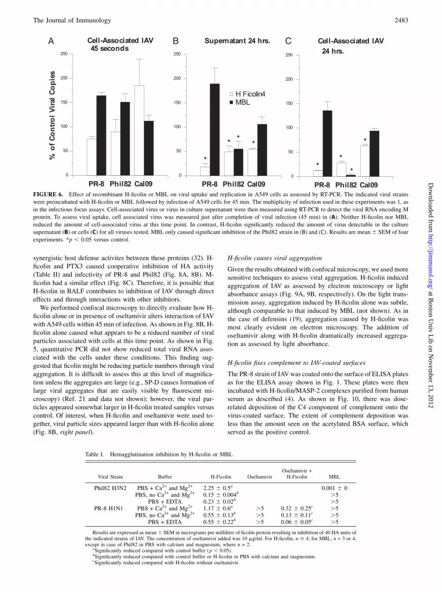
FIGURE 6. Effect of recombinant H-ficolin or MBL on viral uptake and replication in A549 cells as assessed by RT-PCR. The indicated viral strains
were preincubated with H-ficolin or MBL followed by infection of A549 cells for 45 min. The multiplicity of infection used in these experiments was 1, as
in the infectious focus assays. Cell-associated virus or virus in culture supernatant were then measured using RT-PCR to detect the viral RNA encoding M
protein. To assess viral uptake, cell associated virus was measured just after completion of viral infection (45 min) in (A). Neither H-ficolin nor MBL
reduced the amount of cell-associated virus at this time point. In contrast, H-ficolin significantly reduced the amount of virus detectable in the culture
supernatant (B) or cells (C) for all viruses tested. MBL only caused significant inhibition of the Phil82 strain in (B) and (C). Results are mean6 SEM of four
experiments. *p , 0.05 versus control.
Table I. Hemagglutination inhibition by H-ficolin or MBL
Viral Strain Buffer H-Ficolin OseltamivirOseltamivir +H-Ficolin MBL
Phil82 H3N2 PBS + Ca2+ and Mg2+ 2.25 6 0.5a 0.001 6 0PBS, no Ca2+ and Mg2+ 0.15 6 0.004b .5
PBS + EDTA 0.23 6 0.02b .5PR-8 H1N1 PBS + Ca2+ and Mg2+ 1.17 6 0.6a .5 0.32 6 0.25c .5
PBS, no Ca2+ and Mg2+ 0.55 6 0.13b .5 0.13 6 0.11c .5PBS + EDTA 0.55 6 0.22b .5 0.06 6 0.05c .5
Results are expressed as mean6 SEM in micrograms per milliliter of ficolin protein resulting in inhibition of 40 HA units ofthe indicated strains of IAV. The concentration of oseltamivir added was 10 mg/ml. For H-ficolin, n $ 4; for MBL, n = 3 or 4,except in case of Phil82 in PBS with calcium and magnesium, where n = 2.
aSignificantly reduced compared with control buffer (p , 0.05).bSignificantly reduced compared with control buffer or H-ficolin in PBS with calcium and magnesium.cSignificantly reduced compared with H-ficolin without oseltamivir.
The Journal of Immunology 2483
at Boston U
niv Lib on N
ovember 13, 2012
http://jimm
unol.org/D
ownloaded from
DiscussionIn this study, to our knowledge, we demonstrate for the first timethat H-ficolin has antiviral activity against IAV. We also measurethe levels of H-ficolin in BALF for the first time, to our knowl-edge, and show that H-ficolin in human serum and BALF bind to
IAV. Importantly, H-ficolin effectively inhibited the 2009 pan-demic H1N1 strain of IAV, which is resistant to other innateinhibitors, including SP-D, MBL, and pentraxin. We finally showthat H-ficolin/MASP-2 complexes fix complement to IAV-coatedsurfaces. Overall, our findings suggest that H-ficolin has a role ininnate defense against IAV in the airway and could contribute tothe rarity of viremia in IAV infection. Pan et al. (10) recentlydemonstrated that L-ficolin inhibits IAV and that treatment of IAVwith L-ficolin protects mice against effects of IAV infection. Ourfindings differ somewhat from those of Pan et al.; they showed thatinhibition mediated by L-ficolin was calcium dependent andpresumably mediated by the lectin property of L-ficolin. Wecannot readily account for this difference, but we performed ex-tensive experiments to confirm that the inhibition mediated by H-ficolin is not mediated by its lectin property. The mechanism ofneutralization caused by IAV was rather that of a g-inhibitor(i.e., a glycosylated protein that provides a decoy sialylated ligandfor binding by the viral HA). Binding and inhibition by g-inhib-itors is not calcium dependent. Unexpectedly, we found thatbinding and inhibition by H-ficolin was actually increased in theabsence of calcium, which differs from findings with PTX3 whereabsence of calcium did not alter anti-IAV activity (26).Other important g-inhibitors of IAV include SP-A, gp-340,
mucins, a2-macroglobulin, and pentraxin (26, 27, 29, 33). Por-cine SP-D is distinctive because it acts as a combined b- andg-inhibitor (34). The effectiveness of g-inhibitors depends in parton the extent to which the viral neuraminidase is able to cleavethese sialic acids and release the virus from attachment to theinhibitor. As an example, mucins are relatively ineffective asinhibitors of IAV because the sialic acid ligands on mucins arereadily cleaved by the viral neuraminidase. As a result, incubationof IAV with the combination of mucins and the neuraminidaseinhibitor oseltamivir results in a marked increase in viral inhibi-tion compared with that achieved with either alone (28, 29). Otherg-inhibitors like SP-A, porcine SP-D, gp-340, pentraxin, andficolins have at least partial resistance to the activity of neur-aminidase because they have antiviral activity in the absence ofoseltamivir. In the case of SP-A, however, this intrinsic antiviralactivity can be further increased by oseltamivir. We show thatoseltamivir increases neutralizing, HA inhibitory, and aggregatingactivity of H-ficolin. With or without oseltamivir, the HA inhibi-tory and neutralizing activity of H-ficolin alone was much greaterthan for either mucins or SP-A (e.g., HA inhibitory concentrationsfor H-ficolin are ∼5-fold lower than those of SP-A) (29). Wepropose that H-ficolin has a role in host defense against IAVin vivo (as has been demonstrated for SP-A), and that one of themechanisms through which oseltamivir is beneficial in treatingIAV is through potentiating the activity of g-inhibitors, includingH-ficolin.SP-D has the major role in the innate inhibitory activity of BALF
for seasonal IAV strains (18, 31). As a result, BALF causes muchless inhibition of SP-D resistant strains such as PR-8; this is im-portant because pandemic IAV strains are generally resistant toinhibition by SP-D as well. There is some level of inhibition ofsuch strains by BALF, and this is increased significantly in thepresence of oseltamivir as shown in Table II. A significant part ofthis activity appears to be mediated by ficolins based on our ficolindepletion results. The contribution of H-ficolin to the antiviralactivity of BALF could be mediated by H-ficolin acting inde-pendently or via cooperative interactions of H-ficolin with otherinhibitors. For example, we show that H- and M-ficolins havecooperative HA-inhibitory effects when combined with PTX3. M-ficolin has been shown to bind to PTX3, whereas H-ficolin doesnot (32, 35); however, M-ficolin did not have a significantly
FIGURE 7. Presence of a(2,3)- or a(2,6)-linked sialic acids on
recombinant ficolins and effect of neuraminidase treatment of H-ficolin on
antiviral activity. (A) The presence of a(2,3)- or a(2,6)-linked sialic acids
on recombinant H-, L-, and M-ficolins was assessed by lectin blotting
using SNA and MAA as sialic acid detecting molecules as described in
Materials and Methods. For comparison, MBL (which has no N-linked
oligosaccharide attachments) was tested as well. (B) Untreated H-ficolin,
neuraminidase-treated H-ficolin, or neuraminidase alone were pre-
incubated with PR-8 or Phil82 IAV followed by infection of MDCK cells
with the virus samples and infectious focus assay. Only untreated H-ficolin
significantly inhibited viral infectivity. (C) SDS-PAGE of untreated and
neuraminidase treated H-ficolin. Results in (B) are mean 6 SEM of four
experiments. *p , 0.01 versus control.
Table II. HA inhibition by BALF, H-ficolin, and pentraxin
Viral Strain
Inhibitor Phil82 H3N2 PR-8 H1N1
BALF 2.6 6 0.86 12 6 3.7BALF + oseltamivir 0.66 6 0.21a 2.16 6 0.41a
H-ficolin 1.8 6 0.16 0.36 6 0.07PTX3 .10 .10H-ficolin + pentraxin 1.09 6 0a 0.186 6 0.04b
Results are expressed as mean 6 SEM of four experiments in micrograms permilliliter of H-ficolin or BALF protein resulting in inhibition of 40 HA units of theindicated strains of IAV. The concentration of oseltamivir added was 10 mg/ml. PTX3had no independent HA inhibitory activity in this assay up to the maximum concen-tration tested (i.e., 10 mg/ml). Note that BALF results are expressed as total BALFprotein.
aOseltamivir significantly reduced the amount of BALF protein needed to inhibitIAV.
bThe effect of the combination of H-ficolin and pentraxin was significantlygreater than the effect of either protein alone.
2484 H-FICOLIN INHIBITS INFLUENZA AVIRUSES
at Boston U
niv Lib on N
ovember 13, 2012
http://jimm
unol.org/D
ownloaded from

greater cooperative interaction with PTX3 than H-ficolin. Thisfinding suggests that the binding of PTX3 to M-ficolin does notsignificantly contribute to combined antiviral effects. Because
both PTX3 and ficolins resemble C1q and fix complement, furtherstudy of the combined effects on complement fixation in thepresence of IAV would be interesting (32). We propose thatg-inhibitors provide an important level of innate protection againstIAV strains that are able to bypass the action of SP-D or MBL.The activity of g-inhibitors for specific viral strains depends on
the specific types of sialic acid linkage present on the proteins (34,36). In general the HA of avian or mouse adapted IAV strains havepreferential binding to a(2,3)-linked sialic acids, whereas humanstrains prefer binding to a(2,6)-linked sialic acids. These prefer-ences coincide with the type of sialic acid linkages found, re-spectively, on avian (or mouse) versus human epithelia targeted bythe virus. The collectins, other than SP-A and porcine SP-D, de-
FIGURE 8. Effect of oseltamivir or PTX3 on viral
neutralizing activity of H-ficolin or on effects of H-
ficolin on viral attachment or uptake by A549 cells as
assessed with confocal microscopy. (A–C) Results of
infectious focus assays using H- or M-ficolin alone or
combined with either oseltamivir (5 mg/ml) or PTX3
(5 mg/ml). Results are mean 6 SEM of four experi-
ments. Addition of both oseltamivir and PTX3 sig-
nificantly increased neutralizing activity compared
with H-ficolin alone at either the 2.5- or 5-mg/ml
concentration of H-ficolin. This was true both for the
Phil82 (A) or PR-8 (B) strains of IAV. PTX3 or osel-
tamivir alone did not reduce viral infectivity in this
assay. The results for PTX3 are shown at the zero
concentration of ficolins in the curves labeled PTX3.
PTX3 significantly increased the neutralizing activity
of M-ficolin as shown in (C). (D) Confocal micro-
scopic pictures of virus (red) after 45 min incubation
with A549 cells (cell membrane green and nucleus
blue). The multiplicity of infection for the confocal
experiments was 200 (i.e., higher than in infectious
focus and quantitative PCR assays). Results are rep-
resentative of three experiments. Nuclei are labeled
with DAPI350 (blue), cell membranes with WGA
Oregon green, and virus with Alexa Flour 594 (red).
FIGURE 9. Viral aggregation induced by H-ficolin or MBL. (Top)
Representative (of four experiments) electron microscopic images of PR-8
IAV alone (control) versus IAV pretreated with the indicated concen-
trations of H-ficolin or MBL. In these experiments, 10 mg/ml H-ficolin
caused a similar degree of viral aggregation as MBL. (Bottom) The ability
of H-ficolin to cause viral aggregation was also tested by light absorbance
assay. In this assay, increased light transmission results from viral aggre-
gation. This assay is less sensitive for detecting viral aggregation than
electron microscopy; however, 20 and 40 mg/ml of H-ficolin did cause
significant increase in light transmission 2 min after addition to the viral
suspension. Addition of oseltamivir (10 mg/ml) during incubation of IAV
with H-ficolin resulted in much more pronounced and sustained viral ag-
gregation. No aggregation was seen with oseltamivir alone or with the PR-
8 virus alone (black diamonds, bottom). Results in the bottom panel are
mean 6 SEM of four experiments.
FIGURE 10. H-ficolin/MASP-2 complexes fix complement in presence
of IAV. ELISA plates were coated with IAVas in Fig. 1. H-ficolin/MASP-2
complexes were purified from human serum and incubated with the surface
bound IAV as described in Materials and Methods. Complement fixation
was detected using Ab to complement component C4. H-ficolin caused
dose-related deposition of C4 onto the viral surface (n = 5). As a positive
control, 2 mg/ml of the H-ficolin/MASP-2 complex was incubated with
wells coated with acetylated BSA. This resulted in fixation of complement
as reported (mean6 SEM OD450 for C4 was 1.26 0.1). *p, 0.05 versus
IAV-coated plates incubated with complement in the absence of H-ficolin.
The Journal of Immunology 2485
at Boston U
niv Lib on N
ovember 13, 2012
http://jimm
unol.org/D
ownloaded from

pend on the presence of glycans on viral envelope proteins fortheir antiviral activity (37). High mannose glycans on the viral HAare most important for inhibition by MBL, SP-D, or the bovineserum collectins (conglutinin, CL43, and CL46) (38). Because theactivity of g-inhibitors is not dependent on viral envelope proteinglycosylation, they have activity against strains that are resistantto MBL or SP-D. In fact, g-inhibitors appear to have greater ac-tivity against some hypoglycosylated viral strains (e.g., PR-8) thanagainst strains like Phil82 that have abundant glycans on the HA(21). This finding could reflect the fact that the addition of glycansto the globular domain of the HA reduces HA binding affinity forsialylated ligands. This appears to be the case for H-ficolin, whichhas substantially greater inhibitory activity for the PR-8 strain. Inthis case of PR-8 virus, however, it could also depend on thepresence of a(2,3)-linked sialic acids on the g-inhibitor, becausePR-8 is highly selective for this type of sialic acid linkage. Theficolins expressed both types of sialic acid linkage, and this mightaccount for their ability to inhibit both the human seasonal strainPhil82 and the mouse-adapted PR-8 strain.Known pandemic strains of IAV isolated over the past 100 y all
had reduced glycosylation of their HA compared with seasonalH1N1 and H3N2 strains (22). The ficolins are of particular interestin that they retain activity against the pandemic Cal09 H1N1strain. This strain is not inhibited by SP-D or MBL effectively andrequires significantly increased concentrations of SP-A for inhi-bition (23). Furthermore, pentraxin also did not inhibit this strain(23). It will be of great interest to determine whether ficolins in-hibit other pandemic or avian strains (e.g., H5N1).The ability of collectins and defensins to induce viral aggre-
gation contributes to their viral neutralizing activity. Ficolins re-semble the collectins MBL and SP-A on a structural basis becauseof similarities in their oligomeric structure and collagen domains.In fact, replacement of the N-terminal and collagen domains ofMBLwith those of L-ficolin results in chimeric molecules that haveequal or greater viral aggregating and neutralizing activity thanMBL (12, 13). Like the collectins, H-ficolin is able to induce viralaggregation; this could be an important contributor to its antiviralactivity in vivo. Aggregation could reduce particle numbers forinfection of cells (Fig. 8B) and promote viral clearance byphagocytes or mucociliary mechanisms. Further study will beneeded to understand more completely how H-ficolin alters theviral life cycle. We show that viral neutralization by H-ficolininvolves interactions with the epithelial cells and or virus priorto internalization and that H-ficolin does not appear to alter thetotal amount of viral RNA attaching to or being taken up by cells.It is possible that through inducing subtle viral aggregation, orthrough engagement of distinct cellular receptors, H-ficolin altersprocessing of the virus after internalization.In this study, we provide the first measurements, to our
knowledge, of levels of H-ficolin in normal donor BAL fluid. Wealso show H-ficolin in BALF or serum binds to IAV and that in-cubation of IAV with these fluids results in the precipitation of H-ficolin from the mixture. Therefore, native H-ficolin, as it is foundin normal blood or respiratory secretions, binds to IAV. Further-more, depletion of ficolins from these fluids reduces their antiviralactivity. The ficolin depletion procedure we used may have re-moved any M- or L-ficolin present in serum and BALF along withthe H-ficolin. H-ficolin is, however, the most abundant ficolin inserum. Further study would be needed to determine levels of M- orL-ficolin in BALF, although it is likely they are less than H-ficolin.Another important avenue for further investigation will be studiesof the role of ficolins in host defense against IAV in vivo in mousemodels; however, mice lack a functional H-ficolin gene, and gene-deletion studies will not be possible (39). As noted in the intro-
duction, subjects with H-ficolin deficiency with increased sus-ceptibility to infection have been reported (7, 8). It is tempting tospeculate that individuals lacking or with reduced levels of H-ficolin may also be more susceptible to IAV infection. Suscepti-bility to IAV infection could also result in more frequent bacterialrespiratory infections because IAV predisposes to bacterial in-fection.
DisclosuresThe authors have no financial conflicts of interest.
References1. Thiel, S. 2007. Complement activating soluble pattern recognition molecules
with collagen-like regions, mannan-binding lectin, ficolins and associated pro-teins. Mol. Immunol. 44: 3875–3888.
2. Garred, P., and N. Borregaard. 2010. The ficolins. J. Innate Immun. 2: 1–2.3. Akaiwa, M., Y. Yae, R. Sugimoto, S. O. Suzuki, T. Iwaki, K. Izuhara, and
N. Hamasaki. 1999. Hakata antigen, a new member of the ficolin/opsonin p35family, is a novel human lectin secreted into bronchus/alveolus and bile. J.Histochem. Cytochem. 47: 777–786.
4. Zacho, R. M., L. Jensen, R. Terp, J. C. Jensenius, and S. Thiel. 2012. Studies ofthe pattern recognition molecule H-ficolin: specificity and purification. J. Biol.Chem. 287: 8071–8081.
5. Sallenbach, S., S. Thiel, C. Aebi, M. Otth, S. Bigler, J. C. Jensenius,L. J. Schlapbach, and R. A. Ammann. 2011. Serum concentrations of lectin-pathway components in healthy neonates, children and adults: mannan-bindinglectin (MBL), M-, L-, and H-ficolin, and MBL-associated serine protease-2(MASP-2). Pediatr. Allergy Immunol. 22: 424–430.
6. Hummelshoj, T., L. M. Fog, H. O. Madsen, R. B. Sim, and P. Garred. 2008.Comparative study of the human ficolins reveals unique features of Ficolin-3(Hakata antigen). Mol. Immunol. 45: 1623–1632.
7. Munthe-Fog, L., T. Hummelshøj, C. Honore, H. O. Madsen, H. Permin, andP. Garred. 2009. Immunodeficiency associated with FCN3 mutation and ficolin-3deficiency. N. Engl. J. Med. 360: 2637–2644.
8. Schlapbach, L. J., S. Thiel, U. Kessler, R. A. Ammann, C. Aebi, andJ. C. Jensenius. 2011. Congenital H-ficolin deficiency in premature infants withsevere necrotising enterocolitis. Gut 60: 1438–1439.
9. Liu, J., M. A. Ali, Y. Shi, Y. Zhao, F. Luo, J. Yu, T. Xiang, J. Tang, D. Li, Q. Hu,et al. 2009. Specifically binding of L-ficolin to N-glycans of HCV envelopeglycoproteins E1 and E2 leads to complement activation. Cell. Mol. Immunol. 6:235–244.
10. Pan, Q., H. Chen, F. Wang, V. T. Jeza, W. Hou, Y. Zhao, T. Xiang, Y. Zhu,Y. Endo, T. Fujita, and X. L. Zhang. 2012. L-ficolin binds to the glycoproteinshemagglutinin and neuraminidase and inhibits influenza A virus infection bothin vitro and in vivo. J. Innate Immun. 4: 312–324.
11. Keirstead, N. D., C. Lee, D. Yoo, A. S. Brooks, and M. A. Hayes. 2008. Porcineplasma ficolin binds and reduces infectivity of porcine reproductive and respi-ratory syndrome virus (PRRSV) in vitro. Antiviral Res. 77: 28–38.
12. Chang, W. C., K. L. Hartshorn, M. R. White, P. Moyo, I. C. Michelow, H. Koziel,B. T. Kinane, E. V. Schmidt, T. Fujita, and K. Takahashi. 2011. Recombinantchimeric lectins consisting of mannose-binding lectin and L-ficolin are potentinhibitors of influenza A virus compared with mannose-binding lectin. Biochem.Pharmacol. 81: 388–395.
13. Michelow, I. C., M. Dong, B. A. Mungall, L. M. Yantosca, C. Lear, X. Ji,M. Karpel, C. L. Rootes, M. Brudner, G. Houen, et al. 2010. A novel L-ficolin/mannose-binding lectin chimeric molecule with enhanced activity against Ebolavirus. J. Biol. Chem. 285: 24729–24739.
14. Krarup, A., S. Thiel, A. Hansen, T. Fujita, and J. C. Jensenius. 2004. L-ficolin isa pattern recognition molecule specific for acetyl groups. J. Biol. Chem. 279:47513–47519.
15. Hartshorn, K. L., M. Collamer, M. Auerbach, J. B. Myers, N. Pavlotsky, andA. I. Tauber. 1988. Effects of influenza A virus on human neutrophil calciummetabolism. J. Immunol. 141: 1295–1301.
16. Frederiksen, P. D., S. Thiel, C. B. Larsen, and J. C. Jensenius. 2005. M-ficolin, aninnate immune defence molecule, binds patterns of acetyl groups and activatescomplement. Scand. J. Immunol. 62: 462–473.
17. Kuraya, M., Z. Ming, X. Liu, M. Matsushita, and T. Fujita. 2005. Specificbinding of L-ficolin and H-ficolin to apoptotic cells leads to complement acti-vation. Immunobiology 209: 689–697.
18. Hartshorn, K. L., E. C. Crouch, M. R. White, P. Eggleton, A. I. Tauber,D. Chang, and K. Sastry. 1994. Evidence for a protective role of pulmonary sur-factant protein D (SP-D) against influenza A viruses. J. Clin. Invest. 94: 311–319.
19. Tecle, T., M. R. White, D. Gantz, E. C. Crouch, and K. L. Hartshorn. 2007.Human neutrophil defensins increase neutrophil uptake of influenza A virus andbacteria and modify virus-induced respiratory burst responses. J. Immunol. 178:8046–8052.
20. Hartshorn, K., K. Sastry, D. Chang, M. White, and E. Crouch. 2000. Enhancedantinfluenza activity of a recombinant pulmonary surfactant protein D and serumconglutinin fusion protein. Am. J. Physiol. 278: L90–L98.
21. Hartshorn, K. L., M. R. White, V. Shepherd, K. Reid, J. C. Jensenius, andE. C. Crouch. 1997. Mechanisms of anti-influenza activity of surfactant proteinsA and D: comparison with serum collectins. Am. J. Physiol. 273: L1156–L1166.
2486 H-FICOLIN INHIBITS INFLUENZA AVIRUSES
at Boston U
niv Lib on N
ovember 13, 2012
http://jimm
unol.org/D
ownloaded from

22. Qi, L., J. C. Kash, V. G. Dugan, B. W. Jagger, Y. F. Lau, Z. M. Sheng,E. C. Crouch, K. L. Hartshorn, and J. K. Taubenberger. 2011. The ability ofpandemic influenza virus hemagglutinins to induce lower respiratory pathology isassociated with decreased surfactant protein D binding. Virology 412: 426–434.
23. Job, E. R., Y. M. Deng, M. D. Tate, B. Bottazzi, E. C. Crouch, M. M. Dean,A. Mantovani, A. G. Brooks, and P. C. Reading. 2010. Pandemic H1N1 influenzaA viruses are resistant to the antiviral activities of innate immune proteins of thecollectin and pentraxin superfamilies. J. Immunol. 185: 4284–4291.
24. Hartshorn, K. L., K. Sastry, M. R. White, E. M. Anders, M. Super,R. A. Ezekowitz, and A. I. Tauber. 1993. Human mannose-binding proteinfunctions as an opsonin for influenza A viruses. J. Clin. Invest. 91: 1414–1420.
25. Reading, P. C., L. S. Morey, E. C. Crouch, and E. M. Anders. 1997. Collectin-mediated antiviral host defense of the lung: evidence from influenza virus in-fection of mice. J. Virol. 71: 8204–8212.
26. Reading, P. C., S. Bozza, B. Gilbertson, M. Tate, S. Moretti, E. R. Job,E. C. Crouch, A. G. Brooks, L. E. Brown, B. Bottazzi, et al. 2008. Antiviralactivity of the long chain pentraxin PTX3 against influenza viruses. J. Immunol.180: 3391–3398.
27. Hartshorn, K. L., A. Ligtenberg, M. R. White, M. Van Eijk, M. Hartshorn,L. Pemberton, U. Holmskov, and E. Crouch. 2006. Salivary agglutinin and lungscavenger receptor cysteine-rich glycoprotein 340 have broad anti-influenzaactivities and interactions with surfactant protein D that vary according to do-nor source and sialylation. Biochem. J. 393: 545–553.
28. White, M. R., E. J. Helmerhorst, A. Ligtenberg, M. Karpel, T. Tecle,W. L. Siqueira, F. G. Oppenheim, and K. L. Hartshorn. 2009. Multiple com-ponents contribute to ability of saliva to inhibit influenza viruses. Oral Micro-biol. Immunol. 24: 18–24.
29. White, M. R., E. Crouch, M. van Eijk, M. Hartshorn, L. Pemberton, I. Tornoe,U. Holmskov, and K. L. Hartshorn. 2005. Cooperative anti-influenza activities ofrespiratory innate immune proteins and neuraminidase inhibitor. Am. J. Physiol.Lung Cell. Mol. Physiol. 288: L831–L840.
30. Garlatti, V., L. Martin, M. Lacroix, E. Gout, G. J. Arlaud, N. M. Thielens, andC. Gaboriaud. 2010. Structural insights into the recognition properties of humanficolins. J. Innate Immun. 2: 17–23.
31. Hartshorn, K. L., M. R. White, T. Mogues, T. Ligtenberg, E. Crouch, andU. Holmskov. 2003. Lung and salivary scavenger receptor glycoprotein-340contribute to the host defense against influenza A viruses. Am. J. Physiol.Lung Cell. Mol. Physiol. 285: L1066–L1076.
32. Ma, Y. J., A. Doni, T. Hummelshøj, C. Honore, A. Bastone, A. Mantovani,N. M. Thielens, and P. Garred. 2009. Synergy between ficolin-2 and pentraxin 3boosts innate immune recognition and complement deposition. J. Biol. Chem.284: 28263–28275.
33. Benne, C. A., C. A. Kraaijeveld, J. A. G. van Strijp, E. Brouwer, M. Harmsen,J. Verhoef, L. M. G. van Golde, and J. F. van Iwaarden. 1995. Interactions ofsurfactant protein A with influenza A viruses: binding and neutralization. J.Infect. Dis. 171: 335–341.
34. van Eijk, M., M. R. White, E. C. Crouch, J. J. Batenburg, A. B. Vaandrager,L. M. Van Golde, H. P. Haagsman, and K. L. Hartshorn. 2003. Porcine pul-monary collectins show distinct interactions with influenza A viruses: role of theN-linked oligosaccharides in the carbohydrate recognition domain. J. Immunol.171: 1431–1440.
35. Gout, E., C. Moriscot, A. Doni, C. Dumestre-Perard, M. Lacroix, J. Perard,G. Schoehn, A. Mantovani, G. J. Arlaud, and N. M. Thielens. 2011. M-ficolininteracts with the long pentraxin PTX3: a novel case of cross-talk betweensoluble pattern-recognition molecules. J. Immunol. 186: 5815–5822.
36. Mikerov, A. N., M. White, K. Hartshorn, G. Wang, and J. Floros. 2008.Inhibition of hemagglutination activity of influenza A viruses by SP-A1 andSP-A2 variants expressed in CHO cells. Med. Microbiol. Immunol. (Berl.) 197:9–12.
37. Hartshorn, K. L. 2010. Role of surfactant protein A and D (SP-A and SP-D) inhuman antiviral host defense. Front Biosci (Schol Ed) 2: 527–546 (Schol Ed).
38. Hartshorn, K. L., M. R. White, D. R. Voelker, J. Coburn, K. Zaner, andE. C. Crouch. 2000. Mechanism of binding of surfactant protein D to influenza Aviruses: importance of binding to haemagglutinin to antiviral activity. Biochem.J. 351: 449–458.
39. Endo, Y., Y. Liu, K. Kanno, M. Takahashi, M. Matsushita, and T. Fujita. 2004.Identification of the mouse H-ficolin gene as a pseudogene and orthology be-tween mouse ficolins A/B and human L-/M-ficolins. Genomics 84: 737–744.
The Journal of Immunology 2487
at Boston U
niv Lib on N
ovember 13, 2012
http://jimm
unol.org/D
ownloaded from