hong kong journal of paediatrics
TRANSCRIPT
New Series
Offical Publication of:Hong Kong College of PaediatriciansHong Kong Paediatric Society
HK J Paediatr (New Series) Vol 26. No. 3 July 2021
ISSN 2309-5393 (online)ISSN 1013-9923 (print)
Indexed in EMBASE/Excerpta Medica,Science Citation Index Expanded (SCIE) and ScopusFull text online at www.hkjpaed.org
EditorialPhysical and Mental Well-being of Children and 123the Way ForwardIp
Original ArticlesPrevalence of Vitamin D Deficiency and Insufficiency 125and Its Risk Factors in Paediatric Patients withEpilepsy on Anti-epileptic DrugsMo, Yuen, Fung, Cheung, Shek, Leung
Attention Deficit Hyperactivity Disorder in Paediatric 135Patients with Malignant Haematologic Diseases orEpilepsy: Experience at a Tertiary Care Hospital inKoreaHan, Lee, Cho, Chung, Lee
Comparison of Alcohol and Phenol Neurolysis in 141Children with Spasticity: A Pilot Matched ControlledTrialLeung, Chan, Man, Ko
Tolerability and Efficacy of Racecadotril in Acute 149Diarrhoea, A Prospective, Randomised, ParallelStudy in an Indian Tertiary Care Teaching HospitalSarangi, Biswal, Dash, Dhanawat
Case ReportsEnterovirus D68 Myelitis: The First Reported Case 155in Hong KongMo, Yuen
An Extremely Rare Association of Cleft Lip and 158Palate, Basal Sphenoethmoidal Cephalocele andCorpus Callosal Agenesis in a Three-month OldPatient Presenting with Facial Cleft and Nasal MassUrgel, Viterbo
Diffusion Weighted Imaging and MR Spectroscopic 161Findings in Maple Syrup Urine Disease:The Importance of Early Radiological Diagnosisin the Prevention of Cerebral Parenchymal DamageGüney, Yeniçeri, Do an, Akçay
Median Cleft of Lower Lip with Central Cleft 165Palate and Ankyloglossia: A Case ReportTang, Chan, Tsui, Chao
Contemporary Practice in PaediatricsHong Kong Universal Newborn Hearing Screening 168(UNHS) Care Path Protocol under Joint Committeeon UNHSMa, Chan, Koh, Yeung, Wong, Sung, Kwong, Shek,Lee,Wong, Lam, Lee, Tso, Yeung, Hau, Tse, Ho, Wong;HA-DH Joint Committee on Care Path for UniversalNewborn Hearing Screening
Joint Position Statement on BioNTech Vaccination 175in Adolescents with Allergic DiseasesLeung, Tse, Chan; On behalf of Hong Kong Collegeof Paediatricians and Hong Kong Society forPaediatric Immunology Allergy and Infectious Diseases
MCQs 180
HONG KONG JOURNAL OF PAEDIATRICS
Medcom Limited
HK J Paediatr (new series) 2021;26:123-124
Hong Kong Journal of Paediatrics (New Series)
An Official Publication ofHong Kong College of Paediatricians &Hong Kong Paediatric Societyc/o Hong Kong College of Paediatricians, Room 801,Hong Kong Academy of Medicine Jockey Club Building,99 Wong Chuk Hang Road, Aberdeen,Hong Kong.
Editorial Board
Chief EditorCHEUNG Yiu Fai
Associate EditorsHON Kam Lun IP Patrick
Honorary SecretaryFUNG Cheuk Wing
MembersBUT Wan Man CHAN Chi Fung CHAO Sih Yin FUNG Po Gee KWAN Yat Wah KWONG Ling LAM Hung San LEE Mun Yau LEE So Lun LEUNG SY LIU Sze Wai LO Fai Man LUK Chi Kong WONG Hiu Lei YEUNG Wai Lan
Honorary Advisors to the EditorialBoardAndrew BUSH, United KingdomDon M. ROBERTON, AustraliaDavid K. STEVENSON, USAGUI Yong-Hao, China
Business ManagerTSOI Nai Shun
PublisherHong Kong Journal of Paediatrics is publishedby Medcom Ltd, Flat E8, 10/F, Ka Ming Court,688-690 Castle Peak Road, Kowloon, Hong KongSAR. Tel: (852) 2578 3833, Fax: (852) 2578 3929,Email: [email protected]
Indexed in EMBASE/Excerpta Medica, ScienceCitation Index Expanded (SCIE) and Scopus
Website: www.hkjpaed.org
ISSN 2309-5393 (online)ISSN 1013-9923 (print)
Editorial
Emerging evidence from well-conducted longitudinal studies have highlightedthe importance of early childhood investments, as children are more vulnerableto the early environment that can affect their long-term health and productivityin adult life.1 The Coronavirus Disease 2019 (COVID-19) has rampaged acrossAsia and around the world over the past year and a half. In early 2020, almost athird of the world's population was under lockdown to try to bring the pandemicunder control. This has had profound impacts on the nurturing environment,affecting almost all children and their families. In addition, the various strategiesimplemented to reduce the spread of COVID-19 including social distancing andschool closures can have adverse effects on different aspects of childdevelopment including learning, cognitive skills, physical health, mental health,psychosocial well-being, and peer relationships. It is therefore crucial to studyand monitor the impact of disease pandemics on the physical and mental healthof children as well as on family functioning to alert professionals andstakeholders about their needs and to guide policymakers on planning effectiveinterventions.
Since the first paediatric COVID-19 case admitted in February 2020, therehave been more than 960 infected children managed by our dedicated colleaguesin different hospitals in Hong Kong. The paediatric hospitals and othercollaborative departments have been reporting the clinical characteristics ofHong Kong children infected with COVID-19 since the first three waves, whichshowed that most cases were linked to transmission through family members inhouseholds from diverse backgrounds.2 There are emerging reports of physicaland mental health issues in families and communities associated with the COVID-19 pandemic. A study commissioned by the Hong Kong government foundinfants and young children have significantly lower levels of serum vitamin Dcompared with levels before the pandemic, possibly due to the lack of outdooractivities and exposure to sunlight as a result of the stringent COVID-19 controlmeasures.3 On the other hand, another population study published in EuropeanChild and Adolescent Psychiatry reported an increase in the risk of hyperactivity,inattentive symptoms, and other behavioural problems in children during COVID-19 pandemic,4 which was particularly significant among children with specialeducational needs and other disabilities. These children are highly vulnerableand are greatly affected by interruptions in rehabilitation training and medicalcare. Hence, it is important to examine the impact of the COVID-19 outbreakon this group of children. In particular, we need to examine the prepandemicbaseline prevalence of vitamin D deficiency/insufficiency and hyperactivity/inattention to assess the current situation in children with disabilities.
The July issue includes relevant studies on the prevalence of vitamin Ddeficiency/insufficiency and Attention-deficit/Hyperactivity Disorder(ADHD) among children with chronic illness and on the effectiveness ofinterventions targeted at children with spasticity. In the original studyconducted by Mo et al on children with epilepsy receiving antiepileptic
Physical and Mental Well-being ofChildren and the Way Forward
124 Physical & Mental Well-being of Children & the Way Forward
t r ea tments , they found v i tamin D defic iency /insufficiency was prevalent among this group ofvulnerable children.5 Interestingly, another recent studyreported similar findings of increased risk of vitamin Dinsufficiency among infants and young children with lowblood levels of Vitamin D associated with inadequate sunlightexposure and nutritional consumption.6 This is likely to bean even more alarming problem that should be monitoredby professionals as we continue to treat our paediatricpatients during the later phase of the COVID-19 pandemic.
Handicapped children including those with significantphysical disabilities such as spastic cerebral palsy or thosewith neurodevelopmental disorders such as ADHD andautistic spectrum disorder (ASD) are most vulnerable tothe harmful effects of COVID-19. Children with chronicillness and disabilities are at increased risk of mental healthand adjustment problems during COVID-19 outbreaks,particularly when schools and other buffering platforms arenot functioning. During the peak of the second wave in2020, paediatricians and fellow surgeons in the Hong KongPaediatric Society organised visits to special needs schoolsfor physically and mentally handicapped children andbrought along face masks, alcohol hand rubs, andthermometers that most were in short supply. Althoughresidents with severe spastic cerebral palsy and multipledisabilities were being cared for in the special needsschools, the training of students with ASD, hyperactivity,and intellectual disability was heavily interrupted. Manyhandicapped children have found it difficult to receive basictraining, and many have experienced developmentalregression and physical deterioration with increasedspasticity and contractures.
In Hong Kong, there are currently 56,640 students withspecial educational needs in public schools, of which 23,230 have specific learning difficulties, 14,580 have ADHD,and 11,870 have ASD (Education Bureau, June 2021).Children and adolescents, particularly those with learningdifficulties and ADHD, have faced big challenges withonline learning during school closures related to theCOVID-19 pandemic. A retrospective study conducted byHan et al in South Korea reported that children with chronicillness have a much higher prevalence of ADHD than thegeneral population.7 Paediatric patients with epilepsy andconcomitant ADHD found it more difficult to control theirseizures and responded less favourably to antiepilepticmedications. Another study led by Leung et al in a major
paediatric rehabilitative unit in Hong Kong compareddifferent treatment modalities for children with cerebralpalsy.8 Both phenol and alcohol nerve block were found tobe effective in reducing focal spasticity among paediatricpatients, which can potentially improve their long-termfunction and quality of life.
This summer, we are going to start the first territory-wide COVID-19 vaccination program for students andteenagers in Hong Kong. As paediatricians and professionalslooking after children's health and holistic development,we have witnessed firsthand the many difficultiesexperienced by our patients and their families. As wecontinue on this challenging journey, it is heartening toknown that it will not be long until we see all the cheerfulfaces of our children in the community. May God blessHong Kong and all our beloved children.
P IP
Associate Editor
References
1. Heckman JJ. Skill formation and the economics of investing indisadvantaged children. Science 2006;312:1900-2.
2. Chua GT, Wong JSC, Lam I, et al. Clinical Characteristics andTransmission of COVID-19 in Children and Youths During 3Waves of Outbreaks in Hong Kong. JAMA Netw Open 2021;4:e218824.
3. Wong RS, Tung KTS, So HK, et al. Impact of COVID-19 Pandemicon Serum Vitamin D Level among Infants and Toddlers: AnInterrupted Time Series Analysis and before-and-after Comparison.Nutrients 2021;13:1270.
4. Tso WWY, Wong RS, Tung KTS, et al. Vulnerability and resiliencein children during the COVID-19 pandemic. Eur Child AdolescPsychiatry 2020:1-16.
5. Mo CY, Yuen CL, Fung TH, Cheung HN, Shek ACC, Leung SY.Prevalence of vitamin D deficiency and insufficiency and its riskfactors in paediatric patients with epilepsy on anti-epileptic drugs.HK J Paediatr (new series) 2021;26:125-34.
6. Tung KTS, Wong RS, Tsang HW, et al. An assessment of riskfactors for insufficient levels of vitamin D during early infancy.Nutrients 2021;13:1068.
7. Han Y, Lee JW, Cho B, Chung NG, Lee IG. Attention deficithyperactivity disorder in paediatric patients with malignanthaematologic diseases or epilepsy: Experience at a tertiary carehospital in Korea. HK J Paediatr (new series) 2021;26:135-40.
8. Leung KY, Chan WK, Man WK, Ko CH. Comparison of alcoholand phenol neurolysis in children with spasticity: A pilot matchedcontrolled trial. HK J Paediatr (new series) 2021;26:141-8.
HK J Paediatr (new series) 2021;26:125-134
Prevalence of Vitamin D Deficiency and Insufficiency andIts Risk Factors in Paediatric Patients with Epilepsy on
Anti-epileptic Drugs
CY MO, CL YUEN, TH FUNG, HN CHEUNG, ACC SHEK, SY LEUNG
Abstract Objective: The aim of our study is to evaluate the prevalence of vitamin D deficiency and insufficiencyin children with epilepsy on anti-epileptic drugs (AED) in a regional hospital in Hong Kong. In addition,we would evaluate the possible risk factors of vitamin D deficiency and insufficiency in these patients.Method: A cross-sectional study was conducted in a regional hospital in April to May 2018 on paediatricpatients who were on AED for at least one year. Review of medical records, anthropometric measurements,dietary and sunlight exposure behaviour assessments and blood tests (vitamin D assay and bone profile)were performed. Vitamin D deficiency is defined as serum 25-hydroxyvitamin D (25-OHD) level<30 nmol/L and insufficiency as 25-OHD level between 30-50 nmol/L. Results: Seventy-one childrenaged 3 to 18 years old were recruited into the study. The prevalence of vitamin D deficiency andinsufficiency were 16.9% (12/71) and 52.1% (37/71) respectively. The two groups added up to aprevalence of 69.0% (49/71). In logistic regression analysis, the lack of holiday trip abroad within pastthree months (odds ratio 0.1, 95% confidence interval 0.02-0.73) was found to be a statistically significantindependent risk factor for vitamin D deficiency and insufficiency. Conclusion: Vitamin D deficiencyand insufficiency are highly prevalent in paediatric patients with epilepsy on AEDs in Hong Kong. However,it is similar to the prevalence found in healthy adolescents. This suggests that adolescents taking AEDsmay not have an additional risk in lowering their vitamin D levels in this locality. We therefore would notadvocate for routine screening for vitamin D level in adolescent epilepsy patients on AEDs in this locality,as it will not be cost-effective. Given that this is a cross sectional study, further interventional studiesexploring the role of holiday trip abroad on vitamin D levels in paediatric epilepsy patients are warrantedto substantiate a causality relationship.
Key words Anti-epileptic drugs; Epilepsy; Paediatrics; Prevalence; Vitamin D deficiency
Department of Paediatrics, Kwong Wah Hospital, 25 WaterlooRoad, Kowloon, Hong Kong SAR, China
CY MO MBBS, FHKAM(Paed), FHKCPaedCL YUEN MBBS, FHKAM(Paed), FHKCPaedTH FUNG MBBS, FHKAM(Paed), FHKCPaedSY LEUNG MSc
Department of Paediatrics, Queen Elizabeth Hospital,30 Gascoigne Road, Kowloon, Hong Kong SAR, China
HN CHEUNG MBChB, MRCP(UK)ACC SHEK MBBS, FHKCPath, FHKAM
Correspondence to: Dr CY MO
Email: [email protected]
Received September 29, 2019
Original Article
Introduction
Vitamin D deficiency is common in children worldwide,with a reported prevalence ranging from 15% to beyond50%.1-4 Prevalence varies among countries due todifferences in risk factors and geographic reasons,including skin pigmentation, amount of sun exposure anddietary vitamin D intake.
Our body's sources of vitamin D are largely derived fromdermal synthesis following ultraviolet B (UVB) solarradiation.5 Very few foods contain vitamin D naturally.They include oil-rich fish, egg yolk and animal innardssuch as liver. Recommendation from the National Academyof Medicine for vitamin D intake in children between
Vitamin D Deficiency and Risk Factors126
1 and 18 years of age is 600 international units daily.6
Hence, to ensure better nutritional intake, many foodproducts in the market are fortified with vitamin D, suchas cereal, milk products and fruit juices. The vitamin Dabsorbed is then converted by enzymes in the liver to 25-hydroxyvitamin D (25-OHD), the major circulating form,followed by conversion in the kidney to 1,25-OHD, theactive form of vitamin D.
Screening for vitamin D status is most reliablydetermined by assay of serum 25-OHD. The precisedefinition of vitamin D deficiency or insufficiency is stilla matter of debate. According to the Global ConsensusRecommendations on Prevention of Nutritional Rickets,7
vitamin D status can be classified into 3 categories:1. Sufficient when 25-OHD level >50 nmol/L2. Insufficient when 25-OHD level between 30-50 nmol/L3. Deficient when 25-OHD level <30 nmol/L
Vitamin D deficiency carries significant healthimplications, especially in paediatric population. In infantsand children, persistently low vitamin D levels may causerickets,5 which would result in soft and deformed bones,poor growth and bone fractures. In advanced vitamin D-deficient rickets, children could develop seizures or tetany,or may present as apnoeic spells, stridor, wheezing orhypotonia, due to severe hypocalcaemia. In addition,vitamin D deficiency reduces intestinal phosphorusabsorption. Low serum phosphorus levels may causemuscle weakness and difficulties in standing or walking.Similarly, in adults, vitamin D deficiency may result inosteomalacia and osteoporosis, leading to higher chancesof bone fractures. Treatment of vitamin D deficiency inchildren can optimise bone health and reduce rate ofpremature osteoporotic fractures later in adulthood.5,8
Vitamin D deficiency is also associated with medicalconditions, including obesity, inflammatory bowel disease,liver and kidney diseases.6
Several medications could affect vitamin D levels dueto their effects on the metabolic pathway. Long term use ofanti-epileptic drugs (AEDs) is found to be a risk factor forvitamin D deficiency and impaired bone health in childrenwith epilepsy in previous studies.9-11 It is postulated thathepatic cytochrome P450 (CYP) enzyme-inducing AEDsenhance hepatic metabolism of 25-OHD,10 which wouldthen be converted into inactive metabolites. Examples ofenzyme-inducing AEDs include carbamazepine, phenytoinand topiramate. Non-enzyme-inducing AEDs can alsoaffect bone health through direct effects on bone cells andinhibition of calcitonin secretion.10,11 Examples of this
group of AEDs include sodium valproate, levetiracetam,lamotrigine and clobazam.
In Malaysia, a study showed that up to 22.5% childrenwith epilepsy taking AEDs had vitamin D deficiency and19.7% had vitamin D insufficiency12. In Hong Kong, a studyshowed that 33.5% of infants at 3 months old had vitaminD deficiency (defined as serum 25-OHD concentration lessthan 50 nmol/L).13 On the other hand, Cheung et alsuggested that up to 64.7% and 11.4% of healthyadolescents aged 12 to 16 years were insufficient anddeficient in vitamin D respectively (defined insufficiencyas 25-OHD concentration 25-50 nmol/L and deficiency as25-OHD less than 25 nmol/L).14 However, there are so farno data on the prevalence of vitamin D deficiency andinsufficiency in paediatric patients with epilepsy in thislocality. There are also no clear guidelines on screeningfor vitamin D deficiency, or on the efficacy of prophylacticvitamin D supplementation.
The aim of this study is to evaluate the prevalence andpotential risk factors for vitamin D deficiency andinsufficiency in paediatric patients with epilepsy on AEDs,in a local hospital in Hong Kong.
Method
A prospective cross-sectional study was conducted inApril and May 2018. Paediatric patients aged 3 to 18 years,who attended paediatric neurology clinic of Kwong WahHospital and have been on AEDs for at least 1 year wererecruited. These patients were identified using the ClinicalData Analysis and Reporting System (CDARS) andInternational Classification of Diseases code (ICD-9).According to the study by Fong et al in 2016, prevalenceof vitamin D deficiency in children with epilepsy on AEDsin Malaysia was 22.5%.12 Taking the confidence intervalat 95%, with the desired precision at 20% confidenceinterval width, the required sample size in this study wouldbe at least 67 subjects. Ethical approval was obtained fromthe Kowloon Central Cluster Research Ethics Committee.
Exclusion criteria were defined as follows:1. Current and prior intake of vitamin D or calcium
supplement within past 6 months,2. Ketogenic diet taken at present or over past 2 years,3. Presence of hepatic, skeletal, renal or endocrine
disorders,4. Patient or legal guardian refusal to participate in this
study.
Mo et al 127
BMI z-score by using the Cole's LMS method,18 based onthe normal references published for Hong Kong Chinesechildren.19 Non-obese was defined as BMI z-score <=1.645(<=95th percentile). The normality of data was assessed byShapiro-Wilk test. As most of the distributions of thevariables were non-normal, the descriptive statistics ofthese continuous variables were presented using themedian and interquartile range (IQR). Differences invariables between groups were analysed using the Mann-Whitney U test or Student t-test. Categorical variables wereanalysed using the Chi-square test or Fisher exact test (usedwhen more than 20% of cells have expected frequencies<5). To identify the risk factors associated with vitamin Ddeficiency and insufficiency, a multivariate logisticregression model was built using a stepwise selectionprocedure. The candidate variables included those withp-values <=0.1 in the univariate analysis. At each step ofthe model selection procedure, a variable was entered orremoved from the model using the p-value cut-off 0.05.The odds ratio (OR) and the associated 95% confidenceinterval (CI) of each covariate in the final model wasreported. To compare patients taking enzyme-inducingAEDs to patients taking non- enzyme-inducing AEDs,differences in variables were further analysed utilising theKruskal-Wallis H test. A 2-tailed p-value <0.05 wasconsidered statistically significant. All statistical analyseswere performed using IBM SPSS Statistics for -Windows,Version 22.0 (IBM Corp. in Armonk, NY, USA).
Results
One hundred and sixteen epilepsy patients on AEDsand followed up in our paediatric neurology clinic wereidentified using CDARS in our hospital. Within this group,45 of them had to be excluded from the study as 31 of themwere less than 3 years old or more than 18 years old duringour study period. Ten patients were on AEDs for less than1 year. Two patients were on vitamin D supplement and2 patients did not consent to participate in the study asthey refused to have blood tests. Hence, after exclusion,71 participants were finally included in our study.
The demographic data, anthropometric measurements,sun exposure parameters and biochemical results arepresented in Table 1. All our subjects were Asians. Theirmedian age was 11.0 years old (IQR 8.0 to 16.0) and 59.0%(42/71) of them were male. Their median weight was33.8 kg (IQR 25.4 to 47.8), BMI 17.0 kg/m2 (IQR 15.6 to19.4) and BMI z-score 0.151 (IQR -0.704 to 0.885).
Data Collection
Patients were interviewed by health care professionalsat enrolment and medical records were reviewed. Thefollowing data were collected:1. Demographic data including age, sex, ethnicity,
maturity at birth, birth weight and socioeconomic class,which derived from the education level, occupationand income of the head of the household.15
2. Anthropometric measurements which include bodyweight, height and body mass index (BMI).
3. Epilepsy history and past medical history: AED regimenand duration of use, comorbid conditions includingdevelopmental delay, intellectual disability andconcomitant drugs use. AEDs regime was then furtherclassified into hepatic enzyme-inducing and non-enzyme-inducing groups.
4. Dietary assessment using a modified short foodfrequency questionnaire that recalls patient's intakeover the past week.16 Household measurement utensilswere used to estimate portion sizes and analysis ofdietary intake of vitamin D was performed.
5. Sun exposure behaviour – estimated from the time spentin outdoor activities per week and body parts that wereexposed to the sun, which in turn derived a sun indexas adapted from Barger-Lux and Heaney.17 The sun indexequates to hours of sun exposure per week multipliedby the fraction of body surface area (BSA) exposed tosunlight. Sun exposure behaviour was also estimatedfrom whether patients had recent holiday trip abroadfor at least 3 days, up to 3 months prior to the study.
6. Blood tests including vitamin D level and bone profile(calcium, phosphate and alkaline phosphatase (ALP))were taken from patients. Vitamin D level was assessedby measuring serum 25OHD using the Waters TQDLiquid Chromatography-Tandem Mass SpectrometerSystem. For both 25OHD2 and 25OHD3, the limit ofquantitation was 5 nmol/L with linear responsesbetween 5 and 700 nmol/L. The coefficient of variationintra-and inter-assay were <6.7% and <8.8%respectively.
Statistical Analyses
Each subject's vitamin D level was classified as deficient,insufficient and sufficient according to the GlobalConsensus Recommendations on Prevention of NutritionalRickets.10 BMI (kg/m2) was calculated and converted into
Vitamin D Deficiency and Risk Factors128
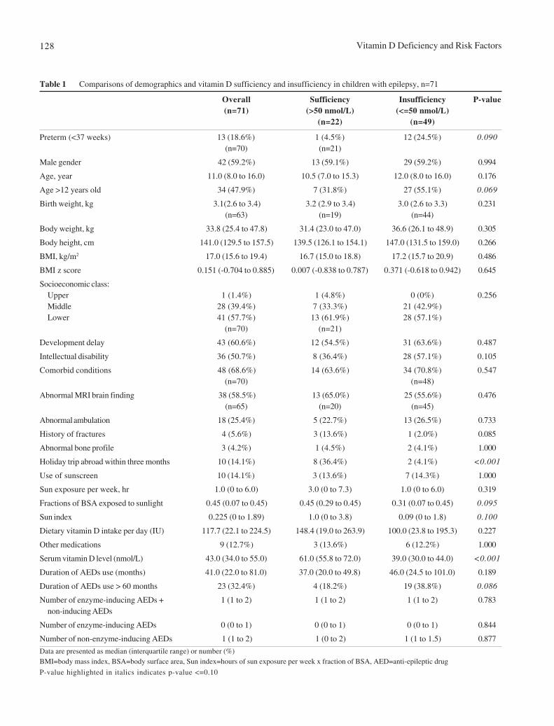
Table 1 Comparisons of demographics and vitamin D sufficiency and insufficiency in children with epilepsy, n=71
Overall Sufficiency Insufficiency P-value(n=71) (>50 nmol/L) (<=50 nmol/L)
(n=22) (n=49)
Preterm (<37 weeks) 13 (18.6%) 1 (4.5%) 12 (24.5%) 0.090(n=70) (n=21)
Male gender 42 (59.2%) 13 (59.1%) 29 (59.2%) 0.994
Age, year 11.0 (8.0 to 16.0) 10.5 (7.0 to 15.3) 12.0 (8.0 to 16.0) 0.176
Age >12 years old 34 (47.9%) 7 (31.8%) 27 (55.1%) 0.069
Birth weight, kg 3.1(2.6 to 3.4) 3.2 (2.9 to 3.4) 3.0 (2.6 to 3.3) 0.231(n=63) (n=19) (n=44)
Body weight, kg 33.8 (25.4 to 47.8) 31.4 (23.0 to 47.0) 36.6 (26.1 to 48.9) 0.305
Body height, cm 141.0 (129.5 to 157.5) 139.5 (126.1 to 154.1) 147.0 (131.5 to 159.0) 0.266
BMI, kg/m2 17.0 (15.6 to 19.4) 16.7 (15.0 to 18.8) 17.2 (15.7 to 20.9) 0.486
BMI z score 0.151 (-0.704 to 0.885) 0.007 (-0.838 to 0.787) 0.371 (-0.618 to 0.942) 0.645
Socioeconomic class:Upper 1 (1.4%) 1 (4.8%) 0 (0%) 0.256Middle 28 (39.4%) 7 (33.3%) 21 (42.9%)Lower 41 (57.7%) 13 (61.9%) 28 (57.1%)
(n=70) (n=21)
Development delay 43 (60.6%) 12 (54.5%) 31 (63.6%) 0.487
Intellectual disability 36 (50.7%) 8 (36.4%) 28 (57.1%) 0.105
Comorbid conditions 48 (68.6%) 14 (63.6%) 34 (70.8%) 0.547(n=70) (n=48)
Abnormal MRI brain finding 38 (58.5%) 13 (65.0%) 25 (55.6%) 0.476(n=65) (n=20) (n=45)
Abnormal ambulation 18 (25.4%) 5 (22.7%) 13 (26.5%) 0.733
History of fractures 4 (5.6%) 3 (13.6%) 1 (2.0%) 0.085
Abnormal bone profile 3 (4.2%) 1 (4.5%) 2 (4.1%) 1.000
Holiday trip abroad within three months 10 (14.1%) 8 (36.4%) 2 (4.1%) <0.001
Use of sunscreen 10 (14.1%) 3 (13.6%) 7 (14.3%) 1.000
Sun exposure per week, hr 1.0 (0 to 6.0) 3.0 (0 to 7.3) 1.0 (0 to 6.0) 0.319
Fractions of BSA exposed to sunlight 0.45 (0.07 to 0.45) 0.45 (0.29 to 0.45) 0.31 (0.07 to 0.45) 0.095
Sun index 0.225 (0 to 1.89) 1.0 (0 to 3.8) 0.09 (0 to 1.8) 0.100
Dietary vitamin D intake per day (IU) 117.7 (22.1 to 224.5) 148.4 (19.0 to 263.9) 100.0 (23.8 to 195.3) 0.227
Other medications 9 (12.7%) 3 (13.6%) 6 (12.2%) 1.000
Serum vitamin D level (nmol/L) 43.0 (34.0 to 55.0) 61.0 (55.8 to 72.0) 39.0 (30.0 to 44.0) <0.001
Duration of AEDs use (months) 41.0 (22.0 to 81.0) 37.0 (20.0 to 49.8) 46.0 (24.5 to 101.0) 0.189
Duration of AEDs use > 60 months 23 (32.4%) 4 (18.2%) 19 (38.8%) 0.086
Number of enzyme-inducing AEDs + 1 (1 to 2) 1 (1 to 2) 1 (1 to 2) 0.783non-inducing AEDs
Number of enzyme-inducing AEDs 0 (0 to 1) 0 (0 to 1) 0 (0 to 1) 0.844
Number of non-enzyme-inducing AEDs 1 (1 to 2) 1 (0 to 2) 1 (1 to 1.5) 0.877
Data are presented as median (interquartile range) or number (%)BMI=body mass index, BSA=body surface area, Sun index=hours of sun exposure per week x fraction of BSA, AED=anti-epileptic drug
P-value highlighted in italics indicates p-value <=0.10
Mo et al 129
Forty-three of them had developmental delay (60.6%) and36 of them (50.7%) had intellectual disability. Besidesepilepsy, 48 subjects (68.6%) had other comorbid diseases.Nine subjects were also taking other medications on topof AEDs (12.7%), including baclofen, risperidone, folicacid and iron sulphate. Three had abnormal bone profile(4.2%) with mildly raised ALP level.
The prevalence of vitamin D deficiency andinsufficiency were 16.9% (12/71) and 52.1% (37/71)respectively, adding up to a prevalence rate of 69.0% (49/71) in total. The median 25-OHD level was 43 nmol/L(IQR 10 to 96).
In univariate analysis, potential risk factors that wereassociated with vitamin D insufficiency include: preterm(<37 weeks), age >12 years old, fraction of BSA exposedto sunlight, sun index, lack of recent holiday trip abroadwithin 3 months, history of fractures and use of AED formore than 60 months (Table 1). Amongst these factors,multivariate logistic regression analysis identified that lackof recent holiday trip abroad within 3 months wasindependently associated with risk of vitamin Dinsufficiency, with odds ratio 0.1 (95% Cl 0.02 to 0.73)(Table 2).
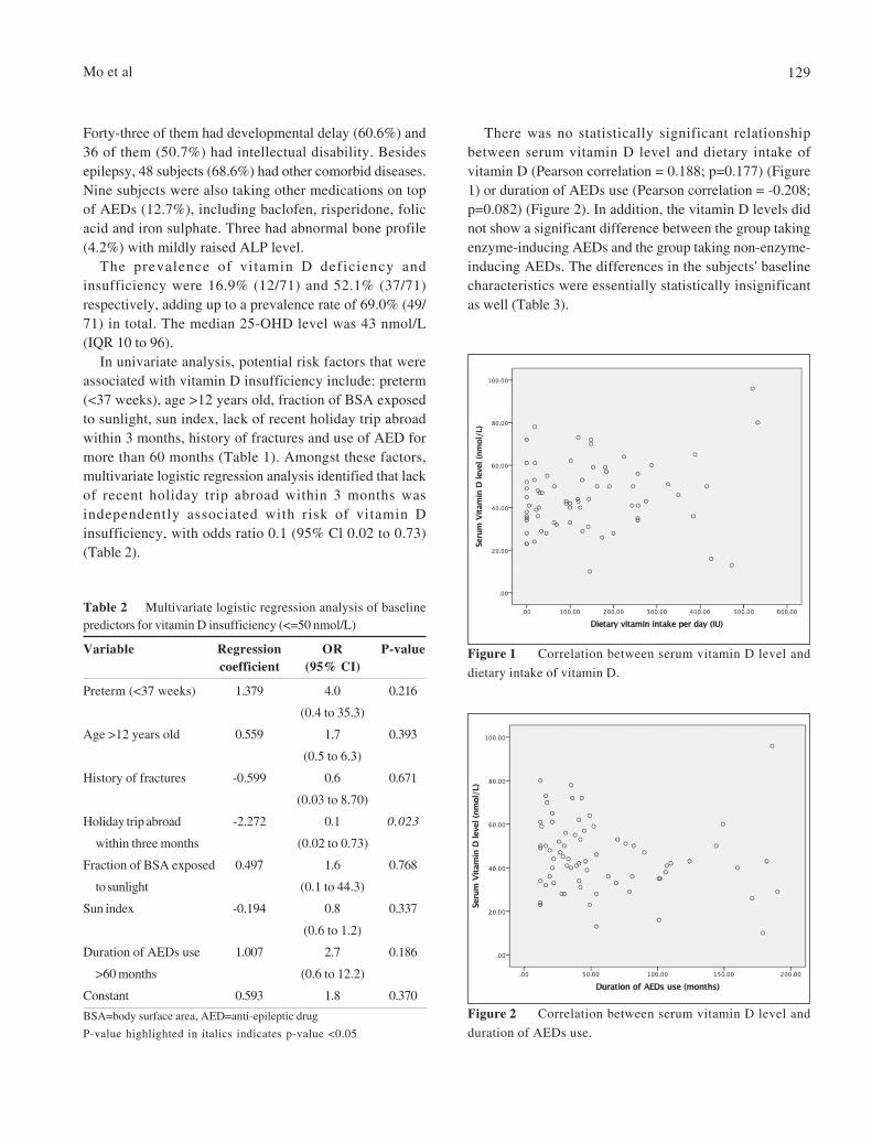
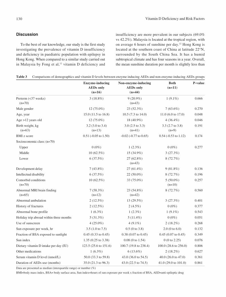
There was no statistically significant relationshipbetween serum vitamin D level and dietary intake ofvitamin D (Pearson correlation = 0.188; p=0.177) (Figure1) or duration of AEDs use (Pearson correlation = -0.208;p=0.082) (Figure 2). In addition, the vitamin D levels didnot show a significant difference between the group takingenzyme-inducing AEDs and the group taking non-enzyme-inducing AEDs. The differences in the subjects' baselinecharacteristics were essentially statistically insignificantas well (Table 3).
Table 2 Multivariate logistic regression analysis of baselinepredictors for vitamin D insufficiency (<=50 nmol/L)
Variable Regression OR P-valuecoefficient (95% CI)
Preterm (<37 weeks) 1.379 4.0 0.216
(0.4 to 35.3)
Age >12 years old 0.559 1.7 0.393
(0.5 to 6.3)
History of fractures -0.599 0.6 0.671
(0.03 to 8.70)
Holiday trip abroad -2.272 0.1 0.023
within three months (0.02 to 0.73)
Fraction of BSA exposed 0.497 1.6 0.768
to sunlight (0.1 to 44.3)
Sun index -0.194 0.8 0.337
(0.6 to 1.2)
Duration of AEDs use 1.007 2.7 0.186
>60 months (0.6 to 12.2)
Constant 0.593 1.8 0.370
BSA=body surface area, AED=anti-epileptic drug
P-value highlighted in italics indicates p-value <0.05
Figure 1 Correlation between serum vitamin D level and
dietary intake of vitamin D.
Figure 2 Correlation between serum vitamin D level and
duration of AEDs use.
Vitamin D Deficiency and Risk Factors130
Discussion
To the best of our knowledge, our study is the first studyinvestigating the prevalence of vitamin D insufficiencyand deficiency in paediatric population with epilepsy inHong Kong. When compared to a similar study carried outin Malaysia by Fong et al,12 vitamin D deficiency and
Table 3 Comparisons of demographics and vitamin D levels between enzyme inducing AEDs and non-enzyme-inducing AEDs groups
Enzyme-inducing Non-enzyme-inducing Both P-valueAEDs only AEDs only (n=11)
(n=16) (n=44)
Preterm (<37 weeks) 3 (18.8%) 9 (20.9%) 1 (9.1%) 0.666(n=70) (n=43)
Male gender 12 (75.0%) 23 (52.3%) 7 (63.6%) 0.270
Age, year 15.0 (11.5 to 16.8) 10.5 (7.3 to 14.0) 11.0 (6.0 to 17.0) 0.048
Age >12 years old 12 (75.0%) 18 (40.9%) 4 (36.4%) 0.046
Birth weight, kg 3.2 (3.0 to 3.4) 3.0 (2.5 to 3.3) 3.3 (2.7 to 3.8) 0.191 (n=63) (n=13) (n=41) (n=9)
BMI z score 0.51 (-0.05 to 1.50) -0.02 (-0.77 to 0.65) 0.54 (-0.53 to 1.12) 0.174
Socioeconomic class: (n=70)
Upper 0 (0%) 1 (2.3%) 0 (0%) 0.277
Middle 10 (62.5%) 15 (34.9%) 3 (27.3%)
Lower 6 (37.5%) 27 (62.8%) 8 (72.7%)(n=43)
Development delay 7 (43.8%) 27 (61.4%) 9 (81.8%) 0.136
Intellectual disability 6 (37.5%) 22 (50.0%) 8 (72.7%) 0.196
Comorbid conditions 10 (62.5%) 33 (75.0%) 5 (50.0%) 0.257(n=70) (n=10)
Abnormal MRI brain finding 7 (58.3%) 23 (54.8%) 8 (72.7%) 0.560(n=65) (n=12) (n=42)
Abnormal ambulation 2 (12.5%) 13 (29.5%) 3 (27.3%) 0.401
History of fractures 2 (12.5%) 2 (4.5%) 0 (0%) 0.377
Abnormal bone profile 1 (6.3%) 1 (2.3%) 1 (9.1%) 0.543
Holiday trip abroad within three months 5 (31.3%) 5 (11.4%) 0 (0%) 0.051
Use of sunscreen 4 (25.0%) 4 (9.1%) 2 (18.2%) 0.268
Sun exposure per week, hr 3.5 (1.0 to 7.5) 0.5 (0 to 3.8) 2.0 (0 to 6.0) 0.132
Fraction of BSA exposed to sunlight 0.45 (0.33 to 0.45) 0.38 (0.07 to 0.45) 0.45 (0.07 to 0.45) 0.349
Sun index 1.35 (0.25 to 3.38) 0.08 (0 to 1.54) 0 (0 to 2.25) 0.076
Dietary vitamin D intake per day (IU) 123.5 (25.6 to 151.6) 100.7 (19.8 to 238.4) 100.0 (28.6 to 256.0) 0.806
Other medications 1 (6.3%) 6 (13.6%) 2 (18.2%) 0.627
Serum vitamin D level (nmol/L) 50.0 (33.3 to 59.8) 43.0 (36.0 to 54.5) 40.0 (26.0 to 47.0) 0.361
Duration of AEDs use (months) 35.0 (21.3 to 96.3) 43.0 (22.5 to 74.5) 41.0 (29.0 to 101.0) 0.861
Data are presented as median (interquartile range) or number (%)
BMI=body mass index, BSA= body surface area, Sun index=hours of sun exposure per week x fraction of BSA, AED=anti-epileptic drug
insufficiency are more prevalent in our subjects (69.0%vs 42.2%). Malaysia is located at the tropical region, withon average 6 hours of sunshine per day.12 Hong Kong islocated at the southern coast of China at latitude 22°N,surrounded by the South China Sea. It has a humidsubtropical climate and has four seasons in a year. Overall,the mean sunshine duration per month is slightly less than
Mo et al 131
explore the exact regions these subjects visited, or theamount of time they spent under the sun each day duringtheir trips. We postulate that one may have longer time ofoutdoor activities during holiday trips, which wouldincrease their chances of sunlight exposure and in turnvitamin D synthesis. More studies are needed to exploreinto this factor of holiday trip abroad and its associationwith a person's vitamin D levels. It is worth noting that oursubjects travelled abroad up to 3 months prior to our studyperiod. This may imply that after intense exposure tosunlight (during their trips in this study), one's vitamin Dlevel could be maintained at a sufficient range for a certainperiod of time. Indeed, Kift et al found that adequate solarultraviolent radiation exposure during the year couldprevent 83% of healthy white-skinned adolescents fromvitamin D deficiency during winter.24 Nonetheless, beingof different ethnic origin and skin pigmentation, furtherlongitudinal studies are needed in this locality toinvestigate how long it lasts to maintain a person'sadequate serum vitamin D level after a period of intensesunlight exposure, and whether it can become a protectivefactor for vitamin D deficiency and insufficiency.
Our study did not recruit healthy paediatric subjects ascontrol and compare their vitamin D levels to our patients.Cheung et al looked at the prevalence of vitamin Dinsufficiency in healthy adolescents aged 12-16 years oldin Hong Kong between 2009 and 2014.13 They found out
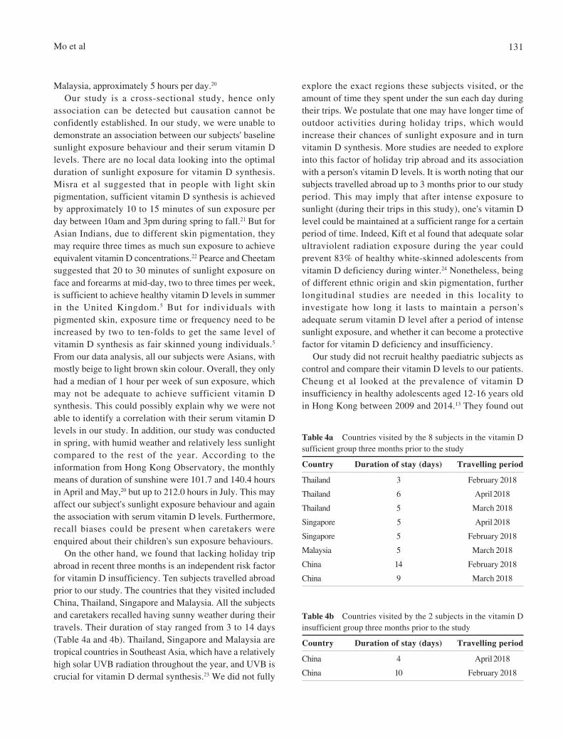
Table 4a Countries visited by the 8 subjects in the vitamin Dsufficient group three months prior to the study
Country Duration of stay (days) Travelling period
Thailand 3 February 2018
Thailand 6 April 2018
Thailand 5 March 2018
Singapore 5 April 2018
Singapore 5 February 2018
Malaysia 5 March 2018
China 14 February 2018
China 9 March 2018
Malaysia, approximately 5 hours per day.20
Our study is a cross-sectional study, hence onlyassociation can be detected but causation cannot beconfidently established. In our study, we were unable todemonstrate an association between our subjects' baselinesunlight exposure behaviour and their serum vitamin Dlevels. There are no local data looking into the optimalduration of sunlight exposure for vitamin D synthesis.Misra et al suggested that in people with light skinpigmentation, sufficient vitamin D synthesis is achievedby approximately 10 to 15 minutes of sun exposure perday between 10am and 3pm during spring to fall.21 But forAsian Indians, due to different skin pigmentation, theymay require three times as much sun exposure to achieveequivalent vitamin D concentrations.22 Pearce and Cheetamsuggested that 20 to 30 minutes of sunlight exposure onface and forearms at mid-day, two to three times per week,is sufficient to achieve healthy vitamin D levels in summerin the United Kingdom.5 But for individuals withpigmented skin, exposure time or frequency need to beincreased by two to ten-folds to get the same level ofvitamin D synthesis as fair skinned young individuals.5
From our data analysis, all our subjects were Asians, withmostly beige to light brown skin colour. Overall, they onlyhad a median of 1 hour per week of sun exposure, whichmay not be adequate to achieve sufficient vitamin Dsynthesis. This could possibly explain why we were notable to identify a correlation with their serum vitamin Dlevels in our study. In addition, our study was conductedin spring, with humid weather and relatively less sunlightcompared to the rest of the year. According to theinformation from Hong Kong Observatory, the monthlymeans of duration of sunshine were 101.7 and 140.4 hoursin April and May,20 but up to 212.0 hours in July. This mayaffect our subject's sunlight exposure behaviour and againthe association with serum vitamin D levels. Furthermore,recall biases could be present when caretakers wereenquired about their children's sun exposure behaviours.
On the other hand, we found that lacking holiday tripabroad in recent three months is an independent risk factorfor vitamin D insufficiency. Ten subjects travelled abroadprior to our study. The countries that they visited includedChina, Thailand, Singapore and Malaysia. All the subjectsand caretakers recalled having sunny weather during theirtravels. Their duration of stay ranged from 3 to 14 days(Table 4a and 4b). Thailand, Singapore and Malaysia aretropical countries in Southeast Asia, which have a relativelyhigh solar UVB radiation throughout the year, and UVB iscrucial for vitamin D dermal synthesis.23 We did not fully
Table 4b Countries visited by the 2 subjects in the vitamin Dinsufficient group three months prior to the study
Country Duration of stay (days) Travelling period
China 4 April 2018
China 10 February 2018
Vitamin D Deficiency and Risk Factors132
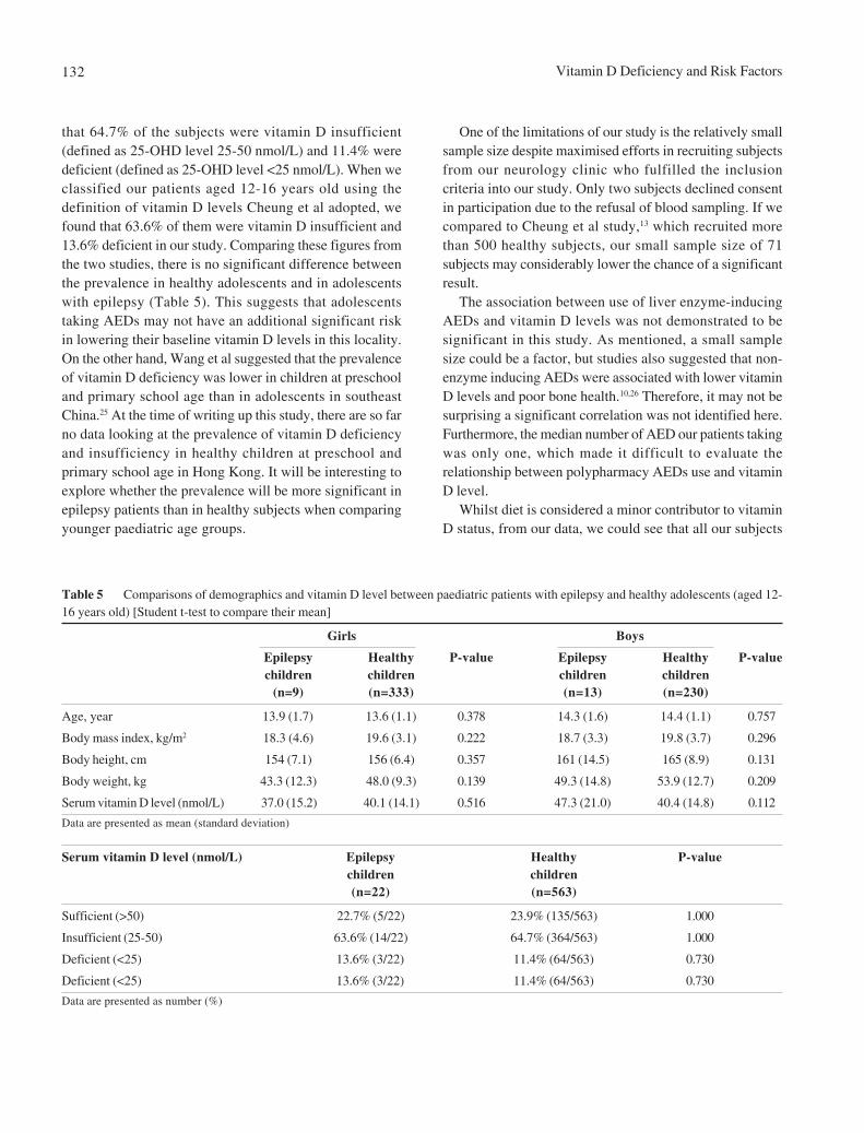
that 64.7% of the subjects were vitamin D insufficient(defined as 25-OHD level 25-50 nmol/L) and 11.4% weredeficient (defined as 25-OHD level <25 nmol/L). When weclassified our patients aged 12-16 years old using thedefinition of vitamin D levels Cheung et al adopted, wefound that 63.6% of them were vitamin D insufficient and13.6% deficient in our study. Comparing these figures fromthe two studies, there is no significant difference betweenthe prevalence in healthy adolescents and in adolescentswith epilepsy (Table 5). This suggests that adolescentstaking AEDs may not have an additional significant riskin lowering their baseline vitamin D levels in this locality.On the other hand, Wang et al suggested that the prevalenceof vitamin D deficiency was lower in children at preschooland primary school age than in adolescents in southeastChina.25 At the time of writing up this study, there are so farno data looking at the prevalence of vitamin D deficiencyand insufficiency in healthy children at preschool andprimary school age in Hong Kong. It will be interesting toexplore whether the prevalence will be more significant inepilepsy patients than in healthy subjects when comparingyounger paediatric age groups.
One of the limitations of our study is the relatively smallsample size despite maximised efforts in recruiting subjectsfrom our neurology clinic who fulfilled the inclusioncriteria into our study. Only two subjects declined consentin participation due to the refusal of blood sampling. If wecompared to Cheung et al study,13 which recruited morethan 500 healthy subjects, our small sample size of 71subjects may considerably lower the chance of a significantresult.
The association between use of liver enzyme-inducingAEDs and vitamin D levels was not demonstrated to besignificant in this study. As mentioned, a small samplesize could be a factor, but studies also suggested that non-enzyme inducing AEDs were associated with lower vitaminD levels and poor bone health.10,26 Therefore, it may not besurprising a significant correlation was not identified here.Furthermore, the median number of AED our patients takingwas only one, which made it difficult to evaluate therelationship between polypharmacy AEDs use and vitaminD level.
Whilst diet is considered a minor contributor to vitaminD status, from our data, we could see that all our subjects
Table 5 Comparisons of demographics and vitamin D level between paediatric patients with epilepsy and healthy adolescents (aged 12-16 years old) [Student t-test to compare their mean]
Girls Boys
Epilepsy Healthy P-value Epilepsy Healthy P-valuechildren children children children
(n=9) (n=333) (n=13) (n=230)
Age, year 13.9 (1.7) 13.6 (1.1) 0.378 14.3 (1.6) 14.4 (1.1) 0.757
Body mass index, kg/m2 18.3 (4.6) 19.6 (3.1) 0.222 18.7 (3.3) 19.8 (3.7) 0.296
Body height, cm 154 (7.1) 156 (6.4) 0.357 161 (14.5) 165 (8.9) 0.131
Body weight, kg 43.3 (12.3) 48.0 (9.3) 0.139 49.3 (14.8) 53.9 (12.7) 0.209
Serum vitamin D level (nmol/L) 37.0 (15.2) 40.1 (14.1) 0.516 47.3 (21.0) 40.4 (14.8) 0.112
Data are presented as mean (standard deviation)
Serum vitamin D level (nmol/L) Epilepsy Healthy P-valuechildren children(n=22) (n=563)
Sufficient (>50) 22.7% (5/22) 23.9% (135/563) 1.000
Insufficient (25-50) 63.6% (14/22) 64.7% (364/563) 1.000
Deficient (<25) 13.6% (3/22) 11.4% (64/563) 0.730
Deficient (<25) 13.6% (3/22) 11.4% (64/563) 0.730
Data are presented as number (%)
Mo et al 133
had inadequate dietary vitamin D intake, based on therecommended daily intake suggested by Ross et al.6 Thismay explain the reason why we were not able to demonstratea significant result on the correlation between dietaryintake and vitamin D levels. Moreover, it may be of publichealth concern, in which education on sufficient dietaryvitamin D intake should be reinforced to the wholepaediatric population.
In our study, we did not perform dual-energy X-rayabsorptiometry (DXA) scan on our patients to determinetheir bone mineral density (BMD) and correlate with serumvitamin D levels. Nevertheless, prior study has alreadydemonstrated the association between low vitamin D leveland low BMD in paediatric patients.27
To improve the study, larger sample size would certainlyincrease the precision of estimating the prevalence anddelineating any additional potential risk factors for vitaminD deficiency. This could be achieved through collaborationamong different paediatric centres in Hong Kong. Inaddition, repeating blood tests at different time points ofthe year could help to look at seasonal variation and assessany lasting effect of sunlight exposure during the year onsubsequent vitamin D levels. Furthermore, measuringsunlight exposure accurately would also be an importantarea to increase precision. This can be done via wearingUV sensitive badges by subjects to collect reliable UVBexposure, which eliminates recall bias when enquiringcaretakers for subjects' sunlight exposure behaviours.
Conclusion
Our study demonstrated that vitamin D insufficiencyand deficiency are highly prevalent in paediatric patientswith epilepsy on AEDs. However, the figures arecomparable to that of the healthy adolescents in HongKong. Therefore, we would not advocate for routinescreening for vitamin D levels in adolescent patients withepilepsy in this locality, which would not be cost-effective.Given the potential positive correlation between serumvitamin D levels and recent holiday trip abroad, furtherstudies could be considered to investigate their relationshipand implication.
Disclosure
None of the authors of this study has any conflict ofinterests to disclose.
Acknowledgement
This work has been supported by the Clinical ResearchCentre, Kwong Wah Hospital.
Financial Support and Sponsorship
This work was supported by the Tung Wah Groups ofHospitals Research Fund 2017/2018.
References
1. Mansbach JM, Ginde AA, Camargo CA. Serum 25-HydroxyvitaminD levels among US children aged 1 to 11 years: Do children needmore vitamin D? Pediatrics 2009;124:1404-10.
2. Gordon CM, Feldman HA, Sinclair L, et al. Prevalence of vitaminD deficiency among healthy infants and toddlers. Arch PediatrAdolesc Med 2008;162:505-12.
3. Poh BK, Rojroongwasinkul N, Le Nguyen BK, et al. 25-hydroxy-vitamin D demography and the risk of vitamin D insufficiency inthe South East Asian Nutrition Surveys (SEANUTS). Asia Pac JClin Nutr 2016;25:538-48.
4. Khor GL, Chee WSS, Shariff ZM, et al. High prevalence of vitaminD insufficiency and its association with BMI-for-age among primaryschool children in Kuala Lumpur, Malaysia. BMC Public Health2011;11:95.
5. Pearce SHS, Cheetham TD. Diagnosis and management of vitaminD deficiency. BMJ 2010;340:b5564.
6. Institute of Medicine; Food and Nutrition Board; Committee toReview Dietary Reference Intakes for Vitamin D and Calcium;Ross AC, Taylor CL, Yaktine AL, Del Valle HB, editors. DietaryReference Intakes for Calcium and Vitamin D. Washington (DC):National Academies Press (US); 2011.
7. Munns CF, Shaw N, Kiely M, et al. Global ConsensusRecommendations on Prevention and Management of NutritionalRickets. J Clin Endocrinol Metab 2016;101:394-415.
8. Winzenberg T, Powell S, Shaw KA, Jones G. Effects of vitaminD supplementation on bone density in healthy children: systematicreview and meta-analysis. BMJ 2011;342:c7254.
9. Samaniego EA , Sheth RD. Bone consequences of epilepsy andantiepileptic medications. Semin Pediatr Neurol 2007;14:196-200.
10. Shellhaas RA, Joshi SM. Vitamin D and bone health among childrenwith epilepsy. Pediatr Neurol 2010;42:385-93.
11. Fong CY, Riney CJ. Vitamin D deficiency among children withepilepsy in South Queensland. J Child Neurol 2014;29:368-73.
12. Fong CY, Kong AN, Poh BK, et al. Vitamin D deficiency and itsrisk factors in Malaysian children with epilepsy. Epilepsia 2016;57:1271-9.
13. Chan KCC, Tam WH, Chan MH, Chan RS, Li AM. Vitamin Ddeficiency among healthy infants in Hong Kong: a pilot study.Hong Kong Med J 2018;24(Suppl 3):S32-5.
14. Cheung TF, Cheuk KY, Yu FWP, et al. Prevalence of vitamin Dinsufficiency among adolescents and its correlation with boneparameters using high-resolution peripheral quantitative computedtomography. Osteoporos Int 2016;27:2477-88.
Vitamin D Deficiency and Risk Factors134
15. Gururaj, Maheshwaran. Kuppuswamy's Socio-Economic StatusScale – A Revision of Income Parameter For 2014. InternationalJournal of Recent Trends in Science and Technology 2014;11:1-2.
16. Nucci AM, Russell CS, Luo R, et al. The effectiveness of a shortfood frequency questionaire in determining vitamin D intake inchildren. Dermatoendocrinol 2013;5:205-10.
17. Barger-Lux MJ, Heaney RP. Effects of above average summersun exposure on serum 25-hydroxyvitamin D and calciumabsorption. J Clin Endocrinol Metab 2002;87:4952-6.
18. Cole TJ. The LMS method for constructing normalized growthstandards. Eur J Clin Nutr 1990;44:45-60.
19. Leung SS, Cole TJ, Tse LY, Lau JT. Body mass index referencecurves for Chinese children. Ann Hum Biol 1998;25:169-74.
20. Climatological Information Services. Hong Kong Observatory.The Government of the Hong Kong Special AdministrativeRegion.
21. Misra M, Pacaud D, Petryk A, et al. Vitamin D deficiency in childrenand its management: review of current knowledge andrecommendations. Pediatrics 2008;122:398-417.
22. Hollis BW. Circulating 25-hydroxyvitamin D levels indicative ofvitamin D sufficiency: implications for establishing a new effectivedietary intake recommendation for vitamin D. J Nutr 2005;135:317-22.
23. Mendes MM, Hart KH, Botelho PB, Lanham-New SA. Vitamin Dstatus in the tropics: Is sunlight exposure the main determinant?Nutrition Bulletin 2018;43:428-34.
24. Kift R, Rhodes LE, Farrar MD, Webb AR. Is sunlight exposureenough to avoid wintertime vitamin D deficiency in United Kingdompopulation groups? Int J Environ Res Public Health 2018;15:1624.
25. Wang LL, Wang HY, Wen HK, Tao HQ, Zhao XW. Vitamin statusamong infants, children, and adolescents in southeastern China. JZhejiang Univ Sci B 2016;17:545-52.
26. Durá-Travé T, Gallinas-Victoriano F, Malumbres-Chacón M,Moreno-Gónzalez P, Aguilera-Albesa S, Yoldi-Petri ME. VitaminD deficiency in children with epilepsy taking valproate andlevetiracetam as monotherapy. Epilepsy Res 2018;139:80-4.
27. Bischoff-Ferrari HA. Vitamin D and fracture prevention. EndocrinolMetab Clin North Am 2010;39:347-53.
HK J Paediatr (new series) 2021;26:135-140
Attention Deficit Hyperactivity Disorder in PaediatricPatients with Malignant Haematologic Diseases or Epilepsy:
Experience at a Tertiary Care Hospital in Korea
JY HAN, JW LEE, B CHO, NG CHUNG, IG LEE
Abstract Background: Paediatric patients with epilepsy and malignant haematologic diseases (MHD) are atincreased risk of mental health problems compared to the general population. The purpose of this studywas to determine the prevalence of attention-deficit hyperactivity disorder (ADHD) among paediatricpatients treated for MHD or epilepsy compared to that in healthy children in South Korea. Methods:We retrospectively reviewed 184 patients diagnosed with epilepsy and 172 patients diagnosed withmalignant haematologic diseases in the paediatric department of Seoul St. Mary's Hospital from May2009 to May 2013. Normal controls were selected from the out-patient clinic among those who visitedthe clinic for vaccination. Results: Paediatric patients with epilepsy or MHD exhibited significantlyhigher rate of ADHD compared to the controls (37.5% or 29.6% vs. 11.9%, P=0.039). Among childrenwith both MHD and ADHD, 69.5% had the inattentive subtype and 30.5% had the combined subtype ofADHD. Among children with both epilepsy and ADHD, 70.6% had the inattentive subtype, 23.5% had thecombined subtype, and 5.9% had the hyperactive type of ADHD. There were statistically significantdifferences between MHD patients with ADHD and MHD patients without ADHD of sex, age at onset ofhaematologic diseases (≤5 years), intrathecal chemotherapy, treatment duration and cranial radiation.Patients with epilepsy and concomitant ADHD showed significantly poorer response to epilepsy treatmentthan patients without ADHD. Conclusions: Paediatric patients with MHD and epilepsy are at significantrisk for ADHD. Baseline testing of all patients with MHD or epilepsy is needed to assess theirneuropsychological and academic skills over time to facilitate early intervention and prevent academicfailure.
Key words Attention deficit hyperactivity disorder; Epilepsy; Malignant haematologic diseases
Department of Pediatrics, College of Medicine, The CatholicUniversity of Korea, 222 Banpodaero, Seocho-gu, Seoul06591, Republic of Korea
JY HAN MD, PhDJW LEE MD, PhDB CHO MD, PhDNG CHUNG MD, PhDIG LEE MD, PhD
Correspondence to: Dr IG LEE
Email: [email protected]
Received January 23, 2019
Original Article
Introduction
With the introduction of multimodal therapy in the early1970s, 5-year survival rates for most paediatric malignanthaematologic diseases have improved dramatically. Overthe past four decades, improved prognosis of childhoodacute lymphoblastic leukaemia (ALL) and acute myeloidleukaemia (AML) has resulted in 5-year survivor rate ofaround 80% in developed country.1,2 Although medicaladvances have resulted in increased life expectancy ofmany chronically ill children, a substantial proportion of
ADHD in Children with MHD and Epilepsy136
these children experience long-term neurocognitivesequelae associated with treatment- and disease-relatedfactors.
Children surviving cancer frequently experiencedeficits in attention, learning, and memory, that aresecondary to the disease process itself and requisitetreatment that may include surgery, chemotherapy and/orcranial radiation therapy.3 Attention problems occurfrequently among childhood cancer survivors. It has beenestimated that approximately one-quarter of ALL survivorsdemonstrate significant dysfunction.4
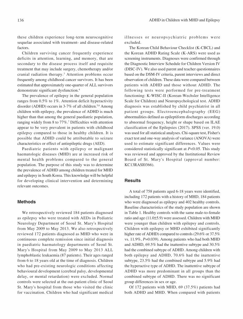
The prevalence of epilepsy in the general populationranges from 0.5% to 1%. Attention deficit hyperactivitydisorder (ADHD) occurs in 3-7% of all children.5,6 Amongchildren with epilepsy, the prevalence of ADHD is muchhigher than that among the general paediatric population,ranging widely from 8 to 77%.7 Difficulties with attentionappear to be very prevalent in patients with childhoodepilepsy compared to those in healthy children. It ispossible that ADHD could be attributable to seizurecharacteristics or effect of antiepileptic drugs (AED).
Paediatric patients with epilepsy or malignanthaematologic diseases (MHD) are at increased risk ofmental health problems compared to the generalpopulation. The purpose of this study was to determinethe prevalence of ADHD among children treated for MHDand epilepsy in South Korea. This knowledge will be helpfulfor developing clinical intervention and determiningrelevant outcomes.
Methods
We retrospectively reviewed 184 patients diagnosedas epilepsy who were treated with AEDs in PediatricNeurology Department of Seoul St. Mary's Hospitalfrom May 2009 to May 2013. We also retrospectivelyreviewed 172 patients diagnosed as MHD who were incontinuous complete remission since initial diagnosisin paediatric haematology departments of Seoul St.Mary's Hospital from May 2009 to May 2013 ALLlymphoblastic leukaemia (87 patients). Their ages rangedfrom 6 to 18 years old at the time of diagnosis. Childrenwho had pre-existing neurologic conditions affectingbehavioural development (cerebral palsy, developmentaldelay, or mental retardation) were excluded. Normalcontrols were selected at the out-patient clinic of SeoulSt. Mary's hospital from those who visited the clinicfor vaccination. Children who had significant medical
i l l ne s se s o r neu ropsych ia t r i c p rob lems wereexcluded.
The Korean Child Behaviour Checklist (K-CBCL) andthe Korean ADHD Rating Scale (K-ARS) were used asscreening instruments. Diagnoses were confirmed throughthe Diagnostic Interview Schedule for Children Version IV(DISC-IV). We also used parent and teacher questionnairesbased on the DSM-IV criteria, parent interviews and directobservation of children. These data were compared betweenpatients with ADHD and those without ADHD. Thefollowing tests were performed for pre-treatmentfunctioning: K-WISC-II (Korean-Wechsler IntelligenceScale for Children) and Neuropsychological test. ADHDdiagnosis was established by child psychiatrist in allpat ient groups. Electroencephalography (EEG)abnormalities defined as epileptiform discharges accordingto abnormal frequency, height or shape based on ILAEclassification of the Epilepsies (2017). SPSS (ver. 19.0)was used for all statistical analyses. Chi-square test, Fisher'sexact test and one-way analysis of variance (ANOVA) wereused to estimate significant differences. Values wereconsidered statistically significant at P<0.05. This studywas reviewed and approved by the Institutional ReviewBoard of St. Mary's Hospital (approval number:KC13RASI0366).
Results
A total of 758 patients aged 6-18 years were identified,including 172 patients with a history of MHD, 184 patientswho were diagnosed as epilepsy and 402 healthy controls.Baseline characteristics of the study population are shownin Table 1. Healthy controls with the same male-to-femaleratio and age (11.0±5.9) were assessed. Children with MHDwere younger than children with epilepsy and controls.Children with epilepsy or MHD exhibited significantlyhigher rate of ADHD) compared to controls (29.6% or 37.5%vs. 11.9%, P=0.039). Among patients who had both MHDand ADHD, 69.5% had the inattentive subtype and 30.5%had the combined subtype of ADHD. Among children withboth epilepsy and ADHD, 70.6% had the inattentivesubtype, 23.5% had the combined subtype and 5.9% hadthe hyperactive type of ADHD. The inattentive subtype ofADHD was more predominant in all groups than thecombined subtype of ADHD. There was no significantgroup differences in sex or age.
Of 172 patients with MHD, 69 (37.5%) patients hadboth ADHD and MHD. When compared with patients
Han et al 137
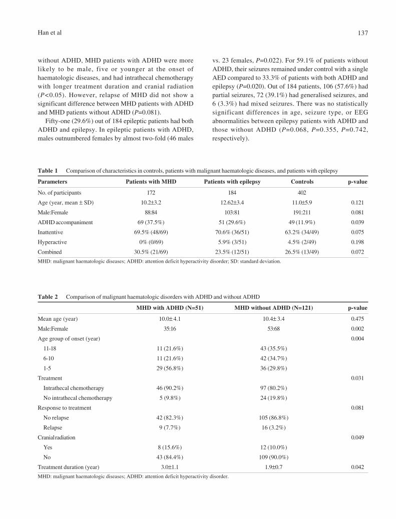
vs. 23 females, P=0.022). For 59.1% of patients withoutADHD, their seizures remained under control with a singleAED compared to 33.3% of patients with both ADHD andepilepsy (P=0.020). Out of 184 patients, 106 (57.6%) hadpartial seizures, 72 (39.1%) had generalised seizures, and6 (3.3%) had mixed seizures. There was no statisticallysignificant differences in age, seizure type, or EEGabnormalities between epilepsy patients with ADHD andthose without ADHD (P=0.068, P=0.355, P=0.742,respectively).
without ADHD, MHD patients with ADHD were morelikely to be male, five or younger at the onset ofhaematologic diseases, and had intrathecal chemotherapywith longer treatment duration and cranial radiation(P<0.05). However, relapse of MHD did not show asignificant difference between MHD patients with ADHDand MHD patients without ADHD (P=0.081).
Fifty-one (29.6%) out of 184 epileptic patients had bothADHD and epilepsy. In epileptic patients with ADHD,males outnumbered females by almost two-fold (46 males
Table 1 Comparison of characteristics in controls, patients with malignant haematologic diseases, and patients with epilepsy
Parameters Patients with MHD Patients with epilepsy Controls p-value
No. of participants 172 184 402
Age (year, mean ± SD) 10.2±3.2 12.62±3.4 11.0±5.9 0.121
Male:Female 88:84 103:81 191:211 0.081
ADHD accompaniment 69 (37.5%) 51 (29.6%) 49 (11.9%) 0.039
Inattentive 69.5% (48/69) 70.6% (36/51) 63.2% (34/49) 0.075
Hyperactive 0% (0/69) 5.9% (3/51) 4.5% (2/49) 0.198
Combined 30.5% (21/69) 23.5% (12/51) 26.5% (13/49) 0.072
MHD: malignant haematologic diseases; ADHD: attention deficit hyperactivity disorder; SD: standard deviation.
Table 2 Comparison of malignant haematologic disorders with ADHD and without ADHD
MHD with ADHD (N=51) MHD without ADHD (N=121) p-value
Mean age (year) 10.0± 4.1 10.4± 3.4 0.475
Male:Female 35:16 53:68 0.002
Age group of onset (year) 0.004
11-18 11 (21.6%) 43 (35.5%)
6-10 11 (21.6%) 42 (34.7%)
1-5 29 (56.8%) 36 (29.8%)
Treatment 0.031
Intrathecal chemotherapy 46 (90.2%) 97 (80.2%)
No intrathecal chemotherapy 5 (9.8%) 24 (19.8%)
Response to treatment 0.081
No relapse 42 (82.3%) 105 (86.8%)
Relapse 9 (7.7%) 16 (3.2%)
Cranial radiation 0.049
Yes 8 (15.6%) 12 (10.0%)
No 43 (84.4%) 109 (90.0%)
Treatment duration (year) 3.0±1.1 1.9±0.7 0.042
MHD: malignant haematologic diseases; ADHD: attention deficit hyperactivity disorder.
ADHD in Children with MHD and Epilepsy138
Discussion
The prevalence of ADHD in Korean studies has beenreported to be 3.9-13.3%.8,9 Estimated ADHD prevalenceduring paediatric period has shown wide variation aroundthe world, ranging from 0.9% to 20%.10,11 In our study,ADHD prevalence was 11.9% in healthy controls. It isgenerally accepted that the combined type of ADHD is themost common type in the general population.7 However,the inattentive type of ADHD was more common in healthy,epileptic and MHD children in our study.
ADHD is a common neuropsychiatric disorder thatimpairs social, academic and vocational functions inchildren and adolescents. By their young adulthood, ADHDyouth are at high risk for a wide range of adverse psychiatricoutcomes, including elevated prevalence of antisocial,addictive, behavioural and anxiety disorders.12
The scope of chronic health condition is broad. It canaffect the neuropsychiatric system. Survivors of paediatricbrain tumour and ALL are at risk for lasting cognitiveimpairment attributable to disease and treatments that canaffect the central nervous system (e.g., cranial radiationtherapy, intrathecal chemotherapy).13,14 Patients diagnosedwith ALL at younger age suffer greater cognitive deficitsthan patients diagnosed at older age, especially when they
are treated with cranial irradiation.15 Neurocognitiveimpairment is estimated to occur in 20-40% of long-termsurvivors of childhood ALL. Neurocognitive deficits cannegatively impact learning and academic achievement ofpatients compromising their educational and vocationalopportunities.16 Despite these well-established findings,there have been few empirically validated interventionsto remediate cognitive impairments emerging secondaryto treatment for MHD.17
Results of our study demonstrate that the rate ofsymptoms of ADHD in survivors of MHD is higher thanthat in healthy controls, suggesting that MHD has asignificant relation with ADHD in paediatric patients. Inaddition, in males under 5 years of age at onset, intrathecalchemotherapy and cranial radiation might be necessary topredict ADHD. Therefore, early detection and establishmentof countermeasure for ADHD are needed to maintain theirquality of life.
Paediatric patients with epilepsy are more likely to havedifficulties with 'attention problems' than healthy controls.It is often assumed to be the consequence of recurrentseizures, medical treatment, or characteristics of epilepsy.18
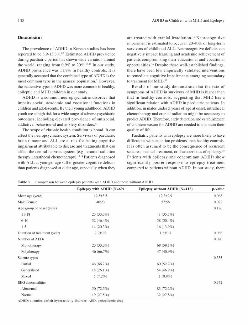
Patients with epilepsy and concomitant ADHD showsignificantly poorer response to epilepsy treatmentcompared to patients without ADHD. In our study, there
Table 3 Comparison between epilepsy patients with ADHD and those without ADHD
Epilepsy with ADHD (N=69) Epilepsy without ADHD (N=115) p-value
Mean age (year) 12.5±3.5 12.3±2.9 0.068
Male:Female 46:23 57:58 0.022
Age group of onset (year) 0.126
11-18 23 (33.3%) 41 (35.7%)
6-10 32 (46.4%) 58 (50.4%)
1-5 14 (20.3%) 16 (13.9%)
Duration of treatment (year) 2.2±0.8 1.8±0.7 0.036
Number of AEDs 0.020
Monotherapy 23 (33.3%) 68 (59.1%)
Polytherapy 46 (66.7%) 47 (40.9%)
Seizure types 0.355
Partial 46 (66.7%) 60 (52.2%)
Generalised 18 (26.1%) 54 (46.9%)
Mixed 5 (7.2%) 1 (0.9%)
EEG abnormalities 0.742
Abnormal 50 (72.5%) 83 (72.2%)
Normal 19 (27.5%) 32 (27.8%)
ADHD: attention deficit hyperactivity disorder; AED, antiepileptic drug.
Han et al 139
was no difference in ADHD accompaniments betweenpartial and generalised seizure types. However, treatmentduration and polytherapy could influence the diagnosisof ADHD. The presence of ADHD in patients with epilepsycould be related to therapeutic response to AEDs and couldbe a useful predictive factor for response in early stage.Another study has found that ADHD is related to illnessand seizure frequency.19 ADHD can increase the risk ofsubsequent epilepsy and vice versa.20 Thus, it is importantto manage ADHD in children with epilepsy. However,ADHD might be under treated in children with epilepsy.21
Stimulants remain to be the first-choice pharmacologicalagents for the management of ADHD. Psychosocial,behavioural and educational strategies that can enhancespecific behaviours may improve educational and socialfunctioning in children with ADHD.22 Evidence from a one-year efficacy trial of methylphenidate (MPH) in paediatriccancer survivors experiencing cognitive late effects hasrecently emerged, suggesting long-term cognitive andbehavioural benefits of stimulant treatment in thispopulation.23 There are also reports of good response toMPH in children with epilepsy.24 Children could beselected for early intervention trials to reduce symptomsof ADHD. The goal of a comprehensive epilepsy clinic/service is to perform an assessment and provide treatmentnot only for seizures, but also for cognitive and behaviouraldifficulties experienced by children with epilepsy.25
Children with MHD and epilepsy are at significant risk forADHD. There is a need for more studies focusing on safeand efficacious intervention for symptoms of ADHD.Baseline testing of all patients with MHD or epilepsy isneeded to assess their neuropsychological and academicskills over time to facilitate early intervention and preventacademic failure. These results highlight the need forcontinued monitoring of ADHD in survivors of paediatricMHD and epilepsy. Future studies shall address these issuesand guide paediatrician, nurses and teachers to worktogether to develop individualised plans that can helpchildren with MHD or epilepsy fulfill their educationalgoals. Long-term social prognosis of these children appearsto be of considerable importance. Future studies are neededto confirm our results in a larger population.
Conclusions
This study shows that paediatric patients with MHD orepilepsy are at significant risk for ADHD. Children withMHD were younger than children with epilepsy and
controls. Children with epilepsy or MHD exhibited 2 to 3folds higher rate of ADHD compared to controls. Whencompared with patients without ADHD, MHD patients withADHD were more likely to be male, five or younger at theonset of haematologic diseases, and had intrathecalchemotherapy with longer treatment duration and cranialradiation. Patients with epilepsy and concomitant ADHDshowed significantly poorer response to epilepsy treatmentthan patients without ADHD. Thus, baseline testing of allMHD or epilepsy children is needed to check theirneuropsychological and academic skills over time tofacilitate early intervention and prevent academic failure.
Declaration of Interest
None.
Acknowledgement
We would like to acknowledge the St. Mary's Hospitaland patients.
References
1. Gatta G, Capocaccia R, Coleman MP, Ries LA, Berrino F.Childhood cancer survival in Europe and the United States. Cancer2002;95:1767-72.
2. Rubnitz JE, Gibson B, Smith FO. Acute myeloid leukemia. HematolOncol Clin North Am 2010;24:35-63.
3. Nicholls E, Hildenbrand AK, Aggarwal R, McCarthy L, Daly B.The use of stimulant medication to treat neurocognitive deficitsin patients with pediatric cancer, traumatic brain injury, andsickle cell disease: a review. Postgrad Med 2012;124:78-90.
4. Krull KR, Brouwers P, Jain N, et al. Folate pathway geneticpolymorphisms are related to attention disorders in childhoodleukemia survivors. J Pediatr 2008;152:101-5.
5. Association AP. Diagnostic criteria from dsM-iV-tr: AmericanPsychiatric Pub; 2000.
6. Sander JW. The epidemiology of epilepsy revisited. Cur OpinNeurol 2003;16:165-70.
7. Dunn DW, Austin JK, Harezlak J, Ambrosius WT. ADHD andepilepsy in childhood. Dev Med Child Neurol 2003;45:50-4.
8. Yang SJ, Cheong S, Hong SD. Prevalence and correlates of attentiondeficit hyperactivity disorder: school-based mental health servicesin Seoul. J Korean Neuropsychiatr Assoc 2006;45:69-76.
9. Yang YH, Kim JW, Kim YN, Cho SC, Kim BN. Screening forattention deficit/hyperactivity disorder for children in Seoul.J Korean Neuropsychiatr Assoc 2008;47:292-8.
10. Goodman R, Dos Santos DN, Nunes AR, de Miranda DP, Fleitlich-Bilyk B, Almeida Filho N. The Ilha de Maré study: a survey ofchild mental health problems in a predominantly African-Brazilian
ADHD in Children with MHD and Epilepsy140
rural community. Soc Psychiatry Psychiatr Epidemiol 2005;40:11-7.
11. Cornejo JW, Osio O, Sanchez Y, et al. [Prevalence of attentiondeficit hyperactivity disorder in Colombian children and teenagers].Rev Neurol. 2004;40:716-22.
12. Biederman J, Monuteaux MC, Mick E, et al. Young adult outcomeof attention deficit hyperactivity disorder: a controlled 10-yearfollow-up study. Psychol Med 2006;36:167-79.
13. Peterson CC, Johnson CE, Ramirez LY, et al. A meta-analysis ofthe neuropsychological sequelae of chemotherapy-only treatmentfor pediatric acute lymphoblastic leukemia. Pediatr Blood Cancer2008;51:99-104.
14. Fossen A, Abrahamsen TG, Storm-Mathisen I. Psychologicaloutcome in children treated for brain tumor. Pediatr Hematol Oncol1998;15:479-88.
15. Buizer AI, de Sonneville LM, van den Heuvel-Eibrink MM,Veerman AJ. Chemotherapy and attentional dysfunction insurvivors of childhood acute lymphoblastic leukemia: effect oftreatment intensity. Pediatr Blood Cancer 2005;45:281-90.
16. Mitby PA, Robison LL, Whitton JA, et al. Utilization of specialeducation services and educational attainment among long-termsurvivors of childhood cancer: a report from the Childhood CancerSurvivor Study. Cancer 2003;97:1115-26.
17. Butler RW, Mulhern RK. Neurocognitive interventions for childrenand adolescents surviving cancer. J Pediatr Psychol 2005;30:65-78.
18. Jones JE, Watson R, Sheth R, et al. Psychiatric comorbidity inchildren with new onset epilepsy. Dev Med Child Neurol 2007;49:493-7.
19. Caplan R, Siddarth P, Stahl L, et al. Childhood absence epilepsy:behavioral, cognitive, and linguistic comorbidities. Epilepsia 2008;49:1838-46.
20. Chou I-C, Chang Y-T, Chin Z-N, et al. Correlation between epilepsyand attention deficit hyperactivity disorder: a population-basedcohort study. PLoS One. 2013;8:e57926.
21. Boyes C. Question 2 Should a child with ADHD and epilepsy begiven Ritalin? Arch Dis Child 2010;95:759-61.
22. Wigal S, Swanson JM, Feifel D, et al. A double-blind, placebo-controlled trial of dexmethylphenidate hydrochloride and d, l-threo-methylphenidate hydrochloride in children with attention-deficit/hyperactivity disorder. J Am Acad Child Adolesc Psychiatry 2004;43:1406-14.
23. Conklin HM, Khan RB, Reddick WE, et al. Acute neurocognitiveresponse to methylphenidate among survivors of childhood cancer:a randomized, double-blind, cross-over trial. J Pediatr Psychol2007;32:1127-39.
24. Kaufmann R, Goldberg-Stern H, Shuper A. Attention-deficitdisorders and epilepsy in childhood: incidence, causative relationsand treatment possibilities. J Child Neurol 2009;24:727-33.
25. Dunn DW, Austin JK, Perkins SM. Prevalence of psychopathologyin childhood epilepsy: categorical and dimensional measures. DevMed Child Neurol 2009;51:364-72.
HK J Paediatr (new series) 2021;26:141-148
Comparison of Alcohol and Phenol Neurolysis in Children withSpasticity: A Pilot Matched Controlled Trial
KY LEUNG, WK CHAN, WK MAN, CH KO
Abstract Purpose: To evaluate the effectiveness of phenol and alcohol neurolysis as treatment of spasticity.Method: This is a non-randomisred matched controlled trial. Non-ambulatory spastic children weregiven same volume of phenol to one nerve and alcohol to the contralateral nerve. Spasticity were assessedby passive range of movement and Modified Ashworth Scale before treatment, and at one, three and sixmonths after treatment. Findings: Five pairs of obturator nerves and one pair of musculocutaneousnerves were recruited. Both phenol and alcohol neurolysis were found to be effective to reduce spasticityat one month, with no significant inter-group difference in passive range of movement gain and ModifiedAshworth Scale reduction. No significant complication was identified. Conclusion: Both phenol andalcohol are effective treatment of focal spasticity in children. As a nerve blocking agent, alcohol is a safeand effective alternative to replace phenol in paediatric patients.
Key words Alcohol; Cerebral palsy; Neurolysis; Phenol; Spasticity
Department of Paediatrics and Adolescent Medicine, PrincessMargaret Hospital, 2-10 Princess Margaret Hospital Road,Lai Chi Kok, Kowloon, Hong Kong SAR, China
KY LEUNG FHKAM(Paediatrics), FHKPaed, MRCPCHWK CHAN FHKAM(Paediatrics), FHKPaed, MRCPCH
Department of Physiotherapy, Caritas Medical Centre,111 Wing Hong Street, Shamshuipo, Kowloon, Hong KongSAR, China
WK MAN MSc
Department of Paediatrics and Adolescent Medicine, CaritasMedical Centre, 111 Wing Hong Street, Shamshuipo,Kowloon, Hong Kong SAR, China
CH KO FHKAM(Paediatrics), FRCP (Glasg)
Correspondence to: Dr KY LEUNG
Email: [email protected]
Received February 26, 2019
Original Article
Introduction
Spasticity results from the loss of inhibition of motorneurons, causing excessive velocity-dependent increasein resistance to movement.1 Spasticity causes functional
disability, pain and even joint deformity.2 Spasticity canalso cause difficulty in maintaining hygiene, developmentof pressure sores and increase risk of osteoporotic bonefracture during daily care procedure.3
The goals for tone reduction treatment includefunctional improvement, ease of care, and prevention ofsecondary pain, contractures and orthopaedic problems.Conventional approach includes physiotherapy,occupational therapy and use of orthosis to maintain rangeof joint motions and function. At later stage, orthopaedicsurgery helps correct secondary lever arm deviations.Generalised and focal spasticity may be reduced by oraldrugs, local injections by phenol, alcohol or botulinumtoxin, selective dorsal rhizotomy and intrathecal baclofen.1
Oral medications are commonly used in cerebral palsiedchildren with generalised spasticity. Dose escalation isoften limited by significant systemic side effects.Rhizotomy surgery and intrathecal baclofen implantationare invasive and largely irreversible; careful selection ofappropriate candidates is mandatory to achieve optimaltherapeutic effect. Local injection is a widely approvedm o d a l i t y o f t r e a t m e n t f o r f o c a l s p a s t i c i t y .Chemodenervation refers to interruption of nerve-muscletransmission with an injectable agent. Botulinum toxin A
Alcohol vs Phenol Neurolysis142
exerts the effect by causing neuromuscular blockage whilephenol and alcohol injections cause neurolysis. Phenol andalcohol injection have a lower cost and faster onset ofaction compared to botulinum toxin A injection.3-5 Inaddition, a good nerve block may achieve better tonereduction effect than botulinum toxin A, and the effect maylast longer. The technique is demanding; commoncomplications include pain and transient dysaesthesia.3,5
C o m m o n n e r v e s f o r n e u r o l y s i s i n c l u d e t h emusculocutaneous nerve in prominent elbow flexion, andthe obturator nerve in excessive hip adduction. Clinicalappl icat ions a lso include ankle plantarf lexion(gastrocnemius branches of posterior tibial nerve) andshoulder adduction (thoracodorsal nerve).
Phenol causes nerve destruction by inducing proteinprecipitation. There is loss of cellular fatty elements,separation of the myelin sheath from the axon, and axonaloedema.4 Alcohol causes nerve coagulation and musclenecrosis by denaturing proteins and is more efficient indestroying cell bodies, with resultant Walleriandegeneration and tissue fibrosis.4 Phenol in concentrationbetween 5% and 7% and alcohol in concentration of 45-100% are used for neurolysis.5 Injections are given inspecific nerve after identifying the nerve by electricalstimulation. While both agents are widely approved forfocal spasticity, there are limited studies to compare theclinical efficacy between phenol and alcohol for neurolysisin paediatric patients.6,7 There is only one study comparingphenol and alcohol neurolysis of tibial nerve for treatmentof spastic foot after stroke in adult.8 As there has been aworld-wide shortage in supply of therapeutic phenol sincethe fall of 2016, many centres have to administer alcoholas an alternative. The aim of this study is to ascertain theeffectiveness and safety of alcohol as compared to phenolas a neurolysis agent for treatment of childhood spasticity.
Methods
Study Design and SettingThis is a non-randomised matched controlled trial
conducted in a paediatric rehabilitation unit in CaritasMedical Centre in Hong Kong. All eligible patients werespastic children recruited during the study period fromOctober to December in 2016. Patients who were on oralanti-spasticity medications did not have dose adjustmentwithin three months before the procedure. The studyprotocol was approved by Kowloon West Cluster ResearchEthics Committee of Hospital Authority.
SubjectsNerve pairs of paediatric patients given 5% phenol
injection (minimal 0.5 ml, maximum 1 ml) to one side andsame volume of 50% or 75% alcohol injection to thecontralateral side were recruited with following inclusionand exclusion criteria:
Inclusion criteria:• Age ≤18 years old• Presenting with excessive bilateral hip adductor
spasticity and/or bilateral elbow flexion spasticity• Bilateral involvement with similar baseline tone between
the left and right extremities in the same patient• Informed Consent for the procedure obtained from
patient's family• Able to tolerate physiotherapy for treating spasticity
Exclusion criteria:• Unstable medical condition• Unilateral spasticity• Received recent phenol injection / alcohol injection
(within 12 months)• History of adverse reaction to phenol or alcohol
Injection ProtocolAll procedures were performed by same operator. The
supervising neurologist (CHK) would randomly draw theside (right or left) to receive phenol, and the other sidewould be assigned for alcohol. Both operator and assessorwere blinded to the assignment, which was concealed untildata analysis. Twenty-two gauge Teflon coated needleswere used. The needle hub of the needle was connected tothe nerve stimulator (Nicolet Viking ElectrodiagnosticUnit). The nerve blocks were performed under electric nervestimulation. Once the nerve was localised as evidenced bycontractions of the adductor muscle (for obturator nerve)or biceps muscle (for musculocutaneous nerve), the needleposition was adjusted until minimum current was neededto produce traction, suggesting that the level of the needlewas close enough to the nerve. At this point, the neurolyticagent (5% phenol) from a syringe connected to a needlewas injected slowly until abolition of muscle contractionwas obtained. The same volume of alcohol at 50-75% wassubsequently injected to the contralateral nerve undersimilar techniques.
Outcome MeasuresPrimary outcomes include (1) Passive range of
movement (PROM) of hip abduction / elbow extension and
Leung et al 143
both phenol injection and alcohol injection were assessedduring the study period. Among them, two patients receivedinjection over obturator nerves and musculocutaneousnerves. Two pairs of nerves failed to meet all the inclusioncriteria. Finally, six subjects (four male and two female)were recruited with five pairs of obturator nerves and onepair of musculocutaneous nerves (Figure 1). The mean agewas 8.15 years (range 3.96-16.35). All subjects sufferedfrom chronic spasticity. Five subjects had cerebral palsyand one had syringomyelia. Two of them were classified atlevel III Gross Motor Function Classification system(GMFCS) and the remaining three were classified at levelV with long-term residential care.
Clinical VariablesThe results of each individual subjects were summarised
in Table 1.
Phenol GroupTable 2 shows the clinical outcomes at various post-
injection time-points in the two groups. For PROM,difference in PROM at different time-points ofassessment was screened with Friedman test (p=0.011).The gain in PROM at one month post injection wasstatistically significant (p=0.034). However, no statisticalsignificance was found when compared to pre-treatmentat three months (p=0.713) and six months (p=0.713)post injection.
For tone reduction with assessment by MAS, Friedmantest revealed significant difference in MAS at differenttime-points of assessment (p=0.010). The reduction inMAS at one month post injection was statisticallysignificant (p=0.034). However, change in MAS was foundto have no statistical significance when compared topre-treatment at three months (p=0.059) and six months(p=0.317) post injection.
Alcohol GroupFor alcohol injection, the findings were similar as phenol
injection. Friedman test revealed significant difference inPROM at different time-points of assessment (p=0.002).The improvement in PROM at one month post injectionwas statistically significant (p=0.024) and PROM wasfound to have no statistically significant differencecompared to pre-treatment at three months (p=0.180) andsix months (p=0.180) post injection.
For MAS, Friedman test showed significant differencein the MAS at different time-points of assessment (p=0.004).The reduction in MAS at one month post injection was
(2) Modified Ashworth Scale (MAS) (Appendix 1) ofbilateral hip adductor / biceps. Assessment of patient wasperformed by an independent physiotherapist beforetreatment, 1 month, 3 months and 6 months after treatment.The assessor was blinded to the agents being injected intoeither side of the same patient. Secondary outcome is theincidence of complications such as paraesthesia, localreactions, compartment syndrome, and infection relatedto the procedure.
PROM of hip abduction with knee in 90-degree flexedposition and with knee in neutral position were assessedand documented by the assessor. Hip abduction with kneein 90-degree flexed position were used for statisticalanalysis unless assessment was not possible due to thephysical condition of the patient. In those cases, PROM ofhip abduction with knee in neutral position were used.
Data CollectionDemographic data including age, sex and background
medical history was obtained from the electronic patientrecord system. Physiotherapy assessment notes wereretrieved to obtain the primary outcome.
Statistical AnalysisData analysis was performed using SPSS for Windows,
version 22. A p value <0.05 was considered statisticallysignificant. Overall changes in the muscle tone and rangeof movement were analysed by Friedman test. Wilcoxon'ssign rank test was used as to determine differences betweenthe various time-points in the post-treatment periodcompared with pre-treatment measurements within a singletreatment group. Mann-Whitney-U test was used tocompare the effect between the two treatments at samepost-treatment time points.
Sample Size EstimationLehmann9 recommends that when using a nonparametric
test, the researcher should first compute the sample sizerequired for the parametric equivalent and then add 15% as anadjustment. Running a power analysis on a repeated measuresFriedman ANOVA with eight measurements, a power of 0.80,an alpha level of 0.05, and a large effect size (f=0.40), therequired sample size is seven.10
Results
Demographic VariablesA total of six patients with eight pairs of nerve received
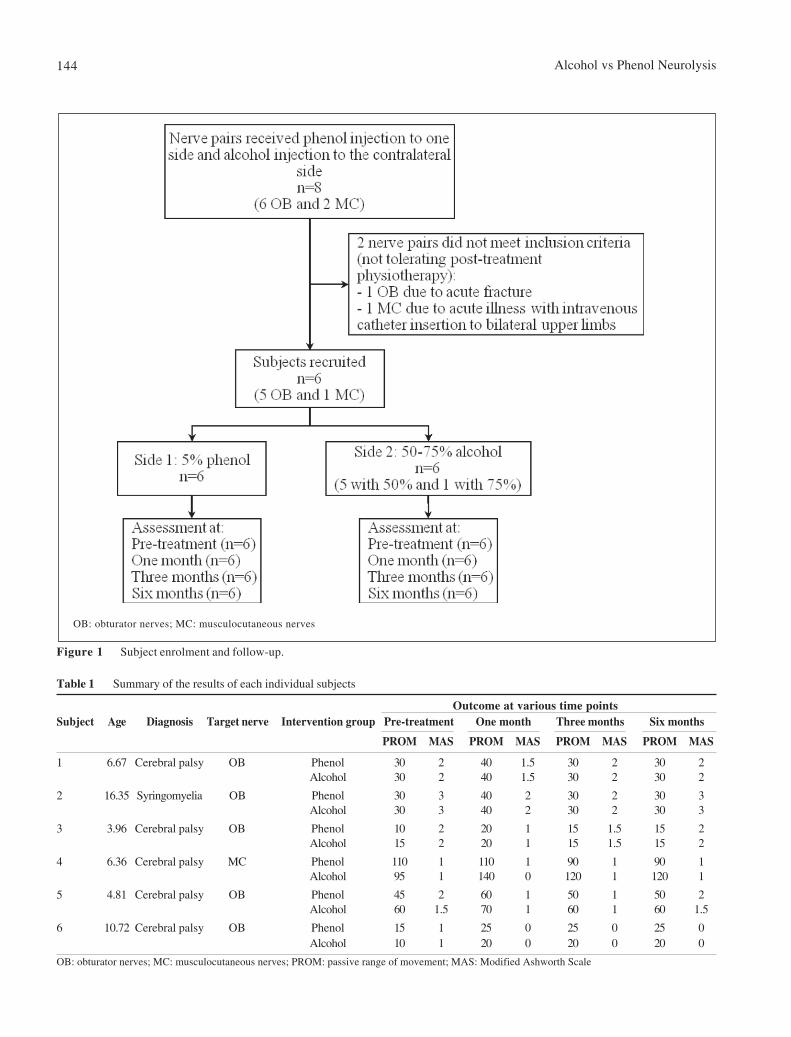
Alcohol vs Phenol Neurolysis144
Table 1 Summary of the results of each individual subjects
Outcome at various time pointsSubject Age Diagnosis Target nerve Intervention group Pre-treatment One month Three months Six months
PROM MAS PROM MAS PROM MAS PROM MAS
1 6.67 Cerebral palsy OB Phenol 30 2 40 1.5 30 2 30 2Alcohol 30 2 40 1.5 30 2 30 2
2 16.35 Syringomyelia OB Phenol 30 3 40 2 30 2 30 3Alcohol 30 3 40 2 30 2 30 3
3 3.96 Cerebral palsy OB Phenol 10 2 20 1 15 1.5 15 2Alcohol 15 2 20 1 15 1.5 15 2
4 6.36 Cerebral palsy MC Phenol 110 1 110 1 90 1 90 1Alcohol 95 1 140 0 120 1 120 1
5 4.81 Cerebral palsy OB Phenol 45 2 60 1 50 1 50 2Alcohol 60 1.5 70 1 60 1 60 1.5
6 10.72 Cerebral palsy OB Phenol 15 1 25 0 25 0 25 0Alcohol 10 1 20 0 20 0 20 0
OB: obturator nerves; MC: musculocutaneous nerves; PROM: passive range of movement; MAS: Modified Ashworth Scale
Figure 1 Subject enrolment and follow-up.
OB: obturator nerves; MC: musculocutaneous nerves
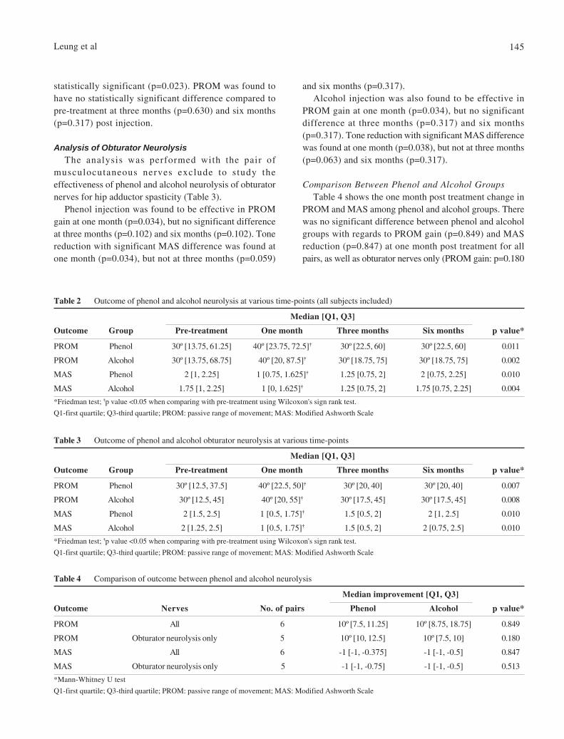
Leung et al 145
and six months (p=0.317).Alcohol injection was also found to be effective in
PROM gain at one month (p=0.034), but no significantdifference at three months (p=0.317) and six months(p=0.317). Tone reduction with significant MAS differencewas found at one month (p=0.038), but not at three months(p=0.063) and six months (p=0.317).
Comparison Between Phenol and Alcohol GroupsTable 4 shows the one month post treatment change in
PROM and MAS among phenol and alcohol groups. Therewas no significant difference between phenol and alcoholgroups with regards to PROM gain (p=0.849) and MASreduction (p=0.847) at one month post treatment for allpairs, as well as obturator nerves only (PROM gain: p=0.180
Table 2 Outcome of phenol and alcohol neurolysis at various time-points (all subjects included)
Median [Q1, Q3]
Outcome Group Pre-treatment One month Three months Six months p value*
PROM Phenol 30º [13.75, 61.25] 40º [23.75, 72.5]† 30º [22.5, 60] 30º [22.5, 60] 0.011
PROM Alcohol 30º [13.75, 68.75] 40º [20, 87.5]† 30º [18.75, 75] 30º [18.75, 75] 0.002
MAS Phenol 2 [1, 2.25] 1 [0.75, 1.625]† 1.25 [0.75, 2] 2 [0.75, 2.25] 0.010
MAS Alcohol 1.75 [1, 2.25] 1 [0, 1.625]† 1.25 [0.75, 2] 1.75 [0.75, 2.25] 0.004
*Friedman test; †p value <0.05 when comparing with pre-treatment using Wilcoxon's sign rank test.
Q1-first quartile; Q3-third quartile; PROM: passive range of movement; MAS: Modified Ashworth Scale
Table 3 Outcome of phenol and alcohol obturator neurolysis at various time-points
Median [Q1, Q3]
Outcome Group Pre-treatment One month Three months Six months p value*
PROM Phenol 30º [12.5, 37.5] 40º [22.5, 50]† 30º [20, 40] 30º [20, 40] 0.007
PROM Alcohol 30º [12.5, 45] 40º [20, 55]† 30º [17.5, 45] 30º [17.5, 45] 0.008
MAS Phenol 2 [1.5, 2.5] 1 [0.5, 1.75]† 1.5 [0.5, 2] 2 [1, 2.5] 0.010
MAS Alcohol 2 [1.25, 2.5] 1 [0.5, 1.75]† 1.5 [0.5, 2] 2 [0.75, 2.5] 0.010
*Friedman test; †p value <0.05 when comparing with pre-treatment using Wilcoxon's sign rank test.
Q1-first quartile; Q3-third quartile; PROM: passive range of movement; MAS: Modified Ashworth Scale
Table 4 Comparison of outcome between phenol and alcohol neurolysis
Median improvement [Q1, Q3]
Outcome Nerves No. of pairs Phenol Alcohol p value*
PROM All 6 10º [7.5, 11.25] 10º [8.75, 18.75] 0.849
PROM Obturator neurolysis only 5 10º [10, 12.5] 10º [7.5, 10] 0.180
MAS All 6 -1 [-1, -0.375] -1 [-1, -0.5] 0.847
MAS Obturator neurolysis only 5 -1 [-1, -0.75] -1 [-1, -0.5] 0.513
*Mann-Whitney U test
Q1-first quartile; Q3-third quartile; PROM: passive range of movement; MAS: Modified Ashworth Scale
statistically significant (p=0.023). PROM was found tohave no statistically significant difference compared topre-treatment at three months (p=0.630) and six months(p=0.317) post injection.
Analysis of Obturator NeurolysisThe analysis was performed with the pair of
musculocutaneous nerves exclude to study theeffectiveness of phenol and alcohol neurolysis of obturatornerves for hip adductor spasticity (Table 3).
Phenol injection was found to be effective in PROMgain at one month (p=0.034), but no significant differenceat three months (p=0.102) and six months (p=0.102). Tonereduction with significant MAS difference was found atone month (p=0.034), but not at three months (p=0.059)
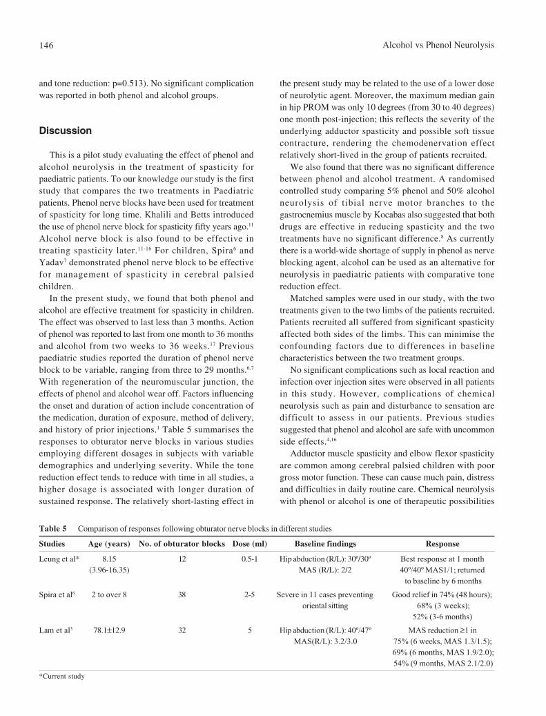
Alcohol vs Phenol Neurolysis146
and tone reduction: p=0.513). No significant complicationwas reported in both phenol and alcohol groups.
Discussion
This is a pilot study evaluating the effect of phenol andalcohol neurolysis in the treatment of spasticity forpaediatric patients. To our knowledge our study is the firststudy that compares the two treatments in Paediatricpatients. Phenol nerve blocks have been used for treatmentof spasticity for long time. Khalili and Betts introducedthe use of phenol nerve block for spasticity fifty years ago.11
Alcohol nerve block is also found to be effective intreating spasticity later.11-16 For children, Spira6 andYadav7 demonstrated phenol nerve block to be effectivefor management of spasticity in cerebral palsiedchildren.
In the present study, we found that both phenol andalcohol are effective treatment for spasticity in children.The effect was observed to last less than 3 months. Actionof phenol was reported to last from one month to 36 monthsand alcohol from two weeks to 36 weeks.17 Previouspaediatric studies reported the duration of phenol nerveblock to be variable, ranging from three to 29 months.6,7
With regeneration of the neuromuscular junction, theeffects of phenol and alcohol wear off. Factors influencingthe onset and duration of action include concentration ofthe medication, duration of exposure, method of delivery,and history of prior injections.1 Table 5 summarises theresponses to obturator nerve blocks in various studiesemploying different dosages in subjects with variabledemographics and underlying severity. While the tonereduction effect tends to reduce with time in all studies, ahigher dosage is associated with longer duration ofsustained response. The relatively short-lasting effect in
the present study may be related to the use of a lower doseof neurolytic agent. Moreover, the maximum median gainin hip PROM was only 10 degrees (from 30 to 40 degrees)one month post-injection; this reflects the severity of theunderlying adductor spasticity and possible soft tissuecontracture, rendering the chemodenervation effectrelatively short-lived in the group of patients recruited.
We also found that there was no significant differencebetween phenol and alcohol treatment. A randomisedcontrolled study comparing 5% phenol and 50% alcoholneurolysis of tibial nerve motor branches to thegastrocnemius muscle by Kocabas also suggested that bothdrugs are effective in reducing spasticity and the twotreatments have no significant difference.8 As currentlythere is a world-wide shortage of supply in phenol as nerveblocking agent, alcohol can be used as an alternative forneurolysis in paediatric patients with comparative tonereduction effect.
Matched samples were used in our study, with the twotreatments given to the two limbs of the patients recruited.Patients recruited all suffered from significant spasticityaffected both sides of the limbs. This can minimise theconfounding factors due to differences in baselinecharacteristics between the two treatment groups.
No significant complications such as local reaction andinfection over injection sites were observed in all patientsin this study. However, complications of chemicalneurolysis such as pain and disturbance to sensation aredifficult to assess in our patients. Previous studiessuggested that phenol and alcohol are safe with uncommonside effects.4,16
Adductor muscle spasticity and elbow flexor spasticityare common among cerebral palsied children with poorgross motor function. These can cause much pain, distressand difficulties in daily routine care. Chemical neurolysiswith phenol or alcohol is one of therapeutic possibilities
Table 5 Comparison of responses following obturator nerve blocks in different studies
Studies Age (years) No. of obturator blocks Dose (ml) Baseline findings Response
Leung et al* 8.15 12 0.5-1 Hip abduction (R/L): 30º/30º Best response at 1 month(3.96-16.35) MAS (R/L): 2/2 40º/40º MAS1/1; returned
to baseline by 6 months
Spira et al6 2 to over 8 38 2-5 Severe in 11 cases preventing Good relief in 74% (48 hours);oriental sitting 68% (3 weeks);
52% (3-6 months)
Lam et al3 78.1±12.9 32 5 Hip abduction (R/L): 40º/47º MAS reduction ≥1 inMAS(R/L): 3.2/3.0 75% (6 weeks, MAS 1.3/1.5);
69% (6 months, MAS 1.9/2.0); 54% (9 months, MAS 2.1/2.0)
*Current study
Leung et al 147
References
1. Patel DR, Soyode O. Pharmacologic interventions for reducingspasticity in cerebral palsy. Indian J Pediatr 2005;72:869-72.
2. Ward AB. Long-term modification of spasticity. J Rehabil Med2003;41(Suppl):60-5.
3. Lam K, Wong D, Tam CK, et al. Ultrasound and electricalstimulator-guided obturator nerve block with phenol in thetreatment of Hip adductor spasticity in long-term care patients: arandomized, triple blind, placebo controlled study. J Am Med DirAssoc 2015;16:238-46.
4. Ghai A, Garg N, Hooda S, Gupta T. Spasticity-Pathogenesis,prevention and treatment strategies. Saudi J Anaesth 2013;7:453-60.
5. Elovic EP, Esquenazi A, Alter KE, Lin JL, Alfaro A, Kaelin DL.Chemodenervation and nerve blocks in the diagnosis andmanagement of spasticity and muscle overactivity. PM R 2009;1:842-51.
6. Spira R. Management of spasticity in cerebral palsied childrenby peripheral nerve block with phenol. Dev Med Child Neurol1971;13:164-73.
7. Yadav SL, Singh U, Dureja GP, Singh KK, Chaturvedi S. Phenolblock in the management of spastic cerebral palsy. Indian J Pediatr1994;61:249-55
8. Kocabas H, Salli A, Demir AH, Ozerbil OM. Comparison of phenoland alcohol neurolysis of tibial nerve motor branches to thegastrocnemius muscle for treatment of spastic foot after stroke:a randomized controlled pilot study. Eur J Phys Rehabil Med2010;46:5-10.
9. Lehmann EL. Nonparametrics: Statistical methods based on ranks.New York, NY: Springer, 2006.
10. Faul F, Erdfelder E, Buchner A, Lang AG. G*Power Version 3.1.7 [computer software]. Uiversität Kiel, Germany, 2013.
11. Khalili AA, Betts HB. Peripheral nerve block with phenol in themanagement of spasticity: indications and complications. JAMA1967;200:1155-7.
12. Viel EJ, Perennou D, Ripari J, Pélissier J, Eledjam JJ. Neurolyticblockade of the obturator nerve for intractable spasticity ofadductor thigh muscles. Eur J Pain 2002;6:97-104.
13. Ghai A, Sangwan SS, Hooda S, Kiran S, Garg N. Obturatorneurolysis using 65% alcohol for adductor muscle spasticity.Saudi J Anaesth 2012;6:282.
14. Kong KH, Chua KS. Neurolysis of the musculocutaneous nervewith alcohol to treat poststroke elbow flexor spasticity. ArchPhys Med Rehabil 1999;80:1234-6.
15. Kong KH, Chua KS. Outcome of obturator nerve block withalcohol for the treatment of hip adductor spasticity. Int J RehabilRes 1999;22:327-30.
16. Chua KS, Kong KH. Clinical and functional outcome after alcoholneurolysis of the tibial nerve for ankle-foot spasticity. Brain Inj2001;15:733-9.
17. Tilton AH. Injectable neuromuscular blockade in the treatmentof spasticity and movement disorders. J Child Neurol 2003;18:S50-66.
18. Bohannon RW, Smith MB. Interrater reliability of a modifiedAshworth scale of muscle spasticity. Phys Ther 1986;67:206-7.
to manage spasticity. The major advantages of chemicalneurolysis include low cost of the drugs, rapid onset ofaction and potency for large muscle groups like hipadductors.5
LimitationsThe number of subjects included in this study is small.
The number of subjects recruited in this study was actuallylimited by the unstable availability of phenol. Only onepair of musculocutaneous nerve was included in this studyand only one case received alcohol injection with 75%concentration. A larger sample size is necessary for furtherstudies to analyse an optimal drug concentration, the lowesteffective dose and for subgroup analysis. The therapeuticeffect , duration of action and safety of higherconcentration of alcohol at 75-100% has yet been explored.More outcomes can be assessed in future studies such ashygiene score and pain score. Side effects of treatmentmay be under-reported because majority of our patientswere unable to communicate the pain and sensorycomplications.
Conclusion
This study showed that obturator and musculocutaneousneurolysis with 5% phenol or 50-75% alcohol is aneffective treatment to reduce spasticity in children withchronic neurological disease. This procedure can beintroduced to this group of patients earlier to preventcomplications of spasticity and improve their quality oflife. As a nerve blocking agent, alcohol is a safe andeffective alternative to replace phenol in paediatricpatients.
Acknowledgements
The authors thank all the clinical staff and patients whotook part in this study, in particular Miss Ellen Yu,statistician from Princess Margaret Hospital for herassistance in statistical analysis.
Declaration of Interests
None
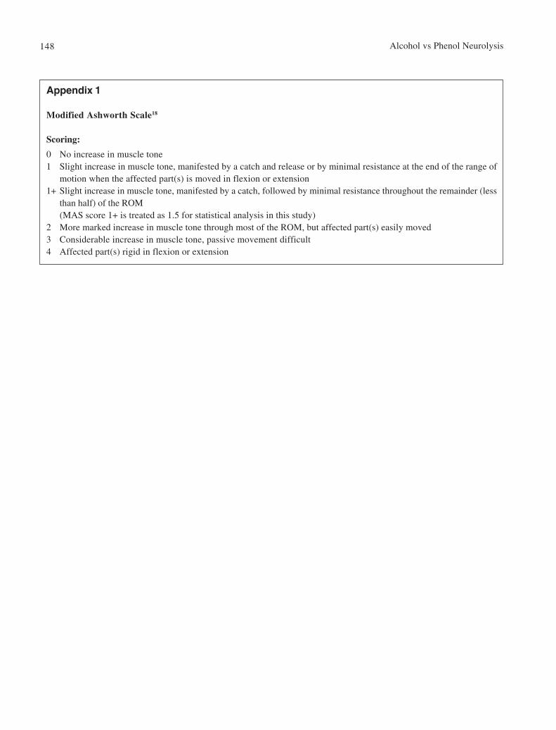
Alcohol vs Phenol Neurolysis148
Appendix 1
Modified Ashworth Scale18
Scoring:
0 No increase in muscle tone1 Slight increase in muscle tone, manifested by a catch and release or by minimal resistance at the end of the range of
motion when the affected part(s) is moved in flexion or extension1+ Slight increase in muscle tone, manifested by a catch, followed by minimal resistance throughout the remainder (less
than half) of the ROM(MAS score 1+ is treated as 1.5 for statistical analysis in this study)
2 More marked increase in muscle tone through most of the ROM, but affected part(s) easily moved3 Considerable increase in muscle tone, passive movement difficult4 Affected part(s) rigid in flexion or extension
HK J Paediatr (new series) 2021;26:149-154
Tolerability and Efficacy of Racecadotril in Acute Diarrhoea,A Prospective, Randomised, Parallel Study in an Indian
Tertiary Care Teaching Hospital
R SARANGI, B BISWAL, B DASH, A DHANAWAT
Abstract Objective: To study the tolerability and efficacy of racecadotril versus placebo in hospitalised childrenwith acute diarrhoea. Methods: This prospective randomised placebo-controlled double blind trial wascarried among 3-36 months age hospitalised children with acute diarrhoea. Out of 140 children, 67children received placebo and 73 received racecadotril at 1.5 mg/kg, three times a day till recovery orfive days whichever was earlier. Stool volume in 48 hours, duration of hospitalisation and tolerability ofthe drug were the outcome measures. Result: The mean stool volume in 48 hours (179.5±17.31 g/kgbody weight versus 191.04±33.4 g/kg body weight, p=0.0001) and mean duration of hospital stay(62.4±11.4 hours versus 95.6±19.5 hours, p=0.0001) were significantly lower in racecadotril armcompared to placebo arm respectively. No serious adverse event was documented in either group.Conclusion: Racecadotril is both safe and effective in children with acute diarrhoea in decreasing thestool volume and duration of hospitalisation.
Key words Acute diarrhoea; Teaching hospital; Tolerability
Department of Pediatrics, IMS and SUM Hospital, SOADeemed to be University, Bhubaneswar-751003, Orissa,India
R SARANGI MDB BISWAL MDB DASH MDA DHANAWAT MD
Correspondence to: Dr B BISWALEmail: [email protected]
Received April 12, 2019
Original Article
Introduction
Acute diarrhoea in children accounts a major healthburden in different parts of world with an estimated 2billion episodes each year and 1.9 million deaths mostlyin developing countries, amounting to 18% of under-fivemortality.1 Diarrhoea is the third leading cause of childhoodmortality in India and it accounts for 13% of all deaths peryear in under-five children.2 The mainstay of therapy inacute diarrhoea, is the Oral Rehydration Therapy and its
global adoption has reduced the mortality and morbiditydue to diarrhoea, over the last 25 years. Racecadotril (acetorphan) is an anti-secretary agent;inhibits enkephalinase, which is a cell membrane peptidaseenzyme, located in various tissues, predominantly in theepithelium of the small intestine.3 This peptidase isresponsible for both the digestion of exogenous peptidesand endogenous peptides such as enkephalins andneurokinin.4 So the drug enhances the activity ofendogenous enkephalins, leading to decrease inintracellular cAMP level thereby reducing secretion ofwater and electrolytes,5,6 without altering the motility,intestinal transit time or bacterial overgrowth.7
Though there are several studies available fromdeveloped world with mixed outcome, there are limiteds tudies regard ing ef f icacy and to lerabi l i ty ofracecadotril in the developing country. So this study wasplanned in Indian children and the aim of this doubleblind, prospective randomised placebo-controlled trialwas to compare the tolerabili ty and efficacy ofracecadotril versus placebo among hospitalised childrenwith acute diarrhoea.
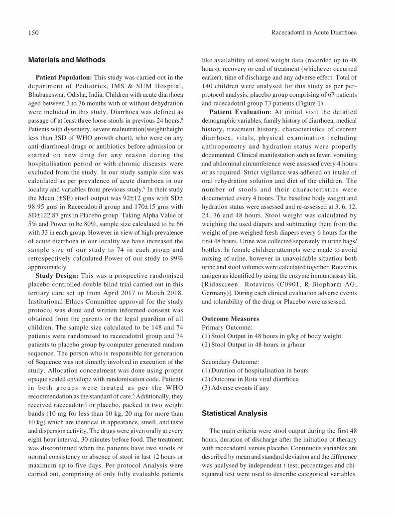
Racecadotril in Acute Diarrhoea150
Materials and Methods
Patient Population: This study was carried out in thedepartment of Pediatrics, IMS & SUM Hospital,Bhubaneswar, Odisha, India. Children with acute diarrhoeaaged between 3 to 36 months with or without dehydrationwere included in this study. Diarrhoea was defined aspassage of at least three loose stools in previous 24 hours.8
Patients with dysentery, severe malnutrition(weight/heightless than 3SD of WHO growth chart), who were on anyanti-diarrhoeal drugs or antibiotics before admission orstarted on new drug for any reason during thehospitalisation period or with chronic diseases wereexcluded from the study. In our study sample size wascalculated as per prevalence of acute diarrhoea in ourlocality and variables from previous study.9 In their studythe Mean (±SE) stool output was 92±12 gms with SD±98.95 gms in Racecadotril group and 170±15 gms withSD±122.87 gms in Placebo group. Taking Alpha Value of5% and Power to be 80%, sample size calculated to be 66with 33 in each group. However in view of high prevalenceof acute diarrhoea in our locality we have increased thesample size of our study to 74 in each group andretrospectively calculated Power of our study to 99%approximately.
Study Design: This was a prospective randomisedplacebo-controlled double blind trial carried out in thistertiary care set up from April 2017 to March 2018.Institutional Ethics Committee approval for the studyprotocol was done and written informed consent wasobtained from the parents or the legal guardian of allchildren. The sample size calculated to be 148 and 74patients were randomised to racecadotril group and 74patients to placebo group by computer generated randomsequence. The person who is responsible for generationof Sequence was not directly involved in execution of thestudy. Allocation concealment was done using properopaque sealed envelope with randomisation code. Patientsin both groups were t rea ted as per the WHOrecommendation as the standard of care.8 Additionally, theyreceived racecadotril or placebo, packed in two weightbands (10 mg for less than 10 kg, 20 mg for more than10 kg) which are identical in appearance, smell, and tasteand dispersion activity. The drugs were given orally at everyeight-hour interval, 30 minutes before food. The treatmentwas discontinued when the patients have two stools ofnormal consistency or absence of stool in last 12 hours ormaximum up to five days. Per-protocol Analysis werecarried out, comprising of only fully evaluable patients
like availability of stool weight data (recorded up to 48hours), recovery or end of treatment (whichever occurredearlier), time of discharge and any adverse effect. Total of140 children were analysed for this study as per per-protocol analysis, placebo group comprising of 67 patientsand racecadotril group 73 patients (Figure 1).
Patient Evaluation: At initial visit the detaileddemographic variables, family history of diarrhoea, medicalhistory, treatment history, characteristics of currentdiarrhoea, vitals, physical examination includinganthropometry and hydration status were properlydocumented. Clinical manifestation such as fever, vomitingand abdominal circumference were assessed every 4 hoursor as required. Strict vigilance was adhered on intake oforal rehydration solution and diet of the children. Thenumber of stools and their characteristics weredocumented every 4 hours. The baseline body weight andhydration status were assessed and re-assessed at 3, 6, 12,24, 36 and 48 hours. Stool weight was calculated byweighing the used diapers and subtracting them from theweight of pre-weighed fresh diapers every 6 hours for thefirst 48 hours. Urine was collected separately in urine bags/bottles. In female children attempts were made to avoidmixing of urine, however in unavoidable situation bothurine and stool volumes were calculated together. Rotavirusantigen as identified by using the enzyme immunoassay kit,[Ridascreen_ Rotavirus (C0901, R-Biopharm AG,Germany)]. During each clinical evaluation adverse eventsand tolerability of the drug or Placebo were assessed.
Outcome MeasuresPrimary Outcome:(1) Stool Output in 48 hours in g/kg of body weight(2) Stool Output in 48 hours in g/hour
Secondary Outcome:(1) Duration of hospitalisation in hours(2) Outcome in Rota viral diarrhoea(3) Adverse events if any
Statistical Analysis
The main criteria were stool output during the first 48hours, duration of discharge after the initiation of therapywith racecadotril versus placebo. Continuous variables aredescribed by mean and standard deviation and the differencewas analysed by independent t-test, percentages and chi-squared test were used to describe categorical variables.
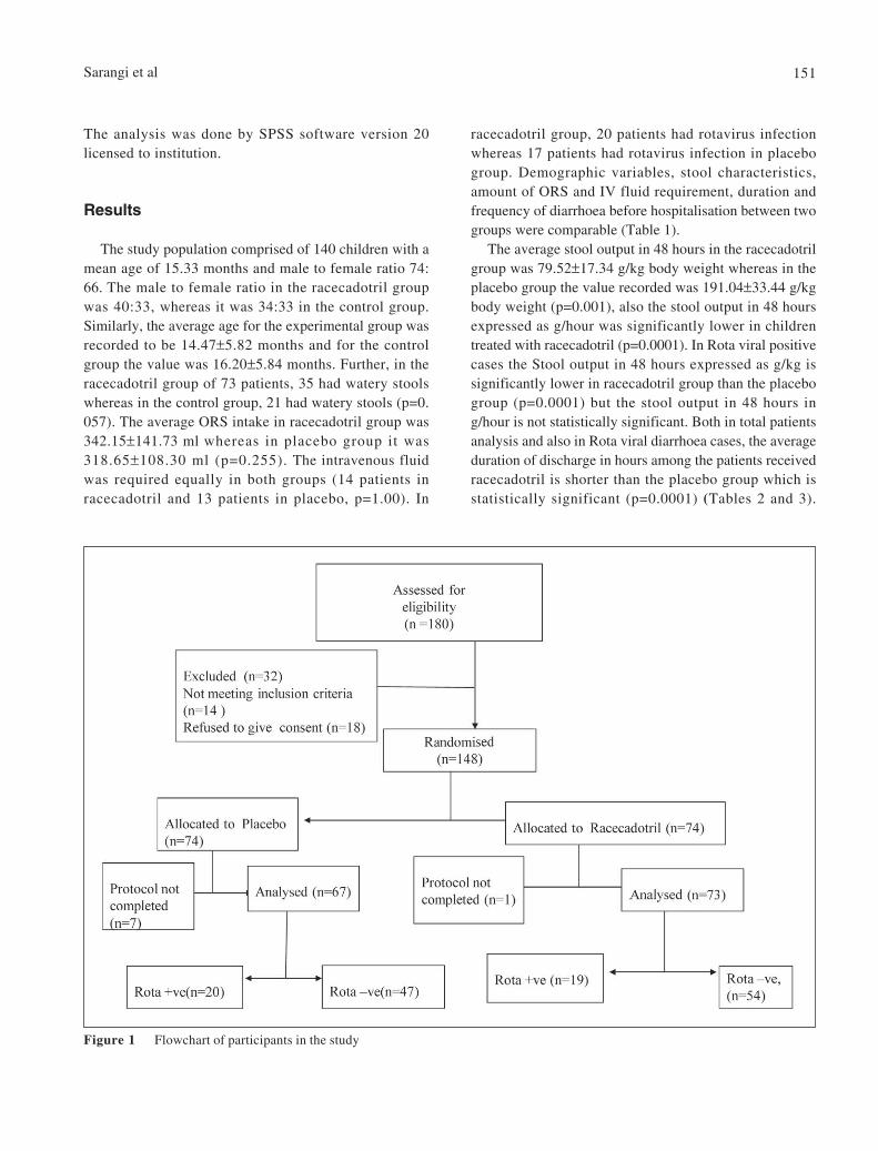
Sarangi et al 151
racecadotril group, 20 patients had rotavirus infectionwhereas 17 patients had rotavirus infection in placebogroup. Demographic variables, stool characteristics,amount of ORS and IV fluid requirement, duration andfrequency of diarrhoea before hospitalisation between twogroups were comparable (Table 1).
The average stool output in 48 hours in the racecadotrilgroup was 79.52±17.34 g/kg body weight whereas in theplacebo group the value recorded was 191.04±33.44 g/kgbody weight (p=0.001), also the stool output in 48 hoursexpressed as g/hour was significantly lower in childrentreated with racecadotril (p=0.0001). In Rota viral positivecases the Stool output in 48 hours expressed as g/kg issignificantly lower in racecadotril group than the placebogroup (p=0.0001) but the stool output in 48 hours ing/hour is not statistically significant. Both in total patientsanalysis and also in Rota viral diarrhoea cases, the averageduration of discharge in hours among the patients receivedracecadotril is shorter than the placebo group which isstatistically significant (p=0.0001) (Tables 2 and 3).
The analysis was done by SPSS software version 20licensed to institution.
Results
The study population comprised of 140 children with amean age of 15.33 months and male to female ratio 74:66. The male to female ratio in the racecadotril groupwas 40:33, whereas it was 34:33 in the control group.Similarly, the average age for the experimental group wasrecorded to be 14.47±5.82 months and for the controlgroup the value was 16.20±5.84 months. Further, in theracecadotril group of 73 patients, 35 had watery stoolswhereas in the control group, 21 had watery stools (p=0.057). The average ORS intake in racecadotril group was342.15±141.73 ml whereas in placebo group it was318.65±108.30 ml (p=0.255). The intravenous fluidwas required equally in both groups (14 patients inracecadotril and 13 patients in placebo, p=1.00). In
Figure 1 Flowchart of participants in the study
Racecadotril in Acute Diarrhoea152
Adverse events like abdominal distension, vomiting,rebound constipation were observed in 6.9%, 2.8%, 1.4%of cases in racecadotril group and 7.8%, 3.5%, 1.6% ofcases in placebo group respectively, which were notstatistically significant. However, no serious adverse eventswere noted in both placebo and racecadotril group.
Discussion
In this randomised controlled trial, racecadotril given(1.5 mg/kg) three times a day maximum for 5 days tochildren aged 3-36 months admitted with acute diarrhoearesulted in significant reduction in stool output in 48 hoursand duration of hospitalisation. There is also significanttherapeutic response amongst the children receivingracecadotril in Rota viral diarrhoea cases.
Table 1 Baseline characteristics of children with acute diarrhoea
Baseline characters Racecadotril (n=73) Placebo (n=67) P value
Age in months, (Mean±SD) 16.2±5.84 14.47±5.82 0.081
Sex (M:F) 40:33 34:33 0.734
Stool consistency (Watery) 35/73 21/76 0.057
Amount of ORS (ml) before hospitalisation in 24 hour; (Mean±SD) 343.15±141.73 318.65±108.30 0.255
Number of patients required IVF before hospitalisation 14/73 13/67 1.00
Number of stool before hospitalisation in 24 hours; (Mean±SD) 9.712±0.297 9.328±0.309 0.373
Duration of diarrhoea before hospitalisation in hours; (Mean±SD) 30.85±0.971 36.55±1.207 0.0666
Rotavirus positive cases 19/73 20/67 0.849
Previous studies comprising of children found thatracecadotril use in moderate to severe diarrhoea decreasedthe stool volume by 56% to 60% after 48 hours of treatmentin comparison with placebo.5,9,10 In a recently publishedsystematic review of RCTs concluded that racecadotril ismore effective than placebo in terms of decreasing theduration of diarrhoea and stool volume in children.11
Liang et al in their systemic review found, Racecadotrilmay reduce the risk of rehydration failure (RR 0.41,95%CI 0.13 to 1.23). However there were insufficient datato show effect on duration of diarrhoea, number of stoolsin the first 48 hours.12
Multiple efficacy parameters were taken in differentstudies in a Meta-analysis by Eberlin et al, about the useof racecadotril in the treatment of acute diarrhoea inchildren. Stool output in 48 hours in gm/kg and stool outputin gms/hr and duration of hospital stay in hours are
Table 2 Comparisons of efficacy of racecadotril and placebo in acute diarrhoea
Outcome (Mean±±±±±SD) Racecadotril (n=73) Placebo (n=67) P value
Stool output in 48 hours in grams/kg body weight 79.5±17.3 191.04±33.4 0.0001
Stool output in 48 hours in grams/hour 28.5±7.4 35.9±5.9 0.0001
Duration of hospital stay in hours 62.4±11.4 95.6±9.5 0.0001
Table 3 Comparisons of efficacy of racecadotril and placebo in Rota virus positive cases (n=39)
Outcome (Mean±±±±±SD) Racecadotril (n=19) Placebo (n=20) P value
Stool output in 48 hours in grams/kg body weight 89.47±3.705 208.0±5.410 0.0001
Stool output in 48 hours in grams/hour 34.84±1.388 38.55±1.268 0.0557
Duration of hospital stay (hours) 60.32±2.597 104.4±2.014 0.0001
Sarangi et al 153
insignificant in both the groups. Similar observation wasalso done in different studies and racecadotril found to besafe for use in children with acute diarrhoea.9-11,13,15
As there is paucity of data from a developing countrylike India where the disease burden is very high withsignificant morbidity and mortality, this study was done tofind out the effect of racecadotril in acute diarrhoea. Thisis the only double blinded randomised study from easternpart of India comparing racecadotril versus placebo in acutediarrhoea in hospitalised children, thus minimisingpossibility of observation bias; more so over theintervention was started early (median duration of diarrhoeabefore hospitalisation of 30.85 hours) comparing to otherIndian study, where the median duration of presentation isaround 2 days.14 Potential limitations of our study includesmall sample size, inability to have different subgroupanalysis and cost-effective analysis. Further among femalechildren the exact stool volume calculation wascumbersome.
Conclusion
Racecadotril is both safe and effective in reducing thestool volume and duration of hospitalisation in childrenwith acute diarrhoea. It is also found to be effective in Rotaviral positive cases. Though some more trials with rigorousmethodologies in larger population involving multiplecenters with high disease burden and similar etiologicalspectrum may be needed to support the evidence.
Declaration of Interest
The authors declare that there is no conflict ofinterest.
References
1. World Gastroenterology Global Guidelines: Acute diarrhoea in adultsand children: a global perspective 2012.
2. Lakshminarayanan S, Jayalakshmy R. Diarrhoeal diseases amongchildren in India: Current scenario and future perspectives.J Natu Sci Biol Med 2015;6:24-8.
3. Eberlin M, Mück T, Michel MC. A comprehensive review of thepharmacodynamics, pharmacokinetics, and clinical effects of theneutral endopeptidase inhibitor racecadotril. Front Pharmacol 2012;3:93.
4. Primi MP, Bueno L, Baumer Ph, Berard H, Lecomte JM.Racecadotril demonstrates intestinal antisecretory activity in vivo.
considered important primary end point in multiple studies,and hence, taken for comparison for assessing efficacy ofracecadotril in acute diarrhoea in our study.13
In the present study, there is significant reduction instool output in 48 hours (79.5±17.3 g/kg body weight) inracecadotril group in comparison to placebo group(191.04±33.4 g/kg body weight). Liang et al in theirsystemic review found lower stool output in the first 48hours in two trials.12 Another double blind randomised studycomprising of 135 boys of similar age group, alsosuggestive of significant reduction in 48 hr stool output(92±12 g/kg) in racecadotril as compared to placebo(170±15 g/kg) group (p<0.001).9 Study done in south-Indiaby Kang et al concluded, racecadotril did not reduceduration of diarrhoea, volume of stool or fluid requirementin under five children with acute gastroenteritis with orwithout rotavirus infection.14 The reason for the lack ofimpact may be possible co-infection in developing country.However, Santos et al in their study among 189 childrenin 3-36 month age group revealed no difference in termsof frequency of stool or duration of diarrhoea amongracecadotril and placebo group.15 This may be explained asthe later study was in outpatient children and stool volumeassessment was by parents, so presence of observation biascould not be excluded. The present study alsodemonstrates significant reduction in duration ofhospitalisation/duration of diarrhoea after treatment withracecadotril in comparison to placebo. Similar type ofobservation was documented in recently published meta-analysis.13
There is significant reduction of 48 hr stool volumeand discharge duration in hours in rota viral diarrhoeatreated with racecadotril in comparison to placebo incurrent study. Cèzard et al in their study demonstrated upto 50% reduction in stool volume with racecadotril asadjuvant therapy to ORS in the treatment of severe diarrhoeaboth in rota and non-rota viral diarrhoea among infants andchildren.10 Similar observation was made by Alvarez et althat racecadotril found to be effective in terms of reducingthe stool frequency within the first 48 hours and isassociated with a shorter disease course and fewer visitsto emergency services, as well as hospital admissions.16
However few studies using different efficacy parametersalso found racecadotril is equally effective in both rotapositive and rota-negative diarrhea.17,18
Racecadotril is found to be safe and well tolerable drugas no serious adverse event was documented in this studyand side effects documented are abdominal distension,vomiting, rebound constipation which are statistically
Racecadotril in Acute Diarrhoea154
Aliment Pharmacol Ther 1999;13:3-7.5. Tormo R, Polanco I, Salazar-Lindo E, Goulet O. Acute infectious
diarrhoea in children: new insights in antisecretory treatment withracecadotril. Acta Paediatr 2008;97:1008-15.
6. Matheson AJ, Noble S. Racecadotril. Drugs 2000;59:829-35.7. Hamza H, Ben Khalifa H, Baumer P, Berard H, Lecomte JM.
Racecadotril versus placebo in the treatment of acute diarrhoea inadults. Aliment Pharmacol Ther 1999;13:15-9.
8. World Health Organization (WHO). The Treatment of Diarrhoea:A Manual for Physicians and Other Senior Health Workers. 4thGeneva, Switzerland: WHO; 2005(WHO/COD/SER / 80.2).
9. Salazar-Lindo E, Santiesteban-Ponce J, Chea-Woo E, GutierrezM. Racecadotril in the treatment of acute watery diarrhoea inchildren. N Eng J Med 2000;343:463-7.
10. Cèzard J, Duhamel J, Meyer M, et al. Efficacy and tolerability ofRacecadotril in acute diarrhoea in children. Gastroenterology 2001;120:799-805.
11. Gordon M, Akobeng A. Racecadotril for acute diarrhoea in children:systematic review and meta-analyses. Arch Arch Dis Child 2016;101:234-40.
12. Liang Y, Zhang L, Zeng L, Gordon M, Wen J. Racecadotril foracute diarrhoea in children. Cochrane Database Syst Rev 2019;12:
CD009359.13. Eberlin M, Chen M, Mueck T, Däbritz J. Racecadotril in the
treatment of acute diarrhoea in children: a systematic,comprehensive review and meta-analysis of randomisedcontrolled trials. BMC Pediatrics 2018;18:124.
14. Kang G, Thuppal SV, Srinivasan R, et al. Racecadotril in themanagement of rotavirus and non-rotavirus diarrhoea in under-five children: Two randomised, double-blind, placebo-controlled trials. Indian Pediatr 2016;53:595-600.
15. Santos M, Maranon R, Miguez C, Vazquez P, Sanchez C. Useof racecodatril as outpatient treatment for acute gastroenteritis:a prospective, randomised, parallel study. J Pediatr 2009;155:62-7.
16. Alvarez CG, Pinel SG, Taboada CL, et al. Efectividad deracecadotrilo en el tratamiento de la gastroenteritis aguda. ActaPediatr Esp 2009;67:117-22.
17. Michael SSA, Ali AMA, Ezzat DA, Tayel SA. Evaluation ofracecadotril in treatment of acute diarrhoea in children. AsianJ Pharm Clin Res 2014;2:227-30.
18. Cojocaru B, Bocquet N, Timsit S, et al. Effect of racecadotrilin the management of acute diarrhoea in infants and children.Arch Pediatr 2002;9:774-9.
HK J Paediatr (new series) 2021;26:155-157
Enterovirus D68 Myelitis:The First Reported Case in Hong Kong
CY MO, CL YUEN
Abstract A 3-year-old Chinese boy presented with acute onset of left facial nerve palsy with significantly impairedneck and upper limb power. Magnetic resonance imaging of brain and spine showed T2W hyperintensesignals over both cerebral peduncles and posterior pons, and a long segment at cervical spinal cord.Enterovirus D68 was positive in stool. The boy was treated as a case of viral myelitis and given a courseof corticosteroid and intravenous immunoglobulin. Despite rehabilitation, he still sustained significantresidual right upper limb weakness two years after the acute illness. This is the first reported localpaediatric case of enterovirus D68 infection related acute flaccid myelitis.
Key words Acute flaccid myelitis; Enterovirus D68; Myelitis
Department of Paediatrics, Kwong Wah Hospital, 25 WaterlooRoad, Kowloon, Hong Kong SAR, China
CY MO MBBS, FHKAM(Paed), FHKCPaed
Department of Paediatrics and Adolescent Medicine, TuenMun Hospital, 23 Tsing Chung Koon Road, Tuen Mun,N.T., Hong Kong SAR, China
CL YUEN MBBS, FHKAM(Paed), FHKCPaed
Correspondence to: Dr CY MO
Email: [email protected]
Received March 16, 2020
Case Presentation
Our case is a 3-year-old boy with good past health. Hewas admitted to our unit on 26th September 2017, presentedwith generalised weakness and neck pain for three days.He also had fever and coryzal symptoms five dayspreceding symptoms onset.
On admission, the boy was irritable but had full Glasgowcoma scale. Blood pressure, heart rate, respiratory rate andtemperature were normal. Pupils were equal and reactiveto light, with normal extraocular movement. However, hehad left facial nerve palsy which was compatible with lowermotor neuron lesion. He was not able to sit up straightwithout support. His neck and truncal tone were markedly
Case Report
decreased with generalised hypotonia, affecting upperlimbs more than lower limbs. Muscle power was grade 1/5over right shoulder, grade 2/5 over left shoulder, grade3/5 over bilateral elbows and grade 4/5 over bilateral wrists.Lower limb power was grade 4/5 over both proximal anddistal parts. Reflexes were present and symmetrical overfour limbs. The anal tone was intact. There were no rash orinsect bite marks. The patient did not cooperate withsensory level and cerebellar exams. Other systemsexamination was unremarkable.
Full septic workups were performed. Complete bloodcount, liver function, electrolytes, serum glucose,c-reactive protein and immunoglobulin pattern werenormal. Nasopharyngeal aspirate (NPA) for respiratoryviruses and mycoplasma pneumoniae polymerase chainreaction (PCR) were negative. Epstein bar virus andmeasles virus IgM, borrelia burgdorferi, brucella, syphilisand anti-HIV antibodies were negative. Stool for polio virusand blood culture were also negative. Cerebrospinal fluid(CSF) microscopy found elevated white blood cell 37/cmm with 97% lymphocyte and red blood cell 3/cmm. CSFgram stain, Indian ink stain, acid-fast bacilli smear, bacterialculture and viral PCR were negative. CSF protein and IgGwere not elevated. There was no detectable CSF oligoclonalband. Besides, autoimmune markers were all negative andvitamin B12 and folate levels were not low. Anti-aquaporin4 antibody was also negative.
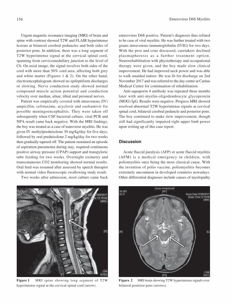
Enterovirus D68 Myelitis156
Urgent magnetic resonance imaging (MRI) of brain andspine with contrast showed T2W and FLAIR hyperintenselesions at bilateral cerebral peduncles and both sides ofposterior pons. In addition, there was a long segment ofT2W hyperintense signal at the cervical spinal cord,spanning from cervicomedullary junction to the level ofC6. On axial image, the signal involves both sides of thecord with more than 50% cord area, affecting both greyand white matter (Figures 1 & 2). On the other hand,electroencephalogram showed no epileptiform dischargesor slowing. Nerve conduction study showed normalcompound muscle action potential and conductionvelocity over median, ulnar, tibial and peroneal nerves.
Patient was empirically covered with intravenous (IV)ampicillin, cefotaxime, acyclovir and oseltamivir forpossible meningoencephalitis. They were taken offsubsequently when CSF bacterial culture, viral PCR andNPA result came back negative. With the MRI findings,the boy was treated as a case of transverse myelitis. He wasgiven IV methylprednisolone 30 mg/kg/day for five days,followed by oral prednisolone 2 mg/kg/day for two weeksthen gradually tapered off. The patient sustained an episodeof aspiration pneumonia during stay, required continuouspositive airway pressure (CPAP) support and transpylorictube feeding for two weeks. Overnight oximetry andtranscutaneous CO2 monitoring showed normal results.Oral feed was resumed after assessed by speech therapistwith normal video fluoroscopic swallowing study result.
Two weeks after admission, stool culture came back
enterovirus D68 positive. Patient's diagnosis thus refinedto be case of viral myelitis. He was further treated with twograms intravenous immunoglobulin (IVIG) for two days.With the pros and cons discussed, caretakers declinedplasmapheres is as a fur ther t rea tment opt ion.Neurorehabilitation with physiotherapy and occupationaltherapy were given, and the boy made slow clinicalimprovement. He had improved neck power and was ableto walk unaided indoor. He was fit for discharge on 2ndNovember 2017 and was referred to the day centre at CaritasMedical Centre for continuation of rehabilitation.
Anti-aquaporin 4 antibody was repeated three monthslater with anti-myelin-oligodendrocyte glycoprotein(MOG) IgG. Results were negative. Progress MRI showedresolved abnormal T2W hyperintense signals at cervicalspinal cord, bilateral cerebral peduncle and posterior pons.The boy continued to make slow improvement, thoughstill had significantly impaired right upper limb powerupon writing up of this case report.
Discussion
Acute flaccid paralysis (AFP) or acute flaccid myelitis(AFM) is a medical emergency in children, withpoliomyelitis once being the most classical cause. Withthe invention of polio vaccine, poliomyelitis becomesextremely uncommon in developed countries nowadays.Other differential diagnoses include causes of myelopathy
Figure 1 MRI spine showing long segment of T2W
hyperintense signal at the cervical spinal cord (arrow).
Figure 2 MRI brain showing T2W hyperintense signals over
bilateral posterior pons (arrows).
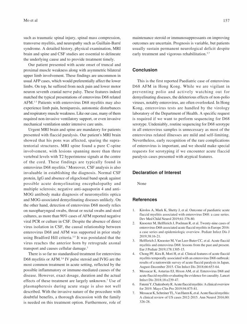
Mo et al 157
maintenance steroid or immunosuppressants on improvingoutcomes are uncertain. Prognosis is variable, but patientsusually sustain permanent neurological deficit despiteearly treatment and vigorous rehabilitation.3,7
Conclusion
This is the first reported Paediatric case of enterovirusD68 AFM in Hong Kong. While we are vigilant inpreventing polio and actively watching out fordemyelinating diseases, the deleterious effects of non-polioviruses, notably enterovirus, are often overlooked. In HongKong, enterovirus tests are handled by the virologylaboratory of the Department of Health. A specific requestis required if we want to perform sequencing for D68serotype. Undeniably, routine sequencing for D68 serotypein all enterovirus samples is unnecessary as most of theenterovirus related illnesses are mild and self-limiting.Nevertheless, early recognition of the rare complicationsof enterovirus is important, and we should make specialrequests for serotyping if we encounter acute flaccidparalysis cases presented with atypical features.
Declaration of Interest
None
References
1. Kirolos A, Mark K, Shetty J, et al. Outcome of paediatric acuteflaccid myelitis associated with enterovirus D68: a case series.Dev Med Child Neurol 2019;61:376-80.
2. Knoester M, Helfferich J, Poelman R, et al. Twenty-nine cases ofenterovirus-D68-associated acute flaccid myelitis in Europe 2016:a case series and epidemiologic overview. Pediatr Infect Dis J2019;38:16-21.
3. Helfferich J, Knoester M, Van Leer-Buter CC, et al. Acute flaccidmyelitis and enterovirus D68: lessons from the past and present.Eur J Pediatr 2019;178:1305-15.
4. Chong PF, Kira R, Mori H, et al. Clinical features of acute flaccidmyelitis temporally associated with an enterovirus D68 outbreak:results of a nationwide survey of acute flaccid paralysis in Japan,August-December 2015. Clin Infect Dis 2018;66:653-64.
5. Messacar K, Asturias EJ, Hixon AM, et al. Enterovirus D68 andacute flaccid myelitis-evaluating the evidence for causality. LancetInfect Dis 2018;18:e239-47.
6. Fatemi Y, Chakraborty R. Acute flaccid myelitis: A clinical overviewfor 2019. Mayo Clin Pro 2019;94:875-81.
7. Messacar K, Schreiner TL, Van Haren K, et al. Acute flaccid myelitis:A clinical review of US cases 2012-2015. Ann Neurol 2016;80:326-28.
such as traumatic spinal injury, spinal mass compression,transverse myelitis, and neuropathy such as Guillain-Barrésyndrome. A detailed history, physical examination, MRIbrain and spine and CSF studies are essential to delineatethe underlying cause and to provide treatment timely.
Our patient presented with acute onset of truncal andproximal muscle weakness along with asymmetric bilateralupper limb involvement. These findings are uncommon inusual AFP cases, which would preferentially affect the lowerlimbs. On top, he suffered from neck pain and lower motorneuron seventh cranial nerve palsy. These features indeedmatched the typical presentations of enterovirus D68 relatedAFM.1-3 Patients with enterovirus D68 myelitis may alsoexperience limb pain, hemiparesis, autonomic disturbancesand respiratory muscle weakness. Like our case, many of themrequired non-invasive ventilatory support, or even invasivemechanical ventilation under intensive care units.
Urgent MRI brain and spine are mandatory for patientspresented with flaccid paralysis. Our patient's MRI brainshowed that his pons was affected, sparing the supra-tentorial structures. MRI spine found a pure C-spineinvolvement, with lesions spanning more than threevertebral levels with T2 hyperintense signals at the centreof the cord. These findings are typically found inenterovirus D68 myelitis.4 Moreover, CSF analysis is alsoinvaluable in establishing the diagnosis. Normal CSFprotein, IgG and absence of oligoclonal band speak againstpossible acute demyelinating encephalopathy andmultiple sclerosis; negative anti-aquaporin 4 and anti-MOG antibody make diagnosis of neuromyelitis opticaand MOG-associated demyelinating diseases unlikely. Onthe other hand, detection of enterovirus D68 mostly relieson nasopharyngeal swabs, throat or rectal swabs and stoolcultures, as more than 90% cases of AFM reported negativeviral PCR or culture in CSF. Despite the absence of directvirus isolation in CSF, the causal relationship betweenenterovirus D68 and AFM was supported in prior studyusing Bradford Hill criteria.3,5 It was postulated that thevirus reaches the anterior horn by retrograde axonaltransport and causes cellular damage.3
There is so far no standardised treatment for enterovirusD68 myelitis or AFM.3,6 IV pulse steroid and IVIG are themost common treatment in acute setting, reflected by thepossible inflammatory or immune-mediated causes of thedisease. However, exact dosage, duration and the actualeffects of these treatment are largely unknown.7 Use ofplasmapheresis during acute stage is also not welldescribed. With the invasive nature of the procedure withdoubtful benefits, a thorough discussion with the familyis needed on this treatment option. Furthermore, role of
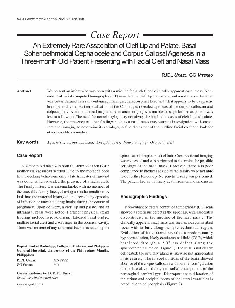
HK J Paediatr (new series) 2021;26:158-160
An Extremely Rare Association of Cleft Lip and Palate, BasalSphenoethmoidal Cephalocele and Corpus Callosal Agenesis in a
Three-month Old Patient Presenting with Facial Cleft and Nasal Mass
RJDL URGEL, GG VITERBO
Abstract We present an infant who was born with a midline facial cleft and clinically apparent nasal mass. Non-enhanced facial computed tomography (CT) revealed the cleft lip and palate, and nasal mass - the latterwas better defined as a sac containing meninges, cerebrospinal fluid and what appears to be dysplasticbrain parenchyma. Further evaluation of the CT images revealed agenesis of the corpus callosum andcolpocephaly. A non-enhanced magnetic resonance imaging was unable to be performed as patient waslost to follow-up. The need for neuroimaging may not always be implied in cases of cleft lip and palate.However, the presence of other findings such as a nasal mass may warrant investigation with cross-sectional imaging to determine its aetiology, define the extent of the midline facial cleft and look forother possible anomalies.
Key words Agenesis of corpus callosum; Encephalocele; Neuroimaging; Orofacial cleft
Department of Radiology, College of Medicine and PhilippineGeneral Hospital, University of the Philippines Manila,Philippines
RJDL URGEL MD, FPCRGG VITERBO MD
Correspondence to: Dr RJDL URGEL
Email: [email protected]
Received April 3, 2020
Case Report
A 3-month old male was born full-term to a then G3P2mother via caesarean section. Due to the mother's poorhealth-seeking behaviour, only a late trimester ultrasoundwas done, which revealed the presence of a facial cleft.The family history was unremarkable, with no member ofthe traceable family lineage having a similar condition. Alook into the maternal history did not reveal any episodesof infection or unwanted drug intake during the course ofpregnancy. Upon delivery, a cleft lip and palate, and anintranasal mass were noted. Pertinent physical examfindings include hypertelorism, flattened nasal bridge,midline facial cleft and a soft mass in the oronasal cavity.There was no note of any abnormal back masses along the
Case Report
spine, sacral dimple or tuft of hair. Cross sectional imagingwas requested and was performed to determine the possibleaetiology of the nasal mass. However, there was poorcompliance to medical advice as the family were not ableto do further follow-up. No genetic testing was performed.The patient had an untimely death from unknown causes.
Radiographic Findings
Non-enhanced facial computed tomography (CT) scanshowed a soft tissue defect in the upper lip, with associateddiscontinuity in the midline of the hard palate. Theclinically apparent nasal mass was seen as a circumscribedfocus with its base along the sphenoethmoidal region.Evaluation of its contents revealed a predominantlyhypodense lesion, likely cerebrospinal fluid (CSF), whichherniated through a 2 .02 cm defect a long thesphenoethmoidal region (Figure 1). The sella is not clearlydelineated; the pituitary gland is likewise not appreciatedin its entirety. The imaged portions of the brain showedabsence of the corpus callosum with parallel configurationof the lateral ventricles, and radial arrangement of theparasagittal cerebral gyri. Disproportionate dilatation ofthe atrium and occipital horns of the lateral ventricles isnoted, due to colpocephaly (Figure 2).
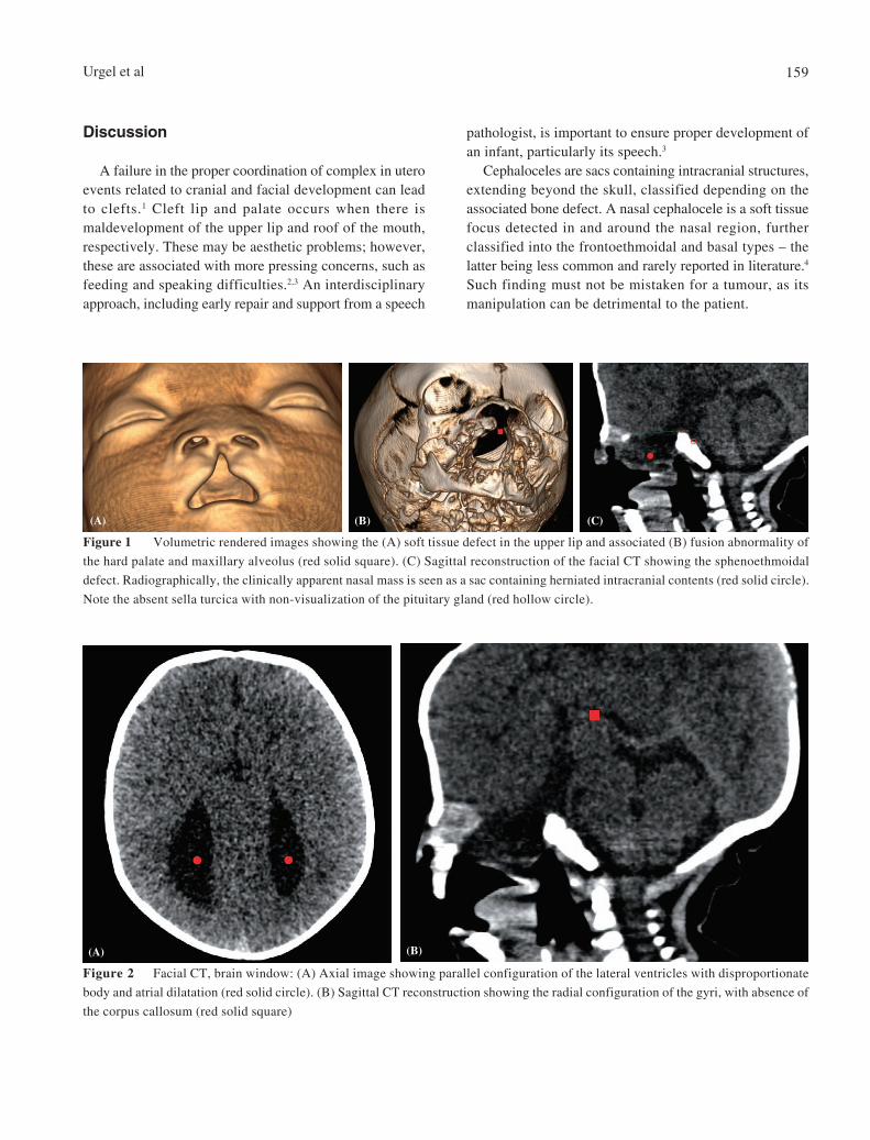
Urgel et al 159
Discussion
A failure in the proper coordination of complex in uteroevents related to cranial and facial development can leadto clefts.1 Cleft lip and palate occurs when there ismaldevelopment of the upper lip and roof of the mouth,respectively. These may be aesthetic problems; however,these are associated with more pressing concerns, such asfeeding and speaking difficulties.2,3 An interdisciplinaryapproach, including early repair and support from a speech
pathologist, is important to ensure proper development ofan infant, particularly its speech.3
Cephaloceles are sacs containing intracranial structures,extending beyond the skull, classified depending on theassociated bone defect. A nasal cephalocele is a soft tissuefocus detected in and around the nasal region, furtherclassified into the frontoethmoidal and basal types – thelatter being less common and rarely reported in literature.4
Such finding must not be mistaken for a tumour, as itsmanipulation can be detrimental to the patient.
Figure 2 Facial CT, brain window: (A) Axial image showing parallel configuration of the lateral ventricles with disproportionate
body and atrial dilatation (red solid circle). (B) Sagittal CT reconstruction showing the radial configuration of the gyri, with absence of
the corpus callosum (red solid square)
(A) (B)
Figure 1 Volumetric rendered images showing the (A) soft tissue defect in the upper lip and associated (B) fusion abnormality of
the hard palate and maxillary alveolus (red solid square). (C) Sagittal reconstruction of the facial CT showing the sphenoethmoidal
defect. Radiographically, the clinically apparent nasal mass is seen as a sac containing herniated intracranial contents (red solid circle).
Note the absent sella turcica with non-visualization of the pituitary gland (red hollow circle).
(A) (B) (C)
Midline Cranial and Facial Defects160
Facial clefts and cephaloceles can occur simultaneouslywith high probability, as both are due to failure of structuralfusion during embryogenesis. However, their associationhas not been adequately published in available literature.In our case, the incidentally seen rounded intranasal masscorresponded to a CSF-containing sac that herniatedthrough a sphenoethmoidal defect. The rare incidence ofbasal cephaloceles and an even rarer association with cleftlip and palate was even made noteworthy by the absenceof the corpus callosum. In agenesis of the corpus callosum,there is lack of the structure linking both cerebralhemispheres. It can be seen in isolation or as part of asyndrome and can be due to exogeneous factors duringpregnancy or be genetically inherited.5
Radiologic imaging is not always needed in cases ofcleft lip and palate. In our case, the presence of a nasalmass was the primary reason for performing a CT, as aneoplastic tumour in any site is not common for an infant.Its presence suggested a congenital abnormality or vascularpathology. With cross-sectional imaging, the exactaetiology of the nasal mass was better defined. Likewise,the presence of other congenital anomalies was detectedwith CT; magnetic resonance imaging may provide moreinformation, but unfortunately was not done to poor patientcompliance and follow-up. Ultrasound may also bebeneficial despi te i ts l imitat ions in paediatr icneuroimaging. Consent was obtained from the patient'smother for case publication.
Conclusion
Radiologic investigation of cases of cleft lip and palateis not always needed but may be considered in patientspresenting with other cranial abnormalities. Performingadditional imaging procedures in such cases will not onlyhelp in identifying associated pathologies not seenclinically but will also help the clinicians in choosing andplanning the right management for patients.
Declaration of Interest
No conflict of interest to declare.
References
1. Monasterio FO, Taylor JA. Major craniofacial clefts: case seriesand treatment philosophy. Plastic and Reconstructive Surgery 2008;122:534-43.
2. Goswami M, Jangra B, Bhushan U. Management of feedingproblem in a patient with cleft lip/palate. Int J Clin Pediatr Dent2016;9:143-5.
3. Nagarajan R, Savitha VH, Subramaniyan B. Communicationdisorders in individuals with cleft lip and palate: an overview. IndianJ Plast Surg 2009;42(Suppl):S137-43.
4. Howing EW. Nasal encephaloceles. Childs Nerv Syst 2000;16:702-6.
5. Schell-Apacik CC, Wagner K, Bihler M, Ertl-Wagner B, et al.Agenesis and dysgenesis of the corpus callosum: clinical, geneticand neuroimaging findings in a series of 41 patients. Am J MedGenet Part A 2008;146A:2501-11.
HK J Paediatr (new series) 2021;26:161-164
Diffusion Weighted Imaging and MR Spectroscopic Findings inMaple Syrup Urine Disease: The Importance of Early Radiological
Diagnosis in the Prevention of Cerebral Parenchymal Damage
B GÜNEY, iÖ YENIçERI, E DO AN, G AKçAY
Abstract Maple Syrup Urine Disease is a very rare autosomal recessive inherited disease that results from thedefect in the catalytic activity of the alpha ketoacid dehydrogenase enzyme complex. The deficiency inthe catabolism of branched chain amino acids naturally results in the accumulation of these amino acids(leucine, isoleucine and valine) and toxic metabolic end products in human blood and urine. In thisstudy, the importance in early diagnosis of cranial DWI imaging and MR spectroscopy and evaluation oftreatment efficacy in a newborn who was referred to the emergency room with ketoacidotic coma wasreported.
Key words Diffusion weighted imaging; Maple syrup urine disease; MR spectroscopy
Mu la S tk Koçman University, Faculty of Medicine,Radiology, Mu la; Pamukkale University, Faculty ofMedicine, Pediatrics, Denizli, Turkey
B GÜNEY MDÖ YENIÇERI MD
E DO AN MDG AKÇAY MD
Correspondence to: Dr B GÜNEY
Email: [email protected]
Received January 22, 2020
Introduction
Maple Syrup Urine Disease (MSUD) is a very rareautosomal recessive inherited disease that results from thedefect in the catalytic activity of the alpha ketoaciddehydrogenase enzyme complex. The deficiency in thecatabolism of branched-chain amino acids (BCAA)naturally results in the accumulation of these amino acids(leucine, isoleucine and, valine) and toxic metabolic endproducts in human blood and urine.1,2 The estimatedfrequency of MSUD in the world varies from 1: 185.000 to1:940.000.3 The very rare occurrence of the disease causesit not to be included in routine a screening programs inmany countries. However, it is thought that the real
Case Report
incidence is higher because of the undiagnosed deathcauses as the disease is not in screening program. Autosomalrecessive genetic diseases such as MSUD are more commonin countries like in Turkey where consanguineous marriagesare frequent and screening programs are limited.4,5
Accumulating BCAA and their metabolites in MSUDdisease are markedly neurotoxic. If the patient is not treatedwith BCAA-free formulation, irreversible brain damage willoccur. In untreated case of MSUD, ketoacidosis,hypoglycaemia, lethargy and epileptic seizures are seenover time. If patient can survive, the developmental andmental retardation develop in later stage of life.6
In this study, we aimed to discuss the importance ofDiffusion Weighted-Magnetic Resonance Imaging (DW-MRI) and Magnetic Resonance Spectroscopy (MRS) inthe diagnosis and in the early treatment of the disease as a9 day old male with MSUD who was admitted to emergencydepartment with ketoacidotic coma and treated in theneonatal intensive care unite.
Case Report
A male, 9 days old, term newborn with birth-weight of2800 grams, and born by cesarean section was brought toour emergency service by his family. He was foundimmobile in his bed and with purple colour. The family
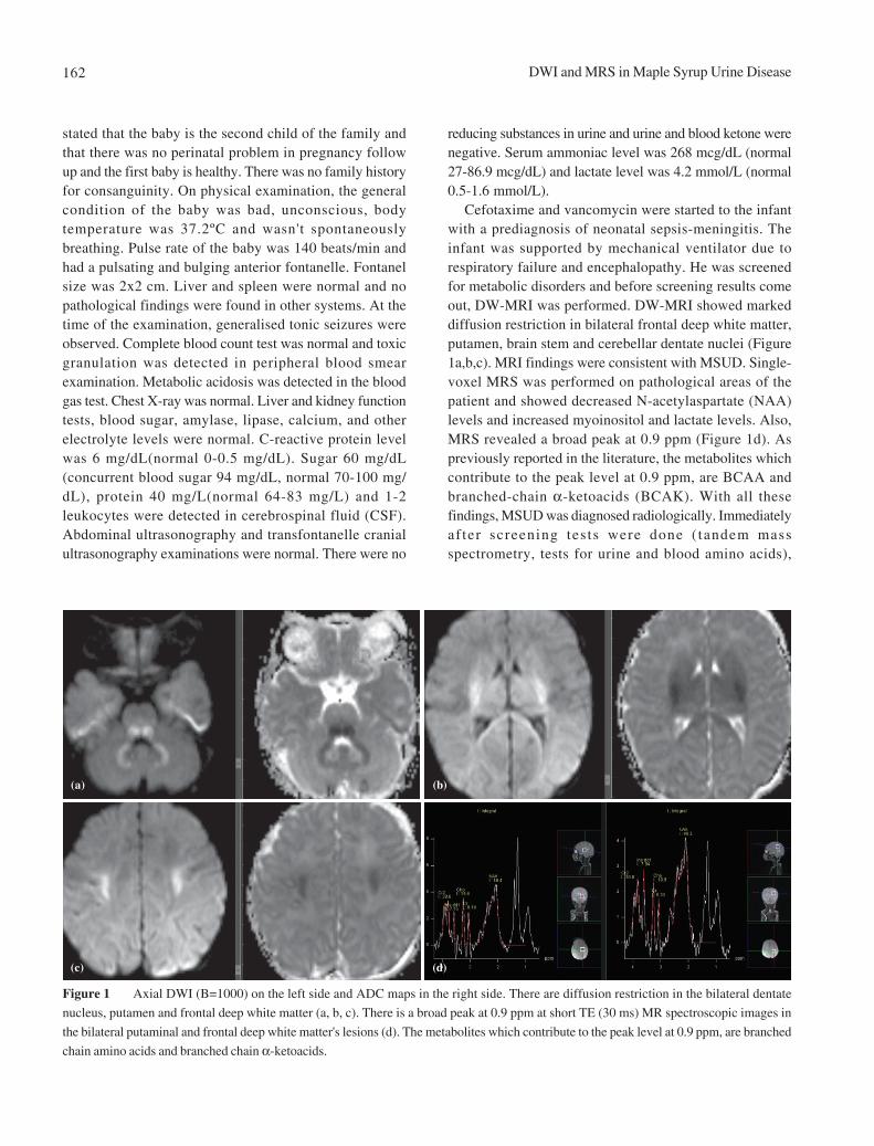
DWI and MRS in Maple Syrup Urine Disease162
stated that the baby is the second child of the family andthat there was no perinatal problem in pregnancy followup and the first baby is healthy. There was no family historyfor consanguinity. On physical examination, the generalcondition of the baby was bad, unconscious, bodytemperature was 37.2ºC and wasn't spontaneouslybreathing. Pulse rate of the baby was 140 beats/min andhad a pulsating and bulging anterior fontanelle. Fontanelsize was 2x2 cm. Liver and spleen were normal and nopathological findings were found in other systems. At thetime of the examination, generalised tonic seizures wereobserved. Complete blood count test was normal and toxicgranulation was detected in peripheral blood smearexamination. Metabolic acidosis was detected in the bloodgas test. Chest X-ray was normal. Liver and kidney functiontests, blood sugar, amylase, lipase, calcium, and otherelectrolyte levels were normal. C-reactive protein levelwas 6 mg/dL(normal 0-0.5 mg/dL). Sugar 60 mg/dL(concurrent blood sugar 94 mg/dL, normal 70-100 mg/dL), protein 40 mg/L(normal 64-83 mg/L) and 1-2leukocytes were detected in cerebrospinal fluid (CSF).Abdominal ultrasonography and transfontanelle cranialultrasonography examinations were normal. There were no
reducing substances in urine and urine and blood ketone werenegative. Serum ammoniac level was 268 mcg/dL (normal27-86.9 mcg/dL) and lactate level was 4.2 mmol/L (normal0.5-1.6 mmol/L).
Cefotaxime and vancomycin were started to the infantwith a prediagnosis of neonatal sepsis-meningitis. Theinfant was supported by mechanical ventilator due torespiratory failure and encephalopathy. He was screenedfor metabolic disorders and before screening results comeout, DW-MRI was performed. DW-MRI showed markeddiffusion restriction in bilateral frontal deep white matter,putamen, brain stem and cerebellar dentate nuclei (Figure1a,b,c). MRI findings were consistent with MSUD. Single-voxel MRS was performed on pathological areas of thepatient and showed decreased N-acetylaspartate (NAA)levels and increased myoinositol and lactate levels. Also,MRS revealed a broad peak at 0.9 ppm (Figure 1d). Aspreviously reported in the literature, the metabolites whichcontribute to the peak level at 0.9 ppm, are BCAA andbranched-chain α-ketoacids (BCAK). With all thesefindings, MSUD was diagnosed radiologically. Immediatelyaf ter screening tes ts were done ( tandem massspectrometry, tests for urine and blood amino acids),
Figure 1 Axial DWI (B=1000) on the left side and ADC maps in the right side. There are diffusion restriction in the bilateral dentate
nucleus, putamen and frontal deep white matter (a, b, c). There is a broad peak at 0.9 ppm at short TE (30 ms) MR spectroscopic images in
the bilateral putaminal and frontal deep white matter's lesions (d). The metabolites which contribute to the peak level at 0.9 ppm, are branched
chain amino acids and branched chain α-ketoacids.
(a) (b)
(c) (d)
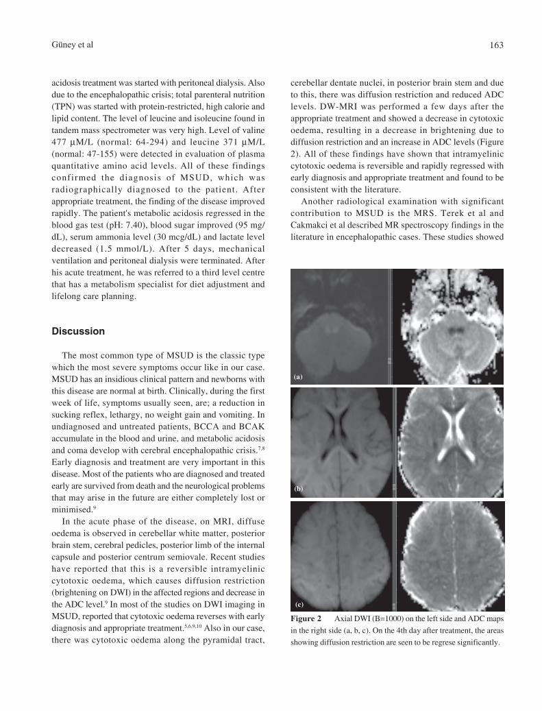
Güney et al 163
acidosis treatment was started with peritoneal dialysis. Alsodue to the encephalopathic crisis; total parenteral nutrition(TPN) was started with protein-restricted, high calorie andlipid content. The level of leucine and isoleucine found intandem mass spectrometer was very high. Level of valine477 µM/L (normal: 64-294) and leucine 371 µM/L(normal: 47-155) were detected in evaluation of plasmaquantitative amino acid levels. All of these findingsconfirmed the diagnosis of MSUD, which wasradiographically diagnosed to the patient. Afterappropriate treatment, the finding of the disease improvedrapidly. The patient's metabolic acidosis regressed in theblood gas test (pH: 7.40), blood sugar improved (95 mg/dL), serum ammonia level (30 mcg/dL) and lactate leveldecreased (1.5 mmol/L). After 5 days, mechanicalventilation and peritoneal dialysis were terminated. Afterhis acute treatment, he was referred to a third level centrethat has a metabolism specialist for diet adjustment andlifelong care planning.
Discussion
The most common type of MSUD is the classic typewhich the most severe symptoms occur like in our case.MSUD has an insidious clinical pattern and newborns withthis disease are normal at birth. Clinically, during the firstweek of life, symptoms usually seen, are; a reduction insucking reflex, lethargy, no weight gain and vomiting. Inundiagnosed and untreated patients, BCCA and BCAKaccumulate in the blood and urine, and metabolic acidosisand coma develop with cerebral encephalopathic crisis.7,8
Early diagnosis and treatment are very important in thisdisease. Most of the patients who are diagnosed and treatedearly are survived from death and the neurological problemsthat may arise in the future are either completely lost orminimised.9
In the acute phase of the disease, on MRI, diffuseoedema is observed in cerebellar white matter, posteriorbrain stem, cerebral pedicles, posterior limb of the internalcapsule and posterior centrum semiovale. Recent studieshave reported that this is a reversible intramyeliniccytotoxic oedema, which causes diffusion restriction(brightening on DWI) in the affected regions and decrease inthe ADC level.9 In most of the studies on DWI imaging inMSUD, reported that cytotoxic oedema reverses with earlydiagnosis and appropriate treatment.5,6,9,10 Also in our case,there was cytotoxic oedema along the pyramidal tract,
cerebellar dentate nuclei, in posterior brain stem and dueto this, there was diffusion restriction and reduced ADClevels. DW-MRI was performed a few days after theappropriate treatment and showed a decrease in cytotoxicoedema, resulting in a decrease in brightening due todiffusion restriction and an increase in ADC levels (Figure2). All of these findings have shown that intramyeliniccytotoxic oedema is reversible and rapidly regressed withearly diagnosis and appropriate treatment and found to beconsistent with the literature.
Another radiological examination with significantcontribution to MSUD is the MRS. Terek et al andCakmakci et al described MR spectroscopy findings in theliterature in encephalopathic cases. These studies showed
Figure 2 Axial DWI (B=1000) on the left side and ADC maps
in the right side (a, b, c). On the 4th day after treatment, the areas
showing diffusion restriction are seen to be regrese significantly.
(a)
(b)
(c)
DWI and MRS in Maple Syrup Urine Disease164
a decrease in NAA peak and an increase in the lactate andmyoinositol peak in involved areas and an abnormallyincreased broad peak due to BCAA and BCAKaccumulation at 0.9 ppm.5,11 Our patient also has a largeMR spectroscopic peak at 0.9 ppm at low TE (TE 30) single-voxel MR spectroscopy.
Conclusion
As a result, reversible cerebral parenchymal injury andneurotoxicity in MSUD, if not diagnosed early anduntreated, become irreversible and cause severeneurological deficits in the future. DWI and MRspectroscopy are important MR techniques in earlydiagnosis of the disease. Additionally, DWI seems to beradiologically important in evaluating the efficacy oftreatment and therefore, follow up of regression of cerebralparenchymal intramyelinic cytotoxic oedema.
Declaration of Interest
No conflict of interest was declared by the authors.
Financial Disclosure
The authors declared that this case has received nofinancial support.
References
1. Ha JS, Kim TK, Eun BL, et al. Maple syrup urine encephalopathy:a follow-up study in the acute stage using diffusion-weightedMRI. Pediatr Radiol 2004;34:163-6.
2. Sakai M, Inoue Y, Oba H, et al. Age dependence of diffusion-weighted magnetic resonance imaging findings in maple syrupurine disease encephalopathy. J Comput Assist Tomogr 2005;29:524-7.
3. Ogier de Baulny H, Saudubray JM. Branched-chain organicacidurias. Semin Neonatol 2002;7:65-74.
4. Gorzelany K, Dursun A, Co kun T, et al. Molecular genetics ofmaple syrup urine disease in the Turkish population. Turk JPediatr 2009;51:97-102.
5. Terek D, Koroglu O, Yalaz M, et al. Diagnostic Tools of EarlyBrain Disturbances in an asymptomatic neonate with Maple SyrupUrine Disease. Neuropediatrics 2013;44:208-12.
6. Kilicarslan R, Alkan A, Demirkol D, Toprak H, Sharifov R. Maplesyrup urine disease: diffusion-weighted MRI findings during acutemetabolic encephalopathic crisis. Jpn J Radiol;30:522-5.
7. Muller K, Kahn T, Wendel U. Is demyelination a feature of maplesyrup urine disease? Pediatr Neurol 1993;9:375-82.
8. Riviello JJ Jr, Rezvani I, DiGeorge AM, Foley CM. Cerebraledema causing death in children with maple syrup urine disease.J Pediatr 1991;119:42-5.
9. Ferraz-Filho JR, Floriano VH, Quirici MB, Albuquerque RP,Souza AS. Contribution of the diffusion-weighted MRI in thediagnosis and follow-up of encephalopathy caused by maplesyrup urine disease in a full-term newborn. Arq Neuropsiquiatr.2009;67:719-23.
10. Jain A, Jagdeesh K, Mane R, Singla S. Imaging in classic form ofmaple syrup urine disease: a rare metabolic central nervoussystem. J Clin Neonatol 2013;2:98-100.
11. Cakmakci H, Pekcevik Y, Yis U, Unalp A, Kurul S. Diagnosticvalue of proton MR spectroscopy and diffusion-weighted MRimaging in childhood inherited neurometabolic brain diseasesand review of the literature. Eur J Radiol 2010;74:161-71.
HK J Paediatr (new series) 2021;26:165-167
Median Cleft of Lower Lip with Central Cleft Palate andAnkyloglossia: A Case Report
PMY TANG, EKW CHAN, BSY TSUI, NSY CHAO
Abstract Purpose: Median cleft of the lower lip is an uncommon condition. We report a case of a non-syndromicChinese girl with a central cleft palate, median lower cleft lip and anklylossia. Methods: A full term 6momths old female child presented to our clinic for central cleft palate. Her palate was repaired uneventfullyat 12 months of age. During the regular post palatoplasty follow up when she was 5-year-old, we noticeda subtle notch in the midline lower lip and ankyloglossia. Findings: As subsequent speech assessmentshowed hyponasal speech, velo-pharyngeal insufficiency corrective surgery together with surgical tonguetie release and surgical repair of the median cleft lower lip were offered. The parents needed more time tothink about it. Conclusions: This case report highlighted the importance of longitudinal clinical followup of patient with cleft anomalies.
Key words Ankyloglossia; Median cleft lower lip
Department of Surgery, Hong Kong Children's Hospital,1 Shing Cheong Road, Kowloon Bay, Kowloon, Hong KongSAR, China
PMY TANG FRCS(Paed Surg)EKW CHAN FRCS(Paed Surg)BSY TSUI FRCS(Paed Surg)NSY CHAO FRCS(Paed Surg)
Correspondence to: Dr PMY TANG
Email: [email protected]
Received February 25, 2020
Introduction
Median cleft of the lower lip is an uncommon condition.Despite a cleft lip is often considered as a congenitalanatomical anomaly, sometimes, the clinical presentationcan be so subtle that it can be missed by multiple healthcareprofessionals, especially when the lesion is asymptomatic.The incidence of median cleft lower lip in the Chinesepopulation has not been reported. Hereby we report a caseof a non-syndromic Chinese girl with central cleft palate,and a delayed presentation of median lower cleft lip andanklyloglossia.
Case Report
Case Report
A full term 6 months old female child was brought toour clinic for incidental finding of central cleft palate. Shewas born in China with unremarkable antenatal history.Her parents were of non-consanguineous marriage andthere was no known maternal exposure of drugs orradiation. She had no family history of cleft lip or palate.On clinical examination at 6 months old, there was noobvious cleft lip and a wide central cleft palate involvingthe soft and hard palate. There was no cleft alveolus. Shealso had a 4 cm non-complicated haemangioma at thebuttock, otherwise she has no other dermatological orskeletal anomalies. Audiological and clinical assessmentby the Ear, Nose and Throat surgeon revealed bilateralotitis media effusion with moderate hearing loss. Electivecleft palate repair and bilateral grommet insertion wasperformed at one year of age as per our centre's protocol.Pre-op routine ultrasound examination and echocardiogramconfirmed no structural cardiac, urinary or central nervoussystem anomalies. Von Langenbeck's palatoplasty withintravelo-veloplasty was performed under generalanaesthesia uneventfully. Her post op recovery wasuncomplicated and she continued to receive regular
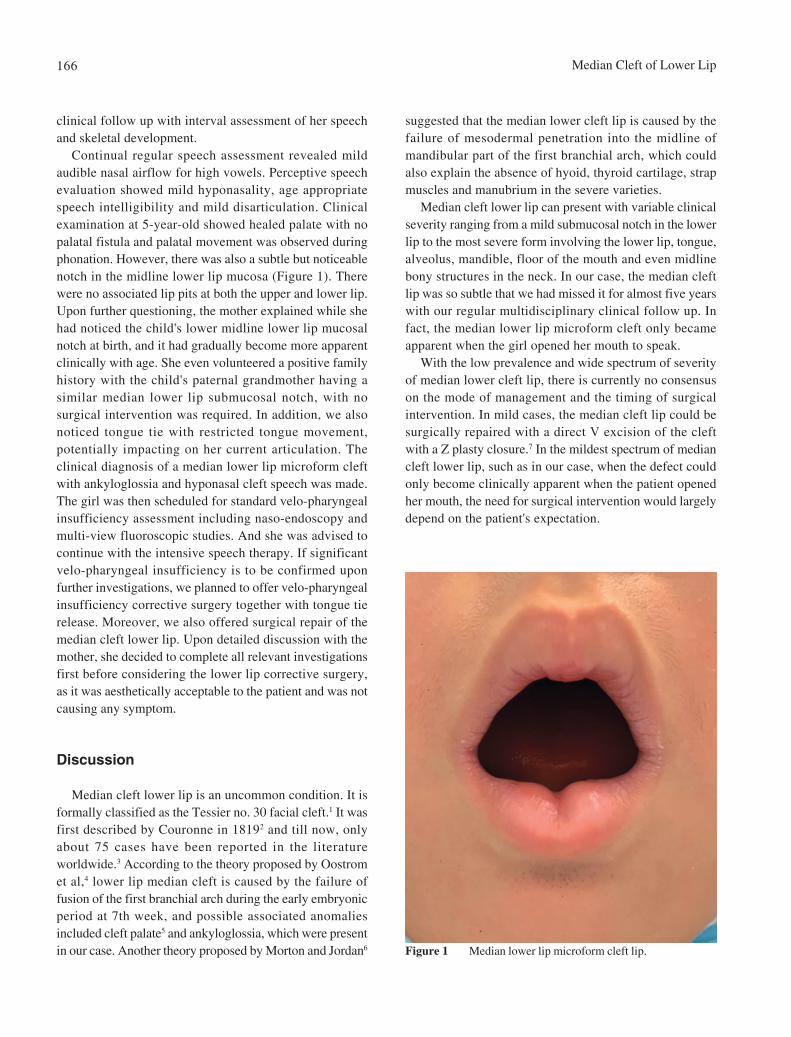
Median Cleft of Lower Lip166
clinical follow up with interval assessment of her speechand skeletal development.
Continual regular speech assessment revealed mildaudible nasal airflow for high vowels. Perceptive speechevaluation showed mild hyponasality, age appropriatespeech intelligibility and mild disarticulation. Clinicalexamination at 5-year-old showed healed palate with nopalatal fistula and palatal movement was observed duringphonation. However, there was also a subtle but noticeablenotch in the midline lower lip mucosa (Figure 1). Therewere no associated lip pits at both the upper and lower lip.Upon further questioning, the mother explained while shehad noticed the child's lower midline lower lip mucosalnotch at birth, and it had gradually become more apparentclinically with age. She even volunteered a positive familyhistory with the child's paternal grandmother having asimilar median lower lip submucosal notch, with nosurgical intervention was required. In addition, we alsonoticed tongue tie with restricted tongue movement,potentially impacting on her current articulation. Theclinical diagnosis of a median lower lip microform cleftwith ankyloglossia and hyponasal cleft speech was made.The girl was then scheduled for standard velo-pharyngealinsufficiency assessment including naso-endoscopy andmulti-view fluoroscopic studies. And she was advised tocontinue with the intensive speech therapy. If significantvelo-pharyngeal insufficiency is to be confirmed uponfurther investigations, we planned to offer velo-pharyngealinsufficiency corrective surgery together with tongue tierelease. Moreover, we also offered surgical repair of themedian cleft lower lip. Upon detailed discussion with themother, she decided to complete all relevant investigationsfirst before considering the lower lip corrective surgery,as it was aesthetically acceptable to the patient and was notcausing any symptom.
Discussion
Median cleft lower lip is an uncommon condition. It isformally classified as the Tessier no. 30 facial cleft.1 It wasfirst described by Couronne in 18192 and till now, onlyabout 75 cases have been reported in the literatureworldwide.3 According to the theory proposed by Oostromet al,4 lower lip median cleft is caused by the failure offusion of the first branchial arch during the early embryonicperiod at 7th week, and possible associated anomaliesincluded cleft palate5 and ankyloglossia, which were presentin our case. Another theory proposed by Morton and Jordan6
suggested that the median lower cleft lip is caused by thefailure of mesodermal penetration into the midline ofmandibular part of the first branchial arch, which couldalso explain the absence of hyoid, thyroid cartilage, strapmuscles and manubrium in the severe varieties.
Median cleft lower lip can present with variable clinicalseverity ranging from a mild submucosal notch in the lowerlip to the most severe form involving the lower lip, tongue,alveolus, mandible, floor of the mouth and even midlinebony structures in the neck. In our case, the median cleftlip was so subtle that we had missed it for almost five yearswith our regular multidisciplinary clinical follow up. Infact, the median lower lip microform cleft only becameapparent when the girl opened her mouth to speak.
With the low prevalence and wide spectrum of severityof median lower cleft lip, there is currently no consensuson the mode of management and the timing of surgicalintervention. In mild cases, the median cleft lip could besurgically repaired with a direct V excision of the cleftwith a Z plasty closure.7 In the mildest spectrum of mediancleft lower lip, such as in our case, when the defect couldonly become clinically apparent when the patient openedher mouth, the need for surgical intervention would largelydepend on the patient's expectation.
Figure 1 Median lower lip microform cleft lip.
Tang et al 167
And as per our centre's usual practice, release of tonguetie was usually only performed according to the NationalInstitute for Health and Care Excellence guidelines8 – whenthe tongue tie was impairing breastfeeding; therefore, weseldom perform "routine" tongue tie release in non-verbalpatients as frequently, this anatomical anomaly couldremain asymptomatic without surgical intervention.However, in this case, as the tongue tie would potentiallybe one of the contributing factors for her speechimpairment, surgical release of tongue tie was offered aftercomprehensive speech assessment.
Interestingly, there seems to be degree of geneticinheritance in the median cleft lip in our patient as herpaternal grandmother was said to have the similar clinicalpresentation. Unfortunately, we were unable to carry outgenetic testing due to resource limitation. Apart fromchromosomal aberrations,9 other possible associatedanomalies such as congenital heart deformities,10 PierreRobin anomaly,11 hand anomalies12 and foot deformities13
have been reported in the literatures.There was currently no literature describing any
association between buttock haemangioma and medianlower lip cleft. In our case, the buttock haemangiomaspontaneously resolved with time without medicaltreatment. Given this natural disease course, it was likelyto be the common infantile haemangioma and thus, it couldvery well just be an incidental finding of a commonclinical condition (infantile haemangioma) in a patientwith another uncommon condition (median lower cleftlip with cleft palate).
Conclusion
We described the clinical findings and managementof a non-syndromic Chinese girl with central cleftpalate, buttock haemangioma and a delayed presentationof median cleft lower lip and ankyloglossia. Theimportance of longitudinal clinical follow up of thesepatients was highlighted, and thorough physicalexamination and detailed clinical & family historyshould always be performed.
Conflict of Interest
The authors declare they have no competing interest.
References
1. Paul Tessier. Anatomical classification of facial, cranio-facialand laterao-facial clefts. J Maxillofac Surg 1976:69-92.
2. Monroe CW. Midline cleft of the lower lip, mandible and tonguewith flexion contracture of the neck: case report and review ofthe literature. Plast Reonstr Surg 1966;38:312-9.
3. Rao AYN. Complete midline cleft of lower lip, mandible, tongue,floor of mouth with neck contracture: a case report and review ofliterature. Craniomaxillofac Trauma Reconstr 2015;8:363-9.
4. Oostrom CA, Vermeij-Keers C, GIlber PM, van der Meulen JC.Median cleft of the lower lip and mandible: case reports, a newembryologic hypothesis, and subdivision. Plast Reconstr Surg1996;97:313-20.
5. Chidzonga MM, Shija JK. Congenital median cleft of the lower lip,bifid tongue with ankyloglossia, cleft palate and submentalepidermoid cyst: report of a case. J Oral Maxillofac Surg 1988;46:809-12.
6. Henry K, Kawamoto, Jr. Rare craniofacial clefts. In Joseph G.McCarthy. Plast Surg. WB. Saunders Company, 1990;2927-41
7. Seyhan T, Kykync H. Median cleft of the lower lip: report oftwo new cases and review of the literature. Ann Otol RhinolLaryngol 2002;111:217-21.
8. Division of ankyloglossia (tongue tie) for breastfeeding.Interventional procedures guidance (IPG149). Dec 2005.
9. Rey A, Vasquez MP, Jennequin P, Marie MP. Fenteslabiomandibulaires. A propos D'un cas. Revue de la literature.Rec Stomatol Chir Maxillofac 1982;83:39-44.
10. Davis AD. Medial cleft of the lower lip and mandible: a casereport. Plast reconstr Surg 1950;6:62-7.
11. Ranta R. Incomplete median cleft of the lower lip associatedwith cleft palate, the Pierre Robin anomaly or hypodontia. Int JOral Sug 1984;13:555-8.
12. Iregbulem LM. Median cleft of the lower lip: case report. PlastReconstr Surg 1978;61:787-9.
13. Ashley LM, Richardson GE. Multiple congenital anomalies in astillborn infant. Anat Rec 1943;86:457-73.
HK J Paediatr (new series) 2021;26:168-174
Hong Kong Universal Newborn Hearing Screening (UNHS)Care Path Protocol under Joint Committee on UNHS
TPY MA, WY CHAN, C KOH, W YEUNG, KY WONG, J SUNG, NS KWONG, CC SHEK, SL LEE,CME WONG, C LAM, F LEE, K TSO, K YEUNG, E HAU, R TSE, KM HO, W WONG;
HA-DH JOINT COMMITTEE ON CARE PATH FOR UNIVERSAL NEWBORN HEARING SCREENING
Abstract The Joint Committee on Universal Newborn Hearing Screening (UNHS) was formed to ensure theeffective implementation of UNHS in Hong Kong and to achieve effective communication among variousservices in Hospital Authority and Department of Health. Meetings with multidisciplinary professionalshad been conducted during the years of 2013 and 2017. A consensus on a common care path for infantsidentified to have hearing impairment had been reached.
Key words Hearing; Newborn; Screening
HA-DH Joint Committee on Care Path for UniversalNewborn Hearing Screening, Hong Kong SAR, China
Correspondence to: Dr. TPY MA
Email: [email protected]
Received March 9, 2020
Background
The Joint Committee on Universal Newborn HearingScreening (UNHS) was formed to ensure the effectiveimplementation of UNHS in Hong Kong and to achieveeffective communication among various services inHospital Authority (HA) and Department of Health (DH).Meetings with multidisciplinary professionals had beenconducted during the years from 2013 to 2017. Aconsensus on a common care path for infants identified tohave hearing impairment had been reached (Appendix I).
Children with hearing impairment are usually classifiedaccording to the degree and nature of the impairment.Hearing impairment can be conductive, sensorineural, ormixed in nature. Hearing is measured in decibels hearinglevel (dB HL) with the threshold of 0 dB for each frequencydenoting the value at which normal young adults perceivea pure tone of a given intensity and frequency 50% of thetime. The table below shows one of the more commonlyused classification systems for the degree of hearingimpairment in Hong Kong.
Contemporary Practice
Degree of hearing Hearing impairment rangeimpairment (dB HL)
Mild grade 26 to 40
Moderate grade 41 to 55
Moderately severe grade 56 to 70
Severe grade 71 to 90
Profound grade >90
Goodman A. Reference zero levels for pure tone audiometers. ASHA 1965;
7:262-3.1
Children with hearing impairment (HI) greater than orequal to moderate grade are classified as having significanthearing impairment. Early identification, diagnosis, andintervention are crucial to the successful management ofthese children to alleviate the possible adverse outcomeon the language and communication, cognition, reading,and social-emotional development.
Over 5% of the world population that is 360 millionpeople has significant hearing impairment (328 millionadults and 32 million children).2 The internationalprevalence of permanent childhood hearing impairment(PCHI) of moderate or greater degrees is about 1 to 3 per1,000 children, based on screening and/or medicalrecords.3,4 The number increases to 1 in every 40 for infantswho require care in the Neonatal Intensive Care Unit.5 InHong Kong, for every 1,000 children under the age of 15years, 1.3 of them were registered with significant HI inthe Central Registry for Rehabilitation in 2014. At the Child
Ma et al 169
Assessment Service (CAS) of the Hong Kong DH, newcases received with PCHI of moderate or greater degreesremained relatively stable over the past decade, with 61 newcases in 2007 and 49 in 2016. With the universal screeningprogramme in place and referring to the mean number ofbirths per year in Hong Kong over the period and CAS'scatchment coverage of over 85% of the population, anincidence of about 0.14% of children having PCHI isestimated, comparable to prevalence of developed regions.
The rate of the coexistence of additional disabilities ormedical conditions with hearing impairment in children ishigh. Most estimates suggest that between 30% and 40%of children with hearing impairment have one or moreadditional disabilities.6 Studies have reported that thefrequency of additional disabilities is similar across alllevels of hearing impairment, pointing to the need forchildren with mild or unilateral impairment, as well as thosewith more severe impairment, to receive thorough medicaland developmental evaluations.7 Among those withadditional disabilit ies, the commonest ones aredevelopmental delay (33%), cerebral palsy (20%), visionproblem (7.6%) and autism spectrum disorder (7.6%).8
Children with significant hearing impairment are at riskof growing up with deficits in language, communicativeabilities, cognition and literacy, psychosocial functioningand possible problems in balance and gross motorproficiency.9
Many in developed countries changed from using theterm "Universal Newborn Hearing Screening" to "EarlyHearing Detection and Intervention (EHDI)" programs. Thechange underscores that successfully identifying and servinginfants and young children with PCHI, requires goingbeyond screening to address issue related to audiologicaldiagnosis, appropriate medical and educational intervention,and coordination with the child's family.10
Hearing Screening for Children in Hong Kong:Universal Hearing Screening After Birth in HAHospitals
HA hospitals would provide universal newborn hearingscreening and make initial diagnosis for patients withcongenital or delayed hearing loss.
Screening Methods and Confirmatory TestsCurrently 2-stage Automated Auditory Brainstem
Response (AABR) screening program (or alternativeacceptable screening program in future) would be offered
to all neonates born in HA hospitals or outborn babiestransferred in from other hospitals. Babies who had failedthese screening tests (with AABR threshold > 35 dB)would be referred for confirmatory Brainstem AuditoryEvoked Potential (BAEP) test. Initial counselling wouldbe offered to their parents and information sheet wouldbe given to them. In keeping with the latest AmericanAcademy of Pediatrics Guidelines for newborn hearingscreening, BAEP should preferably be performed before4 months of age to facilitate early detection and subsequentinterventions for babies with congenital hearing loss. Babieswho failed confirmatory BAEP test would be referred toEar, Nose and Throat (ENT) Clinic; at which comprehensivehearing assessment by otolaryngologist (ENT specialists)would be performed. These babies would also be referredto paediatric clinics for follow-up and investigations.
Infants with Risk FactorsFor infants with risk factors associated with permanent
congenital, delayed-onset or progressive hearingimpairment in childhood, follow-up at HA PaediatricClinics should be considered.
These risk factors include:1. Family history of permanent childhood hearing
impairment2. Neonatal intensive care for more than 5 days3. Extracorporeal membrane oxygenation, assisted
ventilation, exposure to ototoxic drugs (which toxiclevel was detected on drug monitoring*)
4. Severe neonatal jaundice requiring exchangetransfusion
5. In utero infections, such as cytomegalovirus (CMV),herpes, rubella, syphilis and toxoplasmosis
6. Head trauma, especially fracture base of skull andfracture of temporal bone
7. Caregivers' concern regarding hearing loss8. Significant craniofacial anomalies9. Physical findings, such as white forelock, that are
associated with a syndrome known to include asensorineural or permanent conductive hearing loss
10. Syndromes associated with hearing loss or progressiveor late-onset hearing loss such as neurofibromatosis,Pendred syndrome and Jervell and Lange Nielsonsyndrome
11. Culture-positive postnatal infections associated withsensorineural hearing loss, including confirmedbacterial and viral (especially herpes viruses andvaricella) meningitis
Universal Newborn Hearing Screening170
* For hospitals in which drug level service is notavailable, they might offer repeat hearing test a fewmonths after discharge.
Screening for Congenital CMV InfectionUrine CMV screening (within 3 weeks of age) should
be considered for infants who have failed the NewbornHearing screening twice. Congenital CMV infection is themost frequent non-hereditary cause of sensorineuralhearing impairment worldwide. Studies had demonstratedear ly adminis t ra t ion of ant iv i ra l t rea tment insymptomatically infected infants with CMV infectionprevents hearing deterioration at 6 months and maypotentially prevent hearing deterioration at > or =1 year.11,12
Supplementary Screening in Maternal & ChildHealth Centres (MCHCs) of Family HealthService
MCHCs would provide screening for those infants whohad not been screened by their birthing hospitals (e.g.private hospitals), currently by 2-stage AutomatedOtoacoustic Emission (AOAE) method. The procedure andpotential limitations of AOAE test would be explained toparents before the screening.
For those infants who failed AABR screening once,parents would be advised to return to their birth hospitalsfor completion of the AABR test. If the parents refuse toreturn to the hospitals (where initial AABR was performed),their babies would be referred to HA ENT for furthermanagement of failed AABR. These infants would bescreened irrespective of their risk status except those withabsent auditory canal. AOAE screening would not beprovided for those who had already been referred foraudiological assessment or babies more than 4 months ofage.
First AOAE screening would be performed for infantsaged between 2 weeks to 4 months. Preferably this firstscreening would be performed within 2 weeks after thefirst MCHC registration. Second AOAE test would beoffered to those infants who failed the initial AOAE test,preferably within one week after the first AOAE. For thosewho failed AOAE test twice, they would be referred to HAENT or audiologist services for confirmatory testing.
Routine hearing surveillance and developmentalsurveillance would be provided to all children even forthose who had passed both AOAE or who had passed the
UNHS program in HA or private hospitals. If they failto ach ieve the pre -de te rmined deve lopmenta lmilestones or if there are parental concerns on possiblehearing impairments, these children would be referredto HA ENT or audiology services for further evaluationsif indicated.
Management of Children with ConfirmedHearing Impairment
Referral to Other Services After Identification of HearingLoss
Referrals may be made to CAS, The Duchess ofKent Children's Hospital at Sandy Bay Child AssessmentCentre (DKCAC), Clinical Genetic Service (CGS),ophthalmologists or other specialties as clinicallyindicated. Patients with mild grade hearing impairmentwould be followed-up at HA ENT Clinics. Behavioural tests(paediatric audiometry) would be performed for babieswith hearing impairment at around 6-9 months of age forbetter delineation on the degree of hearing impairmentsfor these babies. For babies with severe or profound hearingimpairment, early imaging studies (CT and MRI) would beperformed to look for any structural anomalies and to planfor cochlear implantations. These babies should also bereferred to ophthalmologist for assessment in view of thehigh rate of associated visual abnormalities. For babies withpersistent HI, referral will be made to appropriate servicefor hearing aids arrangements.
CAS and DKCACCAS or DKCAC would receive referral from HA
hospitals, private hospitals, Education Bureau and MCHCfor further workup and management for patients withhearing impairment. After identification of hearingimpairment, CAS or DKCAC would take up the co-ordination role in multi-disciplinary management of thesechildren.
Referral could be made to CAS or DKCAC by the ENTsurgeons for infants with the following criteria, after ENTassessment:1. Significant hearing impairment cases (permanent
hearing impairment of moderate degree or worse inthe better ear)
2. Unilateral severe to profound sensorineural hearingimpairment
3. Bilateral severe to profound high frequency hearingimpairment
Ma et al 171
Children with mild sensorineural hearing loss could befollowed-up by ENT. Patients with suspected syndromaldiagnosis or high risk of developmental delay could alsobe directly referred to CAS or DKCAC by the HA hospitalsPaediatrics Department for follow-up and detailedevaluation.
After receiving referral for significant hearingimpairment, nurse interview would be conducted for briefhistory taking at CAS. Audiologists would provideinformation on available community resources andcounselling to the parents. Audiologists in CAS or DKCACmay perform hearing assessment again if the child is olderthan 6 months of age. School placement for Early Educationand Training Centre (EETC) (deaf) or EETC and SpecialChild Care Centre (deaf) would be arranged for them.
In CAS, when the hearing impairment is diagnosed at 3months to 6 months of age, the child would be assessed bypaediatric doctor for a comprehensive physical anddevelopmental assessment within 2 months after thereferral. A team conference with paediatric doctor,audiologist and medical social worker would be conductedto formulate the final management plan for the patient. Ifthe child is aged more than 6 months, CAS would providephysical and developmental assessment by paediatricdoctor, interim language support by speech therapist, anda team conference with paediatric doctor, speech therapist,audiologist and medical social worker would be arrangedfor management plan and counselling. The child may bereferred to HA specialist for further investigation andmanagement if necessary.
In DKCAC, when the hearing impairment is diagnosed,the child would be assessed by the paediatric doctors for acomprehensive assessment and an aetiology work-up within2 months. Subsequently, the child would go through amulti-disciplinary assessment with paediatric doctors,audiologists, speech therapists and medical social workersto formulate his/her final management plan. The child mayalso be referred to other HA specialists for furtherinvestigation and management when necessary.
Assessment of the child's functioning in the aspects ofhearing, communication and language, cognition, literacyand motor development helps to determine what type oftraining the child needs. Together with audiologists,developmental paediatricians take comprehensive medicaland family histories to identify possible risk factors andaetiology. Detailed physical examinations are performed
for possible aetiology and any associated physicalabnormalities. Developmental paediatricians assess thestrengths and weaknesses of the child in all developmentalareas and evaluate the presence of any associatedcomorbidities. A thorough evaluation of the child's levelof communication, speech and language development bythe speech therapist is important and should includeassessment on verbal comprehension, verbal expression,speech production, and speech perception. Assessment ofspoken language for these children should follow similarprocedures as those for the hearing children but adequatevisual cues should be noticed. Phonological errors anderror patterns should be analysed. Speech perception testsinclude assessment of the discrimination and identificationof suprasegmental and segmental features should beincluded to reflect the degree of communication handicapin everyday situation.
Audiologists at CAS would perform yearly phonefollow-up for children with hearing aids until these childrenenter primary school. They would follow-up on problemsof using hearing aid, any further audiological results,language development and difficulties in school coping, etc.
Paediatric doctors at CAS would review these childrenat 18 to 24 months of age. This aims to assess for anyimprovement after application of hearing aids or cochlearimplant and review of placement. If parents decline referralfor preschool services for children with hearingimpairment, speech therapists would review their languageprogress at 24 to 30 months of age. For children withsignificant hearing impairment, a combined multi-disciplinary assessment by paediatric doctor (+/- clinicalpsychologist), audiologist and speech therapist would bearranged before the children go to primary school. Grossmotor screening would also be performed for this groupof children to look for potential motor balance problems.Advice on school placement, accommodation, continualauditory and language training would be given.
Paediatric doctors at DKCAC would also review thesepatients at 18 to 24 months of age for developmentalassessment and review of replacement. Speech therapistswould then review them at 24 to 30 months of age forspeech and language progress. Prior to their admission toprimary school, an evaluation by paediatric doctors wouldbe conducted. Gross motor screening would also beperformed for them to look for potential motor balanceproblems.
Universal Newborn Hearing Screening172
CGSCGS would provide genetic assessment, testing and
counselling for patients with hearing impairments andsuspected genetic diseases.
Referral criteria for children with hearing impairmentto CGS:1. Suspected genetic or syndromic cause of hearing
impairment2. Positive family history of congenital hearing loss,
especially among first degree relatives3. Specific inner ear malformations or agenesis and/or
coexisting abnormalities involving other organsystems
4. Parenteral consanguinity5. Parental request, especially for family planning
purpose6. For interpretation and counselling of relevant genetic
testing results7. Hear ing impairment in a chi ld exposed to
aminoglycoside antibiotics
Please note that referral could be made irrespective ofthe severity of hearing impairment. Patients withoutsyndromic features could also be referred to CGS if therewas parental consanguinity or there was parental request,especially for family planning purposes.
Concern ing non-syndromic - re la t ed hea r ingimpairments, only 3 gene tests are available for the timebeing (GJB-2 gene, GJB-6 gene and mitochondrial DNA155C2A gene). These genetic testing had low pickup ratesof only around 30%.
Declaration of Interests
This research received no specific grant from anyfunding agency in the public, commercial, or not-for-profitsectors. All authors have disclosed no conflicts of interest.
Acknowledgement
HA Working Group on Universal Neonatal Hearing Screening
in Hong Kong.
Endorsement
This protocol had been endorsed by COC (Paed) in November
2017.
This protocol had been presented to COC (ENT) in November
2017.
References
1. Goodman A. Reference zero levels for pure tone audiometers.ASHA 1965; 7:262-3.
2. World Health Organization. Millions of people in the worldhave hearing loss that can be treated or prevented. 2013.Available from: https://www.who.int/pbd/deafness/news/Millionslivewithhearingloss.pdf
3. Centers for Disease Control and Prevention. Data and StatisticsAbout Hearing Loss in Children, 2009. Available from:https://www.cdc.gov/ncbddd/hearingloss/data.html.
4. Bussé A, Hoeve H, Nasserinejad K, Mackey A, Simonsz H,Goedegebure A. Prevalence of permanent neonatal hearingimpairment: systematic review and Bayesian meta-analysis. IntJ Audiol 2020;59:475-85.
5. Janssen RM. Permanent childhood hearing Impairment andthe universal newborn hearing Screening program in Britishcolumbia. UBC Medical Journal. 2013. Available from:https://med-fom-ubcmj.sites.olt.ubc.ca/files/2015/11/ubcmj_2_1_2010_16-19.pdf.
6. Punch R. Children who are deaf or hard of hearing withadditional disabilities. Victorian Deaf Education Institute, 2015.
7. Wiley S, Arjmand E, Meinzen-Derr J, Dixon M. Findingsfrom multidisciplinary evaluation of children with permanenthearing loss. Int J Pediatr Otorhinolaryngol 2011;75:1040-4.
8. Cupples L, Ching TYC, Crowe K, et al. Outcomes of 3-year-old children with hearing loss and different types of additionaldisabilities. J Deaf Stud Deaf Educ 2014;19):20-39.
9. Joint Committee on Infant Hearing; American Academy ofAudiology; American Academy of Pediatrics; AmericanSpeech-Language-Hearing Association; Directors of Speechand Hearing Programs in State Health and Welfare Agencies.Year 2000 position statement: principles and guidelines forearly hearing detection and intervention programs. JointCommittee on Infant Hearing, American Academy ofAudiology, American Academy of Pediatrics, AmericanSpeech-Language-Hearing Association, and Directors ofSpeech and Hearing Programs in State Health and WelfareAgencies. Pediatrics 2000;106:798-817.
10. White KR, Forsman I, Eichwald J, Munoz K. The evolutionof early hearing detection and intervention programs in theUnited States. Semin Perinatol 2010;34:170-9.
11. Kimberlin DW, Lin CY, Sánchez PJ, et al; National Instituteof Allergy and Infectious Diseases Collaborative Antiviral StudyGroup. Effect of ganciclovir therapy on hearing insymptomatic congenital cytomegalovirus disease involving thecentral nervous system: a randomized, controlled trial. JPediatr 2003;143:16-25.
12. Rawlinson WD, Boppana SB, Fowler KB, et al . Congenitalcytomegalovirus infection in pregnancy and the neonate:consensus recommendations for prevention, diagnosis, andtherapy. Lancet Infect Dis 2017;17:e177-88.
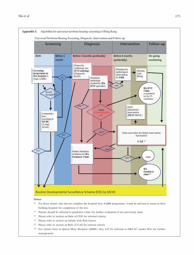
Ma et al 173
Appendix I. Algorithm for universal newborn hearing screening in Hong Kong
Notes:
*1 For those infants who did not complete the hospital base AABR programme, would be advised to return to their
birthing hospital for completion of the test
*2 Patient should be referred to paediatric clinic for further evaluation if not previously done
*3 Please refer to section on Role of CGS for referred criteria
*4 Please refer to section on infants with Risk factors
*5 Please refer to section on Role of CAS for referral criteria
*6 For infants born in Queen Mary Hospital (QMH), they will be referred to DKCAC (under HA) for further
management
Universal Newborn Hearing Screening, Diagnosis, Intervention and Follow-up
Universal Newborn Hearing Screening174
Appendix II. Members of the HA-DH Joint Committee on Care Path for UNHS
Hospital AuthorityDr. William Wong, Con(Paed), PWH (Chairperson)
Dr. Wilson Yeung, COS(Paed), PYNEH (Chairperson)
Dr. KY Wong, ex-Con(Paed), QMH
Dr. NS Kwong, COS(Paed & AM), TMH/POH
Dr. CC Shek, COS(Paed & AM), PMH
Dr. John Sung, AC(ENT), AHNH
Dr. SL Lee, Con(Paed), DKCH
Dr. Terence Ma, AC(Paed), PWH (Editor of this Protocol)
Mr. Eddie Wong, SO(ENT), QEH
Department of HealthDr. Catherine Lam, ex-CAS Consultant Paediatrician
Dr. Florence Lee, Con Paed, CAS
Dr. Rita Ho, PMO(FHS)
Dr. Karen Tso, SMO(FHS)3
Dr. Carline Koh, SMO(FHS) NTE2 (Editor of this Protocol)
Dr. Karen Yeung, MO(FHS)
Dr. Edgar Hau, MO, CGS (representing Dr. Stephan Lam)
Dr. Cyrus Chan, Paediatrician, CAS (Editor of this Protocol)
Ms. Rosa Tse, Audiologist, CAS
Education BureauMr. KM Ho, Specialist (Audiological Services)
HK J Paediatr (new series) 2021;26:175-179
Joint Position Statement on BioNTech Vaccination inAdolescents with Allergic Diseases
Abstract This Statement was prepared to encourage Hong Kong adolescents aged 12-17 years old to receiveBioNTech vaccine and recommend when to refer them for vaccine allergy evaluation. Importantly, thepersonal and societal benefits of BioNTech vaccination outweigh the risk of non-severe adverse vaccinereactions. BioNTech is currently contraindicated only in individuals with history of anaphylaxis to thisvaccine or its components (e.g., polyethylene glycol [PEG]) by appropriate allergy testing. Subjectswith history of immediate and severe allergic reactions to PEG-containing drugs or vaccines or immediateallergic reaction to the first dose of BioNTech warrant paediatric allergy referral to evaluate their fitnessto receive this vaccine. Allergy to unrelated drugs, food, insect venoms as well as asthma, allergic rhinitisand eczema are not contraindications for BioNTech. Patients with a history of severe allergic reactionsafter foods, unrelated drugs or vaccines can receive BioNTech with a longer observation of 30 minutespost-vaccination.
Key words Adolescent; Allergy; Contraindication; COVID-19; mRNA vaccine
Vice President and Chairman of Education Committee, HongKong College of Paediatricians; and Department ofPaediatrics, The Chinese University of Hong Kong, Princeof Wales Hospital, Hong Kong SAR, China
TF LEUNG MD, FHKAM(Paed)
President, Hong Kong College of Paediatricians, Hong KongSAR, China
WY TSE FHKAM(Paed)
President, Hong Kong Society for Paediatric ImmunologyAllergy and Infectious Diseases; and Department of Paediatrics,Queen Elizabeth Hospital, Hong Kong SAR, China
WH CHAN FHKAM(Paed)
Correspondence to: Prof TF LEUNG
Email: [email protected]
Received June 15, 2021
Contemporary Practice in Paediatrics
TF LEUNG, WY TSE, WH CHAN;ON BEHALF OF HONG KONG COLLEGE OF PAEDIATRICIANS AND
HONG KONG SOCIETY FOR PAEDIATRIC IMMUNOLOGY ALLERGY AND INFECTIOUS DISEASES
Background
The COVID-19 pandemic has struck the world for 18months now. Although COVID-19 mainly affects theelderly and those with chronic medical conditions,
children can also suffer physically from this viralinfection. Furthermore, children and adolescents aredisproportionally affected by the adverse psychosocialeffects of prolonged school closure, denial of playopportunities in playgrounds, social restriction,isolation in quarantine facilities and rising rates ofdomestic violence and child abuse.1
Meticulous infection control practice, social distancingand mass COVID-19 vaccination are the key strategies tocontain the present pandemic and minimise its public healthand societal impact. The first COVID-19 vaccines approvedin Hong Kong include BioNTech BNT162b2 mRNA vaccine(Comirnaty) and inactivated virus vaccine (CoronaVac). Asof 15 June 2021, Hong Kong has administered a total of2,964,805 vaccine doses, including 1,203,222 (17.7%)with the second vaccine dose, out of its 7,510,000population (as of 2019).2,3 This suboptimal vaccine uptakeraises concerns about our inability to establish sufficientherd immunity in our community to halt or mitigate theCOVID-19 pandemic. Our College has recently issued anappeal to adults in stable health to receive COVID-19vaccines as an effective and safe method to protectthemselves and their children from being infected.4
Allergy and BioNTech in HK Adolescents176
The FDA expanded the emergency use authorisation ofthe BioNTech vaccine to include adolescents aged 12-15years on 10 May 2021, in addition to its existing licensurefor subjects aged 16 years and older.5 In Hong Kong, theSecretary for Food and Health announced on 3 June 2021to lower the age limit for the BioNTech vaccinationprogramme from 16 to 12 years. Despite the large numbersof adults and adolescents who have already receivedmRNA vaccines worldwide, adverse events have only rarelybeen reported. Nevertheless, there have been reports ofsevere allergic reactions to these vaccines which, in viewof the high prevalence of allergic diseases in adolescents,have led to growing concerns amongst parents aboutwhether youngsters with allergic diseases are fit to receivethis vaccine.
This Position Statement encourages adolescents aged12-17 years old to receive the BioNTech vaccine andincludes a fact sheet on this vaccine6-8 (Appendix 1) andrecommendations about when to consider referral to apaediatric allergy clinic for evaluation.
Position Statement on the Relevance ofAllergies to BioNTech Vaccination
The personal and societal benefits of BioNTechvaccination outweigh the risk of non-severe adversevaccine reactions in adolescents. In particular, a very recentstudy showed that this vaccine was highly effective againstCOVID-19 and safe when administered to 12-to-15-year-old recipients.9
At th i s t ime , the BioNTech vacc ina t ion i scontraindicated only under the following condition:individuals with a history of anaphylaxis10 (Appendix 2)to this vaccine or its components (e.g., PEG) as confirmedby appropriate allergy testing.
Subjects require paediatric allergy referral andevaluation of their fitness to receive the BioNTech vaccineif they have:- History of immediate and severe allergic reactions to
drugs or vaccines containing PEG (Appendix 3); or- History of immediate allergic reaction to the first dose
of BioNTech vaccine"Severe" refers to occurrence of any non-cutaneous
adverse reactions (e.g., respiratory, cardiovascular or severegastrointestinal [Appendix 2]); "immediate" means within4 hours.
Allergy to unrelated drugs, food, insect venoms orinhalant allergens (e.g., house dust mites, pollens, animaldander, moulds) as well as asthma, allergic rhinitis andeczema are not contraindications for BioNTechvaccination. Individuals with these allergic conditions canreceive the BioNTech vaccine.
The above recommendations are supported byauthorities and organisations including the US Food andDrug Administration and drug package insert, US Centersfor Disease Control and Prevention, American Academyand College of Allergy, Asthma and Immunology,Australasian Society of Clinical Immunology and Allergy,Ministry of Health of Singapore and the ScientificCommittee on Vaccine Preventable Diseases of the Centrefor Health Protection, Department of Health, theGovernment of the HK Special Administrative Region.5,11-15
COVID-19 vaccine recipients should be observed for atleast 15 minutes after vaccination. Patients with a historyof severe allergic reactions after foods, unrelated drugs orvaccines can proceed with BioNTech vaccination, but theyare advised to be given a longer observation (30 minutes)after vaccination.16
Clinicians administering the BioNTech vaccine shouldbe prepared to recognise symptoms of anaphylaxis as earlyas possible, promptly manage such severe allergic reactions,and activate further emergency medical services whilecontinuing to care for the patient.16 Patients who developimmediate allergic reaction after the first dose of BioNTechvaccination should be referred to paediatric allergy clinicsof the Hospital Authority or College-accredited PaediatricImmunology, Allergy and Infectious Diseases (PIAID)Fellows in private practice who are registered under theMedical Council of Hong Kong for evaluation. Unless suchevaluation excludes BioNTech allergy, these patientsshould not receive the second dose of this vaccine.
As COVID-19 vaccines are administered to millions ofpeople globally in the upcoming months, morepharmacovigilance and post-marketing surveillance datawill be available to assess the incidence of vaccine allergyand its risk factors as well as the occurrence of rare immune-related adverse events. In case of any change in the licensedage limit for an inactivated virus vaccine and availabilityof other marketed COVID-19 vaccines and newergeneration of vaccines in the future, there will be additionalvaccination options and allergy testing strategies.17 OurCollege and Society will update this document in duecourse.
Leung et al 177
This document was endorsed by the Councils of bothorganisations on 15 June 2021.
References
1. Tse WY, Kwan MYW. Impacts of the COVID-19 pandemic onthe physical and mental health of children. HK Med J 2021; inpress.
2. The Government of Hong Kong Special Administrative Region.2021. Available from https://www.covidvaccine.gov.hk/en/.
3. The Government of Hong Kong Special Administrative Region.Hong Kong - the facts. 2021. Available from https://www.gov.hk/en/about/abouthk/facts.htm.
4. Hong Kong College of Paediatricians, Hong Kong Paediatric Society andHong Kong Society for Paediatric Immunology, Allergy and InfectiousDiseases joint recommendation on appealing to adults to receive COVID-19 vaccination for the psychosocial and physical health of our children.Available from http://www.paediatrician.org.hk/index.php?option=com_content&view=article&id=427&catid=2&Itemid=26.
5. US Food and Drug Administration. Coronavirus (COVID-19)update: FDA authorizes Pfizer-BioNTech COVID-19 vaccine foremergency use in adolescents in another important action in fightagainst pandemic, 2021. Available from https://www.fda.gov/news-events/press-announcements/coronavirus-covid-19-update-fda-authorizes-pfizer-biontech-covid-19-vaccine-emergency-use.
6. United States Centers for Disease Control and Prevention. Allergicreactions including anaphylaxis after receipt of the first dose ofPfizer-BioNTech COVID-19 vaccine - United States, December14-23, 2020.
7. Anagnostou K, Turner PJ. Myths, facts and controversies in thediagnosis and management of anaphylaxis. Arch Dis Child 2019;104:83-90.
8. Stone CA, Jr, Liu Y, Relling MV, et al. Immediate hypersensitivityto polyethylene glycols and polysorbates: more common than wehave recognized. J Allergy Clin Immunol Pract 2019;7:1533-40.e8.
9. Polack FP, Thomas SJ, Kitchin N, et al. Safety and efficacy of theBNT162b2 mRNA Covid-19 vaccine. N Engl J Med 2020; 383:2603-15.
10. Cardona V, Ansotegui IJ, Ebisawa M, et al. World allergyorganization anaphylaxis guidance 2020. World Allergy OrganJ 2020;13:100472.
11. American Academy of Allergy, Asthma and Immunology. Anupdate on COVID-19 for the practicing allergist/immunologist.2021. Available from https://education.aaaai.org/resources-for-a-i-clinicians/Update-for-AI_COVID-19.
12. ACAAI updates to guidance on risk of allergic reactions toCOVID-19 vaccines. 2021. Available from https://acaai.org/news/acaai-updates-guidance-risk-allergic-reactions-covid-19-vaccines.
13. Australasian Society of Clinical Immunology and Allergy.Allergy, immunodeficiency, autoimmunity and COVID-19vaccination frequently asked questions (FAQ). Available fromhttps://www.allergy.org.au/patients/ascia-covid-19-vaccination-faq.
14. Ministry of Health of Singapore. FAQs - safety and efficacy of theCOVID-19 vaccine. 2021. Available from https://www.moh.gov.sg/covid-19/vaccination/faqs/faqs---safety-and-efficacy-of-the-covid-19-vaccine.
15. Scientific Committee on Emerging and Zoonotic Diseases andScientific Committee on Vaccine Preventable Diseases, Centrefor Health Protection, Department of Health, the Government ofHong Kong Special Administrative Region. Consensus interimrecommendations on the use of COVID-19 vaccines in HongKong (as of 9 June 2021). Available from https://www.chp.gov.hk/files/pdf/consensus_interim_recommendations_on_the_use_of_covid19_vaccines_in_hk.pdf.
16. Banerji A, Wickner PG, Saff R, et al. mRNA vaccines to preventCOVID-19 disease and reported allergic reactions: currentevidence and suggested approach. J Allergy Clin Immunol Pract2021;9:1423-37.
17. Banerji A, Wolfson AR, Wickner PG, et al. COVID-19 vaccinationin patients with reported allergic reactions: updated evidenceand suggested approach. J Allergy Clin Immunol Pract 2021;9:2135-8.
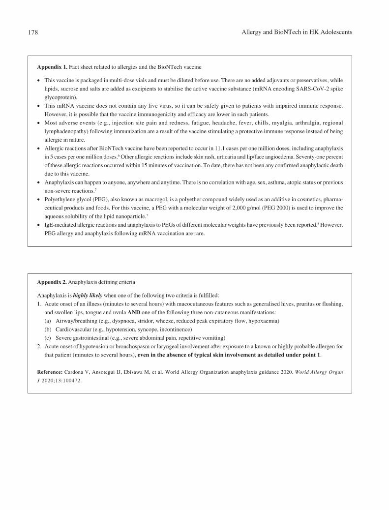
Allergy and BioNTech in HK Adolescents178
• This vaccine is packaged in multi-dose vials and must be diluted before use. There are no added adjuvants or preservatives, while
lipids, sucrose and salts are added as excipients to stabilise the active vaccine substance (mRNA encoding SARS-CoV-2 spike
glycoprotein).
• This mRNA vaccine does not contain any live virus, so it can be safely given to patients with impaired immune response.
However, it is possible that the vaccine immunogenicity and efficacy are lower in such patients.
• Most adverse events (e.g., injection site pain and redness, fatigue, headache, fever, chills, myalgia, arthralgia, regional
lymphadenopathy) following immunization are a result of the vaccine stimulating a protective immune response instead of being
allergic in nature.
• Allergic reactions after BioNTech vaccine have been reported to occur in 11.1 cases per one million doses, including anaphylaxis
in 5 cases per one million doses.6 Other allergic reactions include skin rash, urticaria and lip/face angioedema. Seventy-one percent
of these allergic reactions occurred within 15 minutes of vaccination. To date, there has not been any confirmed anaphylactic death
due to this vaccine.
• Anaphylaxis can happen to anyone, anywhere and anytime. There is no correlation with age, sex, asthma, atopic status or previous
non-severe reactions.7
• Polyethylene glycol (PEG), also known as macrogol, is a polyether compound widely used as an additive in cosmetics, pharma-
ceutical products and foods. For this vaccine, a PEG with a molecular weight of 2,000 g/mol (PEG 2000) is used to improve the
aqueous solubility of the lipid nanoparticle.7
• IgE-mediated allergic reactions and anaphylaxis to PEGs of different molecular weights have previously been reported.8 However,
PEG allergy and anaphylaxis following mRNA vaccination are rare.
Appendix 1. Fact sheet related to allergies and the BioNTech vaccine
Anaphylaxis is highly likely when one of the following two criteria is fulfilled:
1. Acute onset of an illness (minutes to several hours) with mucocutaneous features such as generalised hives, pruritus or flushing,
and swollen lips, tongue and uvula AND one of the following three non-cutaneous manifestations:
(a) Airway/breathing (e.g., dyspnoea, stridor, wheeze, reduced peak expiratory flow, hypoxaemia)
(b) Cardiovascular (e.g., hypotension, syncope, incontinence)
(c) Severe gastrointestinal (e.g., severe abdominal pain, repetitive vomiting)
2. Acute onset of hypotension or bronchospasm or laryngeal involvement after exposure to a known or highly probable allergen for
that patient (minutes to several hours), even in the absence of typical skin involvement as detailed under point 1.
Reference: Cardona V, Ansotegui IJ, Ebisawa M, et al. World Allergy Organization anaphylaxis guidance 2020. World Allergy Organ
J 2020;13:100472.
Appendix 2. Anaphylaxis defining criteria
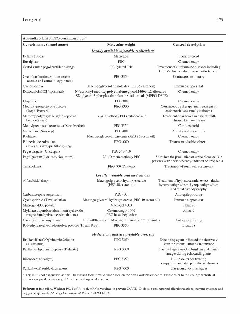
Leung et al 179
Appendix 3. List of PEG-containing drugs*
Generic name (brand name) Molecular weight General description
Locally available injectable medicationsBetamethasone Macrogols Corticosteroid
Busulphan PEG Chemotherapy
Certolizumab pegol prefilled syringe PEGylated Fab' Treatment of autoimmune diseases includingCrohn's disease, rheumatoid arthritis, etc.
Cyclofem (medroxyprogesterone PEG 3350 Contraceptive therapy acetate and estradiol cypionate)
Cyclosporin A Macrogoglycerol ricinoleate (PEG 35 castor oil) Immunosuppressant
Doxorubicin HCl (liposomal) N-(carbonyl-methoxypolyethylene glycol 2000)-1,2-distearoyl Chemotherapy-SN-glycero-3-phosphoethanolamine sodium salt (MPEG-DSPE)
Etoposide PEG 300 Chemotherapy
Medroxyprogesterone acetate PEG 3350 Contraceptive therapy and treatment of(Depo-Provera) endometrial and renal carcinoma
Methoxy polyethylene glycol-epoetin 30-kD methoxy PEG butanoic acid Treatment of anaemia in patients withbeta (Mircera) chronic kidney disease
Methylprednisolone acetate (Depo-Medrol) PEG 3350 Corticosteroid
Nimodipine (Nimotop) PEG 400 Anti-hypertensive drug
Paclitaxel Macrogoglycerol ricinoleate (PEG 35 castor oil) Chemotherapy
Paliperidone palmitate PEG 4000 Treatment of schizophrenia(Invega Trinza) prefilled syringe
Pegaspargase (Oncaspar) PEG 345-410 Chemotherapy
Pegfilgrastim (Neulasta, Neulastim) 20-kD monomethoxy PEG Stimulate the production of white blood cells inpatients with chemotherapy-induced neutropaenia
Temsirolimus PEG 400 (Diluent) Treatment of renal cell carcinoma
Locally available oral medicationsAlfacalcidol drops Macrogolglycerol hydroxystearate Treatment of hypocalcaemia, osteomalacia,
(PEG 40 castor oil) hyperparathyroidism, hypoparathyroidismand renal osteodystrophy
Carbamazepine suspension PEG 400 Anti-epileptic drug
Cyclosporin A (Teva) solution Macrogolglycerol hydroxystearate (PEG 40 castor oil) Immunosuppressant
Macrogol 4000 powder Macrogol 4000 Laxative
Mylanta suspension (aluminium hydroxide, Cetomacrogol 1000 Antacidmagnesium hydroxide, simethicone) (PEG hexadecyl ether)
Oxcarbazepine suspension PEG-400-stearate; Macrogol stearate (PEG stearate) Anti-epileptic drug
Polyethylene glycol electrolyte powder (Klean-Prep) PEG 3350 Laxative
Medications that are available overseasBrilliant Blue G Ophthalmic Solution PEG 3350 Disclosing agent indicated to selectively
(TissueBlue) stain the internal limiting membrane
Perflutren lipid microsphere (Definity) PEG 5000 Contrast agent used to brighten and clarifyimages during echocardiograms
Rilonacept (Arcalyst) PEG 3350 IL-1 blocker for treatingcryopyrin-associated periodic syndromes
Sulfur hexafluoride (Lumason) PEG 4000 Ultrasound contrast agent
* This list is not exhaustive and will be revised from time to time based on the best available evidence. Please refer to the College website athttp://www.paediatrician.org.hk/ for the most updated version.
Reference: Banerji A, Wickner PG, Saff R, et al. mRNA vaccines to prevent COVID-19 disease and reported allergic reactions: current evidence andsuggested approach. J Allergy Clin Immunol Pract 2021;9:1423-37.
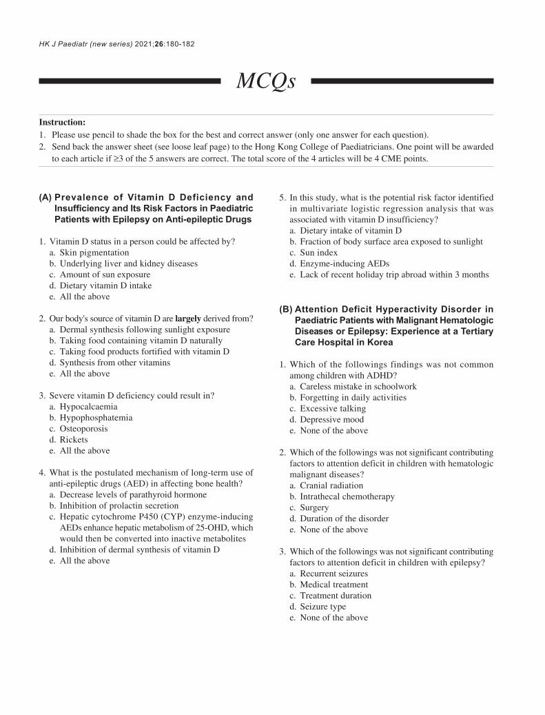
HK J Paediatr (new series) 2021;26:180-182
MCQs
Instruction:1. Please use pencil to shade the box for the best and correct answer (only one answer for each question).2. Send back the answer sheet (see loose leaf page) to the Hong Kong College of Paediatricians. One point will be awarded
to each article if ≥3 of the 5 answers are correct. The total score of the 4 articles will be 4 CME points.
(A) Prevalence of Vitamin D Deficiency andInsufficiency and Its Risk Factors in PaediatricPatients with Epilepsy on Anti-epileptic Drugs
1. Vitamin D status in a person could be affected by?a. Skin pigmentationb. Underlying liver and kidney diseasesc. Amount of sun exposured. Dietary vitamin D intakee. All the above
2. Our body's source of vitamin D are largely derived from?a. Dermal synthesis following sunlight exposureb. Taking food containing vitamin D naturallyc. Taking food products fortified with vitamin Dd. Synthesis from other vitaminse. All the above
3. Severe vitamin D deficiency could result in?a. Hypocalcaemiab. Hypophosphatemiac. Osteoporosisd. Ricketse. All the above
4. What is the postulated mechanism of long-term use ofanti-epileptic drugs (AED) in affecting bone health?a. Decrease levels of parathyroid hormoneb. Inhibition of prolactin secretionc. Hepatic cytochrome P450 (CYP) enzyme-inducing
AEDs enhance hepatic metabolism of 25-OHD, whichwould then be converted into inactive metabolites
d. Inhibition of dermal synthesis of vitamin De. All the above
5. In this study, what is the potential risk factor identifiedin multivariate logistic regression analysis that wasassociated with vitamin D insufficiency?a. Dietary intake of vitamin Db. Fraction of body surface area exposed to sunlightc. Sun indexd. Enzyme-inducing AEDse. Lack of recent holiday trip abroad within 3 months
(B) Attention Deficit Hyperactivity Disorder inPaediatric Patients with Malignant HematologicDiseases or Epilepsy: Experience at a TertiaryCare Hospital in Korea
1. Which of the followings findings was not commonamong children with ADHD?a. Careless mistake in schoolworkb. Forgetting in daily activitiesc. Excessive talkingd. Depressive moode. None of the above
2. Which of the followings was not significant contributingfactors to attention deficit in children with hematologicmalignant diseases?a. Cranial radiationb. Intrathecal chemotherapyc. Surgeryd. Duration of the disordere. None of the above
3. Which of the followings was not significant contributingfactors to attention deficit in children with epilepsy?a. Recurrent seizuresb. Medical treatmentc. Treatment durationd. Seizure typee. None of the above
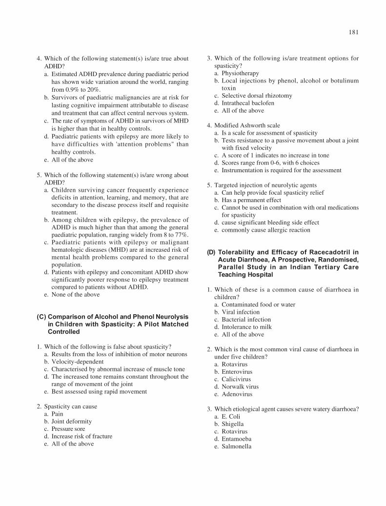
181
4. Which of the following statement(s) is/are true aboutADHD?a. Estimated ADHD prevalence during paediatric period
has shown wide variation around the world, rangingfrom 0.9% to 20%.
b. Survivors of paediatric malignancies are at risk forlasting cognitive impairment attributable to diseaseand treatment that can affect central nervous system.
c. The rate of symptoms of ADHD in survivors of MHDis higher than that in healthy controls.
d. Paediatric patients with epilepsy are more likely tohave difficulties with 'attention problems" thanhealthy controls.
e. All of the above
5. Which of the following statement(s) is/are wrong aboutADHD?a. Children surviving cancer frequently experience
deficits in attention, learning, and memory, that aresecondary to the disease process itself and requisitetreatment.
b. Among children with epilepsy, the prevalence ofADHD is much higher than that among the generalpaediatric population, ranging widely from 8 to 77%.
c. Paediatric patients with epilepsy or malignanthematologic diseases (MHD) are at increased risk ofmental health problems compared to the generalpopulation.
d. Patients with epilepsy and concomitant ADHD showsignificantly poorer response to epilepsy treatmentcompared to patients without ADHD.
e. None of the above
(C) Comparison of Alcohol and Phenol Neurolysisin Children with Spasticity: A Pilot MatchedControlled
1. Which of the following is false about spasticity?a. Results from the loss of inhibition of motor neuronsb. Velocity-dependentc. Characterised by abnormal increase of muscle toned. The increased tone remains constant throughout the
range of movement of the jointe. Best assessed using rapid movement
2. Spasticity can causea. Painb. Joint deformityc. Pressure sored. Increase risk of fracturee. All of the above
3. Which of the following is/are treatment options forspasticity?a. Physiotherapyb. Local injections by phenol, alcohol or botulinum
toxinc. Selective dorsal rhizotomyd. Intrathecal baclofene. All of the above
4. Modified Ashworth scalea. Is a scale for assessment of spasticityb. Tests resistance to a passive movement about a joint
with fixed velocityc. A score of 1 indicates no increase in toned. Scores range from 0-6, with 6 choicese. Instrumentation is required for the assessment
5. Targeted injection of neurolytic agentsa. Can help provide focal spasticity reliefb. Has a permanent effectc. Cannot be used in combination with oral medications
for spasticityd. cause significant bleeding side effecte. commonly cause allergic reaction
(D) Tolerability and Efficacy of Racecadotril inAcute Diarrhoea, A Prospective, Randomised,Parallel Study in an Indian Tertiary CareTeaching Hospital
1. Which of these is a common cause of diarrhoea inchildren?a. Contaminated food or waterb. Viral infectionc. Bacterial infectiond. Intolerance to milke. All of the above
2. Which is the most common viral cause of diarrhoea inunder five children?a. Rotavirusb. Enterovirusc. Calicivirusd. Norwalk viruse. Adenovirus
3. Which etiological agent causes severe watery diarrhoea?a. E. Colib. Shigellac. Rotavirusd. Entamoebae. Salmonella

182
Answers of April issue 2021
(A) 1. c; 2. b; 3. d; 4. e; 5. c
(B) 1. c; 2. e; 3. a; 4. b; 5. d
(C) 1. e; 2. e; 3. e; 4. d; 5. c
(D) 1. e; 2. a; 3. e; 4. b; 5. e
4. Which is true about pharmacological mechanism ofaction of Racecadotril?a. Racecadotril is an anti-secretary agentb. Racecadotril inhibits enkephalinasec. Inhibits cell membrane peptidase enzymed. Racecadotril also termed as Aceterphane. All of the above
5. Which statement is true regarding effect of Racecadotrilin children with diarrhoea?a. Racecadotril reduces duration of diarrhoeab. Racecadotril reduces stool outputc. Racecadotril reduces duration of hospitalisationd. Racecadotril use in Rotaviral diarrhoea reduces stool
outpute. All of the above
HK J Paediatr (new series) 2021;26:183-184
4. SI units should be used or included in parentheses.
Ethical Consideration
For original clinical study, authors must state that theprotocol for the research project has been approved by the EthicsCommittee of the institution within which the work wasundertaken. All investigations on human subjects must includea statement that informed consents have been obtained. Patientanonymity must be preserved. Photographs and video clippingsneed to be prepared to prevent human subjects being recognizedunless prior written permission has been obtained. Whenreporting experiments on animals, authors should indicatewhether the institutional and national guide for the care anduse of laboratory animals was followed.
The manuscript should usually be arranged as follows:Title page
This page should include the full names, and affiliations ofall authors. A short title of no more than 40 characters shouldalso be given. Up to three academic degrees for each author areallowed. If an author’s affiliation has changed since the workwas done, list the new affiliations as well. Limit the number ofauthors to 4 for case reports and clinical quiz.Abstract and Key words
The abstract should be no more than 150 words summarisingthe purpose, methods, findings and conclusions. Authors shouldprovide no more than five key words to assist with cross-indexing of the paper. Key words should be taken from IndexMedicus.IntroductionMethodsResultsDiscussionReferences
Number references in the order they appear in the text.References should follow the Vancouver style and should appearin the text, tables and legends as Arabic numerals in superscript.Journal titles should be abbreviated in accordance with IndexMedicus. List all authors and/or editors up to six; if more thansix, list the first three and "et al".
Examples of References:Articles in Journals1. Standard journal article Greenberg DL, Root RK. Decision
making by analogy. N Engl J Med 1995;332:592-6.2. Organisation as author The Royal Marsden Hospital Bone-
Marrow Transplantation Team. Failure of syngeneic bone-marrow graft without preconditioning in post-hepatitismarrow aplasia. Lancet 1977;2:742-4.
3. No author given Coffee drinking and cancer of the pancreas[editorial]. BMJ 1981;283:628.
4. Issue with supplement Gardos G, Cole JO, Haskell D, MarbyD, Paine SS, Moore P. The natural history of tardive dyskinesia.J Clin Psychopharmacol 1988;8(4 Suppl):31S-37S.
The Hong Kong Journal of Paediatrics (HKJP) is a jointquarterly publication of the Hong Kong College of Paediatricians(HKCPaed) and The Hong Kong Paediatric Society (HKPS).The HKJP publishes original research papers, review articles,case reports, editorials, commentaries, letters to the editor andconference proceedings. Topics of interest will include allsubjects that relate to clinical practice and research in paediatricsand child health.
Manuscripts are accepted on the condition that they aresubmitted solely to the HKJP and have not been publishedelsewhere previously and are not under consideration by anotherjournal. A complete report following presentation or publicationof preliminary findings elsewhere can be considered.
Categories of articles include the following:
Original Articles The text should not usually exceed 5,000words excluding references; the number of tables, figures,or both should normally be not more than six, and referencesnot more than 50.
Review Articles Reviews are usually invited systematic criticalassessments of literature.
Case Reports Length should not exceed 1,500 words; thenumber of tables or figures used should not be more thantwo, and references should not be more than 10. Limit thenumber of authors to 4.
Commentaries Commentary on current topics is welcome.Length should not exceed 1,200 words; no tables or figuresallowed, and references should not be more than 20.
Clinical Quiz The clinical quiz should be educational. It shouldi) include the description of a case in no more than 250 wordsand 3 clinical photos or figures, and ii) provide answers onthe diagnosis, clinical features and findings, and managementof the condition in no more than 1,000 words, 10 references,and 3 photos, figures or tables.
Letters to the Editor Letters discussing a recent article in theHKJP are welcome. Original letters that do not refer to anHKJP article may also be considered. Letters should notexceed 500 words and have no more than five references.Published letters may be edited.
Manuscript Preparation
1. Use Arabic numerals for numbers above nine, for designators(e.g. case 5, day 2, etc.) and for units of measure; numbersshould be spelled out if below 10, at the beginning and endof sentences, and for fractions below one.
2. Manuscripts should be submitted as a Word document inBritish English in the following format: Typed double-spaced, page size 22 cm. x 29 cm. (8 1/2 in. x 11 in.), pagemargins 2.54 cm (1 in), font size 12 pt.
3. Do not use abbreviations in the title or abstract and limittheir use in the text. Standard abbreviations may be used andshould be defined on first mention in the text unless it is astandard unit of measurement.
Instructions to Authors
184
5. Type of article indicated as needed [i] Fuhrman SA, JoinerKA. Binding of the third component of complement C3 byToxoplasma gondii [abstract]. Clin Res 1987;35:475A. [ii]Feldman N. Laparoscopic nephrectomy [letter]. N Engl J Med1991;325:1110.
Books and Other Monographs6. Personal author(s) Colson JH, Armour WJ. Sports injuries
and their treatment. 2nd ed. London: S. Paul, 1986.7. Editor(s), compiler as author Dausset J, Colombani J,
editors. Histocompatibility testing 1972. Copenhagen:Munksgaard, 1973.
8. Chapter in a book Steiner RE, Bydder GM. Clinical nuclearmagnetic resonance imaging. In: Dawaon AM, CompstonND, Besser GM, editors. Recent advances in medicine no.19. Edinburgh: Churchill Livingston, 1984:39-56.
9. Conference paper Harley NH. Comparing radon daughterdosimetric and risk models. In: Gammage RB, Kaye SV,editors. Indoor air and human health. Proceedings of theSeventh Life Sciences Symposium; 1984 Oct 29-31;Knoxville(TN). Chelsea(MI): Lewis, 1985:69-78.
10. Dissertation Cairns R.B Infrared spectroscopic studies ofsolid oxygen [dissertation]. Berkeley, (CA): Univ. ofCalifornia, 1965.
11. Scientific and technical report Akutsu T. Total heartreplacement device. Bethesda (MD): National Institutes ofHealth, National Heart and Lung Institute; 1974 Apr ReportNo: NIH-NHLI-69-2185-4.
Unpublished Material12. In press Lillywhite HD, Donald JA. Pulmonary blood flow
regulation in an aquatic snake. Science. In press.13. A reference to unpublished work which has not yet been
accepted for publication should not appear in the referencelist but should be cited in the text as unpublished data orpersonal communication.
TablesEach table should begin on a separate page. Number tables
consecutively in the order of their first citation in the text andsupply a brief title for each. Give each column a short orabbreviated heading. Place explanatory matter in footnotes,not in the heading. Vertical rules and horizontal rules shouldbe omitted.
Illustrations (Figures)Each illustration must be submitted as a separate figure file.
The file name should be the same as the figure number. Preferredformats for digital artwork submission include EncapsulatedPostScript (EPS), Portable Document Format (PDF), and TaggedImage Format (TIFF). Letters, numbers and symbols should beclear and of sufficient size to retain legibility when reduced.Photographs of persons must be retouched to make the subjectunidentifiable, or be accompanied by written permission fromthe subject to use the photograph.
Number illustrations consecutively according to the orderin which they have been first cited in the text. Titles and detailedexplanations should be confined to legends and not included
in illustrations. The legends should be numbered and providedon a separate double-spaced page.
Supplementary Video ClipsVideo clips can be submitted with your manuscript in MP4
file format with H.264 codec. The size of the video should notexceed 5 MB. Patient anonymity must be preserved unless priorwritten permission has been obtained. If accepted, the video willappear online on the Journal's website, http://www.hkjpaed.org.
Declaration of InterestAll sources of funding for research are to be explicitly stated.
All authors must disclose any financial and personalrelationships with other people or organisations that couldinappropriately influence their work. It is the sole responsibilityof authors to disclose any affiliation with any organisation witha financial interest, direct or indirect, in the subject matter ormaterials discussed in the manuscript that may affect the conductor reporting of the work submitted.
If there are no declarations, authors should explicitly statethat there are none. This must be stated at the point ofsubmission (within the manuscript, after the main text, under asubheading "Declaration of Interest"). Manuscript submissioncannot be completed unless a declaration of interest statement(either stating the disclosures or reporting that there are none)is included. This will be made available to reviewers and willappear in the published article.
The intent of this policy is not to prevent authors with anyparticular relationship or interest from publishing their work,but rather to adopt transparency such that reviewers, editors,the publisher, and most importantly, readers can make objectivejudgments concerning the work product.
CorrespondenceAll manuscripts, correspondence and subscription
should be addressed to Chief Editor, Hong Kong Journalof Paediatrics Limited, c/o Hong Kong College ofPaediatricians, Room 801, HK Academy of Medicine JockeyClub Building, 99 Wong Chuk Hang Road, Aberdeen,Hong Kong, e-mail:[email protected].
DisclaimerThe opinions expressed in the HKJP are those of the authors
and not those of HKCPaed, HKPS or the publisher. The HKJP,HKCPaed and HKPS do not hold any legal responsibility forthe opinions expressed in the articles, unless this is clearlyspecified. The authors should hold legal responsibility for thecontent and opinion expressed. Manuscripts and discs aresubmitted at the sender's risk and the HKJP, HKCPaed andHKPS assume no responsibility for return of material.
OffprintsOffprints can be ordered at a cost at the time of proof-
reading.
DistributionThe HKJP is published quarterly and adopts an open access
policy. Electronic copies of all articles are available online athttp://www.hkjpaed.org. Members of HKCPaed and HKPSwill receive quarterly e-mail notification of the Table ofContents in each of the Journal issues.