hiv-1 antisense transcription is preferentially activated in primary monocyte-derived cells
TRANSCRIPT

Published Ahead of Print 3 October 2012. 2012, 86(24):13785. DOI: 10.1128/JVI.01723-12. J. Virol.
Jean-Michel MesnardIsabelle Clerc, Bruno Beaumelle, Benoit Barbeau and Sylvain Laverdure, Antoine Gross, Charlotte Arpin-André, Monocyte-Derived CellsPreferentially Activated in Primary HIV-1 Antisense Transcription Is
http://jvi.asm.org/content/86/24/13785Updated information and services can be found at:
These include:
REFERENCEShttp://jvi.asm.org/content/86/24/13785#ref-list-1at:
This article cites 23 articles, 14 of which can be accessed free
CONTENT ALERTS more»articles cite this article),
Receive: RSS Feeds, eTOCs, free email alerts (when new
http://journals.asm.org/site/misc/reprints.xhtmlInformation about commercial reprint orders: http://journals.asm.org/site/subscriptions/To subscribe to to another ASM Journal go to:
on January 18, 2013 by INIS
T-C
NR
S B
iblioVie
http://jvi.asm.org/
Dow
nloaded from

HIV-1 Antisense Transcription Is Preferentially Activated in PrimaryMonocyte-Derived Cells
Sylvain Laverdure,a,b Antoine Gross,a,b Charlotte Arpin-André,a,b Isabelle Clerc,c Bruno Beaumelle,a,b Benoit Barbeau,d andJean-Michel Mesnarda,b
Université Montpellier 1, Centre d’études d’agents Pathogènes et Biotechnologies pour la Santé (CPBS),a and CNRS, UM5236, CPBS,b Montpellier, France; Institut deGénétique Moléculaire de Montpellier (IGMM), CNRS, UMR5535, Montpellier, Francec; and Université du Québec à Montréal, Département des sciences biologiques andCentre de recherche BioMed, Montréal (Québec), Canadad
In this study, an antisense luciferase-expressing human immunodeficiency virus type 1 (HIV-1) molecular clone was used to in-fect primary cells. We found that antisense transcription activity from the 3= long terminal repeat (LTR) was significantly moreabundant in monocyte-derived cells than in activated T lymphocytes. Moreover, by analyzing antisense transcription in infectedmonocyte-derived dendritic cells (MDDCs), we observed that the majority of HIV-1-infected MDDCs with significant antisensetranscription activity did not produce Gag. We also confirmed that the negative-strand-encoded antisense protein (ASP) wasexpressed in monocyte-derived cells.
Human T-cell leukemia virus type 1 (HTLV-1) is the first ret-rovirus from which the production of antisense transcripts
from the 3= long terminal repeat (LTR) has been clearly demon-strated (11, 17, 19, 22). Antisense transcription in human immu-nodeficiency virus type 1 (HIV-1) has also been characterized (10,16, 20). Interestingly, such antisense transcripts can potentiallyencode proteins (9, 14, 15, 18). We have indeed demonstrated thatthe antisense protein (ASP) can be expressed in HIV-1-infectedcells (12). We focused here on the regulation of HIV-1 antisensetranscription in primary cells.
In this study, we used pNL4.3LucE�, a proviral DNA in whichthe Firefly luciferase gene had been inserted in nef (13). We re-moved its reporter gene to reinsert the luciferase gene in the sameposition but in the inverse orientation as already described (16).This vector, pNL4.3AsLucE� (Fig. 1A), permits quantification ofantisense transcription from the 3= LTR, since the reporter genewas placed under the control of the antisense transcription pro-moter (16). As these proviral DNAs are env deficient, vesicularstomatitis virus envelope (VSVg)-pseudotyped HIV-1 particleswere produced. Their decreased requirement for Nef, togetherwith their high infectious titers and their broad host range, madethese viruses useful for our experiments, which involved differenttarget cells for infection (1). Moreover, the restriction of mono-cyte-derived cells to HIV-1 is also observed in this case (2). Toproduce pseudotyped viruses, HEK-293T cells were cotransfectedwith proviral DNA (30 �g) and a VSVg expression vector (18 �g).Cells were grown for 2 days before viruses were collected. Virustiters were determined by infecting Jurkat cells with 2-fold serialdilutions of virus stocks. At 2 days postinfection (p.i.), the per-centage of infected cells was determined by flow cytometry follow-ing intracellular staining of Gag with KC57 antibodies.
Peripheral blood mononuclear cells (PBMC) were isolatedfrom buffy coats, and CD14� cells were isolated with anti-humanCD14-coated magnetic beads (Miltenyi Biotec). Differentiation ofCD14� monocytes into macrophages or dendritic cells was per-formed as already described (6, 7). CD4� T cells were isolatedfrom CD14� peripheral blood lymphocytes with a CD4� T-cellenrichment kit (Stem Cell Technologies). Cell populations wereinfected at a multiplicity of infection (MOI) of 2 for both virus
strains, and Firefly luciferase activity was measured at differentdays p.i. Infection of activated primary CD4� T lymphocytesshowed a strong activation of sense transcription from the 5= LTR,while antisense transcription from the 3= LTR remained low (Fig.1B and E); levels of antisense activity were about 1,000-fold lowerthan that of sense transcription. Infection of monocyte-derivedmacrophages (MDMs) again showed higher sense than antisensetranscription levels. However, antisense transcription activity wassignificantly higher than in T cells (Fig. 1E), increasing steadilyand reaching 1,000-fold induction at 21 days p.i. (Fig. 1C). Regu-lation of antisense transcription was quite different in infectedmonocyte-derived dendritic cells (MDDCs). During the 2 firstdays of infection, sense and antisense transcription levels werelow, and both increased at 6 days p.i. and remained high at up to21 days p.i. (Fig. 1D and E). Interestingly, levels of antisense tran-scription in MDDCs were only 25- to 100-fold lower than that ofsense transcription.
We next studied the effects of the Tat transactivator on anti-sense transcription activity. Controversial data have been pub-lished so far (16, 20, 21). Most results were obtained from cell linestransfected with a Tat expression plasmid and reporter vectorscontaining only one LTR. We readdressed these results usingpNL4.3Luc(Reni)/AsLuc(Fire)E�, corresponding to the NL4.3molecular clone expressing an antisense Firefly luciferase geneunder the control of the 3= LTR and the Renilla luciferase geneafter insertion into the env gene under the control of the Tat-dependent sense transcription (Fig. 2A). In addition, we intro-duced a mutation into the tat gene (pNL4.3Luc(Reni)/AsLuc(Fire)E��Tat), blocking its expression. Tat mutationreduced Renilla luciferase activity, while Firefly luciferase ex-
Received 4 July 2012 Accepted 25 September 2012
Published ahead of print 3 October 2012
Address correspondence to Jean-Michel Mesnard,[email protected].
Copyright © 2012, American Society for Microbiology. All Rights Reserved.
doi:10.1128/JVI.01723-12
December 2012 Volume 86 Number 24 Journal of Virology p. 13785–13789 jvi.asm.org 13785
on January 18, 2013 by INIS
T-C
NR
S B
iblioVie
http://jvi.asm.org/
Dow
nloaded from
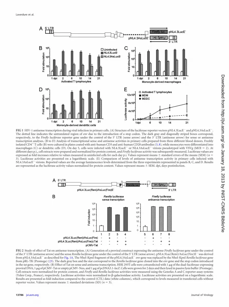
FIG 1 HIV-1 antisense transcription during viral infection in primary cells. (A) Structure of the luciferase reporter vectors pNL4.3LucE� and pNL4.3AsLucE�.The dotted line indicates the untranslated region of env due to the introduction of a stop codon. The dark gray and diagonally striped boxes correspond,respectively, to the Firefly luciferase reporter gene under the control of the 5= LTR (sense arrow) and the 3= LTR (antisense arrow) for sense or antisensetranscription analyses. (B to D) Analysis of transcriptional sense and antisense activities in primary cells prepared from three different blood donors. Freshlyisolated CD4� T cells (B) were cultured in plates coated with anti-human CD3 and anti-human CD28 antibodies (3, 8), while monocytes were differentiated intomacrophages (C) or dendritic cells (D). On day 5, cells were infected with NL4.3LucE� or NL4.3AsLucE� virions pseudotyped with VSVg (MOI � 2). Atdifferent days p.i., cell extracts were prepared and normalized for protein content, and Firefly luciferase activity was subsequently measured. Luciferase values areexpressed as fold increases relative to values measured in uninfected cells for each day p.i. Values represent means � standard errors of the means (SEM) (n �3). Luciferase activities are presented on a logarithmic scale. (E) Comparison of levels of antisense transcription activity in primary cells infected withNL4.3AsLucE� virions. Reported values are the average luminescence levels determined from the three experiments represented in panels B, C, and D. Resultsare represented as the luciferase activity values normalized for protein content. Values represent means � SEM. dpi, days postinfection.
FIG 2 Study of effect of Tat on antisense transcription. (A) Generation of a proviral construct expressing the antisense Firefly luciferase gene under the controlof the 3= LTR (antisense arrow) and the sense Renilla luciferase gene under the control of the 5= LTR (sense arrow). pNL4.3Luc(Reni)/AsLuc(Fire)E� was derivedfrom pNL4.3AsLucE� as described for Fig. 1A. The NheI-KpnI fragment of the pNL4.3AsLucE� env gene was replaced by the NheI-KpnI Renilla luciferase genefrom pRL-TK (Promega) (23). The dark gray box and the star correspond to the Renilla luciferase gene cloned into the env gene and the stop codon introducedin the tat gene, respectively. (B) Effect of Tat on sense and antisense transcription. HEK 293T cells were cotransfected with 1 �g of the dual-luciferase-expressingproviral DNA, 1 �g of pCMV-Tat or empty pCMV-Neo, and 1 �g of pcDNA3.1-lacZ. Cells were grown for 2 days and then lysed in passive lysis buffer (Promega).Cell extracts were normalized for protein content, and Firefly and Renilla luciferase activities were measured using the Genofax A and C reporter assay systems(Yelen Corp., France), respectively. Luciferase activities were normalized to �-galactosidase activity. Luciferase activities are presented on a logarithmic scale.Results are presented as fold induction compared to the control (CTL) data (white columns), which correspond to levels measured in transfected cells withoutreporter vector. Values represent means � standard deviations (SD) (n � 3).
Laverdure et al.
13786 jvi.asm.org Journal of Virology
on January 18, 2013 by INIS
T-C
NR
S B
iblioVie
http://jvi.asm.org/
Dow
nloaded from

pression was not altered by the absence of Tat (Fig. 2B). WhenpNL4.3Luc(Reni)/AsLuc(Fire)E��Tat was cotransfected witha Tat expression vector, sense transcription, unlike antisensetranscription, was stimulated.
Higher antisense transcription activity was preferentially ob-served in HIV-1-infected MDDCs at 14 days p.i. However, thequestion remained whether both transcriptional activities couldcoexist within the same cell. Therefore, we analyzed the antisensetranscription in single infected MDDCs. To identify cells capableof activating antisense transcription from the 3= LTR, we insertedegfp under the control of the antisense transcription promoter (seeFig. 3A). The novel virus (NL4.3AsegfpE�) was pseudotyped withVSVg to infect MDDCs. As a control, we produced virions from asense enhanced green fluorescent protein (EGFP)-expressingNL4.3 clone (pNL4.3egfpE�) in which egfp had been inserted innef (Fig. 3B). At 14 days p.i., transcriptional activity of each in-fected MDDC was assessed by analyzing the resulting EGFP ex-pression. Expression of gag was also analyzed by intracellularstaining of Gag. Few single cells infected with NL4.3AsegfpE� vi-rus were able to express both Gag and EGFP, while that was not thecase for the NL4.3egfpE� virus-infected cells (Fig. 3). A totalof 70.5% � 3.1% of the EGFP-positive cells infected with
NL4.3AsegfpE� virus did not produce Gag, while 86.5% � 1.5%of the EGFP-positive cells infected with NL4.3egfpE� virus pro-duced Gag (Fig. 3C). Interestingly, 90.1% � 1.2% of Gag-express-ing cells infected with NL4.3AsegfpE� virus did not express anti-sense egfp (Fig. 3D). When the same approach was used in thepresence of zidovudine (20 �M), Gag could not be detected, con-firming that the Gag signal was not derived from the infectingviruses (data not shown).
ASP expression was next studied in HIV-1-infected primarycells. Cells were infected with pseudotyped NL4.3ASP-Flag vi-rions, expressing an ASP tagged with the Flag epitope at itsC-terminal end (12). While no staining was detected in acti-vated T lymphocytes, we were able to detect ASP expression inMDMs (Fig. 4A) and MDDCs (Fig. 4B and C). No staining wasdetected in MDMs (Fig. 4D) or MDDCs (Fig. 4E) infected withNL4.3ASPmut12-Flag virions containing a stop codon atamino acid 12 of ASP (12).
We have provided strong evidence for the existence of anti-sense transcription in infected primary cells and for the produc-tion of ASP in monocyte-derived cells. Indeed, HIV-1 antisensetranscription is more active in monocyte-derived cells than inactivated T cells and unaltered by Tat expression. Moreover, at 14
FIG 3 Analysis of antisense transcription activity in individual infected MDDCs. Freshly isolated monocytes were differentiated into dendritic cells and theninfected with NL4.3AsegfpE� or NL4.3egfpE� virions pseudotyped with VSVg (MOI � 2). pNL4.3AsegfpE� was constructed by replacing the Firefly luciferasegene of pNL4.3LucE� with the egfp reporter gene in the inverse orientation. The molecular organization of the antisense (A) or sense (B) EGFP-expressing NL4.3clones is depicted. At 14 days p.i., cells were fixed for staining with anti-p24 antibody coupled to phycoerythrin (anti-Gag) before observation under anepifluorescence microscope. Images were acquired using the scan slide command of Metamorph, and analyses were performed using ImageJ software. Cellsexpressing both Gag (red) and EGFP (green) appear yellow on merged images. A total of 772 and 994 cells infected with NL4.3AsegfpE� and NL4.3egfpE�
virions, respectively, were analyzed (n � 3). Graphics represent (C) the percentages (means � SEM) of Gag-negative or Gag-positive cells among EGFP-expressing cells or (D) the percentages of EGFP-negative or EGFP-positive cells among Gag-expressing cells.
Antisense Transcription in HIV-1
December 2012 Volume 86 Number 24 jvi.asm.org 13787
on January 18, 2013 by INIS
T-C
NR
S B
iblioVie
http://jvi.asm.org/
Dow
nloaded from

days p.i., about two-thirds of HIV-1-infected MDDCs showingefficient antisense transcription activity did not express the struc-tural Gag protein. Interestingly, our previous results had alreadysuggested this inverse correlation between Gag and negative-stranded-encoded protein expression in HTLV-1-infected cells(4, 5, 11).
ACKNOWLEDGMENTS
This work was supported by institutional grants from the Centre Nationalde la Recherche Scientifique (CNRS) and the Université Montpellier 1(UM 1), a grant to J.-M.M. from the CNRS for an international project of
scientific cooperation, a grant to J.-M.M. from the Agence Nationale deRecherches sur le Sida et les hépatites virales (ANRS), and a grant to B.B.from The Canadian Foundation for AIDS Research (CanFAR). B.B. holdsa Canada Research Chair in Human Retrovirology (Tier 2). S.L. was sup-ported by fellowships from the Ministère de l’Enseignement Supérieur etde la Recherche, and I.C. was supported by ANRS and Sidaction.
Confocal microscopy was performed using Montpellier RIO Imaging(MRI) facilities.
REFERENCES1. Aiken C. 1997. Pseudotyping human immunodeficiency virus type 1
(HIV-1) by the glycoprotein of vesicular stomatitis virus targets HIV-1
FIG 4 ASP expression in infected monocyte-derived cells. MDM (A and D) or MDDC (B, C, and E) cells were infected with VSVg-pseudotyped virusNL4.3ASP-Flag (A to C) or NL4.3ASPmut12-Flag (D and E) virions. At 14 days p.i., ASP expression was visualized by fluorescence microscopy after immuno-staining with a primary anti-Flag antibody and a secondary antibody coupled to Alexa Fluor 568. The morphology of the cells was assessed by Normaskidifferential interference contrast (NDIC).
Laverdure et al.
13788 jvi.asm.org Journal of Virology
on January 18, 2013 by INIS
T-C
NR
S B
iblioVie
http://jvi.asm.org/
Dow
nloaded from

entry to an endocytic pathway and suppresses both the requirement forNef and the sensitivity to cyclosporin A. J. Virol. 71:5871–5877.
2. Arfi V, et al. 2008. Characterization of the early steps of infection ofprimary blood monocytes by human immunodeficiency virus type 1. J.Virol. 82:6557– 6565.
3. Arpin-André C, Mesnard JM. 2007. The PDZ domain-binding motif ofthe human T cell leukemia virus type 1 Tax protein induces mislocaliza-tion of the tumor suppressor hScrib in T cells. J. Biol. Chem. 282:33132–33141.
4. Barbeau B, Mesnard JM. 2011. Making sense out of antisense transcrip-tion in human T-cell lymphotropic viruses (HTLVs). Viruses 3:456 – 468.
5. Belrose G, et al. 2011. Opposite effects of valproate on Tax and HBZexpressions in T-lymphocytes from HTLV-1 asymptomatic carriers andHAM/TSP patients. Blood 118:2483–2491.
6. Billard E, Cazevieille C, Dornand J, Gross A. 2005. High susceptibility ofhuman dendritic cells to invasion by the intracellular pathogens Brucellasuis, B. abortus, and B. melitensis. Infect. Immun. 73:8418 – 8424.
7. Billard E, Dornand J, Gross A. 2007. Brucella suis prevents humandendritic cell maturation and antigen presentation through regulation oftumor necrosis factor alpha secretion. Infect. Immun. 75:4980 – 4989.
8. Billard E, Dornand J, Gross A. 2007. Interaction of Brucella suis andBrucella abortus rough strains with human dendritic cells. Infect. Immun.75:5916 –5923.
9. Briquet S, Vaquero C. 2002. Immunolocalization studies of an antisenseprotein in HIV-1-infected cells and viral particles. Virology 292:177–184.
10. Bukrinsky MI, Etkin AI, Ivanovsky DI. 1990. Plus strand of the HIVprovirus DNA is expressed at early stages of infection. AIDS Res. Hum.Retroviruses 6:425– 426.
11. Cavanagh M-H, et al. 2006. HTLV-I antisense transcripts initiating in the3=LTR are alternatively spliced and polyadenylated. Retrovirology 3:15.doi:10.1186/1742-4690-3-15.
12. Clerc I, et al. 2011. Polarized expression of the membrane ASP proteinderived from HIV-1 antisense transcription in T cells. Retrovirology 8:74.doi:10.1186/1742-4690-8-74.
13. Connor RI, Chen BK, Choe S, Landau NR. 1995. Vpr is required forefficient replication of human immunodeficiency virus type-1 in mono-nuclear phagocytes. Virology 206:935–944.
14. Gaudray G, et al. 2002. The complementary strand of HTLV-1 RNAgenome encodes a bZIP transcription factor that down-regulates the viraltranscription. J. Virol. 76:12813–12822.
15. Halin M, et al. 2009. Human T-cell leukemia virus type 2 produces aspliced antisense transcript encoding a protein that lacks a classical bZIPdomain but still inhibits Tax2-mediated transcription. Blood 114:2427–2437.
16. Landry S, et al. 2007. Detection, characterization and regulation of antisensetranscripts in HIV-1. Retrovirology 4:71. doi:10.1186/1742-4690-4-71.
17. Larocca D, Chao LA, Seto MH, Brunck TK. 1989. Human T-cell leuke-mia virus minus strand transcription in infected T-cells. Biochem. Bio-phys. Res. Comm. 163:1006 –1013.
18. Larocque E, et al. 2011. Human T-cell lymphotropic virus type 3 (HTLV-3)- and HTLV-4-derived antisense transcripts encode proteins with sim-ilar Tax-inhibiting functions but distinct subcellular localization. J. Virol.85:12673–12684.
19. Li M, Kesic M, Yin H, Yu L, Green PL. 2009. Kinetic analysis of humanT-cell leukemia virus type 1 gene expression in cell culture and infectedanimals. J. Virol. 83:3788 –3797.
20. Michael NL, et al. 1994. Negative-strand RNA transcripts are produced inhuman immunodeficiency virus type 1-infected cells and patients by anovel promoter downregulated by Tat. J. Virol. 68:979 –987.
21. Peeters A, Lambert PF, Deacon NJ. 1996. A fourth Sp1 site in the humanimmunodeficiency virus type 1 long terminal repeat is essential for nega-tive-sense transcription. J. Virol. 70:6665– 6672.
22. Usui T, et al. 2008. Characteristic expression of HTLV-1 basic zipperfactor (HBZ) transcripts in HTLV-1 provirus-positive cells. Retrovirology5:34. doi:10.1186/1742-4690-5-34.
23. Zhang H, et al. 2004. Novel single-cell-level phenotypic assay for residualdrug susceptibility and reduced replication capacity of drug-resistant hu-man immunodeficiency virus type 1. J. Virol. 78:1718 –1729.
Antisense Transcription in HIV-1
December 2012 Volume 86 Number 24 jvi.asm.org 13789
on January 18, 2013 by INIS
T-C
NR
S B
iblioVie
http://jvi.asm.org/
Dow
nloaded from