antisense rna inhibition of rubisco activase expression
TRANSCRIPT
The Plant Journal (1994) 5(6), 787-798
Antisense RNA inhibition of Rubisco activase expression
Cai-Zhong Jiang 1, W. Paul Quick 2, Rhu Aired 2, Daniel Kliebenstein 1 and Steven R. Rodermel 1,* i Department of Botany, 353 Bessey Hall, Iowa State University, Ames, IA 50011, USA, and 2Robert Hill Institute and Department of Animal and Plant Sciences, The University of Sheffield, Sheffield S 10 2UQ, UK
Summary
Ribulose bisphosphate carboxylase (Rubisco) acti- vase catalyzes the activation of Rubieco in vivo. Activase antisense DNA mutants of tobacco have been generated to explore the control that ectivase exerts on the photosynthetic process. These mutants have up to 90% reductions in activase protein levels as a consequence of an inhibition of activase mRNA accumulation. It is shown that photosynthesis, measured as the rate of CO= exchange (CER), is modestly decreased in plants exposed to high irradiances. The decreases in CER in the transgenic plants are accompanied by corresponding decreases in Rubieco activation, indicating that activase has a direct effect on photosynthetic rates in the antisense plants by influencing the activation state of Rubisco. It is concluded that in high light conditions, control of photosynthesis is largely shared between Rubisco and activase. Plant growth is also impaired in mutant plants that have severe reductions in activase. The inhibition of activase in the antisense plants does not have an impact on the accumulation of Rubisco large subunit or small subunit mRNAs or proteins. This indicates that the concerted expression of the genes for activase (Rca) and Rubisco (rbcL and rbcS) in response to light, developmental factors and cir- cadian controls is not due to feedback regulation of rbcL or rbcS by the amount of activase protein.
Introduction
Ribulose bisphosphate carboxylase/oxygenase (Rubisco) is localized in the chloroplast stroma of photosynthetic eucaryotes, where it catalyzes the first step of both the photosynthetic carbon assimilation and photorespiratory pathways (reviewed in Woodrow and Berry, 1988). Prior
Received 30 March 1993; revised 13 September 1993; accepted 28 January 1994. *For correspondence (fax +1 515 294 1337).
to serving as a catalyst, however, Rubisco must be activated by the binding of an 'activator' CO2 and Mg 2÷ to a lysine residue near the active site of the enzyme (reviewed in Miziorko and Lorimer, 1983). In higher plants and eucaryotic algae the Rubisco activation process is catalyzed by the chloroplast enzyme, Rubisco activase (reviewed in Portis, 1990). Activase requires ATP for activity, and is inhibited by ADP (Robinson and Portis, 1989; Streusand and Portis, 1987). It is thought that ATP hydrolysis dissociates bound ribulose-l,5-bisphosphate (RuBP) and other sugar phosphates from the active site, allowing access for the activator CO2 and Mg 2* (Portis, 1990; Wang and Portis, 1992; Werneke et aL, 1988). The activation process also involves two sequential, light- dependent steps: an increase in activase activity, followed by the activase-catalyzed activation of Rubisco (Campbell and Ogren, 1990; Lan et aL, 1992; Woodrow and Mott, 1992).
While much attention has focused on the biochemistry and molecular biology of activase since its discovery in the mid-1980s (reviewed in Portis, 1990), less attention has been paid to understanding the control that activase exerts on the photosynthetic process. The most direct way to determine the contribution of an enzyme to the control of flux through a pathway is to progressively decrease the amount of the enzyme, then measure the impact on the flux. This allows one to determine the flux- control coefficient (Kacser and Porteous, 1987), which is a measure of the fractional change in the steady-state flux through a pathway as a function of a fractional change in the amount of a particular enzyme. A flux- control coefficient of 1.0 indicates that the enzyme exerts complete control over the pathway, while values less than unity indicate that the enzyme exerts progressively less control over the pathway, and that control is shared with other components of the pathway. Flux-control measure- ments provide an accurate estimate of control strength by a particular enzyme in a given system, provided that the concentrations of other proteins do not change. Clearly, these sorts of studies require mutants that are specifically perturbed in the accumulation of a single protein species.
Antisense RNA technology is a powerful tool to generate mutants that express varying amounts of a given protein (reviewed in Murray and Crockett, 1992). We have previously generated and characterized transgenic tobacco plants that express antisense rbcS mRNAs, encoding the nuclear DNA-encoded small sub- unit (SS) of Rubisco (Rodermel et aL, 1988). Expression of rbcS antisense sequences in these plants resulted in
787

788 Cai-Zhong Jiang et al.
reductions of up to 90% in SS protein accumulation that were accompanied by corresponding reductions in the accumulation of the chloroplast DNA-encoded (rbcL) large subunit (LS) of the enzyme. Interestingly, flux: control analyses on these mutants (Quick et al., 1991a, 1991b, 1992; Stiff et al., 1991) revealed that under ambient growth conditions, photosynthesis is inhibited only marginally in antisense plants containing up to 40% less Rubisco than wild-type plants (a flux-control co- efficient of 0.1 ). The reductions in Rubisco in these plants were primarily compensated for by enhanced levels of Rubisco activation. Because these reductions were also accompanied by higher ATP/ADP ratios, we suggested that the higher levels of Rubisco activation in the mutants may be due to enhanced Rubisco activase activity (Quick et al., 1991a). If so, this would suggest that activase is capable, under some circumstances, of exerting con- siderable control on the photosynthetic process.
Previous investigations on this topic have been limited to the rca mutant of Arabidopsis, which lacks detectable activase (Somerville et al., 1982). Studies of this mutant have clearly shown that activase is necessary for high levels of Rubisco activation in the light, and hence for high rates of photosynthesis (Salvucci eta/., 1986; re- viewed in King, 1991; Portis, 1990). However, to examine fully the control that activase exerts on photosynthesis requires an examination of plants with a wide range of activase. As a first step toward this goal, we describe in this report the successful generation and characterization of Rubisco activase antisense DNA mutants of tobacco.
Results
Generation of Rca antisense DNA mutants
Antisense RNA in plants is generally thought to act by inhibiting target mRNA accumulation, likely via the de-
gradation of sense:antisense duplex mRNAs (reviewed in Rodermel and Bogorad, 1992). Consequently, it is con- sidered prudent when designing an antisense experiment to use homologous target gene sequences in the anti- sense construct, and if the target gene is a member of a multigene family, to use highly expressed target gene sequences. We have recently isolated and sequenced three Nicotiana tabacum Rca cDNA clones (Qian and Rodermel, 1993). One of these (pJQ4) is a near-full- length cDNA, and the other two (pJQ11 and pTA1.1) are partial cDNAs (diagrammed schematically in Figure 1). Genomic Southern hybridization analyses revealed that Rca is a small multigene family of at least four or five members in N. tabacum (data not shown). This suggests that the three cDNAs we have isolated may represent most of the Rca genes in this species. Because the coding regions of these cDNAs are highly homologous (96-99%), we concluded that a coding sequence from one of them, when expressed in an antisense orientation, might be able to inhibit Rca expression in transgenic tobacco plants.
Unfortunately, there are no generalizations concerning the best region of a target gene to use in a given antisense DNA construct (reviewed in Rodermel and Bogorad, 1992). Therefore, as a first approach to inhibit activase expression, we decided to include an entire Rca cDNA sequence (the pTAI.1 insert) in our antisense vector. This cDNA encompasses 3'-coding and flanking sequences of the gene (Figure 1). The generation of an antisense vector containing this cDNA ('pCZJ4') is illus- trated in Figure 1 and outlined in detail in 'Experimental procedures', pCZJ4 was introduced into the nuclear genome of N. tabacum (SR1) plants by Agrobacterium- mediated DNA transfer methods, and six kanamycin- resistant plants were randomly selected for further study. Five of these plants were maintained in tissue culture (transformants '1'-'5'), while the sixth was selfed and its
Figure 1. Structure of the antisense Rca DNA vector (pCZJ4). Three Rca cDNAs (pJQ4, pJQ11 and pTA1.1) were isolated from N. tabacum (Qian and Rodermel, 1993). The translation initiation ('ATG') and translation termination signals ('TAA') of these cDNAs are shown, and their 3'-flanking regions are indicated by cross- hatching. The Rca antisense DNA construct contains a copy of the Rca cDNA insert from pTAI.t fused to CaMV 35S promoter and nos terminator elements. This construct also contains between its left (LB) and right borders (RB) an NPT-II gene fused to nos promoter and terminator elements, as well as a lacZ gene, which is not shown (in- dicated by crooked lines). All restriction sites are shown for Hindlll (H), Kpnl (K), and EcoRI (R). Other sites include Pstl (P) and BamHI (B).

T1 progeny were maintained in soil (transformant '6'). Genomic Southern hybridization experiments revealed that transformants '1' and '5' contained at least one intact copy of the antisense Rca gene sequence; transformants '2', '4' and '6' contained at least two intact copies of this sequence; and transformant '3', though it contained foreign DNA sequences, lacked an intact copy of the antisense sequence (data not shown). This non-intact copy likely represents a partially transferred, deleted, or re-arranged antisense DNA sequence, and is probably non-functional. Transformants '1', '5' and '6' contained similar, non-functional sequences (e.g. Figure 4).
Northern hybridization experiments were performed to assess the steady-state levels of Rca mRNAs in the five transformed plants maintained on tissue culture medium; non-transformed SR1 plants growing under identical conditions served as controls (designated plants 'A', 'B' and 'C'). The results of a representative experiment (Figure 2) show that compared with the controls, the levels of Rca mRNAs are reduced in all of the transgenic plants with the exception of transformant '3'. Quantitative phosphorimage analyses revealed that these reductions range from ~25-33% (transformants '1', '4' and '5') to ~50% (transformant '2') of average wild-type Rca mRNA levels. Because transformant '3' contains normal Rca mRNA amounts and apparently lacks an intact Rca antisense gene (data not shown), it may be a 'pseudo- wild type' plant.
Western immunoblotting experiments were conducted to determine whether the reductions in Rca mRNA
Rca antisense DNA mutants of tobacco 789
amounts in the transgenic plants are accompanied by reductions in activase protein accumulation. Soluble proteins were extracted from the top three (expanding) leaves of transformed and control plants growing in tissue culture, and equal amounts of protein were applied to each lane of an SDS polyacrylamide gel. After blotting, the filter was incubated with polyclonal antibodies against spinach Rubisco activase (gift of Dr Raymond Zielinski, University of Illinois). We have found that this antibody detects a prominent band with an apparent molecular mass of ~42 kDa, consistent with the data of Salvucci et a/. (1987). Figure 3 shows that less activase protein accumulates in the transformed than control plants. Quantitative phosphorimage analyses revealed that activase amounts in transformants '1', '2', '4' and '5' range from 40 to 55% of average control levels, while the amount of activase in transformant '3' is 80% of that found in the controls; this is within the range of values we have observed in wild-type plants (_+ 20%, see Table 1). We have obtained similar results in Western blot ex- periments in which the amount of protein applied to each gel lane was normalized to leaf area or chlorophyll content, rather than to soluble protein content (data not shown).
Figure 2. Steady-state levels of Rca, rbcL and rbcS mRNAs in the transformed and control plants. Northern hybridization experiments were conducted with 10 p,g of total cell RNA per lane from transformed ('1' through '5') and wild-type ('A', 'B' and 'C') plants. The filters were probed with inserts from pTB5, specific for the ~1.6 kb tobacco rbcL mRNAs; pSEM1, specific for the ~1.0 kb tobacco rbcS mRNAs; and pTAI,1, which detects ~1.7 kb tobacco Rca mRNAs.
Figure 3. Protein accumulation in the transformed and control plants. Equal amounts of soluble protein (25 ug) from the transformed ('1' through '5') and control ('A', 'B' and 'C') plants were electrophoresed through a 12.5% discontinuous SDS-polyacrylamide gel. Lane'S' contains soluble protein from an rbcS antisense DNA mutant. The gel was either stained with Coomassie blue (top) or transferred to a nitrocellulose membrane and incubated with antibodies against spinach Rubisco activase (bottom). Western blot analyses using antibodies against the tobacco LS and SS proteins were used to determine the location of the LS (~55 kDa) and SS (~ 14 kDa) bands on the stained gels (Rodermel et al., 1988).

790 Cai-Zhong Jiang et al.
Table 1. Photosynthesis in the T1 progeny and wild-type plants
CER c Initial activity d Act±vase a CER b (nmol CO2 mg -1 (nmol CO2 mg -1 Activation e
Sample (%) (~mol CO2 m -2 sec -1) protein sec -1) protein sec -1) (%)
B 121.16 14.64 ± 0.49 7.98 12.88 96.39 ± 7.40 6-2 115.21 13.58 ± 1.01 7.75 11.45 94.17 ± 5.18 6-6 108.34 18.06 + 0.91 6.85 10.30 99.85 ± 3.33 6-4 107.43 13.35 + 0.01 8.50 12.86 99.31 ± 6.99 C 100.00 14.60 + 1.41 7.05 11.05 101.12 ± 3.25 6-10 85.61 14.40 + 0.12 6.45 11.52 97.78 ± 6.13 A 83.35 14.48 + 0.42 7.83 11.28 92.60 + 4.21 D 78,70 15.25 + 0.05 6.60 11.39 100.61 + 1.49 6-9 45,80 12.67 + 1.05 6.38 10.78 95.25 ± 8.03 6-1 39,24 15.05 + 2.18 5.45 9.20 91.49 + 6.34 6-5 34.09 16.37 + 0.04 5.90 8.83 90.20 + 4.74 6-8 27.34 11.70 ± 1.23 4.50 8.56 77,78 ± 5.95 6-7 15.59 16.36 _+ 1.29 5.08 8.20 86,11 +_ 7.91 6-3 10.39 9.56 ± 0.40 4.78 9.10 83.07 + 2.27
aActivase protein levels were determined by phosphorimage analysis of the data in Figure 5. The relative levels of act±vase were estimated by comparison with control plant 'C' (100%). bCO2-exchange rate, measured in the first fully expanded leaves of greenhouse-grown T1 progeny plants at 1200 liE m -2 sec -1 , Numbers are given as mean ± SD on a leaf area basis. CCER expressed on a per protein basis. Total protein contents of leaf discs were determined as described in 'Experimental procedures'. dRubisco initial activities on a per protein basis, measured at the site where CER measurements were taken (column 3). eRubisco activation is expressed as percentage of 'initial'/'total' Rubisco activity. Numbers are given as mean + SD.
Are act±vase and Rubisco expression coupled?
Zielinski et al. (1989) and Rundle and Zielinski (1991) have shown that act±vase expression is coordinated with that of rbcS and rbcL at the transcriptional level during barley leaf development. We have noted a similar phenomenon during tobacco leaf development (Jiang and Rodermel, unpublished). The fact that act±vase meta- bolism is specifically perturbed in the Rca ant±sense DNA mutants allowed us to ask whether Rubisco expression is coupled with that of act±vase. That is, is Rubisco gene expression feedback-regulated by act±vase mRNA or protein? The Northern experiments (Figure 2) revealed that decreases in Rca mRNAs in the transformed plants are not matched by corresponding decreases in rbcL or rbcS mRNA levels. LS and SS protein accumulation is also similar in the transformed and control plants (Figure 3). Therefore, these data suggest that alterations in Rca mRNA and act±vase protein levels do not affect rbcL or rbcS expression--i.e. Rubisco expression is not strongly coupled with that of act±vase.
We have also tested the converse hypothesis. Is act±vase expression coupled with that of Rubisco? As illustrated in Figure 3, LS and SS proteins are co- ordinately decreased in amount in the rbcS ant±sense DNA mutants (lane'S' versus lanes 'A', 'B' and 'C'). This is in accord with our previous findings (Rodermel et aL, 1988). However, Figure 3 also shows that act±vase
protein levels are unperturbed in these mutants. This suggests that LS and SS protein levels do not influence Rca protein accumulation in the Rubisco ant±sense mutants--±.e, act±vase expression is not strongly coupled with that of Rubisco.
T1 progeny of the Rca ant±sense DNA transformants
One of the primary transformants (transformant '6') was self-fertilized, and the T1 progeny seedlings were main- tained in soil, either in a growth chamber or in the green- house. Ten greenhouse-grown progeny seedlings were randomly selected for the studies below (designated transformants '6-1' through '6-10'). To determine the copy number of Rca ant±sense DNA sequences in these plants, genomic DNAs were digested to completion with Hindlll and Kpnl, and the filters were probed with a 1.6 kbp HindllllBamHI subfragment of pDO396 that contains 35S promoter sequences (Ow et aL, 1986). As shown in Figure 4, the parental transformant contains at least one intact copy of the Rca ant±sense gene (the 3.8 kbp band), as well as an additional band that may contain partially transferred, re-arranged or deleted (non-functional) Rca ant±sense DNA sequences; transformant '6' likely con- tains at least two functional ant±sense sequences (data not shown). At least six of the progeny plants contain the 3.8 kbp band (plants '6-1', '6-3', '6-5', '6-7', '6-8' and '6-9'), and one plant is a wild-type segregant (plant '6-4').

Rca antisense DNA mutants of tobacco 791
Figure 5. Protein accumulation in the TI progeny plants. Western immunoblotting experiments were conducted as outlined in the legend to Figure 3. Soluble proteins were isolated from the 10 T~ progeny plants ('1' through '10'), and from four control (non-transformed) plants that were maintained under identical conditions in the greenhouse (desig- nated 'A' through 'D').
Figure 4. Rca antisense DNA gene dosage in the T1 progeny plants. Genomic DNAs were isolated from a primary transformant (transformant '6', designated 'P'); from 10 T~ progeny plants from the selfing of trans- formant 6 (designated 't' through '10'), and a control (non-transformed) SRt plant (designated 'C'). Southern filters containing Hindlll and Kpnl double digests were probed with 35S promoter sequences from pDO396 (Ow et al., 1986). Approximate sizes are indicated in kbp.
These data indicate that the two different-sized fragments that hybridize 35S promoter sequences are not closely linked.
Western immunoblotting experiments were conducted to assess the levels of activase protein in the progeny plants. Soluble proteins were isolated from the first fully expanded leaves of the transformed and four control plants (including plant '6-4', the wild-type segregant), and equal amounts of protein were applied to each lane of an SDS-polyacrylamide gel. The filters were incubated with spinach activase antiserum. Figure 5 shows that activase levels are markedly depressed in all of the transformed plants that contain the 3.8 kbp antisense Rca fragment (i.e. an intact, functional Rca antisense DNA sequence). Phosphorimage analyses revealed that activase levels are reduced up to 90% in these plants (see Table 1), and that the plants with the highest repression of activase have the strongest hybridization to the 3.8. kbp band (plants '6-3', '6-7' and '6-8', Figure 4). Because the levels of activase protein were unperturbed in several of the transformants that did not contain the 3.8. kbp band (plants '6-2', '6-4', '6-6', and '6-10'), these are likely 'pseudo wild-type' plants. We obtained similar results in Western immunoblot experiments in which the amount of protein loaded on the gel was standardized to chlorophyll content or leaf area, rather than to protein amount (data not shown).
It is interesting that the reductions in activase levels in the progeny plants are greater than in the primary transformants (e.g. Figure 3), even though both sets of plants contained similar copy numbers of the antisense
DNA construct. One reason for this may be that the progeny plants were maintained on soil, while the primary transformants were maintained on sucrose-supplemented tissue culture medium.
Photosynthesis, Rubisco activation and plant growth
CO2-exchange rates (CER) were measured to assess the impact of decreased activase levels on photosynthesis. Experiments were performed on the primary transfor- mants growing in soil, as well as on the T1 progeny plants. In the first set of experiments, photosynthetic rates were measured on the first fully expanded leaves of vegetatively propagated primary transformants (trans- formants '1' through '5') and control plants ('A', 'B' and 'C') growing in soil in a growth chamber (300 ~E m -2 sec-1; 25°C; 16 h day/8 h night). Transformant 3 (the 'pseudo-wild-type') also served as a control in these ex- periments. CERs (on a leaf area basis) were measured on leaves from plants after they had been transferred to various light intensities (150, 300, 600 and 1200 I~E m -2 sec -~) for at least 30 min. The results of these experi- ments (Figure 6) show that photosynthetic rates in the transformed and control plants are indistinguishable at light intensities below 600 ~E m -2 sec -1. However, the controls have slightly higher rates of photosynthesis at higher light intensities (1200 I.[E m -2 sec-1). We have previously determined that this is a saturating light inten- sity for photosynthesis in tobacco under these growth conditions (Stitt et aL, 1991). The mean CER of the four transformants at this light intensity was 13.29 + 0.43 i~mol CO2 m -2 sec -I, while the mean CER of the four control plants was 15.97 + 1.05 I~mol CO2 m -2 sec-1; the trans- formant mean was ~87% that of the control mean. Similar results were obtained when the CERs were standardized to protein amounts rather than to leaf area. The lack of overlap in the standard deviations between the trans- formed and control plants in this experiment indicates that decreases in activase have a small, but statistically significant impact on photosynthetic rates at saturating light intensities.

792 Cai-Zhong Jiang et al.
These results were confirmed in a second set of ex- periments, in which CERs were measured at saturating light intensities on leaves from vegetatively propagated primary transformants and control plants maintained in the greenhouse. Under these conditions the mean CER of the four control plants at 1200 I~E m -2 sec -1 was 16.64 _+ 0.90 i~mol CO2 m -2 sec -~, while the mean CER of the four transformed plants was 13.06 _+ 1.10 l~mol CO2 m -2 sec -1 (data not shown). The transformant mean in this case was ~78% of the control mean. Again, the lack of overlap in the standard deviations between the trans- formed and control plants indicates that photosynthetic rates are modestly affected at high light intensities by reductions in activase protein amounts.
In a third series of experiments, CERs were measured on the first fully expanded leaves of T 1 progeny and con- trol plants growing in soil in the greenhouse. The leaves from these plants were sampled three times under saturating light conditions (1200 I~E m -2 sec-~). The results of a representative experiment (Table 1) show that photosynthetic rates (on a leaf area basis) appear to be markedly reduced in some of the transformed plants that have an intact copy of the Rca antisense gene (e.g. transformants '6-3' and '6-8'). However, when CERs
are standardized to protein amounts (Table 1), all of the transformants with the 3.8 kbp Rca antisense fragment have much reduced photosynthetic rates (i.e. plants '6-1 ', '6-3', '6-5', '6-7' '6-8' and '6-9'). Moreover, there is a linear relationship between activase amount and photosynthetic rate at saturating light intensities (Figure 7).
To examine whether the decreases in activase have any impact on Rubisco activation levels, the activation state of the enzyme (ratio of 'initial' activity to 'total' activity) was determined in soluble extracts of leaves from the Tt progeny and control plants growing in soil in the greenhouse. The samples were from the same leaves that were used for the CER measurements (i.e. at 1200 p.E m -2 sec-~). Table 1 shows that compared with the control plants, the transformed plants with an intact copy of the Rca antisense gene have reduced Rubisco acti- vation states. In addition, a linear relationship is obtained when 'initial' Rubisco activities (on a per protein basis) are plotted as a function of activase protein amounts (Figure 7). Because the slope of this line matches the slope of the line in the CER/activase plot, the decline in CER in the antisense plants is accompanied by a cor- responding decline in Rubisco activity.
Full activation of Rubisco is typically achieved at irradiances saturating for CER. Indeed, Rubisco exerts maximum control on photosynthesis in tobacco at
20 I saturating irradiance (Quick et al., 1991a; Stitt et aL, S ; 1991) where an apparent control strength of 0.7 was
I 15 ........ calculated. However, theoretical Vmax activity is not 1 achieved at saturating irradiance in plants that have
reduced activase activity. At saturating irradiance, these
8 _ 3'o '=t •
---~--. 4
0 o
Light Intensity (I~E. m "2 "s "1)
Figure 6. Rates of photosynthesis in the transformed and control plants. CO2-exchange rates (CER, in ~mol CO2 m -2 sec -1) were determined on the first fully expanded leaves of vegetatively propagated plants growing in soil in the growth chamber. Each data point represents the mean and standard deviation of three measurements on three separate leaves over the course of approximately 2 weeks (nine total measurements per plant). The plants were the primary Rca antisense DNA transformants (trans- formants '1' through '5') and control plants ('A', 'B' and 'C').
0 2 5 5 0 7 5 1 0 0 1 2 5
A c t i v a s e Prote in Level (%)
Figure 7. Affect of decreased activase levels on photosynthesis and Rubisco activation state. Relative activase amounts were plotted versus CER or initial Rubisco activity, using the data in Table 1. CER and Rubisco activities were calculated on a per protein basis and are expressed in nmol CO2 mg -1 protein sec -1, The slope of the straight line in the activase/CER plot is 0.28 (correlation coefficient of 0.75), while the slope of the straight line in the activase/Rubisco activity plot is 0.33 (correlation coefficient of 0.76). Identical slopes were obtained when CER or initial Rubisco activities were normalized to wild-type (%WT) on the y-axis.

plants can be considered analogous to plants that have genetically reduced Rubisco Vmax activity. Figure 8 shows the dependence of light-saturated CER on the maximum Rubisco activity attainable in these conditions (i.e. the initial Rubisco activity obtained at saturating irradiance). A steep and linear correlation between CER and Rubisco activity was observed with a slope (normalized to the average wild-type values of CER and Rubisco activity) of 1.17. Although this is not directly equivalent to a flux- control coefficient (Vmax activity would be required on the x-axis), it is none the less interesting that the value is much higher than that obtained for the Rubisco antisense DNA mutants. This could indicate that Rubisco activase plays an important role in reducing the control exerted by Rubisco on photosynthesis in high light conditions•
As a first step toward analyzing the impact of reduc- tions in activase on metabolic flux, we measured the levels of RuBP and PGA in 16 randomly selected Tt progeny plants of transformant '6' and in four control plants under ambient growth conditions. The plants in these experiments were different from those used in the previous figures• Metabolite levels were measured from freeze-clamped samples of first fully expanded leaves at the same site where photosynthetic measurements (CER) were made. Figure 9(a) shows that photosynthetic rates are not affected in the mutant plants until activase is severely reduced in amount• This is consistent with the data in Figure 6 showing that photosynthesis is not affected in plants with moderate reductions in activase under low-to-moderate light intensities• PGA levels also appear to be relatively constant in the transformants, though plants with very low levels of activase may have slightly lower amounts (Figure 9c). However, a sharp increase in RuBP levels was observed as activase levels
~J
|
l t .
8.
6.
4.
2.
e
r-0.93
• • • a - | |
2 4 6 8 1 0 1 2 1 4
Maximum Attainable "in vivo" Rub@so@ Activity
(nmol COz • rag protein -t-r 1 )
Figure 8. Plot of CER versus maximum attainable Rubisco activities ('initial' activities) in vivo in saturating light. The data points are from Table 1.
.,--,
Rca antisense DNA mutants of tobacco
12
9 -
6 -
3-
0 1 5 0
~ . 120~
90~
I~ 60~
302 0
100
~. 80
40
zo
0 5
4
3
2
I
0
8
6
4
i
8
o@ o @ (a OoO°oogO 8
O - W T
0 -mutants
I
0 0 °
° e o 0 0
0 08@0
I@
(b)
O
8 0 @@@gO; O O0 @ @
O
0
(c)
793
O (d)
O O O O@
O
o8
o OOoo~o @
Z O
O 0
O0 0 ~ •
o o 0800@ •
(e)
O
o,o o ls o l o 030
Rubisco Act@vase (mg.m -2) Figure g. Metabolite pools, CER, and dry weight of the wild-type and mutant plants. Wik:l-type and progeny plants from the selfing of transformant '6' were grown under ambient conditions in a growth chamber (25°C, 60% relative humidity, 370 p.p.m. CO2, and 320 p.E m -2 sec-1). (a) CER, (b) RuBP amounts, (c) PGA amounts, (d) RuBP/PGA ratios, (e) dry weight. Plant dry weights were measured from plants grown as described in Quick eta/. (1991b). Act@vase protein was determined by ELISA using purified spinach Rubisco act@vase as a standard.

794 Cai-Zhong Jiang et al.
were decreased (Figure 9b). This was associated with an increase in the ratio of RuBP/PGA (Figure 9d), consistent with the observation that Rubisco activation was pro- gressively impaired as the amount of Rubisco activase was reduced (Table 1). The observation that PGA levels remained largely constant was somewhat surprising, but may reflect the similar rates of photosynthesis measured in these plants and the low control strength of Rubisco for photosynthesis in tobacco plants grown in these conditions (Quick et aL, 1991a).
The photosynthetic measurements revealed that re- ductions in activase have little impact on CER or plant growth under ambient conditions until activase levels are severely reduced (Figure 9e). We have observed a similar finding in plants grown in the greenhouse under moderate light conditions (data not shown). Figure 10 shows that there was no significant difference in plant height throughout the life cycle of the antisense mutant plants, indicating that plant development was not affected in these plants.
D i s c u s s i o n
Rubisco activase expression is coordinated with that of Rubisco at the transcriptional level during barley and tobacco leaf development (Rundle and Zielinski, 1991; Zielinski et aL, 1989; Jiang and Rodermel, unpublished). While the factors that serve to integrate Rubisco and activase expression are not understood, the fact that activase metabolism is specifically perturbed in the Rca antisense DNA mutants allowed us to ask whether
Figure 10. Growth of the T1 progeny and control plants. Plant height (cm) was monitored at 2-5 day intervals for nearly 12 weeks postgermination. The samples included the six T1 transformants with an intact copy of the Rca antisense gene ('Mutant'); the four T1 transformants that lack the 3.8 kbp Rca antisense gene fragment ('Pseudo WT'); and 11 control plants ('WT'). The latter include 10 non-transformed SR1 plants and one wild-type T1 segregant. The values were averaged for each group of plants, and the standard deviation was calculated for each time point. The first inflorescences were visible in all three sets of plants 59 + 3.5 days after planting.
Rubisco expression is coupled with that of activase. Previous studies have shown that Rubisco accumulates normally in the Arabidopsis rca mutant, which lacks activase (Somerville, 1986; Somerville eta/., 1982). This suggests that activase does not exert feedback (either directly or indirectly) on Rubisco subunit expression. However, it is possible in a null background that there is a lack of signal production and therefore a lack of regulated expression of Rubisco. In this report we have demon- strated that Rca antisense DNA mutant plants that ex- press a wide spectrum of activase protein amounts have normal amounts of Rubisco subunit mRNAs and proteins. This confirms and extends the rca mutant findings, and indicates that rbcS and rbcL gene expression are not feedback-regulated by activase. In addition, we have found that activase protein levels are normal in the Rubisco antisense DNA mutants, which have specific decreases in their amount of Rubisco. This suggests that activase expression is not feedback-regulated by Rubisco subunit mRNAs or proteins. Considered together, these data indicate that the signals that co- ordinate the transcriptional expression of Rca, rbcS and rbcL are not generated in response to the levels of transcription or translation products of these genes. Rather, such integrative signals must reside elsewhere (e.g. other nuclear-encoded proteins).
We have previously used the Rubisco antisense DNA mutants as a model system for flux-control analyses to estimate the control strength of Rubisco on photo- synthetic flux under a variety of short-term and long-term conditions (Quick eta/., 1991a, 1991b, 1992; Stitt et al., 1991 ). These investigations revealed that activase, under some conditions, may play a central role in regulating the photosynthetic process, prompting us to generate Rca antisense DNA mutants of tobacco to assess further this role. The antisense mutants in this paper display reduc- tions of up to 90% in activase amount, thus affording a wide range of activase expression over which to conduct physiological and biochemical measurements. In addition, the reductions in activase in these mutants do not appear to be accompanied by major changes in the abundance of other proteins, as monitored by the abundance of individual protein bands on SDS-PAGE gels (e.g. the LS and SS bands are the same in mutant and wild-type plants). Consequently, these mutants are ideal for use in flux-control analyses to assess the impact of activase on photosynthesis.
Our examination of the Rca antisense DNA mutants revealed that decreases in activase abundance have a moderate impact on CER at saturating light intensities (1200 I~E m -2 sec-1). When activase amounts are plotted against CER (on a per protein basis) under these con- ditions, a straight line with a slope of about 0.28 is obtained (Figure 7). A straight line with a similar slope

(0.33) is also obtained under high light conditions when activase amounts are plotted against Rubisco 'initial' activity (on a per protein basis) (Figure 7). It should be pointed out that the rca mutant of Arabidopsis, which lacks activase protein, has ~30% of wild-type Rubisco ('initial') activity levels and photosynthetic rates (Salvucci et aL, 1986). It is therefore possible that there may be a dramatic decrease in both Rubisco activation state and CER under high illumination conditions in tobacco plants that contain somewhere between 0 and 10% wild-type activase levels. It will be hard to test this hypothesis because of difficulties associated with isolating null alleles of genes that are members of multigene families (such as Rca). However, we are trying to increase the inhibition of activase even more than in the present ex- periments by generating mutant plants with a higher copy number of Rca antisense genes.
Considered together with the observation that Rubisco activation states are decreased in the antisense plants (Table 1 ), the present data suggest that under saturating light conditions there is a direct correspondence between activase content, Rubisco activation state and CER--i.e. the decreases in activase in the antisense plants have a direct affect on photosynthesis via decreasing the pro- portion of activated Rubisco. The increase in the slope of Rubisco versus photosynthesis in the antisense activase mutants (Figure 8) compared with data obtained from the rbcS antisense mutants indicates that activase may be an important factor controlling photosynthesis in high light and that control is shared largely between Rubisco and activase in these conditions--i.e, the flux-control co- efficient of 0.7 for Rubisco (Stitt et al., 1991) and the slope of 0.28 obtained from Figure 7 for Rubisco activase indicates that photosynthesis may be predominantly con- trolled by these two enzymes in high light. Consistent with this notion, we found that the dry weight of the Rca anti- sense DNA mutant plants is significantly reduced in plants with severe reductions in activase amounts. The data in this report therefore suggest that these mutants are an ideal system for investigating the impact of activase on photosynthesis and plant growth. Detailed studies involving measurements of growth, metabolite pool sizes, chlorophyll fluorescence and 02 evolution are in progress to define these relationships.
Experimental procedures
Generation of Rca antisense DNA transformants and growth of plants
pTA1.1 contains a 1.1 kbp Rca cDNA from Nicotiana tabacum cloned into the Pstl site of pBluescript (Qian and Rodermel, 1993). pTAI.1 was digested with BamHI and Hindlll, releasing the cDNA insert, and the ends of the Rca-containing subfrag-
Rca antisense DNA mutants of tobacco 795
ment were filled in with Klenow DNA polymerase, pDO396 is a cauliflower mosaic virus (CaMV) 35S promotar/nopaline synthase (nos) terminator cassette, in which the 1.6 kbp pro- moter and 1.1 kbp 3'-terminator elements are separated from one another by a convenient BamHI cloning site (Ow et aL, 1986). Following digestion with BamHI, the ends of this vector were filled in with Klenow polymerase and blunt-end ligated to the 1.1 kbp Rca-containing subfragment of pTAI.I. The orien- tation of this fragment within the resulting recombinants was determined by restriction digestion, and a plasmid in which the Rca sequences were in reverse ('antisense') orientation with respect to the 35S promoter sequences was identified (desig- nated 'pCZJ2'). To generate a plasmid suitable for Agro- bacterium-mediated DNA transformations, pCZJ2 was digested with Hindlll and Kpnl, releasing a ~3.8 kbp subfragment bearing the fused 35S promoter, antisense Rca, and nos terminator elements. This fragment was subcloned into the Hindlll and Kpnl sites of pBin19--a binary vector that contains between its left and right borders a lacZ gene interrupted by a polylinker region (Bevan, 1984). pBin 19 also contains a copy of the NPT-II gene (conferring kanamycin resistance) fused to nos promoter and nos terminator elements. The resultant antisense vector was designated 'pCZJ4'.
pCZJ4 was introduced into Agrobacterium strain LBA4404 (Hoekema et al., 1983) by electroporation, and transformed cells were selected by growth on rifampicin- and kanamycin- containing medium. Transformed cells were then used to infect leaf discs of N. tabacum cv. Petit Havana SR1 (Maliga et al., 1973) by previously described procedures (Rodermel eta/., 1988). The infected discs were placed on tissue culture medium containing 50 pg m1-1 kanamycin (Horsch et aL, 1985), and after several weeks, putative transformants ('To generation') were removed from the discs and propagated in tissue culture under non-selective conditions on MS media supplemented with 1% sucrose. Six of the kanamycin-resistant plants were randomly selected for further investigation (designated plants '1'-'6'). As controls, non-transformed SR1 plants were germinated from seed and maintained under the same conditions as the trans- formed plants. The transformed and control plants were also maintained vegetatively in soil in the greenhouse. Some of the primary transformants were allowed to self, and the progeny seedlings ('T1 generation') were also maintained in soil in the greenhouse.
Isolation and detection of DNA and RNA
Genomic DNA was isolated as outlined in Doyle andDoyle (1990), and DNA amounts were determined by the diphenyl- amine assay (described in Munro and Fleck, '1966). Total cell RNA was isolated as described in Rodermel et a/. (1988) from the top three (expanding) leaves of transformed and control plants growing on tissue culture medium. All tissues were har- vested at the same time and frozen in liquid nitrogen for future use; we and others have observed that Rca mRNA accumulation is under circadian control (Martino-Catt and Ort, 1992; Jiang and Rodermel, unpublished).
For Southem hybridizations, genomic DNAs were digested to completion 'with various restriction enzymes, electrophoresed through 0.8% TAE (Tds/acetate/EDTA) gels, and transferred to Gene Sci'een Plus filters (New England Nuclear) using an alkaline transfer procedure (Ausubel et al., 1992). For Northern hybridizations, equal amounts of total cell RNA (10 ~g) were electrophoresed through 1.2% MOPS-formaldehyde gels and transferred to Zeta-probe blotting membranes (Bio-Rad)
796 Cai-Zhong Jiang et al.
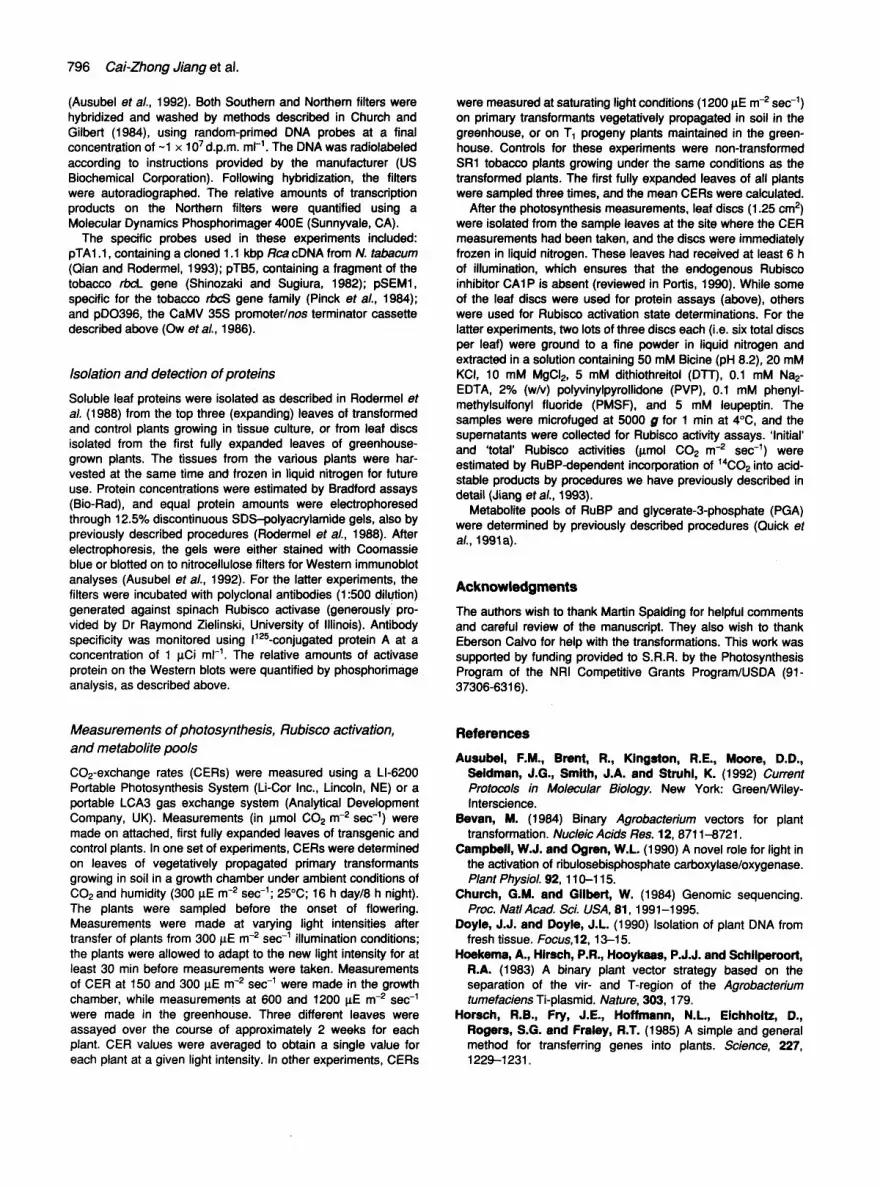
(Ausubel et aL, 1992). Both Southern and Northem filters were hybridized and washed by methods described in Church and Gilbert (1984), using random-primed DNA probes at a final concentration of ~1 x 10⁷d.p.m. m1-1. The DNA was radiolabeled according to instructions provided by the manufacturer (US Biochemical Corporation). Following hybridization, the filters were autoradiographed. The relative amounts of transcdption products on the Northern filters were quantified using a Molecular Dynamics Phosphodmager 400E (Sunnyvale, CA).
The specific probes used in these experiments included: pTA1.1, containing a cloned 1.1 kbp Rca cDNA from N. tabacum (Qian and Rodermel, 1993); pTB5, containing a fragment of the tobacco rbcL gene (Shinozaki and Sugiura, 1982); pSEM1, specific for the tobacco rbcS gene family (Pinck et al., 1984); and pDO396, the CaMV 35S promoter/nos terminator cassette described above (Ow et al., 1986).
Isolation and detection of proteins
Soluble leaf proteins were isolated as described in Rodermel et aL (1988) from the top three (expanding) leaves of transformed and control plants growing in tissue culture, or from leaf discs isolated from the first fully expanded leaves of greenhouse- grown plants. The tissues from the various plants were har- vested at the same time and frozen in liquid nitrogen for future use. Protein concentrations were estimated by Bradford assays (Bio-Rad), and equal protein amounts were electrophoresed through 12.5% discontinuous SDS-polyecrylamide gels, also by previously described procedures (Rodermel et aL, 1988). After electrophoresis, the gels were either stained with Coomassie blue or blotted on to nitrocellulose filters for Western immunoblot analyses (Ausubel et aL, 1992). For the latter experiments, the filters were incubated with polyclonai antibodies (1:500 dilution) generated against spinach Rubisco activase (generously pro- vided by Dr Raymond Zielinski, University of Illinois). Antibody specificity was monitored using 1125-conjugated protein A at a concentration of 1 I~Ci ml -~. The relative amounts of activase protein on the Western blots were quantified by phosphorimage analysis, as described above.
Measurements of photosynthesis, Rubisco activation, and metabolite pools
CO2-exchange rates (CERs) were measured using a LI-6200 Portable Photosynthesis System (Li-Cor Inc., Lincoln, NE) or a portable LCA3 gas exchange system (Analytical Development Company, UK). Measurements (in pmol C02 m -2 sec -1) were made on attached, first fully expanded leaves of transgenic and control plants. In one set of experiments, CERs were determined on leaves of vegetatively propagated primary transformants growing in soil in a growth chamber under ambient conditions of CO2 and humidity (300 ~E m -2 sec-1; 25°C; 16 h day/8 h night). The plants were sampled before the onset of flowering. Measurements were made at varying light intensities after transfer of plants from 300 I~E m -2 sec -~ illumination conditions; the plants were allowed to adapt to the new light intensity for at least 30 min before measurements were taken. Measurements of CER at 150 and 300 I~E m -2 sec -1 were made in the growth chamber, while measurements at 600 and 1200 I~E m -2 sec -~ were made in the greenhouse. Three different leaves were assayed over the course of approximately 2 weeks for each plant. CER values were averaged to obtain a single value for each plant at a given light intensity. In other experiments, CERs
were measured at saturating light conditions (1200 p.E m -2 sec -1) on primary transformants vegetatively propagated in soil in the greenhouse, or on T1 progeny plants maintained in the green- house. Controls for these experiments were non-transformed SR1 tobacco plants growing under the same conditions as the transformed plants. The first fully expanded leaves of all plants were sampled three times, and the mean CERs were calculated.
After the photosynthesis measurements, leaf discs (1.25 cm 2) were isolated from the sample leaves at the site where the CER measurements had been taken, and the discs were immediately frozen in liquid nitrogen. These leaves had received at least 6 h of illumination, which ensures that the endogenous Rubisco inhibitor CA1P is absent (reviewed in Portis, 1990). While some of the leaf discs were used for protein assays (above), others were used for Rubisco activation state determinations. For the latter experiments, two lots of three discs each (i.e. six total discs per leaf) were ground to a fine powder in liquid nitrogen and extracted in a solution containing 50 mM Bicine (pH 8.2), 20 mM KCI, 10 mM MgCI2, 5 mM dithiothreitol (DTT), 0.1 mM Na2- EDTA, 2% (w/v) polyvinylpyrollidone (PVP), 0.1 mM phenyl- methylsulfonyl fluoride (PMSF), and 5 mM leupeptin. The samples were microfuged at 5000 g for 1 min at 4°C, and the supernatants were collected for Rubisco activity assays. 'Initial' and 'total' Rubisco activities (l~mOl CO2 m -2 sec -~) were estimated by RuBP-dependent incorporation of ~4CO2 into acid- stable products by procedures we have previously described in detail (Jiaog et al., 1993).
Metabolite pools of RuBP and glycerate-3-phosphate (PGA) were determined by previously described procedures (Quick et al., 1991a).
Acknowledgments
The authors wish to thank Martin Spalding for helpful comments and careful review of the manuscript. They also wish to thank Eberson Caivo for help with the transformations. This work was supported by funding provided to S.R.R. by the Photosynthesis Program of the NRI Competitive Grants Program/USDA (91- 37306-6316).
References
Ausubel, F.M., Brent, R., Kingston, R.E., Moore, D.D., Seidman, J.G., Smith, J.A. and Struhl, K. (1992) Current Protocols in Molecular Biology. New York: Green/Wiley- Interscience.
Bevan, M. (1984) Binary Agrobacterium vectors for plant transformation. Nucleic Acids lies. 12, 8711-8721.
Campbell, W.J. end Ogren, W.L. (1990) A novel role for light in the activation of ribulosebisphosphate carboxylase/oxygenase. Plant Physiol. 92, 110-115.
Church, G.M. and Gilbert, W. (1984) Genomic sequencing. Prec. Natl Acad. Sci. USA, 81, 1991-1995.
Doyle, J.J. and Doyle, J.L. (1990) Isolation of plant DNA from fresh tissue. Focus,12, 13-15.
Hoekema, A., Hirsch, P.R., Hooykaae, P.J.J. and Schilperoort, R.A. (1983) A binary plant vector strategy based on the separation of the vir- and T-region of the Agrobacterium turnefaciens Ti-plasmid. Nature, 303, 179.
Horech, R.B., Fry, J.E., Hoffmann, N.L., Eichholtz, D., Rogers, S.G. and Fraley, R.T. (1985) A simple and general method for transferring genes into plants. Science, 227, 1229-1231.

Jisng, C.-Z., Rodermel, S.R. and Shibles, R.M. (1993) Photosynthesis, Rubisco activity and amount, and their regu- lation by transcription in senescing soybean leaves. Plant Physiol. 101,105-112.
Kacser, H. and Porteous, J.W. (1987) Control of metabolism: what do we have to measure? Trends Biochem. Sci. 12, 5-14.
King, J. (1991) The Genetic Basis of Plant Physiological Processes. New York: Oxford University Press.
Lan, Y., Woodrow, I.E. and Mort, K.A. (1992) Light-dependent changes in ribulose bisphosphate carboxylase activase activity in leaves. Plant Physiol. 99, 304-309.
Maliga, P., Sz-Breznovits, A. and Marton, L. (1973) Strep- tomycin-resistant plants from callus culture of haploid tobacco. Nature New Biol. 244, 29-30.
Martino-Catt, S. and Ort, D.R. (1992) Low temperature interrupts circadian regulation of transcriptional activity in chilling-sensitive plants. Proc. Nat/ Acad. Sci. USA, 89, 3731-3735.
Miziorko, H.M. and Lorimer, G.H. (1983) Ribulose-1,5-bisphos- phate carboxylase-oxygenase. Ann. Rev. Biochem. 52, 507- 535.
Munro, H.J. and Fleck, A. (1966) The determination of nucleic acids. In Methods of Biochemical Analysis, Volume XIV (Glick, D., ed.). New York: John Wiley & Sons, pp. 113-176.
Murray, J.A.H. and Crockett, N. (1992) Antisense Techniques: an overview. In Antisense RNA and DNA. Modem Ceil Biology Series, Volume 4 (Murray, J.A.H., ed.). New York: Wiley-Liss, Inc., pp. 1--49.
Ow, D., Wood, K.V., DeLuca, M., de Wet, J.R., Hellnski, D.R. and Howell, S.H. (1986) Transient and stable expression of the firefly luciferase gene in plant cells and transgenic plants. Science, 234, 856-859.
Pinck, M., Guilley, E., Durr, A., Hoff, M., Pinck, L. and Fleck, J. (1984) Complete sequence of one of the mRNAs coding for the small subunit of ribulose bisphosphate carboxylase of Nicotiana sy/vestris. Biochimie, 66, 539-545.
Portis, A.R., Jr (1990) Rubisco activase. Biochim. Biophys. Acta, 1015, 1 5-28.
Qien, J. and Rodermel, S.R. (1993) Ribulose-1,5-bisphosphate carboxylase/oxygenase activase cDNAs from Nicotiana tab- acum. Plant Physiol. 102, 683--684.
Quick, W.P., Schurr, U., Schelbe, R., Schulze, E.-D., Rodermel, S.R., Bogorad, L. and SUtt, M. (1991a) De- creased ribulose-l,5-bisphosphate carboxylase-oxygenase in tobacco transformed with 'antisense' rbcS. I. Impact on photo- synthesis in ambient growth conditions. Planta, 183, 542-554.
Quick, W.P., Sohurr, U., Fichtner, K., Schulze, E.-D., Rodermel, S.R., Bogorad, L. and SUtt, M. (1991b) The impact of decreased Rubisco on photosynthesis, growth, allocation and storage in tobacco plants which have been transformed with antisense rbcS. PlantJ. 1, 51-58.
Quick, W.P., Fichtner, K., Schulze, E.-D., et el. (1992) Decreased ribulose-1,5-bisphosphate carboxylase-oxygenase in tobacco transformed with 'antisense' rbcS. IV. Impact on photosynthesis in conditions of altered nitrogen supply. Planta, 188, 522-531.
Robinson, S.P. and Portis, A.R., Jr (1989) Adenosine triphos- phate hydrolysis by purified Rubisco activase. Arch. Biochem. Biophys. 268, 93-99.
Rca antisense DNA mutants of tobacco 797
Rodermel, S.R. and Bogorad, L. (1992) Antisense mRNA inhibition of ribulose bisphosphate carboxylase---the most abundant protein in photosynthetic cells. In Antisense RNA and DNA. Modern Cell Biology Series, Volume 4 (Murray, J.A.H., ed.). New York: Wiley-Liss, Inc., pp. 121-135.
Rodermel, S.R., Abbott, M.S. and Bogorad, L. (1988) Nuclear--organelle interactions: nuclear antisense gene inhibits ribulose bisphosphate carboxylase enzyme levels in transformed tobacco plants. Cell, 55, 673-681.
Rundle, S.J. and ZleUnski, R.E. (1991) Alterations in barley ribulose-l,5-bisphosphate carboxylase/oxygenase activase gene expression during development and in response to illumination. J. Biol. Chem. 266, 14 802-14 807.
Salvucci, M.E., Portis, A.R., Jr and Ogren, W.L. (1986) Light and CO2 response of ribulose-1,5-bisphosphate carboxylase/ oxygenase activation in Arabidopsis leaves. Plant Physiol. 80, 655-659.
Salvucci, M.E., Werneke, J.M., Ogren, W.L. and PorUs, A.R., Jr (1987) Purification and species distribution of rubisco activase. Plant Physiol. 84, 930-936.
Shlnozaki, K. and Suglura, M. (1982) The nucleotide sequence of the tobacco chloroplast gene for the large subunit of ribulose-l,5-bisphosphate carboxylase/oxygenase. Gene, 20, 91-102.
Somerville, C.R. (1986) Analysis of photosynthesis with mutants of higher plants and algae. Ann. Rev. Plant Physiol. 37, 467-507.
Somerville, C.R., Portis, A.R., Jr and Ogren, W.L. (1982) A mutant of Arabidopsis thaliana which lacks activation of RuBP carboxylase in vivo. Plant PhysioL 70, 381-387.
Stitt, M., Quick, W.P., Sohurr, U., Schulze, E.-D., Rodermel, S.R. and Bogorad, L. (1991) Decreased ribulose-l,5- bisphosphate carboxylase-oxygenase in tobacco transformed with 'antisense' rbcS. I1. Flux-control coefficients for photo- synthesis in varying light, CO2, and air humidity. P/anta, 183, 555-566.
Streusand, V.J. and PorUs, A.R., Jr (1987) Rubisco activase mediates ATP-dependent activation of ribulose bisphophate carboxylase. Plant Physiol. 85, 152-154.
Wang, Z.Y. and PorUs, A.R., Jr (1992) Dissociation of ribulose- 1,5-bisphosphate bound to ribulose-l,5-bisphosphate carb- oxylase/oxygenase and its enhancement by ribulose- 1,5-bisphosphate carboxylase/oxygenase activase-mediated hydrolysis of ATP. Plant Physiol. 99, 1348-1353.
Werneke, J.M., Chatfield, J.M. and Ogren, W.L. (1988) Cat- alysis of ribulosebisphosphate carboxylase/oxygenase activa- tion by the product of a Rubisco activase cDNA clone expressed in Escherichia coil Plant Physiol. 87, 917-920,
Woodrow, i.E. and Berry, J.A. (1988) Enzymatic regulation of photosynthetic CO2 fixation in C3 plants. Ann. Rev. Plant Physiol. 39, 533-594.
Woodrow, I.E and Mott, K.A. (1992) Biphasic activation of ribulose bisphosphate carboxylase in spinach leaves as deter- mined from nonsteady-state CO2 exchange. Plant Physiol. 99, 298-303.
Zlellnski, R.E., Wemeke, J.M. and Jenkins, M.E. (1989) Co- ordinate expression of rubisco activase and rubisco during barley leaf cell development. Plant Physiol. 90, 516-521.

798 Cai-Zhong Jiang et al,
Note added in proof
While this paper was in review, another group reported the generation and characterization of Rubisco activase antisense DNA mutants of tobacco (C.J. Mate, G.S. Hudson, S. von Caemmerer, J.R. Evans and T.J. Andrews, Plant Physiology, 102, 1119-1128, 1993). Mate et aL were primarily interested in the mechanism and kinetics of Rubisco activation, and examined mutant plants with very low activase levels (less than 25% wild-type amounts) growing under non-physiological high CO2 concentrations. Our studies, in contrast, focused on the control that activase exerts on photosynthesis and included topics that Mate et al. did not address~.g, molecular analyses of nuclear-chloroplast interactions, metabolite assays, control analyses, and plant growth assays. In addition, our experiments were conducted on transgenic plants that contained a wide range of activase concentrations and that were growing in air. Our experiments and those of Mate et aL are thus complementary studies on the use of activase antisense mutants to better understand the function and role of this enzyme in photosynthesis.