history of orchid propagation: a mirror of the history of biotechnology
TRANSCRIPT
REVIEW ARTICLE
History of orchid propagation: a mirror of the historyof biotechnology
Tim Wing Yam Æ Joseph Arditti
Received: 4 April 2008 / Accepted: 3 July 2008 / Published online: 31 January 2009
� Korean Society for Plant Biotechnology and Springer 2009
Part I: Seed germination
Abstract Orchid seeds are nearly microscopic in size.
Because of that, many fanciful theories were proposed for
the origin of orchids. Almost 400 years separate the time
when orchid seeds were seen for the first time and the
development of a practical asymbiotic method for their
germination. The seeds were first observed and drawn
during the sixteenth century. Seedlings were first described
and illustrated in 1804. The association between orchid and
fungi was observed as early as 1824, while the requirement
for mycorrhiza for seed germination was established in
1899. An asymbiotic method for orchid seed germination
was developed in 1921. After Knudson’s media B and C
were formulated, orchids growing and hybridization
became widespread. Hybrids which early growers may not
have even imagined became possible.
Keywords Clonal propagation � In vitro propagation �Mycorrhiza � Orchid seeds � Propagation � Seed germination
Introduction
A convincing argument can be made that research into
orchid propagation (and the procedures themselves) were
always in the forefront of biotechnology (or at least prop-
agation methods) of their time. The first method for
horticultural orchid seed germination (Moore 1849; for
reviews, see Arditti 1984; Yam et al. 2002) was a major
and radical departure from the manner in which other seeds
were germinated 160 years ago. David Moore’s (1807–
1879) approach was an innovative major horticultural and
biological advance (Moore 1849). Half a century after
Moore’s discovery, Noel Bernard (1874–1911) made
another quantum jump when he formulated a method for
symbiotic germination of orchid seeds in vitro (Bernard
1899, 1909a; Bernard 1990; for reviews, see Boullard
1985; Arditti 1990; Rasmussen 1995; Yam et al. 2002). His
is probably the first method for in vitro propagation of any
plant. It utilizes what were at the time modern and
advanced microbiological procedures. Bernard also pre-
dicted that a day would come when orchid growers would
have laboratories as part of their establishments. This is the
case at present not only for orchids, but also for other
plants.
Lewis Knudson’s (1884–1958) method for the asymbi-
otic germination of orchid seeds (Knudson 1921, 1922a;
for reviews, see Arditti 1984, 1990; Yam et al. 2002) was
the first practical procedure for in vitro propagation of any
plant in pure (i.e., axenic) culture. His method was a sig-
nificant conceptual and technological innovation which
foreshadowed modern biotechnology.
David Moore may have based his work (Moore 1849) on
reports that orchid seeds can germinate if scattered at the
base of a mature plant. However, Bernard’s discovery and
method were not based on any previous procedures and/or
Joseph Arditti is Professor Emeritus.
T. W. Yam (&)
Singapore Botanic Gardens, Cluny Road,
Singapore, Singapore
e-mail: [email protected]
J. Arditti
Department of Developmental and Cell Biology,
University of California, Irvine, CA 92604, USA
e-mail: [email protected]
123
Plant Biotechnol Rep (2009) 3:1–56
DOI 10.1007/s11816-008-0066-3

research by others. They were solely a result of his bril-
liance (Bernard 1899, 1909a; Boullard 1985; Arditti 1990;
Bernard 1990; Yam et al. 2002). Knudson developed the
asymbiotic method as a result of a sharp mind, incisive
reasoning, and on the basis of his own pioneering research
with other plants (Knudson 1921, 1922a, b; for a review,
see Arditti 1990). The micropropagation of orchids by
means of tissue culture has a more complex history which
is not free of controversy and includes unusual episodes
(Arditti 1977b, 1985, 2001; Arditti and Arditti 1985; Tor-
rey 1985a, b; Arditti and Krikorian 1996; Easton 2001).
Seed germination accidentally or in nature
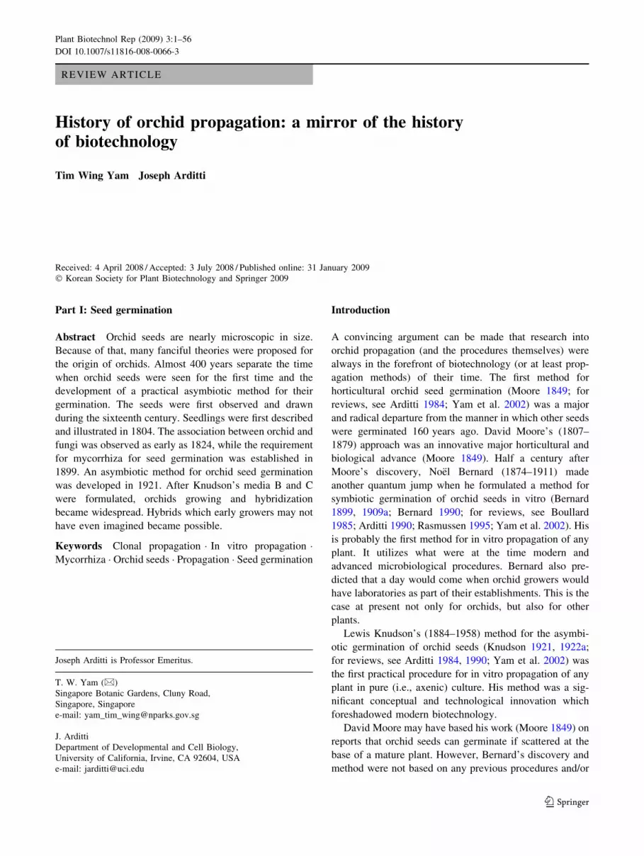
Orchid seeds (Fig. 1) are dust-like and nearly impossible to
observe individually with unaided eyes. It is very probable
that they remained unnoticed for much of history. If the
ancient Greeks noticed these seeds, their scientists and
philosophers did not write about them, not even Theo-
phrastus (370–285 B.C.), who is often called the Father of
Botany and who was also the first Western naturalist to
describe an orchid, or Dioscorides (ca. 20–70 A.D.) who
wrote about orchids later (Lashley and Arditti 1982; Arditti
1992). The Roman naturalist Caius Plinius Secundus (Pliny
The Elder; A.D. 23 or 24–79) wrote about orchids in his
Treatise on Natural History without even alluding to orchid
seeds (Lawler 1984; Arditti 1992; Jacquet 1994). Orchids
and their seeds were not mentioned in the Ebers papyri (ca.
1500 B.C.).
Despite the fact that the use of orchids to stimulate
lactation originated in ancient Mesopotamia (Lawler 1984;
Jacquet 1994), there is no mention of these plants and their
seeds in Assyrian writing of the Ashubanipal period (668–
627 B.C.). Many plants are mentioned in the bible, but
orchids and their seeds are not (Dunn and Arditti 2009).
There are no published reports on whether seeds are
mentioned in the rich Islamic-Arabic literature on natural
history, botany, and even orchids (Jacquet 1994). The
Turkish Tercume-i Cedide Fil-Havasil Mufrede by Mehmet
Ali which dates back to 1691–1692 describes salep, an
orchid product (Sezik 1967, 1984; Arditti 1992), but seeds
are not mentioned. And none of these sources mention
orchid seedlings. If seeds are mentioned in the ancient
Chinese, Indian, Japanese, and Korean literature, no one
seems to have discovered the writings.
Plinius and Plinius: first descriptions of orchid seeds
There are three known early descriptions of orchid seeds in
the west. All were published many years after they were
written with the first description being the second to be
published. The first to be published, Herbarium Amboin-
ense, is a six-volume work written between 1654 and
1702 by Georgius Everhadus Rumphius (1627–1702;
Fig. 2), ‘Plinius Indicus’, in Ambon, Indonesia (de Wit
1977; Beekman 2003) and published by Professor Joannes
Burman (1706– or 1707–1779) in Holland half a cen-
tury (1741–1750) after Rumphius’s death (Wehner et al.
2002).
Conrad Gesner (1516–1565; Fig. 3), ‘Plinius Germani-
cus’, a Swiss scientist, was actually the first to describe and
draw orchid seeds (Arditti 1992; Jacquet 1994; Wehner
et al. 2002). However, his book, Opera Botanica (Gesner
1751), was published between 1751 and 1771 by Christo-
pher Jacob Trew (1695–1769). This does not really matter
because no one seems to have paid much if any attention to
orchid seeds for a long time even after Herbarium Ambo-
inense and Opera Botanica were published.
The artists of a Spanish scientific expedition to New
Grenada (now Colombia) also drew orchid seeds, which
they did between 1783 and 1816. These, the second
drawings after Gesner’s, are the first to indicate size. It is
not clear at present if the artists who drew them had access
to or were familiar with Herbarium Amboinense and Opera
Botanica. Publication of these illustrations (Perez-Arbelaez
et al. 1954; Schweinfurth et al. 1963, 1969; Fernandez
Perez 1985) was delayed 150 years (Arditti and Ghani
2000).
Bock or Tragus: fancy, not facts
Since orchid seeds were neither seen nor known, it is not
surprising that several fanciful ideas were proposed to
explain the origin of orchids. A number of authors asso-
ciated several birds and four-legged animals with orchids.
One of the more interesting associations is between goats
and Himantoglossum hircinum (L.) Spreng. [Satyrium
hircinum L., Loroglossum hircinum (L.) Rich.]. This
orchid produces caproid acid, a substance which smells
like goats. This explains the supposition that it is derived
from goat semen which dropped to the ground during
copulation by goats and ‘‘fermented’’ into orchids (Arditti
1972).
Jeremy (Jerome) Bock, who is also known as Hierony-
mus Tragus (1498–1554), suggested that orchids came
about from semen of birds and beasts which fell to the
ground when they copulated. He wrote: ‘‘As soon as the
flowers abscise little pods arise in which no more is found
than pure dust or flour. These plants arise miraculously
from the seeds or sperm of junipers, blackbirds and thru-
shes, in Latin they are named turi and nerubae; these
Satyrions are found nowhere else except in the meadows
where these birds search for food’’. Had Bock (Tragus)
2 Plant Biotechnol Rep (2009) 3:1–56
123
recognized the true nature of the ‘‘dust or flour’’ and
appreciated its nature he would have preceded Rumphius in
describing orchid seeds (for reviews, see Arditti 1992; Yam
et al. 2002).
Athanasius Kircher (1601–1680), a German Jesuit,
expounded on Bock’s ideas in his Mundus Subterraneus
(published 1664–1665 in Amsterdam) and wrote that ‘‘these
plants arise from the latent survival force in the cadavers of
Fig. 1 Orchid seeds (Beer 1863)
Plant Biotechnol Rep (2009) 3:1–56 3
123

certain animals [and] animal semen [that] falls to the ground
in mountains and meadows.’’ As proof, Kircher drew images
of flowers (these and other illustration which are relevant to
this section can be seen in Arditti 1992; Yam et al. 2002)
which resemble animals (birds, goats, humanoid, sheep)
whose cadavers and semen gave rise to orchids.
Naumburg and Wachter: eighteenth century
observations and reports
Samuel Johann Naumburg (1768–1799), Professor at Erfurt
(Thuringia, Germany), wrote a paper which includes
drawings of orchid ovaries. He stated in the paper that:
‘‘Das Saamenbehaltniß ißt eine Kapßel [in free translation:
‘‘the seed container is a capsule’’]… Die Kapßeln enhalten
eine grosse Menge ganz kleiner brauner Saamen [‘‘the
capsules contain many very small brown seeds]’’ and in a
footnote: ‘‘Semina plurima, minima, brunnea [‘‘seeds
many, small, brown’’]’’ (Naumburg 1794).
A forester named Johann Karl Augustin Wachter (1773–
1846) became intrigued by Naumburg’s paper and hand-
pollinated orchids after reading it (Wachter 1799–1801). He
drew what appears to be a swollen ovary of ‘‘Ophrys Nidus’’
(probably Neottia nidus-avis) and wrote that the ovary of
Orchis militaris became swollen after hand pollination and
produced ‘‘eine grosse Menge Samen’’ (a great many seeds).
If additional reports were published after that they are
either buried deep in seldom seen and little known book(s)
and/or journal(s) or lost because subsequent authors did not
cite additional reports. Page by page searches through some
of the old literature in several libraries have also not
uncovered any relevant publications.
Observations and reports in the nineteenth century
As the nineteenth century started, the general belief among
botanists was that orchids rarely produced seeds which,
even if present, never germinated. However, early in this
Figs. 2–4 First observers of
orchid seeds and seedlings.
2 Georgius Everhardus
Rumphius, ca. 1627–1702.
3 Conrad Gesner, 1516–1565.
4 Richard Anthony Salisbury,
1761–1829 (A); seedlings of
Orchis morio (B), and
Limodorum verecundum (C)
4 Plant Biotechnol Rep (2009) 3:1–56
123

century, an eminent British botanist described germinating
orchid seeds and developing seedlings for the very first
time. They happened to be those of European species
(Salisbury 1804; for reviews, see Arditti 1967, 1979, 1984,
1990, 1992). Many other important observations and dis-
coveries were made later in the nineteenth century.
Salisbury: seeds and germination of temperate climate
orchids
Richard Anthony Salisbury (1761–1829; Fig. 4A) was
eccentric, hard to get along with, mired in scandal during part
of his life and apparently disdainful of at least some Victorian
moral constraints. He was also an excellent botanist (for more
details and illustrations, see Yam et al. 2002) and a con-
temporary of (1) Robert Brown (1773–1858), the noted
British botanist who studied orchid pollination and fertiliza-
tion and discovered cell nuclei while doing it, (2) John
Lindley (1799–1865), who is often referred to as the father of
orchidology, and (3) Charles Darwin (1809–1882) who is
probably the most influential thinker of all time. At that time,
orchid seedlings were not known or believed to occur in
nature. This changed on 5 January 1802 when Salisbury read
a paper to the Linnean Society describing germinating orchid
seeds and seedlings. The talk was published 2 years later
(Salisbury 1804), It was enhanced by illustrations of seeds
and seedlings of Orchis morio Linn. and Limodorum vere-
cundum Prodr. (Fig. 4Ba–f, Ca–e). Despite being the first
modern description of seeds and seedlings it did not seem to
have drawn much if any attention and/or to have stimulated
additional studies and/or reports at the time regarding British
or other European orchid seeds and/or seedlings.
Link: a tropical orchid seedling and a missed
opportunity
Salisbury’s drawings depicted the external appearance and
morphology of orchid seedlings and seeds. They did not
show structural features of either or the mycorrhizal asso-
ciation of the seedlings. However, the German botanist
Heinrich Friedrich Link (1767–1851; Fig. 5A) illustrated
these characteristics very well (Link 1824, 1839–1842,
1840, 1849a, b). Link’s drawings (Fig. 5Ba–k) may not have
been the first but they are excellent even by current stan-
dards. It is also interesting to note that Link drew seeds and
seedlings of a tropical orchid, Oeceoclades maculata, before
anyone else. It is obvious that Link saw mycorrhizae in cells
of seedlings, but he did not appreciate their importance.
Cameron: seedlings in a garden
Sometime between 1835 and 1838, David Cameron (ca.
1787–1848), Curator of the Birminham Botanical Garden
at the time (and before that, gardener for Robert Barclay of
Barclay’s Bank fame), saw ‘‘self-sown seedlings in several
of the pots’’ which contained ‘‘British Orchidaceae [which
were] cultivated [with] alpine plants.’’ Some of the seed-
lings were ‘‘very small, and evidently seedlings of that
year, others were much stronger. Of plants so obtained
several… Gymnadenia conopsea, Orchis maculata, and O.
latifolia…’’ flowered (Cameron 1844, 1848).
Herbert: foreshadowing Darwin
Among British clergymen, the Rev. Stephen Hales (1677–
1761) studied water uptake by plants; the Rev. John Hen-
slow (1796–1861) was Professor of Botany at Cambridge,
wrote about flower structure of orchids and other plants
(Henslow 1888) and befriended young Charles Darwin
(1809–1882); and the less well known but more eccentric
Rev. James Neil used plants as the subjects of his religious
parables (Neil 1880). The Honorable and Very Reverend
William Herbert (1778–1847; Fig. 6), Dean of Manchester,
followed in this tradition (Herbert 1846, 1847). He seems
to have been an enlightened clergyman having ‘‘asserted
that it was preposterous to suppose all the existing form of
vegetables… to have been so specially created by the
Almighty, and… I suspected that the various forms… to
have also branched out from smaller number of original
types…’’ (Herbert 1847). These views, published in 1847,
12 years before publication of Darwin’s Origin of Species
in 1859, lead to attacks on him ‘‘as a person who was
minishing from the power and wisdom of God’’ (Herbert
1847). Dean Herbert countered these attacks by suggesting
that ‘‘immense operations of ages before the creation of
man… were not compressed within a diurnal week of our
terrestrial life, but filled a gigantic page in the great volume
of antecedent time’’ (Herbert 1847).
Herbert’s arguments regarding plants was even more
explicit: ‘‘I am… unwilling to assent to the assertion, that
every plant… or even a distinct species, or… genus, had a
special creation’’ (Herbert 1847). After laying this theo-
logical foundation, Herbert stated: ‘‘If I can show that in
one genus of plants cross breeding is not only easy, but
more easily obtained than fertility by the plant’s own
pollen, and that in others, so closely allied to it as to make
it a question whether they are not sections of one genus,
cross-breeding cannot be affected generally, and in no case
easily; that in some genera of plants many or all the cross-
bred varieties are fertile, and in other nearly allied thereto
all, or almost are sterile… [he proceeds with examples
from animals]… the assertion that the races… must have
had separate origin because their crossed product is ster-
ile… must fall to the ground’’ (Herbert 1847).
Herbert tested his hypothesis with the intent to prove his
point by experimenting with the hybridization of many
Plant Biotechnol Rep (2009) 3:1–56 5
123

plants including orchids, his view being that ‘‘cross
breeding amongst Orchidaceous plants would perhaps lead
to very startling results; but unfortunately they are not
easily raised by seed’’ (Herbert 1847, communicated in
October 1846). He reported that he produced orchid seeds
and raised seedlings of Bletia, Cattleya, Ophrys aranifera
and Orchis monorchis (now Herminium monorchis).
Unfortunately, he did not describe his method of seed
Fig. 5 Early drawings of orchid mycorrhiza. A Heirich Friedrich Link, 1767–1851. B Germinating seed and seedlings of Oeceoclades maculata(the sequence is g, a and c, h and j, b, e and f, i and k; d is a cross-section of a root)
6 Plant Biotechnol Rep (2009) 3:1–56
123
germination. This is an important weakness because
knowledge of how to germinate orchid seeds, if it existed at
the time, was not widespread. Herbert also failed to
describe his seeds and seedlings. It is possible that he used
Cameron’s method or one similar to it, but the lack of
information reduces the value and importance of his report.
However, it must be remembered that Dean Herbert
hybridized plants and germinated seeds to prove a theo-
logical point. Orchid seed germination was not his main
interest.
Irmisch: anatomical and morphological observations
In Germany, the first reports of orchid seed and seedlings
were by Johann Friedrich Thilo Irmisch, 1816–1879;
Fig. 7), a major figure in plant morphology during the
nineteenth century (Mullerott 1980). Orchids were not his
primary interest, but he published several papers on Ger-
man species (Irmisch 1842, 1854a, b, 1863, 1877). They
contained morphological and/or anatomical line drawings.
The excellent drawings in his major work on orchids
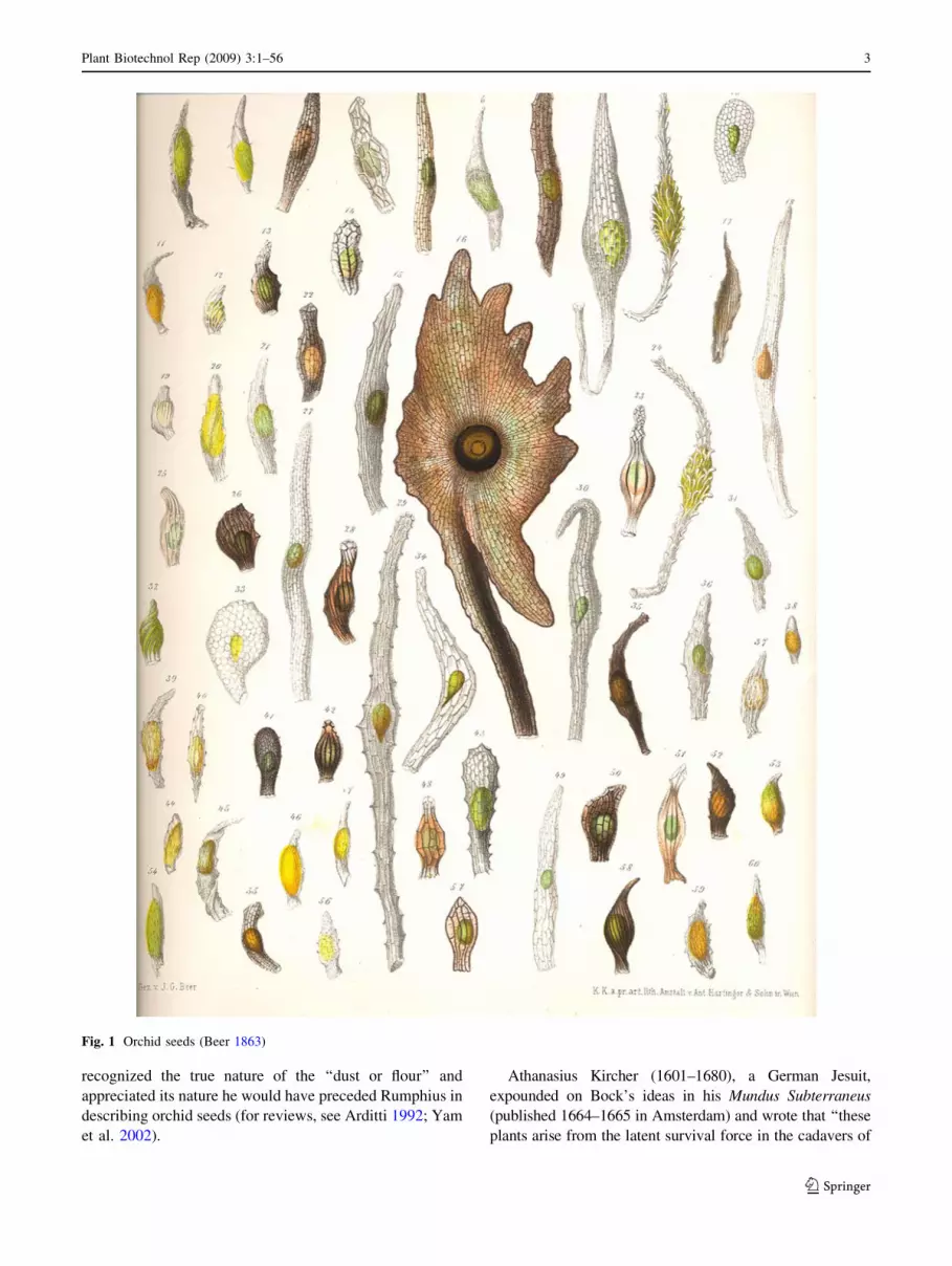
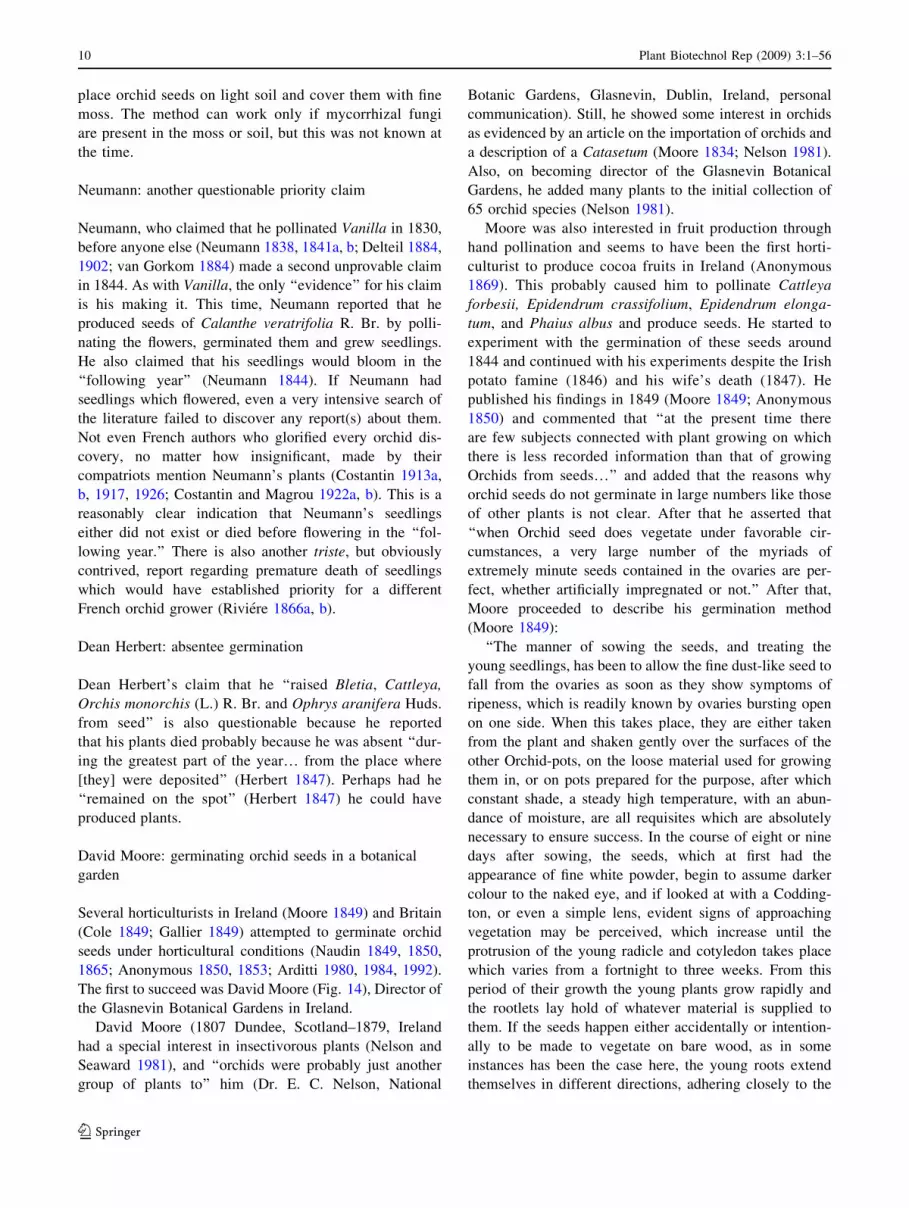
Figs. 6–15 Students of seed germination. 6 The Honorable and Very
Reverend William Herbert Dean of Manchester, 1778–1847. 7 Johann
Friedrich Thilo Irmisch, 1816–1879. 8 Jean-Henri-Fabre, 1823–1915.
9 Melchior Treub, 1851–1910. 10 Lycopod (B) and orchid (C)
seedlings (there is no A in this figure). 11 Charles Francois Antoine
Morren, 1807–1858 (A); Edouard Morren, 1833–1886 (B); slightly
magnified Vanilla seeds (C); side (D) and front (E) view of a Vanillaseed, 9166; Vanilla seed showing detail of testa, 9142. 12 Joseph
Henri Francois Neumann, 1850–1858. 13 Louis Claude Noisette,
1772–1849. 14 David Moore, 1807–1879. 15 John Harris, 1782–1855
Plant Biotechnol Rep (2009) 3:1–56 7
123
(Irmish 1853) may well be the first detailed anatomical and
morphological illustrations of seed germination and seed-
ling development, especially as they pertain to European
terrestrial species.
Fabre: Orchid germination and filaments
Jean-Henri Fabre (1823–1915; Fig. 8), is mainly associated
with studies of insect behavior (Fabre 1856). He became
interested in orchids due to ‘‘the asymmetry of their blos-
soms, the unusual structure of their pollen, and their
innumerable seeds’’ (Legros 1971), but was primarily
concerned with the structure of orchid tubers (Fabre 1855,
1856). This led him to a study entitled, ‘‘Inquiries
Respecting the Tubercules of Himantoglossum hircinum,’’
which was published as a thesis in 1855 (Legros 1971).
On studying Ophrys apifera subsequently, Fabre
observed many bulbiform bodies. He also saw the seeds of
this species and described them as being microscopic,
covered with a fusiform seed coat and containing a
spheroid embryo measuring 0.25 mm in diameter (Fabre
1856). Fabre described the seeds as germinating after
extended ‘‘incubation’’ in humus. He wrote that on swell-
ing they change in shape only at the apex and are covered
with long delicate filaments. The filaments could have been
trichomes which are often produced by protocorms or
mycorrhizal hyphae. If they were the latter, Fabre failed to
appreciate their importance. He also wrote that the seed-
lings become spheroids (i.e., protocorms) following
continued growth (Fabre 1856).
Treub: from lycopods to orchids
Melchior Treub (1851 Holland–1910 France; Fig. 9)
studied at the University of Leiden when the Chair of
Botany was occupied by Willem Frederik Reinier Suringar
(1832–1898) who was interested in lichens. Probably
because of this Treub’s dissertation dealt with the nature of
lichens (Schroter 1912; Went 1911).
Lichens did not hold Treub’s interest for long and he
moved on to studies of other plants including orchids. His
work was excellent and he had a promising future in
Holland. But as fate would have it, Herman Christian Carl
Scheffer (Holland 1855–Indonesia 1880), director of the
Botanical Garden in Buitenzorg (now Bogor), died and
Treub became his successor (Went 1911). He served in that
capacity until 1909.
Treub made a number of major contributions to orchid
studies in general and the understanding of their embry-
ology, seeds, and seedlings in particular. While still in
Holland, he studied the embryology and seed development
of several species (Treub 1879). The line drawings (by
Treub himself) are excellent. However, his most important
contribution to orchid science was unintentional. He made
it in a paper on the embryology of club mosses (Treub
1890) by proposing the term protocorme for an early stage
in the germination of lycopods (Fig. 10). No _el Bernard (see
below) must have read Treub’s paper and 10 years later
applied the term to orchid seedlings (Arditti 1989, 1990,
1992).
Prescottia: first seedlings of a tropical orchid outside
the tropics
The first reported germination of the seeds of a tropical
orchid, Prescottia plantaginea Lindl. outside the tropics,
was observed in a horticultural establishment in the UK.
However, the date is in question and the seed source is not
certain (for a review, see Arditti 1984). Two dates are listed
for the production of these seedlings. One is 1822 and the
other is 1832. It is possible that the seeds, and therefore
seedlings, may have been produced before 1832, but the
available information does not point to 1822 with certainty
(for a discussion, see Arditti 1992, pp. 40–42).
Prescottia plantaginea could have been introduced into
cultivation shortly after John Forbes (1798–1823), a British
collector in Brazil, sent plants to the garden of the Horti-
cultural Society of London in the autumn of 1822. The
source of the seeds which produced the seedlings in the UK
is not clear, and there are no known reports that they were
seen by anyone. P. plantaginea produces seeds apomicti-
cally or through self pollination. Therefore, one possibility
is that the seeds were produced after the plants arrived in
the UK and ripened after 1822. Another possibility is that
fruits may have been present on the plants that were sent to
the UK. If plants collected in their natural habitats bore
fruits, the capsules continued to develop en route, ripened
on arrival, and the seeds could have matured a short time
after the plants arrived in Britain in 1822.
Regardless of how the seeds got to the Horticultural
Society gardens, they could germinate because a suitable
mycorrhizal fungus was probably present at the site. Such a
fungus could have come from a British native orchid or
from the roots of mature plants of P. plantaginea. Many
seedlings were raised at the Chiswick garden of the Hor-
ticultural Society (Anonymous 1858a, b; Hoene 1949; for a
review, see Arditti 1992).
The seedlings and the method by which they were
produced did not draw much attention at the time and
Lindley wrote about them only in 1858 (Anonymous
1858a, b). One reason for this could have been the lack of
popularity of the genus. Another may have been the acci-
dental nature of the germination. If the germination was
intentional there is a good chance that it would have been
published by whoever did it. In fact, it is surprising that no
one took credit for it at the time.
8 Plant Biotechnol Rep (2009) 3:1–56
123
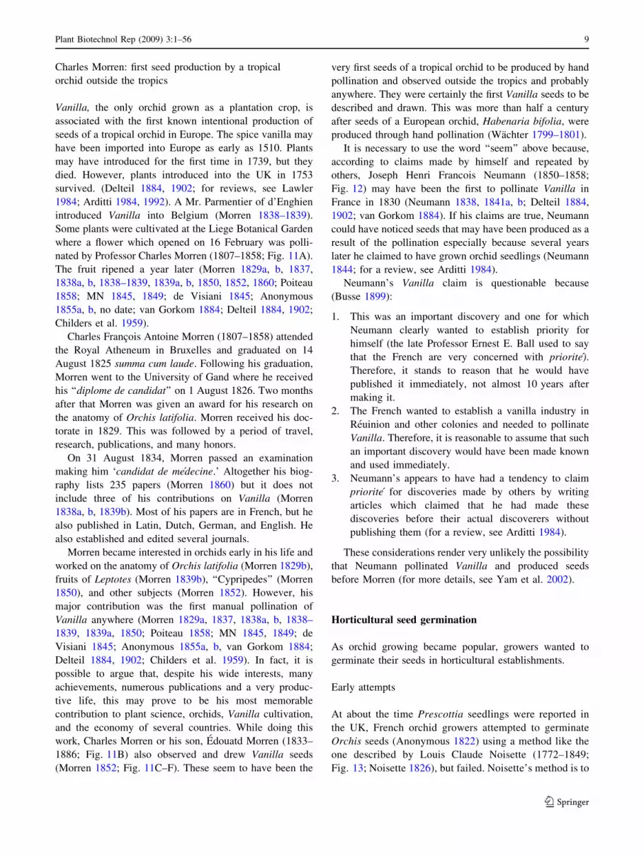
Charles Morren: first seed production by a tropical
orchid outside the tropics
Vanilla, the only orchid grown as a plantation crop, is
associated with the first known intentional production of
seeds of a tropical orchid in Europe. The spice vanilla may
have been imported into Europe as early as 1510. Plants
may have introduced for the first time in 1739, but they
died. However, plants introduced into the UK in 1753
survived. (Delteil 1884, 1902; for reviews, see Lawler
1984; Arditti 1984, 1992). A Mr. Parmentier of d’Enghien
introduced Vanilla into Belgium (Morren 1838–1839).
Some plants were cultivated at the Liege Botanical Garden
where a flower which opened on 16 February was polli-
nated by Professor Charles Morren (1807–1858; Fig. 11A).
The fruit ripened a year later (Morren 1829a, b, 1837,
1838a, b, 1838–1839, 1839a, b, 1850, 1852, 1860; Poiteau
1858; MN 1845, 1849; de Visiani 1845; Anonymous
1855a, b, no date; van Gorkom 1884; Delteil 1884, 1902;
Childers et al. 1959).
Charles Francois Antoine Morren (1807–1858) attended
the Royal Atheneum in Bruxelles and graduated on 14
August 1825 summa cum laude. Following his graduation,
Morren went to the University of Gand where he received
his ‘‘diplome de candidat’’ on 1 August 1826. Two months
after that Morren was given an award for his research on
the anatomy of Orchis latifolia. Morren received his doc-
torate in 1829. This was followed by a period of travel,
research, publications, and many honors.
On 31 August 1834, Morren passed an examination
making him ‘candidat de medecine.’ Altogether his biog-
raphy lists 235 papers (Morren 1860) but it does not
include three of his contributions on Vanilla (Morren
1838a, b, 1839b). Most of his papers are in French, but he
also published in Latin, Dutch, German, and English. He
also established and edited several journals.
Morren became interested in orchids early in his life and
worked on the anatomy of Orchis latifolia (Morren 1829b),
fruits of Leptotes (Morren 1839b), ‘‘Cypripedes’’ (Morren
1850), and other subjects (Morren 1852). However, his
major contribution was the first manual pollination of
Vanilla anywhere (Morren 1829a, 1837, 1838a, b, 1838–
1839, 1839a, 1850; Poiteau 1858; MN 1845, 1849; de
Visiani 1845; Anonymous 1855a, b, van Gorkom 1884;
Delteil 1884, 1902; Childers et al. 1959). In fact, it is
possible to argue that, despite his wide interests, many
achievements, numerous publications and a very produc-
tive life, this may prove to be his most memorable
contribution to plant science, orchids, Vanilla cultivation,
and the economy of several countries. While doing this
work, Charles Morren or his son, Edouatd Morren (1833–
1886; Fig. 11B) also observed and drew Vanilla seeds
(Morren 1852; Fig. 11C–F). These seem to have been the
very first seeds of a tropical orchid to be produced by hand
pollination and observed outside the tropics and probably
anywhere. They were certainly the first Vanilla seeds to be
described and drawn. This was more than half a century
after seeds of a European orchid, Habenaria bifolia, were
produced through hand pollination (Wachter 1799–1801).
It is necessary to use the word ‘‘seem’’ above because,
according to claims made by himself and repeated by
others, Joseph Henri Francois Neumann (1850–1858;
Fig. 12) may have been the first to pollinate Vanilla in
France in 1830 (Neumann 1838, 1841a, b; Delteil 1884,
1902; van Gorkom 1884). If his claims are true, Neumann
could have noticed seeds that may have been produced as a
result of the pollination especially because several years
later he claimed to have grown orchid seedlings (Neumann
1844; for a review, see Arditti 1984).
Neumann’s Vanilla claim is questionable because
(Busse 1899):
1. This was an important discovery and one for which
Neumann clearly wanted to establish priority for
himself (the late Professor Ernest E. Ball used to say
that the French are very concerned with priorite).
Therefore, it stands to reason that he would have
published it immediately, not almost 10 years after
making it.
2. The French wanted to establish a vanilla industry in
Reuinion and other colonies and needed to pollinate
Vanilla. Therefore, it is reasonable to assume that such
an important discovery would have been made known
and used immediately.
3. Neumann’s appears to have had a tendency to claim
priorite for discoveries made by others by writing
articles which claimed that he had made these
discoveries before their actual discoverers without
publishing them (for a review, see Arditti 1984).
These considerations render very unlikely the possibility
that Neumann pollinated Vanilla and produced seeds
before Morren (for more details, see Yam et al. 2002).
Horticultural seed germination
As orchid growing became popular, growers wanted to
germinate their seeds in horticultural establishments.
Early attempts
At about the time Prescottia seedlings were reported in
the UK, French orchid growers attempted to germinate
Orchis seeds (Anonymous 1822) using a method like the
one described by Louis Claude Noisette (1772–1849;
Fig. 13; Noisette 1826), but failed. Noisette’s method is to
Plant Biotechnol Rep (2009) 3:1–56 9
123
place orchid seeds on light soil and cover them with fine
moss. The method can work only if mycorrhizal fungi
are present in the moss or soil, but this was not known at
the time.
Neumann: another questionable priority claim
Neumann, who claimed that he pollinated Vanilla in 1830,
before anyone else (Neumann 1838, 1841a, b; Delteil 1884,
1902; van Gorkom 1884) made a second unprovable claim
in 1844. As with Vanilla, the only ‘‘evidence’’ for his claim
is his making it. This time, Neumann reported that he
produced seeds of Calanthe veratrifolia R. Br. by polli-
nating the flowers, germinated them and grew seedlings.
He also claimed that his seedlings would bloom in the
‘‘following year’’ (Neumann 1844). If Neumann had
seedlings which flowered, even a very intensive search of
the literature failed to discover any report(s) about them.
Not even French authors who glorified every orchid dis-
covery, no matter how insignificant, made by their
compatriots mention Neumann’s plants (Costantin 1913a,
b, 1917, 1926; Costantin and Magrou 1922a, b). This is a
reasonably clear indication that Neumann’s seedlings
either did not exist or died before flowering in the ‘‘fol-
lowing year.’’ There is also another triste, but obviously
contrived, report regarding premature death of seedlings
which would have established priority for a different
French orchid grower (Riviere 1866a, b).
Dean Herbert: absentee germination
Dean Herbert’s claim that he ‘‘raised Bletia, Cattleya,
Orchis monorchis (L.) R. Br. and Ophrys aranifera Huds.
from seed’’ is also questionable because he reported
that his plants died probably because he was absent ‘‘dur-
ing the greatest part of the year… from the place where
[they] were deposited’’ (Herbert 1847). Perhaps had he
‘‘remained on the spot’’ (Herbert 1847) he could have
produced plants.
David Moore: germinating orchid seeds in a botanical
garden
Several horticulturists in Ireland (Moore 1849) and Britain
(Cole 1849; Gallier 1849) attempted to germinate orchid
seeds under horticultural conditions (Naudin 1849, 1850,
1865; Anonymous 1850, 1853; Arditti 1980, 1984, 1992).
The first to succeed was David Moore (Fig. 14), Director of
the Glasnevin Botanical Gardens in Ireland.
David Moore (1807 Dundee, Scotland–1879, Ireland
had a special interest in insectivorous plants (Nelson and
Seaward 1981), and ‘‘orchids were probably just another
group of plants to’’ him (Dr. E. C. Nelson, National
Botanic Gardens, Glasnevin, Dublin, Ireland, personal
communication). Still, he showed some interest in orchids
as evidenced by an article on the importation of orchids and
a description of a Catasetum (Moore 1834; Nelson 1981).
Also, on becoming director of the Glasnevin Botanical
Gardens, he added many plants to the initial collection of
65 orchid species (Nelson 1981).
Moore was also interested in fruit production through
hand pollination and seems to have been the first horti-
culturist to produce cocoa fruits in Ireland (Anonymous
1869). This probably caused him to pollinate Cattleya
forbesii, Epidendrum crassifolium, Epidendrum elonga-
tum, and Phaius albus and produce seeds. He started to
experiment with the germination of these seeds around
1844 and continued with his experiments despite the Irish
potato famine (1846) and his wife’s death (1847). He
published his findings in 1849 (Moore 1849; Anonymous
1850) and commented that ‘‘at the present time there
are few subjects connected with plant growing on which
there is less recorded information than that of growing
Orchids from seeds…’’ and added that the reasons why
orchid seeds do not germinate in large numbers like those
of other plants is not clear. After that he asserted that
‘‘when Orchid seed does vegetate under favorable cir-
cumstances, a very large number of the myriads of
extremely minute seeds contained in the ovaries are per-
fect, whether artificially impregnated or not.’’ After that,
Moore proceeded to describe his germination method
(Moore 1849):
‘‘The manner of sowing the seeds, and treating the
young seedlings, has been to allow the fine dust-like seed to
fall from the ovaries as soon as they show symptoms of
ripeness, which is readily known by ovaries bursting open
on one side. When this takes place, they are either taken
from the plant and shaken gently over the surfaces of the
other Orchid-pots, on the loose material used for growing
them in, or on pots prepared for the purpose, after which
constant shade, a steady high temperature, with an abun-
dance of moisture, are all requisites which are absolutely
necessary to ensure success. In the course of eight or nine
days after sowing, the seeds, which at first had the
appearance of fine white powder, begin to assume darker
colour to the naked eye, and if looked at with a Codding-
ton, or even a simple lens, evident signs of approaching
vegetation may be perceived, which increase until the
protrusion of the young radicle and cotyledon takes place
which varies from a fortnight to three weeks. From this
period of their growth the young plants grow rapidly and
the rootlets lay hold of whatever material is supplied to
them. If the seeds happen either accidentally or intention-
ally to be made to vegetate on bare wood, as in some
instances has been the case here, the young roots extend
themselves in different directions, adhering closely to the
10 Plant Biotechnol Rep (2009) 3:1–56
123

bark, and make great progress compared with the growth of
the stems, thus affording beautiful examples of the manner
in which epiphytal plants fix themselves so firmly… ‘‘The
principal difficulty to contend with in rearing the young
seedlings has been found to consist in their treatment
during the first year, particularly the winter months… The
second year’s growth has been one during which the plants
made much progress and the only two kinds which have
been brought to a flowering state have bloomed the third
season. These are Epidendrum crassifolium and Phaius
albus, the latter being now in flower, exactly 3 years from
the sowing of the seeds.’’
Moore was director of the Glasnevin Botanic Gardens
for 30 years after publication of his paper on orchid seed
germination. He continued to import orchids from various
parts of the world but does not seem to have continued to
work on the germination of orchid seeds and the cultivation
of seedlings. Moore was also a member of many learned
societies. In 1864, he was awarded an honorary doctorate
by the University of Zurich. He was made a member of The
Royal Dublin Society in 1878.
In his waning days, Moore took part in religious
polemics and contributed to a collection of ‘‘lectures’’ by
several anti-evolutionists. His claim was that he proved
creation through ‘‘design in the structure and fertilization
of plants’’ (Moore 1875). By using the word ‘‘design’’
Moore may have foreshadowed current pseudoscientific
claims of ‘‘intelligent design.’’ This is surprising in view of
Moore’s cordial correspondence with Darwin regarding
insectivorous plants and potatoes during June and July of
1874. Darwin also wrote him a very nice letter on 3 May
1879 (i.e., about two months before his death; Nelson
1981; Nelson and Seaward 1981).
Moore’s other ‘‘contribution’’ to orchids rivals and
perhaps exceeds seed germination in importance. It was his
eldest son with his third wife Margaret Baker, Frederik
Moore (1857–1950), who developed a passion for orchids
and became well known as an orchid expert.
Richard Gallier and J. Cole: orchid seed germination
by two gardeners
Two British horticulturists, Richard Gallier and J. Cole,
also shared their experiences (Cole 1849; Gallier 1849;
Anonymous 1850) after Moore published his report. Gal-
lier was gardener for J. Tildesley, Esq., of West Bromwich,
Staffordshire in Britain. Cole held a similar position for J.
Willmore at Oldford near Birmingham. As he was located
near Birmingham it is possible that he could have inter-
acted with David Cameron (see above), but if he did there
are no known records of such an interaction. Very little is
known about Cole and Gallier and attempts to obtain
information about them failed.
Cole’s note was the first to be published after Moore’s
report. He wrote that his employer informed him:
‘‘that Bletia [now Phaius] Tankervillæ was some years
since obtained from seeds sown in common soil; also
Epidendrum elongatum sown on blocks of wood cov-
ered with moss. I have sown other sorts of Orchids at
various times and in different ways, but without suc-
cess… a few have been hybridised successfully here,
so far as obtaining seed to all appearance perfect… and
it has been sown, but it did not vegetate. Cattleya
labiata was crossed with C. guttata, and swelled its
pod (sic); Calanthe veratrifolia with Bletia Tankerv-
illæ; Dendrobium moniliforme with other Dendrobes;
and Stanhopea Wardii with one of the other Stanho-
pes… I have the hybridised seed pod of Stanhopea
Wardii by me, and shall be pleased to present some of
the seeds to Mr. Moore or any other gentleman who
may take an interest in raising seedlings.’’
J. Cole also stated that he intended to carry out further
experiments and planned to report his findings. If he did,
we could not find any reports he may have published. The
germination methods described by Cole can be successful
and do not seem to have been published before his letter.
The reputation of The Gardeners’ Chronicle (the most
important horticultural publication in the world at the time
which was nicknamed ‘‘The Times of Horticulture’’ in
allusion to the famed Times of London) provides every
reason to believe that Cole’s report was accurate and
factual.
Gallier’s report was also published in the The Garden-
ers’ Chronicle (Gallier 1849). He crossed Dendrobium
nobile with Dendrobium chrysanthum, obtained seed and:
‘‘sowed it in three ways: some on a log, with natural
moss growing on it, suspended in a shady part of the
Orchid-house; some was sown on an inverted flower
pot, the inside of which was stuffed with sphagnum,
and placed in a pan of water… neither of these two
sowings vegetated.’’
For his third method, Gallier used two pots, one filled
with sand and the other with water. He spread the seeds on a
floating piece of cork covered with a bell jar. Then he
placed the entire contraption in a shady part of the green-
house. Two seeds germinated after three weeks. Eventually,
Gallier had five plants all of which died when he removed
the cork from under the bell jar and suspended it from the
roof of his greenhouse (Gallier 1849).
A Belgian journal reported Moore’s, and Cole’ experi-
ences (Anonymous 1850) and stated that orchid propagation
through seed germination in the greenhouses of Europe
would open a new avenue for the culture of ‘‘these bizarre
plants’’ (Anonymous 1850).
Plant Biotechnol Rep (2009) 3:1–56 11
123
John Dominy, John Harris and Harry Veitch: the first
orchid hybrid
Since this review is limited to orchid seeds and seedlings,
the history of orchid hybridization will be mentioned only
as it relates to seed germination. Calanthe Dominyi, the
first commercial orchid hybrid, was produced in the Ve-
itch establishment in the UK. A gregarious surgeon
named John Harris (1782–1855, Fig. 15; Arditti 1980),
advised an excellent horticulturist, John Dominy (1816–
1891, Fig. 16) who was employed by an enlightened
nursery owner, Harry J. Veitch (1840–1924; Fig. 17), the
owner of the well known orchid nursery which was
established by his father, James Veitch (1792–1863;
Fig. 18) to cross orchids. Dominy made the first cross in
1853, seeds were harvested in 1854 and the first plant
bloomed in October 1856 (Veitch 1885, 1886; Veitch and
Sons 1887–1894). This well documented chronology
indicates that the first germination of orchid seeds as part
of: (1) a commercial venture, and (2) the first successful
attempt to produce a hybrid took place in 1854 in
England.
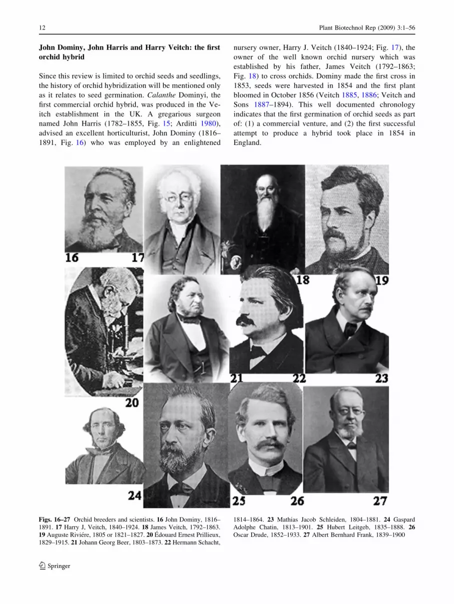
Figs. 16–27 Orchid breeders and scientists. 16 John Dominy, 1816–
1891. 17 Harry J, Veitch, 1840–1924. 18 James Veitch, 1792–1863.
19 Auguste Riviere, 1805 or 1821–1827. 20 Edouard Ernest Prillieux,
1829–1915. 21 Johann Georg Beer, 1803–1873. 22 Hermann Schacht,
1814–1864. 23 Mathias Jacob Schleiden, 1804–1881. 24 Gaspard
Adolphe Chatin, 1813–1901. 25 Hubert Leitgeb, 1835–1888. 26Oscar Drude, 1852–1933. 27 Albert Bernhard Frank, 1839–1900
12 Plant Biotechnol Rep (2009) 3:1–56
123

Even making the first cross was not simple. To fully
appreciate history as it unfolded it is best to quote a person
who was not only present as it happened but also made it
happen, Harry J. Veitch (1886):
‘‘… very few [horticulturists and gardeners] pos-
sessed even an elementary knowledge of botany.
They could… distinguish… the stamens and pistils of
many flowers… and they were aware of the functions
of those organs, but the confluence of those organs
into the solid column of an Orchid flower was to them
a profound mystery.
It was Mr. [actually Dr.] John Harris, a surgeon, of
Exeter, who suggested to Dominy the possibility of
muling Orchids, and who pointed out to him the
reproductive organs seated in the column, and
showed that the application of the pollinia to the
stigmatic surface was analogous to the dusting of the
stigma of other flowers with pollen. This simple fact
being once fairly grasped, the work of hybridization
proceeded apace… Capsules were produced in
abundance… dehiscing… and seed was at length in
hand. Then arose a great difficulty… which still
exists… to discover the most suitable method of
raising seedlings. The seeds of Orchids are… so
minute… that an ordinary pocket lens is powerless to
enable one to know whether the seeds are likely to
contain a germ or are mere lifeless dust. Following,
or at least believing that we were following Nature…every method or available means that could be
thought of was brought into request to secure the
germination of the seed. It was sown upon locks of
wood, pieces of tree fern stems, strips of cork, upon
the moss that surfaced the pots of the growing plants,
in fact in any situation that seemed to promise
favorable results. But… we seem far off as ever from
hitting upon a method by which at least a moderate
amount of success may be calculated upon.
Seeds we get in profusion, but… little of it germi-
nates… The seeds of hundreds of capsules have been
sown without yielding a single result. In very many cases
only a solitary plant had been raised from a capsule that
must have contained thousands of seeds; in very few
instances indeed has the number of seedlings reached a
hundred. It is true that we have raised many seedlings in
the aggregate, but many of them have appeared when
least expected, and when we consider the myriads of
seeds that have been sown, and the comparatively few
plants raised, we cannot be said to have achieved great
success…’’
The seed germination method used at the Veitch
establishment was similar to the methods used by Moore,
Cole, and Gallier.
Orchid seed germination: 1850–1899
Several horticulturists germinated orchids and produced
hybrids during the second half of the nineteenth century.
John and John: Orchid Hybrids
Dominy (Fig. 16), Cole, Gallier, and Veitch (Fig. 17, 18;
DCGV) made their seed germination methods known by
publishing them in The Gardeners’ Chronicle. Within a
year of the original articles there was also at least one
report in Belgium in French about Moore’s and Cole’s
findings (Anonymous 1850) in a magazine which was
probably also read in France. Three additional reports were
published a number of years after that in France and Bel-
gium (Bergman 1879, 1881, 1882, 1889). Further, Dominy
and Harris were great conversationalists who enjoyed and
readily took part in extensive conversations (Arditti 1980).
This was enhanced by the fact that Dominy possessed ‘‘…wide knowledge which he was always willing to commu-
nicate orally…’’ (Anonymous 1891a). Therefore, the news
that orchid seeds can be germinated probably travelled fast
among those who were producing seeds and attempting to
germinate them (Anderson 1862, 1863; Gosse 1862, 1863),
most without success: ‘‘… there is nothing unusual in
obtaining seed-pods [sic] and seed in abundance from more
than one species; but I have never yet been fortunate
enough to get the seed to germinate’’ (Anderson 1862).
And when the seeds did germinate, ‘‘… all of the [seed-
lings] found the means of getting out of the world by a
route I never could fathom’’ (Beaton 1862). Thus, it is not
surprising that a large number of hybrids were produced
(i.e., seeds of additional orchids were germinated) shortly
after the reports by Moore, Cole, and Gallier and close on
the heels of Dominy’s first hybrid (Veitch 1886).
The second hybrid, a Cattleya flowered first in 1859.
The first Paphiopedilum flowered in 1869 (Veitch 1885,
1886; Veitch and Sons 1887–1894; for a review, see Arditti
1984). Successful germination of orchid seeds and estab-
lishment of seedlings remained newsworthy and a subject
for discussion for several years (Anonymous 1869, 1891a,
b; Douglas 1882a, b; Scheidweiler 1844, 1845; Godefroy-
Lebeuf 1886; Ignotus 1894). The Moore–Cole–Gallier–
Dominy–Veitch (MCGDV) method was also described in a
book on orchid cultivation in India (Jenning 1875), but it is
not clear whether it was used there or taken from British
sources and inserted in the book. There are no known
hybrids and instances of orchid seed germination during
that period from India or any other area except the UK and
Europe. This book is also found in the library of the Sin-
gapore Botanic Gardens but it is not clear when it was
acquired. Even if it was acquired in the 1880s, the seeds
which produced the first artificial orchid hybrid in
Plant Biotechnol Rep (2009) 3:1–56 13
123
Singapore, Spathoglottis Primrose, were germinated in
vitro in the 1920s on a medium formulated by Lewis
Knudson (Vanda Miss Joaquim, discovered by Miss Agnes
Joaquim in her garden in 1893 is believed to be a natural
hybrid by orchid scientists and knowleadgeable growers).
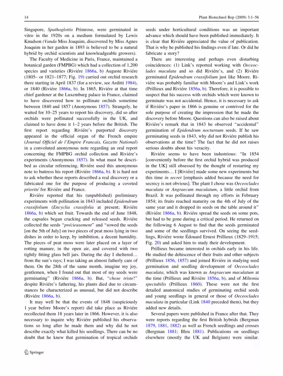
The Faculty of Medicine in Paris, France, maintained a
botanical garden (FMPBG) which had a collection of 1,200
species and varieties (Riviere 1866a, b) Auguste Riviere
(1805– or 1821–1877; Fig. 19) carried out orchid research
there starting in April 1837 (for a review, see Arditti 1984),
or 1840 (Riviere 1866a, b). In 1865, Riviere at that time
chief gardener at the Luxemburg palace in France, claimed
to have discovered how to pollinate orchids sometime
between 1840 and 1857 (Anonymous 1857). Strangely, he
waited for 10–25 years to report his discovery, did so after
orchids were pollinated successfully in the UK, and
claimed to have done it 1–2 years before the British. The
first report regarding Riviere’s purported discovery
appeared in the official organ of the French empire
(Journal Officiel de l’Empire Francais, Gazette National)
in a convoluted anonymous note regarding an oral report
concerning the FMPBG orchid collection and Riviere’s
experiments (Anonymous 1857). In what must be descri-
bed as circular referencing, Riviere used this anonymous
note to buttress his report (Riviere 1866a, b). It is hard not
to ask whether these reports described a real discovery or a
fabricated one for the purpose of producing a coveted
priorite for Riviere and France.
Riviere reported that his (unpublished) preliminary
experiments with pollination in 1843 included Epidendrum
crassifolium (Encyclia crassifolia at present; Riviere
1866a, b) which set fruit. Towards the end of June 1848,
the capsules began cracking and released seeds. Riviere
collected the seeds ‘‘precieusement’’ and ‘‘sowed the seeds
[on the 5th of July] on two pieces of peat moss lying in two
dishes in order to keep, by imbibition, a decent humidity.
The pieces of peat moss were later placed on a layer of
rotting manure, in the open air, and covered with two
tightly fitting glass bell jars. During the day I sheltered…from the sun’s rays; I was taking an almost fatherly care of
them. On the 28th of the same month, imagine my joy,
gentlemen, when I found out that most of my seeds were
germinating’’ (Riviere 1866a, b). But, ‘‘chose triste!’’
despite Riviere’s fathering, his plants died due to circum-
stances he characterized as unusual, but did not describe
(Riviere 1866a, b).
It may well be that the events of 1848 (suspiciously
1 year before Moore’s report) did take place as Riviere
recollected them 18 years later in 1866. However, it is also
necessary to inquire why Riviere published his observa-
tions so long after he made them and why did he not
describe exactly what killed his seedlings. There can be no
doubt that he knew that germination of tropical orchids
seeds under horticultural conditions was an important
advance which should have been published immediately. It
is clear that Riviere appreciated the value of publication.
That is why he published his findings even if late. Or did he
fabricate a story?
There are interesting and perhaps even disturbing
coincidences: (1) Link’s reported working with Oeceoc-
lades maculata and so did Riviere’s, and (2) Riviere
germinated Epidendrum crassifolium just like Moore. Ri-
viere was probably familiar with Moore’s and Link’s work
(Prillieux and Riviere 1856a, b). Therefore, it is possible to
suspect that his success with orchids which were known to
germinate was not accidental. Hence, it is necessary to ask
if Riviere’s paper in 1866 is genuine or contrived for the
sole purpose of creating the impression that he made the
discovery before Moore. Questions can also be raised about
Riviere’s remark that in 1843 he observed ‘‘accidental’’
germination of Epidendrum nocturnum seeds. If he saw
germinating seeds in 1843, why did not Riviere publish his
observations at the time? The fact that he did not raises
serious doubts about his veracity.
Riviere seems to have been industrious: ‘‘In 1854
[conveniently before the first orchid hybrid was produced
in the UK] still obsessed by the thought of restarting my
experiments… I [Riviere] made some new experiments but
this time in secret [emphasis added because the need for
secrecy is not obvious]. The plant I chose was Oeceoclades
maculata or Angraecum maculatum, a little orchid from
Brazil. It was pollinated through my efforts in February
1854; its fruits reached maturity on the 4th of July of the
same year and it dropped its seeds on the table around it’’
(Riviere 1866a, b). Riviere spread the seeds on some pots,
but had to be gone during a critical period. He returned on
the following 6 August to find that the seeds germinated
and some of the seedlings survived. On seeing the seed-
lings, Riviere wrote Edouard Ernest Prillieux (1829–1915;
Fig. 20) and asked him to study their development.
Prillieux became interested in orchids early in his life.
He studied the dehiscence of their fruits and other subjects
(Prillieux 1856, 1857) and joined Riviere in studying seed
germination and seedling development of Oeceoclades
maculata, which was known as Angraecum maculatum at
the time (Prillieux and Riviere 1856a, b), and of Miltonia
spectabilis (Prillieux 1860). These were not the first
detailed anatomical studies of germinating orchid seeds
and young seedlings in general or those of Oeceoclades
maculata in particular (Link 1840 preceded them), but they
added new details.
Several papers were published in France after that. They
were reports regarding the first British hybrids (Bergman
1879, 1881, 1882) as well as French seedlings and crosses
(Bergman 1881; Bleu 1881). Publications on seedlings
elsewhere (mostly the UK and Belgium) were similar.
14 Plant Biotechnol Rep (2009) 3:1–56
123
There were no major advances in the technology, horti-
culture, and basic understanding of orchid seed
germination and seedling culture until 1899 (for reviews,
see Arditti 1967, 1979, 1984, 1990, 1992 and literature
cited therein), despite the publication of numerous articles
(Anonymous 1855a, b, 1887, 1896; Anderson 1862, 1863;
Beaton 1862; Gosse 1862, 1863; Jenning 1875; Bergman
1879, 1881; Bleu 1881; W S 1887; L 1892, 1893, 1894;
Scheidweiler 1844; Maron 1898).
Even if to some extent more artistic than purely botan-
ical, horticultural, or biological, the most notable orchid
seed publication to appear between 1850 and 1899 was
Beitrage zur Morphologie und Biologie der Familie der
Orchideen, a book by Joseph or Johann Georg Beer (1803–
1873; Fig. 21) which was published in 1863. In Beitrage,
Beer described and illustrated orchid fruits, seeds (fron-
tispiece), and seedlings. All seeds are depicted in color and
magnified 100 times. The drawings are morphologically
accurate and artistically magnificent. Beer’s artistic ability,
patience, and botanical expertise are obvious. His are
probably the first detailed color renditions of orchid seeds
and seedlings to be published.
The role of mycorrhiza in orchid seed germination
Despite the fact that orchid seeds were being germinated
under horticultural conditions their requirements were not
known.
Many observations but no discovery
Several botanists saw orchid mycorrhiza during the last
half of the nineteenth century, but only one of them
appreciated its importance.
• Heinrich Friedrich Link (1767–1851; Fig. 5) may have
been the first botanist to draw orchid endophytes. His
drawing shows fungi inside root cells of Goodyera
procera (Goodyera repens R. Br.) very clearly (Link
1824, 1839–1842, 1840, 1849a, b).
• Schleiden von Reissek suggested in 1846 that fungi
may be present in the roots of several orchids, Neottia
nidus-avis among them (von Reissek 1847).
• Johann Georg Beer (Fig. 21) drew orchid seeds,
seedlings and organs in great and beautiful detail (Beer
1854, 1863).
• Hermann Schacht (1814–1864; Fig. 22) saw hyphae in
roots of Corallorhiza, Epipogium, Goodyera, Limodo-
rum and Neottia (Schacht 1854a, b).
• Mathias Jacob Schleiden (1804–1881 Fig. 23) observed
hyphae while studying roots and tuber cells of Neottia
nidus avis L (Schleiden 1854).
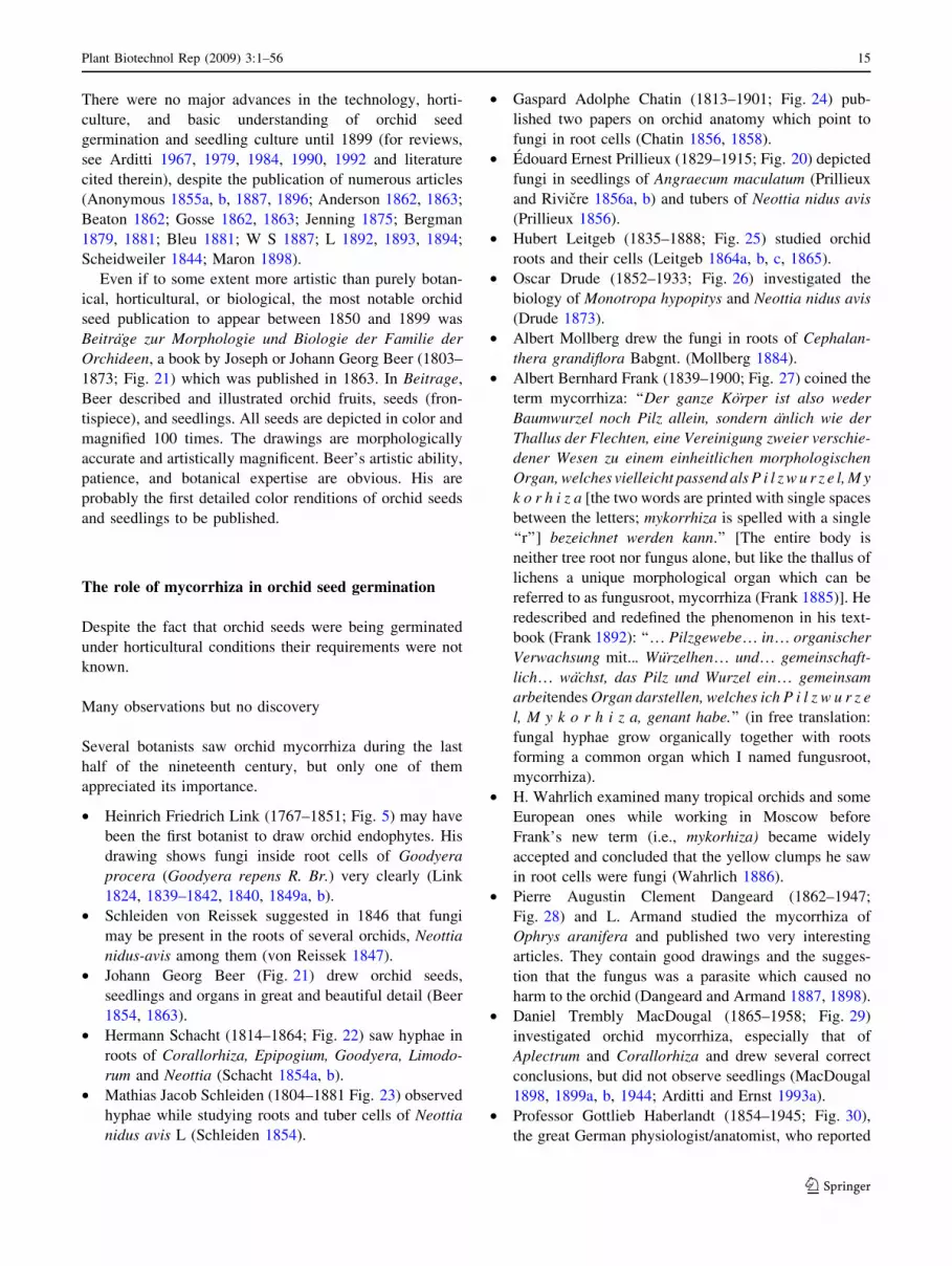
• Gaspard Adolphe Chatin (1813–1901; Fig. 24) pub-
lished two papers on orchid anatomy which point to
fungi in root cells (Chatin 1856, 1858).
• Edouard Ernest Prillieux (1829–1915; Fig. 20) depicted
fungi in seedlings of Angraecum maculatum (Prillieux
and Rivicre 1856a, b) and tubers of Neottia nidus avis
(Prillieux 1856).
• Hubert Leitgeb (1835–1888; Fig. 25) studied orchid
roots and their cells (Leitgeb 1864a, b, c, 1865).
• Oscar Drude (1852–1933; Fig. 26) investigated the
biology of Monotropa hypopitys and Neottia nidus avis
(Drude 1873).
• Albert Mollberg drew the fungi in roots of Cephalan-
thera grandiflora Babgnt. (Mollberg 1884).
• Albert Bernhard Frank (1839–1900; Fig. 27) coined the
term mycorrhiza: ‘‘Der ganze Korper ist also weder
Baumwurzel noch Pilz allein, sondern anlich wie der
Thallus der Flechten, eine Vereinigung zweier verschie-
dener Wesen zu einem einheitlichen morphologischen
Organ, welches vielleicht passend als P i l z w u r z e l, M y
k o r h i z a [the two words are printed with single spaces
between the letters; mykorrhiza is spelled with a single
‘‘r’’] bezeichnet werden kann.’’ [The entire body is
neither tree root nor fungus alone, but like the thallus of
lichens a unique morphological organ which can be
referred to as fungusroot, mycorrhiza (Frank 1885)]. He
redescribed and redefined the phenomenon in his text-
book (Frank 1892): ‘‘… Pilzgewebe… in… organischer
Verwachsung mit... Wurzelhen… und… gemeinschaft-
lich… wachst, das Pilz und Wurzel ein… gemeinsam
arbeitendes Organ darstellen, welches ich P i l z w u r z e
l, M y k o r h i z a, genant habe.’’ (in free translation:
fungal hyphae grow organically together with roots
forming a common organ which I named fungusroot,
mycorrhiza).
• H. Wahrlich examined many tropical orchids and some
European ones while working in Moscow before
Frank’s new term (i.e., mykorhiza) became widely
accepted and concluded that the yellow clumps he saw
in root cells were fungi (Wahrlich 1886).
• Pierre Augustin Clement Dangeard (1862–1947;
Fig. 28) and L. Armand studied the mycorrhiza of
Ophrys aranifera and published two very interesting
articles. They contain good drawings and the sugges-
tion that the fungus was a parasite which caused no
harm to the orchid (Dangeard and Armand 1887, 1898).
• Daniel Trembly MacDougal (1865–1958; Fig. 29)
investigated orchid mycorrhiza, especially that of
Aplectrum and Corallorhiza and drew several correct
conclusions, but did not observe seedlings (MacDougal
1898, 1899a, b, 1944; Arditti and Ernst 1993a).
• Professor Gottlieb Haberlandt (1854–1945; Fig. 30),
the great German physiologist/anatomist, who reported
Plant Biotechnol Rep (2009) 3:1–56 15
123
the presence of fungal mycelium in root cells of Neottia
nidus-avis (the orchids which led N. Bernard to his
discovery; Fig. 31), Corallorhiza innata, Epipogon
gmelini, and Wullschlaegelia ‘‘but attached no signif-
icance to it’’ (Haberlandt 1914; Pridgeon 1990).
• Melchior Treub (1851–1910; Fig. 9), who reported
seeing endophytes in seedlings and young plants of
lycopods (Treub 1890). He formulated the term ‘‘pro-
tocorm’’ to describe seedlings of lycopods. Noel
Bernard used the term to describe the early stage of
orchid seed germination. With time, the initial use of
‘‘protocorm’’ for lycopods was forgotten and the term is
currently used almost exclusively for orchids. A
reference to endophyte(s) in protocorm(s) by Treub
was misinterpreted leading to the suggestion that he
saw orchid mycorrhiza without appreciating its impor-
tance. This is not the case. Treub did not work with
orchid seedlings and probably never saw their endo-
phytes (for reviews which include discussions of the
history of orchid mycorrhiza, see Arditti 1992; Magnus
1900; Harley 1969; Warcup 1975; Arditti 1979, 1984,
1990, 1992; Hadley 1982; Harley and Smith 1983;
some reviews are part of history themselves: Burgeff
1909, 1932, 1936, 1938, 1943, 1954, 1959).
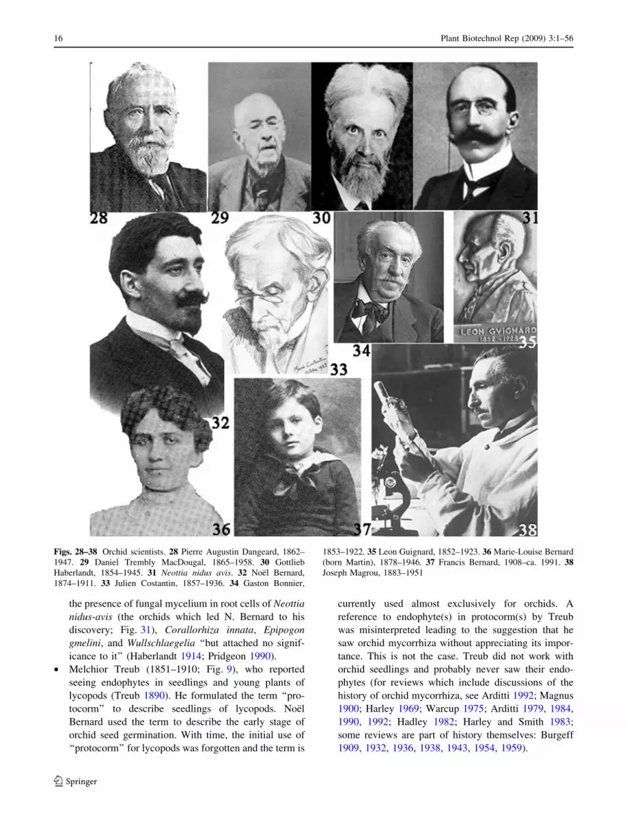
Figs. 28–38 Orchid scientists. 28 Pierre Augustin Dangeard, 1862–
1947. 29 Daniel Trembly MacDougal, 1865–1958. 30 Gottlieb
Haberlandt, 1854–1945. 31 Neottia nidus avis. 32 Noel Bernard,
1874–1911. 33 Julien Costantin, 1857–1936. 34 Gaston Bonnier,
1853–1922. 35 Leon Guignard, 1852–1923. 36 Marie-Louise Bernard
(born Martin), 1878–1946. 37 Francis Bernard, 1908–ca. 1991. 38Joseph Magrou, 1883–1951
16 Plant Biotechnol Rep (2009) 3:1–56
123

A single observations and a major discovery
Of the botanists listed above who saw orchid endophytes
(Beer, Chatin, Drude, Link, Frank, Leitgeb, Mollberg, Pril-
lieux, Reissek, Schacht, Schleiden, and Wahrlich), none
drew correct conclusions about the role of the fungus. In their
defence it must be said that they studied roots and rhizomes
for the most part. It is not easy to draw proper conclusions
about the role of the fungus in orchid seed germination from
seeing it in these organs. None of the horticulturists who
germinated orchid seeds on the surfaces of potting media
which supported mature orchids seemed to even suspect the
participation in the process of another organism, especially
fungus. The reason for this is simple: They never saw the
endophyte. Even had they seen the fungi it is reasonable to
assume that they would have assumed them to be pathogens.
Only the ‘‘genius of Pasteur applied to orchids’’ and a
‘‘Mozart of Plant Biology’’ (Bernard 1990) could appre-
ciate the role of the fungi in orchid seed germination. Noel
Bernard (1874–1911; Fig. 32) possessed these unique
characteristics. He saw Neottia nidus-avis seedlings which
harbored the endophyte and drew correct conclusion about
the nature of the fungi and their function in orchid seed
germination (Le Dantec 1911; Perez 1911, 1912; Bernard
1921; Derx 1936; Blarighem et al. 1937; Magrou 1937a, b;
Moreau 1958; Boullard 1985; Arditti 1979, 1984, 1990,
1992; Bernard 1990).
Noel Bernard, his life and times
Noel Bernard, was born on 13 March 1874, the son of
Francois Bernard, 46, and his second wife, Marie Mar-
guerite Sabot, 19. According to one report, his father died
in December 1879, but the late Prof. Francis Bernard,
Noel’s son, stated that his father was orphaned at the age of
12 (i.e., Francois died in 1886). Marie Marguerite, a young
mother and widow, had to work very hard to support her
son and herself, but had trouble making ends meet. Noel
had to help as soon as he could and became a mathematics
tutor while still a young student.
An outstanding student with a fascinating, sometimes
abrasive, personality (Boullard 1985), Noel was admitted
to the Ecole Normale Superieure and the Ecole Politech-
nique. At the age of 21, Bernard decided to become a
biologist and Julien Costantin (1857–1936; Fig. 33)
became his mentor. Costantin considered him to be his star
pupil. And, having lost a son at war, Costantin may have
found a replacement in Bernard.
Bernard earned his Licencie in Sciences Naturelles in
November 1897 and decided to specialize in orchids, but
was drafted before continuing with his studies. As a sol-
dier, he was stationed at the Mulum barracks near
Fontainebleu forest. There, while taking a walk on 3 May
1899, Bernard made his great discovery (Bernard 1899).
Together with the very discovery of orchids (by Theo-
phrastus in Europe and unknown individuals elsewhere)
and the establishment of the Orchidaceae (by Lindley),
Bernard’s is one of the five most important orchid dis-
coveries, the other two being Prof. Lewis Knudson’s
method for asymbiotic seed germination and Dr. Gavino
Rotor’s first micropropagation.
After completing his service, Bernard returned to the
Ecole Normale Superieure where he worked with Julien
Costantin (Fig. 33) and Gaston Bonnier (Fig. 34) while
living on a property owned by Leon Guignard (Fig. 35). In
1901, Bernard took a position at the University of Caen.
Bernard married Marie Louise Martin (Fig. 36), a
mathematics teacher, on 8 August 1907. He was 33 and she
was 29. On 30 April 1908, the pregnant Marie Luiose went
bike riding, fell and their son Francis (Fig. 37) was born
prematurely. Bernard kept the tiny (1.5-kg) baby alive by
feeding him a mixture of malt, water and orange or lemon
juice and placing him in an incumator (which may have
been one of the incubators left over from Pasteur’s days
and still in service).
Later in 1908, Bernard became Professor of Botany at
Poitiers. There, he made numerous major contributions to
botany, orchids, potatoes and symbiosis, Sadly, he only had
3 years to live.
Early in 1910, Bernard’s cousin Joseph Magrou (1883–
1951; Fig. 38) and the family physician diagnosed him with
tuberculosis, an incurable disease in those days (Bernard
predicted that someday there would be a cure; Bernard
1911a). Bernard and his wife moved to an estate in Mauroc,
not far from Poitiers. He died at 03:00 on 26 January 1911
after much suffering and was buried in a small cemetery at
Saint Benoıt near Mauroc. His grave was marked with a
concrete plate which bears the inscription (Boullard 1985):
Noel Bernard, Professeur A La Faculte des
Sciences de l’Universite de Poitiers–1874/1911
Like Noel Bernard’s mother, Marie Margaret Sabot, his
wife Marie Louise Martin did not remarry. She raised
Francis as a single mother earning a living as an educator
and a school administrator. Marie Louise died in 1946.
Francis was then 3 years old. Eventually, he became a
noted myrmecologist and a marine biologist. Francis Ber-
nard died on 16 June 1990, but not before writing a memoir
of his father (Bernard 1990). He was survived by his wife,
Michelle, two grown sons and four grandsons (for more
details, see Yam et al. 2002).
Noe l Bernard: mycorrhiza and orchid seed germination
As mentioned above, a number of botanists saw, described,
and drew fungi in orchid seedlings, roots, and rhizomes,
Plant Biotechnol Rep (2009) 3:1–56 17
123

but none of them discovered the role of the endophyte.
Bernard did. What he observed during his walk on 3 May
1899 were seedlings of Neottia nidus-avis, 3 mm (Boullard
1985) to 5 mm long (Bernard 1899) all of them colonized
by fungi (Fig. 39). He also saw germinating seeds of
Neottia. No one reported seeing them before him.
Bernard described his discovery in a paper published 15
May 1899 (Bernard 1899; Boullard 1985). He reported
seeing details reported by others before him also saw and
described: (1) parenchymatous cells which contained
starch, (2) a network of hyphae in some cells layers, and (3)
epidermal cells free of fungi and starch grains (Boullard
1985). He also noted that all germinating seeds contained
fungi. His genius came into play at this point and he wrote
that ‘‘mycorrhizae are indispensable for the plant [meaning
the seeds of course] during the germination period [and]
Neottia nidus-avis plants are associated with their fungi
during all stages of development’’ (Bernard 1899).
After subsequent research, Bernard provided additional
details: ‘‘Although the fungi can live apart from their host
plants, the orchids themselves require the presence of their
guests for their own development. I have sown the seeds of
many orchids ‘aseptically’… under these conditions they
have not freely germinated; they swell, and later on they
Figs. 39–45 Students of orchid
mycorrhiza. 39 Roots and
seedling of Neottia nidus avis:
A roots before the development
of flower stalks; B seed showing
the vegetative tip of the embryo
(v), suspensor (s), seed coat (u)
and opening at the base of the
latter, 958.86; C Seed at the
start of germination following
fungal penetration (symbols the
same as in B), 966.49; D cross-
section of seedlings during the
first year of development
showing living (p) and
degenerating (d) pelotons and
points at which the first roots
will develop (r), 940.37; Eexternal view of a developing
seedling showing apical bud (b),
swelling which will eventually
forma protocorm (t), embryonic
axis (a) and remnant of seed
coat (u), 95.14; F a more
advanced seedling (symbols as
in E), 94.96; G front view of
the seedling in F showing areas
of future root development,
97.98; H excised root showing
swelling (t) and fungus infected
area of root, 93.17; IPhalaenopsis seedling,
18 months old, produced
through the Bernard method. 40Joseph Charlesworth, 1851–
1920. 41 Gurney Wilson. 42John Ramsbottom, 1885–1974.
43 The Charlesworth
greenhouses filled with flasks in
which seeds were germinated
symbiotically. 44 Hans Edmund
Nicola Burgeff, 1883–1978. 45Ernst Stahl, 1848–1919
18 Plant Biotechnol Rep (2009) 3:1–56
123
get green, but their growth remains insignificant. On the
other hand, if germs of the appropriate fungus are sown
with the seeds, they commence to germinate almost
immediately in a very regular manner… I have examined a
large number of young orchids which had germinated in
very varying conditions, and I always noticed that they
were invaded by the fungus from the beginning of their
life. The orchids are therefore practically dependent on
their parasitic fungi, since they do not grow without them.’’
It would have been easy for Bernard to simply describe
what he saw like those before him. He did not, and instead
studied the physiological, evolutionary, and symbiotic
implications. He could have concluded that the fungi were
pathogens. A third possibility was to conclude that the
fungi entered the seedlings after they germinated, not
before. His genius was that he did not reach these
conclusions.
Bernard studied orchids, potatoes, fungi, symbiosis, and
some genetics during the last 2 years of his life. His pro-
ductivity was immense and excellent (Bernard 1899, 1900,
1902a, b, c, 1903, 1904a, b, c, 1905a, b, 1906a, b, c, d,
1907, 1908, 1909a, b, 1911a, b; for a translation of some of
Bernard’s papers into English, see Jacquet 2007) despite
having to care for an injured wife and a premature baby
while being very ill.
Francis Bernard described his father as a precocious
genius. He compared him to Mozart for several reasons,
one of them being that his ‘‘greatest period of… creativity
[was] up to around [the age of] 22–35’’ (Bernard 1990).
According to Francis Bernard, ‘‘decline sets in after this
age…’’ Given N. Bernard’s early death it is impossible to
state with certainty if decline would have set early in his
life. At the end of his life when he was 36–37, Noel Ber-
nard discovered phytoalexins and devised the zones of
inhibition (‘‘halo’’) method of studying the effects of
antifungal and antimicrobial compounds. This suggests that
he would have continued to be a productive scientist. One
of his papers on phytoalexins was published during his
lifetime (Bernard 1909b). The second (Bernard 1911b) was
completed and edited by his mentor Julien Costantin
(1857–1936) and his cousin Joseph Magrou (1883–1951)
who continued some of Bernard’s work (Magrou 1925,
1937a, b; Magrou and Magrou 1935; Anonymous 1951;
Mariat and Segretain 1952).
Noel Bernard did not devise a practical method for
asymbiotic germination of orchid seeds, but his research
just before he died indicates that he might have done so had
he lived long enough (Arditti 1990). He did predict that a
time would come when orchid gardens will include labo-
ratories. Bernard did not explain why orchid seeds,
especially ones of temperate climate species, require fungi
for germination. However, no one has done that to this day
(for reviews, see Arditti et al. 1990; Rasmussen 1995; Yam
et al. 2002). Francis Bernard was correct in suggesting that
his father was a genius, but he may have erred in classi-
fying him as one who might have declined after turning 35
(for more details, see Arditti 1984; Yam et al. 2002).
The twentieth century: great strides forward
The first horticultural methods for orchid seed germination
were developed in the middle of the nineteenth century.
Bernard made his discovery in 1899. However, truly major
discoveries in orchid seed germination, both basic and
practical, were made during the twentieth century.
Applications of Bernard’s discovery
The Moore–Cole–Gallier–Dominy–Veitch (MCGDV)
methods of orchid seed germination were used by British
orchid growers almost immediately after their publication
in The Gardeners’ Chronicle in 1849 (Neumann 1844;
Scheidweiler 1844; Moore 1849; Cole 1849; Gallier 1849;
Anonymous 1906a, b, 1921, 1925; Black 1906; Manhardt
1906; Wilson 1906; Grignan 1912, 1914a, b, 1916; Denis
1914; Bauer 1915; for reviews, see Arditti 1980, 1984,
1990, 1992). And the availability of a seed germination
method made possible the start of orchid hybridization.
During that period (1849–ca. 1910), seeds were gener-
ally sown on the surface of potting mixes in pots which
supported orchids. Despite appearing to be simple this
method required an operator who had ‘‘an ‘eye’ for a likely
surface’’ (Black 1906). After the surface was selected the
operator had to ‘‘clip [it] over… evenly and give it a good
watering’’ (Black 1906). Seed were distributed uniformly.
The pots were placed in a well illuminated area away from
direct sunlight (Black 1906). With all that, success required
the sowing of ‘‘Odontoglossum seeds… on pots containing
Odontoglossum plants, Cypripedium [Paphiopedilum] on
Cypripedium [Paphiopedilum], etc.’’ (Black 1906). The
only exception was Laeliocattleya seeds. They could ger-
minate on any compost. This suggests that Laeliocattleya
seeds could germinate in the presence of fungi from dif-
ferent orchids, but the growers did not know it at the time.
These methods or similar ones were used by growers and
breeders in the UK, France, and perhaps Belgium (Jancke
1907, 1915; Hefka 1914; Young 1893; Anonymous 1894;
Burbery 1894; Wrigley 1895), but there is no convincing
evidence that they were used anywhere else. The methods
were not very effective and germination was uncertain
(Hammerschmidt 1915; for reviews, see Arditti 1984,
1990). In spite of and maybe because of the problems with
these methods, attempts were made by growers to enhance
seedling growth through CO2 enrichment (Witt 1913;
Fischer 1914), a procedure far ahead of its time.
Plant Biotechnol Rep (2009) 3:1–56 19
123
Commercial in vitro symbiotic germination
Joseph Charlesworth (1850– or 1851–1920; Fig. 40;
Anonymous 1920) collected in the Andes sometime
between 1887 and 1889. Probably this is why he decided to
specialize in Odontoglossum, first as an importer (there was
no CITES then) and subsequently as a hybridizer. His
hybridization program became very extensive by 1894 and
he started to sell plants in 1898 (R A R 1887). He devel-
oped his business and breeding program rapidly and by
1906 his company could offer many seedlings for sale.
Charlesworth’s was not satisfied with the MCGDV
germination methods and on reading Bernard’s he decided
to develop new methods involving mycorrhizal fungi
(Anonymous 1922a, b). He encountered difficulties and
turned to Gurney Wilson (Fig. 41) for help. Wilson rec-
ommended contacting mycologist John Ramsbottom
(1885–1974: Fig. 42). The two met at the Royal Horti-
cultural Society Holland House exhibition and
Charlesworth prevailed upon Ramsbottom to visit the
Charlesworth establishment at Haywards Heath in 1913.
Charleworth and Ramsbottom established a good
working relationship. They carried basic and applied
research and developed a successful in vitro symbiotic
method for orchid seed germination (Anonymous 1921;
Ramsbottom 1922a, b, c, 1929). Charlesworth’s died in
1920 but by 1922 his firm had many in vitro seed cultures
in greenhouses (Ramsbottom 1922a; Fig. 43). The 1922
Charlesworth catalog had 2,245 entries and ‘‘an immense
number of choice hybrids’’ and even several color plates.
According to a writer (probably Gurney Wilson) in the
Orchid Review in that year, the descriptions were
‘‘remarkably correct in nomenclature and typographical
details’’ (Anonymous 1922c). By 1924, the Charlesworth
catalog offered 2,422 items (Anonymous 1924a, b). This
attests to the success of the Charlesworth–Ramsbottom
symbiotic method for orchid seed germination. As a result,
it gained widespread use throughout the world until Pro-
fessor Lewis Knudson formulated his asymbiotic method
(for reviews, see Arditti 1967, 1979, 1984, 1990, 1992;
Yam et al. 2002). The development of this method may be
one reason why volume 25 (1917) of the Orchid Review
was dedicated to him.
Ramsbottom joined the British Museum in 1910,
became Keeper of Botany in 1930 and held this position
until his retirement in 1950. He remained active 20 years
after his retirement in the British Museum, the Linnean
Society of London, the British Society of Mycopathology,
the National Rose Society (UK), the Royal Horticultural
Society, the South London Botanical Institute, and other
groups. He never returned to orchids and when asked by
one of us (JA) in 1968 to reminisce about his days with
Charlesworth he wrote: ‘‘I am a little surprised and much
amused… at 82 I am much out of touch! However I shall
be very pleased to have a chat… one day, preferably in the
afternoon…’’ His visitor (J.A.) described the chat as ‘‘…entertaining and informative despite the fact that Rams-
bottom was recovering from a stroke at the time. One point
he made was that orchids were never his primary interest
and that he worked on them only due to the association
with Charlesworth.’’ He died in 1974 [for additional details
about Joseph Charlesworth as well as John Ramsbottom
and, the chat between him and a then young orchid scientist
(J.A.), see Arditti 1990; Yam et al. 2002].
Hans Burgeff: Orcheomyces and Mycelium radicis
Two major works on orchid fungi were published in 1909.
One was Noel Bernard’s definitive book length review on
orchid mycorrhiza, L’evolution dans la symbiose, les or-
chidees et leur champignons commensaux (Bernard 1909a).
He was then 35 years old with 2 years to live. The second
work was a dissertation by a 26-year-old German botanist,
Hans Edmund Nikola Burgeff (1883–1976; Fig. 44), called
Die Wurzelpilze der Orchideen, ihre Kultur und ihr Leben
in der Pflanze (Burgeff 1909). He remained active for more
than 50 years after that with orchids being his main interest
(Haber 1963; Knapp 1978) and wrote reviews, research
papers, and more books on orchids (Burgeff 1909, 1911,
1932, 1936, 1938, 1943, 1959). Before becoming a pro-
fessor, Burgeff worked with a number of well-known
German plant scientists of his era. They included Peter
Clausen in Freiburg and Berlin who taught him fungus
culture methods; Ernst Stahl (1848–1919; Fig. 45) from
whom he learned synecology; Wilhelm Pfeffer (1845–
1920; Fig. 46) in whose laboratory he acquired knowledge
of plant physiology; and Karl von Goebel (1855–1932;
Fig. 47) who also had an interest in orchids. He also
worked at the Bogor Botanical Gardens in Indonesia. After
leaving Goebel’s laboratory he was Professor at Halle
(1920–1921), Munich (1921–1923) and Gottingen (1923–
1925). He left Gottingen to become Director of the
Botanical Institute at the University of Wurzburg where he
was also a Professor and worked on orchid seed germina-
tion, mycorrhiza, and symbiosis (Burgeff 1932). Burgeff
remained at Wurzburg until his retirement in 1952 but
continued to work on the germination of seeds of terrestrial
orchids (Burgeff 1954) and conservation.
Burgeff developed methods for the isolation and culture
of endophytes and symbiotic orchid seed germination
(Burgeff 1911). He concluded that orchid endophytes were
separate group of fungi named them Orcheomyces and
established a ponderous and awkward nomenclatural sys-
tem which used the words Mycelium radicis (M. R.)
followed by the name of the orchid from which the fungus
was isolated. An example is Mycelium radicis
20 Plant Biotechnol Rep (2009) 3:1–56
123

Thrixspermum arachnites. In fact, orchid fungi are not part
of a separate group. Burgeff was mistaken.
He also believed that there was strong orchid/fungus
specificity. In this he was partially correct because tem-
perate orchids (north in the Northern Hemisphere and south
in the Southern Hemisphere) in general often do not ger-
minate asymbiotically and may require specific fungi (for
reviews, see Burgeff 1936; Arditti 1979, 1992; Rasmussen
1995; Yam et al. 2002). Burgeff tried to germinate orchid
seeds asymbiotically but had no success. He used Knud-
son’s media (B and C) after their publication and attempted
to improve them (Burgeff 1936). On his trip to Indonesia,
Burgeff visited Singapore and introduced Prof. R. E.
Holttum (1895–1990; Fig. 48), then Director of the Sin-
gapore Botanic Gardens, how to use Knudson’s method
(Yam 1995, 2007). This lead to the production of the first
intentional hybrid in Singapore, Spathoglottis Primrose in
1932 (all orchid scientists and most knowledgeable
Figs. 46–51 Asymbiotic germination of orchid seeds. 46 Wilhelm
Pfeffer, 1845–1920. 47 Karl von Goebel, 1855–1932. 48 Richard Eric
Holttum, 1895–1990. 49 Magnolia brand milk bottles which were
used for orchid seed germination in Singapore. This brand was and
still is owned by the Cold Storage Grocery Company. 50 Lewis
Knudson, 1884–1958. 51 Knudson’s asymbiotic cultures: A One of
Knudson’s earliest experiments: test tubes with three-month-old
cultures; B asymbiotic Cymbidium seedlings in Erlenmeyer flask,
90.68; C older seedlings: a Cattleya, b Laeliocattleya, 93, c potted
15-month-old Cattleya seedling, 90.44; D some of Knudson’s earliest
cultures: Erlenmeyer flasks containing seedlings transferred from
smaller test tubes
Plant Biotechnol Rep (2009) 3:1–56 21
123
orchidists believe that the National Flower of Singapore
Vanda Miss Joaquim, discovered in 1893 by Miss Agnes
Joaquim is a natural hybrid; Yam et al. 2002; Arditti and
Hew 2007). Many hybrids were produced in Singapore
after that by germinating the seeds in Magnolia-brand milk
bottles (Fig. 49) which were sold by the Cold Storage
grocery store.
Asymbiotic germination
Bernard tried to germinate Bletilla hyacinthina seeds on
several media supplemented with salep (a decoction made
of dried Orchis tubers; Lawler 1984) and determined that
the optimal level was 2% (Bernard 1903, 1904a, b, 1909a;
Burgeff 1959). Orchis tubers contain 16–61% mucilage
(this varies with the species), 0.5–25% starch (which can-
not be hydrolyzed or utilized by orchid seeds), 0.9–2.7%
reducing sugars (many of which can support seed germi-
nation), 0.2–1.4% sucrose (which supports orchid seed),
and low levels of nitrogenous substances (Sezik 1967,
1984; Ernst and Rodriguez 1984; Lawler 1984). Therefore,
the 2% salep in Bernard’s medium was too low a con-
centration to provide sufficient levels of appropriate
sugars for germination. Bernard also attempted to germi-
nate Laelia seeds on a medium with a combination of salep
and sucrose. This medium may have pointed to an asym-
biotic medium for orchid seed germination, but if it did,
Bernard did not live long enough to carry out further
experiments.
As mentioned above, Burgeff’s attempts to germinate
orchid seeds asymbiotically also failed. He explained his
failure as being due to his culture vessels which were
‘‘made of ordinary glass, which gives off alkali [that
brings] an end to the life of the seedlings by a rapidly
increasing alkalinity’’ (Burgeff 1959). This explanation is
not convincing because:
• Orchid seeds are known to germinate in a wide variety
of food and drink bottles and jars of all kinds. These are
usually made of ordinary glass.
• Both orchid seeds and seedlings tolerate a wide pH
range during germination (Piriyakanjanakul and Vaj-
rabhaya 1980).
• If Burgeff was aware that alkalinity which accumulated
in the medium caused the problem he could have
transferred his seedlings to new medium as often as
needed to prevent the death of the seedlings.
• Burgeff formulated a potassium phosphate (KH2PO4/
K2HPO4) buffer for the Knudson B medium (Burgeff
1936, 1959) and could have used it for his excessively
alkaline culture media.
• He could have used culture vessels made of non-toxic
glass. A photograph in one of his books (Burgeff 1936)
is of a Jena Glass brand Erlenmeyer flask. Jena Glass is
not ‘‘ordinary glass.’’
The only logical conclusion from these facts is that
Burgeff did not formulate a good and proper medium and
did not maintain his cultures under suitable conditions.
Lewis Knudson: asymbiotic seed germination
Using ‘‘data from the experiments of Bernard and Burg-
eff,’’ Lewis Knudson (Fig. 50), a 38-year-old American
plant physiologist at Cornell University concluded that
‘‘the fungus might… digest some of the starch, pentosans
and nitrogenous substances; which are digestion products,
together with secretions from or products produced on
decomposition of the fungus might be the cause for ger-
mination’’ and that ‘‘it is conceivable that germination is
induced not by any action of the fungus within the embryo,
but by products produced externally on digestion or
secreted by the fungus’’ (Knudson 1922a). On the basis of
this reasoning Knudson decided that ‘‘germination of
orchid seeds might be obtained by the use of certain sug-
ars’’ (Knudson 1922a).
The Sea Captain’s Son
Lewis (no middle name) Knudson (according to Giltner
Knudson, Lewis’s younger son, the family always pro-
nounced the name with a K as in Kent and u as in urea,
Knudson, not Newdson) was the son of a Norwegian sea
captain (who emigrated to US as an adult, lived in Mil-
waukee, Wisconsin and was a commander of ships on the
great lakes) and his American-born wife. He was born on
15 October 1884, attended primary, junior high and high
schools in the Milwaukee Public School system graduating
from the latter on 1 July 1904 with an average grade of
81% (a B or 3.0 average in present terms and certainly not
a grade that would have gotten him into one of the modern
American highly selective universities and not an indica-
tion of future greatness).
Knudson attended the University of Missouri where he
earned a B.Sc. in agriculture and graduated on 30 January
1908. He was appointed assistant in plant physiology at
Cornell University immediately after that and promoted to
Instructor before the end of his first term there. Three years
later (1911) he received his doctorate and was appointed
Assistant Professor of Plant Physiology. A year after that
he was appointed Acting Head of his Department and
became Full Professor by 1921. When the Department of
Plant Physiology became part of the Department of Botany,
Lewis Knudson’s title was changed to Professor of Botany.
He was head of that department in 1941 and held the post
until his retirement on 30 June 1952 ruling it ‘‘with an iron
22 Plant Biotechnol Rep (2009) 3:1–56
123
fist in a steel glove’’ (Wedding 1990). Knudson remained
in Ithaca as Professor Emeritus until his instant death of
heart attack in his home while having a drink on Sunday
evening 31 August 1958. Altogether he was a man liked by
most, feared by some, on bad terms with very few and
respected by all (for more details about his life, some
provided by his son Giltner and others who knew him, see
Arditti 1990; Wedding 1990; Yam et al. 2002; Giltner and
JA knew each other).
‘‘Lewie’’: plant physiologist
Today, ‘‘Lewie’’ (as he was called behind his back by the
graduate students in his department; Wedding 1990) is
known for his work with orchids, but his research included
fungi (Knudson 1913a, 1913b), sugar metabolism (Knud-
son 1915, 1916, 1917), osmotic pressure (Knudson and
Ginsburg 1921), amino acids (Knudson 1933a), aseptic
growth of whole plants (Knudson 1915, 1916; Knudson
and Lindstrom 1919; Knudson and Smith 1919), in vitro
culture of detached root-cap cells about 50 years before
cell cultures became common (Knudson 1919), Calluna
vulgaris (Knudson 1928, 1929b, 1932, 1933c), amylase
production by plant roots (Knudson and Smith 1919); X-
ray effects on plants (Knudson 1933b, 1934b, 1940b,
1941c), ferns (Knudson 1933b, 1934b, 1940b, 1941a, b, c),
and chloroplasts (Knudson 1934a, b, c, d, 1936). His
research on sugars, aseptic culture and amylase probably
led Knudson to orchids.
Knudson: ‘‘Germination of Orchid Seeds Might be
obtained by the Use of Certain Sugars’’
The use of salep by Bernard and Burgeff as well as some
carbohydrates by the latter (starch, sucrose, glucose) led
Knudson to use a ‘‘certain’’ sugar in a culture medium
(Knudson 1922a). He concluded correctly that salep con-
tains nutrients which could be utilized by seeds and
seedlings (Knudson 1922a, 1989; Ernst and Rodriguez
1984; Arditti 1989; Janick 1989). He also theorized that the
fungus hydrolyzed large molecules and rendered their
component moities available to the seeds (Knudson
1922a).
Knudson first attempted to germinate Cattleya
schroederae 9 Cattleya gigas seeds in December 1918
on extracts of peat and canna tubers. Within a month
(January 1919) seeds on these media formed protocorms.
Those on peat failed to grow into seedlings. Protocorms
on canna extract (which probably contained soluble
sugars) produced 1–2 leaves in five months (April 1919).
In 1919, he attempted to germinate seeds of Cattleya
labiata 9 Cattleya aurea on carrot (Daucus carota) and
beet (Beta vulgaris) concoctions. The seeds germinated
and seedlings developed on both media. These findings
convinced him that orchid seeds can germinate without
fungus and led him to further experiments (Arditti
1990).
The next experiment was probably obvious and inevi-
table. Also in 1919 he placed seeds of Cattleya mossiae on
Pfeffer’s solution (Table 1) enriched with 1% sucrose. The
seeds germinated and in seven months produced proto-
corms 1 mm in diameter with one leaf. Not much happened
on the sugar-free medium. This was Knudson’s first solu-
tion. It was probably his undesignated solution A (Engman
1984; Arditti 1990; Yam et al. 2002).
Knudson assumed that if the fungus hydrolyzed sucrose,
glucose would be one of the products, and on 18 July 1919
placed seeds of Cattleya intermedia 9 Cattleya law-
renceana on Pfeffer’s solution and a modification of it he
labeled ‘‘Medium B’’ (Knudson 1921, 1922a, b, 1924,
1925) which contained sucrose or glucose. Knudson
departed for Spain and France after starting his cultures and
did not return until approximately 1 year later. On exam-
ining his cultures on 9 June 1920 he observed that
seedlings on both glucose and sucrose were well developed
(Fig. 51), but the media were dehydrated.
In subsequent experiments with seeds of Laeliocattleya,
Cattleya and Epidendrum, Knudson found that 0.8% (8 g
or 0.044 moles) glucose l-1 was the most suitable sugar
concentration (Knudson 1922a). The most widely used
sugar concentration in Knudson’s media B and C is 2%
(20 g or 0.058 moles) sucrose l-1 (Table 1). On complete
hydrolysis that much sucrose will produce 0.058 moles
glucose and 0.058 moles fructose (i.e., ca. 10 g of each
monosaccharide) for a total of 0.116 moles of sugars or 2.6
times the optimal glucose concentration. However, hydro-
lysis in media is not complete (Ernst et al. 1971; Ernst and
Arditti 1972, 1990; for a review and more details, see Yam
et al. 2002).
Knudson: ‘‘Chance favors the prepared mind’’
Chance (the quote is by Louis Pasteur) and good fortune
may have had a part in some of Knudson’s early experi-
ments. First, his experiments would have probably failed if
he had used seeds which do not germinate easily. The seeds
used in his experiments came from Theodore L. Mead of
Oviedo, Florida, a well-known orchid grower at the time.
Luckily the seeds Mr. Mead provided were those of Cat-
tleya and other orchids which germinate easily (for more
details, see Arditti 1984; Yam et al. 2002).
Knudson used cane sugar (sucrose from sugar cane) in
his medium B (Table 1). Beet sugar is also sucrose but
several reports indicate that it does not support orchid seed
germination as well as that obtained from cane (for
reviews, see Arditti 1967, 1979; Arditti and Ernst 1984).
Plant Biotechnol Rep (2009) 3:1–56 23
123
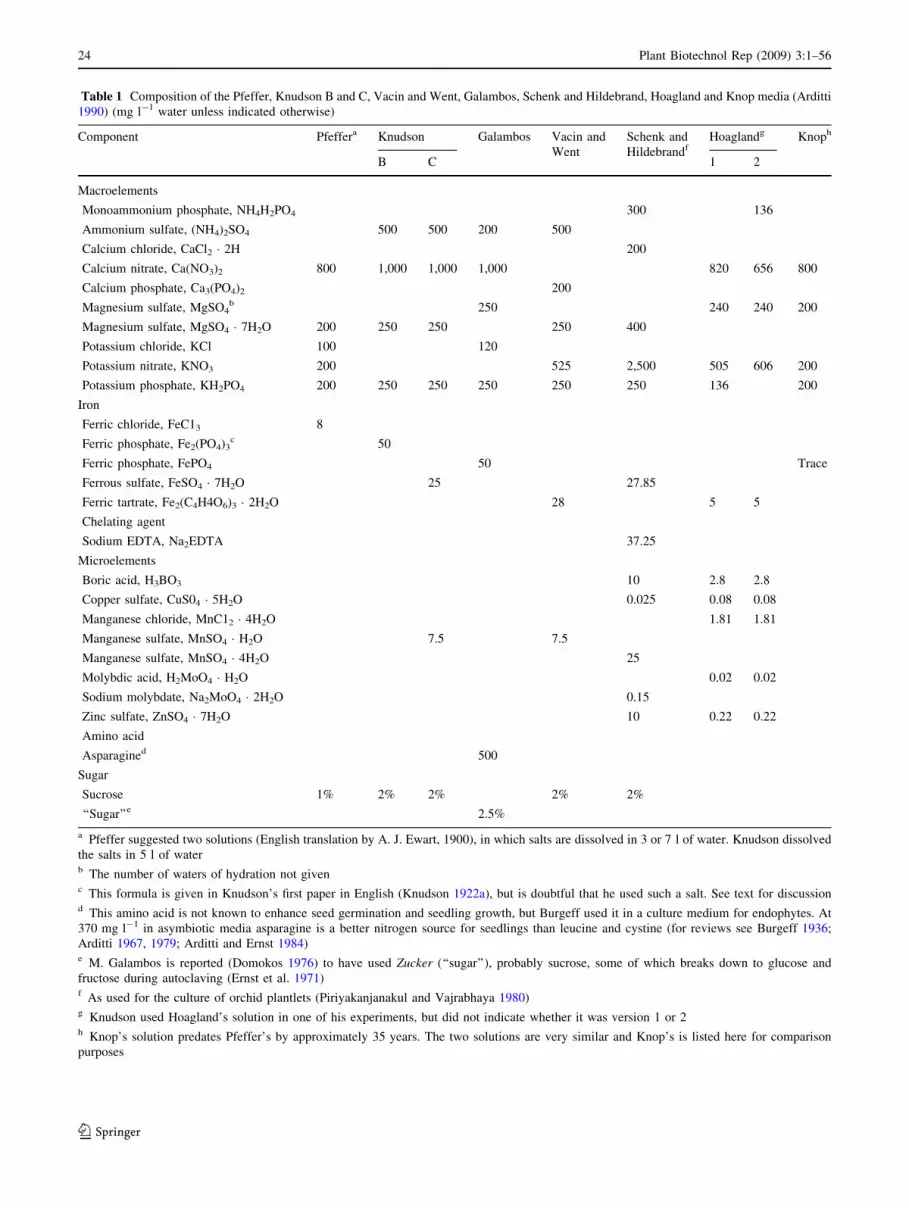
Table 1 Composition of the Pfeffer, Knudson B and C, Vacin and Went, Galambos, Schenk and Hildebrand, Hoagland and Knop media (Arditti
1990) (mg l-1 water unless indicated otherwise)
Component Pfeffera Knudson Galambos Vacin and
Went
Schenk and
HildebrandfHoaglandg Knoph
B C 1 2
Macroelements
Monoammonium phosphate, NH4H2PO4 300 136
Ammonium sulfate, (NH4)2SO4 500 500 200 500
Calcium chloride, CaCl2 � 2H 200
Calcium nitrate, Ca(NO3)2 800 1,000 1,000 1,000 820 656 800
Calcium phosphate, Ca3(PO4)2 200
Magnesium sulfate, MgSO4b 250 240 240 200
Magnesium sulfate, MgSO4 � 7H2O 200 250 250 250 400
Potassium chloride, KCl 100 120
Potassium nitrate, KNO3 200 525 2,500 505 606 200
Potassium phosphate, KH2PO4 200 250 250 250 250 250 136 200
Iron
Ferric chloride, FeC13 8
Ferric phosphate, Fe2(PO4)3c 50
Ferric phosphate, FePO4 50 Trace
Ferrous sulfate, FeSO4 � 7H2O 25 27.85
Ferric tartrate, Fe2(C4H4O6)3 � 2H2O 28 5 5
Chelating agent
Sodium EDTA, Na2EDTA 37.25
Microelements
Boric acid, H3BO3 10 2.8 2.8
Copper sulfate, CuS04 � 5H2O 0.025 0.08 0.08
Manganese chloride, MnC12 � 4H2O 1.81 1.81
Manganese sulfate, MnSO4 � H2O 7.5 7.5
Manganese sulfate, MnSO4 � 4H2O 25
Molybdic acid, H2MoO4 � H2O 0.02 0.02
Sodium molybdate, Na2MoO4 � 2H2O 0.15
Zinc sulfate, ZnSO4 � 7H2O 10 0.22 0.22
Amino acid
Asparagined 500
Sugar
Sucrose 1% 2% 2% 2% 2%
‘‘Sugar’’e 2.5%
a Pfeffer suggested two solutions (English translation by A. J. Ewart, 1900), in which salts are dissolved in 3 or 7 l of water. Knudson dissolved
the salts in 5 l of waterb The number of waters of hydration not givenc This formula is given in Knudson’s first paper in English (Knudson 1922a), but is doubtful that he used such a salt. See text for discussiond This amino acid is not known to enhance seed germination and seedling growth, but Burgeff used it in a culture medium for endophytes. At
370 mg l-1 in asymbiotic media asparagine is a better nitrogen source for seedlings than leucine and cystine (for reviews see Burgeff 1936;
Arditti 1967, 1979; Arditti and Ernst 1984)e M. Galambos is reported (Domokos 1976) to have used Zucker (‘‘sugar’’), probably sucrose, some of which breaks down to glucose and
fructose during autoclaving (Ernst et al. 1971)f As used for the culture of orchid plantlets (Piriyakanjanakul and Vajrabhaya 1980)g Knudson used Hoagland’s solution in one of his experiments, but did not indicate whether it was version 1 or 2h Knop’s solution predates Pfeffer’s by approximately 35 years. The two solutions are very similar and Knop’s is listed here for comparison
purposes
24 Plant Biotechnol Rep (2009) 3:1–56
123

Had Knudson used beet rather than cane sugar his exper-
iments may have been less successful. The reasons for the
difference between cane and beet sugar are not known even
now. They may be due to the presence of minute levels of
impurities.
Wilhelm Pfeffer listed two mineral solutions in his
Physiology of Plants (Pfeffer 1900), both containing the
same concentrations of salts dissolved in either 7 or 3 l of
water. Knudson averaged the two volumes of water
[(7 ? 3)/2 = 5], dissolved the minerals in 5 l of water
(that was probably his unlabeled solution A) and was
successful. However, the seeds in his experiments would
have germinated equally well on either the 3 or 7 l Pfef-
fer’s solutions (Arditti 1990; Table 1).
Knudson: from B to C
Knudson found that his medium B ‘‘was not entirely sat-
isfactory for seeds of’’ Paphiopedilum, Vanilla, and North
American species of Cypripedium. In one instance, he
encountered difficulties with Cattleya, Phalaenopsis, and
Vanda. His conclusion was that the difficulties were
brought about by the absence of microelements. Because of
that, he added boron, copper, manganese, and zinc to
medium B ‘‘without any improvement in the case of
Cypripedium and Vanilla’’ (Knudson 1946a). Results were
better when he added iron and manganese to what he called
his solution C (Table 1) which he described as being
‘‘theoretically better than [solution] B,’’ and superior in the
case of Cattleya (Knudson 1946b).
Conclusion
Almost 400 years separate the time when orchid seeds
were first seen and the development of a practical asym-
biotic method for their germination. Claims, counter claims
and acrimony followed Knudson’s discovery, but these are
beyond the scope of this review (for details, see Arditti
1984, 1990, 1992; Knudson 1927, 1929a, 1930, 1935,
1940a, 1951, 1952;Yam et al. 2002). Knudson also made
other contributions to orchid seed science (Knudson 1947,
1948, 1950). The important point is that after Knudson’s
media B and C were formulated, orchid growing and
hybridization became widespread. Hybrids which early
growers may not have even imagined became possible.
Examples of this are colored Phalaenopsis hybrids. In the
early days of orchid growing and even as late as 1958–
1959 (Scott and Arditti 1959), there were only white
Phalaenopsis hybrids and relatively few intergeneric
crosses. Today, multicolored Phalaenopsis flowers are as
common as multigenerics like Aranda, Darwinara, Knud-
sonara, Lindleyara, Mokara, and many others.
Part II: Micropropagation
Abstract A commonly held view is that Prof. Georges
Morel is the sole discoverer of orchid micropropagation
and that he was the first to culture an orchid shoot tip in
1960. In fact, the first in vitro orchid propagation was
carried out by Dr. Gavino Rotor in 1949. Hans Thomale
was the first to culture an orchid shoot tip in 1956. The
methods used by Morel to culture his shoot tips were
developed by others many years before he adapted them to
orchids. This review also traces the history of several
techniques, additives, and peculiarities (agitated liquid
cultures, coconut water, banana pulp, a patent and what
appears to be an empty claim) which are associated with
orchid micropropagation. A summary of plant hormone
history is also outlined because micropropagation could not
have been developed without phytohormones.
Orchid micropropagation: the origins
Orchid micropropagation did not originate suddenly as a
brand new idea in the mind of a single person despite an
unfortunately successful self-serving attempt to create this
impression (Morel 1960). The origins of orchid micro-
propagation are intermingled with the history of tissue
culture, phytohormones and other areas of plant physiology
(Arditti and Krikorian 1996; Arditti 2008).1 Its origins are
in several areas of research and arose from the work of
numerous scientists (Arditti and Krikorian 1996; Arditti
2008) rather than a single individual. These research lines
will be presented separately until they converged and lead
to orchid micropropagation. A summary of plant hormone
history will also be outlined because micropropagation
could not have been developed without phytohormones
(see Krikorian 1995 for a more extensive history).
Plant hormones and culture media additives of plant
origin
A little more than a century ago the existence of plant
hormones was only being suggested. At the present time,
1 Important details were removed from or ‘‘softened’’ in chapter 1 of
Micropropagation of Orchids, first edition (Arditti and Ernst 1993a)
under pressure in an effort not to offend or displease certain
individuals and/or groups. A subsequent review (Arditti and Krikorian
1996) set the record straight by presenting the history accurately. The
history chapter in the second edition of Micropropagation of Orchids(MO2; Arditti 2008) is also accurate. It is based on Arditti and
Krikorian (1996). The review here is based on these two accurate
historical presentations (Arditti and Krikorian 1996; Arditti 2008).
We thank Dr. A. D. Krikorian for allowing the use of the joint paper
for MO2 and by extension also here.
Plant Biotechnol Rep (2009) 3:1–56 25
123
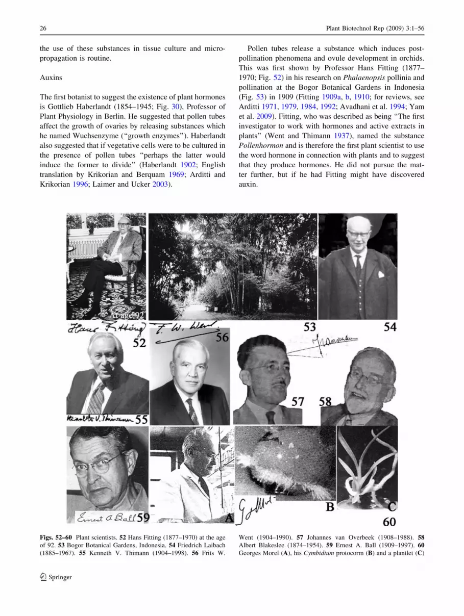
the use of these substances in tissue culture and micro-
propagation is routine.
Auxins
The first botanist to suggest the existence of plant hormones
is Gottlieb Haberlandt (1854–1945; Fig. 30), Professor of
Plant Physiology in Berlin. He suggested that pollen tubes
affect the growth of ovaries by releasing substances which
he named Wuchsenzyme (‘‘growth enzymes’’). Haberlandt
also suggested that if vegetative cells were to be cultured in
the presence of pollen tubes ‘‘perhaps the latter would
induce the former to divide’’ (Haberlandt 1902; English
translation by Krikorian and Berquam 1969; Arditti and
Krikorian 1996; Laimer and Ucker 2003).
Pollen tubes release a substance which induces post-
pollination phenomena and ovule development in orchids.
This was first shown by Professor Hans Fitting (1877–
1970; Fig. 52) in his research on Phalaenopsis pollinia and
pollination at the Bogor Botanical Gardens in Indonesia
(Fig. 53) in 1909 (Fitting 1909a, b, 1910; for reviews, see
Arditti 1971, 1979, 1984, 1992; Avadhani et al. 1994; Yam
et al. 2009). Fitting, who was described as being ‘‘The first
investigator to work with hormones and active extracts in
plants’’ (Went and Thimann 1937), named the substance
Pollenhormon and is therefore the first plant scientist to use
the word hormone in connection with plants and to suggest
that they produce hormones. He did not pursue the mat-
ter further, but if he had Fitting might have discovered
auxin.
Figs. 52–60 Plant scientists. 52 Hans Fitting (1877–1970) at the age
of 92. 53 Bogor Botanical Gardens, Indonesia. 54 Friedrich Laibach
(1885–1967). 55 Kenneth V. Thimann (1904–1998). 56 Frits W.
Went (1904–1990). 57 Johannes van Overbeek (1908–1988). 58Albert Blakeslee (1874–1954). 59 Ernest A. Ball (1909–1997). 60Georges Morel (A), his Cymbidium protocorm (B) and a plantlet (C)
26 Plant Biotechnol Rep (2009) 3:1–56
123

The first indication that Pollenhormon may be or con-
tains auxin was provided by Friedrich Laibach (1885–
1967; Fig. 54) who reported that diethyl ether can extract
an active principle from it (Laibach 1930, 1932, 1933a, b;
Maschmann and Laibach 1932; Laibach and Maschmann
1933). After Laibach, Kenneth V. Thimann (1904–1998,
Fig. 55) showed that the ether extract contained auxin (for
reviews, see Went and Thimann 1937; also see Thimann
1980; Avadhani et al. 1994; Arditti and Krikorian 1996;
Yam et al. 2009).
Frits W. Went (1926, 1928; Fig. 56) discovered auxin in
Utrech, Holland, before Laibach extracted it from Pollen-
hormon. Went carried out his work (actually a Ph.D.
Dissertation) after Fitting suggested the existence of Pol-
lenhormon [which Fitting did not equate with auxin even in
his 90s in a letter to one of us (JA)]. In letters and con-
versations with one of us (JA), Went indicated that he
made no connection between Fitting work with orchids and
his research on Avena coleoptiles and Pollenhormon and
auxin (Yam et al. 2009). This is not surprising for the time
(1920s). Auxin was identified as indole-3-acetic acid (IAA)
in 1934 (Went and Thimann 1937; Haagen-Smit 1951) and
made successful tissue culture possible (Gautheret 1935,
1937, 1983, 1985; Loo 1945a, b). Currently IAA and
several of its analogs [i.e., synthetic auxins like 2,4-
dichlorophenoxy acetic acid (2,4-D), naphthalene acetic
acid (NAA) and indolebutyric acid (IBA), for example] are
indispensable for tissue culture in general and orchid mi-
cropropagation in particular.
Coconut water and cytokinins
Haberlandt suggested in his classic paper that ‘‘one might
also consider the utilization of embryo sac fluids’’ (Ha-
berlandt 1902; Krikorian and Berquam 1969; Laimer and
Ucker 2003). E. Hannig tested the effects of embryo sac
fluids of Raphanus and Cochlearia on the growths of their
own embryos (Hannig 1904; Krikorian and Berquam 1969;
Laimer and Ucker 2003). It is very likely that European
botanists at the time were not familiar with the liquid
endosperm of coconuts. Only, those who spent time in the
tropics became familiar with the colorless liquid endo-
sperm in green coconuts. This is coconut water (CW), not
coconut milk which is a white milky liquid produced by
squeezing, extracting or grating the solid white endosperm
(meat) of mature nuts (jelly-like and clear in green nut) that
becomes copra when dried.
One Dutch botanist who spent time in the tropics [the
Bogor (Buitenzorg) Botanical Gardens in Indonesia;
Fig. 53] and became acquainted with CW was Johannes
van Overbeek (1908–1988; Fig. 57). He and M. E. Conklin
suggested its use to Albert Blakeslee (1874–1954; Fig. 58)
for the culture in vitro immature embryos of Datura
stramonium. The embryos grew well in the presence of CW
(van Overbeek et al. 1941, 1942) and an effective complex
additive was introduced into plant tissue culture (van
Overbeek et al. 1944; Caplin and Stewart 1948; Steward
and Shantz 1955; Pollard et al. 1961; Raghavan 1966;
Tulecke et al. 1961, 1975; Krikorian 1975, 1982, 1988,
1995; Steward and Krikorian 1975; Gautheret 1985). Ern-
est A. Ball (Fig. 59) was the first to use CW for the culture
of apical meristems (Ball 1946; Krikorian 1975, 1982). L.
Duhamet used coconut water to culture crown gall tissues
(Duhamet 1950). Georges Morel (Fig. 60) cultured Amor-
phophallus rivieri, Sauromatum guttatum, Gladiolus, Iris
and lily in CW-enriched media (Morel 1950). Nowadays,
CW is used widely in tissue culture and micropropagation
of many plants and there are several incotrrect priority
claims.
F. Mariat was first to publish on the incorporation of
CW (mistakenly by referring to it as coconut milk) and
copra extract as additives to orchid seed germination media
(Mariat 1951; for reviews see Arditti 1967, 1977a, b, 1979,
2008; Arditti and Ernst 1984, 1993b). Today, CW is used
extensively in orchid micropropagation (for reviews see
Arditti 1967, 1977a, b, 1979, 2008; Arditti and Ernst 1984,
1993a). There is no agreement in the literature regarding
the reasons for its effects. Research on plant tissue culture
expanded between 1940 and 1965, and efforts were made
to solve the problem of tissues which could not be cultured.
Tobacco pith was one of these tissues (Gautheret 1985;
Skoog 1994). In an effort to culture this tissue, Folke
Skoog (1908–2001; Fig. 61) and his group at the Univer-
sity of Wisconsin, Madison formulated several media and
assayed the effects of many additives (Skoog 1944; Skoog
and Tsui 1948, 1951; Skoog and Miller 1957). Herring
sperm DNA which was stored for a very extensive period
was one of the substances they tested. The period was long
enough to suggest that this DNA was used for orchid seed
germination experiments by Professor John T. Curtis
(1913–1961; Fig. 62; for a review see Arditti 1967).
However, according to one of the co-discoverers of cyto-
kinins, Professor Carlos O. Miller (b. 1923; Fig. 63) in
correspondence with one of use (JA), Curtis and Skoog
would not have shared reagents due to a very strained
relationship. Regardless of its source the old DNA lead to
the discovery of the first cytokinin, kinetin (Strong 1958;
Miller 1961, 1967; Leopold 1964; Skoog et al. 1965;
Gautheret 1985; Skoog 1994).
With the requirements for auxin and several vitamins
(Gautheret 1945) for plant tissue culture already well
known, the discovery of cytokinins made possible the
formulation of the Murashige and Skoog (MS) medium by
Toshio Murashige (b. 1930; Fig. 64) and Folke Skoog
(Murashige and Skoog 1962; Smith and Gould 1989;
Skoog 1994). This medium is used very widely for the
Plant Biotechnol Rep (2009) 3:1–56 27
123

culture of many plants including orchids (for reviews see
Arditti and Ernst 1993a; Arditti 2008).
Banana
Powdered banana was apparently first added to a medium
for orchid seed germination in Brazil (Graeflinger 1950 as
cited by Withner 1959b). Incorporation of banana in cul-
ture media became widespread very quickly with several of
the subsequent users claiming to have been the originators
of the practice. The most common practice at present is to
add pulp of ripe bananas to media (Withner 1955; Ernst
1967; for reviews see Arditti 1967; Pages 1971, Arditti and
Ernst 1984; Yam et al. 2002). The reasons for the effects of
banana are not known. Attempts to find answers by frac-
tionating banana pulp through serial extractions with a
number of solvents produced inconclusive results (Arditti
1968).
Figs. 61–73 Plant researchers. 61 Folke Skoog (1908–2001). 62 John
T. Curtis (1913–1961). 63 Carlos Miller. 64 Toshio Murashige (b.
1930). 65 Roger Gautheret (1910–1997). 66 H. Vochting. 67 Julius
Sachs (1832–1897). 68 William J. Robbins. 69 Walter Kotte (1893–
1970). 70 Philip R. White (1901–1968). 71 Pierre Noubecourt (1895–
1961). 72 Loo Shih Wei (Western Style: Shih Wei Loo; 1907–1998).
73 Theodor Schwann (1810–1882)
28 Plant Biotechnol Rep (2009) 3:1–56
123

Several other plant homogenates have been evaluated
for their effects on seed germination and seedling growth
(Arditti 1967, 1979; Ernst 1967; Arditti and Ernst 1984).
Few if any of these are used to culture orchid explants. A
number of these are added to cultures of protocorm-like
bodies or developing plantlets (see specific procedures).
The beginnings
Orchid micropropagation has its roots in early tissue cul-
ture research with other plants.
Tissue and organ culture of non-orchidaceous plants
Roger J. Gautheret (1910–1997; Fig. 65) was an early, well-
known and influential figure in the history of plant tissue
culture from his base in France. In his later years, he became
a (not unbiased) historian of the field. In one of his accounts
he wrote that ‘‘the progress of plant tissue culture was made
possible by only a few genuine discoveries [which]… did not
appear suddenly, but after a long and slow journey, unpre-
tentiously covered by pioneers’’ (Gautheret 1985).
According to him, a multi-talented, Frenchman, Henri-Louis
Duhamel du Monceau (1700–1782; Fig. 74), a student of
Figs. 74–81 Plant tissue culture. 74 Henri-Louis Duhamel du
Monceau (1700–1782). 75 Gavino Rotor Jr. (1917–2005). 76 Rotor’s
cultures of Phalaenopsis flower stalk sections. 77 Lucie Mayer. 78
Hans Thomale (1919–2002). 79 Thomale’s explants of Dactylorhiza(Orchis) maculata producing shoots and roots in vitro. 80 George
Morel’s letter to Thomale. 81 Ralph W. Wetmore (1892–1989)
Plant Biotechnol Rep (2009) 3:1–56 29
123
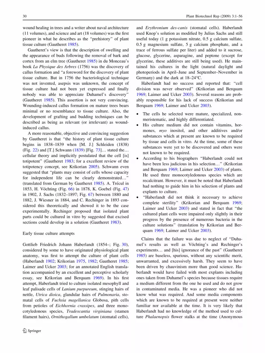
wound healing in trees and a writer about naval architecture
(11 volumes), and science and art (18 volumes) was the first
pioneer in what he describes as the ‘‘prehistory’’ of plant
tissue culture (Gautheret 1985).
Gautheret’s view is that the description of swelling and
the appearance of buds following the removal of bark and
cortex from an elm tree (Gautheret 1985) in du Monceau’s
book La Physique des Arbres (1756) was the discovery of
callus formation and ‘‘a foreword for the discovery of plant
tissue culture. But in 1756 the bacteriological technique
was not invented, asepsis was unknown, the concept of
tissue culture had not been yet expressed and finally
nobody was able to appreciate Duhamel’s discovery’’
(Gautheret 1985). This assertion is not very convincing.
Wounding-induced callus formation on mature trees bears
minimal or no resemblance to tissue culture. Also, the
development of grafting and budding techniques can be
described as being as relevant (or irrelevant) as wound-
induced callus.
A more reasonable, objective and convincing suggestion
by Gautheret is that ‘‘the history of plant tissue culture
begins in 1838–1839 when [M. J.] Schleiden (1838)
(Fig. 22) and [T.] Schwann (1839) [Fig. 73]… stated the…cellular theory and implicitly postulated that the cell [is]
totipotent’’ (Gautheret 1983; for a excellent review of the
totipotency concept, see Krikorian 2005). Schwann even
suggested that ‘‘plants may consist of cells whose capacity
for independent life can be clearly demonstrated…’’
(translated from German by Gautheret 1985). A. Trecul in
1853, H. Vochting (Fig. 66) in 1878, K. Goebel (Fig. 47)
in 1902, J. Sachs (1832–1897; Fig. 67) between 1880 and
1882, J. Wiesner in 1884, and C. Rechinger in 1893 con-
sidered this theoretically and showed it to be the case
experimentally. Rechinger proposed that isolated plant
parts could be cultured in vitro by suggested that excised
sections could develop in a solution (Gautheret 1983).
Early tissue culture attempts
Gottlieb Friedrich Johann Haberlandt (1854–; Fig. 30),
considered by some to have originated physiological plant
anatomy, was first to attempt the culture of plant cells
(Haberlandt 1902; Krikorian 1975, 1982; Gautheret 1985;
Laimer and Ucker 2003; for an annotated English transla-
tion accompanied by an excellent and perceptive scholarly
essay, see Krikorian and Berquam 1969). In his first
attempt, Haberlandt tried to culture isolated mesophyll and
leaf palisade cells of Lanium purpureum, stinging hairs of
nettle, Utrica dioica, glandular hairs of Pulmonaria, sto-
matal cells of Fuchsia magellanica Globosa, pith cells
from petioles of Eichhornia crassipes, and three mono-
cotyledonous species, Tradescantia virginiana (stamen
filament hairs), Ornithogallum umbelatum (stomatal cells),
and Erythronium des-canis (stomatal cells). Haberlandt
used Knop’s solution as modified by Julius Sachs and still
useful today (1 g potassium nitrate, 0.5 g calcium sulfate,
0.5 g magnesium sulfate, 5 g calcium phosphate, and a
trace of ferrous sulfate per liter) and added to it sucrose,
glucose, glycerine, asparagine, and peptone (except for
glycerine, these additives are still being used). He main-
tained his cultures in the light (natural daylight and
photoperiods in April–June and September–November in
Germany) and the dark at 18–24�C.
Haberlandt had no success and reported that: ‘‘cell
division was never observed’’ (Krikorian and Berquam
1969; Laimer and Ucker 2003). Several reasons are prob-
ably responsible for his lack of success (Krikorian and
Berquam 1969; Laimer and Ucker 2003).
• The cells he selected were mature, specialized, non-
meristematic, and highly differentiated.
• His culture medium did not contain vitamins, hor-
mones, myo inositol, and other additives and/or
substances which at present are known to be required
by tissue and cells in vitro. At the time, some of these
substances were yet to be discovered and others were
not known to be required.
• According to his biographers ‘‘Haberlandt could not
have been less judicious in his selection…’’ (Krikorian
and Berquam 1969; Laimer and Ucker 2003) of plants.
He used three monocotyledonous species which are
recalcitrant. However, it must be noted that Haberlandt
had nothing to guide him in his selection of plants and
explants to culture.
• ‘‘Haberlandt did not think it necessary to achieve
complete sterility’’ (Krikorian and Berquam 1969;
Laimer and Ucker 2003) and stated in fact that ‘‘the
cultured plant cells were impaired only slightly in their
progress by the presence of numerous bacteria in the
culture solutions’’ (translation by Krikorian and Ber-
quam 1969; Laimer and Ucker 2003).
Claims that the failure was due to neglect of ‘‘Duha-
mel’s results as well as Vochting’s and Rechinger’s
experiments… and [his] ignorance of the past’’ (Gautheret
1985) are baseless, spurious, without any scientific merit,
unwarranted, and excessively harsh. They seem to have
been driven by chauvinism more than good science. Ha-
berlandt would have failed with most explants including
ones taken from Duhamel’s species because tissues require
a medium different from the one he used and do not grow
in contaminated media. He was a pioneer who did not
know what was required. And some media components
which are known to be required at present were neither
familiar nor available at the time. It is very likely that
Haberlandt had no knowledge of the method used to cul-
ture Phalaenopsis flower stalks at the time (Anonymous
30 Plant Biotechnol Rep (2009) 3:1–56
123
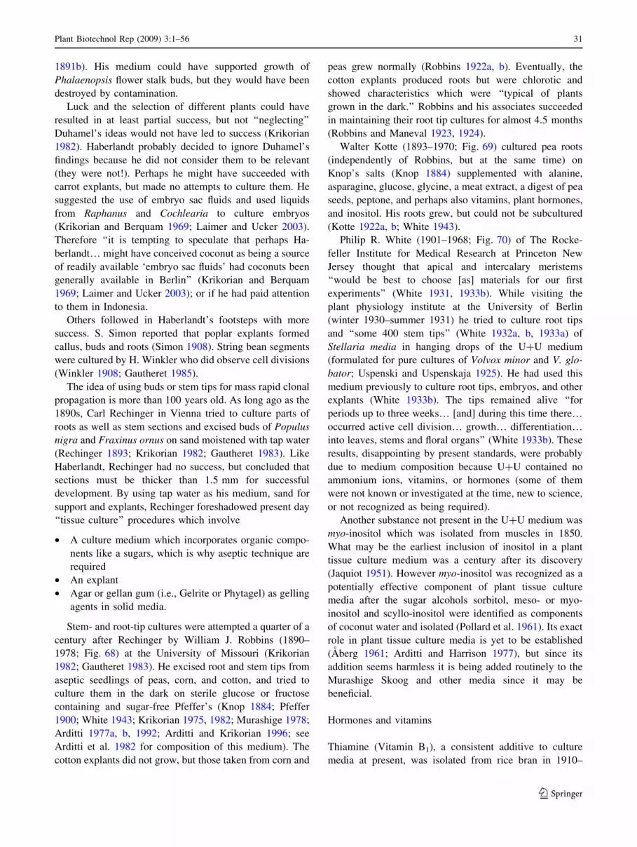
1891b). His medium could have supported growth of
Phalaenopsis flower stalk buds, but they would have been
destroyed by contamination.
Luck and the selection of different plants could have
resulted in at least partial success, but not ‘‘neglecting’’
Duhamel’s ideas would not have led to success (Krikorian
1982). Haberlandt probably decided to ignore Duhamel’s
findings because he did not consider them to be relevant
(they were not!). Perhaps he might have succeeded with
carrot explants, but made no attempts to culture them. He
suggested the use of embryo sac fluids and used liquids
from Raphanus and Cochlearia to culture embryos
(Krikorian and Berquam 1969; Laimer and Ucker 2003).
Therefore ‘‘it is tempting to speculate that perhaps Ha-
berlandt… might have conceived coconut as being a source
of readily available ‘embryo sac fluids’ had coconuts been
generally available in Berlin’’ (Krikorian and Berquam
1969; Laimer and Ucker 2003); or if he had paid attention
to them in Indonesia.
Others followed in Haberlandt’s footsteps with more
success. S. Simon reported that poplar explants formed
callus, buds and roots (Simon 1908). String bean segments
were cultured by H. Winkler who did observe cell divisions
(Winkler 1908; Gautheret 1985).
The idea of using buds or stem tips for mass rapid clonal
propagation is more than 100 years old. As long ago as the
1890s, Carl Rechinger in Vienna tried to culture parts of
roots as well as stem sections and excised buds of Populus
nigra and Fraxinus ornus on sand moistened with tap water
(Rechinger 1893; Krikorian 1982; Gautheret 1983). Like
Haberlandt, Rechinger had no success, but concluded that
sections must be thicker than 1.5 mm for successful
development. By using tap water as his medium, sand for
support and explants, Rechinger foreshadowed present day
‘‘tissue culture’’ procedures which involve
• A culture medium which incorporates organic compo-
nents like a sugars, which is why aseptic technique are
required
• An explant
• Agar or gellan gum (i.e., Gelrite or Phytagel) as gelling
agents in solid media.
Stem- and root-tip cultures were attempted a quarter of a
century after Rechinger by William J. Robbins (1890–
1978; Fig. 68) at the University of Missouri (Krikorian
1982; Gautheret 1983). He excised root and stem tips from
aseptic seedlings of peas, corn, and cotton, and tried to
culture them in the dark on sterile glucose or fructose
containing and sugar-free Pfeffer’s (Knop 1884; Pfeffer
1900; White 1943; Krikorian 1975, 1982; Murashige 1978;
Arditti 1977a, b, 1992; Arditti and Krikorian 1996; see
Arditti et al. 1982 for composition of this medium). The
cotton explants did not grow, but those taken from corn and
peas grew normally (Robbins 1922a, b). Eventually, the
cotton explants produced roots but were chlorotic and
showed characteristics which were ‘‘typical of plants
grown in the dark.’’ Robbins and his associates succeeded
in maintaining their root tip cultures for almost 4.5 months
(Robbins and Maneval 1923, 1924).
Walter Kotte (1893–1970; Fig. 69) cultured pea roots
(independently of Robbins, but at the same time) on
Knop’s salts (Knop 1884) supplemented with alanine,
asparagine, glucose, glycine, a meat extract, a digest of pea
seeds, peptone, and perhaps also vitamins, plant hormones,
and inositol. His roots grew, but could not be subcultured
(Kotte 1922a, b; White 1943).
Philip R. White (1901–1968; Fig. 70) of The Rocke-
feller Institute for Medical Research at Princeton New
Jersey thought that apical and intercalary meristems
‘‘would be best to choose [as] materials for our first
experiments’’ (White 1931, 1933b). While visiting the
plant physiology institute at the University of Berlin
(winter 1930–summer 1931) he tried to culture root tips
and ‘‘some 400 stem tips’’ (White 1932a, b, 1933a) of
Stellaria media in hanging drops of the U?U medium
(formulated for pure cultures of Volvox minor and V. glo-
bator; Uspenski and Uspenskaja 1925). He had used this
medium previously to culture root tips, embryos, and other
explants (White 1933b). The tips remained alive ‘‘for
periods up to three weeks… [and] during this time there…occurred active cell division… growth… differentiation…into leaves, stems and floral organs’’ (White 1933b). These
results, disappointing by present standards, were probably
due to medium composition because U?U contained no
ammonium ions, vitamins, or hormones (some of them
were not known or investigated at the time, new to science,
or not recognized as being required).
Another substance not present in the U?U medium was
myo-inositol which was isolated from muscles in 1850.
What may be the earliest inclusion of inositol in a plant
tissue culture medium was a century after its discovery
(Jaquiot 1951). However myo-inositol was recognized as a
potentially effective component of plant tissue culture
media after the sugar alcohols sorbitol, meso- or myo-
inositol and scyllo-inositol were identified as components
of coconut water and isolated (Pollard et al. 1961). Its exact
role in plant tissue culture media is yet to be established
(Aberg 1961; Arditti and Harrison 1977), but since its
addition seems harmless it is being added routinely to the
Murashige Skoog and other media since it may be
beneficial.
Hormones and vitamins
Thiamine (Vitamin B1), a consistent additive to culture
media at present, was isolated from rice bran in 1910–
Plant Biotechnol Rep (2009) 3:1–56 31
123
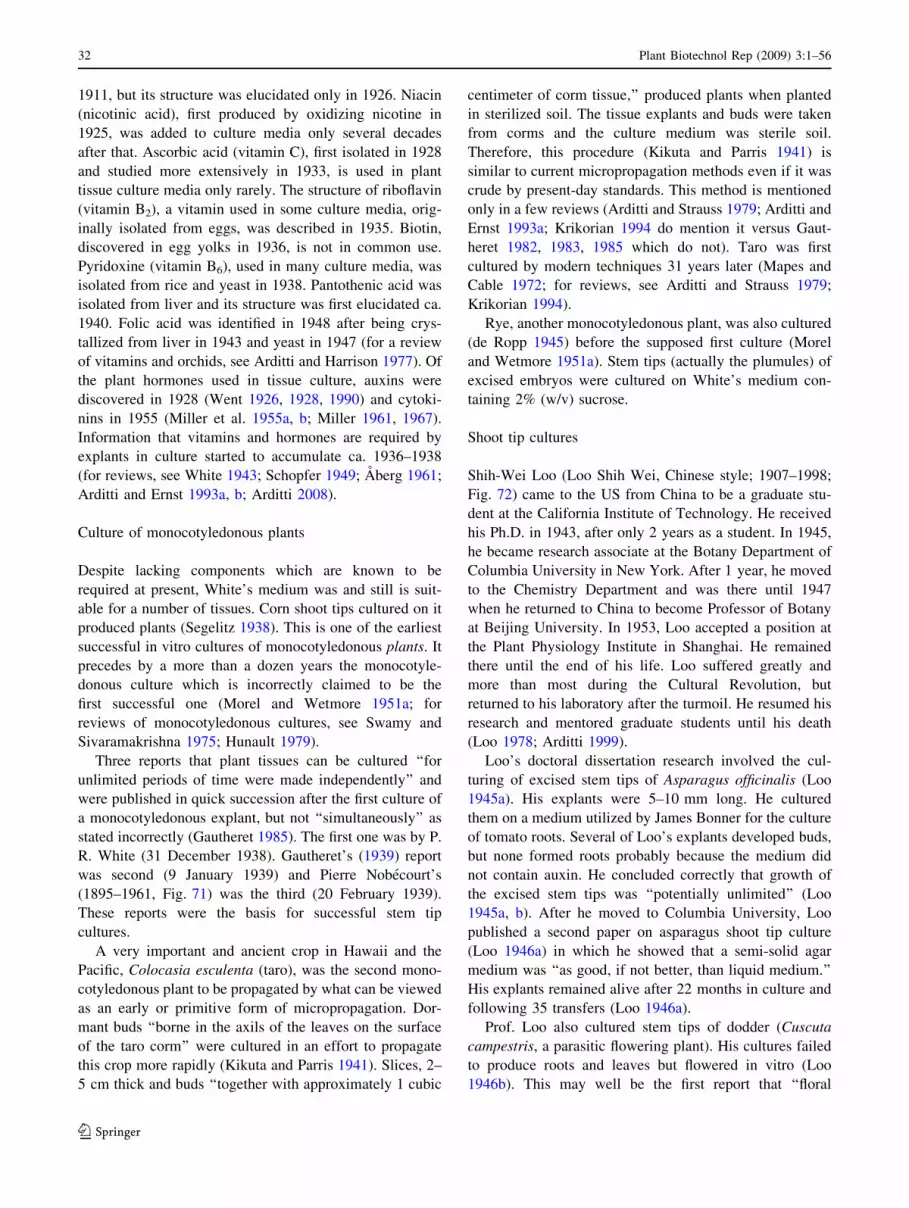
1911, but its structure was elucidated only in 1926. Niacin
(nicotinic acid), first produced by oxidizing nicotine in
1925, was added to culture media only several decades
after that. Ascorbic acid (vitamin C), first isolated in 1928
and studied more extensively in 1933, is used in plant
tissue culture media only rarely. The structure of riboflavin
(vitamin B2), a vitamin used in some culture media, orig-
inally isolated from eggs, was described in 1935. Biotin,
discovered in egg yolks in 1936, is not in common use.
Pyridoxine (vitamin B6), used in many culture media, was
isolated from rice and yeast in 1938. Pantothenic acid was
isolated from liver and its structure was first elucidated ca.
1940. Folic acid was identified in 1948 after being crys-
tallized from liver in 1943 and yeast in 1947 (for a review
of vitamins and orchids, see Arditti and Harrison 1977). Of
the plant hormones used in tissue culture, auxins were
discovered in 1928 (Went 1926, 1928, 1990) and cytoki-
nins in 1955 (Miller et al. 1955a, b; Miller 1961, 1967).
Information that vitamins and hormones are required by
explants in culture started to accumulate ca. 1936–1938
(for reviews, see White 1943; Schopfer 1949; Aberg 1961;
Arditti and Ernst 1993a, b; Arditti 2008).
Culture of monocotyledonous plants
Despite lacking components which are known to be
required at present, White’s medium was and still is suit-
able for a number of tissues. Corn shoot tips cultured on it
produced plants (Segelitz 1938). This is one of the earliest
successful in vitro cultures of monocotyledonous plants. It
precedes by a more than a dozen years the monocotyle-
donous culture which is incorrectly claimed to be the
first successful one (Morel and Wetmore 1951a; for
reviews of monocotyledonous cultures, see Swamy and
Sivaramakrishna 1975; Hunault 1979).
Three reports that plant tissues can be cultured ‘‘for
unlimited periods of time were made independently’’ and
were published in quick succession after the first culture of
a monocotyledonous explant, but not ‘‘simultaneously’’ as
stated incorrectly (Gautheret 1985). The first one was by P.
R. White (31 December 1938). Gautheret’s (1939) report
was second (9 January 1939) and Pierre Nobecourt’s
(1895–1961, Fig. 71) was the third (20 February 1939).
These reports were the basis for successful stem tip
cultures.
A very important and ancient crop in Hawaii and the
Pacific, Colocasia esculenta (taro), was the second mono-
cotyledonous plant to be propagated by what can be viewed
as an early or primitive form of micropropagation. Dor-
mant buds ‘‘borne in the axils of the leaves on the surface
of the taro corm’’ were cultured in an effort to propagate
this crop more rapidly (Kikuta and Parris 1941). Slices, 2–
5 cm thick and buds ‘‘together with approximately 1 cubic
centimeter of corm tissue,’’ produced plants when planted
in sterilized soil. The tissue explants and buds were taken
from corms and the culture medium was sterile soil.
Therefore, this procedure (Kikuta and Parris 1941) is
similar to current micropropagation methods even if it was
crude by present-day standards. This method is mentioned
only in a few reviews (Arditti and Strauss 1979; Arditti and
Ernst 1993a; Krikorian 1994 do mention it versus Gaut-
heret 1982, 1983, 1985 which do not). Taro was first
cultured by modern techniques 31 years later (Mapes and
Cable 1972; for reviews, see Arditti and Strauss 1979;
Krikorian 1994).
Rye, another monocotyledonous plant, was also cultured
(de Ropp 1945) before the supposed first culture (Morel
and Wetmore 1951a). Stem tips (actually the plumules) of
excised embryos were cultured on White’s medium con-
taining 2% (w/v) sucrose.
Shoot tip cultures
Shih-Wei Loo (Loo Shih Wei, Chinese style; 1907–1998;
Fig. 72) came to the US from China to be a graduate stu-
dent at the California Institute of Technology. He received
his Ph.D. in 1943, after only 2 years as a student. In 1945,
he became research associate at the Botany Department of
Columbia University in New York. After 1 year, he moved
to the Chemistry Department and was there until 1947
when he returned to China to become Professor of Botany
at Beijing University. In 1953, Loo accepted a position at
the Plant Physiology Institute in Shanghai. He remained
there until the end of his life. Loo suffered greatly and
more than most during the Cultural Revolution, but
returned to his laboratory after the turmoil. He resumed his
research and mentored graduate students until his death
(Loo 1978; Arditti 1999).
Loo’s doctoral dissertation research involved the cul-
turing of excised stem tips of Asparagus officinalis (Loo
1945a). His explants were 5–10 mm long. He cultured
them on a medium utilized by James Bonner for the culture
of tomato roots. Several of Loo’s explants developed buds,
but none formed roots probably because the medium did
not contain auxin. He concluded correctly that growth of
the excised stem tips was ‘‘potentially unlimited’’ (Loo
1945a, b). After he moved to Columbia University, Loo
published a second paper on asparagus shoot tip culture
(Loo 1946a) in which he showed that a semi-solid agar
medium was ‘‘as good, if not better, than liquid medium.’’
His explants remained alive after 22 months in culture and
following 35 transfers (Loo 1946a).
Prof. Loo also cultured stem tips of dodder (Cuscuta
campestris, a parasitic flowering plant). His cultures failed
to produce roots and leaves but flowered in vitro (Loo
1946b). This may well be the first report that ‘‘floral
32 Plant Biotechnol Rep (2009) 3:1–56
123
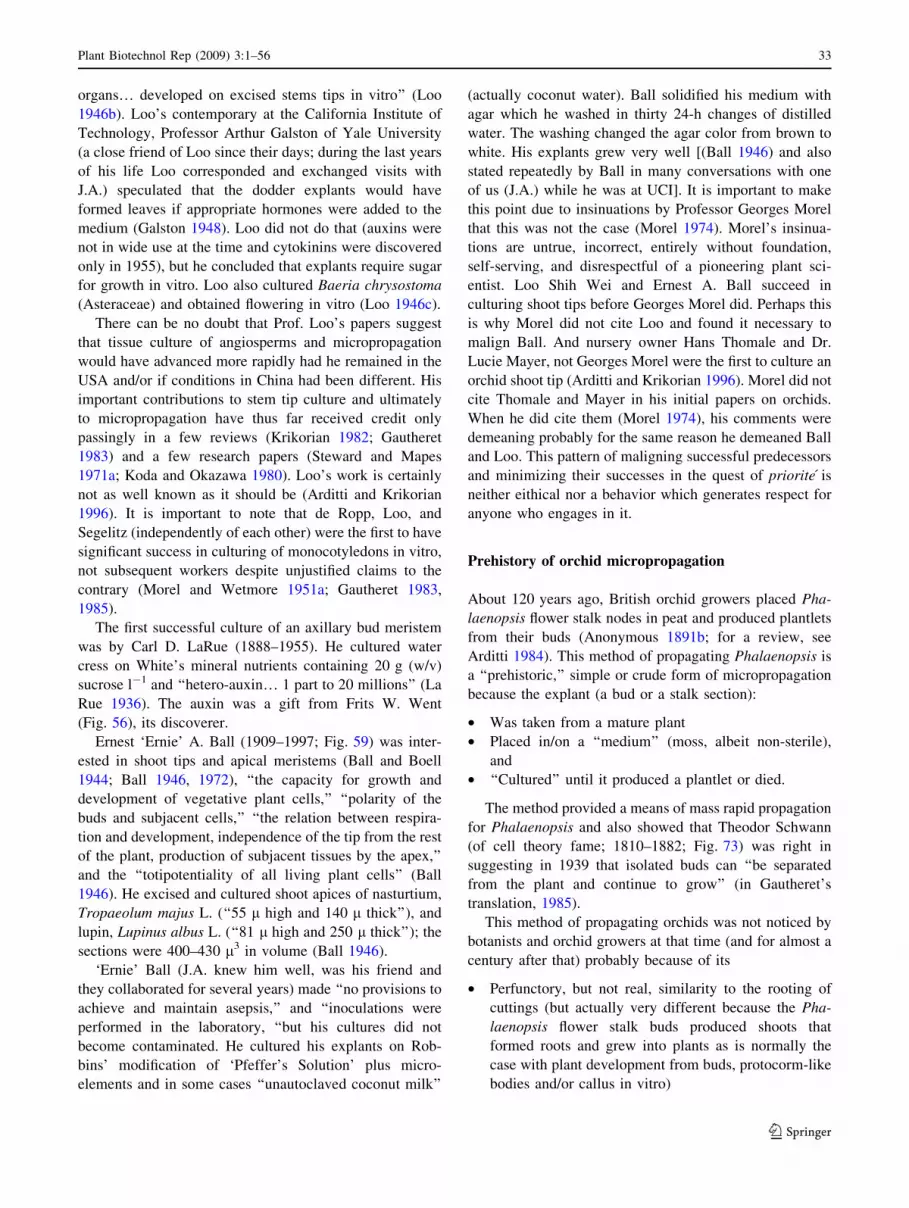
organs… developed on excised stems tips in vitro’’ (Loo
1946b). Loo’s contemporary at the California Institute of
Technology, Professor Arthur Galston of Yale University
(a close friend of Loo since their days; during the last years
of his life Loo corresponded and exchanged visits with
J.A.) speculated that the dodder explants would have
formed leaves if appropriate hormones were added to the
medium (Galston 1948). Loo did not do that (auxins were
not in wide use at the time and cytokinins were discovered
only in 1955), but he concluded that explants require sugar
for growth in vitro. Loo also cultured Baeria chrysostoma
(Asteraceae) and obtained flowering in vitro (Loo 1946c).
There can be no doubt that Prof. Loo’s papers suggest
that tissue culture of angiosperms and micropropagation
would have advanced more rapidly had he remained in the
USA and/or if conditions in China had been different. His
important contributions to stem tip culture and ultimately
to micropropagation have thus far received credit only
passingly in a few reviews (Krikorian 1982; Gautheret
1983) and a few research papers (Steward and Mapes
1971a; Koda and Okazawa 1980). Loo’s work is certainly
not as well known as it should be (Arditti and Krikorian
1996). It is important to note that de Ropp, Loo, and
Segelitz (independently of each other) were the first to have
significant success in culturing of monocotyledons in vitro,
not subsequent workers despite unjustified claims to the
contrary (Morel and Wetmore 1951a; Gautheret 1983,
1985).
The first successful culture of an axillary bud meristem
was by Carl D. LaRue (1888–1955). He cultured water
cress on White’s mineral nutrients containing 20 g (w/v)
sucrose l-1 and ‘‘hetero-auxin… 1 part to 20 millions’’ (La
Rue 1936). The auxin was a gift from Frits W. Went
(Fig. 56), its discoverer.
Ernest ‘Ernie’ A. Ball (1909–1997; Fig. 59) was inter-
ested in shoot tips and apical meristems (Ball and Boell
1944; Ball 1946, 1972), ‘‘the capacity for growth and
development of vegetative plant cells,’’ ‘‘polarity of the
buds and subjacent cells,’’ ‘‘the relation between respira-
tion and development, independence of the tip from the rest
of the plant, production of subjacent tissues by the apex,’’
and the ‘‘totipotentiality of all living plant cells’’ (Ball
1946). He excised and cultured shoot apices of nasturtium,
Tropaeolum majus L. (‘‘55 l high and 140 l thick’’), and
lupin, Lupinus albus L. (‘‘81 l high and 250 l thick’’); the
sections were 400–430 l3 in volume (Ball 1946).
‘Ernie’ Ball (J.A. knew him well, was his friend and
they collaborated for several years) made ‘‘no provisions to
achieve and maintain asepsis,’’ and ‘‘inoculations were
performed in the laboratory, ‘‘but his cultures did not
become contaminated. He cultured his explants on Rob-
bins’ modification of ‘Pfeffer’s Solution’ plus micro-
elements and in some cases ‘‘unautoclaved coconut milk’’
(actually coconut water). Ball solidified his medium with
agar which he washed in thirty 24-h changes of distilled
water. The washing changed the agar color from brown to
white. His explants grew very well [(Ball 1946) and also
stated repeatedly by Ball in many conversations with one
of us (J.A.) while he was at UCI]. It is important to make
this point due to insinuations by Professor Georges Morel
that this was not the case (Morel 1974). Morel’s insinua-
tions are untrue, incorrect, entirely without foundation,
self-serving, and disrespectful of a pioneering plant sci-
entist. Loo Shih Wei and Ernest A. Ball succeed in
culturing shoot tips before Georges Morel did. Perhaps this
is why Morel did not cite Loo and found it necessary to
malign Ball. And nursery owner Hans Thomale and Dr.
Lucie Mayer, not Georges Morel were the first to culture an
orchid shoot tip (Arditti and Krikorian 1996). Morel did not
cite Thomale and Mayer in his initial papers on orchids.
When he did cite them (Morel 1974), his comments were
demeaning probably for the same reason he demeaned Ball
and Loo. This pattern of maligning successful predecessors
and minimizing their successes in the quest of priorite is
neither eithical nor a behavior which generates respect for
anyone who engages in it.
Prehistory of orchid micropropagation
About 120 years ago, British orchid growers placed Pha-
laenopsis flower stalk nodes in peat and produced plantlets
from their buds (Anonymous 1891b; for a review, see
Arditti 1984). This method of propagating Phalaenopsis is
a ‘‘prehistoric,’’ simple or crude form of micropropagation
because the explant (a bud or a stalk section):
• Was taken from a mature plant
• Placed in/on a ‘‘medium’’ (moss, albeit non-sterile),
and
• ‘‘Cultured’’ until it produced a plantlet or died.
The method provided a means of mass rapid propagation
for Phalaenopsis and also showed that Theodor Schwann
(of cell theory fame; 1810–1882; Fig. 73) was right in
suggesting in 1939 that isolated buds can ‘‘be separated
from the plant and continue to grow’’ (in Gautheret’s
translation, 1985).
This method of propagating orchids was not noticed by
botanists and orchid growers at that time (and for almost a
century after that) probably because of its
• Perfunctory, but not real, similarity to the rooting of
cuttings (but actually very different because the Pha-
laenopsis flower stalk buds produced shoots that
formed roots and grew into plants as is normally the
case with plant development from buds, protocorm-like
bodies and/or callus in vitro)
Plant Biotechnol Rep (2009) 3:1–56 33
123

• Place of publication, which was one of the earliest and
now obscure and rare orchid journal that is hard to find
• Language (French), because ‘‘an increasing number of
scientists… read no modern languages other than
English’’ (Krikorian and Berquam 1969).
• Age, because not many scientists take the time to read
the old literature regardless of language and promi-
nence (or lack of it) of journals.
At least one orchid grower noticed the articles.
According to a short note (Anonymous 1891b), a grower
named Perrenoud (no first name given) saw the reports in a
so-called ‘‘journaux anglais’’ (which we have not been able
to trace), modified it by placing sections of Phalaenopsis
roots in humid enclosures and obtained a plant. No other
details are available. However, it is known that Phalaen-
opsis roots can produce buds and plants (for a review, see
Churchill et al. 1972). This method is reminiscent of mi-
cropropagation. Therefore, it can be viewed as being part
of the pre-history of orchid (or any plant) micropropagation
(Arditti and Krikorian 1996) and biotechnology.
If this method would have been noticed and given the
attention it deserves, it and its discoverer (a still unknown
British orchid grower) could have been an important road
mark on the path to plant tissue culture and micropropa-
gation. It is obviously more
• Relevant as a ‘‘foreword’’
• Related to tissue culture
• Like micropropagation
than (what appear to be the chauvinistically) publicized and
glorified observations and writings by the Seigneur du
Monceau et de Vrigny, Henri-Louis Duhamel du Monceau
in France (Gautheret 1985; Fig. 74).
First micropropagation of orchids
The modern history of orchid micropropagation (and in
fact micropropagation in general) started when a:
1. New [tissue culture or in vitro], simple and practical
method for vegetative [clonal] propagation of Pha-
laenopsis [orchids] was developed at Cornell
[University]’’ 5 years (Rotor 1949) before the first
published report regarding orchid shoot tip cultures.
The medium Rotor used to culture the Phalaenopsis
flower stalk nodes was Knudson C (KC), a solution
formulated for the asymbiotic germination of orchid
seeds by Lewis Knudson (1884–1958; Fig. 50), Pro-
fessor of Plant Physiology at Cornell University (see
Arditti 1990 for biography). Knudson’s first solution,
the Knudson B medium (KB) was a modification
of Pfeffer’s solution which was formulated by the
German plant physiologist Wilhelm Pfeffer (1845–
1920; Fig. 46). Knudson improved KB in 1946 as his
solution C (Knudson C, KC; Knudson 1946a, b) which
became a useful medium for orchid seed germination.
Today, KC is used extensively for orchid seed
germination (Arditti et al. 1982) and the microprop-
agation of some orchids (Arditti and Ernst 1993a; Yam
and Arditti 2007; Arditti 2008).
2. A German nurseryman suggested that shoot tip
cultures can be used for micropropagation (Thomale
1956, 1957).
Gavino Rotor Jr. (1917–2005; Fig. 75) was born in
Manila and received his B.S. in Agriculture from the
University of the Philippines in 1937. He came to the US in
1946, received his M.S. degree at Cornell University in
1947 and Ph.D. in 1952 with a dissertation on the responses
of orchids to temperature and day length. In a letter (to J.
A.), Dr. Rotor wrote that he conceived the idea of propa-
gating orchids while attending a lecture by Knudson on the
role of sugars in plant growth. However, he provided no
details on how a lecture on sugars led him to the idea of
culturing Phalaenopsis flower stalk nodes. He sectioned
inflorescences into segments and inserted node sections,
each bearing a single bud into KC hoping that the buds
would develop plants. The buds produced leaves within
14–60 days. Roots appeared after the formation of 2–3
leaves (Fig. 76). Of the 65 buds he cultured, only 7 failed
to produce plants (Rotor 1949). In his letter, Rotor wrote
that Knudson’s ‘‘eyes brightened when [Rotor] showed him
the first successful propagation… and told him how [he]
got the idea from [Knudson’s] lecture’’ (Arditti 1990).
Given his Phalaenopsis flower stalk node cultures, there
can be absolutely no doubt that Dr. Gavino Rotor is the
inventor not only of orchid micropropagation but also of
mass rapid clonal propagation of plants in vitro (i.e., mi-
cropropagation). His method utilized:
• A defined culture medium
• Aseptic technique
• Explants.
And he drew attention to the propagation potential of his
method. It can be argued that Rotor’s method was not
micropropagation as the term is understood at present
because:
• It produced only a single shoot per explant
• Explants contained pre-existing buds
• Callus formation or proliferation was not involved.
However, such an argument would be spurious and
contrived because production of multiple plantlets from a
single explant, absence of pre-existing buds and production
and proliferation of callus are neither parts of the definition
34 Plant Biotechnol Rep (2009) 3:1–56
123
of microporpagation nor a requirement for mass rapid
clonal propagation in vitro. Roger Gautheret discounted the
historical relevance of Rotor’s discovery when one of us
(J.A.) informed him of it. He probably did it in an effort to
glorify his friend, colleague, and compatriot, Georges
Morel (since the word chauvinism is attributed to Nicolas
Chauvin, a soldier in Napoleon’s Grande Armee, this is not
surprising).
Rotor’s micropropagation method was barely noticed,
used, or appreciated at the time. This could be due to the
fact that it was published in a magazine for hobby growers.
American Orchid Society Bulletin (AOSB, now called
Orchids), which is not a scientific journal. Orchid growers
who read the AOSB could have been daunted by the pro-
cedure and probably failed to appreciate its importance.
Scientists would have seen the method and could have put
it to good use, but they probably did not read the AOSB.
The method became forgotten. When it was finally redis-
covered, claims of priority by Prof. Georges Morel had
become a widely accepted urban legend.
Also in the late 1940s, Professor John T. Curtis (1913–
1961; Fig. 62) and his associates in the Department of
Botany at the University of Wisconsin described formation
of multiple growing points on proliferating seedling-derived
callus of Cymbidium and Vanda (Curtis and Nichol 1948).
‘Calloid’ is the term they applied to protuberances which
developed in asymbiotic culture following treatment with
barbiturates. They also reported that these tissue masses had
the capacity to grow into plants (Curtis and Nichol 1948),
appreciated the potential for clonal propagation, and wrote:
‘‘the practical ability to produce clonal lines of plants of
potentially unlimited numbers would be of obvious value in
many types of genetic and plant production work.’’
It must be noted here that in their initial reports the
nearly forgotten Hans Thomale (Thomale 1954, 1956,
1957; Haas-von Schmude et al. 1995; Arditti 2001; Easton
2001) and the unjustifiably widely celebrated Georges
Morel (1960) also called attention to the potential of their
findings, but they did it after Rotor.
First culture of an orchid shoot tip or the second aseptic
culture of an orchid explant
Pelargonium zonale and cyclamen, Cyclamen persicum,
were cultured by Lucie Mayer (Fig. 77) on a relatively
simple medium (Mayer 1956) before the formulation of the
Murashioge Skoog medium. Ms Mayer’s work intrigued
Hans Thomale (1919–2002; Fig. 78), German nursery
owner. Mayer and Thomale joined forces for the first cul-
ture of sections (‘‘Teilstucken’’ or ‘‘Pflanzenteile’’), tissues
(‘‘Gewebe’’) and shoot tips of orchids (Thomale 1956,
pp. 89–90, fig 39; Fig. 79).
Hans Thomale was born in Herne, Westphalia, Ger-
many, raised in Cologne and resided and grew orchids in
Lemgo for many years. He began to study chemistry and
medicine before World War II started, but was drafted and
had to interrupt his studies. After the war, he grew potatoes
in a nursery and married Dr. Liselotte Kuhlman, the
daughter of the owner. When he became interested in
orchid seed germination, Thomale taught himself how to
do it by using Prof. Hans Burgeff’s (1883–1976; Fig. 44)
book, Samenkeimung der Orchideen. In 1946, he estab-
lished a laboratory and utilized it to germinate and clonally
propagate both tropical orchids and species native to
Germany.
On 23 September 1956, Thomale reported to a meeting
of the Deutsche Orchideen Gesellschaft (DOG; German
Orchid Society) that explants of Dactylorhiza (Orchis)
maculata (Fig. 79) and several tropical orchids produced
shoots in vitro. Thomale recollected, somewhat tentatively,
that Mr. Lecoufle of the French orchid firm Vacherot and
Lecoufle (see below) was present at that meeting. He also
included a photograph of the Orchis maculata culture
(Fig. 79) in the second edition of his book Orchideen
(Thomale 1957). Thomale appreciated immediately the
potential of his discovery. He wrote (Arditti and Ernst
1993a, b; Haas-von Schmude et al. 1995):
‘‘It should be noted that efforts to find a propagation
method for European terrestrial orchids, based on the
work by Dr. L. Mayer [Mayer 1956], through the
culture of sterile explants on an agar medium were
successful. It is well known that vegetative parts of
orchids, for example, sterile sections of Phalaenopsis
flower stalks [Rotor 1949], which bear at least one
adventitious bud [Note in Arditti and Krikorian 1996:
these buds are lateral on the flower stalk and not
necessarily adventitious, at least not in the strict ense
of the word], can produce shoots when cultured on an
agar medium. Recently it has become possible to
culture undifferentiated tissues on certain nutrient
media to produce roots and shoots from them. Since
sufficient details were not available by the time this
book went to press [i.e., the second edition which
appeared in 1957; the first edition was published in
1954], it is only possible to mention that whole plants
can be produced from tissue explants one cubic
centimeter in size. This is a form of vegetative mul-
tiplication whose potential cannot be overlooked’’
[emphasis added]!
Thomale’s book and his prescient statement about the
utilization of explant cultures in vitro as a method of mass
rapid propagation were published (Thomale 1957) before
the first reports of Cymbidium ‘‘meristem’’ cultures (Morel
1960; Wimber 1963), but limited attention was paid to
Plant Biotechnol Rep (2009) 3:1–56 35
123
them. Thomale behaved professionally and ethically by
referring to Rotor’s work (Thomale 1956, 1957). Had he
not done that Thomale could have created the false
impression that he originated the concept of clonal propa-
gation in vitro. Thomale did not describe his techniques in
detail. Instead, he referred to the procedures published by
Mayer. Dr. Lucie Mayer took part in Thomale’s first
attempts to culture orchid explants (Haas-von Schmude
et al. 1995; personal communication from E. Lucke and Dr.
N. Haas-von Schmude, Wettenberg, Germany). After her
retirement to Madeira, Portugal, Dr. Mayer recalled (in a
letter to one of us, J.A.) that she and Thomale also excised
and cultured Cymbidium stem tips. Thomale’s work did not
become well known for several reasons:
• His first report was published in a German language
magazine for orchid hobbyists which is mostly known
only in Germany (Thomale 1956)
• The second publication was also in German in a little
known book (Thomale 1957, the second edition of
Thomale 1954) written mostly for hobbyists and
commercial orchid growers.
• Few scientists read Thomale’s work. Practical growers
who read the books were probably bewildered by the
technique, failed to understand it and never used it
(there is parallel between Rotor’s and Thomale’s
publications).
Georges Morel (1916–1973; Fig. 60) who is undeserv-
edly, but often given, exclusive credit for being the first to
culture an orchid explant in vitro (Arditti and Arditti 1985;
Haas-von Schmude et al. 1995; Arditti and Krikorian 1996;
Easton 2001; Arditti 2001) knew about Thomale’s report at
least as early as 1965 (Fig. 80). However, he did not cite it for
almost 10 years. When Morel did cite it, in a chapter written
for Carl. L. Withner’s The Orchids—Scientific Studies
(Withner 1974), it was 14 years after his celebrity in the
orchid world was a well-established fact (Morel 1974; Haas-
von Schmude et al. 1995). At that time, Thomale was known
only for his medium GD for the germination of Paphioped-
ilum seeds (Thomale 1957). Even when he cited Thomale’s
work and published his photograph [labeling it as ‘‘after
Thomale’’ rather than indicating that Hans Thomale pro-
vided a copy because Morel asked for one (Fig. 80), Morel
diminished it by adding the qualifying statements like:
• ‘‘Pieces from the bulb of Orchis maculata, aseptically
cultivated on nutrient medium, soon regenerated stems
and roots…’’ and on the photograph caption ‘‘regener-
ation of roots and shoots occuring on a piece of tuber of
Orchis maculata.’’ The wording (‘‘stems and roots’’)
minimizes Thomale’s achievement by implying that
what was produced were not plants. Morel should have
stated ‘‘… soon produced plants.’’
• ‘‘[Cases like this] are very exceptional.’’ They are
certainly not! Regardless, Thomales deserves credit for
his achievement, not denigration.
Hans Thomale was finally given the recognition which
was due to him, fortunately while he was still alive, due to
the efforts four individuals who believed in fairness (Haas-
von Schmude et al. 1995; Arditti and Krikorian 1996;
Arditti 2001). Unfortunately, by then total (but undeserved)
credit for priority of discovery was given to Morel. One
reason for this could be Morel’s deserved international
standing as a prominent plant scientist. His many friends
who repeatedly spoke and wrote on his behalf were another
reason. His extensive travels and frequent lectures were a
third reason because he used them for self-publicity. A
fourth reason were orchid scientists who did not know the
correct history, idolizing hobbyists and appreciative com-
mercial growers who placed Morel on a pedestal as being
the sole inventor. Resistance to new knowledge (Gaffron
1969) and/or revision of established fiction were/are a fifth
reason. Examples are:
• A note marking Thomale’s 75th birthday (Lucke 1994)
which does not even mention his discovery because a
statement to that effect was edited out by the editors of
Orchidee (Dr. Norbert Haas-von Schmude, Wettenberg,
Germany, personal communication).
• An article marking the 25th anniversary of ‘‘mericlon-
ing’’ (Arditti and Arditti 1985) which was ‘‘revised’’ on
the advice of a reviewer.
• The history chapter in a book on orchid micropropa-
gation (Arditti and Ernst 1993a) was edited heavily in
an effort not to offend those who favor the urban legend
of Morel as the sole discoverer.
Today, since Hans Thomale’s work and his accurate
prediction about micropropagation are known, it is no
longer correct to state that ‘‘… Georg[e] Morel has realized
for the first time the multiplication of Orchids (sic) by stem
tips in vitro culture. Dr. (sic) Thomale seems to be unaware
of the tissue culture history’’ (R. J. Gautheret, Paris, in a
letter to J.A.). Patriotism, nationalism, and dedication to a
‘‘… Late collaborator…’’ (Gautheret, personal communi-
cation), friend and compatriote are not sufficient
justification for ignoring historical facts and an Orwellian
(i.e., 1984 style) restating of history. The one unaware of
tissue culture history was Gautheret.
Plant diseases and shoot tips
The concept that stem tips, root cuttings, and even leaves
can be used to produce healthy clones of horticultural
plants is more than 60 years old (see Krikorian 1982; North
36 Plant Biotechnol Rep (2009) 3:1–56
123

1953 for literature citations). During World War II, Arthur
W. Dimock (1908–1972) published a method for producing
Verticillium-free clones of chrysanthemums through the
use of tip cuttings taken from 4- to 6-inch-long (ca. 10–
15 cm) shoots which were shown to be disease free (Di-
mock 1943a, b). This method was later improved and used
to free plants of other diseases (Brierly 1952; Dimock
1951b). Such methods were also used to produce disease-
free carnations (Dimock 1943a, b, 1951b; Forsberg 1950;
Andreasen 1951; Guba 1952; Hellmers 1955; Thammen
et al. 1956). It is even possible to argue that those who
originated these methods were among the first to use the
terms ‘‘cultured’’ (Forsberg 1950) and ‘‘culturing’’ (Di-
mock 1951a; Guba 1952) in relation to producing disease-
free plants.
Observations that tips of virus-infected roots could be
free of infection were made 75 years ago (White 1934a, b,
1943). Earlier, virus or ‘‘abnormalities’’ were not seen in
stem tips of tobacco, tomato, and Solanum nodiflorum
(Sheffield 1933, 1942; Clinch 1932). Spotted wilt virus was
eliminated from Dahlia through the use of stem tip cuttings
(Holmes 1948a, b, 1955). This method was also used for
the elimination of leaf spots in sweet potato, Ipomea ba-
tatas, which are associated with the internal-cork disease in
this crop, (Holmes 1956a) as well as aspermy virus
(Holmes 1956b) and other viruses (Brierly and Olson 1956)
in Chrysanthemum.
The elimination of spotted wilt of Dahlia (Holmes
1948a, b) through use of stem tip cuttings left little or no
doubt that apical meristems are virus-free. Confirmation of
this was obtained in work with tobacco mosaic-infected
tobacco, Nicotiana tabacum, variety Samsun (Limasset and
Cornuet 1949).
These findings are not consistent with current informa-
tion which is that apical meristems are not always virus-
free. This inconsistency is the reason for difficulties which
have been encountered in freeing many clones and culti-
vars of viruses (Kassanis 1967). Also, even when apical
meristems are virus-free it is not always possible to obtain
pathogen-free plants. In fact ‘‘in the orchid industry…Before ‘mericloning’ orchid viruses were a minor prob-
lem… However [they] are now common, wide-spread and
costly’’ (Langhans et al. 1977) because careless culturing
spread rather than contained or eliminated viruses (Tous-
saint et al. 1984).
Viral infection of certain potato and Dahlia cultivars
was a problem facing French horticulture ca. 1950 (Le-
coufle 1974a, b). The culture of stem tips provided a means
of freeing these plants of viruses in view of what was
known about dahlias (Holmes 1948a, b) and tobacco (Li-
masset and Cornuet 1949). Pierre Limasset (1911–1988;
Fig. 82) and Pierre Cornuet (b. 1925; Fig. 83) ‘‘suggested
to their colleagues Georges Morel and Claude Martin to
cultivate shoot meristems of infected plants’’ (Gautheret
1983, 1985). Morel and Martin followed the suggestion.
Their attempts were successful and virus-free dahlias
(Morel and Martin 1952) and potato (Morel and Martin
1955a, b; Morel and Muller 1964; Gautheret 1983, 1985)
plants were produced from infected ones.
‘‘George Morel was an amateur orchid grower [who]
had in his greenhouse a plant of Cymbidium Alexaderi
‘Westonbirt’… the most famous Cymbidium of all time,
which was sadly, totally infected by Cymbidium mosaic
virus’’ (Vacherot 2000). The success with Dahlia, potatoes
and other plants (Morel and Martin 1955b; Morel 1964a, b)
led Morel ‘‘[to apply] the same technique as he was using
on his potatoes to the Cymbidium [and] produced a pro-
tocorm [sic]’’ (Morel 1960; Vacherot 2000; Fig. 60B) and
later a plant (Fig. 60C). As indicated above, this effort was
heralded in numerous lectures and publications. One cat-
alog stated that ‘‘a beautiful thing happened to the orchids
when they operated on a sick potato [because] Dr. Georges
Morel, distinguished French botanist, discovered the orchid
meristem process while he was trying to figure out a way to
prevent virus in potatoes’’ (Orchids Orlando 1968). Less
lyrical, but just as inaccurate paeans and odes clutter the
scientific, horticultural, and hobby literature (examples are:
Bertsch 1966, 1967; Marston and Vourairai 1967; Vacherot
1966, 1977; Borriss and Hiibel 1968; Vanseveren and
Freson 1969; Hahn 1970; Kukułczanka and Sarosiek 1971;
Lecoufle 1971; Lucke 1974; Allenberg 1976; Champagnat
1977; Rao 1977; Loo 1978; Murashige 1978; Goh 1983;
Hetherington 1992). Correct versions of history are rare
(Arditti 1977a, b, 2001; Haas-von Schmude et al. 1995;
Arditti and Krikorian 1996; Easton 2001; Yam and Arditti
2007; Arditti 2008). Accuracy was sacrificed in a few
instances as a bow to editorial pressure (Arditti and Arditti
Figs. 82–83 Plant virologists. 82 Pierre Limasset. 83 Piere Cornuet.
(Note: these figures were located after the final editing of the
manuscript and are included here despite their very low quality
because they are the only ones of these individuals that could be
found)
Plant Biotechnol Rep (2009) 3:1–56 37
123

1985; Lucke 1994; Dr. Norbert Haas-von Schmude, Wet-
tenberg, Germany, personal communication) or in an effort
not to offend established interests and views (Arditti and
Ernst 1993a).
The third aseptic culture of an orchid explant
‘‘The secret of originality is hiding your sources’’
(Attributed to Mark Twain or Albert Einstein)
Many papers on orchid micropropagation start with a
citation or at least a mention of Morel’s first paper on
Cymbidium shoot tip culture (Morel 1960). Assertions that
‘‘the first application [of micropropagation] concerned the
clonal propagation of orchids (Morel 1960)’’ are found in
historical accounts by one of the founders of plant tissue
culture (Gautheret 1983, 1985). Such reviews are influen-
tial because they are read extensively and then quoted or
re-stated in subsequent publications. As a result, an urban
legend was elevated to universal (albeit incorrect) truth and
scientific dogma. Once this occurs, forces which resist
knowledge strive to preserve the status quo and thus defend
the urban legend (Gaffron 1969; Arditti 2004).
Questioning the accepted views can lead to unpleasant
interactions (Arditti 1985; Torrey 1985a, b). Demands to
revise manuscripts became conditions for publication
(Arditti and Arditti 1985; Lucke 1994). Still, the prevailing
historical accounts were reexamined critically and facts
were placed in an accurate perspective (Arditti and
Krikorian 1996). The resulting review (Arditti and Kriko-
rian 1996) is one of the major sources used for an history
chapter in the second edition of a book on orchid micro-
propagation (Arditti 2008) as well as the present account
and two previous ones (Arditti 2004; Yam and Arditti
2007). Sadly, presenting an accurate history may create an
inaccurate impression that the intent is to sully some rep-
utations because [to quote physicist Ernst Mach (1838–
1916) as quoted by the late prominent plant physiologist
Hans Gaffron in 1969: ‘‘It is hardly possible to state any
truth strongly without apparent injustice to some other.’’]
This only seems so because inaccuracies are so prevalent in
the orchid literature.
Georges Morel (1916–1973; Fig. 60), entered l’Institut de
Chemie in Paris where he studied agriculture and plant
pathology. After that he joined L’Institut National de la
Recherche Agronomique (INRA), the French institute of
Agricultural Research (Gautheret 1977), where he became
very influential (Vacherot 2000) and ‘‘chef de travaux’’ in
1941. In 1943, Morel joined Gautheret’s laboratory
(Lecoufle 1974a, b) and worked there towards his doctorate.
Despite the difficulties caused by the Nazi occupation (Paris
was liberated in 1944), Morel was successful in his research.
Morel received his doctorate in 1948, went to the USA
during the same year and worked until 1951 with Professor
Ralph W. Wetmore (1892–1989; Fig. 81) in the Biological
Laboratories at Harvard University. They worked on tissue
culture of monocotyledonous plants (Morel and Wetmore
1951a) and ferns (Morel and Wetmore 1951b). On his
return to France, Morel was appointed Maıtre de recher-
ches (in 1951 or 1952) and in 1956 Director de recherches
of the Station Centrale de Physiologie Vegetale of the
Centre National des Recherches Agronomiques, Ministiere
de l’Agriculture (Lecoufle 1974a, b).
Despite its great fame in the orchid world, Morel’s first
report shoot tip culture of Cymbidium (Morel 1960) was
more a news item than a scientific paper. It lacked detail,
was sketchy, and stated that the explants were cultured on a
non-existent medium he named ‘‘Knudson III.’’ Morel’s
conclusion was ‘‘that it is relatively easy to free a Cym-
bidium from the mosaic virus… each bud will give several
plants so the stock of a rare or expensive variety can be
increased… [and that] experiments of the same kind are
now being conducted with… Cattleya, Odontoglossum,
and Miltonia, contaminated with different viruses’’ (Morel
1960). Its major contribution was the introduction of a new
term into orchid terminolgy, ‘‘protocorm-like body’’ (PLB)
to describe the ‘‘small flat bulblet looking exactly like [a]
protocorm’’ (Fig. 80B) which was produced by the cul-
tured Cymbidium stem tips (for an history of the term
‘‘protocorm’’, see Arditti and Ernst 1993a, b; Arditti and
Krikorian 1996, Arditti 2008).
It would have been impossible for anyone to repeat
Morel’s work for lack of sufficient details. To reconstruct
the procedures and medium or media used by Morel it
would have been necessary to carefully study much of
Morel’s previous work including a rather obscure paper on
potatoes (Morel and Martin 1955a) and an even less well-
known one on ‘‘parasites obligatoires et de tissus veget-
aux’’ (Morel 1948) as well as two not very easy to find
papers by Dutch authors, one on Iris (Baruch and Quak
1966) and the other on potatoes (Quak 1961). In addition, it
would have been necessary to assume that the potato and
Iris papers were relevant. But why would any one assume
that? Even if orchid scientists could find the composition of
the potato or Iris media there were no indications that they
would be suitable for orchids. In fact, the potato medium is
very different from the one subsequently used for orchids
by Morel. And orchid commercial and hobby growers
could not be expected to engage this type of literature
search.
Interestingly, Vacherot and Lecoufle (V&L) ‘La Tuil-
erie’, Boissy-Saint Leger (Seine-et-Oise), a French orchid
firm had enough information to start commercial micro-
propagation of ‘‘rare or expensive’’ orchids before any
other establishment. They moved quickly enough to have a
38 Plant Biotechnol Rep (2009) 3:1–56
123
clonally propagated plant of Vuylstekeara Rutiland
‘Colombia’ bloom in December 1965 (Vacherot 1966;
Lecoufle 1967), but a recent report suggests that the first
plants to be cultured were ‘‘some of [their] finest cymbi-
diums’’ (Vacherot 2000).
A report that ‘‘… at ‘La Tuilerie’ our first mericlone to
flower [was]… Vuylstekeara Rutiland ‘Colombia’… in
December, 1965… ‘‘(Lecoufle 1967) suggests that ‘‘mer-
icloning’’ started at V&L before or at about the time
Morel’s first paper was published because ‘‘… it will take
just as long to grow the plants produced from meristem
tissue as it takes to grow a new hybrid from seed’’ (Scully
1964). It may have started as early as 1956 despite what
seems to be a delayed publication of Morel’s first paper in
1960 and subsequent papers (Morel 1963, 1964a, b, 1965a,
b, 1970, 1971a, b, c, 1974; for much more detailed history
and discussions as well as speculations regarding the rea-
sons for the delayed publications and lack of details, see
Arditti and Krikorian 1996; Arditti 2008).
The lack of citations in Morel’s first paper must also be
considered. Loo and Ball (whose techniques were used),
Limasset (see Fig. 82) and Cornuet (see Fig. 83; who
suggested the idea of culturing shoot tips), Rotor (the first
investigator to clonally propagate an orchid in vitro),
Thomale and Mayer (first to culture an orchid shoot tip),
and others were not cited. This is not in line with the
standard practice by scientists. Two visitors to Morel’s
laboratory in the mid-1960s (one a student and the other a
sabbatical year researcher) suggested in conversations with
one of us (J.A.) about 25 years ago that
• Such lack of citation was not unusual for French scientists,
but we have seen and/or read many papers by French
scientists with extensive lists of cited publications
• Morel did not spend a lot of time in the library, but he
was clearly aware of Thomales work (Fig. 80), and
many papers from his laboratory contain adequate and
detailed citations (for examples, see most of the papers
we cite, i.e., Morel 1948, 1950, 1963, 1964a, b, 1970,
1971a, b, 1974; Morel and Wetmore 1951a, b; Morel
and Martin 1952, 1955a, b; Morel and Muller 1964;
Champagnat 1965, 1971, 1977; Champagnat et al.
1966; Champagnat et al. 1968; Champagnat and Morel
1969, 1972; Champagnat et al. 1970; as well as those
by his associates, Champagnat 1965, 1971, 1977). He
also commented on one of our papers (Churchill et al.
1971a, b) a very short time after it was published, a fact
which indicates that he read the literature.
When asked by one of us (J.A.) after a lecture he gave at
the World Orchid Conference in Sydney in 1969 about
Ball’s contribution to his work, Morel’s reply was a testy:
‘‘Ah, Ball.’’ This non-reply and a suggestion by someone
who knew him that Morel liked prominence suggest that he
did not cite those he should have cited in an effort to claim
the discovery for himself. There is also another view.
Morel was described by the late Professor John Torrey of
Harvard University as (1) ‘‘… one of the pioneers in the
study of shoot meristem culture as well as an early advo-
cate for its practical use in multiplication of virus free
plants… interested in the free exchange of scientific
information and discoveries [who] ‘did not take any patent
because I feel that a scientist does not have to do this… ’’’
(Torrey 1985b), and (2) a very nice, kind, and modest man.
Innovators
‘‘Good artists copy; great artists steal’’
Pablo Picasso
In the opinions of many, Morel’s orchid work was very
original and highly innovative, but an impartial analysis of
the historical facts leads to a different conclusion. None of
Morel’s work with potatoes, dahlias, and orchids was ori-
ginal. Media for plant tissue culture in general and stem
tips of orchids in particular were formulated (Knop 1884;
Loo 1945a, b, 1946a, b, c; Knudson 1946a, b; Rotor 1949;
Mayer 1956, Thomale 1956, 1957) before Morel devised
his own substrate by modifying those that were already in
existence. A number of explants (buds, nodes, shoot tip)
from several monocotyledonous species (Robbins 1922a,
b; Segelitz 1938; Kikuta and Parris 1941) and orchid in
particular (Rotor 1949; Thomale 1956, 1957) were cultured
before Morel accomplished it (Morel and Wetmore 1951a).
Several methods were published prior to his. Plants were
freed of virus infection through shoot tip culture or rooting
before Morel’s work with potatoes, dahlias, and orchids.
And Morel’s work on potatoes and dahlias was suggested
by others, namely P. Limasset and P. Cornuet (Gautheret
1983, p. 402, 1985, p. 42).
Georges Morel’s first notable achievement was the
production of protocorm-like bodies (PLBs) which could
be subcultured. This made micro-(mass rapid clonal)
propagation of orchids possible. Morel accomplished this
by cleverly using existing methods and culture procedures
and combining them into a very useful new application.
His second important achievement was publicity for an
idea which was needed. Morel deserves credit for
thoughtfully applying existing methods and information to
a new technology. However, he should not be accorded the
adulation normally given to individuals who conceive new
ideas, make basic discoveries, and articulate new principles
(Easton 2001; Arditti 2001, 2004, 2008; Yam and Arditti
2007).
Roger Gautheret, one of the early tissue culture inves-
tigators, wrote that ‘‘Ball is really the father of the so-called
Plant Biotechnol Rep (2009) 3:1–56 39
123

micropropagation method’’ (Gautheret 1985, pp. 16–17),
but the same arguments can be made about LaRue and Loo
(La Rue 1936; Loo 1945a, b, 1946a, b, c; Ball 1946). It is
possible that Gautheret credited Ball because he demon-
strated that it is possible to culture stem tips in vitro. But,
LaRue and Loo did the same without being credited by
Gautheret. Ball (who collaborated with us during his tenure
at UCI) probably did not appreciate the practical implica-
tions of his work, or if he did, he failed to state it in print
(Ball 1950). The same is true of others (La Rue 1936; Loo
1945a, b, 1946c; Krikorian 1982; Arditti and Ernst 1993a,
b; Arditti and Krikorian 1996; Arditti 2008).
Credits
Ball, LaRue and Loo were interested in the basic science of
meristem growth and development. Therefore, they were
not as much ‘‘fathers’’ of micropropagation as they were
‘‘uncles’’. Morel is also not a ‘‘father’’ because Gavino
Rotor Jr. was first to clonally propagate an orchid, or any
plant, in vitro. Hans Thomale was first to culture an orchid
shoot tip explant and to call attention to the micropropa-
gation potential of these cultures. And the first to publish a
detailed shoot tip culture method in proper scientific format
was Professor Donald Wimber (1930–1997) of the Uni-
versity of Oregon (Wimber 1963, 1965).
The first properly published method for shoot tip
cultures or the fourth aseptic culture of an orchid
explant
The Dos Pueblos Orchid Company (DP) in Goleta (near
Santa Barbara), California was owned by a wealthy oilman
named Samuel Mosher (1893–1970; Fig. 84). It was
described at the time as ‘‘the world’s largest establishment
for the breeding and growing of Cymbidium orchids’’
(Anonymous, n.d.). A modern and well-equipped labora-
tory was part of DP. An excellent cytogeneticist named Dr.
Donald E. Wimber (1930–1997; Fig. 85) worked in it on
orchid chromosomes and seed germination. Observations
of seedlings and young plants led Wimber to tissue culture.
His first attempt pre-dated both Thomale’s and Morel’s
work, but was never published. It involved the production
of PLB from young leaves and thin transverse sections of
shoot axes of Cymbidium lowianum which were cultured
on the Vacin and Went medium (letter dated 13 December
1976 to J.A.). Sections of these PLBs produced plantlets
when cultured on agar medium.
Mosher and the manager of DP, Kermit Hernlund, were
not impressed due to the slow growth of the tissues.
Wimber appreciated his new method of plantlet
production: ‘‘I knew I had something, but was rather fearful
that some sort of chromosomal change might have occur-
red so that a faithful reproduction of the parent might not
occur.’’ Thus, ‘‘If the cytogeneticist in Wimber had been
less persuasive than the propagator he could have been the
one credited with the discovery of mass rapid clonal
propagation of orchids’’ (Arditti 2008).
Wimber published his first paper on clonal propagation
of Cymbidium in 1963 (Wimber 1963). Like Morel’s first
paper on shoot tip culture of Cymbidium, Wimber’s was
also published in the American Orchid Society Bulletin.
However his paper was very different from Morel’s. Unlike
Morel, Wimber wrote a real scientific paper which included
all the information anyone would need to repeat his work.
A subsequent paper elaborated on the initial procedures
(Wimber 1965). Wimber also called attention to the
propagation possibilities inherent in shoot tip cultures. All
this despite the fact that he developed his method while
working for a commercial concern which had every right to
keep the details secret. Anyone with the requisite training
could easily repeat Wimber’s work after reading his paper.
Because Wimber’s was a real scientific (even if non-peer
reviewed) paper (Wimber 1963), rather than a detail-free
news bulletin (Morel 1960), it is reasonable to argue that
Prof. Donald Wimber was the first to publish on clonal
propagation of orchids through stem tip culture.
Who are the pioneers?
Dr. Gavino Rotor’s discovery in 1949 is directly traceable
to Prof. Lewis Knudson through his teaching and the
Knudson C culture medium. His approach was not based
on any previous or similar work and therefore was the most
original. On the other hand, he did not excise explants (i.e.,
he did not remove the buds from the flower stalks) and
obtained only one plant per explant. Also, his method used
the simplest medium.
Chronologically the third discoverer, Dr. Donald Wim-
ber, originated the idea of shoot tip cultures as a result of
his own research on orchid protocorms and seedlings. He is
a close second to Rotor in originality because his work is
based on observations he made himself. His method did
involve explants (i.e., excised shoot tips) and produced
multiple plantlets.
The work of Hans Thomale, (chronologically the second
discoverer) and Georges Morel (the fourth and last dis-
coverer) can be traced to Haberlandt, Loo, and Ball. A
well-read, practical horticulturist, Thomale developed his
method after reading Dr. L. Mayer’s paper. Morel’s pro-
cedure has several origins: (1) Ball’s and Loo’s research,
(2) Knudson’s and/or Knop’s media, and (3) Limasset’s
and Cornuet’s suggestion. Being based on previous work of
40 Plant Biotechnol Rep (2009) 3:1–56
123

the same nature by others with different plants as they are,
Thomale’s and Morel’s methods are the least original.
Rotor’s method was used sporadically for a while. It was
not very successful or practical and was eventually aban-
doned and forgotten. Thomale’s method was apparently
never used. Wimber’s and Morel’s methods are the most
practically useful (immediately after publication and too
many years after an initial announcement, respectively).
Rotor and Thomale never received the credit they
deserve for their discoveries. Their papers are mentioned in
the literature seldom if ever. Wimber received much less
credit than he deserves. Morel received much more credit,
publicity, fame, adulation, glory, and funding than he
deserved.
Rotor indicated in private correspondence (to J.A.) that
the lack of recognition did not concern him. Thomale and
Mayer expressed gratitude (also in letters) when their
contributions were made known. Wimber was not dis-
turbed (in a conversation with J.A.) by the lack of
recognition. Given Morel’s pursuit of glory it is safe to
assume that he was pleased by his fame (for additional
details, see Arditti and Krikorian 1996; Arditti 2008).
Figs. 84–91 Orchid growers and plant scientists. 84 Samuel Mosher
(1893–1970). 85 Donald E. Wimber (1930–1997) and one of his
cultures. 86 James F. Bonner (1910–1996). 87 Robert Ernst (b.1916).
88 Fredrick C. Steward alone (a) and with Russel C. Mott and a
Cymbidium plant grown from a single cell (b). 89 Marion O. Mapes
(1913–1981). 90 Kathryn Mears. 91 Yoneo Sagawa
Plant Biotechnol Rep (2009) 3:1–56 41
123
The following direct quote (Arditti 2008) is one view
about the allocation of credit for the discovery of
microproipagatiuon:
1. ‘‘Dr. Gavino Rotor Jr. for developing the first tissue
culture (or in vitro) clonal propagation method for
orchids or any other plant even if he did not use an
explant as the terms is understood today. The Cornell
University Department of Horticulture website
reported him as deceased in its 12 April 2002 update.
2. Hans Thomale for the: (1) first clonal propagation
method of orchids involving a bud or tip explant, and
(2) the earliest clear suggestion that tissue culture has
the potential of being used for mass rapid clonal
propagation.
3. Prof. Donald E. Wimber for being the first to publish a
detailed and reproducible method for the microprop-
agation of orchids through the culture of shoot tip
explants.
4. Dr. Georges Morel for: (1) suggesting (after being
alerted to the possibility by Limasset and Cornuet) that
shoot tip culture can be used to free orchid plants of
viruses, (2) generating considerable publicity for mass
rapid clonal propagation through tissue culture, (3)
calling the attention of commercial growers to the
method, (4) coining the term ‘‘protocorm-like body.’’
5. The firm of Vacherot and Lecoufle for the first
commercial use of shot tip cultures for mass rapid
clonal propagation (on their own and/or with the
advice of Dr. Georges Morel and/or Dr. Walter
Bertsch).
Root cultures
Professor Lewis Knudson who worked on tannic acid
(Knudson 1913a, 1913b), before becoming interested in
sugars (Knudson 1915, 1916), used aseptically cultured
roots to investigate enzyme secretion and carbohydrate
metabolism (Knudson 1917; Krikorian and Berquam 1969;
Krikorian 1975, 1982; Arditti 1990). He also studied root
cap cells and demonstrated that they slough off while still
alive and can be kept alive in culture for several weeks.
However, they did not divide and died (Knudson 1919;
Gautheret 1985) perhaps due to their nature and because
auxins and cytokinins were yet to be discovered and used
by Knudson. His root cultures and research on carbohy-
drate metabolism caused Knudson to read the literature on
the germination of orchid seeds. This led him to orchid
seeds and the discovery of the asymbiotic method for
orchid seed germination (Knudson 1921, 1922a, b).
W. J. Robbins (1890–1979) followed a different path.
His aim was to test a hypothesis suggested by Jaques
Loeb in 1917 that a hormone produced by leaves had an
effect on root development in the leaf notches of
Bryophyllum (Krikorian and Berquam 1969). To carry
out the test he decided to compare growth of excised root
tips in sugar-containing and sugar-free salt solutions
(Loeb 1917; Krikorian and Berquam 1969). He thought
that growth in a sugar containing medium ‘‘… would
demonstrate that sugar was the ‘hormone’ furnished by
the leaf and necessary for the growth of roots in the leaf
notches’’ (Robbins 1957, cited by Krikorian and Berquam
1969). After that, he developed a method for long-term
culture of corn roots (Robbins 1922a, b; Krikorian and
Berquam 1969; Krikorian 1975, 1982; Gautheret 1983,
1985).
At the same time, W. Kotte (1893–1970), at one time
director of the Pfanzenschutzamtes in Freiburg and who
also worked in Haberlandt’s laboratory, was successful in
culturing short root tip explants of corn and peas on several
glucose, alanine, asparagine, and Justus Liebig’s meat
extract containing modifications of Knop’s solution (Kotte
1922a, b; Krikorian 1975, 1982; Gautheret 1983, 1985). He
wanted to study the growth of meristematic tissues because
‘‘… isolated meristematic tissues have not yet been cul-
tured’’ (Kotte 1922a, translated by Krikorian and Berquam
1969).
Several additional workers tried to culture root tips, but
obtained only limited growth. P. R. White was the first to
have success with ‘‘indefinite’’ cultures of tomato root tips in
1934 (White 1934a). He was encouraged by Nobel laureate
Wendell Stanley Sr. who needed a system for plant virus
studies and multiplication. Three years after that, James F.
Bonner (1910–1996; Fig. 86) and Robbins and White
demonstrated (separately and independently) the impor-
tance of thiamine or its components thiazole and pyrimidine
in root cultures (Bonner 1937; Robbins and Bartley 1937;
White 1937; Gautheret 1985). An interesting sidelight to
this reported by Professor Frank B. Salisbury in a biography
of James Bonner is quoted here in full: ‘‘Phillip White had
grown tomato roots through repeated transfers by adding
yeast extract to a medium that contained the essential min-
eral nutrients and sucrose as an energy source. James set out
to find what it was in the yeast extract that allowed the
growth of the excised tomato roots. He obtained some
vitamin B1 (thiamine), which had just been synthesized, and
it made the pea roots grow nicely, although growth slowed
after six to eight transfers. James was ecstatic about his
discovery and wrote to Phillip White to ‘tell him the joyous
news’ White never answered, but he published similar
experiments quickly in the Proceedings of the National
Academy of Sciences. James’s paper was written first, but it
appeared only later in Science, with a longer paper in the
American Journal of Botany. James’s conclusion: ‘Be
careful how you spread the joyous news’‘‘(http://www.nap.
edu/readingroom/books/biomems/jbonner.pdf).
42 Plant Biotechnol Rep (2009) 3:1–56
123

An additional intrigue associated with this work is an
allegation that Bonner’s technician fabricated results and
quit as soon as the papers were published. He founded a
company which sold a vitamin B1 preparation to horticul-
turists and gardeners using Bonner’s paper as proof that
vitamin B1 enhanced plant growth. When asked about it,
Bonner found a humorous reason not to answer (one of us,
J.A., knew Bonner). Numerous researchers subsequently
worked on root cultures: H. E. Street is the most prominent
among them (Street 1973, 1977, 1979; Krikorian 1982;
Gautheret 1983, 1985).
Findings similar to Bonner’s with tomato roots were
made in experiments with Cymbidium seedlings also in the
California Institute of Technology (where Bonner spent his
entire scientific career) by a student in the laboratory of
Prof. Frits W. Went, the discoverer of auxin. The raw data
languished for many years in a notebook until Went visited
the University of California, Irvine, and told one of (J.A.)
about the work. As a result of the conversation, he mailed
the notebook to J.A. who interpreted the data and wrote a
paper based on them (Hijner and Arditti 1973). Went
refused to be listed as a coauthor due to his long-standing
policy not to add his name to papers by his students.
However, he insisted that J.A. must be listed as a coauthor.
The first printed suggestion that orchid roots can and
should be cultured was in a theoretical article rather than
one which reported research findings (Beechey 1970). We
initiated a research project involving the culture of Epi-
dendrum root tips and about the same time. Mary Ellen
Farrar (later Churchill), an undergraduate student, modified
a medium originally developed for the culture of wheat
root tips (Ojima and Fujiwara 1962) for the purpose. The
roots elongated, became thinner, remained alive for a long
time, but lost their chlorophyll after 2 years (Churchill
et al. 1972). Phalaenopsis roots, which sometimes produce
plantlets spontaneously in nature (Anonymous 1885; Rei-
chenbach Fil 1885; Fowlie 1987) proved difficult to culture
initially, but were cultured eventually (Tanaka et al. 1976).
Roots of Neottia nidus-avis (Champagnat 1971) and other
orchids (for a review, see Churchill et al. 1973) which also
produce buds and/or plantlets in nature seem not to have
been cultured. Rhizome tips and roots of many orchids
have been cultured during the last 20 years (for reviews,
lists of orchids that were cultured and procedures, see
Arditti and Ernst 1993a; Arditti and Krikorian 1996; Arditti
2008).
Leaf cultures
A number of the early attempts to culture plant cells and
tissues by Haberlandt and others were made with leaf
explants. These attempts failed because the cells were
differentiated (Krikorian and Berquam 1969; Krikorian
1975, 1982; Steward and Krikorian 1975; Gautheret 1983,
1985). However, attempts to culture mature differentiated
palisade parenchyma of some plants were successful (Joshi
and Ball 1968a, b).
Leaf cuttings can be made of Restrepia species (Webb
1981). However, this did not lead to the development of
tissue culture procedures for leaf explants. The tendency of
juvenile leaves on protocorms to produce protocorm-like
bodies led to the development of micropropagation meth-
ods through culture of leaf bases (Champagnat et al. 1970).
A claim that these procedures were developed even earlier
(Morel 1960, 1965a, b, 1966, 1970) is not supported by the
available evidence (‘‘Keine Angabe vorliegend’’, meaning
‘‘no statements are available’’ in Zimmer 1978a, b).
The first unambiguous and well-documented report that
leaves can produce protocorm-like bodies was made in
cultures derived from Cymbidium shoot tips (Wimber
1965). An earlier observations in 1955 was that embryonic
leaves of Cymbidium lowianum placed on Vacin and Went
medium formed protocorm-like bodies was not published
(personal communication from the late Prof. Donald E.
Wimber; Arditti 1977a).
Leaf tips were first used to propagate orchids (Epiden-
drum and Laeliocattleya) as a result of unsuccessful
attempts in our laboratory to culture foliar explants similar
to those taken from peanuts (Joshi and Ball 1968a, b). After
these explants failed to grow, we attempted to culture leaf
tips and succeeded almost immediately.
A major advantage of leaf tip cultures is that removal of
explants does not endanger the donor plant. Because of
that, orchid growers and propagators were interested in
these methods. To make them widely available, they were
published in a number of journals and several languages
(Arditti et al. 1971; Ball et al. 1971; Churchill et al. 1971a,
b, 1972; Ball et al. 1971).
To succeed with these procedures, the explant must be
taken before the leaf tips differentiate fully and no longer
have an ability to form callus. The proper stage to take
explants is while the tip is still pointed and before a notch
is formed. If this is not done, the explants die rather than
develop when placed in culture. Therefore, these methods
require attention to detail and cannot be reproduced easily.
Several failures to repeat them led to questions following
their publication. The doubts were resolved following
reports that the leaves of many orchids were cultured
successfully (for reviews, lists of orchids that were cultured
and procedures, see Arditti and Ernst 1993a, b; Arditti and
Krikorian 1996; Arditti 2008).
Stems
The culture of Arundina stem sections was first reported at
the 5th World Orchid Conference in Long Beach,
Plant Biotechnol Rep (2009) 3:1–56 43
123
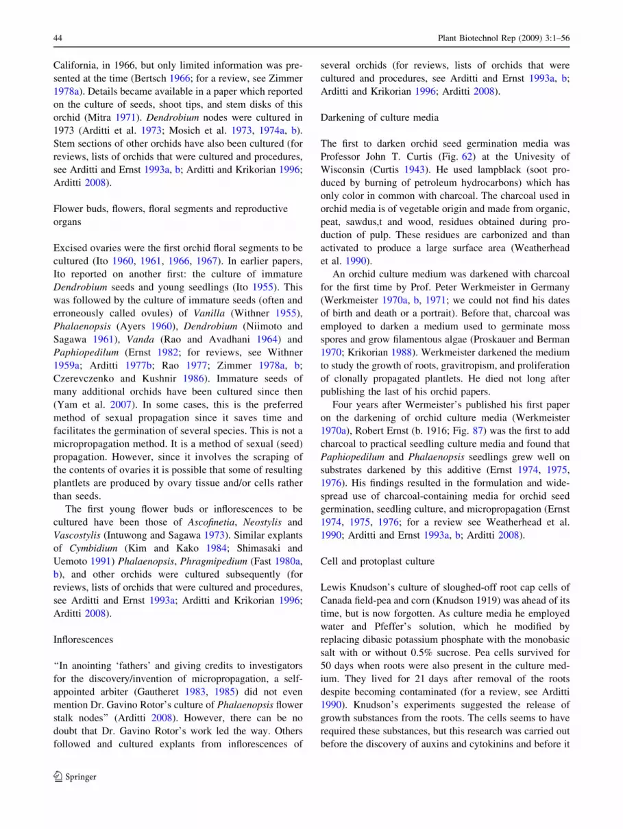
California, in 1966, but only limited information was pre-
sented at the time (Bertsch 1966; for a review, see Zimmer
1978a). Details became available in a paper which reported
on the culture of seeds, shoot tips, and stem disks of this
orchid (Mitra 1971). Dendrobium nodes were cultured in
1973 (Arditti et al. 1973; Mosich et al. 1973, 1974a, b).
Stem sections of other orchids have also been cultured (for
reviews, lists of orchids that were cultured and procedures,
see Arditti and Ernst 1993a, b; Arditti and Krikorian 1996;
Arditti 2008).
Flower buds, flowers, floral segments and reproductive
organs
Excised ovaries were the first orchid floral segments to be
cultured (Ito 1960, 1961, 1966, 1967). In earlier papers,
Ito reported on another first: the culture of immature
Dendrobium seeds and young seedlings (Ito 1955). This
was followed by the culture of immature seeds (often and
erroneously called ovules) of Vanilla (Withner 1955),
Phalaenopsis (Ayers 1960), Dendrobium (Niimoto and
Sagawa 1961), Vanda (Rao and Avadhani 1964) and
Paphiopedilum (Ernst 1982; for reviews, see Withner
1959a; Arditti 1977b; Rao 1977; Zimmer 1978a, b;
Czerevczenko and Kushnir 1986). Immature seeds of
many additional orchids have been cultured since then
(Yam et al. 2007). In some cases, this is the preferred
method of sexual propagation since it saves time and
facilitates the germination of several species. This is not a
micropropagation method. It is a method of sexual (seed)
propagation. However, since it involves the scraping of
the contents of ovaries it is possible that some of resulting
plantlets are produced by ovary tissue and/or cells rather
than seeds.
The first young flower buds or inflorescences to be
cultured have been those of Ascofinetia, Neostylis and
Vascostylis (Intuwong and Sagawa 1973). Similar explants
of Cymbidium (Kim and Kako 1984; Shimasaki and
Uemoto 1991) Phalaenopsis, Phragmipedium (Fast 1980a,
b), and other orchids were cultured subsequently (for
reviews, lists of orchids that were cultured and procedures,
see Arditti and Ernst 1993a; Arditti and Krikorian 1996;
Arditti 2008).
Inflorescences
‘‘In anointing ‘fathers’ and giving credits to investigators
for the discovery/invention of micropropagation, a self-
appointed arbiter (Gautheret 1983, 1985) did not even
mention Dr. Gavino Rotor’s culture of Phalaenopsis flower
stalk nodes’’ (Arditti 2008). However, there can be no
doubt that Dr. Gavino Rotor’s work led the way. Others
followed and cultured explants from inflorescences of
several orchids (for reviews, lists of orchids that were
cultured and procedures, see Arditti and Ernst 1993a, b;
Arditti and Krikorian 1996; Arditti 2008).
Darkening of culture media
The first to darken orchid seed germination media was
Professor John T. Curtis (Fig. 62) at the Univesity of
Wisconsin (Curtis 1943). He used lampblack (soot pro-
duced by burning of petroleum hydrocarbons) which has
only color in common with charcoal. The charcoal used in
orchid media is of vegetable origin and made from organic,
peat, sawdus,t and wood, residues obtained during pro-
duction of pulp. These residues are carbonized and than
activated to produce a large surface area (Weatherhead
et al. 1990).
An orchid culture medium was darkened with charcoal
for the first time by Prof. Peter Werkmeister in Germany
(Werkmeister 1970a, b, 1971; we could not find his dates
of birth and death or a portrait). Before that, charcoal was
employed to darken a medium used to germinate moss
spores and grow filamentous algae (Proskauer and Berman
1970; Krikorian 1988). Werkmeister darkened the medium
to study the growth of roots, gravitropism, and proliferation
of clonally propagated plantlets. He died not long after
publishing the last of his orchid papers.
Four years after Wermeister’s published his first paper
on the darkening of orchid culture media (Werkmeister
1970a), Robert Ernst (b. 1916; Fig. 87) was the first to add
charcoal to practical seedling culture media and found that
Paphiopedilum and Phalaenopsis seedlings grew well on
substrates darkened by this additive (Ernst 1974, 1975,
1976). His findings resulted in the formulation and wide-
spread use of charcoal-containing media for orchid seed
germination, seedling culture, and micropropagation (Ernst
1974, 1975, 1976; for a review see Weatherhead et al.
1990; Arditti and Ernst 1993a, b; Arditti 2008).
Cell and protoplast culture
Lewis Knudson’s culture of sloughed-off root cap cells of
Canada field-pea and corn (Knudson 1919) was ahead of its
time, but is now forgotten. As culture media he employed
water and Pfeffer’s solution, which he modified by
replacing dibasic potassium phosphate with the monobasic
salt with or without 0.5% sucrose. Pea cells survived for
50 days when roots were also present in the culture med-
ium. They lived for 21 days after removal of the roots
despite becoming contaminated (for a review, see Arditti
1990). Knudson’s experiments suggested the release of
growth substances from the roots. The cells seems to have
required these substances, but this research was carried out
before the discovery of auxins and cytokinins and before it
44 Plant Biotechnol Rep (2009) 3:1–56
123
became known that vitamins are required for the culture of
plant cells and explants. Still, Knudson can be viewed as a
pioneer in the culture of isolated plant cells.
The first isolated cells to be cultured successfully were
those of tobacco, Nicotiana tabacum, and marigold, Ta-
getes erecta (for reviews, see Muir et al. 1954; Steward and
Krikorian 1975; Krikorian 1975, 1982; Gautheret 1983,
1985). Shortly after that, isolated mesophyll cells of Ara-
chis hypogea were prompted to divide in culture and
produced what can best be described as protocorm-like
bodies or structures which look like them (Joshi and Ball
1968a, b).
Using an apparatus that rotates nipple culture flasks
slowly (1 r.p.m.) around a horizontal axis, Professor
Frederick Campion Steward (1904–1994; Fig. 88a, b),
Russell C. Mott (Fig. 88b), Marion O. Mapes (1913–1981;
Fig. 89) and Kathryn Mears (Fig. 90) obtained suspension
cultures of carrot cells and eventually regenerated plants
from them (for reviews, see Krikorian 1975, 1982, 1989;
Steward and Krikorian 1975; Gautheret 1983, 1985; Arditti
and Ernst 1993a; Arditti and Krikorian 1996). Cymbidium
cell cultures were established using the same system. Plants
were regenerated from these cells subsequently (Steward
and Mapes 1971b). Two decades later, Phalaenopsis plants
were regenerated from embryoids derived from a loose-
celled callus (Sajise and Sagawa 1990; Sajise et al. 1990)
in Professor Yoneo Sagawa’s (Fig. 91) laboratory at the
University of Hawaii. Other orchids cells have also been
cultured (for reviews, see Arditti and Ernst 1993a, b;
Arditti 2008).
The first preparation of orchid protoplasts resulted from
work with leaves (i.e., mesophyll cells) of Cymbidium
Ceres and ‘‘virus free protocorms of Cymbidium pumilum,
Brassia maculata, and Cattleya schombocattleya’’ (Cape-
sius and Meyer 1977). The protoplasts were used to isolate
nuclei but no efforts seem to have been made to produce
callus masses or regenerate plants from them. There is no
‘‘Cattleya schombocattleya.’’ Therefore what was meant
could have been ‘‘Cattleya, Schombocattleya,’’ ‘‘Cattleya
or Schombocattleya,’’ ‘‘Cattleya and Schombocattleya,’’ or
‘‘Cattleya X Schombocattleya.’’
Production of orchid protoplasts and subsequent fusion
between and within genera was first reported in 1978 (in an
orchid magazine for hobby and commercial growers rather
than a peer reviewed journal), but the ultimate fate of the
fusion products has not been described in the literature
(Teo and Neumann 1978a, b, c). As a result, there are still
unanswered questions about these reports and they have
been discounted entirely. Early isolations of orchid pro-
toplasts have been reported from several laboratories (Chen
et al. 1995; for reviews, lists of orchids that were cultured
and procedures, see Arditti and Ernst 1993a; Arditti and
Krikorian 1996; Arditti 2008).
Coda
The first plants to be propagated in vitro from seeds, ini-
tially symbiotically in joint culture with mycorrhizal fungi
and later asymbiotically on sugar containing medium, are
orchids. Their seeds are still germinated in vitro asymbi-
otically (see ‘‘Part I’’).
Orchids are also the first plants to be propagated clonally
in vitro through tissue culture methods which are now
referred to as micropropagation (Yam and Arditti 1990).
These techniques were first developed with flower stalk
nodes of Phalaenopsis orchids as early as 1949 (Rotor
1949), and shoot tips of Orchis maculata in 1954 (Thomale
1954). The culture of Cymbidium shoot tips was reported
later (Morel 1960; Wimber 1963). Of these, Rotor’s (1949)
was an original idea. The other two methods (Thomale
1954; Morel 1960; Wimber 1963) were based on the work
of others. Hans Thomale and Don Wimber credited those
whose work, methods and ideas he used from the outset.
Georges Morel did not. Tissue culture techniques are used
extensively at present to propagate orchids and many other
plants and in biotechnology in the US (Zimmerman 1996)
and elsewhere.
Other related firsts for orchids are the discovery of cell
nuclei by Robert Brown and phytoalexins by Noel Bernard
(for a review, see Arditti 1992).
Dedication
I dedicate my contribution to this historical account to
Anne Westfall, Chief of Staff in the President’s Office at
the University of Southern California (USC), because of
my son, Jonathan. Both Jonathan and I hold degrees from
USC (B.A. in 2008 and Ph.D., in 1965, respectively)
Joseph Arditti.
References
Aberg B (1961) Vitamins as growth factors in higher plants. In:
Ruhland W (ed) Handbuch der Pflanzenphysiologi XIV. Growth
and growth substances. Springer, Berlin, pp 418–449
Allenberg H (1976) Notizen zur keimung, meristemkultur and
regeneration von erdorchideen. Orchidee 27:28–31
Anderson J (1862) Orchids producing seed-pods. J Hortic Cottag
Gard Ctry Gentlem 3(NS, old series 28):46
Anderson J (1863) Orchid cultivation, cross-breeding and hybridising.
J Hortic Cottag Gard Ctry Gentlem 4(NS; old series 29):206–208
Andreasen B (1951) Carnation session. N Y State Flower Growers
Bull 66:1–3
Anonymous (1822) Orchis. Nouvo cours complet d’agriculture
theorique et pratique, Membres de la Section d’Agriculture
d’Institut de France (ed) Nouvelle edition. De Terville, Libraire
et Editeur, Paris, pp 44–47
Plant Biotechnol Rep (2009) 3:1–56 45
123
Anonymous (1850) Multiplication des orchidees de graine. J Hortic
Pratique Belgique 7:234–235
Anonymous (1853) David Moore—obituary notice. Trans Proc
Botanical Soc Edinb 14:36–39
Anonymous (1855a) Fecondation artificielle des orchidees. Illus
Hortic 2:43–44 (probably by Lemaire C)
Anonymous (1855b) Possibilite de la culture du vanilier en Europe.
Illus Hortic 2:45–46 (may be by Lemaire C)
Anonymous (1857) Note about a report by Bernard de Rennes. Le
Moniteur Universel. J Officiel de l’Empire Francais 39:154
Anonymous (1858a) Calanthe Dominii (hybrida), hybrid Calanthe.
Curtis Botanical Mag 42:5042 (probably by Lindley J)
Anonymous (1858b) Note. Gardeners’ Chron 18:4 (probably by
Lindley J)
Anonymous (1869) Note. Gardeners’ Chron 29:108
Anonymous (1885) Buds out of place. Gardeners’ Chron 25:249
Anonymous (1887) Cypripedium Arthurianum X. Gardeners Chron
2:129–130
Anonymous (1891a) John Dominy. Gardeners’ Chron 9:278
Anonymous (1891b) Un moyen de multiplier les orchidees pour
l’avenir. L’Orchidophile 11:335
Anonymous (1894) Orchid hybridisation. Orchid Rev 2:83–85
(Probably by Rolfe RA)
Anonymous (1896) Seed-vessels of orchids. Orchid Rev 4:312
(probably by Rolfe RA)
Anonymous (1906a) Orchideenpilze, ihre Bedeutung und Nutzan-
wendung. Orchis 1:33, 34, 43 (probably by Udo Dammer,
founding editor of this journal)
Anonymous (1906b) Note. Orchid Rev 14:290–291 (probably by
Rolfe RA, founding editor of this, the oldest orchid publication
in existence)
Anonymous (1920) Obituary. Joseph Charlesworth. Orchid Rev
28:137–142 (probably by Rolfe RA)
Anonymous (1921) Orchid fungus. Orchid Rev 29:17–18 (probably
by Wilson G, the second editor of this journal)
Anonymous (1922a) Orchid mycorrhiza. Orchid Rev 30:78–81
(probably by Wilson G)
Anonymous (1922b) Nonsymbiotic germination of seeds. Orchid Rev
30:135 (probably by Wilson G)
Anonymous (1922c) Charlesworth catalogue. Orchid Rev 30:76
(probably by Wilson G)
Anonymous (1924a) Charlesworth catalogue. Orchid Rev 32:248
(probably by Wilson G)
Anonymous (1924b) Note. Orchid Rev 32:193 (probably by
Wilson G)
Anonymous (1925) Les germinations d’orchidees a la societe de
pathologie vegetale. PHM Revnue Hortic 97:458
Anonymous (1951) Joseph Magrou, 1883–1951. Ann Inst Pasteur
80:327–331
Arditti J (1967) Factors affecting the germination of orchid seeds. Bot
Rev 33:1–97
Arditti J (1968) Germination and growth of orchids on banana fruit
tissue and some of its extracts. Am Orchid Soc Bull 37:112–116
Arditti J (1971) Orchids and the discovery of auxin. Am Orchid Soc
Bull 40:211–214
Arditti J (1972) Caproic acid in Satyrium flowers: Biochemical
origins of a legend. Am Orchid Soc Bull 41:298–300
Arditti J (1977a) Clonal propagation of orchids—by means of
leaf cultures in vitro: a short review. Orchid Rev (England) 85:
102–103
Arditti J (1977b) Clonal propagation of orchids by means of tissue
culture—a manual. In: Arditti J (ed) Orchid biology: reviews
and perspectives, vol I. Cornell University Press, New York,
pp 203–293
Arditti J (1979) Aspects of the physiology of orchids. Adv Bot Res
7:421–655
Arditti J (1980) John Harris–surgeon and orchidiologist. Orchid Rev
88:200–206
Arditti J (1984) An history of orchid hybridization, seed germination
and tissue culture. Bot J Linn Soc 89:359–381
Arditti J (1985) Plant pioneers. Am Sci 73:510
Arditti J (1989) History of several important research contributions by
South East Asia scientists. Malayan Orchid Rev (Singapore)
23:64–80
Arditti J (1990) Lewis Knudson (1884–1958): his science, his times
and his legacy. Lindleyana 5:1–79
Arditti J (1992) Fundamentals of orchid biology (reprinted, 2007 by
Zip Publishing, 1313 Chesapeake Ave, Columbus, Ohio 43212,
USA. http://www.zipedu.com/Arditti). Wiley, New York
Arditti J (1999) Loo Shih-wei. Plant Sci Bull 45:11–12
Arditti J (2001) Letter. Orchids 70:472
Arditti J (2004) History of orchid micropropagation: an urban legend
dispelled. In: Proceedings of the 8th Asia Pacific Orchid
Conference. Taiwan Orchid Growers Association, No. 158, Erjia
Gueiren Township, Tainan County 711, Taiwan, pp 285–292
Arditti J (2008) Micropropagation of orchids, 2nd edn. Blackwell,
Cambridge
Arditti J, Arditti M (1985) Mericloning: the 25th anniversary. Orchid
Rev 93:412–413
Arditti J, Ernst R (1984) Physiology of orchid seed germination. In:
Arditti J (ed) Orchid biology: reviews and perspectives, vol III.
Cornell University Press, New York, pp 177–222
Arditti J, Ernst R (1993a) Micropropagation of orchids. Wiley, New
York
Arditti J, Ernst R (1993b) Several early scientific investigators and
their research: a contribution towards the history of orchids in
the United States of America. In: Rittershausen W (ed) 100 years
of orchids, the orchid review centenary year book 1893–1993.
Bardfiled Books, Great Bardfield, Braintree, Essex CM7 4RZ,
UK, pp 125–142
Arditti J, Ghani AKA (2000) Numerical and physical properties of
orchid seeds and their biological implications. New Phytol
145:367–421
Arditti J, Harrison CR (1977) Vitamin requirements and metabolism
in orchids. In: Arditti J (ed) Orchid biology, reviews and
perspectives, vol 1. Cornell University Press, Ithaca, pp 157–175
Arditti J, Hew CS (2007) The origin of Vanda Miss Joaquim. In:
Cameron KM, Arditti J, Kull T (eds) Orchid biology reviews and
perspectives, vol IX. The New York Botanical Garden Press,
New York, pp 261–309
Arditti J, Krikorian AD (1996) Orchid micropropagation: the path
from laboratory to commercialization and an account of several
unappreciated investigators. Bot J Linn Soc 122:183–241
Arditti J, Strauss MS (1979) Taro tissue culture manual. Information
Document No. 44–1979. South Pacific Commission, Noumea,
New Caledonia
Arditti J, Ball EA, Churchill ME (1971) Propagacion clonal de
orquideas utilizando apices de hojas. Orquidea (Mexico)
2:290–300
Arditti J, Mosich SK, Ball EA (1973) Dendrobium node cultures:
a new means of clonal propagation. Aust Orchid Rev 38:
175–179
Arditti J, Clements MA, Fast G, Hadley G, Nishimura G, Ernst R
(1982) Orchid seed germination and seedling cutlure—a manual.
In: Arditti J (ed) Orchid biology, reviews and perspectives, vol
II. Cornell University Press, Ithaca, pp 243–370
Arditti J, Ernst R, Yam TW, Glabe C (1990) The contribution of
orchid mycorrhizal fungi to seed germination. A speculative
review. Lindleyana 5:249–255
Avadhani PN, Nair H, Arditti J, Hew CS (1994) Physiology of orchid
flowers. In: Arditti J (ed) Orchid biology, reviews and perspec-
tives, vol 6. Wiley-Interscience, New York, pp 189–362
46 Plant Biotechnol Rep (2009) 3:1–56
123
Ayers J (1960) Embryo culture of Phalaenopsis seedlings. Am Orchid
Soc Bull 29:518–519
Ball EA (1946) Development in sterile culture of stem tips and
subjacent regions of Tropaeolum majus L., and of Lupinus albusL. Am J Bot 33:301–318
Ball EA (1950) Regeneration of the shoot apex of Lupinus albus after
operation upon the central initials. Science 112:16–17
Ball EA (1972) The surface ‘‘histogen’’ of living shoot apices. In:
Miller MW, Kuehnert CC (eds) The dynamics of meristem
populations. Plenum Press, New York, pp 75–100
Ball EA, Boell EJ (1944) Respiratory rates of the shoot tips and
maturing tissues of Lupinus albus and Tropaeolum majus. Proc
Natl Acad Sci USA 30:35–50
Ball EA, Arditti J, Churchill ME (1971) Clonal propagation of
orchids from leaf tips. Orchid Rev 79:281–288
Baruch ER, Quak F (1966) Virus-free plants of Iris ‘Wedgewood’
obtained by meristem culture. Neth J Plant Pathol 72:270–273
Bauer P (1915) Zur Hybridisation und Samlingszucht de Orchideen.
Mollers Deutscher Gartner-Zeitung 30:191–192
Beaton D (1862) A yarn out of a wee pickle tow. J Hortic Cottag Gard
Ctry Gentlem 3(NS, old series 28):60–62
Beechey C (1970) Propagation of orchids from aerial roots. Am
Orchid Soc Bull 41:1085–1088
Beekman EM (2003) Rumphius’ orchids: orchid texts from the
Ambonese Herbal. Yale University Press, New Haven
Beer JG (1854) Praktische Studien an der Familie der Orchideen nbst
Kulturanweisungen und Beschreibung aller schonbluhenden
tropischen Orchideen. Carl Gerold’s Sohn, Vienna
Beer JG (1863) Beitrage zur morphologie und biologie der familie der
orchideen. Carl Gerold’s Sohn, Vienna
Bergman E (1879) Note sur les orchidee obtenues de semis en
Angleterre. J la Soc Centrale D’Hortic Fr 1:259–266
Bergman E (1881) Orchidees de semis. PHM Revnue Hortic
5:346–348
Bergman E (1882) Les orchidees de semis. J Soc Natl Hortic Fr 14:41–43,
85–93, 159–169, 211–223, 274–292, 422–446, 494–507
Bergman E (1889) Les orchidees de semis. L’Orchidophile 9:43–45,
84–86, 122–124, 137–138, 184–186, 218–219, 236–239, 265–
268, 319–323, 392–394
Bernard N (1899) Sur la germination de Neottia nidus-avis. C R Hebd
Seances Acad Sci Paris 128:1253–1255
Bernard N (1900) Sur quelques germinations difficules. Revue
Generale de Botanique 12:108–120
Bernard N (1902a) Infection et tuberisation chez les Ophrydees et la
ficaire. Rev Gen Bot 14:17–25
Bernard N (1902b) Note. Ann Soc Bot Lyon 27:4
Bernard N (1902c) Infection et tuberization chez les vegetaux. Rev
Gen Sci Pures Apliques 8:26
Bernard N (1903) Le germination des orchidees. C R Hebd Seances
Acad Sci Paris 137:483–485
Bernard N (1904a) Le champignon endophyte des orchidees. C R
Hebd Seances Acad Sci Paris 138:828–830
Bernard N (1904b) Recherches experimentales sur les orchdees. Rev
Gen Bot 16:405–470
Bernard N (1904c) Note. Bot Gaz 37:228–229
Bernard N (1905a) Maladies parasitaire et evolution des vegetaus.
Histoire des orchidees. La Revue des Idees 2:514–527
Bernard N (1905b) Nouvelles espcces d’endyphytes d’orchidees. C R
Hebd Seances Acad Sci Paris 140:1272–1275
Bernard N (1906a) On the germination of orchids. In: 3rd Interna-
tional Conference on Genetics. Royal Horticultural Society,
London, pp 292–295
Bernard N (1906b) Symbioses d’orchidees et de divers champignons
endophytes. C R Hebd Seances Acad Sci Paris 142:52–54
Bernard N (1906b) Le champignons de orchidee, leur role et leur
utilisation. Orchis 1:1, 12, 13, 18, 27–28
Bernard N (1906d) Fungus cooperation in orchid roots. Orchid Rev
14:201–203
Bernard N (1907) Titres et travaux. Valin, Caen, France
Bernard N (1908) Le Mendelisme. la Revue du Mois 7:33–53
Bernard N (1909a) L’evolution dans la symbiose, les orchidees et
leurs champignons commensaux. Ann Sci Natl Bot 9:1–196
Bernard N (1909b) Remarque sur l’immunite ches les plantes. Bull
Inst Pasteur 7:369–386
Bernard N (1911a) Un mariage en l’an 3000. La Revue du Mois
11:668–675
Bernard N (1911b) Sur la fonction fungicides des bulbes d’ophydees.
Ann Sci Natl Bot 14:221–234
Bernard ML (Mrs. No _el Bernard) (1921) Avant-Propos. In: Bernard N
(ed) Principles de Biologie Vegetale. Paris, Alcan Editeur, pp I–IX
Bernard F (1990) Memories and thoughts on my father Noel Bernard.
Lindleyana 5:55–57
Bertsch W (1966) A new frontier: orchid propagation by meristem
tissue culture. In: de Garmo LR (ed) Proceedings of 5th World
Orchid Conference, 1965. Long Beach, California, pp 225–229
Bertsch W (1967) A new frontier: orchid propagation by meristem
tissue culture. Am Orchid Soc Bull 36:32–38
Black JM (1906) Calendar of operations for April. Orchid Rev
14:114–118
Blarighem L, Magrou J, Melin E, Moreau F, Pinoy E (1985) Rayner
MC (1937) Avant-Propos in Symbiose et parasitiems: L’oeuvre
de No _el Bernard. Palais de la decouverte. Masson et Cie, Paris
(cited by Boullard)
Bleu A (1881) Semis d’orchidees. Cattleya hybrides du Cattleyaintermedia amethystina et du C. aclandiae. PHM Rev Hortic
53:305–306
Bonner JT (1937) Vitamin B1, a growth factor for higher plants.
Science 85:183–184
Borriss H, Hiibel M (1968) Die vegetative Vermehrung von
Orchideen durch Meristem-Kultur. Dtsch Gartenbau 15:22–24
Boullard B (1985) Un biologiste d’exeption, Noel Bernard 1874–
1911. University of Rouen Press, France
Brierly P (1952) Indexing the Bletsvulle mums. Flor Rev 110:19–20
Brierly P, Olson CJ (1956) Development and production of virus-free
Chrysanthemum propagative material. Plant Dis Rep (Suppl)
238:63–67
Burbery HA (1894) Calendar of operations for August. Orchid Rev
2:247–252
Burgeff H (1909) Die Wurzelpilze der Orchideen, ihre Kultur und ihr
Leben in der Pflanze. Gustav Fischer, Jena
Burgeff H (1911) Die Anzucht tropischer Orchideen aus Samen.
Gustav Fischer, Jena
Burgeff H (1932) Saprophytismus und Symbiose, Studien an
tropischer Orchideen. Gustav Fischer, Jena
Burgeff H (1936) Samenkeimung der Orchideen und Entwicklung
ihrer Keimpflanzen. Gustav Fischer, Jena
Burgeff H (1938) Mycorrhiza. In: Verdoorn F (ed) Manual of
pteridology. Martinus Nijhoff, The Netherlands, pp 159–191
Burgeff H (1943) Problematic der Mycorhiza. Die Naturwissens-
chanften 31:558–567
Burgeff H (1954) Samenkeimung und Kultur europaischer Erdor-
chideen nebst Versuchen zu ihrer Verbreitung. Gustav Fischer,
Jena
Burgeff H (1959) Mycorrhiza of orchids. In: Withner CL (ed) Orchids,
a scientific survey. Ronald Press, New York, pp 361–395
Busse W (1899) Vanille. Arbeiten aus dem Kaiserlichen
Gesundheitsamte 15:1–113
Cameron D (1844) Note on British orchidaceae. Phytologist 1:62
Cameron D (1848) On the cultivation of British orchids. J Br Hortic
Soc 3:28–33
Capesius I, Meyer Y (1977) Isolation of nuclei from protoplasts of
orchids. Cytobiologie 15:485–490
Plant Biotechnol Rep (2009) 3:1–56 47
123

Caplin SM, Stewart FC (1948) Effects of coconut milk on the growth
of explants from carrot root. Science 108:655–657
Champagnat M (1965) Particularites du developpement des apex
de Cymbidium cultives in-vitro. Bull Soc Fr Physiol Veg 11:
225–226
Champagnat M (1971) Recherches sur la multiplication vegetative de
Neottia nidus-avis Rich. Ann Sci Natl Bot Biol Veg 12:209–247
Champagnat M (1977) Culture de bourgeons et multiplication
vegetative des orchidees. In: Gautheret RJ (ed) La culture de
tissus et des cellules des vegetaux. resultats generaux et
realisations pratiques. Travaux dedies a la memoire de Georges
Morel. Masson, Paris, Masson, pp 238–253
Champagnat M, Morel G (1969) Multiplication vegetative de
Cattleya a partir de bourgeons cultivees in vitro. Mem Soc Bot
Fr 116:111–132
Champagnat M, Morel G (1972) La culture in vitro des tissus de
tubercules d’Ophrys. C R Acad Sci Paris 274:3379–3380
Champagnat M, Morel G, Chabut P, Cognet AM (1966) Recherches
morphologiqueset histologiques sur la multiplication vegetative
de quelques orchidees du genre Cymbidium. Rev Gen Bot
73:706–746
Champagnat M, Morel G, Gambade G (1968) Particularites morpho-
logiques et pourvoir de regeneration du Cymbidium virescenscultive in vitro. Mem Soc Bot Fr 115:236–244
Champagnat M, Morel G, Mounetou B (1970) La multiplication
vegetative des Cattleya a partir do jeunes feuiles cultivees
aseptiquement in vitro. Ann Sci Natl Bot Biol Veg 11:97–114
Chatin A (1856) Anatomie des plantes aeriennes de l’ordre des
orchidees. 1er Memoire: Anatomie des racines. Mem Soc Sci Nat
Cherbourg 4:5–18
Chatin A (1858) Anatomie des plantes aeriennes de l’ordre des
orchidees. 2e Memoire: Anatomie du rhizome, de la tige et des
feuilles (1). Mem Soc Sci Nat Cherbourg 6:33–69 plates I, II
Chen WH, Hsieh RM, Tsai WT, Fu YM, Chyou MS, Wu CC, Lin YS
(1995) Phalaenopsis at TSC. Am Orchid Soc Bull 64:492–495
Childers NF, Cibes HR, Hernindez-Medina E (1959) Vanilla–the
orchid of commerce. In: Withner CL (ed) The orchids, a
scientific survey. Roland Press, New York, pp 477–508
Churchill ME, Arditti J, Ball EA (1971a) Clonal propagation of
orchids from leaf tips. Am Orchid Soc Bull 42:109–113
Churchill ME, Flick BH, Ball EA, Arditti J (1971b) Vermehrung von
Orchideen durch Blattspitzen. Orchidee 22:147–151
Churchill ME, Ball EA, Arditti J (1972) Tissue culture of orchids. II.
Methods for root tips. Am Orchid Soc Bull 41:726–730
Churchill ME, Ball EA, Arditti J (1973) Tissue culture of orchids. I.
Methods for leaf tips. New Phytol 72:161–166
Clinch P (1932) Cytological studies of potato plants affected with
certain virus diseases. Sci Proc R Dublin Soc (N S) 20:57–69
Cole J (1849) Orchids from seed. Gardeners’ Chron 1:582
Costantin J (1913a) Les etapes d’une decouverte biologique. Les
hybrides d’orchidees. Rev Sci Rev Rose 51:257–264 (a more
proper date for this publication may be 1912–1913)
Costantin J (1913b) The development of orchid cultivation and its
bearing upon evolutionary ideas. Smithsonian Institution Annual
Report Part I: 345–358
Costantin J (1917) La vie des orchidees. Ernest Flammarion, Paris
Costantin J (1926) Actualites biologiques. La vie asymbiotique des
orchidees. Ann Sci Natl Bot 8:I–XVI (not 1–16 as sometimes cited)
Costantin J, Magrou J (1922a) Actualites biologiques. Applications
industrielles d’une grande decouverte Franeaise. Ann Sci Natl
Bot 4:11–111
Costantin J, Magrou J (1922b) L’elevage des orchidees. Applications
industrielles d’une grande decouverte botanique Franeaise. La
Nature 2:360–366
Curtis JT (1943) Germination and seedling development in five
species of Cypripedium. Am J Bot 30:199–206
Curtis JT, Nichol MA (1948) Culture of proliferating orchid embryos
in vitro. Bull Torrey Bot Club 75:358–373
Czerevczenko TM, Kushnir GP (1986) Orhidei v kulture. Naukova
Dumka, Kiev, Ukraine
Dangeard PA, Armand L (1887) Observations e biologie cellulaire.
Le Botaniste 8:289–313
Dangeard PA, Armand L (1898) Observations e biologie cellulaire
(mycorhizes d’Ophrys aranifera). Rev Mycol 20:13–18 plate:
CLXXXII
De Ropp RS (1945) Studies in the physiology of leaf growth. 1. The
effect of various accessory growth factors on the growth of the
first leaf of isolated stem tips of rye. Ann Bot 9:370–381
de Visiani R (1845) Del methodo e delle avvertenze che si usano
nell’orto botanico de Padova per cultura, fecondazione e
fruttificazione della vaniglia. Memorie dell’IR Istituto Veneto
di Scienze, Lettere ed Arti 2:3–18
De Wit HCD (1977) Orchids in Rumphius’ Herbarium Amboinense.
In: Arditti J (ed) Orchid biology, reviews and perspectives, vol I.
Cornell University Press, New York, pp 47–94
Delteil A (1884) La vanille, sa culture et sa preparation. Challamel
Aine, Paris
Delteil A (1902) La vanille, sa culture et sa preparation, 5th edn.
Augustine Challamel, Paris
Denis F (1914) Semis de Phalaenopsis avec emploi de champignons
endophytes. PHM Rev Hortic 86(NS 14):351
Derx HG (1936) Uit het leven van No _el Bernard 1974–1911.
Orchideeen: 242–252
Dimock AW (1943a) A method for establishing Verticilllium-free
clones of perennial plants. Phytopathology 33:3
Dimock AW (1943b) Coming up-to-date in Veticillium wilt and Septorialeaf spot of chrysanthemum. Chrysanth Soc Am Bull 11:3–10
Dimock AW (1951a) Culturing. N Y State Flower Growers Bull
66:1–2
Dimock AW (1951b) Production of Chrysanthemum propagation
material free from certain major pathogens. Plant Disease Rep
(Supplement) 238:59–62
Douglas J (1882a) Orchids from seed. Gardeners’ Chron 17:16
Domokos M (1976) Ein Beitrag zur Geschichte der Orchideenzuch-
tung. Orchidee 27:178–181
Douglas J (1882b) Orchids from seed. Gardeners’ Chron 17:82
Drude O (1873) Die Biologie von Monotropa hypopitys und Neottia
nidus avis unter verhleichender Heranziehung anderer Orchi-
deen. Preisschrift, Gottingen (not seen, cited by Burgeff, 1909)
Duhamet L (1950) Action du lait de coco sur la croissance des tissues
de crown gall de Scorsonere cultives in vitro. C R Acad Sci Paris
230:770–771
Dunn AS, Arditti J (2009) Are Orchids mentioned in the Bible? In:
Kull T, Arditti J, Wong SM (eds) Orchid biology: reviews and
perspectives, vol X. Springer, Dordrecht
Easton A (2001) Profiles in orchid history: clonal multiplication.
Orchids 70:236–239
Engman R (1984) The orchid solution. Cornell Alumni News 87:19–22
Ernst R (1967) Effect of select organic nutrient additives on growth
in vitro of Phalaenopsis seedlings. Am Orchid Soc Bull 36:
386–394
Ernst R (1974) The use of activated charcoal in asymbiotic seedling
culture of Paphiopedilum. Am Orchid Soc Bull 43:35–38
Ernst R (1975) Studies on asymbiotic culture of orchids. Am Orchid
Soc Bull 44:12–18
Ernst R (1976) Charcoal or glasswool in asymbiotic culture of
orchids. In Senghas K (ed) Proceedings of the 8th World Orchid
Conference, Frankfurt, published by the German Orchid Society,
pp 379–383
Ernst R (1982) Paphiopedilum. In: Arditti J (ed) Orchid biology:
reviews and perspectives, vol II. Cornell University Press, New
York, pp 350–353
48 Plant Biotechnol Rep (2009) 3:1–56
123
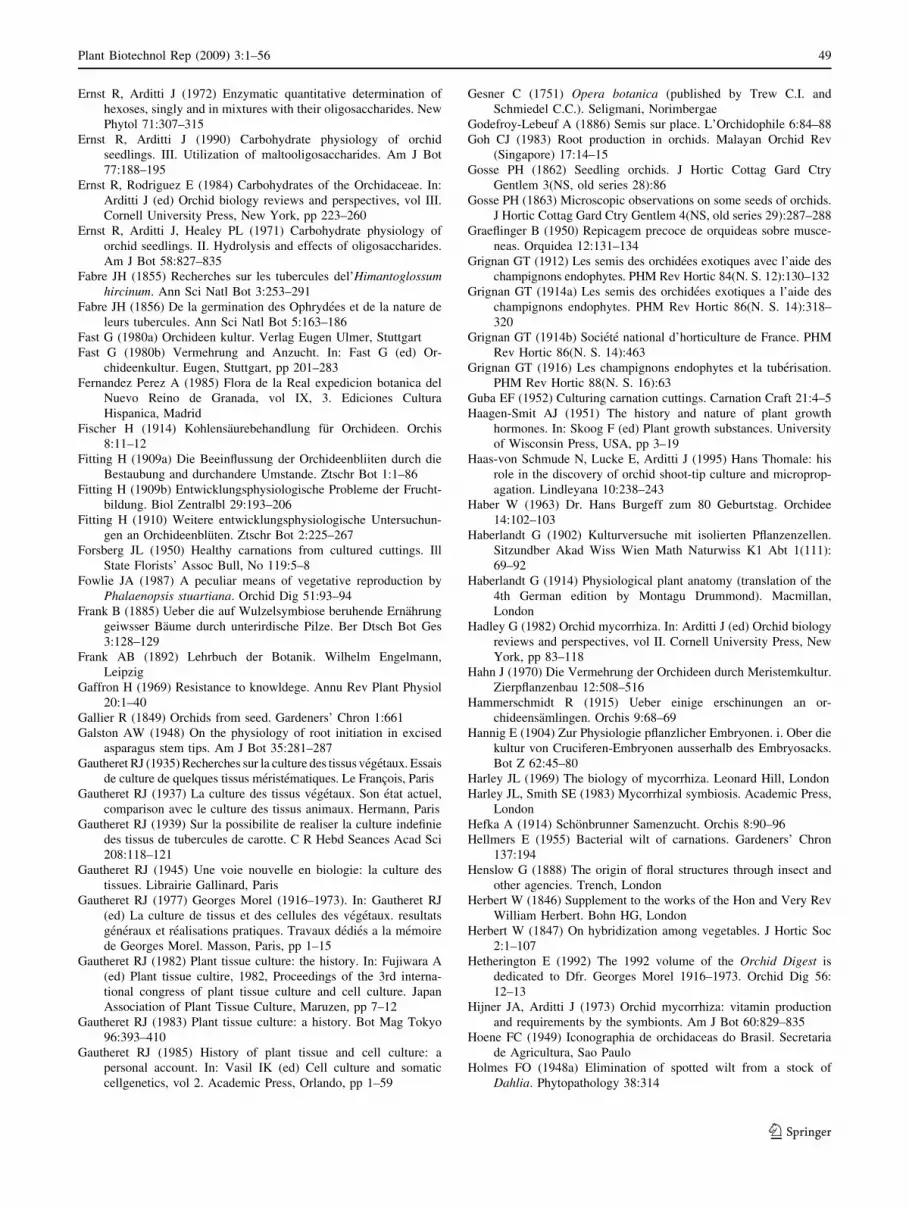
Ernst R, Arditti J (1972) Enzymatic quantitative determination of
hexoses, singly and in mixtures with their oligosaccharides. New
Phytol 71:307–315
Ernst R, Arditti J (1990) Carbohydrate physiology of orchid
seedlings. III. Utilization of maltooligosaccharides. Am J Bot
77:188–195
Ernst R, Rodriguez E (1984) Carbohydrates of the Orchidaceae. In:
Arditti J (ed) Orchid biology reviews and perspectives, vol III.
Cornell University Press, New York, pp 223–260
Ernst R, Arditti J, Healey PL (1971) Carbohydrate physiology of
orchid seedlings. II. Hydrolysis and effects of oligosaccharides.
Am J Bot 58:827–835
Fabre JH (1855) Recherches sur les tubercules del’Himantoglossumhircinum. Ann Sci Natl Bot 3:253–291
Fabre JH (1856) De la germination des Ophrydees et de la nature de
leurs tubercules. Ann Sci Natl Bot 5:163–186
Fast G (1980a) Orchideen kultur. Verlag Eugen Ulmer, Stuttgart
Fast G (1980b) Vermehrung and Anzucht. In: Fast G (ed) Or-
chideenkultur. Eugen, Stuttgart, pp 201–283
Fernandez Perez A (1985) Flora de la Real expedicion botanica del
Nuevo Reino de Granada, vol IX, 3. Ediciones Cultura
Hispanica, Madrid
Fischer H (1914) Kohlensaurebehandlung fur Orchideen. Orchis
8:11–12
Fitting H (1909a) Die Beeinflussung der Orchideenbliiten durch die
Bestaubung and durchandere Umstande. Ztschr Bot 1:1–86
Fitting H (1909b) Entwicklungsphysiologische Probleme der Frucht-
bildung. Biol Zentralbl 29:193–206
Fitting H (1910) Weitere entwicklungsphysiologische Untersuchun-
gen an Orchideenbluten. Ztschr Bot 2:225–267
Forsberg JL (1950) Healthy carnations from cultured cuttings. Ill
State Florists’ Assoc Bull, No 119:5–8
Fowlie JA (1987) A peculiar means of vegetative reproduction by
Phalaenopsis stuartiana. Orchid Dig 51:93–94
Frank B (1885) Ueber die auf Wulzelsymbiose beruhende Ernahrung
geiwsser Baume durch unterirdische Pilze. Ber Dtsch Bot Ges
3:128–129
Frank AB (1892) Lehrbuch der Botanik. Wilhelm Engelmann,
Leipzig
Gaffron H (1969) Resistance to knowldege. Annu Rev Plant Physiol
20:1–40
Gallier R (1849) Orchids from seed. Gardeners’ Chron 1:661
Galston AW (1948) On the physiology of root initiation in excised
asparagus stem tips. Am J Bot 35:281–287
Gautheret RJ (1935) Recherches sur la culture des tissus vegetaux. Essais
de culture de quelques tissus meristematiques. Le Francois, Paris
Gautheret RJ (1937) La culture des tissus vegetaux. Son etat actuel,
comparison avec le culture des tissus animaux. Hermann, Paris
Gautheret RJ (1939) Sur la possibilite de realiser la culture indefinie
des tissus de tubercules de carotte. C R Hebd Seances Acad Sci
208:118–121
Gautheret RJ (1945) Une voie nouvelle en biologie: la culture des
tissues. Librairie Gallinard, Paris
Gautheret RJ (1977) Georges Morel (1916–1973). In: Gautheret RJ
(ed) La culture de tissus et des cellules des vegetaux. resultats
generaux et realisations pratiques. Travaux dedies a la memoire
de Georges Morel. Masson, Paris, pp 1–15
Gautheret RJ (1982) Plant tissue culture: the history. In: Fujiwara A
(ed) Plant tissue cultire, 1982, Proceedings of the 3rd interna-
tional congress of plant tissue culture and cell culture. Japan
Association of Plant Tissue Culture, Maruzen, pp 7–12
Gautheret RJ (1983) Plant tissue culture: a history. Bot Mag Tokyo
96:393–410
Gautheret RJ (1985) History of plant tissue and cell culture: a
personal account. In: Vasil IK (ed) Cell culture and somatic
cellgenetics, vol 2. Academic Press, Orlando, pp 1–59
Gesner C (1751) Opera botanica (published by Trew C.I. and
Schmiedel C.C.). Seligmani, Norimbergae
Godefroy-Lebeuf A (1886) Semis sur place. L’Orchidophile 6:84–88
Goh CJ (1983) Root production in orchids. Malayan Orchid Rev
(Singapore) 17:14–15
Gosse PH (1862) Seedling orchids. J Hortic Cottag Gard Ctry
Gentlem 3(NS, old series 28):86
Gosse PH (1863) Microscopic observations on some seeds of orchids.
J Hortic Cottag Gard Ctry Gentlem 4(NS, old series 29):287–288
Graeflinger B (1950) Repicagem precoce de orquideas sobre musce-
neas. Orquidea 12:131–134
Grignan GT (1912) Les semis des orchidees exotiques avec l’aide des
champignons endophytes. PHM Rev Hortic 84(N. S. 12):130–132
Grignan GT (1914a) Les semis des orchidees exotiques a l’aide des
champignons endophytes. PHM Rev Hortic 86(N. S. 14):318–
320
Grignan GT (1914b) Societe national d’horticulture de France. PHM
Rev Hortic 86(N. S. 14):463
Grignan GT (1916) Les champignons endophytes et la tuberisation.
PHM Rev Hortic 88(N. S. 16):63
Guba EF (1952) Culturing carnation cuttings. Carnation Craft 21:4–5
Haagen-Smit AJ (1951) The history and nature of plant growth
hormones. In: Skoog F (ed) Plant growth substances. University
of Wisconsin Press, USA, pp 3–19
Haas-von Schmude N, Lucke E, Arditti J (1995) Hans Thomale: his
role in the discovery of orchid shoot-tip culture and microprop-
agation. Lindleyana 10:238–243
Haber W (1963) Dr. Hans Burgeff zum 80 Geburtstag. Orchidee
14:102–103
Haberlandt G (1902) Kulturversuche mit isolierten Pflanzenzellen.
Sitzundber Akad Wiss Wien Math Naturwiss K1 Abt 1(111):
69–92
Haberlandt G (1914) Physiological plant anatomy (translation of the
4th German edition by Montagu Drummond). Macmillan,
London
Hadley G (1982) Orchid mycorrhiza. In: Arditti J (ed) Orchid biology
reviews and perspectives, vol II. Cornell University Press, New
York, pp 83–118
Hahn J (1970) Die Vermehrung der Orchideen durch Meristemkultur.
Zierpflanzenbau 12:508–516
Hammerschmidt R (1915) Ueber einige erschinungen an or-
chideensamlingen. Orchis 9:68–69
Hannig E (1904) Zur Physiologie pflanzlicher Embryonen. i. Ober die
kultur von Cruciferen-Embryonen ausserhalb des Embryosacks.
Bot Z 62:45–80
Harley JL (1969) The biology of mycorrhiza. Leonard Hill, London
Harley JL, Smith SE (1983) Mycorrhizal symbiosis. Academic Press,
London
Hefka A (1914) Schonbrunner Samenzucht. Orchis 8:90–96
Hellmers E (1955) Bacterial wilt of carnations. Gardeners’ Chron
137:194
Henslow G (1888) The origin of floral structures through insect and
other agencies. Trench, London
Herbert W (1846) Supplement to the works of the Hon and Very Rev
William Herbert. Bohn HG, London
Herbert W (1847) On hybridization among vegetables. J Hortic Soc
2:1–107
Hetherington E (1992) The 1992 volume of the Orchid Digest is
dedicated to Dfr. Georges Morel 1916–1973. Orchid Dig 56:
12–13
Hijner JA, Arditti J (1973) Orchid mycorrhiza: vitamin production
and requirements by the symbionts. Am J Bot 60:829–835
Hoene FC (1949) Iconographia de orchidaceas do Brasil. Secretaria
de Agricultura, Sao Paulo
Holmes FO (1948a) Elimination of spotted wilt from a stock of
Dahlia. Phytopathology 38:314
Plant Biotechnol Rep (2009) 3:1–56 49
123

Holmes FO (1948b) Elimination of spotted wilt from dahlias by
propagation of stem cuttings. Phytopathology 45:224–226
Holmes FO (1955) Elimination of spotted wilt from dahlias by
propagation of tip cuttings. Phytopathology 45:224–226
Holmes FO (1956a) Elimination of foliage spotting from sweetpotato.
Phytopathology 46:502–504
Holmes FO (1956b) Elimination of aspermy virus from the nightin-
gale chrysanthemum. Phytopathology 46:599–600
Hunault G (1979) Recherches sur le comportement des fragments
d’organes et des tissus des Monocotyledones cultives in vitro.V.
Discussion. Revue de Cytologie et de Biologie Begetales Le
Botaniste 2:259–287
Ignotus (1894) L’Arrosage des semis. J Orchid 5:233–235
Intuwong O, Sagawa Y (1973) Clonal propagation of Sarcanthinae
orchids by aseptic culture of inflorescences. Am Orchid Soc Bull
42:209–215
Irmisch T (1842) Bemerkungen uber die Epipactisarten der deutschen
Flora. Linnaea 16:417–462
Irmisch T (1854a) Einige Beobachtungen an einheimischen Orchi-
deen. Flora 37:513–525
Irmisch T (1854b) Bemerkungen uber Malaxis paludosa Sw. Flora
37:625–627
Irmisch T (1863) Ein kleiner Beitrag zur Naturgeschichte der
Microstylis monophylla. Flora 46:1–8
Irmisch T (1877) Einige Bemerkungen uber Neottia Nidus-avis und
einige andere Orchideen. Abh Natuwiss Ver Bremen 5:503–509
Irmish T (1853) Beitrage zur biologie und morphologie der orchideen.
Amb Abel, Leipzig
Ito I (1955) Germination of seeds from immature pod and subsequent
growth of seedlings in Dendrobium nobile Lndl. Sci Rep Saikyo
Univ Agric 7:35–42
Ito I (1960) Culture of orchid seedlings by way of completing the
growth of cut flowers. Jpn Orchid Soc Bull 6:4–7
Ito I (1961) In vitro culture of ovary and seed in orchids. Olericulture
Lab., Fac Agric Kyoto Prefectual University, Kyoto
Ito I (1966) In vitro culture of ovary in orchids (I) Effects of sugar,
peptone, and coconut milk upon the growth of ovary of Dendr-obium nobile. Sci Rep Kyoto Prefectual Univ Agric 18:38–50
Ito I (1967) In vitro culture of ovary in orchids (II) Influence of
vitamins upon the growth of ovary of Dendrobium nobile and
other several problems. Sci Rep Kyoto Prefectual Univ Agric
19:6–12
Jacquet P (1994) History of orchids in Europe, from antiquity to the
17th century. In: Arditti J (ed) Orchid biology reviews and
perspectives, vol VI. Wiley-Interscience, New York, pp 33–102
Jacquet P (2007) A translation of the writings of Noel Bernard. In:
Cameron KM, Arditti J, Kull T (eds) Orchid biology reviews and
perspectives, vol IX. The New York Botanical Garden Press,
New York, pp 311–431
Jancke H (1907) Anzucht der Orchdeen aus Samen. Orchis 2:19–20
30–32
Jancke H (1915) Wie wird am besten Cattleya- und Laelia-Samen
aufgehoben, und wie lange halt er sich keimfahig unter gunstigen
Bedingungen. Orchis 9:3–4
Janick J (1989) Classic papers in horticultural science. Prentice-Hall,
New Jersey
Jaquiot J (1951) Action du meso-inositol et de l’adenine sur la
formation de bourgeons par le tıssu cambial d’ulmus campestriscultive in vitro. C R Acad Sci Paris 233:815–817
Jenning S (1875) Orchids and how to grow them in India and other
tropical climates. Reeve, London
Joshi PC, Ball EA (1968a) Growth of isolated palisade cells of
Arachis hypogea. Dev Biol 17:308–325
Joshi PC, Ball EA (1968b) Growth values and regenerative poten-
tiality in mesophyll cultures of Arachis hypogea. Z Pflanzenphysiol
9:5–75
Kassanis B (1967) Plant tissue culture. In: Maramorsch K, Koprowski
H (eds) Methods in virology, vol 1. Academic Press, New York,
pp 537–566
Kikuta K, Parris GK (1941) Propagating taro by the normally dormant
buds present onhull and corms. Hawaii Agric Exp Stn Prog
Notes 21:1–9
Kim KW, Kako S (1984) Studies on clonal propagation in the
Cymbidium floral organ culture in vitro. J Korean Soc Hort Sci
25:65–71
Knapp E (1978) Hans Burgeff, 1883-1976. Ber Dtsch Bot Ges
91:261–273
Knop W (1884) Bereitung einer concentrierten Nahrstofflosung fur
Pflanzen. Landwirtsch Vers Stn 30:292–294
Knudson L (1913a) Tannic acid fermentation. Int J Biol Chem
14:159–184
Knudson L (1913b) Tannic acid fermentation. Int J Biol Chem
14:1185–1202
Knudson L (1915) Toxicity of galactose for certain of the higher
plants. Ann M Bot Gard 2:659–666
Knudson L (1916) Influence of certain carbohydrates on green plants.
Cornell Univ Agric Exp Sta Mem 9:1–75
Knudson L (1917) The toxicity of galactose and mannose for green
plants and the antagonistic action of other sugars toward these.
Am J Bot 4:430–437
Knudson L (1919) Viability of detached root cap cells. Am J Bot
6:309–310
Knudson L (1921) La germinacion no simbiotica de las semillas de
orquideas. Bol Real Soc Espanola Hist Nat 21:250–260
Knudson L (1922a) Nonsymbiotic germination of orchid seeds. Bot
Gaz 73:1–25
Knudson L (1922b) Raising seedlings on sugar. Gard Mag 36:162–163
Knudson L (1924) Further observations on nonsymbiotic germination
of orchid seeds. Bot Gaz 77:212–219
Knudson L (1925) Physiological study of the symbiotic germination
of orchid seeds. Bot Gaz 79:345–379
Knudson L (1927) Symbiosis and asymbiosis relative to orchids. New
Phytol 26:328–336
Knudson L (1928) Is the fungus necessary for development of
seedlings of Calluna vulgaris? Am J Bot 15:624–625
Knudson L (1929a) Physiological investigations on orchid seed
germination. In: Duggar BM (ed) Proceedings of the Interna-
tional Congress of Plant Sciences. Ithaca New York, 16–23
August, 1926. Gearge Banta, Wisconsin, pp 1183–1189
Knudson L (1929b) Seed germination and growth in Calluna vulgaris.
New Phytol 28:369–376
Knudson L (1930) Flower production by orchid grown non-symbi-
otically. Bot Gaz 89:192–199
Knudson L (1932) Non-symbiotic germination of Calluna vulgaris(heather). 45th Annual Report Cornell University Agricultural
Experiment Station, p 111
Knudson L (1933a) Investigations on the use of certain amino acids
by green plants. Am J Bot 20:680
Knudson L (1933b) Effects of X-rays on fern prothalii. Am J Bot
20:680–681
Knudson L (1933c) Non-symbiotic development of seedlings of
Calluna vulgaris. New Phytol 32:115–127
Knudson L (1934a) Non-symbiotic germination of orchid seed. Am J
Bot 21:712
Knudson L (1934b) Giant plastids in ferns produced from X-rayed
spores. Am J Bot 21:712–713
Knudson L (1934c) Storage and viability of orchid seed. Am Orchid
Soc Bull 2:66
Knudson L (1934d) Orchids without a greenhouse. Am Orchid Soc
Bull 2:68
Knudson L (1935) Germination of orchid seed. Am Orchid Soc Bull
3:65–66
50 Plant Biotechnol Rep (2009) 3:1–56
123

Knudson L (1936) Has the chloroplast a semipermeable membrane?
Am J Bot 23:694
Knudson L (1940a) Viability of orchid seed. Am Orchid Soc Bull
9:36–38
Knudson L (1940b) Permanent changes of chloroplasts induced by X
rays in the gametophyte of polypodium aureum. Bot Gaz
101:712–758
Knudson L (1941a) Germination of Goodyera pubescens. Am Orchid
Soc Bull 10:199–201
Knudson L (1941b) Investigations on delayed photosynthesis in
chlorophyll-bearing embryos of orchids, and other studies on
orchid seed. N Y State Agric Exp Stn Ann Rep 54:117–118
Knudson L (1941c) The influence of X-rays on the growth and
metabolic processes in ferns. N Y State Agric Exp Stn Ann Rep
54:118
Knudson L (1946a) Exhibit of Vanilla germination and other orchid
seedlings. Am Orchid Soc Bull 15: 210–213, 238–239
Knudson L (1946b) A new nutrient solution for the germination of
orchid seed. Am Orchid Soc Bull 15:214–217
Knudson L (1947) Orchid seed germination by the use of fungicidal
or fungistatic agents. Am Orchid Soc Bull 16:443–445
Knudson L (1948) Chlorox and calcium hypochlorite as disinfectants
for orchid seed. Am Orchid Soc Bull 17:348–353
Knudson L (1950) Germination of seeds of Vanilla. Am J Bot
37:241–247
Knudson L (1951) Nutrient solutions for orchids. Bot Gaz 112:528–
532
Knudson L (1952) Nutrient solutions for orchid seed germination. Am
Orchid Soc Bull 21:94–97
Knudson L (1989) Non symbiotic germination of orchid seed. Reprint
of the original article from Bot Gaz 73:1–25 (1922) with
introductory essay by Arditti J. In: Janick J (ed) Classic papers in
horticultural science. Prentice-Hall, New Jersey, pp 14–39
Knudson L, Clark DG (1930) Relation between time of exposure of
orchid seed to sugar, and seedling development. Am J Bot
17:1049
Knudson L, Ginsburg S (1921) Suggestions with respect to the
measurement of osmotic pressure. Am J Bot 3:164–170
Knudson L, Lindstrom EW (1919) Influence of sugars on the growth
of albino plants. Am J Bot 6:401–405
Knudson L, Smith RS (1919) Secretion of amylase by plant roots. Bot
Gaz 68:460–466
Koda Y, Okazawa Y (1980) Cytokinin production by Asparagusshoot apex cultured in vitro. Physiol Plant 48:193–197
Kotte W (1922a) Wurzelmeristem in Gewebekultur. Ber Dtsch Bot
Ges 40:269–272
Kotte W (1922b) Kulturversuche mit isolierten Wurzelspitzen. Beitr
Allg Bot 2:413–434
Krikorian AD (1975) Excerpts from the history of plant physiology
and development. In: Historical and current aspects of plant
physiology, a symposium honoring Steward FC, College of
Agriculture and Life Science, Cornell University, Ithaca, NY,
pp 9–97
Krikorian AD (1982) Cloning higher plants from aseptically cultured
tissues and cells. Biol Rev 57:151–218
Krikorian AD (1988) Plant tissue culture: Perceptions and realities.
Proc Indian Acad Sci (Plant Sci) 98:425–464
Krikorian AD (1989) Introductory essay for reprinting of: Stewart FC,
Mapes MO, Mears K (1958) Growth and organized development
of cultured cells. II. Organization in cultures grown from freely
suspended cells. Am J Bot 45:705–708. In: Janick J (ed) Classic
papers in horticultural science. Prentice-Hall, Englewood Cliffs,
pp 40–51
Krikorian AD (1994) In vitro culture of root and tuber crops. In: Vasil
IK, Thorpe T (eds) Plant tissue culture. Kluwer, Dordrecht,
pp 379–411
Krikorian AD (1995) Hormones in tissue culture and micropropaga-
tion. In: Davies P (ed) Plant hormones, physiology, biochemistry
and molecular biology. Kluwer, Dordrecht, pp 774–796
Krikorian AD (2005) Totipotency and proof of the concept: An
historical perspective. In: Murch SJ, Saxena PK (eds) Journey of
a single cell to a plant. Science Publishers, Enfield, pp 1–33
Krikorian AD, Berquam DL (1969) Plant cell and tissue cultures: the
role of Haberlandt. Bot Rev 35:59–88
Kukułczanka K, Sarosiek J (1971) Merystematyczne kultury stor-
czykow. Wiad Bot 15:29–40
L L (1892) Note. J Orchid 3:343 (probably by Lucien Linden)
L L (1893) Apropos d’hybridation. J Orchid 4:52 (probably by Lucien
Linden)
L L (1894) Hybridation des Orchidees. 1. Historique. J Orchid 5:173–
176 (probably by Lucien Linden)
La Rue CD (1936) Tissue culture of spermatophytes. Proc Natl Acad
Sci USA 22:201–209
Laibach F (1930) Untersuchungen Uber die Postfloration tropischer
Orchideen. Planta 9:341–387
Laibach F (1932) Pollenhormon and Wuchsstoff. Ber Dtsch Bot Ges
50:383–390
Laibach F (1933a) Wuchsstoffversuche mit lebenden Orchideenpol-
linien. Ber Dtsch Bot Ges 51:336–340
Laibach F (1933b) Versuche wit Wuchsstoffpaste. Ber Dtsch Bat Ges
51:386–392
Laibach F, Maschmann E (1933) Uber den Wuchstoff der Orchi-
deenpollinien. Jahrb Wiss Bot 78:399–430
Laimer M, Ucker W (2003) Plant tissue culture, 100 years since
Gottlieb Haberlandt. Springer, Vienna
Langhans RW, Horst RK, Earle ED (1977) Disease-free plants via
tissue culture propagation. Hortic Sci 121:149–150
Lashley L, Arditti J (1982) A partial history of the term orchid.
Orchid Rev (England) 90:189–193
Lawler LJ (1984) Ethnobotany of the Orchidaceae. In: Arditti J (ed)
Orchid biology reviews and perspectives, vol III. New York,
Cornell University Pressm, pp 27–149
Le Dantec F (1911) No _el Bernard. La Grande revue 385–398
Lecoufle M (1967) A mass blooming of mericlones. Am Orchid Soc
Bull 36:405–406
Lecoufle M (1971) The impact of meristem propagation of orchids.
In: Proc 6th World Orchid Conference. Sydney, Australia
(1969), pp 107–110
Lecoufle M (1974a) Georges Morel. L’Orchidophile 15:294–297
Lecoufle M (1974b) Georges Morel. Orchidee 25:29–31
Legros GV (1971) Fabre, poet of science. Horizon Press, New York
Leitgeb H (1864a) On globular cell-thickenings in the envelope of the
roots of some orchids. Ann Mag Nat Hist 14:80
Leitgeb H (1864b) On the a _erial roots of the Orchidaceae. Ann Mag
Nat Hist 14:159–160
Leitgeb H (1864c) Uber kugelformige Zelleverdickungen in der
Wurzelhulle einiger Orchideen. Sitzungsber Wien Akad Wiss
49:275–276
Leitgeb H (1865) Die Luftwurzeln der Orchideen. Denkschr Math
Naturwiss Class Kaiserliche Akad Wiss Wien 24:179–222
Leopold AC (1964) Plant growth and development. McGraw-Hill,
New York
Limasset P, Cornuet P (1949) Recherche du virus de la mosaique du
Tabac dans les meristemes des plantes infetees. C R Acad Sci
Paris 228:1971–1972
Link HF (1824) Elementa philosophiae botanicae. Haude and Spener,
Berlin
Link HF (1839–1842) Icones selectae anatomico-botanicae. Haude
and Spener, Berlin
Link HF (1840) Icones selectae anatomico-botanicae, Fasc. II.
Ausgewahlte Anatomisch botanische Abildungen, vol 2. Lude-
ritz, Berlin
Plant Biotechnol Rep (2009) 3:1–56 51
123

Link HF (1849a) Bemerkungen uber den Bau der Orchideen,
besonders der Vandeen. Bot Z 7:745–750
Link HF (1849b) Bemerkungen uber den Bau der Orchideen, Erste
Abhandlug. Physikalische Abhandlungen der Koniglichen Akade-
mie der Wissenschaften zur Berlin Aus dem Jahre 37:103–117
Loeb J (1917) Influence of the leaf upon root formation and geotropic
curvature in the stem of Eryophyllum calycinum and the
possibility of a hormone theory of these processes. Bot Gaz
63:25–50
Loo SW (1945a) Cultivation of excised stem tips of intact plants
under sterile conditions. PhD Dissertation, California Institute of
Technology, Pasadena
Loo SW (1945b) Cultivation of excised stem tips of Asparagus in
vitro. Am J Bot 32:13–17
Loo SW (1946a) Further experiments on the culture of Asparagusstem tips in vitro. Am J Bot 33:156–159
Loo SW (1946b) Cultivation of excised stem tips of dodder in vitro.
Am J Bot 33:295–300
Loo SW (1946c) Preliminary experiments on the cultivation Baeriachrysostoma under sterile conditions. Am J Bot 33:382–389
Loo SW (1978) Plant tissue and cell culture. Recent advances and
applications. Acta Phytophysiol Sinica 4:91–112
Lucke E (1974) Tendenzen der Orchideenvermehrung. Erwerbstgart-
ner 19:729–732
Lucke E (1994) Hans Thomale zum 75 Geburtstag. Orchidee 45:258–
259
MacDougal DT (1898) The mycorrhizae of Aplectrum. Bull Torrey
Bot Club 25:110–112
MacDougal DT (1899a) Symbiotic saprophytism. Ann Bot 13:1–47
MacDougal DT (1899b) Symbiosis and saprophytism. Bo Gaz
28:220–222
MacDougal DT (1944) Mycorrhizal symbiosis in Aplectrum, Coral-lorhiza and Pinus. Plant Physiol 19:440–465
Magnus W (1900) Studien an der endotropen Mycorrhiza von Neottianidus avis. Jahrb Wiss Bot 35:206–272
Magrou J (1925) Role des champignons endophytes dans la culture
des orchidees. Rev Pathol Veg Entomol Agric 12:180–184
Magrou J (1937a) Les orchidees et l’oevre de No _el Bernard. In:
Symbiose et Parasitisme/L’oevre de No _el Bernard. Paris,
Masson Editeur, pp 10–36
Magrou J (1937b) Sur la culture des champignons de mycorrhizes.
Ann Sci Natl Bot 19:359–370
Magrou J, Magrou M (1935) Sur les variations d’activite des
rhizoctones d’orchidees. Ann Sci Natl Bot 15:303–305
Manhardt F (1906) Die Hybridisation der Orchideen und die Anzucht
aus Samen. Orchis 1:62–64
Mapes MO, Cable WJ (1972) Mericloning of taro. (Colocasiaesculenta). Hawaii Agricultural Experiment Station Journal No
1694
Mariat F (1951) Influence du lait du coco et du coprah sur le
developement de jeunes plantules de Cattleya. Bull Soc Bot Fr
98:260–263
Mariat F, Segretain G (1952) L’oevre scientifique de Joseph Magrou.
Memoires de la Societe Bot de Fr (Reprint with no volume
number provided by Professor F. Mariat, Institut Pasteur), pp
177–181
Maron C (1898) Laelio-Cattleya radiata et Laelia elegans de semis.
PHM Rev Hortic 39:335
Marston ME, Vourairai P (1967) Multiplication of orchid clones by
shoot meristem culture. A review of the literature. UnivNottingham, Dep Hortic Misc Pulication 17:1–8
Maschmann E, Laibach F (1932) Uber Wuchsstoffe. Biochem Z
255:446–452
Mayer L (1956) Wachstum and Organbildung an in vitro kultivierten
Segmenten von Pelargonium zonale and Cyclamen persicum.
Planta 47:401–446
Miller CO (1961) Kinetin and related compounds in plant growth.
Annu Rev Plant Physiol 12:395–408
Miller CO (1967) Zeatin and zeatin riboside from a mycorrhizal
fungus. Science 157:1055–1057
Miller C, Skoog F, Okumura FS, von Saltza MH, Strong FM (1955a)
Structure and synthesis of kinetin. J Am Chem Soc 77:2662–
2663
Miller C, Skoog F, von Saltza MH, Strong FM (1955b) Kinetin, a cell
division factor from desoxyribonucleic acid. J Am Chem Soc
77:1392
Mitra GC (1971) Studies of seeds, shoot–tips & stem–disks of an
orchid grown in aseptic culture. Indian J Exp Biol 9:79–85
MN (1845) Recherches et reflexions sur l’hybridation ches les acotyle-
dones et les monocotyledones. Ann Soc R Agric Bot Gand Sci
Acces 1:323–332
MN (1849) Fruits de la serre. Ann Soc R Agric Bot Gand Sci Acces
5:13–14
Mollberg A (1884) Untersuchungen uber die Pilze in den Wurzeln der
Orchideen. Jena Z Medizin Naturwiss 17:519–535
Moore D (1834) An account of a new Catasetum from Brazil, with
observations on the importation of and treatment of ‘‘orchida-
ceous plants’’. Irish Farmer’s Gardener’s Mag 1:390–392
Moore D (1849) On growing orchids from seeds. Garden Chron
35:549
Moore D (1875) Design in the structure and fertilization of plants. In:
Johnson W (ed) Science and revelation: a series of lectures in
reply to the theories of Tyndall. Huxley, Darwin, Spence, etc.
Scribner, New York, pp 3–32
Moreau F (1958) Sur une botaniste aux affinites normandes No _el
Bernard (1874–1911). Mem Acad Sci Arts Belles Arts Caen
14:103
Morel G (1948) Recherches sur la culture associee de parasites
obligatories et de tissus vegetaux. Ann Epiphyt (NS) 14:123–124
Morel G (1950) Sur la culture des tissus de deux monocotyledones. C
R Acad Sci Paris 230:1099–1101
Morel G (1960) Producing virus–free cymbidiums. Am Orchid Soc
Bull 29:495–497
Morel G (1963) La culture in vitro du meristeme apical de certaines
orchidees. C R Acad Sci Paris 256:4955–4957
Morel G (1964a) Tissue culture-a new means of clonal propagation of
orchids. Am Orchid Soc Bull 33:473–478
Morel G (1964b) La culture in vitro du meristeme apical. Rev Cytol
Biol Veg 27:304–314
Morel G (1965a) Clonal propagation of orchids by meristem culture.
Cymbidium Soc News 20:3–11
Morel G (1965b) Eine neue Methode erbgleicher Vermehrung: Die
Kultur von Triebspitzen–Meristemen. Orchidee 16:165–176
Morel G (1966) Meristem culture clonal propagation of orchids.
Orchid Dig 30:45–49
Morel G (1970) Neues ouf dem Gebiet der Meristem–Forschung.
Orchidee 20:433–443
Morel G (1971a) Experiments on Pleione and Cypripedium propa-
gation. In: Proceedings of 4th International Rock Garden
Conerence, Harrogate, UK, pp 72–77
Morel G (1971b) Les Pleiones-culture-multiplication. Bulletino di
Informazione di Gardino Sperimentale di Acclimatazione per
Piante Alpine ed Erbacee Perenni di San Bomalino Distranag
4:9–13
Morel G (1971c) The principles of clonal propagation of orchids. In:
Crrigan (ed) Proceedings of the 6th World Orchid Conference
(1969), Sydney, pp 101–106
Morel G (1974) Clonal multiplication of orchids. In: Withner CL (ed)
The orchids: scientific studies. Wiley-Interscience, New York,
pp 169–222
Morel G, Martin S (1952) Guerison de dahlias atteints d’une maladie
a virus. C R Acad Sci Paris 235:1324–1325
52 Plant Biotechnol Rep (2009) 3:1–56
123

Morel G, Martin S (1955a) Guerison de pommes de terre atteintes de
maladies a virus. C R Acad Agric Fr 119:877–878
Morel G, Martin S (1955b) Guerison de plantes atteintes de maladies
a virus par culture de meristemes apicaux. Report of the XIV
International Horticulture Congress, The Hague, Netherlands,
vol 1, pp 303–310. Veenman & Zonern, Wageningen
Morel G, Muller JF (1964) Le culture in vitro du meristeme apical de
la pomme de terre. C R Acad Sci Paris 258:5250–5252
Morel G, Wetmore R (1951a) Tissue culture of monocotyledons. Am
J Bot 38:138–140
Morel G, Wetmore R (1951b) Fern callus tissue culture. Am J Bot
38:141–143
Morren C (1829a) Verhandeling over de ware wijze, waarop de
voortgang der standelkruiden (orchides) met tweeknollige wor-
tels plaats heeft, en bepaling van den natuurlijken vorm, dien zij
zouden aannemen, Indien zij niet aan eene standvastige misw-
assing (avortment) onderworpen waren. Beidragen tot de
Natuurkundige Wetenschappen 4:358–384
Morren C (1829b) Queritur orchidis latifoliae descriptio botanica et
anatomica. In: Goesin PF (ed) Belgium, Gand
Morren C (1837) Note sur la premicre fructification due vanillier en
Europe. Ann Soc R Hortic Paris 20:331–334
Morren C (1838a) Notice sur la vanille indigene, dont il a reussi a
produire la fructification. Bull Cl Lett Sci Morales Polit 4:225–237
Morren C (1838b) Sur la fructification de la vanille obtenue au moyen
de la fecondation artificielle. C R Hebd Seances Acad Sci Paris
6:489–492
Morren C (1838–1839) Fructification de la Vanille. J Agric Pratique
Ser I 2:114–116
Morren C (1839a) On the production of vanilla in Europe. Ann Nat
Hist 3:1–9
Morren C (1839b) Note sur les fruits aromatiques de Leptotes bicolor.
Bull Cl Lett Sci Morales Polit 6:582
Morren C (1850) Memorandum sur la vanille, son histoire et sa
culture. Bull Cl Lett Sci Morales Polit 17:108–133
Morren E (1852) Histoire de la vanille. La Belgique Hortic. J Jardins
Serres Vergers 6:313–374
Morren E (1860) Charles de l’Esclise, sa vie et ses œuvres, 2nd ed
edn. Annoot-Braeckman, Belgium, Gand
Mosich SK, Ball EA, Arditti J (1973) Propagation clonal de
Denrobium por mediodel cultivo de nodos. Orquidea (Mexico)
3:244–260
Mosich SK, Ball EA, Arditti J (1974a) Clonal propagation of orchids
by means of node cultures. Am Orchid Soc Bull 43:1005–1061
Mosich SK, Ball EA, Flick BH, Arditti J (1974b) Klonvermehrung
von Dendrobium durch die Kultivierung von Stammnodien.
Orchidee 25:129–134
Muir WH, Hildebrandt AC, Ricker AJ (1954) Plant tissue cultures
produced from single isolated plant cells. Science 119:877–878
Mullerott M (1980) Thilo Irmisch 1816–1879. Ein Bibliographischer
versuch nebst proben seines wissenschaftlichen Briefwechsels.
Hoppea, Denk der Regensburger Botanischen Gesellschaft
39:51–76
Murashige T (1978) Plant tissue culture: History, current status and
prospects. Tissue Cultire Assoc Rep 12:41–47
Murashige T, Skoog F (1962) A revised medium for rapid growth and
bioassays withtobacco tissue cultures. Physiol Plant 15:473–497
Naudin C (1849) Peut-on multiplier de graines des orchidees? PHM
Rev Hortic 3:582–584
Naudin C (1850) Nouvelles experiences sur les semis d’orchidees.
PHM Rev Hortic 4:9–12
Naudin C (1865) Nore sur hybridism in vegetables. Nat Hist Rev
5:567–573
Naumburg DJS (1794) Ehescheidung oder Auswanderung der Mannervon ihren Weibern im Pflanzenreiche. Annalen der Botanik
(Zurich) Nuentes Stuck 12–32
Neil J (1880) Rays from the realms of nature; or, parables of plant
life. Cassell, Petter, Galpin, London
Nelson EC (1981) Two centuries of orchids at Glasnevin. Orchid Rev
89(136–140):191–194
Nelson EC, Seaward MRD (1981) Charles Darwin’s correspondence
with David Moore of Glasnevin on insectivorous plants and
potatoes. Biol J Linn Soc Lond 15:157–164
Neumann J (1838) Vanille. Ann Flore et Pomone ou J Jardins Champs
6:316–320
Neumann J (1841a) Vanila a feuilles planes, Vanilla planifolia.
Herbier Generale de L’Amateur 2:1–5
Neumann J (1841b) Vanila a feuilles planes, Vanilla planifolia.
L’Horticulteur Universel Ser 2 2:169–175
Neumann J (1844). Nota. PHM Revnue Hortic 6:38
Niimoto DH, Sagawa Y (1961) Ovule development in Dendrobium.
Am Orchid Soc Bull 30:813–819
Noisette LC (1826) Manuel complet du jardiniere, vol 3. Rouscelon,
Librarie-Editeur, Paris, pp 270–296
North C (1953) Experiments with root cuttings of Brussels sprouts.
Ann Appl Bot 40:250–261
Ojima K, Fujiwara A (1962) Studies on the growth promoting
substance of the excised wheat root. III. Effects of tryptophan
and some related substances. Tohoku J Agri Res 13:69–98
Orchids Orlando (1968) Catalog. 1717 Edgewater Drive, Orlando, FL
32804 (no date, but post 1968)
Pages P (1971) Banana homogenate, coconut water, peptone and auxins
as nutrient supplements in the in vitro culture of Dendrobium and
Phalaenopsis ovules. SEARCA Bull No. 2. South East Asia
Regional Center for Graduate Study and Research in Agriculture.
College, Laguna, Philippines
Perez C (1911) No _el Bernard. la Revue du Mois 11:641–657
Perez C (1912) No _el Bernard. Annuare de L’Association Amicale de
Secours des Anciens Elcves de l’Ecole Normale Superieure.
Reprint with no volume number: 3–7 (provided by Professor
Franeois Mariat, Institut Pasteur, France)
Perez-Arbelaez E, Alvarez LE, Uribe L, Balguerias de Quesada E,
Sanches Bela A, Riva Goday S (1954) Flora de la Real
expedicion botanica del Nuevo Reino de Granada. Vol I.
Ediciones de Cultura Hispanica, Spain, Madrid
Pfeffer W (1900) The physiology of plants, 2nd edn, Translated by
Ewart AJ. Clarendon Press, London
Piriyakanjanakul J, Vajrabhaya T (1980) Nutrition of orchid plantlets:
effect of initial pH. In: Sukshom Kashemsanta MR (ed)
Proceedings of the 9th World Orchid Conference (1978),
Bangkok, Thailand, 9th World Orchid Conference at the Amarin
Press, pp 67–74
Poiteau (1858) Note sure le vanille. Annales de le Societe Royale
d’Horticulture de Paris et Journal Special del’Etat et des Progres
du Jardinage 23:99–102
Pollard JK, Shantz EM, Steward FC (1961) Hexitols in coconut milk:
their role in nurture of dividing cells. Plant Physiol 36:492–501
Pridgeon AM (1990) Foreword. Lindleyana 5:4
Prillieux E (1856) De la structure anatomique et du mode de
vegetation du Neottia nidus avis. Ann Sci Natl Bot 5:267
Prillieux E (1857) Observations sur la dehiscence du fruit des orchdees.
Bull Soc Bot Fr 1:1–7 (this is the pagination in an original reprint in
JA’s possession, page numbers may be different in the journal itself)
Prillieux E (1860) Observations sur la germination du Miltonia spectabiliset de diverses autres orchidees. Ann Sci Natl Bot 13:288–296
Prillieux E, Riviere A (1856a) Etude de la germination d’une orchidee
(Angraecum maculatum). Bull Soc Bot Fr 3:1–3 (this is the
pagination in an original reprint in my possession, page numbers
in the journal itself may be 28–30)
Prillieux E, Riviere A (1856b) Observations sur la la germination et le
developpement d’une orchidee (Angraecum maculatum). Ann
Sci Natl Bot 5:119–136
Plant Biotechnol Rep (2009) 3:1–56 53
123
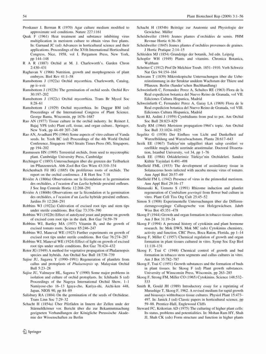
Proskauer J, Berman R (1970) Agar culture medium modified to
approximate soil conditions. Nature 227:1161
Quak F (1961) Heat treatment and substance inhibiting virus
multiplication in meristem culture to obtain virus free plants.
In: Garnaud JC (ed) Advances in horticultural science and their
applications. Proceedings of the XVth International Horticultural
Congress, Nice, 1958, vol I. Pergamon Press, New York,
pp 144–148
R A R (1887) Orchid at M. J. Charleworth’s. Garden Chron
2:430–431
Raghavan V (1966) Nutrition, growth and morphogenesis of plant
embryos. Biol Rev 41:1–58
Ramsbottom J (1922a) Orchid mycorrhiza. Charleworth, Catalog,
pp ii–xvii
Ramsbottom J (1922b) The germination of orchid seeds. Orchid Rev
30:197–202
Ramsbottom J (1922c) Orchid mycorrhiza. Trans Br Mycol Soc
8:28–61
Ramsbottom J (1929) Orchid mycorrhiza. In: Duggar BM (ed)
Proceedings of the International Congress of Plant Science.
George Banta, Wisconsin, pp 1676–1687
Rao AN (1977) Tissue culture in the orchid industry. In: Reinert J,
Bajaj YPS (eds) Plant cell, tissue, and organ culture.. Springer,
New York, pp 44–69 207–248
Rao AN, Avadhani PN (1964) Some aspects of vitro culture of Vanda
seeds. In: Yeoh BC (ed) Proceedings of the 4th World Orchid
Conference. Singapore 1963 Straits Times Press (M), Singapore,
pp 194–202
Rasmussen HN (1995) Terrestrial orchids, from seed to mycotrophic
plant. Cambridge University Press, Cambridge
Rechinger C (1893) Untersuchungen uber die grenzen der Teilbarkeit
im Pflanzenreich. Abh Zool-Bot Ges Wien 43:310–334
Reichenbach Fil HG (1885) On proliferous roots of orchids. The
report on the orchid conference. J R Hort Soc 7:18
Riviere A (1866a) Observations sur la fecondation et la germination
des orchidees, a l’ocasion d’un Laelia hybride presente enfleurs.
J Soc Imp Centrale Hortic 12:268–291
Riviere A (1866b) Observations sur la fecondation et la germination
des orchidees, a l’ocasion d’un Laelia hybride presente enfleurs.
Jardins Fr 12:268–291
Robbins WJ (1922a) Cultivation of excised root tips and stem tips
under sterile conditions. Bot Gaz 73:376–390
Robbins WJ (1922b) Effect of autolyzed yeast and peptone on growth
of excised corn root tips in the dark. Bot Gaz 74:59–79
Robbins WJ, Bartley MA (1937) Vitamin B1 and the growth of
excised tomato roots. Science 85:246–247
Robbins WJ, Maneval WE (1923) Further experiments on growth of
excised root tips under sterile conditions. Bot Gaz 76:274–287
Robbins WJ, Maneval WE (1924) Effect of light on growth of excised
root tips under sterile conditions. Bot Gaz 78:424–432
Rotor JG (1949) A method for vegetative propagation of Phalaenopsis
species and hybrids. Am Orchid Soc Bull 18:738–739
Sajise JU, Sagawa Y (1990–1991) Regeneration of plantlets from
callus and protoplasts of Phalaenopsis sp. Malaysian Orchid
Bull 5:23–28
Sajise JU, Valmayor HL, Sagawa Y (1990) Some major problems in
isolation and culture of orchid protoplasts. In: Ichihashi S (ed)
Proceedings of the Nagoya International Orchid Show, 1–1
Namiyose-cho 16–13 Igaya-cho, Kariya-shi, Aichi-ken 448,
Japan, NIOS 90, pp 84–89
Salisbury RA (1804) On the germination of the seeds of Orchideae.
Trans Linn Soc 7:29–32
Schacht H (1854a) Uber Pilzfaden in Innern der Zellen unde der
Starmehlkorner vor. Bericht uber die zur Bekantnntmachung
geeigneten Verhandlungen der Konigliche Preussische Akade-
mie der Wissenschaften zu Berlin
Schacht H (1854b) Beitrage zur Anatomie und Physiologie der
Gewachse. Muller
Scheidweiler (1844) Jeunes plantes d’orchidees de semis. PHM
Revnue Hortic 6:36–38
Scheidweiler (1845) Jeunes plantes d’orchdees provenues de graines.
J Hortic Pratique 2:14–15
Schleiden MJ (1854) Grundzuge der botanik, 3rd edn. Leipzig
Schopfer WH (1949) Plants and vitamins. Chronica Botanica,
Waltham
Schroter C (1912) Prof Dr Melchior Treub. 1851–1910. Verh Schweiz
Nat Ges 94:154–164
Schwann T (1839) Mikroskopische Untersuchungen iiber die Uebe-
reinstimmung in der Struktur unddem Wachstum der Thiere und
Pflanzen. Berlin (Sander’schen Buchhandlung)
Schweinfurth C, Fernandez Perez A, Schultes RE (1963) Flora de la
Real expedicion botanica del Nuevo Reino de Granada, vol VII.
Ediciones Cultura Hispanica, Madrid
Schweinfurth C, Fernandez Perez A, Garay LA (1969) Flora de le
Real expedicion botanica del Nuevo Reino de Granada, vol VIII.
Ediciones Cultura Hispanica, Madrid
Scott RJ, Arditti J (1959) Cymbidiums from pod to pot. Am Orchid
Soc Bull 28:823–829
Scully RM (1964) Meristem propagation-1964’s topic. Am Orchid
Soc Bull 33:1024–1025
Segelitz G (1938) Der Einfluss von Licht und Dunkelheit auf
Wurzelbildung und Wurzelwachstum. Planta 28:617–643
Sezik EE (1967) Turkiye’nin salpgilleri tikari salep cesitleri ve
ozellikle mugla salebi uzerinde arastirmalar. Doctoral Disserta-
tion, Istanbul University, vol 34, pp 1–76
Sezik EE (1984) Orkidelerimiz Turkiye’nin Orchideleri. Sandoz
Kultur Yayinlari 6:491–498
Sheffield FML (1933) The development of assimilatory tissue in
Solanaceous hosts infected with aucuba mosaic virus of tomato.
Ann Appl Biol 20:57–69
Sheffield FM L (1942) Presence of virus in the primordial meristem.
Ann Appl Biol 29:16–17
Shimasaki K, Uemoto S (1991) Rhizome induction and plantlet
regeneration of Cymbidium goeringii from flower bud culture in
vitro. Plant Cell Tiss Org Cult 25:45–52
Simon S (1908) Experimentelle Untersuchungen uber die Differen-
zierungsvorgange Callusgewebe von Holzgewachsen. Jahrb
Wiss Bot 45:351–478
Skoog F (1944) Growth and organ formation in tobacco tissue culture.
Am J Bot 31:19–24
Skoog F (1994) A personal history of cytokinin and plant hormone
research. In: Mok DWS, Mok MC (eds) Cytokinins chemistry,
activity and function. CRC Press, Boca Raton, Florida, pp 1–14
Skoog F, Miller C (1957) Chemical regulation of growth and organ
formation in plant tissues cultured in vitro. Symp Soc Exp Biol
11:118–131
Skoog F, Tsui C (1948) Chemical control of growth and bud
formation in tobacco stem segments and callus cultures in vitro.
Am J Bot 35:782–787
Skoog F, Tsui C (1951) Growth substances and the formation of buds
in plant tissues. In: Skoog F (ed) Plant growth substances.
University of Winsconsin Press, Wisconsin, pp 263–285
Skoog F, Strong FM, Miller CO (1965) Cytokinins. Science 148:532–
533
Smith R, Gould JH (1989) Introductory essay for a reprinting of
Murashige T, Skoog F, 1962. A revised medium for rapid growth
and bioassays withtobacco tissue cultures. Physiol Plant 15:473–
497. In: Janick J (ed) Classic papers in horticultural science, pp
59–88. Prentice-Hall, Englewood Cliffs
Steward FC, Krikorian AD (1975) The culturing of higher plant cells:
Its status, problems and potentialities. In: Mohan Ram HY, Shah
JJ, Shah CK (eds) Form structure and function in higher plants
54 Plant Biotechnol Rep (2009) 3:1–56
123
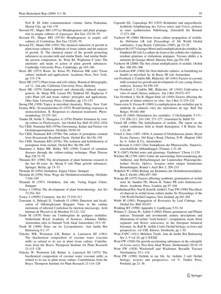
Prof B. M. Johri commemoration volume. Sartia Prakashan,
Meerut City, pp 144–170
Steward FC, Mapes MO (1971a) Morphogenesis and plant propaga-
tion in aseptic cultures of Asparagus. Bot Gaz 132:70–79
Steward FC, Mapes MO (1971b) Morphogenesis in aseptic cell
cultures of Cymbidium. Bot Gaz 132:65–70
Steward FC, Shantz EM (1955) The chemical induction of growth in
plant tissue cultures. I. Methods of tissue culture and the analysis
of growth. II. The chemical nature of the growth promoting
substances in coconut milk and similar fluids. And similar fluids:
the present composition. In: Wain RJ, Wightman F (eds) The
chemistry and mode of action of plant growth substances.
Cambridge University Press, Cambridge, pp 165–186
Street HE (1973) Roots. In: Kruse PJ, Patterson MK (eds) Tissue
culture: methods and applications. Academic Press, New York,
pp 173–178
Street HE (1977) Plant tissue and cell culture. Botanical Monographs,
vol 11, 2nd edn. California University Press, Berkeley
Street HE (1979) Embryogencsis and chemically induced organo-
genesis. In: Sharp WR, Larsen PO, Paddock EF, Raghavan V
(eds) Plant cell and tissue culture: principles and applications.
Ohio State University Press, Columbus, pp 123–153
Strong FM (1958) Topics in microbial chemistry. Wiley, New York
Swamy BGL, Sivaramakrishna D (1975) Would healing responses in
monocotyledons. II. Responses to chemical treatments. Phyto-
morphology 25:255–261
Tanaka M, Senda Y, Hasegawa A (1976) Plantlet formation by root-
tip culture in Phalaenopsis. Am Orchid Soc Bull 45:1022–1024
Teo CKH, Neumann KH (1978a) Gewinnung, Kultur and Fusion von
Orchideenprotoplasten. Orchidee 29:90–92
Teo CKH, Neumann KH (1978b) The culture of protoplasts isolated
from Renantanda Rosalind Cheok. Orchid Rev 86:156–158
Teo CKH, Neumann KH (1978c) The isolation and hybridisation of
protoplasts from orchids. Orchid Rev 86:186–189
Thammen J, Baker RR, Holley WD (1956) Control of carnation
diseases through the culture-cutting technique. Plant Disease
Rep 238:72–76
Thimann KV (1980) The development of plant hormone research in
the last 60 years. In: Skoog F (ed) Plant growth substances.
Springer, Berlin, pp 15–33
Thomale H (1954) Orchideen. Eugen Ulmer, Stuttgart
Thomale H (1956) Neue Wege der Orchideenvermehrung. Orchidee
7:104–105
Thomale H (1957) Orchideen, 2nd edn. Verlag Eugen Ulmer,
Stuttgart
Torrey J (1985a) The development of plant biotechnology. Am Sci
73:354–363
Torrey J (1985b) Comment. Am Sci 73:510–511
Toussaint A, Dekegel D, Vanheule G (1984) Detection and locali-
zation of Odontoglossum Ringspot Virus in the cauline
meristems of infected Cymbidium by electron microscopy. Arch
Internat de Physiol et de Biochim 91:121–122
Treub M (1879) Notes sur l’embrogenie de quelques orchidees.
Netherlands Royal Academy of Sciences, Johannes Muller,
Amsterdam (also in Natuurk Verh Akad Amsterdam) 19:1–50
Treub M (1890) Etues sur les Lycopodiacees. Ann Jardin Bot
Buitenzorg 8:1–37
Tulecke WR, Weinstein LH, Rutner A, Laurencot HJ (1961)
The biochemical composition of coconut water (coconut
milk) as related to its use in plant tissue culture. Contribu-
tions from the Boyce Thompson Institute for Plant Research
21:115–128
Tulecke W, Weinstein LH, Rutner A, Laurencot HJ (1975) The
biochemical composition of coconut water (coconut milk) as
related to its use in plant tissue culture. Contributions from the
Boyce Thompson Institute for Plant Research 21:114–128
Uspenski EE, Uspenskaja WJ (1925) Reinkultur und ungeschlesch-
lechtliche fortpflanzung des Volvox minor und Volvox globatorin einer synthetischen Nahrlosung. Zeitschrift fur Botanik
17:273–308
Vacherot M (1966) Meristem tissue culture propagation of orchids.
In: DeGarmo LR (ed) Proceedings of 5th World Orchid
conference. Long Beach, California (1985), pp 23–25
Vacherot M (1977) Georges Morel and la multiplication des orchdees. In:
Gautheret RJ (ed) La culture de tissus et des cellules des vegetaux,
resultats generaux et realisations pratiques. Travaux dedies a la
memoire de Georges Morel, Masson, Paris, pp 254–258
Vacherot M (2000) The first clonal multiplication of orchids. Orchid
Rev 108:305–306
van Gorkom KW (1884) De Oost-Indische cultures, in betrekking tot
handel en nijverhed. In: de Bussy JH (ed) Amsterdam
van Overbeek J, Conklin ME, Blakeslee AF (1941) Factors in coconut
milk essential for growth and development of very young Daturaembryos. Science 94:350–351
van Overbeek J, Conklin ME, Blakeslee AF (1942) Cultivation in
vitro of small Datura embryos. Am J Bot 29:472–477
Van Overbeek J, Siu R, Hagen-Smit AJ (1944) Factors affecting the
growth of datura embryos in vitro. Am J Bot 31:219–224
Vanseveren N, Freson R (1969) La multiplication des orchidees par la
methode de culturein vitro des meristemes de tige. Bull Nat
Belges 50:444–460
Veitch H (1885) Hybridation des orchidees. L’Orchidophile 5:171–
174 208–213, 243–249, 271–277, (translated by Ballif O)
Veitch HJ (1886) The hybridization of orchids. The report on the
orchid conference held at South Kensignton. J R Hortic Soc
1:22–49
Veitch J, Sons (1887–1894) A manual of Orchidaceous plants. Royal
Exotic Nursery, 544 King’s Road, Chelsea, N.W. (Reprint 1963
by A. Asher & Co, Amsterdam)
von Reissek S (1847) Uber Endophyten der Pflanzenzelle. Naturwis-
senschaftliche Abhandlungen (Vienna) 1:31–46
W S (1887) Orchid notes and gleanings. Gardeners’ Chron 11:129
Wachter JK (1799–1801) Ueber die merwurdige Orstveranderung der
Anthered, und Befruchtungsart der Linneischen Pfanzengeshc-
lechter Orchis, Ophrys, Serapias nebst einiger botanischen
Bemerkungen. Romer’s Arch Bot 2: 209–215
Wahrlich W (1886) Beitrag zur Kenntniss der Orchideenwurzelpilze.
Bot Z 44(481–490):497–505
Warcup JH (1975) Factors affecting symbiotic germination of orchid
seed. In: Sanders FE, Moose B, Tinker PB (eds) Endomycror-
rhizas. Academic Press, London, pp 87–104
Weatherhead MA, Nair H, Ernst R, Arditti J, Yam TW (1990) The effects
of charcoal in orchid tissue culture media. In: Proceedings of the
13th World Orchid Congress, New Zealand, pp 263–265
Webb M (1981) Propagation of Restrepias by Leaf Cuttings. Am
Orchid Soc Bull 50:419
Wedding RT (1990) Appendix I. Lindleyana 5:53–54
Wehner U, Zierau W, Arditti J (2002) Plinius germanicus and Plinius
indicus: Sixteenth and seventeenth century descriptions and
illustrations of orchid ‘‘trash baskets’’, resupination, seeds, floral
segments and flower senescence in the European botanical
literature. In: Kull K, Arditti J (eds) Orchid biology, reviews and
perspectives, vol VIII. Kluwer, Dordrecht, pp 1–81
Went FAFC (1911) Melchior Treub. Ann du Jardin Bot Buitenzorg
Ser 2, 9 vol 24, pp I–XXXII
Went FW (1926) On growth-accelerating substances in the coleoptile
of Avena sativa. Proc Kon Akad Weten, (Netherlands) 30:10–19
Went FW (1928) Wuchsstoff und Wachstum. Recuelle Travaux
Neerlandaise 25:1–116
Went FW (1990) Orchids in my life. In: Ardittie J (ed) Orchid
biology: reviews and perspectives, vol V. Timber Press,
Portland, pp 21–36
Plant Biotechnol Rep (2009) 3:1–56 55
123

Went FW, Thimann KV (1937) Phytohormones. Macmillan, New
York
Werkmeister P (1970a) Uber die Lichtinduktion der geotropen
Orientirung von Luft—und Bodenwurzeln in Gewebekulturen
von Cymbidium. Ber Dtsch Bot Ges 83:19–26
Werkmeister P (1970b) Die Steuerung von Vermehrung (Prolifera-
tion) and Wachstum in der Meristemkultur von Cymbidiumund die Verwendung eines Kohlen-Nahrmediums. Orchidee
21:126–131
Werkmeister P (1971) Light induction of geotropism and the control
of proliferation and growth of Cymbidium in tissue culture. Bot
Gaz 132:346–350
White PR (1931) Plant tissue cultures. The history and present status of
the problem. Arkiv fur Experimentelle Zellforschung 10:501–518
White PR (1932a) Influence of some environmental conditions on the
growth of excised root tips of wheat in liquid media. Plant
Physiol 7:613–628
White PR (1932b) Plant tissue cultures. A preliminary report of
results obtained in the culturing of certain plant meristems. Arkiv
fur Experimentelle Zellforschung 10:501–518
White PR (1933a) Concentration of inorganic ions as related to
growth of excised root-tips of wheat seedlings. Plant Physiol
12:602–620
White PR (1933b) Results of preliminary experiments on the
culturing of isolated stem tips of Stellaria media. Protoplasma
19:97–116
White PR (1934a) Potentially unlimited growth of excised tomato
root tips in a liquid medium. Plant Physiol 9:585–600
White PR (1934b) Multiplication of viruses of tobacco and Aucuba
mosaics in growing excised tomato root tips. Phytopathology
24:1003–1011
White PR (1937) Vitamin Bl in the nutrition of excised tomato roots.
Plant Physiol 12:803–811
White PR (1943) A handbook of plant tissue culture. Jacques Cattrell
Press, Lancaster
Wilson G (1906) Fungus co-operation in orchid roots. Orchid Rev
14:154–155
Wimber DE (1963) Clonal multiplication of cymbidiums through
tissue culture of the shootmeristem. Am Orchid Soc Bull
32:105–107
Wimber DE (1965) Additional observations on clonal multiplication
of cymbidiums through cultureof shoot meristems. Cym Soc
News 20:7–10
Winkler H (1908) Besprechung der Arbeit G. Haberlandt’s Kultur
Versuche mit isolierten Pflanzenzellen. Bot Z 60:262–264
Withner CL (1955) Ovule culture and growth of Vanilla seedlings.
Am Orchid Soc Bull 24:380–382
Withner CL (1959a) The orchids, a scientific survey. Ronald Press,
New York
Withner CL (1959b) Orchid culture media and nutrient solutions. In:
Withner CL (ed) The orchids, a scientific survey. Ronald Press,
New York, pp 589–599
Withner CL (1974) The orchids, scientific studies. Wiley, New York
Witt ON (1913) Die Ausstellung der Orchideen-Sektion am 14, 15.
und 16 November 1913. Orchis 7:116–129
Wrigley OO (1895) Dendrobium seedlings at Bury. Orchid Rev 3:176
Yam TW (1995) Orchids of the Singapore Botanic Gardens, 1st edn.
National Parks Board, Singapore
Yam TW (2007) Orchids of the Singapore Botanic Gardens, 3rd edn.
National Parks Board, Singapore
Yam TW, Arditti J (1990) Orchid seed germination and microprop-
agation: a short history. Malayan Orchid Rev (Singapore) 24:
38–47
Yam TW, Arditti J (2007) Milestones in orchid germination and
micropropagation. Orchid Rev 115:334–339
Yam TW, Nair H, Hew CS, Arditti J (2002) Orchid seeds and their
germination: An historical account. In: Kull T, Arditti J (eds)
Orchid biology: reviews and perspectives, vol VIII. Kluwer,
Dordrecht, pp 387–504
Yam TW, Ghani AKA, Ichihashi S, Thame A, Rao AN, Avadhani
PN, Nair H, Hew CS, Arditti J, Tatarenko V (2007) Time from
pollination to fruit ripening, seed maturation, and germination.
In: Cameron KM, Arditti J, Kull T (eds) Orchid biology reviews
and perspectives, vol IX. The New York Botanical Garden Press,
New York, pp 433–513
Yam TW, Chow YN, Avadhani PN, Hew CS, Arditti J, Kurzweil H
(2009) Pollination Effects on Orchid Flowers and the First
Suggestion by Professor Hans Fitting (1877–1970) that Plants
Produce Hormones. In: Kull T, Arditti J, Wong S (eds) Orchid
biology: reviews and perspectives, vol X. Kluwer, Dordrecht
Young R (1893) Seedling orchids. Orchid Rev 1:275
Zimmer K (1978a) Die labormassige vegetative Vermehrung der
Orchideen. In: Briger FG, Maatsch R, Senghas K (eds) Rudolf
Schlechter’s Orchideen, vol 3, 3rd edn. Paul Parey, Berlin,
pp 158–159
Zimmer K (1978b) Zur vegetativen Vermehrung von Orchideen.
Dtsch Gartenbau 34:330–334
Zimmerman RH (1996) Commercial applications of tissue culture to
horticultural crops in the United States. J Korean Soc Hortic Sci
37:486–490
56 Plant Biotechnol Rep (2009) 3:1–56
123